EP1491631B1 - Methods of screening for transporter inhibitors - Google Patents
Methods of screening for transporter inhibitors Download PDFInfo
- Publication number
- EP1491631B1 EP1491631B1 EP03715597.5A EP03715597A EP1491631B1 EP 1491631 B1 EP1491631 B1 EP 1491631B1 EP 03715597 A EP03715597 A EP 03715597A EP 1491631 B1 EP1491631 B1 EP 1491631B1
- Authority
- EP
- European Patent Office
- Prior art keywords
- transporter
- virus
- channel
- activity
- transporters
- Prior art date
- Legal status (The legal status is an assumption and is not a legal conclusion. Google has not performed a legal analysis and makes no representation as to the accuracy of the status listed.)
- Expired - Lifetime
Links
- 238000000034 method Methods 0.000 title claims description 108
- 238000012216 screening Methods 0.000 title claims description 14
- 239000003112 inhibitor Substances 0.000 title description 6
- 108010078791 Carrier Proteins Proteins 0.000 claims description 173
- 241000700605 Viruses Species 0.000 claims description 143
- 230000000694 effects Effects 0.000 claims description 53
- 101000836291 Homo sapiens Solute carrier organic anion transporter family member 1B1 Proteins 0.000 claims description 42
- 102100027233 Solute carrier organic anion transporter family member 1B1 Human genes 0.000 claims description 41
- 108090000623 proteins and genes Proteins 0.000 claims description 41
- 210000004779 membrane envelope Anatomy 0.000 claims description 35
- 241000701447 unidentified baculovirus Species 0.000 claims description 34
- 102000046014 Peptide Transporter 1 Human genes 0.000 claims description 33
- 108091006594 SLC15A1 Proteins 0.000 claims description 33
- 230000034303 cell budding Effects 0.000 claims description 32
- 102000040811 transporter activity Human genes 0.000 claims description 32
- 108091092194 transporter activity Proteins 0.000 claims description 32
- 108010003533 Viral Envelope Proteins Proteins 0.000 claims description 27
- 239000000126 substance Substances 0.000 claims description 27
- 101100243399 Caenorhabditis elegans pept-2 gene Proteins 0.000 claims description 25
- 230000003612 virological effect Effects 0.000 claims description 23
- 102000004169 proteins and genes Human genes 0.000 claims description 20
- 238000006243 chemical reaction Methods 0.000 claims description 18
- 108010082406 peptide permease Proteins 0.000 claims description 18
- 238000012360 testing method Methods 0.000 claims description 10
- 238000012258 culturing Methods 0.000 claims description 8
- 108010089503 Organic Anion Transporters Proteins 0.000 claims description 6
- 102000007990 Organic Anion Transporters Human genes 0.000 claims description 6
- 101710091045 Envelope protein Proteins 0.000 claims description 4
- 101710188315 Protein X Proteins 0.000 claims description 4
- 230000003100 immobilizing effect Effects 0.000 claims description 2
- 102100021696 Syncytin-1 Human genes 0.000 claims 1
- 210000004027 cell Anatomy 0.000 description 58
- 239000000758 substrate Substances 0.000 description 52
- 239000000243 solution Substances 0.000 description 41
- 230000032258 transport Effects 0.000 description 27
- 239000011734 sodium Substances 0.000 description 26
- 238000009792 diffusion process Methods 0.000 description 22
- 239000000427 antigen Substances 0.000 description 19
- 108091007433 antigens Proteins 0.000 description 19
- 102000036639 antigens Human genes 0.000 description 19
- 235000018102 proteins Nutrition 0.000 description 18
- 108091006146 Channels Proteins 0.000 description 16
- 150000002891 organic anions Chemical class 0.000 description 16
- 239000012528 membrane Substances 0.000 description 15
- 150000001413 amino acids Chemical class 0.000 description 14
- 239000013598 vector Substances 0.000 description 14
- 108020004414 DNA Proteins 0.000 description 13
- 230000014509 gene expression Effects 0.000 description 13
- 230000009056 active transport Effects 0.000 description 12
- 210000004408 hybridoma Anatomy 0.000 description 12
- 238000010791 quenching Methods 0.000 description 12
- 230000000171 quenching effect Effects 0.000 description 12
- ZLMJMSJWJFRBEC-UHFFFAOYSA-N Potassium Chemical compound [K] ZLMJMSJWJFRBEC-UHFFFAOYSA-N 0.000 description 11
- 239000007788 liquid Substances 0.000 description 11
- 239000011591 potassium Substances 0.000 description 11
- 229910052700 potassium Inorganic materials 0.000 description 11
- 206010028980 Neoplasm Diseases 0.000 description 9
- 201000011510 cancer Diseases 0.000 description 9
- 238000002474 experimental method Methods 0.000 description 9
- 238000002965 ELISA Methods 0.000 description 8
- 235000001014 amino acid Nutrition 0.000 description 8
- 229940024606 amino acid Drugs 0.000 description 8
- 108090000765 processed proteins & peptides Proteins 0.000 description 8
- WQZGKKKJIJFFOK-GASJEMHNSA-N Glucose Natural products OC[C@H]1OC(O)[C@H](O)[C@@H](O)[C@@H]1O WQZGKKKJIJFFOK-GASJEMHNSA-N 0.000 description 7
- 239000012981 Hank's balanced salt solution Substances 0.000 description 7
- 241000124008 Mammalia Species 0.000 description 7
- 230000027455 binding Effects 0.000 description 7
- 150000001875 compounds Chemical class 0.000 description 7
- 239000012228 culture supernatant Substances 0.000 description 7
- 239000008103 glucose Substances 0.000 description 7
- 210000004379 membrane Anatomy 0.000 description 7
- 102000004196 processed proteins & peptides Human genes 0.000 description 7
- 102000004190 Enzymes Human genes 0.000 description 6
- 108090000790 Enzymes Proteins 0.000 description 6
- 241000238631 Hexapoda Species 0.000 description 6
- 101000821899 Homo sapiens Solute carrier family 15 member 1 Proteins 0.000 description 6
- 229920002678 cellulose Polymers 0.000 description 6
- 239000001913 cellulose Substances 0.000 description 6
- 238000004587 chromatography analysis Methods 0.000 description 6
- 229940088598 enzyme Drugs 0.000 description 6
- 108010004620 glycylsarcosine Proteins 0.000 description 6
- VYAMLSCELQQRAE-UHFFFAOYSA-N glycylsarcosine zwitterion Chemical compound OC(=O)CN(C)C(=O)CN VYAMLSCELQQRAE-UHFFFAOYSA-N 0.000 description 6
- 230000002401 inhibitory effect Effects 0.000 description 6
- 210000004962 mammalian cell Anatomy 0.000 description 6
- 229920001184 polypeptide Polymers 0.000 description 6
- 230000001235 sensitizing effect Effects 0.000 description 6
- 206010035226 Plasma cell myeloma Diseases 0.000 description 5
- 108010037150 Transient Receptor Potential Channels Proteins 0.000 description 5
- 102000011753 Transient Receptor Potential Channels Human genes 0.000 description 5
- 230000007910 cell fusion Effects 0.000 description 5
- 210000000170 cell membrane Anatomy 0.000 description 5
- 239000002299 complementary DNA Substances 0.000 description 5
- 230000001419 dependent effect Effects 0.000 description 5
- LOKCTEFSRHRXRJ-UHFFFAOYSA-I dipotassium trisodium dihydrogen phosphate hydrogen phosphate dichloride Chemical compound P(=O)(O)(O)[O-].[K+].P(=O)(O)([O-])[O-].[Na+].[Na+].[Cl-].[K+].[Cl-].[Na+] LOKCTEFSRHRXRJ-UHFFFAOYSA-I 0.000 description 5
- 229940079593 drug Drugs 0.000 description 5
- 239000003814 drug Substances 0.000 description 5
- 238000010230 functional analysis Methods 0.000 description 5
- 102000054502 human SLC15A1 Human genes 0.000 description 5
- 238000012933 kinetic analysis Methods 0.000 description 5
- 210000004698 lymphocyte Anatomy 0.000 description 5
- 239000002609 medium Substances 0.000 description 5
- 201000000050 myeloid neoplasm Diseases 0.000 description 5
- 239000002777 nucleoside Substances 0.000 description 5
- 150000003833 nucleoside derivatives Chemical class 0.000 description 5
- 150000002892 organic cations Chemical class 0.000 description 5
- 239000002953 phosphate buffered saline Substances 0.000 description 5
- 210000002966 serum Anatomy 0.000 description 5
- 238000001179 sorption measurement Methods 0.000 description 5
- 238000012546 transfer Methods 0.000 description 5
- 108091005462 Cation channels Proteins 0.000 description 4
- 241000282693 Cercopithecidae Species 0.000 description 4
- 101150093369 His2A gene Proteins 0.000 description 4
- 108010021625 Immunoglobulin Fragments Proteins 0.000 description 4
- 102000008394 Immunoglobulin Fragments Human genes 0.000 description 4
- 241001465754 Metazoa Species 0.000 description 4
- 241000269370 Xenopus <genus> Species 0.000 description 4
- 238000001042 affinity chromatography Methods 0.000 description 4
- 239000000969 carrier Substances 0.000 description 4
- 201000010099 disease Diseases 0.000 description 4
- 208000037265 diseases, disorders, signs and symptoms Diseases 0.000 description 4
- JKKFKPJIXZFSSB-CBZIJGRNSA-N estrone 3-sulfate Chemical compound OS(=O)(=O)OC1=CC=C2[C@H]3CC[C@](C)(C(CC4)=O)[C@@H]4[C@@H]3CCC2=C1 JKKFKPJIXZFSSB-CBZIJGRNSA-N 0.000 description 4
- 229950008385 estrone sulphate Drugs 0.000 description 4
- 230000003053 immunization Effects 0.000 description 4
- 210000000287 oocyte Anatomy 0.000 description 4
- 239000008177 pharmaceutical agent Substances 0.000 description 4
- 238000002360 preparation method Methods 0.000 description 4
- 238000000746 purification Methods 0.000 description 4
- 102000003922 Calcium Channels Human genes 0.000 description 3
- 108090000312 Calcium Channels Proteins 0.000 description 3
- 102100027723 Endogenous retrovirus group K member 6 Rec protein Human genes 0.000 description 3
- 102000004310 Ion Channels Human genes 0.000 description 3
- 108090000862 Ion Channels Proteins 0.000 description 3
- 241000699670 Mus sp. Species 0.000 description 3
- 239000002671 adjuvant Substances 0.000 description 3
- 210000000628 antibody-producing cell Anatomy 0.000 description 3
- 210000004748 cultured cell Anatomy 0.000 description 3
- 238000003372 electrophysiological method Methods 0.000 description 3
- 239000000284 extract Substances 0.000 description 3
- 150000004676 glycans Chemical class 0.000 description 3
- 239000001963 growth medium Substances 0.000 description 3
- 238000004128 high performance liquid chromatography Methods 0.000 description 3
- 230000005764 inhibitory process Effects 0.000 description 3
- 238000004255 ion exchange chromatography Methods 0.000 description 3
- 230000001537 neural effect Effects 0.000 description 3
- 230000007935 neutral effect Effects 0.000 description 3
- 239000002773 nucleotide Substances 0.000 description 3
- 125000003729 nucleotide group Chemical group 0.000 description 3
- 230000035790 physiological processes and functions Effects 0.000 description 3
- 229920001282 polysaccharide Polymers 0.000 description 3
- 239000005017 polysaccharide Substances 0.000 description 3
- 230000002285 radioactive effect Effects 0.000 description 3
- 239000000941 radioactive substance Substances 0.000 description 3
- 239000008279 sol Substances 0.000 description 3
- 239000006228 supernatant Substances 0.000 description 3
- 238000002560 therapeutic procedure Methods 0.000 description 3
- 238000001262 western blot Methods 0.000 description 3
- BFSVOASYOCHEOV-UHFFFAOYSA-N 2-diethylaminoethanol Chemical compound CCN(CC)CCO BFSVOASYOCHEOV-UHFFFAOYSA-N 0.000 description 2
- 102100033350 ATP-dependent translocase ABCB1 Human genes 0.000 description 2
- 102100021178 ATP-sensitive inward rectifier potassium channel 12 Human genes 0.000 description 2
- 102100022094 Acid-sensing ion channel 2 Human genes 0.000 description 2
- IJGRMHOSHXDMSA-UHFFFAOYSA-N Atomic nitrogen Chemical compound N#N IJGRMHOSHXDMSA-UHFFFAOYSA-N 0.000 description 2
- 101100189913 Caenorhabditis elegans pept-1 gene Proteins 0.000 description 2
- 108010062745 Chloride Channels Proteins 0.000 description 2
- 102000011045 Chloride Channels Human genes 0.000 description 2
- 102100023457 Chloride channel protein 1 Human genes 0.000 description 2
- 108010047041 Complementarity Determining Regions Proteins 0.000 description 2
- 108020004635 Complementary DNA Proteins 0.000 description 2
- 102000003688 G-Protein-Coupled Receptors Human genes 0.000 description 2
- 108090000045 G-Protein-Coupled Receptors Proteins 0.000 description 2
- 102100034471 H(+)/Cl(-) exchange transporter 5 Human genes 0.000 description 2
- 101000614708 Homo sapiens ATP-sensitive inward rectifier potassium channel 12 Proteins 0.000 description 2
- 101000906651 Homo sapiens Chloride channel protein 1 Proteins 0.000 description 2
- 101000577126 Homo sapiens Monocarboxylate transporter 4 Proteins 0.000 description 2
- 101000577129 Homo sapiens Monocarboxylate transporter 5 Proteins 0.000 description 2
- 101000577131 Homo sapiens Monocarboxylate transporter 6 Proteins 0.000 description 2
- 101001049841 Homo sapiens Potassium channel subfamily K member 1 Proteins 0.000 description 2
- 101000974739 Homo sapiens Potassium channel subfamily K member 16 Proteins 0.000 description 2
- 101000974732 Homo sapiens Potassium channel subfamily K member 17 Proteins 0.000 description 2
- 101001049829 Homo sapiens Potassium channel subfamily K member 5 Proteins 0.000 description 2
- 101001049828 Homo sapiens Potassium channel subfamily K member 6 Proteins 0.000 description 2
- 101001050878 Homo sapiens Potassium channel subfamily K member 9 Proteins 0.000 description 2
- 101001047093 Homo sapiens Potassium voltage-gated channel subfamily H member 1 Proteins 0.000 description 2
- 101000640020 Homo sapiens Sodium channel protein type 11 subunit alpha Proteins 0.000 description 2
- 101000908580 Homo sapiens Spliceosome RNA helicase DDX39B Proteins 0.000 description 2
- 241000283953 Lagomorpha Species 0.000 description 2
- 102100024032 Linker for activation of T-cells family member 1 Human genes 0.000 description 2
- 241000829100 Macaca mulatta polyomavirus 1 Species 0.000 description 2
- 108010047230 Member 1 Subfamily B ATP Binding Cassette Transporter Proteins 0.000 description 2
- 102100025273 Monocarboxylate transporter 5 Human genes 0.000 description 2
- 241000699666 Mus <mouse, genus> Species 0.000 description 2
- 108010067902 Peptide Library Proteins 0.000 description 2
- 102100023242 Potassium channel subfamily K member 1 Human genes 0.000 description 2
- 102100022796 Potassium channel subfamily K member 16 Human genes 0.000 description 2
- 102100023204 Potassium channel subfamily K member 2 Human genes 0.000 description 2
- 102100023202 Potassium channel subfamily K member 5 Human genes 0.000 description 2
- 102100023203 Potassium channel subfamily K member 6 Human genes 0.000 description 2
- 102100024986 Potassium channel subfamily K member 9 Human genes 0.000 description 2
- 102100034355 Potassium voltage-gated channel subfamily A member 3 Human genes 0.000 description 2
- 102100022810 Potassium voltage-gated channel subfamily H member 1 Human genes 0.000 description 2
- 241000288906 Primates Species 0.000 description 2
- 241000283984 Rodentia Species 0.000 description 2
- 229920002684 Sepharose Polymers 0.000 description 2
- 102100033974 Sodium channel protein type 11 subunit alpha Human genes 0.000 description 2
- 102100024690 Spliceosome RNA helicase DDX39B Human genes 0.000 description 2
- IQFYYKKMVGJFEH-XLPZGREQSA-N Thymidine Chemical compound O=C1NC(=O)C(C)=CN1[C@@H]1O[C@H](CO)[C@@H](O)C1 IQFYYKKMVGJFEH-XLPZGREQSA-N 0.000 description 2
- 101710120037 Toxin CcdB Proteins 0.000 description 2
- 102100029611 Transient receptor potential cation channel subfamily V member 4 Human genes 0.000 description 2
- XSQUKJJJFZCRTK-UHFFFAOYSA-N Urea Chemical compound NC(N)=O XSQUKJJJFZCRTK-UHFFFAOYSA-N 0.000 description 2
- 108020005202 Viral DNA Proteins 0.000 description 2
- 108010022133 Voltage-Dependent Anion Channel 1 Proteins 0.000 description 2
- 102100037820 Voltage-dependent anion-selective channel protein 1 Human genes 0.000 description 2
- 238000002835 absorbance Methods 0.000 description 2
- 238000004458 analytical method Methods 0.000 description 2
- 238000009175 antibody therapy Methods 0.000 description 2
- 210000004369 blood Anatomy 0.000 description 2
- 239000008280 blood Substances 0.000 description 2
- 239000000872 buffer Substances 0.000 description 2
- 230000001413 cellular effect Effects 0.000 description 2
- 238000010367 cloning Methods 0.000 description 2
- 238000010276 construction Methods 0.000 description 2
- OVBPIULPVIDEAO-LBPRGKRZSA-N folic acid Chemical compound C=1N=C2NC(N)=NC(=O)C2=NC=1CNC1=CC=C(C(=O)N[C@@H](CCC(O)=O)C(O)=O)C=C1 OVBPIULPVIDEAO-LBPRGKRZSA-N 0.000 description 2
- 239000012634 fragment Substances 0.000 description 2
- 230000004927 fusion Effects 0.000 description 2
- 229960003692 gamma aminobutyric acid Drugs 0.000 description 2
- BTCSSZJGUNDROE-UHFFFAOYSA-N gamma-aminobutyric acid Chemical compound NCCCC(O)=O BTCSSZJGUNDROE-UHFFFAOYSA-N 0.000 description 2
- 238000002744 homologous recombination Methods 0.000 description 2
- 230000006801 homologous recombination Effects 0.000 description 2
- FDGQSTZJBFJUBT-UHFFFAOYSA-N hypoxanthine Chemical compound O=C1NC=NC2=C1NC=N2 FDGQSTZJBFJUBT-UHFFFAOYSA-N 0.000 description 2
- 230000002163 immunogen Effects 0.000 description 2
- 238000000338 in vitro Methods 0.000 description 2
- 239000007928 intraperitoneal injection Substances 0.000 description 2
- 210000003734 kidney Anatomy 0.000 description 2
- 150000002605 large molecules Chemical class 0.000 description 2
- 238000012423 maintenance Methods 0.000 description 2
- 239000000463 material Substances 0.000 description 2
- 238000005259 measurement Methods 0.000 description 2
- 238000002703 mutagenesis Methods 0.000 description 2
- 231100000350 mutagenesis Toxicity 0.000 description 2
- 230000035772 mutation Effects 0.000 description 2
- 239000002245 particle Substances 0.000 description 2
- 101150016475 pept-2 gene Proteins 0.000 description 2
- 229920001223 polyethylene glycol Polymers 0.000 description 2
- -1 polypropylenes Polymers 0.000 description 2
- 108020001213 potassium channel Proteins 0.000 description 2
- 239000002244 precipitate Substances 0.000 description 2
- 238000001556 precipitation Methods 0.000 description 2
- 239000000047 product Substances 0.000 description 2
- 238000003127 radioimmunoassay Methods 0.000 description 2
- 230000001105 regulatory effect Effects 0.000 description 2
- 229920005989 resin Polymers 0.000 description 2
- 239000011347 resin Substances 0.000 description 2
- 230000035945 sensitivity Effects 0.000 description 2
- 238000000926 separation method Methods 0.000 description 2
- UCSJYZPVAKXKNQ-HZYVHMACSA-N streptomycin Chemical compound CN[C@H]1[C@H](O)[C@@H](O)[C@H](CO)O[C@H]1O[C@@H]1[C@](C=O)(O)[C@H](C)O[C@H]1O[C@@H]1[C@@H](NC(N)=N)[C@H](O)[C@@H](NC(N)=N)[C@H](O)[C@H]1O UCSJYZPVAKXKNQ-HZYVHMACSA-N 0.000 description 2
- 238000007920 subcutaneous administration Methods 0.000 description 2
- 239000007929 subcutaneous injection Substances 0.000 description 2
- 235000000346 sugar Nutrition 0.000 description 2
- 239000000725 suspension Substances 0.000 description 2
- 238000001890 transfection Methods 0.000 description 2
- 238000013519 translation Methods 0.000 description 2
- 241000701161 unidentified adenovirus Species 0.000 description 2
- HSINOMROUCMIEA-FGVHQWLLSA-N (2s,4r)-4-[(3r,5s,6r,7r,8s,9s,10s,13r,14s,17r)-6-ethyl-3,7-dihydroxy-10,13-dimethyl-2,3,4,5,6,7,8,9,11,12,14,15,16,17-tetradecahydro-1h-cyclopenta[a]phenanthren-17-yl]-2-methylpentanoic acid Chemical compound C([C@@]12C)C[C@@H](O)C[C@H]1[C@@H](CC)[C@@H](O)[C@@H]1[C@@H]2CC[C@]2(C)[C@@H]([C@H](C)C[C@H](C)C(O)=O)CC[C@H]21 HSINOMROUCMIEA-FGVHQWLLSA-N 0.000 description 1
- XZKIHKMTEMTJQX-UHFFFAOYSA-N 4-Nitrophenyl Phosphate Chemical compound OP(O)(=O)OC1=CC=C([N+]([O-])=O)C=C1 XZKIHKMTEMTJQX-UHFFFAOYSA-N 0.000 description 1
- TVZGACDUOSZQKY-LBPRGKRZSA-N 4-aminofolic acid Chemical compound C1=NC2=NC(N)=NC(N)=C2N=C1CNC1=CC=C(C(=O)N[C@@H](CCC(O)=O)C(O)=O)C=C1 TVZGACDUOSZQKY-LBPRGKRZSA-N 0.000 description 1
- 102100033400 4F2 cell-surface antigen heavy chain Human genes 0.000 description 1
- 102100028162 ATP-binding cassette sub-family C member 3 Human genes 0.000 description 1
- 102100028163 ATP-binding cassette sub-family C member 4 Human genes 0.000 description 1
- 102100028186 ATP-binding cassette sub-family C member 5 Human genes 0.000 description 1
- 102100028187 ATP-binding cassette sub-family C member 6 Human genes 0.000 description 1
- 102100033056 ATP-sensitive inward rectifier potassium channel 1 Human genes 0.000 description 1
- 102100021176 ATP-sensitive inward rectifier potassium channel 10 Human genes 0.000 description 1
- 102100021177 ATP-sensitive inward rectifier potassium channel 11 Human genes 0.000 description 1
- 102100022781 ATP-sensitive inward rectifier potassium channel 15 Human genes 0.000 description 1
- 102100021240 ATP-sensitive inward rectifier potassium channel 8 Human genes 0.000 description 1
- 108010068806 Acid Sensing Ion Channels Proteins 0.000 description 1
- 102000001671 Acid Sensing Ion Channels Human genes 0.000 description 1
- 102100021624 Acid-sensing ion channel 1 Human genes 0.000 description 1
- 102100022097 Acid-sensing ion channel 3 Human genes 0.000 description 1
- 102100022099 Acid-sensing ion channel 4 Human genes 0.000 description 1
- 101710099897 Acid-sensing ion channel 4 Proteins 0.000 description 1
- 229920000936 Agarose Polymers 0.000 description 1
- 102000002260 Alkaline Phosphatase Human genes 0.000 description 1
- 108020004774 Alkaline Phosphatase Proteins 0.000 description 1
- 102100037242 Amiloride-sensitive sodium channel subunit alpha Human genes 0.000 description 1
- 102100037232 Amiloride-sensitive sodium channel subunit beta Human genes 0.000 description 1
- 102100022531 Amiloride-sensitive sodium channel subunit delta Human genes 0.000 description 1
- 102100022534 Amiloride-sensitive sodium channel subunit gamma Human genes 0.000 description 1
- 108050005273 Amino acid transporters Proteins 0.000 description 1
- 102000034263 Amino acid transporters Human genes 0.000 description 1
- 102000037829 Anion channels Human genes 0.000 description 1
- 108091006515 Anion channels Proteins 0.000 description 1
- 102000004888 Aquaporin 1 Human genes 0.000 description 1
- 108090001004 Aquaporin 1 Proteins 0.000 description 1
- 206010003445 Ascites Diseases 0.000 description 1
- CIWBSHSKHKDKBQ-JLAZNSOCSA-N Ascorbic acid Natural products OC[C@H](O)[C@H]1OC(=O)C(O)=C1O CIWBSHSKHKDKBQ-JLAZNSOCSA-N 0.000 description 1
- DCXYFEDJOCDNAF-UHFFFAOYSA-N Asparagine Natural products OC(=O)C(N)CC(N)=O DCXYFEDJOCDNAF-UHFFFAOYSA-N 0.000 description 1
- 108090001008 Avidin Proteins 0.000 description 1
- DWRXFEITVBNRMK-UHFFFAOYSA-N Beta-D-1-Arabinofuranosylthymine Natural products O=C1NC(=O)C(C)=CN1C1C(O)C(O)C(CO)O1 DWRXFEITVBNRMK-UHFFFAOYSA-N 0.000 description 1
- 102100028282 Bile salt export pump Human genes 0.000 description 1
- 101000650779 Boana raniceps Raniseptin-5 Proteins 0.000 description 1
- 108091003079 Bovine Serum Albumin Proteins 0.000 description 1
- 102100022595 Broad substrate specificity ATP-binding cassette transporter ABCG2 Human genes 0.000 description 1
- 101100000858 Caenorhabditis elegans act-3 gene Proteins 0.000 description 1
- OYPRJOBELJOOCE-UHFFFAOYSA-N Calcium Chemical compound [Ca] OYPRJOBELJOOCE-UHFFFAOYSA-N 0.000 description 1
- 102100039536 Calcium-activated chloride channel regulator 1 Human genes 0.000 description 1
- 102100039532 Calcium-activated chloride channel regulator 2 Human genes 0.000 description 1
- 102100039534 Calcium-activated chloride channel regulator 4 Human genes 0.000 description 1
- 102100039535 Calcium-activated chloride channel regulator family member 3 Human genes 0.000 description 1
- 102100023073 Calcium-activated potassium channel subunit alpha-1 Human genes 0.000 description 1
- 102100023074 Calcium-activated potassium channel subunit beta-1 Human genes 0.000 description 1
- 102100023243 Calcium-activated potassium channel subunit beta-3 Human genes 0.000 description 1
- 102100023241 Calcium-activated potassium channel subunit beta-4 Human genes 0.000 description 1
- OKTJSMMVPCPJKN-UHFFFAOYSA-N Carbon Chemical compound [C] OKTJSMMVPCPJKN-UHFFFAOYSA-N 0.000 description 1
- 229920002101 Chitin Polymers 0.000 description 1
- 229920001661 Chitosan Polymers 0.000 description 1
- 102100023509 Chloride channel protein 2 Human genes 0.000 description 1
- 102100023461 Chloride channel protein ClC-Ka Human genes 0.000 description 1
- 102100023459 Chloride channel protein ClC-Kb Human genes 0.000 description 1
- 102100034667 Chloride intracellular channel protein 1 Human genes 0.000 description 1
- 102100023511 Chloride intracellular channel protein 2 Human genes 0.000 description 1
- 102100023510 Chloride intracellular channel protein 3 Human genes 0.000 description 1
- 102100023508 Chloride intracellular channel protein 4 Human genes 0.000 description 1
- 102100023506 Chloride intracellular channel protein 6 Human genes 0.000 description 1
- VYZAMTAEIAYCRO-UHFFFAOYSA-N Chromium Chemical compound [Cr] VYZAMTAEIAYCRO-UHFFFAOYSA-N 0.000 description 1
- 241000699800 Cricetinae Species 0.000 description 1
- 235000019750 Crude protein Nutrition 0.000 description 1
- 102100029142 Cyclic nucleotide-gated cation channel alpha-3 Human genes 0.000 description 1
- 102100029141 Cyclic nucleotide-gated cation channel beta-1 Human genes 0.000 description 1
- 102100029140 Cyclic nucleotide-gated cation channel beta-3 Human genes 0.000 description 1
- 102100023419 Cystic fibrosis transmembrane conductance regulator Human genes 0.000 description 1
- ZZZCUOFIHGPKAK-UHFFFAOYSA-N D-erythro-ascorbic acid Natural products OCC1OC(=O)C(O)=C1O ZZZCUOFIHGPKAK-UHFFFAOYSA-N 0.000 description 1
- 102000053602 DNA Human genes 0.000 description 1
- 108010033806 Degenerin Sodium Channels Proteins 0.000 description 1
- 208000024940 Dent disease Diseases 0.000 description 1
- 229920002307 Dextran Polymers 0.000 description 1
- 102100034362 E3 ubiquitin-protein ligase KCMF1 Human genes 0.000 description 1
- 102100035493 E3 ubiquitin-protein ligase NEDD4-like Human genes 0.000 description 1
- 102000003837 Epithelial Sodium Channels Human genes 0.000 description 1
- 108090000140 Epithelial Sodium Channels Proteins 0.000 description 1
- 102100021469 Equilibrative nucleoside transporter 1 Human genes 0.000 description 1
- 102100021468 Equilibrative nucleoside transporter 2 Human genes 0.000 description 1
- 241000588724 Escherichia coli Species 0.000 description 1
- 102100031560 Excitatory amino acid transporter 3 Human genes 0.000 description 1
- 241000282326 Felis catus Species 0.000 description 1
- 102100033063 G protein-activated inward rectifier potassium channel 1 Human genes 0.000 description 1
- 102100021239 G protein-activated inward rectifier potassium channel 2 Human genes 0.000 description 1
- 102100021241 G protein-activated inward rectifier potassium channel 3 Human genes 0.000 description 1
- 102100021237 G protein-activated inward rectifier potassium channel 4 Human genes 0.000 description 1
- 101000633104 Gallus gallus Transient receptor potential cation channel subfamily V member 4 Proteins 0.000 description 1
- 102100034477 H(+)/Cl(-) exchange transporter 3 Human genes 0.000 description 1
- 102100034472 H(+)/Cl(-) exchange transporter 4 Human genes 0.000 description 1
- 102100034473 H(+)/Cl(-) exchange transporter 6 Human genes 0.000 description 1
- 102100028685 H(+)/Cl(-) exchange transporter 7 Human genes 0.000 description 1
- 241001554831 Hamadryas <angiosperm> Species 0.000 description 1
- 101001029059 Homo sapiens ATP-binding cassette sub-family C member 10 Proteins 0.000 description 1
- 101000986633 Homo sapiens ATP-binding cassette sub-family C member 3 Proteins 0.000 description 1
- 101000986629 Homo sapiens ATP-binding cassette sub-family C member 4 Proteins 0.000 description 1
- 101000986622 Homo sapiens ATP-binding cassette sub-family C member 5 Proteins 0.000 description 1
- 101000986621 Homo sapiens ATP-binding cassette sub-family C member 6 Proteins 0.000 description 1
- 101000944272 Homo sapiens ATP-sensitive inward rectifier potassium channel 1 Proteins 0.000 description 1
- 101000614701 Homo sapiens ATP-sensitive inward rectifier potassium channel 11 Proteins 0.000 description 1
- 101001047033 Homo sapiens ATP-sensitive inward rectifier potassium channel 14 Proteins 0.000 description 1
- 101001047184 Homo sapiens ATP-sensitive inward rectifier potassium channel 15 Proteins 0.000 description 1
- 101000754290 Homo sapiens Acid-sensing ion channel 1 Proteins 0.000 description 1
- 101000901079 Homo sapiens Acid-sensing ion channel 2 Proteins 0.000 description 1
- 101000901082 Homo sapiens Acid-sensing ion channel 3 Proteins 0.000 description 1
- 101000740448 Homo sapiens Amiloride-sensitive sodium channel subunit alpha Proteins 0.000 description 1
- 101000740426 Homo sapiens Amiloride-sensitive sodium channel subunit beta Proteins 0.000 description 1
- 101000822355 Homo sapiens Amiloride-sensitive sodium channel subunit delta Proteins 0.000 description 1
- 101000822373 Homo sapiens Amiloride-sensitive sodium channel subunit gamma Proteins 0.000 description 1
- 101000823298 Homo sapiens Broad substrate specificity ATP-binding cassette transporter ABCG2 Proteins 0.000 description 1
- 101000888572 Homo sapiens Calcium-activated chloride channel regulator 1 Proteins 0.000 description 1
- 101000888580 Homo sapiens Calcium-activated chloride channel regulator 2 Proteins 0.000 description 1
- 101000888577 Homo sapiens Calcium-activated chloride channel regulator 4 Proteins 0.000 description 1
- 101000888578 Homo sapiens Calcium-activated chloride channel regulator family member 3 Proteins 0.000 description 1
- 101001049859 Homo sapiens Calcium-activated potassium channel subunit alpha-1 Proteins 0.000 description 1
- 101001049849 Homo sapiens Calcium-activated potassium channel subunit beta-1 Proteins 0.000 description 1
- 101001049845 Homo sapiens Calcium-activated potassium channel subunit beta-2 Proteins 0.000 description 1
- 101001049846 Homo sapiens Calcium-activated potassium channel subunit beta-3 Proteins 0.000 description 1
- 101001049842 Homo sapiens Calcium-activated potassium channel subunit beta-4 Proteins 0.000 description 1
- 101000906633 Homo sapiens Chloride channel protein 2 Proteins 0.000 description 1
- 101000906658 Homo sapiens Chloride channel protein ClC-Ka Proteins 0.000 description 1
- 101000906654 Homo sapiens Chloride channel protein ClC-Kb Proteins 0.000 description 1
- 101000946430 Homo sapiens Chloride intracellular channel protein 1 Proteins 0.000 description 1
- 101000906639 Homo sapiens Chloride intracellular channel protein 2 Proteins 0.000 description 1
- 101000906641 Homo sapiens Chloride intracellular channel protein 3 Proteins 0.000 description 1
- 101000906636 Homo sapiens Chloride intracellular channel protein 4 Proteins 0.000 description 1
- 101000906624 Homo sapiens Chloride intracellular channel protein 5 Proteins 0.000 description 1
- 101000906631 Homo sapiens Chloride intracellular channel protein 6 Proteins 0.000 description 1
- 101000771071 Homo sapiens Cyclic nucleotide-gated cation channel alpha-3 Proteins 0.000 description 1
- 101000771075 Homo sapiens Cyclic nucleotide-gated cation channel beta-1 Proteins 0.000 description 1
- 101000771083 Homo sapiens Cyclic nucleotide-gated cation channel beta-3 Proteins 0.000 description 1
- 101000907783 Homo sapiens Cystic fibrosis transmembrane conductance regulator Proteins 0.000 description 1
- 101000994641 Homo sapiens E3 ubiquitin-protein ligase KCMF1 Proteins 0.000 description 1
- 101001023703 Homo sapiens E3 ubiquitin-protein ligase NEDD4-like Proteins 0.000 description 1
- 101000822020 Homo sapiens Equilibrative nucleoside transporter 1 Proteins 0.000 description 1
- 101000822017 Homo sapiens Equilibrative nucleoside transporter 2 Proteins 0.000 description 1
- 101000866302 Homo sapiens Excitatory amino acid transporter 3 Proteins 0.000 description 1
- 101000944266 Homo sapiens G protein-activated inward rectifier potassium channel 1 Proteins 0.000 description 1
- 101000614714 Homo sapiens G protein-activated inward rectifier potassium channel 2 Proteins 0.000 description 1
- 101000614721 Homo sapiens G protein-activated inward rectifier potassium channel 3 Proteins 0.000 description 1
- 101000614712 Homo sapiens G protein-activated inward rectifier potassium channel 4 Proteins 0.000 description 1
- 101000710229 Homo sapiens H(+)/Cl(-) exchange transporter 4 Proteins 0.000 description 1
- 101000710225 Homo sapiens H(+)/Cl(-) exchange transporter 5 Proteins 0.000 description 1
- 101000710240 Homo sapiens H(+)/Cl(-) exchange transporter 6 Proteins 0.000 description 1
- 101000766971 Homo sapiens H(+)/Cl(-) exchange transporter 7 Proteins 0.000 description 1
- 101001026236 Homo sapiens Intermediate conductance calcium-activated potassium channel protein 4 Proteins 0.000 description 1
- 101001047038 Homo sapiens Inward rectifier potassium channel 13 Proteins 0.000 description 1
- 101001047190 Homo sapiens Inward rectifier potassium channel 16 Proteins 0.000 description 1
- 101000944277 Homo sapiens Inward rectifier potassium channel 2 Proteins 0.000 description 1
- 101000944267 Homo sapiens Inward rectifier potassium channel 4 Proteins 0.000 description 1
- 101001135499 Homo sapiens Kv channel-interacting protein 1 Proteins 0.000 description 1
- 101000614690 Homo sapiens Kv channel-interacting protein 2 Proteins 0.000 description 1
- 101000937642 Homo sapiens Malonyl-CoA-acyl carrier protein transacylase, mitochondrial Proteins 0.000 description 1
- 101001003205 Homo sapiens Methylosome subunit pICln Proteins 0.000 description 1
- 101000590830 Homo sapiens Monocarboxylate transporter 1 Proteins 0.000 description 1
- 101000577115 Homo sapiens Monocarboxylate transporter 2 Proteins 0.000 description 1
- 101000577121 Homo sapiens Monocarboxylate transporter 3 Proteins 0.000 description 1
- 101000577033 Homo sapiens Monocarboxylate transporter 7 Proteins 0.000 description 1
- 101000614405 Homo sapiens P2X purinoceptor 1 Proteins 0.000 description 1
- 101000614335 Homo sapiens P2X purinoceptor 2 Proteins 0.000 description 1
- 101000614332 Homo sapiens P2X purinoceptor 3 Proteins 0.000 description 1
- 101001098179 Homo sapiens P2X purinoceptor 4 Proteins 0.000 description 1
- 101001098172 Homo sapiens P2X purinoceptor 5 Proteins 0.000 description 1
- 101001098175 Homo sapiens P2X purinoceptor 7 Proteins 0.000 description 1
- 101001094700 Homo sapiens POU domain, class 5, transcription factor 1 Proteins 0.000 description 1
- 101000974745 Homo sapiens Potassium channel subfamily K member 10 Proteins 0.000 description 1
- 101000974747 Homo sapiens Potassium channel subfamily K member 12 Proteins 0.000 description 1
- 101000974742 Homo sapiens Potassium channel subfamily K member 13 Proteins 0.000 description 1
- 101000974737 Homo sapiens Potassium channel subfamily K member 15 Proteins 0.000 description 1
- 101001049835 Homo sapiens Potassium channel subfamily K member 3 Proteins 0.000 description 1
- 101001049831 Homo sapiens Potassium channel subfamily K member 4 Proteins 0.000 description 1
- 101001049824 Homo sapiens Potassium channel subfamily K member 7 Proteins 0.000 description 1
- 101000994626 Homo sapiens Potassium voltage-gated channel subfamily A member 1 Proteins 0.000 description 1
- 101001049860 Homo sapiens Potassium voltage-gated channel subfamily A member 10 Proteins 0.000 description 1
- 101000994632 Homo sapiens Potassium voltage-gated channel subfamily A member 2 Proteins 0.000 description 1
- 101000994669 Homo sapiens Potassium voltage-gated channel subfamily A member 3 Proteins 0.000 description 1
- 101001026209 Homo sapiens Potassium voltage-gated channel subfamily A member 4 Proteins 0.000 description 1
- 101001026214 Homo sapiens Potassium voltage-gated channel subfamily A member 5 Proteins 0.000 description 1
- 101001026192 Homo sapiens Potassium voltage-gated channel subfamily A member 6 Proteins 0.000 description 1
- 101001026195 Homo sapiens Potassium voltage-gated channel subfamily A member 7 Proteins 0.000 description 1
- 101000997292 Homo sapiens Potassium voltage-gated channel subfamily B member 1 Proteins 0.000 description 1
- 101000997296 Homo sapiens Potassium voltage-gated channel subfamily B member 2 Proteins 0.000 description 1
- 101000997283 Homo sapiens Potassium voltage-gated channel subfamily C member 1 Proteins 0.000 description 1
- 101001135496 Homo sapiens Potassium voltage-gated channel subfamily C member 3 Proteins 0.000 description 1
- 101001135493 Homo sapiens Potassium voltage-gated channel subfamily C member 4 Proteins 0.000 description 1
- 101001135489 Homo sapiens Potassium voltage-gated channel subfamily D member 1 Proteins 0.000 description 1
- 101001135486 Homo sapiens Potassium voltage-gated channel subfamily D member 2 Proteins 0.000 description 1
- 101001135471 Homo sapiens Potassium voltage-gated channel subfamily D member 3 Proteins 0.000 description 1
- 101000974726 Homo sapiens Potassium voltage-gated channel subfamily E member 1 Proteins 0.000 description 1
- 101000974720 Homo sapiens Potassium voltage-gated channel subfamily E member 2 Proteins 0.000 description 1
- 101000974715 Homo sapiens Potassium voltage-gated channel subfamily E member 3 Proteins 0.000 description 1
- 101000974712 Homo sapiens Potassium voltage-gated channel subfamily E regulatory beta subunit 5 Proteins 0.000 description 1
- 101000974748 Homo sapiens Potassium voltage-gated channel subfamily F member 1 Proteins 0.000 description 1
- 101001047102 Homo sapiens Potassium voltage-gated channel subfamily G member 1 Proteins 0.000 description 1
- 101001047098 Homo sapiens Potassium voltage-gated channel subfamily G member 2 Proteins 0.000 description 1
- 101001047090 Homo sapiens Potassium voltage-gated channel subfamily H member 2 Proteins 0.000 description 1
- 101001077431 Homo sapiens Potassium voltage-gated channel subfamily H member 3 Proteins 0.000 description 1
- 101001077434 Homo sapiens Potassium voltage-gated channel subfamily H member 4 Proteins 0.000 description 1
- 101001077426 Homo sapiens Potassium voltage-gated channel subfamily H member 5 Proteins 0.000 description 1
- 101000994648 Homo sapiens Potassium voltage-gated channel subfamily KQT member 4 Proteins 0.000 description 1
- 101000994656 Homo sapiens Potassium voltage-gated channel subfamily KQT member 5 Proteins 0.000 description 1
- 101001077449 Homo sapiens Potassium voltage-gated channel subfamily S member 1 Proteins 0.000 description 1
- 101001077448 Homo sapiens Potassium voltage-gated channel subfamily S member 2 Proteins 0.000 description 1
- 101001077441 Homo sapiens Potassium voltage-gated channel subfamily S member 3 Proteins 0.000 description 1
- 101001009079 Homo sapiens Potassium/sodium hyperpolarization-activated cyclic nucleotide-gated channel 2 Proteins 0.000 description 1
- 101001032038 Homo sapiens Potassium/sodium hyperpolarization-activated cyclic nucleotide-gated channel 4 Proteins 0.000 description 1
- 101000653784 Homo sapiens Protein S100-A12 Proteins 0.000 description 1
- 101000974731 Homo sapiens Small conductance calcium-activated potassium channel protein 1 Proteins 0.000 description 1
- 101001026230 Homo sapiens Small conductance calcium-activated potassium channel protein 2 Proteins 0.000 description 1
- 101001026232 Homo sapiens Small conductance calcium-activated potassium channel protein 3 Proteins 0.000 description 1
- 101000631760 Homo sapiens Sodium channel protein type 1 subunit alpha Proteins 0.000 description 1
- 101000654356 Homo sapiens Sodium channel protein type 10 subunit alpha Proteins 0.000 description 1
- 101000684826 Homo sapiens Sodium channel protein type 2 subunit alpha Proteins 0.000 description 1
- 101000684820 Homo sapiens Sodium channel protein type 3 subunit alpha Proteins 0.000 description 1
- 101000693993 Homo sapiens Sodium channel protein type 4 subunit alpha Proteins 0.000 description 1
- 101000694017 Homo sapiens Sodium channel protein type 5 subunit alpha Proteins 0.000 description 1
- 101000694025 Homo sapiens Sodium channel protein type 7 subunit alpha Proteins 0.000 description 1
- 101000654381 Homo sapiens Sodium channel protein type 8 subunit alpha Proteins 0.000 description 1
- 101000654386 Homo sapiens Sodium channel protein type 9 subunit alpha Proteins 0.000 description 1
- 101000684813 Homo sapiens Sodium channel subunit beta-1 Proteins 0.000 description 1
- 101000684822 Homo sapiens Sodium channel subunit beta-2 Proteins 0.000 description 1
- 101000639970 Homo sapiens Sodium- and chloride-dependent GABA transporter 1 Proteins 0.000 description 1
- 101001094098 Homo sapiens Sodium- and chloride-dependent GABA transporter 3 Proteins 0.000 description 1
- 101000577210 Homo sapiens Sodium-dependent phosphate transport protein 2A Proteins 0.000 description 1
- 101000869719 Homo sapiens Sodium-dependent phosphate transporter 2 Proteins 0.000 description 1
- 101000685663 Homo sapiens Sodium/nucleoside cotransporter 1 Proteins 0.000 description 1
- 101000821827 Homo sapiens Sodium/nucleoside cotransporter 2 Proteins 0.000 description 1
- 101000821902 Homo sapiens Solute carrier family 22 member 11 Proteins 0.000 description 1
- 101000713275 Homo sapiens Solute carrier family 22 member 3 Proteins 0.000 description 1
- 101000713272 Homo sapiens Solute carrier family 22 member 4 Proteins 0.000 description 1
- 101001094021 Homo sapiens Solute carrier family 22 member 7 Proteins 0.000 description 1
- 101001093997 Homo sapiens Solute carrier family 22 member 8 Proteins 0.000 description 1
- 101001094053 Homo sapiens Solute carrier family 22 member 9 Proteins 0.000 description 1
- 101000822028 Homo sapiens Solute carrier family 28 member 3 Proteins 0.000 description 1
- 101000821972 Homo sapiens Solute carrier family 4 member 11 Proteins 0.000 description 1
- 101000716695 Homo sapiens Solute carrier family 5 member 4 Proteins 0.000 description 1
- 101000868456 Homo sapiens Solute carrier organic anion transporter family member 1A2 Proteins 0.000 description 1
- 101000836226 Homo sapiens Solute carrier organic anion transporter family member 2B1 Proteins 0.000 description 1
- 101000763003 Homo sapiens Two pore channel protein 1 Proteins 0.000 description 1
- 101000867811 Homo sapiens Voltage-dependent L-type calcium channel subunit alpha-1C Proteins 0.000 description 1
- 101000867817 Homo sapiens Voltage-dependent L-type calcium channel subunit alpha-1D Proteins 0.000 description 1
- 101000867848 Homo sapiens Voltage-dependent L-type calcium channel subunit alpha-1F Proteins 0.000 description 1
- 101000932790 Homo sapiens Voltage-dependent L-type calcium channel subunit alpha-1S Proteins 0.000 description 1
- 101000932850 Homo sapiens Voltage-dependent L-type calcium channel subunit beta-1 Proteins 0.000 description 1
- 101000983956 Homo sapiens Voltage-dependent L-type calcium channel subunit beta-2 Proteins 0.000 description 1
- 101000983936 Homo sapiens Voltage-dependent L-type calcium channel subunit beta-3 Proteins 0.000 description 1
- 101000983947 Homo sapiens Voltage-dependent L-type calcium channel subunit beta-4 Proteins 0.000 description 1
- 101000935123 Homo sapiens Voltage-dependent N-type calcium channel subunit alpha-1B Proteins 0.000 description 1
- 101000935117 Homo sapiens Voltage-dependent P/Q-type calcium channel subunit alpha-1A Proteins 0.000 description 1
- 101000867844 Homo sapiens Voltage-dependent R-type calcium channel subunit alpha-1E Proteins 0.000 description 1
- 101000867850 Homo sapiens Voltage-dependent T-type calcium channel subunit alpha-1G Proteins 0.000 description 1
- 101000932804 Homo sapiens Voltage-dependent T-type calcium channel subunit alpha-1H Proteins 0.000 description 1
- 101000932785 Homo sapiens Voltage-dependent T-type calcium channel subunit alpha-1I Proteins 0.000 description 1
- 101000850658 Homo sapiens Voltage-dependent anion-selective channel protein 3 Proteins 0.000 description 1
- 101000910759 Homo sapiens Voltage-dependent calcium channel gamma-1 subunit Proteins 0.000 description 1
- 101000910758 Homo sapiens Voltage-dependent calcium channel gamma-2 subunit Proteins 0.000 description 1
- 101000910745 Homo sapiens Voltage-dependent calcium channel gamma-3 subunit Proteins 0.000 description 1
- 101000910748 Homo sapiens Voltage-dependent calcium channel gamma-4 subunit Proteins 0.000 description 1
- 101000868383 Homo sapiens Voltage-dependent calcium channel gamma-5 subunit Proteins 0.000 description 1
- 101000868391 Homo sapiens Voltage-dependent calcium channel gamma-6 subunit Proteins 0.000 description 1
- 101000868398 Homo sapiens Voltage-dependent calcium channel gamma-7 subunit Proteins 0.000 description 1
- 101000868545 Homo sapiens Voltage-dependent calcium channel gamma-8 subunit Proteins 0.000 description 1
- 101000740755 Homo sapiens Voltage-dependent calcium channel subunit alpha-2/delta-1 Proteins 0.000 description 1
- 101000740759 Homo sapiens Voltage-dependent calcium channel subunit alpha-2/delta-2 Proteins 0.000 description 1
- 101000997314 Homo sapiens Voltage-gated potassium channel subunit beta-1 Proteins 0.000 description 1
- 101000997307 Homo sapiens Voltage-gated potassium channel subunit beta-2 Proteins 0.000 description 1
- 101000997303 Homo sapiens Voltage-gated potassium channel subunit beta-3 Proteins 0.000 description 1
- 101000883219 Homo sapiens cGMP-gated cation channel alpha-1 Proteins 0.000 description 1
- UGQMRVRMYYASKQ-UHFFFAOYSA-N Hypoxanthine nucleoside Natural products OC1C(O)C(CO)OC1N1C(NC=NC2=O)=C2N=C1 UGQMRVRMYYASKQ-UHFFFAOYSA-N 0.000 description 1
- HEFNNWSXXWATRW-UHFFFAOYSA-N Ibuprofen Chemical compound CC(C)CC1=CC=C(C(C)C(O)=O)C=C1 HEFNNWSXXWATRW-UHFFFAOYSA-N 0.000 description 1
- 102100021711 Ileal sodium/bile acid cotransporter Human genes 0.000 description 1
- 102100037441 Intermediate conductance calcium-activated potassium channel protein 4 Human genes 0.000 description 1
- 102100022843 Inward rectifier potassium channel 13 Human genes 0.000 description 1
- 102100022774 Inward rectifier potassium channel 16 Human genes 0.000 description 1
- 102100033114 Inward rectifier potassium channel 2 Human genes 0.000 description 1
- 102100033057 Inward rectifier potassium channel 4 Human genes 0.000 description 1
- 108010011185 KCNQ1 Potassium Channel Proteins 0.000 description 1
- 108010006746 KCNQ2 Potassium Channel Proteins 0.000 description 1
- 108010038888 KCNQ3 Potassium Channel Proteins 0.000 description 1
- 102100033173 Kv channel-interacting protein 1 Human genes 0.000 description 1
- 102100021173 Kv channel-interacting protein 2 Human genes 0.000 description 1
- QNAYBMKLOCPYGJ-REOHCLBHSA-N L-alanine Chemical compound C[C@H](N)C(O)=O QNAYBMKLOCPYGJ-REOHCLBHSA-N 0.000 description 1
- CIWBSHSKHKDKBQ-JLAZNSOCSA-M L-ascorbate Chemical compound OC[C@H](O)[C@H]1OC(=O)C(O)=C1[O-] CIWBSHSKHKDKBQ-JLAZNSOCSA-M 0.000 description 1
- DCXYFEDJOCDNAF-REOHCLBHSA-N L-asparagine Chemical compound OC(=O)[C@@H](N)CC(N)=O DCXYFEDJOCDNAF-REOHCLBHSA-N 0.000 description 1
- CKLJMWTZIZZHCS-REOHCLBHSA-N L-aspartic acid Chemical compound OC(=O)[C@@H](N)CC(O)=O CKLJMWTZIZZHCS-REOHCLBHSA-N 0.000 description 1
- KZSNJWFQEVHDMF-BYPYZUCNSA-N L-valine Chemical compound CC(C)[C@H](N)C(O)=O KZSNJWFQEVHDMF-BYPYZUCNSA-N 0.000 description 1
- 208000026709 Liddle syndrome Diseases 0.000 description 1
- 241000282560 Macaca mulatta Species 0.000 description 1
- 101710141347 Major envelope glycoprotein Proteins 0.000 description 1
- OFOBLEOULBTSOW-UHFFFAOYSA-N Malonic acid Chemical compound OC(=O)CC(O)=O OFOBLEOULBTSOW-UHFFFAOYSA-N 0.000 description 1
- 108010093662 Member 11 Subfamily B ATP Binding Cassette Transporter Proteins 0.000 description 1
- 108010052285 Membrane Proteins Proteins 0.000 description 1
- 102100020846 Methylosome subunit pICln Human genes 0.000 description 1
- 241000713869 Moloney murine leukemia virus Species 0.000 description 1
- 102100034068 Monocarboxylate transporter 1 Human genes 0.000 description 1
- 102100025272 Monocarboxylate transporter 2 Human genes 0.000 description 1
- 102100025276 Monocarboxylate transporter 4 Human genes 0.000 description 1
- 102100025274 Monocarboxylate transporter 6 Human genes 0.000 description 1
- 102000014842 Multidrug resistance proteins Human genes 0.000 description 1
- 108050005144 Multidrug resistance proteins Proteins 0.000 description 1
- 102100021339 Multidrug resistance-associated protein 1 Human genes 0.000 description 1
- OVBPIULPVIDEAO-UHFFFAOYSA-N N-Pteroyl-L-glutaminsaeure Natural products C=1N=C2NC(N)=NC(=O)C2=NC=1CNC1=CC=C(C(=O)NC(CCC(O)=O)C(O)=O)C=C1 OVBPIULPVIDEAO-UHFFFAOYSA-N 0.000 description 1
- 101001122350 Neurospora crassa (strain ATCC 24698 / 74-OR23-1A / CBS 708.71 / DSM 1257 / FGSC 987) Dihydrolipoyllysine-residue acetyltransferase component of pyruvate dehydrogenase complex, mitochondrial Proteins 0.000 description 1
- 101100426589 Neurospora crassa (strain ATCC 24698 / 74-OR23-1A / CBS 708.71 / DSM 1257 / FGSC 987) trp-3 gene Proteins 0.000 description 1
- 108091028043 Nucleic acid sequence Proteins 0.000 description 1
- 108091034117 Oligonucleotide Proteins 0.000 description 1
- 108700020796 Oncogene Proteins 0.000 description 1
- 102000043276 Oncogene Human genes 0.000 description 1
- 108091006764 Organic cation transporters Proteins 0.000 description 1
- 241000283973 Oryctolagus cuniculus Species 0.000 description 1
- 102100040444 P2X purinoceptor 1 Human genes 0.000 description 1
- 102100040479 P2X purinoceptor 2 Human genes 0.000 description 1
- 102100040460 P2X purinoceptor 3 Human genes 0.000 description 1
- 102100037601 P2X purinoceptor 4 Human genes 0.000 description 1
- 102100037603 P2X purinoceptor 5 Human genes 0.000 description 1
- 102100037602 P2X purinoceptor 7 Human genes 0.000 description 1
- 102100035593 POU domain, class 2, transcription factor 1 Human genes 0.000 description 1
- 101710084414 POU domain, class 2, transcription factor 1 Proteins 0.000 description 1
- 241000282579 Pan Species 0.000 description 1
- 108090000526 Papain Proteins 0.000 description 1
- 241001631646 Papillomaviridae Species 0.000 description 1
- 229930182555 Penicillin Natural products 0.000 description 1
- JGSARLDLIJGVTE-MBNYWOFBSA-N Penicillin G Chemical compound N([C@H]1[C@H]2SC([C@@H](N2C1=O)C(O)=O)(C)C)C(=O)CC1=CC=CC=C1 JGSARLDLIJGVTE-MBNYWOFBSA-N 0.000 description 1
- 102000057297 Pepsin A Human genes 0.000 description 1
- 108090000284 Pepsin A Proteins 0.000 description 1
- 239000004952 Polyamide Substances 0.000 description 1
- 241001505332 Polyomavirus sp. Species 0.000 description 1
- 239000004721 Polyphenylene oxide Substances 0.000 description 1
- 239000004743 Polypropylene Substances 0.000 description 1
- 239000004793 Polystyrene Substances 0.000 description 1
- 102000004257 Potassium Channel Human genes 0.000 description 1
- 102100022798 Potassium channel subfamily K member 10 Human genes 0.000 description 1
- 102100022801 Potassium channel subfamily K member 12 Human genes 0.000 description 1
- 102100022799 Potassium channel subfamily K member 13 Human genes 0.000 description 1
- 102100022797 Potassium channel subfamily K member 15 Human genes 0.000 description 1
- 102100022757 Potassium channel subfamily K member 17 Human genes 0.000 description 1
- 102100023207 Potassium channel subfamily K member 3 Human genes 0.000 description 1
- 102100023205 Potassium channel subfamily K member 4 Human genes 0.000 description 1
- 102100023201 Potassium channel subfamily K member 7 Human genes 0.000 description 1
- 102100034368 Potassium voltage-gated channel subfamily A member 1 Human genes 0.000 description 1
- 102100023066 Potassium voltage-gated channel subfamily A member 10 Human genes 0.000 description 1
- 102100034369 Potassium voltage-gated channel subfamily A member 2 Human genes 0.000 description 1
- 102100037449 Potassium voltage-gated channel subfamily A member 4 Human genes 0.000 description 1
- 102100037445 Potassium voltage-gated channel subfamily A member 5 Human genes 0.000 description 1
- 102100037448 Potassium voltage-gated channel subfamily A member 6 Human genes 0.000 description 1
- 102100037450 Potassium voltage-gated channel subfamily A member 7 Human genes 0.000 description 1
- 102100034310 Potassium voltage-gated channel subfamily B member 1 Human genes 0.000 description 1
- 102100034311 Potassium voltage-gated channel subfamily B member 2 Human genes 0.000 description 1
- 102100034308 Potassium voltage-gated channel subfamily C member 1 Human genes 0.000 description 1
- 102100033172 Potassium voltage-gated channel subfamily C member 3 Human genes 0.000 description 1
- 102100033165 Potassium voltage-gated channel subfamily C member 4 Human genes 0.000 description 1
- 102100033164 Potassium voltage-gated channel subfamily D member 1 Human genes 0.000 description 1
- 102100033170 Potassium voltage-gated channel subfamily D member 2 Human genes 0.000 description 1
- 102100033184 Potassium voltage-gated channel subfamily D member 3 Human genes 0.000 description 1
- 102100022755 Potassium voltage-gated channel subfamily E member 1 Human genes 0.000 description 1
- 102100022752 Potassium voltage-gated channel subfamily E member 2 Human genes 0.000 description 1
- 102100022753 Potassium voltage-gated channel subfamily E member 3 Human genes 0.000 description 1
- 102100022750 Potassium voltage-gated channel subfamily E regulatory beta subunit 5 Human genes 0.000 description 1
- 102100022800 Potassium voltage-gated channel subfamily F member 1 Human genes 0.000 description 1
- 102100022783 Potassium voltage-gated channel subfamily G member 1 Human genes 0.000 description 1
- 102100022811 Potassium voltage-gated channel subfamily G member 2 Human genes 0.000 description 1
- 102100022807 Potassium voltage-gated channel subfamily H member 2 Human genes 0.000 description 1
- 102100025072 Potassium voltage-gated channel subfamily H member 3 Human genes 0.000 description 1
- 102100025067 Potassium voltage-gated channel subfamily H member 4 Human genes 0.000 description 1
- 102100025071 Potassium voltage-gated channel subfamily H member 5 Human genes 0.000 description 1
- 102100037444 Potassium voltage-gated channel subfamily KQT member 1 Human genes 0.000 description 1
- 102100034354 Potassium voltage-gated channel subfamily KQT member 2 Human genes 0.000 description 1
- 102100034360 Potassium voltage-gated channel subfamily KQT member 3 Human genes 0.000 description 1
- 102100034363 Potassium voltage-gated channel subfamily KQT member 4 Human genes 0.000 description 1
- 102100034365 Potassium voltage-gated channel subfamily KQT member 5 Human genes 0.000 description 1
- 102100025066 Potassium voltage-gated channel subfamily S member 1 Human genes 0.000 description 1
- 102100025065 Potassium voltage-gated channel subfamily S member 2 Human genes 0.000 description 1
- 102100025068 Potassium voltage-gated channel subfamily S member 3 Human genes 0.000 description 1
- 102100027391 Potassium/sodium hyperpolarization-activated cyclic nucleotide-gated channel 2 Human genes 0.000 description 1
- 102100038718 Potassium/sodium hyperpolarization-activated cyclic nucleotide-gated channel 4 Human genes 0.000 description 1
- 239000004365 Protease Substances 0.000 description 1
- 241000700159 Rattus Species 0.000 description 1
- 108091006735 SLC22A2 Proteins 0.000 description 1
- 108091006739 SLC22A6 Proteins 0.000 description 1
- 108091006693 SLC23A1 Proteins 0.000 description 1
- 108091006517 SLC26A6 Proteins 0.000 description 1
- 108091006296 SLC2A1 Proteins 0.000 description 1
- 108091006299 SLC2A2 Proteins 0.000 description 1
- 108091006298 SLC2A3 Proteins 0.000 description 1
- 108091006300 SLC2A4 Proteins 0.000 description 1
- 108091006313 SLC3A2 Proteins 0.000 description 1
- 108091006277 SLC5A1 Proteins 0.000 description 1
- 108091006269 SLC5A2 Proteins 0.000 description 1
- 108091006232 SLC7A5 Proteins 0.000 description 1
- 108091006647 SLC9A1 Proteins 0.000 description 1
- 108091006650 SLC9A2 Proteins 0.000 description 1
- 108091006649 SLC9A3 Proteins 0.000 description 1
- 240000004808 Saccharomyces cerevisiae Species 0.000 description 1
- 101100426590 Schizosaccharomyces pombe (strain 972 / ATCC 24843) trp2 gene Proteins 0.000 description 1
- 101100100680 Schizosaccharomyces pombe (strain 972 / ATCC 24843) trp4 gene Proteins 0.000 description 1
- VYPSYNLAJGMNEJ-UHFFFAOYSA-N Silicium dioxide Chemical compound O=[Si]=O VYPSYNLAJGMNEJ-UHFFFAOYSA-N 0.000 description 1
- XUIMIQQOPSSXEZ-UHFFFAOYSA-N Silicon Chemical compound [Si] XUIMIQQOPSSXEZ-UHFFFAOYSA-N 0.000 description 1
- 101150061657 Slc10a2 gene Proteins 0.000 description 1
- 101150002444 Slc11a2 gene Proteins 0.000 description 1
- 101150030130 Slc13a2 gene Proteins 0.000 description 1
- 101150031991 Slc15a2 gene Proteins 0.000 description 1
- 102100022747 Small conductance calcium-activated potassium channel protein 1 Human genes 0.000 description 1
- 102100037446 Small conductance calcium-activated potassium channel protein 2 Human genes 0.000 description 1
- 102100037442 Small conductance calcium-activated potassium channel protein 3 Human genes 0.000 description 1
- 102000018674 Sodium Channels Human genes 0.000 description 1
- 108010052164 Sodium Channels Proteins 0.000 description 1
- 102100031374 Sodium channel protein type 10 subunit alpha Human genes 0.000 description 1
- 102100023150 Sodium channel protein type 2 subunit alpha Human genes 0.000 description 1
- 102100023720 Sodium channel protein type 3 subunit alpha Human genes 0.000 description 1
- 102100027190 Sodium channel protein type 7 subunit alpha Human genes 0.000 description 1
- 102100031371 Sodium channel protein type 8 subunit alpha Human genes 0.000 description 1
- 102100031367 Sodium channel protein type 9 subunit alpha Human genes 0.000 description 1
- 102100023732 Sodium channel subunit beta-1 Human genes 0.000 description 1
- 102100023722 Sodium channel subunit beta-2 Human genes 0.000 description 1
- 102100033927 Sodium- and chloride-dependent GABA transporter 1 Human genes 0.000 description 1
- 102100035254 Sodium- and chloride-dependent GABA transporter 3 Human genes 0.000 description 1
- 102000058090 Sodium-Glucose Transporter 1 Human genes 0.000 description 1
- 102000058081 Sodium-Glucose Transporter 2 Human genes 0.000 description 1
- 102100025289 Sodium-dependent phosphate transport protein 1 Human genes 0.000 description 1
- 102100025262 Sodium-dependent phosphate transport protein 2A Human genes 0.000 description 1
- 102100029797 Sodium-dependent phosphate transporter 1 Human genes 0.000 description 1
- 102100032419 Sodium-dependent phosphate transporter 2 Human genes 0.000 description 1
- 102100030980 Sodium/hydrogen exchanger 1 Human genes 0.000 description 1
- 102100030382 Sodium/hydrogen exchanger 2 Human genes 0.000 description 1
- 102100030375 Sodium/hydrogen exchanger 3 Human genes 0.000 description 1
- 102100023116 Sodium/nucleoside cotransporter 1 Human genes 0.000 description 1
- 102100021541 Sodium/nucleoside cotransporter 2 Human genes 0.000 description 1
- 108010038615 Solute Carrier Family 22 Member 5 Proteins 0.000 description 1
- 102100036743 Solute carrier family 13 member 1 Human genes 0.000 description 1
- 101710132562 Solute carrier family 13 member 1 Proteins 0.000 description 1
- 102100036804 Solute carrier family 13 member 2 Human genes 0.000 description 1
- 102100023536 Solute carrier family 2, facilitated glucose transporter member 1 Human genes 0.000 description 1
- 102100023537 Solute carrier family 2, facilitated glucose transporter member 2 Human genes 0.000 description 1
- 102100022722 Solute carrier family 2, facilitated glucose transporter member 3 Human genes 0.000 description 1
- 102100033939 Solute carrier family 2, facilitated glucose transporter member 4 Human genes 0.000 description 1
- 102100021493 Solute carrier family 22 member 11 Human genes 0.000 description 1
- 102100032417 Solute carrier family 22 member 2 Human genes 0.000 description 1
- 102100036929 Solute carrier family 22 member 3 Human genes 0.000 description 1
- 102100036928 Solute carrier family 22 member 4 Human genes 0.000 description 1
- 102100036924 Solute carrier family 22 member 5 Human genes 0.000 description 1
- 102100036930 Solute carrier family 22 member 6 Human genes 0.000 description 1
- 102100035270 Solute carrier family 22 member 7 Human genes 0.000 description 1
- 102100035227 Solute carrier family 22 member 8 Human genes 0.000 description 1
- 102100034251 Solute carrier family 23 member 1 Human genes 0.000 description 1
- 102100035281 Solute carrier family 26 member 6 Human genes 0.000 description 1
- 102100021470 Solute carrier family 28 member 3 Human genes 0.000 description 1
- 102100021475 Solute carrier family 4 member 11 Human genes 0.000 description 1
- 102100020883 Solute carrier family 5 member 4 Human genes 0.000 description 1
- 102100032846 Solute carrier organic anion transporter family member 1A2 Human genes 0.000 description 1
- 102100027264 Solute carrier organic anion transporter family member 2B1 Human genes 0.000 description 1
- 102100022000 Solute carrier organic anion transporter family member 3A1 Human genes 0.000 description 1
- 101710091718 Solute carrier organic anion transporter family member 3A1 Proteins 0.000 description 1
- 102100022004 Solute carrier organic anion transporter family member 4A1 Human genes 0.000 description 1
- 101710093289 Solute carrier organic anion transporter family member 4A1 Proteins 0.000 description 1
- 238000000692 Student's t-test Methods 0.000 description 1
- 102000003627 TRPC1 Human genes 0.000 description 1
- 101150017559 TRPC1 gene Proteins 0.000 description 1
- 102000003629 TRPC3 Human genes 0.000 description 1
- 102000003622 TRPC4 Human genes 0.000 description 1
- 102000003621 TRPC5 Human genes 0.000 description 1
- 101150042815 TRPC5 gene Proteins 0.000 description 1
- 102100031234 Transient receptor potential cation channel subfamily M member 2 Human genes 0.000 description 1
- 108700039205 Transient receptor potential cation channel subfamily V member 4 Proteins 0.000 description 1
- 102100029601 Transient receptor potential cation channel subfamily V member 5 Human genes 0.000 description 1
- 102100029569 Transient receptor potential cation channel subfamily V member 6 Human genes 0.000 description 1
- 108050001421 Transient receptor potential channel, canonical 6 Proteins 0.000 description 1
- 101150037542 Trpc3 gene Proteins 0.000 description 1
- 101150099990 Trpc4 gene Proteins 0.000 description 1
- 102100026736 Two pore channel protein 1 Human genes 0.000 description 1
- 108091006286 Type III sodium-phosphate co-transporters Proteins 0.000 description 1
- 102000006275 Ubiquitin-Protein Ligases Human genes 0.000 description 1
- 108010083111 Ubiquitin-Protein Ligases Proteins 0.000 description 1
- KZSNJWFQEVHDMF-UHFFFAOYSA-N Valine Natural products CC(C)C(N)C(O)=O KZSNJWFQEVHDMF-UHFFFAOYSA-N 0.000 description 1
- 229930003268 Vitamin C Natural products 0.000 description 1
- 108010022109 Voltage-Dependent Anion Channel 2 Proteins 0.000 description 1
- 102000003863 Voltage-Gated Sodium Channel beta-3 Subunit Human genes 0.000 description 1
- 108010082322 Voltage-Gated Sodium Channel beta-3 Subunit Proteins 0.000 description 1
- 102100032574 Voltage-dependent L-type calcium channel subunit alpha-1C Human genes 0.000 description 1
- 102100032575 Voltage-dependent L-type calcium channel subunit alpha-1D Human genes 0.000 description 1
- 102100025485 Voltage-dependent L-type calcium channel subunit alpha-1S Human genes 0.000 description 1
- 102100025568 Voltage-dependent L-type calcium channel subunit beta-1 Human genes 0.000 description 1
- 102100025807 Voltage-dependent L-type calcium channel subunit beta-2 Human genes 0.000 description 1
- 102100025838 Voltage-dependent L-type calcium channel subunit beta-3 Human genes 0.000 description 1
- 102100025836 Voltage-dependent L-type calcium channel subunit beta-4 Human genes 0.000 description 1
- 102100025330 Voltage-dependent P/Q-type calcium channel subunit alpha-1A Human genes 0.000 description 1
- 102100037803 Voltage-dependent anion-selective channel protein 2 Human genes 0.000 description 1
- 102100033099 Voltage-dependent anion-selective channel protein 3 Human genes 0.000 description 1
- 102100024142 Voltage-dependent calcium channel gamma-1 subunit Human genes 0.000 description 1
- 102100024143 Voltage-dependent calcium channel gamma-4 subunit Human genes 0.000 description 1
- 102100032867 Voltage-dependent calcium channel gamma-5 subunit Human genes 0.000 description 1
- 102100032869 Voltage-dependent calcium channel gamma-7 subunit Human genes 0.000 description 1
- 102100032335 Voltage-dependent calcium channel gamma-8 subunit Human genes 0.000 description 1
- 102100037059 Voltage-dependent calcium channel subunit alpha-2/delta-1 Human genes 0.000 description 1
- 102100037058 Voltage-dependent calcium channel subunit alpha-2/delta-2 Human genes 0.000 description 1
- 102100034081 Voltage-gated potassium channel subunit beta-1 Human genes 0.000 description 1
- 102100034074 Voltage-gated potassium channel subunit beta-2 Human genes 0.000 description 1
- 102100034073 Voltage-gated potassium channel subunit beta-3 Human genes 0.000 description 1
- JLCPHMBAVCMARE-UHFFFAOYSA-N [3-[[3-[[3-[[3-[[3-[[3-[[3-[[3-[[3-[[3-[[3-[[5-(2-amino-6-oxo-1H-purin-9-yl)-3-[[3-[[3-[[3-[[3-[[3-[[5-(2-amino-6-oxo-1H-purin-9-yl)-3-[[5-(2-amino-6-oxo-1H-purin-9-yl)-3-hydroxyoxolan-2-yl]methoxy-hydroxyphosphoryl]oxyoxolan-2-yl]methoxy-hydroxyphosphoryl]oxy-5-(5-methyl-2,4-dioxopyrimidin-1-yl)oxolan-2-yl]methoxy-hydroxyphosphoryl]oxy-5-(6-aminopurin-9-yl)oxolan-2-yl]methoxy-hydroxyphosphoryl]oxy-5-(6-aminopurin-9-yl)oxolan-2-yl]methoxy-hydroxyphosphoryl]oxy-5-(6-aminopurin-9-yl)oxolan-2-yl]methoxy-hydroxyphosphoryl]oxy-5-(6-aminopurin-9-yl)oxolan-2-yl]methoxy-hydroxyphosphoryl]oxyoxolan-2-yl]methoxy-hydroxyphosphoryl]oxy-5-(5-methyl-2,4-dioxopyrimidin-1-yl)oxolan-2-yl]methoxy-hydroxyphosphoryl]oxy-5-(4-amino-2-oxopyrimidin-1-yl)oxolan-2-yl]methoxy-hydroxyphosphoryl]oxy-5-(5-methyl-2,4-dioxopyrimidin-1-yl)oxolan-2-yl]methoxy-hydroxyphosphoryl]oxy-5-(5-methyl-2,4-dioxopyrimidin-1-yl)oxolan-2-yl]methoxy-hydroxyphosphoryl]oxy-5-(6-aminopurin-9-yl)oxolan-2-yl]methoxy-hydroxyphosphoryl]oxy-5-(6-aminopurin-9-yl)oxolan-2-yl]methoxy-hydroxyphosphoryl]oxy-5-(4-amino-2-oxopyrimidin-1-yl)oxolan-2-yl]methoxy-hydroxyphosphoryl]oxy-5-(4-amino-2-oxopyrimidin-1-yl)oxolan-2-yl]methoxy-hydroxyphosphoryl]oxy-5-(4-amino-2-oxopyrimidin-1-yl)oxolan-2-yl]methoxy-hydroxyphosphoryl]oxy-5-(6-aminopurin-9-yl)oxolan-2-yl]methoxy-hydroxyphosphoryl]oxy-5-(4-amino-2-oxopyrimidin-1-yl)oxolan-2-yl]methyl [5-(6-aminopurin-9-yl)-2-(hydroxymethyl)oxolan-3-yl] hydrogen phosphate Polymers Cc1cn(C2CC(OP(O)(=O)OCC3OC(CC3OP(O)(=O)OCC3OC(CC3O)n3cnc4c3nc(N)[nH]c4=O)n3cnc4c3nc(N)[nH]c4=O)C(COP(O)(=O)OC3CC(OC3COP(O)(=O)OC3CC(OC3COP(O)(=O)OC3CC(OC3COP(O)(=O)OC3CC(OC3COP(O)(=O)OC3CC(OC3COP(O)(=O)OC3CC(OC3COP(O)(=O)OC3CC(OC3COP(O)(=O)OC3CC(OC3COP(O)(=O)OC3CC(OC3COP(O)(=O)OC3CC(OC3COP(O)(=O)OC3CC(OC3COP(O)(=O)OC3CC(OC3COP(O)(=O)OC3CC(OC3COP(O)(=O)OC3CC(OC3COP(O)(=O)OC3CC(OC3COP(O)(=O)OC3CC(OC3COP(O)(=O)OC3CC(OC3CO)n3cnc4c(N)ncnc34)n3ccc(N)nc3=O)n3cnc4c(N)ncnc34)n3ccc(N)nc3=O)n3ccc(N)nc3=O)n3ccc(N)nc3=O)n3cnc4c(N)ncnc34)n3cnc4c(N)ncnc34)n3cc(C)c(=O)[nH]c3=O)n3cc(C)c(=O)[nH]c3=O)n3ccc(N)nc3=O)n3cc(C)c(=O)[nH]c3=O)n3cnc4c3nc(N)[nH]c4=O)n3cnc4c(N)ncnc34)n3cnc4c(N)ncnc34)n3cnc4c(N)ncnc34)n3cnc4c(N)ncnc34)O2)c(=O)[nH]c1=O JLCPHMBAVCMARE-UHFFFAOYSA-N 0.000 description 1
- 230000005856 abnormality Effects 0.000 description 1
- 238000011481 absorbance measurement Methods 0.000 description 1
- 230000021736 acetylation Effects 0.000 description 1
- 238000006640 acetylation reaction Methods 0.000 description 1
- 230000002378 acidificating effect Effects 0.000 description 1
- 238000007792 addition Methods 0.000 description 1
- 238000005377 adsorption chromatography Methods 0.000 description 1
- 239000000556 agonist Substances 0.000 description 1
- 235000004279 alanine Nutrition 0.000 description 1
- PNEYBMLMFCGWSK-UHFFFAOYSA-N aluminium oxide Inorganic materials [O-2].[O-2].[O-2].[Al+3].[Al+3] PNEYBMLMFCGWSK-UHFFFAOYSA-N 0.000 description 1
- 229960003896 aminopterin Drugs 0.000 description 1
- BFNBIHQBYMNNAN-UHFFFAOYSA-N ammonium sulfate Chemical compound N.N.OS(O)(=O)=O BFNBIHQBYMNNAN-UHFFFAOYSA-N 0.000 description 1
- 229910052921 ammonium sulfate Inorganic materials 0.000 description 1
- 239000001166 ammonium sulphate Substances 0.000 description 1
- 235000011130 ammonium sulphate Nutrition 0.000 description 1
- 238000010171 animal model Methods 0.000 description 1
- 239000005557 antagonist Substances 0.000 description 1
- 101150010487 are gene Proteins 0.000 description 1
- 235000009582 asparagine Nutrition 0.000 description 1
- 229960001230 asparagine Drugs 0.000 description 1
- 235000003704 aspartic acid Nutrition 0.000 description 1
- 238000003556 assay Methods 0.000 description 1
- 230000003190 augmentative effect Effects 0.000 description 1
- 239000011324 bead Substances 0.000 description 1
- IQFYYKKMVGJFEH-UHFFFAOYSA-N beta-L-thymidine Natural products O=C1NC(=O)C(C)=CN1C1OC(CO)C(O)C1 IQFYYKKMVGJFEH-UHFFFAOYSA-N 0.000 description 1
- OQFSQFPPLPISGP-UHFFFAOYSA-N beta-carboxyaspartic acid Natural products OC(=O)C(N)C(C(O)=O)C(O)=O OQFSQFPPLPISGP-UHFFFAOYSA-N 0.000 description 1
- 239000003613 bile acid Substances 0.000 description 1
- 238000010364 biochemical engineering Methods 0.000 description 1
- 230000003115 biocidal effect Effects 0.000 description 1
- 230000000903 blocking effect Effects 0.000 description 1
- 102100038623 cGMP-gated cation channel alpha-1 Human genes 0.000 description 1
- 239000011575 calcium Substances 0.000 description 1
- 229910052791 calcium Inorganic materials 0.000 description 1
- 238000004364 calculation method Methods 0.000 description 1
- 239000004202 carbamide Substances 0.000 description 1
- 229910052799 carbon Inorganic materials 0.000 description 1
- 238000004113 cell culture Methods 0.000 description 1
- 230000030833 cell death Effects 0.000 description 1
- 230000010261 cell growth Effects 0.000 description 1
- 230000004700 cellular uptake Effects 0.000 description 1
- 238000005119 centrifugation Methods 0.000 description 1
- 238000012512 characterization method Methods 0.000 description 1
- 239000003153 chemical reaction reagent Substances 0.000 description 1
- 239000011248 coating agent Substances 0.000 description 1
- 238000000576 coating method Methods 0.000 description 1
- 238000007796 conventional method Methods 0.000 description 1
- 230000006378 damage Effects 0.000 description 1
- 230000003247 decreasing effect Effects 0.000 description 1
- 238000012217 deletion Methods 0.000 description 1
- 230000037430 deletion Effects 0.000 description 1
- 238000000432 density-gradient centrifugation Methods 0.000 description 1
- 238000001514 detection method Methods 0.000 description 1
- 238000000502 dialysis Methods 0.000 description 1
- 235000014113 dietary fatty acids Nutrition 0.000 description 1
- 238000003113 dilution method Methods 0.000 description 1
- 238000012377 drug delivery Methods 0.000 description 1
- 230000003028 elevating effect Effects 0.000 description 1
- 238000004945 emulsification Methods 0.000 description 1
- 238000005516 engineering process Methods 0.000 description 1
- 239000013604 expression vector Substances 0.000 description 1
- 239000000194 fatty acid Substances 0.000 description 1
- 229930195729 fatty acid Natural products 0.000 description 1
- 150000004665 fatty acids Chemical class 0.000 description 1
- 238000000855 fermentation Methods 0.000 description 1
- 230000004151 fermentation Effects 0.000 description 1
- 239000012091 fetal bovine serum Substances 0.000 description 1
- 239000000835 fiber Substances 0.000 description 1
- NBVXSUQYWXRMNV-UHFFFAOYSA-N fluoromethane Chemical compound FC NBVXSUQYWXRMNV-UHFFFAOYSA-N 0.000 description 1
- 229960000304 folic acid Drugs 0.000 description 1
- 235000019152 folic acid Nutrition 0.000 description 1
- 239000011724 folic acid Substances 0.000 description 1
- 230000008014 freezing Effects 0.000 description 1
- 238000007710 freezing Methods 0.000 description 1
- 150000008267 fucoses Chemical class 0.000 description 1
- 108020001507 fusion proteins Proteins 0.000 description 1
- 102000037865 fusion proteins Human genes 0.000 description 1
- 238000002523 gelfiltration Methods 0.000 description 1
- 238000007429 general method Methods 0.000 description 1
- 108010084724 gibbon ape leukemia virus receptor Proteins 0.000 description 1
- 239000011521 glass Substances 0.000 description 1
- 230000013595 glycosylation Effects 0.000 description 1
- 238000006206 glycosylation reaction Methods 0.000 description 1
- 239000008187 granular material Substances 0.000 description 1
- 102000057538 human SLCO1B1 Human genes 0.000 description 1
- 230000002209 hydrophobic effect Effects 0.000 description 1
- 238000002649 immunization Methods 0.000 description 1
- 238000003018 immunoassay Methods 0.000 description 1
- 238000010166 immunofluorescence Methods 0.000 description 1
- 229940027941 immunoglobulin g Drugs 0.000 description 1
- 238000001727 in vivo Methods 0.000 description 1
- 238000010348 incorporation Methods 0.000 description 1
- 229910010272 inorganic material Inorganic materials 0.000 description 1
- 239000011147 inorganic material Substances 0.000 description 1
- 238000003780 insertion Methods 0.000 description 1
- 230000037431 insertion Effects 0.000 description 1
- 150000002500 ions Chemical class 0.000 description 1
- 238000002372 labelling Methods 0.000 description 1
- 230000000670 limiting effect Effects 0.000 description 1
- 239000007791 liquid phase Substances 0.000 description 1
- 210000004185 liver Anatomy 0.000 description 1
- 210000005265 lung cell Anatomy 0.000 description 1
- 108700025647 major vault Proteins 0.000 description 1
- 244000005700 microbiome Species 0.000 description 1
- 210000000110 microvilli Anatomy 0.000 description 1
- 239000000203 mixture Substances 0.000 description 1
- 238000012986 modification Methods 0.000 description 1
- 230000004048 modification Effects 0.000 description 1
- 238000010369 molecular cloning Methods 0.000 description 1
- 229940126619 mouse monoclonal antibody Drugs 0.000 description 1
- 108090000743 multidrug resistance protein 3 Proteins 0.000 description 1
- 102000004233 multidrug resistance protein 3 Human genes 0.000 description 1
- 108010066052 multidrug resistance-associated protein 1 Proteins 0.000 description 1
- 229930014626 natural product Natural products 0.000 description 1
- 229910052757 nitrogen Inorganic materials 0.000 description 1
- 239000004745 nonwoven fabric Substances 0.000 description 1
- 210000000633 nuclear envelope Anatomy 0.000 description 1
- 108020004707 nucleic acids Proteins 0.000 description 1
- 102000039446 nucleic acids Human genes 0.000 description 1
- 150000007523 nucleic acids Chemical class 0.000 description 1
- 235000015097 nutrients Nutrition 0.000 description 1
- 238000004806 packaging method and process Methods 0.000 description 1
- 229940055729 papain Drugs 0.000 description 1
- 235000019834 papain Nutrition 0.000 description 1
- 229940049954 penicillin Drugs 0.000 description 1
- 229940111202 pepsin Drugs 0.000 description 1
- 101150088063 pept-1 gene Proteins 0.000 description 1
- 210000003200 peritoneal cavity Anatomy 0.000 description 1
- 239000012071 phase Substances 0.000 description 1
- 239000002504 physiological saline solution Substances 0.000 description 1
- 239000013612 plasmid Substances 0.000 description 1
- 229920002492 poly(sulfone) Polymers 0.000 description 1
- 229920002401 polyacrylamide Polymers 0.000 description 1
- 238000007694 polyacrylamide gel isoelectric focusing Methods 0.000 description 1
- 229920002647 polyamide Polymers 0.000 description 1
- 229920000728 polyester Polymers 0.000 description 1
- 229920000570 polyether Polymers 0.000 description 1
- 102000054765 polymorphisms of proteins Human genes 0.000 description 1
- 229920001155 polypropylene Polymers 0.000 description 1
- 229920002223 polystyrene Polymers 0.000 description 1
- 229920002635 polyurethane Polymers 0.000 description 1
- 239000004814 polyurethane Substances 0.000 description 1
- 229920002451 polyvinyl alcohol Polymers 0.000 description 1
- 235000019422 polyvinyl alcohol Nutrition 0.000 description 1
- 239000011148 porous material Substances 0.000 description 1
- 108010083945 potassium channel protein TREK-1 Proteins 0.000 description 1
- 230000001737 promoting effect Effects 0.000 description 1
- 238000002731 protein assay Methods 0.000 description 1
- 238000001742 protein purification Methods 0.000 description 1
- 238000005215 recombination Methods 0.000 description 1
- 238000011160 research Methods 0.000 description 1
- 230000004044 response Effects 0.000 description 1
- 230000002441 reversible effect Effects 0.000 description 1
- 150000003839 salts Chemical class 0.000 description 1
- 239000000523 sample Substances 0.000 description 1
- 239000000741 silica gel Substances 0.000 description 1
- 229910002027 silica gel Inorganic materials 0.000 description 1
- 229910052710 silicon Inorganic materials 0.000 description 1
- 239000010703 silicon Substances 0.000 description 1
- 238000002741 site-directed mutagenesis Methods 0.000 description 1
- 238000001542 size-exclusion chromatography Methods 0.000 description 1
- 235000020183 skimmed milk Nutrition 0.000 description 1
- 150000003384 small molecules Chemical class 0.000 description 1
- 229910052708 sodium Inorganic materials 0.000 description 1
- 238000002415 sodium dodecyl sulfate polyacrylamide gel electrophoresis Methods 0.000 description 1
- 239000007787 solid Substances 0.000 description 1
- 210000004988 splenocyte Anatomy 0.000 description 1
- 238000010561 standard procedure Methods 0.000 description 1
- 229960005322 streptomycin Drugs 0.000 description 1
- 238000006467 substitution reaction Methods 0.000 description 1
- 150000008163 sugars Chemical class 0.000 description 1
- 125000001174 sulfone group Chemical group 0.000 description 1
- 208000011580 syndromic disease Diseases 0.000 description 1
- 210000001550 testis Anatomy 0.000 description 1
- 229940126622 therapeutic monoclonal antibody Drugs 0.000 description 1
- 229940104230 thymidine Drugs 0.000 description 1
- 238000012876 topography Methods 0.000 description 1
- 230000009261 transgenic effect Effects 0.000 description 1
- 101150079396 trpC2 gene Proteins 0.000 description 1
- 238000000108 ultra-filtration Methods 0.000 description 1
- 241001529453 unidentified herpesvirus Species 0.000 description 1
- 241001430294 unidentified retrovirus Species 0.000 description 1
- 238000003828 vacuum filtration Methods 0.000 description 1
- 239000004474 valine Substances 0.000 description 1
- 235000013311 vegetables Nutrition 0.000 description 1
- 229920002554 vinyl polymer Polymers 0.000 description 1
- 230000029812 viral genome replication Effects 0.000 description 1
- 210000002845 virion Anatomy 0.000 description 1
- 235000019154 vitamin C Nutrition 0.000 description 1
- 239000011718 vitamin C Substances 0.000 description 1
- 238000005406 washing Methods 0.000 description 1
Images


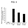
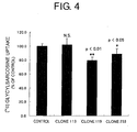
Classifications
-
- C—CHEMISTRY; METALLURGY
- C12—BIOCHEMISTRY; BEER; SPIRITS; WINE; VINEGAR; MICROBIOLOGY; ENZYMOLOGY; MUTATION OR GENETIC ENGINEERING
- C12N—MICROORGANISMS OR ENZYMES; COMPOSITIONS THEREOF; PROPAGATING, PRESERVING, OR MAINTAINING MICROORGANISMS; MUTATION OR GENETIC ENGINEERING; CULTURE MEDIA
- C12N15/00—Mutation or genetic engineering; DNA or RNA concerning genetic engineering, vectors, e.g. plasmids, or their isolation, preparation or purification; Use of hosts therefor
- C12N15/09—Recombinant DNA-technology
- C12N15/63—Introduction of foreign genetic material using vectors; Vectors; Use of hosts therefor; Regulation of expression
- C12N15/79—Vectors or expression systems specially adapted for eukaryotic hosts
- C12N15/85—Vectors or expression systems specially adapted for eukaryotic hosts for animal cells
- C12N15/86—Viral vectors
-
- C—CHEMISTRY; METALLURGY
- C07—ORGANIC CHEMISTRY
- C07K—PEPTIDES
- C07K14/00—Peptides having more than 20 amino acids; Gastrins; Somatostatins; Melanotropins; Derivatives thereof
- C07K14/435—Peptides having more than 20 amino acids; Gastrins; Somatostatins; Melanotropins; Derivatives thereof from animals; from humans
- C07K14/705—Receptors; Cell surface antigens; Cell surface determinants
-
- C—CHEMISTRY; METALLURGY
- C12—BIOCHEMISTRY; BEER; SPIRITS; WINE; VINEGAR; MICROBIOLOGY; ENZYMOLOGY; MUTATION OR GENETIC ENGINEERING
- C12N—MICROORGANISMS OR ENZYMES; COMPOSITIONS THEREOF; PROPAGATING, PRESERVING, OR MAINTAINING MICROORGANISMS; MUTATION OR GENETIC ENGINEERING; CULTURE MEDIA
- C12N7/00—Viruses; Bacteriophages; Compositions thereof; Preparation or purification thereof
-
- A—HUMAN NECESSITIES
- A61—MEDICAL OR VETERINARY SCIENCE; HYGIENE
- A61K—PREPARATIONS FOR MEDICAL, DENTAL OR TOILETRY PURPOSES
- A61K38/00—Medicinal preparations containing peptides
-
- C—CHEMISTRY; METALLURGY
- C12—BIOCHEMISTRY; BEER; SPIRITS; WINE; VINEGAR; MICROBIOLOGY; ENZYMOLOGY; MUTATION OR GENETIC ENGINEERING
- C12N—MICROORGANISMS OR ENZYMES; COMPOSITIONS THEREOF; PROPAGATING, PRESERVING, OR MAINTAINING MICROORGANISMS; MUTATION OR GENETIC ENGINEERING; CULTURE MEDIA
- C12N2710/00—MICROORGANISMS OR ENZYMES; COMPOSITIONS THEREOF; PROPAGATING, PRESERVING, OR MAINTAINING MICROORGANISMS; MUTATION OR GENETIC ENGINEERING; CULTURE MEDIA dsDNA viruses
- C12N2710/00011—Details
- C12N2710/14011—Baculoviridae
- C12N2710/14111—Nucleopolyhedrovirus, e.g. autographa californica nucleopolyhedrovirus
- C12N2710/14121—Viruses as such, e.g. new isolates, mutants or their genomic sequences
-
- C—CHEMISTRY; METALLURGY
- C12—BIOCHEMISTRY; BEER; SPIRITS; WINE; VINEGAR; MICROBIOLOGY; ENZYMOLOGY; MUTATION OR GENETIC ENGINEERING
- C12N—MICROORGANISMS OR ENZYMES; COMPOSITIONS THEREOF; PROPAGATING, PRESERVING, OR MAINTAINING MICROORGANISMS; MUTATION OR GENETIC ENGINEERING; CULTURE MEDIA
- C12N2710/00—MICROORGANISMS OR ENZYMES; COMPOSITIONS THEREOF; PROPAGATING, PRESERVING, OR MAINTAINING MICROORGANISMS; MUTATION OR GENETIC ENGINEERING; CULTURE MEDIA dsDNA viruses
- C12N2710/00011—Details
- C12N2710/14011—Baculoviridae
- C12N2710/14111—Nucleopolyhedrovirus, e.g. autographa californica nucleopolyhedrovirus
- C12N2710/14141—Use of virus, viral particle or viral elements as a vector
- C12N2710/14143—Use of virus, viral particle or viral elements as a vector viral genome or elements thereof as genetic vector
-
- G—PHYSICS
- G01—MEASURING; TESTING
- G01N—INVESTIGATING OR ANALYSING MATERIALS BY DETERMINING THEIR CHEMICAL OR PHYSICAL PROPERTIES
- G01N2333/00—Assays involving biological materials from specific organisms or of a specific nature
- G01N2333/005—Assays involving biological materials from specific organisms or of a specific nature from viruses
- G01N2333/01—DNA viruses
-
- G—PHYSICS
- G01—MEASURING; TESTING
- G01N—INVESTIGATING OR ANALYSING MATERIALS BY DETERMINING THEIR CHEMICAL OR PHYSICAL PROPERTIES
- G01N2500/00—Screening for compounds of potential therapeutic value
- G01N2500/10—Screening for compounds of potential therapeutic value involving cells
Definitions
- the present invention relates to methods for expressing transporters having transporter activity, where the methods comprise using transporter-encoding genes to express transporters on the envelope of budding viruses, wherein the transporter is a peptide transporter or OATP-C.
- the present invention relates to viruses that express transporters having transporter activity,wherein the transporter is a peptide transporter or OATP-C, methods that use these viruses for measuring the transporter activity of a transporter expressed on a viral envelope, and methods of screening for substances that inhibit or promote the transporter activity of the transporters.
- Mammals must take in nutrients from outside the body, and many transporter proteins (transporters) are known to exist in mammalian cells. These transporters mainly act to transport substances essential to the maintenance of life (amino acids, sugars, and such) into cells. In the physiological environment, cells often have multiple transporters that transport the same substrate. In these cases, the individual contribution of transporters to cellular uptake can be estimated using kinetic analysis (calculation of Km, Vmax, and so on; e.g., Wright E.M., Am. J. Physiol. Renal Physiol., 2001, 280: F10-18 ). Thus, identification of transport substrates and kinetic analysis of transporters are extremely important for revealing their physiological function and their potential in drug delivery.
- transporter proteins transporters
- methods for analysing transporter function use the following resources as materials: (1) primary cultured cells and cell membrane vesicles (such as lung cells and brush border membrane vesicles) comprising transporters isolated from living bodies; (2) cell lines derived from transporter-comprising cancer cells and so on (such as Caco-2 cells); (3) mammalian cells introduced with transporter genes (such as LLC-PK1 cells and MDCK cells) and Xenopus oocytes; and (4) insect cell membranes (such as Sf9 cell membranes) in which transporters have been expressed using baculovirus expression systems.
- primary cultured cells and cell membrane vesicles such as lung cells and brush border membrane vesicles
- cell lines derived from transporter-comprising cancer cells and so on such as Caco-2 cells
- mammalian cells introduced with transporter genes such as LLC-PK1 cells and MDCK cells
- Xenopus oocytes Xenopus oocytes
- insect cell membranes such as Sf9 cell membranes
- transporter activity can be measured using electrophysiological methods.
- transporters driven by Na and H ions and substrates having an electric charge at physiological pH
- transporter activity can be detected by measuring the electrical current caused by substrate transport.
- measuring transport activity is difficult when there is no driving force and also when substrates are electrically neutral at physiological pH.
- Kinetic analysis is also difficult in cases where transporter activity is observed but only a weak current can be detected.
- electrophysiological methods require specific equipment, they are not simple or convenient.
- drug-transporters include p-glycoprotein ( Annu. Rev. Biochem. 58: 137 (1989 )), multidrug resistance protein ( Science 258: 1650 (1992 ); Cancer Res. 55: 102 (1995 )), lung resistance protein ( Ann. Oncl. 7: 625 (1996 ); Int. J. Cancer 73: 1021 (1997 )), and organic cation transporter ( Proc. Natl. Acad. Sci. USA 91: 133 (1994 ) ; Molec. Pharmacol. 51: 913 (1997 )).
- Transporter SNPs that bring about functional changes have been recently found. These SNPs are receiving attention as one of the factors causing fluctuations between individuals ( Ryu S. et al., J. Biol. Chem. 275: 39617-39624 (2000 ); Tirona R.G. et al., J. Biol. Chem. 276: 35669-35675 (2001 )).
- functional analysis of transporter SNPs mainly uses mammalian cells introduced with genes. However, this is speculated to be problematic for accurately detecting functional changes caused by SNPs in substrates having a low activity ratio compared to parent cell lines.
- An objective of the present invention is to provide methods for measuring the target transporter activity, which have a low background level and a high degree of sensitivity and wherein the transporter is expressed on a viral envelope.
- another objective of the present invention is to provide methods of screening for substances that inhibit or promote the transport activity of transporters, using the above methods.
- viruses Since viruses have no fundamental need to self-reproduce, the present inventors speculated that there was no physiological value in taking up substances essential to maintenance of life. Thus, they focused on the assumption that endogenous transporters may not be expressed (or maybe expressed in extremely low amounts) on viral envelopes.
- the method for measuring transporter activity using budding baculoviruses that do not express endogenous transporters on their envelopes are thought to have a low background level, and to enable a highly sensitive measurement of target activity. Furthermore, by using such methods, functional changes due to transporter SNPs can be measured for a broader range of substrates, and may be applied to tailor-made therapies.
- the present invention provides:
- the present invention relates to methods for expressing transporters having transporter activity, which methods comprise culturing a host cell infected with a recombinant virus that comprises a gene coding for a transporter, and expressing the transporter on the envelope of a budding virus released from the host, wherein the transporter is a peptide transporter or OATP-C.
- examples of a "transporter” include peptide transporters, amino acid transporters, and sugar transporters. More specifically, transporters such as those listed in Table 1 can be given as examples.
- OAT1 Facilitated diffusion (organic anion) 550 12 NP_004781 Commun. 255 (2), 508-514 (1999 )
- OAT2 Facilitated diffusion (organic anion) 548 12 NP_006663
- OAT3 Facilitated diffusion (organic anion) 568 12 NP_004781 Commun. 255 (2), 508-514 (1999 )
- OAT4 Facilitated diffusion (organic anion) 550 12 AAK68155 J. Biol. Chem.
- Preferable transporters in the context of the method for measuring the activity of a transporter expressed on a viral envelope and/or for the screening method in the present invention are peptide transporters or organic anion transporters, and especially preferable are PepT1, Pept2, and OATP-C.
- the nucleotide and amino acid sequences of PepT1 and PepT2 are known (human PepT1: GenBank XM_007063, J. Biol. Chem. 270(12): 6456-6463 (1995 ); human PepT2: GenBank NP_066568, XM_002922, Biochem. Biophys. Acta. 1235:461-466 (1995 ); mouse PepT1 GenBank AF205540, Biochim.
- cDNA libraries or genomic libraries can be screened to obtain genes coding for transporters. More specifically, for example, cDNA or genomic libraries are screened using probes (antibodies against target transporters, or oligonucleotides that hybridise to nucleotide sequences coding for target transporters). Screening can be carried out, for example, by following the standard methods described by Sambrook et al. in Chapters 10 to 12 of "Molecular Cloning: A Laboratory Manual” (New York, Cold Spring Harbor Laboratory Press, 1989 ). Alternatively, genes encoding target transporters can be isolated using PCR (see e.g., Chapter 14 in the above-mentioned Sambrook et al., 1989).
- a recombinant vector for insect cells comprising a gene encoding a transporter is constructed, and inserted, along with baculoviral DNA, into insect cells such as Sf9.
- the transporter encoded by the recombinant vector is then expressed on mature viral particles (virions), which are released by infected cells to the outside of cells prior to infected cell death.
- recombinant viruses that express the transporter can be obtained.
- a budding virus is a virus that is released from infected cells by budding.
- viruses covered by an envelope can bud from cells infected with these viruses, even when the cells have not been destroyed, and are released continuously.
- adenoviruses that are not covered by an envelope and herpes viruses that are covered by a nuclear envelope, are released from the cells all at once upon their destruction.
- budding viruses are particularly preferable.
- host cells infected with a recombinant virus of the present invention can be suitably selected by those skilled in the art, depending on the type of virus used, so long as viral replication is possible in the host.
- insect Sf9 cells can be used when using baculoviruses.
- protein expression systems using baculoviruses and insect cells may be useful because modifications such as fatty acid acetylation or glycosylation are carried out at the same time as translation or post-translation, in the same way as in mammalian cells.
- modifications such as fatty acid acetylation or glycosylation are carried out at the same time as translation or post-translation, in the same way as in mammalian cells.
- the expression level of heterologous proteins in such systems is greater than that in mammalian cell systems ( Luckow V.A. and Summers M.D., Virol. 167: 56 (1988 )).
- the present invention also provides viruses that express transporters comprising transporter activity on the viral envelope, wherein the transporter is a peptide transporter or OATP-C.
- these viruses include baculoviruses, papillomaviruses, polyomaviruses, simian virus 40 (SV40), adenoviruses, Epstein-Bar virus (EBV), and retroviruses.
- particularly preferable viruses include baculovirus, and budding viruses.
- the transporters expressed by the viruses are preferably of a non-viral origin, for example the transporters in Table 1. Of these, peptide transporters and organic anion transporters are preferable, and Pept 1, PepT2, and OATP-C are even more preferable.
- the viruses expressing peptide transporters or OATP-C having transporter activity on the viral envelope of the present invention can be obtained by, for example, culturing a host that has been infected with a recombinant virus comprising a gene that codes for a transporter.
- a recombinant vector encoding a transporter can be inserted into an insect cell along with a baculovirus, and transporters can be expressed on the envelope of the baculovirus which is released outside of the cell.
- packaging cells such as PA317 can be infected with recombinant Moloney murine leukemia viruses, which are constructed using vectors derived from Moloney viruses introduced with transporter-encoding genes, and the transporters can be expressed on the envelope of the viruses released outside of the cells.
- the viruses of the present invention are not limited to those that are constructed using the above methods. They include viruses constructed using any method as long as transporters can be expressed in viral particles or on viral surfaces.
- Recombinant viruses constructed as described above can be purified using known methods.
- known methods for purifying viruses include: augmented density gradient centrifugation ( Albrechtsen et al., J. Virological Methods 28: 245-256 (1990 ) ; Hewish et al., J. Virological Methods 7: 223-228 (1983 )), size exclusion chromatography ( Hjorth and Mereno-Lopez, J. Virological Methods 5: 151-158 (1982 ); Crooks et al., J. Chrom. 502: 59-68 (1990 ); Mento S.J.
- Viruses of the present invention are not limited to these, and can be purified using the above methods, or combinations thereof.
- the present invention relates to methods for measuring the activity of transporters expressed on a viral envelope, which comprise expressing transporters on viral envelopes.
- measurement of transporter activity using budding baculoviruses can be carried out by the following method. First, if necessary, a substrate to be taken into the virus by the transporters is labelled so as to be detected. For example, the substrate is labelled with radioactive substances, fluorescence, or so on. Next, the substrate is mixed with the budding baculovirus that expresses the transporter, and reacted at 37°C. After a set length of time, the reaction solution is transferred onto a filter such as a cellulose membrane. The substrate taken into the virus is separated by vacuum filtration from the substrate that was not taken up.
- Nonspecific uptake can be detected by the substrate uptake into wild type viruses that do not express the transporter.
- nonspecific uptake can also be evaluated by carrying out experiments on substrate uptake by coexisting the substrate with transporter inhibitors, or if the substrate is radioactive, by coexisting it with an excess of unlabelled substance.
- Non-specific uptake can be evaluated by carrying out uptake experiments at 4°C.
- budding baculovirus solutions expressing a transporter can be added to a 96-well plate and incubated overnight at 4°C to perform plate coating.
- antibodies against proteins such as gp64 protein, which is highly expressed on viral envelopes, can be added to a 96-well plate, and incubated overnight at 4°C. After this, budding baculoviruses that express the transporter are added to the plate.
- Antibodies against membrane proteins, such as anti-gp64 antibodies (Novagen, Clontech) can also be used to coat the plate with the viruses. A substrate is then added to the plate, and reaction begins.
- Non-specific uptake can be detected by substrate uptake into wild-type viruses not expressing the transporter.
- transporter inhibitors can be coexisted with the substrate to detect non-specific uptake.
- the substrate is a radioactive substance
- non-specific uptake can also be evaluated by carrying out uptake experiments by coexisting the substrate with an excess of unlabeled substances.
- uptake experiments can be carried out at 4°C to evaluate non-specific uptake.
- budding baculoviruses can be preserved at 4°C, and do not require any special freezing devices.
- there are no complicated steps such as cell culturing, and there is no requirement for special equipment when measuring activity, as used in electrophysiological methods.
- budding baculovirus expression systems are simple methods for measuring transporter activity.
- the methods of the present invention for measuring the transporter activity that comprise expressing transporters on viral envelopes can also be applied in searching for substances that inhibit or promote the transporter activity.
- methods using budding baculovirus expression systems are simple, and useful in identifying substances that inhibit or promote the transporter activity.
- the methods of the present inventions produce, for example, budding baculoviruses that express target transporters.
- the radioactive or fluorescent substrates of those transporters are mixed with test substances, and added to the transporter-expressing viruses. Before adding the substrates, compounds can be preloaded to the viruses. Transport activity in the absence of a test substrate is taken as 100, and substances that inhibit or promote the transporter activity are searched for by using changes in activity in the presence of the test substrate as an index.
- Whether or not the test compound is inhibiting or promoting the transporter activity can be judged by known methods, for example, by labeling the transport target substrate (e.g. peptides in the case of peptide transporters) with a radioactive substance (such as 14 C) or fluorescent substance, and then measuring the amount of that substrate that is taken up by a transporter-expressing virus, etc.
- a radioactive substance such as 14 C
- fluorescent substance such as 14 C
- test substances in the methods of screening for substances that inhibit or promote transport activity of the transporters of the present invention include, but are not limited to, purified or crude proteins (comprising antibodies), gene library expression products, synthetic peptide libraries, cell extracts, cultured cell supernatants, products of fermentation microorganisms, marine organism extracts, vegetable extracts, synthetic low molecular weight compound libraries, peptides, non-peptide compounds, and natural compounds.
- Transporters expressed on viral envelopes can be contacted with test compounds in the form of, for example, a purified protein, a form bound to a carrier, a fusion protein with another protein, or a membrane fraction.
- carriers on which viruses can be immobilized include synthetic or natural organic high molecular weight compounds, inorganic materials such as glass beads, silica gel, alumina, and active carbon, and these materials coated with polysaccharides or synthetic high molecular weight molecules.
- organic high molecular weight compounds comprise a large number of compounds, including polysaccharides such as agarose, cellulose, chitin, chitosan, sepharose, and dextran, polyesters, polyvinyl cholorides, polystyrenes, polysulfones, polyether sulphones, polypropylenes, polyvinyl alcohols, polyamides, silicon resins, fluorocarbon resins, polyurethanes, polyacrylamides, and derivatives thereof.
- polysaccharides such as agarose, cellulose, chitin, chitosan, sepharose, and dextran
- polyesters polyvinyl cholorides, polystyrenes, polysulfones, polyether sulphones, polypropylenes, polyvinyl alcohols, polyamides, silicon resins, fluorocarbon resins, polyurethanes, polyacrylamides, and derivatives thereof.
- polysaccharides such as agarose
- the form of the carrier is also not particularly limited, and examples include membranes such as a plate, fibers, granules, hollow filaments, nonwoven fabrics, porous forms, and honeycomb forms.
- membranes such as a plate, fibers, granules, hollow filaments, nonwoven fabrics, porous forms, and honeycomb forms.
- simplicity of immobilization makes commercially available plates especially preferable.
- the contact area of test compounds can be controlled.
- Viruses can be immobilized to carriers using, for example, antibodies against the envelope proteins expressed in the viruses.
- immobilization onto carriers can also be achieved using streptoavidin, avidin or such when biotinylated beforehand.
- the physiological function of transporters can be elucidated by searching for inhibitors or promoters of transporter activity. At the same time, those inhibitors or promoters may be applied to developing pharmaceutical agents for diseases caused by transporter abnormalities.
- the present invention's budding baculoviruses that express promoters, and the envelope portions that comprise a transporter of those viruses, can be used as screening antigens or immune antigens when producing transporter antibodies. Preparation of such an antigen can be carried out, for example, according to the methods using baculoviruses described in WO98/46777 .
- an active transporter in the construction of anti-transporter antibodies, it was problematic to use an active transporter as an immunogen.
- transporters that are expressed by the methods of the present invention have been confirmed to have transporter activity.
- an active transporter can be used as an immunogen by using transporter-expressing viruses, or envelope portions that comprise a transporter of those viruses.
- the present disclosure provides methods for constructing anti-transporter antibodies, which methods comprise using, as immunogens, transporter-expressing viruses or envelope portions that comprise a transporter of those viruses.
- the present disclosure also provides the antibodies constructed using these methods.
- Transporter antibodies of the present disclosure can be constructed by those skilled in the art, using known methods where non-human animals are administered, by subcutaneous or intraperitoneal injection, several times with transporter-expressing viruses or envelope portions that comprise a transporter of those viruses.
- the non-human mammals immunized with sensitizing antigens are not particularly limited, however are preferably selected considering compatibility with parent cells used for cell fusion.
- Animals generally used include rodents, lagomorphs, and primates.
- rodents that can be used are mice, rats, and hamsters. As lagomorphs, for example, rabbits can be used. Examples of primates are monkeys. Monkeys that can be used include catarrhines (old-world monkeys) such as cynomolgous monkeys, rhesus monkeys, hamadryas, and chimpanzees.
- catarrhines old-world monkeys
- cynomolgous monkeys rhesus monkeys
- hamadryas chimpanzees.
- Animals can be immunized with a sensitizing antigen using known methods.
- General methods include injecting a sensitizing antigen into a mammal by subcutaneous or intraperitoneal injection. Specifically, a sensitizing antigen is diluted with an appropriate volume of Phosphate-Buffered Saline (PBS) or physiological saline, and if desired, the suspension is mixed with an appropriate volume of a conventional adjuvant, for example, Freund's complete adjuvant. After emulsification, this is applied to the mammals. In addition, after this, the sensitizing antigen that has been mixed with an appropriate volume of Freund's incomplete adjuvant is preferably applied every four to 21 days for several times. When immunizing a sensitizing antigen, an appropriate carrier can also be used. Thus immunization occurs, and the increased level of the desired antibody in the serum can be confirmed using conventional methods.
- PBS Phosphate-Buffered Saline
- physiological saline physiological saline
- the increase in the level of the desired antibody in the serum is confirmed, and blood is then obtained from the mammals sensitized to the antigens.
- Serum can be separated from this blood using known methods.
- serum comprising polyclonal antibodies can be used.
- fractions comprising polyclonal antibodies can be isolated from this serum, and this fraction can also be used.
- fractions that only recognize the transporters disclosed herein can be obtained using affinity columns coupled to these transporters. By purifying these fractions using a protein A or protein G column, immunoglobulin G or M can be prepared.
- immunocytes can be obtained from the mammals, and then subjected to cell fusion.
- immunocytes for cell fusion can preferably be splenocytes.
- mammal myeloma cells are preferable, and more preferable are myeloma cells that have acquired a characteristic for selection of fusion cells using a pharmaceutical agent.
- the above-mentioned cell fusion of immunocytes and myeloma cells can be performed according to known methods, for example, the method of Milstein et al . ( Galfre, G. and Milstein, C., Methods Enzymol. (1981) 73, 3-46 ).
- Hybridomas obtained from the cell fusions can be selected by culturing in a conventional selective culture medium, for example HAT culture medium (medium comprising hypoxanthine, aminopterin, and thymidine). Culture in this HAT culture medium is carried out for a continuous period of usually several days to several weeks, a sufficient time to kill cells other than the target hybridomas (non-fusion cells). Next, conventional limiting dilution methods are carried out, and hybridomas that produce the target antibodies are screened and cloned.
- a conventional selective culture medium for example HAT culture medium (medium comprising hypoxanthine, aminopterin, and thymidine). Culture in this HAT culture medium is carried out for a continuous period of usually several days to several weeks, a sufficient time to kill cells other than the target hybridomas (non-fusion cells). Next, conventional limiting dilution methods are carried out, and hybridomas that produce the target antibodies are screened and cloned.
- human lymphocytes for example human lymphocytes infected with EB virus
- a virus expressing a transporter of the present invention or to an envelope portion comprising a transporter of that virus.
- the sensitized lymphocytes are fused with human-derived myeloma cells that can permanently divide, for example U266.
- hybridomas that produce the desired human antibodies that have the activity to bind to the transporters can be obtained (Unexamined Published Japanese Patent Application No. (JP-A) Sho 63-17688 ).
- the obtained hybridomas are transplanted into mice peritoneal cavities, and ascites are recovered from the mice.
- the monoclonal antibodies thus obtained can be prepared by purification using ammonium sulphate precipitation, protein A or G columns, DEAE ion exchange chromatography, affinity columns to which a transporter of the present disclosure has been coupled, or the like.
- the antibodies disclosed herein can become candidates for agonists and antagonists of these transporters.
- these antibodies can also be applied to antibody therapies for diseases involving transporters of the present disclosure.
- human antibodies and humanized antibodies are preferable due to their low antigenicity.
- antibody-producing cells can be obtained by immunizing transgenic animals that comprise a repertoire of human antibody genes, with a virus expressing a transporter that becomes the antigen, or a portion of the viral envelope comprising the transporter.
- Hybridomas produced by fusing the antibody-producing cells with myeloma cells can be used to obtain human antibodies against the transporter (see International Publication WO92-03918 , W093-2227 , WO94-02602 , WO94-25585 , W096-33735 , and WO96-34096 ).
- immunocytes of antibody-producing sensitized lymphocytes and such that have been immortalized using oncogenes can also be used.
- Monoclonal antibodies obtained in this way can also be obtained as recombinant antibodies produced using gene recombination technologies (for example, see Borrebaeck, C.A.K. and Larrick, J.W., Therapeutic Monoclonal Antibodies, UK, Macmillan Publishers Ltd., 1990 ).
- Recombinant antibodies can be produced by cloning DNA that encodes them from immunocytes such as hybridomas and antibody-producing sensitized lymphocytes, incorporating into a suitable vector, and introducing this into a host.
- the present disclosure also encompasses such recombinant antibodies.
- an antibody fragment can be an Fab, F(ab')2, Fv, or a single chain Fv (scFv) where Fvs of H chain and L chain are linked by a suitable linker ( Huston, J.S. el al., Proc. Natl. Acad. Sci. U.S.A., (1998) 85, 5879-5883 ).
- the antibody fragments can be produced by treating antibodies with an enzyme such as papain or pepsin.
- genes encoding these antibody fragments are constructed, inserted into an expression vector, and expressed in appropriate host cells (see for example, Co, M. S. et al., J. Immunol. (1994) 152, 2968-2976 ; Better, M. and Horwitz, A. H., Methods Enzymol. (1989) 178, 476-496 ; Pluckthun, A. and Skerra, A., Methods Enzymol. (1989) 178, 497-515 ; Lamoyi, E., Methods Enzymol. (1986) 121, 652-663 ; Rousseaux, J. et al., Methods Enzymol. (1986) 121, 663-669 ; Bird, R. E. and Walker, B. W., Trends Biotechnol. (1991) 9, 132-137 ).
- Antibodies bound to various molecules such as polyethylene glycols (PEG), can also be used as the modified antibodies.
- “Antibody” in the present disclosure also encompasses these modified antibodies.
- Such a modified antibody can be obtained by chemically modifying obtained antibodies. These methods have already been established in the art.
- the antibodies disclosed herein can be obtained as chimeric antibodies comprising non-human antibody-derived variable regions and human antibody-derived constant regions, or alternatively, as humanized antibodies comprising non-human antibody-derived complementarity determining regions (CDRs), human antibody-derived framework regions (FRs), and constant regions.
- CDRs non-human antibody-derived complementarity determining regions
- FRs human antibody-derived framework regions
- Antibodies obtained as above can be purified until homogenous.
- the separation and purification of antibodies used in the present disclosure can use conventional separation and purification methods.
- antibodies can be separated and purified by appropriately selecting and combining chromatography columns such as affinity chromatography columns, filters, ultrafiltration, salt precipitation, dialysis, SDS polyacrylamide gel electrophoresis, isoelectric focusing and so on ( Antibodies: A Laboratory Manual. Ed Harlow and David Lane, Cold Spring Harbor Laboratory, 1988 ).
- the concentration of the above-obtained antibodies can be determined by measuring absorbance, by enzyme-linked immunosorbent assays (ELISA), etc.
- Protein A columns, protein G columns, and such can be used as the columns used for affinity chromatography.
- the columns using protein A Hyper D, POROS, Sepharose F.F. (Pharmacia) and so on can be used.
- chromatography other than affinity chromatography examples include ion exchange chromatography, hydrophobic chromatography, gel filtration, reverse chromatography, and adsorption chromatography ( strategies for Protein Purification and Characterisation: A Laboratory Course Manual. Ed Daniel R, Marshak et al., Cold Spring Harbor Laboratory Press, 1996 ). These chromatographies can be carried out using liquid phase chromatography such as HPLC and FPLC.
- Examples of the methods for measuring antigen-binding activities of the antibodies disclosed herein include absorbance measurements, enzyme-linked immunosorbent assays (ELISA), enzyme immunoassays (EIA), radioimmunoassay (RIA), and immunofluorescence.
- ELISA enzyme-linked immunosorbent assays
- EIA enzyme immunoassays
- RIA radioimmunoassay
- the transporters disclosed herein are added to a plate to which the antibodies of the present disclosure have been solid phased. Next, samples comprising a target antibody, for example the culture supernatant of antibody-producing cells or purified antibodies, are added. Secondary antibodies that recognise the antibody, which is labelled with enzymes such as alkaline phosphatase, are then added and the plate is incubated. After washing, an enzyme substrate such as p-nitrophenol phosphate is added, and antigen-binding activity can be evaluated by measuring absorbance.
- BIAcore Pharmacia
- Transporter-binding antibodies can be screened by ELISA using 96-well plates coated with budding baculoviruses. Antibodies against the viral antigens can be removed by ELISA using wild type viruses as the screening antigen. Alternatively, hybridoma culture supernatant and a wild type virus can be reacted, and after antibodies against the viral antigen have been removed, ELISA can be then carried out using a transporter-expressing virus as the screening antigen to acquire transporter-binding antibodies. Function-inhibiting antibodies can also be screened for from the binding antibodies. In other words, a radioactive or fluorescent substrate of the target transporter can be mixed with a solution comprising antibodies, such as hybridoma culture supernatant, and then added to a transporter-expressing virus.
- the solution comprising antibodies, such as hybridoma culture supernatant, can be preloaded onto the virus prior to adding the substrate.
- Transport activity in the absence of antibodies is taken as 100, and function-inhibiting antibodies can be screened for using, as an index, decreased activity in the presence of antibodies.
- Transporter topography at the cellular level can be revealed by binding antibodies to that transporter.
- function-inhibiting antibodies can be added to cell cultures or administered to laboratory animals to make a great contribution to the elucidation of the physiological functions of the transporters. Function-inhibiting antibodies or binding antibodies to transporters associated with disease can be applied as pharmaceutical agents.
- the present disclosure can also be used to evaluate the ways in which transporter activity is altered by changes in amino acid sequence due to mutations, polymorphisms such as SNPs, and so on.
- SNPs polymorphisms
- the methods of the present disclosure to measure the transport activity of each of these OATP-Cs with altered amino acid sequences, the SNPs that influence transport activity can be identified, transporters with high activity can be screened, and so on.
- transporter activity can be measured and transporters with high activity can be screened, or regions that influence transporter activity can be identified.
- Those skilled in the art can prepare transporters with substituted amino acids by using well-known methods. For example, site-specific mutagenesis and such can be used ( Hashimoto-Gotoh, T.
- substances transported by a transporter can be used as test substances and measure transporter activity to screen for substances that are easily transported by transporters, or substances that are difficult to transport.
- the present disclosure can also be applied to proteins other than transporters.
- similar methods for measuring activity, screening and such can be carried out for ion channels such as sodium channels, calcium channels, potassium channels, chloride channels, cation channels, and anion channels.
- ion channels such as sodium channels, calcium channels, potassium channels, chloride channels, cation channels, and anion channels.
- a transporter instead of a transporter, a channel is expressed on the viral envelope, and a substance passed through the channel can be used as a substrate.
- Channels that can be used in the present disclosure include those listed in Table 2.
- the present disclosure can be used for proteins that can transport or transmit a substance, such as transporters and ion channels (especially proteins which are expressed on membranes and can be transported or passed in a substrate-specific manner).
- GPCRs G protein coupled receptors
- Table 2 Symbol Name Sequence ID ACCN1 amiloride-seinsitive cation channel 1, neuronal (degenerin) NM_001094 ACCN2 amiloride-seinsitive cation channel 2, neuronal NM_001095 NM_020039 ACCN3 amiloride-seinsitive cation channel 3, testis NM_004769 NM_020321 NM_020322 AQP1 aquaporin 1 (channel-forming integral protein, 28kD) NM_000385 ASIC4 putative acid-sensing ion channel NM_018674 CACNA1A calcium channel, voltage-dependent, P/Q type, alpha 1A subunit NM_000068 NM_023035 CACNA1B calcium channel, voltage-dependent, L type, alpha 1B subunit NM_000718 CACNA1C calcium channel,
- SCN2A2 sodium channel voltage-gated, type II, alpha 2 polypeptide NM_021007 SCN2B sodium channel, voltage-gated, type II, beta polypeptide NM_004588 SCN3A sodium channel, voltage-gated, type III, alpha polypeptide AF225987 SCN4A sodium channel, voltage-gated, type IV, alpha polypeptide NM_000334 SCN5A sodium channel, voltage-gated, type V, alpha polypeptide (long (electrocardiographic) QT syndrome 3) NM_000335 SCN6A sodium channel, voltage-gated, type VI, alpha polypeptide NM_002976 SCN8A sodium channel, voltage gated, type VIII, alpha polypeptide NM_014191 SCN9A sodium channel, voltage-gated, type IX, alpha polypeptide NM_002977 SCNN1A sodium channel, nonvoltage-gated 1 alpha NM_001038 SCNN1B sodium channel, nonvoltage
- a full-length PepT1 gene was isolated from a human kidney library using PCR. By inserting the full-length human PepT1 gene into pBlueBacHis2A (Invitrogen), the pBlueBacHis-PepTl transfer vector was constructed. A Bac-N-Blue transfection kit (Invitrogen) was then used to introduce this transfer vector into Sf9 cells, along with Bac-N-Blue DNA. Thus, a recombinant virus for the expression of human PepT1 was constructed.
- the supernatant was again centrifuged at 45,000x g for 30 minutes, and the precipitate was again suspended in PBS. This suspension was the budding virus fraction. Expression of PepT1 in the virus and on the Sf-9 cell membrane was confirmed by Western analysis using anti-His antibodies. In addition, protein concentration was measured using Dc Protein Assay kit (Bio-Rad), with BSA as the standard.
- 14 C glycylsarcosine was diluted with HBSS (pH6.0) to a final concentration of 50 ⁇ M, and used as a substrate solution.
- 40 ⁇ L of viral solution 100 ⁇ g protein
- 160 ⁇ L of substrate solution that had been preheated to 37°C was added, and the reaction was started.
- 1 mL of ice-cold HBSS (pH 7.4) (hereinbelow also called "quenching solution”) was added, and the reaction was stopped.
- the virus-comprising reaction solution was immediately vacuum filtered using a mixed cellulose membrane filter, and washed twice with 5 mL of the quenching solution.
- the membrane filter was transferred to a liquid scintillation vial, 5 mL of clear-zol I was added, and the filter was dissolved. After the dissolving, a liquid scintillation counter was used to measure radioactivity on the filter. Non-specific adsorption to the filter was measured in the same way for systems where the quenching solution was added before adding the substrate solution to the viral solution, and values thus obtained were subtracted from the counts for each experiment.
- the PepT1 activity of the PepT1-expressing virus with a His-tag added at its N-terminal is shown in Fig. 1 .
- the full-length PepT2 gene was isolated from a human kidney library. PCR was used to integrate the gene encoding the full-length human PepT2 into pBlueBacHis2A (Invitrogen), and a full-length PepT2 transfer vector (pBlueBac) was constructed. This vector was introduced into Sf-9 cells along with the viral DNA. After cloning the recombinant virus constructed by homologous recombination, a stock with a high recombinant virus activity was constructed. Sf-9 cells were infected with the stock virus, and after culturing for a certain period, PepT2 was expressed in the virus and on the membrane of Sf-P cells. PepT2 expression in the virus and on the membrane of the Sf-9 cells was confirmed by Western analysis using anti-His antibodies. More specifically, except for using the PepT2 gene, operations were carried out according to the methods described in Example 1.
- 3 H glycylsarcosine was diluted with HBSS (pH6.0) to a final concentration of 0.8 ⁇ M, and used as a substrate solution.
- 40 ⁇ L of viral solution 100 ⁇ g protein
- 160 ⁇ L of the substrate solution preheated to 37°C was added to commence the reaction.
- 1 mL of the quenching solution was added, and the reaction was stopped.
- the virus-comprising reaction solution was immediately vacuum filtered using a mixed cellulose membrane filter, and washed twice with 5 mL of the quenching solution.
- the membrane filter was transferred to a liquid scintillation vial, 5 mL of clear-sol I was added, and the filter was dissolved. After the dissolving, a liquid scintillation counter was used to measure radioactivity on the filter. The quenching solution was added before adding the substrate solution to the viral solution, and similar manipulations were performed. Non-specific adsorption to the filter was measured and the obtained value was subtracted from the counts for each experiment.
- the PepT2 activity of the PepT2-expressing virus with a His-tag added to its N-terminal is shown in Fig. 2 .
- OATP-C WT wild type human OATP-C
- adult human liver-derived cDNA was used as a template, and the OATP-C WT cDNA was divided into two fragments and amplified using PCR with the following primer combinations: 5' side OAHC17 primer: 5' gat ggt acc aaa ctg agc atc aac aac aaaac 3' (SE Q ID NO: 1)
- 3'side OAHC21 primer 5' gat ggt acc tac cct ggg atc tct gttttc taa 3' (S EQ ID NO: 3)
- OAHC22 primer 5' gat ggt acc g
- the primers used for mutagenesis were as follows: Primer for OATP-C N130D: 5' gaa act aat atc gat tca tca gaa aat 3' (SEQ ID NO: 5) Primer for OATP-C V174A: 5' atg tgg ata tat gcg ttc atg ggt aat 3' (SEQ ID NO: 6)
- the primers for use in mutagenesis and the selection primers included in kits (for bottom strand use) were both annealed to the template plasmid DNA, which had been made into a single strand. Thus, a new DNA strand was constructed. This was introduced into E . coli, and GeneEditor TM antibiotic-resistant clones were obtained. These clones were sequenced and clones containing mutations were thus selected (pcDNA3/OATP-C N130D and pcDNA3/OATP-C V174A).
- PCR was carried out using the primers below, thus amplifying the respective cDNAs with SalI sites on each end.
- C45 primer 5' gat gtc gac tta aca atg tgt ttc act 3' (SEQ ID NO 7)
- C58 primer 5' gat gtc gac tat gga cca aaa tca aca t 3' (SEQ ID NO: 8)
- 3 H estrone sulphate conjugate was diluted with HBSS (pH7.4) to a final concentration of 10 nM, and used as a substrate solution.
- 20 ⁇ L of viral solution (50 ⁇ g protein) was preincubated at 37°C for 30 minutes. 180 ⁇ L of the substrate solution preheated to 37°C was added, and the reaction was started. After one minute, 1 mL of ice-cold HBSS (pH7.4) (hereinafter referred to as "quenching solution”) was added, and the reaction was stopped.
- the virus-comprising reaction solution was immediately vacuum filtered using a mixed cellulose membrane filter, and washed twice with 5 mL of the quenching solution.
- the membrane filter was transferred to a liquid scintillation vial, 5 mL of clear-sol I was added, and the filter was dissolved. After the dissolving, a liquid scintillation counter was used to measure radioactivity on the filter. To measure non-specific adsorption to the filter, the reaction quenching solution was added before adding the substrate solution and similar manipulations were performed. The obtained value was subtracted from the counts for each experiment.
- the activity of 3 H estrone sulphate conjugate uptake is shown in Fig. 3 for three types of OATP-C-expressing viruses with His-tags added to their N-terminals.
- the detected 3 H estrone sulphate conjugate uptake activity ratios for wild type OATP-C, N130D, and V174A were respectively 57, 41, and 36 times that of a wild type virus not expressing OATP-C.
- virus-derived endogenous OATP-C activity was hardly detected in experiments on the uptake in wild type viruses.
- budding baculovirus expression systems are systems with extremely low background levels.
- OATP-C mutants N130D, V174A
- changes in substrate specificity due to SNPs can also be determined, making also applications to tailor-made therapy possible.
- 14 C glycylsarcosine was diluted with HBSS (pH 6.0) to a final concentration of 50 ⁇ M, and used as a substrate solution.
- a mouse monoclonal antibody recognising the extracellular region of human PepT1 was diluted with PBS to a final concentration of 200 ⁇ g/mL, and used as an antibody solution.
- 20 ⁇ L (50 ⁇ g protein) of solution of budding baculoviruses expressing PepT1 with a His-tag added at the N-terminal was mixed with 20 ⁇ L of the antibody solution and incubated for one hour at 37°C. 160 ⁇ L of substrate solution preheated to 37°C was added, and the reaction was started.
- ice-cold HBSS pH 7.4 (below also called “quenching solution”) was added, and the reaction was stopped.
- the virus-comprising reaction solution was immediately vacuum filtered using a mixed cellulose membrane filter, and washed twice with 5 mL of the quenching solution.
- the membrane filter was transferred to a liquid scintillation vial, 5 mL of clear-sol I was added, and the filter was dissolved.
- a liquid scintillation counter was used to measure radioactivity on the filter. Non-specific adsorption to the filter was measured by adding the reaction quenching solution before adding the substrate solution to the viral solution, and performing similar manipulations. The obtained value was subtracted from the counts for each experiment.
- PepT1 activity inhibition by the anti-human PepT1 monoclonal antibodies is shown in Fig. 4 .
- PepT1 activity in the absence of the antibodies was taken as the control and expressed as 100.
- clone 119 inhibited PepT1 activity by about 20%, and clone 253 by about 10%, compared to the control.
- This PepT1 activity inhibition was statistically significant (Student t-test).
- budding baculovirus expression systems will be useful in the search for substrates that inhibit or promote transporter activity.
- the present invention provides viruses that express transporters having transporter activity on the viral envelope, wherein the transporter is a peptide transporter or OATP-C, and by using these viruses, transporter activity can be measured with a high sensitivity and less background level then in the past.
- the transporter is a peptide transporter or OATP-C
- identification of transport substrates and driving force of transporters, and functional analysis such as kinetic analysis can be carried out more easily and accurately than before.
- substances that inhibit or promote the transport activities of transporters expressed on the viral envelopes can be screened. Since transporters have also been reported to be involved in the transport of drugs into cells, substances that inhibit or promote the activities of transporters associated with diseases can become candidates for new pharmaceutical agents in the context of the present disclosure.
Description
- The present invention relates to methods for expressing transporters having transporter activity, where the methods comprise using transporter-encoding genes to express transporters on the envelope of budding viruses, wherein the transporter is a peptide transporter or OATP-C. In addition, the present invention relates to viruses that express transporters having transporter activity,wherein the transporter is a peptide transporter or OATP-C, methods that use these viruses for measuring the transporter activity of a transporter expressed on a viral envelope, and methods of screening for substances that inhibit or promote the transporter activity of the transporters.
- Mammals must take in nutrients from outside the body, and many transporter proteins (transporters) are known to exist in mammalian cells. These transporters mainly act to transport substances essential to the maintenance of life (amino acids, sugars, and such) into cells. In the physiological environment, cells often have multiple transporters that transport the same substrate. In these cases, the individual contribution of transporters to cellular uptake can be estimated using kinetic analysis (calculation of Km, Vmax, and so on; e.g., Wright E.M., Am. J. Physiol. Renal Physiol., 2001, 280: F10-18). Thus, identification of transport substrates and kinetic analysis of transporters are extremely important for revealing their physiological function and their potential in drug delivery.
- Currently, methods for analysing transporter function use the following resources as materials: (1) primary cultured cells and cell membrane vesicles (such as lung cells and brush border membrane vesicles) comprising transporters isolated from living bodies; (2) cell lines derived from transporter-comprising cancer cells and so on (such as Caco-2 cells); (3) mammalian cells introduced with transporter genes (such as LLC-PK1 cells and MDCK cells) and Xenopus oocytes; and (4) insect cell membranes (such as Sf9 cell membranes) in which transporters have been expressed using baculovirus expression systems. Of these, mostly used are gene expression systems from mammalian cells and Xenopus oocyte cells. However, even in mammalian and Xenopus oocyte cells introduced with transporter genes, activities from endogenous transporters can be detected, thus elevating background levels (Kanai Y. et al., J. Clin. Invest. 93: 397-404 (1994); Kekuda R. et al., J. Biol. Chem. 271: 18657-18661 (1996); Kekuda R. et al., Am. J. Physiol. 272: G1463-1472 (1997); Yabuuchi H. et al., J. Pharmacol. Exp. Ther. 286: 1391-1396 (1998); Hatanaka T. et al., J. Clin. Invest. 107: 1035-1043 (2001)). For this reason, in some types of transporters, there are reports that describe an activity ratio of only two between cells introduced with genes and those not introduced with genes (parent cell lines). Carrying out kinetic analysis can be problematic in such gene-introduced cells with a low activity ratio.
- In Xenopus oocyte cells introduced with transporter genes, transporter activity can be measured using electrophysiological methods. In transporters driven by Na and H ions, and substrates having an electric charge at physiological pH, transporter activity can be detected by measuring the electrical current caused by substrate transport. However, measuring transport activity is difficult when there is no driving force and also when substrates are electrically neutral at physiological pH. Kinetic analysis is also difficult in cases where transporter activity is observed but only a weak current can be detected. In addition, since electrophysiological methods require specific equipment, they are not simple or convenient.
- The activity and substrate specificity of transporters that transfer drugs into cells has been reported to influence the drug's bioavailability (for example, Ganapathy, Leibach, Curr. Biol 3: 695-701 (1991); Nakashima et al., Biochem. Pharm. 33: 3345-3352 (1984); Friedman, Amidon, Pharm. Res. 6:1043-1047 (1989); Okano et al., J. Biol. Chem. 261: 14130-14134 (1986); Muranushi et al., Pharm. Res. 6: 308-312 (1989); Friedman, Amidon, J. Control. Res. 13: 141-146 (1990)). In recent years, research on factors that fluctuate in vivo pharmacokinetics has clarified that drug-metabolising enzymes as well as drug-transporters influence the function of drugs in the body. Known drug-transporters include p-glycoprotein (Annu. Rev. Biochem. 58: 137 (1989)), multidrug resistance protein (Science 258: 1650 (1992); Cancer Res. 55: 102 (1995)), lung resistance protein (Ann. Oncl. 7: 625 (1996); Int. J. Cancer 73: 1021 (1997)), and organic cation transporter (Proc. Natl. Acad. Sci. USA 91: 133 (1994) ; Molec. Pharmacol. 51: 913 (1997)). Analysis of SNPs is being carried out for these drug-transporters in the same way as for drug-metabolizing enzymes. Transporter SNPs that bring about functional changes have been recently found. These SNPs are receiving attention as one of the factors causing fluctuations between individuals (Ryu S. et al., J. Biol. Chem. 275: 39617-39624 (2000); Tirona R.G. et al., J. Biol. Chem. 276: 35669-35675 (2001)). Currently, functional analysis of transporter SNPs mainly uses mammalian cells introduced with genes. However, this is speculated to be problematic for accurately detecting functional changes caused by SNPs in substrates having a low activity ratio compared to parent cell lines.
- The present invention was made considering the above circumstances. An objective of the present invention is to provide methods for measuring the target transporter activity, which have a low background level and a high degree of sensitivity and wherein the transporter is expressed on a viral envelope. In addition, another objective of the present invention is to provide methods of screening for substances that inhibit or promote the transport activity of transporters, using the above methods.
- Since viruses have no fundamental need to self-reproduce, the present inventors speculated that there was no physiological value in taking up substances essential to maintenance of life. Thus, they focused on the assumption that endogenous transporters may not be expressed (or maybe expressed in extremely low amounts) on viral envelopes. The method for measuring transporter activity using budding baculoviruses that do not express endogenous transporters on their envelopes are thought to have a low background level, and to enable a highly sensitive measurement of target activity. Furthermore, by using such methods, functional changes due to transporter SNPs can be measured for a broader range of substrates, and may be applied to tailor-made therapies.
- Specifically, the present invention provides:
- [1] a method for expressing a transporter having transporter activity, wherein the method comprises culturing a host cell infected with a recombinant virus that comprises a gene encoding the transporter, and expressing the transporter on the envelope of a budding virus released from the host wherein the transporter is a peptide transporter or OATP-C;
- [2] the method of [1], wherein the virus is a baculovirus;
- [3] the method of [1] or [2], wherein the transporter is of a non-viral origin;
- [4] the method of [3] wherein the transporter is PepT1, or PEP2;
- [5] a virus that expresses a transporter having transporter activity on the viral envelope, wherein the transporter is a peptide transporter or OATP-C;
- [6] the virus of [5], wherein the transporter is of a non-viral origin,
- [7] the virus of [6] wherein the virus is a baculovirus;
- [8] the virus of any of [5] to [7] wherein the virus is a budding virus;
- [9] the virus of [5] wherein the transporter is PepT1 or PepT2;
- [10] a method for measuring the activity of a transporter expressed on a viral envelope, wherein the method comprises expressing the transporter on the viral envelope;
- [11] the method of [10] wherein the virus is a budding baculovirus;
- [12] the method of [10] or [11] wherein the transporter is a peptide transporter or an organic anion transporter;
- [13] the method of [12] wherein the transporter is PepT1, PepT2, or OATP-C;
- [14] a method of screening for a substance that inhibits or promotes transport activity of a transporter, wherein the method comprises the following steps:
- (a) expressing the transporter on a viral envelope,
- (b) contacting the transporter with a test substance, and
- (c) selecting a substance that inhibits or promotes the transport activity;
- [15] the method of [14] wherein the virus is a baculovirus;
- [16] the method of [14] or [16] wherein the virus is a budding virus;
- [17] the method of any of [14] to [16], wherein the transporter is of a non-viral origin;
- [18] the method of any of [14] to [17], wherein the transporter is a peptide transporter or an organic anion transporter;
- [19] the method of [18] wherein the transporter is PepT1, PepT2, or OATP-C;
- [20] the method of any of [14] to [19], which comprises immobilizing the virus on a support;
- [21] the method of [20] wherein the virus is immobilized on the support through an antibody against an envelope protein expressed on the viral envelope; and,
- [22] the method of [20] wherein the virus is immobilized on the support through a biotin-streptavidin reaction by biotinylating a protein expressed on the viral envelope.
- The present invention relates to methods for expressing transporters having transporter activity, which methods comprise culturing a host cell infected with a recombinant virus that comprises a gene coding for a transporter, and expressing the transporter on the envelope of a budding virus released from the host, wherein the transporter is a peptide transporter or OATP-C. In the context of the present disclosure, examples of a "transporter" include peptide transporters, amino acid transporters, and sugar transporters. More specifically, transporters such as those listed in Table 1 can be given as examples.
Table 1 Transporter Driving force/ transport type Amino acids Transmembrane ncbi Reference 4F2hc LAT regulatory factor 529 1 P08195 Proc. Natl. Acad. Sci. U.S.A. 84 (18), 6526-6530 (1987 ) AE4 Cl/HCO exchange transport 945 14 AAK16733 Commun. 282 (5), 1103-1109 (2001 ) ATB0/AS CT2 Na/neutral amino acid cotransport 541 10 Q15758 J. Biol. Chem. 271 (31),18657-18661 (1996 ) ATB0+ Na/neutral and basic amino acids cotransport 642 12 AAD49223 J. Biol. Chem. 274 (34), 23740-23745 (1999 ) BAT1/b0+ AT Facilitated diffusion (amino acid) 487 12 P82251 Nat. Genet. 23 (1), 52-57 (1999 ) BCRP ATP/primary active transport 655 6 AAC97367 Proc. Natl. Acad. Sci. U.S.A. 95 (26), 15665-15670 (1998 ) BSEP ATP/primary active transport 1321 12 AAC77455 Nat. Genet. 20 (3), 233-238 (1998 ) BTR1 Cl/HCO exchange transport 891 14 AAK16734 Commun. 282 (5), 1103-1109 (2001 ) CNT1 Na/nucleoside cotransport 649 13 NP_004204 Am. J. Physiol. 272 (2), C707-C714 (1997 ) CNT2 Na/nucleoside cotransport 658 14 043868 Am. J. Physiol. 273 (6 Pt 2), F1058-F1065 (1997 ) CNT3 Na/nucleoside cotransport 691 13 NP_071410 J. Biol. Chem. 276 (4), 2914-2927 (2001 ) DRA/CLD Cl/HCO exchange transport 764 P40879 Proc. Natl. Acad. Sci. U.S.A. 90 (9), 4166-4170 (1993 ) EAAC1 Na/acidic amino acid cotransport 525 12 NP_004161 Genomics 20 (2), 335-336 (1994 ) ENT1 Facilitated diffusion (nucleoside) 456 14 NP_004946 Nat. Med. 3 (1), 89-93 (1997 ) ENT2 Facilitated diffusion (nucleoside) 456 14 AAC39526 Biochem. J. 328 (Pt 3), 739-743 (1997 ) FORT Folic acid 591 12 P41440 Commun. 206 (2),681-687 (1995 ) GAT1 Na/GABA cotransport 599 12 NP_003033 FEBS Lett. 269 (1), 181-184 (1990 ) GAT3 Na/GABA cotransport 632 12 P48066 Recept. Channels 2 (3), 207-213 (1994 ) GLUT1 Facilitated diffusion (glucose) 492 12 NP_006507 Science 229 (4717), 941-945 (1985 ) GLUT2 Facilitated diffusion (glucose) 524 12 NP_000331 Proc. Natl. Acad. Sci. U.S.A. 85 (15), 5434-5438 (1988 ) GLUT3 Facilitated diffusion (glucose) 496 12 NP_008862 J. Biol. Chem. 263, 15245-15248 (1988 ) GLUT4 Facilitated diffusion (glucose) 509 12 NP_001033 J. Biol. Chem. 264 (14), 7776-7779 (1989 ) GLVR1/Pi T-1 Na/Pi cotransport 679 10 NP_005406 Cell Growth Differ. 1 (3), 119-127 (1990 ) GLVR2/Pi T-2 Na/Pi cotransport 652 10 NP_006740 J. Virol. 65 (11), 6316-6319 (1991 ) LAT1 Facilitated diffusion (amino acid) 507 12 JGO165 Commun. 255 (2), 283-288 (1999 ) LRP ATP/primary active transport 896 NP_059447 Nat. Med. 1 (6), 578-582 (1995 ) MCT1 H/organic anion cotransport 500 12 NP_003042 Genomics 23 (2), 500-503 (1994 ) MCT2 H/organic anion cotransport 478 12 060669 J. Biol. Chem. 273 (44), 28959-28965 (1998 ) MCT3 H/organic anion cotransport 465 12 015427 Biochem. J. 329 (Pt 2), 321-328 (1998 ) MCT4 H/organic anion cotransport 487 12 015374 Biochem. J. 329 (Pt 2), 321-328 (1998 ) MCT5 H/organic anion cotransport 505 12 015375 Biochem. J. 329 (Pt 2), 321-328 (1998 ) MCT6 H/organic anion cotransport 523 12 015403 Biochem. J. 329 (Pt 2), 321-328 (1998 ) MDR1 ATP/primary active transport 1279 12 AAB69423 Cell 47 (3), 381-389 (1986 ) MDR3 ATP/primary active transport 1279 12 P21439 EMBO J. 6 (11), 3325-3331 (1987 ) MRP1 ATP/primary active transport 1531 17 P33527 Science 258 (5088), 1650-1654 (1992 ) MRP2 ATP/primary active transport 1545 17 Q92887 Cancer Res. 56 (18), 4124-4129 (1996 ) MRP3 ATP/primary active transport 1527 17 NP_003777 Cancer Res. 57 (16), 3537-3547 (1997 ) MRP4 ATP/primary active transport 1325 12 NP_005836 Cancer Res. 57 (16), 3537-3547 (1997 ) MRP5 ATP/primary active transport 1437 12 O15440 Cancer Res. 57 (16), 3537-3547 (1997 ) MRP6 ATP/primary active transport 1503 17 095255 Cancer Res. 59 (1), 175-182 (1999 ) MRP7 ATP/primary active transport 1492 17 Cancer Lett 162 (2), 181-191 (2001 ) NaPi-3B Na/Pi cotransport 690 8 NP_006415 Commun. 258 (3), 578-582 (1999 ) NaSi-1 Na/Si cotransport 595 13 NP_071889 Genomics 70 (3), 354-363 (2000 ) NHE1 Na/H exchange transport 815 12 P19634 Cell 56 (2), 271-280 (1989 ) NHE2 Na/H exchange transport 812 12 NP_003039 Am. J. Physiol. 40 (2), 383-390 (1999 ) NHE3 Na/H exchange transport 834 12 NP_004165 Am. J. Physiol. 269 (1 Pt 1), C198-C206 (1995 ) NPT1 Na/Pi cotransport 467 6-8 Q14916 Genomics 18 (2), 355-359 (1993 ) NPT2/Na Pi-3 Na/Pi cotransport 639 8 NP_003043 Proc. Natl. Acad. Sci. U.S.A. 90, 5979-5983 (1993 ) Nramp2/D CT1 Na/Fe cotransport 568 12 P49281 Mol. Immunol. 34 (12-13), 839-842 (1997 ) NTCP2/A SBT Na/bile acid cotransport 348 7 NP000443 J. Biol. Chem. 270 (45), 27228-27234 (1995 ) OAT1 Facilitated diffusion (organic anion) 550 12 NP_004781 Commun. 255 (2), 508-514 (1999 ) OAT2 Facilitated diffusion (organic anion) 548 12 NP_006663 OAT3 Facilitated diffusion (organic anion) 568 12 NP_004781 Commun. 255 (2), 508-514 (1999 ) OAT4 Facilitated diffusion (organic anion) 550 12 AAK68155 J. Biol. Chem. 275 (6), 4507-4512 (2000 ) OATP-A Facilitated diffusion (organic anion) 670 12 NP_066580 Gastroenterology 109 (4), 1274-1282 (1995 ) OATP-B Facilitated diffusion (organic anion) 709 12 NP_009187 Commun. 273 (1),251-260 (2000 ) OATP-C Facilitated diffusion (organic anion) 691 12 BAA78639 Commun. 273 (1),251-260 (2000 ) OATP-D Facilitated diffusion (organic anion) 710 12 BAA89287 Commun. 273 (1), 251-260 (2000 ) OATP-E Facilitated diffusion (organic anion) 722 12 BAA89288 Commun. 273 (1), 251-260 (2000 ) OCT1 Facilitated diffusion (organic cation) 554 12 NP_003048 Mol. Pharmacol. 51 (6), 913-921 (1997 ) OCT2 Facilitated diffusion (organic cation) 555 12 NP_003049 DNA Cell Biol. 16 (7), 871-881 (1997 ) OCT3 Facilitated diffusion (organic cation) 551 12 NP_035525 Genomics 55 (2), 209-218 (1999 ) OCTN1 H/organic cation 551 11 NP_003050 FEBS Lett. 419 (1), 107-111 (1997 ) OCTN2 Na/organic cation cotransport 557 12 076082 Commun. 246 (3), 589-595 (1998 ) PGT Facilitated diffusion (organic anion) 643 12 NP_005612 Commun. 221 (2), 454-458 (1996 ) rBAT BAT1 regulatory factor 685 1 AAA81778 J. Biol. Chem. 268 (20), 14842-14849 (1993 ) SDCT1/N aDC-1 Na/dicarboxylic acid cotransport 592 8 NP_003975 Am. J. Physiol. 270 (4 Pt 2), F642-F648 (1996 ) SGLT1 Na/glucose cotransport 664 14 NP00334 Proc. Natl. Acad. Sci. U.S.A. 86 (15), 5748-5752 (1989 ) SGLT2 Na/glucose cotransport 672 14 NP_003032 Am. J. Physiol. 263 (3 Pt 2), F459-F465 (1992 ) SGLT3/S AAT1 Na/glucose cotransport 659 14 P31636 J. Biol. Chem. 268 (3), 1509-1512 (1993 ) SLC26A6 Cl/HCO exchange transport 738 11 NP_075062 Genomics 70 (1), 102-112 (2000 ) SVCT1 Na/vitamin C cotransport 598 12 NP_005838 Biochim. Biophys. Acta 1461 (1), 1-9 (1999 ) UT2 Urea (Facilitated diffusion) 397 10 Q15849 FEBS Lett. 386 (2-3), 156-160 (1996 ) - Preferable transporters in the context of the method for measuring the activity of a transporter expressed on a viral envelope and/or for the screening method in the present invention are peptide transporters or organic anion transporters, and especially preferable are PepT1, Pept2, and OATP-C. The nucleotide and amino acid sequences of PepT1 and PepT2 are known (human PepT1: GenBank XM_007063, J. Biol. Chem. 270(12): 6456-6463 (1995); human PepT2: GenBank NP_066568, XM_002922, Biochem. Biophys. Acta. 1235:461-466 (1995); mouse PepT1 GenBank AF205540, Biochim. Biophys. Acta. 1492: 145-154 (2000); mouse PepT2: GenBank NM_021301, Biochim. Biophys. Res. Commun. 276: 734-741 (2000)). Furthermore, the nucleotide and amino acid sequence of OATP-C are also known (Table 1: Commun. 273(1), 251-260 (2000)). However, the transporters of the present invention are not particularly limited thereto in the context of measuring the activity and the screening method of the present invention, as long as they can be expressed on a viral envelope.
- Genes encoding the transporters, for example, those listed in Table 1, are registered with the National Centre for Biotechnology Information (NCBI) under the listed accession numbers. For example, based on this sequence information, cDNA libraries or genomic libraries can be screened to obtain genes coding for transporters. More specifically, for example, cDNA or genomic libraries are screened using probes (antibodies against target transporters, or oligonucleotides that hybridise to nucleotide sequences coding for target transporters). Screening can be carried out, for example, by following the standard methods described by Sambrook et al. in ). Alternatively, genes encoding target transporters can be isolated using PCR (see e.g., Chapter 14 in the above-mentioned Sambrook et al., 1989).
- As methods for expressing transporters on viral envelopes, for example, the method of
WO98/46777 or Loisel et al. - In the present invention, a budding virus is a virus that is released from infected cells by budding. Generally, viruses covered by an envelope can bud from cells infected with these viruses, even when the cells have not been destroyed, and are released continuously. On the other hand, adenoviruses that are not covered by an envelope, and herpes viruses that are covered by a nuclear envelope, are released from the cells all at once upon their destruction. In the present invention, budding viruses are particularly preferable. In addition, host cells infected with a recombinant virus of the present invention can be suitably selected by those skilled in the art, depending on the type of virus used, so long as viral replication is possible in the host. For example, insect Sf9 cells can be used when using baculoviruses. Generally, protein expression systems using baculoviruses and insect cells may be useful because modifications such as fatty acid acetylation or glycosylation are carried out at the same time as translation or post-translation, in the same way as in mammalian cells. In addition, the expression level of heterologous proteins in such systems is greater than that in mammalian cell systems (Luckow V.A. and Summers M.D., Virol. 167: 56 (1988)).
- The present invention also provides viruses that express transporters comprising transporter activity on the viral envelope, wherein the transporter is a peptide transporter or OATP-C. Examples of these viruses include baculoviruses, papillomaviruses, polyomaviruses, simian virus 40 (SV40), adenoviruses, Epstein-Bar virus (EBV), and retroviruses. In the present invention, particularly preferable viruses include baculovirus, and budding viruses. In addition, the transporters expressed by the viruses are preferably of a non-viral origin, for example the transporters in Table 1. Of these, peptide transporters and organic anion transporters are preferable, and
Pept 1, PepT2, and OATP-C are even more preferable. - The viruses expressing peptide transporters or OATP-C having transporter activity on the viral envelope of the present invention can be obtained by, for example, culturing a host that has been infected with a recombinant virus comprising a gene that codes for a transporter. Alternatively, using methods such as the above-mentioned methods of
WO98/46777 and Loisel et al - Recombinant viruses constructed as described above can be purified using known methods. For example, known methods for purifying viruses include: augmented density gradient centrifugation (Albrechtsen et al., J. Virological Methods 28: 245-256 (1990) ; Hewish et al., J. Virological Methods 7: 223-228 (1983)), size exclusion chromatography (Hjorth and Mereno-Lopez, J. Virological Methods 5: 151-158 (1982); Crooks et al., J. Chrom. 502: 59-68 (1990); Mento S.J. (Viagene, Inc.) 1994 Williamsburg Bioprocessing Conference), affinity chromatography using monoclonal antibodies, sulphated fucose-containing polysaccharides and the like (Najayou et al., J. Virological Methods 32: 67-77 (1991); Diaco et al., J. Gen. Virol. 67: 345-351 (1986); Fowler, J. Virological Methods 11: 59-74 (1986); TOKUSAIHYOU No.
97/032010 - The present invention relates to methods for measuring the activity of transporters expressed on a viral envelope, which comprise expressing transporters on viral envelopes. For example, measurement of transporter activity using budding baculoviruses can be carried out by the following method. First, if necessary, a substrate to be taken into the virus by the transporters is labelled so as to be detected. For example, the substrate is labelled with radioactive substances, fluorescence, or so on. Next, the substrate is mixed with the budding baculovirus that expresses the transporter, and reacted at 37°C. After a set length of time, the reaction solution is transferred onto a filter such as a cellulose membrane. The substrate taken into the virus is separated by vacuum filtration from the substrate that was not taken up. The filter is washed several times using an ice-cold buffer, and the substrate concentration in the viruses which are trapped on the filter is determined using a liquid scintillation counter, a fluorescence detector, HPLC, or such. Nonspecific uptake can be detected by the substrate uptake into wild type viruses that do not express the transporter. In addition, nonspecific uptake can also be evaluated by carrying out experiments on substrate uptake by coexisting the substrate with transporter inhibitors, or if the substrate is radioactive, by coexisting it with an excess of unlabelled substance. Non-specific uptake can be evaluated by carrying out uptake experiments at 4°C.
- As an alternative method, budding baculovirus solutions expressing a transporter can be added to a 96-well plate and incubated overnight at 4°C to perform plate coating. Alternatively, antibodies against proteins such as gp64 protein, which is highly expressed on viral envelopes, can be added to a 96-well plate, and incubated overnight at 4°C. After this, budding baculoviruses that express the transporter are added to the plate. Antibodies against membrane proteins, such as anti-gp64 antibodies (Novagen, Clontech), can also be used to coat the plate with the viruses. A substrate is then added to the plate, and reaction begins. After a set time, the plate is washed with ice-cold buffer, and substrates that were not taken up by the viruses are removed. The amount of substrates taken up into the virus is measured using a liquid scintillation counter, fluorescence detector, HPLC, or so on. If non-specific adsorption is high, blocking can be carried out prior to measuring activity, using skim milk or such. Non-specific uptake can be detected by substrate uptake into wild-type viruses not expressing the transporter. In addition, transporter inhibitors can be coexisted with the substrate to detect non-specific uptake. Alternatively, when the substrate is a radioactive substance, non-specific uptake can also be evaluated by carrying out uptake experiments by coexisting the substrate with an excess of unlabeled substances. Furthermore, uptake experiments can be carried out at 4°C to evaluate non-specific uptake.
- Usually, cell membrane vesicles prepared from biological resources, cultured cells, and such are preserved in a deep freezer or in liquid nitrogen. However, budding baculoviruses can be preserved at 4°C, and do not require any special freezing devices. In addition, there are no complicated steps such as cell culturing, and there is no requirement for special equipment when measuring activity, as used in electrophysiological methods. Thus, budding baculovirus expression systems are simple methods for measuring transporter activity.
- The methods of the present invention for measuring the transporter activity that comprise expressing transporters on viral envelopes can also be applied in searching for substances that inhibit or promote the transporter activity. In particular, methods using budding baculovirus expression systems are simple, and useful in identifying substances that inhibit or promote the transporter activity. Specifically, the methods of the present inventions produce, for example, budding baculoviruses that express target transporters. The radioactive or fluorescent substrates of those transporters are mixed with test substances, and added to the transporter-expressing viruses. Before adding the substrates, compounds can be preloaded to the viruses. Transport activity in the absence of a test substrate is taken as 100, and substances that inhibit or promote the transporter activity are searched for by using changes in activity in the presence of the test substrate as an index. Whether or not the test compound is inhibiting or promoting the transporter activity can be judged by known methods, for example, by labeling the transport target substrate (e.g. peptides in the case of peptide transporters) with a radioactive substance (such as 14C) or fluorescent substance, and then measuring the amount of that substrate that is taken up by a transporter-expressing virus, etc.
- Examples of test substances in the methods of screening for substances that inhibit or promote transport activity of the transporters of the present invention include, but are not limited to, purified or crude proteins (comprising antibodies), gene library expression products, synthetic peptide libraries, cell extracts, cultured cell supernatants, products of fermentation microorganisms, marine organism extracts, vegetable extracts, synthetic low molecular weight compound libraries, peptides, non-peptide compounds, and natural compounds.
- Transporters expressed on viral envelopes can be contacted with test compounds in the form of, for example, a purified protein, a form bound to a carrier, a fusion protein with another protein, or a membrane fraction. Herein, examples of carriers on which viruses can be immobilized include synthetic or natural organic high molecular weight compounds, inorganic materials such as glass beads, silica gel, alumina, and active carbon, and these materials coated with polysaccharides or synthetic high molecular weight molecules. Examples of organic high molecular weight compounds comprise a large number of compounds, including polysaccharides such as agarose, cellulose, chitin, chitosan, sepharose, and dextran, polyesters, polyvinyl cholorides, polystyrenes, polysulfones, polyether sulphones, polypropylenes, polyvinyl alcohols, polyamides, silicon resins, fluorocarbon resins, polyurethanes, polyacrylamides, and derivatives thereof. However, so long as the viruses can be immobilized, it is understood that the compositions of the compounds are not especially limited. The form of the carrier is also not particularly limited, and examples include membranes such as a plate, fibers, granules, hollow filaments, nonwoven fabrics, porous forms, and honeycomb forms. However, in the present invention, simplicity of immobilization makes commercially available plates especially preferable. By changing the form, surface area and such of these carriers, the contact area of test compounds can be controlled. Viruses can be immobilized to carriers using, for example, antibodies against the envelope proteins expressed in the viruses. In addition, immobilization onto carriers can also be achieved using streptoavidin, avidin or such when biotinylated beforehand.
- The physiological function of transporters can be elucidated by searching for inhibitors or promoters of transporter activity. At the same time, those inhibitors or promoters may be applied to developing pharmaceutical agents for diseases caused by transporter abnormalities.
- In the context of the present disclosure the present invention's budding baculoviruses that express promoters, and the envelope portions that comprise a transporter of those viruses, can be used as screening antigens or immune antigens when producing transporter antibodies. Preparation of such an antigen can be carried out, for example, according to the methods using baculoviruses described in
WO98/46777 - Conventionally, in the construction of anti-transporter antibodies, it was problematic to use an active transporter as an immunogen. However, transporters that are expressed by the methods of the present invention have been confirmed to have transporter activity. Thus, in the context of the present disclosure, an active transporter can be used as an immunogen by using transporter-expressing viruses, or envelope portions that comprise a transporter of those viruses.
- Therefore, it is extremely useful to construct antibodies using, as immunogens, transporter-expressing viruses and envelope portions that comprise a transporter of those viruses.
- Thus, the present disclosure provides methods for constructing anti-transporter antibodies, which methods comprise using, as immunogens, transporter-expressing viruses or envelope portions that comprise a transporter of those viruses. The present disclosure also provides the antibodies constructed using these methods.
- Transporter antibodies of the present disclosure can be constructed by those skilled in the art, using known methods where non-human animals are administered, by subcutaneous or intraperitoneal injection, several times with transporter-expressing viruses or envelope portions that comprise a transporter of those viruses.
- The non-human mammals immunized with sensitizing antigens are not particularly limited, however are preferably selected considering compatibility with parent cells used for cell fusion. Animals generally used include rodents, lagomorphs, and primates.
- Examples of rodents that can be used are mice, rats, and hamsters. As lagomorphs, for example, rabbits can be used. Examples of primates are monkeys. Monkeys that can be used include catarrhines (old-world monkeys) such as cynomolgous monkeys, rhesus monkeys, hamadryas, and chimpanzees.
- Animals can be immunized with a sensitizing antigen using known methods. General methods include injecting a sensitizing antigen into a mammal by subcutaneous or intraperitoneal injection. Specifically, a sensitizing antigen is diluted with an appropriate volume of Phosphate-Buffered Saline (PBS) or physiological saline, and if desired, the suspension is mixed with an appropriate volume of a conventional adjuvant, for example, Freund's complete adjuvant. After emulsification, this is applied to the mammals. In addition, after this, the sensitizing antigen that has been mixed with an appropriate volume of Freund's incomplete adjuvant is preferably applied every four to 21 days for several times. When immunizing a sensitizing antigen, an appropriate carrier can also be used. Thus immunization occurs, and the increased level of the desired antibody in the serum can be confirmed using conventional methods.
- Herein, in obtaining the polyclonal antibodies against the transporters disclosed herein, the increase in the level of the desired antibody in the serum is confirmed, and blood is then obtained from the mammals sensitized to the antigens. Serum can be separated from this blood using known methods. As polyclonal antibodies, serum comprising polyclonal antibodies can be used. Where necessary, fractions comprising polyclonal antibodies can be isolated from this serum, and this fraction can also be used. For example, fractions that only recognize the transporters disclosed herein can be obtained using affinity columns coupled to these transporters. By purifying these fractions using a protein A or protein G column, immunoglobulin G or M can be prepared.
- In obtaining monoclonal antibodies, the increased level of the desired antibody is confirmed in the mammals sensitized to the above antigen, immunocytes can be obtained from the mammals, and then subjected to cell fusion. In this case, immunocytes for cell fusion can preferably be splenocytes. As the parent cells to which the above-mentioned immunocytes are bound, mammal myeloma cells are preferable, and more preferable are myeloma cells that have acquired a characteristic for selection of fusion cells using a pharmaceutical agent.
- The above-mentioned cell fusion of immunocytes and myeloma cells can be performed according to known methods, for example, the method of Milstein et al. (Galfre, G. and Milstein, C., Methods Enzymol. (1981) 73, 3-46).
- Hybridomas obtained from the cell fusions can be selected by culturing in a conventional selective culture medium, for example HAT culture medium (medium comprising hypoxanthine, aminopterin, and thymidine). Culture in this HAT culture medium is carried out for a continuous period of usually several days to several weeks, a sufficient time to kill cells other than the target hybridomas (non-fusion cells). Next, conventional limiting dilution methods are carried out, and hybridomas that produce the target antibodies are screened and cloned.
- In addition to obtaining the above-mentioned hybridomas by immunizing non-human animals with an antigen, human lymphocytes, for example human lymphocytes infected with EB virus, are sensitized in vitro to a virus expressing a transporter of the present invention, or to an envelope portion comprising a transporter of that virus. The sensitized lymphocytes are fused with human-derived myeloma cells that can permanently divide, for example U266. Thus, hybridomas that produce the desired human antibodies that have the activity to bind to the transporters can be obtained (Unexamined Published Japanese Patent Application No. (JP-A)
Sho 63-17688 - The obtained hybridomas are transplanted into mice peritoneal cavities, and ascites are recovered from the mice. The monoclonal antibodies thus obtained can be prepared by purification using ammonium sulphate precipitation, protein A or G columns, DEAE ion exchange chromatography, affinity columns to which a transporter of the present disclosure has been coupled, or the like. In addition to being used for the purification and detection of the transporters of the present disclosure, the antibodies disclosed herein can become candidates for agonists and antagonists of these transporters. Furthermore, these antibodies can also be applied to antibody therapies for diseases involving transporters of the present disclosure. When using the obtained antibodies for the purpose of application to the human body (antibody therapy), human antibodies and humanized antibodies are preferable due to their low antigenicity.
- For example, antibody-producing cells can be obtained by immunizing transgenic animals that comprise a repertoire of human antibody genes, with a virus expressing a transporter that becomes the antigen, or a portion of the viral envelope comprising the transporter. Hybridomas produced by fusing the antibody-producing cells with myeloma cells can be used to obtain human antibodies against the transporter (see International Publication
WO92-03918 W093-2227 WO94-02602 WO94-25585 W096-33735 WO96-34096 - In addition to producing antibodies by using hybridomas, immunocytes of antibody-producing sensitized lymphocytes and such that have been immortalized using oncogenes can also be used.
- Monoclonal antibodies obtained in this way can also be obtained as recombinant antibodies produced using gene recombination technologies (for example, see Borrebaeck, C.A.K. and Larrick, J.W., Therapeutic Monoclonal Antibodies, UK, Macmillan Publishers Ltd., 1990). Recombinant antibodies can be produced by cloning DNA that encodes them from immunocytes such as hybridomas and antibody-producing sensitized lymphocytes, incorporating into a suitable vector, and introducing this into a host. The present disclosure also encompasses such recombinant antibodies.
- So long as the antibodies disclosed herein bind to the polypeptides of the present disclosure, they can also be antibody fragments, modified antibodies, etc. For example, an antibody fragment can be an Fab, F(ab')2, Fv, or a single chain Fv (scFv) where Fvs of H chain and L chain are linked by a suitable linker (Huston, J.S. el al., Proc. Natl. Acad. Sci. U.S.A., (1998) 85, 5879-5883). Specifically, the antibody fragments can be produced by treating antibodies with an enzyme such as papain or pepsin. Alternatively, genes encoding these antibody fragments are constructed, inserted into an expression vector, and expressed in appropriate host cells (see for example, Co, M. S. et al., J. Immunol. (1994) 152, 2968-2976; Better, M. and Horwitz, A. H., Methods Enzymol. (1989) 178, 476-496; Pluckthun, A. and Skerra, A., Methods Enzymol. (1989) 178, 497-515; Lamoyi, E., Methods Enzymol. (1986) 121, 652-663 ; Rousseaux, J. et al., Methods Enzymol. (1986) 121, 663-669 ; Bird, R. E. and Walker, B. W., Trends Biotechnol. (1991) 9, 132-137).
- Antibodies bound to various molecules such as polyethylene glycols (PEG), can also be used as the modified antibodies. "Antibody" in the present disclosure also encompasses these modified antibodies. Such a modified antibody can be obtained by chemically modifying obtained antibodies. These methods have already been established in the art.
- By using known technologies, the antibodies disclosed herein can be obtained as chimeric antibodies comprising non-human antibody-derived variable regions and human antibody-derived constant regions, or alternatively, as humanized antibodies comprising non-human antibody-derived complementarity determining regions (CDRs), human antibody-derived framework regions (FRs), and constant regions.
- Antibodies obtained as above can be purified until homogenous.
The separation and purification of antibodies used in the present disclosure can use conventional separation and purification methods. For example and without limitation, antibodies can be separated and purified by appropriately selecting and combining chromatography columns such as affinity chromatography columns, filters, ultrafiltration, salt precipitation, dialysis, SDS polyacrylamide gel electrophoresis, isoelectric focusing and so on (Antibodies: A Laboratory Manual. Ed Harlow and David Lane, Cold Spring Harbor Laboratory, 1988). The concentration of the above-obtained antibodies can be determined by measuring absorbance, by enzyme-linked immunosorbent assays (ELISA), etc. - Protein A columns, protein G columns, and such can be used as the columns used for affinity chromatography. For example, as the columns using protein A, Hyper D, POROS, Sepharose F.F. (Pharmacia) and so on can be used.
- Examples of chromatography other than affinity chromatography include ion exchange chromatography, hydrophobic chromatography, gel filtration, reverse chromatography, and adsorption chromatography (Strategies for Protein Purification and Characterisation: A Laboratory Course Manual. Ed Daniel R, Marshak et al., Cold Spring Harbor Laboratory Press, 1996). These chromatographies can be carried out using liquid phase chromatography such as HPLC and FPLC.
- Examples of the methods for measuring antigen-binding activities of the antibodies disclosed herein include absorbance measurements, enzyme-linked immunosorbent assays (ELISA), enzyme immunoassays (EIA), radioimmunoassay (RIA), and immunofluorescence. When using ELISA, the transporters disclosed herein are added to a plate to which the antibodies of the present disclosure have been solid phased. Next, samples comprising a target antibody, for example the culture supernatant of antibody-producing cells or purified antibodies, are added. Secondary antibodies that recognise the antibody, which is labelled with enzymes such as alkaline phosphatase, are then added and the plate is incubated. After washing, an enzyme substrate such as p-nitrophenol phosphate is added, and antigen-binding activity can be evaluated by measuring absorbance. BIAcore (Pharmacia) can be used to evaluate the activity of the antibodies disclosed herein.
- Transporter-binding antibodies can be screened by ELISA using 96-well plates coated with budding baculoviruses. Antibodies against the viral antigens can be removed by ELISA using wild type viruses as the screening antigen. Alternatively, hybridoma culture supernatant and a wild type virus can be reacted, and after antibodies against the viral antigen have been removed, ELISA can be then carried out using a transporter-expressing virus as the screening antigen to acquire transporter-binding antibodies. Function-inhibiting antibodies can also be screened for from the binding antibodies. In other words, a radioactive or fluorescent substrate of the target transporter can be mixed with a solution comprising antibodies, such as hybridoma culture supernatant, and then added to a transporter-expressing virus. The solution comprising antibodies, such as hybridoma culture supernatant, can be preloaded onto the virus prior to adding the substrate. Transport activity in the absence of antibodies is taken as 100, and function-inhibiting antibodies can be screened for using, as an index, decreased activity in the presence of antibodies. Transporter topography at the cellular level can be revealed by binding antibodies to that transporter. In addition, function-inhibiting antibodies can be added to cell cultures or administered to laboratory animals to make a great contribution to the elucidation of the physiological functions of the transporters. Function-inhibiting antibodies or binding antibodies to transporters associated with disease can be applied as pharmaceutical agents.
- The present disclosure can also be used to evaluate the ways in which transporter activity is altered by changes in amino acid sequence due to mutations, polymorphisms such as SNPs, and so on. For example, many SNPs exist in OATP-Cs, and changes in the amino acid sequence due to these SNPs have been reported (J. Biol. Chem., 276 (2001). By using the methods of the present disclosure to measure the transport activity of each of these OATP-Cs with altered amino acid sequences, the SNPs that influence transport activity can be identified, transporters with high activity can be screened, and so on.
- In addition, after mutants have been created by artificial substitution, insertion, deletion, or addition of transporter amino acid sequences, transporter activity can be measured and transporters with high activity can be screened, or regions that influence transporter activity can be identified. Those skilled in the art can prepare transporters with substituted amino acids by using well-known methods. For example, site-specific mutagenesis and such can be used (Hashimoto-Gotoh, T. et al., Gene, 152, 271-275, (1995); Zoller, M J, and Smith , M., Methods Enzymol, 100, 468-500, (1983); Kramer, W et al., Nucleic Acids Res, 12, 9441-9456, (1984); Kramer, W and Fritz, HJ., Method Enzymol, 154, 350-367, (1987); Kunkel, TA., Proc Natl Acad Sci USA, 82, 488-492, (1985); Kunkel, TA., Methods Enzymol, 85, 2763-2766, (1988)).
- Further, when using the present disclosure, substances transported by a transporter can be used as test substances and measure transporter activity to screen for substances that are easily transported by transporters, or substances that are difficult to transport.
- The present disclosure can also be applied to proteins other than transporters. For example, similar methods for measuring activity, screening and such can be carried out for ion channels such as sodium channels, calcium channels, potassium channels, chloride channels, cation channels, and anion channels. In this case, instead of a transporter, a channel is expressed on the viral envelope, and a substance passed through the channel can be used as a substrate. Channels that can be used in the present disclosure include those listed in Table 2. Thus, the present disclosure can be used for proteins that can transport or transmit a substance, such as transporters and ion channels (especially proteins which are expressed on membranes and can be transported or passed in a substrate-specific manner).
- In addition to the above transporters and ion channels, the present disclosure can also be applied to G protein coupled receptors (GPCRs).
Table 2 Symbol Name Sequence ID ACCN1 amiloride-seinsitive cation channel 1, neuronal (degenerin) NM_001094 ACCN2 amiloride-seinsitive cation channel 2, neuronal NM_001095 NM_020039 ACCN3 amiloride-seinsitive cation channel 3, testis NM_004769 NM_020321 NM_020322 AQP1 aquaporin 1 (channel-forming integral protein, 28kD) NM_000385 ASIC4 putative acid-sensing ion channel NM_018674 CACNA1A calcium channel, voltage-dependent, P/Q type, alpha 1A subunit NM_000068 NM_023035 CACNA1B calcium channel, voltage-dependent, L type, alpha 1B subunit NM_000718 CACNA1C calcium channel, voltage-dependent, L type, alpha 1 C subunit NM_000719 CACNA1D calcium channel, voltage-dependent, L type, alpha 1 D subunit NM_000720 CACNA1E calcium channel, vottage-dependent, alpha 1E subunit NM_000721 CACNA1F calcium channel, voltage-dependent, alpha 1F subunit NM_005183 CACNA1G calcium channel, voltage-dependent, alpha 1G subunit NM_018896 CACNA1H calcium channel, vottage-dependent, alpha 1H subunit NM_021098 CACNA1I calcium channel, vottage-dependent, alpha 1I subunit NM_021096 CACNA1S calcium channel, voltage-dependent, L type, alpha 1S subunit NM_000069 CACNA2D calcium channel, voltage-dependent, alpha 2/delta subunit 1 NM_000722 CACNA2D calcium channel, voltage-dependent, alpha 2/delta subunit 2 NM_006030 ICACNB1 calcium channel, voltage-dependent, beta 1 subunit NM_000723 CACNB2 calcium channel, voltage-dependent, beta 2 subunit NM_000724 CACNB3 calcium channel, voltage-dependent, beta 3 subunit NM_000725 CACNB4 calcium channel, voltage-dependent, beta 4 subunit NM_000726 CACNG1 calcium channel, voltage-dependent, gamma subunit 1 NM_000727 CACNG2 calcium channel, vottage-dependent, gamma subunit 2 NM_006078 CACNG3 calcium channel, vottage-dependent, gamma subunit 3 NM_006539 CACNG4 calcium channel, voltage-dependent, gamma subunit 4 NM_014405 CACNG5 calcium channel, voltage-dependent, gamma subunit 5 NM_0144041 CACNG6 calcium channel, voltage-dependent, gamma subunit 6 NM_031897 CACNG7 calcium channel, voltage-dependent, gamma subunit 7 NM_031896 CACNG8 calcium channel, voltage-dependent, gamma subunit 8 AF288388 CLCA1 chloride channel, calcium activated, family member 1 NM_001285 CLCA2 chloride channel, calcium activated, family member 2 NM_006536 CLCA3 chloride channel, calcium activated, family member 3 NM_004921 CLCA4 chloride channel, calcium activated, family member 4 NM_012128 CLCN1 chloride channel 1, skeletal muscle (Thomsen disease, autosomal dominant) NM_000083 CLCN2 chloride channel 2 NM_004366 CLCN3 chloride channel 3 NM_001829 CLCN4 chloride channel 4 NM_001830 CLCN5 chloride channel 5 (nephrolithiasis 2, X-linked, Dent disease) NM_000084 CLCN6 chloride channel 6 NM_001286 NM_021735 NM_021736 NM_021737 CLCN7 chloride channel 7 NM_001287 CLCNKA chloride channel Ka NM_004070 CLCNKB chloride channel Kb NM_000085 CLIC1 chloride intracellular channel 1 NM_001288 NM_001288 CLIC2 chloride intracellular channel 2 NM_001289 CLIC3 chloride intracellular channel 3 NM_004669 CLIC4 chloride intracellular channel 4 NM_013943 CLIC5 chloride intracellular channel 5 NM_016929 CLIC6 chloride intracellular channel 6 BG184920 CLNS1A chloride channel, nucleotide-sensitive, 1A NM_001293 CNGA1 cyclic nucleotide gated channel alpha 1 NM_000087 CNGA3 cyclic nucleotide gated channel alpha 3 NM_001298 CNGB1 cyclic nucleotide gated channel beta 1 NM_001297 CNGB3 cyclic nucleotide gated channel beta 3 NM_019098 DKFZP43 potassium channel modulatory factor NM_020122 ECAC1 epithelial calcium channel 1 NM_019841 ECAC2 epithelial calcium channel 2 AJ243501 AJ243500 HCN2 hyperpolarization activated cyclic nucleotide-gated potassium channel 2 NM_001194 HCN4 hyperpolarization activated cyclic nucleotide-gated potassium channel 4 NM_005477 HSA24339 voltage-gated sodium channel beta-3 subunit (scn3b gene) NM_018400 HSA27226 calcium channel, voltage-dependent, alpha 2/delta 3 subunit NM_018398 KCNA1 potassium voltage-gated channel, shaker-related subfamily, member 1 (episodic ataxia with myokymia) NM_000217 KCNA10 potassium voltage-gated channel, shaker-related subfamily, member 10 NM_005549 KCNA2 potassium voltage-gated channel, shaker-related subfamily, member 2 NM_004974 KCNA3 potassium voltage-gated channel, shaker-related subfamily, member 3 NM_002232 KCNA4 potassium voltage-gated channel, shaker-related subfamily, member 4 NM_002233 KCNA5 potassium voltage-gated channel, shaker-related subfamily, member 5 NM_002234 KCNA6 potassium voltage-gated channel, shaker-related subfamily, member 6 NM_002235 KCNA7 potassium voltage-gated channel, shaker-related subfamily, member 7 NM_031886 KCNAB1 potassium voltage-gated channel, shaker-related subfamily, beta member 1 NM_003471 KCNAB2 potassium voltage-gated channel, shaker-related subfamily, beta member 2 NM_003636 KCNAB3 potassium voltage-gated channel, shaker-related subfamily, beta member 3 NM_004732 KCNB1 potassium voltage-gated channel, Shab-related subfamily, member 1 NM_004975 KCNB2 potassium voltage-gated channel, Shab-related subfamily, member 2 NM_004770 KCNC1 potassium voltage-gated channel, Shaw-related subfamily, member 1 NM_004976 KCNC3 potassium voltage-gated channel, Shaw-related subfamily, member 3 NM_004977 KCNC4 potassium voltage-gated channel, Shaw-retated subfamily, member 4 NM_004978 KCND1 potassium voltage-gated channel, Shal-related subfamily, member 1 NM_004979 KCND2 potassium voltage-gated channel, Shal-related subfamily, member 2 NM_012281 KCND3 potassium voltage-gated channel, Shal-related subfamily, member 3 NM_004980 KCNE1 potassium voltage-gated channel, Isk-related family, member 1 NM_000219 KCNE1L potassium voltage-gated channel, Isk-refated family, member 1-like NM_012282 KCNE2 potassium voltage-gated channel, Isk-related family, member 2 NM_005136 KCNE3 potassium voltage-gated channel, Isk-related family, member 3 NM_005472 KCNF1 potassium voltage-gated channel, subfamily F, member 1 NM_002236 KCNG1 potassium voltage-gated channel, subfamily G, member 1 NM_002237 KCNG2 potassium voltage-gated channel, subfamily G, member 2 NM_012283 KCNH1 potassium voltage-gated channel, subfamily H (eag-related), member 1 NM_002238 KCNH2 potassium voltage-gated channel, subfamily H (eag-related), member 2 NM_000238 KCNH3 potassium voltage-gated channel, subfamily H (eag-related), member 3 AB033108 KCNH4 potassium voltage-gated channel, subfamily H (eag-related), member 4 NM_012285 KCNH5 potassium voltage-gated channel, subfamily H (eag-related), member 5 U69185 KCNIP1 Kv channel-interacting protein 1 NM_014592 KCNIP2 Kv channel-interacting protein 2 NM_014591 KCNJ1 potassium inwardly-rectifying channel, subfamily J, member 1 NM_000220 KCNJ10 potassium inwardly-rectifying channel, subfamily J, member 10 NM_002241 "KCNJ11 potassium inwardly-rectifying channel, subfamily J, member 11 NM_000525 KCNJ12 potassium inwardly-rectifying channel, subfamily J, member 12 NM_021012 KCNJ13 potassium inwardly-rectifying channel,subfamily J, member 13 AJ007557 KCNJ14 potassium inwardly-rectifying channel,subfamily J, member 14 NM_013348 KCNJ15 potassium inwardly-rectifying channel, subfamily J, member 15 NM_002243 Symbol Name Sequence ID KCNJ16 potassium inwardly-rectifying channel, subfamily J, member 16 NM_018658 KCNJ2 potassium inwards-rectifying channel, subfamily J, member 2 NM_000891 KCNJ3 potassium inwardly-rectifying channel, subfamily J, member 3 NM_002239 KCNJ4 potassium inwardly-rectifying channel, subfamily J, member 4 NM_004981 KCNJ5 potassium inwardly-rectifying channel, subfamily J, member 5 NM_000890 KCNJ6 potassium inwardly-rectifying channel, subfamily J, member 6 NM_002240 KCNJ8 potassium inwardly-rectifying channel, subfamily J, member 8 NM_004982 KCNJ9 potassium inwardly-rectifying channel, subfamily J, member 9 NM_004983 KCNJN1 potassium inwardly-rectifying channel, subfamily J, inhibitor 1 NM_002244 KCNK1 potassium channel, subfamily K, member 1 (TWIK-1) NM_002245 KCNK10 potassium channel, subfamily K, member 10 NM_021161 KCNK12 potassium channel, subfamily K, member 12 NM_022055 KCNK13 potassium channel, subfamily K, member 13 NM_022054 KCNK2 potassium channel, subfamily K, member 2 (TREK-1) AF004711 KCNK3 potassium channel, subfamily K, member 3 (TASK-1) NM_002246 KCNK4 potassium inwardly-rectifying channel, subfamily K, member 4 NM_016611 KCNK5 potassium channel, subfamily K, member 5 (TASK-2) NM_003740 KCNK6 potassium channel, subfamily K, member 6 (TWIK-2) NM_004823 KCNK7 potassium channel, subfamily K, member 7 NM_005714 KCNK9 potassium channel, subfamily K, member 9 (TASK-3) NM_016601 KCNMA1 potassium large conductance calcium-activated channel, subfamily M, alpha member 1 NM_002247 KCNMB1 potassium large conductance calcium-activated channel, subfamily M, beta member 1 NM_004137 KCNMB2 potassium large conductance calcium-activated channel, subfamily M, beta member 2 NM_005832 KCNMB3 potassium large conductance calcium-activated channel, subfamily M beta member 3 NM_014407 KCNMB3L potassium large conductance calcium-activated channel, subfamily M, beta member 3-like NM_014406 KCNMB4 potassium large conductance calcium-activated channel, subfamily M, beta member 4 NM_014505 KCNN1 potassium intermediate/small conductance calcium-activated channel, subfamily N, member 1 NM_002248 KCNN2 potassium intermediate/small conductance calcium-activated channel, subfamily N, member 2 NM_021614 KCNN3 potassium intermediate/small conductance calcium-activated channel, subfamily N, member 3 NM_002249 KCNN4 potassium intermediate/small conductance calcium-activated channel, subfamily N, member 4 NM_002250 KCNQ1 potassium voltage-gated channel, KQT-like subfamily, member NM_000218 KCNQ2 potassium voltage-gated channel, KQT-like subfamily, member NM_004518 KCNQ3 potassium voltage-gated channel, KQT-like subfamily, member NM_004519 KCNQ4 potassium voltage-gated channel, KQT-like subfamily, member NM_004700 KCNQ5 potassium voltage-gated channel, KQT-like subfamily, member NM_019842 KCNS1 potassium voltage-gated channel, delayed-rectifier, subfamily S, member 1 NM_002251 KCNS2 potassium voltage-gated channel, delayed-rectifier, subfamily S, member 2 AB032970 KCNS3 potassium voltage-gated channel, delayed-rectifier, subfamily S, member 3 NM_002252 KIAA0439 homolog of yeast ubiquitin-protein ligase Rsp5; potential epithelial sodium channel regulator AB007899 KIAA1169 two-pore channel 1, homolog NM_017901 KV8.1 neuronal potassium channel alpha subunit NM_014379 LOC64181 two pore potassium channel KT3.3 NM_022358 OTRPC4 vanilloid receptor-related osmotically activated channel; OTRPC4 protein NM_021625 P2RX1 purinergic receptor P2X, ligand-gated ion channel, 1 NM_002558 P2RX2 purinergic receptor P2X, ligand-gated ion channel, 2 NM_012226 NM_016318 P2RX3 purinergic receptor P2X, ligand-gated ion channel, 3 NM_002559 P2RX4 purinergic receptor P2X, ligand-gated ion channel, 4 NM_002560 P2RX5 purinergic receptor P2X, ligand-gated ion channel, 5 NM_002561 P2RX7 purinergic receptor P2X, ligand-gated ion channel, 7 NM_002562 SCN10A sodium channel, voltage-gated, type X, alpha polypeptide NM_006514 SCN11A sodium channel, voltage-gated, type XI, alpha polypeptide AF188679 SCN12A sodium channel, voltage-gated, type XII, alpha polypeptide NM_014139 SCN1A sodium channel, voltage-gated, type I, alpha polypeptide AF225985 SCN1B sodium channel, voltage-gated, type I, beta polypeptide NM_001037. SCN2A2 sodium channel, voltage-gated, type II, alpha 2 polypeptide NM_021007 SCN2B sodium channel, voltage-gated, type II, beta polypeptide NM_004588 SCN3A sodium channel, voltage-gated, type III, alpha polypeptide AF225987 SCN4A sodium channel, voltage-gated, type IV, alpha polypeptide NM_000334 SCN5A sodium channel, voltage-gated, type V, alpha polypeptide (long (electrocardiographic) QT syndrome 3) NM_000335 SCN6A sodium channel, voltage-gated, type VI, alpha polypeptide NM_002976 SCN8A sodium channel, voltage gated, type VIII, alpha polypeptide NM_014191 SCN9A sodium channel, voltage-gated, type IX, alpha polypeptide NM_002977 SCNN1A sodium channel, nonvoltage-gated 1 alpha NM_001038 SCNN1B sodium channel, nonvoltage-gated 1, beta (Liddle syndrome) NM_000336 SCNN1D sodium channel, nonvottage-gated 1, delta NM_002978 SCNN1G sodium channel, nonvoltage-gated 1, gamma NM_001039 TALK-1 pancreatic 2P domain potassium channel TALK-1 NM_032115 TASK-4 potassium channel TASK-4; potassium channel TALK-2 NM_031460 TRPC1 transient receptor potential channel 1 NM_003304 TRPC2 transient receptor potential channel 2 X89067 TRPC3 transient receptor potential channel 3 NM_003305 TRPC4 transient receptor potential channel 4 NM_016179 TRPC5 transient receptor potential channel 5 NM_012471 TRPC6 transient receptor potential channel 6 NM_004621 TRPC7 transient receptor potential channel 7 NM_003307 VDAC1 voltage-dependent anion channel 1 NM_003374 VDAC1P voltage-dependent anion channel 1 pseudogene AJ002428 VDAC2 voltage-dependent anion channel 2 NM_003375 VDAC3 voltage-dependent anion channel 3 NM_005662 trp7 putative capacitative calcium channel NM_020389 -
-
Fig. 1 is a graph showing PepT1 activity in PepT1-expressing viruses. The PepT1 activity on the viral envelope was measured as the amount of 14C glycylsarcosine uptake by the viruses. "Wild type" shows the amount taken up by the wild type virus. "His-PepT1" shows the amount taken up by a PepT1-expressing virus with a His-tag added to the N-terminal. -
Fig. 2 is a graph showing PepT2 activity in PepT2-expressing viruses. The PepT2 activity on the viral envelope was measured as the amount of 3H glycylsarcosine uptake by viruses. "Wild type" shows the amount taken up by the wild type virus. "His-PepT2" shows the amount taken up by a PepT2-expressing virus with a His-tag added to the N-terminal. -
Fig. 3 is a graph showing OATP-C activity in OATP-C-expressing viruses. The OATP-C activity on the viral envelope was measured as the amount of 3H estrone sulphate conjugate taken up by viruses. "Wild type" shows the amount taken up by the wild type virus. "OATP-C WT" shows the amount taken up by a wild-type OATP-C-expressing virus. "OATP-C N130D" shows the amount taken up by a N130D mutant OATP-C-expressing virus. "OATP-C V147A" shows the amount taken up by a V147A mutant OATP-C-expressing virus. Each of these OATP-Cs comprises a His-tag added to the N-terminal. -
Fig. 4 is a graph showing the results of detecting inhibition of the PepT1 activity in PepT1-expressing viruses by an anti-human PepT1 monoclonal antibody. The PepT1 activity on the viral envelope was measured as the amount of 14C glycylsarcosine taken up by the viruses. Data is shown as the mean +/- SD (n=3-4). - Herein below, the present invention will be specifically described using Examples, however, it is not to be construed as being limited to thereto.
- A full-length PepT1 gene was isolated from a human kidney library using PCR. By inserting the full-length human PepT1 gene into pBlueBacHis2A (Invitrogen), the pBlueBacHis-PepTl transfer vector was constructed. A Bac-N-Blue transfection kit (Invitrogen) was then used to introduce this transfer vector into Sf9 cells, along with Bac-N-Blue DNA. Thus, a recombinant virus for the expression of human PepT1 was constructed. Specifically, 4 µg of pBlueBacHis-PepTl was added to Bac-N-Blue DNA, and then 1 mL of Grace' s medium (GIBCO) and 20 µL of cell FECTIN reagent was added. This was mixed, incubated for 15 minutes at room temperature, and then added drop-by-drop to
2x 10⁶ Sf9 cells washed once with the Grace' s medium. After incubating for four hours at room temperature, 2 mL of complete medium (Grace's medium which comprises 10% fetal bovine serum (Sigma), 100 units/mL penicillin, and 100 µg/mL streptomycin (GIBCO-BRL)) was added and cultured at 27°C. Recombinant viruses for expressing human PepT1, which were constructed by homologous recombination, were cloned twice according to the instructions attached to the kit. A virus stock of the recombinant viruses was thus obtained. - Construction of budding-type viruses that express human PepT1 was carried out as follows. Specifically, 500 mL of Sf9 cells (
2x 10⁶/mL) were infected with the recombinant viruses prepared as above, so as to achieve MOI=5. After culturing at 27°C for three days, the culture supernatant was centrifuged for 15 minutes at 800x g, and the cells and cell debris were removed. The supernatant recovered by centrifugation was centrifuged at 45,000x g for 30 minutes, and the precipitate was then suspended in PBS. The cellular components were removed by centrifuging for another 15 minutes at 800x g. The supernatant was again centrifuged at 45,000x g for 30 minutes, and the precipitate was again suspended in PBS. This suspension was the budding virus fraction. Expression of PepT1 in the virus and on the Sf-9 cell membrane was confirmed by Western analysis using anti-His antibodies. In addition, protein concentration was measured using Dc Protein Assay kit (Bio-Rad), with BSA as the standard. - 14C glycylsarcosine was diluted with HBSS (pH6.0) to a final concentration of 50 µM, and used as a substrate solution. 40 µL of viral solution (100 µg protein) was preincubated at 37°C for 30 minutes. 160 µL of substrate solution that had been preheated to 37°C was added, and the reaction was started. After one minute, 1 mL of ice-cold HBSS (pH 7.4) (hereinbelow also called "quenching solution") was added, and the reaction was stopped. The virus-comprising reaction solution was immediately vacuum filtered using a mixed cellulose membrane filter, and washed twice with 5 mL of the quenching solution. The membrane filter was transferred to a liquid scintillation vial, 5 mL of clear-zol I was added, and the filter was dissolved. After the dissolving, a liquid scintillation counter was used to measure radioactivity on the filter. Non-specific adsorption to the filter was measured in the same way for systems where the quenching solution was added before adding the substrate solution to the viral solution, and values thus obtained were subtracted from the counts for each experiment.
- The PepT1 activity of the PepT1-expressing virus with a His-tag added at its N-terminal is shown in
Fig. 1 . A PepT1 activity ratio of about seven times that of the wild type virus not expressing PepT1 was detected. - The full-length PepT2 gene was isolated from a human kidney library. PCR was used to integrate the gene encoding the full-length human PepT2 into pBlueBacHis2A (Invitrogen), and a full-length PepT2 transfer vector (pBlueBac) was constructed. This vector was introduced into Sf-9 cells along with the viral DNA. After cloning the recombinant virus constructed by homologous recombination, a stock with a high recombinant virus activity was constructed. Sf-9 cells were infected with the stock virus, and after culturing for a certain period, PepT2 was expressed in the virus and on the membrane of Sf-P cells. PepT2 expression in the virus and on the membrane of the Sf-9 cells was confirmed by Western analysis using anti-His antibodies. More specifically, except for using the PepT2 gene, operations were carried out according to the methods described in Example 1.
- 3H glycylsarcosine was diluted with HBSS (pH6.0) to a final concentration of 0.8 µM, and used as a substrate solution. 40 µL of viral solution (100 µg protein) was preincubated at 37°C for 30 minutes. 160 µL of the substrate solution preheated to 37°C was added to commence the reaction. After one minute, 1 mL of the quenching solution was added, and the reaction was stopped. The virus-comprising reaction solution was immediately vacuum filtered using a mixed cellulose membrane filter, and washed twice with 5 mL of the quenching solution. The membrane filter was transferred to a liquid scintillation vial, 5 mL of clear-sol I was added, and the filter was dissolved. After the dissolving, a liquid scintillation counter was used to measure radioactivity on the filter. The quenching solution was added before adding the substrate solution to the viral solution, and similar manipulations were performed. Non-specific adsorption to the filter was measured and the obtained value was subtracted from the counts for each experiment.
- The PepT2 activity of the PepT2-expressing virus with a His-tag added to its N-terminal is shown in
Fig. 2 . A PepT2 activity ratio of about nine times that of the wild type virus not expressing PepT2 was detected. - cDNA encoding wild type human OATP-C (OATP-C WT) was cloned as follows. Specifically, adult human liver-derived cDNA was used as a template, and the OATP-C WT cDNA was divided into two fragments and amplified using PCR with the following primer combinations: 5' side
OAHC17 primer: 5' gat ggt acc aaa ctg agc atc aac aac aaa aac 3' (SE Q ID NO: 1)
OAHC18 primer: 5' gat ggt acc cat cga gaa tca gta gga gtt atc 3' (S EQ ID NO: 2)
3'side
OAHC21 primer: 5' gat ggt acc tac cct ggg atc tct gtt ttc taa 3' (S EQ ID NO: 3)
OAHC22 primer: 5' gat ggt acc gtt tgg aaa cac aga agc aga agt 3' (SEQ ID NO: 4) - Each of these fragments were subcloned to pT7Blue-T vector (Novagen), and clones without PCR errors were selected. Both were linked at the BglII site which exists in an overlapping region, and then cleaved at the KpnI site that exists on both ends. After incorporation at the KpnI site of pcDNA3 vector (Invitrogen), pcDNA3/OATP-C WT was obtained.
- Next, with pcDNA3/OATP-C WT as a template, in vitro mutageneis using GeneEditor™ (Promega) was used to prepare cDNAs coding for OATP-C N130D in which the 130th asparagine was mutated to aspartic acid, and OATP-C V174A in which the 174th valine was mutated to alanine. The primers used for mutagenesis were as follows:
Primer for OATP-C N130D: 5' gaa act aat atc gat tca tca gaa aat 3' (SEQ ID NO: 5)
Primer for OATP-C V174A: 5' atg tgg ata tat gcg ttc atg ggt aat 3' (SEQ ID NO: 6) - The primers for use in mutagenesis and the selection primers included in kits (for bottom strand use) were both annealed to the template plasmid DNA, which had been made into a single strand. Thus, a new DNA strand was constructed. This was introduced into E. coli, and GeneEditor™ antibiotic-resistant clones were obtained. These clones were sequenced and clones containing mutations were thus selected (pcDNA3/OATP-C N130D and pcDNA3/OATP-C V174A).
- Next, using pcDNA3/OATP-C WT, pcDNA3/OATP-C N130D and pcDNA3/OATP-C V174A as respective templates, PCR was carried out using the primers below, thus amplifying the respective cDNAs with SalI sites on each end.
C45 primer: 5' gat gtc gac tta aca atg tgt ttc act 3' (SEQ ID NO 7)
C58 primer: 5' gat gtc gac tat gga cca aaa tca aca t 3' (SEQ ID NO: 8) - These were digested with SalI, and then inserted into the SalI site of the pBlueBac His2A vector (Invitrogen). Thus transfer vectors encoding each OATP-C protein with a His-tag attached at the N-terminal were constructed (pBlueBac His2A/OATP-C WT, pBlueBac His2A/OATP-C N130D, pBlueBac His2A/OATP-C V174A).
- Using Bac-N-Blue transfection kit (Invitrogen), these vectors were introduced into Sf-9 cells along with viral DNA. After five to eight days, plaque assays were used to clone the recombinant viruses in the culture supernatant. The viruses were then amplified, and a stock of highly active recombinant viruses was prepared. Sf-9 cells were infected with the stock viruses at MOI=1. After four days, recombinant viruses were recovered from the culture supernatant. OATP-C expression on the viral envelope was confirmed by Western analysis using anti-His antibodies.
- 3H estrone sulphate conjugate was diluted with HBSS (pH7.4) to a final concentration of 10 nM, and used as a substrate solution. 20 µL of viral solution (50 µg protein) was preincubated at 37°C for 30 minutes. 180 µL of the substrate solution preheated to 37°C was added, and the reaction was started. After one minute, 1 mL of ice-cold HBSS (pH7.4) (hereinafter referred to as "quenching solution") was added, and the reaction was stopped. The virus-comprising reaction solution was immediately vacuum filtered using a mixed cellulose membrane filter, and washed twice with 5 mL of the quenching solution. The membrane filter was transferred to a liquid scintillation vial, 5 mL of clear-sol I was added, and the filter was dissolved. After the dissolving, a liquid scintillation counter was used to measure radioactivity on the filter. To measure non-specific adsorption to the filter, the reaction quenching solution was added before adding the substrate solution and similar manipulations were performed. The obtained value was subtracted from the counts for each experiment.
- The activity of 3H estrone sulphate conjugate uptake is shown in
Fig. 3 for three types of OATP-C-expressing viruses with His-tags added to their N-terminals. The detected 3H estrone sulphate conjugate uptake activity ratios for wild type OATP-C, N130D, and V174A were respectively 57, 41, and 36 times that of a wild type virus not expressing OATP-C. In addition, virus-derived endogenous OATP-C activity was hardly detected in experiments on the uptake in wild type viruses. Thus, it was revealed that budding baculovirus expression systems are systems with extremely low background levels. Furthermore, since OATP-C mutants (N130D, V174A) can be functionally expressed on viral envelopes, changes in substrate specificity due to SNPs can also be determined, making also applications to tailor-made therapy possible. - 14C glycylsarcosine was diluted with HBSS (pH 6.0) to a final concentration of 50 µM, and used as a substrate solution. In addition, a mouse monoclonal antibody recognising the extracellular region of human PepT1 was diluted with PBS to a final concentration of 200 µg/mL, and used as an antibody solution. 20 µL (50 µg protein) of solution of budding baculoviruses expressing PepT1 with a His-tag added at the N-terminal was mixed with 20 µL of the antibody solution and incubated for one hour at 37°C. 160 µL of substrate solution preheated to 37°C was added, and the reaction was started. After one minute, 1 mL of ice-cold HBSS (pH 7.4) (below also called "quenching solution") was added, and the reaction was stopped. The virus-comprising reaction solution was immediately vacuum filtered using a mixed cellulose membrane filter, and washed twice with 5 mL of the quenching solution. The membrane filter was transferred to a liquid scintillation vial, 5 mL of clear-sol I was added, and the filter was dissolved. After the dissolving, a liquid scintillation counter was used to measure radioactivity on the filter. Non-specific adsorption to the filter was measured by adding the reaction quenching solution before adding the substrate solution to the viral solution, and performing similar manipulations. The obtained value was subtracted from the counts for each experiment.
- The PepT1 activity inhibition by the anti-human PepT1 monoclonal antibodies is shown in
Fig. 4 . PepT1 activity in the absence of the antibodies was taken as the control and expressed as 100. Of the three types of anti-human PepT1 monoclonal antibodies,clone 119 inhibited PepT1 activity by about 20%, and clone 253 by about 10%, compared to the control. This PepT1 activity inhibition was statistically significant (Student t-test). Thus, budding baculovirus expression systems will be useful in the search for substrates that inhibit or promote transporter activity. - The present invention provides viruses that express transporters having transporter activity on the viral envelope, wherein the transporter is a peptide transporter or OATP-C, and by using these viruses, transporter activity can be measured with a high sensitivity and less background level then in the past. Thus, it is expected that by employing the methods of the present invention, identification of transport substrates and driving force of transporters, and functional analysis such as kinetic analysis can be carried out more easily and accurately than before. In addition, by using such viruses, substances that inhibit or promote the transport activities of transporters expressed on the viral envelopes can be screened. Since transporters have also been reported to be involved in the transport of drugs into cells, substances that inhibit or promote the activities of transporters associated with diseases can become candidates for new pharmaceutical agents in the context of the present disclosure. Furthermore, by using the methods disclosed herein for analysis of SNPs in transporter-encoding genes, functional changes due to transporter SNPs can be measured over a more extensive range of substrates. Application to tailor-made therapies is also possible since response to a drug can be analyzed for each individual.
-
- <110> CHUGAI SEIYAKU KABUSHIKI KAISHA
- <120> Method for Screening of Inhibitors of Transporters
- <130> C1-A0206P
- <150>
JP 2002- 96038
<151> 2002-03-29 - <160> 8
- <170> PatentIn version 3.1
- <210> 1
<211> 33
<212> DNA
<213> Artificial - <220>
<223> an artificially synthesized primer sequence - <400> 1
gatggtacca aactgagcat caacaacaaa aac 33 - <210> 2
<211> 33
<212> DNA
<213> Artificial - <220>
<223> an artificially synthesized primer sequence - <400> 2
gatggtaccc atcgagaatc agtaggagtt atc 33 - <210> 3
<211> 33
<212> DNA
<213> Artificial - <220>
<223> an artificially synthesized primer sequence - <400> 3
gatggtacct accctgggat ctctgttttc taa 33 - <210> 4
<211> 33
<212> DNA
<213> Artificial - <220>
<223> an artificially synthesized primer sequence - <400> 4
gatggtaccg tttggaaaca cagaagcaga agt 33 - <210> 5
<211> 27
<212> DNA
<213> Artificial - <220>
<223> an artificially synthesized primer sequence - <400> 5
gaaactaata tcgattcatc agaaaat 27 - <210> 6
<211> 27
<212> DNA
<213> Artificial - <220>
<223> an artificially synthesized primer sequence - <400> 6
atgtggatat atgcgttcat gggtaat 27 - <210> 7
<211> 27
<212> DNA
<213> Artificial - <220>
<223> an artificially synthesized primer sequence - <400> 7
gatgtcgact taacaatgtg tttcact 27 - <210> 8
<211> 28
<212> DNA
<213> Artificial - <220>
<223> an artificially synthesized primer sequence - <400> 8
gatgtcgact atggaccaaa atcaacat 28
Claims (22)
- A method for expressing a transporter having transporter activity, wherein the method comprises culturing a host cell infected with a recombinant virus that comprises a gene encoding the transporter, and expressing the transporter on the envelope of a budding virus released from the host cell, wherein the transporter is peptide transporter or OATP-C.
- The method of claim 1, wherein the virus is a baculovirus.
- The method of claim 1 or 2, wherein the transporter is of a non-viral origin.
- The method of any of claims 1 to 3, wherein the transporter is PepT1or PepT2.
- A virus that expresses a transporter having transporter activity on the viral envelope, wherein the transporter is a peptide transporter or OATP-C.
- The virus of claim 5, wherein the transporter is of a non-viral origin.
- The virus of claim 5 or -6, wherein the virus is a budding virus.
- The virus of any of claims 5 to 7, wherein the virus is a baculovirus.
- The virus of any of claims 5 to 8, wherein the transporter is PepT1 or PepT2.
- A method for measuring the activity of a transporter expressed on a viral envelope, wherein the method comprises expressing the transporter on the viral envelope.
- The method of claim 10, wherein the virus is a budding baculovirus.
- The method of claim 10 or 11, wherein the transporter is a peptide transporter or an organic anion transporter.
- The method of claim 12, wherein the transporter is PepT1, PepT2, or OATP-C.
- A method of screening for a substance that inhibits or promotes transport activity of a transporter, wherein the method comprises the following steps:(a) expressing the transporter on a viral envelope,(b) contacting the transporter with a test substance; and(c) selecting a substance that inhibits or promotes the transport activity.
- The method of claim 14, wherein the virus is a budding virus.
- The method of claim 14 or 15, wherein the virus is a baculovirus.
- The method of any of claims 14 to 16, wherein the transporter is of a non-viral origin.
- The method of any of claims 14 to 17, wherein the transporter is a peptide transporter or an organic anion transporter.
- The method of claim 18, wherein the transporter is PepT1, PepT2, or OATP-C.
- The method of any of claims 14 to 19, which comprises immobilizing the virus on a support.
- The method of claim 20, wherein the virus is immobilized on the support through an antibody against an envelope protein expressed on the viral envelope.
- The method of claim 20, wherein the virus is immobilized on the support through a biotin-streptavidin reaction by biotinylating a protein expressed on the viral envelope.
Applications Claiming Priority (3)
| Application Number | Priority Date | Filing Date | Title |
|---|---|---|---|
| JP2002096038 | 2002-03-29 | ||
| JP2002096038 | 2002-03-29 | ||
| PCT/JP2003/003975 WO2003083116A1 (en) | 2002-03-29 | 2003-03-28 | Emthod of screening transporter inhibitor |
Publications (3)
| Publication Number | Publication Date |
|---|---|
| EP1491631A1 EP1491631A1 (en) | 2004-12-29 |
| EP1491631A4 EP1491631A4 (en) | 2005-07-20 |
| EP1491631B1 true EP1491631B1 (en) | 2013-05-08 |
Family
ID=28671820
Family Applications (1)
| Application Number | Title | Priority Date | Filing Date |
|---|---|---|---|
| EP03715597.5A Expired - Lifetime EP1491631B1 (en) | 2002-03-29 | 2003-03-28 | Methods of screening for transporter inhibitors |
Country Status (5)
| Country | Link |
|---|---|
| US (1) | US7666610B2 (en) |
| EP (1) | EP1491631B1 (en) |
| JP (2) | JPWO2003083116A1 (en) |
| AU (1) | AU2003220930A1 (en) |
| WO (1) | WO2003083116A1 (en) |
Families Citing this family (9)
| Publication number | Priority date | Publication date | Assignee | Title |
|---|---|---|---|---|
| EP1437142A4 (en) * | 2001-10-16 | 2006-09-20 | Chugai Pharmaceutical Co Ltd | Cell growth inhibitors |
| DE60235499D1 (en) * | 2001-12-04 | 2010-04-08 | Chugai Pharmaceutical Co Ltd | CELL GROWTH HEMMER WITH ANTI-PEPT ANTIBODY |
| US7666610B2 (en) | 2002-03-29 | 2010-02-23 | Chugai Seiyaku Kabushiki Kaisha | Expressing transporters on viral envelopes |
| JP3991280B2 (en) * | 2002-06-05 | 2007-10-17 | 中外製薬株式会社 | Antibody production method |
| EP1621553A4 (en) * | 2003-03-28 | 2006-08-23 | Chugai Pharmaceutical Co Ltd | Antibodies that inhibit transport activity of peptide transporters |
| WO2005054861A1 (en) * | 2003-11-22 | 2005-06-16 | Bayer Healthcare Ag | Diagnostics and therapeutics for diseases associated with potassium channel subfamily k, member 10 (kcnk10) |
| EP1731032B1 (en) * | 2004-03-31 | 2016-10-19 | Chugai Seiyaku Kabushiki Kaisha | Transgenic mice carrying gene for soluble form of membrane protein useful for antibody production against a target antigen, and methods using thereof |
| DE102004048391B4 (en) * | 2004-10-01 | 2007-08-16 | Iongate Biosciences Gmbh | Type PepT1 protein assay |
| JP6294311B2 (en) * | 2012-05-23 | 2018-03-14 | ジェネンテック, インコーポレイテッド | How to select a treatment |
Family Cites Families (24)
| Publication number | Priority date | Publication date | Assignee | Title |
|---|---|---|---|---|
| KR100272077B1 (en) | 1990-08-29 | 2000-11-15 | 젠팜인터내셔날,인코포레이티드 | Transgenic non-human animals capable of producing heterologous antibodies |
| WO1993002227A1 (en) | 1991-07-15 | 1993-02-04 | Eco-Tec Limited | Process and apparatus for treating fluoride containing acid solutions |
| SG48760A1 (en) | 1992-07-24 | 2003-03-18 | Abgenix Inc | Generation of xenogenetic antibodies |
| US5285485A (en) | 1993-02-01 | 1994-02-08 | General Electric Company | Composite nuclear fuel container and method for producing same |
| IL108499A (en) | 1993-02-04 | 2000-02-29 | Lilly Co Eli | Mammalian influx peptide transporter |
| CA2161351C (en) | 1993-04-26 | 2010-12-21 | Nils Lonberg | Transgenic non-human animals capable of producing heterologous antibodies |
| US5500346A (en) | 1993-06-29 | 1996-03-19 | Eli Lilly And Company | Monoclonal antibodies to human influx peptide transporter |
| US5849525A (en) | 1994-03-09 | 1998-12-15 | Brigham And Women's Hospital, Inc. | Compositions corresponding to a proton-coupled peptide transporter and methods of making and using same |
| JPH08134100A (en) | 1994-11-09 | 1996-05-28 | Unitika Ltd | Production of saccharide chain-specific antibody receptor |
| EP0822830B1 (en) | 1995-04-27 | 2008-04-02 | Amgen Fremont Inc. | Human anti-IL-8 antibodies, derived from immunized xenomice |
| EP0823941A4 (en) | 1995-04-28 | 2001-09-19 | Abgenix Inc | Human antibodies derived from immunized xenomice |
| JP2843532B2 (en) | 1995-07-25 | 1999-01-06 | 建設基礎エンジニアリング株式会社 | Reticulated formwork with finishing plate |
| KR19990071666A (en) | 1995-11-30 | 1999-09-27 | 이경하 | Sulfamide derivatives |
| FR2761994B1 (en) | 1997-04-11 | 1999-06-18 | Centre Nat Rech Scient | PREPARATION OF MEMBRANE RECEPTORS FROM EXTRACELLULAR BACULOVIRUSES |
| JPH11172A (en) | 1997-06-11 | 1999-01-06 | Snow Brand Milk Prod Co Ltd | Dna, vector and microorganism containing gene of peptide-transporting protein |
| JP4451987B2 (en) | 1998-11-10 | 2010-04-14 | ユニバーシティー オブ ロチェスター | T cells specific for target antigen and methods and vaccines based thereon |
| JP4552056B2 (en) | 1999-11-15 | 2010-09-29 | 独立行政法人科学技術振興機構 | How to acquire acquired immune tolerance |
| JP2001197846A (en) | 2000-01-18 | 2001-07-24 | Ys New Technology Kenkyusho:Kk | Method for creating immunologically tolerant animal |
| EP1437142A4 (en) | 2001-10-16 | 2006-09-20 | Chugai Pharmaceutical Co Ltd | Cell growth inhibitors |
| DE60235499D1 (en) | 2001-12-04 | 2010-04-08 | Chugai Pharmaceutical Co Ltd | CELL GROWTH HEMMER WITH ANTI-PEPT ANTIBODY |
| US7666610B2 (en) | 2002-03-29 | 2010-02-23 | Chugai Seiyaku Kabushiki Kaisha | Expressing transporters on viral envelopes |
| JP3991280B2 (en) | 2002-06-05 | 2007-10-17 | 中外製薬株式会社 | Antibody production method |
| EP1621553A4 (en) | 2003-03-28 | 2006-08-23 | Chugai Pharmaceutical Co Ltd | Antibodies that inhibit transport activity of peptide transporters |
| EP1731032B1 (en) | 2004-03-31 | 2016-10-19 | Chugai Seiyaku Kabushiki Kaisha | Transgenic mice carrying gene for soluble form of membrane protein useful for antibody production against a target antigen, and methods using thereof |
-
2003
- 2003-03-28 US US10/509,343 patent/US7666610B2/en not_active Expired - Fee Related
- 2003-03-28 JP JP2003580549A patent/JPWO2003083116A1/en not_active Withdrawn
- 2003-03-28 AU AU2003220930A patent/AU2003220930A1/en not_active Abandoned
- 2003-03-28 WO PCT/JP2003/003975 patent/WO2003083116A1/en active Application Filing
- 2003-03-28 EP EP03715597.5A patent/EP1491631B1/en not_active Expired - Lifetime
-
2009
- 2009-09-25 JP JP2009220017A patent/JP5258112B2/en not_active Expired - Fee Related
Also Published As
| Publication number | Publication date |
|---|---|
| US20060084119A1 (en) | 2006-04-20 |
| US7666610B2 (en) | 2010-02-23 |
| AU2003220930A1 (en) | 2003-10-13 |
| JP2010046068A (en) | 2010-03-04 |
| JP5258112B2 (en) | 2013-08-07 |
| WO2003083116A1 (en) | 2003-10-09 |
| EP1491631A1 (en) | 2004-12-29 |
| JPWO2003083116A1 (en) | 2005-08-04 |
| EP1491631A4 (en) | 2005-07-20 |
Similar Documents
| Publication | Publication Date | Title |
|---|---|---|
| US6887683B1 (en) | Human G-protein coupled receptors | |
| JP5258112B2 (en) | Transporter inhibitor screening method | |
| EP1126028B1 (en) | Novel g protein-coupled receptor proteins, dnas thereof and ligands to the same | |
| US7160546B2 (en) | Human G-protein chemokine receptor (CCR5) HDGNR10 | |
| US9051392B2 (en) | Inhibitors of pre-ligand assembly doman and function of the tumor necrosis factor receptor family | |
| US20040142471A1 (en) | Preparing membrane receptors from extracellular baciloviruses | |
| BAKOS et al. | Characterization of the human multidrug resistance protein containing mutations in the ATP-binding cassette signature region | |
| KR20040004509A (en) | Novel protein and process for producing the same | |
| EP1378749A1 (en) | Screening method | |
| US20080241124A1 (en) | Human G-Protein Chemokine Receptor (CCR5) HDGNR10 | |
| JPH10510719A (en) | Human G protein chemokine receptor HDGNR10 | |
| US20050260576A1 (en) | Expression system for human brain-specific voltage-gated sodium channel, type 1 | |
| US20040092712A1 (en) | Tlr/cd14 binding inhibitor | |
| US20110112177A1 (en) | Human G-Protein Coupled Receptor (HETGQ23) | |
| Menotti et al. | Substitution in the murine nectin1 receptor of a single conserved amino acid at a position distal from the herpes simplex virus gD binding site confers high-affinity binding to gD | |
| US20050176632A1 (en) | Angiogenesis inhibitors | |
| EP1559721B1 (en) | Fprl1 ligands and use thereof | |
| Ling et al. | Expression of P-glycoprotein isoforms | |
| Fernando et al. | Stable expression of a secreted form of the mouse IFN-gamma receptor by rat cells. | |
| US20060014243A1 (en) | Human G-protein chemokine receptor HSATU68 | |
| JP2007130021A (en) | Human g protein chemokine receptor hdgnr10 | |
| JP2008086324A (en) | Human g protein chemokine receptor hdgnr10 | |
| JP2004229670A (en) | Human g-protein chemokine receptor hdgnr10 | |
| ZA200103152B (en) | G Protein-coupled receptor proteins, DNAS thereof and ligands to the same. | |
| KR19990022378A (en) | Human VIII-protein-coupled receptor (HEET # 23) |
Legal Events
| Date | Code | Title | Description |
|---|---|---|---|
| PUAI | Public reference made under article 153(3) epc to a published international application that has entered the european phase |
Free format text: ORIGINAL CODE: 0009012 |
|
| 17P | Request for examination filed |
Effective date: 20041011 |
|
| AK | Designated contracting states |
Kind code of ref document: A1 Designated state(s): AT BE BG CH CY CZ DE DK EE ES FI FR GB GR HU IE IT LI LU MC NL PT RO SE SI SK TR |
|
| AX | Request for extension of the european patent |
Extension state: AL LT LV MK |
|
| A4 | Supplementary search report drawn up and despatched |
Effective date: 20050608 |
|
| 17Q | First examination report despatched |
Effective date: 20080114 |
|
| GRAP | Despatch of communication of intention to grant a patent |
Free format text: ORIGINAL CODE: EPIDOSNIGR1 |
|
| GRAS | Grant fee paid |
Free format text: ORIGINAL CODE: EPIDOSNIGR3 |
|
| GRAA | (expected) grant |
Free format text: ORIGINAL CODE: 0009210 |
|
| AK | Designated contracting states |
Kind code of ref document: B1 Designated state(s): AT BE BG CH CY CZ DE DK EE ES FI FR GB GR HU IE IT LI LU MC NL PT RO SE SI SK TR |
|
| REG | Reference to a national code |
Ref country code: GB Ref legal event code: FG4D |
|
| REG | Reference to a national code |
Ref country code: CH Ref legal event code: EP Ref country code: AT Ref legal event code: REF Ref document number: 611124 Country of ref document: AT Kind code of ref document: T Effective date: 20130515 |
|
| REG | Reference to a national code |
Ref country code: IE Ref legal event code: FG4D |
|
| REG | Reference to a national code |
Ref country code: DE Ref legal event code: R096 Ref document number: 60343991 Country of ref document: DE Effective date: 20130704 |
|
| REG | Reference to a national code |
Ref country code: AT Ref legal event code: MK05 Ref document number: 611124 Country of ref document: AT Kind code of ref document: T Effective date: 20130508 |
|
| REG | Reference to a national code |
Ref country code: NL Ref legal event code: VDEP Effective date: 20130508 |
|
| PG25 | Lapsed in a contracting state [announced via postgrant information from national office to epo] |
Ref country code: FI Free format text: LAPSE BECAUSE OF FAILURE TO SUBMIT A TRANSLATION OF THE DESCRIPTION OR TO PAY THE FEE WITHIN THE PRESCRIBED TIME-LIMIT Effective date: 20130508 Ref country code: SI Free format text: LAPSE BECAUSE OF FAILURE TO SUBMIT A TRANSLATION OF THE DESCRIPTION OR TO PAY THE FEE WITHIN THE PRESCRIBED TIME-LIMIT Effective date: 20130508 Ref country code: SE Free format text: LAPSE BECAUSE OF FAILURE TO SUBMIT A TRANSLATION OF THE DESCRIPTION OR TO PAY THE FEE WITHIN THE PRESCRIBED TIME-LIMIT Effective date: 20130508 Ref country code: ES Free format text: LAPSE BECAUSE OF FAILURE TO SUBMIT A TRANSLATION OF THE DESCRIPTION OR TO PAY THE FEE WITHIN THE PRESCRIBED TIME-LIMIT Effective date: 20130819 Ref country code: PT Free format text: LAPSE BECAUSE OF FAILURE TO SUBMIT A TRANSLATION OF THE DESCRIPTION OR TO PAY THE FEE WITHIN THE PRESCRIBED TIME-LIMIT Effective date: 20130909 Ref country code: AT Free format text: LAPSE BECAUSE OF FAILURE TO SUBMIT A TRANSLATION OF THE DESCRIPTION OR TO PAY THE FEE WITHIN THE PRESCRIBED TIME-LIMIT Effective date: 20130508 Ref country code: GR Free format text: LAPSE BECAUSE OF FAILURE TO SUBMIT A TRANSLATION OF THE DESCRIPTION OR TO PAY THE FEE WITHIN THE PRESCRIBED TIME-LIMIT Effective date: 20130809 |
|
| PG25 | Lapsed in a contracting state [announced via postgrant information from national office to epo] |
Ref country code: BG Free format text: LAPSE BECAUSE OF FAILURE TO SUBMIT A TRANSLATION OF THE DESCRIPTION OR TO PAY THE FEE WITHIN THE PRESCRIBED TIME-LIMIT Effective date: 20130808 Ref country code: CY Free format text: LAPSE BECAUSE OF FAILURE TO SUBMIT A TRANSLATION OF THE DESCRIPTION OR TO PAY THE FEE WITHIN THE PRESCRIBED TIME-LIMIT Effective date: 20130508 |
|
| PG25 | Lapsed in a contracting state [announced via postgrant information from national office to epo] |
Ref country code: BE Free format text: LAPSE BECAUSE OF FAILURE TO SUBMIT A TRANSLATION OF THE DESCRIPTION OR TO PAY THE FEE WITHIN THE PRESCRIBED TIME-LIMIT Effective date: 20130508 Ref country code: SK Free format text: LAPSE BECAUSE OF FAILURE TO SUBMIT A TRANSLATION OF THE DESCRIPTION OR TO PAY THE FEE WITHIN THE PRESCRIBED TIME-LIMIT Effective date: 20130508 Ref country code: EE Free format text: LAPSE BECAUSE OF FAILURE TO SUBMIT A TRANSLATION OF THE DESCRIPTION OR TO PAY THE FEE WITHIN THE PRESCRIBED TIME-LIMIT Effective date: 20130508 Ref country code: DK Free format text: LAPSE BECAUSE OF FAILURE TO SUBMIT A TRANSLATION OF THE DESCRIPTION OR TO PAY THE FEE WITHIN THE PRESCRIBED TIME-LIMIT Effective date: 20130508 Ref country code: CZ Free format text: LAPSE BECAUSE OF FAILURE TO SUBMIT A TRANSLATION OF THE DESCRIPTION OR TO PAY THE FEE WITHIN THE PRESCRIBED TIME-LIMIT Effective date: 20130508 |
|
| PG25 | Lapsed in a contracting state [announced via postgrant information from national office to epo] |
Ref country code: RO Free format text: LAPSE BECAUSE OF FAILURE TO SUBMIT A TRANSLATION OF THE DESCRIPTION OR TO PAY THE FEE WITHIN THE PRESCRIBED TIME-LIMIT Effective date: 20130508 Ref country code: IT Free format text: LAPSE BECAUSE OF FAILURE TO SUBMIT A TRANSLATION OF THE DESCRIPTION OR TO PAY THE FEE WITHIN THE PRESCRIBED TIME-LIMIT Effective date: 20130508 Ref country code: NL Free format text: LAPSE BECAUSE OF FAILURE TO SUBMIT A TRANSLATION OF THE DESCRIPTION OR TO PAY THE FEE WITHIN THE PRESCRIBED TIME-LIMIT Effective date: 20130508 |
|
| PLBE | No opposition filed within time limit |
Free format text: ORIGINAL CODE: 0009261 |
|
| STAA | Information on the status of an ep patent application or granted ep patent |
Free format text: STATUS: NO OPPOSITION FILED WITHIN TIME LIMIT |
|
| 26N | No opposition filed |
Effective date: 20140211 |
|
| REG | Reference to a national code |
Ref country code: DE Ref legal event code: R097 Ref document number: 60343991 Country of ref document: DE Effective date: 20140211 |
|
| PG25 | Lapsed in a contracting state [announced via postgrant information from national office to epo] |
Ref country code: LU Free format text: LAPSE BECAUSE OF FAILURE TO SUBMIT A TRANSLATION OF THE DESCRIPTION OR TO PAY THE FEE WITHIN THE PRESCRIBED TIME-LIMIT Effective date: 20140328 |
|
| REG | Reference to a national code |
Ref country code: CH Ref legal event code: PL |
|
| REG | Reference to a national code |
Ref country code: IE Ref legal event code: MM4A |
|
| PG25 | Lapsed in a contracting state [announced via postgrant information from national office to epo] |
Ref country code: IE Free format text: LAPSE BECAUSE OF NON-PAYMENT OF DUE FEES Effective date: 20140328 Ref country code: LI Free format text: LAPSE BECAUSE OF NON-PAYMENT OF DUE FEES Effective date: 20140331 Ref country code: CH Free format text: LAPSE BECAUSE OF NON-PAYMENT OF DUE FEES Effective date: 20140331 |
|
| REG | Reference to a national code |
Ref country code: FR Ref legal event code: PLFP Year of fee payment: 14 |
|
| PG25 | Lapsed in a contracting state [announced via postgrant information from national office to epo] |
Ref country code: MC Free format text: LAPSE BECAUSE OF FAILURE TO SUBMIT A TRANSLATION OF THE DESCRIPTION OR TO PAY THE FEE WITHIN THE PRESCRIBED TIME-LIMIT Effective date: 20130508 |
|
| PG25 | Lapsed in a contracting state [announced via postgrant information from national office to epo] |
Ref country code: TR Free format text: LAPSE BECAUSE OF FAILURE TO SUBMIT A TRANSLATION OF THE DESCRIPTION OR TO PAY THE FEE WITHIN THE PRESCRIBED TIME-LIMIT Effective date: 20130508 Ref country code: HU Free format text: LAPSE BECAUSE OF FAILURE TO SUBMIT A TRANSLATION OF THE DESCRIPTION OR TO PAY THE FEE WITHIN THE PRESCRIBED TIME-LIMIT; INVALID AB INITIO Effective date: 20030328 |
|
| REG | Reference to a national code |
Ref country code: FR Ref legal event code: PLFP Year of fee payment: 15 |
|
| PGFP | Annual fee paid to national office [announced via postgrant information from national office to epo] |
Ref country code: DE Payment date: 20170322 Year of fee payment: 15 Ref country code: FR Payment date: 20170322 Year of fee payment: 15 |
|
| PGFP | Annual fee paid to national office [announced via postgrant information from national office to epo] |
Ref country code: GB Payment date: 20170322 Year of fee payment: 15 |
|
| REG | Reference to a national code |
Ref country code: DE Ref legal event code: R119 Ref document number: 60343991 Country of ref document: DE |
|
| GBPC | Gb: european patent ceased through non-payment of renewal fee |
Effective date: 20180328 |
|
| PG25 | Lapsed in a contracting state [announced via postgrant information from national office to epo] |
Ref country code: DE Free format text: LAPSE BECAUSE OF NON-PAYMENT OF DUE FEES Effective date: 20181002 |
|
| PG25 | Lapsed in a contracting state [announced via postgrant information from national office to epo] |
Ref country code: GB Free format text: LAPSE BECAUSE OF NON-PAYMENT OF DUE FEES Effective date: 20180328 |
|
| PG25 | Lapsed in a contracting state [announced via postgrant information from national office to epo] |
Ref country code: FR Free format text: LAPSE BECAUSE OF NON-PAYMENT OF DUE FEES Effective date: 20180331 |