EP1489177A1 - Cdc7-ask kinase complex, substrate of the kinase complex, antibody specific to the substrate, and method of screening compound capable of inhibiting cdc7-ask kinase using the same - Google Patents
Cdc7-ask kinase complex, substrate of the kinase complex, antibody specific to the substrate, and method of screening compound capable of inhibiting cdc7-ask kinase using the same Download PDFInfo
- Publication number
- EP1489177A1 EP1489177A1 EP03710315A EP03710315A EP1489177A1 EP 1489177 A1 EP1489177 A1 EP 1489177A1 EP 03710315 A EP03710315 A EP 03710315A EP 03710315 A EP03710315 A EP 03710315A EP 1489177 A1 EP1489177 A1 EP 1489177A1
- Authority
- EP
- European Patent Office
- Prior art keywords
- cdc7
- protein
- ask
- amino acid
- acid sequence
- Prior art date
- Legal status (The legal status is an assumption and is not a legal conclusion. Google has not performed a legal analysis and makes no representation as to the accuracy of the status listed.)
- Withdrawn
Links
Images




Classifications
-
- C—CHEMISTRY; METALLURGY
- C12—BIOCHEMISTRY; BEER; SPIRITS; WINE; VINEGAR; MICROBIOLOGY; ENZYMOLOGY; MUTATION OR GENETIC ENGINEERING
- C12N—MICROORGANISMS OR ENZYMES; COMPOSITIONS THEREOF; PROPAGATING, PRESERVING, OR MAINTAINING MICROORGANISMS; MUTATION OR GENETIC ENGINEERING; CULTURE MEDIA
- C12N9/00—Enzymes; Proenzymes; Compositions thereof; Processes for preparing, activating, inhibiting, separating or purifying enzymes
- C12N9/10—Transferases (2.)
- C12N9/12—Transferases (2.) transferring phosphorus containing groups, e.g. kinases (2.7)
- C12N9/1205—Phosphotransferases with an alcohol group as acceptor (2.7.1), e.g. protein kinases
-
- A—HUMAN NECESSITIES
- A61—MEDICAL OR VETERINARY SCIENCE; HYGIENE
- A61P—SPECIFIC THERAPEUTIC ACTIVITY OF CHEMICAL COMPOUNDS OR MEDICINAL PREPARATIONS
- A61P35/00—Antineoplastic agents
-
- A—HUMAN NECESSITIES
- A61—MEDICAL OR VETERINARY SCIENCE; HYGIENE
- A61P—SPECIFIC THERAPEUTIC ACTIVITY OF CHEMICAL COMPOUNDS OR MEDICINAL PREPARATIONS
- A61P43/00—Drugs for specific purposes, not provided for in groups A61P1/00-A61P41/00
-
- C—CHEMISTRY; METALLURGY
- C07—ORGANIC CHEMISTRY
- C07K—PEPTIDES
- C07K16/00—Immunoglobulins [IG], e.g. monoclonal or polyclonal antibodies
- C07K16/18—Immunoglobulins [IG], e.g. monoclonal or polyclonal antibodies against material from animals or humans
-
- C—CHEMISTRY; METALLURGY
- C12—BIOCHEMISTRY; BEER; SPIRITS; WINE; VINEGAR; MICROBIOLOGY; ENZYMOLOGY; MUTATION OR GENETIC ENGINEERING
- C12Q—MEASURING OR TESTING PROCESSES INVOLVING ENZYMES, NUCLEIC ACIDS OR MICROORGANISMS; COMPOSITIONS OR TEST PAPERS THEREFOR; PROCESSES OF PREPARING SUCH COMPOSITIONS; CONDITION-RESPONSIVE CONTROL IN MICROBIOLOGICAL OR ENZYMOLOGICAL PROCESSES
- C12Q1/00—Measuring or testing processes involving enzymes, nucleic acids or microorganisms; Compositions therefor; Processes of preparing such compositions
- C12Q1/48—Measuring or testing processes involving enzymes, nucleic acids or microorganisms; Compositions therefor; Processes of preparing such compositions involving transferase
- C12Q1/485—Measuring or testing processes involving enzymes, nucleic acids or microorganisms; Compositions therefor; Processes of preparing such compositions involving transferase involving kinase
-
- G—PHYSICS
- G01—MEASURING; TESTING
- G01N—INVESTIGATING OR ANALYSING MATERIALS BY DETERMINING THEIR CHEMICAL OR PHYSICAL PROPERTIES
- G01N2500/00—Screening for compounds of potential therapeutic value
Definitions
- the present invention relates to methods for measuring the phosphorylation activity of Cdc7-ASK kinase complex.
- the cell cycle can be divided into the G1 phase (DNA synthesis preparatory phase), S phase (DNA synthesis phase), G2 phase (mitosis preparatory phase), and M phase (mitotic phase). Cells divide by going through each stage in order. The progress of the cell cycle is finely regulated by numerous molecules, controlling unnecessary cell growth. To date, a number of molecules involved in cell cycle progression have been cloned and functionally analyzed. Abnormalities in molecules involved in cell cycle progression have been reported in numerous cancers, and can be considered possible causes of carcinogenesis.
- Cdk is a family of molecules with numerous structural similarities, and each of these molecules forms complexes with various cyclin molecules specific to the cell cycle, thus regulating the various stages of the cell cycle.
- particular molecules that specifically inhibit only Cdk-Cyclin have not yet been identified from among these structurally similar molecules. In other words, obtaining a molecule that specifically and selectively acts on cancer cells by targeting Cdk-Cyclin has been difficult.
- numerous anticancer agents have been found that target regulatory factors in cell division or the G1 (stationary) phase, few agents have been discovered that target the process of DNA replication, another important regulatory point of cell growth.
- a serine/threonine kinase different from Cdk-Cyclin has been found to play an important role at the S phase initiation point (the transition from G1 to S).
- variant line Cdc7 which was isolated as a line with a variant cell division cycle (J. Mol. Biol., 59:183-194, 1971)
- the Cdc7 protein kinase has been demonstrated to function just prior to the initiation of chromosomal DNA replication, and to be necessary for activation of each origin of replication throughout the S phase (Mol. Cell. Biol., 6:1590-1598, 1986; Genes Dev., 15:480-490, 1998; and Genes Dev., 15:491-501, 1998).
- yeast Cdc7 kinase activity is known to be dependent on regulatory subunit, Dbf4 (Genetics, 131:21-29, 1992; and Mol. Cell. Biol., 13:2899-2908, 1993).
- Dbf4 in yeast is cyclical, and regulated at both the transcription and post-translation levels (Exp. Cell Res., 180:419-428, 1989). At least some of the increase in Cdc7 kinase activity during the G1-S transition phase is explained by an increase in expression of Dbf4 during the late G1 phase (Mol. Cell. Biol., 13:2899-2908, 1993; and Exp. Cell Res., 180:419-428, 1989). Moreover, since Dbf4 interacts with the origins of replication in cells (Science, 265:1243-1246, 1994), Cdc7 is thought to trigger S phase initiation by directly activating the replication apparatus formed on an origin of replication.
- H37 protein which is a human homolog of yeast Dbf4, and DNA encoding it (Unexamined Published Japanese Patent Application No. (JP-A) 2000-135090, J. Biol. Chem., Vol. 275, No. 37, 29042-29052, 2000).
- JP-A Joint Patent Application No. 2000-135090, J. Biol. Chem., Vol. 275, No. 37, 29042-29052, 2000.
- H37 is an important regulatory factor that governs the switching on and off of DNA replication initiation.
- H37 was subsequently named a human Activator of S phase Kinase (ASK).
- ASK is a unique gene in human chromosomes, and its function is essential for chromosome replication. Based on the results of developmental engineering genetic analyses using mice, Cdc7 function was verified as being essential at the cellular and animal levels. Cdc7 encodes a kinase catalyst subunit, while ASK encodes each of the active subunits. The two form a complex, comprising an active kinase.
- the activity of Cdc7-ASK kinase is strictly regulated by the cell cycle, and increases in the S phase when DNA replication occurs. Although this kinase activity is maintained at a high level throughout the S phase, it is no longer detected from the M phase to the G1 phase. Regulation of this activity mainly depends on fluctuations in the level of ASK protein expression. Namely, the amount of ASK protein fluctuates during the cell cycle, and as a result, the activity of human Cdc7 kinase, which is regulated by being bound to ASK, also fluctuates during the cell cycle.
- human Cdc7-ASK is typically observed to increase in actively growing cancer cells.
- the activity of human Cdc7 kinase activity is thought to increase with increasing expression of ASK protein.
- the present inventors found that the expression level of ASK increases in response to increases in the growth ability of various cultured cancer cells.
- Cdc7-ASK complex a minichromosome maintenance (MCM) complex (heterohexamer), essential for the initiation of chromosome replication (J. Biol. Chem., Vol. 275, No. 37, 29042-29052, 2000).
- MCM minichromosome maintenance
- the present inventors advocate a model in which Cdc7-ASK phosphorylates the MCM present in the replication origin complex, and as a result, induces reconfiguration of the subunit structure, causing the double-stranded DNA split that is essential for replication initiation.
- An object of the present invention is to clarify the mechanism of substrate protein phosphorylation by the Cdc7-ASK complex, and to provide methods for evaluating its activity. In addition, based on these evaluation methods, an object of the present invention is to provide methods for measuring the influence of Cdc7-ASK complex test compounds on the substrate phosphorylation effect. Another object of the present invention is to provide substrates, antibodies, or Cdc7-ASK complexes useful for these methods.
- the present inventors considered the Cdc7-ASK complex to be a novel and important anti-cancer agent target; that is, they thought that inhibitors that could inhibit the phosphorylation effect of the Cdc7-ASK complex could selectively regulate cancer cell growth, and would be candidates for effective anticancer agents.
- the present inventors In order to evaluate the action of inhibiting the Cdc7-ASK complex phosphorylation effect, it was first essential to establish methods to evaluate this effect. To achieve this, the present inventors first clarified the mechanism used by the Cdc7-ASK complex to phosphorylate substrate proteins. On the basis of this finding, the present inventors confirmed that the kinase activity of the Cdc7-ASK complex was measurable, thereby completing the present invention.
- the present inventors then discovered that the influence of test compounds on the Cdc7-ASK complex's substrate protein phosphorylation function can be evaluated by applying these methods to measure kinase activity. On the basis of this finding, the present inventors established methods for evaluating the inhibitory (or promotional) effect of test compounds on Cdc7-ASK kinase, and methods of screening for compounds comprising such effects, thereby completing the present invention.
- the present inventors discovered novel substrate compounds, antibodies, Cdc7-ASK complexes or so on useful to these methods, thereby completing the present invention.
- the present invention relates to methods for measuring the kinase activity of the Cdc7-ASK complex, as below.
- the present invention relates to methods for evaluating the effect of test compounds on the kinase activity of the Cdc7-ASK complex, and screening methods based on these methods.
- the present invention relates to substrate compounds, antibodies or Cdc7-ASK complexes useful to these methods, or methods for preparing the compounds, antibodies or Cdc7-ASK complexes.
- the present invention relates to methods for inhibiting cell growth that comprise the step of administering a compound selected according to the screening method of [6].
- the present invention relates to the use of a compound, selected according to the screening method of [6], in the production of a cell growth inhibitor.
- the present invention relates to methods for measuring the kinase activity of Cdc7-ASK complex, where the methods comprise the following steps:
- a protein comprising the amino acid sequence of SEQ ID NO: 1, or a protein functionally equivalent to that protein, is used as a substrate protein.
- MCM2 protein The structure of MCM2 protein is known.
- Cdc7-ASK complex phosphorylates a specific amino acid residue of the human MCM2 of SEQ ID NO: 3.
- the present inventors have previously demonstrated that Cdc7-ASK complex phosphorylates MCM complex or free MCM2.
- the Cdc7-ASK complex specifically phosphorylates the serine at position 17 from the MCM2 N-terminal is a novel finding, revealed by the present inventors.
- proteins comprising continuous amino acid sequences that comprise the position 17 serine and are selected from the amino acid sequence of SEQ ID NO: 3, can be used as substrate proteins in the present invention, as long as they can be phosphorylated by Cdc7-ASK complex.
- substrate proteins Examples of other substrate proteins that can be used in the present invention are indicated below.
- Tags can be added to these substrate proteins.
- these substrate proteins can be bound to a solid phase, as described later.
- the phosphorylation of a certain protein by the Cdc7-ASK complex can be confirmed based on the results of, for example, the reactions described in Examples. Namely, a phosphate compound and the Cdc7-ASK complex are incubated with a substrate protein under conditions that allow phosphorylation of that protein (described below). Next, the level of phosphorylation at the serine residue at the position corresponding to position 17 in the amino acid sequence shown in SEQ ID NO: 1 is measured.
- the number of amino acids that make up the substrate protein of the present invention can generally be five or more, usually ten or more, and preferably 50 or more.
- the size of the substrate protein can be a short fragment of, for example, 400, generally 300, or 200 or less. Phosphorylation by the Cdc7-ASK complex can be observed more specifically using a short fragment as a substrate protein.
- proteins comprising, for example, the amino acid sequence of SEQ ID NO: 1, can be indicated as preferable substrate proteins.
- the amino acid sequence of SEQ ID NO: 1 is a fragment sequence comprising 130 amino acids on the N-terminal side of the human MCM2 of SEQ ID NO: 3.
- the serine located at the 17th amino acid counting from the N-terminal is phosphorylated by the Cdc7-ASK complex.
- proteins which are phosphorylated by human Cdc7-ASK complex and that comprise amino acid sequences in which one or more amino acids in the amino acid sequence of SEQ ID NO: 1 are substituted, deleted, added and/or inserted can also be used as substrate proteins.
- the number of amino acids subjected to mutation is, for example, one to ten, preferably one to six, and more preferably one to three.
- Mutation of an amino acid sequence may be a mutation that is induced artificially, or that occurs spontaneously.
- conservative substitution can be used.
- the substitute amino acids are preferably those with properties similar to the amino acids prior to substitution. This type of amino acid substitution is referred to as conservative substitution.
- non-polar amino acids since Ala, Val, Leu, Ile, Pro, Met, Phe and Trp are all classified as non-polar amino acids, they have mutually similar properties.
- non-charged amino acids include Gly, Ser, Thr, Cys, Tyr, Asn and Gln.
- acidic amino acids include Asp and Glu.
- basic amino acids include Lys, Arg and His. The amino acids that compose each of these groups have mutually similar properties. Consequently, there is a high possibility that protein function will be maintained when these amino acids are substituted with other amino acids from the same group.
- This type of protein can be obtained by introducing a mutation into the nucleotide sequence of the cDNA of the human MCM2 of SEQ ID NO: 1.
- Technology for introducing mutations into genes comprising known nucleotide sequences is known.
- proteins comprising desired amino acid sequences can be prepared by chemical synthesis.
- the serine located at the position corresponding to position 17 of the amino acid sequence of SEQ ID NO: 1 is important as the phosphorylation target of the Cdc7-ASK complex.
- position homologous to position 17 refers to a position that corresponds to position 17 of the amino acid sequence of SEQ ID NO: 1, when a given amino acid sequence is aligned with the amino acid sequence of SEQ ID NO: 1.
- a protein that comprises an amino acid sequence comprising 90% or more homology to the amino acid sequence of SEQ ID NO: 3, and which is phosphorylated by human Cdc7-ASK complex can also be used as a substrate protein of the present invention.
- human Cdc7-ASK complex phosphorylates not only human MCM2, but also mouse MCM2.
- mouse MCM2 can be used as a substrate protein in the present invention.
- the amino acid sequence of mouse MCM2 is shown in SEQ ID NO: 5, while the nucleotide sequence that encodes this amino acid sequence is shown in SEQ ID NO: 4.
- Mouse MCM2 and human MCMC2 are 99% homologous.
- proteins comprising 99% or more homology to the amino acid sequence of human SEQ ID NO: 3 are useful as substrate proteins of the present invention.
- the level of phosphorylation of the serine at position 17 of human MCM2, or of a serine at a position homologous to this serine is measured as an indicator.
- a protein that comprises an amino acid sequence different from the amino acid sequences of SEQ ID NOs: 1 and 3 as a substrate protein, it is important to preserve the serine at position 17 of SEQ ID NO: 1, or serines at positions homologous to position 17.
- the serine at position 26 is the serine at a position homologous to position 17.
- the substrate protein of the present invention can be obtained based on genetic engineering techniques. Namely, a protein having the desired amino acid sequence can be obtained by expressing a DNA encoding MCM2 in a suitable expression system.
- the nucleotide sequence of a DNA encoding MCM2 is shown in SEQ ID NO: 2.
- the Examples show an example of expressing the MCM2 gene using Escherichia coli .
- the expression product can be purified using techniques such as salting out, gel filtration, ion exchange chromatography or so on. Purification by affinity chromatography, using the binding affinity of a tag with binding affinity that has been fused to MCM2, can also be used.
- binding tags that can be used include a histidine tag comprising several histidines (His-Tag), ⁇ -D-galactosidase, glutathione S-transferase (GST), thioredoxin, maltose-binding protein, Myc, Xpress, and FLAG. If GST is used, for example, the expressed protein can be purified easily with a Glutathione Sepharose 4B column or so on.
- MCM2 may be recovered in a phosphorylated state.
- the protein can be dephosphorylated by treatment with an enzyme such as phosphatase, which acts on phosphate groups.
- the measurement methods of the present invention comprise the step of measurement using the phosphorylation of substrate proteins as an indicator.
- the substrate protein sites to be phosphorylated by the Cdc7-ASK complex are not phosphorylated.
- methods are also known for chemically synthesizing proteins comprising the desired amino acid sequence.
- the present invention's methods for measuring the kinase activity of Cdc7-ASK complex comprise the step of contacting the aforementioned substrate proteins with samples whose Cdc7-ASK complex kinase activity is to be measured, under conditions that allow phosphorylation of the substrate protein.
- conditions that allow phosphorylation of the substrate protein refer to conditions suited to expression of Cdc7-ASK complex kinase activity. More specifically, the temperature, salt concentration, and pH are suited to the expression of enzyme activity, and phosphate compounds are present for substrate protein phosphorylation.
- Conditions suitable for enzyme activity can be exemplified as below. Namely, such reaction conditions can be imparted by contacting the aforementioned components in HEPES buffer adjusted to pH 7.0 to 7.5.
- condition that allow phosphorylation of a substrate protein refers to contacting a sample with a substrate protein in the presence of a compound comprising a phosphate group necessary for substrate protein phosphorylation.
- Phosphate groups can be supplied by, for example, coexisting with adenosine triphosphate (hereinafter described as ATP).
- the substrate protein and phosphate compound that comprise the aforementioned reaction are preferably added in excess with regards to the Cdc7-ASK complex kinase activity in the sample. It is generally difficult to accurately predict the kinase activity of Cdc7-ASK complex contained in a sample. However, those with ordinary skill in the art are able to experimentally predetermine an adequate amount of substrate compound or phosphate compound, such that the components required by the reaction are sufficient for enzyme reaction. For example, when measuring a human-derived biological sample, the amount of substrate protein to be used is, for example, 0.1 ⁇ g or more, and generally 0.2 ⁇ g to 1 ⁇ g.
- the amount of phosphate compound to be added as ATP can be, for example, 0.01 mM or more, and generally 0.1 mM to 1 mM. Furthermore, phosphate compound is added in an amount that is necessary and sufficient for phosphorylation of the substrate protein used in the reaction system.
- the reaction time for phosphorylation is typically ten minutes or more, and preferably 30 minutes or more. Reliable phosphorylation of substrate protein can be expected from, for example, 50 to 100 minutes of reaction. The time required for phosphorylation varies according to the amount of substrate protein used, and the level of Cdc7-ASK complex activity in the reaction solution. Those with ordinary skill in the art are able to suitably set the reaction time required for phosphorylation under given conditions.
- the level of substrate protein phosphorylation over a predetermined time relates to phosphorylation activity in the sample.
- the phosphorylation activity in a sample can also be measured by using, as an indicator, the reaction time required to phosphorylate a substrate protein to a predetermined level. Namely, the greater a sample's phosphorylation activity, the faster the substrate protein phosphorylation will reach a predetermined level.
- phosphorylation activity in the sample can also be measured by using the amount of sample required to phosphorylate the substrate protein to a predetermined level as an indicator. In this embodiment, the amount of sample required for phosphorylation is reversely proportional to the level of phosphorylation activity in the sample.
- the measurement methods of the present invention comprise the step of measuring the phosphorylation level of the substrate protein for the serine residue at the position corresponding to position 17 of the amino acid sequence of SEQ ID NO: 1.
- the level of phosphorylation at this position can be measured immunologically by using, for example, an antibody that recognizes the phosphorylated state of the serine residue at a position corresponding to position 17.
- an antibody can be used for which binding activity decreases with serine dephosphorylation, as compared to binding activity to an antigen-determining group comprising the phosphorylated serine residue of a protein with an amino acid sequence comprising a serine that corresponds to position 17.
- antibodies that bind to antigen-determining groups comprising a non-phosphorylated serine residue, but for which bonding activity is decreased with phosphorylation of the serine residue can also be used.
- antibodies that identify the level of phosphorylation of the aforementioned serine residue, used to measure the level of phosphorylation can be obtained in, for example, the following manner:
- a synthetic peptide that comprises a continuous amino acid sequence comprising the applicable serine residue can be used as the immunogen.
- the length of the synthetic peptide used as the immunogen can be at least three or more, for example five or more, normally ten or more, and preferably ten to 20 amino acids.
- a preferable example of an immunogen is an amino acid sequence composed of 15 continuous amino acids, containing serine at position 17, and selected from the amino acid sequence described in SEQ ID NO: 1.
- the position of the serine can be arbitrary.
- a synthetic peptide was used as an immunogen that comprised an amino acid sequence in which serine was located in the center of 15 amino acids.
- the synthetic peptide can be bonded to a carrier protein.
- Keyhole Limpet hemocyanin and such can be used as the carrier protein.
- the serine residue corresponding to position 17 in the immunogen can be phosphorylated in advance.
- Methods for obtaining synthetic peptides in which a specific amino acid has been phosphorylated are known.
- antibodies comprising the activity of binding to phosphorylated serine can be obtained.
- Immunogens are administered to immunity animals by mixing with suitable adjuvants. Immunization is normally performed several times, and blood is collected from immunized animals after confirming an adequate rise in antibody titer. Serum recovered from the collected blood can be used as antiserum that contains an antibody of the present invention. Purified antibodies can be obtained by purifying immunoglobulins from such antiserums.
- cell lines that produce monoclonal antibodies can be established by immortalizing antibody-producing cells from the immunized animals, and then selecting clones that produce antibodies comprising the desired reactivity.
- antibody-producing cells can be immortalized by fusion with cells such as myeloma cells, to produce hybridomas.
- Antibodies can be confirmed to have desired reactivity by using the synthetic peptide used as the immunogen.
- antibodies that recognize phosphorylated substrate proteins can be obtained by selecting antibodies that bind to synthetic peptides comprising phosphorylated serine.
- an antibody that specifically binds to a phosphorylated substrate protein can be obtained by removing from this antibody any antibodies that bind to synthetic peptides that comprise non-phosphorylated serine. Using this kind of selection process, antibodies that recognize the phosphorylated state of substrate proteins, essential to the present invention, can be obtained by treating with antiserum or purified antibody.
- Hybridomas that produce monoclonal antibodies able to recognize the phosphorylated state of substrate proteins can be selected by screening antibodies produced by the hybridomas, according to similar selection processes.
- the aforementioned antibodies can be labeled in advance. Antibodies bound to phosphorylated sites can be easily detected by labeling antibodies. In addition, if the signals from labels can be amplified, measurements of higher sensitivity can be expected.
- Arbitrary labeling components can be used for antibody labeling.
- fluorescent dyes, enzymes, radioactive substances, and so on can be used as labels.
- substances with high electron density such as ferritin and gold colloids, can also be used as labels.
- fluorescent dyes include fluorescein isothiocyanate (FITC), rhodamine ⁇ -isothiocyanate (RITC), and phycoerythrin (PE).
- enzymes include peroxidase, ⁇ -D-galactosidase, microperoxidase, alkaline phosphatase, acidic phosphatase, and cytochrome c.
- radioactive substances include 125 I, 14 C and 3 H. Methods for labeling antibodies with these labeling components are known.
- antibodies can also be labeled using antibodies (hereinafter referred to as secondary antibodies) that bind to phosphorylation-recognizing antibodies (primary antibodies).
- secondary antibodies antibodies that bind to phosphorylation-recognizing antibodies
- primary antibodies phosphorylation-recognizing antibodies
- a phosphorylation-recognizing antibody is contacted with a substrate protein, and then reacted with a secondary antibody.
- the amount of secondary antibody indirectly bound to the substrate is measured, and the amount of bound primary antibody is then determined from the measured amount of secondary antibody.
- the level of substrate phosphorylation can be determined.
- anti-mouse IgG goat antibody can be used as a secondary antibody.
- a primary antibody can also be indirectly labeled by labeling with a substance with binding affinity, and then using the affinity of a binding partner to that affinitive substance.
- a primary antibody is avidinyllated
- a biotinylated enzyme can be bound to the antibody using the affinity between avidin and biotin.
- Cdc7-ASK substrate protein can be used in an immobilized state by pre-binding it to an insoluble support. Immobilizing the substrate protein makes it easier to detect binding with the aforementioned phosphorylation-recognizing antibodies. In other words, bound and unbound antibodies are easily separated by contacting the antibodies with a substrate protein, removing the liquid phase, and washing the solid phase. The amount of antibody bound (or not bound) to the substrate protein can then be easily measured by measuring the antibody in the solid phase (or liquid phase). Immobilizing substrate proteins on a solid phase also contributes to their stabilization.
- Insoluble supports used for immobilizing substrate proteins include resins such as polystyrene resins, polycarbonate resins, silicon resins and Nylon resins, glass, and micelle particles, and the material used is not particularly restricted. There are also no particular restrictions on the form of the insoluble support, and examples of forms that can be used include trays, spheres, rods, fibers, cells and test tubes.
- the substrate proteins can be immobilized on insoluble supports by physical adsorption or chemical bonding.
- the insoluble support on which a substrate protein has been immobilized can also be blocked as necessary. Albumin, skim milk, and so on are used for blocking. Blocking the insoluble support inhibits non-specific antibody binding. In addition, blocking can also be expected to exhibit a protective effect on the substrate protein.
- the amount of antibody bound (or not bound) to a substrate protein is correlated with a sample's Cdc7-ASK complex kinase activity. More specifically, when using, for example, an antibody that binds to an antigen-determining group comprising a substrate protein's phosphorylated serine, the amount of antibody bound to the substrate protein is proportional to the degree of kinase activity of the Cdc7-ASK complex in the sample.
- the kinase activity in a sample can also be quantitatively determined by comparison with the results of measurements obtained from a Cdc7-ASK complex standard, for which the level of kinase activity is already known.
- the present invention measures the kinase activity of Cdc7-ASK based on the phosphorylation state of a substrate protein region phosphorylated by Cdc7-ASK.
- the kinase activity of Cdc7-ASK can be specifically measured, without being affected by other substances that comprise phosphorylation activity and which may coexist in the sample.
- the state of cell growth can be specifically detected by measuring Cdc7-ASK kinase activity based on the methods of the present invention.
- Cdc7-ASK maintains a high level of activity, particularly in the S phase of the cell cycle, and its activity decreases in the G1 phase.
- measuring the activity of Cdc7-ASK kinase is useful as an indicator of a cell's transition from the G1 phase to the S phase.
- the S phase is the period when cells proceed to replicate nucleic acids in preparation for cell growth.
- finding a large number of cells in the S stage among the cells in a certain biological sample means that cell growth is progressing in that sample. More specifically, if a large number of cells with high-level Cdc7-ASK kinase activity are found in a cancer tissue by using a measurement method of the present invention, this cancer tissue contains many actively growing cells. In other words, it is possible that the cancer is highly malignant.
- the present invention relates to methods for measuring the effect of a test compound on the kinase activity of Cdc7-ASK complex, comprising the following steps:
- Cdc7-ASK complex active substance refers to a complex that is functionally equivalent to the Cdc7-ASK complex. More specifically, a “Cdc7-ASK complex active substance” is defined as a complex comprising the function of phosphorylating a substrate protein serine that is to be phosphorylated. Thus, if it makes up a complex that comprises this activity, the complex can still be used as a Cdc7-ASK complex active substance of the present invention, even if the Cdc7 or ASK comprising the complex comprise structures different to that of human-derived proteins.
- a certain protein complex comprises the function of phosphorylating a substrate protein serine to be phosphorylated can be confirmed based on, for example, a method such as that shown in the Examples. Namely, the aforementioned substrate protein and protein complex are incubated under conditions that allow phosphorylation of the substrate protein. If there is no significant difference observed when comparing the substrate protein phosphorylation level after this incubation with the level after incubation with human Cdc7-ASK complex, then that protein complex is judged to be functionally equivalent to Cdc7-ASK complex.
- the structures of human-derived Cdc7 and ASK are known.
- the nucleotide sequence of a cDNA encoding human Cdc7 is shown in SEQ ID NO: 6, while an amino acid sequence of human Cdc7 is shown in SEQ ID NO: 7.
- the nucleotide sequence of a cDNA encoding human ASK is shown in SEQ ID NO: 8, while an amino acid sequence of human ASK is shown in SEQ ID NO: 9.
- proteins such as the following can be used for each subunit composing a Cdc7-ASK complex active substance of the present invention:
- those subunits that differ from naturally-occurring human Cdc7 or human ASK, but are capable of making up Cdc7-ASK complex active substances comprising kinase activity are respectively referred to as Cdc7 subunits and ASK subunits.
- proteins that can be used as the Cdc7 subunit are those that compose a complex comprising the effect of phosphorylating a substrate protein by forming a complex with an aforementioned human ASK, wherein they are encoded by the following polynucleotides:
- a protein encoded by an aforementioned polynucleotide can be confirmed to make up a complex comprising the effect of phosphorylating a substrate protein by forming a complex with an aforementioned human ASK in the following manner: Namely, the aforementioned polynucleotide is first co-expressed in suitable host cells along with a DNA comprising a nucleotide sequence encoding human ASK. The phosphorylation activity of the resulting expression product is then evaluated by, for example, a method such as that described in the Examples, and then compared with Cdc7-ASK phosphorylation activity.
- the Cdc7 subunit making up the complex is judged to be a protein that comprises a complex comprising the effect of phosphorylating a substrate protein by forming a complex with the aforementioned human ASK.
- proteins encoded by the following polynucleotides and comprising complexes that comprise the effect of phosphorylating substrate proteins by forming complexes with the aforementioned human Cdc7 can be used as ASK subunits that comprise Cdc7-ASK complex active substances of the present invention:
- proteins encoded by the aforementioned polynucleotides can be confirmed to make up complexes comprising the effect of phosphorylating substrate proteins by forming complexes with the aforementioned human Cdc7 in the following manner: Namely, an aforementioned polynucleotide is first co-expressed in suitable host cells along with a DNA comprising a nucleotide sequence that encodes human Cdc7. The phosphorylation activity of the resulting expression product is then evaluated by, for example, a method such as that described in the Examples, and then compared with Cdc7-ASK phosphorylation activity.
- the ASK subunit making up the complex is judged to be a protein that comprises a complex comprising the effect of phosphorylating a substrate protein by forming a complex with the aforementioned human Cdc7.
- a protein comprising an amino acid sequence corresponding to positions 174 to 349 from the N-terminal in the 674 amino acid sequence that comprises human ASK is a region essential to the formation of a complex with Cdc7 and for expression of kinase activity.
- An amino acid sequence that comprises this region is shown in SEQ ID NO: 10.
- Such human ASK protein fragments can be used as ASK subunits that compose the Cdc7-ASK complex active substance of the present invention, as long as they comprise the aforementioned activity.
- polynucleotides that encode the aforementioned Cdc7 subunits or ASK subunits can be obtained by PCR or hybridized screening from an arbitrary cDNA library.
- the Cdc7-ASK complex active substances of the present invention can be extracted from cells or can be produced using genetic engineering techniques. Production methods using genetic engineering techniques are preferable since they allow a large amount of protein to be easily obtained.
- a complex comprising human Cdc7 and ASK expressed in insect cells, for example, can be used as a Cdc7-ASK complex active substance of the present invention.
- the present inventors have already succeeded in producing such complexes (J. Biol. Chem., Vol. 275, No. 37, 29042-29052, 2000). More specifically, the Cdc7-ASK complex active substances used in the present invention can be obtained by using insect cells in accordance with, for example, the following steps (a) to (c):
- Any polynucleotide described in the aforementioned (a) to (e) can be used for a DNA that encodes a human Cdc7 subunit used in the aforementioned step (a).
- Their forms are not particularly restricted, and include cDNA, genomic DNA and synthetic DNA.
- DNAs comprising the nucleotide sequence shown in SEQ ID NO: 6 can be used as DNAs that encode a human Cdc7 subunit.
- any polynucleotide described in the aforementioned (a) to (e) can be used as a DNA that encodes a human ASK subunit used in the aforementioned step (a).
- a DNA comprising the nucleotide sequence shown in SEQ ID NO: 8 can be used as a DNA that encodes a human ASK protein.
- Sf9 cells or Sf21 cells derived from Spodoptera frugiperda , or Tn5 cells derived from Trichoplusiani can be used as the insect cells. These cells are also commercially available (Invitrogen or Pharmingen), and can also be obtained from ATCC.
- DNAs encoding a Cdc7 subunit and a ASK subunit can be introduced into insect cells using a baculovirus.
- a recombinant virus is produced by homologous recombination by subcloning these DNAs into a transfer vector and simultaneously transfecting insect cells with the resulting plasmid and baculovirus DNA.
- This recombinant virus infects insect cells and co-expresses the Cdc7 subunit and ASK subunit within the insect cells.
- the recombinant virus is preferably purified and amplified prior to expression of the protein.
- transfer vectors examples include commercially available pVL1392 (Pharmingen), pPAK8 (Clontech), pAcUW51 (Pharmingen), pAcUW31 (Clontech), and pAcAB3 (Pharmingen).
- Transfer vectors capable of simultaneously subcloning a Cdc7 subunit and an ASK subunit are particularly preferable as transfer vectors.
- two genes can be subcloned by using a transfer vector having two promoters, such as pAcUW51 (Pharmingen), or a transfer vector having three promoters, such as pAcAB3 (Pharmingen).
- different transfer vectors that respectively subclone the Cdc7 subunit and ASK subunit can also be used.
- baculovirus DNA examples include BaculoGoldTM Linearized Baculovirus DNA (Pharmingen) and wild type Baculovirus AcNV DNA (Pharmingen, Invitrogen).
- BaculoGoldTM Linearized Baculovirus DNA (Pharmingen)
- Baculovirus AcNV DNA Baculovirus AcNV DNA
- kit Various types of kit are commercially available for expression systems using baculovirus, and any of these kits may be used.
- the Cdc7 subunit and ASK subunit genes which are exogenous genes, can be co-expressed in insect cells using the aforementioned transfer vectors and baculoviruses.
- the Cdc7-ASK complex active substances used in the present invention can be obtained by culturing transformed cells under conditions that allow expression, and then recovering the expression product. To facilitate recovery, suitable tags can be fused to either Cdc7 or ASK or both.
- a Cdc7- and ASK-subunit complex can be also obtained in prokaryotic cells, according to the methods described below.
- a Cdc7-ASK kinase complex active substance can be prepared according to the following steps:
- a protein functionally equivalent to a protein comprising the amino acid sequence of SEQ ID NO: 10 refers to a protein that can form a complex with a Cdc7 subunit and express kinase activity.
- a DNA that encodes the protein is first co-expressed in suitable host cells along with a DNA comprising a nucleotide sequence that encodes human Cdc7.
- the phosphorylation activity of the resulting expression product is then evaluated by, for example, a method such as that described in the Examples, and this is then compared with the phosphorylation activity of Cdc7-ASK.
- the ASK subunit that makes up the complex is judged to be a protein that makes up a complex comprising the effect of phosphorylating a substrate protein by forming a complex with the aforementioned human Cdc7.
- Proteins that retain the aforementioned activity and comprise homology of 90% or more with the amino acid sequence of SEQ ID NO: 10 can be indicated as functionally equivalent proteins.
- proteins comprising an amino acid sequence in which one or more amino acids are substituted, deleted, added and/or inserted in the amino acid sequence of SEQ ID NO: 10 that retain the aforementioned activity are included in the functionally equivalent proteins.
- the number of mutated amino acids is normally 20 or less, for example ten or less, and preferably five or less, for example, one to three. Amino acids can be substituted by conservative substitution.
- Proteins in which a tag has been fused to the amino acid sequence of SEQ ID NO: 10 are included in the functionally equivalent proteins of the present invention.
- Step (A) uses a DNA encoding a human Cdc7 subunit similar to that previously described, and a DNA encoding a fragment sequence of a human ASK subunit (hereinafter abbreviated as "partial ASK").
- a protein comprising the amino acid sequence shown in SEQ ID NO: 10, or a protein functionally equivalent to the protein, is used as the protein comprising the fragment sequence.
- the amino sequence of SEQ ID NO: 10 is a partial sequence corresponding to the amino acid sequence from the 174th to the 349th amino acids of SEQ ID NO: 9.
- the amino acid sequence described in SEQ ID NO: 11 is encoded by the nucleotide sequence described in SEQ ID NO: 10.
- the nucleotide sequence described in SEQ ID NO: 10 is a partial nucleotide sequence corresponding to the 1027th to the 1564th nucleotide of the nucleotide sequence of SEQ ID NO: 8. Protein fragments comprising this amino acid sequence are regions necessary for the expression of kinase activity by forming a complex with human Cdc7.
- a partial ASK encodes a partial ASK protein, it is not limited to this sequence, and DNAs comprising an arbitrary nucleotide sequence that takes codon degeneracy into account are included.
- the form of this DNA is also unrestricted, and includes cDNA, genomic DNA and synthetic DNA.
- a state that allows monocistronic expression refers to transcribing a DNA encoding a Cdc7 subunit and a DNA encoding an ASK subunit as a single mRNA molecule, and translating them in to two protein molecules.
- these DNAs are arranged so as to enable expression under the regulation of a common transcription regulatory region. They are designed so that a nucleotide sequence such as a terminator, which terminates transcription, is not contained between the two genes.
- the two genes are preferably arranged in close proximity. However, if the two translation frames are consecutive, the genes will be translated as a single fusion protein, and thus a stop codon is arranged between the two genes.
- a construct is indicated for monocistronic expression in Escherichia coli of human Cdc7 and human partial ASK protein comprising the amino acid sequence of SEQ ID NO: 10.
- the vectors used to produce the construct are not limited. More specifically, by sequentially cloning, for example, DNAs encoding a Cdc7 subunit and an ASK subunit into the cloning site of a commercially available vector such as pGEX-2T, as indicated in the Examples, a vector that allows their monocistronic expression can be obtained.
- a desired Cdc7-ASK complex active substance can be obtained by transforming this vector construct in Escherichia coli and recovering the expression product from the culture, according to established methods.
- the expression product can be purified by a technique such as salting out, gel filtration, or ion exchange chromatography.
- a tag can be fused to either or both of the subunits that comprise the complex. Purification can also be used in which a tag with binding affinity for the subunits is fused, followed by affinity chromatography using a substance with binding affinity for this tag.
- binding tags that can be used include histidine tags comprising several histidines (His-Tag), ⁇ -D-galactosidase, glutathione S-transferase (GST), thioredoxin, maltose-binding protein, Myc, Xpress, and FLAG. If GST is used, for example, the expressed protein can be purified easily with a Glutathione Sepharose 4B column or such.
- a substrate protein for measurement of kinase activity according to the present invention can be used as a substrate protein for use in the measurement methods of the present invention, as previously described.
- a test compound is contacted with a substrate protein and a Cdc7-ASK complex active substance in any of the orders described in the aforementioned i) to iii).
- the level of phosphorylation of a substrate protein can be measured according to methods similar to methods for measuring the kinase activity of the present invention, as previously described.
- the level of phosphorylation is measured by an antibody that recognizes the phosphorylated state at a phosphorylated site of a substrate protein.
- the results of measuring the phosphorylation level are correlated with a test compound's effect on the kinase activity of the Cdc7-ASK complex active substance. For example, when the phosphorylation level is reduced in comparison with a control that has not been contacted with a test compound, the test compound is concluded to comprise the activity of inhibiting phosphorylation. In addition, when the level of phosphorylation increases due to a test compound, the test compound is concluded to comprise the effect of promoting the kinase activity of a Cdc7-ASK complex active substance.
- Phosphorylation levels can be compared with a control that has not been contacted with a test compound, as well as with the measurement results of a compound that clearly acts on kinase activity. For example, to discover inhibitory effect on kinase activity, the level of a test compound's inhibitory effect can be also evaluated by comparison with a compound already confirmed to comprise a certain degree of inhibitory effect. Furthermore, effects can be evaluated quantitatively by performing an aforementioned measurement method on a substance whose inhibitory effect on kinase activity has already been determined, and then comparing these results with the measurement results of a test compound.
- Screening methods for compounds comprising the effect of regulating Cdc7-ASK complex kinase activity can be performed based on methods for measuring the effect of a test compound on Cdc7-ASK complex kinase activity, according to the present invention.
- the present invention relates to methods of screening for compounds comprising the effect of regulating Cdc7-ASK complex kinase activity, comprising the following steps:
- phosphorylation level can not only be compared with a control that has not been contacted with a test compound, but also with the measurement results of a compound clearly demonstrating an effect on kinase activity.
- the level of a test compound's inhibitory effect can also be evaluated by comparison with a compound already confirmed to comprise a certain degree of inhibitory effect.
- Compounds comprising an inhibitory effect above a constant level can then be screened by selecting compounds comprising a comparatively higher level of inhibitory effect.
- test compounds examples include natural or synthetic proteins, peptides, antibodies, cell extracts from animals, plants or bacteria, culture supernatants, and low molecular weight compounds. These test compounds can be obtained from compound libraries or gene libraries.
- test compound When the test compound is a natural ingredient, a single compound that inhibits kinase activity can ultimately be identified by fractionating each test compound according to methods known to those skilled in the art (for example, various types of chromatography), and then detecting those test compounds. Compounds inhibiting or promoting kinase activity and isolated by the screening can be used as agents for regulating Cdc7-ASK complex kinase activity.
- Cdc7-ASK complex serves as a key cell growth factor in the living body.
- cell growth can be regulated by regulating Cdc7-ASK complex kinase activity.
- compounds that inhibit Cdc7-ASK complex kinase activity are useful as cell growth inhibitors.
- compounds comprising Cdc7-ASK complex inhibitory effect, and selected according to the screening methods of the present invention are useful for regulating cells such as cancer cells, whose growth should be inhibited.
- the screening methods of the present invention specifically detect phosphorylation of a substrate protein by Cdc7-ASK complex.
- the effect on Cdc7-ASK complex of compounds selected by the screening methods of the present invention can be said to be more specific.
- the resulting agents can be expected to be highly selective for proliferating cells, since they act specifically on cells in the growth phase.
- a compound isolated according to the screening methods of the present invention as an agent to adjust kinase activity
- that compound can be prepared for use in accordance with known pharmaceutical production methods.
- the compound can be administered to a patient along with a pharmaceutically acceptable carrier or medium (such as physiological saline, a vegetable oil, suspending agent, surfactant or stabilizer).
- Administration may be performed transcutaneously, nasally, transtracheally, intramuscularly, intravenously, or orally, according to the properties of the compound.
- a pharmaceutically acceptable carrier or medium such as physiological saline, a vegetable oil, suspending agent, surfactant or stabilizer.
- Administration may be performed transcutaneously, nasally, transtracheally, intramuscularly, intravenously, or orally, according to the properties of the compound.
- the dosage varies according to patient age and body weight, patient condition, administration method, and so on, those with ordinary skill in the art can select a suitable dosage.
- kits of the present invention comprise, for example, substrate proteins, buffers and so on, in addition to the aforementioned antibody.
- Cdc7-ASK complex active substance is also included. Either the substrate protein or the antibody can be labeled as previously described, while the other can be immobilized.
- An enzyme standard and substrate protein standard can be included in the kit in order to test the activity of the Cdc7-ASK complex active substance and the measuring system itself.
- Other ingredients can be added to these standards and above-mentioned antibodies for the purpose of stabilization or such.
- BSA at about 1%
- polyoles such as sucrose and fructose at a final concentration of 0.2% to 10% (preferably 1%)
- the present inventors conducted detailed mutation analyses using fission yeast Hsk1-Him1/Dfp1 as a material. As a result, the present inventors reported that the presence of only Dbf4-motif-m and Dbf4-motif-C is sufficient for kinase activation (Ogino, K. et al. , J. Biol. Chem. 276: 31376-31387, 2001).
- Fission yeast Hsk1-Him1/Dfp1 is a complex that corresponds to human Cdc7-Dbf4.
- the present inventors predicted that a minimum domain comprising only motif-M and motif-C would be also be adequate for kinase activation in human ASK.
- Fig. 1 shows the results of comparing the arrangement of motif-M and motif-C in human ASK, budding yeast Dbf4, and fission yeast Him1/Cfp1.
- an amino acid sequence corresponding to the 173rd to 349th residue was selected from the amino acid sequence of human ASK, shown in SEQ ID NO: 9.
- the DNA encoding this region is precisely divided at introns. The region was selected because introns are advocated to frequently exist at the divisions between functional domains or functional modules.
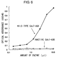
- a plasmid designated as GST-TAT-ASK(minimum)-HA-huCdc7 was prepared in order to express the active domains of human Cdc7 and ASK in E. coli .
- a GST-TAT-ASK-fused protein expression vector, GST-TAT11 was prepared.
- a restriction enzyme site (NotI) was then inserted downstream of ASK, and HA-Cdc7 was inserted at this site.
- a ribosome binding sequence (RBS; Shine-Dalgano sequence) was added immediately in front of the HA-Cdc7. Due to the addition of this potent RBS, the section from GST-TAT-ASK to HA-Cdc7 was transcribed as one sequence, and both proteins could be efficiently translated from a single transcription product. This detailed procedure is described below:
- the resulting amplification products were purified, and then cloned into a GST-TAT11 vector. Each region is arranged in this vector in the order of GST-TAT-HindIII-ASK-EcoRI.
- pGEX-2T-Tat11-TK vector was used as the GST-TAT11 vector.
- This vector comprises a structure in which a TAT sequence comprising 11 amino acids (YGRKKRRQRRR/SEQ ID NO: 13) is inserted into a GST vector.
- the HindIII-EcoRI site of TK can be digested to insert minimum ASK, which has been separately amplified with PCR. Since the HindIII frame is AAG CTT, translation in this vector terminates immediately on reaching the stop codon in the 3FRAME, just downstream of the EcoRI site.
- K90E which comprises an amino acid sequence in which the 90th amino acid, lysine (K), is mutated to glutamic acid (E).
- E. coli C600lon- was transformed with the vector GST-TAT-ASK(minimum)-HA-huCdc7, constructed in Example 2 above, in accordance with ordinary methods.
- C600lon- is a strain of E. coli that is missing lon, which is a major E. coli protease.
- the transformed E. coli was inoculated into 200 ml of LB medium containing 40 ⁇ g/ml of ampicillin, and cultured at 37°C. When the OD 600 of the culture medium reached 0.5, IPTG was added at a concentration of 1 mM, and then cultured for an additional three hours. The bacteria were collected by centrifugation, washed, and then suspended in 20 ml of buffer A (40 mM Hepes/KOH (pH 7.6), 1 mM EDTA, 40 mM potassium glutamate, 10% glycerol, and 1 mM DTT).
- buffer A 40 mM Hepes/KOH (pH 7.6), 1 mM EDTA, 40 mM potassium glutamate, 10% glycerol, and 1 mM DTT.
- the cells were disrupted using ultrasonic treatment, and the resulting products were fractionated into a soluble fraction and a precipitate (insoluble fraction) by centrifugation.
- 1 ml of glutathione Sepharose 4B was added to the soluble fraction.
- the glutathione Sepharose 4B was recovered by centrifugation, packed into a simple column, and washed well with buffer A. The column was washed until the OD 280 of the eluate reached 0.01 or less, using an amount of buffer A equal to or more than 20 times the column volume.
- the glutathione Sepharose 4B was eluted first with buffer A containing 20 mM glutathione, and then with buffer A containing 50 mM glutathione.
- the fraction finally eluted was separated by SDS-PAGE, and then silver stained, confirming the purification of GST-ASK (about 50 kDa) and HA-Cdc7 (about 65 kDa).
- RNAs were prepared from Jurkat cells using ISOGEN (Nippon Gene Cat. No. 311-02501), and cDNA was produced using You-Prime First-Strand Beads (Amersham-Pharmacia Cat. No. 27-9264-01). Using this cDNA as a template, human Mcm2(1-130) cDNA was then amplified using the Expand High Fidelity PCR System (Boehringer-Mannheim Cat. No. 1732-650) and the primers below. The resulting 390 bp PCR product was digested with the BamHI and XhoI restriction enzymes, and then cloned into the BamHI/XhoI site of the E.
- coli expression vector pGEX-4T-1 (Amersham-Pharmacia).
- the nucleotide sequence was determined, and the sequence of the resulting cDNA was confirmed to be human MCM2(1-130).
- the primers used for PCR are shown below:
- E . coli DH5a was transformed with MCM2(1-130)/pGEX-4T-1. This E. coli was cultured for about five hours at 30°C, and the culturing temperature was lowered to 20°C when the optical absorbance (600 nm) reached 0.8. IPTG was added to a final concentration of 0.2 mM, and culture was continued for a further 16 hours at 20°C.
- the collected E. coli were suspended in ice-cold solubilizing buffer (PBS, 0.1% TritonX-100 and 1 mM PMSF) and dissolved by disrupting with an ultrasonic crusher on ice.
- PBS ice-cold solubilizing buffer
- the dissolved crude extract was centrifuged for 30 minutes at 15,000 rpm with a high-speed centrifuge to separate the soluble and insoluble fractions. Each fraction was subjected to SDS-PAGE, and the GST-fusion Mcm2(1-130) was confirmed to be in the soluble fraction. The soluble fraction was added to a column filled with 2 ml of GSH-Sepharose 4B (Amersham-Pharmacia).
- the column was then washed with 50 ml of washing solution (20 mM Tris-HCl (pH 7.9), 0.5 M NaCl and 5 mM imidazole), and then eluate (10 mM glutathione and 50 mM Tris-HCl (pH 9.6) was used to elute 2 ml into each of ten containers.
- Optical absorbance (A280 nm) was measured, and that with absorbance of 0.1 or more was dialyzed overnight against ice-cold PBS.
- Western Blotting using anti-MCM2-specific antibody confirmed that the dialyzed protein was GST-fusion MCM2(1-130).
- Variant MCM2 in which serine and threonine residues 4 to 7 were substituted with alanine or glutamic acid at each site, was prepared for each region. Variants were prepared according to methods that use oligonucleotides. They were then cloned into insect cell expression vector, UW31. UW31 is a vector that can simultaneously express two types of gene products. Recombinant virus solutions co-expressing histidine-tagged MCM7 along with each variant MCM2 were obtained. These were then co-infected into Sf9 insect cells together with a recombinant virus solution that co-expresses histidine-tagged MCM4 and MCM6, and cell extracts were prepared. Affinity purification was then performed on the MCM-2-4-6-7 complex using a nickel column.
- the fractions into which the desired MCM-2-4-6-7 complex eluted were pooled.
- the MCM-2-4-6-7 complex was further purified using a mono-Q column (using the Pharmacia SMART System).
- the peak fractions eluting at 0.3 to 0.35 M NaCl were pooled. After dialyzing to a lower salt concentration, the fraction was used as a substrate for in vitro phosphorylation reactions.
- polypeptides comprising a portion of these preserved sites were prepared with a histidine or GST tag, expressed in E. coli , and then purified. They were prepared by amplifying the intended coding region using a primer oligonucleotide to which a restriction enzyme site had been added, then cloning into a histidine-tagged expression vector (pT7-7/pQE30) or GST-fusion expression vector (pGEX-5X-3). The wild type and an alanine-substituted variant were expressed. These expression vectors were respectively inserted into strain DE3 or C600lon-, and their expression was induced. Affinity purification was then carried out with a nickel column or glutathione Sepharose bead column. These proteins were also used as substrates for in vitro phosphorylation reactions.
- the substrate was MCM2-4-6-7 protein complex comprising variant MCM2
- phosphorylation was not completely absent regardless of the variant used. Therefore, the phosphorylated wild type and variant MCM2 were digested with trypsin, and then spread out using two-dimensional chromatography to analyze whether or not the spots had disappeared and the phosphorylation sites were restricted.
- the following two peptides were produced using a peptide synthesizer, and correspond to the amino acid sequence from the 10th to the 20th residue of the human MCM2 protein, comprising the 17th serine residue found to be the phosphorylated site:
- the synthesized short peptide has low antigenicity. Therefore, animals are generally immunized using peptides that have been bound to carrier proteins.
- Albumin, myoglobin, hemocyanin and so on can be used as carrier proteins, however, hemocyanin (keyhole limpet hemocyanin, hereinafter KLH; CALBIOCHEM) was used this time.
- KLH keyhole limpet hemocyanin
- the carrier protein was covalently bonded using m-maleimidobenzoyl-N-hydroxysuccinimide ester (MBS; SIGMA), which is an agent that crosslinks phospho-Hs-S83 and KLH, in accordance with the attached protocol, and KLH-Mcm2-phospho-S17 was synthesized.
- Adequate antibody titer was confirmed, and from the next week, a cycle consisting of blood collection (during the first week), rest (during the next week) and immunization (during the final week) was repeated four times. Blood was collected from the auricular vein in the same manner as for titer confirmation. Approximately 60 to 70 ml of blood was collected each time. In the final blood collection, blood was collected directly from the heart using a catheter.
- the collected blood was allowed to stand overnight at 4°C, serum was separated from clot, and the serum in the supernatant was recovered. Ammonium sulfate at a final concentration of 50% was added to the separated and recovered serum. The mixture was stired, and then separated by centrifugation. The minimum amount of PBS was added to the precipitate containing the IgG fraction, completely dissolving it. The solution was then dialyzed against PBS. After completely equilibrating with the PBS, the partially purified antibody fraction was applied to a column, and the antibody was purified.
- Mcm2-phospho-S17 synthetic peptide was used to produce the specification column, while Mcm2-S17 synthetic peptide was used to produce the absorption column.
- 1 mg of synthetic peptide was dissolved in 5 ml of 0.1 M carbonate buffer, and then added to CNBr-activated Sepharose 4B (Pharmacia) equilibrated with 1 mM hydrochloric acid. This was gently mixed overnight at 4°C, and added to a column.
- the column was washed with 5- to 10-fold PBS, and equilibrated with 1 M Tris-HCl (pH 7.5), to block the active groups remaining on the surface of the Sepharose 4B. After blocking, the column was washed and equilibrated with PBS, and used for antibody purification.
- the aforementioned partially purified antibody fraction was passed through a specification column. After washing with PBS/0.1% TritonX-100, the anti-phosphorylated peptide antibodies adhered to the column were eluted with 0.17 M glycine-HCl (pH 2.5). The eluted antibodies were immediately neutralized by addition of a suitable amount of 1 M Tris-HCl (pH 8.0), and dialyzed against PBS to obtain anti-Mcm2-phospho-S17 antibody fraction. This was completely equilibrated with PBS, and then passed through the Mcm2-S17 Sepharose 4B column, an absorption column.
- ELISA using a non-phosphorylated peptide-sensitized plate was used to confirm that the antibodies that react with non-phosphorylated peptides were removed. Specificity to the phosphorylated peptide was ultimately confirmed by ELISA using a phosphorylated peptide-sensitized plate.
- the peptide was dissolved in 0.1 M carbonate buffer at a concentration of 1 ⁇ g/ml. This was dispensed at 50 ⁇ l/well to each well of a 96-well microtiter plate for ELISA. This was then left overnight at 4°C to sensitize. Following sensitization, the peptide solution was removed, a blocking solution (1% BSA, 5% sucrose and 0.1% NaN 3 /PBS) was added to each well at 200 ⁇ l/well, and the plate was allowed to stand for about one hour at room temperature. The blocking solution was then completely removed, followed by air-drying in a draft chamber (immobilization).
- a blocking solution 1% BSA, 5% sucrose and 0.1% NaN 3 /PBS
- the plates on which phosphorylated peptide (Mcm2-phospho-S17) were immobilized were used to measure each antibody titer, confirm absorption of anti-phosphorylated-serine-residue-specific antibody, and confirm the specificity of the anti-phosphorylated-peptide antibody.
- plates on which non-phosphorylated peptide (Mcm2-S17) was immobilized were used to confirm column absorption of non-specific antibodies.
- the serum for testing antibody titer was serially diluted in 4-fold increments, starting from a 200-fold dilution using PBS.
- Purified antibody was serially diluted in 4-fold increments starting from a concentration of 1 ⁇ g/ml using PBS.
- the diluted samples were added to the sensitized plates at 50 ⁇ l per well. After addition, the plates were allowed to stand for one hour at room temperature (primary antibody reaction). The reaction solution was then discarded and each well was was washed four or more times with PBS.
- Anti-rabbit immunoglobulin antibody (secondary antibody) labeled with horseradish peroxidase (HRP) was then suitably diluted with PBS and added to each well in 50 ⁇ l aliquots.
- Anti-Mcm2-phospho-S17 antibody was serially diluted in four-fold increments from 4 ⁇ g/ml of antibody solution. Specificity was confirmed by ELISA using the plate on which phosphorylated peptide (Mcm2-phospho-S17) was immobilized, and the plate on which non-phosphorylated peptide (Mcm2-S17) was immobilized.
- Fig. 2 shows the results of confirming the specificity of anti-Mcm2-phospho-S17 polyclonal antibody.
- the optical absorbance of the plate on which Mcm2-phospho-S17 phosphorylated peptide was immobilized increased with increasing antibody concentration.
- the optical absorbance of the plate on which Mcm2-S17 non-phosphorylated peptide was immobilized remained at nearly zero.
- the subject antibody was proven to specifically recognize phosphorylation of the 17th serine residue of human Mcm2.
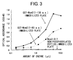
- Regions comprising the 17th serine residue of human Mcm2, the substrate used, were selected in order to measure Cdc7-ASK complex phosphorylation activity in a 96-well microtiter plate. More specifically, the substrates studied were 1) GST fusion proteins with the amino acid regions of residues 1 to 130 and 1 to 80 of human MCM2, which phosphorylation assays using radioisotopes found to be a good substrate for Cdc7-ASK complex, and 2) a non-phosphorylated peptide corresponding to the amino acid sequence from the 10th to 20th amino acid residues (Mcm2-S17).
- GST-Mcm2 (1-130 a.a.) and GST-Mcm2 (1-80 a.a.) were diluted to 5 ⁇ g/ml using 0.1 M carbonate buffer, pipetted at 50 ⁇ l/well in to each well of a microtiter plate for ELISA, and then sensitized overnight at 4°C. The sensitizing solution was removed, a blocking solution (1% BSA, 5% sucrose and 0.1% NaN 3 /PBS) was pipetted in to each well at 200 ⁇ l/well, and the plate was allowed to stand for about one hour at room temperature. The blocking solution was then completely removed. The plates were then air-dried in a draft chamber, and stored at 4°C until use. GST-Mcm2 (1-130 a.a.) and GST-Mcm2 (1-80 a.a.) were immobilized on the inner walls of the microtiter plates by this procedure.
- a protein phosphorylation reaction was then carried out in the wells in which recombinant proteins GST-Mcm2 (1-130 a.a.), GST-Mcm2 (1-80 a.a.) or non-phosphorylated peptide (Mcm2-S17) had been immobilized.
- ELISA was then carried out in succession in the same wells.
- a recombinant Cdc7-ASK complex dilution series was prepared by diluting from one-fold (x1) to 64-fold in two-fold increments using a phosphorylation buffer. 50 ⁇ l aliquots of this solution were added to the wells of the immobilized plates (a protein phosphorylation reaction). After incubating at 30°C for a fixed period of time, the phosphorylation reaction solution was discarded and each well was amply washed four or more times with PBS.
- Primary antibody that is, anti-Mcm2-phospho-S17 polyclonal antibody
- an antibody diluent 1% BSA, 0.1% NaN 3 /PBS
- the primary antibody reaction solution was then discarded and each well was amply washed four or more times with PBS.
- Anti-rabbit immunoglobulin antibody (secondary antibody, MBL) labeled with horseradish peroxidase (HRP) was then diluted 1000-fold with PBS and added to each well in 50 ⁇ l aliquots, then allowed to react for 30 to 60 minutes at room temperature (secondary antibody reaction). The secondary antibody reaction solution was then discarded and each well was washed four or more times with PBS. A coloring substrate solution was then added to each well in 50 ⁇ l aliquots, and color was developed for five to 20 minutes (coloring reaction). The coloring reaction was stopped by adding 50 ⁇ l aliquots of reaction stopping solution to each well, and the optical absorbance at 450 nm was measured using a microplate reader.
- MBL secondary antibody labeled with horseradish peroxidase
- Fig. 4 shows the results of investigating changes in reactivity when the final concentration of ATP added to the phosphorylation reaction solution was changed from 1 ⁇ M to 2 mM, in the method for measuring Cdc7-ASK complex activity using GST-Mcm2 (1-130 a.a.) and Mcm2-phospho-S17 polyclonal antibody, as described above.
- Enzyme activity is shown as a relative activity value (%), where activity at 2 mM is taken as 100.
- Activity of 90% is indicated at 0.1 mM, and the reaction virtually plateaus at 1 mM.
- activity equal to or more than 30% of the maximum activity value was indicated, even in the presence of low concentrations of ATP, reactivity was clearly shown to be ATP-dependent.
- Fig. 5 shows the results of investigating changes in reactivity when the phosphorylation reaction time in the method for measuring Cdc7-ASK complex activity was changed from 0 to 150 minutes. Increases in absorbance were observed to be dependent on reaction time, and the reaction virtually plateaued at 90 minutes.
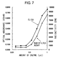
- a radiofilter assay is a method for detecting substrate phosphorylation that uses uptake of the radioisotope [ ⁇ - 32 P]ATP into an acid-insoluble fraction. It is frequently used in measuring phosphorylation activity. The sensitivity of this method and the ELISA measurement system provided in the present invention were compared.
- a recombinant Cdc7-ASK complex dilution series diluted from one-fold (x1) to 64-fold in two-fold increments was prepared using a phosphorylation buffer in 1 ⁇ g of recombinant protein GST-Mcm2 (1-130 a.a.), and then adjusted to a total volume of 50 ⁇ l. After incubating at 30°C for a fixed period of time, the reaction was stopped by adding 1 ml of 10% trichloroacetic acid and 0.2% Na 4 P 2 O 7 . The acid-insoluble fraction was trapped in a GFC filter (Whatman), and washed three times using 2% trichloroacetic acid and 0.02% Na 4 P 2 O 7 . The radioactivity of the [ ⁇ - 32 P]ATP taken up by the substrate was counted with a liquid scintillation counter.
- the results are shown in Fig. 7.
- the results are shown as relative values (%), where both the radioactive count (cpm) in the radiofilter assay, and the optical absorbance in ELISA, are taken to be 100% at the one-fold concentration of Cdc7-ASK complex enzyme.
- the ELISA method could measure about 1/8 the amount of enzyme, since the value was 100% up to x8.
- the amount of substrate used in the ELISA method is a calculated value of 0.25 ⁇ g per well
- the radiofilter assay requires about 1 ⁇ g, or four times this amount.
- there was hardly any background interference in the absence of enzyme thus, these systems are more sensitive, require smaller amounts of substrate and enzyme, and can be measured without using radioisotopes, demonstrating the superiority of the ELISA measurement systems provided by the present invention
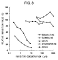
- K-252a (Calbiochem) and Staurosporine are known as kinase inhibitors. They are known as inhibitors of various kinases such as CaM kinase II, protein kinase A, protein kinase C, and protein kinase G.
- the ELISA systems provided by the present invention were studied using K-252a and Staurosporine. Dilution series of each inhibitor were prepared, mixed with Cdc7-ASK complex prepared with a phosphorylation assay buffer, and then adjusted to a total volume of 50 ⁇ l. This was then added to wells in which GST-Mcm2 (1-130 a.a.) was immobilized. Using an above-described method, ELISA was then used to examine Cdc7-ASK complex phosphorylation activity.
- Fig. 8 shows the relative inhibition values (%), where the activity value without an inhibitor is taken as 100. Since both K-252a and Staurosporine inhibitors are kinase inhibitors with low specificity, inhibition of phosphorylation activity by Cdc7-ASK complex was concentration-dependent. The IC50 value was 2 ⁇ M for both of these inhibitors. On the other hand, concentration-dependent inhibitory effect for Cdc7-ASK complex was not observed for Roscovitine, Olomoucine, and UO126. Roscovitine and Olomoucine are cdk2 inhibitors, and UO126 is a MAP kinase inhibitor. These kinase inhibitors were shown to lack Cdc7-ASK complex inhibition activity. Therefore, the ELISA system provided by the present invention was in fact shown to be effective in screening for agents that inhibit Cdc7-ASK complex phosphorylation activity.
- the mechanism by which the substrate protein, MCM2, is phosphorylated by Cdc7-ASK complex was elucidated.
- substrate protein phosphorylation by the Cdc7-ASK complex could be more specifically evaluated.
- the methods for measuring phosphorylation effect of the present invention are useful as methods in which the phosphorylating action of Cdc7-ASK complex can be specifically evaluated.
- the kinase activity of Cdc7-ASK has been clearly shown to increase in actively growing cancer cells (Gene, 1998, Apr 28;211(1):133-40, "A human homolog of the yeast CDC7 gene is overexpressed in some tumors and transformed cell lines.”, Hess G.F., Drong R.F., Weiland K.L., Slightom J.L., Sclafani R.A., and Hollingsworth R.E., Cancer Research, Pharmacia, Upjohn, Inc., 301 Henrietta Street, Kalamazoo, MI 49001, USA).
- the growth ability of certain cancer cells can be predicted based on the kinase activity of Cdc7-ASK.
- Cdc7-ASK kinase activity which can be evaluated by the methods of the present invention, is useful as a carcinogenesis marker.
- Cdc7-ASK kinase activity can be specifically evaluated by the present invention.
- the growth ability of cancer cells can be evaluated more specifically.
- the present invention has realized methods for evaluating the effects of test compounds on the phosphorylation function of the Cdc7-ASK complex, as well as screening methods based on these evaluation methods.
- the present invention's methods for measuring Cdc7-ASK kinase activity, or of screening for compounds that regulate that activity, are useful for the development of cancer medicaments and treatment methods targeting Cdc7-ASK.
- the use of Cdc7-ASK as a target offers the following advantages:
- Cdc7-ASK by targeting Cdc7-ASK, more reliable control of activity can be expected. While Cdk-Cyclin is a family composed by numerous similar genes, Cdc7-ASK is composed of molecules whose structure is more limited. Thus, by using Cdc7-ASK as a target, activity can be reliably controlled.
- Cdc7-ASK is a factor required for the initiation and progress of the S phase. Upon loss of this activity the S phase immediately stops and cell growth is arrested. Moreover, the results of experiments using genetically manipulated mice suggest that interruption of the S phase, namely DNA replication, can potentially cause abnormal structures to accumulate in DNA, which when detected as DNA damage, can induce p53 and eventually cell death. In other words, inhibition of Cdc7-ASK activity can effectively block progression of the S phase in cancer cells, efficiently removing cancer cells by inducing cell death.
- Cdc7-ASK as a target is expected to lead to the discovery of completely new cell growth inhibitors.
- the structure of Cdc7-ASK kinase is unique, even among members of the kinase family.
- the structure of ASK is also different from that of cyclin molecules, on which considerable research has been conducted. This activation is predicted to be due to a novel, and heretofore unknown, mechanism regarding kinase activation.
- searching for activity inhibitors that target Cdc7-ASK is highly likely to lead to the discovery of completely new substances, undiscovered by the various types of kinase screening to date. This means that, for example, new lead compounds may be discovered in the research and development of anti-cancer agents.
- Cdc7-ASK induction of cell death can be expected when using Cdc7-ASK as a target.
- cells activate so-called check-point kinases, such as ATM, Chk1, and Cds1, which respond by delaying the progress of the cell cycle.
- check-point kinases such as ATM, Chk1, and Cds1
- Cdc7-ASK inhibitors are used in combination with inhibitors against these check-point kinases, enhanced induction of cell death, due to inhibited S phase progression caused by inhibiting Cdc7-ASK activity, can be expected.
- Cdc7-ASK complex plays important roles in the initial stages of cell growth in the cell cycle. Moreover, its phosphorylation activity is proportional to the ability of cancer cells to grow. In other words, cancer cell growth can be more specifically controlled by controlling Cdc7-ASK complex activity.
- the screening methods of the present invention are useful as methods for obtaining compounds that can specifically control cancer cell growth.
- the screening methods of the present invention are also able to more specifically evaluate the effects of test compounds on the phosphorylation function of Cdc7-ASK complex. As a result, compounds that act more specifically on cancer can be selected.
- the present invention has also provided a methods for preparing the Cdc7-ASK complex active substances required by each of the aforementioned methods. According to the methods of the present invention, complexes comprising activity similar to that of the Cdc7-ASK complex can be easily prepared in large volumes by using prokaryotic cells.
- the present invention provides substrate proteins that are phosphorylated by the Cdc7-ASK complex.
- the substrate proteins of the present invention are proteins that comprise the structure required for phosphorylation by the Cdc7-ASK complex (or Cdc7-ASK complex active substance). By using such substrate proteins the phosphorylation effect of Cdc7-ASK complex can be more specifically evaluated.
- the Cdc7-ASK complex active substances and substrate proteins prepared based on the present invention are useful in the aforementioned methods.
- the present invention has provided antibodies that identify the level of phosphorylation of substrate proteins phosphorylated by the Cdc7-ASK complex or Cdc7-ASK complex active substance.
- the antibodies of the present invention identify the level of phosphorylation at specific substrate protein sites. These antibodies make it possible to more specifically evaluate the phosphorylation effect of the Cdc7-ASK complex or Cdc7-ASK complex active substance.
- the use of the antibody of the present invention enables the phosphorylation effect of the Cdc7-ASK complex or Cdc7-ASK complex active substance to be easily evaluated based on immunoassay principles.
Landscapes
- Chemical & Material Sciences (AREA)
- Health & Medical Sciences (AREA)
- Life Sciences & Earth Sciences (AREA)
- Organic Chemistry (AREA)
- Engineering & Computer Science (AREA)
- Zoology (AREA)
- Genetics & Genomics (AREA)
- General Health & Medical Sciences (AREA)
- Wood Science & Technology (AREA)
- Molecular Biology (AREA)
- Bioinformatics & Cheminformatics (AREA)
- Medicinal Chemistry (AREA)
- Biochemistry (AREA)
- Proteomics, Peptides & Aminoacids (AREA)
- Immunology (AREA)
- Biotechnology (AREA)
- General Engineering & Computer Science (AREA)
- Biophysics (AREA)
- Microbiology (AREA)
- Public Health (AREA)
- Analytical Chemistry (AREA)
- General Chemical & Material Sciences (AREA)
- Veterinary Medicine (AREA)
- Physics & Mathematics (AREA)
- Chemical Kinetics & Catalysis (AREA)
- Animal Behavior & Ethology (AREA)
- Nuclear Medicine, Radiotherapy & Molecular Imaging (AREA)
- Pharmacology & Pharmacy (AREA)
- Biomedical Technology (AREA)
- Measuring Or Testing Involving Enzymes Or Micro-Organisms (AREA)
- Enzymes And Modification Thereof (AREA)
- Peptides Or Proteins (AREA)
- Investigating Or Analysing Biological Materials (AREA)
- Medicines That Contain Protein Lipid Enzymes And Other Medicines (AREA)
Abstract
The present invention provides methods for measuring the
phosphorylation activity of Cdc7-ASK kinase complex by using as
an indicator the level of phosphorylation at a phosphorylation
site of MCM, which is a substrate of Cdc7-ASK kinase complex.
The effects of test compounds on the phosphorylation activity of
Cdc7-ASK kinase complex can also be evaluated based on these
measurement methods. Compounds that inhibit this
phosphorylation activity are useful as anti-cancer agents having
superior specificity for cancer.
Description
The present invention relates to methods for measuring
the phosphorylation activity of Cdc7-ASK kinase complex.
During cell growth, cells repeat cyclic processes whereby
they replicate their genomic DNA, distribute this DNA equally
among daughter cells, and then divide. This type of cycle is
referred to as a cell cycle. The cell cycle can be divided into
the G1 phase (DNA synthesis preparatory phase), S phase (DNA
synthesis phase), G2 phase (mitosis preparatory phase), and M
phase (mitotic phase). Cells divide by going through each stage
in order. The progress of the cell cycle is finely regulated by
numerous molecules, controlling unnecessary cell growth. To
date, a number of molecules involved in cell cycle progression
have been cloned and functionally analyzed. Abnormalities in
molecules involved in cell cycle progression have been reported
in numerous cancers, and can be considered possible causes of
carcinogenesis.
To date, a variety of anti-cancer agents and anticancer
agents have been developed. Much abnormal cancer cell growth is
recognized as originating from abnormal cell cycle progression.
In line with this, anticancer agents that targeting factors
which regulate cell cycle progression are being vigorously
developed. In particular, the function of Cdk-Cyclin, which has
been most researched as a target for anticancer agents, has been
considerably analyzed. Numerous candidate molecules for
anticancer agents that target Cdk-Cyclin have already been
reported.
Cdk is a family of molecules with numerous structural
similarities, and each of these molecules forms complexes with
various cyclin molecules specific to the cell cycle, thus
regulating the various stages of the cell cycle. At present,
particular molecules that specifically inhibit only Cdk-Cyclin
have not yet been identified from among these structurally
similar molecules. In other words, obtaining a molecule that
specifically and selectively acts on cancer cells by targeting
Cdk-Cyclin has been difficult. In addition, although numerous
anticancer agents have been found that target regulatory factors
in cell division or the G1 (stationary) phase, few agents have
been discovered that target the process of DNA replication,
another important regulatory point of cell growth.
A serine/threonine kinase different from Cdk-Cyclin has
been found to play an important role at the S phase initiation
point (the transition from G1 to S). Namely, in variant line
Cdc7, which was isolated as a line with a variant cell division
cycle (J. Mol. Biol., 59:183-194, 1971), the Cdc7 protein kinase
has been demonstrated to function just prior to the initiation
of chromosomal DNA replication, and to be necessary for
activation of each origin of replication throughout the S phase
(Mol. Cell. Biol., 6:1590-1598, 1986; Genes Dev., 15:480-490,
1998; and Genes Dev., 15:491-501, 1998). In addition, yeast
Cdc7 kinase activity is known to be dependent on regulatory
subunit, Dbf4 (Genetics, 131:21-29, 1992; and Mol. Cell. Biol.,
13:2899-2908, 1993).
Expression of Dbf4 in yeast is cyclical, and regulated at
both the transcription and post-translation levels (Exp. Cell
Res., 180:419-428, 1989). At least some of the increase in Cdc7
kinase activity during the G1-S transition phase is explained by
an increase in expression of Dbf4 during the late G1 phase (Mol.
Cell. Biol., 13:2899-2908, 1993; and Exp. Cell Res., 180:419-428,
1989). Moreover, since Dbf4 interacts with the origins of
replication in cells (Science, 265:1243-1246, 1994), Cdc7 is
thought to trigger S phase initiation by directly activating the
replication apparatus formed on an origin of replication.
The present inventors succeeded in isolating H37 protein,
which is a human homolog of yeast Dbf4, and DNA encoding it
(Unexamined Published Japanese Patent Application No. (JP-A)
2000-135090, J. Biol. Chem., Vol. 275, No. 37, 29042-29052,
2000). H37 is an important regulatory factor that governs the
switching on and off of DNA replication initiation. H37 was
subsequently named a human Activator of S phase Kinase (ASK).
ASK is a unique gene in human chromosomes, and its
function is essential for chromosome replication. Based on the
results of developmental engineering genetic analyses using mice,
Cdc7 function was verified as being essential at the cellular
and animal levels. Cdc7 encodes a kinase catalyst subunit,
while ASK encodes each of the active subunits. The two form a
complex, comprising an active kinase. The activity of Cdc7-ASK
kinase is strictly regulated by the cell cycle, and increases in
the S phase when DNA replication occurs. Although this kinase
activity is maintained at a high level throughout the S phase,
it is no longer detected from the M phase to the G1 phase.
Regulation of this activity mainly depends on fluctuations in
the level of ASK protein expression. Namely, the amount of ASK
protein fluctuates during the cell cycle, and as a result, the
activity of human Cdc7 kinase, which is regulated by being bound
to ASK, also fluctuates during the cell cycle.
In addition, the expression level and activity of human
Cdc7-ASK is typically observed to increase in actively growing
cancer cells. In such cancer cells, the activity of human Cdc7
kinase activity is thought to increase with increasing
expression of ASK protein. Of even further interest, the
present inventors found that the expression level of ASK
increases in response to increases in the growth ability of
various cultured cancer cells.
Subsequent research by the present inventors demonstrated
that the specific substrate phosphorylated by Cdc7-ASK complex
is a minichromosome maintenance (MCM) complex (heterohexamer),
essential for the initiation of chromosome replication (J. Biol.
Chem., Vol. 275, No. 37, 29042-29052, 2000). On the basis of
this finding, the present inventors advocate a model in which
Cdc7-ASK phosphorylates the MCM present in the replication
origin complex, and as a result, induces reconfiguration of the
subunit structure, causing the double-stranded DNA split that is
essential for replication initiation.
On the basis of this, phosphorylation of a substrate
protein by the Cdc7-ASK complex is recognized as being an
important phenomenon in regulating the growth of cancer cells.
However, many aspects of this phosphorylation mechanism remain
to be elucidated.
An object of the present invention is to clarify the
mechanism of substrate protein phosphorylation by the Cdc7-ASK
complex, and to provide methods for evaluating its activity. In
addition, based on these evaluation methods, an object of the
present invention is to provide methods for measuring the
influence of Cdc7-ASK complex test compounds on the substrate
phosphorylation effect. Another object of the present invention
is to provide substrates, antibodies, or Cdc7-ASK complexes
useful for these methods.
The present inventors considered the Cdc7-ASK complex to
be a novel and important anti-cancer agent target; that is, they
thought that inhibitors that could inhibit the phosphorylation
effect of the Cdc7-ASK complex could selectively regulate cancer
cell growth, and would be candidates for effective anticancer
agents.
In order to evaluate the action of inhibiting the Cdc7-ASK
complex phosphorylation effect, it was first essential to
establish methods to evaluate this effect. To achieve this, the
present inventors first clarified the mechanism used by the
Cdc7-ASK complex to phosphorylate substrate proteins. On the
basis of this finding, the present inventors confirmed that the
kinase activity of the Cdc7-ASK complex was measurable, thereby
completing the present invention.
The present inventors then discovered that the influence
of test compounds on the Cdc7-ASK complex's substrate protein
phosphorylation function can be evaluated by applying these
methods to measure kinase activity. On the basis of this
finding, the present inventors established methods for
evaluating the inhibitory (or promotional) effect of test
compounds on Cdc7-ASK kinase, and methods of screening for
compounds comprising such effects, thereby completing the
present invention.
Moreover, while establishing these methods, the present
inventors discovered novel substrate compounds, antibodies,
Cdc7-ASK complexes or so on useful to these methods, thereby
completing the present invention. Namely, the present invention
relates to methods for measuring the kinase activity of the
Cdc7-ASK complex, as below. Alternatively, the present
invention relates to methods for evaluating the effect of test
compounds on the kinase activity of the Cdc7-ASK complex, and
screening methods based on these methods. Moreover, the present
invention relates to substrate compounds, antibodies or Cdc7-ASK
complexes useful to these methods, or methods for preparing the
compounds, antibodies or Cdc7-ASK complexes.
Alternatively, the present invention relates to methods
for inhibiting cell growth that comprise the step of
administering a compound selected according to the screening
method of [6]. In addition, the present invention relates to
the use of a compound, selected according to the screening
method of [6], in the production of a cell growth inhibitor.
The present invention relates to methods for measuring
the kinase activity of Cdc7-ASK complex, where the methods
comprise the following steps:
In the present invention, a protein comprising the amino
acid sequence of SEQ ID NO: 1, or a protein functionally
equivalent to that protein, is used as a substrate protein.
The structure of MCM2 protein is known. The amino acid
sequence of human MCM2, and the nucleotide sequence of the cDNA
that encodes it, are shown in SEQ ID NOs: 2 and 3 respectively.
Cdc7-ASK complex phosphorylates a specific amino acid residue of
the human MCM2 of SEQ ID NO: 3. The present inventors have
previously demonstrated that Cdc7-ASK complex phosphorylates MCM
complex or free MCM2. However, that the Cdc7-ASK complex
specifically phosphorylates the serine at position 17 from the
MCM2 N-terminal is a novel finding, revealed by the present
inventors.
Thus, proteins comprising continuous amino acid sequences
that comprise the position 17 serine and are selected from the
amino acid sequence of SEQ ID NO: 3, can be used as substrate
proteins in the present invention, as long as they can be
phosphorylated by Cdc7-ASK complex. Examples of other substrate
proteins that can be used in the present invention are indicated
below. Tags can be added to these substrate proteins. In
addition, these substrate proteins can be bound to a solid phase,
as described later.
In the present invention, the phosphorylation of a
certain protein by the Cdc7-ASK complex can be confirmed based
on the results of, for example, the reactions described in
Examples. Namely, a phosphate compound and the Cdc7-ASK complex
are incubated with a substrate protein under conditions that
allow phosphorylation of that protein (described below). Next,
the level of phosphorylation at the serine residue at the
position corresponding to position 17 in the amino acid sequence
shown in SEQ ID NO: 1 is measured. When there is no significant
difference observed between the measured level of
phosphorylation and the level when, for example, a protein
comprising the amino acid sequence of SEQ ID NO: 1 is used as a
substrate protein, then that protein is judged to be a protein
phosphorylated by human Cdc7-ASK complex.
The number of amino acids that make up the substrate
protein of the present invention can generally be five or more,
usually ten or more, and preferably 50 or more. In addition,
although there are no restrictions as to the size of the
substrate protein, it can be a short fragment of, for example,
400, generally 300, or 200 or less. Phosphorylation by the
Cdc7-ASK complex can be observed more specifically using a short
fragment as a substrate protein.
In the present invention, proteins comprising, for
example, the amino acid sequence of SEQ ID NO: 1, can be
indicated as preferable substrate proteins. The amino acid
sequence of SEQ ID NO: 1 is a fragment sequence comprising 130
amino acids on the N-terminal side of the human MCM2 of SEQ ID
NO: 3. The serine located at the 17th amino acid counting from
the N-terminal is phosphorylated by the Cdc7-ASK complex.
In addition, proteins which are phosphorylated by human
Cdc7-ASK complex and that comprise amino acid sequences in which
one or more amino acids in the amino acid sequence of SEQ ID NO:
1 are substituted, deleted, added and/or inserted, can also be
used as substrate proteins. In the present invention the number
of amino acids subjected to mutation is, for example, one to ten,
preferably one to six, and more preferably one to three.
Mutation of an amino acid sequence may be a mutation that
is induced artificially, or that occurs spontaneously. When
substituting amino acids, conservative substitution can be used.
In general, to maintain protein function, the substitute amino
acids are preferably those with properties similar to the amino
acids prior to substitution. This type of amino acid
substitution is referred to as conservative substitution.
For example, since Ala, Val, Leu, Ile, Pro, Met, Phe and
Trp are all classified as non-polar amino acids, they have
mutually similar properties. Examples of non-charged amino
acids include Gly, Ser, Thr, Cys, Tyr, Asn and Gln.
Alternatively, examples of acidic amino acids include Asp and
Glu. Moreover, examples of basic amino acids include Lys, Arg
and His. The amino acids that compose each of these groups have
mutually similar properties. Consequently, there is a high
possibility that protein function will be maintained when these
amino acids are substituted with other amino acids from the same
group.
This type of protein can be obtained by introducing a
mutation into the nucleotide sequence of the cDNA of the human
MCM2 of SEQ ID NO: 1. Technology for introducing mutations into
genes comprising known nucleotide sequences is known.
Alternatively, proteins comprising desired amino acid sequences
can be prepared by chemical synthesis.
In the substrate proteins of the present invention, the
serine located at the position corresponding to position 17 of
the amino acid sequence of SEQ ID NO: 1 is important as the
phosphorylation target of the Cdc7-ASK complex. Thus, even when
the substrate protein is a protein comprising an amino acid
sequence mutation, it is important that a serine be preserved at
position 17, or a position homologous to position 17. The term
"position homologous to position 17" refers to a position that
corresponds to position 17 of the amino acid sequence of SEQ ID
NO: 1, when a given amino acid sequence is aligned with the
amino acid sequence of SEQ ID NO: 1. Multiple methods are known
for aligning amino acid sequences. For example, a wide variety
of software, based on algorithms such as the BLAST algorithm, is
in practical use for aligning different amino acid sequences.
In addition, a protein that comprises an amino acid
sequence comprising 90% or more homology to the amino acid
sequence of SEQ ID NO: 3, and which is phosphorylated by human
Cdc7-ASK complex, can also be used as a substrate protein of the
present invention. For example, human Cdc7-ASK complex
phosphorylates not only human MCM2, but also mouse MCM2. Thus,
mouse MCM2 can be used as a substrate protein in the present
invention. The amino acid sequence of mouse MCM2 is shown in
SEQ ID NO: 5, while the nucleotide sequence that encodes this
amino acid sequence is shown in SEQ ID NO: 4. Mouse MCM2 and
human MCMC2 are 99% homologous. Thus, proteins comprising 99%
or more homology to the amino acid sequence of human SEQ ID NO:
3 (the full-length amino acid sequence of human MCM2) are useful
as substrate proteins of the present invention.
In the present invention, the level of phosphorylation of
the serine at position 17 of human MCM2, or of a serine at a
position homologous to this serine, is measured as an indicator.
When using a protein that comprises an amino acid sequence
different from the amino acid sequences of SEQ ID NOs: 1 and 3
as a substrate protein, it is important to preserve the serine
at position 17 of SEQ ID NO: 1, or serines at positions
homologous to position 17. For example, in mouse MCM2, the
serine at position 26 is the serine at a position homologous to
position 17. Thus, when using mouse MCM2 fragments as substrate
proteins, it is preferable to use proteins that comprise an
amino acid sequence comprising a serine at position 26.
The substrate protein of the present invention can be
obtained based on genetic engineering techniques. Namely, a
protein having the desired amino acid sequence can be obtained
by expressing a DNA encoding MCM2 in a suitable expression
system. The nucleotide sequence of a DNA encoding MCM2 is shown
in SEQ ID NO: 2.
For example, the Examples show an example of expressing
the MCM2 gene using Escherichia coli. The expression product
can be purified using techniques such as salting out, gel
filtration, ion exchange chromatography or so on. Purification
by affinity chromatography, using the binding affinity of a tag
with binding affinity that has been fused to MCM2, can also be
used. Examples of binding tags that can be used include a
histidine tag comprising several histidines (His-Tag), β-D-galactosidase,
glutathione S-transferase (GST), thioredoxin,
maltose-binding protein, Myc, Xpress, and FLAG. If GST is used,
for example, the expressed protein can be purified easily with a
Glutathione Sepharose 4B column or so on.
Depending on the expression system, MCM2 may be recovered
in a phosphorylated state. When the substrate protein has been
phosphorylated in the expression system, the protein can be
dephosphorylated by treatment with an enzyme such as phosphatase,
which acts on phosphate groups. The measurement methods of the
present invention comprise the step of measurement using the
phosphorylation of substrate proteins as an indicator. Thus, it
is particularly preferable that the substrate protein sites to
be phosphorylated by the Cdc7-ASK complex are not phosphorylated.
In addition, methods are also known for chemically synthesizing
proteins comprising the desired amino acid sequence.
The present invention's methods for measuring the kinase
activity of Cdc7-ASK complex comprise the step of contacting the
aforementioned substrate proteins with samples whose Cdc7-ASK
complex kinase activity is to be measured, under conditions that
allow phosphorylation of the substrate protein. In the present
invention, "conditions that allow phosphorylation of the
substrate protein" refer to conditions suited to expression of
Cdc7-ASK complex kinase activity. More specifically, the
temperature, salt concentration, and pH are suited to the
expression of enzyme activity, and phosphate compounds are
present for substrate protein phosphorylation. "Conditions
suitable for enzyme activity" can be exemplified as below.
Namely, such reaction conditions can be imparted by contacting
the aforementioned components in HEPES buffer adjusted to pH 7.0
to 7.5.
In the present invention, "conditions that allow
phosphorylation of a substrate protein" refer to contacting a
sample with a substrate protein in the presence of a compound
comprising a phosphate group necessary for substrate protein
phosphorylation. Phosphate groups can be supplied by, for
example, coexisting with adenosine triphosphate (hereinafter
described as ATP).
The substrate protein and phosphate compound that
comprise the aforementioned reaction are preferably added in
excess with regards to the Cdc7-ASK complex kinase activity in
the sample. It is generally difficult to accurately predict the
kinase activity of Cdc7-ASK complex contained in a sample.
However, those with ordinary skill in the art are able to
experimentally predetermine an adequate amount of substrate
compound or phosphate compound, such that the components
required by the reaction are sufficient for enzyme reaction.
For example, when measuring a human-derived biological sample,
the amount of substrate protein to be used is, for example, 0.1
µg or more, and generally 0.2 µg to 1 µg. The amount of
phosphate compound to be added as ATP can be, for example, 0.01
mM or more, and generally 0.1 mM to 1 mM. Furthermore,
phosphate compound is added in an amount that is necessary and
sufficient for phosphorylation of the substrate protein used in
the reaction system.
The reaction time for phosphorylation is typically ten
minutes or more, and preferably 30 minutes or more. Reliable
phosphorylation of substrate protein can be expected from, for
example, 50 to 100 minutes of reaction. The time required for
phosphorylation varies according to the amount of substrate
protein used, and the level of Cdc7-ASK complex activity in the
reaction solution. Those with ordinary skill in the art are
able to suitably set the reaction time required for
phosphorylation under given conditions.
In the measurement methods of the present invention, the
level of substrate protein phosphorylation over a predetermined
time relates to phosphorylation activity in the sample.
Alternatively, the phosphorylation activity in a sample can also
be measured by using, as an indicator, the reaction time
required to phosphorylate a substrate protein to a predetermined
level. Namely, the greater a sample's phosphorylation activity,
the faster the substrate protein phosphorylation will reach a
predetermined level. Moreover, phosphorylation activity in the
sample can also be measured by using the amount of sample
required to phosphorylate the substrate protein to a
predetermined level as an indicator. In this embodiment, the
amount of sample required for phosphorylation is reversely
proportional to the level of phosphorylation activity in the
sample.
When the phosphorylation activity in a sample far exceeds
that predicted, the majority of the substrate protein will be
phosphorylated in a short period of time. If this happens,
accurate evaluation of phosphorylation activity may be
impossible. Thus, when such results are obtained, it is
preferable to reduce the amount of sample added to the reaction,
and remeasure phosphorylation activity.
The measurement methods of the present invention comprise
the step of measuring the phosphorylation level of the substrate
protein for the serine residue at the position corresponding to
position 17 of the amino acid sequence of SEQ ID NO: 1. The
level of phosphorylation at this position can be measured
immunologically by using, for example, an antibody that
recognizes the phosphorylated state of the serine residue at a
position corresponding to position 17.
More specifically, an antibody can be used for which
binding activity decreases with serine dephosphorylation, as
compared to binding activity to an antigen-determining group
comprising the phosphorylated serine residue of a protein with
an amino acid sequence comprising a serine that corresponds to
position 17. Conversely, antibodies that bind to antigen-determining
groups comprising a non-phosphorylated serine
residue, but for which bonding activity is decreased with
phosphorylation of the serine residue, can also be used.
In the measurement methods of the present invention,
antibodies that identify the level of phosphorylation of the
aforementioned serine residue, used to measure the level of
phosphorylation, can be obtained in, for example, the following
manner: First, a synthetic peptide that comprises a continuous
amino acid sequence comprising the applicable serine residue can
be used as the immunogen. The length of the synthetic peptide
used as the immunogen can be at least three or more, for example
five or more, normally ten or more, and preferably ten to 20
amino acids. A preferable example of an immunogen is an amino
acid sequence composed of 15 continuous amino acids, containing
serine at position 17, and selected from the amino acid sequence
described in SEQ ID NO: 1. The position of the serine can be
arbitrary. For example, in the Examples, a synthetic peptide
was used as an immunogen that comprised an amino acid sequence
in which serine was located in the center of 15 amino acids.
The synthetic peptide can be bonded to a carrier protein.
Keyhole Limpet hemocyanin and such can be used as the carrier
protein.
The serine residue corresponding to position 17 in the
immunogen can be phosphorylated in advance. Methods for
obtaining synthetic peptides in which a specific amino acid has
been phosphorylated are known. When the serine in the immunogen
is phosphorylated, antibodies comprising the activity of binding
to phosphorylated serine can be obtained.
Immunogens are administered to immunity animals by mixing
with suitable adjuvants. Immunization is normally performed
several times, and blood is collected from immunized animals
after confirming an adequate rise in antibody titer. Serum
recovered from the collected blood can be used as antiserum that
contains an antibody of the present invention. Purified
antibodies can be obtained by purifying immunoglobulins from
such antiserums.
Alternatively, cell lines that produce monoclonal
antibodies can be established by immortalizing antibody-producing
cells from the immunized animals, and then selecting
clones that produce antibodies comprising the desired reactivity.
For example, antibody-producing cells can be immortalized by
fusion with cells such as myeloma cells, to produce hybridomas.
Antibodies can be confirmed to have desired reactivity by
using the synthetic peptide used as the immunogen. For example,
antibodies that recognize phosphorylated substrate proteins can
be obtained by selecting antibodies that bind to synthetic
peptides comprising phosphorylated serine. Moreover, an
antibody that specifically binds to a phosphorylated substrate
protein can be obtained by removing from this antibody any
antibodies that bind to synthetic peptides that comprise non-phosphorylated
serine. Using this kind of selection process,
antibodies that recognize the phosphorylated state of substrate
proteins, essential to the present invention, can be obtained by
treating with antiserum or purified antibody.
Hybridomas that produce monoclonal antibodies able to
recognize the phosphorylated state of substrate proteins can be
selected by screening antibodies produced by the hybridomas,
according to similar selection processes.
In the measurement methods of the present invention, the
aforementioned antibodies can be labeled in advance. Antibodies
bound to phosphorylated sites can be easily detected by labeling
antibodies. In addition, if the signals from labels can be
amplified, measurements of higher sensitivity can be expected.
Arbitrary labeling components can be used for antibody
labeling. For example, fluorescent dyes, enzymes, radioactive
substances, and so on can be used as labels. In addition,
substances with high electron density, such as ferritin and gold
colloids, can also be used as labels.
Examples of fluorescent dyes include fluorescein
isothiocyanate (FITC), rhodamine β-isothiocyanate (RITC), and
phycoerythrin (PE). Examples of enzymes include peroxidase, β-D-galactosidase,
microperoxidase, alkaline phosphatase, acidic
phosphatase, and cytochrome c. Examples of radioactive
substances include 125I, 14C and 3H. Methods for labeling
antibodies with these labeling components are known.
In addition, antibodies can also be labeled using
antibodies (hereinafter referred to as secondary antibodies)
that bind to phosphorylation-recognizing antibodies (primary
antibodies). For example, a phosphorylation-recognizing
antibody is contacted with a substrate protein, and then reacted
with a secondary antibody. The amount of secondary antibody
indirectly bound to the substrate is measured, and the amount of
bound primary antibody is then determined from the measured
amount of secondary antibody. Thus, the level of substrate
phosphorylation can be determined. For example, when using a
mouse monoclonal antibody as a primary antibody, anti-mouse IgG
goat antibody can be used as a secondary antibody.
Alternatively, a primary antibody can also be indirectly
labeled by labeling with a substance with binding affinity, and
then using the affinity of a binding partner to that affinitive
substance. For example, if a primary antibody is avidinyllated,
a biotinylated enzyme can be bound to the antibody using the
affinity between avidin and biotin.
In the measurement methods of the present invention,
Cdc7-ASK substrate protein can be used in an immobilized state
by pre-binding it to an insoluble support. Immobilizing the
substrate protein makes it easier to detect binding with the
aforementioned phosphorylation-recognizing antibodies. In other
words, bound and unbound antibodies are easily separated by
contacting the antibodies with a substrate protein, removing the
liquid phase, and washing the solid phase. The amount of
antibody bound (or not bound) to the substrate protein can then
be easily measured by measuring the antibody in the solid phase
(or liquid phase). Immobilizing substrate proteins on a solid
phase also contributes to their stabilization.
Insoluble supports used for immobilizing substrate
proteins include resins such as polystyrene resins,
polycarbonate resins, silicon resins and Nylon resins, glass,
and micelle particles, and the material used is not particularly
restricted. There are also no particular restrictions on the
form of the insoluble support, and examples of forms that can be
used include trays, spheres, rods, fibers, cells and test tubes.
The substrate proteins can be immobilized on insoluble supports
by physical adsorption or chemical bonding. The insoluble
support on which a substrate protein has been immobilized can
also be blocked as necessary. Albumin, skim milk, and so on are
used for blocking. Blocking the insoluble support inhibits non-specific
antibody binding. In addition, blocking can also be
expected to exhibit a protective effect on the substrate protein.
In the present invention, the amount of antibody bound
(or not bound) to a substrate protein is correlated with a
sample's Cdc7-ASK complex kinase activity. More specifically,
when using, for example, an antibody that binds to an antigen-determining
group comprising a substrate protein's
phosphorylated serine, the amount of antibody bound to the
substrate protein is proportional to the degree of kinase
activity of the Cdc7-ASK complex in the sample. The kinase
activity in a sample can also be quantitatively determined by
comparison with the results of measurements obtained from a
Cdc7-ASK complex standard, for which the level of kinase
activity is already known.
The present invention measures the kinase activity of
Cdc7-ASK based on the phosphorylation state of a substrate
protein region phosphorylated by Cdc7-ASK. Thus, the kinase
activity of Cdc7-ASK can be specifically measured, without being
affected by other substances that comprise phosphorylation
activity and which may coexist in the sample. As a result, the
state of cell growth can be specifically detected by measuring
Cdc7-ASK kinase activity based on the methods of the present
invention.
Cdc7-ASK maintains a high level of activity, particularly
in the S phase of the cell cycle, and its activity decreases in
the G1 phase. Thus, measuring the activity of Cdc7-ASK kinase
is useful as an indicator of a cell's transition from the G1
phase to the S phase. The S phase is the period when cells
proceed to replicate nucleic acids in preparation for cell
growth. Thus, for example, finding a large number of cells in
the S stage among the cells in a certain biological sample means
that cell growth is progressing in that sample. More
specifically, if a large number of cells with high-level Cdc7-ASK
kinase activity are found in a cancer tissue by using a
measurement method of the present invention, this cancer tissue
contains many actively growing cells. In other words, it is
possible that the cancer is highly malignant.
In addition, the present invention relates to methods for
measuring the effect of a test compound on the kinase activity
of Cdc7-ASK complex, comprising the following steps:
In the aforementioned method, the term "Cdc7-ASK complex
active substance" refers to a complex that is functionally
equivalent to the Cdc7-ASK complex. More specifically, a "Cdc7-ASK
complex active substance" is defined as a complex comprising
the function of phosphorylating a substrate protein serine that
is to be phosphorylated. Thus, if it makes up a complex that
comprises this activity, the complex can still be used as a
Cdc7-ASK complex active substance of the present invention, even
if the Cdc7 or ASK comprising the complex comprise structures
different to that of human-derived proteins.
Whether a certain protein complex comprises the function
of phosphorylating a substrate protein serine to be
phosphorylated can be confirmed based on, for example, a method
such as that shown in the Examples. Namely, the aforementioned
substrate protein and protein complex are incubated under
conditions that allow phosphorylation of the substrate protein.
If there is no significant difference observed when comparing
the substrate protein phosphorylation level after this
incubation with the level after incubation with human Cdc7-ASK
complex, then that protein complex is judged to be functionally
equivalent to Cdc7-ASK complex.
The structures of human-derived Cdc7 and ASK are known.
The nucleotide sequence of a cDNA encoding human Cdc7 is shown
in SEQ ID NO: 6, while an amino acid sequence of human Cdc7 is
shown in SEQ ID NO: 7. In addition, the nucleotide sequence of
a cDNA encoding human ASK is shown in SEQ ID NO: 8, while an
amino acid sequence of human ASK is shown in SEQ ID NO: 9. With
regards to these known Cdc7 and ASK, for example, proteins such
as the following can be used for each subunit composing a Cdc7-ASK
complex active substance of the present invention: In the
present invention, those subunits that differ from naturally-occurring
human Cdc7 or human ASK, but are capable of making up
Cdc7-ASK complex active substances comprising kinase activity,
are respectively referred to as Cdc7 subunits and ASK subunits.
First, proteins that can be used as the Cdc7 subunit are
those that compose a complex comprising the effect of
phosphorylating a substrate protein by forming a complex with an
aforementioned human ASK, wherein they are encoded by the
following polynucleotides:
In the present invention, a protein encoded by an
aforementioned polynucleotide can be confirmed to make up a
complex comprising the effect of phosphorylating a substrate
protein by forming a complex with an aforementioned human ASK in
the following manner: Namely, the aforementioned polynucleotide
is first co-expressed in suitable host cells along with a DNA
comprising a nucleotide sequence encoding human ASK. The
phosphorylation activity of the resulting expression product is
then evaluated by, for example, a method such as that described
in the Examples, and then compared with Cdc7-ASK phosphorylation
activity. As a result, if there is no significant difference
observed between the two phosphorylation activities, the Cdc7
subunit making up the complex is judged to be a protein that
comprises a complex comprising the effect of phosphorylating a
substrate protein by forming a complex with the aforementioned
human ASK.
On the other hand, proteins encoded by the following
polynucleotides and comprising complexes that comprise the
effect of phosphorylating substrate proteins by forming
complexes with the aforementioned human Cdc7, can be used as ASK
subunits that comprise Cdc7-ASK complex active substances of the
present invention:
In the present invention, proteins encoded by the
aforementioned polynucleotides can be confirmed to make up
complexes comprising the effect of phosphorylating substrate
proteins by forming complexes with the aforementioned human Cdc7
in the following manner: Namely, an aforementioned
polynucleotide is first co-expressed in suitable host cells
along with a DNA comprising a nucleotide sequence that encodes
human Cdc7. The phosphorylation activity of the resulting
expression product is then evaluated by, for example, a method
such as that described in the Examples, and then compared with
Cdc7-ASK phosphorylation activity. As a result, if there is no
significant difference observed between the phosphorylation
activities of the two, the ASK subunit making up the complex is
judged to be a protein that comprises a complex comprising the
effect of phosphorylating a substrate protein by forming a
complex with the aforementioned human Cdc7.
For example, the present inventors clearly demonstrated
that a protein comprising an amino acid sequence corresponding
to positions 174 to 349 from the N-terminal in the 674 amino
acid sequence that comprises human ASK is a region essential to
the formation of a complex with Cdc7 and for expression of
kinase activity. An amino acid sequence that comprises this
region is shown in SEQ ID NO: 10. Such human ASK protein
fragments can be used as ASK subunits that compose the Cdc7-ASK
complex active substance of the present invention, as long as
they comprise the aforementioned activity.
In the present invention, polynucleotides that encode
the aforementioned Cdc7 subunits or ASK subunits can be obtained
by PCR or hybridized screening from an arbitrary cDNA library.
Human libraries as well as those from mammals other than humans
such as mice or rats, or eukaryotic cells such as Caenorhabditis
and Schizosaccharomyces, can be used as the cDNA libraries.
The Cdc7-ASK complex active substances of the present
invention can be extracted from cells or can be produced using
genetic engineering techniques. Production methods using
genetic engineering techniques are preferable since they allow a
large amount of protein to be easily obtained. A complex
comprising human Cdc7 and ASK expressed in insect cells, for
example, can be used as a Cdc7-ASK complex active substance of
the present invention.
For example, the present inventors have already succeeded
in producing such complexes (J. Biol. Chem., Vol. 275, No. 37,
29042-29052, 2000). More specifically, the Cdc7-ASK complex
active substances used in the present invention can be obtained
by using insect cells in accordance with, for example, the
following steps (a) to (c):
Any polynucleotide described in the aforementioned (a) to
(e) can be used for a DNA that encodes a human Cdc7 subunit used
in the aforementioned step (a). Their forms are not
particularly restricted, and include cDNA, genomic DNA and
synthetic DNA. For example, DNAs comprising the nucleotide
sequence shown in SEQ ID NO: 6 can be used as DNAs that encode a
human Cdc7 subunit.
Similarly, any polynucleotide described in the
aforementioned (a) to (e) can be used as a DNA that encodes a
human ASK subunit used in the aforementioned step (a). For
example, a DNA comprising the nucleotide sequence shown in SEQ
ID NO: 8 can be used as a DNA that encodes a human ASK protein.
Sf9 cells or Sf21 cells derived from Spodoptera
frugiperda, or Tn5 cells derived from Trichoplusiani, can be
used as the insect cells. These cells are also commercially
available (Invitrogen or Pharmingen), and can also be obtained
from ATCC. DNAs encoding a Cdc7 subunit and a ASK subunit can
be introduced into insect cells using a baculovirus. For
example, a recombinant virus is produced by homologous
recombination by subcloning these DNAs into a transfer vector
and simultaneously transfecting insect cells with the resulting
plasmid and baculovirus DNA. This recombinant virus infects
insect cells and co-expresses the Cdc7 subunit and ASK subunit
within the insect cells. In order to increase the amount of
protein expressed, the recombinant virus is preferably purified
and amplified prior to expression of the protein.
Examples of transfer vectors that can be used include
commercially available pVL1392 (Pharmingen), pPAK8 (Clontech),
pAcUW51 (Pharmingen), pAcUW31 (Clontech), and pAcAB3
(Pharmingen). Transfer vectors capable of simultaneously
subcloning a Cdc7 subunit and an ASK subunit are particularly
preferable as transfer vectors. For example, two genes can be
subcloned by using a transfer vector having two promoters, such
as pAcUW51 (Pharmingen), or a transfer vector having three
promoters, such as pAcAB3 (Pharmingen). Alternatively,
different transfer vectors that respectively subclone the Cdc7
subunit and ASK subunit can also be used.
In addition, examples of baculovirus DNA that can be used
include BaculoGold™ Linearized Baculovirus DNA (Pharmingen) and
wild type Baculovirus AcNV DNA (Pharmingen, Invitrogen).
Various types of kit are commercially available for expression
systems using baculovirus, and any of these kits may be used.
The Cdc7 subunit and ASK subunit genes, which are
exogenous genes, can be co-expressed in insect cells using the
aforementioned transfer vectors and baculoviruses. The Cdc7-ASK
complex active substances used in the present invention can be
obtained by culturing transformed cells under conditions that
allow expression, and then recovering the expression product.
To facilitate recovery, suitable tags can be fused to either
Cdc7 or ASK or both.
A Cdc7- and ASK-subunit complex can be also obtained in
prokaryotic cells, according to the methods described below.
For example, when using Escherichia coli as the prokaryotic
cells, a Cdc7-ASK kinase complex active substance can be
prepared according to the following steps:
According to the present inventors' studies, in an
Escherichia coli system, inserting and expressing a partial ASK
(SEQ ID NO: 10), which is not the full length human ASK protein,
eventually enabled acquisition of a Cdc7-ASK complex active
substance that comprised kinase activity. In the present
invention, a protein functionally equivalent to a protein
comprising the amino acid sequence of SEQ ID NO: 10 refers to a
protein that can form a complex with a Cdc7 subunit and express
kinase activity.
Whether or not a certain protein expresses kinase
activity by composing a complex with a Cdc7 subunit can be
confirmed as follows: Namely, a DNA that encodes the protein is
first co-expressed in suitable host cells along with a DNA
comprising a nucleotide sequence that encodes human Cdc7. The
phosphorylation activity of the resulting expression product is
then evaluated by, for example, a method such as that described
in the Examples, and this is then compared with the
phosphorylation activity of Cdc7-ASK. As a result, if no
significant difference is observed between the two
phosphorylation activities, the ASK subunit that makes up the
complex is judged to be a protein that makes up a complex
comprising the effect of phosphorylating a substrate protein by
forming a complex with the aforementioned human Cdc7.
Proteins that retain the aforementioned activity and
comprise homology of 90% or more with the amino acid sequence of
SEQ ID NO: 10 can be indicated as functionally equivalent
proteins. In addition, proteins comprising an amino acid
sequence in which one or more amino acids are substituted,
deleted, added and/or inserted in the amino acid sequence of SEQ
ID NO: 10 that retain the aforementioned activity are included
in the functionally equivalent proteins. The number of mutated
amino acids is normally 20 or less, for example ten or less, and
preferably five or less, for example, one to three. Amino acids
can be substituted by conservative substitution. Proteins in
which a tag has been fused to the amino acid sequence of SEQ ID
NO: 10 are included in the functionally equivalent proteins of
the present invention.
Step (A) uses a DNA encoding a human Cdc7 subunit similar
to that previously described, and a DNA encoding a fragment
sequence of a human ASK subunit (hereinafter abbreviated as
"partial ASK"). A protein comprising the amino acid sequence
shown in SEQ ID NO: 10, or a protein functionally equivalent to
the protein, is used as the protein comprising the fragment
sequence. The amino sequence of SEQ ID NO: 10 is a partial
sequence corresponding to the amino acid sequence from the 174th
to the 349th amino acids of SEQ ID NO: 9. The amino acid
sequence described in SEQ ID NO: 11 is encoded by the nucleotide
sequence described in SEQ ID NO: 10. The nucleotide sequence
described in SEQ ID NO: 10 is a partial nucleotide sequence
corresponding to the 1027th to the 1564th nucleotide of the
nucleotide sequence of SEQ ID NO: 8. Protein fragments
comprising this amino acid sequence are regions necessary for
the expression of kinase activity by forming a complex with
human Cdc7. Herein, as long as a partial ASK encodes a partial
ASK protein, it is not limited to this sequence, and DNAs
comprising an arbitrary nucleotide sequence that takes codon
degeneracy into account are included. The form of this DNA is
also unrestricted, and includes cDNA, genomic DNA and synthetic
DNA.
In the present invention, the term "a state that allows
monocistronic expression" refers to transcribing a DNA encoding
a Cdc7 subunit and a DNA encoding an ASK subunit as a single
mRNA molecule, and translating them in to two protein molecules.
In order to allow monocistronic expression, these DNAs are
arranged so as to enable expression under the regulation of a
common transcription regulatory region. They are designed so
that a nucleotide sequence such as a terminator, which
terminates transcription, is not contained between the two genes.
The two genes are preferably arranged in close proximity.
However, if the two translation frames are consecutive, the
genes will be translated as a single fusion protein, and thus a
stop codon is arranged between the two genes.
In addition, arranging a ribosome binding sequence (RBS;
Shine-Dalgano sequence) between the two genes is effective in
facilitating efficient translation of each of the two mRNA
protein-encoding regions. In the Examples, a construct is
indicated for monocistronic expression in Escherichia coli of
human Cdc7 and human partial ASK protein comprising the amino
acid sequence of SEQ ID NO: 10. The vectors used to produce the
construct are not limited. More specifically, by sequentially
cloning, for example, DNAs encoding a Cdc7 subunit and an ASK
subunit into the cloning site of a commercially available vector
such as pGEX-2T, as indicated in the Examples, a vector that
allows their monocistronic expression can be obtained.
A desired Cdc7-ASK complex active substance can be
obtained by transforming this vector construct in Escherichia
coli and recovering the expression product from the culture,
according to established methods.
The expression product can be purified by a technique
such as salting out, gel filtration, or ion exchange
chromatography. In methods for producing a complex that use
these genetic engineering techniques, a tag can be fused to
either or both of the subunits that comprise the complex.
Purification can also be used in which a tag with binding
affinity for the subunits is fused, followed by affinity
chromatography using a substance with binding affinity for this
tag. Examples of binding tags that can be used include
histidine tags comprising several histidines (His-Tag), β-D-galactosidase,
glutathione S-transferase (GST), thioredoxin,
maltose-binding protein, Myc, Xpress, and FLAG. If GST is used,
for example, the expressed protein can be purified easily with a
Glutathione Sepharose 4B column or such.
A substrate protein for measurement of kinase activity
according to the present invention can be used as a substrate
protein for use in the measurement methods of the present
invention, as previously described. In the present invention, a
test compound is contacted with a substrate protein and a Cdc7-ASK
complex active substance in any of the orders described in
the aforementioned i) to iii).
In the present invention, the level of phosphorylation of
a substrate protein can be measured according to methods similar
to methods for measuring the kinase activity of the present
invention, as previously described. For example, the level of
phosphorylation is measured by an antibody that recognizes the
phosphorylated state at a phosphorylated site of a substrate
protein.
The results of measuring the phosphorylation level are
correlated with a test compound's effect on the kinase activity
of the Cdc7-ASK complex active substance. For example, when the
phosphorylation level is reduced in comparison with a control
that has not been contacted with a test compound, the test
compound is concluded to comprise the activity of inhibiting
phosphorylation. In addition, when the level of phosphorylation
increases due to a test compound, the test compound is concluded
to comprise the effect of promoting the kinase activity of a
Cdc7-ASK complex active substance.
Phosphorylation levels can be compared with a control
that has not been contacted with a test compound, as well as
with the measurement results of a compound that clearly acts on
kinase activity. For example, to discover inhibitory effect on
kinase activity, the level of a test compound's inhibitory
effect can be also evaluated by comparison with a compound
already confirmed to comprise a certain degree of inhibitory
effect. Furthermore, effects can be evaluated quantitatively by
performing an aforementioned measurement method on a substance
whose inhibitory effect on kinase activity has already been
determined, and then comparing these results with the
measurement results of a test compound.
Screening methods for compounds comprising the effect of
regulating Cdc7-ASK complex kinase activity can be performed
based on methods for measuring the effect of a test compound on
Cdc7-ASK complex kinase activity, according to the present
invention. Namely, the present invention relates to methods of
screening for compounds comprising the effect of regulating
Cdc7-ASK complex kinase activity, comprising the following
steps:
In the screening methods of the present invention,
phosphorylation level can not only be compared with a control
that has not been contacted with a test compound, but also with
the measurement results of a compound clearly demonstrating an
effect on kinase activity. For example, to discover a kinase
activity inhibitory effect, the level of a test compound's
inhibitory effect can also be evaluated by comparison with a
compound already confirmed to comprise a certain degree of
inhibitory effect. Compounds comprising an inhibitory effect
above a constant level can then be screened by selecting
compounds comprising a comparatively higher level of inhibitory
effect.
In the screening methods of the present invention,
examples of substances that can be used as test compounds
include natural or synthetic proteins, peptides, antibodies,
cell extracts from animals, plants or bacteria, culture
supernatants, and low molecular weight compounds. These test
compounds can be obtained from compound libraries or gene
libraries.
When the test compound is a natural ingredient, a single
compound that inhibits kinase activity can ultimately be
identified by fractionating each test compound according to
methods known to those skilled in the art (for example, various
types of chromatography), and then detecting those test
compounds. Compounds inhibiting or promoting kinase activity
and isolated by the screening can be used as agents for
regulating Cdc7-ASK complex kinase activity.
Cdc7-ASK complex serves as a key cell growth factor in
the living body. Thus, cell growth can be regulated by
regulating Cdc7-ASK complex kinase activity. For example,
compounds that inhibit Cdc7-ASK complex kinase activity are
useful as cell growth inhibitors. More specifically, compounds
comprising Cdc7-ASK complex inhibitory effect, and selected
according to the screening methods of the present invention, are
useful for regulating cells such as cancer cells, whose growth
should be inhibited.
The screening methods of the present invention
specifically detect phosphorylation of a substrate protein by
Cdc7-ASK complex. Thus, the effect on Cdc7-ASK complex of
compounds selected by the screening methods of the present
invention can be said to be more specific. When using such
compounds to regulate cancer, for example, the resulting agents
can be expected to be highly selective for proliferating cells,
since they act specifically on cells in the growth phase.
These compounds are particularly useful as candidate
compounds for cancer therapeutic agents. When using a compound
isolated according to the screening methods of the present
invention as an agent to adjust kinase activity, that compound
can be prepared for use in accordance with known pharmaceutical
production methods. For example, the compound can be
administered to a patient along with a pharmaceutically
acceptable carrier or medium (such as physiological saline, a
vegetable oil, suspending agent, surfactant or stabilizer).
Administration may be performed transcutaneously, nasally,
transtracheally, intramuscularly, intravenously, or orally,
according to the properties of the compound. Although the
dosage varies according to patient age and body weight, patient
condition, administration method, and so on, those with ordinary
skill in the art can select a suitable dosage.
In addition, the present invention relates to a kit
comprising an antibody capable of recognizing the phosphorylated
state of a substrate used in the aforementioned measurement
methods or screenings. When used to detect kinase activity,
kits of the present invention comprise, for example, substrate
proteins, buffers and so on, in addition to the aforementioned
antibody. In addition, when used to screen for compounds that
inhibit or promote kinase activity, Cdc7-ASK complex active
substance is also included. Either the substrate protein or the
antibody can be labeled as previously described, while the other
can be immobilized.
An enzyme standard and substrate protein standard can be
included in the kit in order to test the activity of the Cdc7-ASK
complex active substance and the measuring system itself.
Other ingredients can be added to these standards and above-mentioned
antibodies for the purpose of stabilization or such.
For example, BSA at about 1%, or polyoles such as sucrose and
fructose at a final concentration of 0.2% to 10% (preferably 1%),
can be added to the standards as protein denaturation
preventives after freeze-drying.
All prior art references cited herein are incorporated by
reference in the present specification.
The following provide a more detailed explanation of the
present invention based on Examples.
All commonly used techniques described in the Examples were
carried out in accordance with the publications below. The
methods introduced in the various publications can be used for
the experimental methods used in the Examples, without any
particular restriction.
The present inventors conducted detailed mutation analyses
using fission yeast Hsk1-Him1/Dfp1 as a material. As a result,
the present inventors reported that the presence of only Dbf4-motif-m
and Dbf4-motif-C is sufficient for kinase activation
(Ogino, K. et al., J. Biol. Chem. 276: 31376-31387, 2001).
Fission yeast Hsk1-Him1/Dfp1 is a complex that corresponds to
human Cdc7-Dbf4. On the basis of this fact, the present
inventors predicted that a minimum domain comprising only motif-M
and motif-C would be also be adequate for kinase activation in
human ASK. Fig. 1 shows the results of comparing the
arrangement of motif-M and motif-C in human ASK, budding yeast
Dbf4, and fission yeast Him1/Cfp1.
More specifically, an amino acid sequence corresponding to
the 173rd to 349th residue (SEQ ID NO: 10) was selected from the
amino acid sequence of human ASK, shown in SEQ ID NO: 9. The
DNA encoding this region is precisely divided at introns. The
region was selected because introns are advocated to frequently
exist at the divisions between functional domains or functional
modules.
A plasmid designated as GST-TAT-ASK(minimum)-HA-huCdc7 was
prepared in order to express the active domains of human Cdc7
and ASK in E. coli. First, a GST-TAT-ASK-fused protein
expression vector, GST-TAT11, was prepared. A restriction
enzyme site (NotI) was then inserted downstream of ASK, and HA-Cdc7
was inserted at this site. At this time, a ribosome
binding sequence (RBS; Shine-Dalgano sequence) was added
immediately in front of the HA-Cdc7. Due to the addition of
this potent RBS, the section from GST-TAT-ASK to HA-Cdc7 was
transcribed as one sequence, and both proteins could be
efficiently translated from a single transcription product.
This detailed procedure is described below:
The resulting amplification products were purified, and
then cloned into a GST-TAT11 vector. Each region is arranged in
this vector in the order of GST-TAT-HindIII-ASK-EcoRI. pGEX-2T-Tat11-TK
vector was used as the GST-TAT11 vector. This vector
comprises a structure in which a TAT sequence comprising 11
amino acids (YGRKKRRQRRR/SEQ ID NO: 13) is inserted into a GST
vector. The HindIII-EcoRI site of TK can be digested to insert
minimum ASK, which has been separately amplified with PCR.
Since the HindIII frame is AAG CTT, translation in this vector
terminates immediately on reaching the stop codon in the 3FRAME,
just downstream of the EcoRI site.
Next, the EcoRI site of GST-TAT-HindIII-ASK-EcoRI was
changed to a NotI site using an EcoRI-NotI adapter
(AATTGCGGCCGC/SEQ ID NO: 14). The full-length coding region was
then amplified from the cDNA of human Cdc7, using the primers
below:
In the nucleotide sequence of huCdc7-RBS-N(NotI), regions
corresponding to RBS and ATG are indicated by lower case letters.
The amino acid sequence encoded by the nucleotide sequence
starting at ATG is MYPYDVPDYAFSPQRD (SEQ ID NO: 17). The
MYPYDVPD of this sequence corresponds to the HA epitope. With
this vector, a protein is expressed that comprises an amino acid
sequence in which the amino acid sequence starting at the 14th
amino acid of human Cdc7 is coupled with the HA tag. As
previously mentioned, both genes are translated due to the
addition of potent RBS.
Furthermore, wild type and kinase-deactivated type Cdc7
were prepared. The kinase-deactivated type is referred to as
K90E, which comprises an amino acid sequence in which the 90th
amino acid, lysine (K), is mutated to glutamic acid (E).
E. coli C600lon- was transformed with the vector GST-TAT-ASK(minimum)-HA-huCdc7,
constructed in Example 2 above, in
accordance with ordinary methods. C600lon- is a strain of E.
coli that is missing lon, which is a major E. coli protease.
The transformed E. coli was inoculated into 200 ml of LB
medium containing 40 µg/ml of ampicillin, and cultured at 37°C.
When the OD600 of the culture medium reached 0.5, IPTG was added
at a concentration of 1 mM, and then cultured for an additional
three hours. The bacteria were collected by centrifugation,
washed, and then suspended in 20 ml of buffer A (40 mM Hepes/KOH
(pH 7.6), 1 mM EDTA, 40 mM potassium glutamate, 10% glycerol,
and 1 mM DTT). The cells were disrupted using ultrasonic
treatment, and the resulting products were fractionated into a
soluble fraction and a precipitate (insoluble fraction) by
centrifugation. 1 ml of glutathione Sepharose 4B was added to
the soluble fraction. After stirring at 4°C for one hour, the
glutathione Sepharose 4B was recovered by centrifugation, packed
into a simple column, and washed well with buffer A. The column
was washed until the OD280 of the eluate reached 0.01 or less,
using an amount of buffer A equal to or more than 20 times the
column volume. Subsequently, the glutathione Sepharose 4B was
eluted first with buffer A containing 20 mM glutathione, and
then with buffer A containing 50 mM glutathione.
The fraction finally eluted was separated by SDS-PAGE, and
then silver stained, confirming the purification of GST-ASK
(about 50 kDa) and HA-Cdc7 (about 65 kDa).
Total RNAs were prepared from Jurkat cells using ISOGEN
(Nippon Gene Cat. No. 311-02501), and cDNA was produced using
You-Prime First-Strand Beads (Amersham-Pharmacia Cat. No. 27-9264-01).
Using this cDNA as a template, human Mcm2(1-130) cDNA
was then amplified using the Expand High Fidelity PCR System
(Boehringer-Mannheim Cat. No. 1732-650) and the primers below.
The resulting 390 bp PCR product was digested with the BamHI and
XhoI restriction enzymes, and then cloned into the BamHI/XhoI
site of the E. coli expression vector, pGEX-4T-1 (Amersham-Pharmacia).
The nucleotide sequence was determined, and the
sequence of the resulting cDNA was confirmed to be human MCM2(1-130).
The primers used for PCR are shown below:
E. coli DH5a was transformed with MCM2(1-130)/pGEX-4T-1.
This E. coli was cultured for about five hours at 30°C, and the
culturing temperature was lowered to 20°C when the optical
absorbance (600 nm) reached 0.8. IPTG was added to a final
concentration of 0.2 mM, and culture was continued for a further
16 hours at 20°C. The collected E. coli were suspended in ice-cold
solubilizing buffer (PBS, 0.1% TritonX-100 and 1 mM PMSF)
and dissolved by disrupting with an ultrasonic crusher on ice.
The dissolved crude extract was centrifuged for 30 minutes at
15,000 rpm with a high-speed centrifuge to separate the soluble
and insoluble fractions. Each fraction was subjected to SDS-PAGE,
and the GST-fusion Mcm2(1-130) was confirmed to be in the
soluble fraction. The soluble fraction was added to a column
filled with 2 ml of GSH-Sepharose 4B (Amersham-Pharmacia). The
column was then washed with 50 ml of washing solution (20 mM
Tris-HCl (pH 7.9), 0.5 M NaCl and 5 mM imidazole), and then
eluate (10 mM glutathione and 50 mM Tris-HCl (pH 9.6) was used
to elute 2 ml into each of ten containers. Optical absorbance
(A280 nm) was measured, and that with absorbance of 0.1 or more
was dialyzed overnight against ice-cold PBS. Western Blotting
using anti-MCM2-specific antibody confirmed that the dialyzed
protein was GST-fusion MCM2(1-130).
First, the primary structures of human, mouse, frog,
drosophila, fission yeast, budding yeast, and so on were aligned
to identify preserved serine and threonine residues. This was
based on the hypothesis that functionally important
phosphorylation sites are preserved across species. Eight sites
(F1, F2, F3, F4, F5, F6, NF1 and NF2) were selected where these
preserved serine and threonine residues formed clusters, being
present in relatively large numbers.
Variant MCM2, in which serine and threonine residues 4 to 7
were substituted with alanine or glutamic acid at each site, was
prepared for each region. Variants were prepared according to
methods that use oligonucleotides. They were then cloned into
insect cell expression vector, UW31. UW31 is a vector that can
simultaneously express two types of gene products. Recombinant
virus solutions co-expressing histidine-tagged MCM7 along with
each variant MCM2 were obtained. These were then co-infected
into Sf9 insect cells together with a recombinant virus solution
that co-expresses histidine-tagged MCM4 and MCM6, and cell
extracts were prepared. Affinity purification was then
performed on the MCM-2-4-6-7 complex using a nickel column.
The fractions into which the desired MCM-2-4-6-7 complex
eluted were pooled. Next, the MCM-2-4-6-7 complex was further
purified using a mono-Q column (using the Pharmacia SMART
System). The peak fractions eluting at 0.3 to 0.35 M NaCl were
pooled. After dialyzing to a lower salt concentration, the
fraction was used as a substrate for in vitro phosphorylation
reactions.
In addition, and at the same time, polypeptides comprising
a portion of these preserved sites were prepared with a
histidine or GST tag, expressed in E. coli, and then purified.
They were prepared by amplifying the intended coding region
using a primer oligonucleotide to which a restriction enzyme
site had been added, then cloning into a histidine-tagged
expression vector (pT7-7/pQE30) or GST-fusion expression vector
(pGEX-5X-3). The wild type and an alanine-substituted variant
were expressed. These expression vectors were respectively
inserted into strain DE3 or C600lon-, and their expression was
induced. Affinity purification was then carried out with a
nickel column or glutathione Sepharose bead column. These
proteins were also used as substrates for in vitro
phosphorylation reactions.
When the substrate was MCM2-4-6-7 protein complex
comprising variant MCM2, phosphorylation was not completely
absent regardless of the variant used. Therefore, the
phosphorylated wild type and variant MCM2 were digested with
trypsin, and then spread out using two-dimensional
chromatography to analyze whether or not the spots had
disappeared and the phosphorylation sites were restricted.
In addition, as a result of phosphorylation by Cdc7, the
SDS-PAGE mobility of MCM2, both in vivo and in vitro, shifts
downward . Using this phenomenon as an indicator, an attempt
was made to identify variants without the shift.
From these results, sites phosphorylated by the Cdk and
Cdc7 of the MCM2 N-terminal (S27/S41 and S26/S40) were
identified. Moreover, this phosphorylation was clearly a cause
of the SDS-PAGE shift. The serine residue at position 17 of
human MCM2 (S17), which corresponds to the serine residue at
position 26 of mouse MCM2 (S26), was demonstrated to be
efficiently phosphorylated by Cdc7 when a polypeptide of 130
amino acids from the N-terminal was used as a substrate. On the
basis of these results, a protein comprising 130 amino acids
from the N-terminal of human MCM2 (SEQ ID NO: 1) was used as a
substrate protein for an ELISA assay, described below.
Since it is clear that the Cdc7-ASK complex specifically
phosphorylates the 17th serine residue of human Mcm2, an anti-phosphorylation-specific
antibody that specifically recognizes
the phosphorylated 17th serine residue was next produced, as a
tool for detecting phosphorylation of the 17th serine residue.
The following two peptides were produced using a peptide
synthesizer, and correspond to the amino acid sequence from the
10th to the 20th residue of the human MCM2 protein, comprising
the 17th serine residue found to be the phosphorylated site:
The aforementioned peptide sequences are depicted using the
single-letter code in the direction from the amino terminal to
the carboxyl terminal. "S(p)" indicates the phosphorylated
serine residue. The cysteine (C) residue of the amino terminal
was introduced to allow the peptide to covalently bond with a
carrier protein. Using HPLC, these peptides were confirmed to
have a purity of 95% or more. Phosphorylated peptides were used
as immunogens. Phosphorylated peptides and non-phosphorylated
peptides were used for measuring antibody titer and purifying
antibodies.
Alone, the synthesized short peptide has low antigenicity.
Therefore, animals are generally immunized using peptides that
have been bound to carrier proteins. Albumin, myoglobin,
hemocyanin and so on can be used as carrier proteins, however,
hemocyanin (keyhole limpet hemocyanin, hereinafter KLH;
CALBIOCHEM) was used this time. The carrier protein was
covalently bonded using m-maleimidobenzoyl-N-hydroxysuccinimide
ester (MBS; SIGMA), which is an agent that crosslinks phospho-Hs-S83
and KLH, in accordance with the attached protocol, and
KLH-Mcm2-phospho-S17 was synthesized.
20 µg/100 µl and 10 µg/100 µl of KLH-Mcm2-phospho-S17 were
used to respectively immunize rabbits and mice once. 100 µl of
Freund's complete adjuvant (Yatron) was then added. Immunogens
were then prepared by emulsification. The rabbits were
immunized by subcutaneous injection to the back. Immunization
was carried out from three to five times every two weeks. After
blood was collected from the auricular vein, serum was recovered.
Antibody titer was measured by ELISA using a microtiter plate on
which Mcm2-phospho-S17 and Mcm2-S17 were immobilized.
Adequate antibody titer was confirmed, and from the next
week, a cycle consisting of blood collection (during the first
week), rest (during the next week) and immunization (during the
final week) was repeated four times. Blood was collected from
the auricular vein in the same manner as for titer confirmation.
Approximately 60 to 70 ml of blood was collected each time. In
the final blood collection, blood was collected directly from
the heart using a catheter.
The collected blood was allowed to stand overnight at 4°C,
serum was separated from clot, and the serum in the supernatant
was recovered. Ammonium sulfate at a final concentration of 50%
was added to the separated and recovered serum. The mixture was
stired, and then separated by centrifugation. The minimum
amount of PBS was added to the precipitate containing the IgG
fraction, completely dissolving it. The solution was then
dialyzed against PBS. After completely equilibrating with the
PBS, the partially purified antibody fraction was applied to a
column, and the antibody was purified.
Mcm2-phospho-S17 synthetic peptide was used to produce the
specification column, while Mcm2-S17 synthetic peptide was used
to produce the absorption column. 1 mg of synthetic peptide was
dissolved in 5 ml of 0.1 M carbonate buffer, and then added to
CNBr-activated Sepharose 4B (Pharmacia) equilibrated with 1 mM
hydrochloric acid. This was gently mixed overnight at 4°C, and
added to a column. The column was washed with 5- to 10-fold PBS,
and equilibrated with 1 M Tris-HCl (pH 7.5), to block the active
groups remaining on the surface of the Sepharose 4B. After
blocking, the column was washed and equilibrated with PBS, and
used for antibody purification.
To purify the anti-phosphorylated peptide antibodies, the
aforementioned partially purified antibody fraction was passed
through a specification column. After washing with PBS/0.1%
TritonX-100, the anti-phosphorylated peptide antibodies adhered
to the column were eluted with 0.17 M glycine-HCl (pH 2.5). The
eluted antibodies were immediately neutralized by addition of a
suitable amount of 1 M Tris-HCl (pH 8.0), and dialyzed against
PBS to obtain anti-Mcm2-phospho-S17 antibody fraction. This was
completely equilibrated with PBS, and then passed through the
Mcm2-S17 Sepharose 4B column, an absorption column. Passing
through this absorption column removes antibodies that also
react with the non-phosphorylated peptides present in the anti-Mcm2-phospho-S17
antibody fraction. Anti-phosphorylated
peptide-specific antibodies pass through the column without
being adsorbed. The absorption column was washed with PBS/0.1%
TritonX-100. Antibodies bound to the absorption column were
eluted with 0.17 M glycine-HCl (pH 2.5). Following elution, the
column was again equilibrated with PBS/0.1% TritonX-100, and the
anti-Mcm2-phospho-S17 antibody fraction was repeatedly passed
through the absorption column until the non-phosphorylated
peptide antibodies were almost completely absorbed. ELISA using
a non-phosphorylated peptide-sensitized plate was used to
confirm that the antibodies that react with non-phosphorylated
peptides were removed. Specificity to the phosphorylated
peptide was ultimately confirmed by ELISA using a phosphorylated
peptide-sensitized plate.
The peptide was dissolved in 0.1 M carbonate buffer at a
concentration of 1 µg/ml. This was dispensed at 50 µl/well to
each well of a 96-well microtiter plate for ELISA. This was
then left overnight at 4°C to sensitize. Following
sensitization, the peptide solution was removed, a blocking
solution (1% BSA, 5% sucrose and 0.1% NaN3/PBS) was added to
each well at 200 µl/well, and the plate was allowed to stand for
about one hour at room temperature. The blocking solution was
then completely removed, followed by air-drying in a draft
chamber (immobilization).
The plates on which phosphorylated peptide (Mcm2-phospho-S17)
were immobilized were used to measure each antibody titer,
confirm absorption of anti-phosphorylated-serine-residue-specific
antibody, and confirm the specificity of the anti-phosphorylated-peptide
antibody. In addition, plates on which
non-phosphorylated peptide (Mcm2-S17) was immobilized were used
to confirm column absorption of non-specific antibodies.
The serum for testing antibody titer was serially diluted
in 4-fold increments, starting from a 200-fold dilution using
PBS. Purified antibody was serially diluted in 4-fold
increments starting from a concentration of 1 µg/ml using PBS.
The diluted samples were added to the sensitized plates at 50 µl
per well. After addition, the plates were allowed to stand for
one hour at room temperature (primary antibody reaction). The
reaction solution was then discarded and each well was washed
four or more times with PBS. Anti-rabbit immunoglobulin
antibody (secondary antibody) labeled with horseradish
peroxidase (HRP) was then suitably diluted with PBS and added to
each well in 50 µl aliquots. This was then reacted for 30 to 60
minutes at room temperature (secondary antibody reaction).
Anti-rabbit IgG (H+L-chain) conjugated with peroxidase (MBL) was
used as the secondary antibody. The secondary antibody reaction
solution was then discarded and each well was washed four or
more times with PBS. Coloring substrate solution (750 µM
tetramethylbenzidine (TMB)) was then added to each well in 50 µl
aliquots, and color was developed for five to 20 minutes at 30°C
(coloring reaction). The coloring reaction was stopped by
adding 50 µl aliquots of reaction stopping solution (1.5 N
H3PO4) to each well. Finally, optical absorbance at 450 nm was
measured using a microplate reader.
Anti-Mcm2-phospho-S17 antibody was serially diluted in
four-fold increments from 4 µg/ml of antibody solution.
Specificity was confirmed by ELISA using the plate on which
phosphorylated peptide (Mcm2-phospho-S17) was immobilized, and
the plate on which non-phosphorylated peptide (Mcm2-S17) was
immobilized.
Fig. 2 shows the results of confirming the specificity of
anti-Mcm2-phospho-S17 polyclonal antibody. As a result, the
optical absorbance of the plate on which Mcm2-phospho-S17
phosphorylated peptide was immobilized increased with increasing
antibody concentration. The optical absorbance of the plate on
which Mcm2-S17 non-phosphorylated peptide was immobilized
remained at nearly zero. Thus, the subject antibody was proven
to specifically recognize phosphorylation of the 17th serine
residue of human Mcm2.
Regions comprising the 17th serine residue of human Mcm2,
the substrate used, were selected in order to measure Cdc7-ASK
complex phosphorylation activity in a 96-well microtiter plate.
More specifically, the substrates studied were 1) GST fusion
proteins with the amino acid regions of residues 1 to 130 and 1
to 80 of human MCM2, which phosphorylation assays using
radioisotopes found to be a good substrate for Cdc7-ASK complex,
and 2) a non-phosphorylated peptide corresponding to the amino
acid sequence from the 10th to 20th amino acid residues (Mcm2-S17).
GST-Mcm2 (1-130 a.a.) and GST-Mcm2 (1-80 a.a.) were diluted
to 5 µg/ml using 0.1 M carbonate buffer, pipetted at 50 µl/well
in to each well of a microtiter plate for ELISA, and then
sensitized overnight at 4°C. The sensitizing solution was
removed, a blocking solution (1% BSA, 5% sucrose and 0.1%
NaN3/PBS) was pipetted in to each well at 200 µl/well, and the
plate was allowed to stand for about one hour at room
temperature. The blocking solution was then completely removed.
The plates were then air-dried in a draft chamber, and stored at
4°C until use. GST-Mcm2 (1-130 a.a.) and GST-Mcm2 (1-80 a.a.)
were immobilized on the inner walls of the microtiter plates by
this procedure.
A protein phosphorylation reaction was then carried out in
the wells in which recombinant proteins GST-Mcm2 (1-130 a.a.),
GST-Mcm2 (1-80 a.a.) or non-phosphorylated peptide (Mcm2-S17)
had been immobilized. ELISA was then carried out in succession
in the same wells.
A recombinant Cdc7-ASK complex dilution series was prepared
by diluting from one-fold (x1) to 64-fold in two-fold increments
using a phosphorylation buffer. 50 µl aliquots of this solution
were added to the wells of the immobilized plates (a protein
phosphorylation reaction). After incubating at 30°C for a fixed
period of time, the phosphorylation reaction solution was
discarded and each well was amply washed four or more times with
PBS. Primary antibody, that is, anti-Mcm2-phospho-S17
polyclonal antibody, was diluted with an antibody diluent (1%
BSA, 0.1% NaN3/PBS) to a concentration of 1 µg/ml, added in 50
µl aliquots to each well, and allowed to react for 30 to 60
minutes at room temperature (primary antibody reaction). The
primary antibody reaction solution was then discarded and each
well was amply washed four or more times with PBS. Anti-rabbit
immunoglobulin antibody (secondary antibody, MBL) labeled with
horseradish peroxidase (HRP) was then diluted 1000-fold with PBS
and added to each well in 50 µl aliquots, then allowed to react
for 30 to 60 minutes at room temperature (secondary antibody
reaction). The secondary antibody reaction solution was then
discarded and each well was washed four or more times with PBS.
A coloring substrate solution was then added to each well in 50
µl aliquots, and color was developed for five to 20 minutes
(coloring reaction). The coloring reaction was stopped by
adding 50 µl aliquots of reaction stopping solution to each well,
and the optical absorbance at 450 nm was measured using a
microplate reader.
The results are shown in Fig. 3. These results clearly
demonstrate that the protein phosphorylation activity of a Cdc7-ASK
complex active substance can be measured by ELISA, using the
anti-phosphorylated peptide antibodies provided in the present
invention. Recombinant protein GST-Mcm2 (1-130 a.a.) was
determined to be the most preferable substrate for use in this
phosphorylation activity measurement system. One reason why
non-phosphorylated peptide (Mcm2-S17) cannot be used in this
activity measurement system is presumed to be difficulty in
Cdc7-ASK complex recognition of the target 17th serine residue,
since the region comprising the phosphorylated site is short.
Similar observations also suggest the possibility that GST-Mcm2
(10-20 a.a.) can also be provided as a substrate in the present
measurement system.
Fig. 4 shows the results of investigating changes in
reactivity when the final concentration of ATP added to the
phosphorylation reaction solution was changed from 1 µM to 2 mM,
in the method for measuring Cdc7-ASK complex activity using GST-Mcm2
(1-130 a.a.) and Mcm2-phospho-S17 polyclonal antibody, as
described above. Enzyme activity is shown as a relative
activity value (%), where activity at 2 mM is taken as 100.
Activity of 90% is indicated at 0.1 mM, and the reaction
virtually plateaus at 1 mM. Although activity equal to or more
than 30% of the maximum activity value was indicated, even in
the presence of low concentrations of ATP, reactivity was
clearly shown to be ATP-dependent.
Fig. 5 shows the results of investigating changes in
reactivity when the phosphorylation reaction time in the method
for measuring Cdc7-ASK complex activity was changed from 0 to
150 minutes. Increases in absorbance were observed to be
dependent on reaction time, and the reaction virtually plateaued
at 90 minutes.
The use of other kinases in the present measurement system
was examined. A GST-Mcm2 (1-130 a.a.)-sensitized plate and
anti-Mcm2-phospho-S17 polyclonal antibody were used. The
aforementioned Cdc7-ASK complex (wild type, WT) and inactive
(KD) Cdc7-ASK complex were used as enzymes. Activity was
measured for each dilution series.
The results are shown in Fig. 6. Optical absorbance, i.e.,
phosphorylation of the 17th serine residue, was observed to
decrease as WT Cdc7-ASK complex was diluted by up to four-fold.
This was not observed for KD Cdc7-ASK complex, in which
phosphorylation activity is not present. On the basis of these
findings, the present measurement system was proved able to
measure Cdc7-ASK complex-specific phosphorylation activity.
A radiofilter assay is a method for detecting substrate
phosphorylation that uses uptake of the radioisotope [γ-32P]ATP
into an acid-insoluble fraction. It is frequently used in
measuring phosphorylation activity. The sensitivity of this
method and the ELISA measurement system provided in the present
invention were compared.
A recombinant Cdc7-ASK complex dilution series diluted from
one-fold (x1) to 64-fold in two-fold increments was prepared
using a phosphorylation buffer in 1 µg of recombinant protein
GST-Mcm2 (1-130 a.a.), and then adjusted to a total volume of 50
µl. After incubating at 30°C for a fixed period of time, the
reaction was stopped by adding 1 ml of 10% trichloroacetic acid
and 0.2% Na4P2O7. The acid-insoluble fraction was trapped in a
GFC filter (Whatman), and washed three times using 2%
trichloroacetic acid and 0.02% Na4P2O7. The radioactivity of the
[γ-32P]ATP taken up by the substrate was counted with a liquid
scintillation counter.
The results are shown in Fig. 7. The results are shown as
relative values (%), where both the radioactive count (cpm) in
the radiofilter assay, and the optical absorbance in ELISA, are
taken to be 100% at the one-fold concentration of Cdc7-ASK
complex enzyme. Compared to the measurement system using
radioisotope [γ-32P]ATP, the ELISA method could measure about 1/8
the amount of enzyme, since the value was 100% up to x8. In
addition, while the amount of substrate used in the ELISA method
is a calculated value of 0.25 µg per well, the radiofilter assay
requires about 1 µg, or four times this amount. Furthermore, in
the ELISA method there was hardly any background interference in
the absence of enzyme. Thus, these systems are more sensitive,
require smaller amounts of substrate and enzyme, and can be
measured without using radioisotopes, demonstrating the
superiority of the ELISA measurement systems provided by the
present invention
K-252a (Calbiochem) and Staurosporine are known as kinase
inhibitors. They are known as inhibitors of various kinases
such as CaM kinase II, protein kinase A, protein kinase C, and
protein kinase G. The ELISA systems provided by the present
invention were studied using K-252a and Staurosporine. Dilution
series of each inhibitor were prepared, mixed with Cdc7-ASK
complex prepared with a phosphorylation assay buffer, and then
adjusted to a total volume of 50 µl. This was then added to
wells in which GST-Mcm2 (1-130 a.a.) was immobilized. Using an
above-described method, ELISA was then used to examine Cdc7-ASK
complex phosphorylation activity.
The results are shown in Fig. 8. Fig. 8 shows the relative
inhibition values (%), where the activity value without an
inhibitor is taken as 100. Since both K-252a and Staurosporine
inhibitors are kinase inhibitors with low specificity,
inhibition of phosphorylation activity by Cdc7-ASK complex was
concentration-dependent. The IC50 value was 2 µM for both of
these inhibitors. On the other hand, concentration-dependent
inhibitory effect for Cdc7-ASK complex was not observed for
Roscovitine, Olomoucine, and UO126. Roscovitine and Olomoucine
are cdk2 inhibitors, and UO126 is a MAP kinase inhibitor. These
kinase inhibitors were shown to lack Cdc7-ASK complex inhibition
activity. Therefore, the ELISA system provided by the present
invention was in fact shown to be effective in screening for
agents that inhibit Cdc7-ASK complex phosphorylation activity.
According to the present invention, the mechanism by which
the substrate protein, MCM2, is phosphorylated by Cdc7-ASK
complex was elucidated. On the basis of this, substrate protein
phosphorylation by the Cdc7-ASK complex could be more
specifically evaluated. In other words, the methods for
measuring phosphorylation effect of the present invention are
useful as methods in which the phosphorylating action of Cdc7-ASK
complex can be specifically evaluated. For example, the
kinase activity of Cdc7-ASK has been clearly shown to increase
in actively growing cancer cells (Gene, 1998, Apr 28;211(1):133-40,
"A human homolog of the yeast CDC7 gene is overexpressed in
some tumors and transformed cell lines.", Hess G.F., Drong R.F.,
Weiland K.L., Slightom J.L., Sclafani R.A., and Hollingsworth
R.E., Cancer Research, Pharmacia, Upjohn, Inc., 301 Henrietta
Street, Kalamazoo, MI 49001, USA). Thus, the growth ability of
certain cancer cells can be predicted based on the kinase
activity of Cdc7-ASK. In addition, by comparing the amount of
expressed Cdc7 in numerous human cancer cells and normal cells,
the present inventors also confirmed that Cdc7 is extremely
strongly expressed in nearly all cultured cancer cells
(including fibroblast cell lines and lymphoid lines). Thus,
Cdc7-ASK kinase activity, which can be evaluated by the methods
of the present invention, is useful as a carcinogenesis marker.
Cdc7-ASK kinase activity can be specifically evaluated by the
present invention. Thus, the growth ability of cancer cells can
be evaluated more specifically.
Moreover, by applying the aforementioned measurement
methods, the present invention has realized methods for
evaluating the effects of test compounds on the phosphorylation
function of the Cdc7-ASK complex, as well as screening methods
based on these evaluation methods.
The present invention's methods for measuring Cdc7-ASK
kinase activity, or of screening for compounds that regulate
that activity, are useful for the development of cancer
medicaments and treatment methods targeting Cdc7-ASK. Compared
to using known intracellular kinases (such as Cdk-Cyclin) or
proteins as targets, the use of Cdc7-ASK as a target offers the
following advantages:
Firstly, by targeting Cdc7-ASK, more reliable control of
activity can be expected. While Cdk-Cyclin is a family composed
by numerous similar genes, Cdc7-ASK is composed of molecules
whose structure is more limited. Thus, by using Cdc7-ASK as a
target, activity can be reliably controlled.
Secondly, using Cdc7-ASK as a target makes it possible to
specifically control cell growth. Cdc7-ASK is a factor required
for the initiation and progress of the S phase. Upon loss of
this activity the S phase immediately stops and cell growth is
arrested. Moreover, the results of experiments using
genetically manipulated mice suggest that interruption of the S
phase, namely DNA replication, can potentially cause abnormal
structures to accumulate in DNA, which when detected as DNA
damage, can induce p53 and eventually cell death. In other
words, inhibition of Cdc7-ASK activity can effectively block
progression of the S phase in cancer cells, efficiently removing
cancer cells by inducing cell death.
Thirdly, the use of Cdc7-ASK as a target is expected to
lead to the discovery of completely new cell growth inhibitors.
The structure of Cdc7-ASK kinase is unique, even among members
of the kinase family. In addition, the structure of ASK is also
different from that of cyclin molecules, on which considerable
research has been conducted. This activation is predicted to be
due to a novel, and heretofore unknown, mechanism regarding
kinase activation. Thus, searching for activity inhibitors that
target Cdc7-ASK is highly likely to lead to the discovery of
completely new substances, undiscovered by the various types of
kinase screening to date. This means that, for example, new
lead compounds may be discovered in the research and development
of anti-cancer agents.
Fourthly, induction of cell death can be expected when
using Cdc7-ASK as a target. In response to the unexpected
arrest of DNA replication, cells activate so-called check-point
kinases, such as ATM, Chk1, and Cds1, which respond by delaying
the progress of the cell cycle. If Cdc7-ASK inhibitors are used
in combination with inhibitors against these check-point kinases,
enhanced induction of cell death, due to inhibited S phase
progression caused by inhibiting Cdc7-ASK activity, can be
expected.
In this manner, Cdc7-ASK complex plays important roles in
the initial stages of cell growth in the cell cycle. Moreover,
its phosphorylation activity is proportional to the ability of
cancer cells to grow. In other words, cancer cell growth can be
more specifically controlled by controlling Cdc7-ASK complex
activity. Thus, the screening methods of the present invention
are useful as methods for obtaining compounds that can
specifically control cancer cell growth.
Moreover, the screening methods of the present invention
are also able to more specifically evaluate the effects of test
compounds on the phosphorylation function of Cdc7-ASK complex.
As a result, compounds that act more specifically on cancer can
be selected.
In addition, the present invention has also provided a
methods for preparing the Cdc7-ASK complex active substances
required by each of the aforementioned methods. According to
the methods of the present invention, complexes comprising
activity similar to that of the Cdc7-ASK complex can be easily
prepared in large volumes by using prokaryotic cells.
Furthermore, the present invention provides substrate
proteins that are phosphorylated by the Cdc7-ASK complex. The
substrate proteins of the present invention are proteins that
comprise the structure required for phosphorylation by the Cdc7-ASK
complex (or Cdc7-ASK complex active substance). By using
such substrate proteins the phosphorylation effect of Cdc7-ASK
complex can be more specifically evaluated. The Cdc7-ASK
complex active substances and substrate proteins prepared based
on the present invention are useful in the aforementioned
methods.
In addition, the present invention has provided antibodies
that identify the level of phosphorylation of substrate proteins
phosphorylated by the Cdc7-ASK complex or Cdc7-ASK complex
active substance. The antibodies of the present invention
identify the level of phosphorylation at specific substrate
protein sites. These antibodies make it possible to more
specifically evaluate the phosphorylation effect of the Cdc7-ASK
complex or Cdc7-ASK complex active substance. In addition, the
use of the antibody of the present invention enables the
phosphorylation effect of the Cdc7-ASK complex or Cdc7-ASK
complex active substance to be easily evaluated based on
immunoassay principles.


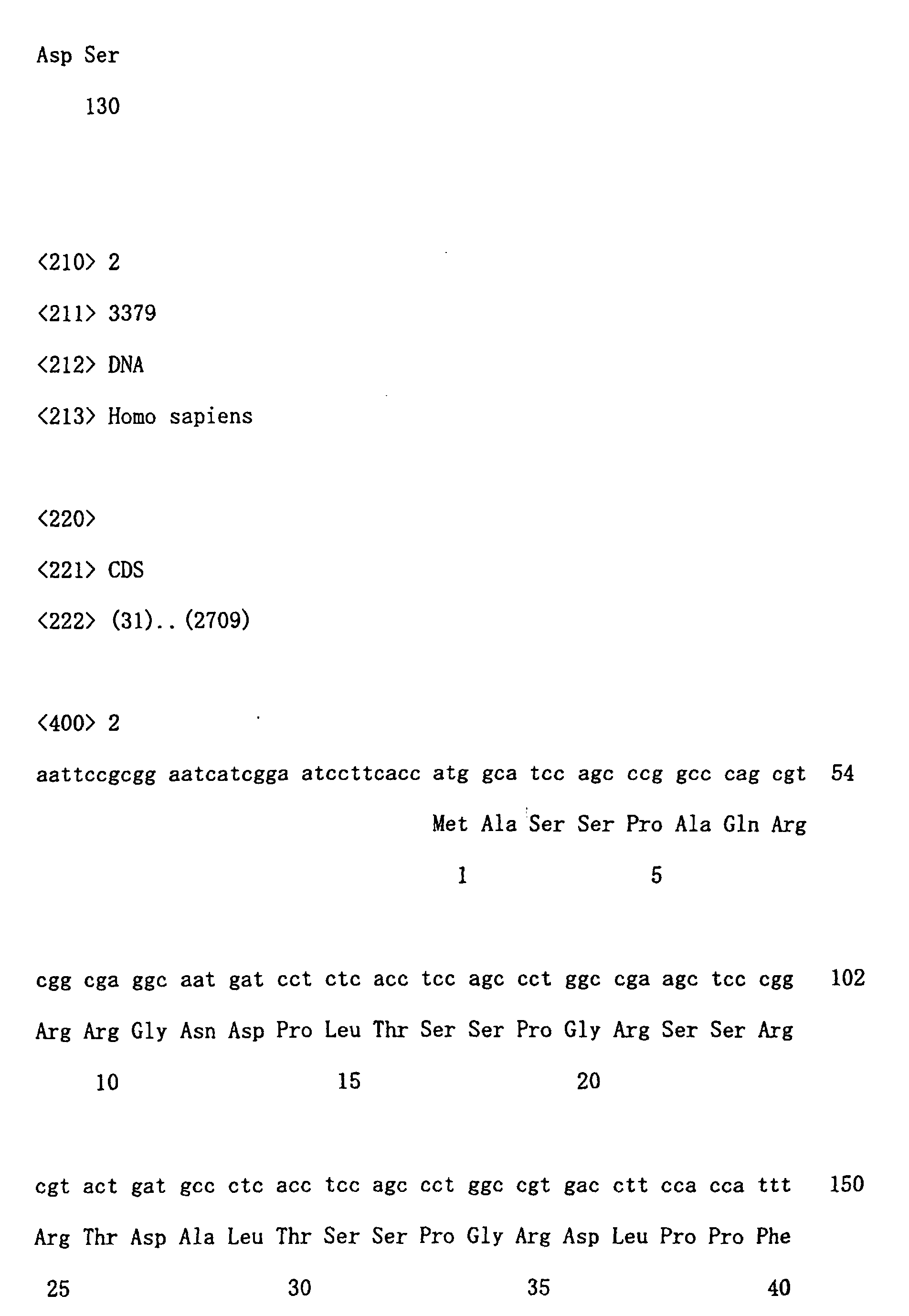


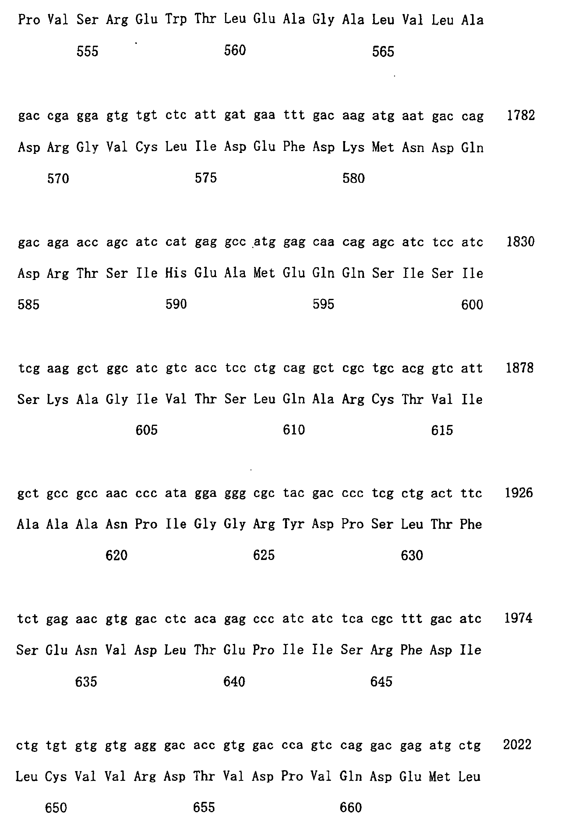
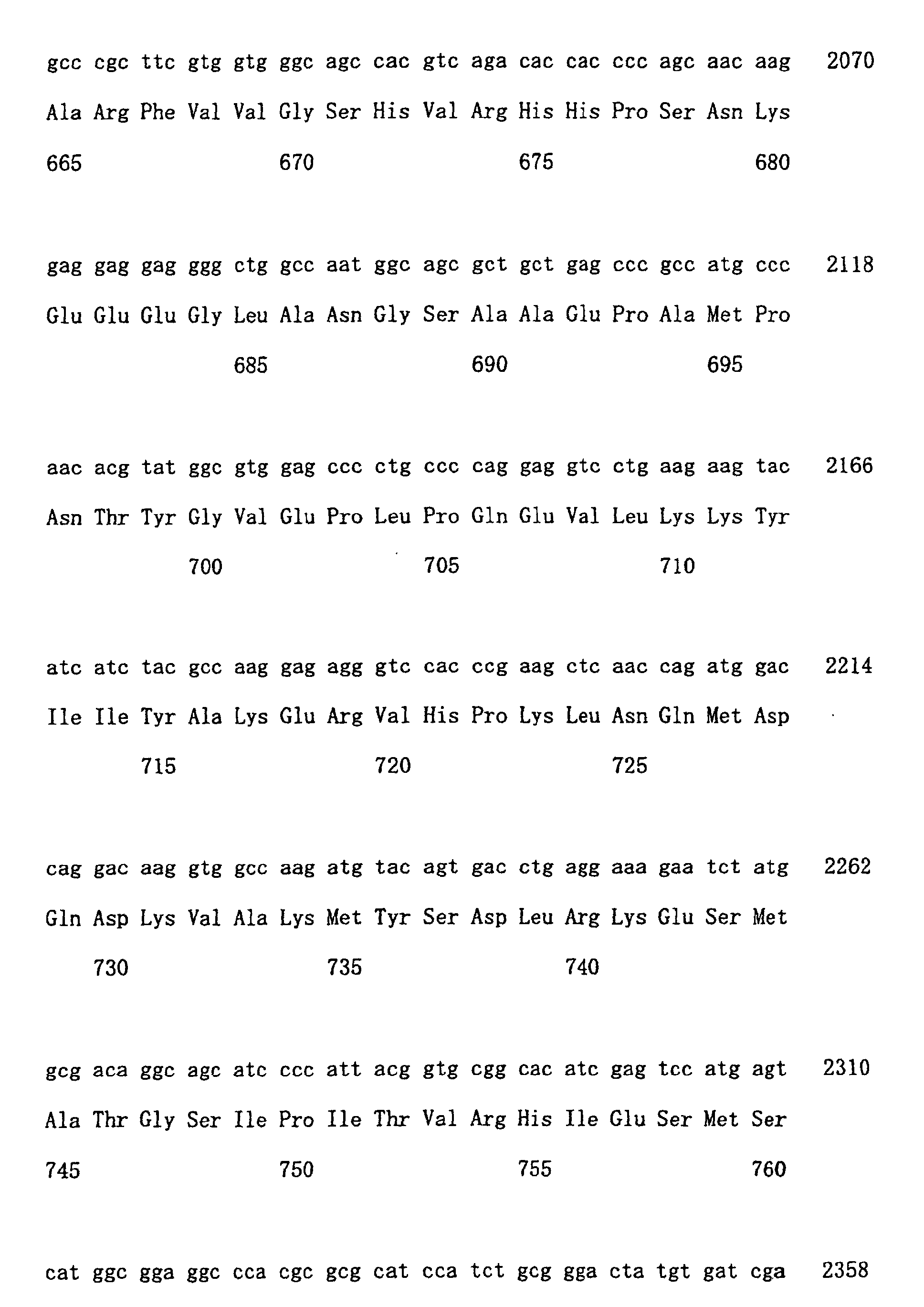
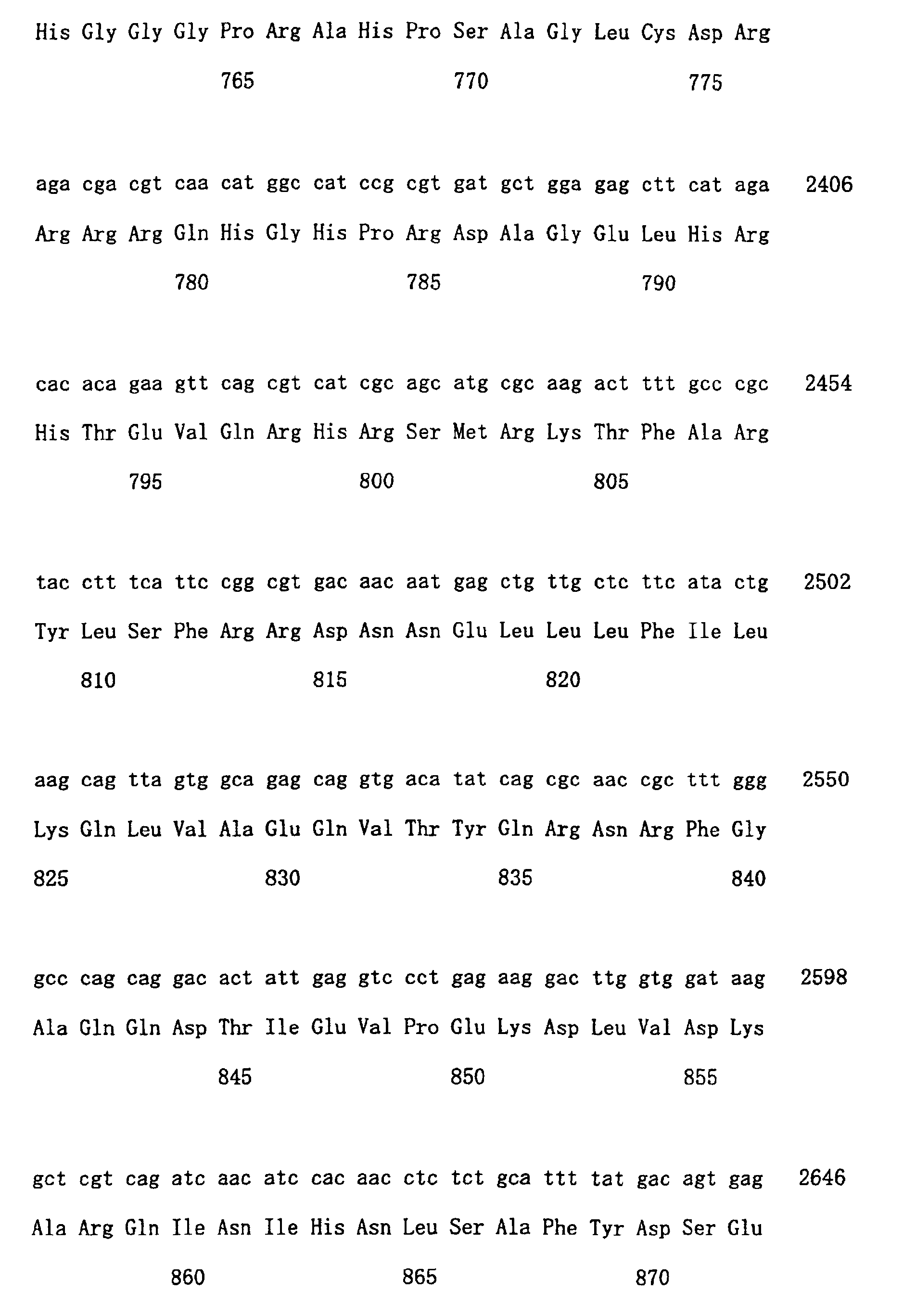
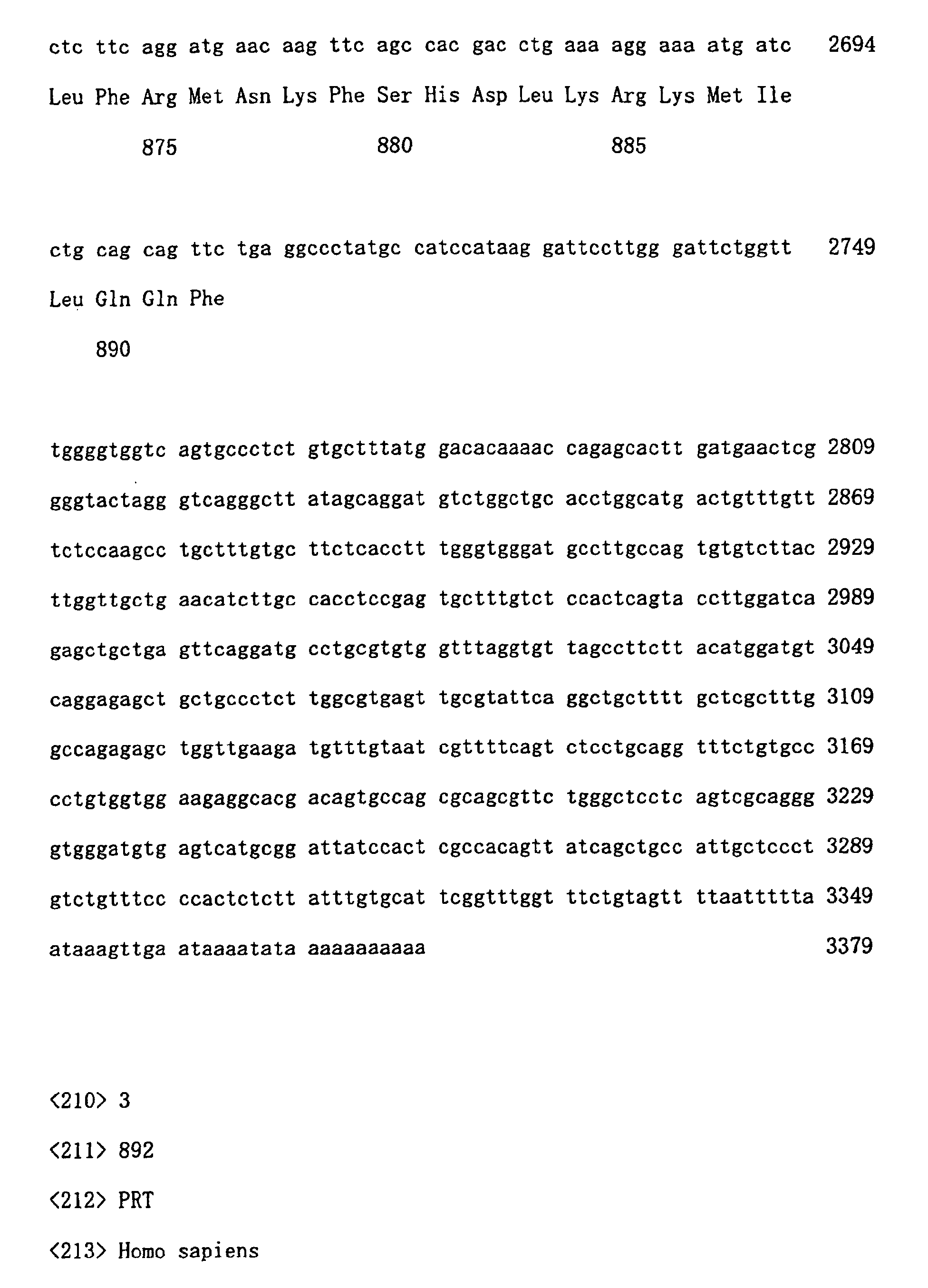


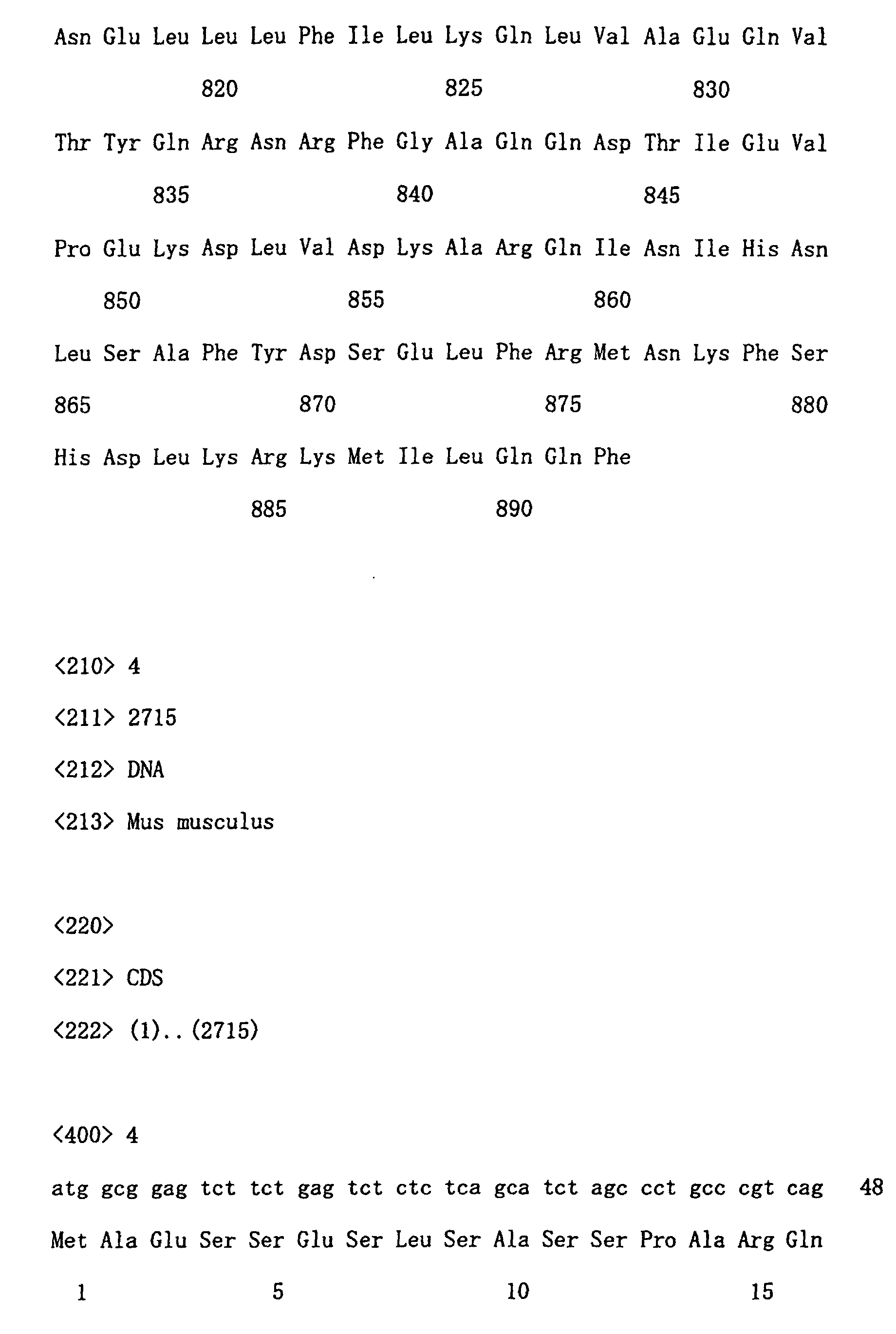
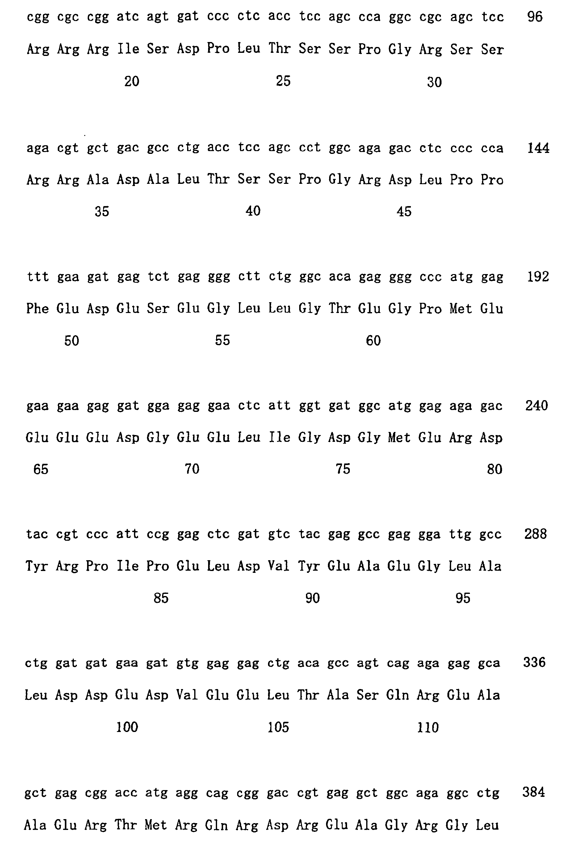
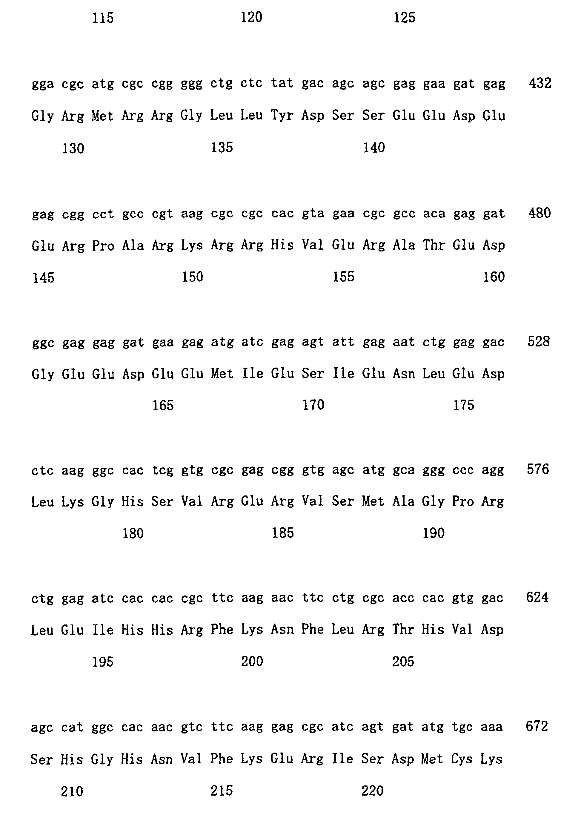


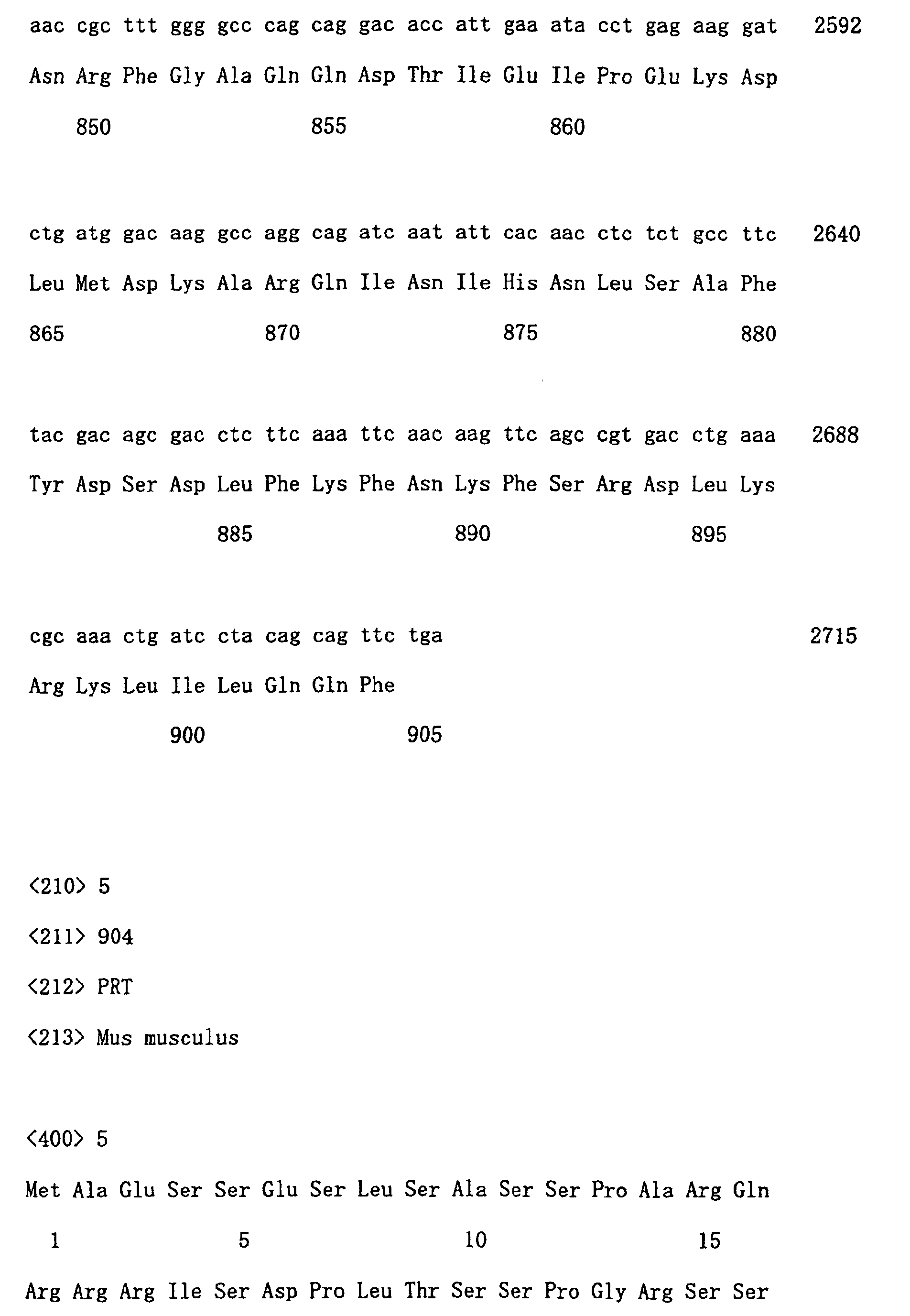
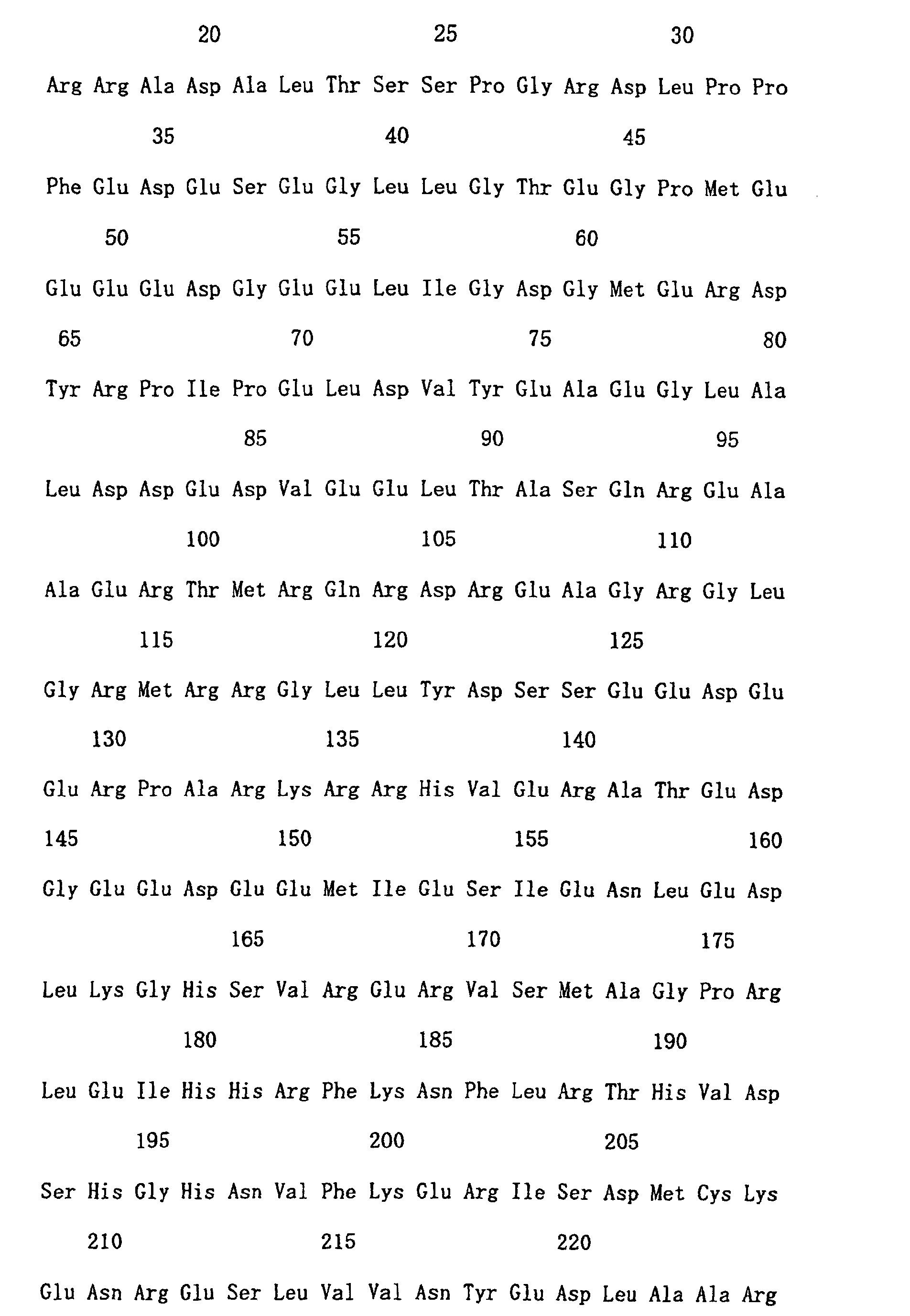


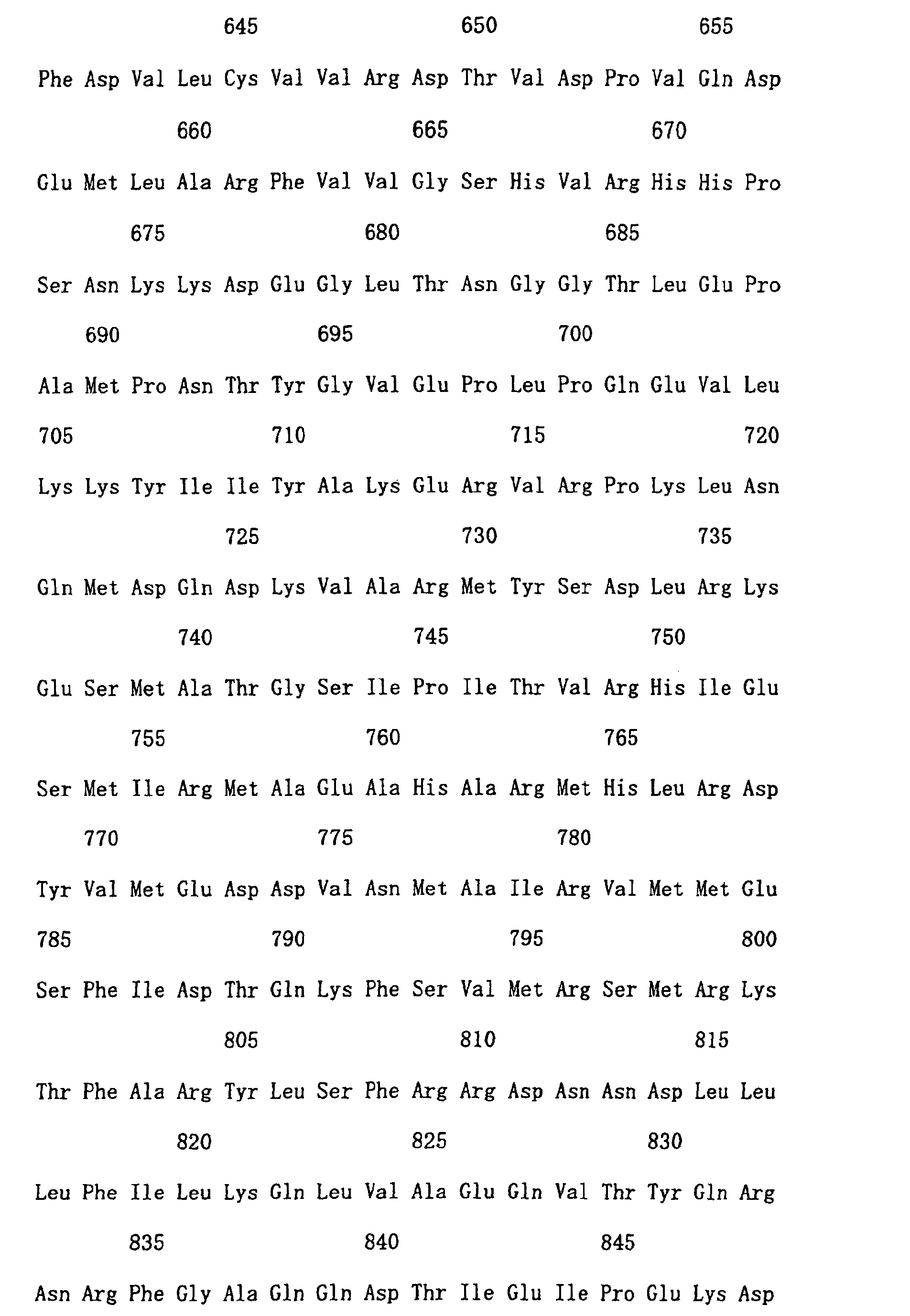
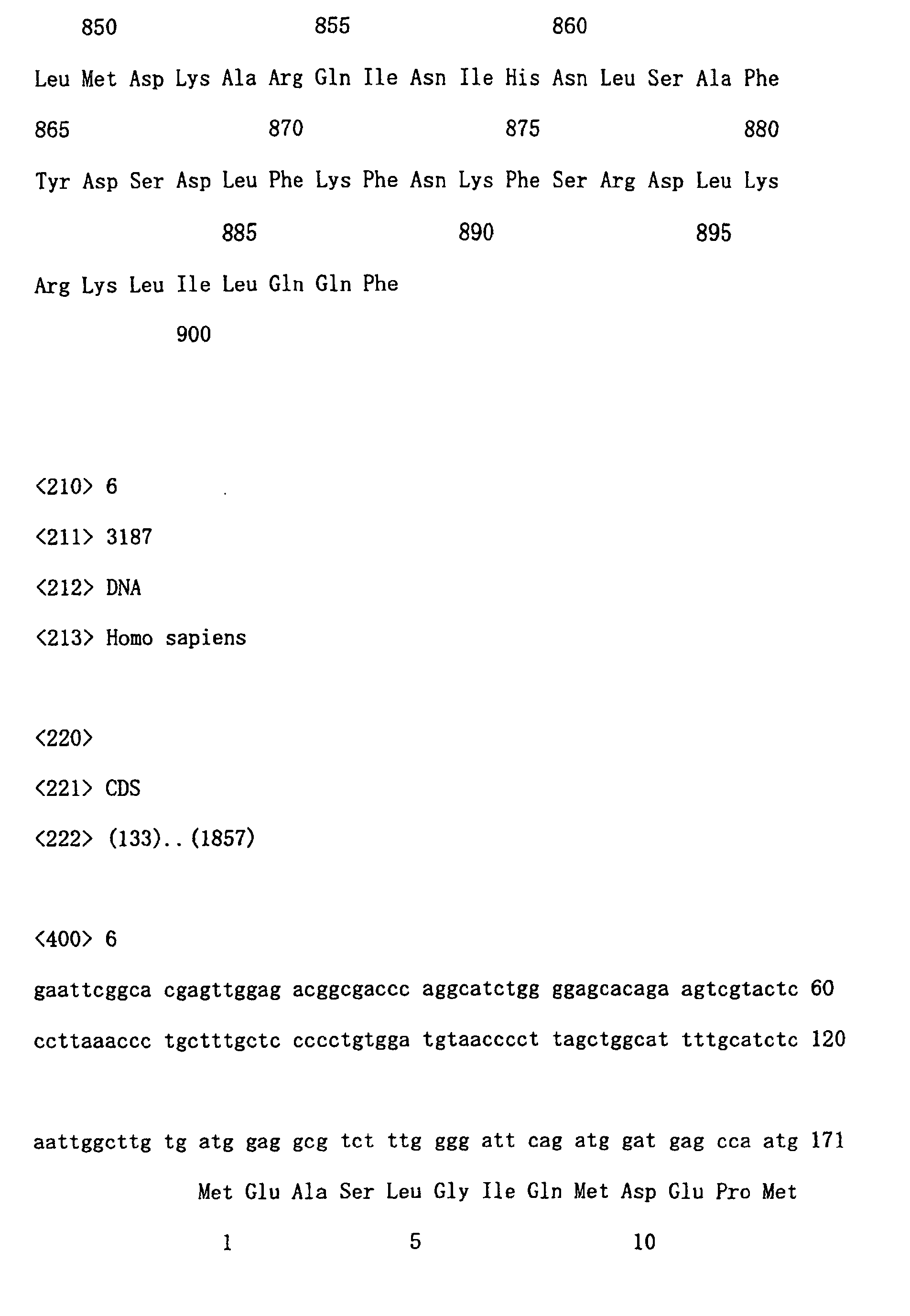


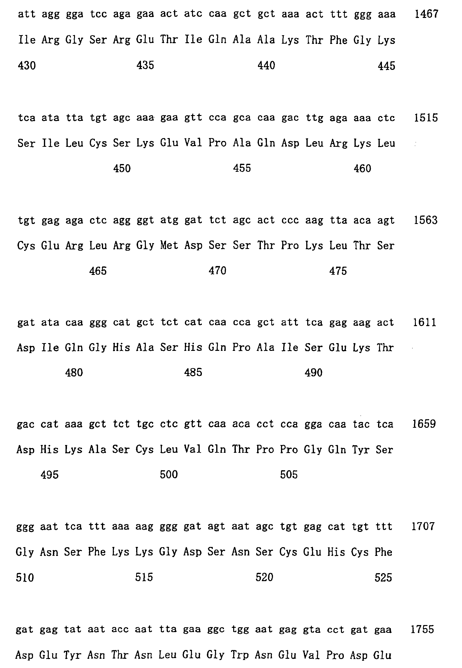
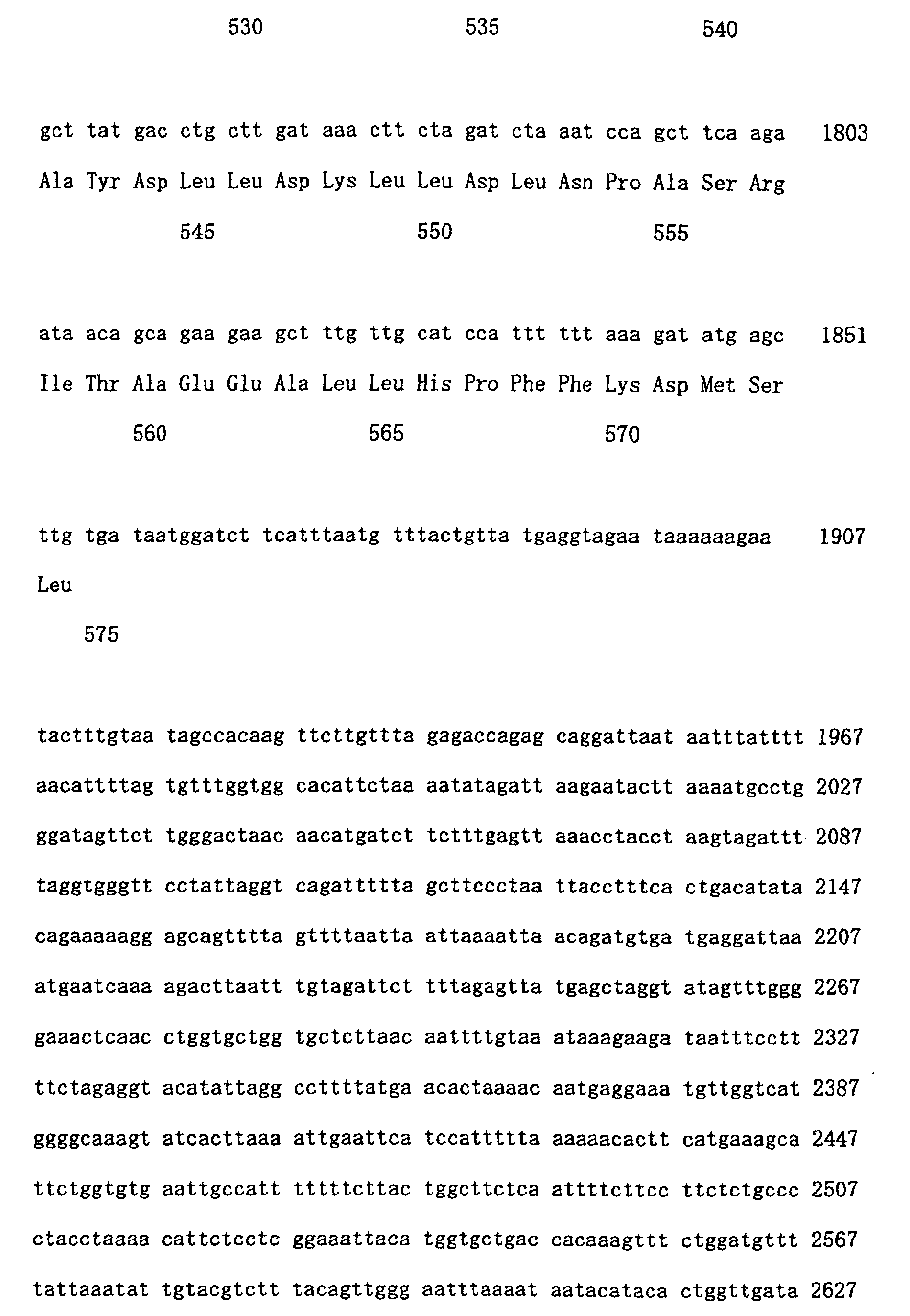

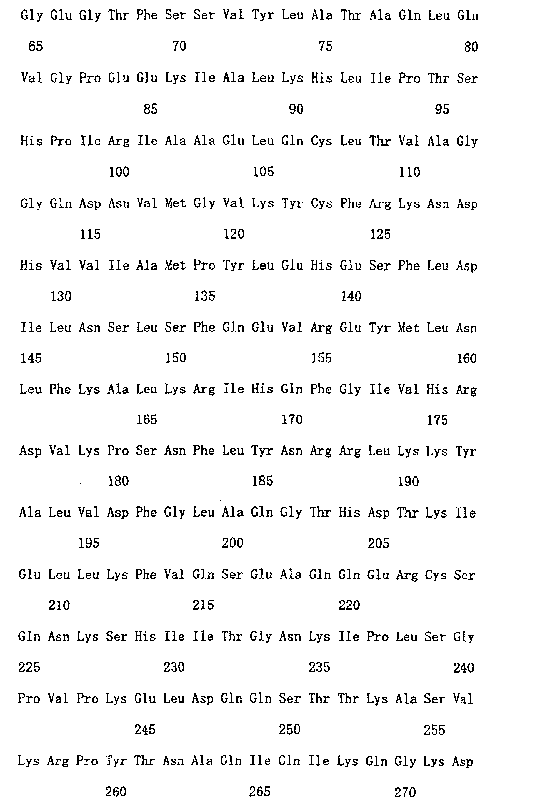
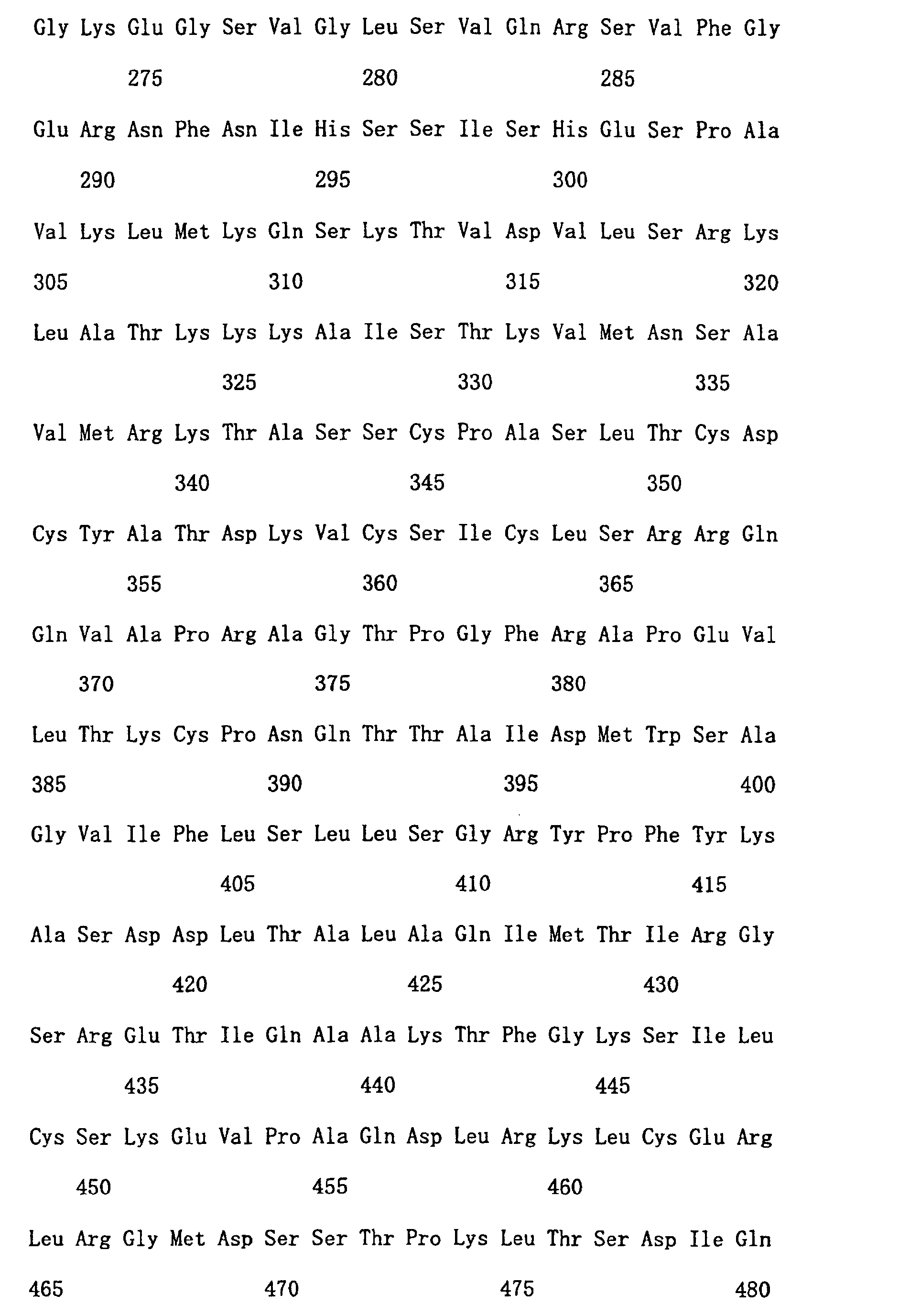


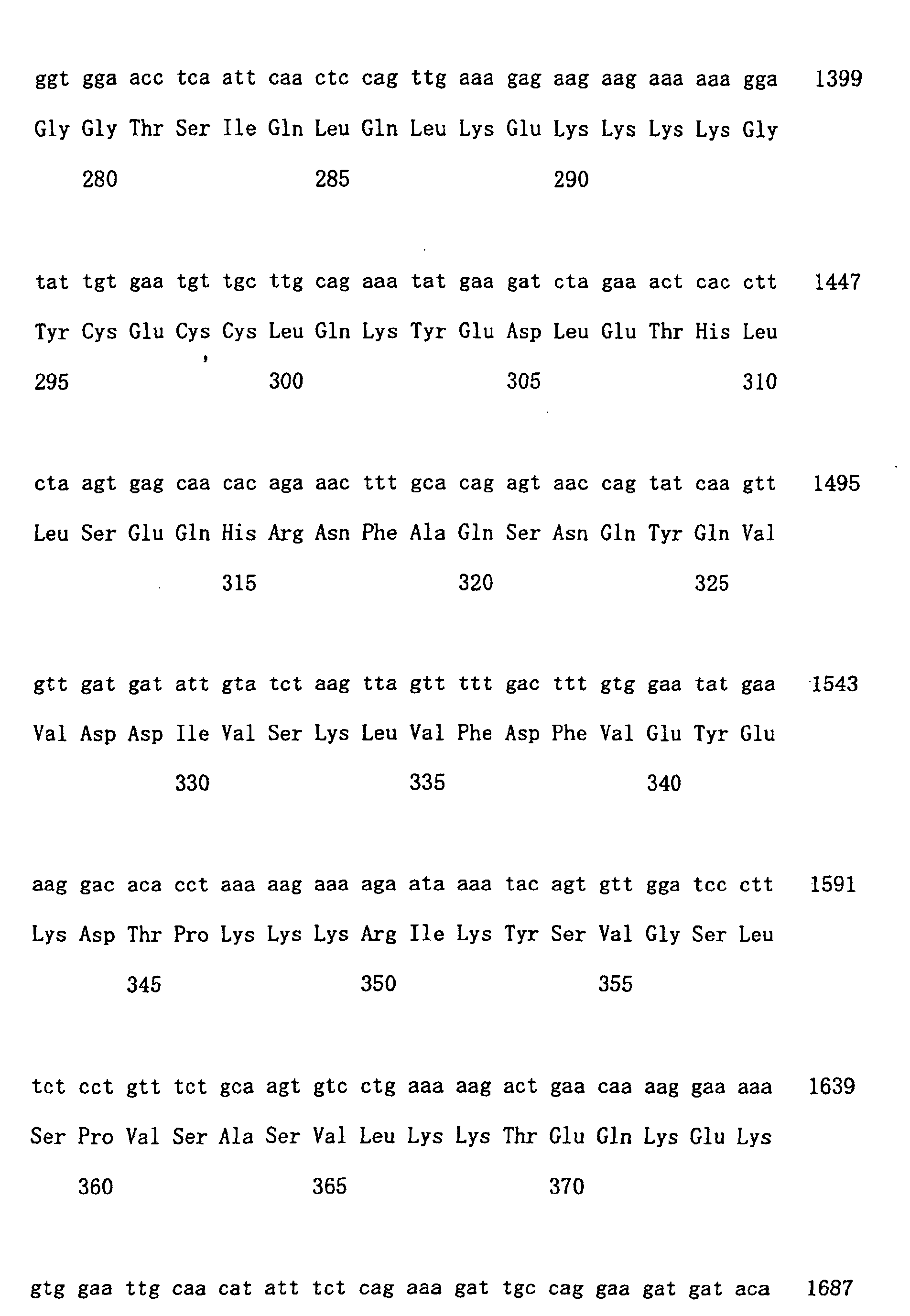

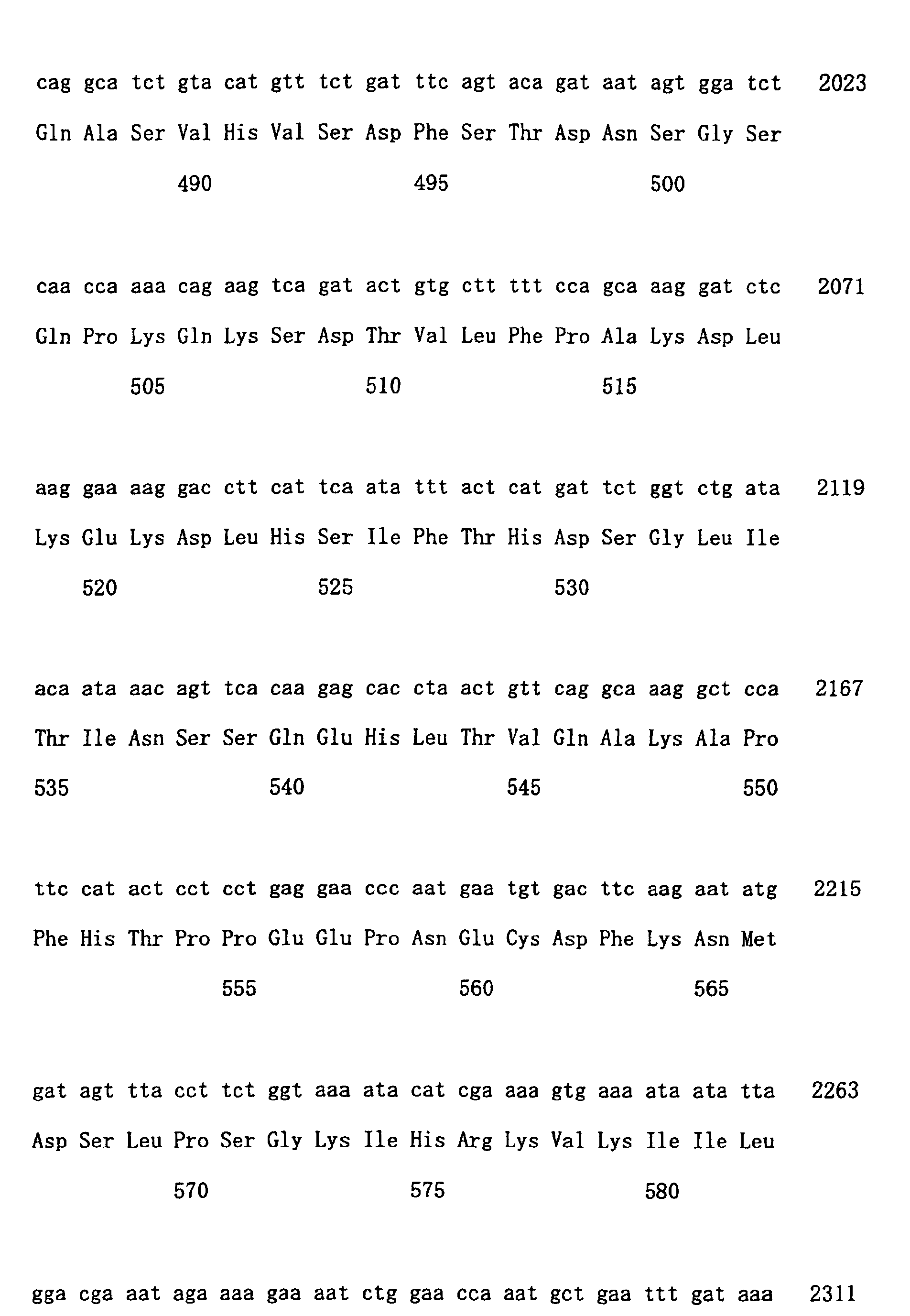


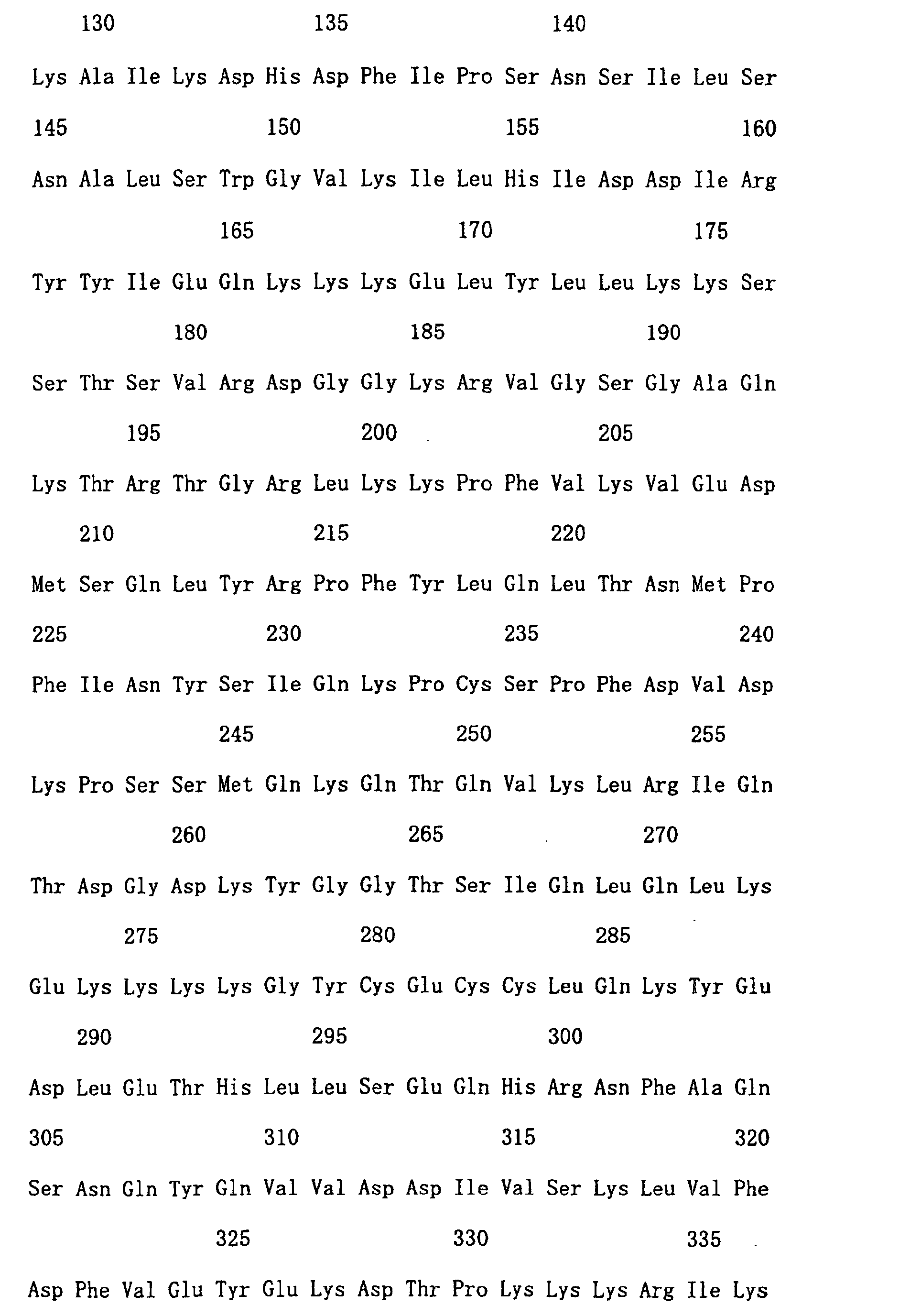
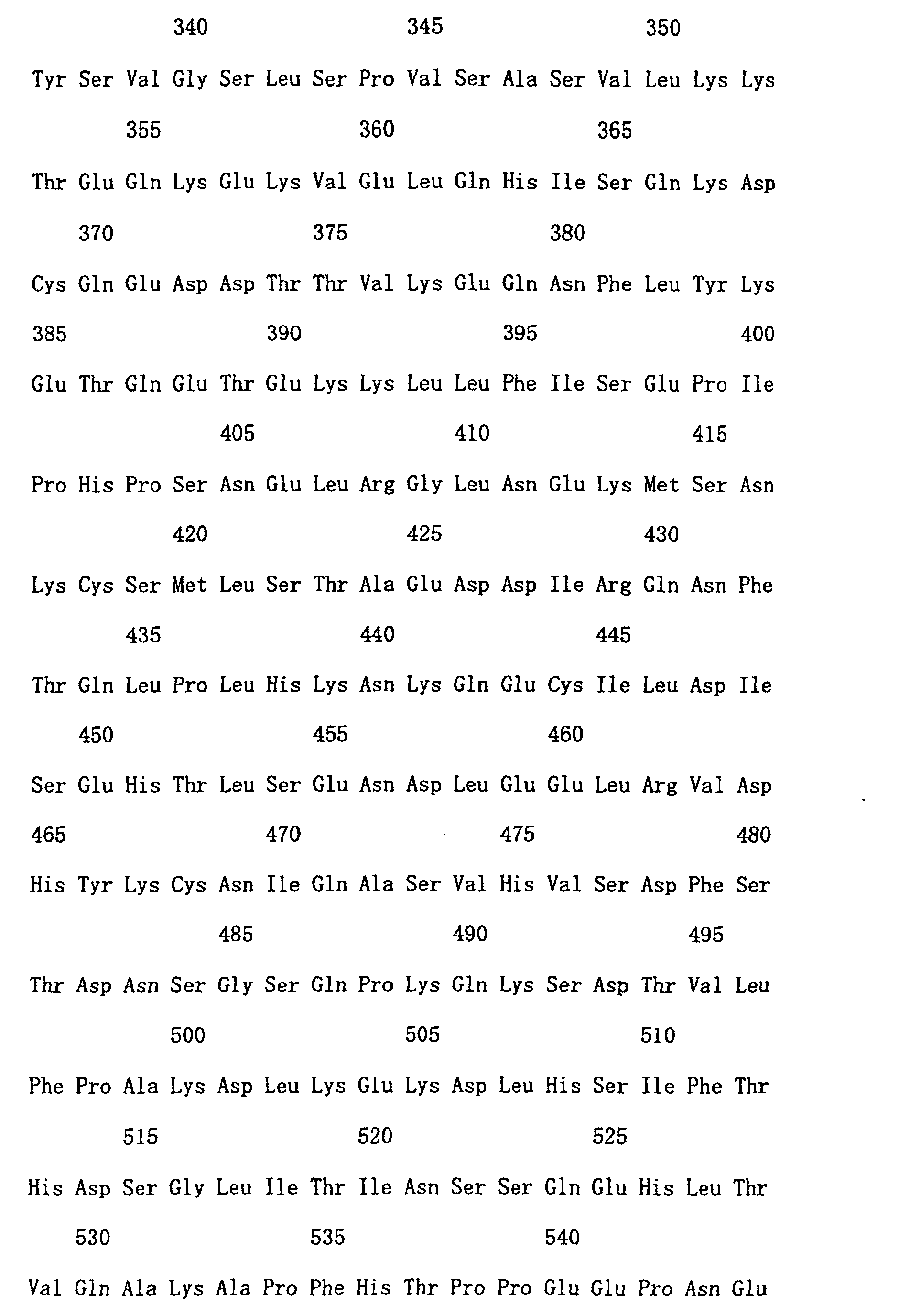

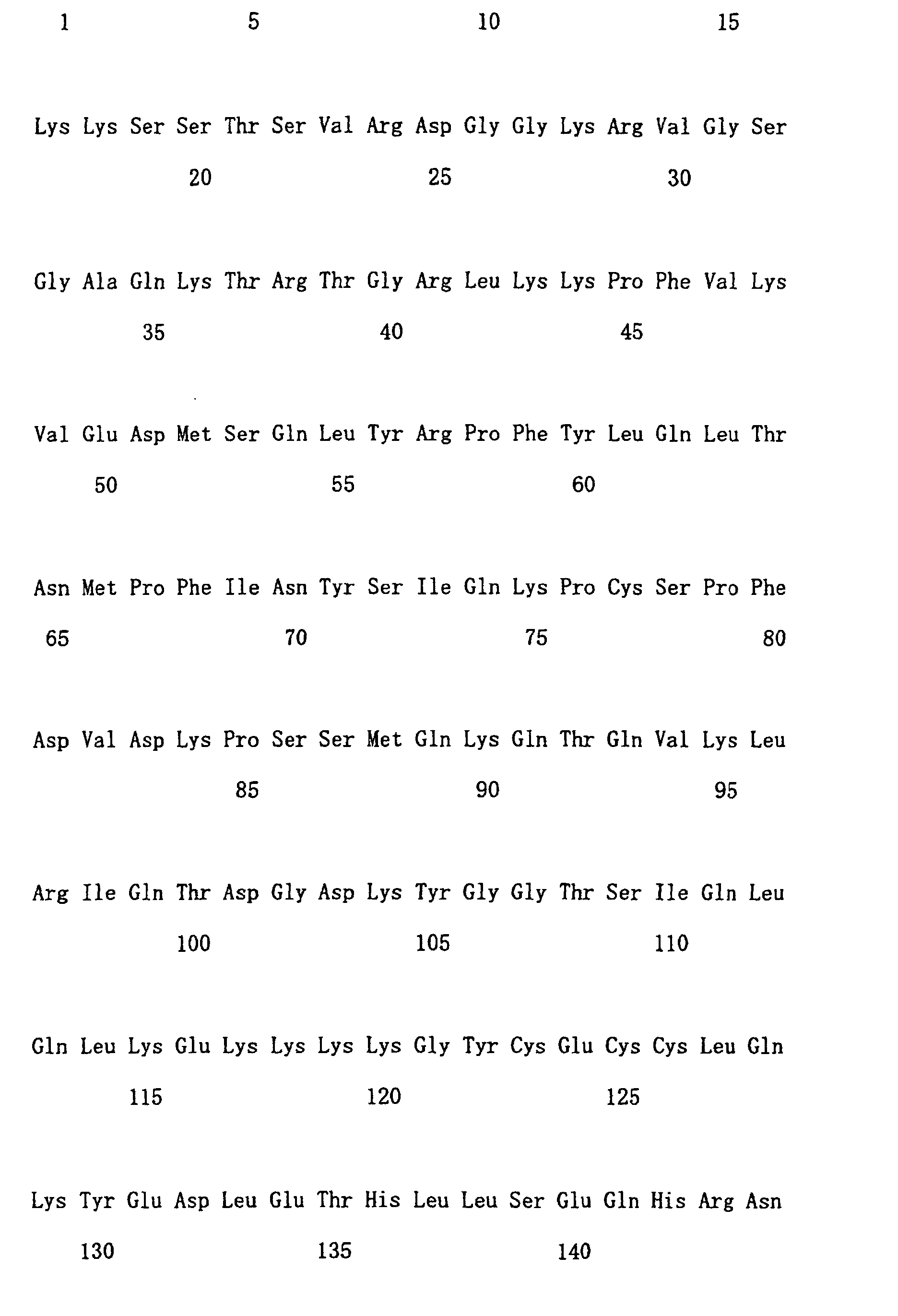


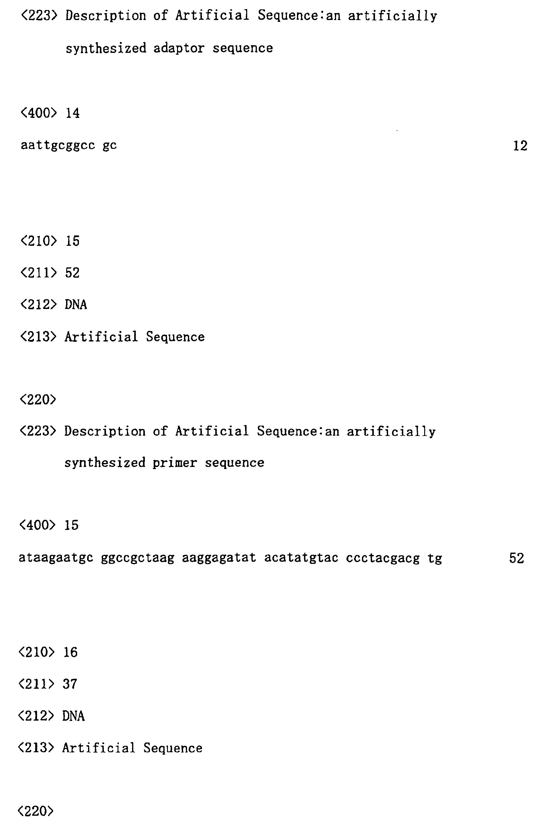
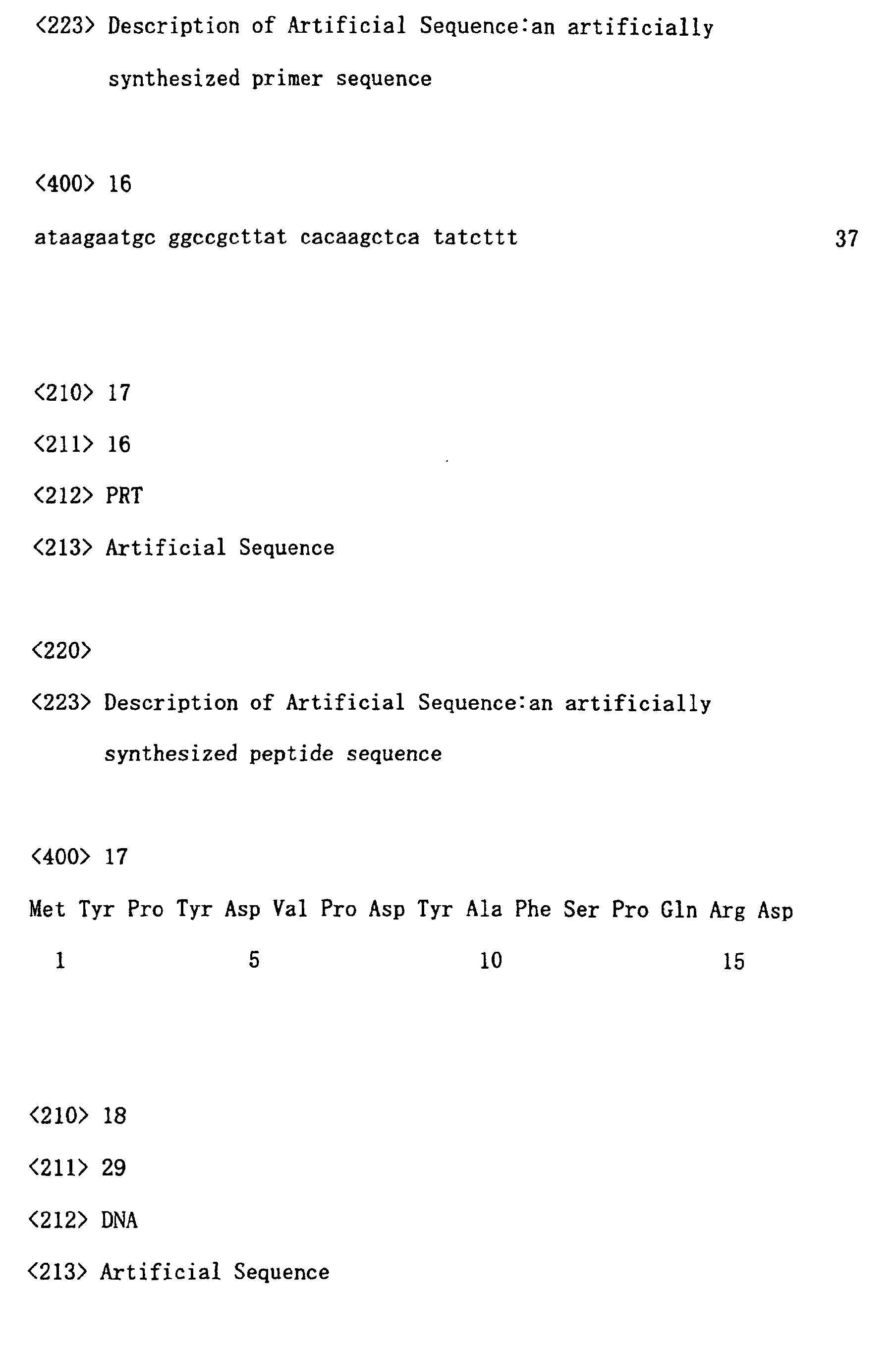

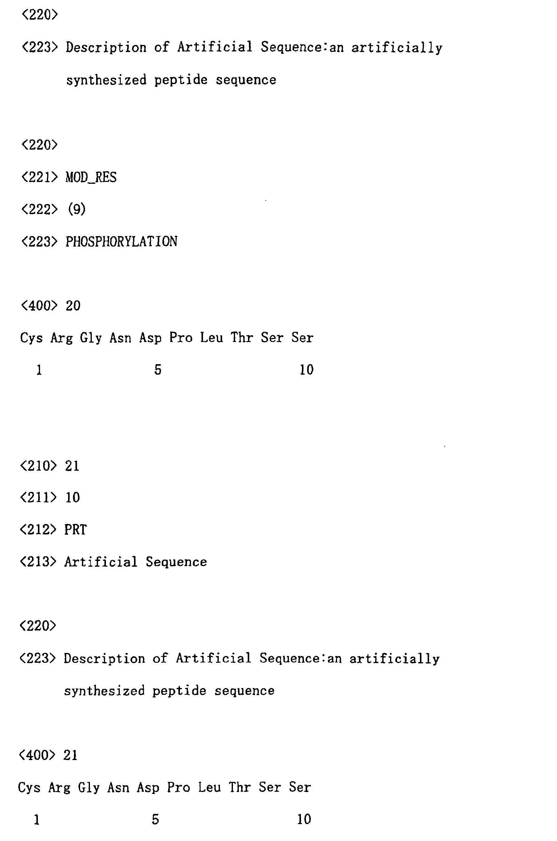
Claims (14)
- A method for measuring the kinase activity of a Cdc7-ASK complex, comprising the following steps:(a) contacting a substrate protein with the Cdc7-ASK complex under conditions that allow phosphorylation of the substrate protein, wherein the substrate protein is a protein comprising the amino acid sequence of SEQ ID NO: 1, or a protein functionally equivalent to that protein;(b) measuring the level of phosphorylation of a serine residue of the substrate protein at the position corresponding to position 17 in the amino acid sequence of SEQ ID NO: 1; and(c) measuring the kinase activity of the Cdc7-ASK complex using the level of phosphorylation as an indicator.
- The method according to claim 1, wherein the level of phosphorylation is measured based on the level of binding of an antibody that identifies the level of phosphorylation of the serine residue.
- The method according to claim 1, wherein the Cdc7-ASK complex is derived from a biological sample.
- A method for measuring the effects of a test compound on the kinase activity of a Cdc7-ASK complex, comprising the following steps:(a) contacting a test compound, a substrate protein and a Cdc7-ASK complex active substance, wherein the substrate protein is a protein comprising the amino acid sequence of SEQ ID NO: 1, or a protein functionally equivalent to that protein, and wherein they are contacted in any of the following orders i) to iii):i) the test compound and substrate protein are contacted, followed by contacting the Cdc7-ASK complex active substance,ii) the substrate protein and Cdc7-ASK complex active substance are contacted in the presence of the test compound, oriii) the substrate protein and Cdc7-ASK complex active substance are contacted, followed by contacting the test compound;(b) measuring the level of phosphorylation for a serine residue of the substrate protein at the position corresponding to position 17 in the amino acid sequence shown in SEQ ID NO: 1; and(c) measuring the effect of the test compound on the kinase activity of the Cdc7-ASK complex active substance using the level of phosphorylation as an indicator.
- A method of screening for compounds comprising the effect of regulating the kinase activity of a Cdc7-ASK complex, comprising the following steps:(a) measuring the effect of a test compound on the kinase activity of the Cdc7-ASK complex according to the method described in claim 4; and(b) selecting a test compound with a high or low level of phosphorylation by comparison with a control that has not been contacted with the test compound.
- A screening method according to claim 5, wherein a compound having a low level of phosphorylation is selected in step (b) of claim 5.
- An inhibitor of cell growth comprising a compound selected according to the screening method of claim 6 as its active ingredient.
- A kit for measuring the kinase activity of a Cdc7-ASK complex comprising:(a) a substrate protein comprising a continuous amino acid sequence that comprises a serine residue at position 17 of the amino acid sequence of SEQ ID NO: 1, and that which is selected from the amino acid sequence of SEQ ID NO: 1; and(b) an antibody that identifies the level of phosphorylation of the serine residue of the substrate protein at the position corresponding to position 17 of the amino acid sequence of SEQ ID NO: 1.
- A kit for evaluating the effect of a test compound on the kinase activity of a Cdc7-ASK complex, comprising:(a) a Cdc7-ASK complex active substance; and,(b) a substrate protein comprising a continuous amino acid sequence that comprises the serine residue at position 17 of the amino acid sequence of SEQ ID NO: 1, and is selected from this amino acid sequence.
- A process for producing a Cdc7-ASK complex active substance comprising the following steps:(a) introducing a DNA encoding human Cdc7 protein and a DNA encoding a protein comprising the amino acid sequence described in SEQ ID NO: 10 or a protein functionally equivalent to the protein, into prokaryotic cells in a state that allows monocistronic expression;(b) expressing the two DNAs; and(c) recovering the expressed protein.
- An antibody that identifies the level of phosphorylation of the serine residue at position 17 of a protein comprising the amino acid sequence of SEQ ID NO: 1.
- A protein according to any of the following (a) to (d):(a) a protein comprising the amino acid sequence of SEQ ID NO: 1;(b) a protein comprising a continuous amino acid sequence that is selected from the amino acid sequence described in SEQ ID NO: 3, and comprises the serine of position 17;(c) a protein comprising an amino acid sequence in which one or more amino acids in the amino acid sequence of SEQ ID NO: 1 are substituted, deleted, added and/or inserted, wherein the protein is phosphorylated by human Cdc7-ASK complex; and(d) a protein comprising an amino acid sequence comprising 90% or more homology with the amino acid sequence of SEQ ID NO: 3, wherein the protein is phosphorylated by human Cdc7-ASK complex.
- A protein comprising a continuous amino acid that comprises the amino acid sequence of SEQ ID NO: 10 and that which is selected from the amino acid sequence of SEQ ID NO: 9.
- A polypeptide according to claim 13 that comprises the amino acid sequence of SEQ ID NO: 10.
Applications Claiming Priority (3)
| Application Number | Priority Date | Filing Date | Title |
|---|---|---|---|
| JP2002067702 | 2002-03-12 | ||
| JP2002067702 | 2002-03-12 | ||
| PCT/JP2003/002918 WO2003076623A1 (en) | 2002-03-12 | 2003-03-12 | Cdc7-ASK KINASE COMPLEX, SUBSTRATE OF THE KINASE COMPLEX, ANTIBODY SPECIFIC TO THE SUBSTRATE, AND METHOD OF SCREENING COMPOUND CAPABLE OF INHIBITING Cdc7-ASK KINASE USING THE SAME |
Publications (2)
| Publication Number | Publication Date |
|---|---|
| EP1489177A1 true EP1489177A1 (en) | 2004-12-22 |
| EP1489177A4 EP1489177A4 (en) | 2006-09-13 |
Family
ID=27800289
Family Applications (1)
| Application Number | Title | Priority Date | Filing Date |
|---|---|---|---|
| EP03710315A Withdrawn EP1489177A4 (en) | 2002-03-12 | 2003-03-12 | CDC7-ASK KINASE COMPLEX, ITS SUBSTRATE, SPECIFIC ANTIBODY FOR THIS SUBSTRATE, AND METHOD FOR SCREENING A COMPOUND CAPABLE OF INHIBITING CDC7-ASK KINASE |
Country Status (5)
| Country | Link |
|---|---|
| US (1) | US20050250166A1 (en) |
| EP (1) | EP1489177A4 (en) |
| JP (1) | JP4270548B2 (en) |
| AU (1) | AU2003221356A1 (en) |
| WO (1) | WO2003076623A1 (en) |
Cited By (2)
| Publication number | Priority date | Publication date | Assignee | Title |
|---|---|---|---|---|
| WO2006116442A3 (en) * | 2005-04-27 | 2007-07-05 | Tripath Imaging Inc | Monoclonal antibodies and methods for their use in the detection of cervical disease |
| CN101193915B (en) * | 2005-04-27 | 2012-05-30 | 三路影像公司 | Monoclonal antibody and method for detecting cervical disease |
Families Citing this family (5)
| Publication number | Priority date | Publication date | Assignee | Title |
|---|---|---|---|---|
| EP1622934A2 (en) * | 2003-05-05 | 2006-02-08 | Pharmacia Italia S.p.A. | Truncated forms of human dbf4, complexes with their interacting partners and methods for identification of inhibitors thereof |
| JP4721903B2 (en) * | 2004-01-29 | 2011-07-13 | 大日本住友製薬株式会社 | Novel tumor antigen protein and use thereof |
| EP1951284A4 (en) * | 2005-11-14 | 2010-05-05 | Yeda Res & Dev | Improved variants of pigment epithelium derived factor and uses thereof |
| GB201511196D0 (en) | 2015-06-25 | 2015-08-12 | Cytosystems Ltd | Monoclonal antibodies |
| US9977026B2 (en) * | 2015-08-01 | 2018-05-22 | The Florida State University Research Foundation, Inc. | Detection of elevated levels of phosphorylated MCM and method of increasing MCM phosphorylation capacity |
Family Cites Families (6)
| Publication number | Priority date | Publication date | Assignee | Title |
|---|---|---|---|---|
| JPS60185719A (en) * | 1984-03-06 | 1985-09-21 | Ajinomoto Co Inc | Antitumor agent |
| JP4315301B2 (en) * | 1998-10-30 | 2009-08-19 | 独立行政法人科学技術振興機構 | Human H37 protein and cDNA encoding this protein |
| JP2000325086A (en) * | 1999-05-21 | 2000-11-28 | Igaku Seibutsugaku Kenkyusho:Kk | Method for measuring kinase activity |
| EP1207395A4 (en) * | 1999-08-04 | 2003-08-13 | Med & Biological Lab Co Ltd | Method for measuring kinase activity of cyclin/cdk complex |
| JP2001161398A (en) * | 1999-12-06 | 2001-06-19 | Igaku Seibutsugaku Kenkyusho:Kk | Method for measuring protein kinase activity and kit for measuring protein kinase activity |
| WO2001096557A2 (en) * | 2000-06-16 | 2001-12-20 | Pharmacia Italia S.P.A. | A human homologue of the dbf4/ask1 protein, nucleic acids, and methods related to the same |
-
2003
- 2003-03-12 AU AU2003221356A patent/AU2003221356A1/en not_active Abandoned
- 2003-03-12 US US10/507,275 patent/US20050250166A1/en not_active Abandoned
- 2003-03-12 WO PCT/JP2003/002918 patent/WO2003076623A1/en not_active Ceased
- 2003-03-12 JP JP2003574827A patent/JP4270548B2/en not_active Expired - Fee Related
- 2003-03-12 EP EP03710315A patent/EP1489177A4/en not_active Withdrawn
Cited By (6)
| Publication number | Priority date | Publication date | Assignee | Title |
|---|---|---|---|---|
| WO2006116442A3 (en) * | 2005-04-27 | 2007-07-05 | Tripath Imaging Inc | Monoclonal antibodies and methods for their use in the detection of cervical disease |
| US7595380B2 (en) | 2005-04-27 | 2009-09-29 | Tripath Imaging, Inc. | Monoclonal antibodies and methods for their use in the detection of cervical disease |
| EP2332988A3 (en) * | 2005-04-27 | 2011-08-31 | Tripath Imaging, Inc. | Monoclonal antibodies and methods for their use in the detection of cervical disease |
| AU2006241226B2 (en) * | 2005-04-27 | 2011-09-15 | Tripath Imaging, Inc. | Monoclonal antibodies and methods for their use in the detection of cervical disease |
| US8076093B2 (en) | 2005-04-27 | 2011-12-13 | Tripath Imaging, Inc. | Monoclonal antibodies and methods for their use in the detection of cervical disease |
| CN101193915B (en) * | 2005-04-27 | 2012-05-30 | 三路影像公司 | Monoclonal antibody and method for detecting cervical disease |
Also Published As
| Publication number | Publication date |
|---|---|
| JP4270548B2 (en) | 2009-06-03 |
| US20050250166A1 (en) | 2005-11-10 |
| EP1489177A4 (en) | 2006-09-13 |
| WO2003076623A1 (en) | 2003-09-18 |
| AU2003221356A1 (en) | 2003-09-22 |
| JPWO2003076623A1 (en) | 2005-07-07 |
Similar Documents
| Publication | Publication Date | Title |
|---|---|---|
| Scott et al. | Reversible phosphorylation at the C-terminal regulatory domain of p21Waf1/Cip1 modulates proliferating cell nuclear antigen binding | |
| Matsuoka et al. | Activation of cyclin-dependent kinase 4 (cdk4) by mouse MO15-associated kinase | |
| Goto et al. | Phosphorylation of vimentin by Rho-associated kinase at a unique amino-terminal site that is specifically phosphorylated during cytokinesis | |
| US6232081B1 (en) | Method for the detection of NF-κB regulatory factors | |
| JP2004222728A (en) | Human cyclin E | |
| US8491900B2 (en) | Antibodies to signal transduction protein TAB2 | |
| US20050250166A1 (en) | Cdc7-ask kinase complex, substrates of the kinase complex, antibody specific to the substrate, and method of screening compound capable of inhibiting cdc7-ask kinase using the same | |
| AU2001265947B2 (en) | Enzymatic assays for screening anti-cancer agents | |
| AU2001265947A1 (en) | Enzymatic assays for screening anti-cancer agents | |
| EP1207395A1 (en) | Method for measuring kinase activity of cyclin/cdk complex | |
| US7108992B2 (en) | ATM kinase compositions and methods | |
| JP2003527824A (en) | Novel cells signaling polypeptides and nucleic acids | |
| JPWO2001038527A1 (en) | Novel TAB2 protein | |
| US20090291439A1 (en) | Phosphatases involved in the regulation of cardiomyocyte differentiation | |
| US6258776B1 (en) | Calcium-regulated kinase | |
| US7871786B2 (en) | GEF-H1b: biomarkers, complexes, assays and therapeutic uses thereof | |
| JPH11326328A (en) | Screening method for substance that inhibits binding to XIAP | |
| US6638724B1 (en) | BLNK proteins | |
| WO1998032852A9 (en) | Blnk proteins | |
| JP2003512047A (en) | Novel germinal center kinase cell cycle proteins, compositions and uses | |
| JP2006143669A (en) | Antibody recognizing phosphorylated unnatural amino acid and use thereof | |
| CA2331152A1 (en) | Cell cycle regulating factor |
Legal Events
| Date | Code | Title | Description |
|---|---|---|---|
| PUAI | Public reference made under article 153(3) epc to a published international application that has entered the european phase |
Free format text: ORIGINAL CODE: 0009012 |
|
| 17P | Request for examination filed |
Effective date: 20041012 |
|
| AK | Designated contracting states |
Kind code of ref document: A1 Designated state(s): AT BE BG CH CY CZ DE DK EE ES FI FR GB GR HU IE IT LI LU MC NL PT RO SE SI SK TR |
|
| AX | Request for extension of the european patent |
Extension state: AL LT LV MK |
|
| A4 | Supplementary search report drawn up and despatched |
Effective date: 20060810 |
|
| 17Q | First examination report despatched |
Effective date: 20070116 |
|
| STAA | Information on the status of an ep patent application or granted ep patent |
Free format text: STATUS: THE APPLICATION IS DEEMED TO BE WITHDRAWN |
|
| 18D | Application deemed to be withdrawn |
Effective date: 20070527 |