-
Homeodomain-interacting protein kinases (HIPKs) possess a
kinase domain that display strong sequence similarity with those of other
protein kinases belonging to the family of dual specificity Yak1 related kinases
(DYRK). One main feature of DYRKs is their dual ability to autophosphorylate
on tyrosine residues and to phosphorylate their substrates on serine and
threonine residues (Matsuo et al., 2001; Hofmann et al., 2000). However,
although HIPKs are able to autophosphorylate and to phosphorylate their
substrates on serine and threonine residues, their phosphorylation on tyrosine
residues might be mediated by so far uncharacterized kinases (Hofmann et al.,
2000 ; Pierantoni et al., 2001). As such, HIPKs constitute a subfamily of DYRKs
composed of at least five members: HIPK1, HIPK2, HIPK3, STANK and PKM
(Kim et al., 1998 ; Wang et al., 2000). At present, HIPK2 is one of the best
characterized member of the HIPK family.
-
In addition to its kinase domain, HIPK2 contains a domain
required for the interaction with several NK homeoproteins, a domain rich in
proline, glutamic acid, serine and threonine (PEST) and a domain rich in
tyrosine and histidine. The function of these two latter domains is not yet
determined although it has been shown that PEST domains may be involved in
the stability control of proteins (Rogers et al., 1986). HIPK2 is localized mainly
in nuclei and concentrates within a variable numbers of small nuclear domains.
The targeting of HIPK2 into these domains is conditioned by its Ubc9-mediated
post-translational modification with the ubiquitin-like protein SUMO-1 (Kim et
al., 1999). Under certain physiological conditions, HIPK2 is recruited to nuclear
domains 10 (ND10) also called PML bodies. This recruitment requires the
interaction of HIPK2 with the PML-3 protein (Hofmann et al., 2002).
-
HIPK2 interacts with several NK homeodomain transcription
factors that have important functions during embryonic development and
organogenesis. In particular, HIPK2 is a component of a transcriptional
corepressor complex containing NK-3, Groucho and a histone deacetylase
complex (Kim et al., 1998 ; Choi et al., 1999). HIPK2 regulates the formation
and the activity of this complex. It has also been suggested that HIPK2
intervene in the transduction of extracellular signals from the cell surface to the
nucleus such as those induced by the tumor necrosis factor (Li et al., 2000 ;
Wang et al., 2001a ; Morgan et al., 2002) or those mediated by CD43 and
STAT3 (Matsuo et al., 2001 ; Wang et al. 2000). More recently, it has been
demonstrated that HIPK2 is a novel activator of the p53 tumor suppressor
protein (Hofmann et al., 2002 ; Wang et al., 2001b ; D'Orazi et al., 2002).
HIPK2 interacts also with p73 a member of the p53 family and regulates
progression through the cell cycle (Pierantoni et al., 2001 ; Kim et al., 2002).
-
The genome of HSV-1 contains at least 79 genes among 42 of
which are dispensable for production of infectious viral particles in cell cultures.
Because these dispensable genes should be nevertheless expected to play
very important roles in virus-host interactions in vivo, many studies have been
performed these last decades to elucidate their functions.
-
US11 was initially described as one of these so-called non-essential
genes (Longnecker et al., 1986 ; Brown et al., 1987). US11 protein is
among the most abundant viral proteins present in cells late in infection and is
packaged in the tegument of the native virions to be delivered into cells after
infection (Bhat et al., 1983 ; Gressner et al., 1974 ; Roller et al., 1996). Soon
after infection, US11 protein is found in the cytoplasm, either as heterogeneous
oligomers or associated with ribosomes or both (Puvion-Dutilleul, 1987 ;
MacLean et al., 1987 ; Diaz et al., 1993). Later during infection, US11 protein
accumulates into nucleoli and is also found into RNP fibrills as well as in
clusters of interchromatin granules. Retention of US11 within nucleoli and
nuclear export are mediated by a unique motif embedded into the C-terminal
part of the protein (Catez et al., 2002). In addition, this part of the protein
contains a motif that confers to US11 an intercellular trafficking activity
(Koshizuka et al., 2001).
-
Since the first identification of US11 as a DNA-binding protein,
strong evidences have been brought showing that this protein possess also
RNA-binding activities which appear essential for several of its biological
functions, like control of mRNA transport and translation (Diaz et al., 1996 ; Duc
et al., 2000), and regulation of PKR activity (Poppers et al., 2000 ; Cassady et
al., 1998). Moreover, even though US11 protein is dispensable for viral growth
in cell cultures, it plays an important role for the replication of HSV-1 in the
adrenal gland, an organ important for viral penetration in the spinal cord and
brain (Nishiyama et al., 1993) and in cells submitted to thermal stress (Brown et
al., 1987). US11 has also been implicated in the recovery of protein synthesis
and survival in heat shock treated cells (Diaz-Latoud et al., 1997).
-
The inventors have designed a yeast two-hybrid screen of a
mouse library using US11 as a bait in order to identify cellular factors
susceptible to interact with this protein. This allowed them to isolate a cDNA
whose sequence encompassed the PEST domain of the murine HIPK2 protein.
Co-immunoprecipitation of US11 with transiently expressed Flag-tagged
HIPK2/PEST provided further evidence for their tight association in mammalian
cells. Moreover, the observation that some modified forms of US11 containing
either a single or a very few amino acids substitutions interacted slightly or do
not interacted at all attest for the specificity of this interaction.
-
Thereby, the inventors demonstrated that US11 interacts with
HIPK2 and that this interaction occurs through the PEST domain of HIPK2.
-
Interestingly, the HIPK2 intracellular distribution was shown to be
strongly modified after co-expression of US11. More specifically, US11
completely abolished the dot like pattern of HIPK2 nuclear distribution and
seemed to induce an accumulation of this protein within the nucleoplasm. More
importantly, colony formation assays demonstrated that the HIPK2/p53
induction of apoptosis was inhibited by US11 indicating that this protein could
antagonize HIPK2-induced apoptosis of HSV-1 or HSV-2
-
The inventors thus identified US11 as a new mediator of the
HSV-1 and HSV-2 dependent inhibitions of apoptosis.
Definitions
-
A "polynucleotide" or "nucleotide sequence" is a serie of
nucleotides (or nucleoside plus phosphate group) in a nucleic acid, such as
DNA and RNA, and means any chain of two or more nucleotides. A nucleotide
sequence typically carries genetic information, including the information used
by cellular machinery to make proteins and enzymes. These terms include
double or single stranded genomic and cDNA, RNA, any synthetic and
genetically manipulated polynucleotide, and both sense and anti-sense
polynucleotide (although only sense strands are being represented herein).
This includes single- and double-stranded molecules, i.e., DNA-DNA, DNA-RNA
and RNA-RNA hybrids, as well as "protein nucleic acids" (PNA) formed by
conjugating bases to an amino acid backbone. This also includes nucleic acids
containing modified bases, for example thio-uracil, thio-guanine and fluoro-uracil.
-
The nucleic acids herein may be flanked by natural regulatory
(expression control) sequences, or may be associated with heterologous
sequences, including promoters, internal ribosome entry sites (IRES) and other
ribosome binding site sequences, enhancers, response elements, suppressors,
signal sequences, polyadenylation sequences, introns, 5'- and 3'- non- coding
regions, and the like. The nucleic acids may also be modified by many means
known in the art. Non-limiting examples of such modifications include
methylation, "caps", substitution of one or more of the naturally occurring
nucleotides with an analog, and internucleotide modifications such as, for
example, those with uncharged linkages (e.g., methyl phosphonates,
phosphotriesters, phosphoroamidates, carbamates, etc.) and with charged
linkages (e.g., phosphorothioates, phosphorodithioates, etc.). Polynucleotides
may contain one or more additional covalently linked moieties, such as, for
example, proteins (e.g., nucleases, toxins, antibodies, signal peptides, poly-L-lysine,
etc.), intercalators (e.g., acridine, psoralen, etc.), chelators (e.g., metals,
radioactive metals, iron, oxidative metals, etc.), and alkylators. The
polynucleotides may be derivatized by formation of a methyl or ethyl
phosphotriester or an alkyl phosphoramidate linkage. Furthermore, the
polynucleotides herein may also be modified with a label capable of providing a
detectable signal, either directly or indirectly. Exemplary labels include
radioisotopes, fluorescent molecules, biotin, and the like.
-
A "promoter" or "promoter sequence" is a DNA regulatory region
capable of binding RNA polymerase in a cell and initiating transcription of a
downstream (3' direction) coding sequence. For purposes of defining the
present invention, the promoter sequence is bounded at its 3' terminus by the
transcription initiation site and extends upstream (5' direction) to include the
minimum number of bases or elements necessary to initiate transcription at
levels detectable above background. Within the promoter sequence will be
found a transcription initiation site (conveniently defined for example, by
mapping with nuclease SI), as well as protein binding domains (consensus
sequences) responsible for the binding of RNA polymerase. The promoter may
be operatively associated with other expression control sequences, including
enhancer and repressor sequences.
-
Promoters which may be used to control gene expression include,
but are not limited to, cytomegalovirus (CMV) promoter (V.S. Patents No.
5,385,839 and No. 5,168,062), the SV40 early promoter region (Benoist and
Chambon, 1981), the promoter contained in the 3' long terminal repeat of Rous
sarcoma virus (Yamamoto, et al., 1980), the herpes thymidine kinase promoter
(Wagner et al., 1981), the regulatory sequences of the metallothionein gene
(Brinster et al., 1982) ; prokaryotic expression vectors such as the beta-lactamase
promoter (Villa-Komaroff et al., 1978), or the tac promoter (DeBoer
et al., 1983); see also "Useful proteins from recombinant bacteria" in Scientific
American 1980, 242:74-94; promoter elements from yeast or other fungi such
as the Gal 4 promoter, the ADC (alcohol dehydrogenase) promoter, PGK
(phosphoglycerol kinase) promoter, alkaline phosphatase promoter; and
transcriptional control regions that exhibit hematopoietic tissue specificity, in
particular: beta-globin gene control region which is active in myeloid cells
(Mogram et al., 1985 ; Kollias et al., 1986), hematopoietic stem cell
differentiation factor promoters, erythropoietin receptor promoter (Maouche et
al., 1991), etc.
-
A "coding sequence" or a sequence "encoding" an expression
product, such as a RNA, polypeptide, protein, or enzyme, is a nucleotide
sequence that, when expressed, results in the production of that RNA,
polypeptide, protein, or enzyme, i.e., the nucleotide sequence encodes an
amino acid sequence for that polypeptide, protein or enzyme. A coding
sequence for a protein may include a start codon (usually ATG) and a stop
codon.
-
The term "gene", also called a "structural gene" means a DNA
sequence that codes for or corresponds to a particular sequence of amino
acids which comprise all or part of one or more proteins or enzymes, and may
or may not include regulatory DNA sequences, such as promoter sequences,
which determine for example the conditions under which the gene is
expressed. Some genes, which are not structural genes, may be transcribed
from DNA to RNA, but are not translated into an amino acid sequence. Other
genes may function as regulators of structural genes or as regulators of DNA
transcription.
-
A coding sequence is "under the control of" or "operatively
associated with" transcriptional and translational control sequences in a cell
when RNA polymerase transcribes the coding sequence into RNA, particularly
mRNA, which is then trans-RNA spliced (if it contains introns) and translated
into the protein encoded by the coding sequence.
-
The terms "vector", and "expression vector" mean the vehicle by
which a DNA or RNA sequence (e.g. a foreign gene) can be introduced into a
host cell, so as to transform the host and promote expression (e.g. transcription
and translation) of the introduced sequence. Vectors include plasmids, phages,
viruses, etc.; they are discussed in greater detail below.
-
Vectors typically comprise the DNA of a transmissible agent, into
which foreign DNA is inserted. A common way to insert one segment of DNA
into another segment of DNA involves the use of enzymes called restriction
enzymes that cleave DNA at specific sites (specific groups of nucleotides)
called restriction sites. A "cassette" refers to a DNA coding sequence or
segment of DNA that codes for an expression product that can be inserted into
a vector at defined restriction sites. The cassette restriction sites are designed
to ensure insertion of the cassette in the proper reading frame. Generally,
foreign DNA is inserted at one or more restriction sites of the vector DNA, and
then is carried by the vector into a host cell along with the transmissible vector
DNA. A segment or sequence of DNA having inserted or added DNA, such as
an expression vector, can also be called a "DNA construct." A common type of
vector is a "plasmid", which generally is a self-contained molecule of double-stranded
DNA, usually of bacterial origin, that can readily accept additional
(foreign) DNA and which can readily be introduced into a suitable host cell. A
plasmid vector often contains coding DNA and promoter DNA and has one or
more restriction sites suitable for inserting foreign DNA. Coding DNA is a DNA
sequence that encodes a particular amino acid sequence for a particular
protein or enzyme. Promoter DNA is a DNA sequence which initiates,
regulates, or otherwise mediates or controls the expression of the coding DNA.
Promoter DNA and coding DNA may be from the same gene or from different
genes, and may be from the same or different organisms. A large number of
vectors, including plasmid and fungal vectors, have been described for
replication and/or expression in a variety of eukaryotic and prokaryotic hosts.
Non-limiting examples include pKK plasmids (Clonetech), pUC plasmids, pET
plasmids (Novagen, Inc., Madison, WI), pRSET or pREP plasmids (Invitrogen,
San Diego, CA), or pMAL plasmids (New England Biolabs, Beverly, MA), and
many appropriate host cells, using methods disclosed or cited herein or
otherwise known to those skilled in the relevant art. Recombinant cloning
vectors will often include one or more replication systems for cloning or
expression, one or more markers for selection in the host, e.g. antibiotic
resistance, and one or more expression cassettes.
-
The terms "express" and "expression" mean allowing or causing
the information in a gene or DNA sequence to become manifest, for example
producing a protein by activating the cellular functions involved in transcription
and translation of a corresponding gene or DNA sequence. A DNA sequence is
expressed in or by a cell to form an "expression product" such as a protein. The
expression product itself, e.g. the resulting protein, may also be said to be
"expressed" by the cell. An expression product can be characterized as
intracellular, extracellular or secreted. The term "intracellular" means something
that is inside a cell. The term "extracellular" means something that is outside a
cell. A substance is "secreted" by a cell if it appears in significant measure
outside the cell, from somewhere on or inside the cell.
-
The terms "transfection" and "transformation" mean the
introduction of a foreign nucleic acid into a cell. These terms " mean the
introduction of a "foreign" (i.e. extrinsic or extracellular) gene, DNA or RNA
sequence to a host cell, so that the host cell will express the introduced gene or
sequence to produce a desired substance, typically a protein or enzyme coded
by the introduced gene or sequence. The introduced gene or sequence may
also be called a "cloned" or "foreign" gene or sequence, may include regulatory
or control sequences, such as start, stop, promoter, signal, secretion, or other
sequences used by a cell's genetic machinery. The gene or sequence may
include nonfunctional sequences or sequences with no known function. A host
cell that receives and expresses introduced DNA or RNA has been
"transfected" or "transformed" and is a "transfectant", a "transformant" or a
"clone." The DNA or RNA introduced to a host cell can come from any source,
including cells of the same genus or species as the host cell, or cells of a
different genus or species.
-
The term "host cell" means any cell of any organism that is
selected, modified, transformed, grown, or used or manipulated in any way, for
the production of a substance by the cell, for example the expression by the
cell of a gene, a DNA or RNA sequence, a protein or an enzyme. Host cells can
further be used for screening or other assays, as described infra.
-
The term "expression system" means a host cell and compatible
vector under suitable conditions, e.g. for the expression of a protein coded for
by foreign DNA carried by the vector and introduced to the host cell. Common
expression systems include E. coli host cells and plasmid vectors, insect host
cells and Baculovirus vectors, and mammalian host cells and vectors. In a
specific embodiment, the protein of interest is expressed in COS-1 or C2C12
cells. Other suitable cells include CHO cells, HeLa cells, 293T (human kidney
cells), mouse primary myoblasts, and NIH 3T3 cells.
-
As used herein, the term "homologous" in all its grammatical
forms and spelling variations refers to the relationship between proteins that
possess a "common evolutionary origin," including proteins from superfamilies
(e.g., the immunoglobulin superfamily) and homologous proteins from different
species (e.g., myosin light chain, etc.) (Reeck et al., 1987). Such proteins (and
their encoding genes) have sequence homology, as reflected by their sequence
similarity, whether in terms of percent similarity or the presence of specific
residues or motifs at conserved positions as may be determined by alignment
using, for example, the GCG (Genetics Computer Group, Program Manual for
the GCG Package, Version 7, Madison, Wisconsin) pileup program, or any of
the programs BLAST, FASTA, etc...
-
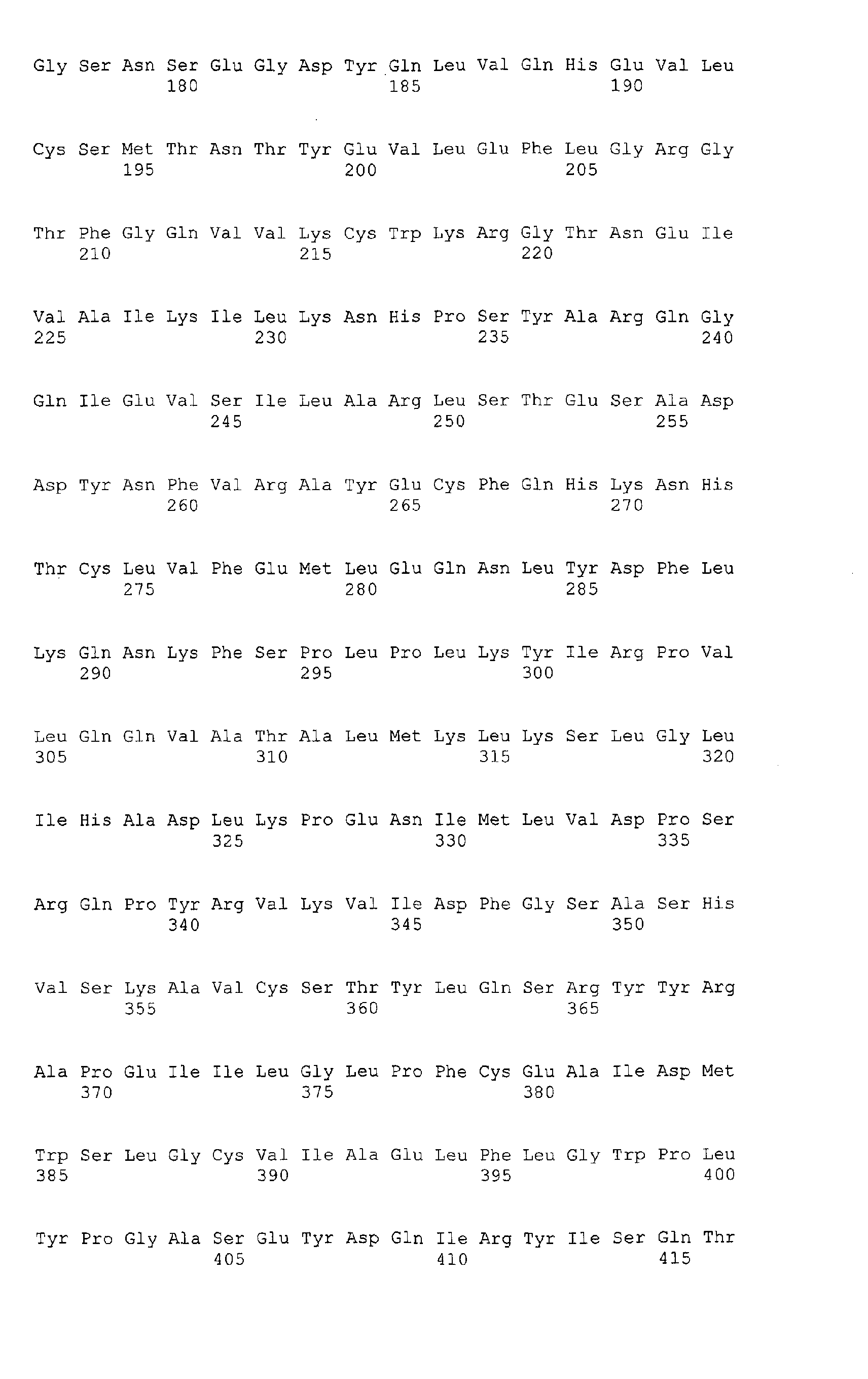
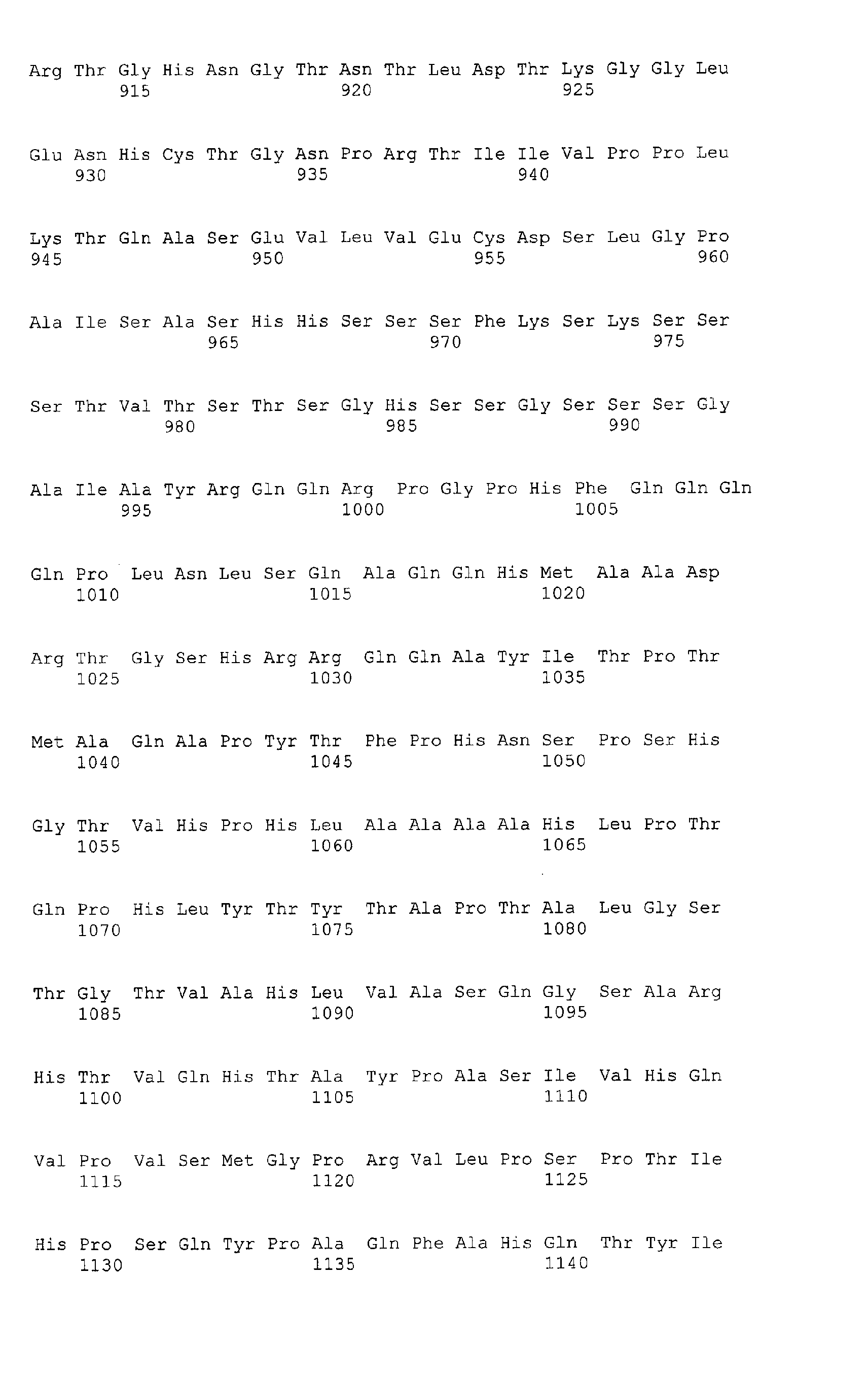
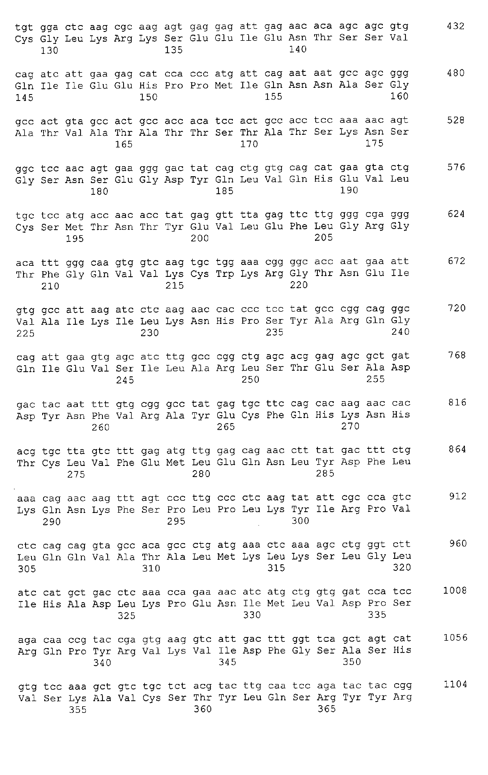
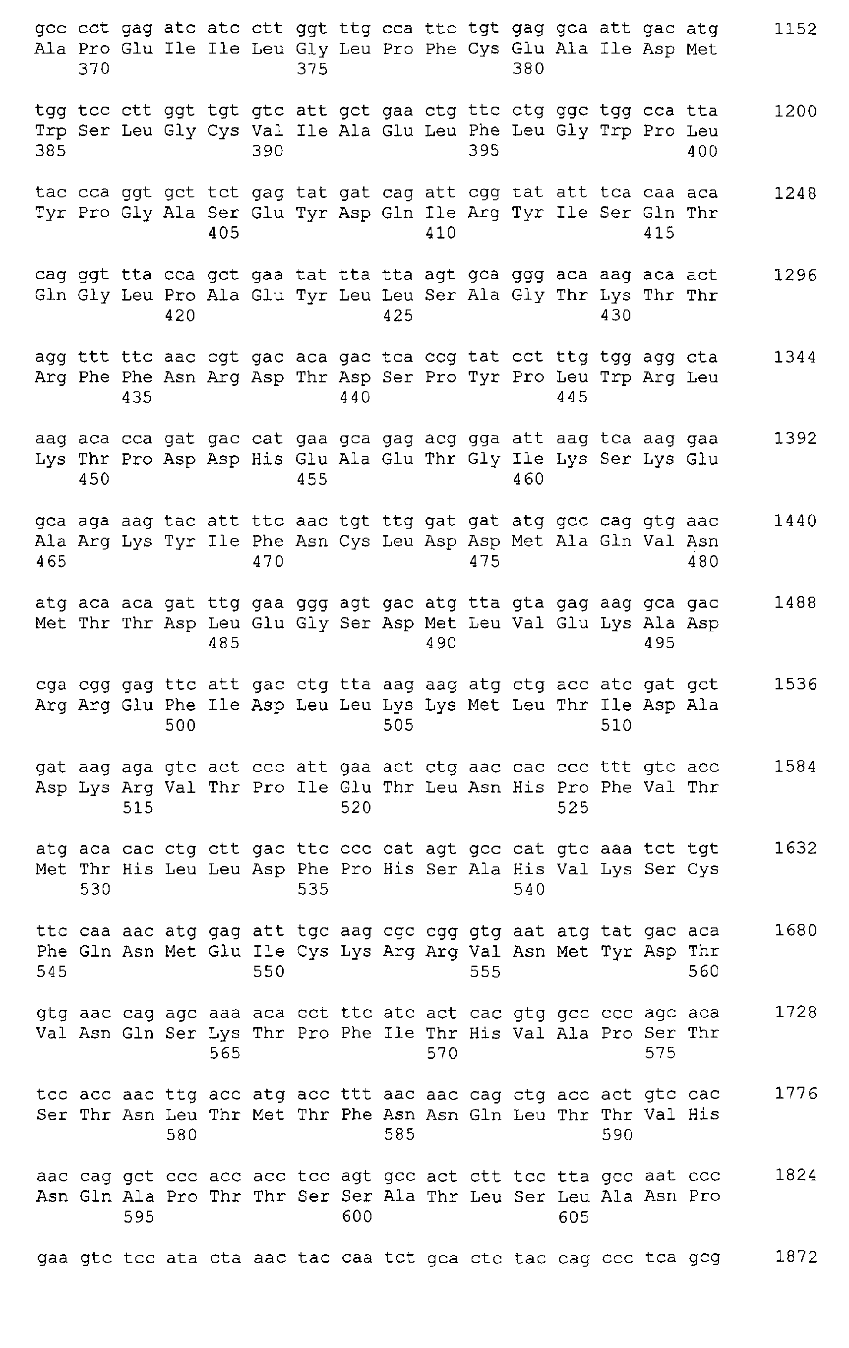
The term "HIPK2" denotes the human Homeodomain-interacting
protein kinase 2, but also homologous proteins from other mammalian species,
such as mice or hamster, and splicing variant thereof. Cloning of human HIPK2
has been reported in Hofmann et al. (2000). mRNA sequences of human
HIPK2 and of an alternative splicing variant thereof are deposited in Genbank
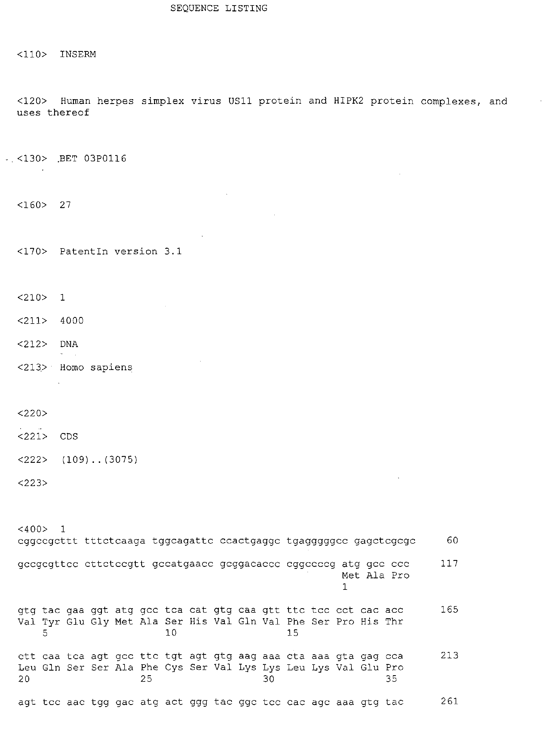
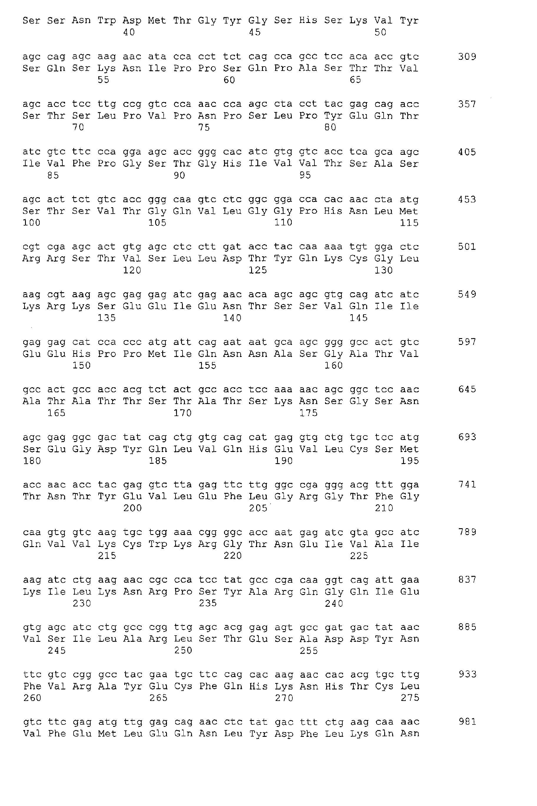
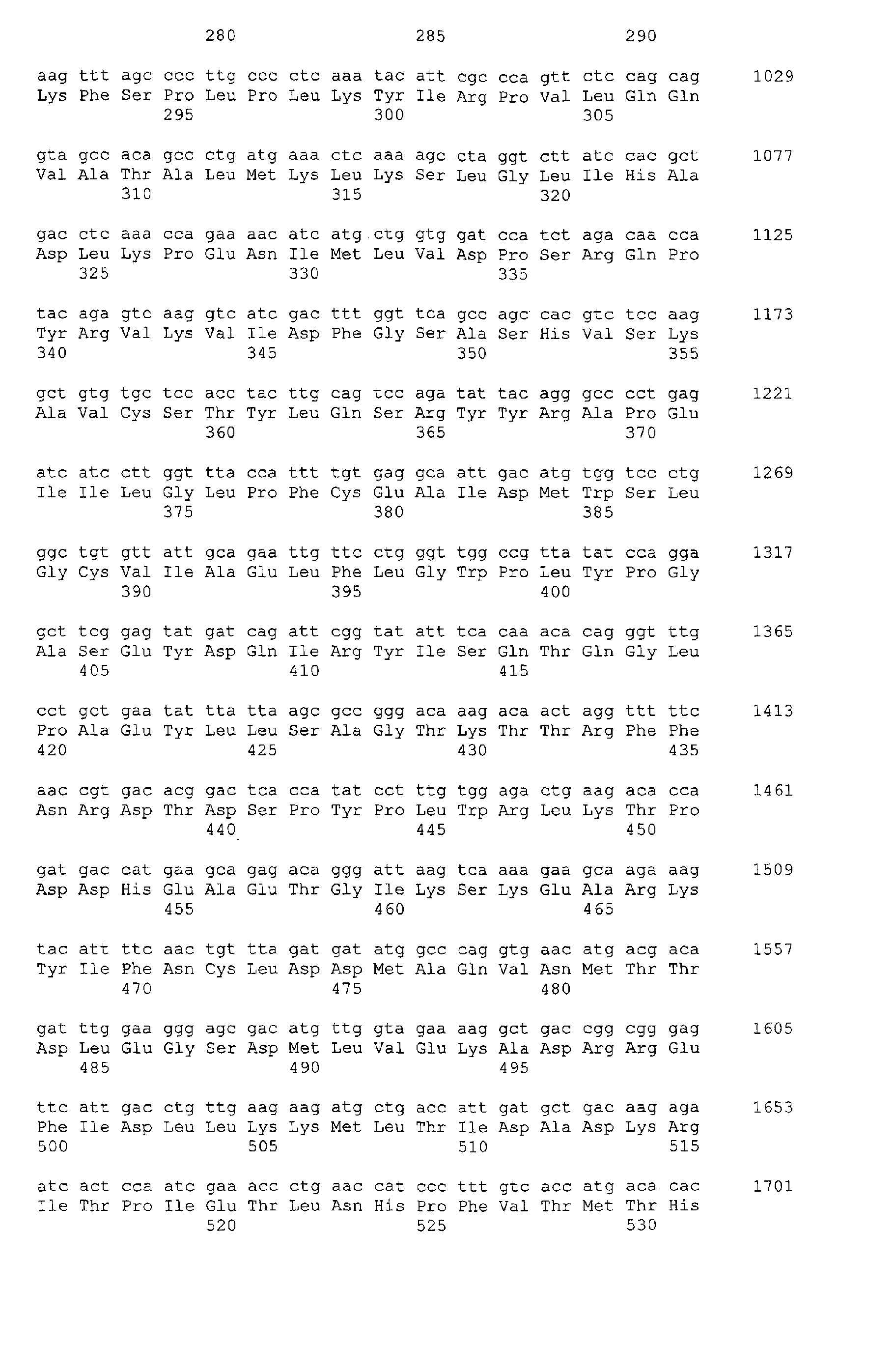
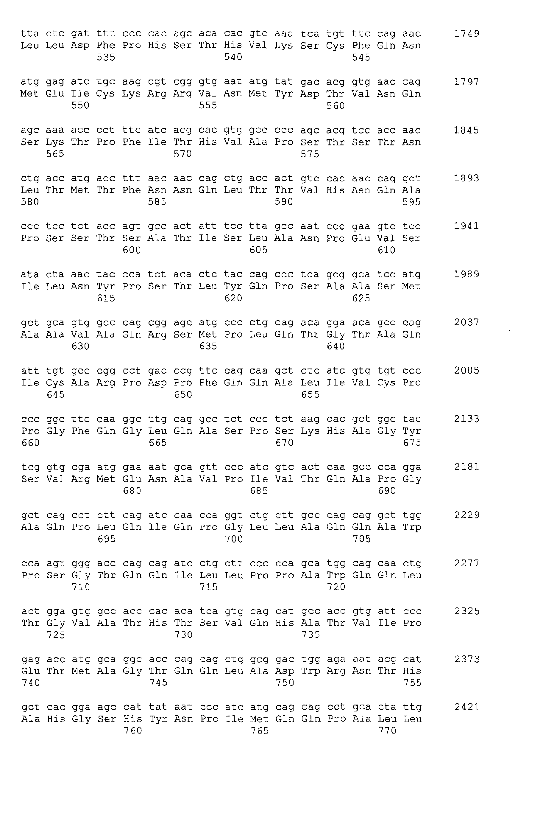
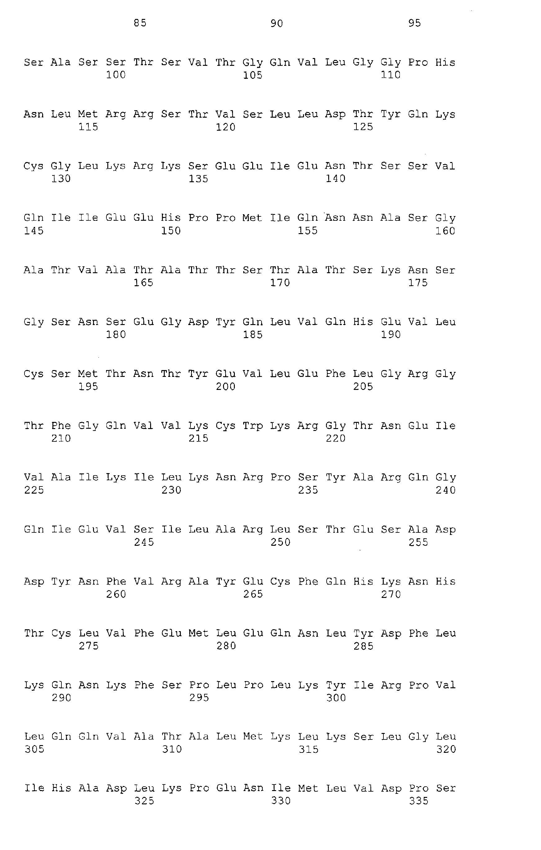
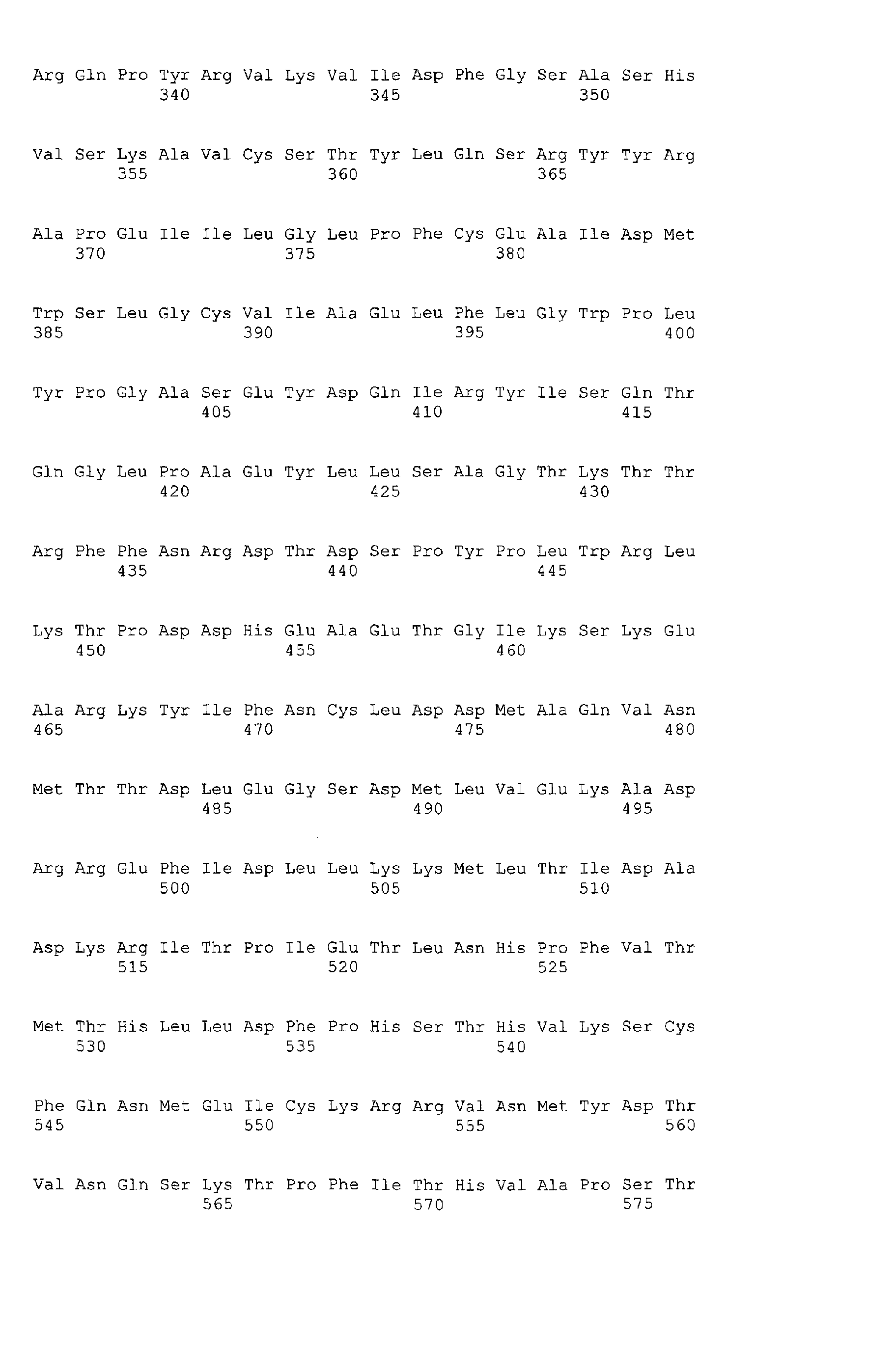
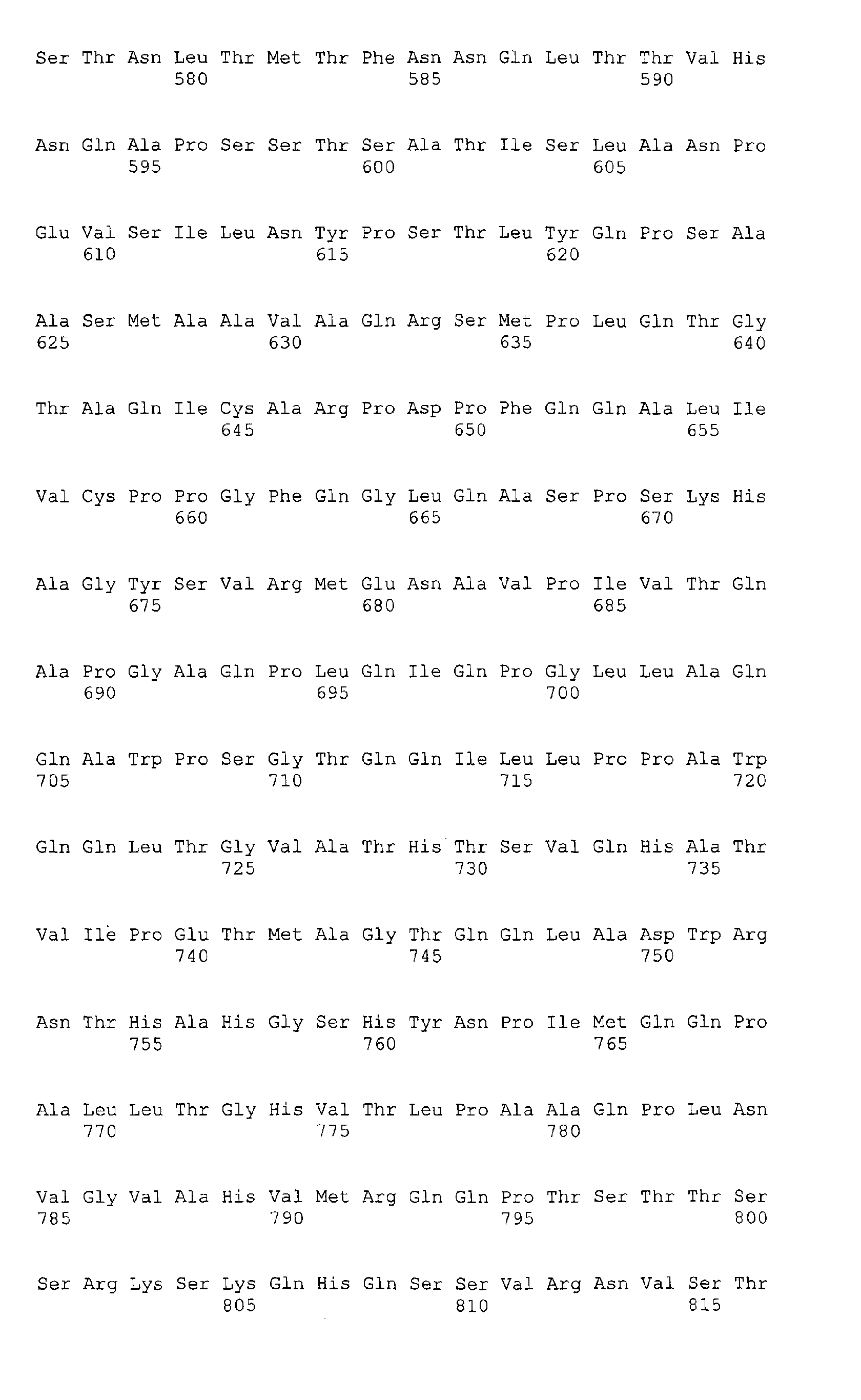
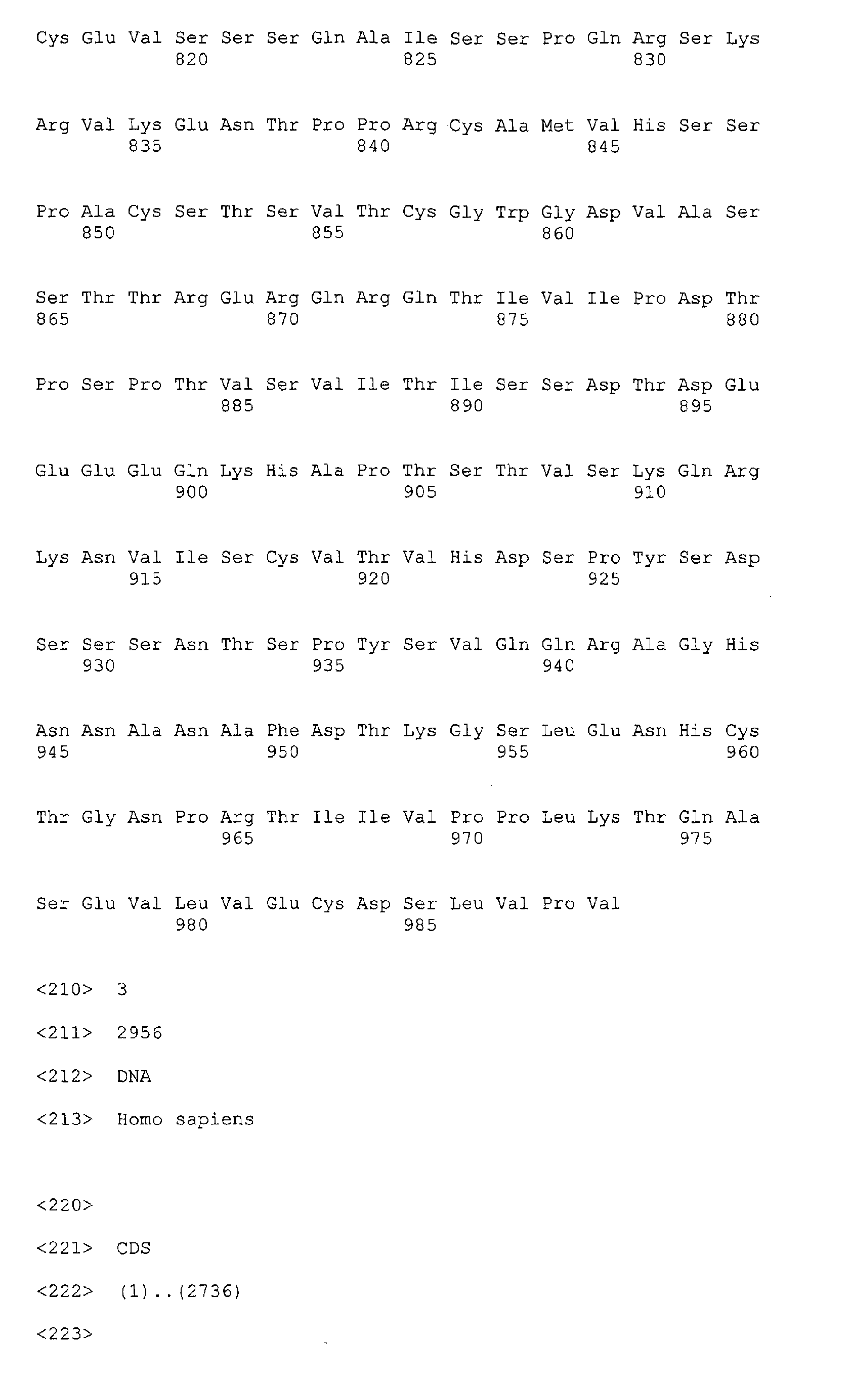
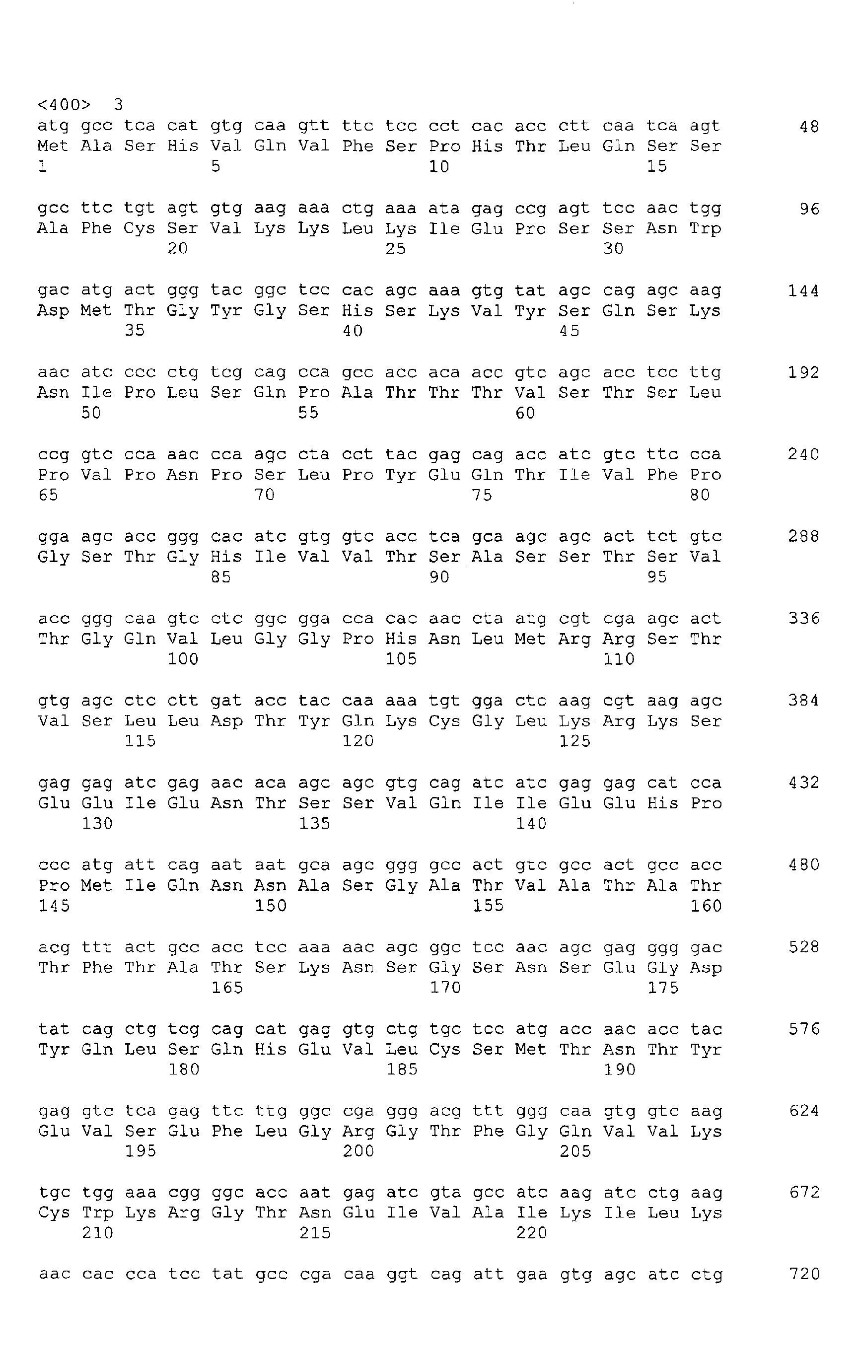
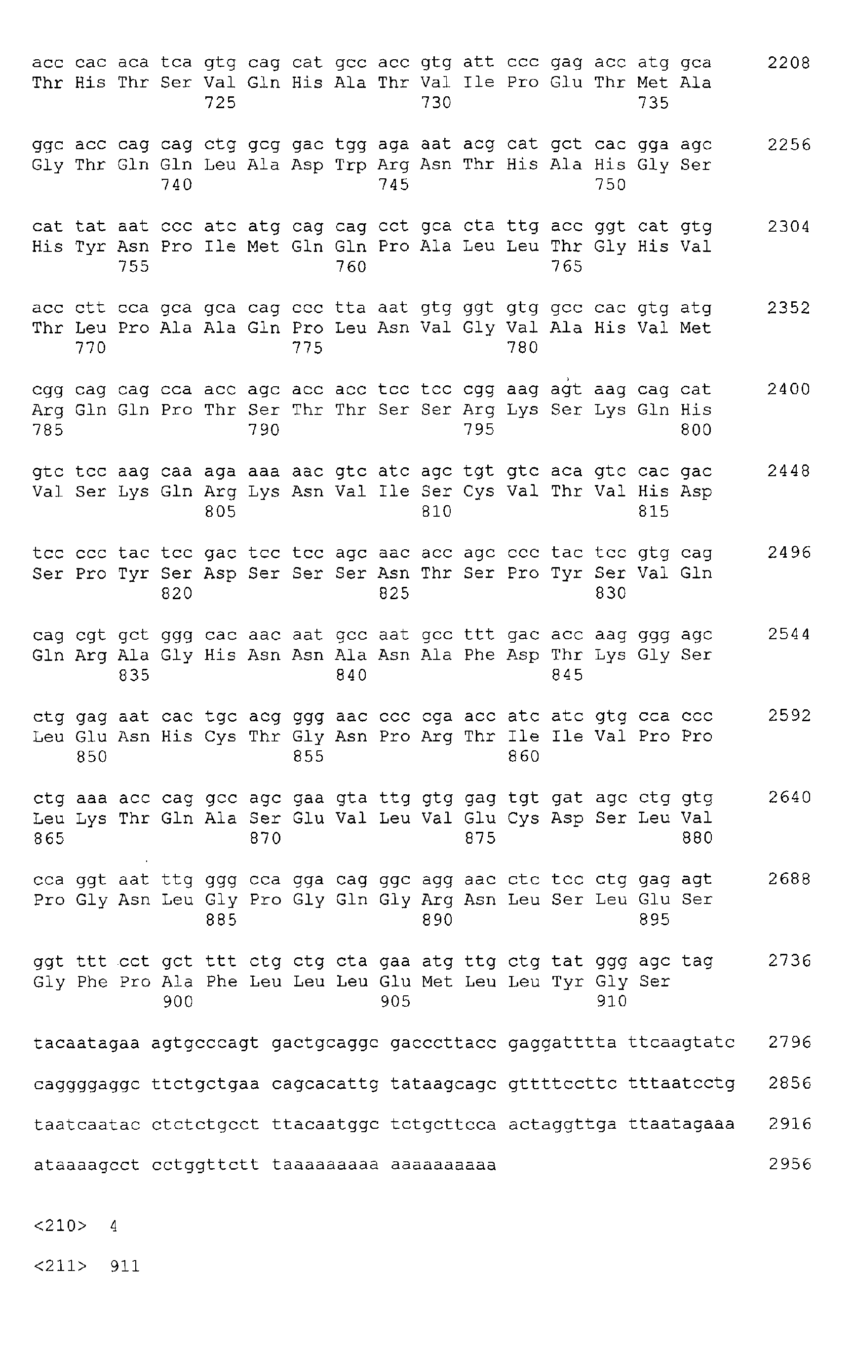
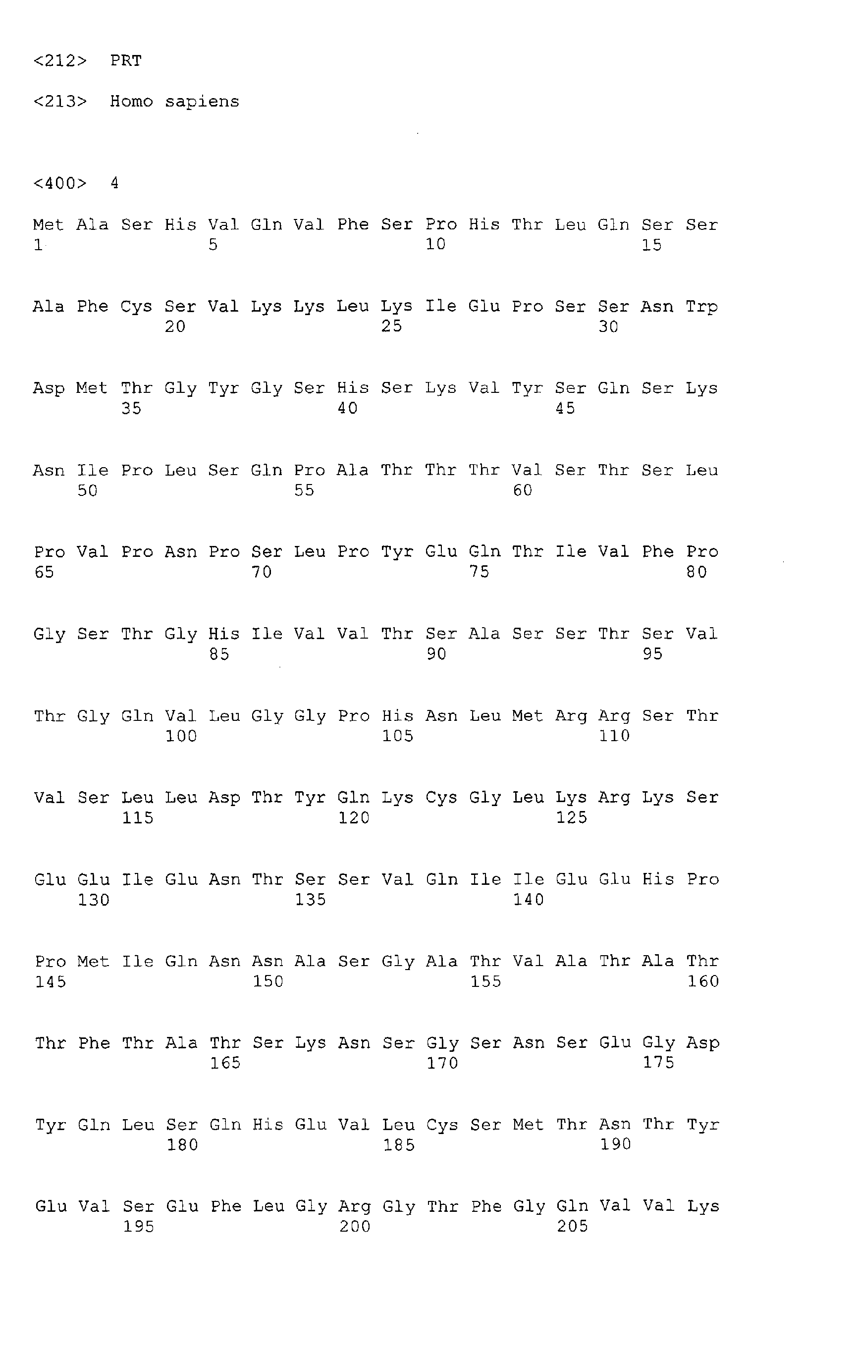
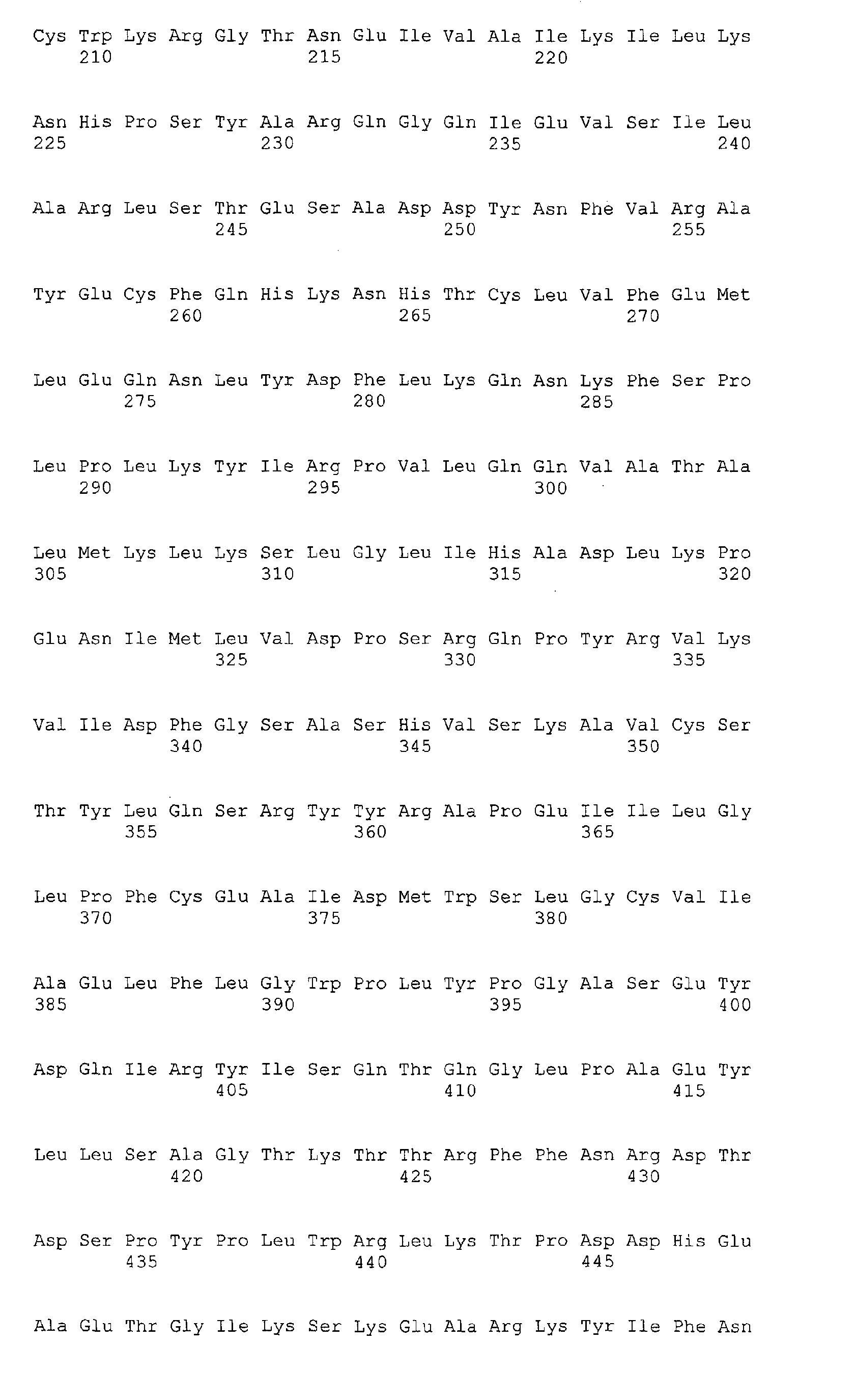
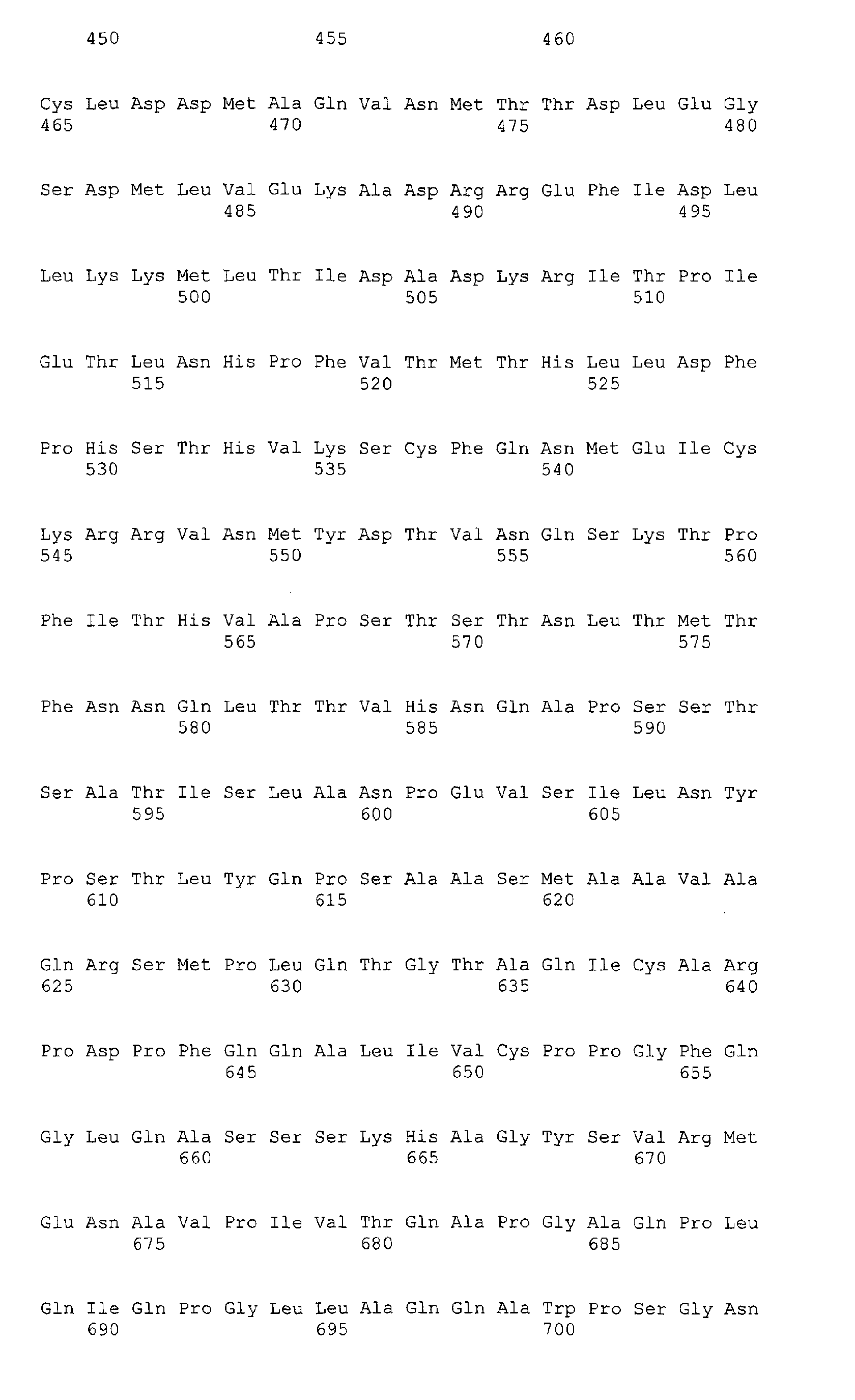
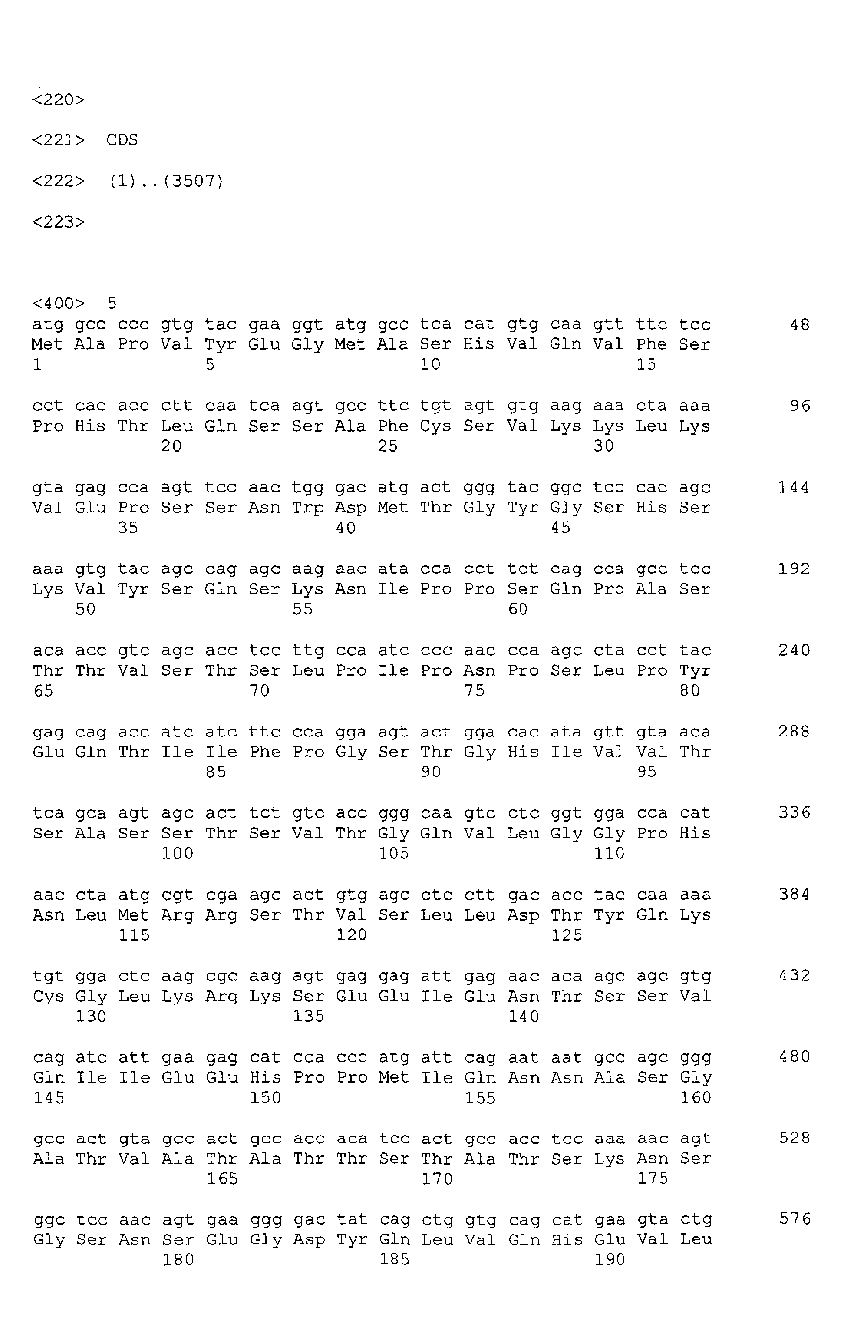
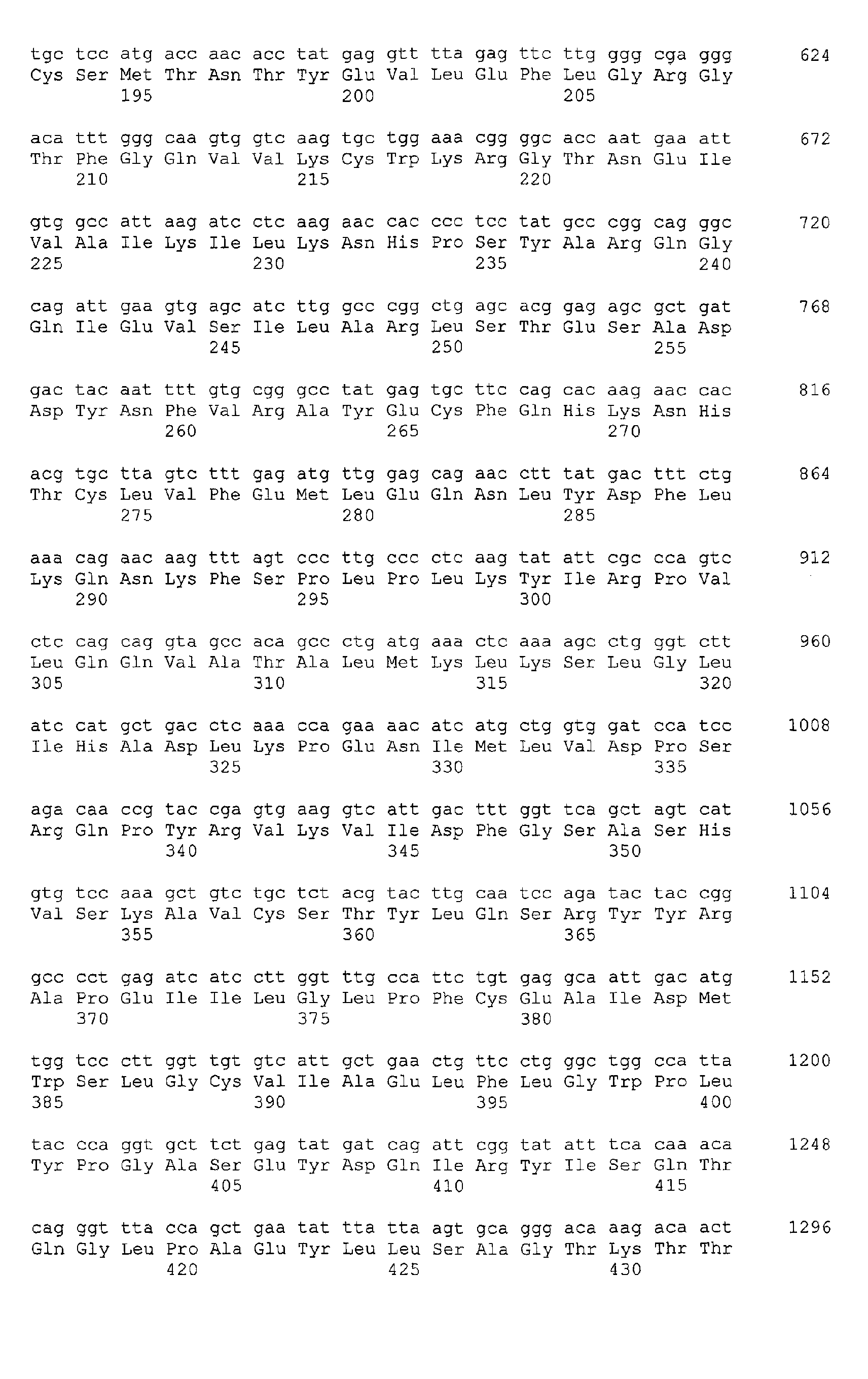
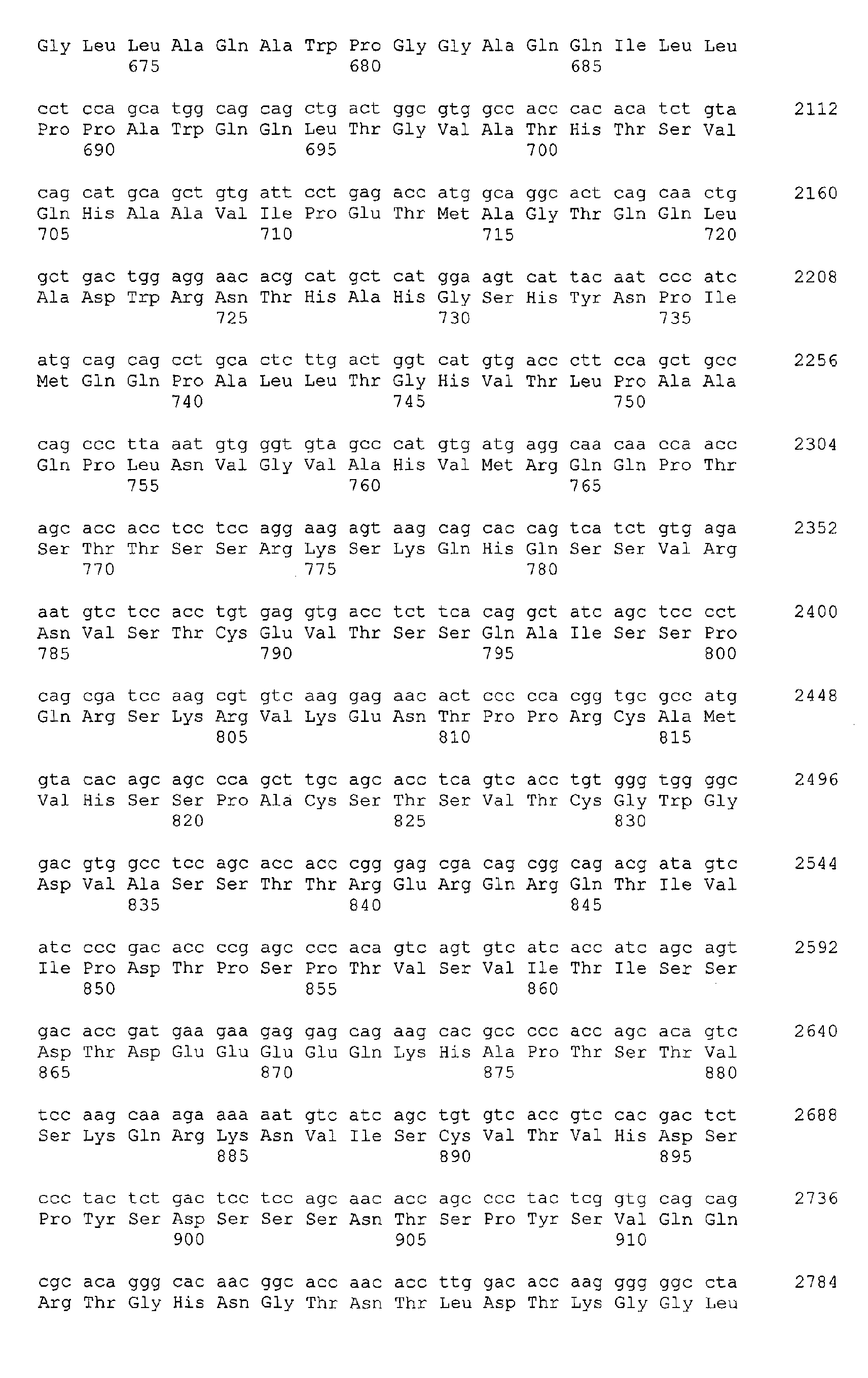
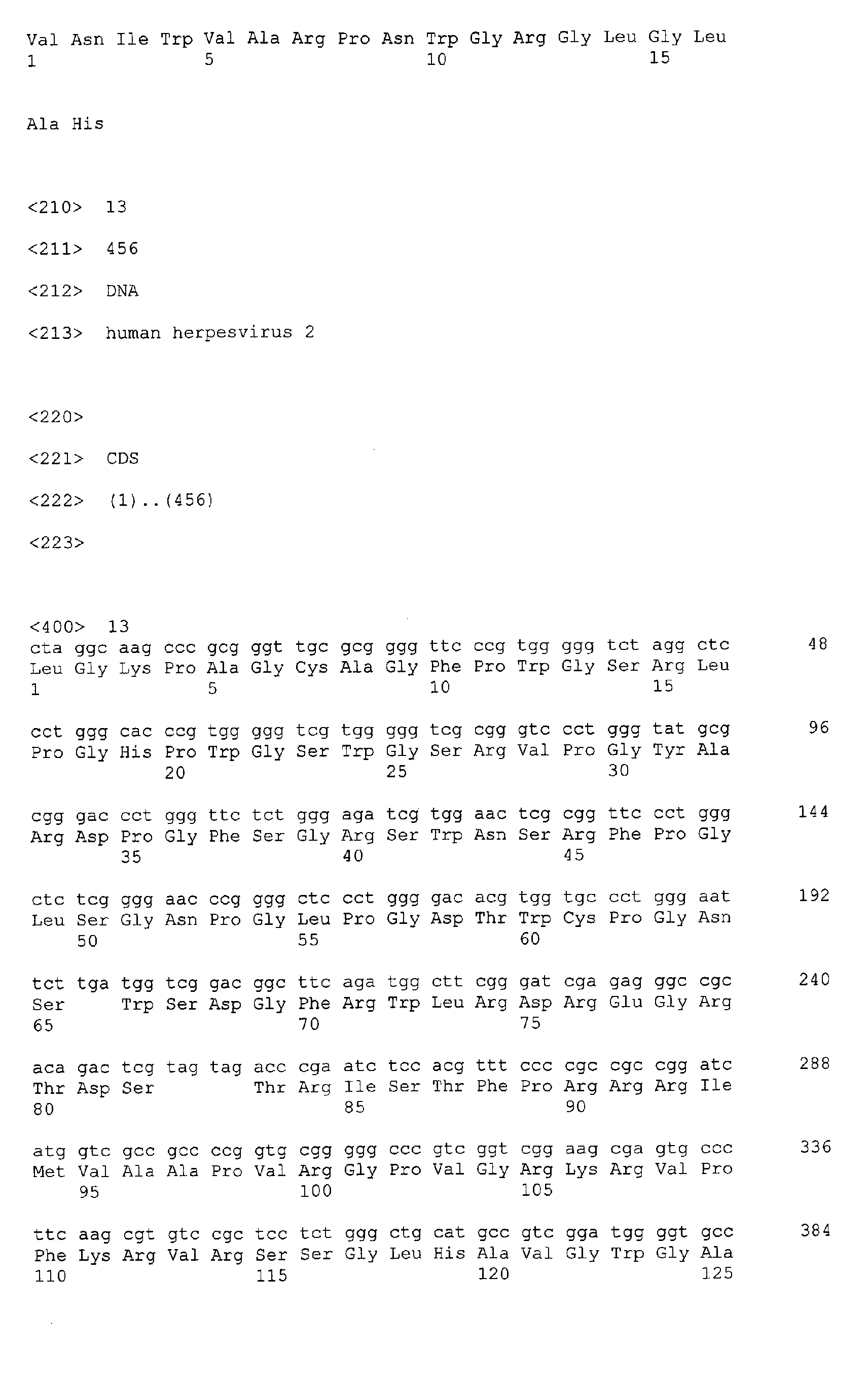
under accession number NM_022740 (polynucleotide sequence : SEQ ID No:1,
amino acid sequence: SEQ ID No:2) and AF207702 (polynucleotide sequence :
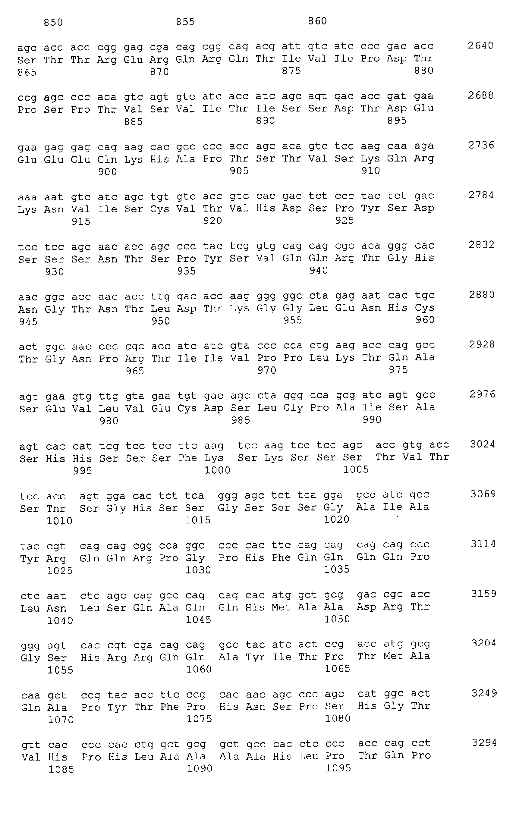
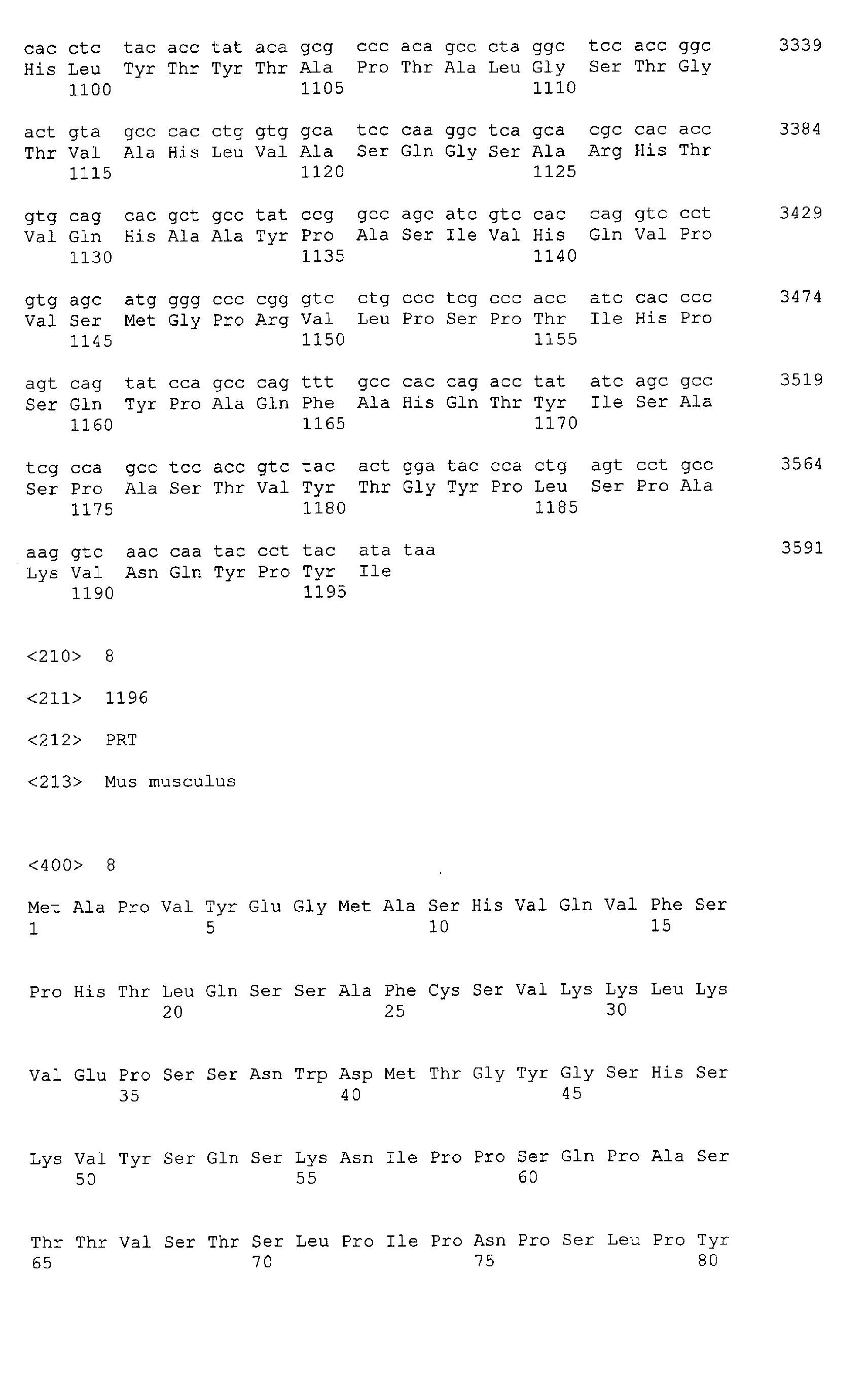
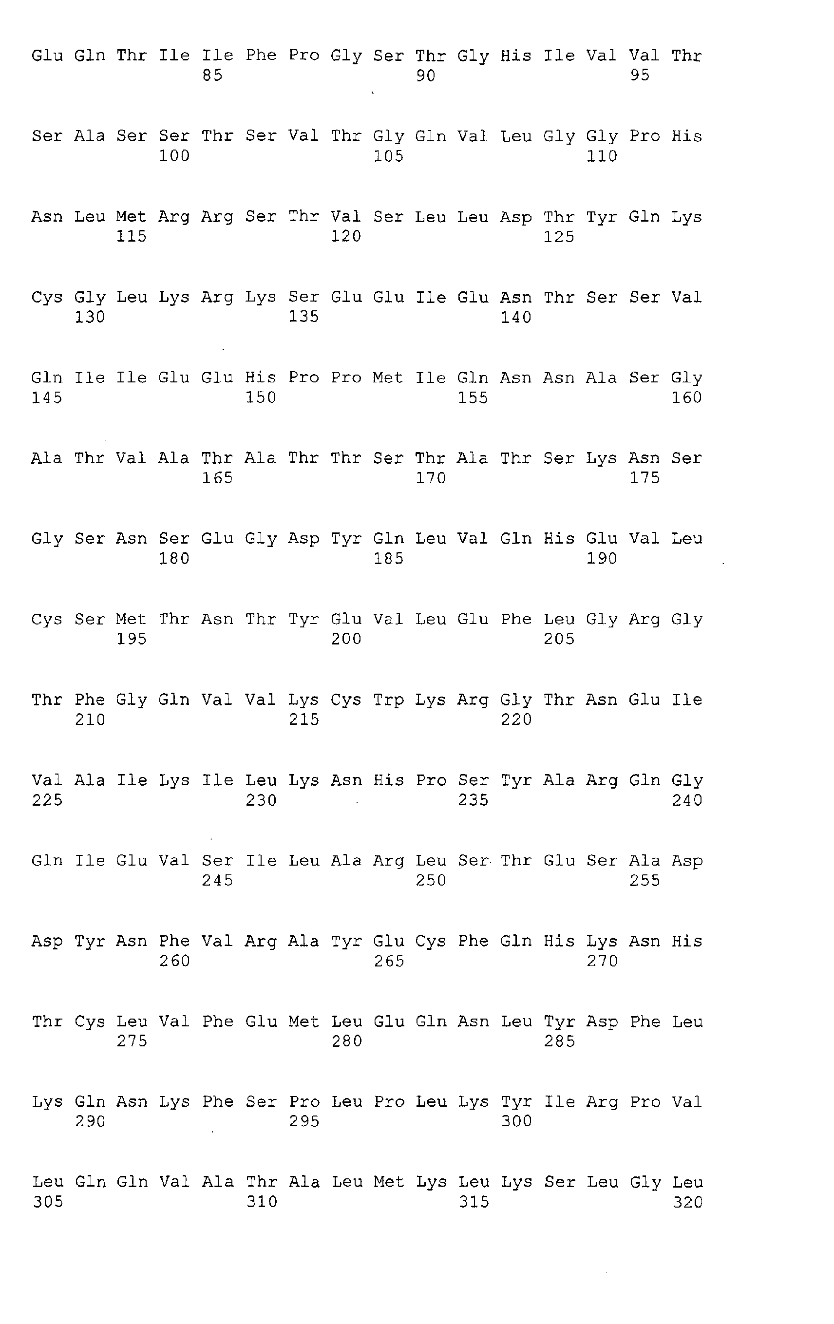
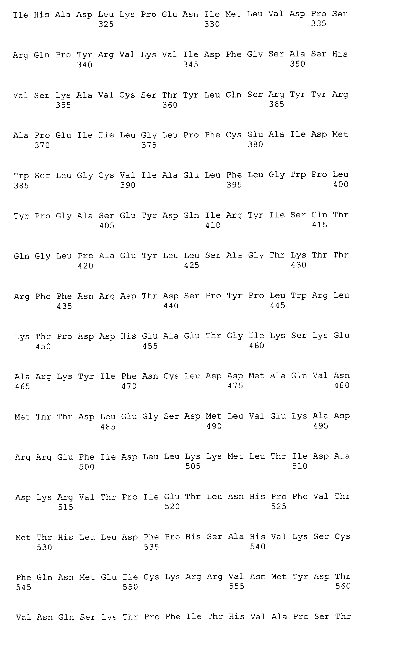
SEQ ID No:3, amino acid sequence: SEQ ID No:4). Two mouse splicing
variants (hereafter called HIPK2a and HIPK2b) have been identified by the
inventors of the instant application and the corresponding mRNA and amino
acid sequences are deposited in Genbank under accession numbers
AF333791 (HIPK2a: polynucleotide sequence SEQ ID No: 5, amino acid
sequence: SEQ ID No: 6) and AF333792 (HIPK2b: polynucleotide sequence
SEQ ID No: 7, amino acid sequence: SEQ ID No: 8).
-
As used herein, the term "PEST" or "PEST domain" is meant for
the HIPK2 domain rich in proline, glutamic acid, serine and threonine. For
instance, mouse and hamster PEST domains have been defined as consisting
of amino acid residues 839 to 934, and 812 to 907, respectively, of HIPK2
protein (Kim et al., 1998, and Kim et al., 2002). Human HIPK2 contains two
potential PEST sequences from position 872 to position 901 and position 922
to position 941 (Wang et al. 2001a).
-
The term "herpes simplex virus" (HSV) is intended for viruses
from the Alphaherpesvirinae subfamily of herpes viruses that belong to the
genus simplexvirus. Examples of HSV include the human herpes simplex virus
type 1 (HSV-1) or type 2 (HSV-2).
-
"US11" refers to the Unique Short sequence 11 protein of a
herpes simplex virus type 1 and type 2. In the context of the invention, said
US11 protein may originate from may be of any specie, genotype and subtype
of herpes simplex virus, where appropriate. The term US11 includes in
particular US11 from HSV-1 and HSV-2. A HSV-1 coding sequence for US11 is
described for instance in the Genbank database under accession number
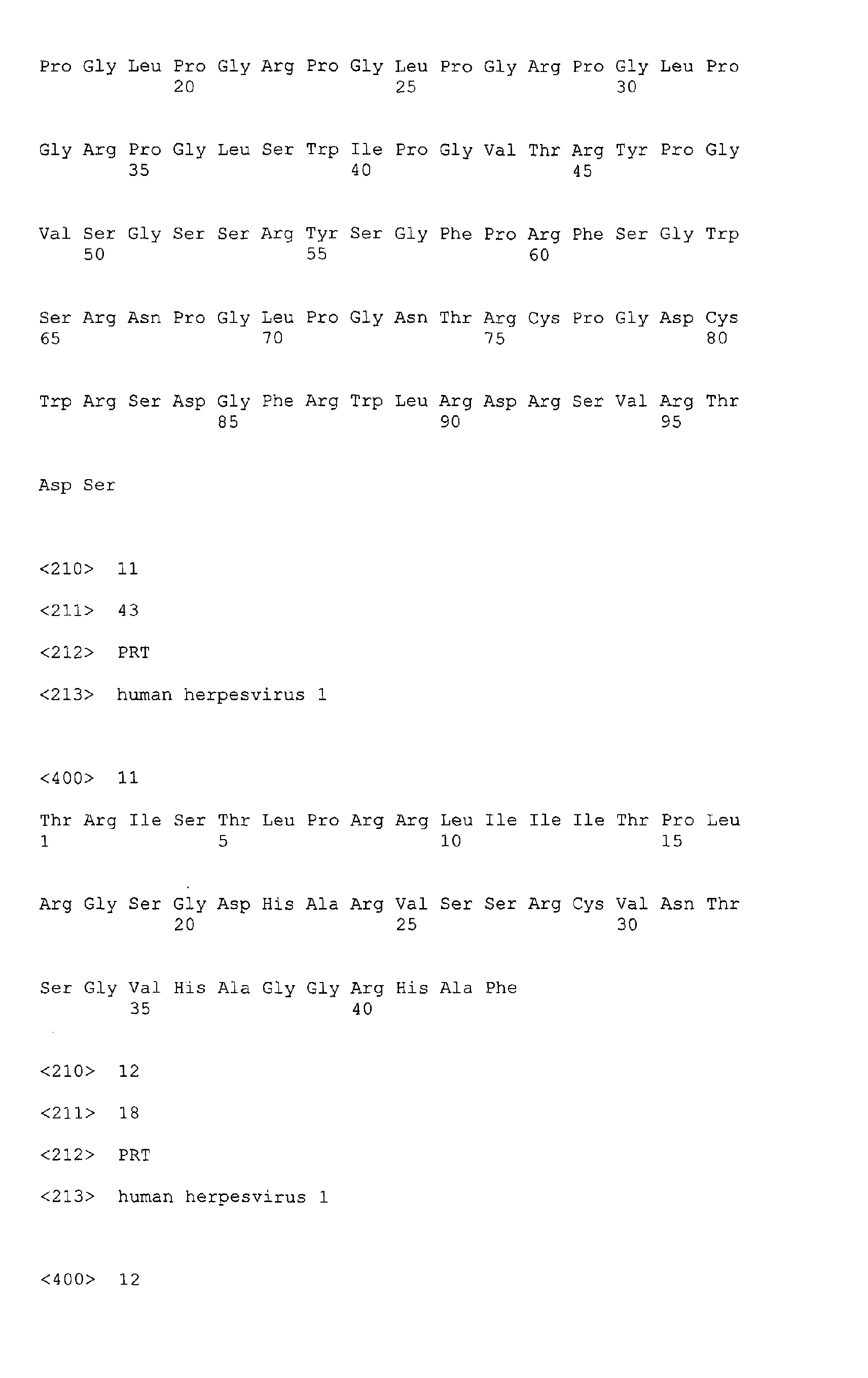
X02138 (polynucleotide sequence: SEQ ID No: 9, amino acid sequence: SEQ
ID No: 10). Similarly a HSV-2 coding sequence for US11 is described in the
Genbank database under accession number NC_001798 (polynucleotide
sequence: SEQ ID No: 11, amino acid sequence: SEQ ID No: 12).
-
The inventors have shown that US11 and HIPK2 can interact, in
particular by way of the PEST domain of HIPK2, to form US11/HIPK2
complexes. The binding of US11 to HIPK2 has been demonstrated by the
inventors as implicated in rescuing the cells from the apoptotic process elicited
by HIPK2.
-
Thus, in the context of the present invention, the term "modulator"
is intended for a molecule that modulates, i.e. antagonizes or promotes, the
US11/HIPK2 complex. More specifically, said modulator may act by modulating
either the interaction between US11 and HIPK2, or the activity of a
US11/HIPK2 complex. Typically, the term modulator encompasses molecules
with antagonist and agonist activity.
-
The term "antagonist" is intended for a molecule that may (i)
interfere with the formation of, or disrupt, a US11/HIPK2 complex, or (ii)
interfere or inhibit the activity of a US11/HIPK2 complex, in particular the
apoptosis rescuing activity associated with the US11/HIPK2 complex. In other
words, antagonists according to the invention may include molecules capable
of preventing or limiting the interaction of US11 with HIPK2, or molecules
capable of inducing a dissociation of a US11/HIPK2 complex, partially or totally,
where said complex is formed. Examples of antagonists may include for
instance an isolated HIK2 PEST domain, or a fragment of HIPK2 comprising
the PEST domain, that may act as a competitor with HIPK2 for binding with
US11.
-
By "agonist", is meant a molecule that is involved in, or promotes,
(i) formation of a US11/HIPK2 complex, or (ii) the activity of a US11/HIPK2
complex.
-
The term "candidate molecule" includes virtually any candidate
molecule which may interact with US11 and/or HIPK2 and/or US11/HIPK2
complex. The candidate molecule may be peptide or non-peptide in nature. Any
compound isolated from natural sources such as plants, animals or even
marine, forest or soil samples, may be assayed, as may any synthetic chemical
or recombinant protein.
-
The term "herpes simplex virus infection" denotes the condition of
a subject infected with HSV, either in a primary or a latent infectious state, as
well as in a reactivation period. Primary infection generally occurs through a
break in the mucus membranes of the mouth or throat, via the eye or genitals
or directly via minor abrasions in the skin. Initial infection is usually
asymptomatic, although there may be minor local vesicular lesions. Local
multiplication ensues, followed by viraemia and systemic infection. There then
follows life-long latent infection with periodic reactivation. The delicate balance
of latency may be upset by various disturbances, physical (injury, U.V,
hormones, etc) or psychological (stress, emotional upset). Reactivation of
latent virus leads to recurrent disease wherein virus travels back to surface of
body and replicates, causing tissue damage. HSV-1 is primarily associated with
oral and ocular lesions whereas HSV-2 is primarily associated with genital and
anal lesions.
-
Accordingly, "treatment of a herpes simplex virus infection" is
meant for the prevention of the onset of a primary infection and/or for the
prevention of occurrence of a viral reactivation period in the subject with latent
infection, as well as the treatment of the lesions caused by the virus
reactivation.
-
As used herein "pharmaceutically acceptable carrier" includes any
solvents, dispersion media, coatings, antibacterial and antifungal agents,
isotonic and absorption delaying agents and the like that do not produce an
adverse, allergic or other untoward reaction when administered to an animal, or
a human, as appropriate. The use of such media and agents for
pharmaceutical active substances is well known in the art. Except insofar as
any conventional media or agent is incompatible with the active ingredient, its
use in the therapeutic compositions is contemplated.
-
According to the invention, the term "subject" or "patient" is meant
for any living specie likely to be infected with a herpes simplex virus, in
particular with HSV. Mammals, and in particular human, monkey or bovine are
examples of hosts for herpes simplex viruses.
Protein complexes, compositions and kits
-
The invention provides an isolated protein complex, further called
"US11/HIPK2 complex" or "US11/HIPK2 protein complex", comprising at least
two interacting proteins, said complex being selected from the group consisting
of (a) US11 and HIPK2, (b) US11 and a fragment of HIPK2, (c) a fragment of
US11 and HIPK2, and (d) a fragment of US11 and a fragment of HIPK2.
-
The interacting proteins may be native or recombinant.
Recombinant proteins may be produced in eukaryotic or prokaryotic systems
according to conventional procedures of molecular biology, microbiology, and
recombinant DNA techniques well-known by the one skilled in the art. Such
techniques are explained fully in the literature. See, e.g., Sambrook, Fritsch &
Maniatis, Molecular Cloning: A Laboratory Manual, Second Edition (1989) Cold
Spring Harbor Laboratory Press, Cold Spring Harbor, New York (herein
"Sambrook et al., 1989") ; DNA Cloning: A Practical Approach, Volumes I and II
(D.N. Glover ed. 1985) ; Oligonucleotide Synthesis (M.J. Gait ed. 1984) ;
Nucleic Acid Hybridization [B.D. Hames & S.J. Higgins eds. (1985)] ;
Transcription and Translation [B.D. Hames & S.J. Higgins, eds. (1984)] ;
Animal Cell Culture [R.I. Freshney, ed. (1986)] ; Immobilized Cells and
Enzymes [IRL Press, (1986)] ; B. Perbal, A Practical Guide To Molecular
Cloning (1984) ; F.M. Ausubel et al. (eds.), Current Protocols in Molecular
Biology, John Wiley & Sons, Inc. (1994).
-
Advantageously, the interacting proteins can be produced as a
fusion protein that may include a peptide linker between the interacting
proteins, which may serve to promote the interaction of the proteins.
-
A "fragment of HIPK2 protein" or "HIPK2 fragment" is meant for N-terminally
and/or C-terminally truncated form of HIPK2 that retains capacity of
binding with native US11, or a fragment thereof. Preferably said HIPK2
fragment consists of or comprises the PEST domain.
-
A "fragment of US11 protein" or "US11 fragment" is meant for N-terminally
and/or C-terminally truncated form of US11 that retains capacity of
binding with native HIPK2, or with a fragment thereof such as a fragment
containing the HIPK2 PEST domain.
-
Production of US11 or HIPK2 fragments may be readily achieved
by the one skilled in the art. Assaying their capacity to retain binding capacity
may be performed by detecting protein interactions with various systems,
including the yeast two-hybrid system, affinity chromatography, co-immunoprecipitation,
subcellular fractionation isolation of large molecular
complexes and proteomics.
-
According to the above embodiment, the US11 and HIPK2
proteins are provided in the form of a US11/HIPK2 complex. Alternatively, the
US11 and HIPK2 proteins may be provided together within a composition, or
separately in a kit.
-
Accordingly the invention further provides a protein composition
comprising at least two interacting proteins selected from the group consisting
of (a) US11 or a fragment of US11, and (b) HIPK2 or a fragment of HIPK2,
optionally in a pharmaceutically acceptable carrier.
-
The invention also provides a kit comprising (a) US11 or a
fragment of US11, and (b) HIPK2 or a fragment of HIPK2.
-
In the sections below, unless otherwise specified, the term
"HIPK2 protein" includes a HIPK2 fragment. Similarly, unless otherwise
specified, the term "US11 protein" includes a US11 fragment.
Vectors and Host cells
-
The invention also relates to an isolated host cell coexpressing
US11 and HIPK2 proteins. In particular a host cell according to the invention
comprises, or has been transformed with a US11 expressing vector and
optionally a HIPK2 expressing vector.
-
Since HIPK2 expression may be very low in cells, it may be
preferable to overexpress HIPK2, e.g. by transforming the cells with a HIPK2
expressing vector. Otherwise, HIPK2 may be overexpressed and/or activated
for instance by exposing a host cell to UVs, or by treating said cell with arsenic
(AS2O3).
-
Where appropriate, US11 and HIPK2 polynucleotide sequences
may be carried on two separate vectors or on a same vector (for example a
vector carrying an International Ribosome Entry Site (IRES) for the expression
of the second coding sequence). Another possibility is that both sequences are
cloned in frame, under the control of a same promoter so as to ensure
coexpression of US11 and HIPK2 proteins. Accordingly a vector comprising a
polynucleotide sequence encoding US11 and HIPK2 proteins, in particular in
the form of a fusion protein that optionally includes a peptide linker between
US11 and HIPK2 proteins, is within the scope of the invention. The choice of a
suitable expression system is within the ordinary skills in the art. Preferably,
said host cell is a p53 expressing cell such as Hek 293 cells (also named
293T), HCT116, U2OS, and RKO.
Antibodies against US11/HIPK2 complex
-
As shown above, US11 interacts with HIPK2 to form a complex.
For the purpose of antibody production, a complex of the two proteins may be
prepared, e.g., by mixing purified preparations of each of the two proteins. If
desired, the protein complex may be stabilized by cross-linking the proteins in
the complex, by methods known to those of skill in the art.
-
Accordingly, the invention proposes an isolated antibodies
"specific" for the US11/HIPK2 complex, i.e. antibodies that are reactive with
said complex but that are not reactive with US11 and HIPK2 proteins.
-
The antibodies of the present invention can be monoclonal or polyclonal
antibodies, single chain or double chain, chimeric antibodies, humanized
antibodies, or portions of an immunoglobulin molecule, including those portions
known in the art as antigen binding fragments Fab, Fab', F(ab')2 and F(v). They
can also be immunoconjugated ,e.g. with a toxine, or labelled antibodies.
-
Whereas polyclonal antibodies may be used, monoclonal
antibodies are preferred for they are more reproducible in the long run.
-
Procedures for raising "polyclonal antibodies" are also well
known. Polyclonal antibodies can be obtained from serum of an animal
immunized against the US11/HIPK2 complex, which can be produced by
genetic engineering for example according to standard methods well-known by
one skilled in the art. Typically, such antibodies can be raised by administering
the protein complex subcutaneously to New Zealand white rabbits which have
first been bled to obtain pre-immune serum. The antigens can be injected at a
total volume of 100 µl per site at six different sites. Each injected material will
contain adjuvants with or without pulverized acrylamide gel containing the
protein or polypeptide after SDS-polyacrylamide gel electrophoresis. The
rabbits are then bled two weeks after the first injection and periodically boosted
with the same antigen three times every six weeks. A sample of serum is then
collected 10 days after each boost. Polyclonal antibodies are then recovered
from the serum by affinity chromatography using the corresponding antigen to
capture the antibody. This and other procedures for raising polyclonal
antibodies are disclosed in Harlow et al. (1988) which is hereby incorporated by
reference.
-
A "monoclonal antibody" in its various grammatical forms refers to
a population of antibody molecules that contain only one specie of antibody
combining site capable of immunoreacting with a particular epitope. A
monoclonal antibody thus typically displays a single binding affinity for any
epitope with which it immunoreacts. A monoclonal antibody may therefore
contain an antibody molecule having a plurality of antibody combining sites,
each immunospecific for a different epitope, e.g. a bispecific monoclonal
antibody. Although historically a monoclonal antibody was produced by
immortalization of a clonally pure immunoglobulin secreting cell line, a
monoclonally pure population of antibody molecules can also be prepared by
the methods of the present invention.
-
Laboratory methods for preparing monoclonal antibodies are well
known in the art (see, for example, Harlow et al., 1988). Monoclonal antibodies
(mAbs) may be prepared by immunizing purified US11/HIPK2 protein complex
from any of a variety of mammalian species into a mammal, e.g. a mouse, rat,
human and the like mammal. The antibody-producing cells in the immunized
mammal are isolated and fused with myeloma or heteromyeloma cells to
produce hybrid cells (hybridoma). The hybridoma cells producing the
monoclonal antibodies are utilized as a source of the desired monoclonal
antibody. This standard method of hybridoma culture is described in Kohler and
Milstein (1975).
-
While mAbs can be produced by hybridoma culture, the invention
is not to be so limited. Also contemplated is the use of mAbs produced by an
expressing nucleic acid cloned from a hybridoma of this invention. That is, the
nucleic acid expressing the molecules secreted by a hybridoma of this
invention can be transferred into another cell line to produce a transformant.
The transformant is genotypically distinct from the original hybridoma but is
also capable of producing antibody molecules of this invention, including
immunologically active fragments of whole antibody molecules, corresponding
to those secreted by the hybridoma. See, for example, U.S. Pat. No. 4,642,334
to Reading; PCT Publication No. WO 890099 to Robinson et al.; European
Patent Publications No. 0239400 to Winter et al. and No. 0125023 to Cabilly et
al.
-
Antibody generation techniques not involving immunisation are
also contemplated such as for example using phage display technology to
examine naive libraries (from non-immunised animals); see Barbas et al.
(1992), and Waterhouse et al. (1993).
Screening methods
-
The invention relates to a method, in particular in vitro, of
screening or identifying candidate molecules capable of modulating a
US11/HIPK2 complex. Such molecules capable of modulating the US11 and
HIPK2 interaction or of modulating the US11/HIPK2 complex activity comprise
molecules useful for preventing or treating a herpes simplex virus infection.
-
More specifically, the method of screening or identifying candidate
molecules capable of modulating a US11/HIPK2 complex may comprise the
steps consisting of :
- a) contacting at least one candidate molecule with a US11/HIPK2
protein complex as described above, and/or with US11 and HIPK2 proteins;
and
- b) determining the presence or absence of binding of the
candidate molecule to said US11/HIPK2 complex, US11 protein or HIPK2
protein;
wherein the binding of the candidate molecule to said
US11/HIPK2 complex, US11 protein or HIPK2 protein, is indicative of a
modulator of the US11/HIPK2 complex.-
-
According to a first embodiment, said modulator of US11/HIPK2
complex may interfere with or promote the apoptosis rescuing activity
associated with the US11/HIPK2 complex, without necessarily disrupting, even
partially, said complex. Such a modulator may thus be identified by the above
method by specifically detecting binding to the US11/HIPK2 complex. A method
of screening or identifying a modulator of US11/HIPK2 complex activity may
thus comprise the steps of :
- a) contacting at least one candidate molecule with a US11/HIPK2
protein complex as described above; and
- b) determining the presence or absence of binding of the
candidate molecule to said US11/HIPK2 complex;
wherein the binding of the candidate molecule to said
US11/HIPK2 complex is indicative of a modulator of the US11/HIPK2 complex.-
-
Alternatively, the interfering or promoting activity of the modulator
of US11/HIPK2 activity may be assayed according to a functional test, such as
a cell colony assay. A method of screening or identifying candidate molecules
capable of modulating activity of a US11/HIPK2 complex may thus comprise
the steps consisting of :
- a) contacting a p53 expressing cell containing a HIPK2
expressing vector and a US11 expressing vector with a p53-mediated
apoptosis inducer, in the presence or in the absence of a candidate molecule,
under conditions sufficient to allow apoptosis to occur where the candidate
molecule is absent; and
- b) determining the number of cell colonies in the presence and in
the absence of the candidate molecule;
wherein an altered number of colonies as estimated in the
presence of the candidate molecule, compared with the number of colonies as
estimated in the absence of the candidate molecule, is indicative of a molecule
capable of modulating activity of a US11/HIPK2 complex. -
-
The principle of this cell colony assay is explained more
thoroughly below.
-
Said modulator may be an antagonist of US11/HIPK2 complex
activity. Accordingly, in the above method, a candidate molecule would be
identified as an antagonist where a decreased number of colonies is detected,
as estimated in the presence of the candidate molecule, compared with the
number of colonies as estimated in the absence of the candidate molecule.
Antagonists of US11/HIPK2 complex activity should constitute molecules useful
for the treatment of a herpes simplex infection.
-
Alternatively, said modulator may be an agonist of US11/HIPK2
complex activity. In that case, in the above method, a candidate molecule
would be identified as an agonist where an increased number of colonies is
detected, as estimated in the presence of the candidate molecule, compared
with the number of colonies as estimated in the absence of the candidate
molecule.
-
According to a second embodiment, said modulator of
US11/HIPK2 complex is a modulator of the interaction of US11 with HIPK2.
-
The invention more particularly provides a method of screening or
identifying candidate molecules wherein a candidate molecule is assayed for its
capacity to modulate the US11 and HIPK2 interaction, i.e, of preventing or
promoting the binding of US11 with HIPK2, or also to dissociate, partly or
totally, the so-formed US11/HIPK2 complexes.
-
According to this embodiment, the invention relates to a method
of screening or identifying candidate molecules capable of modulating the
interaction of US11 with HIPK2, which method comprises the steps consisting
of:
- a) contacting at least one candidate molecule with a HIPK2
protein and a US11 protein, under conditions suitable to allow US11 and HIPK2
to interact in the absence of the candidate molecule; and
- b) determining the interaction of US11 and HIPK2 in the absence
and in the presence of the candidate molecule;
wherein a modulation of the interaction of US11 and HIPK2 is
indicative of a molecule capable of modulating the US11 and HIPK2 interaction.-
-
Modulators of US11 and HIPK2 interaction include agonists and
antagonists of US11/HIPK2 interaction
-
Preferably, the invention relates to method of screening
molecules capable of modulating the US11 and HIPK2 interaction, and in
particular of molecules useful for preventing or treating a herpes simplex virus
infection, wherein an antagonist of the US11/HIPK2 interaction is identified.
The candidate molecule may be assayed for its capacity to interfere with or
disrupt the interaction of US11 with HIPK2. Said method of screening thus
comprises the steps consisting of :
- a) contacting at least one candidate molecule with a HIPK2
protein and a US11 protein, under conditions suitable to allow HIPK2 and US11
to interact in the absence of the candidate molecule; and
- b) determining the interaction of HIPK2 and US11 in the absence
and in the presence of the candidate molecule;
wherein a decreased interaction of US11 and HIPK2 as measured
in the presence of the candidate molecule, compared with HIPK2 and US11
interaction as measured in the absence of the candidate molecule, is indicative
of an antagonist of US11/HIPK2 interaction.-
-
Alternatively, the invention relates to method of identifying an
agonist of US11/HIPK2 interaction, said method comprising the steps
consisting of :
- a) contacting at least one candidate molecule with a HIPK2
protein and a US11 protein, under conditions suitable to allow HIPK2 and US11
to interact in the absence of the candidate molecule; and
- b) determining the interaction of HIPK2 and US11 in the absence
and in the presence of the candidate molecule;
wherein a increased interaction of US11 and HIPK2 as measured
in the presence of the candidate molecule, compared with HIPK2 and US11
interaction as measured in the absence of the candidate molecule, is indicative
of an agonist of the US11/HIPK2 interaction. -
-
Advantageously, a US11 protein, a HIPK2 protein and at least
one candidate molecule are contacted and incubated under conditions suitable
to allow them to interact, and in particular to form or dissociate US11/HIPK2
complexes.
-
Alternatively, at least one candidate molecule and a US11 protein
or a HIPK2 protein are contacted under conditions suitable to allow them to
optionally interact. The missing US11 protein or HIPK2 protein is further added
and the three components are contacted under conditions suitable to allow
them to interact, and optionally to form US11/HIPK2 complexes.
-
Still alternatively, a US11 protein and a HIPK2 protein are
contacted under conditions suitable to formation of US11/HIPK2 complexes. At
least one candidate molecule is further added and the three components are
contacted under conditions suitable to allow them to interact, and optionally to
allow US11/HIPK2 complexes to dissociate.
-
Determining of the capacity of the candidate molecule to
modulate the interaction of US11 with HIPK2, or to bind and modulate a
US11/HIPK2 complex, can be achieved by any appropriate mean well-known
by the one skilled in the art, such as classical separation and/or detection
methods. For instance, US11/HIPK2 complexes may be quantified in the
presence of increasing concentrations of candidate molecules. US11/HIPK2
binding can be carried out using classical analysis procedures such as in vitro
assays of protein binding, e.g., wherein one or both of the US11 or HIPK2
components are attached to a detectable label, and/or are immobilized may be
employed. In general, methods classically used for the detection of a positive
protein-protein interaction, such as two-hybrid system, affinity chromatography,
co-immunoprecipitation, subcellular fractionation and isolation of large
molecular complexes, immunoblotting, or immunolabelling, optionally in
association with detectable markers such as fluorescent, isotopic or
chromogenic labelling, can also be used to identify agents that interfere or
disrupt said interaction. As an example, cells transfected with DNAs coding for
US11 and HIPK2 can be treated with various candidate molecules, and co-immunoprecipitations
can be performed. Alternatively, a derivative of the yeast
two-hybrid system, called the reverse yeast two-hybrid system (Leanna and
Hannink, 1996), can be used, provided that the two proteins interact in the
straight yeast two-hybrid system. Reference can further be made to Current
Protocols in Molecular Biology, Edited by F.M. Ausubel et al. (John Wiley and
Sons).
-
Advantageously, the capacity of the candidate molecule to
modulate the US11/HIPK2 complex may be achieved according to a functional
test such as the colony formation assay described below. Briefly, p53
expressing cells, such as Hek 293 cells, may be transfected with a HIPK2
expressing vector, preferably allowing also the expression of an antibiotic
resistance gene (such as NeoR) so as to make it possible to select the
transformed cells, together with an expression vector coding for US11. After the
beginning of transfection, cells may be selected for resistance to the
appropriate antibiotic (such as Geneticin (G418) for NeoR) in the presence or
not of a candidate molecule, and the number of colonies estimated for each
experimental condition.
-
Exposure to the antibiotic further forces the cells to express
HIPK2 and thus to undergo apoptosis.
-
Cell apoptosis may be amplified by exposing cells to UVs or
contacting cells with IFN-α, so as to enhance HIPK2 expression and thereby
induce apoptosis, in particular p53 mediated apoptosis.
-
In such a test, a decreased number of colonies as estimated in
the presence of the candidate molecule, compared with the number of colonies
as estimated in the absence of the candidate molecule, which is indicative of a
candidate molecule that promotes cell apoptosis and thus of an antagonist of
the US11/HIPK2 interaction or of US11/HIPK2 complex.
-
Conversely, an increased number of colonies as estimated in the
presence of the candidate molecule, compared with the number of colonies as
estimated in the absence of the candidate molecule, which is indicative of a
candidate molecule that rescues cells from apoptosis and thus of an agonist of
the US11/HIPK2 interaction or of US11/HIPK2 complex.
-
Accordingly, the invention further provides an in vitro method of
screening molecules useful for preventing or treating a human herpes simplex
virus infection, wherein a candidate molecule is assayed for its capacity to
interfere with US11-mediated inhibition of cellular apoptosis, and in particular
with the apoptosis rescuing activity associated with the US11/HIPK2 complex.
-
More specifically, said method may comprises the steps
consisting of :
a) cultivating a cell, in particular a p53 expressing cell containing
a HIPK2 expressing vector and a US11 expressing vector, in the presence or in
the absence of a candidate molecule, under conditions sufficient to allow
HIPK2 expression and cell apoptosis to occur where the candidate molecule is
absent; b) determining the number of cell colonies in the presence and in the
absence of the candidate molecule;
wherein a decreased number of colonies as estimated in the
presence of the candidate molecule, compared with the number of colonies as
estimated in the absence of the candidate molecule, is indicative of a molecule
useful for preventing or treating a herpes simplex virus infection.
-
US11 and HIPK2 coding sequences may be carried on two
separate vectors or on a same vector (for example a vector carrying an
International Ribosome Entry Site (IRES) for the expression of the second
coding sequence). Another possibility is that both sequences are cloned in
frame, under the control of a same promoter so as to ensure co-expression of
US11 and HIPK2 proteins.
-
The p53 expressing cell may for instance be selected from the
group consisting of Hek 293 cells (293T) HCT116, U2OS, and RKO. However,
any p53 expressing cell may be suitable to carry out the above method.
-
According to said method the cultivated cell may be further
exposed to UVs and/or contacted with IFN-α so as to enhance HIPK2
expression.
Therapeutical compositions
-
The inventors have demonstrated that US11 is a mediator of the
HSV-1 dependent inhibition of cellular apoptosis. Without willing to be linked to
a particular mechanism of action, US11 could prevent cells infected with HSV-1
or HSV-2 to undergo apoptosis during the reactivation and infection phases of
the virus, and in particular following UV exposure. This inhibition of cell
apoptosis would involve US11 binding to HIPK2 and thereby prevention of p53
activation by HIPK2. Inhibition of cellular apoptosis by US11 should thus make
it possible for reactivated HSV-1 or HSV-2 to replicate in the cells rescued from
apoptosis.
-
As a consequence, an antagonist of the US11/HIPK2 complex,
i.e. a molecule capable of interfering with the formation and/or functioning of
said complex, represents a molecule useful for the treatment of HSV-1 or
HSV-2 infection, more specifically for preventing the virus to replicate under
conditions of physical or psychological disturbance, such as UV exposure.
-
The invention thus provides a composition comprising a
modulator of US11/HIPK2 complex, in particular an antagonist of US11/HIPK2
complex, in a pharmaceutically acceptable carrier.
-
According to a first embodiment, said modulator of US11/HIPK2
complex is an antagonist that interferes specifically with the interaction of US11
with HIPK2. As used herein, the term "specifically" or "specific" is used to
denote an antagonist of US11/HIPK2 interaction, or a modulator of
US11/HIPK2 complex, that does not interfere with or inhibit US11 and/or HIPK2
activity.
-
Agents capable of interfering with US11 binding to HIPK2
interaction or of modulating the US11/HIPK2 complex may be identified by a
screening method as described above. Such agents include for instance
peptides, antibodies, aptamers, or non peptide small molecules.
-
Aptamers are a class of molecule that represents an alternative to
antibodies in term of molecular recognition. Aptamers are oligonucleotide or
oligopeptide sequences with the capacity to recognize virtually any class of
target molecules with high affinity and specificity. Such ligands may be isolated
through Systematic Evolution of Ligands by EXponential enrichment (SELEX)
of a random sequence library, as described in Tuerk C. and Gold L., 1990. The
random sequence library is obtainable by combinatorial chemical synthesis of
DNA. In this library, each member is a linear oligomer, eventually chemically
modified, of a unique sequence. Possible modifications, uses and advantages
of this class of molecules have been reviewed in Jayasena S.D., 1999. Peptide
aptamers consists of a conformationally constrained antibody variable region
displayed by a platform protein, such as E. coli Thioredoxin A that are selected
from combinatorial libraries by two hybrid methods (Colas et al., 1996).
-
According to a second embodiment, said modulator of
US11/HIPK2 complex is an antagonist of US11/HIPK2 complex activity, such
as can be identified by a screening method as described above.
Therapeutical applications
-
As discussed above, the inventors have demonstrated that US11
is involved in the protection of HSV infected cells from the p53-mediated
apoptosis that is induced by HIPK2, for instance upon UV exposure. These
results identify the US11/HIPK2 complex, but also the US11 and HIPK2
proteins, as new targets for the treatment of a human herpes simplex virus
(HSV) infection.
-
Actually, any mean promoting the HIPK2 apoptosis-inducing
activity, or interfering with the US11 inhibition of apoptosis, should be useful for
treatment of a HSV-1 or HSV-2 infection. This includes:
- an antagonist of US11/HIPK2 complex, i.e. an antagonist of the
US11/HIPK2 interaction and/or complex activity, preferably a molecule
interfering with the apoptosis rescuing activity associated with the US11/HIPK2
complex;
- promoting HIPK2-induced apoptosis, for instance by
administering a HIPK2 agonist; and
- interfering with US11 activity, for instance by administering a
US11 antagonist.
-
Accordingly, the invention provides a method for the treatment of
a human HSV infection comprising administering a patient in need thereof with
an effective amount of an agent selected from the group consisting of:
- a) an antagonist US11/HIPK2 complex;
- b) a HIPK2 agonist; and
- c) a US11 antagonist.
-
-
Advantageously, said method enables for preventing replication of
a human HSV in a subject or for restoring apoptosis of cells infected with a
human HSV, in particular by interfering with US11 inhibition of cellular
apoptosis.
-
The invention further relates to the use of an agent selected from
the group consisting of:
- a) an antagonist of US11/HIPK2 complex;
- b) a HIPK2 agonist; and
- c) a US11 antagonist;
for the manufacture of a medicament intended for the treatment or the
prevention of a human HSV infection in a patient in need thereof.-
-
A modulator of the US11/HIPK2 interaction and/or complex may
be identified according to any of the screening methods described above.
Examples of such modulators include for instance the HIPK2 PEST domain,
which may act as a competitor with HIPK2 for US11 binding, as well as
mutated form of US11 or HIPK2 that non longer have the capacity to bind with
their interacting protein, i.e. HIPK2 and US11, respectively. Such mutated
forms may readily be identified by the one skilled in the art by performing
random mutations in the US11 or HIPK2 gene, especially in that portion of the
gene involved in protein interaction, and assessing the capacity of the resulting
mutated proteins to interact with either native US11 or HIPK2, where
appropriate.
-
"HIPK2 agonist" is intended for any compound capable of
promoting HIPK2 in vitro or in vivo activity, in particular induction of cell
apoptosis following UV exposure. Examples of HIPK2 agonists include a
nucleic acid sequence encoding a HIPK2 protein, or a HIPK2 protein, for
instance.
-
"US11 antagonist" is intended for any compound capable of
inhibiting US11 in vitro or in vivo activity, in particular rescue from UV-induced
cell apoptosis. Examples of US11 antagonists may include antibody directed
against US11, antisense nucleic acid inhibiting US11 expression, RNAi, or a
ribozyme.
-
An "antisense nucleic acid" is a single stranded nucleic acid
molecule which, on hybridizing under cytoplasmic conditions with
complementary bases in an RNA or DNA molecule, inhibits the latter's role. If
the RNA is a messenger RNA transcript, the antisense nucleic acid is a
countertranscript or mRNA-interfering complementary nucleic acid. As
presently used, "antisense" broadly includes RNA-RNA interactions, RNA-DNA
interactions, ribozymes and RNase-H mediated arrest. Antisense nucleic acid
molecules can be encoded by a recombinant gene for expression in a cell (e.g.,
U.S. Patent No. 5,814,500 ; U.S. Patent No. 5,811,234), or alternatively they
can be prepared synthetically (e.g., U.S. Patent No. 5,780,607).
-
RNA interference (RNAi) technology prevents the expression of
genes by using small RNA molecules such as « small interfering RNAs"
(siRNAs). This technology in turn takes advantage of the fact that RNAi is a
natural biological mechanism for silencing genes in most cells of many living
organisms, from plants to insects to mammals (Sharp, 2001). RNAi would
prevent a gene from producing a functional protein by ensuring that the
molecule intermediate, the messenger RNA copy of the gene is destroyed.
siRNAs could be used in a naked form and incorporated in a vector, as
described above.
-
In the above therapeutic methods or applications, the therapeutic
agent may be administered orally, parenterally (including subcutaneous
injections, intravenous, intramuscular, intrasternal injection or infusion
techniques), by inhalation spray, or rectally, in dosage unit formulations
containing conventional non-toxic pharmaceutically-acceptable carriers,
adjuvants and vehicles.
-
Such pharmaceutical compositions containing a therapeutic agent
alone or in combination with another active ingredient may thus be in the form
of orally-administrable suspensions or tablets; nasal sprays; sterile injectable
preparations, for example, as sterile injectable aqueous or oleagenous
suspensions or suppositories.
-
When administered orally as a suspension, these compositions
are prepared according to techniques well-known in the art of pharmaceutical
formulation and may contain microcrystalline cellulose for imparting bulk,
alginic acid or sodium alginate as a suspending agent, methylcellulose as a
viscosity enhancer, and sweeteners/flavoring agents known in the art. As
immediate release tablets, these compositions may contain microcrystalline
cellulose, dicalcium phosphate, starch, magnesium stearate and lactose and/or
other excipients, binders, extenders, disintegrants, diluents and lubricants
known in the art.
-
When administered by nasal aerosol or inhalation, these
compositions are prepared according to techniques well-known in the art of
pharmaceutical formulation and may be prepared as solutions in saline,
employing benzyl alcohol or other suitable preservatives, absorption promoters
to enhance bioavailability, fluorocarbons, and/or other solubilizing or dispersing
agents known in the art.
-
The injectable solutions or suspensions may be formulated
according to known art, using suitable non-toxic, parenterally-acceptable
diluents or solvents, such as mannitol, 1,3-butanediol, water, Ringer's solution
or isotonic sodium chloride solution, or suitable dispersing or wetting and
suspending agents, such as sterile, bland, fixed oils, including synthetic mono-or
diglycerides, and fatty acids, including oleic acid.
-
When rectally administered in the form of suppositories, these
compositions may be prepared by mixing the drug with a suitable non-irritating
excipient, such as cocoa butter, synthetic glyceride esters or polyethylene
glycols, which are solid at ordinary temperatures, but liquidity and/or dissolve in
the rectal cavity to release the drug.
-
A therapeutic agent may be administered in any of the foregoing
compositions and according to dosage regimens established in the art, for
instance whenever specific blockade of the US11 or HIPK2 activity is required.
-
The daily dosage of the products may be varied over a wide
range from 0.01 to 1,000 mg per adult human per day. For oral administration,
the compositions are preferably provided in the form of tablets containing 0.01,
0.05, 0.1, 0.5, 1.0, 2.5, 5.0, 10.0, 15.0, 25.0, 50.0, 100, 250 and 500 milligrams
of the active ingredient for the symptomatic adjustment of the dosage to the
patient to be treated. A medicament typically contains from about 0.01 mg to
about 500 mg of the active ingredient, preferably, from about 1 mg to about 100
mg of active ingredient. An effective amount of the drug is ordinarily supplied at
a dosage level of from about 0.0002 mg/kg to about 20 mg/kg of body weight
per day. Preferably, the range is from about 0.001 to 10 mg/kg of body weight
per day, and especially from about 0.001 mg/kg to 7 mg/kg of body weight per
day. The compounds may be administered on a regimen of 1 to 4 times per
day. It will be understood, however, that the specific dose level and frequency
of dosage for any particular patient may be varied and will depend upon a
variety of factors including the activity of the specific compound employed, the
metabolic stability and length of action of that compound, the age, body weight,
general health, sex, diet, mode and time of administration, rate of excretion,
drug combination, the severity of the particular condition, and the host
undergoing therapy.
-
The invention will be further illustrated by the following figures and
examples.
FIGURES
-
- Figure 1 is a schematic representation of the HIPK2 coding
sequence. KD: kinase domain; ID: interaction domain for homeoproteins,
PEST: proline, glutamic acid, serine and threonine rich domain, YH: region rich
in tyrosine and histidine amino acids. Numbers indicate amino acids position.
Black bar: interacting region of HIPK2 with US11.
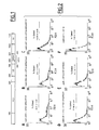
- Figure 2 represents a flow cytometry analysis of the interaction of
VP16-HIPK2/PEST with LexA-US11 and LexA-US11 mutant forms.
Protein/protein interaction measured by the yeast two hybrid analyses using the
GFP gene as a reporter. Yeast strain YRN974 with an integrated LexA-operator-GFP
gene was used for these analyses. A- Yeast cells were
transformed with expression vectors encoding LexA-US11 and VP16-HIPK2/PEST
proteins. B-D Yeast cells were transformed with expression
vectors encoding VP16-HIPK2/PEST combined with various vectors coding for
different LexA-US11 mutants, i.e. LexA-US11-m6, LexA-US11-m12, LexA-US11-m14.
Mutations carried by these proteins are detailed in (25). E- Yeast
cells expressing LexA alone and VP-16-HIPK2/PEST. F- Yeast cells
expressing LexA-US11 and VP-16 alone.
- Figure 3 shows the characterization of the HIPK2 PEST domain
interaction with US11 and US11 mutants in Human cells. HeLa cells were
cotranfected with empty vectors (lane 1) or the indicated expression vectors
containing a cDNA encoding Flag-HIPK2/PEST (lane 2, 7-10), US11 (lane 3
and 7), US11-m6 (lane 4 and 8), US11-m12 (lane 5 and 9), US11-m14 (lane 6
and 10). Cell lysates were subjected to immunoprecipitation (IP) with an anti-Flag
Ab (A and C) or an anti-US11 Ab (B). Purified proteins were analyzed by
western blot using an anti-US11 (A and B) or an anti-Flag (C). Arrowhead
indicates US11 (A and B) or Flag-HIPK2/PEST (C). Molecular sizes are shown
on the left in kDa.
- Figure 4 shows the in vitro phosphorylation of US11 by HIPK2.
Cell were transfected with the 3Flag and 3Flag-HIPK2 expression vectors, and
immunoprecipitation was carried out with an anti-Flag antibody. Kinase activity
was further assayed on the immunoprecipitated protein in presence of a GST-US11
protein or a phosphorylation mutant thereof, GST-US11m55 (Catez et al.
2002) i.e. a US11 protein mutated in the phosphorylation site, as a negative
control.
- Figure 5 represents the structural organization of the murine
HIPK2 gene. The bioinformatic analysis is described in Experimental
procedures. Exons are represented by filled boxes. Hatch marks upstream of
exon 2, in the intron3' of exon 2 and 15 indicate regions for which a portion of
the intron is unknown. Alternative splicing occuring between exons 8/9 are
indexed 2a and 2b.
- Figure 6 is a graphical representation of antagonizing effect of
US11 expression towards HIPK2 cell growth arrest. Hek 293 cells were
transfected with plasmids p3flag-HIPK2b, p3flag-HIPK2b AS combined with
either expression vector for US11 or the corresponding empty vector. 24 hours
after transfection, cells were selected with G418 and surviving colonies were
stained with cristal violet. Colony formation with the control vector p3flag-HIPK2b
AS was set as 100%. Results are mean ± s.d. for three independant
experiments.
-
EXAMPLES
Example 1: Experimental procedures
Cell Culture and transfection
-
HeLa cells were grown in Eagle's essential minimum medium
supplemented with 5% FCS and 2mM glutamine. Hek 293 cells were
maintained in DMEM supplemented with 10% FCS and 2mM glutamine.
Transfection of HeLa cells were carried out using the calcium phosphate
precipitation procedure. The cells were plated on 35 mm diameter Petri dishes
(105 cells/dish) and incubated at 37°C for 24 hours. One hour prior to
transfection, the medium was replaced by 1 ml of fresh medium. The
transfection mix containing 1.5 µg of plasmid DNA was added to the cells.
Experiments were performed 24 hours after transfection.
Antibodies
-
Antibodies used for western blot analysis, immunoprecipitation
and immunofluorescence include: anti-Flag M2 (Sigma-Aldrich), rabbit
polyclonal anti-US11 (24) and Texas Red labeled anti-rabbit IgG (Jackson
ImmunoResearch).
Yeast Two-hybrid system
-
Plasmids, S. cerevisiae strains, selective media and the
transformation protocol have already been described (Vojtek et al., 1995). For
construction of the Lex-A-wild type US11 bait, the cDNA encoding the entire
US11 sequence (Diaz et al., 1996) was inserted into the BTM116 vector,
resulting in production of a Lex-A-wild type US11 fusion protein. The S.
cerevisiae L40 strain was transformed and selected in a medium lacking uracil
and tryptophan. Selected colonies were tested for the expression of the Lex-A-wild
type US11 fusion protein. For this analysis yeast lysates were made using
glass beads in Laemmli buffer (Laemmli et al., 1970) and expression of the
Lex-A-wild type US11 protein was analyzed by western blot using an anti-US11
antibody. A colony containing the Lex-A-wild type US11 bait was then
transformed with the VP16 mouse embryonic days 9.5 and 10.5 cDNA library.
Transformants were selected on medium lacking uracil, tryptophan, leucine and
histidine. Colonies containing a VP16 fusion protein interacting with the bait
were selected on the basis of transactivation of the His3 reporter gene after 3
days of growth. Purified DNA isolated from positives clones was amplified by
PCR using VP16-specific sense and antisense primers (Vojtek et al., 1995) and
analyzed by enzymatic digestion and sequencing. Sequence comparisons were
done with the BLAST program (http://www.ncbi.nlm.nih.gov).
-
For quantification of the interaction and test of the US11 mutant
(US11mt) baits the S. Cerevisiae strain YRN974 with an integrated Lex-A-operator
GFP cassette was used (yEGFP3, (Cormack et al., 1996 ; Cormack et
al., 1997). Ten thousand cells of four independent transformants were analyzed
for fluorescence intensity using a Becton Dickinson FACScan flow cytometer
(Niedenthal et al., 1996).
Molecular cloning of HIPK2a and HIPK2b complete CDS and
Plasmid Constructions
-
The sequence corresponding to the complete CDS of HIPK2
protein was cloned by two sets of RT-PCR performed with OF-1 Swiss adult
heart mouse total mRNA. The primers used were designed according to the
sequence of Kim et al., 1998. The first 5' half part of the sequence was
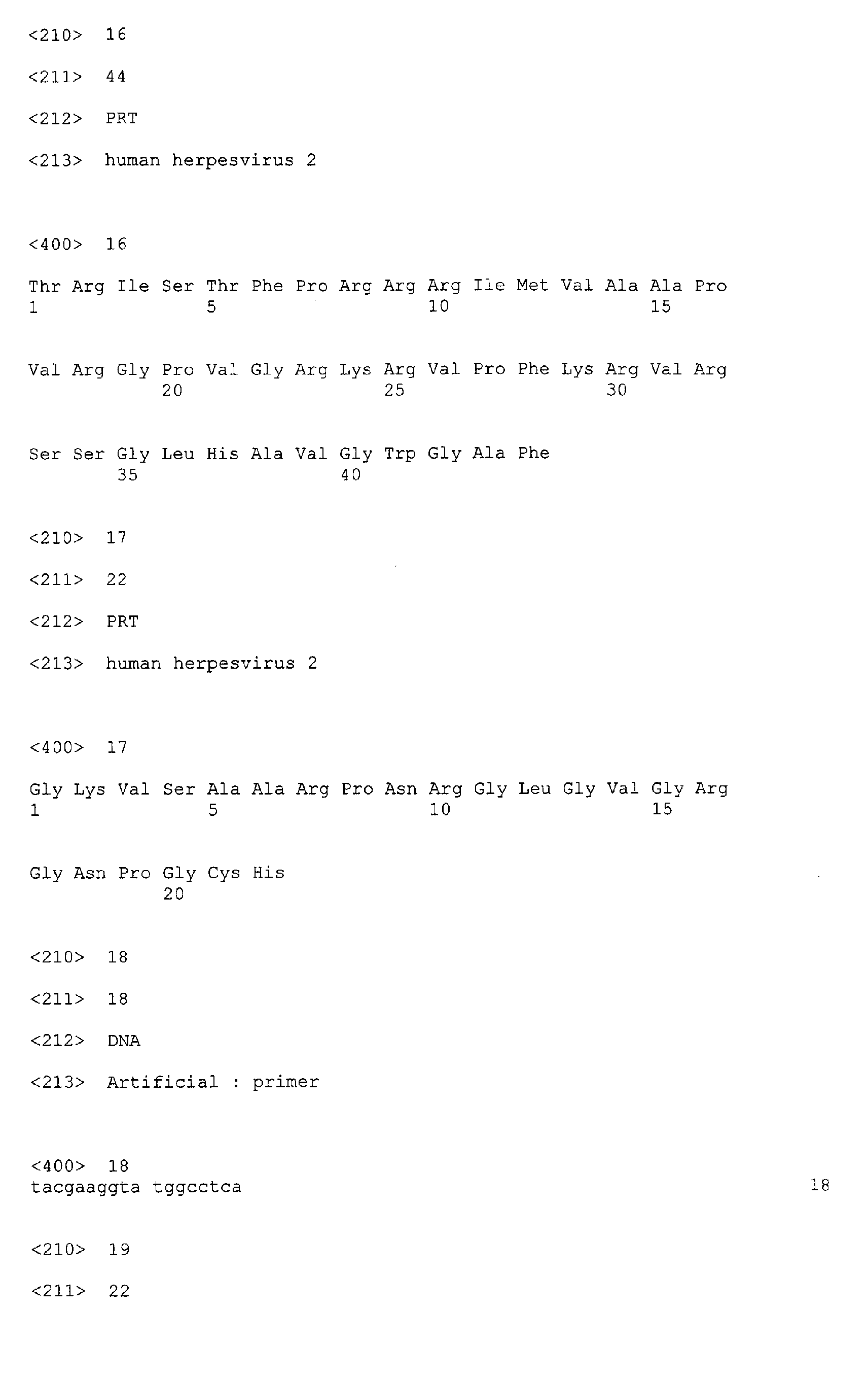
obtained using primer 149: 5'-TACGAAGGTATGGCCTCA-3' (SEQ ID No: 13)
and 1964: ATTGGTAGTTTAGTATGCAGAC-3' (SEQ ID No: 14). The second
part of the sequence was cloned using primers 1648: 5'-AAAGAAGATGCTGACCATCG-3'
(SEQ ID No: 15) and 3723: 5'-AAACATATGGGCCTCCTCCAGTGTTTATA-3'
(SEQ ID No: 16). Both
fragments were separately cloned in the pGEM-T easy vector (Promega) giving
constructs pGEM-T1 (insert 149-1964) and pGEM-T3 (insert 1648-3723).
Sequencing analysis revealed that 50% of the cloned 1648-3723 inserts
contained a deletion of 81 nucleotides compared to the sequence published
previously (3045). Plasmids carrying these shorter 1648-3723 inserts were
named pGEM-T5. The complete CDS were then reconstituted by insertion of
the Clal/Nsil fragments of pGEM-T3 or pGEM-T5 in pGEM-T1 giving constructs
pGEM-HIPK2a and pGEM-HIPK2b respectively. These two plasmids were
sequenced and accession numbers (available from EMBL/Genbank/DDBJ) for
HIPK2a and HIPK2b sequences reported in this application are AF333791 and
AF333792 respectively.
- pEGFP-HIPK2a and pEGFP-HIPK2b expression vectors:
The pEGFP-HIPK2a and pEGFP-HIPK2b plasmids were obtained
by inserting the EcoRI Klenow-filled fragment of constructs pGEM-HIPK2a and
pGEM-HIPK2b into the Smal site of pEGFP-C3 expression vector (Clontech).
These two constructs were then digested by HindIII and recircularized to clone
HIPK2a and HIPK2b CDS in frame with that of the EGFP. These constructions
were confirmed by nucleotide sequencing.
- US11 and US11 mutants vectors:
The US11 expression vector named pG-9, containing the US11
coding sequence placed under the control of CMV immediate-early- promotor
and the corresponding control vector pG-8 in which the US11 coding sequence
was removed were previously cloned in our laboratory (Besse et al., 1996).
Construction of the expression plasmids named, pG61-US11-m6, pG61-US11-m12,
pG61-US11-m14 coding for mutated forms of the US11 protein, are
described in details elsewhere (Catez et al., 2002).
- pSG5-Flag-HIPK2/PEST expression vector:
The Flag-M2 was inserted in the EcoRI/BglII sites of the pSG5
vector (Stratagene). The full length sequence contained in a positive two-hybrid
VP16 clone was amplified by PCR using oligonucleotides 5'-GAGTTTGAGCAGATGTTTA-3'
(SEQ ID No: 17) and 5'-GGAAGATCTGTGAATTCG-3'
(SEQ ID No: 18) which contained a BglII site
(underlined). This amplified fragment was then submitted to restriction digestion
using BamHI and Bg/II enzymes and inserted in the pSG5-flag construct giving
the pSG5-Flag-HIPK2/PEST plasmid.
- p3Flag-HIPK2b and p3Flag-HIPK2b AS expression vector:
These expression vectors were obtained by inserting the EcoRI
fragment of constructs pGEM-HIPK2b into the EcoRI site of plasmid 3XFLAG-CMV-10
(Sigma). This allowed to obtain the HIPK2b sequence cloned in both
orientations, giving the plasmid p3flag-HIPK2b which contains the HIPK2b
cDNA in the sense orientation and the plasmid p3flag-HIPK2b AS which
contains the HIPK2b cDNA in the antisense orientation.
Western blot analysis and Immunoprecipitation
-
Cells were harvested 48 h after the onset of transfection as
follows: they were scraped in phosphate-buffered saline and pelleted by
centrifugation. The cell pellet was resuspended in 1.5 ml of PBS and divided in
three equivalent fractions. After centrifugation, cells from 2 fractions were lysed
in 400 µl of RIPA buffer for immunoprecipitation (50 mM Tris-HCI pH 7.2, 1%
NP40, 1% deoxycholate, 0.1% SDS and 150 mM NaCl, supplemented with
protease inhibitor (CompleteTM, Roche molecular biochemicals)). Cells from the
third fraction were resuspended in Laemmli buffer without glycerol and
bromophenol blue to allow quantification of proteins. Proteins (10 µg) from total
cell lysates were separated by 1D-PAGE in a gel containing 15%
polyacrylamide and transferred to a PVDF membrane (Immobilon-P, Millipore).
-
Proteins of interest were identified by incubating the membranes
with primary antibodies (anti-Flag M2 or anti-US11) in TBS-T buffer (Tris-HCl
20 mM pH 7.4, NaCl 130 mM, 0.1% Tween 20). The immunoblots were
revealed by ECL (AmershamTM) after incubation with a peroxidase-labelled
secondary antibody. For immunoprecipitation, cell extracts were incubated at
4°C for 30 min in RIPA buffer and centrifugated at 75000 rpm (TLA 100.3 rotor)
in a TL-100 Beckman centrifuge for 30 min at 4°C. Supernatants were then
incubated with the anti-Flag M2 or the anti US11 overnight at 4°C.
Immunocomplexes were immobilized on protein-A sepharose beads (Amerham
Pharmacia Biotech, CL4B) by a 30 min incubation at 4°C. The beads were
collected and washed 3 times with RIPA buffer. Bound proteins were eluted
and analyzed by western blotting using the same antibodies.
Immunohistochemistry and indirect immunofluorescence
-
HeLa cells were transfected with the combinations of the following
expression vectors as indicated on the figures: pEGFP-HIPK2a, pEGFP-HIPK2b,
pG-9, pG61-US11-m6, pG61-US11-m12, pG61-US11-m14. Cells
were fixed with 3% paraformaldehyde in PBS for 30 min at room temperature
and washed with a 10 mM glycine-PBS solution. They were then permeabilized
for 5 min with a solution of 1% Triton X-100 in PBS. After a 10 mM glycine-PBS
washing, cells were incubated in a 25 mM glycine-PBS solution for 30 min.
They were incubated with an anti-US11 antibody overnight, washed and
incubated with a secondary anti-rabbit IgG Ab conjugated with Texas Red. Epi-fluorescence
was visualized using an Axiophot microscope from Zeiss coupled
with a CCD camera (LH 750, Lhesa electronic) at excitation wawelengths of
489 nm for GFP and 596 nm for Texas Red.
Semi-quantitative RT-PCR
-
Mouse total RNA collection (Ambion™, Clontech) were reverse
transcribed and specific RNA were amplified using one-step RT-PCR kit
(Qiagen). HIPK2a, HIPK2b mRNAs and 18S rRNA were reverse transcribed
and amplified in the same reaction using primers: 18S rRNA sense 5'-ATGCGGCGGCGTTATTC-3'
(SEQ ID No: 19) and 18S antisense 5'-GCGCGGGCGGTGTGTA-3'
(SEQ ID No: 20); HIPK2a/HIPK2b mRNA sense
5'-GTCACCATGACACACCTGCT-3' (SEQ ID No: 21) antisense 5'-AGGGGGACACACGATGAGAG-3'
(SEQ ID No: 22). PCR products were
separated by agarose gel electrophoresis and quantified using a gel analyzer
(Bio-1D Vilbert Lourmat) after ethidium bromide staining.
Colony formation assay
-
Cells plated in 60 mm diameter Petri dishes were transfected
using the calcium phosphate method with different combinations of the
following plasmids as indicated on the figures: p3Flag-HIPK2b, p3Flag-HIPK2b
AS; pG-9 (US11 expression vector) and pG-8 (empty vector). Total amount of
transfected DNA was 4.5 µg. At 24 hours after transfection, cells were
cultivated in medium containing 1 mg/mL of G418 (Geneticin, an analogue of
gentamicin) for 2 weeks before staining of the surviving colonies with crystal
violet. Colonies were counted using lmagemaster 2D (Pharmacia™).
Theoretical structure of the mouse HIPK2 gene
-
Mouse genomic draft DNA sequences (accession number
AC069142 and AC079129) homologous to HIPK2a and HIPK2b cDNAs were
found using the BLAST program. Unordered pieces were ordered using the
SIM4 program (Florea et al., 1998) to draw the murine HIPK2 gene structure.
Example 2 : Results
US11 protein interacts specifically with the PEST domain of
HIPK2
-
A yeast two-hybrid system was used to identify cellular proteins
that could interact with HSV-1 US11 protein. In this system, the 'bait protein',
LexA-US11, was a fusion protein made of the DNA binding domain of the LexA
operator and of the entire US11 protein. The 'prey proteins' were fusion
proteins made of the activator domain of VP16 and of fragments of proteins of
about one hundred aminoacids long derived from a mouse embryo cDNA
library (Vojtek et al., 1995).
-
Using this system, twenty million of prey fusion proteins have
been tested for their ability to interact with US11 protein. Among those, four
hundred were shown to interact with US11 protein and forty one out of the four
hundred were identified as fusion proteins containing the region encompassing
amino acids 789 to 925 of HIPK2.
-
As shown in figure 1 this region overlaps with the PEST domain of
HIPK2 (amino acids 839-934) and a nuclear speckle retention signal (SRS,
amino acids 860-967). However, because US11 protein and the PEST domain
of HIPK2 both exhibited a high proline content, and because this kind of
domains may interact to each other non specifically (Williamson et al., 1994) it
was determined whether single or a few aminoacids substitutions within US11
sequence could affect its ability to interact with VP16-HIPK2/PEST.
-
Three expression vectors coding for modified forms of the LexA-US11
wild type protein, designated LexA-US11-m6, LexA-US11-m12 and
LexA-US11-m14 were constructed (Catez et al., (2002) for a detailed
description of these modified forms) and individually transfected together with
that coding for VP16-HIPK2/PEST in the yeast strain YRN974. In this strain,
the reporter gene encodes the green fluorescent protein (GFP) under the
control of a LexA-binding-site containing promoter, and the efficiency of the
interaction between fusion proteins is estimated by numbering the fluorescent
cells together with measuring the intensity of their fluorescence with a flow
cytometer.
-
LexA-US11, but also LexA-US11-m6 interacted strongly with
VP16-HIPK2/PEST since 31 and 23.7 % respectively of the cells displayed a
very high level of fluorescence (Fig. 2A and B). Conversely, the interaction of
LexA-US11-m12 and LexA-US11-m14 with VP16-HIPK2/PEST was greatly
impaired since only 14.78 and 12.89 % of the cells displayed a high level of
fluorescence (Fig. 2C and D), a percentage very close to that obtained when
expressing only the LexA part of the fusion protein (Fig. 2E). The percentage of
highly fluorescent cells was also very low when expressing LexA-US11
together with the DNA binding domain of VP16 only (Fig. 2F).
-
Therefore these results lead to the conclusion that the interaction
observed between LexA-US11 and VP16-HIPK2/PEST occurred indeed via the
US11 and HIPK2 parts of the fusion proteins.
-
To firmly validate these results, it has been further determined
whether a specific interaction between the native form of US11 protein and the
PEST domain of HIPK2 could be obtained after their synthesis in eukaryotic
cells (Fig. 3).
-
An expression vector coding for a Flag-tagged HIPK2/PEST
domain was constructed and co-transfected in HeLa cells, either alone or
together with expression vectors coding for US11, US11-m6, US11-m12 and
US11-m14. Forty height hours after transfection, proteins interacting with Flag-HIPK2/PEST
were purified by co-immunoprecipitation using an anti-Flag
antibody and analyzed by western blot using an anti-US11 protein antibody
(Fig. 3).
-
US11 and US11-m6 proteins were efficiently co-immunoprecipitated
with Flag-HIPK2/PEST (Fig. 3A, lane 7 and 8), whereas
co-immunoprecipitation of US11-m12 was much less efficient (Fig. 3A, lane 9).
US11-m14 was never co-immunoprecipitated with Flag-HIPK2/PEST (Fig. 3A,
lane 10) even though both proteins, Flag-HIPK2/PEST and US11-m14, were
effectively synthesized (Fig. 3B and C, lane 10). Because US11-m6 and US11-m14
exhibit respectively only one or two proline residue substitution out of
thirty-five present in US11 and because their ability to interact with the PEST
domain of HIPK2 is very different it can be concluded that the overall proline
content of the two proteins does not account for their interaction.
-
Moreover, the results obtained for US11-m12 in which only an
arginine residue has been replaced by a glutamine clearly demonstrated that
the interaction of US11 with HIPK2 could be greatly impaired without changing
the proline content of both proteins.
-
Therefore, all together these results demonstrate that US11
protein interacts specifically with the PEST domain of HIPK2.
-
The inventors further showed that US11 is phosphorylated by
HIPK2 in vitro (Fig. 4). Since US11 has been shown to be phosphorylated in
vivo, in particular by a cell kinase, this result suggests that HIPK2 may be the
kinase responsible for US11 phosphorylation in vivo.
Structural organization of HIPK2 gene and mRNA expression
-
To analyze further the interaction between US11 and HIPK2 an
experimental strategy to isolate by RT-PCR the entire coding sequence of
HIPK2 was designed. This experimental strategy described in details in the
materials and methods section allowed to isolate and to characterize two
slightly different HIPK2 cDNAs from the heart of adult Swiss mouse. The
deduced sequences from these two cDNAs were designated HIPK2a and
HIPK2b (GenBank accession number AF333791 and AF333792 respectively).
-
The major difference between these cDNAs is an 81 bp deletion
within HIPK2b corresponding to the sequence coding for the domain of HIPK2
that was shown to interact with the homeoproteins (Kim et al., 1998). A BLAST
analysis indicated that the longer cDNA (HIPK2b) coded for the HIPK2 protein
identified previously in mice (Kim et al., 1998) also named NBAK1b or STANK
(AF170302 and AF273680 respectively). The sequence of the shorter one
(HIPK2a) corresponded to that of nuclear body associated kinase1a (NBAK1a,
AF170301), also named PKM and identified previously in hamster (Trost et al.,
2000). The sequence of HIPK2a displays a twenty-seven amino acids deletion
in the region of the molecule initially described by Kim et al. (1998) as
containing the domain interacting with homeoproteins. This difference might
result from an alternative splicing of HIPK2 pre-mRNA.
-
To test this possibility, the theoretical structure of the HIPK2 gene
was elucidated to determine whether potential alternative splicing sites are
found within this gene. For this, large fragments of murine genomic DNA
encompassing HIPK2 sequences were found in Genbank and contiguously
ordered according to the HIPK2 coding sequence (see the materials and
methods section).
-
This analysis indicated that the murine HIPK2 gene, located on
chromosome 6B (Hofmann et al., 2000), is composed of at least 16 exons and
lies into a chromosomal fragment longer than 150 kbp (Fig. 5). Several
sequences are still missing however to reconstitute the entire sequence of the
gene: sequences localized before exon 2 coding for the 5' untranslated region
and therefore encompassing exon 1 and intron 1; sequences localized in
introns 3 and 14 as well as those coding for the 3' untranslated region of the
mRNA. This HIPK2 gene model correlated very well with the model predicted
by the LocusLink software (http://www.ncbi.nlm.nih.gov/LocusLink). Sequences
of every intron/exon junction enabled to validate the intron/exon arrangement
proposed for the HIPK2 gene since all of these sequences are in very good
agreement with those known to be potential splicing site (Sharp et al., 1987).
Finally, the presence of one splicing donor site at the boundary of exon/intron
8, followed respectively by two different acceptor splicing sites at the boundary
of intron8/exon9 supports the hypothesis that HIPK2a and 2b derived from a
differently spliced mRNA (Fig. 5).
-
It was then determined whether both mRNAs coding for either
HIPK2a or HIPK2b were present in mice embryos and into different tissues of
adult mice and estimated their relative amounts. For this, fragments of DNA
corresponding specifically to either HIPK2a or HIPK2b and to the 18S
ribosomal RNAs (rRNAs) were obtained by simultaneous amplification by RT-PCR
using total RNAs extracted from embryos and from various mice tissues.
DNAs were separated by electrophoresis and stained with ethidium bromide.
The ratios between the amount of DNA obtained from 18S rRNA and that
obtained from both HIPK2 mRNAs were calculated.
-
This analysis showed that the two forms of HIPK2 mRNAs were
expressed in all the tissues analyzed (testis, embryo, ovary, thymus, spleen,
liver, brain, lung and heart). Furthermore this analysis showed that both mRNA
are generally present in approximately equal amount except for the kidney and
the brain in which HIPK2b mRNA is respectively the major and the minor form.
Sub-cellular distribution of HIPK2a and HIPK2b
-
The cDNAs coding for HIPK2a and 2b being cloned, the
intracellular distribution of both proteins was then analyzed. Two expression
vectors coding for fusion proteins made of the 'enhanced green fluorescent
protein' (EGFP) and of either HIPK2a or HIPK2b were constructed and
transfected into HeLa cells.
-
Forty-eight hours after transfection, both proteins display an
identical intracellular distribution, present into numerous nuclear dots, excluded
from nucleoli and almost undetectable in the cytoplasm. This intracellular
distribution is in good agreement with that already described for HIPK2 (Kim et
al., 1998 ; Trost et al., 2000). Moreover, several reports demonstrated that
HIPK2 can also be recruited to ND10.
-
Thus it was determined whether HIPK2 co-localized with the
protein PML, one of the major component of ND10. To this aim, PML was
visualized by immunofluorescence using an anti-PML antibody (kindly provided
by Dr R. van Driel, Swammerdam Inst. For Life Sciences, Amsterdam, the
Netherlands) in cells transiently expressing EGFP-HIPK2a and the fine
localization of both proteins was determined by confocal microscopy.
Numerous small nuclear dots contained only one of the two proteins, whereas
other dots contained both proteins.
-
These findings are also in good agreement with previous studies
which reported that HIPK2 localizes to distinct sub-cellular structures, i.e. ND10
and other nuclear speckles of unknown function (Kim et al., 1998 ; Trost et al.,
2000 ; Hofmann et al., 2002 ; d'Orazi et al., 2002).
-
The above results demonstrated that US11 protein of HSV-1
interacts with the PEST domain of HIPK2 which is a common domain to
HIPK2a and HIPK2b. This domain encompasses an SRS ('Speckle Retention
Signal') sequence. Accordingly it was investigated whether the interaction
between US11 and HIPK2a or HIPK2b can disturb the sub-cellular localisation
of one of them.
Sub-cellular distribution of HIPK2a and HIPK2b when co-expressed
with US11
-
To gain insights into the functional significance of the interaction
between HIPK and US11, it was thus investigated whether the intracellular
distribution of one of these proteins might be affected after co-synthesis of the
other.
-
For this, HeLa cells were transfected with expression vectors
coding for either EGFP-HIPK2a or EGFP-HIPK2b together with a vector coding
for wild type US11 protein. At different times after transfection, expression of
the transgenes was monitored by direct visualization of EGFP fusion proteins
and by immunofluorescence for US11 protein.
-
These experiments demonstrated that at each time analyzed after
transfection, the typical pattern of intracellular distribution of each protein was
dramatically different when proteins were synthesized together. For example, at
48 hours after transfection, HIPK2a and HIPK2b did not display a fine
punctuate nuclear pattern but rather were found within nuclear structures that
spread into the nuclei and tend to occupy the entire space, always excluding
however, nucleoli. Another striking feature of US11-induced HIPK2 pattern was
the presence within the cytoplasm of dots containing HIPK2. The usual nuclear
distribution of US11 protein described many times (Puvion-Dutilleul, 1987 ;
Roller et al., 1992) - concentrated into nucleoli and almost undetectable in the
nucleoplasm - was also modified by co-expression of HIPK2 and US11 genes
since in these conditions, US11 protein was detectable in regions surrounding
nucleoli and spread throughout the nucleoplasm. The same modifications of
intracellular distribution of both proteins were also observed when Flag-HIPK2
instead of EGFP-HIPK2 fusion proteins were used.
-
To evaluate whether its interaction with US11 protein could
account for its 'unusual' behavior, the intracellular distribution of EGFP-HIPK2b
was determined after co-expression with vectors coding for modified forms of
US11 that were shown to interact differently with the PEST domain of HIPK2
(Fig. 1). Interestingly, US11-m6, which interacts with the PEST domain of
HIPK2 with the same efficiency than wild type US11 protein, did induce a
modification of intracellular distribution of HIPK2 very similar to that induced by
US11 wild type protein. Conversely, US11-m12, which interacts with the PEST
domain of HIPK2 less efficiently than US11 wild type protein, and US11-m14,
which does not interact with this domain, did not significantly alter this pattern.
-
All together, these results demonstrate that US11 protein induces
a severe alteration in the intracellular distribution of HIPK2 and strongly
suggest that this effect is mediated through US11 protein-HIPK2 interaction.
-
HIPKs are SUMOlated proteins that interact with the SUMO
conjugating enzyme Ubc9. The SUMOlation of HIPKs conditioned the
partitioning of HIPKs within the nuclear speckles. Interaction between HIPK2
and Ubc9 occurs via a region contained within the PEST sequence (Kim et al.,
1999) which perfectly matches with the interaction domain of HIPK2 for US11.
Different hypothesis thus come out. By interacting with the PEST domain of
HIPK2, US11 can modify its SUMOlation by Ubc9 and thus disturb its nuclear
localization. This would provide a modification of the ratio between un-SUMOlated
and SUMOlated HIPKs thus modifying the proportion of nuclear
matrix associated HIPKs (SUMOlated form) versus the soluble non-SUMOlated
form of the protein. This possibility is conceivable since the inventors have
further found that US11 interacts with Ubc9 in the double hybrid system.
Moreover, PEST sequences are predicted to regulate the instability of proteins
(Rogers et al., 1986). Therefore an another possibility is that by interacting with
the PEST domain of HIPK2 proteins US11 might antagonizes with their
degradation by an unknown mechanism. Indeed, whether the degradation of
HIPKs occurs via the proteasome or another pathway still remains to be
determined.
Inhibition of HIPK2-induced cell cycle arrest by US11
-
It has been demonstrated that in cells containing wild-type p53,
HIPK2 induces apoptosis which can be objectivized by a strong decrease of
cell proliferation using colony formation assays (Hofmann et al., 2002 ; d'Orazi
et al., 2002 ; Kim et al., 2002). Moreover, cells expressing US11 were shown to
exhibit an increased survival capacity to thermal stress compared to that of
cells that did not expressed this gene ((Diaz-Latoud et al., 1997).
-
To further investigate the functional relevance of US11/HIPK2
interactions colony formation assays were performed. The p53 wild type
expressing cells Hek 293, were transfected with Flag-HIPK2b expressing
vectors allowing also the expression of the NeoR gene together with expression
vectors coding for US11 or in absence of this latter. As a negative control, Hek
293 cells were transfected with a vector encoding an antisense mRNA of
HIPK2b (HIPK2b AS) with or without US11 expression vectors. Twenty-four
hours after the beginning of the transfection, cells were selected for resistance
to G418 during 2 weeks and the number of colonies was estimated for each
experimental condition (Fig. 6).
-
HIPK2b was shown to prevent cell growth since the number of
G418 resistant colonies was reduced to 17% of those obtained after
transfection of the negative control vectors HIPK2b AS alone and HIPK2b AS
with the US11 expressing vector (Fig. 6). Conversely, when US11 was co-expressed
with HIPK2b it strongly impaired growth arrest induced by HIPK2b
since the number of colonies in these conditions represented 60% of those
obtained after the transfection of the control vectors. This result clearly
indicated that US11 impaired HIPK2 growth arrest.
-
Viruses depend on host cells for their replication and have
developed different strategies to affect various signalling pathways and
counteract apoptosis. Particularly, HSV-1 gene expression, DNA replication
and encapsidation, all take place within nuclei into globular domains designated
replication compartments (Quinlan et al., 1984 ; Liptak et al., 1996 ; Lukonis et
al., 1996 ; Lukonis et al., 1997). Formation of these replication compartments
requires a profound re-organization of pre-existing nuclear domains of the
infected cell which proceeds into several sequential steps. Among the nuclear
domains which are extensively modified during HSV-1 infection, ND10 is one of
the major target. Many studies have shown that early during infection HSV-1
disrupts ND10 by inducing degradation of one of the major component of this
domain, namely the PML protein (see Everett et al., 1999 for review).
Moreover, ND10 has been implicated in transcriptional and apoptotic pathways
regulation. Indeed, ND10 contains several proteins involved in these later
processes, like p53, PML, pRB, CBP, HIPK2 and TRADD (Morgan et al., 2002 ;
Matera et al., 1999).
-
The colony formation assay reported above clearly demonstrated
that US11 antagonizes the HIPK2 inhibition of proliferation in Hek 293 cells.
Since HIPK2 is known to be activated in response to irradiation by UV and to
trigger p53 induced apoptosis (Hofmann et al., 2002 ; d'Orazi et al., 2002 ; Kim
et al., 2002), the inventors postulate that in cells submitted to UV irradiation,
US11 interfere with the HIPK2 induced apoptosis, thus allowing HSV-1 to
replicate.
-
In conclusion, the findings reported here identify US11 as a new
candidate for HSV-1 inhibition of cellular apoptosis and provides a new
pathway of HSV-1 induced nuclear modification that occurs with the targeting
by the virus of a nuclear domain component.
REFERENCES
-
- Barbas CF, Bain JD, Hoekstra DM, Lerner RA. (1992),
Semisynthetic combinatorial antibody libraries: a chemical solution to the
diversity problem. PNAS USA, 89, 4457-4461
- Bhat, R. A., and Thimmapaya, B. Proc. Natl. Acad. Sci. USA 80,
4789-4793 (1983)
- Benoist and Chambon, Nature 1981,290:304-310
- Besse, S., Diaz, J.-J., Pichard, E., Kindbeiter, K., Madjar, J.-J.,
and Puvion-Dutilleul, F. (1996) Chromosoma 104, 434-444
- Brinster et al., Nature 1982, 296:39-42
- Brown, S. M., and Harland, J. (1987) JGV 68, 1-18
- Cassady, K. A., Gross, M., and Roizman, B. (1998) Journal of
Virology 72, 8620-8626
- Catez, F., Erard, M., Schaerer-Uthurralt, N., Kindbeiter, K.,
Madjar, J.-J., and Diaz, J.-J. (2002) Mol Cell Biol 22, 1126-1139
- Choi, C. Y., Kim, Y. H., Kwon, H. J., and Kim, Y. (1999) J Biol
Chem 274, 33194-33197
- Cormack, B. P., Valdivia, R. H., and Falkow, S. (1996) Gene
173, 33-38
- Cormack, B. P., Bertram, G., Egerton, M., Gow, N. A., Falkow,
S., and Brown, A. J. (1997) Microbiology 143, 303-311
- DeBoer, et al., Proc. Natl. Acad. Sci. USA 1983, 80:21-25
- Diaz, J.-J., Simonin, D., Massé, T., Deviller, P., Kindbeiter, K.,
Denoroy, L., and Madjar, J.-J. (1993) J Gen Virol 74 , 397-406
- Diaz, J.-J., Duc Dodon, M., Schaerer-Uthurralt, N., Simonin, D.,
Kindbeiter, K., Gazzolo, L., and Madjar, J.-J. (1996) Nature 379, 273-277
- Diaz-Latoud, C., Diaz, J.-J., Fabre-Jonca, N., Kindbeiter, K.,
Madjar, J.-J., and Arrigo, A.-P. (1997) Cell Stress and Chaperones 2, 119-131
- Duc Dodon, M., Mikaelian, I., Sergeant, A., and Gazzolo, L.
(2000) Virology 270, 43-53
- Everett, R. D., Freemont, P., Saitoh, H., Dasso, M., Orr, A.,
Kathoria, M., and Parkinson, J. (1998) J Virol 72 , 6581-6591
- Everett, R. D. (1999) Trends Biochem Sci 24, 293-295
- Florea, L., Hartzell, G., Zhang, Z., Rubin, G. M., and Miller, W.
(1998) Genome Res 8, 967-974
- Gressner, A. M., and Wool, I. G. (1974) J. Biol. Chem. 249,
6917-6925
- Harlow E. et al., Antibodies: A Laboratory Manual, Cold Spring
Harbor Laboratory, New York, (1988)
- Hofmann, T. G., Mincheva, A., Lichter, P., Droge, W., and
Lienhard Schmitz, M. (2000) Biochimie 82, 1123-1127
- Hofmann, T. G., Moller, A., Sirma, H., Zentgraf, H., Taya, Y.,
Droge, W., Will, H., and Schmitz, M. L. (2002) Nat Cell Biol 4, 1-10
- Jayasena S.D. (1999) Aptamers: an emerging class of
molecules that rival antibodies in diagnostics. Clin Chem. 45(9):1628-50
- Kim, Y. H., Choi, C. Y., Lee, S. J., Conti, M. A., and Kim, Y.
(1998) J Biol Chem 273, 25875-25879
- Kim, Y. H., Choi, C. Y., and Kim, Y. (1999) Proc Natl Acad Sci U
S A 96, 12350-12355.
- Kim, E. J., Park, J. S., and Um, S. J. (2002) J Biol Chem 29, 29
- Kohler and Milstein (1975) Continuous cultures of fused cells
secreting antibody of predefined specificity. Nature, 256, 495-7.
- Kollias et al., Ce II 1986, 46: 89-94
- Koshizuka, T., Takakuwa, H., Goshima, F., Murata, T., and
Nishiyama, Y. (2001) Biochem Biophys Res Commun 288, 597-602
- Laemmli, U. K. (1970) Nature 227, 680-685
- Leopardi, R., Van Sant, C., and Roizman, B. (1997) Proc Natl
Acad Sci U S A 94, 7891-7896
- Li, X., Wang, Y., Debatin, K. M., and Hug, H. (2000) Biochem
Biophys Res Common 277, 513-517
- Liptak, L. M., Uprichard, S. L., and Knipe, D. M. (1996) J Virol
70, 1759-1767
- Longnecker, R., and Roizman, B. (1986) Journal of Virology 58,
583-591
- Lukonis, C. J., and Weller, S. K. (1996) J Virol 70, 1751-1758
- Lukonis, C. J., Burkham, J., and Weller, S. K. (1997) J Virol 71,
4771-4781
- MacLean, C. A., Rixon, F. J., and Marsden, H. S. (1987) JGV
68, 1921-1937
- Maouche et al., Blood 1991, 15:2557
- Matera, A. G. (1999) Trends Cell Biol 9, 302-309
- Matsuo, R., Ochiai, W., Nakashima, K., and Taga, T. (2001) J
Immunol Methods 247, 141-151
- Mogram et al., Nature 1985,315:338-340
- Morgan, M., Thorburn, J., Pandolfi, P. P., and Thorburn, A.
(2002) J Cell Biol 157, 975-984
- Niedenthal, R. K., Riles, L., Johnston, M., and Hegemann, J. H.
(1996) Yeast 12, 773-786
- Nishiyama, Y., Kurachi, R., Daikoku, T., and Umene, K. (1993)
Virology 194, 419-423
- D'Orazi, G., Cecchinelli, B., Bruno, T., Manni, I., Higashimoto,
Y., Saito, S., Gostissa, M., Coen, S., Marchetti, A., Del Sal, G., Piaggio, G.,
Fanciulli, M., Appella, E., and Soddu, S. (2002) Nat Cell Biol 4, 11-19
- Pierantoni, G. M., Fedele, M., Pentimalli, F., Benvenuto, G.,
Pero, R., Viglietto, G., Santoro, M., Chiariotti, L., and Fusco, A. (2001)
Oncogene 20, 6132-6141
- Poppers, J., Mulvey, M., Khoo, D., and Mohr, I. (2000) J Virol
74, 11215-11221
- Puvion-Dutilleul, F. (1987) EJCB 43, 487-498
- Quinlan, M. P., Chen, L. B., and Knipe, D. M. (1984) Cell 36,
857-868.
- Reeck et al., Cell 50:667, 1987
- Rogers, S., Wells, R., and Rechsteiner, M. (1986) Science 234,
364-368
- Roller, R. J., and Roizman, B. (1992) Journal of Virology 66,
3624-3632
- Roller, R. J., Monk, L. L., Stuart, D., and Roizman, B. (1996)
Journal of Virology 70, 2842-2851
- Sharp, P. A. (1987) Science 235, 766-771
- Sharp, Genes Dev, 2001, 15:485-490
- Trost, M., Kochs, G., and Haller, O. (2000) J Biol Chem 275,
7373-7377
- Tuerk C. and Gold L. (1990) Systematic evolution of ligands by
exponential enrichment: RNA ligands to bacteriophage T4 DNA polymerase.
Science. 3;249(4968):505-10
- Villa-Komaroff, et al., Proc. Natl. Acad. Sci. USA 1978,75:3727-3731
- Vojtek, A. B., and Hollenberg, S. M. (1995) Methods Enzymol
255, 331-342
- Wagner et al., Proc. Natl. Acad. Sci. USA 1981, 78: 1441-1445
- Wang, W., Link, V., and Green, J. M. (2000) Cell Immunol 205,
34-39
- Wang, Y., Hofmann, T. G., Runkel, L., Haaf, T., Schaller, H.,
Debatin, K., and Hug, H. (2001a) Biochim Biophys Acta 1518, 168-172
- Wang, Y., Debatin, K. M., and Hug, H. (2001b) BMC Mol Biol 2,
8
- Ward, P. L., and Roizman, B. (1994) Trends in Genetics 10,
267-274
- Waterhouse P, Griffiths AD, Johnson KS, Winter G. (1993)
Combinatorial infection and in vivo recombination: a strategy for making large
phage antibody repertoires. Nucleic Acids Research, 21, 2265-2266
- Williamson, M. (1994) Biochem. J. 297, 249-260