EP1485818B1 - Treatment protocol generation for diseases related to angiogenesis - Google Patents
Treatment protocol generation for diseases related to angiogenesis Download PDFInfo
- Publication number
- EP1485818B1 EP1485818B1 EP02801985A EP02801985A EP1485818B1 EP 1485818 B1 EP1485818 B1 EP 1485818B1 EP 02801985 A EP02801985 A EP 02801985A EP 02801985 A EP02801985 A EP 02801985A EP 1485818 B1 EP1485818 B1 EP 1485818B1
- Authority
- EP
- European Patent Office
- Prior art keywords
- angiogenesis
- vegf
- immature
- evd
- vessels
- Prior art date
- Legal status (The legal status is an assumption and is not a legal conclusion. Google has not performed a legal analysis and makes no representation as to the accuracy of the status listed.)
- Expired - Lifetime
Links
- 230000033115 angiogenesis Effects 0.000 title claims abstract description 75
- 238000011282 treatment Methods 0.000 title claims abstract description 23
- 208000037265 diseases, disorders, signs and symptoms Diseases 0.000 title claims abstract description 18
- 201000010099 disease Diseases 0.000 title claims abstract description 17
- 238000000034 method Methods 0.000 claims abstract description 85
- 230000035800 maturation Effects 0.000 claims abstract description 43
- 230000000694 effects Effects 0.000 claims abstract description 41
- 239000003814 drug Substances 0.000 claims abstract description 29
- 229940079593 drug Drugs 0.000 claims abstract description 29
- 230000001687 destabilization Effects 0.000 claims abstract description 25
- 239000002870 angiogenesis inducing agent Substances 0.000 claims abstract description 22
- 230000003527 anti-angiogenesis Effects 0.000 claims abstract description 19
- 230000009443 proangiogenesis Effects 0.000 claims abstract description 19
- 210000005166 vasculature Anatomy 0.000 claims abstract description 11
- 230000001105 regulatory effect Effects 0.000 claims abstract description 9
- 238000011369 optimal treatment Methods 0.000 claims abstract description 6
- 108010073929 Vascular Endothelial Growth Factor A Proteins 0.000 claims description 143
- 102000005789 Vascular Endothelial Growth Factors Human genes 0.000 claims description 140
- 108010019530 Vascular Endothelial Growth Factors Proteins 0.000 claims description 140
- 210000001519 tissue Anatomy 0.000 claims description 96
- 102100034608 Angiopoietin-2 Human genes 0.000 claims description 43
- 210000002889 endothelial cell Anatomy 0.000 claims description 42
- 102100034594 Angiopoietin-1 Human genes 0.000 claims description 41
- 230000004663 cell proliferation Effects 0.000 claims description 36
- 101000924533 Homo sapiens Angiopoietin-2 Proteins 0.000 claims description 34
- 101000924552 Homo sapiens Angiopoietin-1 Proteins 0.000 claims description 33
- 108010038512 Platelet-Derived Growth Factor Proteins 0.000 claims description 31
- 102000010780 Platelet-Derived Growth Factor Human genes 0.000 claims description 31
- 210000003668 pericyte Anatomy 0.000 claims description 28
- 210000004027 cell Anatomy 0.000 claims description 26
- 230000030833 cell death Effects 0.000 claims description 25
- 230000008569 process Effects 0.000 claims description 25
- 230000006870 function Effects 0.000 claims description 19
- 210000004204 blood vessel Anatomy 0.000 claims description 16
- 230000012010 growth Effects 0.000 claims description 15
- 230000006698 induction Effects 0.000 claims description 14
- 230000035755 proliferation Effects 0.000 claims description 12
- 108010048036 Angiopoietin-2 Proteins 0.000 claims description 9
- 102000037983 regulatory factors Human genes 0.000 claims description 9
- 108091008025 regulatory factors Proteins 0.000 claims description 9
- 108010048154 Angiopoietin-1 Proteins 0.000 claims description 8
- 108091008605 VEGF receptors Proteins 0.000 claims description 8
- 102000009484 Vascular Endothelial Growth Factor Receptors Human genes 0.000 claims description 8
- 230000006907 apoptotic process Effects 0.000 claims description 8
- 230000003247 decreasing effect Effects 0.000 claims description 8
- 230000004044 response Effects 0.000 claims description 8
- 230000003993 interaction Effects 0.000 claims description 7
- 230000010412 perfusion Effects 0.000 claims description 6
- 102000005962 receptors Human genes 0.000 claims description 6
- 108020003175 receptors Proteins 0.000 claims description 6
- 230000007998 vessel formation Effects 0.000 claims description 6
- 238000004590 computer program Methods 0.000 claims description 4
- 230000002123 temporal effect Effects 0.000 claims description 4
- 101100481408 Danio rerio tie2 gene Proteins 0.000 claims description 3
- 101100481410 Mus musculus Tek gene Proteins 0.000 claims description 3
- 206010057248 Cell death Diseases 0.000 claims description 2
- 230000000903 blocking effect Effects 0.000 claims description 2
- 206010028980 Neoplasm Diseases 0.000 description 25
- 230000002792 vascular Effects 0.000 description 13
- 235000015097 nutrients Nutrition 0.000 description 12
- 206010021143 Hypoxia Diseases 0.000 description 10
- 201000011510 cancer Diseases 0.000 description 9
- 230000007954 hypoxia Effects 0.000 description 9
- 230000015572 biosynthetic process Effects 0.000 description 8
- 230000001772 anti-angiogenic effect Effects 0.000 description 7
- 231100000517 death Toxicity 0.000 description 7
- 238000013178 mathematical model Methods 0.000 description 7
- 230000001023 pro-angiogenic effect Effects 0.000 description 7
- 230000004614 tumor growth Effects 0.000 description 7
- 102000009840 Angiopoietins Human genes 0.000 description 6
- 108010009906 Angiopoietins Proteins 0.000 description 6
- 238000004519 manufacturing process Methods 0.000 description 6
- 238000002560 therapeutic procedure Methods 0.000 description 6
- 206010029113 Neovascularisation Diseases 0.000 description 5
- 239000004037 angiogenesis inhibitor Substances 0.000 description 5
- QVGXLLKOCUKJST-UHFFFAOYSA-N atomic oxygen Chemical compound [O] QVGXLLKOCUKJST-UHFFFAOYSA-N 0.000 description 5
- 230000034994 death Effects 0.000 description 5
- 230000001419 dependent effect Effects 0.000 description 5
- 238000011161 development Methods 0.000 description 5
- 230000018109 developmental process Effects 0.000 description 5
- 229910052760 oxygen Inorganic materials 0.000 description 5
- 239000001301 oxygen Substances 0.000 description 5
- 238000004088 simulation Methods 0.000 description 5
- 230000001225 therapeutic effect Effects 0.000 description 5
- 230000008467 tissue growth Effects 0.000 description 5
- 230000005747 tumor angiogenesis Effects 0.000 description 5
- 230000002491 angiogenic effect Effects 0.000 description 4
- 230000000857 drug effect Effects 0.000 description 4
- 230000001404 mediated effect Effects 0.000 description 4
- 238000011160 research Methods 0.000 description 4
- 206010039073 rheumatoid arthritis Diseases 0.000 description 4
- 230000028327 secretion Effects 0.000 description 4
- 102000009524 Vascular Endothelial Growth Factor A Human genes 0.000 description 3
- 238000004458 analytical method Methods 0.000 description 3
- 238000013459 approach Methods 0.000 description 3
- 230000008901 benefit Effects 0.000 description 3
- 230000003511 endothelial effect Effects 0.000 description 3
- 230000002068 genetic effect Effects 0.000 description 3
- 239000003102 growth factor Substances 0.000 description 3
- 230000000302 ischemic effect Effects 0.000 description 3
- 201000001441 melanoma Diseases 0.000 description 3
- 238000013508 migration Methods 0.000 description 3
- 230000008506 pathogenesis Effects 0.000 description 3
- 108090000623 proteins and genes Proteins 0.000 description 3
- 230000035484 reaction time Effects 0.000 description 3
- 238000007634 remodeling Methods 0.000 description 3
- 102400000068 Angiostatin Human genes 0.000 description 2
- 108010079709 Angiostatins Proteins 0.000 description 2
- 102100031162 Collagen alpha-1(XVIII) chain Human genes 0.000 description 2
- 108010079505 Endostatins Proteins 0.000 description 2
- 208000032612 Glial tumor Diseases 0.000 description 2
- 206010018338 Glioma Diseases 0.000 description 2
- 208000006552 Lewis Lung Carcinoma Diseases 0.000 description 2
- 206010027476 Metastases Diseases 0.000 description 2
- 241000699660 Mus musculus Species 0.000 description 2
- 241000699670 Mus sp. Species 0.000 description 2
- 201000004681 Psoriasis Diseases 0.000 description 2
- 241000700159 Rattus Species 0.000 description 2
- 108010090091 TIE-2 Receptor Proteins 0.000 description 2
- 102000012753 TIE-2 Receptor Human genes 0.000 description 2
- 230000009471 action Effects 0.000 description 2
- 230000006978 adaptation Effects 0.000 description 2
- 230000001640 apoptogenic effect Effects 0.000 description 2
- FZCSTZYAHCUGEM-UHFFFAOYSA-N aspergillomarasmine B Natural products OC(=O)CNC(C(O)=O)CNC(C(O)=O)CC(O)=O FZCSTZYAHCUGEM-UHFFFAOYSA-N 0.000 description 2
- 230000001413 cellular effect Effects 0.000 description 2
- 210000004087 cornea Anatomy 0.000 description 2
- 208000029078 coronary artery disease Diseases 0.000 description 2
- 230000007850 degeneration Effects 0.000 description 2
- 238000000338 in vitro Methods 0.000 description 2
- 208000002780 macular degeneration Diseases 0.000 description 2
- 230000007246 mechanism Effects 0.000 description 2
- 230000005012 migration Effects 0.000 description 2
- 230000000394 mitotic effect Effects 0.000 description 2
- 230000002107 myocardial effect Effects 0.000 description 2
- 238000011580 nude mouse model Methods 0.000 description 2
- 238000005457 optimization Methods 0.000 description 2
- 230000007170 pathology Effects 0.000 description 2
- 230000035479 physiological effects, processes and functions Effects 0.000 description 2
- 102000004169 proteins and genes Human genes 0.000 description 2
- 230000004083 survival effect Effects 0.000 description 2
- 210000004509 vascular smooth muscle cell Anatomy 0.000 description 2
- VEEGZPWAAPPXRB-BJMVGYQFSA-N (3e)-3-(1h-imidazol-5-ylmethylidene)-1h-indol-2-one Chemical compound O=C1NC2=CC=CC=C2\C1=C/C1=CN=CN1 VEEGZPWAAPPXRB-BJMVGYQFSA-N 0.000 description 1
- 241000283690 Bos taurus Species 0.000 description 1
- 206010006187 Breast cancer Diseases 0.000 description 1
- 208000026310 Breast neoplasm Diseases 0.000 description 1
- 101100381481 Caenorhabditis elegans baz-2 gene Proteins 0.000 description 1
- 208000005623 Carcinogenesis Diseases 0.000 description 1
- 208000024172 Cardiovascular disease Diseases 0.000 description 1
- 206010008089 Cerebral artery occlusion Diseases 0.000 description 1
- 206010009944 Colon cancer Diseases 0.000 description 1
- 108090000695 Cytokines Proteins 0.000 description 1
- 102000004127 Cytokines Human genes 0.000 description 1
- 101100437784 Drosophila melanogaster bocks gene Proteins 0.000 description 1
- 201000009273 Endometriosis Diseases 0.000 description 1
- 102100031000 Hepatoma-derived growth factor Human genes 0.000 description 1
- 101001083798 Homo sapiens Hepatoma-derived growth factor Proteins 0.000 description 1
- 102000002274 Matrix Metalloproteinases Human genes 0.000 description 1
- 108010000684 Matrix Metalloproteinases Proteins 0.000 description 1
- 102100039364 Metalloproteinase inhibitor 1 Human genes 0.000 description 1
- 108050006599 Metalloproteinase inhibitor 1 Proteins 0.000 description 1
- 208000009857 Microaneurysm Diseases 0.000 description 1
- 241000699666 Mus <mouse, genus> Species 0.000 description 1
- 206010029260 Neuroblastoma Diseases 0.000 description 1
- 208000022873 Ocular disease Diseases 0.000 description 1
- 108700020796 Oncogene Proteins 0.000 description 1
- 206010033128 Ovarian cancer Diseases 0.000 description 1
- 206010063664 Presumed ocular histoplasmosis syndrome Diseases 0.000 description 1
- 201000001263 Psoriatic Arthritis Diseases 0.000 description 1
- 208000036824 Psoriatic arthropathy Diseases 0.000 description 1
- 101100372762 Rattus norvegicus Flt1 gene Proteins 0.000 description 1
- 206010038934 Retinopathy proliferative Diseases 0.000 description 1
- 241000219061 Rheum Species 0.000 description 1
- 108010006830 TIE receptors Proteins 0.000 description 1
- 102000005450 TIE receptors Human genes 0.000 description 1
- 102000040945 Transcription factor Human genes 0.000 description 1
- 108091023040 Transcription factor Proteins 0.000 description 1
- 102000016548 Vascular Endothelial Growth Factor Receptor-1 Human genes 0.000 description 1
- 108010053096 Vascular Endothelial Growth Factor Receptor-1 Proteins 0.000 description 1
- 238000002679 ablation Methods 0.000 description 1
- 206010064930 age-related macular degeneration Diseases 0.000 description 1
- 239000000556 agonist Substances 0.000 description 1
- 239000003098 androgen Substances 0.000 description 1
- 210000000648 angioblast Anatomy 0.000 description 1
- 229940121369 angiogenesis inhibitor Drugs 0.000 description 1
- 239000005557 antagonist Substances 0.000 description 1
- 238000011122 anti-angiogenic therapy Methods 0.000 description 1
- 206010003246 arthritis Diseases 0.000 description 1
- 230000004071 biological effect Effects 0.000 description 1
- 230000007321 biological mechanism Effects 0.000 description 1
- 230000033228 biological regulation Effects 0.000 description 1
- 210000004369 blood Anatomy 0.000 description 1
- 239000008280 blood Substances 0.000 description 1
- 210000000988 bone and bone Anatomy 0.000 description 1
- 210000001185 bone marrow Anatomy 0.000 description 1
- 210000004556 brain Anatomy 0.000 description 1
- 244000309464 bull Species 0.000 description 1
- 238000004364 calculation method Methods 0.000 description 1
- 230000036952 cancer formation Effects 0.000 description 1
- 231100000504 carcinogenesis Toxicity 0.000 description 1
- 230000000747 cardiac effect Effects 0.000 description 1
- 210000004413 cardiac myocyte Anatomy 0.000 description 1
- 230000034303 cell budding Effects 0.000 description 1
- 230000010261 cell growth Effects 0.000 description 1
- 230000012292 cell migration Effects 0.000 description 1
- 230000019522 cellular metabolic process Effects 0.000 description 1
- 239000003795 chemical substances by application Substances 0.000 description 1
- 238000002512 chemotherapy Methods 0.000 description 1
- 230000007012 clinical effect Effects 0.000 description 1
- 238000005094 computer simulation Methods 0.000 description 1
- 239000000470 constituent Substances 0.000 description 1
- 238000010276 construction Methods 0.000 description 1
- 238000002316 cosmetic surgery Methods 0.000 description 1
- 230000006378 damage Effects 0.000 description 1
- 230000002950 deficient Effects 0.000 description 1
- 238000003745 diagnosis Methods 0.000 description 1
- 239000010432 diamond Substances 0.000 description 1
- 238000009792 diffusion process Methods 0.000 description 1
- 208000035475 disorder Diseases 0.000 description 1
- 230000005059 dormancy Effects 0.000 description 1
- 239000000890 drug combination Substances 0.000 description 1
- 230000009977 dual effect Effects 0.000 description 1
- 230000008846 dynamic interplay Effects 0.000 description 1
- 230000013020 embryo development Effects 0.000 description 1
- 230000009762 endothelial cell differentiation Effects 0.000 description 1
- 230000003325 follicular Effects 0.000 description 1
- 102000034356 gene-regulatory proteins Human genes 0.000 description 1
- 108091006104 gene-regulatory proteins Proteins 0.000 description 1
- 208000005017 glioblastoma Diseases 0.000 description 1
- 230000004217 heart function Effects 0.000 description 1
- 201000002222 hemangioblastoma Diseases 0.000 description 1
- XMBWDFGMSWQBCA-UHFFFAOYSA-N hydrogen iodide Chemical class I XMBWDFGMSWQBCA-UHFFFAOYSA-N 0.000 description 1
- 210000000987 immune system Anatomy 0.000 description 1
- 238000001727 in vivo Methods 0.000 description 1
- 239000000411 inducer Substances 0.000 description 1
- 239000003112 inhibitor Substances 0.000 description 1
- 230000005764 inhibitory process Effects 0.000 description 1
- 230000000977 initiatory effect Effects 0.000 description 1
- 235000001705 insufficient nutrition Nutrition 0.000 description 1
- 208000028867 ischemia Diseases 0.000 description 1
- 239000003446 ligand Substances 0.000 description 1
- 238000012067 mathematical method Methods 0.000 description 1
- 239000011159 matrix material Substances 0.000 description 1
- 108020004999 messenger RNA Proteins 0.000 description 1
- 230000009401 metastasis Effects 0.000 description 1
- 210000004925 microvascular endothelial cell Anatomy 0.000 description 1
- 201000007309 middle cerebral artery infarction Diseases 0.000 description 1
- 230000004048 modification Effects 0.000 description 1
- 238000012986 modification Methods 0.000 description 1
- 230000009456 molecular mechanism Effects 0.000 description 1
- 210000001616 monocyte Anatomy 0.000 description 1
- 210000003205 muscle Anatomy 0.000 description 1
- 208000031225 myocardial ischemia Diseases 0.000 description 1
- 210000004165 myocardium Anatomy 0.000 description 1
- 230000009826 neoplastic cell growth Effects 0.000 description 1
- 238000011275 oncology therapy Methods 0.000 description 1
- 230000008520 organization Effects 0.000 description 1
- 230000019039 oxygen homeostasis Effects 0.000 description 1
- 230000035699 permeability Effects 0.000 description 1
- 230000001817 pituitary effect Effects 0.000 description 1
- 230000029279 positive regulation of transcription, DNA-dependent Effects 0.000 description 1
- 230000003334 potential effect Effects 0.000 description 1
- 230000003389 potentiating effect Effects 0.000 description 1
- 238000001959 radiotherapy Methods 0.000 description 1
- 238000002278 reconstructive surgery Methods 0.000 description 1
- 230000007115 recruitment Effects 0.000 description 1
- 230000006884 regulation of angiogenesis Effects 0.000 description 1
- 230000003252 repetitive effect Effects 0.000 description 1
- 230000010076 replication Effects 0.000 description 1
- 238000012552 review Methods 0.000 description 1
- 230000019491 signal transduction Effects 0.000 description 1
- 230000011664 signaling Effects 0.000 description 1
- 238000009097 single-agent therapy Methods 0.000 description 1
- 238000002741 site-directed mutagenesis Methods 0.000 description 1
- 210000002027 skeletal muscle Anatomy 0.000 description 1
- 210000003491 skin Anatomy 0.000 description 1
- 239000000126 substance Substances 0.000 description 1
- 230000001629 suppression Effects 0.000 description 1
- 230000009885 systemic effect Effects 0.000 description 1
- 230000008685 targeting Effects 0.000 description 1
- 210000002435 tendon Anatomy 0.000 description 1
- 230000025366 tissue development Effects 0.000 description 1
- 239000010891 toxic waste Substances 0.000 description 1
- 210000004881 tumor cell Anatomy 0.000 description 1
- 230000005748 tumor development Effects 0.000 description 1
- 230000003827 upregulation Effects 0.000 description 1
- 230000006459 vascular development Effects 0.000 description 1
- 210000003556 vascular endothelial cell Anatomy 0.000 description 1
- 230000006711 vascular endothelial growth factor production Effects 0.000 description 1
- 230000029663 wound healing Effects 0.000 description 1
Images







Classifications
-
- G—PHYSICS
- G01—MEASURING; TESTING
- G01N—INVESTIGATING OR ANALYSING MATERIALS BY DETERMINING THEIR CHEMICAL OR PHYSICAL PROPERTIES
- G01N33/00—Investigating or analysing materials by specific methods not covered by groups G01N1/00 - G01N31/00
- G01N33/48—Biological material, e.g. blood, urine; Haemocytometers
- G01N33/50—Chemical analysis of biological material, e.g. blood, urine; Testing involving biospecific ligand binding methods; Immunological testing
- G01N33/74—Chemical analysis of biological material, e.g. blood, urine; Testing involving biospecific ligand binding methods; Immunological testing involving hormones or other non-cytokine intercellular protein regulatory factors such as growth factors, including receptors to hormones and growth factors
-
- G—PHYSICS
- G01—MEASURING; TESTING
- G01F—MEASURING VOLUME, VOLUME FLOW, MASS FLOW OR LIQUID LEVEL; METERING BY VOLUME
- G01F1/00—Measuring the volume flow or mass flow of fluid or fluent solid material wherein the fluid passes through a meter in a continuous flow
- G01F1/05—Measuring the volume flow or mass flow of fluid or fluent solid material wherein the fluid passes through a meter in a continuous flow by using mechanical effects
- G01F1/20—Measuring the volume flow or mass flow of fluid or fluent solid material wherein the fluid passes through a meter in a continuous flow by using mechanical effects by detection of dynamic effects of the flow
- G01F1/32—Measuring the volume flow or mass flow of fluid or fluent solid material wherein the fluid passes through a meter in a continuous flow by using mechanical effects by detection of dynamic effects of the flow using swirl flowmeters
- G01F1/3209—Measuring the volume flow or mass flow of fluid or fluent solid material wherein the fluid passes through a meter in a continuous flow by using mechanical effects by detection of dynamic effects of the flow using swirl flowmeters using Karman vortices
- G01F1/3218—Measuring the volume flow or mass flow of fluid or fluent solid material wherein the fluid passes through a meter in a continuous flow by using mechanical effects by detection of dynamic effects of the flow using swirl flowmeters using Karman vortices bluff body design
-
- G—PHYSICS
- G01—MEASURING; TESTING
- G01F—MEASURING VOLUME, VOLUME FLOW, MASS FLOW OR LIQUID LEVEL; METERING BY VOLUME
- G01F1/00—Measuring the volume flow or mass flow of fluid or fluent solid material wherein the fluid passes through a meter in a continuous flow
- G01F1/05—Measuring the volume flow or mass flow of fluid or fluent solid material wherein the fluid passes through a meter in a continuous flow by using mechanical effects
- G01F1/20—Measuring the volume flow or mass flow of fluid or fluent solid material wherein the fluid passes through a meter in a continuous flow by using mechanical effects by detection of dynamic effects of the flow
- G01F1/32—Measuring the volume flow or mass flow of fluid or fluent solid material wherein the fluid passes through a meter in a continuous flow by using mechanical effects by detection of dynamic effects of the flow using swirl flowmeters
- G01F1/325—Means for detecting quantities used as proxy variables for swirl
- G01F1/3259—Means for detecting quantities used as proxy variables for swirl for detecting fluid pressure oscillations
- G01F1/3266—Means for detecting quantities used as proxy variables for swirl for detecting fluid pressure oscillations by sensing mechanical vibrations
-
- G—PHYSICS
- G01—MEASURING; TESTING
- G01F—MEASURING VOLUME, VOLUME FLOW, MASS FLOW OR LIQUID LEVEL; METERING BY VOLUME
- G01F1/00—Measuring the volume flow or mass flow of fluid or fluent solid material wherein the fluid passes through a meter in a continuous flow
- G01F1/05—Measuring the volume flow or mass flow of fluid or fluent solid material wherein the fluid passes through a meter in a continuous flow by using mechanical effects
- G01F1/20—Measuring the volume flow or mass flow of fluid or fluent solid material wherein the fluid passes through a meter in a continuous flow by using mechanical effects by detection of dynamic effects of the flow
- G01F1/32—Measuring the volume flow or mass flow of fluid or fluent solid material wherein the fluid passes through a meter in a continuous flow by using mechanical effects by detection of dynamic effects of the flow using swirl flowmeters
- G01F1/325—Means for detecting quantities used as proxy variables for swirl
- G01F1/3273—Means for detecting quantities used as proxy variables for swirl for detecting fluid speed oscillations by thermal sensors
-
- G—PHYSICS
- G01—MEASURING; TESTING
- G01F—MEASURING VOLUME, VOLUME FLOW, MASS FLOW OR LIQUID LEVEL; METERING BY VOLUME
- G01F1/00—Measuring the volume flow or mass flow of fluid or fluent solid material wherein the fluid passes through a meter in a continuous flow
- G01F1/76—Devices for measuring mass flow of a fluid or a fluent solid material
- G01F1/86—Indirect mass flowmeters, e.g. measuring volume flow and density, temperature or pressure
-
- G—PHYSICS
- G01—MEASURING; TESTING
- G01N—INVESTIGATING OR ANALYSING MATERIALS BY DETERMINING THEIR CHEMICAL OR PHYSICAL PROPERTIES
- G01N2333/00—Assays involving biological materials from specific organisms or of a specific nature
- G01N2333/435—Assays involving biological materials from specific organisms or of a specific nature from animals; from humans
- G01N2333/475—Assays involving growth factors
- G01N2333/515—Angiogenesic factors; Angiogenin
Definitions
- the present disclosure generally teaches techniques related to diseases and processes involving Angiogenesis. More particularly it teaches techniques for generating treatment protocols for diseases where angiogenesis is a factor. The techniques are also applicable to normal processes involving Angiogenesis even if no disease is present.
- VEGF vascular endothelial growth factor
- VEGF Vascular endothelial growth factor
- Vascular permeability factor expression influences tumor angiogenesis in human melanoma lines xenografted to nude mice. Am J Pathol. 146(1):197-209.
- VEGF vascular endothelial growth factor
- a plasticity window for blood vessel remodelling is defined by pericyte coverage of the preformed endothelial network and is regulated by PDGF-B and VEGF. Development 125(9):1591-8.
- Angiostatin a novel angiogenesis inhibitor that mediates the suppression of metastases by a Lewis lung carcinoma. Cell. 79:315-328.
- Ciulla TA et al. (2001) Presumed ocular histoplasmosis syndrome: update on epidemiology, pathogenesis, and photodynamic, antiangiogenic, and surgical therapies.Curr Opin Ophthalmol.;12(6):442-9.
- Angiogenesis is a process of new blood vessels formation by budding from the existing ones. Neovascularization provides tissue with vital nutrients and growth factors and enables clearance of toxic waste products of cellular metabolism.
- Angiogenesis has been conventionally recognized as a biological mechanism of dual clinical effect. On the one hand, it allows survival of normal tissues when they become ischemic. That is, it enables functional development of normal tissues, for example, wound healing and embryogenesis. On the other hand, angiogenesis enables tumor tissue growth.
- VEGF vascular endothelial growth factor
- VEGF-induced angiogensis depends on both the aforementioned vasculature growth determinants, namely genetics and nutrient/oxygen availability.
- VEGF-induced angiogenesis leads to increase in nutrient supply to the tissue. Accordingly, nutrient- and oxygen-dependent VEGF expression is down-regulated.
- VEGF level becomes low enough, the newly formed blood vessels regress ⁇ 21-26>, consequently leading to nutrients and oxygen deprivation again. This negative feedback can produce successive cycles of growth and regression of blood vessels. This phenomenon was demonstrated in the mouse xenograft tumor model ⁇ 12>.
- Blood vessels can be rendered insensitive to fluctuations of VEGF concentration by the process of maturation (coverage of capillaries by periendothelial cells 25,26). This process involves pericytes (smooth muscle-like cells) which form an outside layer covering the endothelial cells of the newly formed vessel.
- the major pericyte-stimulating factor is a platelet-derived growth factor (PDGF) ⁇ 27-29>. Interactions between endothelial cells and pericytes, which apparently lead to maturation, are governed by the angiopoietin system.
- This system includes two soluble factors - Angiopoietins 1 and 2 (Angl and Ang2, respectively), and their receptor, Tie2, which is specifically expressed on endothelial cells ⁇ 30-34>.
- Ang1 is Tie2 agonist that promotes maturation
- Ang2 is its natural antagonist ⁇ 31,32>. Regulation of Ang1 and Ang2 expression is not completely understood. According to recent publications it can be influenced by tumor cell- as well as endothelial cell- specific factors ⁇ 35,42-49>. These factors depend on the tissue and the host type ⁇ 42-44>, and can be, accordingly, taken into account in the presented model.
- High Ang 1/Ang 2 ratio and pericytes' presence induces maturation of newly formed vessels.
- low Ang 1/Ang 2 ratio induces destabilization of mature blood vessels, while newly formed vessels remain immature and susceptible to VEGF fluctuations ⁇ 25>.
- angiogenesis as an "ultimate" target for cancer therapy was first recognized in 1971 by J. Folkman ⁇ 36>, and got wide acceptance in early nineties after the discovery of the first specific antiangiogenic substances ⁇ 37,38>.
- Apparent advantages of this approach include its universality for different solid tumors, lack of prominent side effects and lack of resistance development during repetitive treatment cycles.
- Angiogenesis is implicated in the pathogenesis of a variety of disorders: proliferative retinopathies, age-related macular degeneration, tumors, rheumatoid arthritis, psoriasis ⁇ 1;51-56, 66, 67> and coronary heart diseases ⁇ 50>.
- the use of exogenous agents to selectively target neovasculature, or stimulate the growth of new blood vessels into ischaemic tissue is a potentially revolutionary therapy in a wide variety of clinical specialties, which opens new avenues for the diagnosis and therapy of diseases where angiogenesis is a factor; such as, cancer, blinding ocular disorders, rheumatoid arthritis and others. ⁇ 65, 66>.
- the construction of a mathematical model for angiogenesis includes I) in-depth understanding of the biology of angiogenesis, II) the selection of appropriate patient populations for clinical trials, choice of therapeutic end points and means of their assessment, choice of therapeutic strategy (gene versus protein delivery), route of administration, and the side effect profile.
- Logistic-type model proposes by Hahnfeldt et al. ⁇ 40>, analyzes the general vascular dynamics ("carrying capacity of current vascular tree") with regard to production of pro- and antiangiogenic factors by the tumor.
- Analysis of experimental data of Lewis lung carcinoma growth in mice allowed the authors to estimate the model parameters and to examine the effects of antiangiogenic factors angiostatin and endostatin.
- the main problematic assumption of this model is the constant production rates of these factors, as we know that VEGF, for example, production is tightly regulated by tissue hypoxia.
- Model by Tong S and Yuan F ⁇ 69> focused on two-dimensional angiogenesis in the cornea.
- the model considered diffusion of angiogenic factors, uptake of these factors by endothelial cells, and randomness in the rate of sprout formation and the direction of sprout growth.
- the disclosed teachings provide a computer-implemented method for determining an optimal treatment protocol for a disease related to angiogenesis, comprising creating an angiogenesis model including pro-angiogenesis and anti-angiogenesis factors.
- Effective vessel density (EVD) is incorporated as a factor regulating switching on and switching off of at least one component in the angiogenesis model. Effects of vasculature maturation and mature vessels destabilization are incorporated. Pro-angiogenesis and anti-angiogenesis factors, which can influence changes in state of a tissue are selected. Effects of drugs in the pro-angiogenesis and anti-angiogenesis factors are incorporated.
- a plurality of treatment protocols in a protocol space is generated. A best treatment protocol based on a pre-determined criteria.
- the model comprises a tissue volume model, an immature vessel model and a mature vessel model.
- steps to regulate dynamics which influences EVD are incorporated.
- the model simultaneously accounts for tissue cell proliferation, tissue cell death, endothelial cell proliferation, endothelial cell death, immature vessel formation and immature vessel regression, immature vessel maturation and mature vessel destabilization.
- the model incorporates temporal parameters that characterize response rate of at least one element associated with angiogenesis.
- EVD is calculated by combining immature vessel density and mature vessel density.
- parameters incorporated into the model comprises tissue volume, number of free endothelial cells, number of free pericytes, volume of mature vessels, volume of immature vessels and concentration of regulator factors.
- the regulatory factors comprise vascular endothelial growth factor (VEGF), platelet derived growth factor (PDGF), angiopoietin 1 (Ang1) and angiopoietin 2 (Ang2).
- VEGF vascular endothelial growth factor
- PDGF platelet derived growth factor
- Ang1 angiopoietin 1
- Ang2 angiopoietin 2
- EVD is a function of a duration of insufficient perfusion and vice versa.
- the model incorporates threshold levels of regulatory factors and parameter ratios.
- the threshold level is at least one of: a) VEGF concentration below which no endothelial cells proliferation takes place (A); b) minimum number of receptors for VEGF above which endothelial cells proliferation takes place (B); c) VEGF concentration below which endothelial cells, both in the free state as well as when incorporated into immature blood vessels, are subject to apoptosis VEGF thr ; d) the minimal number of free pericytes which stimulates the onset of maturation of immature vessels (C); e) Ang 1/Ang 2 ratio below which mature vessels are destabilized, and above which maturation of immature vessels is enabled (K); f) EVD value that influences the rate of cell proliferation and death; and g) EVD ss value for which the system is in steady state.
- the tissue volume model calculates the tissue volume by a process comprising: comparing EVD against an - EVD ss . If EVD is equal to EVD ss then use a programmed tissue cell proliferation and a programmed tissue cell death (apoptosis) to compute tissue trailme. If EVD > EVD ss then use increased tissue proliferation and decreased tissue cell death to compute tissue volume.If EVD ⁇ EVD ss then use decreased tissue proliferation and increased tissue cell death to compute tissue volume.
- Ang1 and Ang2 induction are incorporated into appropriate steps above following the computation of tissue volume.
- the immature vessel model calculates the immature vessels by a process comprising comparing EVD against an EVD ss . If EVD is equal to EVD ss then set VEGF to a VEGF ss and PDGF to a PDGF ss . If EVD > EVD ss then use decreased VEGF and decreased PDGF. If EVD ⁇ EVD ss then useg increased VEGF and increased PDGF. Compare VEGF against A. Factor endothelial cell proliferation if VEGF > A. Compare VEGF against a VEGF threshold. Factor free endothelial cell deaths if VEGF ⁇ VEGF threshold.
- VEGF receptor number Compare VEGF receptor number against B. If VEGF receptor number is less than B then consider no angiogenisis prior to computing immature vessel regression. If VEGF receptor number is not less than B then compute growth of immature vessels. If VEGF ⁇ A then consider no angiogenesis and compute immature vessel regression. Compute mature vessels based on growth immature vessels, immature vessel regression and mature vessel destabilization.
- immature vessels computation considers no maturation if Ang2/Ang1 > K or if number of free pericytes ⁇ C.
- mature vessel model is computed using a procedure comprising computing immature vessels. Determining if Ang1/Ang2 ⁇ K. Determine if number of free pericytes ⁇ C. Considering immature vessel maturation if both the above steps are false. Factoring no destablization if number of free pericytes is not less than C.
- effects of a drug affecting EC proliferation are factored in computing immature vessels.
- effects of a drug affecting VEGF receptors are factored in computing immature vessels.
- effects of a drug affecting VEGF are factored in computing immature vessels.
- effects of a drug affecting PEGF are factored in computing immature vessel computation.
- model takes into account duration of a tissue cell proliferation, tissue cell death, endothelial cell proliferation, endothelial cell death, pericytes proliferation, immature vessel regression, immature vessel maturation and mature vessel destabilization.
- model takes into account the duration of VEGF induction, PDGF induction, Ang1 and Ang2 induction by tissue cells and Ang1 and Ang2 induction by endothelial cells.
- an optimal treatment protocol for a disease related to angiogenesis comprising an angiogenesis model including pro-angiogenesis and anti-angiogenesis factors; a treatment protocol space generator that generates a protocol space of possible treatments for the disease; a treatment selector that selects an optimal protocol, wherein effective vessel density (EVD) is a factor regulating switching on and switching off of at least one component in the angiogenesis model; wherein the model incorporates effects of vasculature maturation and mature vessels destabilization; and wherein the system is adapted to effect selection of pro-angiogenesis and anti-angiogenesis factors which can influence changes in state of a tissue and incorporating effects of drugs in the pro-angiogenesis and anti-angiogenesis factors.
- EDD effective vessel density
- a computer program product including computer readable media that execute instructions when said computer product program is run on a computer to implement the above techniques on a computer are also part of the disclosed teachings.
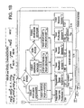
- Fig. 1 depicts a flowchart that shows an implementation of the overall techniques.
- Fig. 2 depicts the flowchart of Fig.1 with possible effects due to Pro-Angiogeneses and Anti- Angiogeneses drugs.
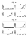
- Fig. 3 shows graphs that illustrate the effects of Anti-VEGF Drugs.
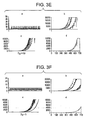
- Fig. 4 shows graphs that illustrate the effects of a combination of Anti-VEGF and Anti-Ang1 Drugs.
- Fig. 5 (a)-(c) shows a table with description of terms used in the Equations included in the specification.
- the disclosed techniques are embodied in exemplary computer'systems and exemplary flowcharts that are implemented by computers.
- the implementations discussed herein are merely illustrative in nature and are by no means intended to be limiting.
- any type of computers can be used to implement the systems and techniques.
- An aspect of the disclosed teachings is a computer program product including computer-readable media comprising instructions.
- the instructions are capable of enabling a computer to implement the systems and techniques described herein.
- the computer-readable media could be any media from which a computer can receive instructions, including but not limited to hard disks, RAMs, ROMs, CDs, magnetic tape, internet downloads, carrier wave with signals, etc.
- instructions can be in any form including source code, object code, executable code, and in any language including higher level, assembly and machine languages.
- the computer system is not limited to any type of computer. It could be implemented in a stand-alone machine or implemented in a distributed fashion, including over the internet.
- the technique shown in the flowchart take into account for the dynamic interactions between tissue volume, angiogenesis (growth and regression of immature blood vessels), and vascular maturation and destabilization.
- the technique shown in the flowchart is combined with a quantitative mathematical model that is described in detail herein.
- a combination of the technique shown in the flowchart and the mathematical computations described would allow a skilled artisan to practice the disclosed technique; including for example, to quantify the dynamics of tissue vascularization and the effect of drug on this process at any given moment.
- the technique describes the interactions between molecular regulatory factors, cell types and multi-cellular structures (such as vessels) which together influence the tissue dynamics.
- the technique takes into account, the temporal parameters which characterize the response rates of each one of the elements included in the angiogenesis process.
- the technique includes a series of simulation steps.
- the parameter values that are output from each simulation step are taken as initial conditions for the next simulation step. These parameter values are compared with the threshold levels. Their current values are calculated according to the arrows shown in the flowchart of Figure 1. At least six major processes are taken into account simultaneously, namely tissue cells proliferation and death, endothelial cells proliferation and death, immature vessels formation and regression, immature vessels maturation and mature vessels destabilization, and possibly others.
- FIG. 1 The techniques depicted in Figure 1 includes three interconnected modules: tissue cell proliferation, angiogenesis (immature vessels growth) and maturation (formation and destabilization of mature vessels). Further, each module operates on three scales: molecular, cellular and macroscopic (namely, vessel densities and tissue volume).
- the tissue module includes tissue cell proliferation sub-module and cell death sub-module. Further, each is subdivided into i) time-invariant, cell type-specific, genetically determined sub-block, and ii) time-variant, nutrient-dependent sub-block. Nutrient-dependent cell proliferation and nutrient-dependent cell death rates are directly or inversely proportional, respectively, to the effective vascular density (EVD), which is a perfused part of vascular tree ⁇ 40>.
- ETD effective vascular density
- tissue growth module Two additional quantities are calculated in the tissue module, namely VEGF and PDGF production. They are inversely related to EVD so that increasing nutrient depletion results in increasing secretion of pro-angiogenic factors ⁇ 7-9>.
- the tissue growth module interacts with the angiogenesis and the maturation modules via the relevant regulatory proteins.
- volume of immature vessels is calculated. Immature vessel volume increases proportionally to VEGF concentration, if VEGF is above a given threshold level. The volume regresses if VEGF is below a given, possibly different, threshold level.
- the latter threshold is generally referred to as "survival level" ⁇ 21-24>.
- volume of mature vessels is calculated according to pericyte concentration ⁇ 41-43> and according to the Ang1/Ang2 ratio ⁇ 44>.
- Pericytes proliferate proportionally to PDGF concentration ⁇ 25-26>.
- Ang1 and Ang2 are continuously secreted by tissue cells and immature vessels, respectively ⁇ 27, 28, 32-34, 41-43, 45>. Additionally, Ang1 and Ang2 can be secreted by tissue cells, if the latter are nutrient-depleted ⁇ 45>.
- tissue volume determined as a function of tissue cell number
- number of free endothelial cells and pericytes volume of immature and mature vessels
- concentrations of the regulatory factors such as VEGF, PDGF, Ang1 and Ang2.
- Ang2/Ang1 Ang2/Ang1
- immature vessel density immature vessel density
- mature vessel density denoting vessels volume divided by tissue volume.
- EVD effective vessel density
- EVD is a critical model variable, which at any moment determines tissue cells proliferation and death, as well as the production of factors, such as VEGF and PDGF. Resistance of tissue cells to antiangiogenic drugs may emerge from tissue adaptation to hypoxia.
- EVD is a function of the duration of insufficient perfusion, (denoted below by EVDn).
- the technique takes into account the threshold levels of regulatory factors and parameter ratios, such as:
- VEGF concentration below which endothelial cells (both in the free state as well as when incorporated into immature blood vessels) are subject to apoptosis (this is denoted below by VEGF thr ;
- the EVD value influences the rate of cell proliferation.
- the EVD value for which the system is in steady state is denoted below by EVD ss (referred to as EVD(bl) in Figures 1B and 2B)
- EVD ss tissue cells proliferation prevails, so that tissue volume increases.
- tissue cell death prevails, and the tissue shrinks.
- the EVD ss is determined by genetic properties of a given tissue and a given host.
- VEGF, PDGF, Ang1, Ang2 secretion level at the steady state of the system will be denoted VEGF ss PDGF ss , Ang1 ss and Ang2 ss .
- the inputs to the represented system includes the tissue volume, blood vessels density, and the inherent parameters characterizing this tissue type at initiation of the process.
- the output at any given moment are parameters like tissue volume, mature and immature vessels sizes, BVD.
- Fig. 1 The flowchart shown in Fig. 1 is discussed in detail herein with reference to specific mathematical equations describing the principal interactions affecting vascular tissue growth.
- the technique describes the interrelationships between tissue growth, the formation of new vessels (angiogenesis) and the maturation of the newly formed vessels.
- the interactions occur across three organization levels: molecular, cellular, and organic level.
- the arrows in the flowchart indicate the specific module interaction.
- the rectangular boxes indicate the point at which a specific sub-process calculation occurs.
- the parameter T x in a box denotes the characteristic reaction time of the action calculated in the box.
- the diamonds indicate the conditions, which determine the direction of processes.
- EVD ss is the value for which the system is in steady state.
- VEGF ss is the VEGF secretion level at the steady state of the system.
- VEGF ,Thr is the VEGF concentration below which endothelial cells, both in the free state as well as when incorporated into immature blood vessels, are subject to apoptosis.
- PDGF ss is the PDGF secretion level at the steady state of the system.
- EVD n in a certain moment n is represented as the sum of a density of immature (EVD n im ) and density of mature vessels (EVD n mat ) at the moment "n".
- EVD n EVD n im + EVD n mat ;
- Fig.1 and 2 the effective vessel density as discussed above is calculated in block 1.1.
- the EVD n im and EVD n mat is the relation of volume of vessels feeding the tissue, to a number of living tissue cells.
- the amount of immature vessels at a moment "n" depends on an amount of both immature and mature vessels at the previous moment "n-1".
- the volume of immature vessels (block 1.4) at the moment "n” is a function of the volumes of immature and mature vessels at the moment "n-1". This function has 5 terms, corresponding to the five exponential terms below. They are computed in blocks 1.5, 1.6, 1.7, 1.8 and 1.9.
- V n im V n - 1 im * e A n - 1 im ⁇ new ⁇ T 0 T 3 - A n - 1 im ⁇ reg ⁇ T 0 T 4 - A n - 1 im ⁇ mat ⁇ T 0 T 6 * ⁇ + V n - 1 m * e A n - 1 mat ⁇ new ⁇ T 0 T 5 + A n - 1 mat ⁇ im ⁇ T 0 T 7 * ⁇ - 1 ;
- the generation of immature vessels by immature vessels is accounted for by block 1.5.
- the generation of immature vessels by mature vessels is accounted for by block 1.6.
- the destabilization of mature vessels is accounted for by block 1.7.
- the maturation of immature vessels is accounted for by block 1.8.
- the degeneration of immature vessels (A im ⁇ reg ) by is accounted for by block 1.9.
- the volume of mature vessels (block 1.10) at the moment "n” is also a function of the volumes of immature and mature vessels at the moment "n-1". This function has 2 terms corresponding to the two exponential terms as shown below. They are calculated in blocks 1.7 and 1.9 respectively.
- V n m V n - 1 im * e A n - 1 im ⁇ mat ⁇ T 0 T 6 * ⁇ - 1 + V n - 1 m ⁇ 2 - e A n - 1 mat ⁇ im ⁇ T 0 T 7 * ⁇ ; 26
- T 1 to T 7 Every sub process described in functions (3) and (4) has its characteristic time, denoted by T 1 to T 7 . Resolution is denoted by T 0 (the period between "n” and "n-1").
- T 0 the period between "n” and "n-1").
- Factor ⁇ represents a factor of the conformity.
- the degeneration of immature vessels is also a function of the concentration of VEGF, level VEGF thr , with the coefficient ⁇ im .
- a n - 1 mat ⁇ new ⁇ ec im * ⁇ mat ec * VEGF n - 1 VEGF thr - ⁇ ec / VEGF n - 1 VEGF thr
- a n - 1 mat ⁇ new 0 IF ( ⁇ mat ec * VEGF n - 1 VEGF thr - ⁇ ec / VEGF n - 1 VEGF thr ) ⁇ 0
- a n - 1 mat ⁇ new ⁇ ec im IF ( ⁇ mat ec * VEGF n - 1 VEGF thr - ⁇ ec / VEGF n - 1 VEGF thr ) ⁇ 1
- the destabilization of mature vessels (block 1.9) is a function of the ratio between Ang1 and Ang2 with the coefficient ⁇ mat im , Eqn.(8).
- a n - 1 im ⁇ reg ⁇ im / VEGF n - 1 VEGF thr ;
- a n - 1 mat ⁇ im ⁇ mat im / Ang ⁇ 1 n - 1 Ang ⁇ 2 n - 1 ;
- a n - 1 im ⁇ mat ⁇ im mat * Ang ⁇ 1 n - 1 Ang ⁇ 2 n - 1 * Nper n - 1 ⁇ mat per / V n - 1 im ;
- Maturation in a given moment is a function of a ratio of Ang1/Ang2 at the same moment, with the coefficient ⁇ im mat . Maturation is also a function of the volume of immature vessels and of the number of free pericytes.
- VEGF n - 1 V n - 1 tis * ⁇ EVD VEGF ⁇ EDV b - EDV n - 2 * T 0 T 8 + VEGF n - 2 * e - 0.691371805 * T 0 T k - 1 VEGF
- VEGF n- 1 > VEGF max
- VEGF n -1 VEGF max
- VEGF n -1 VEGF en
- the initial level VEGF ss characterizes the amount of VEGF secreted when effective tissue vessel density is EVD ss .
- PDGF n - 1 V n - 1 tis * ⁇ EVD PDGF ⁇ EDV b - EDV n - 2 * T 0 T 14 + PDGF n - 2 * e - 0.691371805 * T 0 T k - 1 PDEGF
- VEGF n -1 VEGF max
- VEGF n -1 VEGF en
- the characteristic time T 14 and T n - 1 PDGF The above equations 10 and 11 are involved in blocks marked 1.11 & 1.12.
- Ang ⁇ 2 n - 1 Ang ⁇ 2 en ec + Ang ⁇ 2 ss ec * E ⁇ C n - 2 + Ang ⁇ 2 ed ec * EVD ss - EVD n - 2 * E ⁇ C n - 2 * T 0 T 10 + + Ang ⁇ 2 en tc + Ang ⁇ 2 ss tc * N n - 2 tum + Ang ⁇ 2 ed tc * EVD d - EVD n - 2 * N n - 2 tum * T 0 T 15 + Ang ⁇ 2 n - 2 * e - 0.693171805 * T 0 T k - l Ang ⁇ 2
- Ang2 and Ang1 also depends on the numbers of endothelial cells in immature vessels, which is determined by the Eqs. (13), and the numbers of tissue cells Ang ⁇ 2 en ec , Ang ⁇ 2 b ec , Ang ⁇ 2 ed ec , Ang ⁇ 2 en tc , Ang ⁇ 2 b tc , Ang ⁇ 2 ed tc , T n - 1 Ang ⁇ 2 , ⁇ V im ec , Ang ⁇ 1 en ec , Ang ⁇ 1 b ec , Ang ⁇ 1 ed ec , Ang ⁇ 1 en tc , Ang ⁇ 1 b tc , Ang ⁇ 1 ed tc , T n - 1 Ang ⁇ 1 .
- the Ang1 induction and Ang2 induction are factors in bocks
- the characteristic reaction time for Ang2 generation is T 10 and T 15 , (12), for Ang1 generation it is T 9 and T 11 (14).
- Ang ⁇ 1 n - 1 Ang ⁇ 1 en tc + Ang ⁇ 1 ss tc * N n - 2 tum + Ang ⁇ 1 ed tc * EVD ss - EVD n - 2 * N n - 2 tum * T 0 T 9 + + Ang ⁇ 1 en ec + Ang ⁇ 1 en ec * E ⁇ C n - 2 + Ang ⁇ 1 en ec * EVD ss - EVD n - 2 * E ⁇ C n - 2 * T 0 T 16 + Ang ⁇ 1 n - 2 * e - 0.693171805 * T 0 T k - l Ang ⁇ 1
- the number of tissue cells (block 1.18) in the moment, n depends on their number in the previous moment multiplied by a factor describing the process of cell proliferation and death, r n-1 .
- V n tis V n - 1 tis * e r n - 1 * ⁇
- r n-1 depends on the mitotic index M(I) (mitotic time being T (1), apoptotic index A (I) (apoptotic time beingT (2)), rate of tissue cell growth ⁇ and the rate of the death of tumor cells, ⁇ .
- M(I) mitotic time being T (1)
- apoptotic index A (I) apoptotic time beingT (2)
- rate of tissue cell growth ⁇ rate of tissue cell growth ⁇
- ⁇ rate of the death of tumor cells
- the blocks in Fig.2 are identical to those in Fig.1, except for the additional drug effects shown.
- the drug effects on the overall process are analyzed by setting the selected drug schedule (number of doses, the dose and the dosing interval).
- the analysis of anti-VEGF drug activity shows that a drug which inhibits VEGF has an optimal efficacy when given by certain treatment protocol. Increasing the administered dose above the optimum can bring about the undesired effect of tissue proliferation.as shown in Fig. 3.
- the technique enables one to predict the effects of various drug combination, for example as shown in Fig.4.
Landscapes
- Health & Medical Sciences (AREA)
- General Physics & Mathematics (AREA)
- Physics & Mathematics (AREA)
- Life Sciences & Earth Sciences (AREA)
- Fluid Mechanics (AREA)
- Chemical & Material Sciences (AREA)
- Molecular Biology (AREA)
- Engineering & Computer Science (AREA)
- Analytical Chemistry (AREA)
- Urology & Nephrology (AREA)
- Biomedical Technology (AREA)
- Immunology (AREA)
- Hematology (AREA)
- Cell Biology (AREA)
- Biotechnology (AREA)
- Microbiology (AREA)
- Endocrinology (AREA)
- Food Science & Technology (AREA)
- Medicinal Chemistry (AREA)
- Biochemistry (AREA)
- General Health & Medical Sciences (AREA)
- Pathology (AREA)
- Medicines That Contain Protein Lipid Enzymes And Other Medicines (AREA)
- Pharmaceuticals Containing Other Organic And Inorganic Compounds (AREA)
- Medicines Containing Material From Animals Or Micro-Organisms (AREA)
- Medical Treatment And Welfare Office Work (AREA)
Abstract
Description
- This Application claims priority from a co-pending U. S. Provisional Patent
Applications Serial Number 60/330,592 filed October 25, 2001 and 10/207,772 filed July 31, 2002. - The present disclosure generally teaches techniques related to diseases and processes involving Angiogenesis. More particularly it teaches techniques for generating treatment protocols for diseases where angiogenesis is a factor. The techniques are also applicable to normal processes involving Angiogenesis even if no disease is present.
- The following papers provide useful background information, and are selectively referred to in the remainder of this disclosure by their accompanying reference numbers in brackets (i.e., <3> for the third numbered paper by Yangopoulos et al):
- <1> Folkman J. (1995) Angiogenesis in cancer, vascular, rheumatoid and other disease. Nat Med. 1 (1):27-31.
- <2> Carmeliet P., Jain R. K. (2000) Angiogenesis in cancer and other diseases. Nature. 407: 249-257.14 September 2000
- <3> Yangopoulos GD, Davis S., Gale NW., Rudge JS., Wiegand SJ., Holash J.(2000) Vascular-specific growth factors and blood vessel formation. Nature. 407: 242 - 248. 14 September 2000
- <4> Kerbel RS, (2000) Tumor angiogenesis: past, present and the near future. Carcinogenesis. 21. 3: 505-515. March 2000
- <5> Danielsen T., Rofstad EK (2000) The constitutive level of vascular endothelial growth factor (VEGF) is more important than hypoxia-induced VEGF up-regulation in the angiogenesis of human melanoma xenografts. British Journal of Cancer. 82(9): 1528-1534
- <6> Dor Y., Porat R., Keshet E (2001) Vascular endothelial growth factor and vascular adjustments to perturbations in oxygen homeostasis. AJP-Cell Physiology. 280. 6: C1367-C1374. June 2001
- <7> Ikeda E., Achen MG., Breier G., Risau W (1995) Hypoxia-induced Transcriptional Activation and Increased mRNA Stability of Vascular Endothelial Growth Factor in C6 Glioma Cells. J Biol Chem. 270. 34: 19761-19766, August 25 1995
- <8> Ferrara N, Henzel WJ (1989) Pituitary follicular cells secrete a novel heparin-binding growth factor specific for vascular endothelial cells. Biochem Biophys Res Commun. 161:851-858
- <9> Ferrara N, Houck K, Jakeman L, Leung DW (1992) Molecular and biological properties of the vascular endothelial growth factor family of proteins. Endocr Rev 13:18-32
- <10> Breier G, Albrecht U, Sterrer S, Risau W 1992 Expression of vascular endothelial growth factor during embryonic angiogenesis and endothelial cell differentiation. Development 114:521-532
- <11> Keyt B, Nguyen H, Berleau L, Duarte C, Park J, Chen H, Ferrara N (1996) Identification of VEGF determinants for binding Flt-1 and KDR receptors. Generation of receptor-selective VEGF variants by site-directed mutagenesis. J Biol Chem 271:5638-5646
- <12> Ferrara N, Carver-Moore K, Chen H, Dowd M, Lu L, O'Shea KS, Powell-Braxton L, Hillan KJ, Moore MW (1996) Heterozygous embryonic lethality induced by targeted inactivation of the VEGF gene. Nature 380:439-442
- <13> Carmeliet P, Ferreira V, Breier G, Pollefeyt S, Kieckens L, Gertsenstein M, Fahrig M, Vandenhoeck A, Harpal K, Eberhardt C, Declercq C, Pawling J, Moons L, Collen D, Risau W, Nagy A (1996) Abnormal blood vessel developement and lethality in embryos lacking a single VEGF allele. Nature 380:435-439
- <14> Barleon B, Sozzani S, Zhou D, Weich H, Mantovani A, Marmé D (1996) Migration of human monocytes in response to vascular endothelial growth factor (VEGF) is mediated via the VEGF receptor flt-1. Blood 87:3336-3343
- <15> Neufeld G, Cohen T, Gengrinovitch S, Poltorak Z (1999) Vascular endothelial growth factor (VEGF) and its receptors. FASEB 13:9-22
- <16> Kim KJ, Li B, Winer J, Armanini M, Gillett N, Phillips HS, Ferrara N (1993) Inhibition of vascular endothelial growth factor-induced angiogenesis suppresses tumour growth in vivo. Nature 362:841-844
- <17> Potgens AJ, Lubsen NH, van Altena MC, Schoenmakers JG, Ruiter DJ, de Waal RM (1995) Vascular permeability factor expression influences tumor angiogenesis in human melanoma lines xenografted to nude mice. Am J Pathol. 146(1):197-209.
- <18> Claffey KP, Brown LF, del Aguila LF, Tognazzi K, Yeo KT, Manseau EJ, Dvorak HF. (1996) Expression of vascular permeability factor/vascular endothelial growth factor by melanoma cells increases tumor growth, angiogenesis, and experimental metastasis. Cancer Res. 56(1):172-81
- <19> Shweiki D, Itin A, Soffer D, Keshet E (1992) Vascular endothelial growth factor induced by hypoxia may mediate hypoxia-initiated angiogenesis. Nature 359:843-845
- <20> Dvorak HF, Brown LF, Detmar M, Dvorak AM (1995) Vascular permeability factor/vascular endothelial growth factor, microvascular permeability and angiogenesis. Am J Pathol 146:1029-1039
- <21> Benjamin LE, Keshet E (1997) Conditional switching of vascular endothelial growth factor (VEGF) expression in tumors: Induction of endothelial cell shedding and regression of hemangioblastoma-like vessels by VEGF withdrawal. PNAS 94: 8761-8766. August 1997
- <22> Jain RK, Safabakhsh N, Sckell A, Chen Y, Jiang P, Benjamin LE, Yuan F, Keshet E (1998) Endothelial cell death, angiogenesis, and microvascular function after castration in an androgen-dependent tumor: Role of vascular endothelial growth factor. PNAS 95. 18: 10820-10825. September 1 1998
- <23> Holash, SJ, Wiegandand GD , Yancopoulos GD, (1999) New model of tumor angiogenesis: dynamic balance between vessel regression and growth mediated by angiopoietins and VEGF. Oncogene 18: 5356 - 5362
- <24> Gilead A., Neeman M (1999) Dynamic remodeling of the vascular bed precedes tumor growth: MLS ovarian carcinoma spheroids implanted in nude mice. Neoplasia 1:226-230.
- <25> Benjamin LE et al. (1999) Selective ablation of immature blood vessels in established human tumors follows vascular endothelial growth factor withdrawal. J Clin Invest. 103(2):159-65.
- <26> Benjamin LE et al. (1998) A plasticity window for blood vessel remodelling is defined by pericyte coverage of the preformed endothelial network and is regulated by PDGF-B and VEGF. Development 125(9):1591-8.
- <27> Cynthia L. Grosskreutz, Anand-Apte B, Duplaa C, Quinn TP, Terman BI, Zetter B, D'Amore PA (1999) Vascular Endothelial Growth Factor-Induced Migration of Vascular Smooth Muscle Cells in Vitro. Microvascular Research 58:128-136
- <28> Lindahl P, Johansson BR , Levéen P, Betsholtz C (1997) Pericyte Loss and Microaneurysm Formation in PDGF-B- Deficient Mice. Science 277: 242-245 11 july 1997
- <29> Hellstrom M, Kal'n M, Lindahl P, Abramsson A, Betsholtz C (1999) Role of PDGF-B and PDGFR-b in recruitment of vascular smooth muscle cells. Development 126: 3047-3055
- <30> Davis S, Aldrich TH, Jones PF, Acheson A, Compton DL, Jain V, Ryan TE, Bruno J, Radziejewski C, Maisonpierre PC, Yancopoulos GD (1996) Isolation of Angiopoietin-1, a Ligand for the TIE2 Receptor, by Secretion-Trap Expression Cloning. Cell 87: 1161-1169. December 1996
- <31> Suri C, Jones PE, Patan S, Bartunkova S, Maisonpierre PC, Davis S, Sato TS, Yancopoulos GD (1996) Requisite Role of Angiopoietin-1, a Ligand for the TIE2 Receptor, during Embryonic Angiogenesis. Cell 87: 1171-1180. December 1996
- <32> Maisonpierre PC, Suri C, Jones PF, Bartunkova S, Wiegand SJ, Radziejewski C, Compton D, McClain J, Aldrich TH, Papadopoulos N, Daly TJ, Davis S, Sato TN, Yancopoulos GD (1997) Angiopoietin-2, a Natural Antagonist for Tie2 That Disrupts in vivo Angiogenesis. Science 277. 5322: 55-60. 4 Jul 1997
- <33> Folkman J, D'Amore PA (1996) Blood Vessel Formation: What Is Its Molecular Basis?. Cell 87: 1153-1155. December 1996
- <34> Eggert A, Ikegaki N, Kwiatkowski J, Zhao H, Brodeur GM, Himelstein BP (2000) High-Level Expression of Angiogenic Factors Is Associated with Advanced Tumor Stage in Human Neuroblastomas. Clinical Cancer Research 6: 1900-1908
- <35> Loughna S. and Sato TN. (2001) Angiopoietin and Tie signaling pathways in vascular development. Matrix Biol. 20:319-25.
- <36> Folkman, J. (1971) Tumor angiogenesis: therapeutic implications. N. Engl. J. Med. 285:1182-1186.
- <37> O'Reilly, M.S. et al.(1994) Angiostatin: a novel angiogenesis inhibitor that mediates the suppression of metastases by a Lewis lung carcinoma. Cell. 79:315-328.
- <38> O'Reilly, M.S. et al.(1997) Endostatin: an endogenous inhibitor of angiogenesis and tumor growth. Cell. 88:277-285.
- <39> Anderson AR and Chaplain MA (1998) Continuous and discrete mathematical models of tumor-induced angiogenesis. Bull Math Biol. 60(5):857-99.
- <40> Hahnfeldt P. et al. (1999) Tumor development under angiogenic signaling: a dynamic theory of tumor growth, treatment response, and postvascular dormancy. Cancer Res. 59:4770-5.
- <41> Rolland, Y. et al. (1998) Modeling of the parenchymous vascularization and perfusion. Invest. Radiology 34:171-5.
- <42> Hayes, AJ et al. (2000) Expression and function of angiopoietin-1 in breast cancer. Br. J. Cancer 83:1154-60.
- <43> Ahmad, SA et al. (2001) Differential expression of angiopoietin-1 and angiopoietin-2 in colon carcinoma. Cancer 92:1138-43.
- <44> Koga K. et al. (2001) Expression of angiopoietin-2 in human glioma cells and its role for angiogenesis. Cancer Res. 61:6248-54.
- <45> Oh H. et al. (1999) Hypoxia and Vascular Endothelial Growth Factor selectively up-regulate angiopoietin-2 in bovine microvascular endothelial cells. J Biol Chem 274:15732-9.
- <46> Holash J. et al. (1999) Vessel cooption, regression, and growth in tumors mediated by angiopoietins and VEGF. Science 284:1994-8.
- <47> Mandriota SJ et al. (2000) Hypoxia-inducible angiopoietin-2 expression is mimicked by Iodonium compounds and occurs in the rat brain and skin in response to systemic hypoxia and tissue ischemia. Am J Pathol 156:2077-89.
- <48> Beck H. (2000) Expression of angiopoietin-1, angiopoietin-2, and Tie receptors after middle cerebral artery occlusion in rats. Am J Pathol 157:1473-83.
- <49> Stratmann A. et al. (1998) Cell type-specific expression of angiopoietin-1 and angiopoietin-2 suggests a role in glioblastoma angiogenesis. Am J Pathol 153:1459-66.
- <50> Stegmann TJ et al. (2000) [Induction of myocardial neoangiogenesis by human growth factors. A new therapeutic approach in coronary heart disease] Herz.; 25(6):589-99.
- <51> Kocher AA et al. (2001) Neovascularization of ischemic myocardium by human bone-marrow-derived angioblasts prevents cardiomyocyte apoptosis, reduces remodeling and improves cardiac function.Nat Med.;7(4):430-6.
- <52> Ciulla TA et al. (2001) Presumed ocular histoplasmosis syndrome: update on epidemiology, pathogenesis, and photodynamic, antiangiogenic, and surgical therapies.Curr Opin Ophthalmol.;12(6):442-9.
- <53> Weber AJ, De Bandt M. (2000) Angiogenesis: general mechanisms and implications for rheumatoid arthritis. Joint Bone Spine.;67(5):366-83.
- <54> Fearon U, Veale DJ. (2001) Pathogenesis of psoriatic arthritis. Clin Exp Dermatol.; 26(4): 333-7.
- <55> Healy DL et al. (1998) Angiogenesis: a new theory for endometriosis.Hum Reprod Update; 4(5): 736-40.
- <56> Gustafsson T., Kraus WE. (2001) Exercise-induced angiogenesis-related growth and transcription factors in skeletal muscle, and their modification in muscle pathology. Front Biosci; 6: D75-89.
- <57> Brizzi MF et al. (2001) The molecular mechanisms of angiogenesis: a new approach to cardiovascular diseases. Ital Heart J.;2(2):81-92.
- <58> Gavin JB et al. (1998) Microvascular involvement in cardiac pathology. J Mol Cell Cardiol. ;30(12): 2531-40.
- <59> Karasek MA. (1999) Progress in our understanding of the biology of psoriasis. Cutis. ;64(5): 319-22.
- <60> Kurz H. (2000) Physiology of angiogenesis. J Neurooncol. ;50(1-2): 17-35.
- <61> Aiello LP. (1997) Vascular endothelial growth factor and the eye: biochemical mechanisms of action and implications for novel therapies. Ophthalmic Res; 29(5): 354-62.
- <62> Jain A et al. (2001) Production of cytokines, vascular endothelial growth factor, matrix metalloproteinases, and tissue inhibitor of
metalloproteinases 1 by tenosynovium demonstrates its potential for tendon destruction in rheumatoid arthritis. Arthritis Rheum ;44(8): 1754-60. - <63> Carmeliet P. (1999) Basic Concepts of (Myocardial) Angiogenesis: Role of Vascular Endothelial Growth Factor and Angiopoietin. Curr Interv Cardiol Rep. ;1(4): 322-335.
- <64> Miller JW. (1997) Vascular endothelial growth factor and ocular neovascularization. Am J Pathol.; 151(1): 13-23.
- <65> O'Toole G. et al. (2001) A review of therapeutic angiogenesis and consideration of its potential applications to plastic and reconstructive surgery. Br J Plast Surg. ;54(1): 1-7.
- <66> Halin C. et al. (2001) Antibody-based targeting of angiogenesis. News Physiol Sci. ;16: 191-4.
- <67> Ferrara N. (2000) Vascular endothelial growth factor and the regulation of angiogenesis. Recent Prog Horm Res. ;55: 15-35
- <68> Simons M. et al, (2000) Clinical trials in coronary angiogenesis: issues, problems, consensus: An expert panel summary.
Circulation 12;102(11):E73,86. - <69> Tong S and Yuan F (2001) Numerical simulations of angiogenesis in the cornea. Microvasc Res; 61(1): 14-27.
<70> Bellomo N and Preziosi L (2000) provides a survey of mathematical models and methods dealing with the analysis and simulation of tumour dynamics in competition with the immune system. Mathematical and Computer Modelling; 32:413-452. - For a better understanding of this disclosure, all the terms and parameters used in this disclosure are listed in the Table shown in Fig.5.
- Angiogenesis, or neovascularization, is a process of new blood vessels formation by budding from the existing ones. Neovascularization provides tissue with vital nutrients and growth factors and enables clearance of toxic waste products of cellular metabolism. Angiogenesis has been conventionally recognized as a biological mechanism of dual clinical effect. On the one hand, it allows survival of normal tissues when they become ischemic. That is, it enables functional development of normal tissues, for example, wound healing and embryogenesis. On the other hand, angiogenesis enables tumor tissue growth.
- Intensive research of angiogenesis during the last 15 years has led to better understanding of this complex process <1-7, 57-60>. However, cause and effect relationships in the process of angiogenesis are yet to be clarified. Moreover, the massive research in the field of angiogenic therapy still suffers from the lack of tools for predicting the potential effects of PRO-and ANTI-angiogenic factors.
- The two major determinants of new vasculature, formation are thought to be the genetic features of the tissue and the availability of oxygen and nutrients <5,6>. The dependence of vessel formation an nutrients or oxygen deprivation was shown to be mediated by vascular endothelial growth factor (VEGF), which is a potent inducer of endothelial cell proliferation and migration <8-15>. VEGF is preferentially expressed by tissue cells in the nutrient-deprived areas <7, 16-20, 61-64>. In contrast, basic nutrient-independent VEGF production by the tissue is determined by genetic factors <20>. Consequently, VEGF-induced angiogensis depends on both the aforementioned vasculature growth determinants, namely genetics and nutrient/oxygen availability. VEGF-induced angiogenesis leads to increase in nutrient supply to the tissue. Accordingly, nutrient- and oxygen-dependent VEGF expression is down-regulated. When VEGF level becomes low enough, the newly formed blood vessels regress <21-26>, consequently leading to nutrients and oxygen deprivation again. This negative feedback can produce successive cycles of growth and regression of blood vessels. This phenomenon was demonstrated in the mouse xenograft tumor model <12>.
- Blood vessels can be rendered insensitive to fluctuations of VEGF concentration by the process of maturation (coverage of capillaries by periendothelial cells 25,26). This process involves pericytes (smooth muscle-like cells) which form an outside layer covering the endothelial cells of the newly formed vessel. The major pericyte-stimulating factor is a platelet-derived growth factor (PDGF) <27-29>. Interactions between endothelial cells and pericytes, which apparently lead to maturation, are governed by the angiopoietin system. This system includes two soluble factors -
Angiopoietins 1 and 2 (Angl and Ang2, respectively), and their receptor, Tie2, which is specifically expressed on endothelial cells <30-34>. Ang1 is Tie2 agonist that promotes maturation, while Ang2 is its natural antagonist <31,32>. Regulation of Ang1 and Ang2 expression is not completely understood. According to recent publications it can be influenced by tumor cell- as well as endothelial cell- specific factors <35,42-49>. These factors depend on the tissue and the host type <42-44>, and can be, accordingly, taken into account in the presented model.High Ang 1/Ang 2 ratio and pericytes' presence induces maturation of newly formed vessels. Alternatively,low Ang 1/Ang 2 ratio induces destabilization of mature blood vessels, while newly formed vessels remain immature and susceptible to VEGF fluctuations <25>. - Therefore, it would be advantageous to have techniques for generating and selecting treatment protocols for diseases where angiogenesis is a factor. Further, it is also advantageous to adapt the techniques to study the progression of processes involving angiogenesis.
- The clinical significance of angiogenesis as an "ultimate" target for cancer therapy was first recognized in 1971 by J. Folkman <36>, and got wide acceptance in early nineties after the discovery of the first specific antiangiogenic substances <37,38>. Apparent advantages of this approach include its universality for different solid tumors, lack of prominent side effects and lack of resistance development during repetitive treatment cycles.
- Angiogenesis is implicated in the pathogenesis of a variety of disorders: proliferative retinopathies, age-related macular degeneration, tumors, rheumatoid arthritis, psoriasis <1;51-56, 66, 67> and coronary heart diseases <50>. The use of exogenous agents to selectively target neovasculature, or stimulate the growth of new blood vessels into ischaemic tissue is a potentially revolutionary therapy in a wide variety of clinical specialties, which opens new avenues for the diagnosis and therapy of diseases where angiogenesis is a factor; such as, cancer, blinding ocular disorders, rheumatoid arthritis and others.<65, 66>.
- In order to establish optimal pro-angiogenic or anti-angiogenic treatment protocols (either as a monotherapy or in combination with chemotherapy or radiotherapy) the dynamics of angiogenesis must be better understood. Recent studies <25,26> have shown that newly formed vasculature is very dynamic - blood vessels undergo constant remodeling that involves maturation in response to local levels of angiogenic and maturation factors. Mature and immature vessels may differentially respond to certain PRO and ANTI-angiogenic drugs during tissue growth, myocardial ischemia, macular degeneration and other diseases, leading to success or failure of the treatment <25>.
- Mathematical models and computer simulation of angiogenesis and PRO- and ANTI-angiogenic therapy can be constructed, in order to predict the most promising treatment protocols thus eliminating the need for lengthy and expensive clinical trials.
- The construction of a mathematical model for angiogenesis includes I) in-depth understanding of the biology of angiogenesis, II) the selection of appropriate patient populations for clinical trials, choice of therapeutic end points and means of their assessment, choice of therapeutic strategy (gene versus protein delivery), route of administration, and the side effect profile.<68>
- Several mathematical dynamic models have been proposed, each one of them constructed to illuminate specific aspects of angiogenesis <39-41>. Some of these models examine vascular tree formation in vitro, irrespectively of tumor dynamics, and consequently are not suitable for tumor growth modelling <39>. Others assume that the growing vascular tree is a subject to some optimization with regard to the target tissue perfusion <41>. This optimization, while possibly holding true for normal tissue development, can hardly account for tumor growth, since it is known that tumor vasculature is highly disorganized.
- Logistic-type model, proposes by Hahnfeldt et al. <40>, analyzes the general vascular dynamics ("carrying capacity of current vascular tree") with regard to production of pro- and antiangiogenic factors by the tumor. Analysis of experimental data of Lewis lung carcinoma growth in mice allowed the authors to estimate the model parameters and to examine the effects of antiangiogenic factors angiostatin and endostatin. The main problematic assumption of this model is the constant production rates of these factors, as we know that VEGF, for example, production is tightly regulated by tissue hypoxia.
- Model by Tong S and Yuan F <69> focused on two-dimensional angiogenesis in the cornea. The model considered diffusion of angiogenic factors, uptake of these factors by endothelial cells, and randomness in the rate of sprout formation and the direction of sprout growth.
- None of the aforementioned models takes into account vasculature maturation and mature vessels destabilization, which are very fundamental constituents of angiogenesis dynamics. Moreover, these models, due to their relative abstraction, cannot account for drug-induced, or other, molecular changes in angiogenic dynamics. Note, that, since PRO and ANTI-angiogenic drugs interfere with the dynamics described above at the molecular level, the model which can serve as a tool for predicting drug effect on this process must take into account all the molecular complexity of angiogenesis, including the dynamics of neovasculature maturation and mature vessels destabilisation.
- Therefore, it is desirable to provide a techniques, including computer systems, that overcomes some of the disadvantages noted above.
- To realize the advantages discussed above, the disclosed teachings provide a computer-implemented method for determining an optimal treatment protocol for a disease related to angiogenesis, comprising creating an angiogenesis model including pro-angiogenesis and anti-angiogenesis factors. Effective vessel density (EVD) is incorporated as a factor regulating switching on and switching off of at least one component in the angiogenesis model. Effects of vasculature maturation and mature vessels destabilization are incorporated. Pro-angiogenesis and anti-angiogenesis factors, which can influence changes in state of a tissue are selected. Effects of drugs in the pro-angiogenesis and anti-angiogenesis factors are incorporated. A plurality of treatment protocols in a protocol space is generated. A best treatment protocol based on a pre-determined criteria.
- In another specific enhancement, the model comprises a tissue volume model, an immature vessel model and a mature vessel model.
- In another specific enhancement, steps to regulate dynamics which influences EVD are incorporated.
- In another specific enhancement, the model simultaneously accounts for tissue cell proliferation, tissue cell death, endothelial cell proliferation, endothelial cell death, immature vessel formation and immature vessel regression, immature vessel maturation and mature vessel destabilization.
- In another specific enhancement, the model incorporates temporal parameters that characterize response rate of at least one element associated with angiogenesis.
- More specifically, EVD is calculated by combining immature vessel density and mature vessel density.
- In another specific enhancement, parameters incorporated into the model comprises tissue volume, number of free endothelial cells, number of free pericytes, volume of mature vessels, volume of immature vessels and concentration of regulator factors.
- More specifically, the regulatory factors comprise vascular endothelial growth factor (VEGF), platelet derived growth factor (PDGF), angiopoietin 1 (Ang1) and angiopoietin 2 (Ang2).
- More specifically, EVD is a function of a duration of insufficient perfusion and vice versa.
- More specifically, the model incorporates threshold levels of regulatory factors and parameter ratios.
- Even more specifically, the threshold level is at least one of: a) VEGF concentration below which no endothelial cells proliferation takes place (A); b) minimum number of receptors for VEGF above which endothelial cells proliferation takes place (B); c) VEGF concentration below which endothelial cells, both in the free state as well as when incorporated into immature blood vessels, are subject to apoptosis VEGFthr; d) the minimal number of free pericytes which stimulates the onset of maturation of immature vessels (C); e)
Ang 1/Ang 2 ratio below which mature vessels are destabilized, and above which maturation of immature vessels is enabled (K); f) EVD value that influences the rate of cell proliferation and death; and g) EVDss value for which the system is in steady state. - Even more specifically, the tissue volume model calculates the tissue volume by a process comprising: comparing EVD against an - EVDss. If EVD is equal to EVDss then use a programmed tissue cell proliferation and a programmed tissue cell death (apoptosis) to compute tissue voulme. If EVD > EVDss then use increased tissue proliferation and decreased tissue cell death to compute tissue volume.If EVD < EVDss then use decreased tissue proliferation and increased tissue cell death to compute tissue volume.
- Even more specifically, Ang1 and Ang2 induction are incorporated into appropriate steps above following the computation of tissue volume.
- More specifically, the immature vessel model calculates the immature vessels by a process comprising comparing EVD against an EVDss. If EVD is equal to EVDss then set VEGF to a VEGFss and PDGF to a PDGFss. If EVD > EVDss then use decreased VEGF and decreased PDGF. If EVD < EVDss then useg increased VEGF and increased PDGF. Compare VEGF against A. Factor endothelial cell proliferation if VEGF > A. Compare VEGF against a VEGF threshold. Factor free endothelial cell deaths if VEGF < VEGF threshold. Compare VEGF receptor number against B. If VEGF receptor number is less than B then consider no angiogenisis prior to computing immature vessel regression. If VEGF receptor number is not less than B then compute growth of immature vessels. If VEGF < A then consider no angiogenesis and compute immature vessel regression. Compute mature vessels based on growth immature vessels, immature vessel regression and mature vessel destabilization.
- Even more specifically, immature vessels computation considers no maturation if Ang2/Ang1 > K or if number of free pericytes < C.
- Even more specifically, mature vessel destabilization considers ang1/Tie2 interaction blocking.
- Even more specifically, no destabilization occurs if Ang2/Ang1 is not greater than K
- More specifically, mature vessel model is computed using a procedure comprising computing immature vessels. Determining if Ang1/Ang2 < K. Determine if number of free pericytes < C. Considering immature vessel maturation if both the above steps are false. Factoring no destablization if number of free pericytes is not less than C.
- More specifically, effects of a drug affecting EC proliferation are factored in computing immature vessels.
- More specifically, effects of a drug affecting VEGF receptors are factored in computing immature vessels.
- More specifically, effects of a drug affecting pericyte proliferation are factored in computing immature vessel computation.
- More specifically, effects of a drug affecting VEGF are factored in computing immature vessels.
- More specifically, effects of a drug affecting PEGF are factored in computing immature vessel computation.
- More specifically, effects of a drug affecting Ang1 are factored in computing immature vessels.
- More specifically, effects of a drug affecting Ang2 are factored in computing immature vessel computation.
- In another specific enhancement, model takes into account duration of a tissue cell proliferation, tissue cell death, endothelial cell proliferation, endothelial cell death, pericytes proliferation, immature vessel regression, immature vessel maturation and mature vessel destabilization.
- In another specific enhancement, model takes into account the duration of VEGF induction, PDGF induction, Ang1 and Ang2 induction by tissue cells and Ang1 and Ang2 induction by endothelial cells.
- Another aspect of the disclosed teachings is an optimal treatment protocol for a disease related to angiogenesis, comprising an angiogenesis model including pro-angiogenesis and anti-angiogenesis factors; a treatment protocol space generator that generates a protocol space of possible treatments for the disease; a treatment selector that selects an optimal protocol, wherein effective vessel density (EVD) is a factor regulating switching on and switching off of at least one component in the angiogenesis model; wherein the model incorporates effects of vasculature maturation and mature vessels destabilization; and wherein the system is adapted to effect selection of pro-angiogenesis and anti-angiogenesis factors which can influence changes in state of a tissue and incorporating effects of drugs in the pro-angiogenesis and anti-angiogenesis factors.
- A computer program product including computer readable media that execute instructions when said computer product program is run on a computer to implement the above techniques on a computer are also part of the disclosed teachings.
- The above objectives and advantages of the disclosed teachings will become more apparent by describing in detail preferred embodiment thereof with reference to the attached drawings in which:
- Fig. 1 depicts a flowchart that shows an implementation of the overall techniques.
- Fig. 2 depicts the flowchart of Fig.1 with possible effects due to Pro-Angiogeneses and Anti- Angiogeneses drugs.
- Fig. 3 shows graphs that illustrate the effects of Anti-VEGF Drugs.
- Fig. 4 shows graphs that illustrate the effects of a combination of Anti-VEGF and Anti-Ang1 Drugs.
- Fig. 5 (a)-(c) shows a table with description of terms used in the Equations included in the specification.
- The disclosed techniques are embodied in exemplary computer'systems and exemplary flowcharts that are implemented by computers. The implementations discussed herein are merely illustrative in nature and are by no means intended to be limiting. Also it should be understood that any type of computers can be used to implement the systems and techniques. An aspect of the disclosed teachings is a computer program product including computer-readable media comprising instructions. The instructions are capable of enabling a computer to implement the systems and techniques described herein. It should be noted that the computer-readable media could be any media from which a computer can receive instructions, including but not limited to hard disks, RAMs, ROMs, CDs, magnetic tape, internet downloads, carrier wave with signals, etc. Also instructions can be in any form including source code, object code, executable code, and in any language including higher level, assembly and machine languages. The computer system is not limited to any type of computer. It could be implemented in a stand-alone machine or implemented in a distributed fashion, including over the internet.
- The technique shown in the flowchart take into account for the dynamic interactions between tissue volume, angiogenesis (growth and regression of immature blood vessels), and vascular maturation and destabilization. The technique shown in the flowchart is combined with a quantitative mathematical model that is described in detail herein. A combination of the technique shown in the flowchart and the mathematical computations described would allow a skilled artisan to practice the disclosed technique; including for example, to quantify the dynamics of tissue vascularization and the effect of drug on this process at any given moment.
- The technique describes the interactions between molecular regulatory factors, cell types and multi-cellular structures (such as vessels) which together influence the tissue dynamics. The technique takes into account, the temporal parameters which characterize the response rates of each one of the elements included in the angiogenesis process.
- The technique includes a series of simulation steps. The parameter values that are output from each simulation step are taken as initial conditions for the next simulation step. These parameter values are compared with the threshold levels. Their current values are calculated according to the arrows shown in the flowchart of Figure 1. At least six major processes are taken into account simultaneously, namely tissue cells proliferation and death, endothelial cells proliferation and death, immature vessels formation and regression, immature vessels maturation and mature vessels destabilization, and possibly others.
- The techniques depicted in Figure 1 includes three interconnected modules: tissue cell proliferation, angiogenesis (immature vessels growth) and maturation (formation and destabilization of mature vessels). Further, each module operates on three scales: molecular, cellular and macroscopic (namely, vessel densities and tissue volume).
- The tissue module includes tissue cell proliferation sub-module and cell death sub-module. Further, each is subdivided into i) time-invariant, cell type-specific, genetically determined sub-block, and ii) time-variant, nutrient-dependent sub-block. Nutrient-dependent cell proliferation and nutrient-dependent cell death rates are directly or inversely proportional, respectively, to the effective vascular density (EVD), which is a perfused part of vascular tree <40>.
- Two additional quantities are calculated in the tissue module, namely VEGF and PDGF production. They are inversely related to EVD so that increasing nutrient depletion results in increasing secretion of pro-angiogenic factors <7-9>. The tissue growth module interacts with the angiogenesis and the maturation modules via the relevant regulatory proteins.
- In the angiogenesis module, volume of immature vessels is calculated. Immature vessel volume increases proportionally to VEGF concentration, if VEGF is above a given threshold level. The volume regresses if VEGF is below a given, possibly different, threshold level. The latter threshold is generally referred to as "survival level" <21-24>.
- In the maturation module, volume of mature vessels is calculated according to pericyte concentration <41-43> and according to the Ang1/Ang2 ratio <44>. Pericytes proliferate proportionally to PDGF concentration <25-26>. Ang1 and Ang2 are continuously secreted by tissue cells and immature vessels, respectively <27, 28, 32-34, 41-43, 45>. Additionally, Ang1 and Ang2 can be secreted by tissue cells, if the latter are nutrient-depleted <45>. It is assumed that maturation of immature vessels occurs if pericytes concentration and Ang1/Ang2 ratio are above their respective threshold levels, while under these thresholds immature vessels do not undergo maturation, while mature vessels undergo destabilization and become immature <29-33>.
- It is clear that the parameters used in the technique can include tissue volume (determined as a function of tissue cell number); number of free endothelial cells and pericytes; volume of immature and mature vessels; and concentrations of the regulatory factors such as VEGF, PDGF, Ang1 and Ang2.
- Moreover, several relative parameters (ratios) are calculated, such as Ang2/Ang1, immature vessel density and mature vessel density (denoting vessels volume divided by tissue volume). The latter two densities are combined into effective vessel density, EVD. EVD is a critical model variable, which at any moment determines tissue cells proliferation and death, as well as the production of factors, such as VEGF and PDGF. Resistance of tissue cells to antiangiogenic drugs may emerge from tissue adaptation to hypoxia.
- In order to account for the possible adaptation of tissue cells to insufficient nutrition and to hypoxia it is assumed that EVD is a function of the duration of insufficient perfusion, (denoted below by EVDn).
- The technique takes into account the threshold levels of regulatory factors and parameter ratios, such as:
- VEGF concentration below which no endothelial cells proliferation takes place (denoted below by A);
- The minimum number of receptors for VFGF above which endothelial cells proliferation takes place(denoted below by B);
- VEGF concentration below which endothelial cells (both in the free state as well as when incorporated into immature blood vessels) are subject to apoptosis (this is denoted below by VEGFthr;
- The minimal number of free pericytes which stimulates the onset of maturation of immature vessels(denoted below by C);
- The Ang1/Ang2 ratio below which mature vessels are destabilized, and above which maturation of immature vessels is enabled(denoted below by K).
- The EVD value influences the rate of cell proliferation. The EVD value for which the system is in steady state (tissue cell proliferation rate being equal to tissue cell death rate) is denoted below by EVDss (referred to as EVD(bl) in Figures 1B and 2B) At EVD> EVDss tissue cells proliferation prevails, so that tissue volume increases. At EVD< EVDss tissue cell death prevails, and the tissue shrinks. The EVDss is determined by genetic properties of a given tissue and a given host. VEGF, PDGF, Ang1, Ang2 secretion level at the steady state of the system will be denoted VEGFss PDGFss, Ang1ss and Ang2ss.
- The inputs to the represented system includes the tissue volume, blood vessels density, and the inherent parameters characterizing this tissue type at initiation of the process. The output at any given moment are parameters like tissue volume, mature and immature vessels sizes, BVD.
- The flowchart shown in Fig. 1 is discussed in detail herein with reference to specific mathematical equations describing the principal interactions affecting vascular tissue growth. The technique describes the interrelationships between tissue growth, the formation of new vessels (angiogenesis) and the maturation of the newly formed vessels. The interactions occur across three organization levels: molecular, cellular, and organic level. The arrows in the flowchart indicate the specific module interaction. The rectangular boxes indicate the point at which a specific sub-process calculation occurs. The parameter Tx in a box denotes the characteristic reaction time of the action calculated in the box. The diamonds indicate the conditions, which determine the direction of processes.
- EVDss is the value for which the system is in steady state. VEGFss is the VEGF secretion level at the steady state of the system. VEGF,Thr is the VEGF concentration below which endothelial cells, both in the free state as well as when incorporated into immature blood vessels, are subject to apoptosis. PDGF ss is the PDGF secretion level at the steady state of the system.
- In this mathematical model EVDn in a certain moment n is represented as the sum of a density of immature (EVDn im) and density of mature vessels (EVDn mat) at the moment "n".

- In Fig.1 and 2 the effective vessel density as discussed above is calculated in block 1.1. The mature and immature vessel densities, in turn, are calculated in blocks 1.2 and 1.3 using the following equations:


- The


- All above described processes have an effect on the changes of the amount of vessels. The volume of immature vessels (block 1.4) at the moment "n" is a function of the volumes of immature and mature vessels at the moment "n-1". This function has 5 terms, corresponding to the five exponential terms below. They are computed in blocks 1.5, 1.6, 1.7, 1.8 and 1.9.

- The generation of immature vessels by immature vessels (Aim⇒new ) is accounted for by block 1.5. The generation of immature vessels by mature vessels (Amat⇒new ) is accounted for by block 1.6. The destabilization of mature vessels (Amat⇒im ) is accounted for by block 1.7. The maturation of immature vessels (Aim⇒mat ) is accounted for by block 1.8. The degeneration of immature vessels (Aim⇒reg) by is accounted for by block 1.9.
- The volume of mature vessels (block 1.10) at the moment "n" is also a function of the volumes of immature and mature vessels at the moment "n-1". This function has 2 terms corresponding to the two exponential terms as shown below. They are calculated in blocks 1.7 and 1.9 respectively.

- The maturation of immature vessels (Aim⇒mat) is accounted for by block 1.7 and the destabilization of mature vessels (Amat⇒im) is accounted for by block 1.9.
- Every sub process described in functions (3) and (4) has its characteristic time, denoted by T1 to T7. Resolution is denoted by T0 (the period between "n" and "n-1"). Factor ϕ represents a factor of the conformity.
- The terms in eqns. (3) and (4) are functions of the following concentrations: the generation of immature vessels is a function of the concentration of VEGF with the coefficient


- The degeneration of immature vessels is also a function of the concentration of VEGF, level VEGFthr, with the coefficient µ im .

- The destabilization of mature vessels (block 1.9) is a function of the ratio between Ang1 and Ang2 with the coefficient


- The maturation of immature vessels (block 1.7) is a more complicated function, Eq. (9).

- Maturation in a given moment is a function of a ratio of Ang1/Ang2 at the same moment, with the coefficient


- The egn. 10 shows the functional dependence VEGF from EVD with characteristic time T8 and
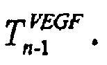

- IF VEGF n-1 > VEGF max THEN VEGF n-1 = VEGF max
- IF VEGF n-1 < VEGFen THEN VEGF n-1 = VEGFen
- The initial level VEGFss characterizes the amount of VEGF secreted when effective tissue vessel density is EVDss .
- In a similar way we obtain the dependence of PDGF (11).

- IF VEGF n-1 > VEGF max THEN VEGF n-1 = VEGF max
- IF VEGF n-1 < VEGFen THEN VEGF n-1 = VEGFen
- The characteristic time T14 and

above equations 
- In Eqn. (12, 14) Ang2 and Ang1 also depends on the numbers of endothelial cells in immature vessels, which is determined by the Eqs. (13), and the numbers of tissue cells


- The characteristic reaction time for Ang2 generation is T10 and T15, (12), for Ang1 generation it is T9 and T11 (14).

- The addition of free pericytes (block 1.17) at any given moment depends on the level of PDGF at the previous moment, the replication of free pericytes, and on the number of free pericytes released from mature vessels (15). Accordingly, these two processes have the coefficients


- The number of tissue cells (block 1.18) in the moment, n, depends on their number in the previous moment multiplied by a factor describing the process of cell proliferation and death, rn-1.

- rn-1 depends on the mitotic index M(I) (mitotic time being T (1), apoptotic index A (I) (apoptotic time beingT (2)), rate of tissue cell growth λ and the rate of the death of tumor cells, µ. The two terms in the equation below are involved in blocks 1.191 and 1.201 respectively. Clearly, they are also factors in blocks 1.192 and 1.202 as well as 1.193 and 1.203.

- The proliferation rate λ and the death rate µ are assumed to be standard sigmoids. Hence we obtain λ and µ as follows:


- Possible drug effects on the pro and anti angiogenesis process indicated in fig.2. Note that the blocks in Fig.2 are identical to those in Fig.1, except for the additional drug effects shown. The drug effects on the overall process are analyzed by setting the selected drug schedule (number of doses, the dose and the dosing interval). For example the analysis of anti-VEGF drug activity shows that a drug which inhibits VEGF has an optimal efficacy when given by certain treatment protocol. Increasing the administered dose above the optimum can bring about the undesired effect of tissue proliferation.as shown in Fig. 3. In addition, the technique enables one to predict the effects of various drug combination, for example as shown in Fig.4.
Claims (40)
- A computer-implemented method for determining an optimal treatment protocol for a disease related to angiogenesis, comprising:creating an angiogenesis model including pro-angiogenesis and anti-angiogenesis factors;incorporating effective vessel density (EVD) as a factor regulating switching on and switching off of at least one component in the angiogenesis model;selecting pro-angiogenesis and anti-angiogenesis factors, which can influence changes in state of a tissue;incorporating effects of drugs in the pro-angiogenesis and anti-angiogenesis factors;generating a plurality of treatment protocols in a protocol space; andselecting a best treatment protocol based on a pre-determined criteria,characterised in that the method further comprises incorporating effects of vasculature maturation and mature vessels destabilization.
- The method of claim 1, wherein the model comprises a tissue volume model, an immature vessel model and a mature vessel model.
- The method of claim 1, wherein steps to regulate dynamics which influences EVD are incorporated.
- The method of claim 1, wherein the model simultaneously accounts for tissue cell proliferation, tissue cell death, endothelial cell proliferation, endothelial cell death, immature vessel formation and immature vessel regression, immature vessel maturation and mature vessel destabilization.
- The method of claim 1, wherein the model incorporates temporal parameters that characterize response rate of at least one element associated with angiogenesis.
- The method of claim 3, wherein EVD is calculated by combining immature vessel density and mature vessel density.
- The method of claim 1, wherein parameters incorporated into the model comprises tissue volume, number of free endothelial cells, number of free pericytes, volume of mature vessels, volume of immature vessels and concentration of regulator factors.
- The method of claim 7, wherein the regulatory factors comprise vascular endothelial growth factor (VEGF), platelet derived growth factor (PDGF), angiopoietin 1 (Ang1) and angiopoietin 2 (Ang2).
- The method of claim 3, wherein EVD is a function of a duration of insufficient perfusion and vice versa.
- The method of claim 8 wherein the model incorporates threshold levels of regulatory factors and parameter ratios.
- The method of claim 10, wherein the threshold level is at least one of:a) VEGF concentration below which no endothelial cells proliferation takes place (A);b) minimum number of receptors for VEGF above which endothelial cells proliferation takes place (B);c) VEGF concentration below which endothelial cells, both in the free state as well as when incorporated into immature blood vessels, are subject to apoptosis VEGFthr;d) the minimal number of free pericytes which stimulates the onset of maturation of immature vessels (C);e) Ang 1/Ang 2 ratio below which mature vessels are destabilized, and above which maturation of immature vessels is enabled (K);f) EVD value that influences the rate of cell proliferation; andg) EVDss value for which the system is in steady state.
- The method of claim 11 wherein the tissue volume model calculates the tissue volume by a process comprising:i) comparing EVD against an - EVDss;ii) if EVD is equal to EVDss then using a programmed tissue cell proliferation and a programmed tissue cell death (apoptosis) to compute tissue vouhne;iii) if EVD > EVDss then using increased tissue proliferation and decreased tissue cell death to compute tissue volume; andiv) if EVD < EVDss then using decreased tissue proliferation and increased tissue cell death to compute tissue volume.
- The method of claim 12, wherein Ang1 and Ang2 induction are incorporated into steps ii, iii and iv prior to compute tissue volume.
- The method of claim 13 wherein effects of drugs affecting Ang1 and Ang2 are considered in computing tissue volume.
- The method of claim 11 wherein the immature vessel model calculates the immature vessels by a process comprising:i) comparing EVD against an EVDss;ii) if EVD is equal to EVDss then setting VEGF to a VEGFss and PDGF to a PDGFss;iii) if EVD > EVDss then using decreased VEGF and decreased PDGF; andiv) if EVD < EVDss then using increased VEGF and increased PDGF;v) comparing VEGF against A;vi) factoring endothelial cell proliferation if VEGF > A;vii) comparing VEGF against a VEGF threshold;viii) factoring free endothelial cell deaths if VEGF > VEGF threshold;ix) comparing VEGF receptor number against B;x) if VEGF receptor number is less than B in step ix then considering no angiogenisis prior to computing immature vessel regression;xi) if VEGF receptor number is not less than B in step ix then computing growth of immature vessels;xii) if VEGF < A then considering no angiogenesis and computing immature vessel regression;xiii) computing immature vessels based on growth immature vessels, immature vessel regression and mature vessel destabilization.
- The method of claim 15, wherein immature vessels computation considers no maturation if Ang2/Ang1 > K or if number of free pericytes < C.
- The method of claim 15, wherein mature vessel destabilization considers ang1/Tie2 interaction blocking.
- The method of claim 17, wherein no destabilization occurs if Ang2/Ang1 is not greater than K.
- The method of claim 15, wherein mature vessel model is computed using a procedure comprising:i) computing immature vessels;ii) determining if Ang1/Ang2 > K;iii) determining if number of free pericytes < C;iv) considering immature vessel maturation if both steps ii and iii are false; andv) factoring no destablization if step ii is false.
- The method of claim 15 wherein effects of a drug affecting EC proliferation are factored in computing immature vessels.
- The method of claim 15 wherein effects of a drug affecting VEGF receptors are factored in computing immature vessels.
- The method of claim 16 wherein effects of a drug affecting pericyte proliferation are factored in computing immature vessel computation.
- The method of claim 15 wherein effects of a drug affecting VEGF are factored in computing immature vessels.
- The method of claim 15 wherein effects of a drug affecting PDGF are factored in computing immature vessel computation.
- The method of claim 15 wherein effects of a drug affecting Ang1 are factored in computing immature vessels.
- The method of claim 15 wherein effects of a drug affecting Ang2 are factored in computing immature vessel computation.
- The method of claim 4, wherein model takes into account duration of a tissue cell proliferation, tissue cell death, endothelial cell proliferation, endothelial cell death, pericytes proliferation, immature vessel regression, immature vessel maturation and mature vessel destabilization.
- The method of claim 7, wherein model takes into account the duration of VEGF induction, PDGF induction, Ang1 and Ang2 induction by tissue cells and Ang1 and Ang2 induction by endothelial cells.
- A computer system for determining an optimal treatment protocol for a disease related to angiogenesis, comprising:an angiogenesis model including pro-angiogenesis and anti-angiogenesis factors;a treatment protocol space generator that generates a protocol space of possible treatments for the disease;a treatment selector that selects an optimal protocol,wherein effective vessel density (EVD) is a factor regulating switching on and switching off of at least one component in the angiogenesis model;
wherein the system is adapted to effect selection of pro-angiogenesis and anti-angiogenesis factors which can influence changes in state of a tissue and incorporating effects of drugs in the pro-angiogenesis and anti-angiogenesis factors,
characterised in that the model incorporates effects of vasculature maturation and mature vessels destabilization. - The computer system of claim 29, wherein the model comprises a tissue volume model, an immature vessel model and a mature vessel model.
- The computer system of claim 29, wherein steps to regulate dynamics which influences EVD are incorporated.
- The computer system of claim 29, wherein the model simultaneously accounts for tissue cell proliferation, tissue cell death, endothelial cell proliferation, endothelial cell death, immature vessel formation and immature vessel regression.
- The computer system of claim 29, wherein the model incorporates temporal parameters that characterize response rate of at least one element associated with angiogenesis.
- The computer system of claim 31, wherein EVD is calculated by combining immature vessel density and mature vessel density.
- The computer system of claim 29, wherein parameters incorporated into the mode comprises tissue volume, number of free endothelial cells, number of free periciytes, volume of mature vessels, volume of immature vessels and concentration of regulator factors.
- The computer system of claim 35, wherein the regulatory factors comprise vascular endothelial growth factor (VEGF), platelet derived growth factor (PDGF), angiopoietin 1 (Ang1) and angiopoietin 2 (Ang2).
- The computer system of claim 31, wherein EVD is a function of a duration of insufficient perfusion and vice versa.
- The computer system of claim 36 wherein the model incorporates threshold levels of regulatory factors and parameter ratios.
- The computer system of claim 38, wherein the threshold level is at least one ofa) VEGF concentration below which no endothelial cells proliferation takes place (A);b) minimum number of receptors for VEGF above which endothelial cells proliferation takes place (B);c) VEGF concentration below which endothelial cells, both in the free state as well as when incorporated into immature blood vessels, are subject to apoptosis VEGFthr;d) the minimal number of free pericytes which stimulates the onset of maturation of immature vessels (C);e) Ang 1/Ang 2 ratio below which mature vessels are destabilized, and above which maturation of immature vessels is enabled (K);f) EVD value that influences the rate of cell proliferation; andg) EVDss value for which the system is in steady state.
- A computer program product, including computer-readable media wherein when said computer program product is run on a computer the product causes the computer to execute a series of instructions to implement a procedure for determining an optimal treatment protocol for a disease related to angiogenesis, said procedure comprising:creating an angiogenesis model including pro-angiogenesis and anti-angiogenesis factors;incorporating effective vessel density (EVD) as a factor regulating switching on and switching off of at least one component in the angiogenesis model;selecting pro-angiogenesis and anti-angiogenesis factors, which can influence changes in state of a tissue;incorporating effects of drugs in the pro-angio genesis and anti-angiogenesis factors;generating a plurality of treatment protocols space; andselecting a best treatment protocol based on a pre-determined criteria, characterised in that said procedure further comprises incorporating effects of vasculature maturation and mature vessels destabilization.
Applications Claiming Priority (5)
| Application Number | Priority Date | Filing Date | Title |
|---|---|---|---|
| US33059201P | 2001-10-25 | 2001-10-25 | |
| US330592P | 2001-10-25 | ||
| US10/207,772 US7418374B2 (en) | 2001-10-25 | 2002-07-31 | Treatment protocol generation for diseases related to angiogenesis |
| US207772 | 2002-07-31 | ||
| PCT/IB2002/004725 WO2003036410A2 (en) | 2001-10-25 | 2002-10-24 | Treatment protocol generation for diseases related to angiogenesis |
Publications (3)
| Publication Number | Publication Date |
|---|---|
| EP1485818A2 EP1485818A2 (en) | 2004-12-15 |
| EP1485818A4 EP1485818A4 (en) | 2005-04-06 |
| EP1485818B1 true EP1485818B1 (en) | 2007-02-14 |
Family
ID=26902569
Family Applications (1)
| Application Number | Title | Priority Date | Filing Date |
|---|---|---|---|
| EP02801985A Expired - Lifetime EP1485818B1 (en) | 2001-10-25 | 2002-10-24 | Treatment protocol generation for diseases related to angiogenesis |
Country Status (7)
| Country | Link |
|---|---|
| US (2) | US7418374B2 (en) |
| EP (1) | EP1485818B1 (en) |
| AT (1) | ATE354132T1 (en) |
| AU (1) | AU2002363044A1 (en) |
| DE (1) | DE60218229T2 (en) |
| DK (1) | DK1485818T3 (en) |
| WO (1) | WO2003036410A2 (en) |
Families Citing this family (1)
| Publication number | Priority date | Publication date | Assignee | Title |
|---|---|---|---|---|
| US8150629B2 (en) * | 2005-11-10 | 2012-04-03 | In Silico Biosciences | Method and apparatus for computer modeling of the interaction between and among cortical and subcortical areas in the human brain for the purpose of predicting the effect of drugs in psychiatric and cognitive diseases |
Family Cites Families (1)
| Publication number | Priority date | Publication date | Assignee | Title |
|---|---|---|---|---|
| EP1068568A4 (en) * | 1998-04-03 | 2004-10-27 | Triangle Pharmaceuticals Inc | Systems, methods and computer program products for guiding the selection of therapeutic treatment regimens |
-
2002
- 2002-07-31 US US10/207,772 patent/US7418374B2/en not_active Expired - Fee Related
- 2002-10-24 AT AT02801985T patent/ATE354132T1/en not_active IP Right Cessation
- 2002-10-24 EP EP02801985A patent/EP1485818B1/en not_active Expired - Lifetime
- 2002-10-24 DE DE60218229T patent/DE60218229T2/en not_active Expired - Lifetime
- 2002-10-24 DK DK02801985T patent/DK1485818T3/en active
- 2002-10-24 AU AU2002363044A patent/AU2002363044A1/en not_active Abandoned
- 2002-10-24 WO PCT/IB2002/004725 patent/WO2003036410A2/en not_active Ceased
-
2008
- 2008-06-03 US US12/132,300 patent/US20080275684A1/en not_active Abandoned
Non-Patent Citations (5)
| Title |
|---|
| ARAKELYAN ET AL, EUR. J. CANCER, vol. 41, 2005, pages 159 - 167 * |
| GERBER ET AL, TCM, vol. 10, no. 5, 2000, pages 223 - 228 * |
| GILEAD ET AL, INT. J. CANCER, vol. 108, no. 4, November 2003 (2003-11-01), pages 524 - 531 * |
| JAIN R.K., NATURE MEDICINE, vol. 9, no. 6, June 2003 (2003-06-01), pages 685 - 693 * |
| Wikipedia online definition of angiogenesis. * |
Also Published As
| Publication number | Publication date |
|---|---|
| DK1485818T3 (en) | 2007-06-18 |
| US20080275684A1 (en) | 2008-11-06 |
| US20030088237A1 (en) | 2003-05-08 |
| WO2003036410A3 (en) | 2004-09-30 |
| DE60218229D1 (en) | 2007-03-29 |
| EP1485818A2 (en) | 2004-12-15 |
| US7418374B2 (en) | 2008-08-26 |
| WO2003036410A2 (en) | 2003-05-01 |
| EP1485818A4 (en) | 2005-04-06 |
| AU2002363044A1 (en) | 2003-05-06 |
| ATE354132T1 (en) | 2007-03-15 |
| DE60218229T2 (en) | 2007-10-31 |
Similar Documents
| Publication | Publication Date | Title |
|---|---|---|
| Arakelyan et al. | A computer algorithm describing the process of vessel formation and maturation, and its use for predicting the effects of anti-angiogenic and anti-maturation therapy on vascular tumor growth | |
| Deshpande et al. | Steady laminar flow through modelled vascular stenoses | |
| Tammela et al. | Angiopoietin-1 promotes lymphatic sprouting and hyperplasia | |
| Michiels | Endothelial cell functions | |
| Brown et al. | Abolished tubuloglomerular feedback and increased plasma renin in adenosine A1 receptor-deficient mice | |
| Charbel et al. | Ultrasonic perivascular flow probe: technique and application in neurosurgery | |
| Bassingthwaighte et al. | Flow estimation by indicator dilution (bolus injection) reduction of errors due to time-averaged sampling during unsteady flow | |
| Fraser et al. | Characterization of an abdominal aortic velocity waveform in patients with abdominal aortic aneurysm | |
| US7418374B2 (en) | Treatment protocol generation for diseases related to angiogenesis | |
| Grant et al. | Growth factor signaling pathways in vascular development and disease | |
| Hashimoto | Dynamic measurement of pressure and flow velocities in glass and silastic model berry aneurysms | |
| Ofjord et al. | Intrarenal flow of microspheres and red blood cells: skimming in slit and tube models | |
| Håggmark et al. | The continuous thermodilution method for measuring high blood flows | |
| Tuma et al. | Handbook of Microcirculation | |
| RU2159075C2 (en) | Method for determining cerebral blood supply disorders | |
| Overholser et al. | Pulmonary vascular resistance distribution and recruitment of microvascular surface area | |
| Hjortdal et al. | Velocity field studies at surgically imposed arterial stenoses on the abdominal aorta in pigs | |
| Miller et al. | Aldosterone secretion and plasma renin during renin infusion and acute salt depletion | |
| Dawson et al. | Effect of vasoconstriction on longitudinal distribution of pulmonary vascular pressure and volume | |
| Fukuyama et al. | Canine basilar arterial flow and effects of common carotid occlusion | |
| Kiser et al. | Measurements of velocity wave forms in the dog aorta | |
| CN207280526U (en) | A kind of ultrasonic flowmeter | |
| NL1011339C2 (en) | A method and apparatus for determining the blood flow of a patient's heart. | |
| Grudemo et al. | Studies of spontaneous fluctuations in congestion and nasal mucosal microcirculation and the effects of oxymetazoline using rhinostereometry and micromanipulator guided laser Doppler flowmetry | |
| Kovtanyuk et al. | Influence of the Endothelial Surface Layer on Blood Flow in Microvessels: Computer Modeling and Simulation. |
Legal Events
| Date | Code | Title | Description |
|---|---|---|---|
| PUAI | Public reference made under article 153(3) epc to a published international application that has entered the european phase |
Free format text: ORIGINAL CODE: 0009012 |
|
| 17P | Request for examination filed |
Effective date: 20040520 |
|
| AK | Designated contracting states |
Kind code of ref document: A2 Designated state(s): AT BE BG CH CY CZ DE DK EE ES FI FR GB GR IE IT LI LU MC NL PT SE SK TR |
|
| AX | Request for extension of the european patent |
Extension state: AL LT LV MK RO SI |
|
| A4 | Supplementary search report drawn up and despatched |
Effective date: 20050223 |
|
| RIC1 | Information provided on ipc code assigned before grant |
Ipc: 7G 06F 17/11 A Ipc: 7G 01N 33/74 B |
|
| GRAP | Despatch of communication of intention to grant a patent |
Free format text: ORIGINAL CODE: EPIDOSNIGR1 |
|
| GRAS | Grant fee paid |
Free format text: ORIGINAL CODE: EPIDOSNIGR3 |
|
| GRAA | (expected) grant |
Free format text: ORIGINAL CODE: 0009210 |
|
| AK | Designated contracting states |
Kind code of ref document: B1 Designated state(s): AT BE BG CH CY CZ DE DK EE ES FI FR GB GR IE IT LI LU MC NL PT SE SK TR |
|
| PG25 | Lapsed in a contracting state [announced via postgrant information from national office to epo] |
Ref country code: AT Free format text: LAPSE BECAUSE OF FAILURE TO SUBMIT A TRANSLATION OF THE DESCRIPTION OR TO PAY THE FEE WITHIN THE PRESCRIBED TIME-LIMIT Effective date: 20070214 Ref country code: BE Free format text: LAPSE BECAUSE OF FAILURE TO SUBMIT A TRANSLATION OF THE DESCRIPTION OR TO PAY THE FEE WITHIN THE PRESCRIBED TIME-LIMIT Effective date: 20070214 Ref country code: NL Free format text: LAPSE BECAUSE OF FAILURE TO SUBMIT A TRANSLATION OF THE DESCRIPTION OR TO PAY THE FEE WITHIN THE PRESCRIBED TIME-LIMIT Effective date: 20070214 Ref country code: FI Free format text: LAPSE BECAUSE OF FAILURE TO SUBMIT A TRANSLATION OF THE DESCRIPTION OR TO PAY THE FEE WITHIN THE PRESCRIBED TIME-LIMIT Effective date: 20070214 |
|
| REG | Reference to a national code |
Ref country code: GB Ref legal event code: FG4D |
|
| REG | Reference to a national code |
Ref country code: CH Ref legal event code: EP |
|
| REF | Corresponds to: |
Ref document number: 60218229 Country of ref document: DE Date of ref document: 20070329 Kind code of ref document: P |
|
| REG | Reference to a national code |
Ref country code: IE Ref legal event code: FG4D |
|
| PG25 | Lapsed in a contracting state [announced via postgrant information from national office to epo] |
Ref country code: SE Free format text: LAPSE BECAUSE OF FAILURE TO SUBMIT A TRANSLATION OF THE DESCRIPTION OR TO PAY THE FEE WITHIN THE PRESCRIBED TIME-LIMIT Effective date: 20070514 |
|
| PG25 | Lapsed in a contracting state [announced via postgrant information from national office to epo] |
Ref country code: BG Free format text: LAPSE BECAUSE OF THE APPLICANT RENOUNCES Effective date: 20070515 |
|
| RAP2 | Party data changed (patent owner data changed or rights of a patent transferred) |
Owner name: OPTIMATA LTD. |
|
| PG25 | Lapsed in a contracting state [announced via postgrant information from national office to epo] |
Ref country code: ES Free format text: LAPSE BECAUSE OF FAILURE TO SUBMIT A TRANSLATION OF THE DESCRIPTION OR TO PAY THE FEE WITHIN THE PRESCRIBED TIME-LIMIT Effective date: 20070525 |
|
| REG | Reference to a national code |
Ref country code: CH Ref legal event code: NV Representative=s name: FIAMMENGHI-FIAMMENGHI |
|
| REG | Reference to a national code |
Ref country code: DK Ref legal event code: T3 |
|
| PG25 | Lapsed in a contracting state [announced via postgrant information from national office to epo] |
Ref country code: PT Free format text: LAPSE BECAUSE OF FAILURE TO SUBMIT A TRANSLATION OF THE DESCRIPTION OR TO PAY THE FEE WITHIN THE PRESCRIBED TIME-LIMIT Effective date: 20070716 |
|
| NLT2 | Nl: modifications (of names), taken from the european patent patent bulletin |
Owner name: OPTIMATA LTD. Effective date: 20070523 |
|
| NLV1 | Nl: lapsed or annulled due to failure to fulfill the requirements of art. 29p and 29m of the patents act | ||
| ET | Fr: translation filed | ||
| PG25 | Lapsed in a contracting state [announced via postgrant information from national office to epo] |
Ref country code: SK Free format text: LAPSE BECAUSE OF FAILURE TO SUBMIT A TRANSLATION OF THE DESCRIPTION OR TO PAY THE FEE WITHIN THE PRESCRIBED TIME-LIMIT Effective date: 20070214 |
|
| PLBE | No opposition filed within time limit |
Free format text: ORIGINAL CODE: 0009261 |
|
| STAA | Information on the status of an ep patent application or granted ep patent |
Free format text: STATUS: NO OPPOSITION FILED WITHIN TIME LIMIT |
|
| PG25 | Lapsed in a contracting state [announced via postgrant information from national office to epo] |
Ref country code: CZ Free format text: LAPSE BECAUSE OF FAILURE TO SUBMIT A TRANSLATION OF THE DESCRIPTION OR TO PAY THE FEE WITHIN THE PRESCRIBED TIME-LIMIT Effective date: 20070214 |
|
| 26N | No opposition filed |
Effective date: 20071115 |
|
| PG25 | Lapsed in a contracting state [announced via postgrant information from national office to epo] |
Ref country code: GR Free format text: LAPSE BECAUSE OF FAILURE TO SUBMIT A TRANSLATION OF THE DESCRIPTION OR TO PAY THE FEE WITHIN THE PRESCRIBED TIME-LIMIT Effective date: 20070515 |
|
| PG25 | Lapsed in a contracting state [announced via postgrant information from national office to epo] |
Ref country code: MC Free format text: LAPSE BECAUSE OF NON-PAYMENT OF DUE FEES Effective date: 20071031 |
|
| PG25 | Lapsed in a contracting state [announced via postgrant information from national office to epo] |
Ref country code: EE Free format text: LAPSE BECAUSE OF FAILURE TO SUBMIT A TRANSLATION OF THE DESCRIPTION OR TO PAY THE FEE WITHIN THE PRESCRIBED TIME-LIMIT Effective date: 20070214 |
|
| PGFP | Annual fee paid to national office [announced via postgrant information from national office to epo] |
Ref country code: IE Payment date: 20081015 Year of fee payment: 7 |
|
| PG25 | Lapsed in a contracting state [announced via postgrant information from national office to epo] |
Ref country code: CY Free format text: LAPSE BECAUSE OF FAILURE TO SUBMIT A TRANSLATION OF THE DESCRIPTION OR TO PAY THE FEE WITHIN THE PRESCRIBED TIME-LIMIT Effective date: 20070214 |
|
| PG25 | Lapsed in a contracting state [announced via postgrant information from national office to epo] |
Ref country code: LU Free format text: LAPSE BECAUSE OF NON-PAYMENT OF DUE FEES Effective date: 20071024 |
|
| PG25 | Lapsed in a contracting state [announced via postgrant information from national office to epo] |
Ref country code: TR Free format text: LAPSE BECAUSE OF FAILURE TO SUBMIT A TRANSLATION OF THE DESCRIPTION OR TO PAY THE FEE WITHIN THE PRESCRIBED TIME-LIMIT Effective date: 20070214 |
|
| REG | Reference to a national code |
Ref country code: CH Ref legal event code: NV Representative=s name: BOVARD AG PATENTANWAELTE |
|
| PG25 | Lapsed in a contracting state [announced via postgrant information from national office to epo] |
Ref country code: IE Free format text: LAPSE BECAUSE OF NON-PAYMENT OF DUE FEES Effective date: 20091026 |
|
| PGFP | Annual fee paid to national office [announced via postgrant information from national office to epo] |
Ref country code: DK Payment date: 20101025 Year of fee payment: 9 |
|
| REG | Reference to a national code |
Ref country code: CH Ref legal event code: PFA Owner name: OPTIMATA LTD. Free format text: OPTIMATA LTD.#7, ABBA HILLEL ST.#RAMAT-GAN 52522 (IL) -TRANSFER TO- OPTIMATA LTD.#7, ABBA HILLEL ST.#RAMAT-GAN 52522 (IL) |
|
| REG | Reference to a national code |
Ref country code: DK Ref legal event code: EBP |
|
| PG25 | Lapsed in a contracting state [announced via postgrant information from national office to epo] |
Ref country code: DK Free format text: LAPSE BECAUSE OF NON-PAYMENT OF DUE FEES Effective date: 20111031 |
|
| PGFP | Annual fee paid to national office [announced via postgrant information from national office to epo] |
Ref country code: CH Payment date: 20141031 Year of fee payment: 13 Ref country code: DE Payment date: 20141111 Year of fee payment: 13 Ref country code: GB Payment date: 20141030 Year of fee payment: 13 Ref country code: FR Payment date: 20141031 Year of fee payment: 13 |
|
| PGFP | Annual fee paid to national office [announced via postgrant information from national office to epo] |
Ref country code: IT Payment date: 20141031 Year of fee payment: 13 |
|
| REG | Reference to a national code |
Ref country code: DE Ref legal event code: R119 Ref document number: 60218229 Country of ref document: DE |
|
| REG | Reference to a national code |
Ref country code: CH Ref legal event code: PL |
|
| GBPC | Gb: european patent ceased through non-payment of renewal fee |
Effective date: 20151024 |
|
| PG25 | Lapsed in a contracting state [announced via postgrant information from national office to epo] |
Ref country code: LI Free format text: LAPSE BECAUSE OF NON-PAYMENT OF DUE FEES Effective date: 20151031 Ref country code: DE Free format text: LAPSE BECAUSE OF NON-PAYMENT OF DUE FEES Effective date: 20160503 Ref country code: IT Free format text: LAPSE BECAUSE OF NON-PAYMENT OF DUE FEES Effective date: 20151024 Ref country code: CH Free format text: LAPSE BECAUSE OF NON-PAYMENT OF DUE FEES Effective date: 20151031 Ref country code: GB Free format text: LAPSE BECAUSE OF NON-PAYMENT OF DUE FEES Effective date: 20151024 |
|
| REG | Reference to a national code |
Ref country code: FR Ref legal event code: ST Effective date: 20160630 |
|
| PG25 | Lapsed in a contracting state [announced via postgrant information from national office to epo] |
Ref country code: FR Free format text: LAPSE BECAUSE OF NON-PAYMENT OF DUE FEES Effective date: 20151102 |