EP1485410B1 - Diagnostic and therapeutic use of human maguin proteins and nucleic acids for neurodegenerative diseases - Google Patents
Diagnostic and therapeutic use of human maguin proteins and nucleic acids for neurodegenerative diseases Download PDFInfo
- Publication number
- EP1485410B1 EP1485410B1 EP03744814A EP03744814A EP1485410B1 EP 1485410 B1 EP1485410 B1 EP 1485410B1 EP 03744814 A EP03744814 A EP 03744814A EP 03744814 A EP03744814 A EP 03744814A EP 1485410 B1 EP1485410 B1 EP 1485410B1
- Authority
- EP
- European Patent Office
- Prior art keywords
- maguin
- human
- disease
- alzheimer
- gene coding
- Prior art date
- Legal status (The legal status is an assumption and is not a legal conclusion. Google has not performed a legal analysis and makes no representation as to the accuracy of the status listed.)
- Expired - Lifetime
Links
- 241000282414 Homo sapiens Species 0.000 title claims abstract description 197
- 230000004770 neurodegeneration Effects 0.000 title abstract description 16
- 208000015122 neurodegenerative disease Diseases 0.000 title abstract description 14
- 230000001225 therapeutic effect Effects 0.000 title abstract description 7
- 150000007523 nucleic acids Chemical class 0.000 title description 16
- 108020004707 nucleic acids Proteins 0.000 title description 7
- 102000039446 nucleic acids Human genes 0.000 title description 7
- 101000749830 Rattus norvegicus Connector enhancer of kinase suppressor of ras 2 Proteins 0.000 title description 5
- 108090000623 proteins and genes Proteins 0.000 claims abstract description 153
- 208000024827 Alzheimer disease Diseases 0.000 claims abstract description 97
- 238000000034 method Methods 0.000 claims abstract description 54
- 208000037265 diseases, disorders, signs and symptoms Diseases 0.000 claims abstract description 34
- 201000010099 disease Diseases 0.000 claims abstract description 33
- 210000004556 brain Anatomy 0.000 claims abstract description 32
- 238000012216 screening Methods 0.000 claims abstract description 9
- 102000004169 proteins and genes Human genes 0.000 claims description 45
- 238000013519 translation Methods 0.000 claims description 44
- 238000013518 transcription Methods 0.000 claims description 36
- 230000035897 transcription Effects 0.000 claims description 36
- 239000000126 substance Substances 0.000 claims description 19
- 238000012360 testing method Methods 0.000 claims description 16
- 150000001875 compounds Chemical class 0.000 claims description 13
- 230000003862 health status Effects 0.000 claims description 12
- 230000001575 pathological effect Effects 0.000 claims description 9
- 230000004075 alteration Effects 0.000 claims description 8
- 238000003745 diagnosis Methods 0.000 claims description 8
- 239000003153 chemical reaction reagent Substances 0.000 claims description 7
- 239000003795 chemical substances by application Substances 0.000 claims description 7
- 239000003814 drug Substances 0.000 claims description 7
- 238000012544 monitoring process Methods 0.000 claims description 7
- 238000010186 staining Methods 0.000 claims description 7
- 238000000338 in vitro Methods 0.000 claims description 5
- 208000031124 Dementia Alzheimer type Diseases 0.000 claims description 4
- 238000011161 development Methods 0.000 claims description 4
- 238000012760 immunocytochemical staining Methods 0.000 claims description 3
- 101100114006 Rattus norvegicus Cnksr2 gene Proteins 0.000 abstract description 38
- 230000014509 gene expression Effects 0.000 abstract description 27
- 230000001965 increasing effect Effects 0.000 abstract description 7
- 102000010660 flotillin Human genes 0.000 abstract 2
- 108060000864 flotillin Proteins 0.000 abstract 2
- 102000018697 Membrane Proteins Human genes 0.000 abstract 1
- 108010052285 Membrane Proteins Proteins 0.000 abstract 1
- 210000004323 caveolae Anatomy 0.000 abstract 1
- 230000000069 prophylactic effect Effects 0.000 abstract 1
- 239000000047 product Substances 0.000 description 71
- 235000018102 proteins Nutrition 0.000 description 42
- 239000013615 primer Substances 0.000 description 40
- 230000000694 effects Effects 0.000 description 34
- 210000004027 cell Anatomy 0.000 description 31
- 108020004414 DNA Proteins 0.000 description 29
- 239000012634 fragment Substances 0.000 description 27
- 210000005153 frontal cortex Anatomy 0.000 description 27
- 239000002299 complementary DNA Substances 0.000 description 25
- 239000000523 sample Substances 0.000 description 25
- 238000003752 polymerase chain reaction Methods 0.000 description 23
- 235000001014 amino acid Nutrition 0.000 description 20
- 239000002773 nucleotide Substances 0.000 description 20
- 125000003729 nucleotide group Chemical group 0.000 description 20
- 108090000765 processed proteins & peptides Proteins 0.000 description 20
- 230000002123 temporal effect Effects 0.000 description 20
- 229940024606 amino acid Drugs 0.000 description 18
- 150000001413 amino acids Chemical class 0.000 description 17
- 210000001320 hippocampus Anatomy 0.000 description 17
- 210000002569 neuron Anatomy 0.000 description 16
- 102000004196 processed proteins & peptides Human genes 0.000 description 15
- 108091032973 (ribonucleotides)n+m Proteins 0.000 description 14
- 102100040283 Peptidyl-prolyl cis-trans isomerase B Human genes 0.000 description 13
- 108010048032 cyclophilin B Proteins 0.000 description 13
- 108010044156 peptidyl-prolyl cis-trans isomerase b Proteins 0.000 description 13
- 229920001184 polypeptide Polymers 0.000 description 13
- LFQSCWFLJHTTHZ-UHFFFAOYSA-N Ethanol Chemical compound CCO LFQSCWFLJHTTHZ-UHFFFAOYSA-N 0.000 description 12
- 125000000539 amino acid group Chemical group 0.000 description 12
- 241001465754 Metazoa Species 0.000 description 11
- 125000003275 alpha amino acid group Chemical group 0.000 description 10
- 238000004458 analytical method Methods 0.000 description 10
- 239000000499 gel Substances 0.000 description 9
- 239000012528 membrane Substances 0.000 description 9
- 230000001537 neural effect Effects 0.000 description 9
- 108700019745 Disks Large Homolog 4 Proteins 0.000 description 8
- 102000047174 Disks Large Homolog 4 Human genes 0.000 description 8
- TWRXJAOTZQYOKJ-UHFFFAOYSA-L Magnesium chloride Chemical compound [Mg+2].[Cl-].[Cl-] TWRXJAOTZQYOKJ-UHFFFAOYSA-L 0.000 description 8
- 108091034117 Oligonucleotide Proteins 0.000 description 8
- 102000000470 PDZ domains Human genes 0.000 description 8
- 108050008994 PDZ domains Proteins 0.000 description 8
- 230000027455 binding Effects 0.000 description 8
- 210000005013 brain tissue Anatomy 0.000 description 8
- 238000005516 engineering process Methods 0.000 description 8
- 239000000203 mixture Substances 0.000 description 8
- 210000003538 post-synaptic density Anatomy 0.000 description 8
- 108010092804 postsynaptic density proteins Proteins 0.000 description 8
- 210000000225 synapse Anatomy 0.000 description 8
- 210000001519 tissue Anatomy 0.000 description 8
- 230000003321 amplification Effects 0.000 description 7
- 238000001514 detection method Methods 0.000 description 7
- 230000002163 immunogen Effects 0.000 description 7
- 238000003199 nucleic acid amplification method Methods 0.000 description 7
- 230000008569 process Effects 0.000 description 7
- 108091026890 Coding region Proteins 0.000 description 6
- 102100031181 Glyceraldehyde-3-phosphate dehydrogenase Human genes 0.000 description 6
- 102000004282 Ribosomal protein S9 Human genes 0.000 description 6
- 108090000878 Ribosomal protein S9 Proteins 0.000 description 6
- 239000000539 dimer Substances 0.000 description 6
- 108020004445 glyceraldehyde-3-phosphate dehydrogenase Proteins 0.000 description 6
- 238000011880 melting curve analysis Methods 0.000 description 6
- 238000002493 microarray Methods 0.000 description 6
- 210000002682 neurofibrillary tangle Anatomy 0.000 description 6
- 230000001242 postsynaptic effect Effects 0.000 description 6
- 102000007469 Actins Human genes 0.000 description 5
- 108010085238 Actins Proteins 0.000 description 5
- 108020004635 Complementary DNA Proteins 0.000 description 5
- 101000578934 Rattus norvegicus Membrane-associated guanylate kinase, WW and PDZ domain-containing protein 2 Proteins 0.000 description 5
- 102000007238 Transferrin Receptors Human genes 0.000 description 5
- 108010033576 Transferrin Receptors Proteins 0.000 description 5
- JLCPHMBAVCMARE-UHFFFAOYSA-N [3-[[3-[[3-[[3-[[3-[[3-[[3-[[3-[[3-[[3-[[3-[[5-(2-amino-6-oxo-1H-purin-9-yl)-3-[[3-[[3-[[3-[[3-[[3-[[5-(2-amino-6-oxo-1H-purin-9-yl)-3-[[5-(2-amino-6-oxo-1H-purin-9-yl)-3-hydroxyoxolan-2-yl]methoxy-hydroxyphosphoryl]oxyoxolan-2-yl]methoxy-hydroxyphosphoryl]oxy-5-(5-methyl-2,4-dioxopyrimidin-1-yl)oxolan-2-yl]methoxy-hydroxyphosphoryl]oxy-5-(6-aminopurin-9-yl)oxolan-2-yl]methoxy-hydroxyphosphoryl]oxy-5-(6-aminopurin-9-yl)oxolan-2-yl]methoxy-hydroxyphosphoryl]oxy-5-(6-aminopurin-9-yl)oxolan-2-yl]methoxy-hydroxyphosphoryl]oxy-5-(6-aminopurin-9-yl)oxolan-2-yl]methoxy-hydroxyphosphoryl]oxyoxolan-2-yl]methoxy-hydroxyphosphoryl]oxy-5-(5-methyl-2,4-dioxopyrimidin-1-yl)oxolan-2-yl]methoxy-hydroxyphosphoryl]oxy-5-(4-amino-2-oxopyrimidin-1-yl)oxolan-2-yl]methoxy-hydroxyphosphoryl]oxy-5-(5-methyl-2,4-dioxopyrimidin-1-yl)oxolan-2-yl]methoxy-hydroxyphosphoryl]oxy-5-(5-methyl-2,4-dioxopyrimidin-1-yl)oxolan-2-yl]methoxy-hydroxyphosphoryl]oxy-5-(6-aminopurin-9-yl)oxolan-2-yl]methoxy-hydroxyphosphoryl]oxy-5-(6-aminopurin-9-yl)oxolan-2-yl]methoxy-hydroxyphosphoryl]oxy-5-(4-amino-2-oxopyrimidin-1-yl)oxolan-2-yl]methoxy-hydroxyphosphoryl]oxy-5-(4-amino-2-oxopyrimidin-1-yl)oxolan-2-yl]methoxy-hydroxyphosphoryl]oxy-5-(4-amino-2-oxopyrimidin-1-yl)oxolan-2-yl]methoxy-hydroxyphosphoryl]oxy-5-(6-aminopurin-9-yl)oxolan-2-yl]methoxy-hydroxyphosphoryl]oxy-5-(4-amino-2-oxopyrimidin-1-yl)oxolan-2-yl]methyl [5-(6-aminopurin-9-yl)-2-(hydroxymethyl)oxolan-3-yl] hydrogen phosphate Polymers Cc1cn(C2CC(OP(O)(=O)OCC3OC(CC3OP(O)(=O)OCC3OC(CC3O)n3cnc4c3nc(N)[nH]c4=O)n3cnc4c3nc(N)[nH]c4=O)C(COP(O)(=O)OC3CC(OC3COP(O)(=O)OC3CC(OC3COP(O)(=O)OC3CC(OC3COP(O)(=O)OC3CC(OC3COP(O)(=O)OC3CC(OC3COP(O)(=O)OC3CC(OC3COP(O)(=O)OC3CC(OC3COP(O)(=O)OC3CC(OC3COP(O)(=O)OC3CC(OC3COP(O)(=O)OC3CC(OC3COP(O)(=O)OC3CC(OC3COP(O)(=O)OC3CC(OC3COP(O)(=O)OC3CC(OC3COP(O)(=O)OC3CC(OC3COP(O)(=O)OC3CC(OC3COP(O)(=O)OC3CC(OC3COP(O)(=O)OC3CC(OC3CO)n3cnc4c(N)ncnc34)n3ccc(N)nc3=O)n3cnc4c(N)ncnc34)n3ccc(N)nc3=O)n3ccc(N)nc3=O)n3ccc(N)nc3=O)n3cnc4c(N)ncnc34)n3cnc4c(N)ncnc34)n3cc(C)c(=O)[nH]c3=O)n3cc(C)c(=O)[nH]c3=O)n3ccc(N)nc3=O)n3cc(C)c(=O)[nH]c3=O)n3cnc4c3nc(N)[nH]c4=O)n3cnc4c(N)ncnc34)n3cnc4c(N)ncnc34)n3cnc4c(N)ncnc34)n3cnc4c(N)ncnc34)O2)c(=O)[nH]c1=O JLCPHMBAVCMARE-UHFFFAOYSA-N 0.000 description 5
- 239000011543 agarose gel Substances 0.000 description 5
- 238000003556 assay Methods 0.000 description 5
- 230000015572 biosynthetic process Effects 0.000 description 5
- 230000006870 function Effects 0.000 description 5
- 238000003018 immunoassay Methods 0.000 description 5
- 238000011002 quantification Methods 0.000 description 5
- XLYOFNOQVPJJNP-UHFFFAOYSA-N water Chemical compound O XLYOFNOQVPJJNP-UHFFFAOYSA-N 0.000 description 5
- 208000037259 Amyloid Plaque Diseases 0.000 description 4
- 101710137189 Amyloid-beta A4 protein Proteins 0.000 description 4
- 102100022704 Amyloid-beta precursor protein Human genes 0.000 description 4
- 101710151993 Amyloid-beta precursor protein Proteins 0.000 description 4
- CKLJMWTZIZZHCS-REOHCLBHSA-N L-aspartic acid Chemical compound OC(=O)[C@@H](N)CC(O)=O CKLJMWTZIZZHCS-REOHCLBHSA-N 0.000 description 4
- WHUUTDBJXJRKMK-VKHMYHEASA-N L-glutamic acid Chemical compound OC(=O)[C@@H](N)CCC(O)=O WHUUTDBJXJRKMK-VKHMYHEASA-N 0.000 description 4
- KZSNJWFQEVHDMF-BYPYZUCNSA-N L-valine Chemical compound CC(C)[C@H](N)C(O)=O KZSNJWFQEVHDMF-BYPYZUCNSA-N 0.000 description 4
- 108050001185 Pleckstrin homology domains Proteins 0.000 description 4
- 102000010995 Pleckstrin homology domains Human genes 0.000 description 4
- KZSNJWFQEVHDMF-UHFFFAOYSA-N Valine Natural products CC(C)C(N)C(O)=O KZSNJWFQEVHDMF-UHFFFAOYSA-N 0.000 description 4
- DZHSAHHDTRWUTF-SIQRNXPUSA-N amyloid-beta polypeptide 42 Chemical compound C([C@@H](C(=O)N[C@@H](C)C(=O)N[C@@H](CCC(O)=O)C(=O)N[C@@H](CC(O)=O)C(=O)N[C@H](C(=O)NCC(=O)N[C@@H](CO)C(=O)N[C@@H](CC(N)=O)C(=O)N[C@@H](CCCCN)C(=O)NCC(=O)N[C@@H](C)C(=O)N[C@H](C(=O)N[C@@H]([C@@H](C)CC)C(=O)NCC(=O)N[C@@H](CC(C)C)C(=O)N[C@@H](CCSC)C(=O)N[C@@H](C(C)C)C(=O)NCC(=O)NCC(=O)N[C@@H](C(C)C)C(=O)N[C@@H](C(C)C)C(=O)N[C@@H]([C@@H](C)CC)C(=O)N[C@@H](C)C(O)=O)[C@@H](C)CC)C(C)C)NC(=O)[C@H](CC=1C=CC=CC=1)NC(=O)[C@@H](NC(=O)[C@H](CC(C)C)NC(=O)[C@H](CCCCN)NC(=O)[C@H](CCC(N)=O)NC(=O)[C@H](CC=1N=CNC=1)NC(=O)[C@H](CC=1N=CNC=1)NC(=O)[C@@H](NC(=O)[C@H](CCC(O)=O)NC(=O)[C@H](CC=1C=CC(O)=CC=1)NC(=O)CNC(=O)[C@H](CO)NC(=O)[C@H](CC(O)=O)NC(=O)[C@H](CC=1N=CNC=1)NC(=O)[C@H](CCCNC(N)=N)NC(=O)[C@H](CC=1C=CC=CC=1)NC(=O)[C@H](CCC(O)=O)NC(=O)[C@H](C)NC(=O)[C@@H](N)CC(O)=O)C(C)C)C(C)C)C1=CC=CC=C1 DZHSAHHDTRWUTF-SIQRNXPUSA-N 0.000 description 4
- 238000013459 approach Methods 0.000 description 4
- 229940009098 aspartate Drugs 0.000 description 4
- 210000001124 body fluid Anatomy 0.000 description 4
- 239000010839 body fluid Substances 0.000 description 4
- 239000000872 buffer Substances 0.000 description 4
- 238000002474 experimental method Methods 0.000 description 4
- 229930195712 glutamate Natural products 0.000 description 4
- 229910001629 magnesium chloride Inorganic materials 0.000 description 4
- 210000001161 mammalian embryo Anatomy 0.000 description 4
- 238000004519 manufacturing process Methods 0.000 description 4
- 230000008506 pathogenesis Effects 0.000 description 4
- 102000005962 receptors Human genes 0.000 description 4
- 108020003175 receptors Proteins 0.000 description 4
- 230000001105 regulatory effect Effects 0.000 description 4
- 238000003757 reverse transcription PCR Methods 0.000 description 4
- 238000007423 screening assay Methods 0.000 description 4
- 230000000946 synaptic effect Effects 0.000 description 4
- 239000004474 valine Substances 0.000 description 4
- CSCPPACGZOOCGX-UHFFFAOYSA-N Acetone Chemical compound CC(C)=O CSCPPACGZOOCGX-UHFFFAOYSA-N 0.000 description 3
- 108700028369 Alleles Proteins 0.000 description 3
- 239000004475 Arginine Substances 0.000 description 3
- DCXYFEDJOCDNAF-UHFFFAOYSA-N Asparagine Natural products OC(=O)C(N)CC(N)=O DCXYFEDJOCDNAF-UHFFFAOYSA-N 0.000 description 3
- 238000000018 DNA microarray Methods 0.000 description 3
- 239000003155 DNA primer Substances 0.000 description 3
- IAZDPXIOMUYVGZ-UHFFFAOYSA-N Dimethylsulphoxide Chemical compound CS(C)=O IAZDPXIOMUYVGZ-UHFFFAOYSA-N 0.000 description 3
- 238000002965 ELISA Methods 0.000 description 3
- PEDCQBHIVMGVHV-UHFFFAOYSA-N Glycerine Chemical compound OCC(O)CO PEDCQBHIVMGVHV-UHFFFAOYSA-N 0.000 description 3
- 108020004202 Guanylate Kinase Proteins 0.000 description 3
- DCXYFEDJOCDNAF-REOHCLBHSA-N L-asparagine Chemical compound OC(=O)[C@@H](N)CC(N)=O DCXYFEDJOCDNAF-REOHCLBHSA-N 0.000 description 3
- ZDXPYRJPNDTMRX-VKHMYHEASA-N L-glutamine Chemical compound OC(=O)[C@@H](N)CCC(N)=O ZDXPYRJPNDTMRX-VKHMYHEASA-N 0.000 description 3
- AGPKZVBTJJNPAG-WHFBIAKZSA-N L-isoleucine Chemical compound CC[C@H](C)[C@H](N)C(O)=O AGPKZVBTJJNPAG-WHFBIAKZSA-N 0.000 description 3
- ROHFNLRQFUQHCH-YFKPBYRVSA-N L-leucine Chemical compound CC(C)C[C@H](N)C(O)=O ROHFNLRQFUQHCH-YFKPBYRVSA-N 0.000 description 3
- ROHFNLRQFUQHCH-UHFFFAOYSA-N Leucine Natural products CC(C)CC(N)C(O)=O ROHFNLRQFUQHCH-UHFFFAOYSA-N 0.000 description 3
- KDXKERNSBIXSRK-UHFFFAOYSA-N Lysine Natural products NCCCCC(N)C(O)=O KDXKERNSBIXSRK-UHFFFAOYSA-N 0.000 description 3
- 239000004472 Lysine Substances 0.000 description 3
- 108091028043 Nucleic acid sequence Proteins 0.000 description 3
- 108010029485 Protein Isoforms Proteins 0.000 description 3
- 102000001708 Protein Isoforms Human genes 0.000 description 3
- 238000010240 RT-PCR analysis Methods 0.000 description 3
- 241000700159 Rattus Species 0.000 description 3
- 238000010171 animal model Methods 0.000 description 3
- ODKSFYDXXFIFQN-UHFFFAOYSA-N arginine Natural products OC(=O)C(N)CCCNC(N)=N ODKSFYDXXFIFQN-UHFFFAOYSA-N 0.000 description 3
- 229960001230 asparagine Drugs 0.000 description 3
- 235000009582 asparagine Nutrition 0.000 description 3
- 230000004071 biological effect Effects 0.000 description 3
- 210000000170 cell membrane Anatomy 0.000 description 3
- 238000006243 chemical reaction Methods 0.000 description 3
- 210000000349 chromosome Anatomy 0.000 description 3
- 238000003776 cleavage reaction Methods 0.000 description 3
- 238000010367 cloning Methods 0.000 description 3
- 238000001816 cooling Methods 0.000 description 3
- 230000018109 developmental process Effects 0.000 description 3
- 239000003623 enhancer Substances 0.000 description 3
- ZDXPYRJPNDTMRX-UHFFFAOYSA-N glutamine Natural products OC(=O)C(N)CCC(N)=O ZDXPYRJPNDTMRX-UHFFFAOYSA-N 0.000 description 3
- 102000006638 guanylate kinase Human genes 0.000 description 3
- 238000009396 hybridization Methods 0.000 description 3
- 229960000310 isoleucine Drugs 0.000 description 3
- AGPKZVBTJJNPAG-UHFFFAOYSA-N isoleucine Natural products CCC(C)C(N)C(O)=O AGPKZVBTJJNPAG-UHFFFAOYSA-N 0.000 description 3
- 239000000463 material Substances 0.000 description 3
- 238000005259 measurement Methods 0.000 description 3
- 210000002241 neurite Anatomy 0.000 description 3
- 239000002858 neurotransmitter agent Substances 0.000 description 3
- 238000004393 prognosis Methods 0.000 description 3
- 238000011160 research Methods 0.000 description 3
- 238000010839 reverse transcription Methods 0.000 description 3
- 230000007017 scission Effects 0.000 description 3
- 238000000926 separation method Methods 0.000 description 3
- 238000012163 sequencing technique Methods 0.000 description 3
- 230000019491 signal transduction Effects 0.000 description 3
- 208000024891 symptom Diseases 0.000 description 3
- MTCFGRXMJLQNBG-REOHCLBHSA-N (2S)-2-Amino-3-hydroxypropansäure Chemical compound OC[C@H](N)C(O)=O MTCFGRXMJLQNBG-REOHCLBHSA-N 0.000 description 2
- FWBHETKCLVMNFS-UHFFFAOYSA-N 4',6-Diamino-2-phenylindol Chemical compound C1=CC(C(=N)N)=CC=C1C1=CC2=CC=C(C(N)=N)C=C2N1 FWBHETKCLVMNFS-UHFFFAOYSA-N 0.000 description 2
- 102000002659 Amyloid Precursor Protein Secretases Human genes 0.000 description 2
- 108010043324 Amyloid Precursor Protein Secretases Proteins 0.000 description 2
- 102000013455 Amyloid beta-Peptides Human genes 0.000 description 2
- 108010090849 Amyloid beta-Peptides Proteins 0.000 description 2
- 101710095339 Apolipoprotein E Proteins 0.000 description 2
- 241000283707 Capra Species 0.000 description 2
- 206010012289 Dementia Diseases 0.000 description 2
- 108700000854 Drosophila CNK Proteins 0.000 description 2
- 102000004190 Enzymes Human genes 0.000 description 2
- 108090000790 Enzymes Proteins 0.000 description 2
- 201000011240 Frontotemporal dementia Diseases 0.000 description 2
- DHMQDGOQFOQNFH-UHFFFAOYSA-N Glycine Chemical compound NCC(O)=O DHMQDGOQFOQNFH-UHFFFAOYSA-N 0.000 description 2
- 108090000862 Ion Channels Proteins 0.000 description 2
- 102000004310 Ion Channels Human genes 0.000 description 2
- QNAYBMKLOCPYGJ-REOHCLBHSA-N L-alanine Chemical compound C[C@H](N)C(O)=O QNAYBMKLOCPYGJ-REOHCLBHSA-N 0.000 description 2
- AYFVYJQAPQTCCC-GBXIJSLDSA-N L-threonine Chemical compound C[C@@H](O)[C@H](N)C(O)=O AYFVYJQAPQTCCC-GBXIJSLDSA-N 0.000 description 2
- 102000043136 MAP kinase family Human genes 0.000 description 2
- 108091054455 MAP kinase family Proteins 0.000 description 2
- 101000805948 Mus musculus Harmonin Proteins 0.000 description 2
- 102000004868 N-Methyl-D-Aspartate Receptors Human genes 0.000 description 2
- 108090001041 N-Methyl-D-Aspartate Receptors Proteins 0.000 description 2
- HOKKHZGPKSLGJE-GSVOUGTGSA-N N-Methyl-D-aspartic acid Chemical compound CN[C@@H](C(O)=O)CC(O)=O HOKKHZGPKSLGJE-GSVOUGTGSA-N 0.000 description 2
- 102000004108 Neurotransmitter Receptors Human genes 0.000 description 2
- 108090000590 Neurotransmitter Receptors Proteins 0.000 description 2
- 238000000636 Northern blotting Methods 0.000 description 2
- 108700026244 Open Reading Frames Proteins 0.000 description 2
- 241000283973 Oryctolagus cuniculus Species 0.000 description 2
- 238000010222 PCR analysis Methods 0.000 description 2
- 108091000080 Phosphotransferase Proteins 0.000 description 2
- 239000013614 RNA sample Substances 0.000 description 2
- CGNLCCVKSWNSDG-UHFFFAOYSA-N SYBR Green I Chemical compound CN(C)CCCN(CCC)C1=CC(C=C2N(C3=CC=CC=C3S2)C)=C2C=CC=CC2=[N+]1C1=CC=CC=C1 CGNLCCVKSWNSDG-UHFFFAOYSA-N 0.000 description 2
- MTCFGRXMJLQNBG-UHFFFAOYSA-N Serine Natural products OCC(N)C(O)=O MTCFGRXMJLQNBG-UHFFFAOYSA-N 0.000 description 2
- AYFVYJQAPQTCCC-UHFFFAOYSA-N Threonine Natural products CC(O)C(N)C(O)=O AYFVYJQAPQTCCC-UHFFFAOYSA-N 0.000 description 2
- 239000004473 Threonine Substances 0.000 description 2
- 229920004890 Triton X-100 Polymers 0.000 description 2
- 239000013504 Triton X-100 Substances 0.000 description 2
- XSQUKJJJFZCRTK-UHFFFAOYSA-N Urea Chemical compound NC(N)=O XSQUKJJJFZCRTK-UHFFFAOYSA-N 0.000 description 2
- 230000002159 abnormal effect Effects 0.000 description 2
- 239000000556 agonist Substances 0.000 description 2
- 235000004279 alanine Nutrition 0.000 description 2
- 230000000890 antigenic effect Effects 0.000 description 2
- 210000003050 axon Anatomy 0.000 description 2
- 230000033228 biological regulation Effects 0.000 description 2
- 230000000903 blocking effect Effects 0.000 description 2
- 238000004364 calculation method Methods 0.000 description 2
- 125000003178 carboxy group Chemical group [H]OC(*)=O 0.000 description 2
- 210000005056 cell body Anatomy 0.000 description 2
- 230000001413 cellular effect Effects 0.000 description 2
- 210000001638 cerebellum Anatomy 0.000 description 2
- 210000003710 cerebral cortex Anatomy 0.000 description 2
- 230000008859 change Effects 0.000 description 2
- HVYWMOMLDIMFJA-DPAQBDIFSA-N cholesterol Chemical compound C1C=C2C[C@@H](O)CC[C@]2(C)[C@@H]2[C@@H]1[C@@H]1CC[C@H]([C@H](C)CCCC(C)C)[C@@]1(C)CC2 HVYWMOMLDIMFJA-DPAQBDIFSA-N 0.000 description 2
- 208000010877 cognitive disease Diseases 0.000 description 2
- 230000000295 complement effect Effects 0.000 description 2
- 238000012790 confirmation Methods 0.000 description 2
- 238000010276 construction Methods 0.000 description 2
- 230000003436 cytoskeletal effect Effects 0.000 description 2
- NHVNXKFIZYSCEB-XLPZGREQSA-N dTTP Chemical compound O=C1NC(=O)C(C)=CN1[C@@H]1O[C@H](COP(O)(=O)OP(O)(=O)OP(O)(O)=O)[C@@H](O)C1 NHVNXKFIZYSCEB-XLPZGREQSA-N 0.000 description 2
- 230000003412 degenerative effect Effects 0.000 description 2
- 238000002405 diagnostic procedure Methods 0.000 description 2
- 238000009826 distribution Methods 0.000 description 2
- 230000003828 downregulation Effects 0.000 description 2
- 239000012636 effector Substances 0.000 description 2
- 238000001962 electrophoresis Methods 0.000 description 2
- 230000002255 enzymatic effect Effects 0.000 description 2
- 230000002068 genetic effect Effects 0.000 description 2
- 230000013595 glycosylation Effects 0.000 description 2
- 238000006206 glycosylation reaction Methods 0.000 description 2
- 238000010438 heat treatment Methods 0.000 description 2
- 230000001939 inductive effect Effects 0.000 description 2
- 230000003993 interaction Effects 0.000 description 2
- 108020004999 messenger RNA Proteins 0.000 description 2
- 230000004048 modification Effects 0.000 description 2
- 238000012986 modification Methods 0.000 description 2
- 230000002981 neuropathic effect Effects 0.000 description 2
- 210000004940 nucleus Anatomy 0.000 description 2
- 239000002751 oligonucleotide probe Substances 0.000 description 2
- 230000003287 optical effect Effects 0.000 description 2
- 230000037361 pathway Effects 0.000 description 2
- 102000020233 phosphotransferase Human genes 0.000 description 2
- 229920002401 polyacrylamide Polymers 0.000 description 2
- 102000054765 polymorphisms of proteins Human genes 0.000 description 2
- 108091033319 polynucleotide Proteins 0.000 description 2
- 102000040430 polynucleotide Human genes 0.000 description 2
- 239000002157 polynucleotide Substances 0.000 description 2
- 238000002360 preparation method Methods 0.000 description 2
- 230000003518 presynaptic effect Effects 0.000 description 2
- 238000012545 processing Methods 0.000 description 2
- 210000002966 serum Anatomy 0.000 description 2
- 108091006024 signal transducing proteins Proteins 0.000 description 2
- 102000034285 signal transducing proteins Human genes 0.000 description 2
- 210000000130 stem cell Anatomy 0.000 description 2
- 238000006467 substitution reaction Methods 0.000 description 2
- 230000008685 targeting Effects 0.000 description 2
- 210000003478 temporal lobe Anatomy 0.000 description 2
- 238000002560 therapeutic procedure Methods 0.000 description 2
- 238000005382 thermal cycling Methods 0.000 description 2
- 238000012795 verification Methods 0.000 description 2
- 238000001262 western blot Methods 0.000 description 2
- 108020005345 3' Untranslated Regions Proteins 0.000 description 1
- 208000006888 Agnosia Diseases 0.000 description 1
- 241001047040 Agnosia Species 0.000 description 1
- 102100022524 Alpha-1-antichymotrypsin Human genes 0.000 description 1
- 102100029470 Apolipoprotein E Human genes 0.000 description 1
- 102000004506 Blood Proteins Human genes 0.000 description 1
- 108010017384 Blood Proteins Proteins 0.000 description 1
- 208000014644 Brain disease Diseases 0.000 description 1
- 102100032220 Calcium and integrin-binding family member 2 Human genes 0.000 description 1
- CURLTUGMZLYLDI-UHFFFAOYSA-N Carbon dioxide Chemical compound O=C=O CURLTUGMZLYLDI-UHFFFAOYSA-N 0.000 description 1
- 108020004705 Codon Proteins 0.000 description 1
- 101710181291 Connector enhancer of kinase suppressor of ras Proteins 0.000 description 1
- 208000011990 Corticobasal Degeneration Diseases 0.000 description 1
- 102000010831 Cytoskeletal Proteins Human genes 0.000 description 1
- 108010037414 Cytoskeletal Proteins Proteins 0.000 description 1
- 230000004568 DNA-binding Effects 0.000 description 1
- 108010014303 DNA-directed DNA polymerase Proteins 0.000 description 1
- 102000016928 DNA-directed DNA polymerase Human genes 0.000 description 1
- AHCYMLUZIRLXAA-SHYZEUOFSA-N Deoxyuridine 5'-triphosphate Chemical compound O1[C@H](COP(O)(=O)OP(O)(=O)OP(O)(O)=O)[C@@H](O)C[C@@H]1N1C(=O)NC(=O)C=C1 AHCYMLUZIRLXAA-SHYZEUOFSA-N 0.000 description 1
- 241000255581 Drosophila <fruit fly, genus> Species 0.000 description 1
- 241000588724 Escherichia coli Species 0.000 description 1
- 108700039887 Essential Genes Proteins 0.000 description 1
- 108700024394 Exon Proteins 0.000 description 1
- 102000030782 GTP binding Human genes 0.000 description 1
- 108091000058 GTP-Binding Proteins 0.000 description 1
- 101000606500 Gallus gallus Inactive tyrosine-protein kinase 7 Proteins 0.000 description 1
- 206010064571 Gene mutation Diseases 0.000 description 1
- 102000018899 Glutamate Receptors Human genes 0.000 description 1
- 108010027915 Glutamate Receptors Proteins 0.000 description 1
- 239000004471 Glycine Substances 0.000 description 1
- 229920002527 Glycogen Polymers 0.000 description 1
- 229920002683 Glycosaminoglycan Polymers 0.000 description 1
- 208000023105 Huntington disease Diseases 0.000 description 1
- 102100034343 Integrase Human genes 0.000 description 1
- 108091092195 Intron Proteins 0.000 description 1
- FFEARJCKVFRZRR-BYPYZUCNSA-N L-methionine Chemical compound CSCC[C@H](N)C(O)=O FFEARJCKVFRZRR-BYPYZUCNSA-N 0.000 description 1
- OUYCCCASQSFEME-QMMMGPOBSA-N L-tyrosine Chemical compound OC(=O)[C@@H](N)CC1=CC=C(O)C=C1 OUYCCCASQSFEME-QMMMGPOBSA-N 0.000 description 1
- 102000000853 LDL receptors Human genes 0.000 description 1
- 108010001831 LDL receptors Proteins 0.000 description 1
- 108010020004 Microtubule-Associated Proteins Proteins 0.000 description 1
- 102000009664 Microtubule-Associated Proteins Human genes 0.000 description 1
- 208000001089 Multiple system atrophy Diseases 0.000 description 1
- 108010085220 Multiprotein Complexes Proteins 0.000 description 1
- 102000007474 Multiprotein Complexes Human genes 0.000 description 1
- 208000028389 Nerve injury Diseases 0.000 description 1
- 208000012902 Nervous system disease Diseases 0.000 description 1
- 208000025966 Neurological disease Diseases 0.000 description 1
- 238000012408 PCR amplification Methods 0.000 description 1
- 229910019142 PO4 Inorganic materials 0.000 description 1
- 240000005373 Panax quinquefolius Species 0.000 description 1
- 235000003140 Panax quinquefolius Nutrition 0.000 description 1
- 229930040373 Paraformaldehyde Natural products 0.000 description 1
- 206010033799 Paralysis Diseases 0.000 description 1
- 208000018737 Parkinson disease Diseases 0.000 description 1
- 102000035195 Peptidases Human genes 0.000 description 1
- 108091005804 Peptidases Proteins 0.000 description 1
- 102000004160 Phosphoric Monoester Hydrolases Human genes 0.000 description 1
- 108090000608 Phosphoric Monoester Hydrolases Proteins 0.000 description 1
- 208000000609 Pick Disease of the Brain Diseases 0.000 description 1
- 102000004257 Potassium Channel Human genes 0.000 description 1
- 102000012412 Presenilin-1 Human genes 0.000 description 1
- 108010036933 Presenilin-1 Proteins 0.000 description 1
- 102000012419 Presenilin-2 Human genes 0.000 description 1
- 108010036908 Presenilin-2 Proteins 0.000 description 1
- 102000015499 Presenilins Human genes 0.000 description 1
- 108010050254 Presenilins Proteins 0.000 description 1
- 208000024777 Prion disease Diseases 0.000 description 1
- 239000004365 Protease Substances 0.000 description 1
- 102000001253 Protein Kinase Human genes 0.000 description 1
- 108010076504 Protein Sorting Signals Proteins 0.000 description 1
- 238000002123 RNA extraction Methods 0.000 description 1
- 108010092799 RNA-directed DNA polymerase Proteins 0.000 description 1
- 108091036333 Rapid DNA Proteins 0.000 description 1
- 241000700157 Rattus norvegicus Species 0.000 description 1
- 241000283984 Rodentia Species 0.000 description 1
- 101710184528 Scaffolding protein Proteins 0.000 description 1
- 238000012300 Sequence Analysis Methods 0.000 description 1
- VMHLLURERBWHNL-UHFFFAOYSA-M Sodium acetate Chemical compound [Na+].CC([O-])=O VMHLLURERBWHNL-UHFFFAOYSA-M 0.000 description 1
- 108010006785 Taq Polymerase Proteins 0.000 description 1
- 208000034799 Tauopathies Diseases 0.000 description 1
- 241000255588 Tephritidae Species 0.000 description 1
- 102100034686 Tight junction protein ZO-1 Human genes 0.000 description 1
- 108050001370 Tight junction protein ZO-1 Proteins 0.000 description 1
- 108700029229 Transcriptional Regulatory Elements Proteins 0.000 description 1
- 239000007983 Tris buffer Substances 0.000 description 1
- 102000001742 Tumor Suppressor Proteins Human genes 0.000 description 1
- 108010040002 Tumor Suppressor Proteins Proteins 0.000 description 1
- 102000014384 Type C Phospholipases Human genes 0.000 description 1
- 108010079194 Type C Phospholipases Proteins 0.000 description 1
- 230000021736 acetylation Effects 0.000 description 1
- 238000006640 acetylation reaction Methods 0.000 description 1
- 230000004913 activation Effects 0.000 description 1
- 210000004100 adrenal gland Anatomy 0.000 description 1
- 206010064930 age-related macular degeneration Diseases 0.000 description 1
- 230000004931 aggregating effect Effects 0.000 description 1
- 230000032683 aging Effects 0.000 description 1
- 108010091628 alpha 1-Antichymotrypsin Proteins 0.000 description 1
- 230000007792 alzheimer disease pathology Effects 0.000 description 1
- 150000001408 amides Chemical class 0.000 description 1
- 210000004727 amygdala Anatomy 0.000 description 1
- 206010002022 amyloidosis Diseases 0.000 description 1
- 206010002026 amyotrophic lateral sclerosis Diseases 0.000 description 1
- 238000000137 annealing Methods 0.000 description 1
- 239000005557 antagonist Substances 0.000 description 1
- 230000005875 antibody response Effects 0.000 description 1
- 239000000427 antigen Substances 0.000 description 1
- 108091007433 antigens Proteins 0.000 description 1
- 102000036639 antigens Human genes 0.000 description 1
- 201000007201 aphasia Diseases 0.000 description 1
- 238000010420 art technique Methods 0.000 description 1
- 210000001130 astrocyte Anatomy 0.000 description 1
- 230000008901 benefit Effects 0.000 description 1
- 238000009835 boiling Methods 0.000 description 1
- 238000009395 breeding Methods 0.000 description 1
- 230000001488 breeding effect Effects 0.000 description 1
- 238000010804 cDNA synthesis Methods 0.000 description 1
- 239000004202 carbamide Substances 0.000 description 1
- 235000011089 carbon dioxide Nutrition 0.000 description 1
- 230000015556 catabolic process Effects 0.000 description 1
- 238000005119 centrifugation Methods 0.000 description 1
- 210000001175 cerebrospinal fluid Anatomy 0.000 description 1
- 208000013677 cerebrovascular dementia Diseases 0.000 description 1
- 235000012000 cholesterol Nutrition 0.000 description 1
- 230000003920 cognitive function Effects 0.000 description 1
- 238000004891 communication Methods 0.000 description 1
- 238000011109 contamination Methods 0.000 description 1
- 230000001276 controlling effect Effects 0.000 description 1
- 230000001086 cytosolic effect Effects 0.000 description 1
- SUYVUBYJARFZHO-RRKCRQDMSA-N dATP Chemical compound C1=NC=2C(N)=NC=NC=2N1[C@H]1C[C@H](O)[C@@H](COP(O)(=O)OP(O)(=O)OP(O)(O)=O)O1 SUYVUBYJARFZHO-RRKCRQDMSA-N 0.000 description 1
- SUYVUBYJARFZHO-UHFFFAOYSA-N dATP Natural products C1=NC=2C(N)=NC=NC=2N1C1CC(O)C(COP(O)(=O)OP(O)(=O)OP(O)(O)=O)O1 SUYVUBYJARFZHO-UHFFFAOYSA-N 0.000 description 1
- RGWHQCVHVJXOKC-SHYZEUOFSA-J dCTP(4-) Chemical compound O=C1N=C(N)C=CN1[C@@H]1O[C@H](COP([O-])(=O)OP([O-])(=O)OP([O-])([O-])=O)[C@@H](O)C1 RGWHQCVHVJXOKC-SHYZEUOFSA-J 0.000 description 1
- HAAZLUGHYHWQIW-KVQBGUIXSA-N dGTP Chemical compound C1=NC=2C(=O)NC(N)=NC=2N1[C@H]1C[C@H](O)[C@@H](COP(O)(=O)OP(O)(=O)OP(O)(O)=O)O1 HAAZLUGHYHWQIW-KVQBGUIXSA-N 0.000 description 1
- 230000006378 damage Effects 0.000 description 1
- 230000007423 decrease Effects 0.000 description 1
- 230000006735 deficit Effects 0.000 description 1
- 238000006731 degradation reaction Methods 0.000 description 1
- 238000004925 denaturation Methods 0.000 description 1
- 230000036425 denaturation Effects 0.000 description 1
- 210000001787 dendrite Anatomy 0.000 description 1
- 230000001066 destructive effect Effects 0.000 description 1
- 239000000032 diagnostic agent Substances 0.000 description 1
- 229940039227 diagnostic agent Drugs 0.000 description 1
- 238000007435 diagnostic evaluation Methods 0.000 description 1
- 238000013154 diagnostic monitoring Methods 0.000 description 1
- 238000010790 dilution Methods 0.000 description 1
- 239000012895 dilution Substances 0.000 description 1
- 208000037765 diseases and disorders Diseases 0.000 description 1
- 208000035475 disorder Diseases 0.000 description 1
- 238000002224 dissection Methods 0.000 description 1
- 229940079593 drug Drugs 0.000 description 1
- 239000003937 drug carrier Substances 0.000 description 1
- 239000003596 drug target Substances 0.000 description 1
- 230000008482 dysregulation Effects 0.000 description 1
- 238000013399 early diagnosis Methods 0.000 description 1
- 238000010828 elution Methods 0.000 description 1
- 210000001353 entorhinal cortex Anatomy 0.000 description 1
- 230000003628 erosive effect Effects 0.000 description 1
- 230000007717 exclusion Effects 0.000 description 1
- 230000010326 executive functioning Effects 0.000 description 1
- 230000001747 exhibiting effect Effects 0.000 description 1
- 238000010195 expression analysis Methods 0.000 description 1
- 239000000284 extract Substances 0.000 description 1
- 238000000605 extraction Methods 0.000 description 1
- 230000004373 eye development Effects 0.000 description 1
- 239000012530 fluid Substances 0.000 description 1
- MHMNJMPURVTYEJ-UHFFFAOYSA-N fluorescein-5-isothiocyanate Chemical compound O1C(=O)C2=CC(N=C=S)=CC=C2C21C1=CC=C(O)C=C1OC1=CC(O)=CC=C21 MHMNJMPURVTYEJ-UHFFFAOYSA-N 0.000 description 1
- 108020001507 fusion proteins Proteins 0.000 description 1
- 102000037865 fusion proteins Human genes 0.000 description 1
- 238000010363 gene targeting Methods 0.000 description 1
- 102000034356 gene-regulatory proteins Human genes 0.000 description 1
- 108091006104 gene-regulatory proteins Proteins 0.000 description 1
- 230000009395 genetic defect Effects 0.000 description 1
- 210000004907 gland Anatomy 0.000 description 1
- 229940096919 glycogen Drugs 0.000 description 1
- RQFCJASXJCIDSX-UUOKFMHZSA-N guanosine 5'-monophosphate Chemical compound C1=2NC(N)=NC(=O)C=2N=CN1[C@@H]1O[C@H](COP(O)(O)=O)[C@@H](O)[C@H]1O RQFCJASXJCIDSX-UUOKFMHZSA-N 0.000 description 1
- 230000036541 health Effects 0.000 description 1
- 230000000971 hippocampal effect Effects 0.000 description 1
- HNDVDQJCIGZPNO-UHFFFAOYSA-N histidine Natural products OC(=O)C(N)CC1=CN=CN1 HNDVDQJCIGZPNO-UHFFFAOYSA-N 0.000 description 1
- 210000003917 human chromosome Anatomy 0.000 description 1
- 230000002209 hydrophobic effect Effects 0.000 description 1
- 230000001900 immune effect Effects 0.000 description 1
- 238000003125 immunofluorescent labeling Methods 0.000 description 1
- 238000003364 immunohistochemistry Methods 0.000 description 1
- 238000001114 immunoprecipitation Methods 0.000 description 1
- 238000012744 immunostaining Methods 0.000 description 1
- 230000036540 impulse transmission Effects 0.000 description 1
- 238000001727 in vivo Methods 0.000 description 1
- 238000011534 incubation Methods 0.000 description 1
- 210000004692 intercellular junction Anatomy 0.000 description 1
- 230000003834 intracellular effect Effects 0.000 description 1
- 230000002427 irreversible effect Effects 0.000 description 1
- 238000002955 isolation Methods 0.000 description 1
- 239000003446 ligand Substances 0.000 description 1
- 238000012417 linear regression Methods 0.000 description 1
- 230000029226 lipidation Effects 0.000 description 1
- 210000004185 liver Anatomy 0.000 description 1
- 239000012160 loading buffer Substances 0.000 description 1
- 238000011068 loading method Methods 0.000 description 1
- 210000004072 lung Anatomy 0.000 description 1
- 208000002780 macular degeneration Diseases 0.000 description 1
- 210000005075 mammary gland Anatomy 0.000 description 1
- 239000003550 marker Substances 0.000 description 1
- 239000011159 matrix material Substances 0.000 description 1
- 230000035800 maturation Effects 0.000 description 1
- 238000002844 melting Methods 0.000 description 1
- 230000008018 melting Effects 0.000 description 1
- 230000006386 memory function Effects 0.000 description 1
- 206010027175 memory impairment Diseases 0.000 description 1
- 230000003340 mental effect Effects 0.000 description 1
- 229930182817 methionine Natural products 0.000 description 1
- 238000012775 microarray technology Methods 0.000 description 1
- 210000000274 microglia Anatomy 0.000 description 1
- 208000027061 mild cognitive impairment Diseases 0.000 description 1
- 238000010369 molecular cloning Methods 0.000 description 1
- 208000005264 motor neuron disease Diseases 0.000 description 1
- 239000012120 mounting media Substances 0.000 description 1
- 210000003097 mucus Anatomy 0.000 description 1
- 201000006417 multiple sclerosis Diseases 0.000 description 1
- 230000003387 muscular Effects 0.000 description 1
- 230000035772 mutation Effects 0.000 description 1
- 201000003631 narcolepsy Diseases 0.000 description 1
- 210000005036 nerve Anatomy 0.000 description 1
- 230000008764 nerve damage Effects 0.000 description 1
- 230000000626 neurodegenerative effect Effects 0.000 description 1
- 210000004498 neuroglial cell Anatomy 0.000 description 1
- 230000014511 neuron projection development Effects 0.000 description 1
- 230000007171 neuropathology Effects 0.000 description 1
- 210000002511 neuropil thread Anatomy 0.000 description 1
- 230000000324 neuroprotective effect Effects 0.000 description 1
- 210000003463 organelle Anatomy 0.000 description 1
- 210000001672 ovary Anatomy 0.000 description 1
- 208000021090 palsy Diseases 0.000 description 1
- 229920002866 paraformaldehyde Polymers 0.000 description 1
- 239000004031 partial agonist Substances 0.000 description 1
- 230000036961 partial effect Effects 0.000 description 1
- 230000007170 pathology Effects 0.000 description 1
- 239000008188 pellet Substances 0.000 description 1
- 238000012510 peptide mapping method Methods 0.000 description 1
- 230000007030 peptide scission Effects 0.000 description 1
- 239000008194 pharmaceutical composition Substances 0.000 description 1
- 235000021317 phosphate Nutrition 0.000 description 1
- 229930004090 phosphatidylinositide Natural products 0.000 description 1
- 150000003904 phospholipids Chemical class 0.000 description 1
- 150000003013 phosphoric acid derivatives Chemical class 0.000 description 1
- 230000026731 phosphorylation Effects 0.000 description 1
- 238000006366 phosphorylation reaction Methods 0.000 description 1
- 210000002826 placenta Anatomy 0.000 description 1
- 230000004481 post-translational protein modification Effects 0.000 description 1
- 108020001213 potassium channel Proteins 0.000 description 1
- 238000001556 precipitation Methods 0.000 description 1
- 230000002265 prevention Effects 0.000 description 1
- 230000003449 preventive effect Effects 0.000 description 1
- 239000002987 primer (paints) Substances 0.000 description 1
- 208000037821 progressive disease Diseases 0.000 description 1
- 230000000750 progressive effect Effects 0.000 description 1
- 210000002307 prostate Anatomy 0.000 description 1
- 238000003498 protein array Methods 0.000 description 1
- 238000000159 protein binding assay Methods 0.000 description 1
- 108020001580 protein domains Proteins 0.000 description 1
- 108060006633 protein kinase Proteins 0.000 description 1
- 230000009145 protein modification Effects 0.000 description 1
- 230000004850 protein–protein interaction Effects 0.000 description 1
- 238000012372 quality testing Methods 0.000 description 1
- 230000002285 radioactive effect Effects 0.000 description 1
- 239000000941 radioactive substance Substances 0.000 description 1
- 238000003127 radioimmunoassay Methods 0.000 description 1
- 102000009929 raf Kinases Human genes 0.000 description 1
- 108010077182 raf Kinases Proteins 0.000 description 1
- 239000011535 reaction buffer Substances 0.000 description 1
- 230000007115 recruitment Effects 0.000 description 1
- 239000013643 reference control Substances 0.000 description 1
- 238000007670 refining Methods 0.000 description 1
- 230000008929 regeneration Effects 0.000 description 1
- 238000011069 regeneration method Methods 0.000 description 1
- 230000022532 regulation of transcription, DNA-dependent Effects 0.000 description 1
- 230000008439 repair process Effects 0.000 description 1
- 238000012552 review Methods 0.000 description 1
- 239000003161 ribonuclease inhibitor Substances 0.000 description 1
- 210000003296 saliva Anatomy 0.000 description 1
- 238000002864 sequence alignment Methods 0.000 description 1
- 230000011664 signaling Effects 0.000 description 1
- 150000003384 small molecules Chemical class 0.000 description 1
- 235000017281 sodium acetate Nutrition 0.000 description 1
- 239000001632 sodium acetate Substances 0.000 description 1
- 241000894007 species Species 0.000 description 1
- 210000000952 spleen Anatomy 0.000 description 1
- 239000007858 starting material Substances 0.000 description 1
- 239000008223 sterile water Substances 0.000 description 1
- 210000002784 stomach Anatomy 0.000 description 1
- 238000003860 storage Methods 0.000 description 1
- 210000004895 subcellular structure Anatomy 0.000 description 1
- YCUVUDODLRLVIC-VPHDGDOJSA-N sudan black b Chemical compound C1=CC(=C23)NC(C)(C)NC2=CC=CC3=C1\N=N\C(C1=CC=CC=C11)=CC=C1\N=N\C1=CC=CC=C1 YCUVUDODLRLVIC-VPHDGDOJSA-N 0.000 description 1
- 239000006228 supernatant Substances 0.000 description 1
- 230000008625 synaptic signaling Effects 0.000 description 1
- 210000001550 testis Anatomy 0.000 description 1
- 229940124597 therapeutic agent Drugs 0.000 description 1
- 231100000331 toxic Toxicity 0.000 description 1
- 230000002588 toxic effect Effects 0.000 description 1
- 210000003437 trachea Anatomy 0.000 description 1
- 230000002103 transcriptional effect Effects 0.000 description 1
- 230000009261 transgenic effect Effects 0.000 description 1
- 108091008578 transmembrane receptors Proteins 0.000 description 1
- 102000027257 transmembrane receptors Human genes 0.000 description 1
- 230000000472 traumatic effect Effects 0.000 description 1
- LENZDBCJOHFCAS-UHFFFAOYSA-N tris Chemical compound OCC(N)(CO)CO LENZDBCJOHFCAS-UHFFFAOYSA-N 0.000 description 1
- 239000000225 tumor suppressor protein Substances 0.000 description 1
- OUYCCCASQSFEME-UHFFFAOYSA-N tyrosine Natural products OC(=O)C(N)CC1=CC=C(O)C=C1 OUYCCCASQSFEME-UHFFFAOYSA-N 0.000 description 1
- 230000003827 upregulation Effects 0.000 description 1
- 210000002700 urine Anatomy 0.000 description 1
- 230000007332 vesicle formation Effects 0.000 description 1
- 238000005406 washing Methods 0.000 description 1
- 238000001086 yeast two-hybrid system Methods 0.000 description 1
Images


Classifications
-
- C—CHEMISTRY; METALLURGY
- C12—BIOCHEMISTRY; BEER; SPIRITS; WINE; VINEGAR; MICROBIOLOGY; ENZYMOLOGY; MUTATION OR GENETIC ENGINEERING
- C12Q—MEASURING OR TESTING PROCESSES INVOLVING ENZYMES, NUCLEIC ACIDS OR MICROORGANISMS; COMPOSITIONS OR TEST PAPERS THEREFOR; PROCESSES OF PREPARING SUCH COMPOSITIONS; CONDITION-RESPONSIVE CONTROL IN MICROBIOLOGICAL OR ENZYMOLOGICAL PROCESSES
- C12Q1/00—Measuring or testing processes involving enzymes, nucleic acids or microorganisms; Compositions therefor; Processes of preparing such compositions
- C12Q1/68—Measuring or testing processes involving enzymes, nucleic acids or microorganisms; Compositions therefor; Processes of preparing such compositions involving nucleic acids
- C12Q1/6876—Nucleic acid products used in the analysis of nucleic acids, e.g. primers or probes
- C12Q1/6883—Nucleic acid products used in the analysis of nucleic acids, e.g. primers or probes for diseases caused by alterations of genetic material
-
- C—CHEMISTRY; METALLURGY
- C07—ORGANIC CHEMISTRY
- C07K—PEPTIDES
- C07K14/00—Peptides having more than 20 amino acids; Gastrins; Somatostatins; Melanotropins; Derivatives thereof
- C07K14/435—Peptides having more than 20 amino acids; Gastrins; Somatostatins; Melanotropins; Derivatives thereof from animals; from humans
- C07K14/46—Peptides having more than 20 amino acids; Gastrins; Somatostatins; Melanotropins; Derivatives thereof from animals; from humans from vertebrates
- C07K14/47—Peptides having more than 20 amino acids; Gastrins; Somatostatins; Melanotropins; Derivatives thereof from animals; from humans from vertebrates from mammals
- C07K14/4701—Peptides having more than 20 amino acids; Gastrins; Somatostatins; Melanotropins; Derivatives thereof from animals; from humans from vertebrates from mammals not used
- C07K14/4711—Alzheimer's disease; Amyloid plaque core protein
-
- A—HUMAN NECESSITIES
- A61—MEDICAL OR VETERINARY SCIENCE; HYGIENE
- A61K—PREPARATIONS FOR MEDICAL, DENTAL OR TOILETRY PURPOSES
- A61K38/00—Medicinal preparations containing peptides
-
- C—CHEMISTRY; METALLURGY
- C12—BIOCHEMISTRY; BEER; SPIRITS; WINE; VINEGAR; MICROBIOLOGY; ENZYMOLOGY; MUTATION OR GENETIC ENGINEERING
- C12Q—MEASURING OR TESTING PROCESSES INVOLVING ENZYMES, NUCLEIC ACIDS OR MICROORGANISMS; COMPOSITIONS OR TEST PAPERS THEREFOR; PROCESSES OF PREPARING SUCH COMPOSITIONS; CONDITION-RESPONSIVE CONTROL IN MICROBIOLOGICAL OR ENZYMOLOGICAL PROCESSES
- C12Q2600/00—Oligonucleotides characterized by their use
- C12Q2600/158—Expression markers
Definitions
- the present invention relates to methods of diagnosing, prognosticating and monitoring the progression of Alzheimer's disease in a subject. Furthermore, methods of therapy control and screening for modulating agents of Alzheimer's disease are provided. The invention also discloses pharmaceutical compositions, the use of a kit, and the use of recombinant animal models.
- AD Alzheimer's disease
- these diseases constitute an enormous health, social, and economic burden.
- AD is the most common neurodegenerative disease, accounting for about 70% of all dementia cases, and it is probably the most devastating age-related neurodegenerative condition affecting about 10% of the population over 65 years of age and up to 45% over age 85 (for a recent review see Vickers et al., Progress in Neurobiology 2000, 60: 139-165 ).
- Vickers et al., Progress in Neurobiology 2000, 60: 139-165 Presently, this amounts to an estimated 12 million cases in the US, Europe, and Japan. This situation will inevitably worsen with the demographic increase in the number of old people ("aging of the baby boomers") in developed countries.
- AD Alzheimer's disease
- amyloid- ⁇ (A ⁇ ) protein evolves from the cleavage of the amyloid precursor protein (APP) by different kinds of proteases.
- the cleavage by the ⁇ / ⁇ -secretase leads to the formation of A ⁇ peptides of different lengths, typically a short more soluble and slow aggregating peptide consisting of 40 amino acids and a longer 42 amino acid peptide, which rapidly aggregates outside the cells, forming the characteristic amyloid plaques (Selkoe, Physiological Rev 2001, 81: 741-66 ; Greenfield et al., Frontiers Bioscience 2000, 5: D72-83 ).
- AD patients Two types of plaques, diffuse plaques and neuritic plaques, can be detected in the brain of AD patients, the latter ones being the classical, most prevalent type. They are primarily found in the cerebral cortex and hippocampus.
- the neuritic plaques have a diameter of 50 ⁇ m to 200 ⁇ m and are composed of insoluble fibrillar amyloids, fragments of dead neurons, of microglia and astrocytes, and other components such as neurotransmitters, apolipoprotein E, glycosaminoglycans, ⁇ 1-antichymotrypsin and others.
- the generation of toxic A ⁇ deposits in the brain starts very early in the course of AD, and it is discussed to be a key player for the subsequent destructive processes leading to AD pathology.
- NFTs neurofibrillary tangles
- abnormal neurites described as neuropil threads
- a loss of neurons can be observed. It is discussed that said neuron loss may be due to a damaged microtubule-associated transport system ( Johnson and Jenkins, J Alzheimers Dis 1996, 1: 38-58 ; Johnson and Hartigan, J Alzheimers Dis 1999, 1: 329-351 ).
- AD neurofibrillary tangles and their increasing number correlates well with the clinical severity of AD ( Schmitt et al., Neurology 2000, 55: 370-376 ).
- AD is a progressive disease that is associated with early deficits in memory formation and ultimately leads to the complete erosion of higher cognitive function.
- the cognitive disturbances include among other things memory impairment, aphasia, agnosia and the loss of executive functioning.
- a characteristic feature of the pathogenesis of AD is the selective vulnerability of particular brain regions and subpopulations of nerve cells to the degenerative process. Specifically, the temporal lobe region and the hippocampus are affected early and more severely during the progression of the disease.
- AD Alzheimer's disease
- AD apolipoprotein E gene
- the polymorphic plasmaprotein ApoE plays a role in the intercellular cholesterol and phospholipid transport by binding low-density lipoprotein receptors, and it seems to play a role in neurite growth and regeneration. Efforts to detect further susceptibility genes and disease-linked polymorphisms, lead to the assumption that specific regions and genes on human chromosomes 10 and 12 may be associated with late-onset AD ( Myers et al., Science 2000, 290: 2304-5 ; Bertram et al., Science 2000, 290: 2303 ; Scott et al., Am J Hum Genet 2000, 66: 922-32 ).
- Neurons conduct signals in the form of electrical impulses.
- the communication between the cells of a neuronal network involves several chemical steps, including the production, release, and transport of signal molecules and the recognition of such messengers by a receptor. These steps take place at the anatomical contact of an axon with the dendrites (axonodendritic), the cell body (axonosomatic), or rarely with the axon of another neuron, and also between neurons and cells of muscle- and gland tissue. Trillions of such specialized cell junctions (synapses) in the human brain are crucial for controlling mental activity and learning processes ( Tessier-Lavigne and Goodman, Science 1996, 274: 1123-1133 ).
- a synapse is composed of the presynaptic element, the synaptic cleft with a spacing distance of about 20-30 nm and the postsynaptic component. They are responsible for altering the membrane potential of the postsynaptic neuron or other effector cells.
- the quality and intensity of information transferred relies on the number, location, and distribution of synapses.
- the basis of learning and memory is believed to be due to brain plasticity, i.e. to the plasticity of synapses.
- the storage and processing of information cause alterations in the structure, chemistry, and strength of synapses and the formation of new synapses ( Poirazi and Mel, Neuron 2001, 29: 779-796 ).
- the postsynaptic density is a specialized synaptic signaling assemblage, composed of a specific set of proteins which are assembled together and linked to the cytoplasmic face of the postsynaptic membrane.
- An important function of the postsynaptic density is the provision of a structural matrix consisting of cytoskeletal and regulatory proteins which localize and accumulate neurotransmitter receptors (e.g. glutamate receptors) and anchor signaling molecules at the postsynaptic membrane ( Sheng and Kim, Current Opinion Neurobiology 1996, 6: 602-608 ).
- Neurotransmitter receptors convert the extracellular chemical signals into intracellular signals. Neurotransmitters are released from the presynaptic membrane into the synaptic cleft via exocytotic processes (vesicle formation).
- PSD postynaptic scaffolding protein postsynaptic density
- SAP synapte-associated protein
- PSD-95/SAP90 and its isoforms belong to a family of membrane-associated guanylate kinases (MAGUK) ( Hirao et al., Journal of Biological Chemistry 1998, 273: 21105-21110 ; Hirao et al., Journal of Biological Chemistry 2000, 275: 2966-2972 ).
- the members of the MAGUK protein family are multiple PDZ-domain containing proteins which interact with many neuronal adhesion proteins, receptors (e.g. N-methyl-D-aspartate (NMDA)) and ion-channels (e.g. potassium channels) through these domains and mediate their assembling at the PSD ( Kim et al., Neuron 1996, 17: 103-113 ).
- NMDA N-methyl-D-aspartate
- ion-channels e.g. potassium channels
- MAGUIN neuronal membrane-associated guanylate kinase-interacting protein
- MAGUIN proteins have a chimerical molecular structure consisting of several protein modules, an N-terminally located sterile alpha-motif (SAM) (aa 8-75), a PDZ-domain (aa 156-296) and a C-terminally located Pleckstrin-homology (PH)-domain (aa 571-667).
- SAM Sterile Alpha Motif
- PDZ protein-protein interactions
- the term PDZ is named after three proteins (i.e. PSD-95/SAP90, Drosophila discs-large tumor suppressor protein (Dig-A) and the tight junction protein ZO-1), originally identified as proteins sharing the same repeats of about 90 amino acids. These amino acids form a distinctive structure of two ⁇ helices and six ⁇ sheets which mediate the interaction with the carboxyl termini of various proteins. Often, PDZ-domains of different proteins can heterodimerize with each other.
- the target proteins are transmembrane receptors, ion channels, or signaling proteins.
- PDZ-domain binding seems to be important in receptor clustering and in recruiting signal transduction molecules to the plasma membrane.
- PDZ-domain harboring proteins are for example tyrosine phosphatases and the previously described membrane-associated guanylate kinase-like proteins ( Doyle et al., Cell 1996, 85: 1067-1076 ).
- the Pleckstrin-homology (PH)-domain forms an anti-parallel perpendicular ⁇ -sheet sandwich with a succeeding ⁇ -helical structure.
- the ⁇ -sheet-loops are important for high affinity binding to specific phosphatidylinositide phosphates, allowing signaling proteins containing the PH-domain to anchor to membranes, for example as studied for GTP binding proteins, protein kinases and phospholipase C isoforms ( Lemmon et al., Trends Cell Biology 1997, 7: 237-242 ).
- Rat MAGUIN-1 and rat MAGUIN-2 cover 3099 and 2691 nucleotides of coding sequence, respectively, (GenBank accession numbers AF102853, AF102854) which encode for proteins of 1032 aa amino acids (rat MAGUIN-1) (GenBank accession number aad04568) and 896 amino acids (rat MAGUIN-2) (GenBank accession number aad04567), respectively.
- Rat MAGUIN-2 lacks the 3' terminal PDZ binding motif of rat MAGUIN-1.
- the rat MAGUIN proteins are localized in the cell body and in neurites where they are associated with the plasma-membrane via their PH-domains. Full-length rat MAGUIN-1 could be recovered from neuronal membrane fractions.
- Rat MAGUIN-1 but not rat MAGUIN-2, interacts via the PDZ-binding motif with the PDZ-domain of PSD-95/SAP90 and the synaptic scaffolding molecule (S-SCAM) ( Hirao et al., Journal of Biological Chemistry 1998, 273: 21105-21110 ).
- S-SCAM synaptic scaffolding molecule
- rat MAGUIN-1 interaction of the PH-domain of rat MAGUIN-1 with the kinase domain of Raf could be confirmed in vitro and in vivo, but their is no evidence for the activation of Raf or its recruitment to the plasma membrane by rat MAGUIN-1.
- rat MAGUIN-1 and rat MAGUIN-2 are still not clear.
- CNK binds kinase suppressor for Ras and Raf kinase, functions in the Ras/MAP kinase pathway, and has been found to play a profound role in the regulation of eye development ( Therrien et al., Cell 1998, 95: 343-353 ).
- Drosophila CNK contains SAM, PDZ and PH-domains
- rat MAGUIN-1 is discussed as a rodent homolog to CNK.
- level as used herein is meant to comprise a gage of, or a measure of the amount of, or a concentration of a transcription product, for instance an mRNA, or a translation product, for instance a protein or polypeptide.
- activity shall be understood as a measure for the ability of a transcription product or a translation product to produce a biological effect or a measure for a level of biologically active molecules.
- activity also refers to enzymatic activity.
- level and/or “activity” further refer to gene expression levels or gene activity. Gene expression can be defined as the utilization of the information contained in a gene by transcription and translation leading to the production of a gene product.
- “Dysregulation” shall mean an upregulation or downregulation of gene expression.
- a gene product comprises either RNA or protein and is the result of expression of a gene. The amount of a gene product can be used to measure how active a gene is.
- the term "gene” as used in the present specification and in the claims comprises both coding regions (exons) as well as non-coding regions (e.g. non-coding regulatory elements such as promotors or enhancers, introns, leader and trailer sequences).
- the term “ORF” is an acronym for "open reading frame” and refers to a nucleic acid sequence that does not possess a stop codon in at least one reading frame and therefore can potentially be translated into a sequence of amino acids.
- Regulatory elements shall comprise inducible and non-inducible promotors, enhancers, operators and other elements that drive and regulate gene expression.
- fragment as used herein is meant to comprise e.g. an alternatively spliced, or truncated, or otherwise cleaved transcription product or translation product.
- derivative as used herein refers to a mutant, or an RNA-edited, or a chemically modified, or otherwise altered transcription product, or to a mutant, or chemically modified, or otherwise altered translation product.
- a “derivative” may be generated by processes such as altered phosphorylation, or glycosylation, or acetylation, or lipidation, or by altered signal peptide cleavage or other types of maturation cleavage. These processes may occur post-translationally.
- the term "modulator” as used in the present invention and in the claims refers to a molecule capable of changing or altering the level and/or the activity of a gene, or a transcription product of a gene, or a translation product of a gene.
- a “modulator” is capable of changing or altering the biological activity of a transcription product or a translation product of a gene.
- Said modulation may be an increase or a decrease in enzyme activity, a change in binding characteristics, or any other change or alteration in the biological, functional, or immunological properties of said translation product of a gene.
- agent refers to any substance, chemical, composition or extract that have a positive or negative biological effect on a cell, tissue, body fluid, or within the context of any biological system, or any assay system examined. They can be agonists, antagonists, partial agonists or inverse agonists of a target.
- agents, reagents, or compounds may be nucleic acids, natural or synthetic peptides or protein complexes, or fusion proteins.
- oligonucleotide primer or “primer” refer to short nucleic acid sequences which can anneal to a given target polynucleotide by hybridization of the complementary base pairs and can be extended by a polymerase. They may be chosen to be specific to a particular sequence or they may be randomly selected, e.g. they will prime all possible sequences in a mix. The length of primers used herein may vary from 10 nucleotides to 80 nucleotides.
- Probes are short nucleic acid sequences of the nucleic acid sequences described and disclosed herein or sequences complementary therewith. They may comprise full length sequences, or fragments, derivatives, isoforms, or variants of a given sequence. The identification of hybridization complexes between a "probe” and an assayed sample allows the detection of the presence of other similar sequences within that sample.
- homolog or homology is a term used in the art to describe the relatedness of a nucleotide or peptide sequence to another nucleotide or peptide sequence, which is determined by the degree of identity and/or similarity between said sequences compared.
- variant refers to any polypeptide and protein, in reference to polypeptides and proteins disclosed in the present invention, in which one or more amino acids are added and/or substituted and/or deleted and/or inserted at the N-terminus, and/or the C-terminus, and/or within the native amino acid sequences of the native polypeptides or proteins of the present invention.
- variant shall include any shorter or longer version of a polypeptide or protein.
- “Variants” shall also comprise a sequence that has at least about 80% sequence identity, more preferably at least about 90% sequence identity, and most preferably at least about 95% sequence identity with the amino acid sequences of SEQ ID NO. 1 and/or SEQ ID NO. 2, respectively.
- Derivatives, variants, and fragments may include, but are not limited to, a functional SAM, a functional PDZ and a functional PH domain or other functional modules within the polypeptide sequence of human MAGUIN-1 and/or human Maguin-2 proteins.
- "Variants" of a protein molecule shown in SEQ ID NO. 1 and/or SEQ ID NO. 2 may include, for example, proteins with conservative amino acid substitutions in highly conservative regions. For example, isoleucine, valine and leucine can each be substituted for one another. Aspartate and glutamate can be substituted for each other. Glutamine and asparagine can be substituted for each other. Serine and threonine can be substituted for each other.
- Amino acid substitutions in less conservative regions include, for example, isoleucine, valine and leucine, which can each be substituted for one another. Aspartate and glutamate can be substituted for each other. Glutamine and asparagine can be subsituted for each other. Serine and threonine can be substituted for each other. Glycine and alanine can be substituted for each other. Alanine and valine can be substituted for each other. Methionine can be substituted for each of leucine, isoleucine or valine, and vice versa. Lysine and arginine can be substituted for each other.
- Proteins and polypeptides include variants, fragments and chemical derivatives of the protein comprising SEQ ID NO. 1 and/or SEQ ID NO. 2. They can include proteins and polypeptides which can be isolated from nature or which can be produced by recombinant and/or synthetic means.
- Native proteins or polypeptides refer to naturally-occurring truncated or secreted forms, naturally occurring variant forms (e.g. splice variants) and naturally occurring allelic variants.
- protein and polypeptide refers to a linear series of amino acid residues connected to one another by peptide bonds between the alpha-amino group and carboxy groups of adjacent amino acid residues. Other covalent bonds, such as amide and disulfide bonds, may also be present.
- isolated as used herein is considered to refer to molecules that are removed from their natural environment, i.e.
- sequences encoding such molecules can be linked by the hand of man to polynucleotides, to which they are not linked in their natural state, and that such molecules can be produced by recombinant and/or synthetic means. Even if for said purposes those sequences may be introduced into living or non-living organisms by methods known to those skilled in the art, and even if those sequences are still present in said organisms, they are still considered to be isolated.
- the terms "risk”, “susceptibility”, and “predisposition” are tantamount and are used with respect to the probability of developing a neurodegenerative disease, preferably Alzheimer's disease.
- AD-type neuropathology refers to neuropathological, neurophysiological, histopathological and clinical hallmarks as described in the instant invention and as commonly known from state-of-the-art literature (see: Iqbal, Swaab, Winblad and Wisniewski, Alzheimer's Disease and Related Disorders (Etiology, Pathogenesis and Therapeutics), Wiley & Sons, New York, Weinheim, Toronto, 1999 ; Scinto and Daffner, Early Diagnosis of Alzheimer's Disease, Humana Press, Totowa, New Jersey, 2000 ; Mayeux and Christen, Epidemiology of Alzheimer's Disease: From Gene to Prevention, Springer Press, Berlin, Heidelberg, New York, 1999 ; Younkin, Tanzi and Christen, Presenilins and Alzheimer's Disease, Springer Press, Berlin, Heidelberg, New York, 1998 ).
- Neurodegenerative diseases or disorders according to the present invention comprise Alzheimer's disease, Parkinson's disease, Huntington's disease, amyotrophic lateral sclerosis, Pick's disease, fronto-temporal dementia, progressive nuclear palsy, corticobasal degeneration, cerebro-vascular dementia, multiple system atrophy, argyrophilic grain dementia and other tauopathies, and mild-cognitive impairment.
- Further conditions involving neurodegenerative processes are, for instance, age-related macular degeneration, narcolepsy, motor neuron diseases, prion diseases, traumatic nerve injury and repair, and multiple sclerosis.
- the invention features a method of diagnosing or prognosticating Alzheimer's disease in a subject, or determining whether a subject is at increased risk of developing said disease.
- the method comprises: determining a level, or an activity, or both said level and said activity of (i) a transcription product of a gene coding for human MAGUIN-1 and/or human MAGUIN-2, and/or of (ii) a translation product of a gene coding for human MAGUIN-1 and/or human MAGUIN-2, and/or of (iii) a fragment, or derivative, or variant of said transcription or translation product in a sample from said subject and comparing said level, and/or said activity to a reference value representing a known disease or health status, thereby diagnosing said Alzheimer's disease in said subject, or determining whether said subject is at increased risk of developing said Alzheimer's disease.
- the invention also relates to the construction and the use of primers and probes which are unique to the nucleic acid sequences, or fragments, or variants thereof, as disclosed in the present invention.
- the oligonucleotide primers and/or probes can be labeled specifically with fluorescent, bioluminescent, magnetic, or radioactive substances.
- the invention further relates to the detection and the production of said nucleic acid sequences, or fragments and variants thereof, using said specific oligonucleotide primers in appropriate combinations.
- PCR-analysis a method well known to those skilled in the art, can be performed with said primer combinations to amplify said gene specific nucleic acid sequences from a sample containing nucleic acids. Such sample may be derived either from healthy or diseased subjects.
- the invention provides nucleic acid sequences, oligonucleotide primers, and probes of at least 10 bases in length up to the entire coding and gene sequences, useful for the detection of gene mutations and single nucleotide polymorphisms in a given sample comprising nucleic acid sequences to be examined, which may be associated with Alzheimer's disease.
- This feature has utility for developing rapid DNA-based diagnostic tests, preferably also in the format of a kit.
- the invention features a method of monitoring the progression of Alzheimer's disease in a subject.
- a level, or an activity, or both said level and said activity, of (i) a transcription product of a gene coding for human MAGUIN-1 and/or human MAGUIN-2, and/or of (ii) a translation product of a gene coding for human MAGUIN-1 and/or human MAGUIN-2, and/or of (iii) a fragment, or derivative, or variant of said transcription or translation product in a sample from said subject is determined.
- Said level and/or said activity is compared to a reference value representing a known disease or health status. Thereby the progression of said disease in said subject is monitored.
- the invention features a method of evaluating a treatment for Alzheimer's disease, comprising determining a level, or an activity, or both said level and said activity of (i) a transcription product of a gene coding for human MAGUIN-1 and/or human MAGUIN-2, and/or of (ii) a translation product of a gene coding for human MAGUIN-1 and/or human MAGUIN-2, and/or of (iii) a fragment, or derivative, or variant of said transcription or translation product in a sample obtained from a subject being treated for said disease. Said level, or said activity, or both said level and said activity are compared to a reference value representing a known disease or health status, thereby evaluating the treatment for said disease.
- said gene coding for the membrane-associated guanylate kinase-interacting proteins is the gene coding for the human neuronal membrane-associated guanylate kinase-interacting protein 1 (SEQ ID NO. 5), also termed (MAGUIN-1), and the human neuronal membrane-associated guanylate kinase-interacting protein 2 (SEQ ID NO. 6).
- the present invention discloses the differential expression and regulation of the human MAGUIN-1 and/or human MAGUIN-2 gene in specific brain regions of Alzheimer's disease patients. Consequently, human MAGUIN-1 and/or human MAGUIN-2 and their corresponding translation products may have a causative role in the regional selective neuronal degeneration typically observed in Alzheimer's disease. Alternatively, human MAGUIN-1 and/or human MAGUIN-2 may confer a neuroprotective function to the remaining surviving nerve cells. Based on these disclosures, the present invention has utility for the diagnostic evaluation and prognosis as well as for the identification of a predisposition to Alzheimer's disease. Furthermore, the present invention provides methods for the diagnostic monitoring of patients undergoing treatment for such a disease.
- the sample to be analyzed and determined is selected from the group comprising brain tissue, or other tissues, or other body cells.
- the sample can also comprise cerebrospinal fluid or other body fluids including saliva, urine, serum plasma, or mucus.
- the methods of diagnosis, prognosis, monitoring the progression or evaluating a treatment for Alzheimer's disease, according to the instant invention can be practiced ex corpore, and such methods preferably relate to samples, for instance, body fluids or cells, removed, collected, or isolated from a subject or patient.
- said reference value is that of a level, or an activity, or both said level and said activity of (i) a transcription product of a gene coding for human MAGUIN-1 and/or human MAGUIN-2, and/or of (ii) a translation product of a gene coding for human MAGUIN-1 and/or human MAGUIN-2, and/or of (iii) a fragment, or derivative, or variant of said transcription or translation product in a sample from a subject not suffering from Alzheimer's disease.
- an alteration in the level and/or activity of a transcription product of the gene coding for human MAGUIN-1 and/or human MAGUIN-2 and/or a translation product of the gene coding for human MAGUIN-1 and/or human MAGUIN-2 protein in a sample cell, or tissue, or body fluid from said subject relative to a reference value representing a known health status indicates a diagnosis, or prognosis, or increased risk of becoming diseased with Alzheimer's disease.
- measurement of the level of transcription products of a gene coding for human MAGUIN-1 and/or human MAGUIN-2 is performed in a sample from a subject using a quantitative PCR-analysis with primer combinations to amplify said gene specific sequences from cDNA obtained by reverse transcription of RNA extracted from a sample of a subject.
- a Northern blot with probes specific for said gene can also be applied. It might also be preferred to measure transcription products by means of chip-based micro-array technologies. These techniques are known to those of ordinary skill in the art (see Sambrook and Russell, Molecular Cloning: A Laboratory Manual, Cold Spring Harbor Laboratory Press, Cold Spring Harbor, New York, 2001 ; Schena M., Microarray Biochip Technology, Eaton Publishing, Natick, MA, 2000 ).
- An example of an immunoassay is the detection and measurement of enzyme activity as disclosed and described in the patent application WO 02/14543 .
- the level and/or an activity of a translation product of a gene coding for human MAGUIN-1 and/or human MAGUIN-2, and/or a fragment, or derivative, or variant of said translation product, and/or the level of activity of said translation product, and/or a fragment, or derivative, or variant thereof can be detected using an immunoassay, an activity assay, and/or a binding assay.
- assays can measure the amount of binding between said protein molecule and an anti-protein antibody by the use of enzymatic, chromodynamic, radioactive, magnetic, or luminescent labels which are attached to either the anti-protein antibody or a secondary antibody which binds the anti-protein antibody.
- other high affinity ligands may be used.
- Immunoassays which can be used include e.g. ELISAs, Western blots and other techniques known to those of ordinary skill in the art (see Harlow and Lane, Using Antibodies: A Laboratory Manual, Cold Spring Harbor Laboratory Press, Cold Spring Harbor, New York, 1999 and Edwards R, Immunodiagnostics: A Practical Approach, Oxford University Press, Oxford; England, 1999 ). All these detection techniques may also be employed in the format of microarrays, protein-arrays, antibody microarrays, tissue microarrays, electronic biochip or protein-chip based technologies (see Schena M., Microarray Biochip Technology, Eaton Publishing, Natick, MA, 2000 ).
- the level, of (i) a transcription product of human MAGUIN-1 and/or human MAGUIN-2, and/or of (ii) a translation product of human MAGUIN-1 and/or human MAGUIN-2, in a series of brain samples taken from said subject over a period of time is compared, in order to monitor the progression of said disease.
- said subject receives a treatment prior to one or more of said sample gatherings.
- said level and/or activity is determined before and after said treatment of said subject.
- the invention features the use of a kit for diagnosing or prognosticating Alzheimer's disease in a subject, or determining the propensity or predisposition of a subject to develop Alzheimer's disease, said kit comprising,
- the invention features the use of a recombinant, non-human animal comprising a non-native gene sequence coding for human MAGUIN-1 and/or human MAGUIN-2, or a fragment, or a variant, or a derivative thereof.
- the generation of said recombinant, non-human animal comprises (i) providing a gene targeting construct containing said gene sequence and a selectable marker sequence, and (ii) introducing said targeting construct into a stem cell of a non-human animal, and (iii) introducing said non-human animal stem cell into a non-human embryo, and (iv) transplanting said embryo into a pseudopregnant non-human animal, and (v) allowing said embryo to develop to term, and (vi) identifying a genetically altered non-human animal whose genome comprises a modification of said gene sequence in both alleles, and (vii) breeding the genetically altered non-human animal of step (vi) to obtain a genetically altered non-human animal whose genome comprises a modification of said endogenous gene, wherein said gene is mis-expressed, or under-expressed, or over-expressed, and wherein said disruption or alteration results in said non-human animal exhibiting a predisposition to developing symptoms of Alzheimer's disease.
- the invention features an assay for screening for a modulator of Alzheimer's disease of one or more substances selected from the group consisting of (i) a gene coding for human MAGUIN-1 and/or human MAGUIN-2, and/or (ii) a transcription product of a gene coding for human MAGUIN-1 and/or human MAGUIN-2, and/or (iii) a translation product of a gene coding for human MAGUIN-1 and/or human MAGUIN-2, and/or (iv) a fragment, or derivative, or variant of (i) to (iii).
- This screening method comprises (a) contacting a cell with a test compound, and (b) measuring the level of one or more substances recited in (i) to (iv), and (c) measuring the level of said substances in a control cell not contacted with said test compound, and (d) comparing the levels of the substance in the cells of step (b) and (c), wherein an alteration in the activity and/or level of said substances in the contacted cells indicates that the test compound is a modulator of said diseases and disorders.
- the invention features a screening assay for a modulator of diseases, Alzheimer's disease of one or more substances selected from the group consisting of (i) a gene coding for human MAGUIN-1 and/or human MAGUIN-2, and/or (ii) a transcription product of a gene coding for human MAGUIN-1 and/or human MAGUIN-2, and/or (iii) a translation product of a gene coding for human MAGUIN-1 and/or human MAGUIN-2, comprising (a) administering a test compound to a test animal which is predisposed to developing or has already developed symptoms of Alzheimer's disease and (b) measuring the level of one or more substances recited in (i) to (iv), and (c) measuring the level of said substances in a matched control animal which is equally predisposed to developing or has already developed said symptoms and to which animal no such test compound has been administered, and (d) comparing the level of the substance in the animals of step (b) and (c), wherein an alteration in the level of
- said test animal and/or said control animal is a recombinant, non-human animal which expresses the gene coding for human MAGUIN-1 and/or human MAGUIN-2, or a fragment, or derivative, or a variant thereof, under the control of a transcriptional regulatory element which is not the native human MAGLHN-1 and/or human MAGUIN-2 gene transcriptional control regulatory element.
- Also disclosed is a method for producing a medicament comprising the steps of (i) identifying a modulator of Alzheimer's disease by a method of the aforementioned screening assays and (ii) admixing the modulator with a pharmaceutical carrier.
- said modulator may also be identifiable by other types of screening assays.
- the invention further features the use of an antibody specifically immunoreactive with an immunogen, wherein said immunogen is a translation product of the human MAGUIN-1 gene shown in SEQ ID NO. 1, or a fragment, or a variant, or a derivative thereof.
- the immunogen may comprise immunogenic or antigenic epitopes or portions of a translation product of said gene, wherein said immunogenic or antigenic portion of a translation product is a polypeptide, and wherein said polypeptide elicits an antibody response in an animal, and wherein said polypeptide is immunospecifically bound by said antibody.
- Methods for generating antibodies are well known in the art (see Harlow et al., Antibodies, A Laboratory Manual, Cold Spring Harbor Laboratory Press, Cold Spring Harbor, New York, 1988 ).
- antibody encompasses all forms of antibodies known in the art, such as polyclonal, monoclonal, chimeric, recombinatorial, anti-idiotypic, humanized, or single chain antibodies, as well as fragments thereof (see Dubel and Breitling, Recombinant Antibodies, Wiley-Liss, New York, NY, 1999 ).
- Antibodies of the present invention are useful, for instance, in a variety of diagnostic and therapeutic methods, based on state-in-the-art techniques (see Harlow and Lane, Using Antibodies: A Laboratory Manual, Cold Spring Harbor Laboratory Press, Cold Spring Harbor, New York, 1999 and Edwards R., Immunodiagnostics: A Practical Approach, Oxford University Press, Oxford, England, 1999 ) such as enzyme-immuno assays (e.g. enzyme-linked immunosorbent assay, ELISA), radioimmuno assays, chemoluminescence-immuno assays, Western-blot, immunoprecipitation and antibody microarrays. These methods involve the detection of translation products of the human MAGUIN-1 gene.
- enzyme-immuno assays e.g. enzyme-linked immunosorbent assay, ELISA
- radioimmuno assays e.g. enzyme-linked immunosorbent assay, ELISA
- radioimmuno assays e.g. enzyme-linked immunosorb
- the invention also features the use of an antibody specifically immunoreactive with an immunogen, wherein said immunogen is a translation product of the human MAGUIN-2 gene shown in SEQ ID NO. 2, or a fragment, a variant, or a derivative thereof.
- said antibodies can be used for detecting the pathological state of a cell in a sample from a subject, comprising immunocytochemical staining of said cell with said antibody, wherein an altered degree of staining, or an altered staining pattern in said cell compared to a cell representing a known health status indicates a pathological state of said cell.
- the invention is particularly suited to detect pathological structures in the brain of a subject. It is also especially suited to detect pathological cells of the muscular system, prostate, stomach, testis, ovary, adrenal glands, mammary glands, liver, spleen, lung, trachea or placenta.
- the pathological state relates to Alzheimer's disease.
- Immunocytochemical staining of a cell can be carried out by a number of different experimental methods well known in the art. It might be preferred, however, to apply an automated method for the detection of antibody binding, wherein the determination of the degree of staining of a cell, or the determination of the cellular or subcellular staining pattern of a cell, or the topological distribution of an antigen on the cell surface or among organelles and other subcellular structures within the cell, are carried out according to the method described in US patent 6150173 .
- Table 1 lists the gene expression levels in the frontal cortex relative to the temporal cortex for the human MAGUIN-1 gene in seven Alzheimer's disease patients, herein identified by internal reference numbers P010, P011, P012, P014, P016, P017, P019 (1.42 to 4.14 fold) and five healthy, age-matched control individuals, herein identified by internal reference numbers C005, C008, C011, C012, C014 (0.30 to 1.36 fold). The values shown are reciprocal values according to the formula described herein.
- Table 2 lists the gene expression levels in the frontal cortex relative to the hippocampus for the human MAGUIN-1 gene in six Alzheimer's disease patients, herein identified by internal reference numbers P010, P011, P012, P014, P016, P019 (1.21 to 3.07 fold) and three healthy, age-matched control individuals, herein identified by internal reference numbers C004, C005, C008 (0.39 to 1.74 fold).
- the values shown are reciprocal values according to the formula described herein.
- Table 3 lists the gene expression levels in the frontal cortex relative to the temporal cortex for the human MAGUIN-2 gene in seven Alzheimer's disease patients, herein identified by internal reference numbers P010, P011, P012, P014, P016, P017, P019 (1.77 to 11.73 fold) and five healthy, age-matched control individuals, herein identified by internal reference numbers C005, C008, C011, C012, C014 (0.30 to 1.42 fold). The values shown are reciprocal values according to the formula described herein.
- Table 4 lists the gene expression levels in the frontal cortex relative to the hippocampus for the human MAGUIN-2 gene in six Alzheimer's disease patients, herein identified by internal reference numbers P010, P011, P012, P014, P016, P019 (0.72 to 9.08 fold) and three healthy, age-matched control individuals, herein identified by internal reference numbers C004, C005, C008 (0.46 to 1.69 fold).
- the values shown are reciprocal values according to the formula described herein.
- Brain tissues from Alzheimer's disease patients and age-matched control subjects were collected within 6 hours post-mortem and immediately frozen on dry ice. Sample sections from each tissue were fixed in paraformaldehyde for histopathological confirmation of the diagnosis. Brain areas for differential expression analysis were identified (see Figure 1) and stored at - 80 °C until RNA extractions were performed.
- FDD fluorescence differential display
- DD differential display
- DD technique was described in detail by von der Kammer et al. (Nucleic Acids Research 1999, 27: 2211-2218 ).
- a set of 64 specifically designed random primers were developed (standard set) to achieve a statistically comprehensive analysis of all possible RNA species.
- the method was modified to generate a preparative DD slab-gel technique, based on the use of fluorescently labelled primers.
- RNA populations from carefully selected post-mortem brain tissues (frontal and temporal cortex) of Alzheimer's disease patients and age-matched control subjects were compared.
- RNA extracted as described above (ii). Equal amounts of 0.05 ⁇ g RNA each were transcribed into cDNA in 20 ⁇ l reactions containing 0.5 mM each dNTP, 1 ⁇ l Sensiscript TM Reverse Transcriptase and 1x RT buffer (Qiagen), 10 U RNase inhibitor (Qiagen) and 1 ⁇ M of either one-base-anchor oligonucleotides HT 11 A, HT 11 G or HT 11 C ( Liang et al., Nucleic Acids Research 1994, 22: 5763-5764 ; Zhao et al., Biotechniques 1995, 18: 842-850 ).
- PCR polymerase chain reaction
- a polymerase chain reaction employing the corresponding one-base-anchor oligonucleotide (1 ⁇ M) along with either one of the Cy3 labelled random DD primers (1 ⁇ M), 1x GeneAmp PCR buffer (Applied Biosystems), 1.5 mM MgCl 2 (Applied Biosystems), 2 ⁇ M dNTP-Mix (dATP, dGTP, dCTP, dTTP Amersham Pharmacia Biotech), 5 % DMSO (Sigma), 1 U AmpliTaq DNA Polymerase (Applied Biosystems) in a 20 ⁇ l final volume.
- PCR polymerase chain reaction
- PCR conditions were set as follows: one round at 94 °C for 30 sec for denaturing, cooling 1 °C/sec down to 40 °C, 40 °C for 4 min for low-stringency annealing of primer, heating 1 °C/sec up to 72 °C, 72 °C for 1 min for extension. This round was followed by 39 high-stringency cycles: 94 °C for 30 sec, cooling 1 °C/sec down to 60 °C, 60 °C for 2 min, heating 1 °C/sec up to 72 °C, 72 °C for 1 min. One final step at 72 °C for 5 min was added to the last cycle (PCR cycler: Multi Cycler PTC 200, MJ Research).
- a full-scale picture was printed, differentially expressed bands marked, excised from the gel, transferred into 1.5 ml containers, overlayed with 200 ⁇ l sterile water and kept at -20°C until extraction.
- Elution and reamplification of differential display products The differential bands were extracted from the gel by boiling in 200 ⁇ l H 2 O for 10 min, cooling down on ice and precipitation from the supernatant fluids by using ethanol (Merck) and glycogen/sodium acetate (Merck) at - 20 °C over night, and subsequent centrifugation at 13.000 rpm for 25 min at 4 °C.
- the obtained preparations were used as templates for reamplification by 15 high-stringency cycles in 25- ⁇ l PCR mixtures containing the corresponding primer pairs as used for the differential display PCR (see above) under identical conditions, with the exception of the initial round at 94 °C for 5 min, followed by 15 cycles of: 94 °C for 45 sec, 60 °C for 45 sec, ramp 1°C/sec to 70 °C for 45 sec, and one final step at 72 °C for 5 min.
- Ratio human MAGUIN - 1 ( MAGUIN - 2 ) temporal ng / cyclophilin B temporal ng human MAGUIN - 1 ( MAGUIN - 2 ) frontal ng / cyclophilin B frontal ng
- Ratio human MAGUIN - 1 ( MAGUIN - 2 ) hippocampus ng / cyclophilin B hippocampus ng human MAGUIN - 1 ( MAGUIN - 2 ) frontal ng / cyclophilin B frontal ng
- Ratio human MAGUIN - 1 ( MAGUIN - 2 ) hippocampus ng / cyclophilin B hippocampus ng human MAGUIN - 1 ( MAGUIN - 2 ) frontal ng / cyclophilin B frontal ng
- a third step the set of reference standard genes was analyzed in parallel to determine the mean average value of the temporal to frontal ratios, and of the hippocampal to frontal ratios, respectively, of expression levels of the reference standard genes for each individual brain sample.
- cyclophilin B was analyzed in step 2 and step 3, and the ratio from one gene to another gene remained constant in different runs, it was possible to normalize the values for human MAGUIN-1 and human MAGUIN-2 to the mean average value of the set of reference standard genes instead of normalizing to one single gene alone.
- the calculation was performed by dividing the ratio shown above by the deviation of cyclophilin B from the mean value of all housekeeping genes.
- the results of such quantitative RT-PCR analysis for the human MAGUIN-1 gene are shown in Figure 3 and 4 and for the human MAGUIN-2 gene in Figure 5 and 6.
- SEQ ID NO. 7 was identical to portions of the human EST sequences ai817268 and bf115709, (shown in Figure 17). These human ESTs showed high homology to rat (Rattus norvegicus) MAGUIN-1. Aligning human ESTs in addition to the identified expressed SEQ ID NO. 7, a complete EST cluster representing the human MAGUIN-1 cDNA, SEQ ID NO. 5, was constructed. The amino acid sequence of a large open reading frame with the potential to encode a protein of 1034 amino acid residues was deduced (SEQ ID NO. 1).
- the human MAGUIN-1 protein as denoted herein, is highly homologous to the rat MAGUIN-1 protein. In addition, it encodes a protein (SEQ ID NO. 1) harboring a number of structurally and functionally important domains.
- SAM amino acid residues 8 to 75
- PDZ amino acid residues 156 to 296
- PH amino acid residues 572 to 667
- the sections were incubated with FITC-conjugated goat anti-rabbit IgG (1:150 diluted in 1% BSA/PBS) for 2 hours at room temperature, and then again washed in PBS. Staining of the nuclei was performed by incubation of the sections with 5 ⁇ M DAPI in PBS for 3min (blue signal).
- the sections were treated with 1% Sudan Black B in 70% ethanol for 2-10 min at room temperature, sequentially dipped in 70% ethanol, destilled water and PBS.
- the sections were coverslipped by 'Vectrashield mounting medium' (Vector Laboratories, Burlingame, CA) and observed under an inverted microscope (IX81, Olympus Optical).
- the digital images were captured with the appropriate software (AnalySiS, Olympus Optical).
Landscapes
- Chemical & Material Sciences (AREA)
- Health & Medical Sciences (AREA)
- Life Sciences & Earth Sciences (AREA)
- Organic Chemistry (AREA)
- Proteomics, Peptides & Aminoacids (AREA)
- Genetics & Genomics (AREA)
- Engineering & Computer Science (AREA)
- Zoology (AREA)
- Analytical Chemistry (AREA)
- Wood Science & Technology (AREA)
- General Health & Medical Sciences (AREA)
- Biophysics (AREA)
- Biochemistry (AREA)
- Molecular Biology (AREA)
- Physics & Mathematics (AREA)
- Microbiology (AREA)
- Immunology (AREA)
- Biotechnology (AREA)
- Bioinformatics & Cheminformatics (AREA)
- General Engineering & Computer Science (AREA)
- Pathology (AREA)
- Biomedical Technology (AREA)
- Neurology (AREA)
- Toxicology (AREA)
- Gastroenterology & Hepatology (AREA)
- Medicinal Chemistry (AREA)
- Measuring Or Testing Involving Enzymes Or Micro-Organisms (AREA)
- Medicines That Contain Protein Lipid Enzymes And Other Medicines (AREA)
- Peptides Or Proteins (AREA)
Abstract
Description
- The present invention relates to methods of diagnosing, prognosticating and monitoring the progression of Alzheimer's disease in a subject. Furthermore, methods of therapy control and screening for modulating agents of Alzheimer's disease are provided. The invention also discloses pharmaceutical compositions, the use of a kit, and the use of recombinant animal models.
- Neurodegenerative diseases, in particular Alzheimer's disease (AD), have a strongly debilitating impact on a patient's life. Furthermore, these diseases constitute an enormous health, social, and economic burden. AD is the most common neurodegenerative disease, accounting for about 70% of all dementia cases, and it is probably the most devastating age-related neurodegenerative condition affecting about 10% of the population over 65 years of age and up to 45% over age 85 (for a recent review see Vickers et al., Progress in Neurobiology 2000, 60: 139-165). Presently, this amounts to an estimated 12 million cases in the US, Europe, and Japan. This situation will inevitably worsen with the demographic increase in the number of old people ("aging of the baby boomers") in developed countries. The neuropathological hallmarks that occur in the brains of individuals with AD are senile plaques, composed of amyloid-β protein, and profound cytoskeletal changes coinciding with the appearance of abnormal filamentous structures and the formation of neurofibrillary tangles.
- The amyloid-β (Aβ) protein evolves from the cleavage of the amyloid precursor protein (APP) by different kinds of proteases. The cleavage by the β/γ-secretase leads to the formation of Aβ peptides of different lengths, typically a short more soluble and slow aggregating peptide consisting of 40 amino acids and a longer 42 amino acid peptide, which rapidly aggregates outside the cells, forming the characteristic amyloid plaques (Selkoe,
Physiological Rev 2001, 81: 741-66
AD is a progressive disease that is associated with early deficits in memory formation and ultimately leads to the complete erosion of higher cognitive function. The cognitive disturbances include among other things memory impairment, aphasia, agnosia and the loss of executive functioning. A characteristic feature of the pathogenesis of AD is the selective vulnerability of particular brain regions and subpopulations of nerve cells to the degenerative process. Specifically, the temporal lobe region and the hippocampus are affected early and more severely during the progression of the disease. On the other hand, neurons within the frontal cortex, occipital cortex, and the cerebellum remain largely intact and are protected from neurodegeneration (Terry et al., Annals of Neurology 1981, 10: 184-92).
The age of onset of AD may vary within a range of 50 years, with early-onset AD occurring in people younger than 65 years of age, and late-onset of AD occurring in those older than 65 years. About 10% of all AD cases suffer from early-onset AD, with only 1-2% being familial, inherited cases. - Currently, there is no cure for AD, nor is there an effective treatment to halt the progression of AD or even to diagnose AD ante-mortem with high probability. Several risk factors have been identified that predispose an individual to develop AD, among them most prominently the
epsilon 4 allele of the three different existing alleles (epsilon - Although there are rare examples of early-onset AD which have been attributed to genetic defects in the genes for amyloid precursor protein (APP) on chromosome 21, presenilin-1 on chromosome 14, and presenilin-2 on
chromosome 1, the prevalent form of late-onset sporadic AD is of hitherto unknown etiologic origin. The mutations found to date account for only half of the familial AD cases, which is less than 2% of all AD patients. The late onset and complex pathogenesis of neurodegenerative disorders pose a formidable challenge to the development of therapeutic and diagnostic agents. It is pivotal to expand the pool of potential drug targets and diagnostic markers. It is therefore an object of the present invention to provide insight into the pathogenesis of neurological diseases and to provide methods, materials, agents, compositions, and animal models which are suited inter alia for the diagnosis and development of a treatment of these diseases. This object has been solved by the features of the independent claims. The subclaims define preferred embodiments of the present invention. - Neurons conduct signals in the form of electrical impulses. The communication between the cells of a neuronal network involves several chemical steps, including the production, release, and transport of signal molecules and the recognition of such messengers by a receptor. These steps take place at the anatomical contact of an axon with the dendrites (axonodendritic), the cell body (axonosomatic), or rarely with the axon of another neuron, and also between neurons and cells of muscle- and gland tissue. Trillions of such specialized cell junctions (synapses) in the human brain are crucial for controlling mental activity and learning processes (Tessier-Lavigne and Goodman, Science 1996, 274: 1123-1133). A synapse is composed of the presynaptic element, the synaptic cleft with a spacing distance of about 20-30 nm and the postsynaptic component. They are responsible for altering the membrane potential of the postsynaptic neuron or other effector cells. The quality and intensity of information transferred relies on the number, location, and distribution of synapses. The basis of learning and memory is believed to be due to brain plasticity, i.e. to the plasticity of synapses. The storage and processing of information cause alterations in the structure, chemistry, and strength of synapses and the formation of new synapses (Poirazi and Mel, Neuron 2001, 29: 779-796). The postsynaptic density (PSD) is a specialized synaptic signaling assemblage, composed of a specific set of proteins which are assembled together and linked to the cytoplasmic face of the postsynaptic membrane. An important function of the postsynaptic density is the provision of a structural matrix consisting of cytoskeletal and regulatory proteins which localize and accumulate neurotransmitter receptors (e.g. glutamate receptors) and anchor signaling molecules at the postsynaptic membrane (Sheng and Kim, Current Opinion Neurobiology 1996, 6: 602-608). Neurotransmitter receptors convert the extracellular chemical signals into intracellular signals. Neurotransmitters are released from the presynaptic membrane into the synaptic cleft via exocytotic processes (vesicle formation). At the postsynaptic membrane they are bound by their specific receptors, resulting in the generation of second messengers. Thus, neurotransmitters function as mediators of nerve impulse transmission across the synapse. The PSD organizes and regulates postsynaptic signal transduction (Kim and Huganir, Current Opinion Cell Biology 1999, 11:248-254). To date, several components of the PSD have been identified, among them the prototypic synaptic scaffolding protein postsynaptic density (PSD)-95/synapse-associated protein (SAP) 90. PSD-95/SAP90 and its isoforms belong to a family of membrane-associated guanylate kinases (MAGUK) (Hirao et al., Journal of Biological Chemistry 1998, 273: 21105-21110; Hirao et al., Journal of Biological Chemistry 2000, 275: 2966-2972). The members of the MAGUK protein family are multiple PDZ-domain containing proteins which interact with many neuronal adhesion proteins, receptors (e.g. N-methyl-D-aspartate (NMDA)) and ion-channels (e.g. potassium channels) through these domains and mediate their assembling at the PSD (Kim et al., Neuron 1996, 17: 103-113).
- In this context, a novel neuronal membrane-associated guanylate kinase-interacting protein, denoted MAGUIN, was found by Yao et al. (Journal of Biological Chemistry 1999, 274: 11889-11896). Using the PDZ-domain sequence of the neurospecific synaptic scaffolding molecule (S-SCAM) as bait, the authors screened a rat brain yeast two-hybrid library and obtained several positive clones, among them two clones subsequently named rat MAGUIN-1 and rat MAGUIN-2 (GenBank accession numbers AF102853 and AF102854, respectively). A Northern blot analysis of different rat tissues with a rat MAGUIN-1 probe revealed brain specific hybridization signals at 4.4 kDa and 5.4 kDa. MAGUIN proteins have a chimerical molecular structure consisting of several protein modules, an N-terminally located sterile alpha-motif (SAM) (aa 8-75), a PDZ-domain (aa 156-296) and a C-terminally located Pleckstrin-homology (PH)-domain (aa 571-667). The Sterile Alpha Motif (SAM) contains four different domain structures, spanning approximately 70 amino acids generating a compact five-helix bundle with a highly conserved hydrophobic core. This motif was found to be part of a number of types of proteins, including signal transduction proteins, playing a role in mediating protein-protein interactions or DNA binding (Schultz et al., Protein Science 1997, 6: 249-253). The term PDZ is named after three proteins (i.e. PSD-95/SAP90, Drosophila discs-large tumor suppressor protein (Dig-A) and the tight junction protein ZO-1), originally identified as proteins sharing the same repeats of about 90 amino acids. These amino acids form a distinctive structure of two α helices and six β sheets which mediate the interaction with the carboxyl termini of various proteins. Often, PDZ-domains of different proteins can heterodimerize with each other. The target proteins are transmembrane receptors, ion channels, or signaling proteins. PDZ-domain binding seems to be important in receptor clustering and in recruiting signal transduction molecules to the plasma membrane. PDZ-domain harboring proteins are for example tyrosine phosphatases and the previously described membrane-associated guanylate kinase-like proteins (Doyle et al., Cell 1996, 85: 1067-1076). The Pleckstrin-homology (PH)-domain forms an anti-parallel perpendicular β-sheet sandwich with a succeeding α-helical structure. The β-sheet-loops are important for high affinity binding to specific phosphatidylinositide phosphates, allowing signaling proteins containing the PH-domain to anchor to membranes, for example as studied for GTP binding proteins, protein kinases and phospholipase C isoforms (Lemmon et al., Trends Cell Biology 1997, 7: 237-242).
- Rat MAGUIN-1 and rat MAGUIN-2 cover 3099 and 2691 nucleotides of coding sequence, respectively, (GenBank accession numbers AF102853, AF102854) which encode for proteins of 1032 aa amino acids (rat MAGUIN-1) (GenBank accession number aad04568) and 896 amino acids (rat MAGUIN-2) (GenBank accession number aad04567), respectively. Rat MAGUIN-2 lacks the 3' terminal PDZ binding motif of rat MAGUIN-1. In neurons, the rat MAGUIN proteins are localized in the cell body and in neurites where they are associated with the plasma-membrane via their PH-domains. Full-length rat MAGUIN-1 could be recovered from neuronal membrane fractions. Rat MAGUIN-1, but not rat MAGUIN-2, interacts via the PDZ-binding motif with the PDZ-domain of PSD-95/SAP90 and the synaptic scaffolding molecule (S-SCAM) (Hirao et al., Journal of Biological Chemistry 1998, 273: 21105-21110). S-SCAM has recently been identified as a multiple PDZ-domain containing protein, interacting with SAP90/PSD-95-associated protein (SAPAP), the NMDA receptor, and neuronal adhesion molecules. Additionally, interaction of the PH-domain of rat MAGUIN-1 with the kinase domain of Raf could be confirmed in vitro and in vivo, but their is no evidence for the activation of Raf or its recruitment to the plasma membrane by rat MAGUIN-1.
To date, the precise function of rat MAGUIN-1 and rat MAGUIN-2 is still not clear. Some clues for the role of rat MAGUIN-1 in the cellular context come from particular protein domains and motifs, specific brain expression patterns, and structural homologies to other proteins like CNK (connector enhancer of kinase suppressor of ras (ksr)), as described for the fruit fly. CNK binds kinase suppressor for Ras and Raf kinase, functions in the Ras/MAP kinase pathway, and has been found to play a profound role in the regulation of eye development (Therrien et al., Cell 1998, 95: 343-353). On the basis of the similarities between Drosophila CNK (contains SAM, PDZ and PH-domains) and rat MAGUIN-1, rat MAGUIN-1 is discussed as a rodent homolog to CNK. Lanigan and Guan published the complete coding sequence of the human connector enhancer of KSR2A mRNA, which is the human homolog of Drosophila CNK interacting with the ras effector protein raf and turned out to be 100% homolog to human MAGUIN-1 (Database accession number AF418269, 2002-01-16). Thus, it is likely that rat MAGUIN-1 assembles components of synaptic junctions and regulators of the MAP kinase pathway and links them to NMDA receptors and neuronal adhesion molecules. To date, no experiments have been described that show a relationship between a differential expression of human MAGUIN genes, neither on a transcriptional nor on a translational level, and the pathology of neurodegenerative disorders. - The disclosure in the present invention of the human MAGUIN-1 gene and the identification of a link of both human MAGUIN-1 and/or human MAGUIN-2 to Alzheimer's disease, offers new ways, inter alia, for the diagnosis and treatment of such diseases.
- The singular forms "a", "an", and "the" as used herein and in the claims include plural reference unless the context dictates otherwise. For example "a cell" means as well a plurality of cells, and so forth. The term "and/or" as used in the present specification and in the claims implies that the phrases before and after this term are to be considered either as alternatives or in combination. For instance, the wording "determination of a level and/or an activity" means that either only a level, or only an activity, or both a level and an activity are determined. The term "level" as used herein is meant to comprise a gage of, or a measure of the amount of, or a concentration of a transcription product, for instance an mRNA, or a translation product, for instance a protein or polypeptide. The term "activity" as used herein shall be understood as a measure for the ability of a transcription product or a translation product to produce a biological effect or a measure for a level of biologically active molecules. The term "activity" also refers to enzymatic activity. The terms "level" and/or "activity" further refer to gene expression levels or gene activity. Gene expression can be defined as the utilization of the information contained in a gene by transcription and translation leading to the production of a gene product. "Dysregulation" shall mean an upregulation or downregulation of gene expression. A gene product comprises either RNA or protein and is the result of expression of a gene. The amount of a gene product can be used to measure how active a gene is. The term "gene" as used in the present specification and in the claims comprises both coding regions (exons) as well as non-coding regions (e.g. non-coding regulatory elements such as promotors or enhancers, introns, leader and trailer sequences). The term "ORF" is an acronym for "open reading frame" and refers to a nucleic acid sequence that does not possess a stop codon in at least one reading frame and therefore can potentially be translated into a sequence of amino acids. "Regulatory elements" as used in the present disclosure shall comprise inducible and non-inducible promotors, enhancers, operators and other elements that drive and regulate gene expression. The term "fragment" as used herein is meant to comprise e.g. an alternatively spliced, or truncated, or otherwise cleaved transcription product or translation product. The term "derivative" as used herein refers to a mutant, or an RNA-edited, or a chemically modified, or otherwise altered transcription product, or to a mutant, or chemically modified, or otherwise altered translation product. For instance, a "derivative" may be generated by processes such as altered phosphorylation, or glycosylation, or acetylation, or lipidation, or by altered signal peptide cleavage or other types of maturation cleavage. These processes may occur post-translationally. The term "modulator" as used in the present invention and in the claims refers to a molecule capable of changing or altering the level and/or the activity of a gene, or a transcription product of a gene, or a translation product of a gene. Preferably, a "modulator" is capable of changing or altering the biological activity of a transcription product or a translation product of a gene. Said modulation, for instance, may be an increase or a decrease in enzyme activity, a change in binding characteristics, or any other change or alteration in the biological, functional, or immunological properties of said translation product of a gene.
The terms "agent", "reagent", or "compound" refer to any substance, chemical, composition or extract that have a positive or negative biological effect on a cell, tissue, body fluid, or within the context of any biological system, or any assay system examined. They can be agonists, antagonists, partial agonists or inverse agonists of a target. Such agents, reagents, or compounds may be nucleic acids, natural or synthetic peptides or protein complexes, or fusion proteins. They may also be antibodies, organic or anorganic molecules or compositions, small molecules, drugs and any combinations of any of said agents above. They may be used for testing, for diagnostic or for therapeutic purposes. The terms "oligonucleotide primer" or "primer" refer to short nucleic acid sequences which can anneal to a given target polynucleotide by hybridization of the complementary base pairs and can be extended by a polymerase. They may be chosen to be specific to a particular sequence or they may be randomly selected, e.g. they will prime all possible sequences in a mix. The length of primers used herein may vary from 10 nucleotides to 80 nucleotides. "Probes" are short nucleic acid sequences of the nucleic acid sequences described and disclosed herein or sequences complementary therewith. They may comprise full length sequences, or fragments, derivatives, isoforms, or variants of a given sequence. The identification of hybridization complexes between a "probe" and an assayed sample allows the detection of the presence of other similar sequences within that sample. As used herein, "homolog or homology" is a term used in the art to describe the relatedness of a nucleotide or peptide sequence to another nucleotide or peptide sequence, which is determined by the degree of identity and/or similarity between said sequences compared. - The term "variant" as used herein refers to any polypeptide and protein, in reference to polypeptides and proteins disclosed in the present invention, in which one or more amino acids are added and/or substituted and/or deleted and/or inserted at the N-terminus, and/or the C-terminus, and/or within the native amino acid sequences of the native polypeptides or proteins of the present invention. Furthermore, the term "variant" shall include any shorter or longer version of a polypeptide or protein. "Variants" shall also comprise a sequence that has at least about 80% sequence identity, more preferably at least about 90% sequence identity, and most preferably at least about 95% sequence identity with the amino acid sequences of SEQ ID NO. 1 and/or SEQ ID NO. 2, respectively. Derivatives, variants, and fragments may include, but are not limited to, a functional SAM, a functional PDZ and a functional PH domain or other functional modules within the polypeptide sequence of human MAGUIN-1 and/or human Maguin-2 proteins. "Variants" of a protein molecule shown in SEQ ID NO. 1 and/or SEQ ID NO. 2 may include, for example, proteins with conservative amino acid substitutions in highly conservative regions. For example, isoleucine, valine and leucine can each be substituted for one another. Aspartate and glutamate can be substituted for each other. Glutamine and asparagine can be substituted for each other. Serine and threonine can be substituted for each other. Amino acid substitutions in less conservative regions include, for example, isoleucine, valine and leucine, which can each be substituted for one another. Aspartate and glutamate can be substituted for each other. Glutamine and asparagine can be subsituted for each other. Serine and threonine can be substituted for each other. Glycine and alanine can be substituted for each other. Alanine and valine can be substituted for each other. Methionine can be substituted for each of leucine, isoleucine or valine, and vice versa. Lysine and arginine can be substituted for each other. One of aspartate and glutamate can be substituted for one of arginine or lysine, and vice versa. Histidine can be substituted for arginine or lysine, and vice versa. Glutamine and glutamate can be substituted for each other. Asparagine and aspartate can be substituted for each other. Other examples of protein modifications include glycosylation and further post-translational modifications. "Proteins and polypeptides" of the present invention include variants, fragments and chemical derivatives of the protein comprising SEQ ID NO. 1 and/or SEQ ID NO. 2. They can include proteins and polypeptides which can be isolated from nature or which can be produced by recombinant and/or synthetic means. Native proteins or polypeptides refer to naturally-occurring truncated or secreted forms, naturally occurring variant forms (e.g. splice variants) and naturally occurring allelic variants. As used herein, protein and polypeptide refers to a linear series of amino acid residues connected to one another by peptide bonds between the alpha-amino group and carboxy groups of adjacent amino acid residues. Other covalent bonds, such as amide and disulfide bonds, may also be present.
The term "isolated" as used herein is considered to refer to molecules that are removed from their natural environment, i.e. isolated from a cell or from a living organism in which they normally occur, and that are separated or essentially purified from the coexisting components with which they are found to be associated in nature. This notion further means that the sequences encoding such molecules can be linked by the hand of man to polynucleotides, to which they are not linked in their natural state, and that such molecules can be produced by recombinant and/or synthetic means. Even if for said purposes those sequences may be introduced into living or non-living organisms by methods known to those skilled in the art, and even if those sequences are still present in said organisms, they are still considered to be isolated. In the present invention, the terms "risk", "susceptibility", and "predisposition" are tantamount and are used with respect to the probability of developing a neurodegenerative disease, preferably Alzheimer's disease. - The term 'AD' shall mean Alzheimer's disease. "AD-type neuropathology" as used herein refers to neuropathological, neurophysiological, histopathological and clinical hallmarks as described in the instant invention and as commonly known from state-of-the-art literature (see: Iqbal, Swaab, Winblad and Wisniewski, Alzheimer's Disease and Related Disorders (Etiology, Pathogenesis and Therapeutics), Wiley & Sons, New York, Weinheim, Toronto, 1999; Scinto and Daffner, Early Diagnosis of Alzheimer's Disease, Humana Press, Totowa, New Jersey, 2000; Mayeux and Christen, Epidemiology of Alzheimer's Disease: From Gene to Prevention, Springer Press, Berlin, Heidelberg, New York, 1999; Younkin, Tanzi and Christen, Presenilins and Alzheimer's Disease, Springer Press, Berlin, Heidelberg, New York, 1998).
Neurodegenerative diseases or disorders according to the present invention comprise Alzheimer's disease, Parkinson's disease, Huntington's disease, amyotrophic lateral sclerosis, Pick's disease, fronto-temporal dementia, progressive nuclear palsy, corticobasal degeneration, cerebro-vascular dementia, multiple system atrophy, argyrophilic grain dementia and other tauopathies, and mild-cognitive impairment. Further conditions involving neurodegenerative processes are, for instance, age-related macular degeneration, narcolepsy, motor neuron diseases, prion diseases, traumatic nerve injury and repair, and multiple sclerosis. - In one aspect, the invention features a method of diagnosing or prognosticating Alzheimer's disease in a subject, or determining whether a subject is at increased risk of developing said disease. The method comprises: determining a level, or an activity, or both said level and said activity of (i) a transcription product of a gene coding for human MAGUIN-1 and/or human MAGUIN-2, and/or of (ii) a translation product of a gene coding for human MAGUIN-1 and/or human MAGUIN-2, and/or of (iii) a fragment, or derivative, or variant of said transcription or translation product in a sample from said subject and comparing said level, and/or said activity to a reference value representing a known disease or health status, thereby diagnosing said Alzheimer's disease in said subject, or determining whether said subject is at increased risk of developing said Alzheimer's disease.
- The invention also relates to the construction and the use of primers and probes which are unique to the nucleic acid sequences, or fragments, or variants thereof, as disclosed in the present invention. The oligonucleotide primers and/or probes can be labeled specifically with fluorescent, bioluminescent, magnetic, or radioactive substances. The invention further relates to the detection and the production of said nucleic acid sequences, or fragments and variants thereof, using said specific oligonucleotide primers in appropriate combinations. PCR-analysis, a method well known to those skilled in the art, can be performed with said primer combinations to amplify said gene specific nucleic acid sequences from a sample containing nucleic acids. Such sample may be derived either from healthy or diseased subjects. Whether an amplification results in a specific nucleic acid product or not, and whether a fragment of different length can be obtained or not, may be indicative for Alzheimer's disease. Thus, the invention provides nucleic acid sequences, oligonucleotide primers, and probes of at least 10 bases in length up to the entire coding and gene sequences, useful for the detection of gene mutations and single nucleotide polymorphisms in a given sample comprising nucleic acid sequences to be examined, which may be associated with Alzheimer's disease. This feature has utility for developing rapid DNA-based diagnostic tests, preferably also in the format of a kit.
- In a further aspect, the invention features a method of monitoring the progression of Alzheimer's disease in a subject. A level, or an activity, or both said level and said activity, of (i) a transcription product of a gene coding for human MAGUIN-1 and/or human MAGUIN-2, and/or of (ii) a translation product of a gene coding for human MAGUIN-1 and/or human MAGUIN-2, and/or of (iii) a fragment, or derivative, or variant of said transcription or translation product in a sample from said subject is determined. Said level and/or said activity is compared to a reference value representing a known disease or health status. Thereby the progression of said disease in said subject is monitored.
- In still a further aspect, the invention features a method of evaluating a treatment for Alzheimer's disease, comprising determining a level, or an activity, or both said level and said activity of (i) a transcription product of a gene coding for human MAGUIN-1 and/or human MAGUIN-2, and/or of (ii) a translation product of a gene coding for human MAGUIN-1 and/or human MAGUIN-2, and/or of (iii) a fragment, or derivative, or variant of said transcription or translation product in a sample obtained from a subject being treated for said disease. Said level, or said activity, or both said level and said activity are compared to a reference value representing a known disease or health status, thereby evaluating the treatment for said disease.
- In a preferred embodiment of the herein claimed methods, kits, recombinant animals, molecules, assays, and uses of the instant invention, said gene coding for the membrane-associated guanylate kinase-interacting proteins is the gene coding for the human neuronal membrane-associated guanylate kinase-interacting protein 1 (SEQ ID NO. 5), also termed (MAGUIN-1), and the human neuronal membrane-associated guanylate kinase-interacting protein 2 (SEQ ID NO. 6).
- The present invention discloses the differential expression and regulation of the human MAGUIN-1 and/or human MAGUIN-2 gene in specific brain regions of Alzheimer's disease patients. Consequently, human MAGUIN-1 and/or human MAGUIN-2 and their corresponding translation products may have a causative role in the regional selective neuronal degeneration typically observed in Alzheimer's disease. Alternatively, human MAGUIN-1 and/or human MAGUIN-2 may confer a neuroprotective function to the remaining surviving nerve cells. Based on these disclosures, the present invention has utility for the diagnostic evaluation and prognosis as well as for the identification of a predisposition to Alzheimer's disease. Furthermore, the present invention provides methods for the diagnostic monitoring of patients undergoing treatment for such a disease.
- It is preferred that the sample to be analyzed and determined is selected from the group comprising brain tissue, or other tissues, or other body cells. The sample can also comprise cerebrospinal fluid or other body fluids including saliva, urine, serum plasma, or mucus. Preferably, the methods of diagnosis, prognosis, monitoring the progression or evaluating a treatment for Alzheimer's disease, according to the instant invention, can be practiced ex corpore, and such methods preferably relate to samples, for instance, body fluids or cells, removed, collected, or isolated from a subject or patient.
- In further preferred embodiments, said reference value is that of a level, or an activity, or both said level and said activity of (i) a transcription product of a gene coding for human MAGUIN-1 and/or human MAGUIN-2, and/or of (ii) a translation product of a gene coding for human MAGUIN-1 and/or human MAGUIN-2, and/or of (iii) a fragment, or derivative, or variant of said transcription or translation product in a sample from a subject not suffering from Alzheimer's disease.
- In preferred embodiments, an alteration in the level and/or activity of a transcription product of the gene coding for human MAGUIN-1 and/or human MAGUIN-2 and/or a translation product of the gene coding for human MAGUIN-1 and/or human MAGUIN-2 protein in a sample cell, or tissue, or body fluid from said subject relative to a reference value representing a known health status indicates a diagnosis, or prognosis, or increased risk of becoming diseased with Alzheimer's disease.
- In preferred embodiments, measurement of the level of transcription products of a gene coding for human MAGUIN-1 and/or human MAGUIN-2 is performed in a sample from a subject using a quantitative PCR-analysis with primer combinations to amplify said gene specific sequences from cDNA obtained by reverse transcription of RNA extracted from a sample of a subject. A Northern blot with probes specific for said gene can also be applied. It might also be preferred to measure transcription products by means of chip-based micro-array technologies. These techniques are known to those of ordinary skill in the art (see Sambrook and Russell, Molecular Cloning: A Laboratory Manual, Cold Spring Harbor Laboratory Press, Cold Spring Harbor, New York, 2001; Schena M., Microarray Biochip Technology, Eaton Publishing, Natick, MA, 2000). An example of an immunoassay is the detection and measurement of enzyme activity as disclosed and described in the patent application
WO 02/14543 - Furthermore, the level and/or an activity of a translation product of a gene coding for human MAGUIN-1 and/or human MAGUIN-2, and/or a fragment, or derivative, or variant of said translation product, and/or the level of activity of said translation product, and/or a fragment, or derivative, or variant thereof, can be detected using an immunoassay, an activity assay, and/or a binding assay. These assays can measure the amount of binding between said protein molecule and an anti-protein antibody by the use of enzymatic, chromodynamic, radioactive, magnetic, or luminescent labels which are attached to either the anti-protein antibody or a secondary antibody which binds the anti-protein antibody. In addition, other high affinity ligands may be used. Immunoassays which can be used include e.g. ELISAs, Western blots and other techniques known to those of ordinary skill in the art (see Harlow and Lane, Using Antibodies: A Laboratory Manual, Cold Spring Harbor Laboratory Press, Cold Spring Harbor, New York, 1999 and Edwards R, Immunodiagnostics: A Practical Approach, Oxford University Press, Oxford; England, 1999). All these detection techniques may also be employed in the format of microarrays, protein-arrays, antibody microarrays, tissue microarrays, electronic biochip or protein-chip based technologies (see Schena M., Microarray Biochip Technology, Eaton Publishing, Natick, MA, 2000).
- In a preferred embodiment, the level, of (i) a transcription product of human MAGUIN-1 and/or human MAGUIN-2, and/or of (ii) a translation product of human MAGUIN-1 and/or human MAGUIN-2, in a series of brain samples taken from said subject over a period of time is compared, in order to monitor the progression of said disease. In further preferred embodiments, said subject receives a treatment prior to one or more of said sample gatherings. In yet another preferred embodiment, said level and/or activity is determined before and after said treatment of said subject.
- In another aspect, the invention features the use of a kit for diagnosing or prognosticating Alzheimer's disease in a subject, or determining the propensity or predisposition of a subject to develop Alzheimer's disease, said kit comprising,
- (a) at least one reagent which is selected from the group consisting of (i) reagents that selectively detect a transcription product of a gene coding for human MAGUIN-1 and/or human MAGUIN-2 (ii) antibodies that selectively detect a translation product of a gene coding for human MAGUIN-1 and/or human MAGUIN-2; and
- (b) instruction for diagnosing Alzheimer's disease or determining the propensity or predisposition of a subject to develop such a disease by
- detecting a level, or an activity, or both said level and said activity, of said transcription product and/or said translation product of a gene coding for human MAGUIN-1 and/or human MAGUIN-2, in a brain sample from said subject; and
- diagnosing Alzheimer's disease, in particular Alzheimer's disease, or determining the propensity or predisposition of said subject to develop such a disease,
- In a further aspect, the invention features the use of a recombinant, non-human animal comprising a non-native gene sequence coding for human MAGUIN-1 and/or human MAGUIN-2, or a fragment, or a variant, or a derivative thereof. The generation of said recombinant, non-human animal comprises (i) providing a gene targeting construct containing said gene sequence and a selectable marker sequence, and (ii) introducing said targeting construct into a stem cell of a non-human animal, and (iii) introducing said non-human animal stem cell into a non-human embryo, and (iv) transplanting said embryo into a pseudopregnant non-human animal, and (v) allowing said embryo to develop to term, and (vi) identifying a genetically altered non-human animal whose genome comprises a modification of said gene sequence in both alleles, and (vii) breeding the genetically altered non-human animal of step (vi) to obtain a genetically altered non-human animal whose genome comprises a modification of said endogenous gene, wherein said gene is mis-expressed, or under-expressed, or over-expressed, and wherein said disruption or alteration results in said non-human animal exhibiting a predisposition to developing symptoms of Alzheimer's disease. Strategies and techniques for the generation and construction of such an animal are known to those of ordinary skill in the art (see e.g. Capecchi, Science 1989, 244: 1288-1292 and Hogan et al., 1994, Manipulating the Mouse Embryo: A Laboratory Manual, Cold Spring Harbor Laboratory Press, Cold Spring Harbor, New York and Jackson and Abbott, Mouse Genetics and Transgenics: A Practical Approach, Oxford University Press, Oxford, England, 1999). It is preferred to make use of such a recombinant non-human animal as an animal model for investigating Alzheimer's disease. Such an animal may be useful for screening, testing and validating compounds, agents and modulators in the development of diagnostics and therapeutics to treat Alzheimer's disease.
- In another aspect, the invention features an assay for screening for a modulator of Alzheimer's disease of one or more substances selected from the group consisting of (i) a gene coding for human MAGUIN-1 and/or human MAGUIN-2, and/or (ii) a transcription product of a gene coding for human MAGUIN-1 and/or human MAGUIN-2, and/or (iii) a translation product of a gene coding for human MAGUIN-1 and/or human MAGUIN-2, and/or (iv) a fragment, or derivative, or variant of (i) to (iii). This screening method comprises (a) contacting a cell with a test compound, and (b) measuring the level of one or more substances recited in (i) to (iv), and (c) measuring the level of said substances in a control cell not contacted with said test compound, and (d) comparing the levels of the substance in the cells of step (b) and (c), wherein an alteration in the activity and/or level of said substances in the contacted cells indicates that the test compound is a modulator of said diseases and disorders.
- In one further aspect, the invention features a screening assay for a modulator of diseases, Alzheimer's disease of one or more substances selected from the group consisting of (i) a gene coding for human MAGUIN-1 and/or human MAGUIN-2, and/or (ii) a transcription product of a gene coding for human MAGUIN-1 and/or human MAGUIN-2, and/or (iii) a translation product of a gene coding for human MAGUIN-1 and/or human MAGUIN-2, comprising (a) administering a test compound to a test animal which is predisposed to developing or has already developed symptoms of Alzheimer's disease and (b) measuring the level of one or more substances recited in (i) to (iv), and (c) measuring the level of said substances in a matched control animal which is equally predisposed to developing or has already developed said symptoms and to which animal no such test compound has been administered, and (d) comparing the level of the substance in the animals of step (b) and (c), wherein an alteration in the level of substances in the test animal indicates that the test compound is a modulator of said disease.
- In a preferred embodiment, said test animal and/or said control animal is a recombinant, non-human animal which expresses the gene coding for human MAGUIN-1 and/or human MAGUIN-2, or a fragment, or derivative, or a variant thereof, under the control of a transcriptional regulatory element which is not the native human MAGLHN-1 and/or human MAGUIN-2 gene transcriptional control regulatory element.
- Also disclosed is a method for producing a medicament comprising the steps of (i) identifying a modulator of Alzheimer's disease by a method of the aforementioned screening assays and (ii) admixing the modulator with a pharmaceutical carrier. However, said modulator may also be identifiable by other types of screening assays.
- Also disclosed is a medicament obtainable by any of the methods according to the herein claimed screening assays.
- Also disclosed are protein molecules shown in SEQ ID NO. 1, and SEQ ID NO. 2, or fragments, or derivatives, or variants thereof, for use as diagnostic targets for detecting Alzheimer's disease.
- The invention further features the use of an antibody specifically immunoreactive with an immunogen, wherein said immunogen is a translation product of the human MAGUIN-1 gene shown in SEQ ID NO. 1, or a fragment, or a variant, or a derivative thereof. The immunogen may comprise immunogenic or antigenic epitopes or portions of a translation product of said gene, wherein said immunogenic or antigenic portion of a translation product is a polypeptide, and wherein said polypeptide elicits an antibody response in an animal, and wherein said polypeptide is immunospecifically bound by said antibody. Methods for generating antibodies are well known in the art (see Harlow et al., Antibodies, A Laboratory Manual, Cold Spring Harbor Laboratory Press, Cold Spring Harbor, New York, 1988). The term "antibody", as employed in the present invention, encompasses all forms of antibodies known in the art, such as polyclonal, monoclonal, chimeric, recombinatorial, anti-idiotypic, humanized, or single chain antibodies, as well as fragments thereof (see Dubel and Breitling, Recombinant Antibodies, Wiley-Liss, New York, NY, 1999). Antibodies of the present invention are useful, for instance, in a variety of diagnostic and therapeutic methods, based on state-in-the-art techniques (see Harlow and Lane, Using Antibodies: A Laboratory Manual, Cold Spring Harbor Laboratory Press, Cold Spring Harbor, New York, 1999 and Edwards R., Immunodiagnostics: A Practical Approach, Oxford University Press, Oxford, England, 1999) such as enzyme-immuno assays (e.g. enzyme-linked immunosorbent assay, ELISA), radioimmuno assays, chemoluminescence-immuno assays, Western-blot, immunoprecipitation and antibody microarrays. These methods involve the detection of translation products of the human MAGUIN-1 gene.
- The invention also features the use of an antibody specifically immunoreactive with an immunogen, wherein said immunogen is a translation product of the human MAGUIN-2 gene shown in SEQ ID NO. 2, or a fragment, a variant, or a derivative thereof.
- In a preferred embodiment of the present invention, said antibodies can be used for detecting the pathological state of a cell in a sample from a subject, comprising immunocytochemical staining of said cell with said antibody, wherein an altered degree of staining, or an altered staining pattern in said cell compared to a cell representing a known health status indicates a pathological state of said cell. The invention is particularly suited to detect pathological structures in the brain of a subject. It is also especially suited to detect pathological cells of the muscular system, prostate, stomach, testis, ovary, adrenal glands, mammary glands, liver, spleen, lung, trachea or placenta.
- Preferably, the pathological state relates to Alzheimer's disease. Immunocytochemical staining of a cell can be carried out by a number of different experimental methods well known in the art. It might be preferred, however, to apply an automated method for the detection of antibody binding, wherein the determination of the degree of staining of a cell, or the determination of the cellular or subcellular staining pattern of a cell, or the topological distribution of an antigen on the cell surface or among organelles and other subcellular structures within the cell, are carried out according to the method described in
US patent 6150173 . - Other features and advantages of the invention will be apparent from the following description of figures and examples which are illustrative only and not intended to limit the remainder of the disclosure in any way.
- Figure 1 depicts the brain regions with selective vulnerability to neuronal loss and degeneration in Alzheimer's disease. Primarily, neurons within the inferior temporal lobe, the entorhinal cortex, the hippocampus, and the amygdala are subject to degenerative processes in Alzheimer's disease (Terry et al., Annals of ). These brain regions are mostly involved in the processing of learning and memory functions. In contrast, neurons within the frontal cortex, the occipital cortex, and the cerebellum remain largely intact and preserved from neurodegenerative processes in Alzheimer's disease. Brain tissues from the frontal cortex (F), the temporal cortex (T), and the hippocampus (H) of Alzheimer's disease patients and healthy, age-matched control individuals were used for the herein disclosed examples. For illustrative purposes, the image of a normal healthy brain was taken from a publication by Strange (Brain Biochemistry and Brain Disorders, Oxford University Press, Oxford, 1992, p.4).
- Figure 2 discloses the initial identification of the differential expression of the human MAGUIN-1 gene in a fluorescence differential display screen. The figure shows a clipping of a large preparative fluorescent differential display gel. PCR products from the frontal cortex (F) and the temporal cortex (T) of two healthy control subjects and six Alzheimer's diseased patients were loaded in duplicate onto a denaturing polyacrylamide gel (from left to right). PCR products were obtained by amplification of the individual cDNAs with the corresponding one-base-anchor oligonucleotide and the specific Cy3 labelled random primers. The arrow indicates the lane where significant differences in intensity of the signals for human MAGUIN-1 transcript derived from frontal cortex, compared to the signals for human MAGUIN-1 transcript derived from the temporal cortex of Alzheimer's patients exist. The differential expression reflects a down-regulation of human MAGUIN RNA expression in the temporal cortex in comparison to the frontal cortex of AD patients. Comparing the signals derived from frontal cortex and temporal cortex of healthy non-AD control subjects with each other, no distinction in signal intensity, i.e. no altered expression level can be detected.
- Figure 3 and Figure 4 illustrate the verification of the differential expression of human MAGUIN-1 in AD brain tissues by quantitative RT-PCR analysis. Quantification of RT-PCR products from RNA samples collected from the frontal cortex (F) and the temporal cortex (T) of AD patients (Figure 3a) and samples from the frontal cortex (F) and the hippocampus (H) of AD patients (Figure 4a) was performed by the LightCycler rapid thermal cycling technique. Likewise, samples of healthy, age-matched control individuals were compared (Figure 3b for frontal cortex and temporal cortex, Figure 4b for frontal cortex and hippocampus). The data were normalized to the combined average values of a set of standard genes which showed no significant differences in their gene expression levels. Said set of standard genes consisted of genes for cyclophilin B, the ribosomal protein S9, the transferrin receptor, GAPDH, and beta-actin. The figure depicts the kinetics of amplification by plotting the cycle number against the amount of amplified material as measured by its fluorescence. Note that the amplification kinetics of Maguin-1 cDNA from both, the frontal and temporal cortices of a normal control individual, and from the frontal cortex and hippocampus of a normal control individual, respectively, during the exponential phase of the reaction are juxtaposed (Figures 3b and 4b, arrowheads), whereas in Alzheimer's disease (Figures 3a and 4a, arrowheads) there is a significant separation of the corresponding curves, indicating a differential expression of Maguin-1 in the respective analyzed brain regions.
- Figure 5 and Figure 6 illustrate the verification of the differential expression of human MAGUIN-2 in AD brain tissues by quantitative RT-PCR analysis. Quantification of RT-PCR products from RNA samples collected from the frontal cortex (F) and the temporal cortex (T) of AD patients (Figure 5a) and samples from the frontal cortex (F) and the hippocampus (H) of AD patients (Figure 6a) was performed by the LightCycler rapid thermal cycling technique. Likewise, samples of healthy, age-matched control individuals were compared (Figure 5b for frontal cortex and temporal cortex, Figure 6b for frontal cortex and hippocampus). The data were normalized to the combined average values of a set of standard genes which showed no significant differences in their gene expression levels. Said set of standard genes consisted of genes for cyclophilin B, the ribosomal protein S9, the transferrin receptor, GAPDH, and beta-actin. The figure depicts the kinetics of amplification by plotting the cycle number against the amount of amplified material as measured by its fluorescence. Note that the amplification kinetics of Maguin-2 cDNA from both, the frontal and temporal cortices of a normal control individual, and from the frontal cortex and hippocampus of a normal control individual, respectively, during the exponential phase of the reaction are juxtaposed (Figures 5b and 6b, arrowheads), whereas in Alzheimer's disease (Figures 5a and 6a, arrowheads) there is a significant separation of the corresponding curves, indicating a differential expression of Maguin-2 in the respective analyzed brain regions.
- Figure 7 discloses the amino acid sequence of the human MAGUIN-1 protein comprising 1034 amino acids; SEQ ID NO. 1. The protein harbors several distinct functional domains which are located as follows: amino acid residues 8 to 75 form the 'Sterile Alpha Motif' (SAM), amino acid residues 156 to 296 constitute the PDZ domain, and the Pleckstrin-homology (PH) domain consists of amino acid residues 572 to 667.
- Figure 8 shows an alignment of the amino acid sequence of SEQ ID NO.1, human MAGUIN-1, with the rat MAGUIN-1 amino acid sequence (GenBank accession number aad04568). The full length human MAGUIN-1 protein consists of 1034 amino acids (residues given in the single-letter amino acid code).
- Figure 9 discloses the amino acid sequence of the human MAGUIN-2 protein comprising 898 amino acids; SEQ ID NO. 2. The protein harbors several distinct functional domains which are located as follows: amino acid residues 8 to 75 form the 'Sterile Alpha Motif' (SAM), amino acid residues 156 to 296 constitute the PDZ domain, and the Pleckstrin-homology (PH) domain consists of amino acid residues 572 to 667.
- Figure 10 shows an alignment of the amino acid sequence of SEQ ID NO. 2, human MAGUIN-2, with rat MAGUIN-2 amino acid sequence (GenBank accession number aad04567). The full length human MAGUIN-2 protein consists of 898 amino acids (residues given in the single-letter amino acid code).
- Figure 11 represents the nucleotide sequence of SEQ ID NO. 3, the coding sequence of the human MAGUIN-1 gene, comprising 3105 nucleotides.
- Figure 12 represents the nucleotide sequence of SEQ ID NO. 4, the coding sequence of the human MAGUIN-2 gene, comprising 2697 nucleotides.
- Figure 13 shows SEQ ID NO. 5, the nucleotide sequence of the human MAGUIN-1 cDNA, comprising 5749 nucleotides.
- Figure 14 shows SEQ ID NO. 6, the nucleotide sequence of the human MAGUIN-2 cDNA; comprising 4350 nucleotides.
- Figure 15 depicts SEQ ID NO. 7, the nucleotide sequence of the 50 bp MAGUIN-1 cDNA fragment, identified and obtained by fluorescence differential display and subsequent cloning.
- Figure 16 outlines the sequence alignment of SEQ ID NO. 7, the 50 bp human MAGUIN- . 1 cDNA fragment, with the 3'UTR nucleotide sequence of SEQ ID NO. 5 (
nucleotide 5693 to 5742), the nucleotide sequence of human MAGUIN-1 cDNA. - Figure 17 charts the schematic alignment of SEQ ID NO. 7, the human MAGUIN-1 cDNA fragment, SEQ ID NO. 6, the human MAGUIN-2 cDNA sequence and the nucleotide sequence of SEQ ID NO. 5, the nucleotide sequence of human MAGUIN-1 cDNA, derived from the alignment of EST nucleotide sequences as found in the GenBank genetic sequence database. EST numbers are written on the left side, all sequences are 5' to 3' directed.
- Figure 18 depicts human cerebral cortex labeled with an affinity-purified rabbit anti-Maguin-1 antiserum raised against a peptide corresponding to amino acids 914-928 (green signals). Strong immunoreactivity of human Maguin-1 was detected in neuronal cell bodies (indicated by arrowheads) and in neurites, whereas glial cells were immuno-negative (see arrows). The same immunostaining pattern was observed by using another antiserum raised against a peptide mapping to amino acids 973-987 of Maguin-1. Blue signals indicate nuclei stained with DAPI.
- Table 1 lists the gene expression levels in the frontal cortex relative to the temporal cortex for the human MAGUIN-1 gene in seven Alzheimer's disease patients, herein identified by internal reference numbers P010, P011, P012, P014, P016, P017, P019 (1.42 to 4.14 fold) and five healthy, age-matched control individuals, herein identified by internal reference numbers C005, C008, C011, C012, C014 (0.30 to 1.36 fold). The values shown are reciprocal values according to the formula described herein.
- Table 2 lists the gene expression levels in the frontal cortex relative to the hippocampus for the human MAGUIN-1 gene in six Alzheimer's disease patients, herein identified by internal reference numbers P010, P011, P012, P014, P016, P019 (1.21 to 3.07 fold) and three healthy, age-matched control individuals, herein identified by internal reference numbers C004, C005, C008 (0.39 to 1.74 fold). The values shown are reciprocal values according to the formula described herein.
- Table 3 lists the gene expression levels in the frontal cortex relative to the temporal cortex for the human MAGUIN-2 gene in seven Alzheimer's disease patients, herein identified by internal reference numbers P010, P011, P012, P014, P016, P017, P019 (1.77 to 11.73 fold) and five healthy, age-matched control individuals, herein identified by internal reference numbers C005, C008, C011, C012, C014 (0.30 to 1.42 fold). The values shown are reciprocal values according to the formula described herein.
- Table 4 lists the gene expression levels in the frontal cortex relative to the hippocampus for the human MAGUIN-2 gene in six Alzheimer's disease patients, herein identified by internal reference numbers P010, P011, P012, P014, P016, P019 (0.72 to 9.08 fold) and three healthy, age-matched control individuals, herein identified by internal reference numbers C004, C005, C008 (0.46 to 1.69 fold). The values shown are reciprocal values according to the formula described herein.
- Brain tissues from Alzheimer's disease patients and age-matched control subjects were collected within 6 hours post-mortem and immediately frozen on dry ice. Sample sections from each tissue were fixed in paraformaldehyde for histopathological confirmation of the diagnosis. Brain areas for differential expression analysis were identified (see Figure 1) and stored at - 80 °C until RNA extractions were performed.
- Total RNA was extracted from post-mortem brain tissue by using the RNeasy kit (Qiagen) according to the manufacturer's protocol. The accurate RNA concentration and the RNA quality were determined with the DNA LabChip system using the Agilent 2100 Bioanalyzer (Agilent Technologies). For additional quality testing of the prepared RNA, i.e. exclusion of partial degradation and testing for DNA contamination, specifically designed intronic GAPDH oligonucleotides and genomic DNA as reference control were utilised to generate a melting curve with the LightCycler technology as described in the manufacturer's protocol (Roche).
- In order to identify changes in gene expression in different tissues we employed a modified and improved differential display (DD) screening method. The original DD screening method is known to those skilled in the art (Liang and Pardee, Science 1995, 267:1186-7). This technique compares two populations of RNA and provides clones of genes that are expressed in one population but not in the other. Several samples can be analyzed simultaneously and both up- and down-regulated genes can be identified in the same experiment. By adjusting and refining several steps in the DD method as well as modifying technical parameters, e.g. increasing redundancy, evaluating optimized reagents and conditions for reverse transcription of total RNA, optimizing polymerase chain reactions (PCR) and separation of the products thereof, a technique was developed which allows for highly reproducible and sensitive results. The applied and improved DD technique was described in detail by von der Kammer et al. (Nucleic Acids Research 1999, 27: 2211-2218). A set of 64 specifically designed random primers were developed (standard set) to achieve a statistically comprehensive analysis of all possible RNA species. Further, the method was modified to generate a preparative DD slab-gel technique, based on the use of fluorescently labelled primers. In the present invention, RNA populations from carefully selected post-mortem brain tissues (frontal and temporal cortex) of Alzheimer's disease patients and age-matched control subjects were compared.
- As starting material for the DD analysis we used total RNA, extracted as described above (ii). Equal amounts of 0.05 µg RNA each were transcribed into cDNA in 20 µl reactions containing 0.5 mM each dNTP, 1 µl Sensiscript™ Reverse Transcriptase and 1x RT buffer (Qiagen), 10 U RNase inhibitor (Qiagen) and 1 µM of either one-base-anchor oligonucleotides HT11A, HT11 G or HT11C (Liang et al., Nucleic Acids Research 1994, 22: 5763-5764; Zhao et al., Biotechniques 1995, 18: 842-850). Reverse transcription was performed for 60 min at 37 °C with a final denaturation step at 93 °C for 5 min. 2 µl of the obtained cDNA each was subjected to a polymerase chain reaction (PCR) employing the corresponding one-base-anchor oligonucleotide (1 µM) along with either one of the Cy3 labelled random DD primers (1 µM), 1x GeneAmp PCR buffer (Applied Biosystems), 1.5 mM MgCl2 (Applied Biosystems), 2 µM dNTP-Mix (dATP, dGTP, dCTP, dTTP Amersham Pharmacia Biotech), 5 % DMSO (Sigma), 1 U AmpliTaq DNA Polymerase (Applied Biosystems) in a 20 µl final volume. PCR conditions were set as follows: one round at 94 °C for 30 sec for denaturing, cooling 1 °C/sec down to 40 °C, 40 °C for 4 min for low-stringency annealing of primer, heating 1 °C/sec up to 72 °C, 72 °C for 1 min for extension. This round was followed by 39 high-stringency cycles: 94 °C for 30 sec, cooling 1 °C/sec down to 60 °C, 60 °C for 2 min, heating 1 °C/sec up to 72 °C, 72 °C for 1 min. One final step at 72 °C for 5 min was added to the last cycle (PCR cycler:
Multi Cycler PTC 200, MJ Research). 8 µl DNA loading buffer were added to the 20 µl PCR product preparation, denatured for 5 min and kept on ice until loading onto a gel. 3.5 µl each were separated on 0.4 mm thick, 6 %-polyacrylamide (Long Ranger)/ 7 M urea sequencing gels in a slab-gel system (Hitachi Genetic Systems) at 2000 V, 60W, 30 mA, for 1 h 40 min. Following completion of the electrophoresis, gels were scanned with a FMBIO II fluorescence-scanner (Hitachi Genetic Systems), using the appropriate FMBIO II Analysis 8.0 software. A full-scale picture was printed, differentially expressed bands marked, excised from the gel, transferred into 1.5 ml containers, overlayed with 200 µl sterile water and kept at -20°C until extraction.
Elution and reamplification of differential display products: The differential bands were extracted from the gel by boiling in 200 µl H2O for 10 min, cooling down on ice and precipitation from the supernatant fluids by using ethanol (Merck) and glycogen/sodium acetate (Merck) at - 20 °C over night, and subsequent centrifugation at 13.000 rpm for 25 min at 4 °C. Pellets were washed twice in ice-cold ethanol (80%), resuspended in 10 mM Tris pH 8.3 (Merck) and dialysed against 10 % glycerol (Merck) for 1 h at room temperature on a 0.025 µm VSWP membrane (Millipore). The obtained preparations were used as templates for reamplification by 15 high-stringency cycles in 25-µl PCR mixtures containing the corresponding primer pairs as used for the differential display PCR (see above) under identical conditions, with the exception of the initial round at 94 °C for 5 min, followed by 15 cycles of: 94 °C for 45 sec, 60 °C for 45 sec, ramp 1°C/sec to 70 °C for 45 sec, and one final step at 72 °C for 5 min.
Cloning and sequencing of differential display products: Re-amplified cDNAs were analyzed with the DNA LabChip system (Agilent 2100 Bioanalyzer, Agilent Technologies) and were ligated into the pCR-Blunt II-TOPO vector and transformed into E.coli Top10F' cells (Zero Blunt TOPO PCR Cloning Kit, Invitrogen) according to the manufacturer's instructions. Cloned cDNA fragments were sequenced by commercially available sequencing facilities. The results of one such fluorescence differential display experiment for the human MAGUIN-1 gene are shown in Figure 2. - Positive corroboration of differential expression of the human MAGUIN-1 gene and human MAGUIN-2 gene was performed using the LightCycler technology (Roche). This technique features rapid thermal cyling for the polymerase chain reaction as well as real-time measurement of fluorescent signals during amplification and therefore allows for highly accurate quantification of RT-PCR products by using a kinetic, rather than an endpoint approach. The ratios of human MAGUIN-1 and human MAGUIN-2 cDNA each from the temporal cortex and frontal cortex, and from the hippocampus and frontal cortex, respectively, were determined (relative quantification).
- First, a standard curve was generated to determine the efficiency of the PCR with specific primers for human MAGUIN-1:
- 5'-CAGCAAGCAGTTGACGGGA-3'
- and 5'-GCCACGAGTCTTGTCAAATTCA -3'
- and human MAGUIN-2:
- 5'-GGGCCTCCCAAAGGGATAT-3'
- and 5'-CCCAATGTAGAAAGCTCGCATT-3'.
- For calculation of the values, first the logarithm of the cDNA concentration was plotted against the threshold cycle number Ct for human MAGUIN-1 and human MAGUIN-2, respectively, and the five reference standard genes. The slopes and the intercepts of the standard curves (i.e. linear regressions) were calculated for all genes. In a second step, cDNAs from temporal cortex and frontal cortex, and from hippocampus and frontal cortex, for human MAGUIN-1 and human MAGUIN-2, respectively, were analyzed in parallel and normalized to cyclophilin B. The Ct values were measured and converted to ng total brain cDNA using the corresponding standard curves:

- The values for temporal cortex and frontal cortex of human MAGUIN-1 and human MAGUIN-2 cDNAs, and the values for hippocampus and frontal cortex of human MAGUIN-1 and human MAGUIN-2 cDNAs, respectively, were normalized to cyclophilin B, and the ratios were calculated using the following formulas:


- In a third step, the set of reference standard genes was analyzed in parallel to determine the mean average value of the temporal to frontal ratios, and of the hippocampal to frontal ratios, respectively, of expression levels of the reference standard genes for each individual brain sample. As cyclophilin B was analyzed in
step 2 andstep 3, and the ratio from one gene to another gene remained constant in different runs, it was possible to normalize the values for human MAGUIN-1 and human MAGUIN-2 to the mean average value of the set of reference standard genes instead of normalizing to one single gene alone. The calculation was performed by dividing the ratio shown above by the deviation of cyclophilin B from the mean value of all housekeeping genes. The results of such quantitative RT-PCR analysis for the human MAGUIN-1 gene are shown in Figure 3 and 4 and for the human MAGUIN-2 gene in Figure 5 and 6. - Searching the EST database of the GenBank database for sequence similarities to the identified differentially expressed human cDNA fragment, SEQ ID NO. 7, as stated in the present invention, it was found that SEQ ID NO. 7 was identical to portions of the human EST sequences ai817268 and bf115709, (shown in Figure 17). These human ESTs showed high homology to rat (Rattus norvegicus) MAGUIN-1. Aligning human ESTs in addition to the identified expressed SEQ ID NO. 7, a complete EST cluster representing the human MAGUIN-1 cDNA, SEQ ID NO. 5, was constructed. The amino acid sequence of a large open reading frame with the potential to encode a protein of 1034 amino acid residues was deduced (SEQ ID NO. 1). The human MAGUIN-1 protein, as denoted herein, is highly homologous to the rat MAGUIN-1 protein. In addition, it encodes a protein (SEQ ID NO. 1) harboring a number of structurally and functionally important domains. One SAM, one PDZ, and one PH domain are located from amino acid residues 8 to 75 (SAM), amino acid residues 156 to 296 (PDZ), and amino acid residues 572 to 667 (PH) (refer to Figure 7).
- For immunofluorescence staining of Maguin-1 in human brain, frozen sections were prepared from post-mortem pre-central gyrus of a donor person (Cryostat Leica CM3050S) and fixed in acetone for 10 min. After washing in PBS, the sections were preincubated with blocking buffer (10% normal goat serum, 0.2% Triton X-100 in PBS) for 30min, and then incubated with affinity-purified rabbit anti-Maguin-1 antisera (1:20-1:50 diluted in blocking buffer, custom-made by Davids Biotechnologie, Regensburg) overnight at 4°C. After rinsing three times in 0.2% Triton X-100/PBS, the sections were incubated with FITC-conjugated goat anti-rabbit IgG (1:150 diluted in 1% BSA/PBS) for 2 hours at room temperature, and then again washed in PBS. Staining of the nuclei was performed by incubation of the sections with 5µM DAPI in PBS for 3min (blue signal). In order to block the autofluoresence of lipofuscin in human brain, the sections were treated with 1% Sudan Black B in 70% ethanol for 2-10 min at room temperature, sequentially dipped in 70% ethanol, destilled water and PBS. The sections were coverslipped by 'Vectrashield mounting medium' (Vector Laboratories, Burlingame, CA) and observed under an inverted microscope (IX81, Olympus Optical). The digital images were captured with the appropriate software (AnalySiS, Olympus Optical).
-
- <110> Evotec NeuroSciences GmbH
- <120> DIAGNOSTIC AND THERAPEUTIC USE OF HUMAN MAGUIN PROTEINS AND NUCLEIC ACIDS FOR NEURODEGENERATIVE DISEASES
- <130> 030640wo ME/BM
- <140> 02006353.3
<141> 2002-03-21 - <160> 24
- <170> PatentIn Ver. 2.1
- <210> 1
<211> 1034
<212> PRT
<213> Homo sapiens - <400> 1
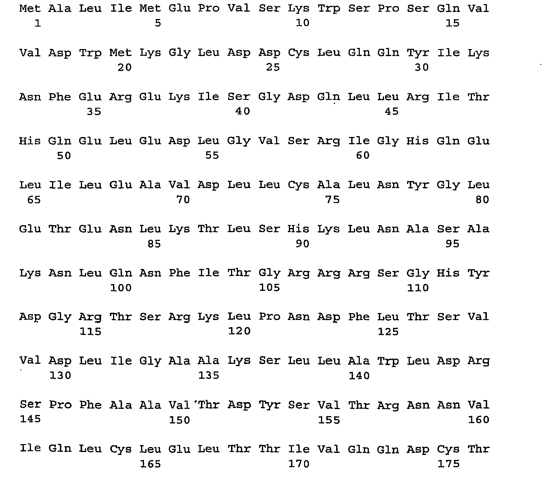


- <210> 2
<211> 948
<212> PRT
<213> Homo sapiens - <400> 2


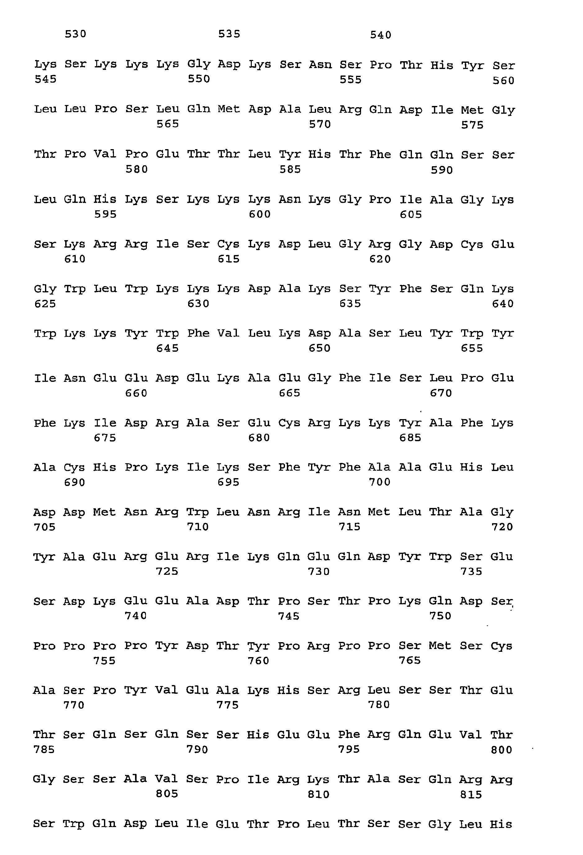

- <210> 3
<211> 3105
<212> DNA
<213> Homo sapiens - <400> 3


- <210> 4
<211> 2683
<212> DNA
<213> Homo sapiens - <400> 4


- <210> 5
<211> 5749
<212> DNA
<213> Artificial Sequence - <220>
<223> Description of Artificial Sequence: cDNA of human Maguin-1 - <400> 5


- <210> 6
<211> 4350
<212> DNA
<213> Artificial Sequence - <220>
<223> Description of Artificial Sequence: cDNA of human Maguin-2 - <400> 6


- <210> 7
<211> 50
<212> DNA
<213> Artificial Sequence - <220>
<223> Description of Artificial Sequence: cDNA fragment of Maguin-1 - <400> 7
ggagagaggc agcactgtaa actgaagtca aataaattca gctcttaatg 50 - <210> 8
<211> 13
<212> DNA
<213> Artificial Sequence - <220>
<223> Description of Artificial Sequence:
One-base-anchor oligonucleotides - <400> 8
httttttttt tta 13 - <210> 9
<211> 13
<212> DNA
<213> Artificial Sequence - <220>
<223> Description of Artificial Sequence:
One-base-anchor oligonucleotides - <400> 9
httttttttt ttg 13 - <210> 10
<211> 13
<212> DNA
<213> Artificial Sequence - <220>
<223> Description of Artificial Sequence:
One-base-anchor oligonucleotides - <400> 10
httttttttt ttc 13 - <210> 11
<211> 19
<212> DNA
<213> Artificial Sequence - <220>
<223> Description of Artificial Sequence: Primer for Maguin-1 - <400> 11
cagcaagcag ttgacggga 19 - <210> 12
<211> 22
<212> DNA
<213> Artificial Sequence - <220>
<223> Description of Artificial Sequence: Primer for Maguin-1 - <400> 12
tgaatttgac aagactcgtg gc 22 - <210> 13
<211> 19
<212> DNA
<213> Artificial Sequence - <220>
<223> Description of Artificial Sequence: Primer for Maguin-2 - <400> 13
gggcctccca aagggatat 19 - <210> 14
<211> 22
<212> DNA
<213> Artificial Sequence - <220>
<223> Description of Artificial Sequence: Primer for Maguin-2 - <400> 14
cccaatgtag aaagctcgca tt 22 - <210> 15
<211> 20
<212> DNA
<213> Artificial Sequence - <220>
<223> Description of Artificial Sequence: primer for cyclophilin B - <400> 15
actgaagcac tacgggcctg 20 - <210> 16
<211> 19
<212> DNA
<213> Artificial Sequence - <220>
<223> Description of Artificial Sequence: primer for cyclophilin B - <400> 16
agccgttggt gtctttgcc 19 - <210> 17
<211> 20
<212> DNA
<213> Artificial Sequence - <220>
<223> Description of Artificial Sequence: Primer for ribosomal protein S9 - <400> 17
ggtcaaattt accctggcca 20 - <210> 18
<211> 22
<212> DNA
<213> Artificial Sequence - <220>
<223> Description of Artificial Sequence: Primer for ribosomal protein S9 - <400> 18
tctcatcaag cgtcagcagt tc 22 - <210> 19
<211> 19
<212> DNA
<213> Artificial Sequence - <220>
<223> Description of Artificial Sequence: primer for beta-actin - <400> 19
tggaacggtg aaggtgaca 19 - <210> 20
<211> 19
<212> DNA
<213> Artificial Sequence - <220>
<223> Description of Artificial Sequence: primer for beta-actin - <400> 20
ggcaagggac ttcctgtaa 19 - <210> 21
<211> 20
<212> DNA
<213> Artificial Sequence - <220>
<223> Description of Artificial Sequence: Primer for GAPDH - <400> 21
cgtcatgggt gtgaaccatg 20 - <210> 22
<211> 21
<212> DNA
<213> Artificial Sequence - <220>
<223> Description of Artificial Sequence: Primer for GAPDH - <400> 22
gctaagcagt tggtggtgca g 21 - <210> 23
<211> 21
<212> DNA
<213> Artificial Sequence - <220>
<223> Description of Artificial Sequence: Primer for transferrin receptor - <400> 23
gtcgctggtc agttcgtgat t 21 - <210> 24
<211> 23
<212> DNA
<213> Artificial Sequence - <220>
<223> Description of Artificial Sequence: Primer for transferrin receptor - <400> 24
agcagttggc tgttgtacct ctc 23
In an analogous manner, the PCR protocol was applied to determine the PCR efficiency of a set of reference genes which were selected as a reference standard for quantification. In the present invention, the mean value of five such reference genes was determined: (1) cyclophilin B, using the specific primers 5'-ACTGAAGCACTACGGGCCTG-3' and 5'-AGCCGTTGGTGTCTTTGCC-3' except for MgCl2 (an additional 1 mM was added instead of 3 mM). Melting curve analysis revealed a single peak at approximately 87 °C with no visible primer dimers. Agarose gel analysis of the PCR product showed one single band of the expected size (62 bp). (2) Ribosomal protein S9 (RPS9), using the specific primers 5'-GGTCAAATTTACCCTGGCCA-3' and 5'- TCTCATCAAGCGTCAGCAGTTC-3' (exception: additional 1 mM MgCl2 was added instead of 3 mM). Melting curve analysis revealed a single peak at approximately 85°C with no visible primer dimers. Agarose gel analysis of the PCR product showed one single band with the expected size (62 bp). (3) beta-actin, using the specific primers 5'-TGGAACGGTGAAGGTGACA-3' and 5'-GGCAAGGGACTTCCTGTAA-3'. Melting curve analysis revealed a single peak at approximately 87°C with no visible primer dimers. Agarose gel analysis of the PCR product showed one single band with the expected size (142 bp). (4) GAPDH, using the specific primers 5'-CGTCATGGGTGTGAACCATG-3' and 5'-GCTAAGCAGTTGGTGGTGCAG-3'. Melting curve analysis revealed a single peak at approximately 83°C with no visible primer dimers. Agarose gel analysis of the PCR product showed one single band with the expected size (81 bp). (5) Transferrin receptor TRR, using the specific primers 5'-GTCGCTGGTCAGTTCGTGATT-3' and 5'-AGCAGTTGGCTGTTGTACCTCTC-3'. Melting curve analysis revealed a single peak at approximately 83°C with no visible primer dimers. Agarose gel analysis of the PCR product showed one single band with the expected size (80 bp).
Claims (12)
- An in vitro method of diagnosing Alzheimer's disease in a subject comprising:determining a level ofin a brain sample from said subject and comparing said level to a reference value representing a known disease or health status, thereby diagnosing said disease in said subject.(i) a transcription product of a gene coding for human MAGUIN-1 and/or human MAGUIN-2, and/or(ii) a translation product of a gene coding for human MAGUIN-1 and/or human MAGUIN-2,
- An in vitro method of monitoring the progression of Alzheimer's disease in
a subject, comprising:determining a level ofin a brain sample from said subject and comparing said level to a reference value representing a known disease or health status, thereby monitoring the progression of said disease in said subject.(i) a transcription product of a gene coding for human MAGUIN-1 and/or human MAGUIN-2, and/or(ii) a translation product of a gene coding for human MAGUIN-1 and/or human MAGUIN-2, - An in vitro method of evaluating a treatment for Alzheimer's disease,
comprising:determining a level ofin a brain sample from a subject being treated for said disease and comparing said level to a reference value representing a known disease or health status, thereby evaluating said treatment for said disease.(i) a transcription product of a gene coding for human MAGUIN-1 and/or human MAGUIN-2, and/or(ii) a translation product of a gene coding for human MAGUIN-1 and/or human MAGUIN-2, - The method according to any of claims 1 to 3 wherein said sample is a cell, or a tissue.
- The method according to any of claims 1 to 4 wherein said reference value is that of a level of(i) a transcription product of a gene coding for human MAGUIN-1 and/or human MAGUIN-2, and/or(ii) a translation product of a gene coding for human MAGUIN-1 and/or human MAGUIN-2,in a sample from a subject not suffering from Alzheimer's disease.
- The method according to any of claims 1 to 5 wherein an alteration in the level of a transcription product of the gene coding for human MAGUIN-1 and/or human MAGUIN-2 and/or a translation product of the gene coding for human MAGUIN-1 and/or human MAGUIN-2, in a sample cell, or tissue from said subject relative to a reference value representing a known health status indicates a diagnosis of Alzheimer's disease in said subject.
- The method according to any of claims 1 to 6, further comprising comparing a level of(i) a transcription product of a gene coding for human MAGUIN-1 and/or human MAGUIN-2, and/or(ii) a translation product of a gene coding for human MAGUIN-1 and/or human MAGUIN-2,in a series of samples taken from said subject over a period of time.
- Use of a kit in a method according to any one of claims 1-7, said kit comprising:(a) at least one reagent which is selected from the group consisting of(i) reagents that selectively detect a transcription product of a gene coding for human MAGUIN-1 and/or human MAGUIN-2, and(ii) antibodies that selectively detect a translation product of a gene coding for human MAGUIN-1 and/or human MAGUIN-2 and(b) an instruction for diagnosing Alzheimer's disease, bywherein a varied level of said transcription product and/or said translation product compared to a reference value representing a known health status; or a level, of said transcription product and/or said translation product similar or equal to a reference value representing a known disease status indicates a diagnosis of Alzheimer's disease.(i) detecting a level of said transcription product and/or said translation product of a gene coding for human MAGUIN-1 and/or human MAGUIN-2 in a brain sample from said subject; and(ii) diagnosing Alzheimer's disease,
- Use of a recombinant, non-human animal comprising a non-native gene sequence coding for human MAGUIN-1 and/or human MAGUIN-2 for screening, testing and validating compounds, agents and modulators in the development of diagnostics and therapeutics to treat Alzheimer's disease.
- An in vitro method for screening for a modulator of Alzheimer's disease said method comprising:(a) contacting a cell with a test compound;(b) measuring the level of one or more substances from the group consisting of(i) a gene coding for human MAGUIN-1 and/or human MAGUIN-2,(ii) a transcription product of a gene coding for human MAGUIN-1 and/or human MAGUIN-2, and(iii) a translation product of a gene coding for human MAGUIN-1 and/or human MAGUIN-2;(c) measuring the level of one or m ore substances recited in (i) to (iii) i n a control cell not contacted with said test compound; and(d) comparing the levels of the substance in the cells of step (b) and (c), wherein an alteration in the level of substances in the contacted cells indicates that the test compound is a modulator of said diseases.
- A protein molecule shown in SEQ ID NO.1 or SEQ ID NO.2 for use as a diagnostic target for detecting Alzheimer's disease.
- Use of an antibody specifically immunoreactive with a protein molecule according to SEQ ID No. 1 or SEQ ID No. 2, for detecting the pathological state of a cell in a brain sample from a subject, comprising immunocytochemical staining of said cell with said antibody, wherein an altered degree of staining, or an altered staining pattern in said cell compared to a cell representing a known health status indicates a pathological state of said cell, and wherein further said pathological state relates to Alzheimer's disease.
Priority Applications (1)
| Application Number | Priority Date | Filing Date | Title |
|---|---|---|---|
| EP03744814A EP1485410B1 (en) | 2002-03-21 | 2003-03-19 | Diagnostic and therapeutic use of human maguin proteins and nucleic acids for neurodegenerative diseases |
Applications Claiming Priority (6)
| Application Number | Priority Date | Filing Date | Title |
|---|---|---|---|
| US36581502P | 2002-03-21 | 2002-03-21 | |
| EP02006353 | 2002-03-21 | ||
| EP02006353 | 2002-03-21 | ||
| US365815P | 2002-03-21 | ||
| EP03744814A EP1485410B1 (en) | 2002-03-21 | 2003-03-19 | Diagnostic and therapeutic use of human maguin proteins and nucleic acids for neurodegenerative diseases |
| PCT/EP2003/002857 WO2003080661A1 (en) | 2002-03-21 | 2003-03-19 | Diagnostic and therapeutic use of human maguin proteins and nucleic acids for neurodegenerative diseases |
Publications (2)
| Publication Number | Publication Date |
|---|---|
| EP1485410A1 EP1485410A1 (en) | 2004-12-15 |
| EP1485410B1 true EP1485410B1 (en) | 2007-06-20 |
Family
ID=56290400
Family Applications (1)
| Application Number | Title | Priority Date | Filing Date |
|---|---|---|---|
| EP03744814A Expired - Lifetime EP1485410B1 (en) | 2002-03-21 | 2003-03-19 | Diagnostic and therapeutic use of human maguin proteins and nucleic acids for neurodegenerative diseases |
Country Status (5)
| Country | Link |
|---|---|
| EP (1) | EP1485410B1 (en) |
| AT (1) | ATE365175T1 (en) |
| AU (1) | AU2003215667A1 (en) |
| DE (1) | DE60314497T2 (en) |
| WO (1) | WO2003080661A1 (en) |
Cited By (3)
| Publication number | Priority date | Publication date | Assignee | Title |
|---|---|---|---|---|
| US12227567B2 (en) | 2017-07-25 | 2025-02-18 | Truebinding, Inc. | Treating cancer by blocking the interaction of TIM-3 and its ligand |
| US12281166B2 (en) | 2020-05-26 | 2025-04-22 | Truebinding, Inc. | Methods of treating inflammatory diseases by blocking Galectin-3 |
| US12497458B2 (en) | 2019-01-30 | 2025-12-16 | Truebinding, Inc. | Anti-GAL3 antibodies and uses thereof |
Family Cites Families (1)
| Publication number | Priority date | Publication date | Assignee | Title |
|---|---|---|---|---|
| JP4070373B2 (en) * | 1998-11-16 | 2008-04-02 | 独立行政法人科学技術振興機構 | Protein MAGUIN |
-
2003
- 2003-03-19 WO PCT/EP2003/002857 patent/WO2003080661A1/en not_active Ceased
- 2003-03-19 AT AT03744814T patent/ATE365175T1/en active
- 2003-03-19 AU AU2003215667A patent/AU2003215667A1/en not_active Abandoned
- 2003-03-19 DE DE60314497T patent/DE60314497T2/en not_active Expired - Lifetime
- 2003-03-19 EP EP03744814A patent/EP1485410B1/en not_active Expired - Lifetime
Non-Patent Citations (1)
| Title |
|---|
| MULDER, C.: "CSF markers relate to pathogenetic mechanisms in Alzheimer's disease", JOURNAL OF NEURAL TRANSMISSION, vol. 109, 2002, pages 1491 - 1498 * |
Cited By (3)
| Publication number | Priority date | Publication date | Assignee | Title |
|---|---|---|---|---|
| US12227567B2 (en) | 2017-07-25 | 2025-02-18 | Truebinding, Inc. | Treating cancer by blocking the interaction of TIM-3 and its ligand |
| US12497458B2 (en) | 2019-01-30 | 2025-12-16 | Truebinding, Inc. | Anti-GAL3 antibodies and uses thereof |
| US12281166B2 (en) | 2020-05-26 | 2025-04-22 | Truebinding, Inc. | Methods of treating inflammatory diseases by blocking Galectin-3 |
Also Published As
| Publication number | Publication date |
|---|---|
| WO2003080661A1 (en) | 2003-10-02 |
| EP1485410A1 (en) | 2004-12-15 |
| AU2003215667A1 (en) | 2003-10-08 |
| ATE365175T1 (en) | 2007-07-15 |
| DE60314497T2 (en) | 2008-02-21 |
| DE60314497D1 (en) | 2007-08-02 |
Similar Documents
| Publication | Publication Date | Title |
|---|---|---|
| US20060259991A1 (en) | Diagnostic and therapeutic use of scn2b protein for neurodegeneraative diseases | |
| EP1516189B1 (en) | Diagnostic and therapeutic use of ensadin-0581 gene and protein for neurodegenerative diseases | |
| EP1497661B1 (en) | Diagnostic use of ensadin-0477 gene and protein for neurodegenerative diseases | |
| EP1776591B1 (en) | Diagnostic and therapeutic use of a plasma membrane atpase | |
| US20060259990A1 (en) | Diagnostic and therapeutic use of tb2 gene and protein for neurodegenerative diseases | |
| WO2004001422A2 (en) | Diagnostic and therapeutic use of ras-gtpase-activating sh3-domain-binding protein 2 (g3bp2) for neurodegenerative diseases | |
| EP1490692B1 (en) | Diagnostic and therapeutic use of caps | |
| US20060052280A1 (en) | Diagnostic and therapeutic use of a golgi protein for neurodegenerative diseases | |
| WO2003080661A1 (en) | Diagnostic and therapeutic use of human maguin proteins and nucleic acids for neurodegenerative diseases | |
| WO2003104811A2 (en) | Diagnostic and therapeutic use of steroidogenic acute regulatory protein for neurodegenerative diseases | |
| US20060141459A1 (en) | Diagnostic and therapeutic use of foap-13 polynucleotides and polypeptides for neurodegenerative diseases | |
| US20050153295A1 (en) | Diagnostic and therapeutic use of human maguin proteins and nucleic acids for neurodegenerative diseases | |
| EP1490694B1 (en) | Camp-regulated phosphorprotein for diagnostic and therapeutic use in neurodegenerative diseases | |
| EP1565751A1 (en) | Diagnostic and therapeutic use of h-rev107 protein for alzheimer disease | |
| EP1552310A2 (en) | Diagnostic and therapeutic use of the nicotinamide mononucleotide adenylyltransferase 2 (nmnat-2) gene and protein for neurodegenerative diseases | |
| US20060073480A1 (en) | Diagnostic and therapeutic use of vault polynucleotides and proteins for neurodegenerative diseases | |
| US20060294602A1 (en) | Diagnostic and therapeutic use of a rab family gtp-binding protein for neurodegenerative diseases | |
| US20060160728A1 (en) | Diagnostic and therapeutic use of ensandin-0138 gene and protein for neurodegenerative diseases | |
| WO2004044592A1 (en) | Diagnostic and therapeutic use of arl7 for alzheimer's disease | |
| US20060051757A1 (en) | Diagnostic and therapeutic use of ensadin-0477 gene and protein for neurodegenerative diseases | |
| WO2004020666A2 (en) | Diagnostic and therapeutic use of thyroid hormone binding protein for neurodegenerative diseases | |
| WO2006008294A2 (en) | Diagnostic and therapeutic use of slim3 for neurodegenerative diseases |
Legal Events
| Date | Code | Title | Description |
|---|---|---|---|
| PUAI | Public reference made under article 153(3) epc to a published international application that has entered the european phase |
Free format text: ORIGINAL CODE: 0009012 |
|
| 17P | Request for examination filed |
Effective date: 20040902 |
|
| AK | Designated contracting states |
Kind code of ref document: A1 Designated state(s): AT BE BG CH CY CZ DE DK EE ES FI FR GB GR HU IE IT LI LU MC NL PT RO SE SI SK TR |
|
| AX | Request for extension of the european patent |
Extension state: AL LT LV MK |
|
| 17Q | First examination report despatched |
Effective date: 20050224 |
|
| 17Q | First examination report despatched |
Effective date: 20050224 |
|
| GRAP | Despatch of communication of intention to grant a patent |
Free format text: ORIGINAL CODE: EPIDOSNIGR1 |
|
| RIN1 | Information on inventor provided before grant (corrected) |
Inventor name: VON DER KAMMER, HEINZ Inventor name: POHLNER, JOHANNES Inventor name: HANES, JOZEF |
|
| GRAS | Grant fee paid |
Free format text: ORIGINAL CODE: EPIDOSNIGR3 |
|
| GRAA | (expected) grant |
Free format text: ORIGINAL CODE: 0009210 |
|
| AK | Designated contracting states |
Kind code of ref document: B1 Designated state(s): AT BE BG CH CY CZ DE DK EE ES FI FR GB GR HU IE IT LI LU MC NL PT RO SE SI SK TR |
|
| REG | Reference to a national code |
Ref country code: GB Ref legal event code: FG4D |
|
| REG | Reference to a national code |
Ref country code: CH Ref legal event code: EP |
|
| REG | Reference to a national code |
Ref country code: IE Ref legal event code: FG4D |
|
| REF | Corresponds to: |
Ref document number: 60314497 Country of ref document: DE Date of ref document: 20070802 Kind code of ref document: P |
|
| PG25 | Lapsed in a contracting state [announced via postgrant information from national office to epo] |
Ref country code: SE Free format text: LAPSE BECAUSE OF FAILURE TO SUBMIT A TRANSLATION OF THE DESCRIPTION OR TO PAY THE FEE WITHIN THE PRESCRIBED TIME-LIMIT Effective date: 20070920 |
|
| REG | Reference to a national code |
Ref country code: CH Ref legal event code: NV Representative=s name: SCHMAUDER & PARTNER AG PATENTANWALTSBUERO |
|
| NLV1 | Nl: lapsed or annulled due to failure to fulfill the requirements of art. 29p and 29m of the patents act | ||
| PG25 | Lapsed in a contracting state [announced via postgrant information from national office to epo] |
Ref country code: BE Free format text: LAPSE BECAUSE OF FAILURE TO SUBMIT A TRANSLATION OF THE DESCRIPTION OR TO PAY THE FEE WITHIN THE PRESCRIBED TIME-LIMIT Effective date: 20070620 |
|
| PG25 | Lapsed in a contracting state [announced via postgrant information from national office to epo] |
Ref country code: CZ Free format text: LAPSE BECAUSE OF FAILURE TO SUBMIT A TRANSLATION OF THE DESCRIPTION OR TO PAY THE FEE WITHIN THE PRESCRIBED TIME-LIMIT Effective date: 20070620 Ref country code: NL Free format text: LAPSE BECAUSE OF FAILURE TO SUBMIT A TRANSLATION OF THE DESCRIPTION OR TO PAY THE FEE WITHIN THE PRESCRIBED TIME-LIMIT Effective date: 20070620 Ref country code: SI Free format text: LAPSE BECAUSE OF FAILURE TO SUBMIT A TRANSLATION OF THE DESCRIPTION OR TO PAY THE FEE WITHIN THE PRESCRIBED TIME-LIMIT Effective date: 20070620 Ref country code: ES Free format text: LAPSE BECAUSE OF FAILURE TO SUBMIT A TRANSLATION OF THE DESCRIPTION OR TO PAY THE FEE WITHIN THE PRESCRIBED TIME-LIMIT Effective date: 20071001 Ref country code: PT Free format text: LAPSE BECAUSE OF FAILURE TO SUBMIT A TRANSLATION OF THE DESCRIPTION OR TO PAY THE FEE WITHIN THE PRESCRIBED TIME-LIMIT Effective date: 20071120 Ref country code: BG Free format text: LAPSE BECAUSE OF FAILURE TO SUBMIT A TRANSLATION OF THE DESCRIPTION OR TO PAY THE FEE WITHIN THE PRESCRIBED TIME-LIMIT Effective date: 20070920 |
|
| EN | Fr: translation not filed | ||
| PG25 | Lapsed in a contracting state [announced via postgrant information from national office to epo] |
Ref country code: SK Free format text: LAPSE BECAUSE OF FAILURE TO SUBMIT A TRANSLATION OF THE DESCRIPTION OR TO PAY THE FEE WITHIN THE PRESCRIBED TIME-LIMIT Effective date: 20070620 |
|
| PLBE | No opposition filed within time limit |
Free format text: ORIGINAL CODE: 0009261 |
|
| STAA | Information on the status of an ep patent application or granted ep patent |
Free format text: STATUS: NO OPPOSITION FILED WITHIN TIME LIMIT |
|
| PG25 | Lapsed in a contracting state [announced via postgrant information from national office to epo] |
Ref country code: GR Free format text: LAPSE BECAUSE OF FAILURE TO SUBMIT A TRANSLATION OF THE DESCRIPTION OR TO PAY THE FEE WITHIN THE PRESCRIBED TIME-LIMIT Effective date: 20070921 Ref country code: IT Free format text: LAPSE BECAUSE OF FAILURE TO SUBMIT A TRANSLATION OF THE DESCRIPTION OR TO PAY THE FEE WITHIN THE PRESCRIBED TIME-LIMIT Effective date: 20070620 Ref country code: DK Free format text: LAPSE BECAUSE OF FAILURE TO SUBMIT A TRANSLATION OF THE DESCRIPTION OR TO PAY THE FEE WITHIN THE PRESCRIBED TIME-LIMIT Effective date: 20070620 |
|
| 26N | No opposition filed |
Effective date: 20080325 |
|
| PG25 | Lapsed in a contracting state [announced via postgrant information from national office to epo] |
Ref country code: RO Free format text: LAPSE BECAUSE OF FAILURE TO SUBMIT A TRANSLATION OF THE DESCRIPTION OR TO PAY THE FEE WITHIN THE PRESCRIBED TIME-LIMIT Effective date: 20070620 |
|
| PG25 | Lapsed in a contracting state [announced via postgrant information from national office to epo] |
Ref country code: FR Free format text: LAPSE BECAUSE OF FAILURE TO SUBMIT A TRANSLATION OF THE DESCRIPTION OR TO PAY THE FEE WITHIN THE PRESCRIBED TIME-LIMIT Effective date: 20080222 |
|
| PG25 | Lapsed in a contracting state [announced via postgrant information from national office to epo] |
Ref country code: MC Free format text: LAPSE BECAUSE OF NON-PAYMENT OF DUE FEES Effective date: 20080331 |
|
| PG25 | Lapsed in a contracting state [announced via postgrant information from national office to epo] |
Ref country code: IE Free format text: LAPSE BECAUSE OF NON-PAYMENT OF DUE FEES Effective date: 20080319 Ref country code: EE Free format text: LAPSE BECAUSE OF FAILURE TO SUBMIT A TRANSLATION OF THE DESCRIPTION OR TO PAY THE FEE WITHIN THE PRESCRIBED TIME-LIMIT Effective date: 20070620 |
|
| PG25 | Lapsed in a contracting state [announced via postgrant information from national office to epo] |
Ref country code: FI Free format text: LAPSE BECAUSE OF FAILURE TO SUBMIT A TRANSLATION OF THE DESCRIPTION OR TO PAY THE FEE WITHIN THE PRESCRIBED TIME-LIMIT Effective date: 20070620 |
|
| PG25 | Lapsed in a contracting state [announced via postgrant information from national office to epo] |
Ref country code: CY Free format text: LAPSE BECAUSE OF FAILURE TO SUBMIT A TRANSLATION OF THE DESCRIPTION OR TO PAY THE FEE WITHIN THE PRESCRIBED TIME-LIMIT Effective date: 20070620 |
|
| REG | Reference to a national code |
Ref country code: CH Ref legal event code: PCAR Free format text: SCHMAUDER & PARTNER AG PATENT- UND MARKENANWAELTE VSP;ZWAENGIWEG 7;8038 ZUERICH (CH) |
|
| PG25 | Lapsed in a contracting state [announced via postgrant information from national office to epo] |
Ref country code: LU Free format text: LAPSE BECAUSE OF NON-PAYMENT OF DUE FEES Effective date: 20080319 Ref country code: HU Free format text: LAPSE BECAUSE OF FAILURE TO SUBMIT A TRANSLATION OF THE DESCRIPTION OR TO PAY THE FEE WITHIN THE PRESCRIBED TIME-LIMIT Effective date: 20071221 |
|
| PG25 | Lapsed in a contracting state [announced via postgrant information from national office to epo] |
Ref country code: TR Free format text: LAPSE BECAUSE OF FAILURE TO SUBMIT A TRANSLATION OF THE DESCRIPTION OR TO PAY THE FEE WITHIN THE PRESCRIBED TIME-LIMIT Effective date: 20070620 |
|
| PGFP | Annual fee paid to national office [announced via postgrant information from national office to epo] |
Ref country code: AT Payment date: 20110323 Year of fee payment: 9 |
|
| REG | Reference to a national code |
Ref country code: AT Ref legal event code: MM01 Ref document number: 365175 Country of ref document: AT Kind code of ref document: T Effective date: 20120319 |
|
| REG | Reference to a national code |
Ref country code: DE Ref legal event code: R082 Ref document number: 60314497 Country of ref document: DE Representative=s name: VON KREISLER SELTING WERNER, DE |
|
| PG25 | Lapsed in a contracting state [announced via postgrant information from national office to epo] |
Ref country code: AT Free format text: LAPSE BECAUSE OF NON-PAYMENT OF DUE FEES Effective date: 20120319 |
|
| REG | Reference to a national code |
Ref country code: DE Ref legal event code: R082 Ref document number: 60314497 Country of ref document: DE Representative=s name: VON KREISLER SELTING WERNER, DE Effective date: 20130114 Ref country code: DE Ref legal event code: R081 Ref document number: 60314497 Country of ref document: DE Owner name: EVOTEC INTERNATIONAL GMBH, DE Free format text: FORMER OWNER: EVOTEC NEUROSCIENCES GMBH, 22525 HAMBURG, DE Effective date: 20130114 Ref country code: DE Ref legal event code: R082 Ref document number: 60314497 Country of ref document: DE Representative=s name: VON KREISLER SELTING WERNER - PARTNERSCHAFT VO, DE Effective date: 20130114 Ref country code: DE Ref legal event code: R082 Ref document number: 60314497 Country of ref document: DE Representative=s name: DOMPATENT VON KREISLER SELTING WERNER - PARTNE, DE Effective date: 20130114 |
|
| REG | Reference to a national code |
Ref country code: CH Ref legal event code: PFA Owner name: EVOTEC INTERNATIONAL GMBH, DE Free format text: FORMER OWNER: EVOTEC NEUROSCIENCES GMBH, DE |
|
| PGFP | Annual fee paid to national office [announced via postgrant information from national office to epo] |
Ref country code: CH Payment date: 20170327 Year of fee payment: 15 |
|
| PGFP | Annual fee paid to national office [announced via postgrant information from national office to epo] |
Ref country code: GB Payment date: 20170327 Year of fee payment: 15 |
|
| PGFP | Annual fee paid to national office [announced via postgrant information from national office to epo] |
Ref country code: DE Payment date: 20170331 Year of fee payment: 15 |
|
| REG | Reference to a national code |
Ref country code: DE Ref legal event code: R119 Ref document number: 60314497 Country of ref document: DE |
|
| REG | Reference to a national code |
Ref country code: CH Ref legal event code: PL |
|
| GBPC | Gb: european patent ceased through non-payment of renewal fee |
Effective date: 20180319 |
|
| PG25 | Lapsed in a contracting state [announced via postgrant information from national office to epo] |
Ref country code: DE Free format text: LAPSE BECAUSE OF NON-PAYMENT OF DUE FEES Effective date: 20181002 |
|
| PG25 | Lapsed in a contracting state [announced via postgrant information from national office to epo] |
Ref country code: CH Free format text: LAPSE BECAUSE OF NON-PAYMENT OF DUE FEES Effective date: 20180331 Ref country code: LI Free format text: LAPSE BECAUSE OF NON-PAYMENT OF DUE FEES Effective date: 20180331 Ref country code: GB Free format text: LAPSE BECAUSE OF NON-PAYMENT OF DUE FEES Effective date: 20180319 |