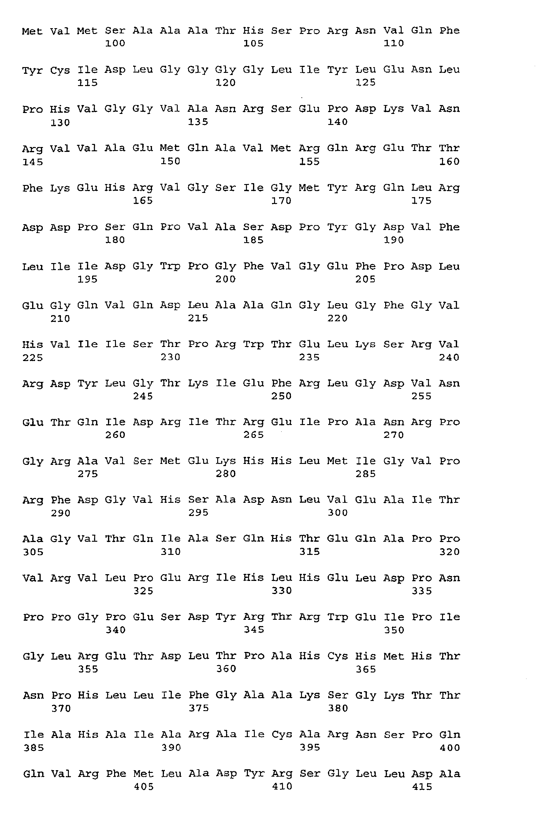
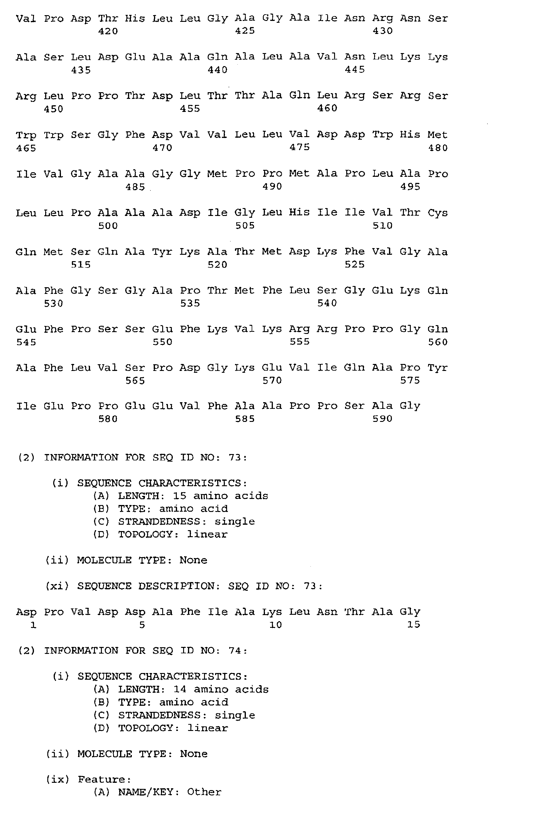
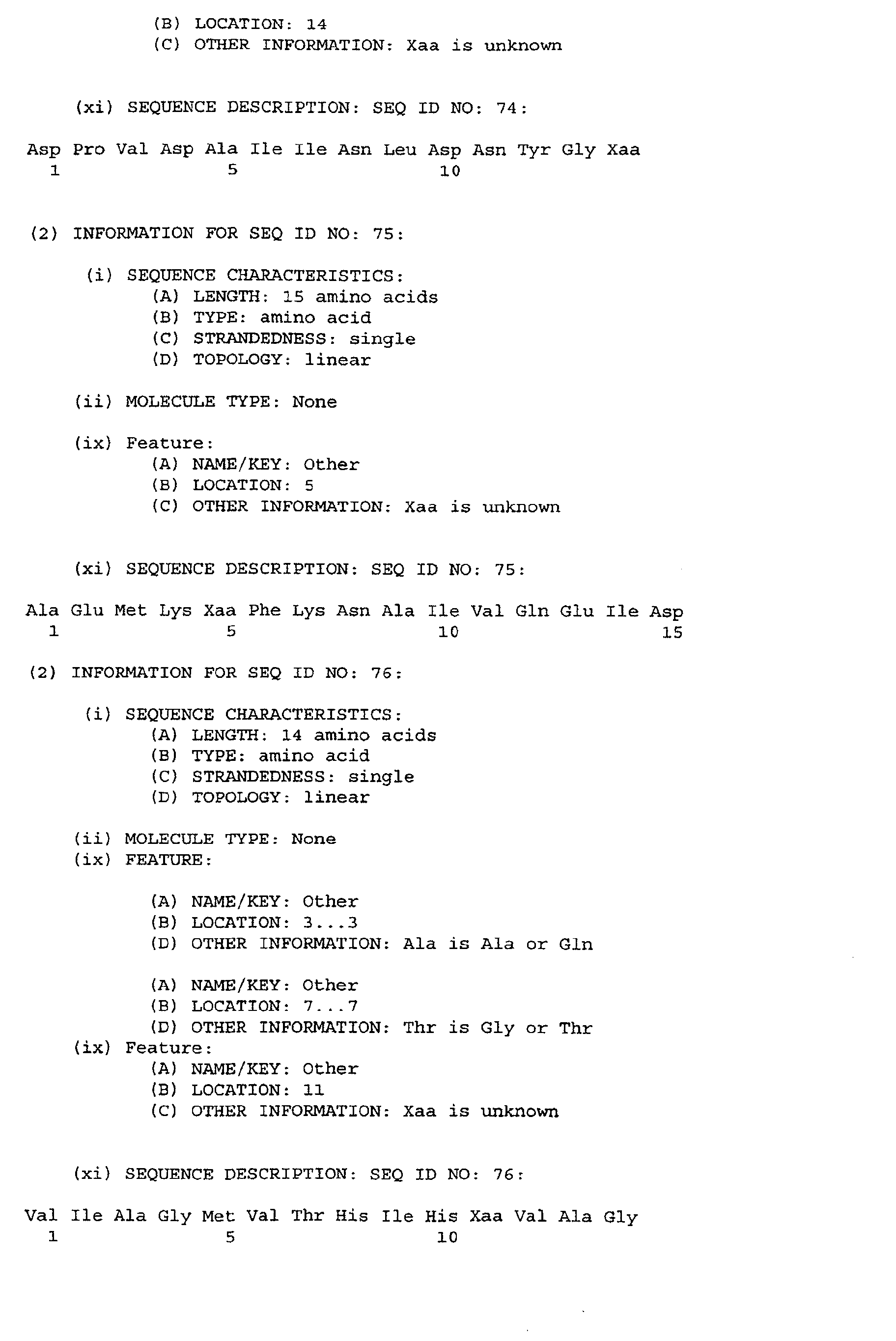
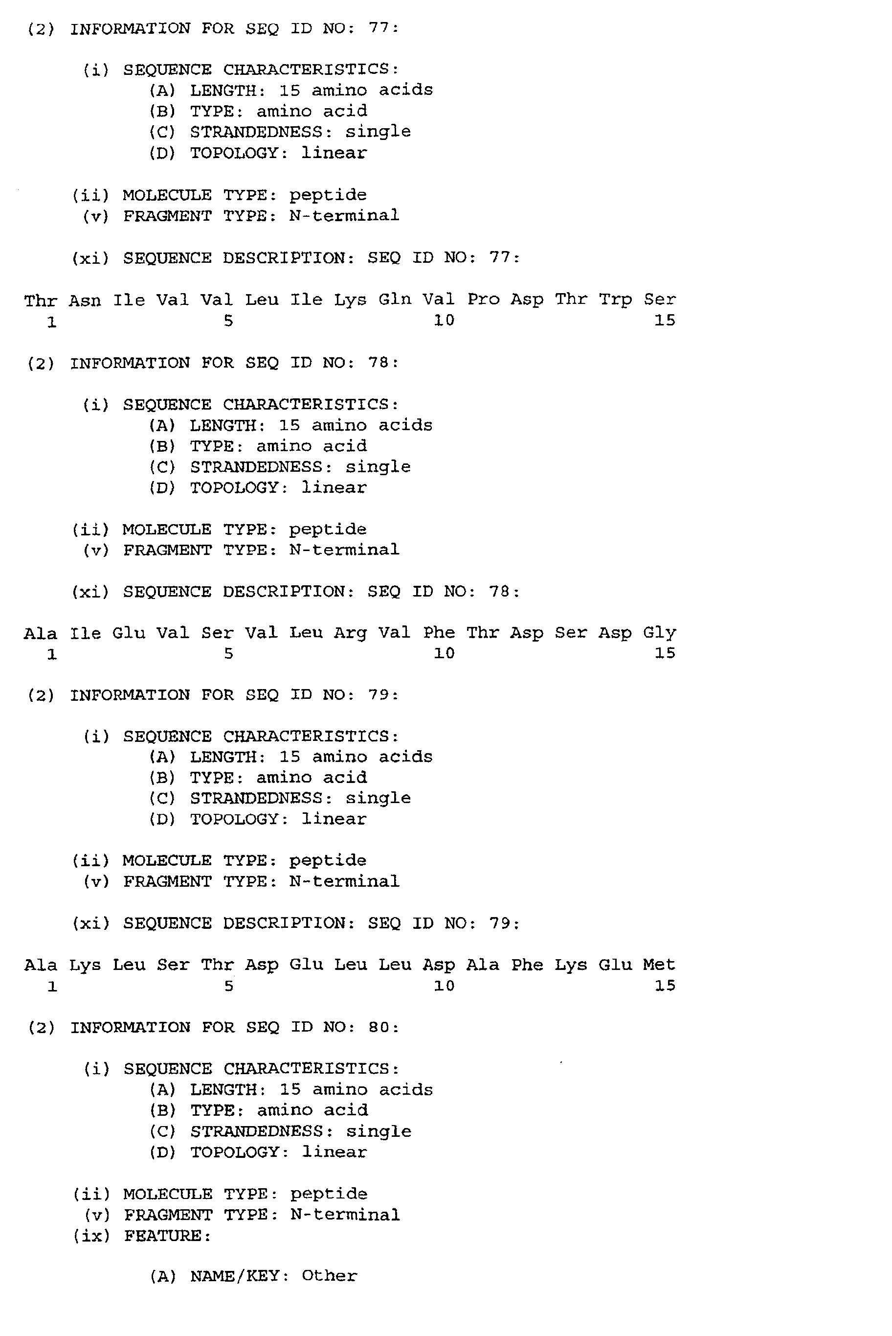
FIELD OF THE INVENTION
-
The present invention relates to a number of immunologically active, novel polypeptide
fragments derived from the Mycobacterium tuberculosis, vaccines and other immunologic
compositions containing the fragments as immunogenic components, and
methods of production and use of the polypeptides. The invention also relates to novel
nucleic acid fragments derived from M. tuberculosis which are useful in the preparation
of the polypeptide fragments of the invention or in the diagnosis of infection with
M. tuberculosis.
BACKGROUND OF THE INVENTION
-
Human tuberculosis (hereinafter designated "TB") caused by Mycobacterium tuberculosis
is a severe global health problem responsible for approximately 3 million deaths
annually, according to the WHO. The worldwide incidence of new TB cases has been
progressively falling for the last decade but the recent years has markedly changed
this trend due to the advent of AIDS and the appearance of multidrug resistant strains
of M. tuberculosis.
-
The only vaccine presently available for clinical use is BCG, a vaccine which efficacy
remains a matter of controversy. BCG generally induces a high level of acquired resistance
in animal models of TB, but several human trials in developing countries have
failed to demonstrate significant protection. Notably, BCG is not approved by the FDA
for use in the United States.
-
This makes the development of a new and improved vaccine against TB an urgent
matter which has been given a very high priority by the WHO. Many attempts to define
protective mycobacterial substances have been made, and from 1950 to 1970
several investigators reported an increased resistance after experimental vaccination.
However, the demonstration of a specific long-term protective immune response with
the potency of BCG has not yet been achieved by administration of soluble proteins or
cell wall fragments, although progress is currently being made by relying on polypeptides
derived from short term-culture filtrate, cf. the discussion below.
-
Immunity to M. tuberculosis is characterized by three basic features; i) Living bacilli
efficiently induces a protective immune response in contrast to killed preparations; ii)
Specifically sensitized T lymphocytes mediate this protection; iii) The most important
mediator molecule seems to be interferon gamma (INF-γ).
-
Short term-culture filtrate (ST-CF) is a complex mixture of proteins released from M.
tuberculosis during the first few days of growth in a liquid medium (Andersen et al.,
1991). Culture filtrates has been suggested to hold protective antigens recognized by
the host in the first phase of TB infection (Andersen et al. 1991, Orme et al. 1993).
Recent data from several laboratories have demonstrated that experimental subunit
vaccines based on culture filtrate antigens can provide high levels of acquired resistance
to TB (Pal and Horwitz, 1992; Roberts et al., 1995; Andersen, 1994; Lindblad
et al., 1997). Culture filtrates are, however, complex protein mixtures and until now
very limited information has been available on the molecules responsible for this protective
immune response. In this regard, only two culture filtrate antigens have been
described as involved in protective immunity, the low mass antigen ESAT-6 (Andersen
et al., 1995 and EP-A-0 706 571) and the 31 kDa molecule Ag85B (EP-0 432 203).
-
There is therefore a need for the identification of further antigens involved in the induction
of protective immunity against TB in order to eventually produce an effective
subunit vaccine.
OBJECT OF THE INVENTION
-
It is an object of the invention to provide novel antigens which are effective as components
in a subunit vaccine against TB or which are useful as components in diagnostic
compositions for the detection of infection with mycobacteria, especially virulence-associated
mycobacteria. The novel antigens may also be important drug targets.
SUMMARY OF THE INVENTION
-
The present invention is i.a. based on the identification and characterization of a number
of previously uncharacterized culture filtrate antigens from M. tuberculosis. In
animal models of TB, T cells mediating immunity are focused predominantly to antigens
in the regions 6-12 and 17-30 kDa of ST-CF. In the present invention 6 antigens
in the low molecular weight region (ORF7-1, ORF7-2, ORF11-1, ORF11-2, ORF11-3,
ORF11-4) have been identified.
-
Furthermore immunological and biological data on several important antigens are
presented.
-
The encoding genes for 8 antigens have been determined. The panel hold antigens
with potential for vaccine purposes as well as for diagnostic purposes, since the
antigens are all secreted by metabolizing mycobacteria.
-
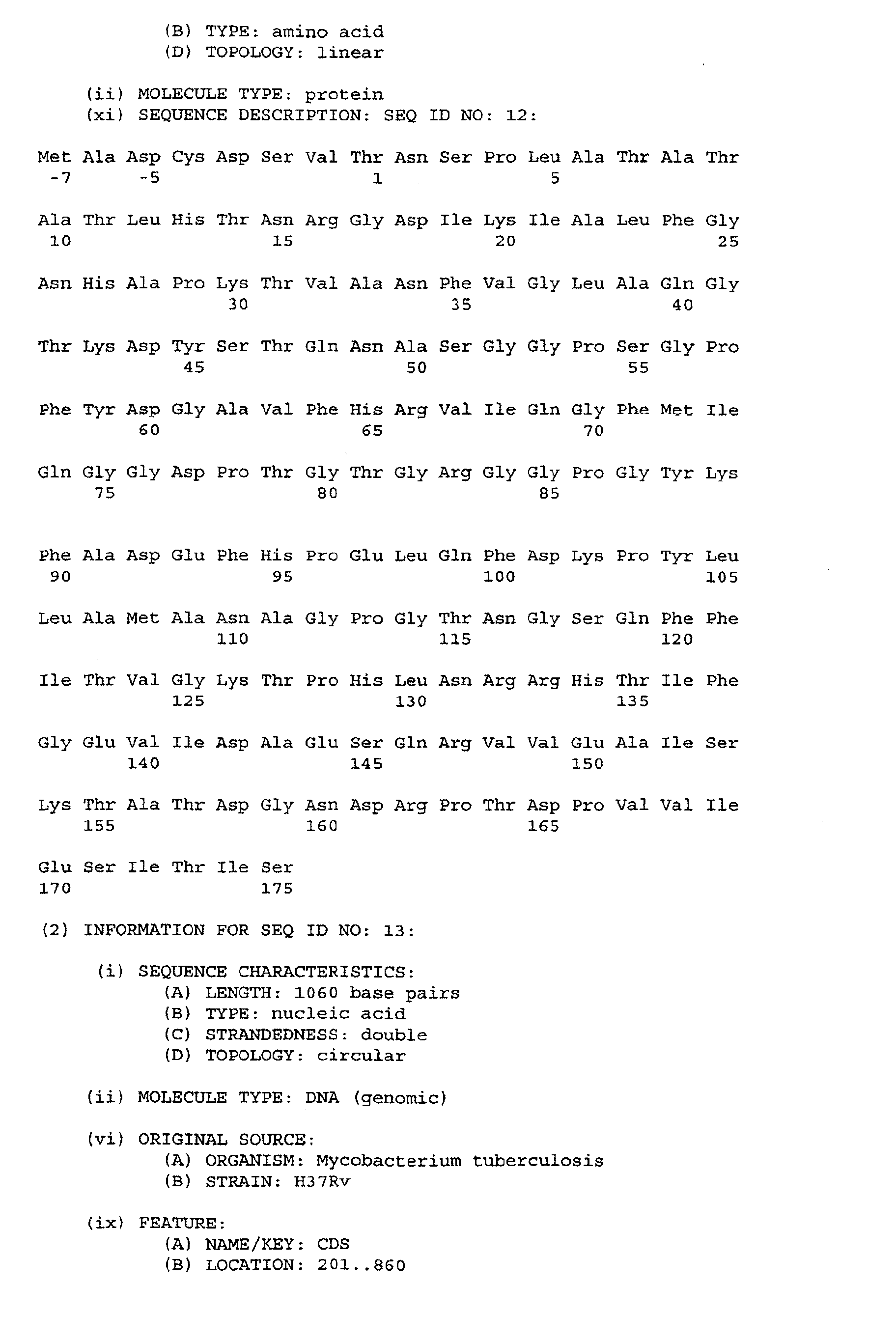
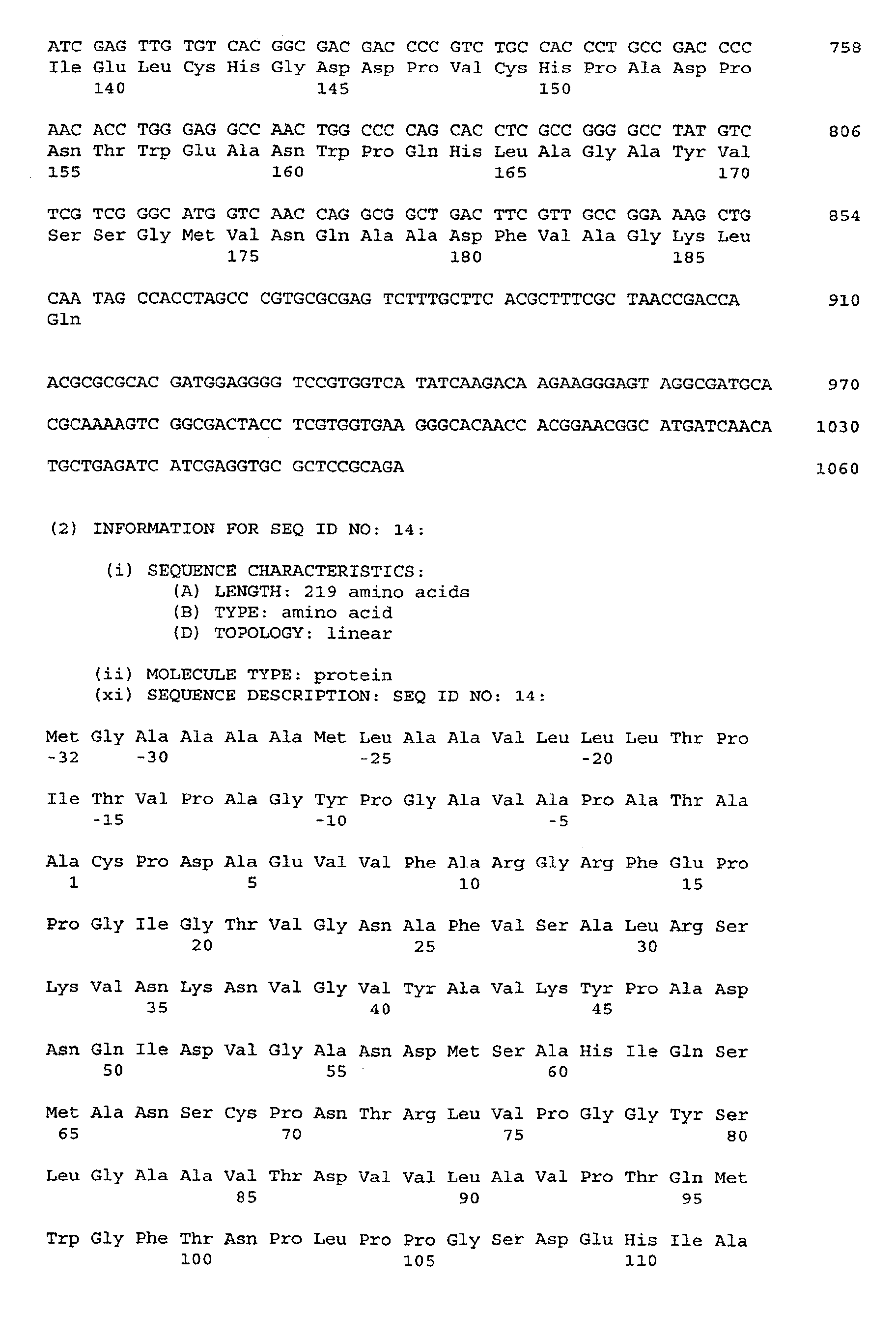
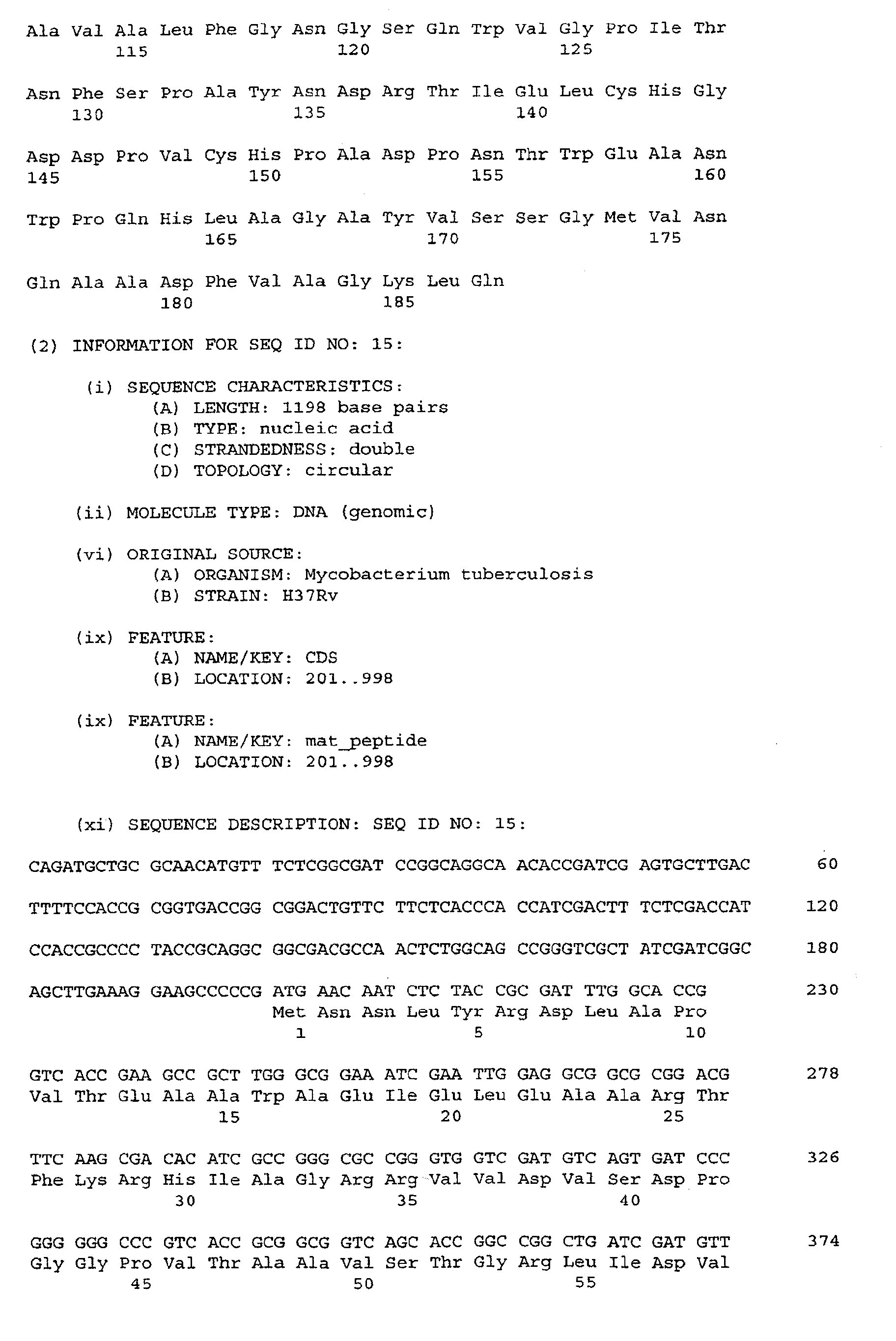
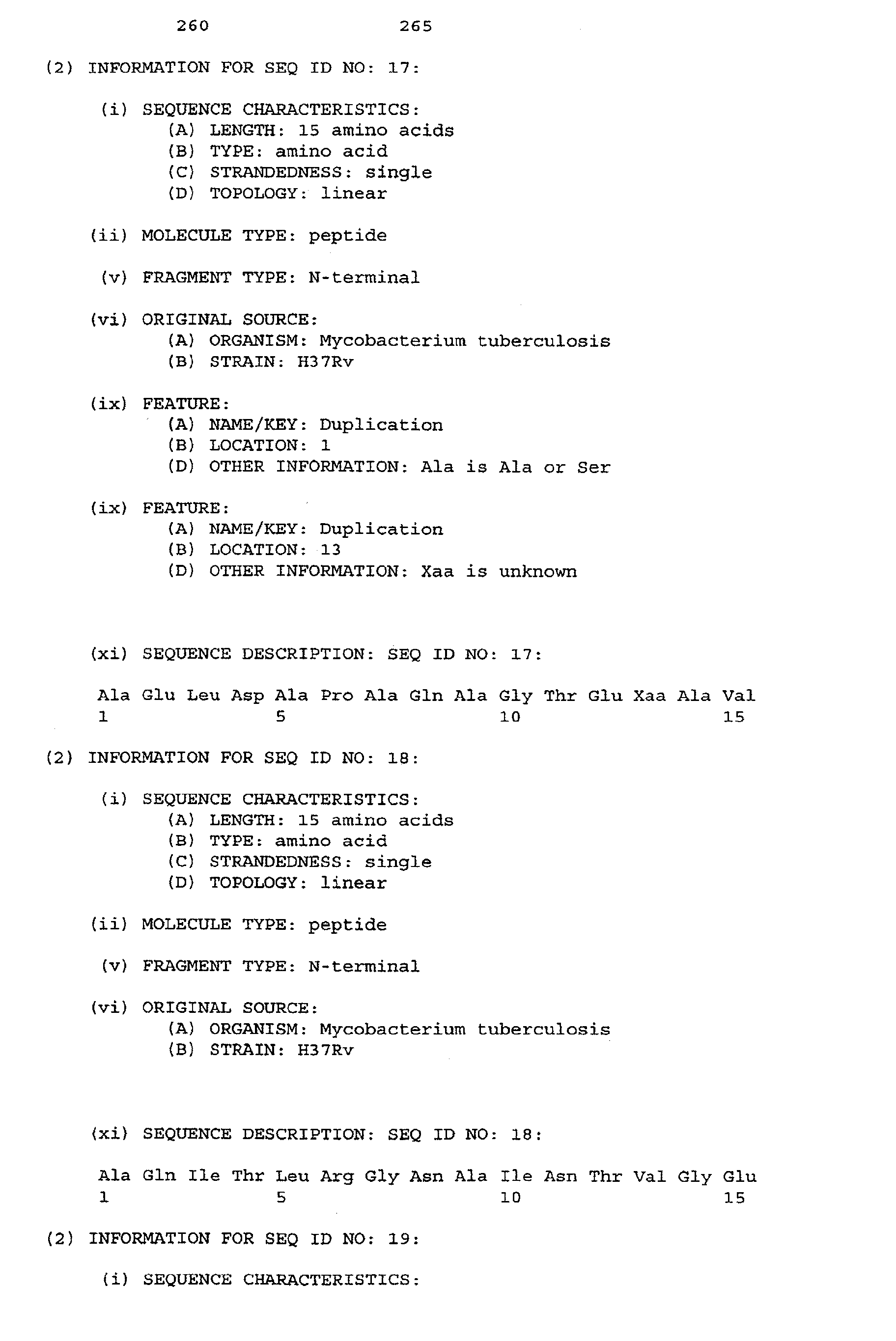
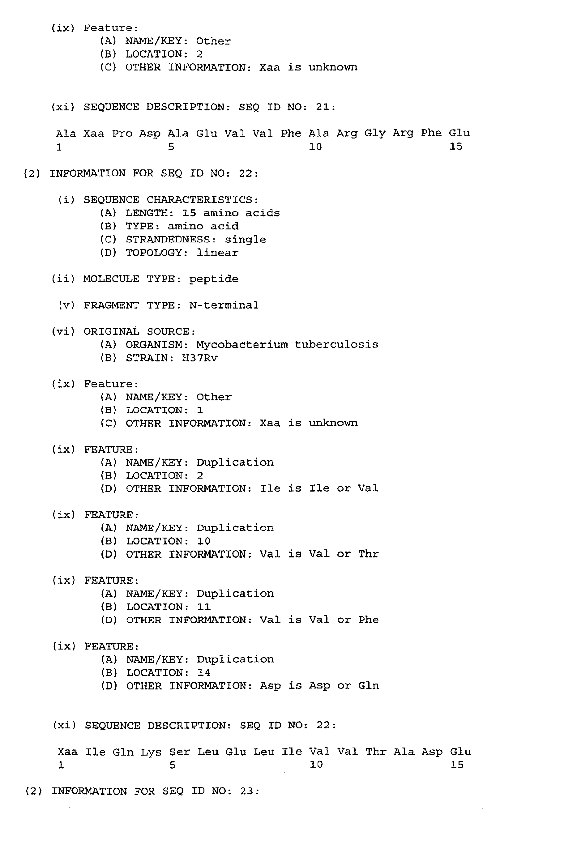
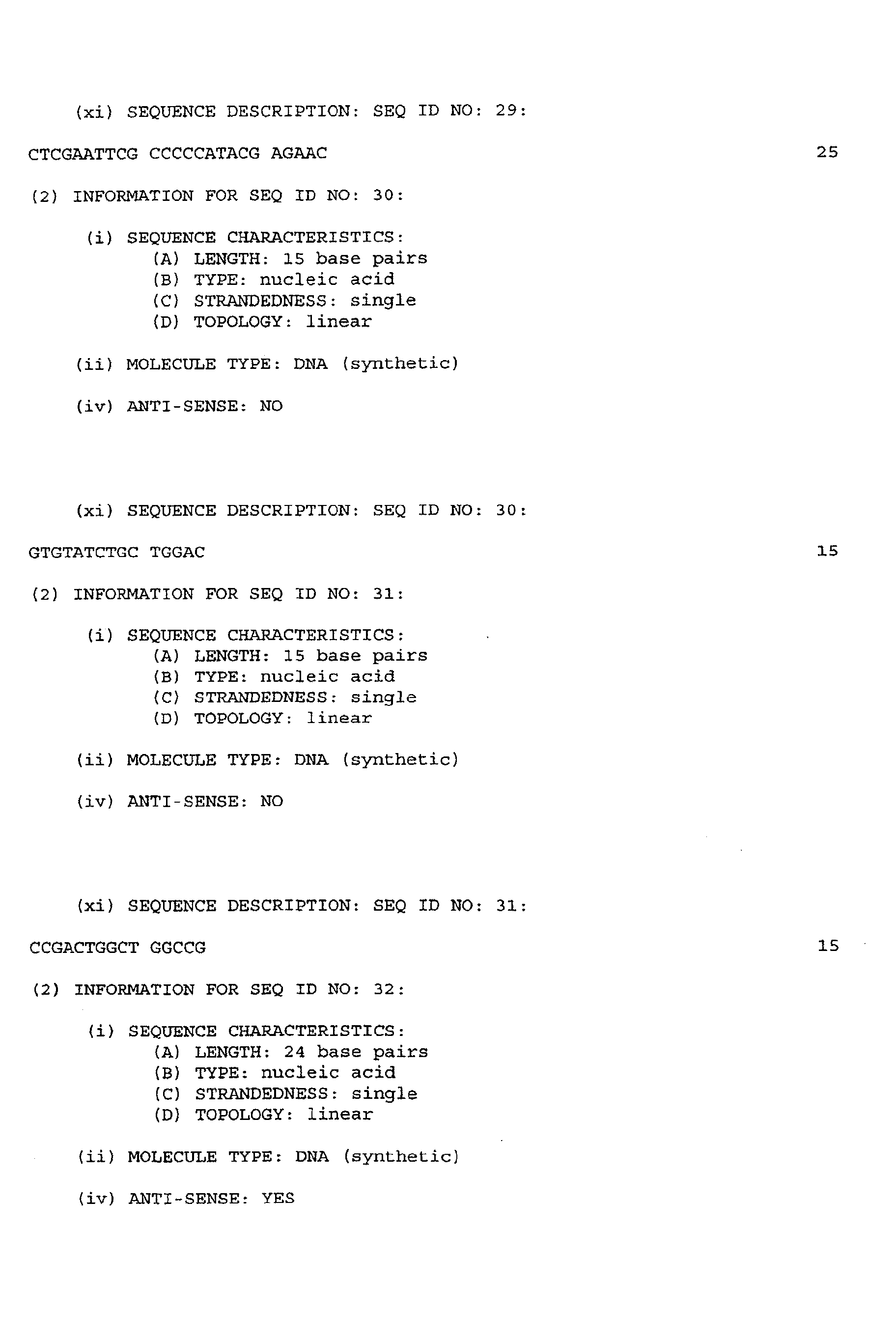
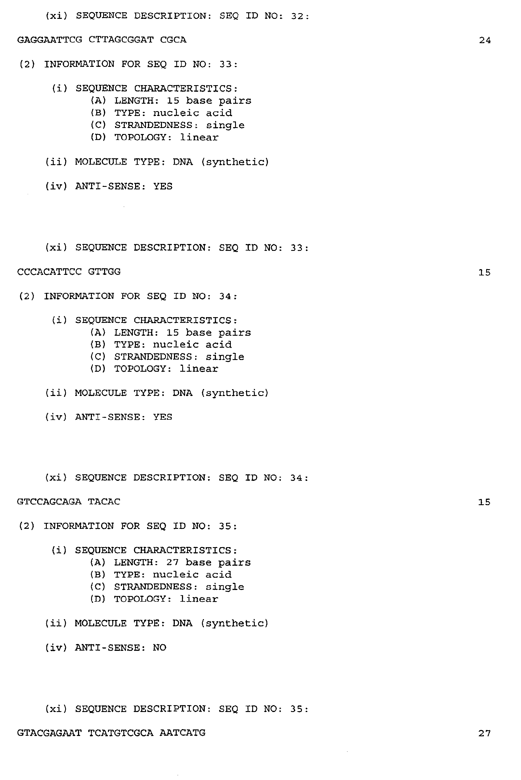
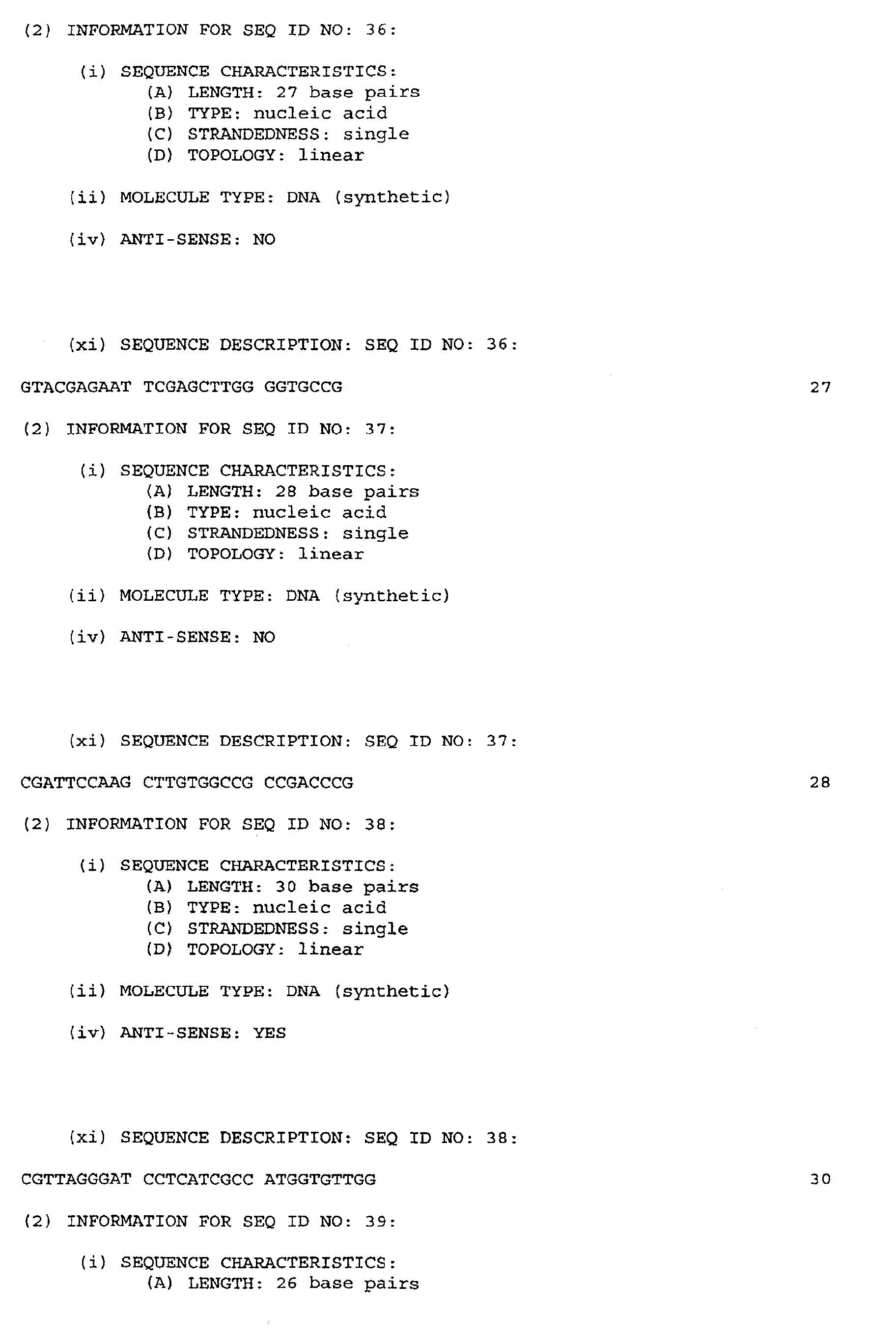
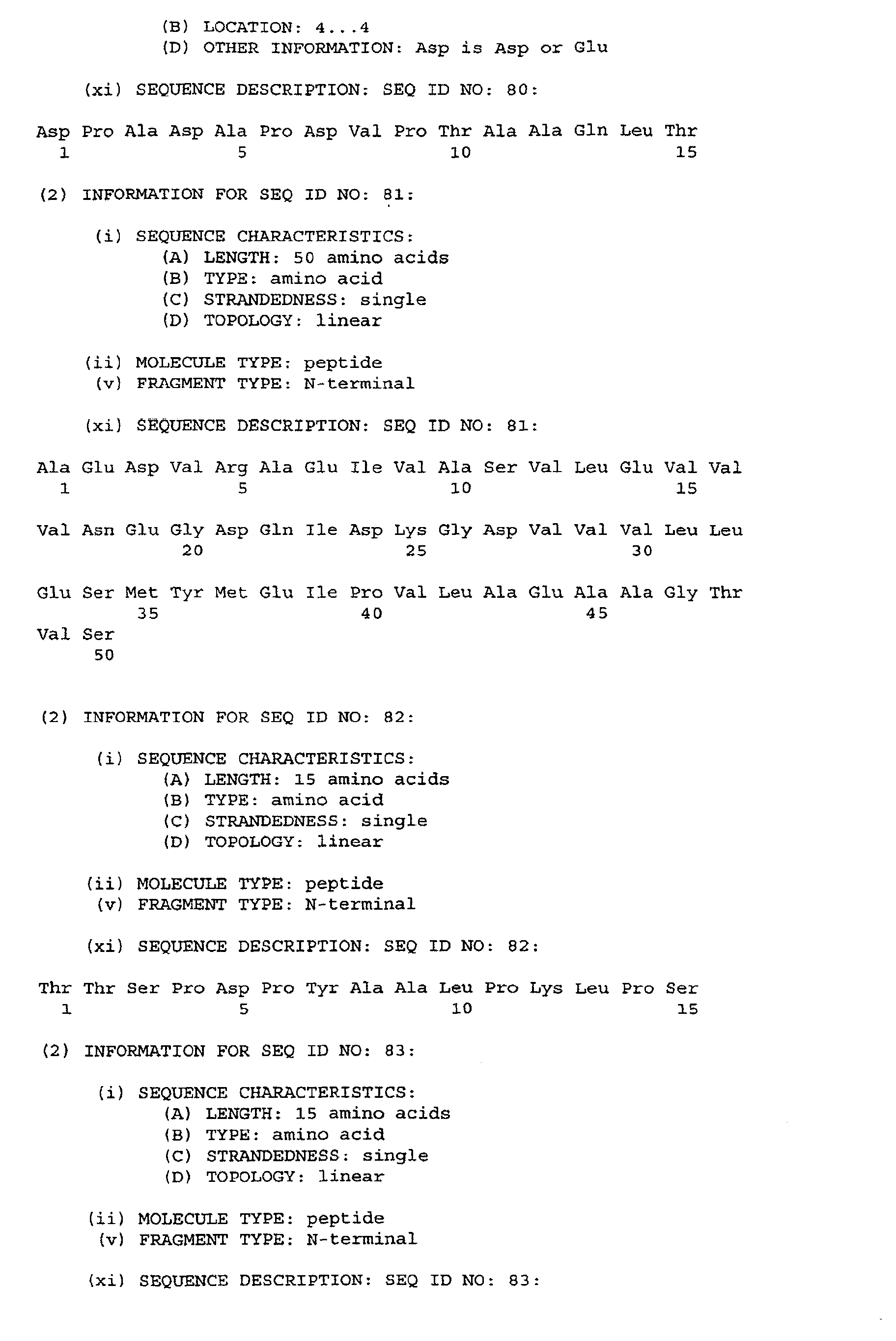
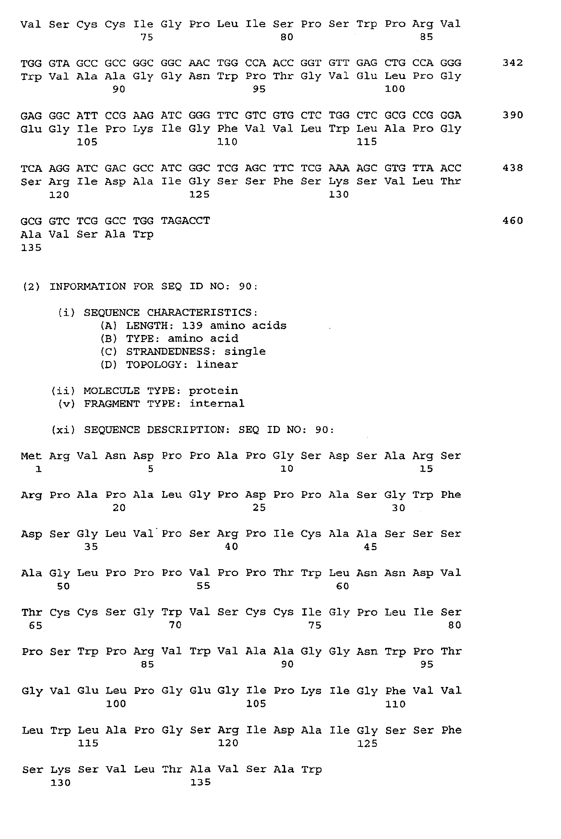
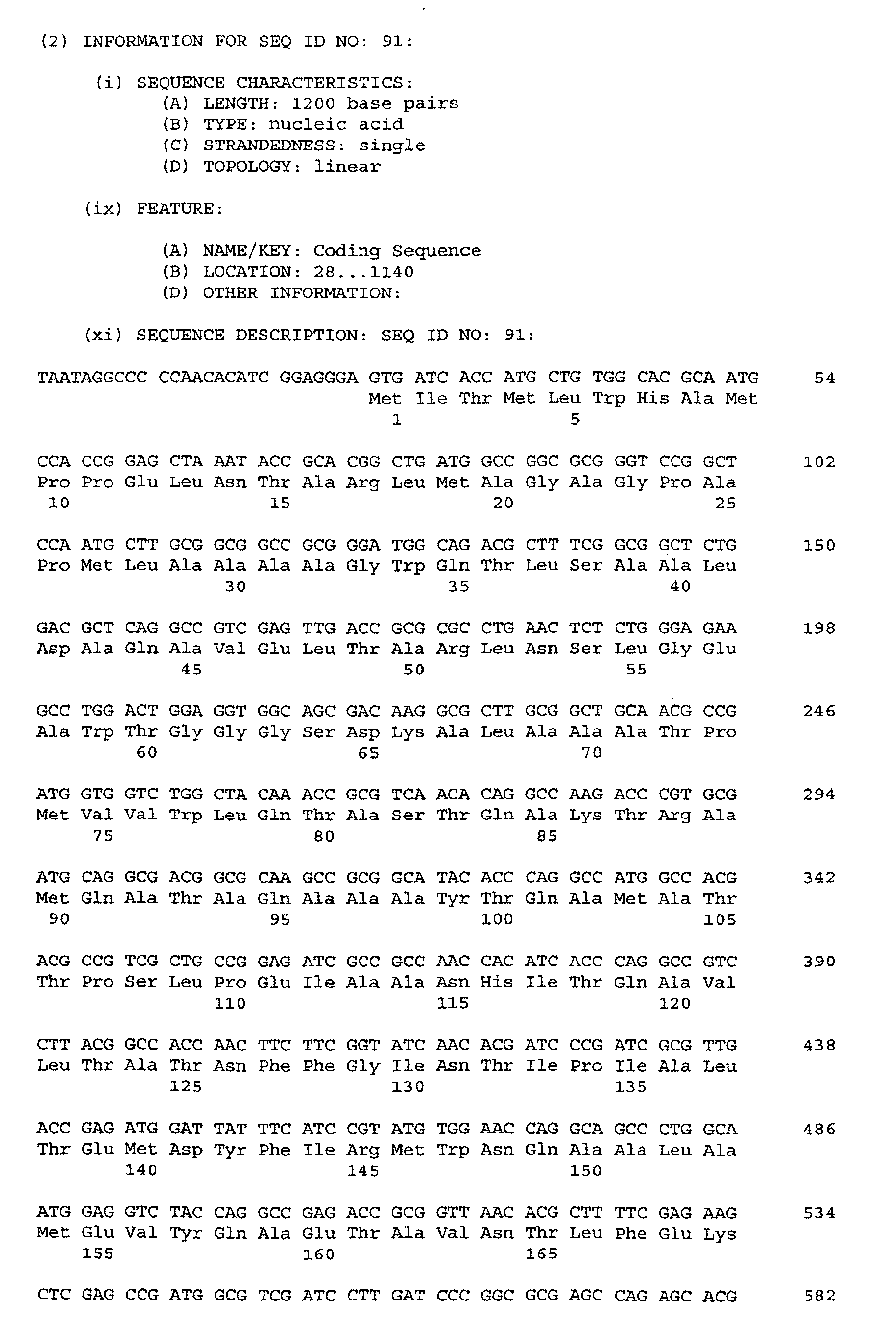
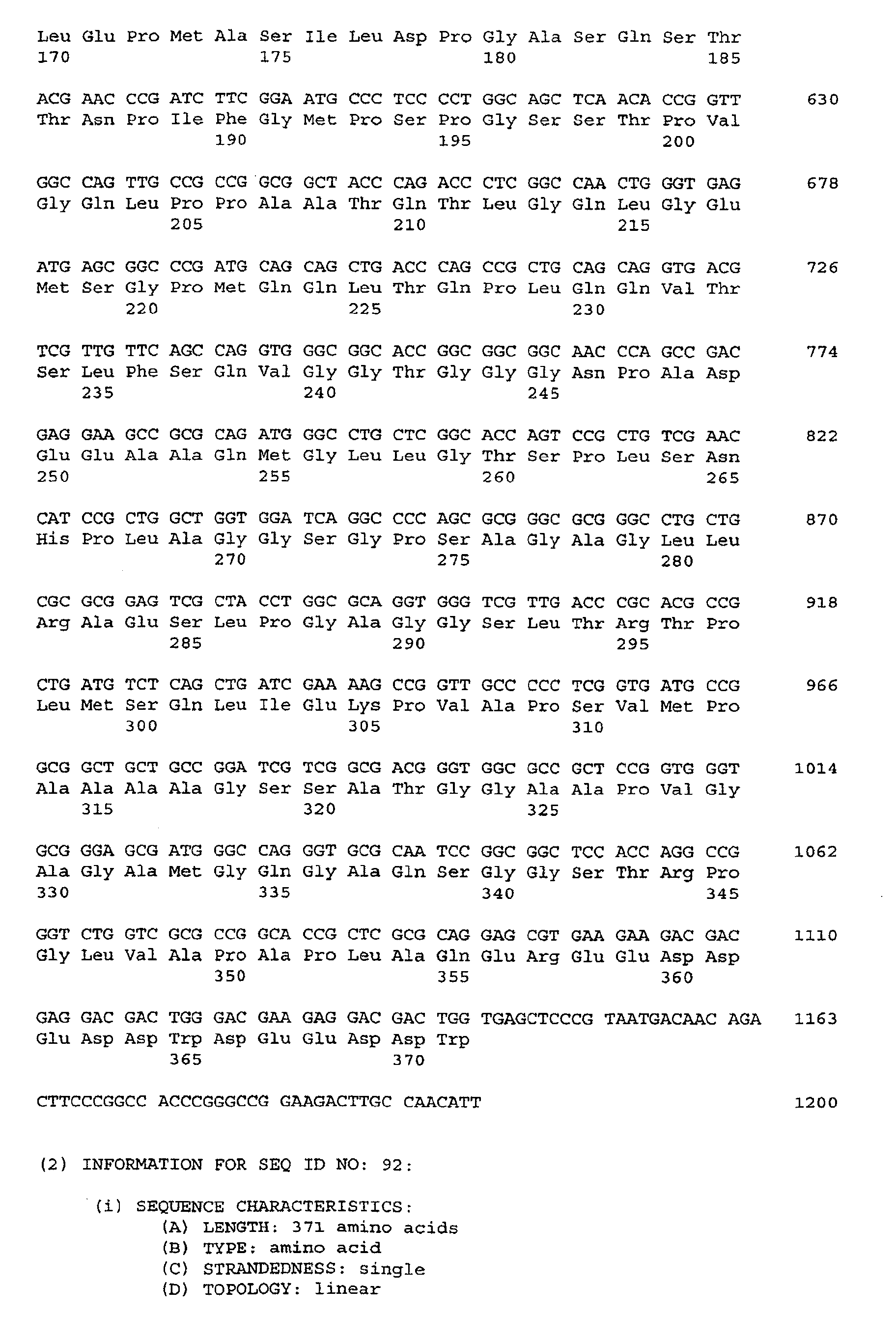
The following table lists the antigens of the invention by the names used herein as
well as by reference to relevant SEQ ID NOs of N-terminal sequences, full amino acid
sequences and sequences of DNA encoding the antigens:
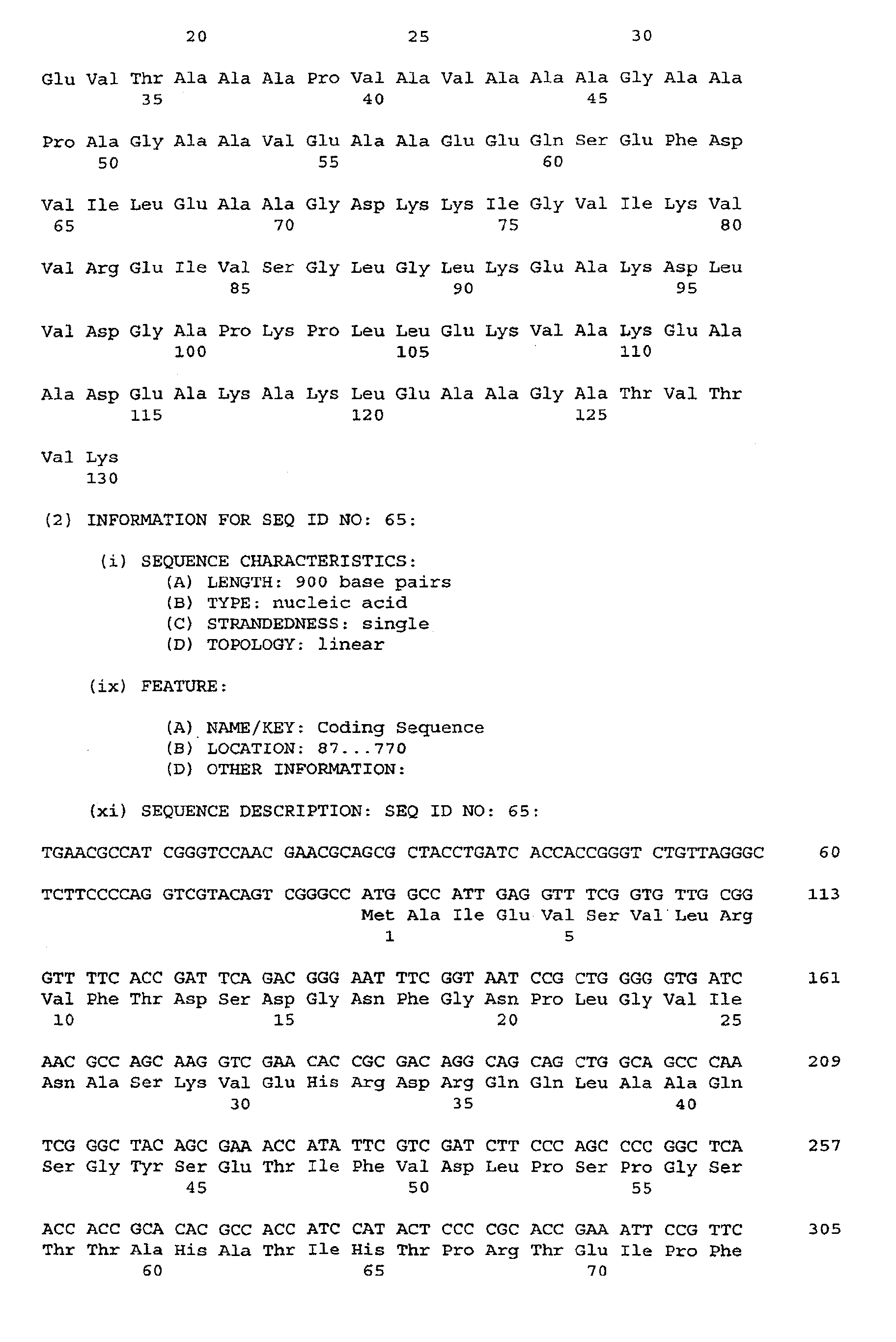
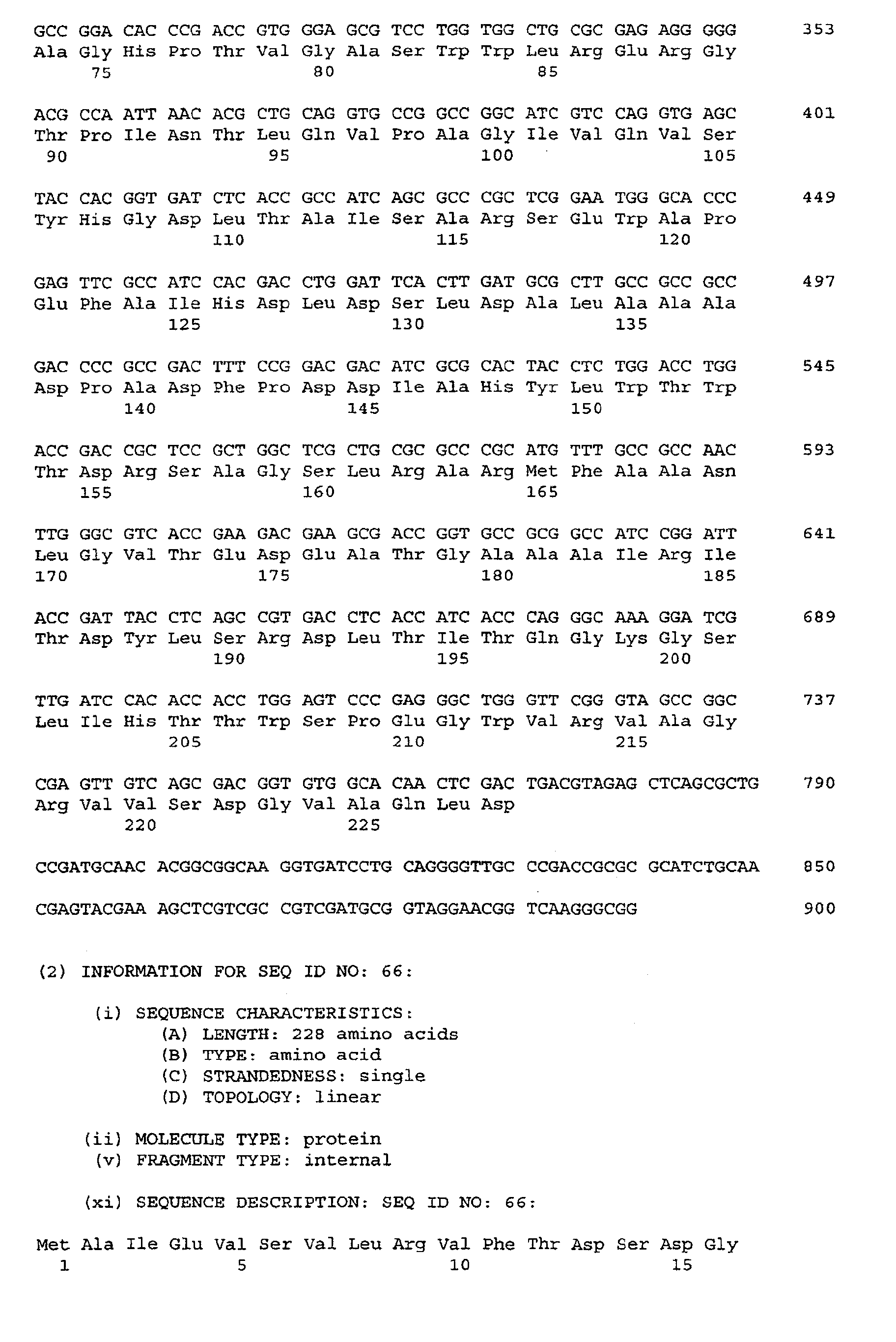
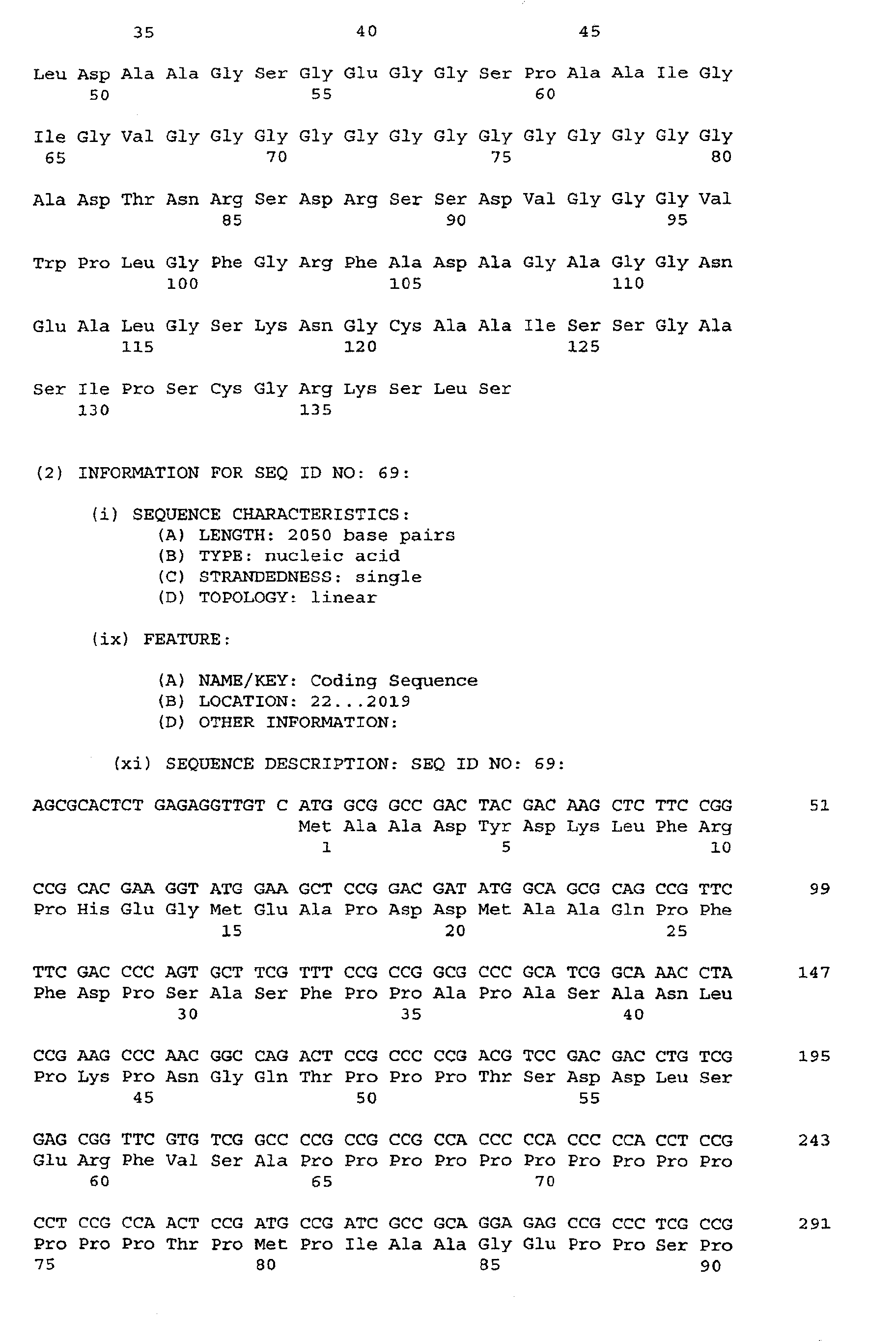
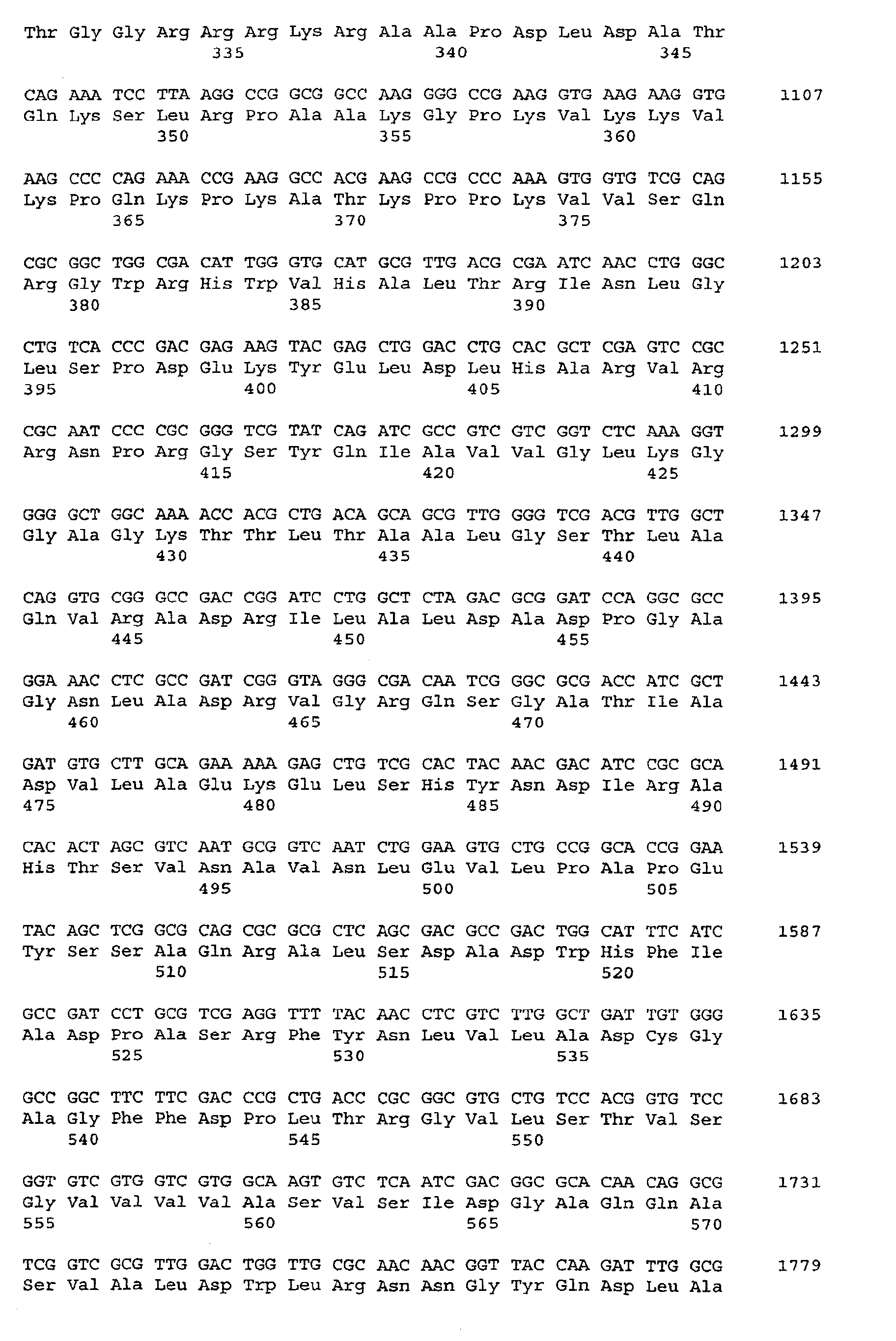
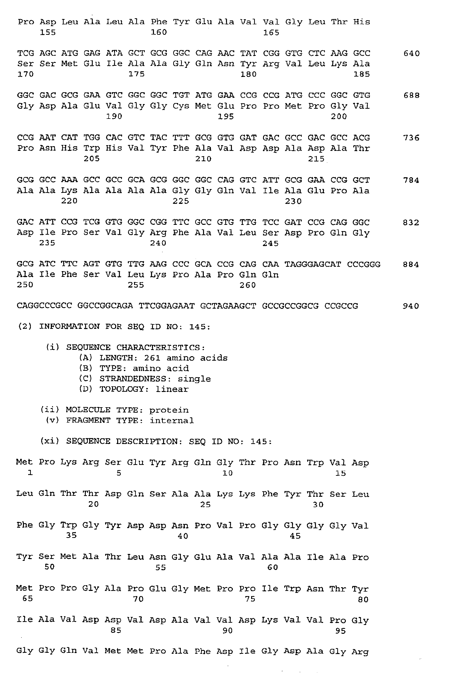
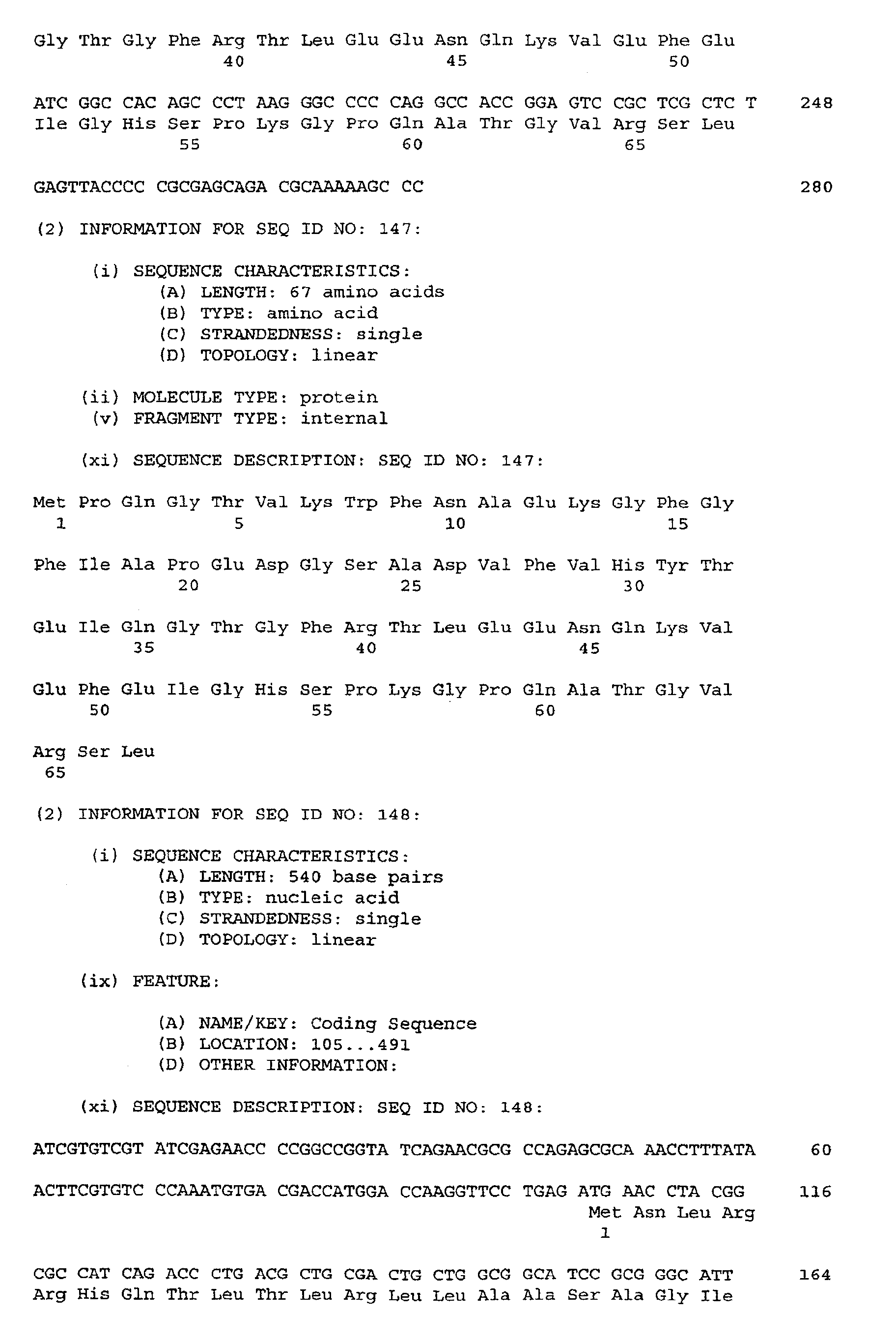
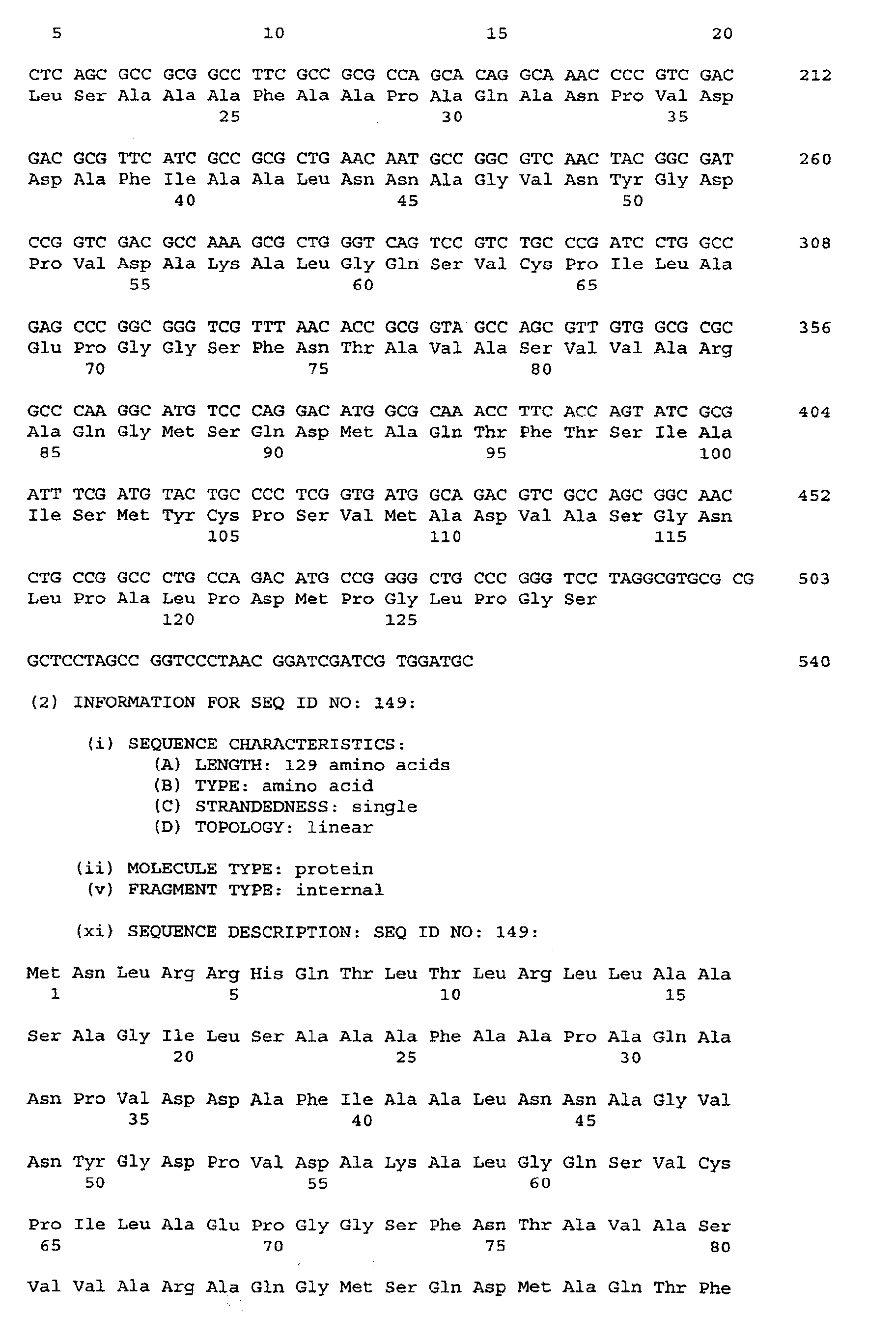
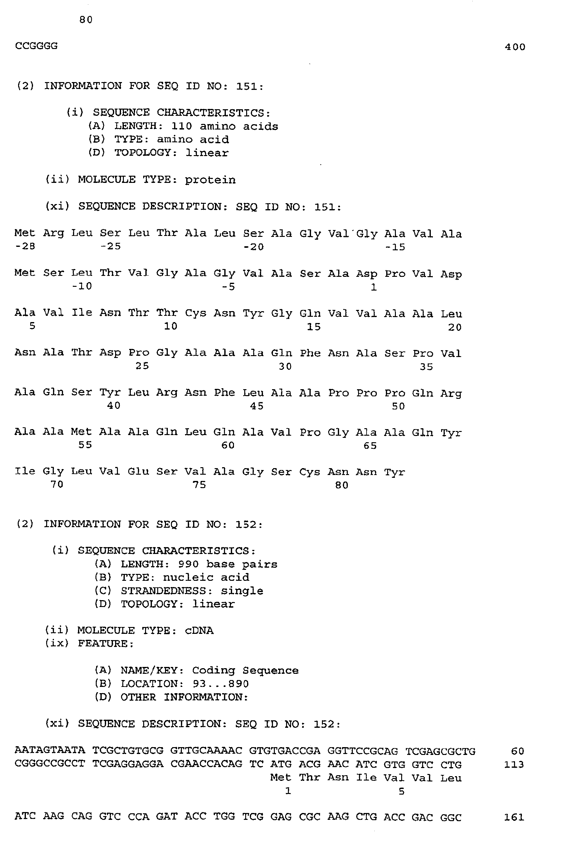
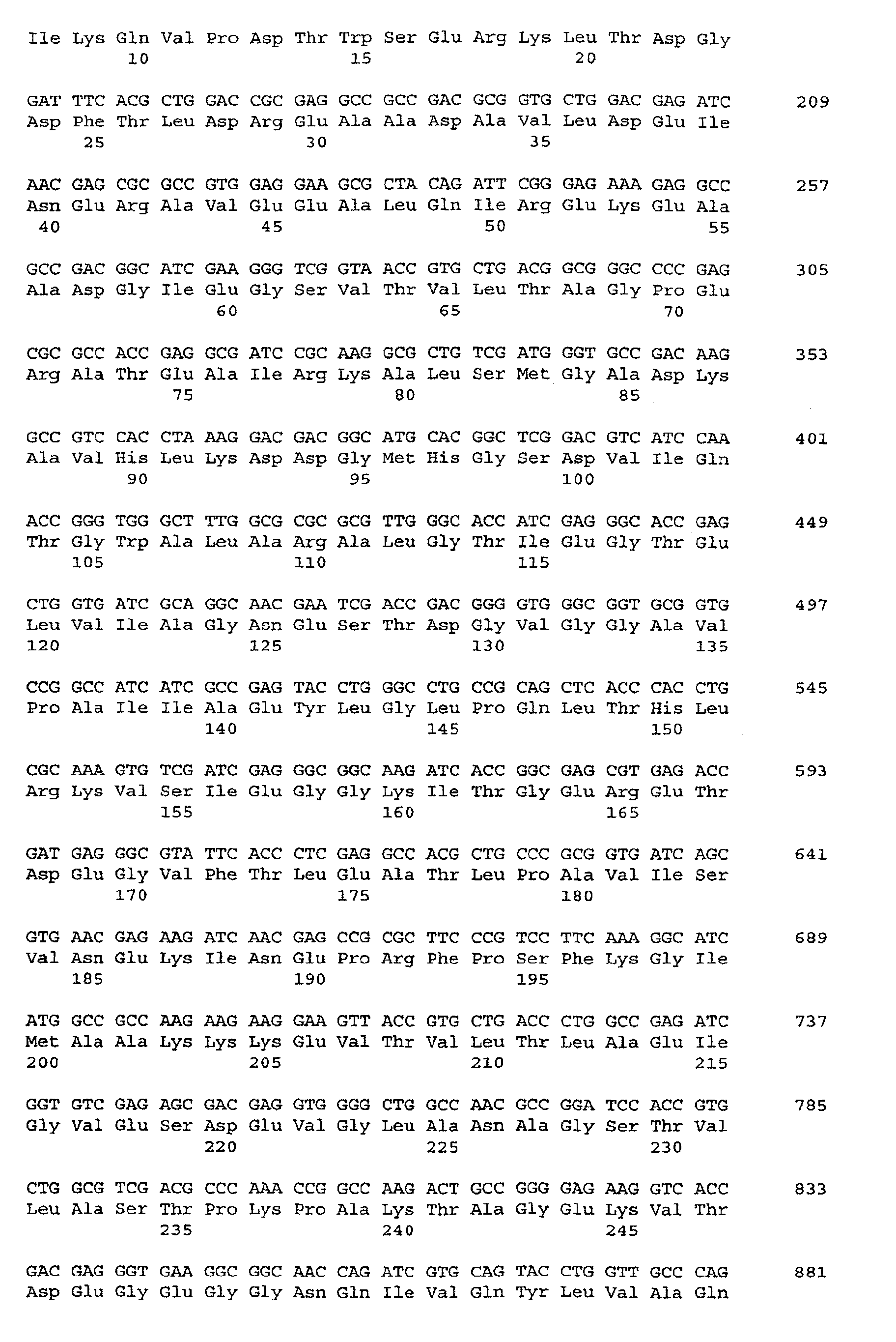
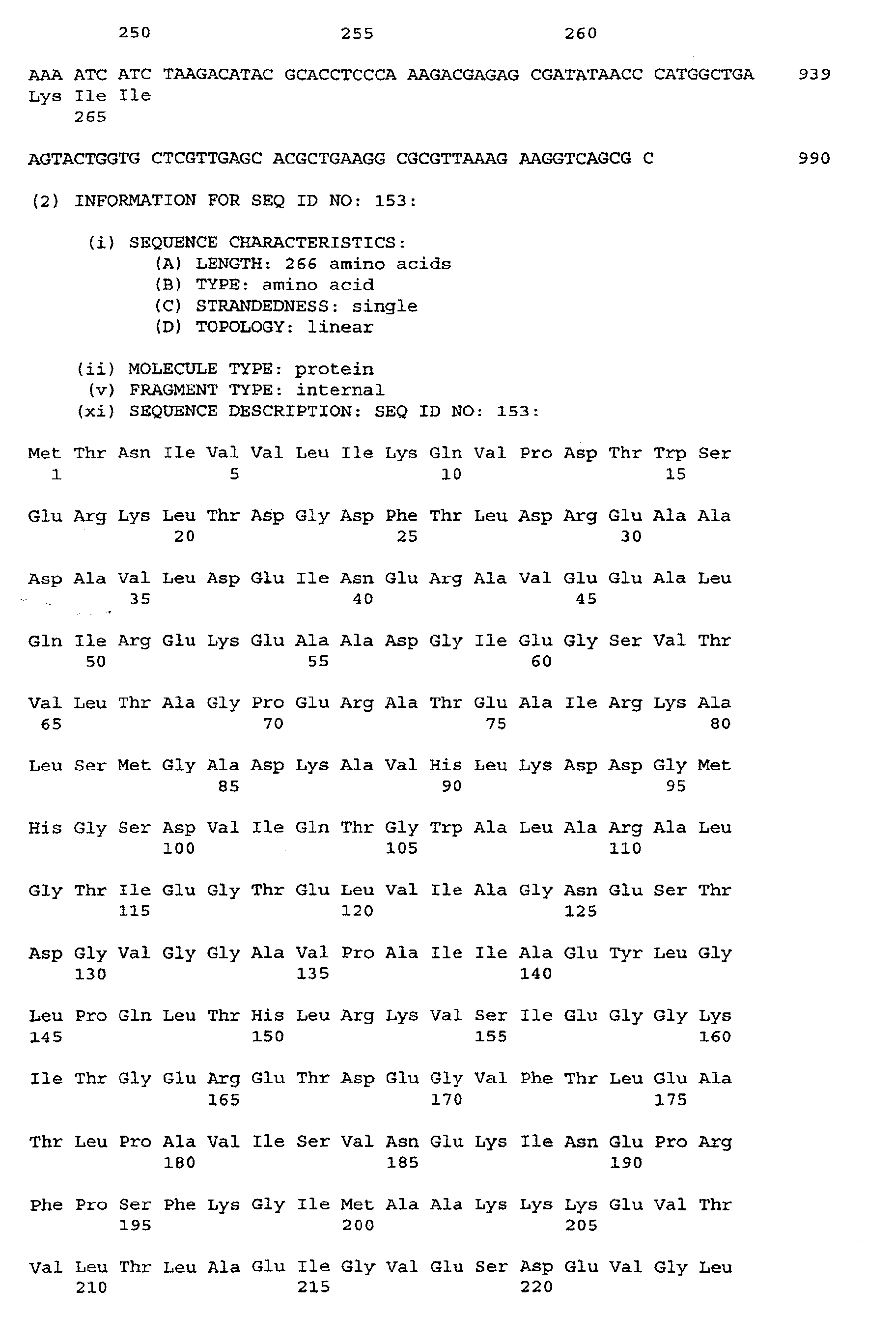
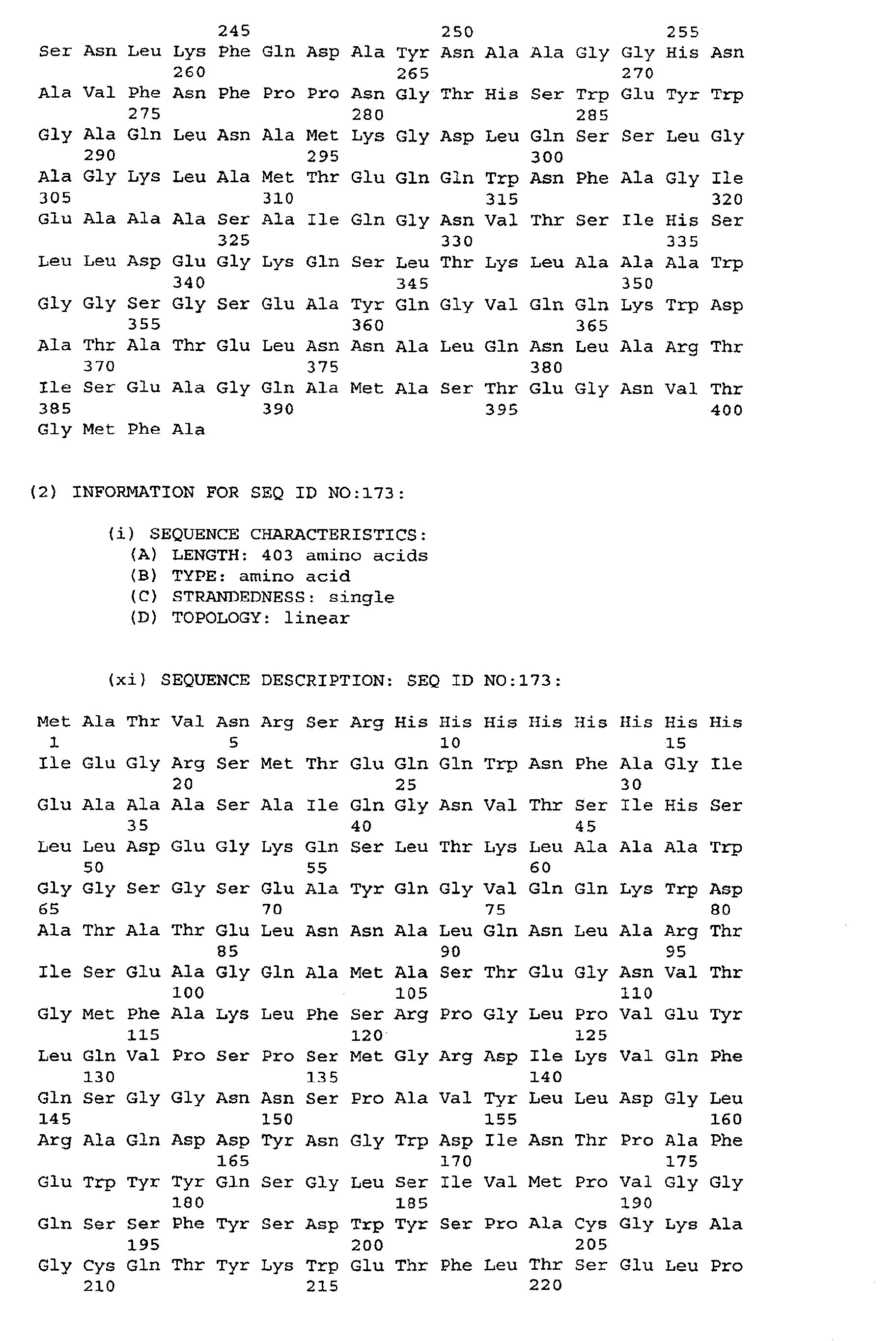
| Antigen | N-terminal sequence SEQ ID NO: | Nucleotide sequence SEQ ID NO: | Amino acid sequence SEQ ID NO: |
| CFP7 | | 1 | 2 |
| CFP7A | 81 | 47 | 48 |
| CFP7B | 168 | 146 | 147 |
| CFP8A | 73 | 148 | 149 |
| CFP8B | 74 | 150 | 151 |
| CFP9 | | 3 | 4 |
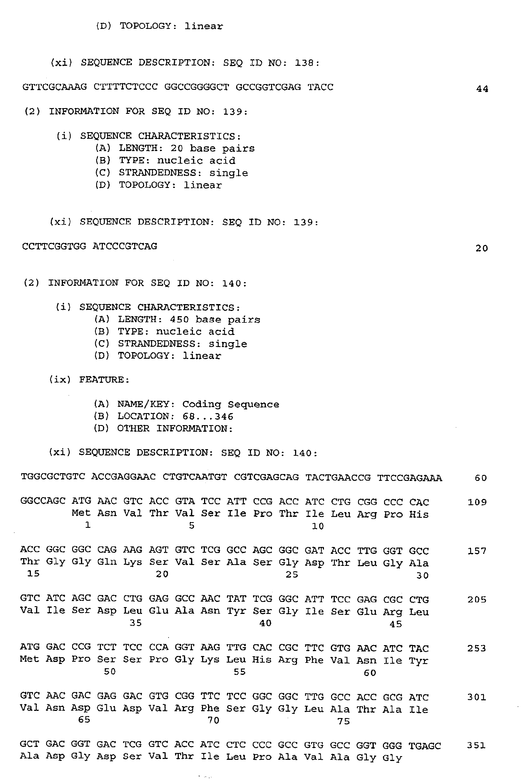
| CFP10A | 169 | 140 | 141 |
| CFP11 | 170 | 142 | 143 |
| CFP16 | 79 | 63 | 64 |
| CFP17 | 17 | 5 | 6 |
| CFP19 | 82 | 49 | 50 |
| CFP19A | | 51 | 52 |
| CFP19B | 80 |
| CFP20 | 18 | 7 | 8 |
| CFP21 | 19 | 9 | 10 |
| CFP22 | 20 | 11 | 12 |
| CFP22A | 83 | 53 | 54 |
| CFP23 | | 55 | 56 |
| CFP23A | 76 |
| CFP23B | 75 |
| CFP25 | 21 | 13 | 14 |
| CFP25A | 78 | 65 | 66 |
| CFP27 | 84 | 57 | 58 |
| CFP28 | 22 |
| CFP29 | 23 | 15 | 16 |
| CFP30A | 85 | 59 | 60 |
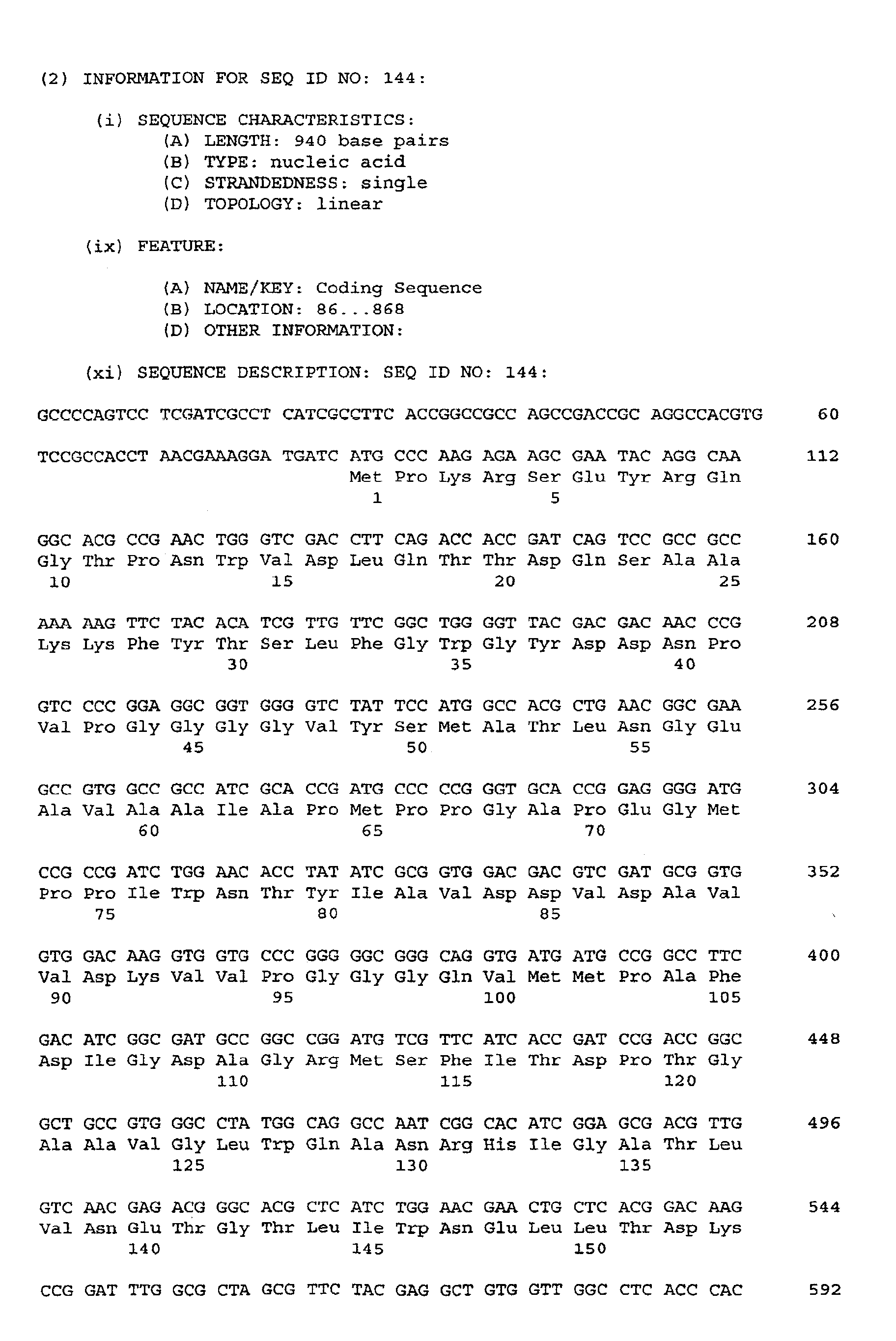
| CFP30B | 171 | 144 | 145 |
| CFP50 | 86 | 61 | 62 |
| MPT51 | | 41 | 42 |
| CWP32 | 77 | 152 | 153 |
| RD1-ORF8 | | 67 | 68 |
| RD1-ORF2 | | 71 | 72 |
| RD1-ORF9B | | 69 | 70 |
| RD1-ORF3 | | 87 | 88 |
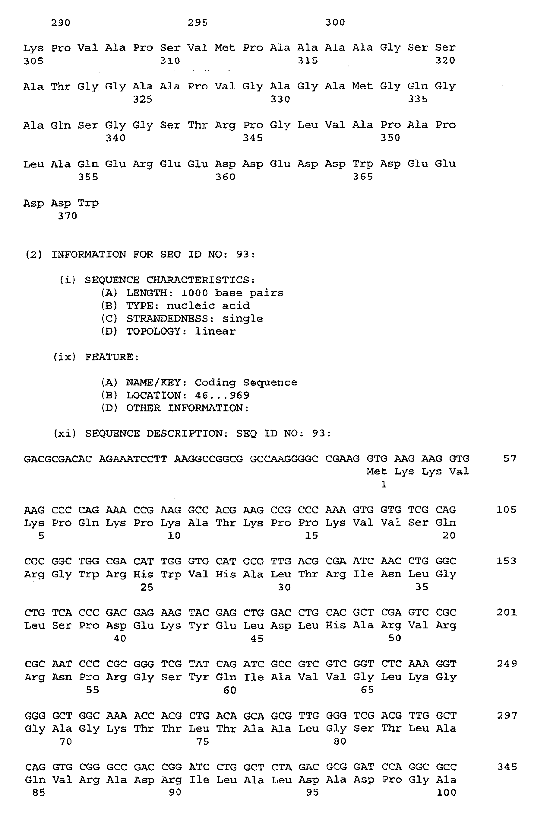
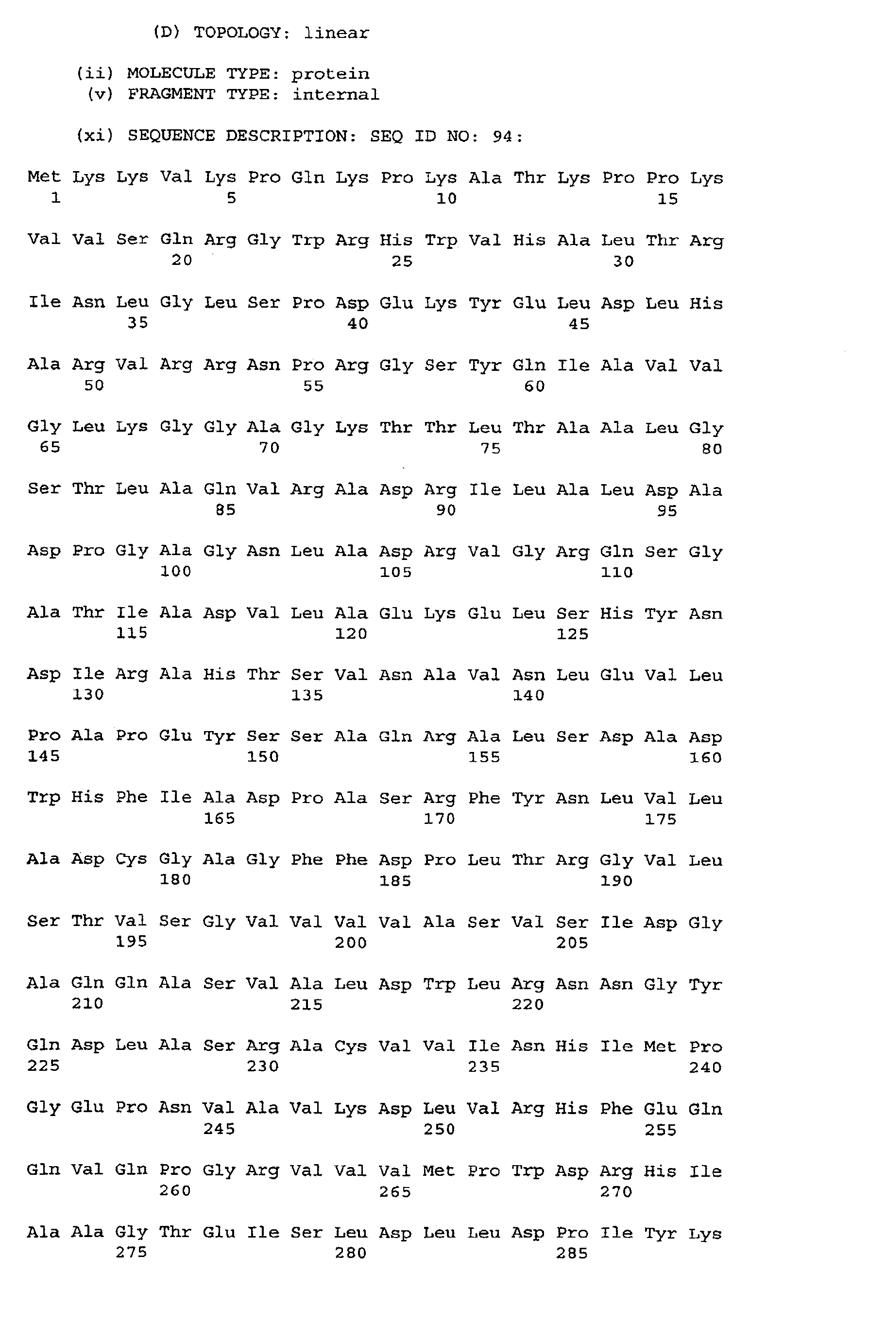
| RD1-ORF9A | | 93 | 94 |
| RD1-ORF4 | | 89 | 90 |
| RD1-ORF5 | | 91 | 92 |
| MPT59-ESAT6 | | | 172 |
| ESAT6-MPT59 | | | 173 |
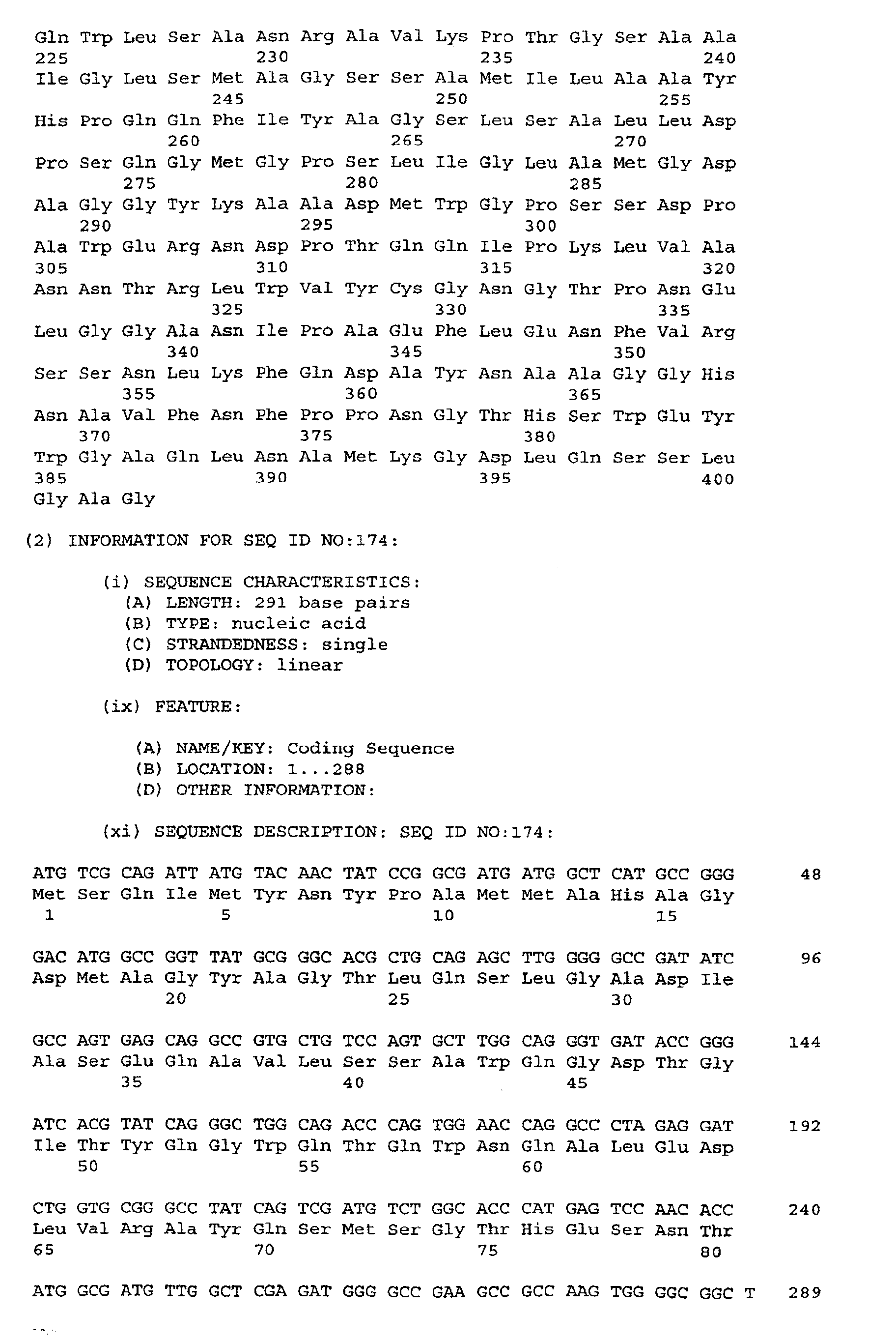
| ORF7-1 | | 174 | 175 |
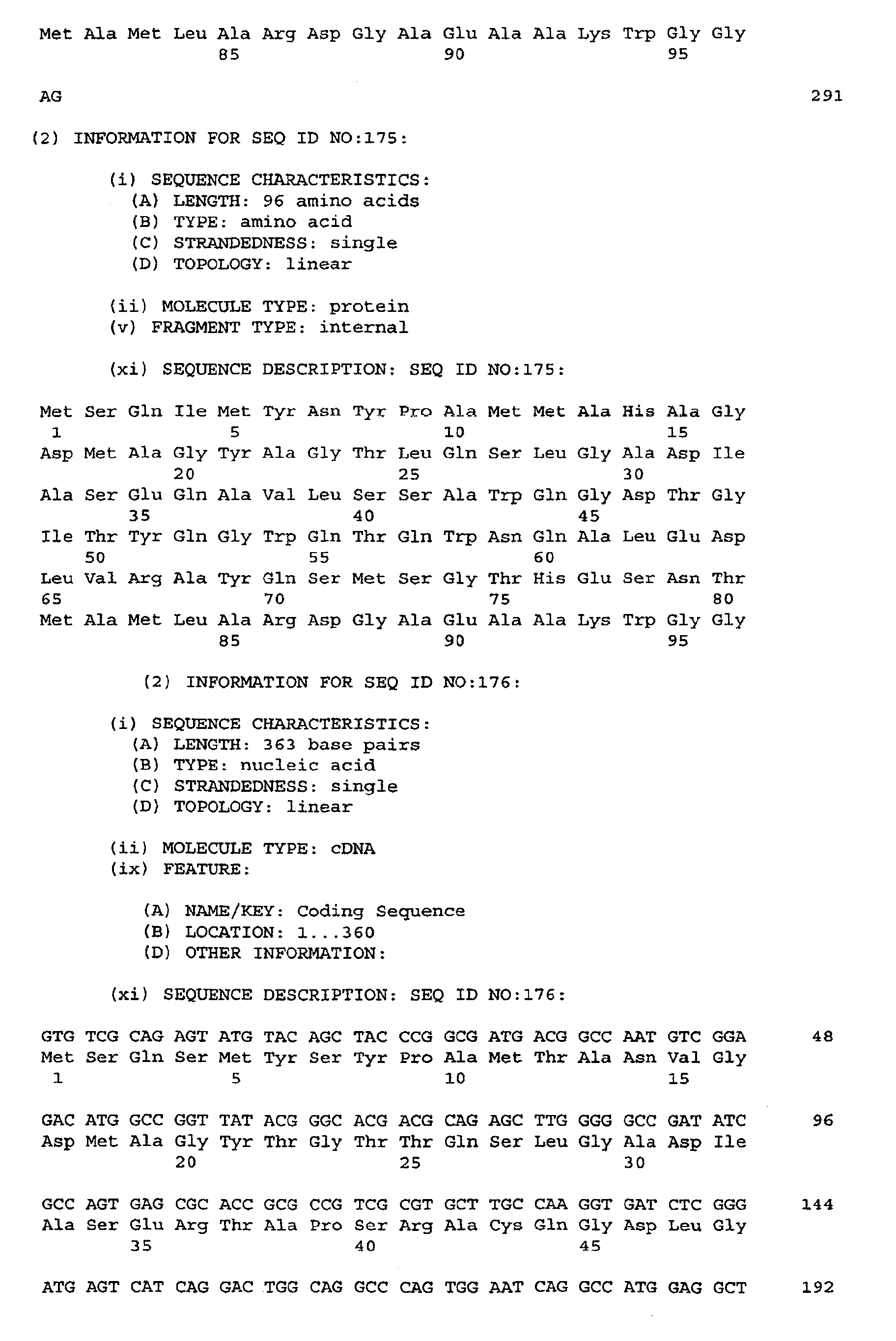
| ORF7-2 | | 176 | 177 |
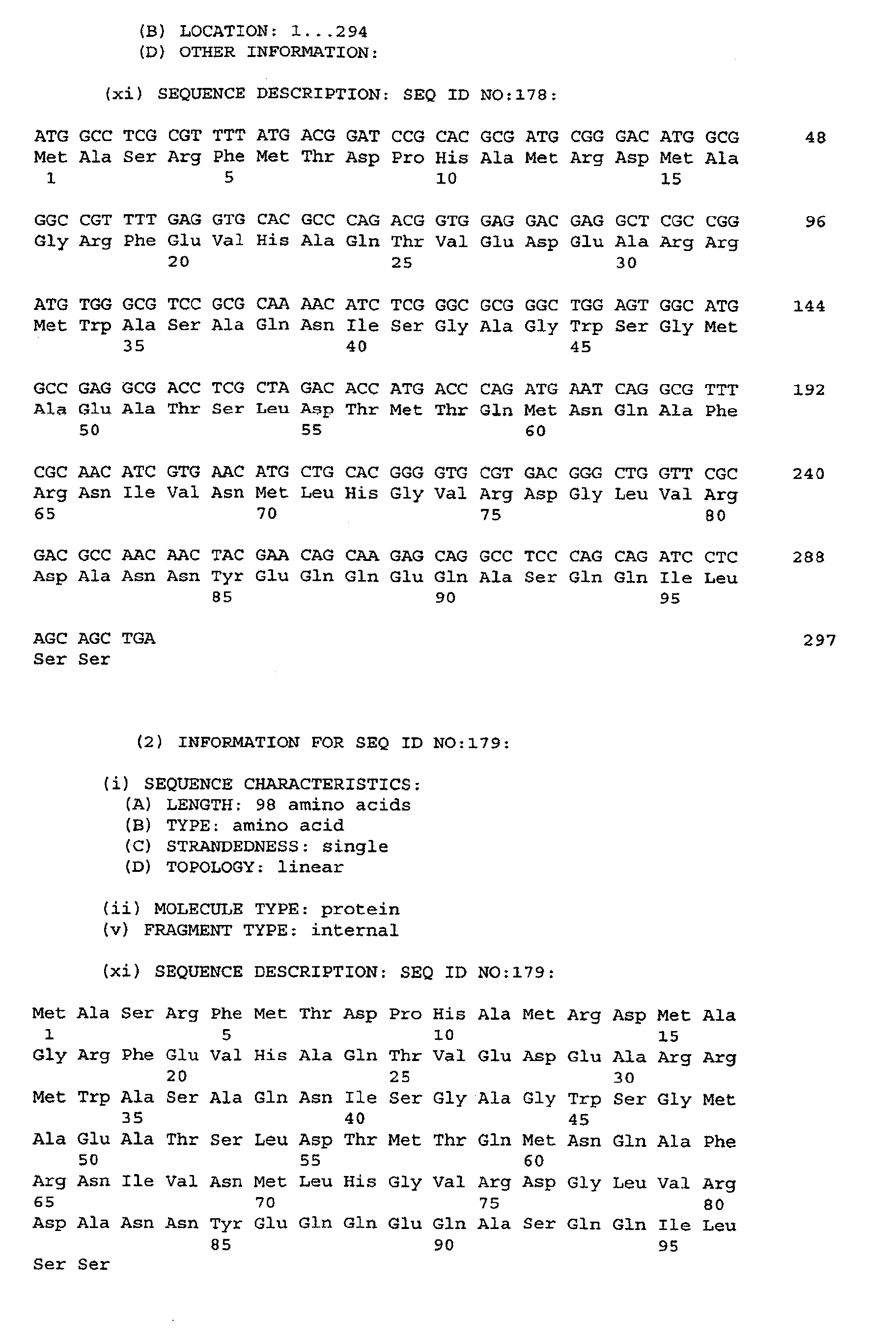
| ORF11-1 | | 178 | 179 |
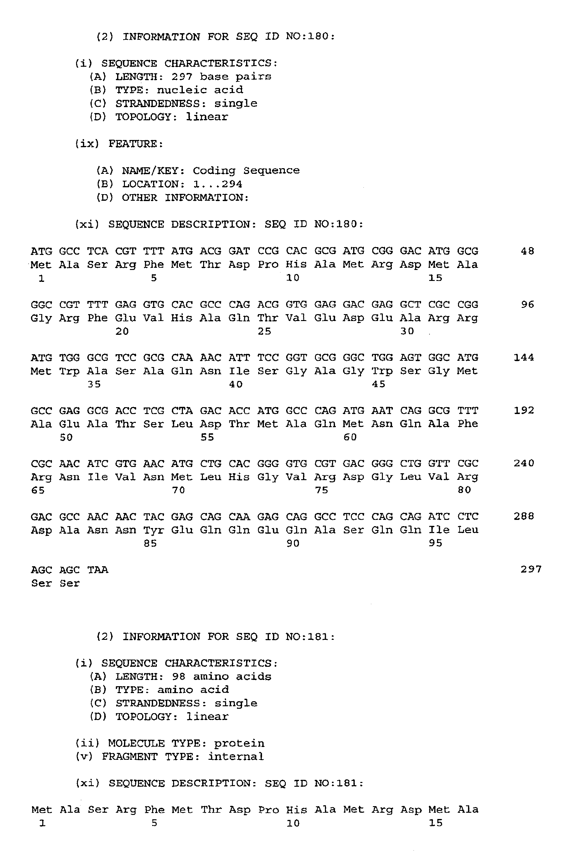
| ORF11-2 | | 180 | 181 |
| ORF11-3 | | 182 | 183 |
| ORF11-4 | | 184 | 185 |
-
It is well-known in the art that T-cell epitopes are responsible for the elicitation of the
acquired immunity against TB, whereas B-cell epitopes are without any significant influence
on acquired immunity and recognition of mycobacteria in vivo. Since such T-cell
epitopes are linear and are known to have a minimum length of 6 amino acid residues,
the present invention is especially concerned with the identification and utilisation
of such T-cell epitopes.
-
Hence, in its broadest aspect the invention relates to a substantially pure polypeptide
fragment which
- a) comprises an amino acid sequence selected from the sequences shown in SEQ
ID NO: 2, 4, 6, 8, 10, 12, 14, 16, any one of 17-23, 42, 48, 50, 52, 54, 56, 58, 60,
62, 64, 66, 68, 70, any one of 72-86, 88, 90, 92, 94, 141, 143, 145, 147, 149,
151, 153, any one of 168-171, 175, 177, 179, 181, 183, and 185
- b) comprises a subsequence of the polypeptide fragment defined in a) which has a
length of at least 6 amino acid residues, said subsequence being immunologically
equivalent to the polypeptide defined in a) with respect to the ability of evoking a protective
immune response against infections with mycobacteria belonging to the tuberculosis
complex or with respect to the ability of eliciting a diagnostically significant
immune response indicating previous or ongoing sensitization with antigens derived
from mycobacteria belonging to the tuberculosis complex, or
- c) comprises an amino acid sequence having a sequence identity with the polypeptide
defined in a) or the subsequence defined in b) of at least 70% and at the
same time being immunologically equivalent to the polypeptide defined in a) with
respect to the ability of evoking a protective immune response against infections with
mycobacteria belonging to the tuberculosis complex or with respect to the ability of
eliciting a diagnostically significant immune response indicating previous or ongoing
sensitization with antigens derived from mycobacteria belonging to the tuberculosis
complex,
with the proviso that
- i) the polypeptide fragment is in essentially pure form when consisting of the amino
acid sequence 1-96 of SEQ ID NO: 2 or when consisting of the amino acid sequence
87-108 of SEQ ID NO: 4 fused to β-galactosidase,
- ii) the degree of sequence identity in c) is at least 95 % when the polypeptide comprises
a homologue of a polypeptide which has the amino acid sequence SEQ ID NO:
12 or a subsequence thereof as defined in b), and
- iii) the polypeptide fragment contains a threonine residue corresponding to position
213 in SEQ ID NO: 42 when comprising an amino acid sequence of at least 6 amino
acids in SEQ ID NO: 42.
-
-
Other parts of the invention pertains to the DNA fragments encoding a polypeptide
with the above definition as well as to DNA fragments useful for determining the presence
of DNA encoding such polypeptides.
DETAILED DISCLOSURE OF THE INVENTION
-
In the present specification and claims, the term "polypeptide fragment" denotes both
short peptides with a length of at least two amino acid residues and at most 10 amino
acid residues, oligopeptides (11-100 amino acid residues), and longer peptides (the
usual interpretation of "polypeptide", i.e. more than 100 amino acid residues in length)
as well as proteins (the functional entity comprising at least one peptide, oligopeptide,
or polypeptide which may be chemically modified by being glycosylated, by being lipidated,
or by comprising prosthetic groups). The definition of polypeptides also comprises
native forms of peptides/proteins in mycobacteria as well as recombinant proteins
or peptides in any type of expression vectors transforming any kind of host, and
also chemically synthesized peptides.
-
In the present context the term "substantially pure polypeptide fragment" means a
polypeptide preparation which contains at most 5% by weight of other polypeptide
material with which it is natively associated (lower percentages of other polypeptide
material are preferred, e.g. at most 4%, at most 3%, at most 2%, at most 1 %, and at
most ½ %). It is preferred that the substantially pure polypeptide is at least 96% pure,
i.e. that the polypeptide constitutes at least 96% by weight of total polypeptide material
present in the preparation, and higher percentages are preferred, such as at least
97%, at least 98%, at least 99%, at least 99,25%, at least 99,5%, and at least
99,75%. It is especially preferred that the polypeptide fragment is in "essentially pure
form", i.e. that the polypeptide fragment is essentially free of any other antigen with
which it is natively associated, i.e. free of any other antigen from bacteria belonging
to the tuberculosis complex. This can be accomplished by preparing the polypeptide
fragment by means of recombinant methods in a non-mycobacterial host cell as will
be described in detail below, or by synthesizing the polypeptide fragment by the well-known
methods of solid or liquid phase peptide synthesis, e.g. by the method described
by Merrifield or variations thereof.
-
The term "subsequence" when used in connection with a polypeptide of the invention
having a SEQ ID NO selected from 2, 4, 6, 8, 10, 12, 14, 16, any one of 17-23, 42,
48, 50, 52, 54, 56, 58, 60, 62, 64, 66, 68, 70, any one of 72-86, 88, 90, 92, 94,
141, 143, 145, 147, 149, 151, 153, any one of 168-171, 175, 177, 179, 181, 183,
and 185 denotes any continuous stretch of at least 6 amino acid residues taken from
the M. tuberculosis derived polypeptides in SEQ ID NO: 2, 4, 6, 8, 10, 12, 14, 16,
any one of 17-23, 42, 48, 50, 52, 54, 56, 58, 60, 62, 64, 66, 68, 70, any one of
72-86, 88, 90, 92, 94, 141, 143, 145, 147, 149, 151, 153, any one of 168-171,
175, 177, 179, 181, 183, and 185 and being immunological equivalent thereto with
respect to the ability of conferring increased resistance to infections with bacteria belonging
to the tuberculosis complex. Thus, included is also a polypeptide from different
sources, such as other bacteria or even from eukaryotic cells.
-
When referring to an "immunologically equivalent" polypeptide is herein meant that
the polypeptide, when formulated in a vaccine or a diagnostic agent (
i.
e. together with
a pharmaceutically acceptable carrier or vehicle and optionally an adjuvant), will
- I) confer, upon administration (either alone or as an immunologically active constituent
together with other antigens), an acquired increased specific resistance in a
mouse and/or in a guinea pig and/or in a primate such as a human being against infections
with bacteria belonging to the tuberculosis complex which is at least 20% of the
acquired increased resistance conferred by Mycobacterium bovis BCG and also at least
20% of the acquired increased resistance conferred by the parent polypeptide comprising
SEQ ID NO: 2, 4, 6, 8, 10, 12, 14, 16, any one of 17-23, 42, 48, 50, 52, 54,
56, 58, 60, 62, 64, 66, 68, 70, any one of 72-86, 88, 90, 92, 94, 141, 143, 145,
147, 149, 151, 153, any one of 168-171, 175, 177, 179, 181, 183, or 185 (said
parent polypeptide having substantially the same relative location and pattern in a 2DE
gel prepared as the 2DE gel shown in Fig. 6, cf. the examples), the acquired increased
resistance being assessed by the observed reduction in mycobacterial counts from
spleen, lung or other organ homogenates isolated from the mouse or guinea pig receiving
a challenge infection with a virulent strain of M. tuberculosis, or, in a primate
such as a human being, being assessed by determining the protection against development
of clinical tuberculosis in a vaccinated group versus that observed in a control
group receiving a placebo or BCG (preferably the increased resistance is higher and
corresponds to at least 50% of the protective immune response elicited by M. bovis
BCG, such as at least 60%, or even more preferred to at least 80% of the protective
immune response elicited by M. bovis BCG, such as at least 90%; in some cases it is
expected that the increased resistance will supersede that conferred by M. bovis BCG,
and hence it is preferred that the resistance will be at least 100%, such as at least
110% of said increased resistance); and/or
- II) elicit a diagnostically significant immune response in a mammal indicating previous
or ongoing sensitization with antigens derived from mycobacteria belonging to
the tuberculosis complex; this diagnostically significant immune response can be in
the form of a delayed type hypersensitivity reaction which can e.g. be determined by
a skin test, or can be in the form of IFN-γ release determined e.g. by an IFN-γ assay as
described in detail below. A diagnostically significant response in a skin test setup will
be a reaction which gives rise to a skin reaction which is at least 5 mm in diameter
and which is at least 65% (preferably at least 75% such as at the least 85%) of the
skin reaction (assessed as the skin reaction diameter) elicited by the parent polypeptide
comprising SEQ ID NO: 2, 4, 6, 8, 10, 12, 14, 16, any one of 17-23, 42, 48, 50,
52, 54, 56, 58, 60, 62, 64, 66, 68, 70, any one of 72-86, 88, 90, 92, 94, 141,
143, 145, 147, 149, 151, 153, any one of 168-171, 175, 177, 179, 181, 183, or
185.
-
-
The ability of the polypeptide fragment to confer increased immunity may thus be assessed
by measuring in an experimental animal, e.g. a mouse or a guinea pig, the reduction
in mycobacterial counts from the spleen, lung or other organ homogenates
isolated from the experimental animal which have received a challenge infection with a
virulent strain of mycobacteria belonging to the tuberculosis complex after previously
having been immunized with the polypeptide, as compared to the mycobacterial
counts in a control group of experimental animals infected with the same virulent
strain, which experimental animals have not previously been immunized against tuberculosis.
The comparison of the mycobacterial counts may also be carried out with mycobacterial
counts from a group of experimental animals receiving a challenge infection
with the same virulent strain after having been immunized with Mycobacterium
bovis BCG.
-
The mycobacterial counts in homogenates from the experimental animals immunized
with a polypeptide fragment according to the present invention must at the most be 5
times the counts in the mice or guinea pigs immunized with Mycobacterium bovis
BCG, such as at the most 3 times the counts, and preferably at the most 2 times the
counts.
-
A more relevant assessment of the ability of the polypeptide fragment of the invention
to confer increased resistance is to compare the incidence of clinical tuberculosis in
two groups of individuals (e.g. humans or other primates) where one group receives a
vaccine as described herein which contains an antigen of the invention and the other
group receives either a placebo or an other known TB vaccine (e.g. BCG). In such a
setup, the antigen of the invention should give rise to a protective immunity which is
significantly higher than the one provided by the administration of the placebo (as determined
by statistical methods known to the skilled artisan).
-
In the context of the present application, the term "wide genetically" should be understood
in a meaning of at least two strains. That is, if a polypeptide is recognised by at
least two different strains, it is considered to have a wide genetically recognition.
A subunit vaccine component is defined as a reagent which stimulates protective immunity
in an animal model of infection with an organism of the M. tuberculosis complex,
when given prior to infection and which also generates a significant immune responses
in human volunteers.
-
The "tuberculosis-complex" has its usual meaning, i.e. the complex of mycobacteria
causing TB which are Mycobacterium tuberculosis, Mycobacterium bovis, Mycobacterium
bovis BCG, and Mycobacterium africanum.
-
In the present context the term "metabolizing mycobacteria" means live mycobacteria
that are multiplying logarithmically and releasing polypeptides into the culture medium
wherein they are cultured.
-
The term "sequence identity" indicates a quantitative measure of the degree of homology
between two amino acid sequences or between two nucleotide sequences of
equal length, of if not of equal length aligned to best possible fit: The sequence
identity can be calculated as (Nref-Ndif )100 / N ref 1, wherein Ndif is the total number of non-identical
residues in the two sequences when aligned and wherein Nref is the number
of residues in one of the sequences. Hence, the DNA sequence AGTCAGTC will have
a sequence identity of 75% with the sequence AATCAATC (Ndif=2 and Nref=8). A
gap is counted as non-identity of the specific residue(s), i.e. the DNA sequence
AGTGTC will have a sequence identity of 75% with the DNA sequence AGTCAGTC
(Ndif = 2 and Nref = 8). Sequence identity can alternatively be calculated by the BLASTP
program ((Pearson W.R and D.J. Lipman (1988) PNAS USA 85:2444-2448) in the
EMBL database (www.ncbi.nlm.gov/cgi-bin/BLAST). Generally, the default settings
with respect to e.g. "scoring matrix" and "gap penalty" will be used for alignment.
-
The sequence identity is used here to illustrate the degree of identity between the
amino acid sequence of a given polypeptide and the amino acid sequence shown in
SEQ ID NO: 2, 4, 6, 8, 10, 12, 14, 16, any one of 17-23, 42, 48, 50, 52, 54, 56,
58, 60, 62, 64, 66, 68, 70, any one of 72-86, 88, 90, 92, 94, 141, 143, 145, 147,
149, 151, 153, any one of 168-171, 175, 177, 179, 181, 183, or 185. The amino
acid sequence to be compared with the amino acid sequence shown in SEQ ID NO: 2,
4, 6, 8, 10, 12, 14, 16, any one of 17-23, 42, 48, 50, 52, 54, 56, 58, 60, 62, 64,
66, 68, 70, any one of 72-86, 88, 90, 92, 94, 141, 143, 145, 147, 149, 151, 153,
any one of 168-171, 175, 177, 179, 181, 183, or 185 may be deduced from a DNA
sequence, e.g. obtained by hybridization as defined below, or may be obtained by
conventional amino acid sequencing methods. The sequence identity is preferably determined
on the amino acid sequence of a mature polypeptide, i.e. without taking any
leader sequence into consideration.
-
As appears from the above disclosure, polypeptides which are not identical to the
polypeptides having SEQ ID NO: 2, 4, 6, 8, 10, 12, 14, 16, any one of 17-23, 42,
48, 50, 52, 54, 56, 58, 60, 62, 64, 66, 68, 70, any one of 72-86, 88, 90, 92, 94,
141, 143, 145, 147, 149, 151, 153, any one of 168-171, 175, 177, 179, 181, 183,
or 185 are embraced by the present invention. The invention allows for minor variations
which do not have an adverse effect on immunogenicity compared to the parent
sequences and which may give interesting and useful novel binding properties or biological
functions and immunogenicities etc.
-
Each polypeptide fragment may thus be characterized by specific amino acid and nucleic
acid sequences. It will be understood that such sequences include analogues and
variants produced by recombinant methods wherein such nucleic acid and polypeptide
sequences have been modified by substitution, insertion, addition and/or deletion of
one or more nucleotides in said nucleic acid sequences to cause the substitution, insertion,
addition or deletion of one or more amino acid residues in the recombinant
polypeptide. When the term DNA is used in the following, it should be understood that
for the number of purposes where DNA can be substituted with RNA, the term DNA
should be read to include RNA embodiments which will be apparent for the man
skilled in the art. For the purposes of hybridization, PNA or LNA may be used instead
of DNA. PNA has been shown to exhibit a very dynamic hybridization profile (PNA is
described in Nielsen P E et al., 1991, Science 254: 1497-1500). LNA (Locked Nucleic
Acids) is a recently introduced oligonucleotide analogue containing bicyclo nucleoside
monomers (Koshkin et al., 1998, 54, 3607-3630;Nielsen, N.K. et al. J.Am.Chem.Soc
1998, 120, 5458-5463).
-
In both immunodiagnostics and vaccine preparation, it is often possible and practical
to prepare antigens from segments of a known immunogenic protein or polypeptide.
-
Certain epitopic regions may be used to produce responses similar to those produced
by the entire antigenic polypeptide. Potential antigenic or immunogenic regions may be
identified by any of a number of approaches, e.g., Jameson-Wolf or Kyte-Doolittle antigenicity
analyses or Hopp and Woods (1981) hydrophobicity analysis (see, e.g., Jameson
and Wolf, 1988; Kyte and Doolittle, 1982; or U.S. Patent No. 4,554,101). Hydrophobicity
analysis assigns average hydrophilicity values to each amino acid residue
from these values average hydrophilicities can be calculated and regions of greatest
hydrophilicity determined. Using one or more of these methods, regions of predicted
antigenicity may be derived from the amino acid sequence assigned to the polypeptides
of the invention.
-
Alternatively, in order to identify relevant T-cell epitopes which are recognized during
an immune response, it is also possible to use a "brute force" method: Since T-cell
epitopes are linear, deletion mutants of polypeptides having SEQ ID NO: 2, 4, 6, 8,
10, 12, 14, 16, any one of 17-23, 42, 48, 50, 52, 54, 56, 58, 60, 62, 64, 66, 68,
70, any one of 72-86, 88, 90, 92, 94, 141, 143, 145, 147, 149, 151, 153, any one
of 168-171, 175, 177, 179, 181, 183, or 185 will, if constructed systematically, reveal
what regions of the polypeptides are essential in immune recognition, e.g. by
subjecting these deletion mutants to the IFN-γ assay described herein. Another method
utilises overlapping oligomers (preferably synthetic having a length of e.g. 20 amino
acid residues) derived from polypeptides having SEQ ID NO: 2, 4, 6, 8, 10, 12, 14,
16, any one of 17-23, 42, 48, 50, 52, 54, 56, 58, 60, 62, 64, 66, 68, 70, any one
of 72-86, 88, 90, 92, 94, 141, 143, 145, 147, 149, 151, 153, any one of 168-171,
175, 177, 179, 181, 183, or 185. Some of these will give a positive response in the
IFN-γ assay whereas others will not.
-
In a preferred embodiment of the invention, the polypeptide fragment of the invention
comprises an epitope for a T-helper cell.
-
Although the minimum length of a T-cell epitope has been shown to be at least 6
amino acids, it is normal that such epitopes are constituted of longer stretches of
amino acids. Hence it is preferred that the polypeptide fragment of the invention has a
length of at least 7 amino acid residues, such as at least 8, at least 9, at least 10, at
least 12, at least 14, at least 16, at least 18, at least 20, at least 22, at least 24, and
at least 30 amino acid residues.
-
As will appear from the examples, a number of the polypeptides of the invention are
natively translation products which include a leader sequence (or other short peptide
sequences), whereas the product which can be isolated from short-term culture filtrates
from bacteria belonging to the tuberculosis complex are free of these sequences.
Although it may in some applications be advantageous to produce these polypeptides
recombinantly and in this connection facilitate export of the polypeptides from
the host cell by including information encoding the leader sequence in the gene for the
polypeptide, it is more often preferred to either substitute the leader sequence with
one which has been shown to be superior in the host system for effecting export, or
to totally omit the leader sequence (e.g. when producing the polypeptide by peptide
synthesis. Hence, a preferred embodiment of the invention is a polypeptide which is
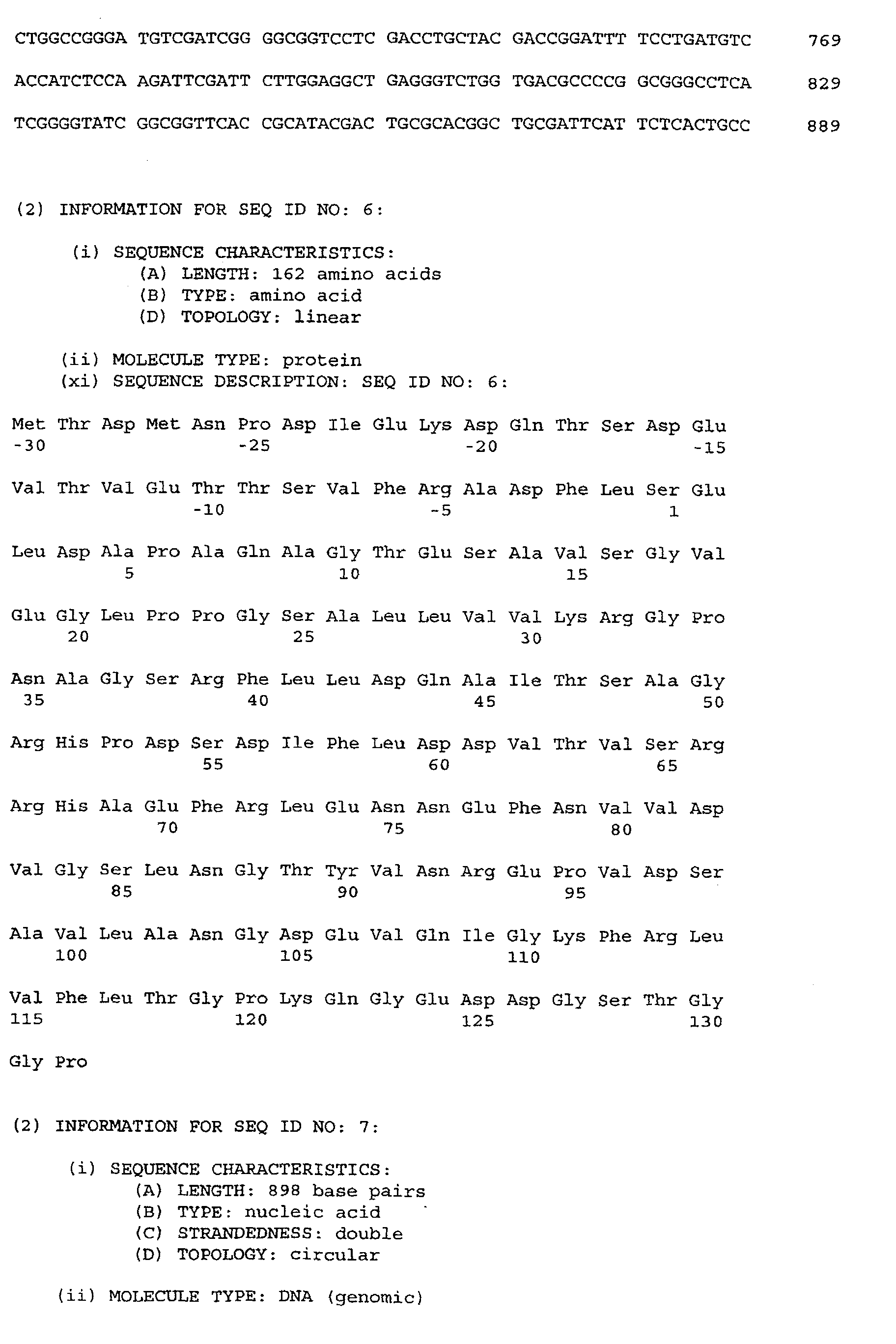
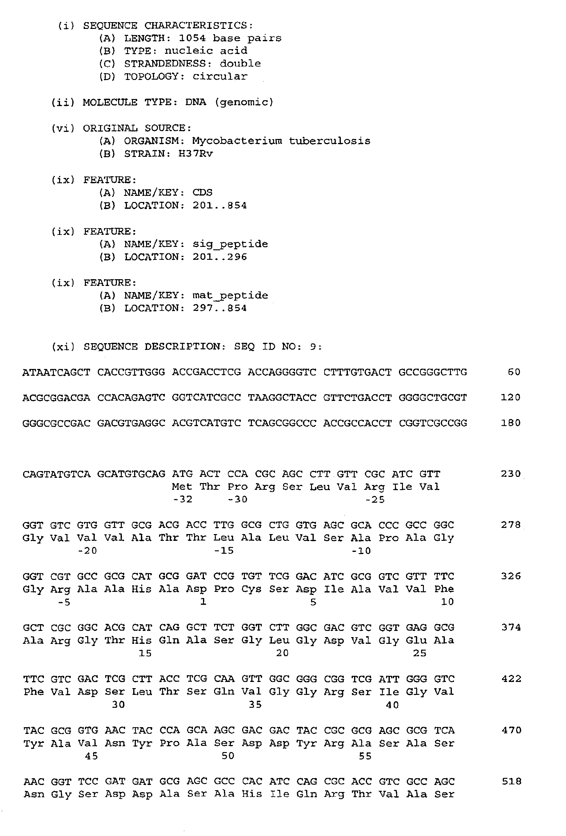
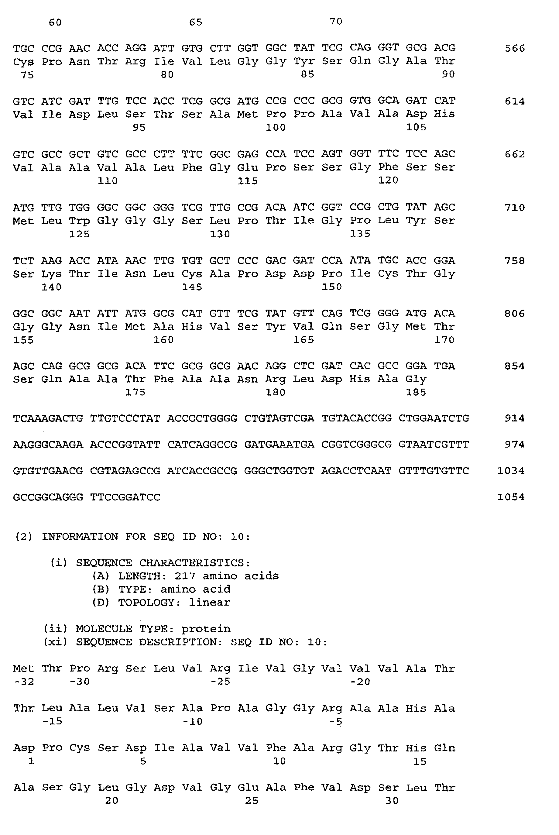
free from amino acid residues -30 to -1 in SEQ ID NO: 6 and/or -32 to -1 in SEQ ID
NO: 10 and/or -8 to -1 in SEQ ID NO: 12 and/or -32 to -1 in SEQ ID NO: 14 and/or
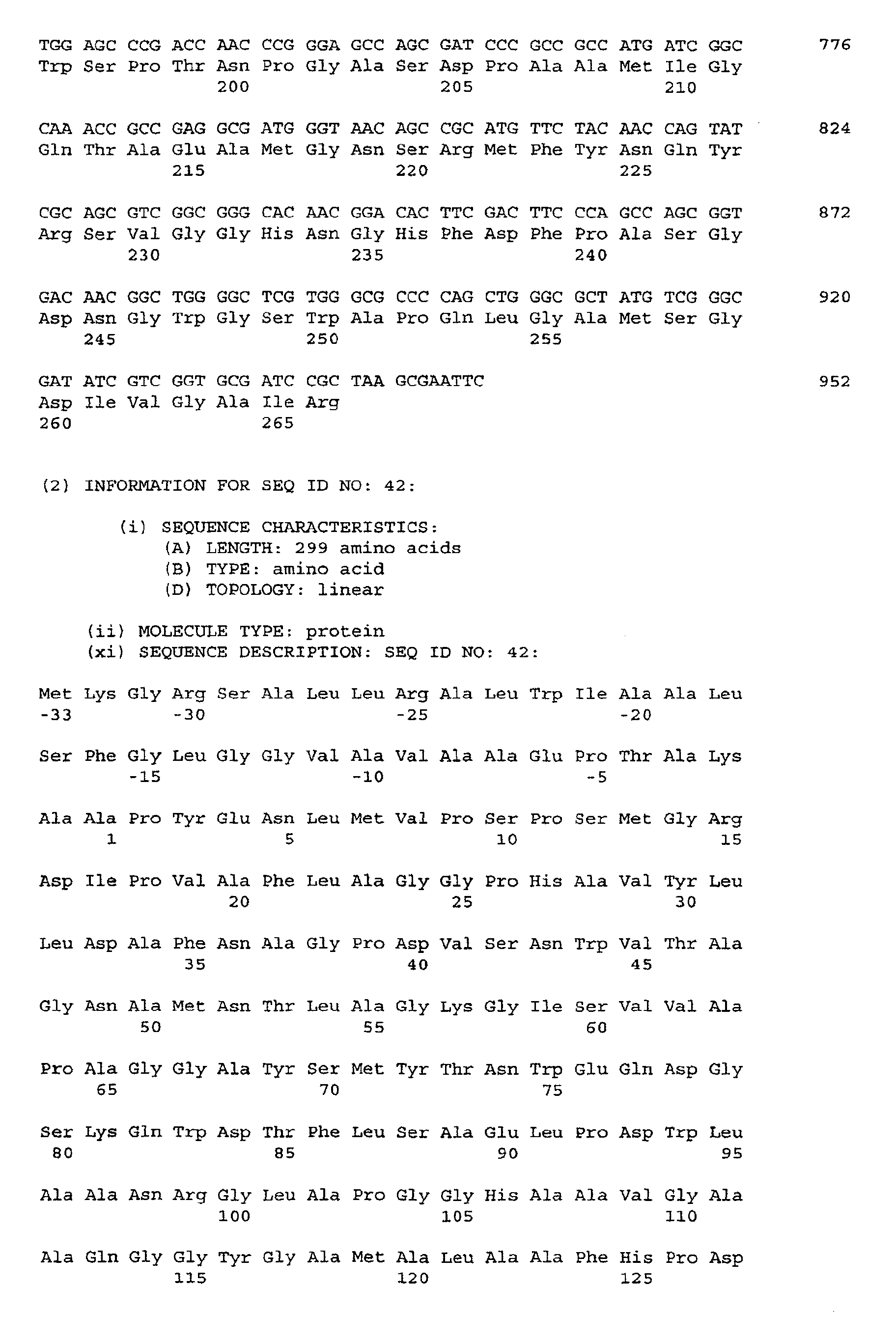
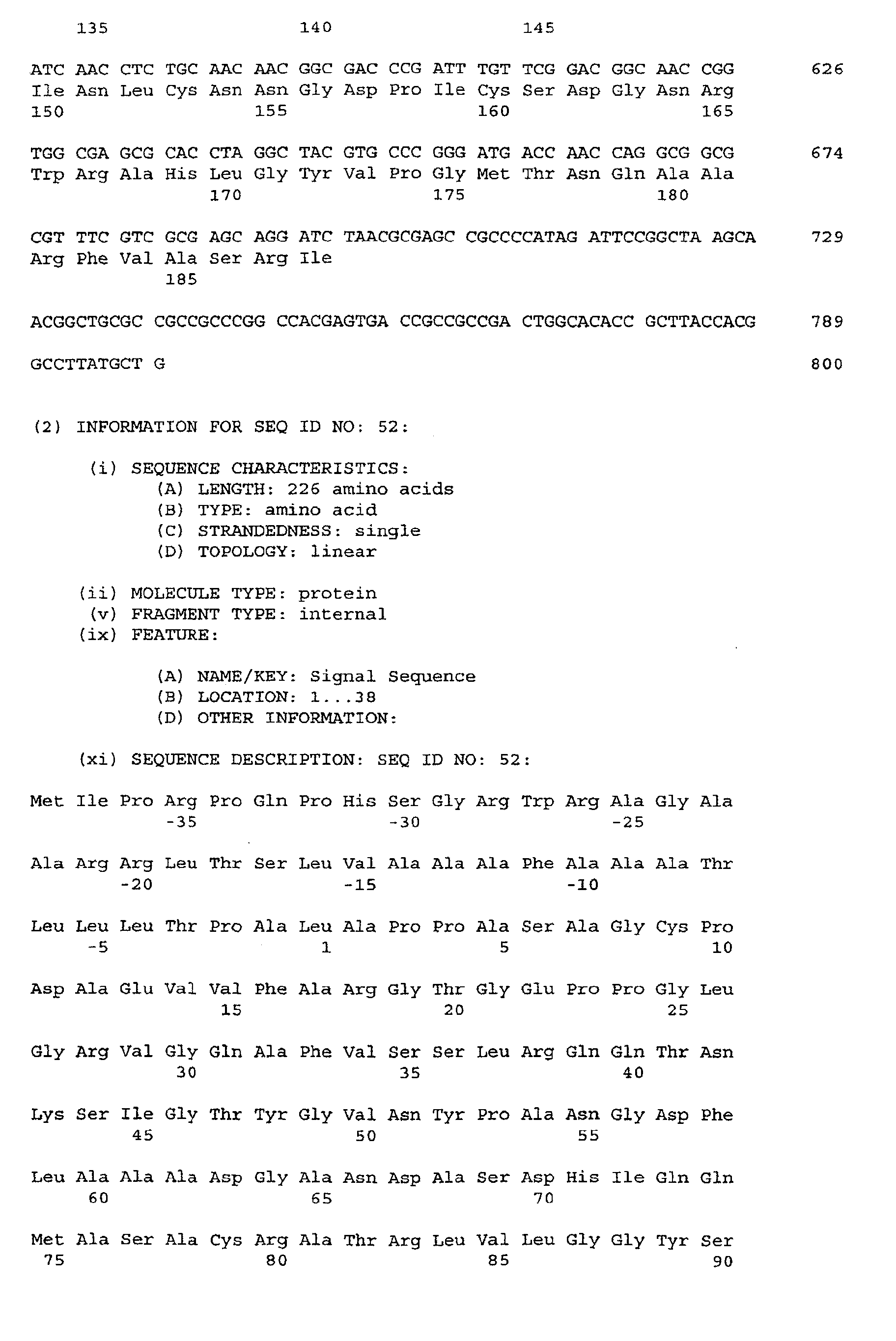
- 33 to -1 in SEQ ID NO: 42 and/or -38 to -1 in SEQ ID NO: 52 and/or -33 to -1 in SEQ
ID NO: 56 and/or -56 to -1 in SEQ ID NO: 58 and/or -28 to -1 in SEQ ID NO: 151.
-
In another preferred embodiment, the polypeptide fragment of the invention is free
from any signal sequence; this is especially interesting when the polypeptide fragment
is produced synthetically but even when the polypeptide fragments are produced recombinantly
it is normally acceptable that they are not exported by the host cell to the
periplasm or the extracellular space; the polypeptide fragments can be recovered by
traditional methods (cf. the discussion below) from the cytoplasm after disruption of
the host cells, and if there is need for refolding of the polypeptide fragments, general
refolding schemes can be employed, cf. e.g. the disclosure in WO 94/18227 where
such a general applicable refolding method is described.
-
A suitable assay for the potential utility of a given polypeptide fragment derived from
SEQ ID NO: 2, 4, 6, 8, 10, 12, 14, 16, any one of 17-23, 42, 48, 50, 52, 54, 56,
58, 60, 62, 64, 66, 68, 70, any one of 72-86, 88, 90, 92, 94, 141, 143, 145, 147,
149, 151, 153, any one of 168-171, 175, 177, 179, 181, 183, or 185 is to assess
the ability of the polypeptide fragment to effect IFN-γ release from primed memory T-lymphocytes.
Polypeptide fragments which have this capability are according to the
invention especially interesting embodiments of the invention: It is contemplated that
polypeptide fragments which stimulate T lymphocyte immune response shortly after
the onset of the infection are important in the control of the mycobacteria causing the
infection before the mycobacteria have succeeded in multiplying up to the number of
bacteria that would have resulted in fulminant infection.
-
It is presently contemplated that when this application refers to IFN-γ release as a
measure of immunogenicity, other cytokines could be relevant, such as IL-12, TNF-α,
IL-4, IL-5, IL-10, IL-6, TGF-β. Usually one or more cytokines will be measured utilising
for example the PCR technique or ELISA. It will be appreciated by the person skilled in
the art, that a significant increase or decrease in any of these cytokines will be indicative
of an immunological effective polypeptide or polypeptide fragment.
-
Thus, an important embodiment of the invention is a polypeptide fragment defined
above which
- 1) induces a release of IFN-γ from primed memory T-lymphocytes withdrawn from
a mouse within 2 weeks of primary infection or within 4 days after the mouse has
been re-challenge infected with mycobacteria belonging to the tuberculosis complex,
the induction performed by the addition of the polypeptide to a suspension comprising
about 200,000 spleen cells per ml, the addition of the polypeptide resulting in a concentration
of 1-4 µg polypeptide per ml suspension, the release of IFN-γ being assessable
by determination of IFN-γ in supernatant harvested 2 days after the addition of
the polypeptide to the suspension, and/or
- 2) induces a release of IFN-γ of at least 1,500 pg/ml above background level from
about 1,000,000 human PBMC (peripheral blood mononuclear cells) per ml isolated
from TB patients in the first phase of infection, or from healthy BCG vaccinated donors,
or from healthy contacts to TB patients, the induction being performed by the
addition of the polypeptide to a suspension comprising the about 1,000,000 PBMC
per ml, the addition of the polypeptide resulting in a concentration of 1-4 µg polypeptide
per ml suspension, the release of IFN-γ being assessable by determination of IFN-γ
in supernatant harvested 2 days after the addition of the polypeptide to the suspension;
and/or
- 3) induces an IFN-γ release from bovine PBMC derived from animals previously
sensitized with mycobacteria belonging to the tuberculosis complex, said release being
at least two times the release observed from bovine PBMC derived from animals not
previously sensitized with mycobacteria belonging to the tuberculosis complex.
-
-
Preferably, in alternatives 1 and 2, the release effected by the polypeptide fragment
gives rise to at least 1,500 pg/ml IFN-γ in the supernatant but higher concentrations
are preferred, e.g. at least 2,000 pg/ml and even at least 3,000 pg/ml IFN-γ in the supernatant.
The IFN-γ release from bovine PBMC can e.g. be measured as the optical
density (OD) index over background in a standard cytokine ELISA and should thus be
at least two, but higher numbers such as at least 3, 5, 8, and 10 are preferred.
-
The polypeptide fragments of the invention preferably comprises an amino acid sequence
of at least 6 amino acid residues in length which has a higher sequence identity
than 70 percent with SEQ ID NO: 2, 4, 6, 8, 10, 12, 14, 16, any one of 17-23,
42, 48, 50, 52, 54, 56, 58, 60, 62, 64, 66, 68, 70, any one of 72-86, 88, 90, 92,
94, 141, 143, 145, 147, 149, 151, 153, any one of 168-171, 175, 177, 179, 181,
183, or 185. A preferred minimum percentage of sequence identity is at least 80%,
such as at least 85%, at least 90%, at least 91 %, at least 92%, at least 93%, at
least 94%, at least 95%, at least 96%, at least 97%, at least 98%, at least 99%, and
at least 99.5%.
-
As mentioned above, it will normally be interesting to omit the leader sequences from
the polypeptide fragments of the invention. However, by producing fusion polypeptides,
superior characteristics of the polypeptide fragments of the invention can be
achieved. For instance, fusion partners which facilitate export of the polypeptide when
produced recombinantly, fusion partners which facilitate purification of the polypeptide,
and fusion partners which enhance the immunogenicity of the polypeptide fragment
of the invention are all interesting possibilities. Therefore, the invention also pertains
to a fusion polypeptide comprising at least one polypeptide fragment defined
above and at least one fusion partner. The fusion partner can, in order to enhance immunogenicity,
e.g. be selected from the group consisting of another polypeptide fragment
as defined above (so as to allow for multiple expression of relevant epitopes),
and an other polypeptide derived from a bacterium belonging to the tuberculosis complex,
such as ESAT-6, CFP7, CFP10, CFP17, CFP21, CFP25, CFP29, MPB59, MPT59,
MPB64, and MPT64 or at least one T-cell epitope of any of these antigens. Other
immunogenicity enhancing polypeptides which could serve as fusion partners are T-cell
epitopes (e.g. derived from the polypeptides ESAT-6, MPB64, MPT64, or MPB59)
or other immunogenic epitopes enhancing the immunogenicity of the target gene
product, e.g. lymphokines such as INF-γ, IL-2 and IL-12. In order to facilitate
expression and/or purification the fusion partner can e.g. be a bacterial fimbrial
protein, e.g. the pilus components pilin and papA; protein A; the ZZ-peptide (ZZ-fusions
are marketed by Pharmacia in Sweden); the maltose binding protein;
gluthatione S-transferase; β-galactosidase; or poly-histidine.
-
Other interesting fusion partners are polypeptides which are lipidated and thereby effect
that the immunogenic polypeptide is presented in a suitable manner to the immune
system. This effect is e.g. known from vaccines based on the Borrelia burgdorferi
OspA polypeptide, wherein the lipidated membrane anchor in the polypeptide confers
a self-adjuvating effect to the polypeptide (which is natively lipidated) when isolated
from cells producing it. In contrast, the OspA polypeptide is relatively silent immunologically
when prepared without the lipidation anchor.
-
As evidenced in Example 6A, the fusion polypeptide consisting of MPT59 fused directly
N-terminally to ESAT-6 enhances the immunogenicity of ESAT-6 beyond what
would be expected from the immunogenicities of MPT59 and ESAT-6 alone. The precise
reason for this surprising finding is not yet known, but it is expected that either
the presence of both antigens lead to a synergistic effect with respect to immunogenicity
or the presence of a sequence N-terminally to the ESAT-6 sequence protects
this immune dominant protein from loss of important epitopes known to be present in
the N-terminus. A third, alternative, possibility is that the presence of a sequence C-terminally
to the MPT59 sequence enhances the immunologic properties of this antigen.
-
Hence, one part of the invention pertains to a fusion polypeptide fragment which
comprises a first amino acid sequence including at least one stretch of amino acids
constituting a T-cell epitope derived from the M. tuberculosis protein ESAT-6 or
MPT59, and a second amino acid sequence including at least one T-cell epitope derived
from a M. tuberculosis protein different from ESAT-6 (if the first stretch of
amino acids are derived from ESAT-6) or MPT59 (if the first stretch of amino acids are
derived from MPT59) and/or including a stretch of amino acids which protects the first
amino acid sequence from in vivo degradation or post-translational processing. The
first amino acid sequence may be situated N- or C-terminally to the second amino acid
sequence, but in line with the above considerations regarding protection of the ESAT-6
N-terminus it is preferred that the first amino acid sequence is C-terminal to the
second when the first amino acid sequence is derived from ESAT-6.
-
Although only the effect of fusion between MPT59 and ESAT6 has been investigated
at present, it is believed that ESAT6 and MPT59 or epitopes derived therefrom could
be advantageously be fused to other fusion partners having substantially the same effect
on overall immunogenicity of the fusion construct. Hence, it is preferred that such
a fusion polypeptide fragment according of the invention is one, wherein the at least
one T-cell epitope included in the second amino acid sequence is derived from a M.
tuberculosis polypeptide (the "parent" polypeptide) selected from the group consisting
of a polypeptide fragment according to the present invention and described in detail
above and in the examples, or the amino acid sequence could be derived from any one
of the M. tuberculosis proteins DnaK, GroEL, urease, glutamine synthetase, the proline
rich complex, L-alanine dehydrogenase, phosphate binding protein, Ag 85 complex,
HBHA (heparin binding hemagglutinin), MPT51, MPT64, superoxide dismutase, 19
kDa lipoprotein, α-crystallin, GroES, MPT59 (when the first amino acid sequence is
derived from ESAT-6), and ESAT-6 (when the first amino acid sequence is derived
from MPT59). It is preferred that the first and second T-cell epitopes each have a sequence
identity of at least 70% with the natively occurring sequence in the proteins
from which they are derived and it is even further preferred that the first and/or
second amino acid sequence has a sequence identity of at least 70% with the protein
from which they are derived. A most preferred embodiment of this fusion polypeptide
is one wherein the first amino acid sequence is the amino acid sequence of ESAT-6 or
MPT59 and/or the second amino acid sequence is the full-length amino acid sequence
of the possible "parent" polypeptides listed above.
-
In the most preferred embodiment, the fusion polypeptide fragment comprises ESAT-6
fused to MPT59 (advantageously, ESAT-6 is fused to the C-terminus of MPT59) and
in one special embodiment, there are no linkers introduced between the two amino
acid sequences constituting the two parent polypeptide fragments.
-
Another part of the invention pertains to a nucleic acid fragment in isolated form
which
- 1) comprises a nucleic acid sequence which encodes a polypeptide or fusion polypeptide
as defined above, or comprises a nucleic acid sequence complementary thereto,
and/or
- 2) has a length of at least 10 nucleotides and hybridizes readily under stringent
hybridization conditions (as defined in the art, i.e. 5-10°C under the melting point Tm,
cf. Sambrook et al, 1989, pages 11.45-11.49) with a nucleic acid fragment which has
a nucleotide sequence selected from
- SEQ ID NO: 1 or a sequence complementary thereto,
- SEQ ID NO: 3 or a sequence complementary thereto,
- SEQ ID NO: 5 or a sequence complementary thereto,
- SEQ ID NO: 7 or a sequence complementary thereto,
- SEQ ID NO: 9 or a sequence complementary thereto,
- SEQ ID NO: 11 or a sequence complementary thereto,
- SEQ ID NO: 13 or a sequence complementary thereto,
- SEQ ID NO: 15 or a sequence complementary thereto,
- SEQ ID NO: 41 or a sequence complementary thereto,
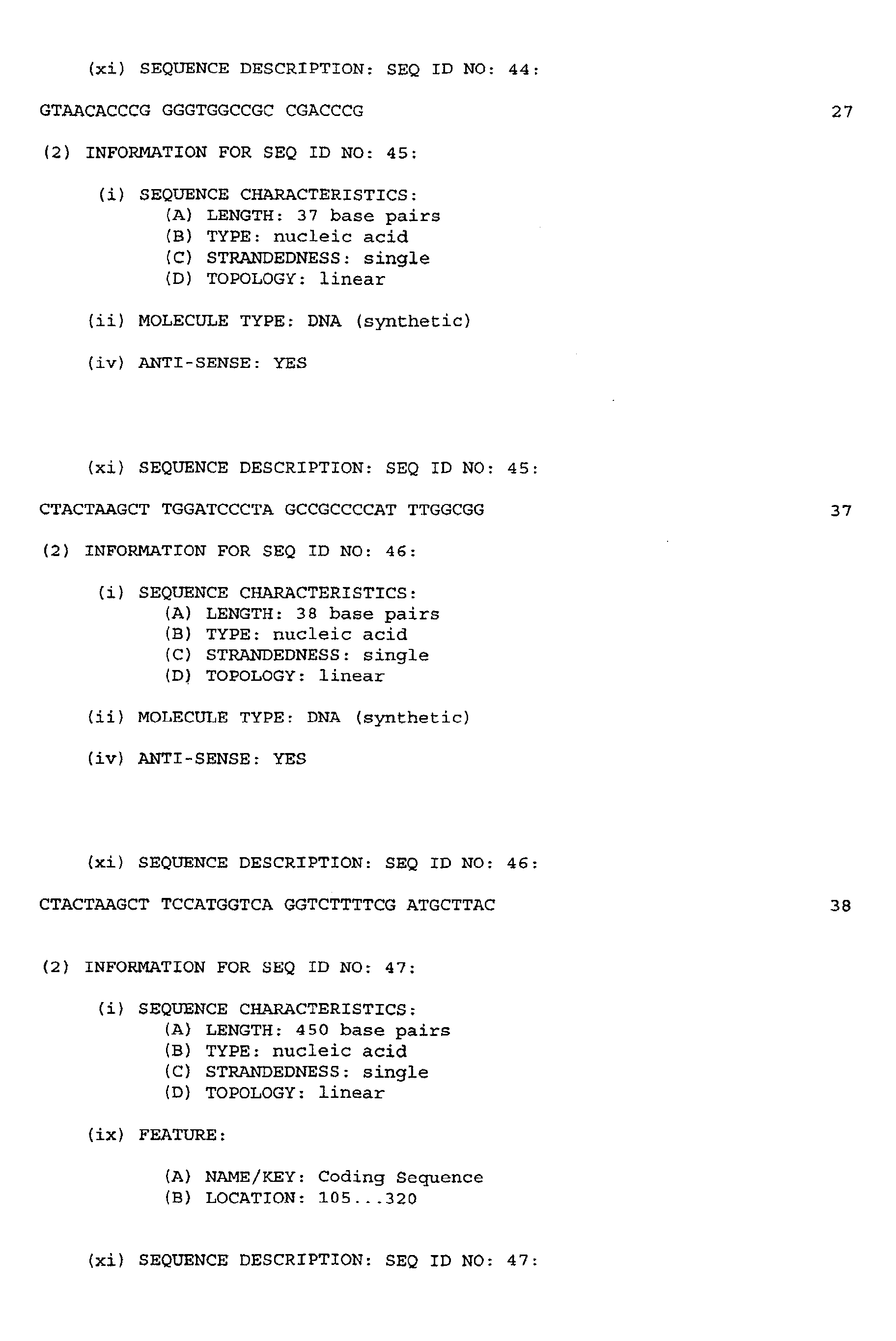
- SEQ ID NO: 47 or a sequence complementary thereto,
- SEQ ID NO: 49 or a sequence complementary thereto,
- SEQ ID NO: 51 or a sequence complementary thereto,
- SEQ ID NO: 53 or a sequence complementary thereto,
- SEQ ID NO: 55 or a sequence complementary thereto,
- SEQ ID NO: 57 or a sequence complementary thereto,
- SEQ ID NO: 59 or a sequence complementary thereto,
- SEQ ID NO: 61 or a sequence complementary thereto,
- SEQ ID NO: 63 or a sequence complementary thereto,
- SEQ ID NO: 65 or a sequence complementary thereto,
- SEQ ID NO: 67 or a sequence complementary thereto,
- SEQ ID NO: 69 or a sequence complementary thereto,
- SEQ ID NO: 71 or a sequence complementary thereto,
- SEQ ID NO: 87 or a sequence complementary thereto,
- SEQ ID NO: 89 or a sequence complementary thereto,
- SEQ ID NO: 91 or a sequence complementary thereto,
- SEQ ID NO: 93 or a sequence complementary thereto,
- SEQ ID NO: 140 or a sequence complementary thereto,
- SEQ ID NO: 142 or a sequence complementary thereto,
- SEQ ID NO: 144 or a sequence complementary thereto,
- SEQ ID NO: 146 or a sequence complementary thereto,
- SEQ ID NO: 148 or a sequence complementary thereto,
- SEQ ID NO: 150 or a sequence complementary thereto,
- SEQ ID NO: 152 or a sequence complementary thereto,
- SEQ ID NO: 174 or a sequence complementary thereto,
- SEQ ID NO: 176 or a sequence complementary thereto,
- SEQ ID NO: 178 or a sequence complementary thereto,
- SEQ ID NO: 180 or a sequence complementary thereto,
- SEQ ID NO: 182 or a sequence complementary thereto, and
- SEQ ID NO: 184 or a sequence complementary thereto
with the proviso that when the nucleic acid fragment comprises a subsequence of
SEQ ID NO: 41, then the nucleic acid fragment contains an A corresponding to position
781 in SEQ ID NO: 41 and when the nucleic acid fragment comprises a subsequence
of a nucleotide sequence exactly complementary to SEQ ID NO: 41, then the
nucleic acid fragment comprises a T corresponding to position 781 in SEQ ID NO: 41.-
-
It is preferred that the nucleic acid fragment is a DNA fragment.
-
To provide certainty of the advantages in accordance with the invention, the preferred
nucleic acid sequence when employed for hybridization studies or assays includes sequences
that are complementary to at least a 10 to 40, or so, nucleotide stretch of
the selected sequence. A size of at least 10 nucleotides in length helps to ensure that
the fragment will be of sufficient length to form a duplex molecule that is both stable
and selective. Molecules having complementary sequences over stretches greater than
10 bases in length are generally preferred, though, in order to increase stability and
selectivity of the hybrid, and thereby improve the quality and degree of specific hybrid
molecules obtained.
-
Hence, the term "subsequence" when used in connection with the nucleic acid fragments
of the invention is intended to indicate a continuous stretch of at least 10 nucleotides
exhibits the above hybridization pattern. Normally this will require a minimum
sequence identity of at least 70% with a subsequence of the hybridization partner
having SEQ ID NO: 1, 3, 5, 7, 9, 11, 12, 15, 21, 41, 47, 49, 51, 53, 55, 57, 59, 61,
63, 65, 67, 69, 71, 87, 89, 91, 93, 140, 142, 144, 146, 148, 150, 152, 174, 176,
178, 180, 182, or 184. It is preferred that the nucleic acid fragment is longer than 10
nucleotides, such as at least 15, at least 20, at least 25, at least 30, at least 35, at
least 40, at least 45, at least 50, at least 55, at least 60, at least 65, at least 70, and
at least 80 nucleotides long, and the sequence identity should preferable also be
higher than 70%, such as at least 75%, at least 80%, at least 85%, at least 90%, at
least 92%, at least 94%, at least 96%, and at least 98%. It is most preferred that the
sequence identity is 100%. Such fragments may be readily prepared by, for example,
directly synthesizing the fragment by chemical means, by application of nucleic acid
reproduction technology, such as the PCR technology of U.S. Patent 4,603,102, or by
introducing selected sequences into recombinant vectors for recombinant production.
-
It is well known that the same amino acid may be encoded by various codons, the codon
usage being related, inter alia, to the preference of the organisms in question expressing
the nucleotide sequence. Thus, at least one nucleotide or codon of a nucleic
acid fragment of the invention may be exchanged by others which, when expressed,
result in a polypeptide identical or substantially identical to the polypeptide encoded
by the nucleic acid fragment in question. The invention thus allows for variations in
the sequence such as substitution, insertion (including introns), addition, deletion and
rearrangement of one or more nucleotides, which variations do not have any
substantial effect on the polypeptide encoded by the nucleic acid fragment or a
subsequence thereof. The term "substitution" is intended to mean the replacement of
one or more nucleotides in the full nucleotide sequence with one or more different
nucleotides, "addition" is understood to mean the addition of one or more nucleotides
at either end of the full nucleotide sequence, "insertion" is intended to mean the
introduction of one or more nucleotides within the full nucleotide sequence, "deletion"
is intended to indicate that one or more nucleotides have been deleted from the full
nucleotide sequence whether at either end of the sequence or at any suitable point
within it, and "rearrangement" is intended to mean that two or more nucleotide
residues have been exchanged with each other.
-
The nucleotide sequence to be modified may be of cDNA or genomic origin as discussed
above, but may also be of synthetic origin. Furthermore, the sequence may be of
mixed cDNA and genomic, mixed cDNA and synthetic or genomic and synthetic origin
as discussed above. The sequence may have been modified, e.g. by site-directed mutagenesis,
to result in the desired nucleic acid fragment encoding the desired polypeptide.
The following discussion focused on modifications of nucleic acid encoding the
polypeptide should be understood to encompass also such possibilities, as well as the
possibility of building up the nucleic acid by ligation of two or more DNA fragments to
obtain the desired nucleic acid fragment, and combinations of the above-mentioned
principles.
-
The nucleotide sequence may be modified using any suitable technique which results
in the production of a nucleic acid fragment encoding a polypeptide of the invention.
-
The modification of the nucleotide sequence encoding the amino acid sequence of the
polypeptide of the invention should be one which does not impair the immunological
function of the resulting polypeptide.
-
A preferred method of preparing variants of the antigens disclosed herein is site-directed
mutagenesis. This technique is useful in the preparation of individual peptides,
or biologically functional equivalent proteins or peptides, derived from the antigen sequences,
through specific mutagenesis of the underlying nucleic acid. The technique
further provides a ready ability to prepare and test sequence variants, for example,
incorporating one or more of the foregoing considerations, by introducing one or more
nucleotide sequence changes into the nucleic acid. Site-specific mutagenesis allows
the production of mutants through the use of specific oligonucleotide sequences
which encode the nucleotide sequence of the desired mutation, as well as a sufficient
number of adjacent nucleotides, to provide a primer sequence of sufficient size and
sequence complexity to form a stable duplex on both sides of the deletion junction
being traversed. Typically, a primer of about 17 to 25 nucleotides in length is preferred,
with about 5 to 10 residues on both sides of the junction of the sequence being
altered.
-
In general, the technique of site-specific mutagenesis is well known in the art as exemplified
by publications (Adelman et al., 1983). As will be appreciated, the technique
typically employs a phage vector which exists in both a single stranded and double
stranded form. Typical vectors useful in site-directed mutagenesis include vectors
such as the M13 phage (Messing et al., 1981). These phage are readily commercially
available and their use is generally well known to those skilled in the art.
-
In general, site-directed mutagenesis in accordance herewith is performed by first obtaining
a single-stranded vector which includes within its sequence a nucleic acid sequence
which encodes the polypeptides of the invention. An oligonucleotide primer
bearing the desired mutated sequence is prepared, generally synthetically, for example
by the method of Crea et al. (1978). This primer is then annealed with the single-stranded
vector, and subjected to DNA polymerizing enzymes such as E. coli polymerase
I Klenow fragment, in order to complete the synthesis of the mutation-bearing
strand. Thus, a heteroduplex is formed wherein one strand encodes the original non-mutated
sequence and the second strand bears the desired mutation. This heteroduplex
vector is then used to transform appropriate cells, such as E. coli cells, and
clones are selected which include recombinant vectors bearing the mutated sequence
arrangement.
-
The preparation of sequence variants of the selected nucleic acid fragments of the invention
using site-directed mutagenesis is provided as a means of producing potentially
useful species of the genes and is not meant to be limiting as there are other
ways in which sequence variants of the nucleic acid fragments of the invention may
be obtained. For example, recombinant vectors encoding the desired genes may be
treated with mutagenic agents to obtain sequence variants (see, e.g., a method described
by Eichenlaub, 1979) for the mutagenesis of plasmid DNA using hydroxylamine.
-
The invention also relates to a replicable expression vector which comprises a nucleic
acid fragment defined above, especially a vector which comprises a nucleic acid fragment
encoding a polypeptide fragment of the invention.
-
The vector may be any vector which may conveniently be subjected to recombinant
DNA procedures, and the choice of vector will often depend on the host cell into
which it is to be introduced. Thus, the vector may be an autonomously replicating
vector, i.e. a vector which exists as an extrachromosomal entity, the replication of
which is independent of chromosomal replication; examples of such a vector are a
plasmid, phage, cosmid, mini-chromosome or virus. Alternatively, the vector may be
one which, when introduced in a host cell, is integrated in the host cell genome and
replicated together with the chromosome(s) into which it has been integrated.
-
Expression vectors may be constructed to include any of the DNA segments disclosed
herein. Such DNA might encode an antigenic protein specific for virulent strains of
mycobacteria or even hybridization probes for detecting mycobacteria nucleic acids in
samples. Longer or shorter DNA segments could be used, depending on the antigenic
protein desired. Epitopic regions of the proteins expressed or encoded by the disclosed
DNA could be included as relatively short segments of DNA. A wide variety of expression
vectors is possible including, for example, DNA segments encoding reporter gene
products useful for identification of heterologous gene products and/or resistance
genes such as antibiotic resistance genes which may be useful in identifying transformed
cells.
-
The vector of the invention may be used to transform cells so as to allow propagation
of the nucleic acid fragments of the invention or so as to allow expression of the polypeptide
fragments of the invention. Hence, the invention also pertains to a transformed
cell harbouring at least one such vector according to the invention, said cell being
one which does not natively harbour the vector and/or the nucleic acid fragment of
the invention contained therein. Such a transformed cell (which is also a part of the
invention) may be any suitable bacterial host cell or any other type of cell such as a
unicellular eukaryotic organism, a fungus or yeast, or a cell derived from a multicellular
organism, e.g. an animal or a plant. It is especially in cases where glycosylation is desired
that a mammalian cell is used, although glycosylation of proteins is a rare event
in prokaryotes. Normally, however, a prokaryotic cell is preferred such as a bacterium
belonging to the genera Mycobacterium, Salmonella, Pseudomonas, Bacillus and Eschericia.
It is preferred that the transformed cell is an E. coli, B. subtilis, or M. bovis
BCG cell, and it is especially preferred that the transformed cell expresses a polypeptide
according of the invention. The latter opens for the possibility to produce the
polypeptide of the invention by simply recovering it from the culture containing the
transformed cell. In the most preferred embodiment of this part of the invention the
transformed cell is Mycobacterium bovis BCG strain: Danish 1331, which is the Mycobacterium
bovis strain Copenhagen from the Copenhagen BCG Laboratory, Statens
Seruminstitut, Denmark.
-
The nucleic acid fragments of the invention allow for the recombinant production of
the polypeptides fragments of the invention. However, also isolation from the natural
source is a way of providing the polypeptide fragments as is peptide synthesis.
-
Therefore, the invention also pertains to a method for the preparation of a polypeptide
fragment of the invention, said method comprising inserting a nucleic acid fragment as
defined above into a vector which is able to replicate in a host cell, introducing the
resulting recombinant vector into the host cell (transformed cells may be selected using
various techniques, including screening by differential hybridization, identification
of fused reporter gene products, resistance markers, anti-antigen antibodies and the
like), culturing the host cell in a culture medium under conditions sufficient to effect
expression of the polypeptide (of course the cell may be cultivated under conditions
appropriate to the circumstances, and if DNA is desired, replication conditions are
used), and recovering the polypeptide from the host cell or culture medium; or
-
isolating the polypeptide from a short-term culture filtrate as defined in claim 1; or
isolating the polypeptide from whole mycobacteria of the tuberculosis complex or from
lysates or fractions thereof, e.g. cell wall containing fractions, or
synthesizing the polypeptide by solid or liquid phase peptide synthesis.
-
The medium used to grow the transformed cells may be any conventional medium
suitable for the purpose. A suitable vector may be any of the vectors described above,
and an appropriate host cell may be any of the cell types listed above. The methods
employed to construct the vector and effect introduction thereof into the host cell
may be any methods known for such purposes within the field of recombinant DNA. In
the following a more detailed description of the possibilities will be given:
-
In general, of course, prokaryotes are preferred for the initial cloning of nucleic sequences
of the invention and constructing the vectors useful in the invention. For example,
in addition to the particular strains mentioned in the more specific disclosure
below, one may mention by way of example, strains such as E. coli K12 strain 294
(ATCC No. 31446), E. coli B, and E. coli X 1776 (ATCC No. 31537). These examples
are, of course, intended to be illustrative rather than limiting.
-
Prokaryotes are also preferred for expression. The aforementioned strains, as well as
E. coli W3110 (F-, lambda-, prototrophic, ATCC No. 273325), bacilli such as Bacillus
subtilis, or other enterobacteriaceae such as Salmonella typhimurium or Serratia marcesans,
and various Pseudomonas species may be used. Especially interesting are
rapid-growing mycobacteria, e.g. M. smegmatis, as these bacteria have a high degree
of resemblance with mycobacteria of the tuberculosis complex and therefore stand a
good chance of reducing the need of performing post-translational modifications of the
expression product.
-
In general, plasmid vectors containing replicon and control sequences which are derived
from species compatible with the host cell are used in connection with these
hosts. The vector ordinarily carries a replication site, as well as marking sequences
which are capable of providing phenotypic selection in transformed cells. For example,
E. coli is typically transformed using pBR322, a plasmid derived from an E. coli species
(see, e.g., Bolivar et al., 1977, Gene 2: 95). The pBR322 plasmid contains genes for
ampicillin and tetracycline resistance and thus provides easy means for identifying
transformed cells. The pBR plasmid, or other microbial plasmid or phage must also
contain, or be modified to contain, promoters which can be used by the microorganism
for expression.
-
Those promoters most commonly used in recombinant DNA construction include the
B-lactamase (penicillinase) and lactose promoter systems (Chang et al., 1978; Itakura
et al., 1977; Goeddel et al., 1979) and a tryptophan (trp) promoter system (Goeddel
et al., 1979; EPO Appl. Publ. No. 0036776). While these are the most commonly
used, other microbial promoters have been discovered and utilized, and details concerning
their nucleotide sequences have been published, enabling a skilled worker to
ligate them functionally with plasmid vectors (Siebwenlist et al., 1980). Certain genes
from prokaryotes may be expressed efficiently in E. coli from their own promoter sequences,
precluding the need for addition of another promoter by artificial means.
-
After the recombinant preparation of the polypeptide according to the invention, the
isolation of the polypeptide may for instance be carried out by affinity chromatography
(or other conventional biochemical procedures based on chromatography), using a
monoclonal antibody which substantially specifically binds the polypeptide according
to the invention. Another possibility is to employ the simultaneous electroelution technique
described by Andersen et al. in J. Immunol. Methods 161: 29-39.
-
According to the invention the post-translational modifications involves lipidation, glycosylation,
cleavage, or elongation of the polypeptide.
-
In certain aspects, the DNA sequence information provided by this invention allows for
the preparation of relatively short DNA (or RNA or PNA) sequences having the ability
to specifically hybridize to mycobacterial gene sequences. In these aspects, nucleic
acid probes of an appropriate length are prepared based on a consideration of the relevant
sequence. The ability of such nucleic acid probes to specifically hybridize to the
mycobacterial gene sequences lend them particular utility in a variety of embodiments.
Most importantly, the probes can be used in a variety of diagnostic assays for detecting
the presence of pathogenic organisms in a given sample. However, either uses are
envisioned, including the use of the sequence information for the preparation of mutant
species primers, or primers for use in preparing other genetic constructs.
-
Apart from their use as starting points for the synthesis of polypeptides of the invention
and for hybridization probes (useful for direct hybridization assays or as primers in
e.g. PCR or other molecular amplification methods) the nucleic acid fragments of the
invention may be used for effecting in vivo expression of antigens, i.e. the nucleic acid
fragments may be used in so-called DNA vaccines. Recent research have revealed that
a DNA fragment cloned in a vector which is non- replicative in eukaryotic cells may be
introduced into an animal (including a human being) by e.g. intramuscular injection or
percutaneous administration (the so-called "gene gun" approach). The DNA is taken
up by e.g. muscle cells and the gene of interest is expressed by a promoter which is
functioning in eukaryotes, e.g. a viral promoter, and the gene product thereafter
stimulates the immune system. These newly discovered methods are reviewed in Ulmer
et al., 1993, which hereby is included by reference.
-
Hence, the invention also relates to a vaccine comprising a nucleic acid fragment according
to the invention, the vaccine effecting in vivo expression of antigen by an animal,
including a human being, to whom the vaccine has been administered, the
amount of expressed antigen being effective to confer substantially increased resistance
to infections with mycobacteria of the tuberculosis complex in an animal, including
a human being.
-
The efficacy of such a "DNA vaccine" can possibly be enhanced by administering the
gene encoding the expression product together with a DNA fragment encoding a polypeptide
which has the capability of modulating an immune response. For instance, a
gene encoding lymphokine precursors or lymphokines (e.g. IFN-γ, IL-2, or IL-12) could
be administered together with the gene encoding the immunogenic protein, either by
administering two separate DNA fragments or by administering both DNA fragments
included in the same vector. It also is a possibility to administer DNA fragments comprising
a multitude of nucleotide sequences which each encode relevant epitopes of
the polypeptides disclosed herein so as to effect a continuous sensitization of the immune
system with a broad spectrum of these epitopes.
-
As explained above, the polypeptide fragments of the invention are excellent candidates
for vaccine constituents or for constituents in an immune diagnostic agent due
to their extracellular presence in culture media containing metabolizing virulent mycobacteria
belonging to the tuberculosis complex, or because of their high homologies
with such extracellular antigens, or because of their absence in M. bovis BCG.
-
Thus, another part of the invention pertains to an immunologic composition
comprising a polypeptide or fusion polypeptide according to the invention. In order to
ensure optimum performance of such an immunologic composition it is preferred that
it comprises an immunologically and pharmaceutically acceptable carrier, vehicle or
adjuvant.
-
Suitable carriers are selected from the group consisting of a polymer to which the
polypeptide(s) is/are bound by hydrophobic non-covalent interaction, such as a plastic,
e.g. polystyrene, or a polymer to which the polypeptide(s) is/are covalently bound,
such as a polysaccharide, or a polypeptide, e.g. bovine serum albumin, ovalbumin or
keyhole limpet haemocyanin. Suitable vehicles are selected from the group consisting
of a diluent and a suspending agent. The adjuvant is preferably selected from the
group consisting of dimethyldioctadecylammonium bromide (DDA), Quil A, poly I:C,
Freund's incomplete adjuvant, IFN-γ, IL-2, IL-12, monophosphoryl lipid A (MPL), and
muramyl dipeptide (MDP).
-
A preferred immunologic composition according to the present invention comprising at
least two different polypeptide fragments, each different polypeptide fragment being a
polypeptide or a fusion polypeptide defined above. It is preferred that the immunologic
composition comprises between 3-20 different polypeptide fragments or fusion polypeptides.
-
Such an immunologic composition may preferably be in the form of a vaccine or in the
form of a skin test reagent.
-
In line with the above, the invention therefore also pertain to a method for producing
an immunologic composition according to the invention, the method comprising preparing,
synthesizing or isolating a polypeptide according to the invention, and solubilizing
or dispersing the polypeptide in a medium for a vaccine, and optionally adding
other M. tuberculosis antigens and/or a carrier, vehicle and/or adjuvant substance.
-
Preparation of vaccines which contain peptide sequences as active ingredients is generally
well understood in the art, as exemplified by U.S. Patents 4,608,251;
4,601,903; 4,599,231; 4,599,230; 4,596,792; and 4,578,770, all incorporated
herein by reference. Typically, such vaccines are prepared as injectables either as
liquid solutions or suspensions; solid forms suitable for solution in, or suspension in,
liquid prior to injection may also be prepared. The preparation may also be emulsified.
The active immunogenic ingredient is often mixed with excipients which are pharmaceutically
acceptable and compatible with the active ingredient. Suitable excipients
are, for example, water, saline, dextrose, glycerol, ethanol, or the like, and combinations
thereof. In addition, if desired, the vaccine may contain minor amounts of auxiliary
substances such as wetting or emulsifying agents, pH buffering agents, or adjuvants
which enhance the effectiveness of the vaccines.
-
The vaccines are conventionally administered parenterally, by injection, for example,
either subcutaneously or intramuscularly. Additional formulations which are suitable
for other modes of administration include suppositories and, in some cases, oral formulations.
For suppositories, traditional binders and carriers may include, for example,
polyalkalene glycols or triglycerides; such suppositories may be formed from mixtures
containing the active ingredient in the range of 0.5% to 10%, preferably 1-2%. Oral
formulations include such normally employed excipients as, for example, pharmaceutical
grades of mannitol, lactose, starch, magnesium stearate, sodium saccharine, cellulose,
magnesium carbonate, and the like. These compositions take the form of solutions,
suspensions, tablets, pills, capsules, sustained release formulations or powders
and contain 10-95% of active ingredient, preferably 25-70%.
-
The proteins may be formulated into the vaccine as neutral or salt forms. Pharmaceutically
acceptable salts include acid addition salts (formed with the free amino groups
of the peptide) and which are formed with inorganic acids such as, for example, hydrochloric
or phosphoric acids, or such organic acids as acetic oxalic, tartaric, mandelic,
and the like. Salts formed with the free carboxyl groups may also be derived
from inorganic bases such as, for example, sodium, potassium, ammonium, calcium,
or ferric hydroxides, and such organic bases as isopropylamine, trimethylamine, 2-ethylamino
ethanol, histidine, procaine, and the like.
-
The vaccines are administered in a manner compatible with the dosage formulation,
and in such amount as will be therapeutically effective and immunogenic. The quantity
to be administered depends on the subject to be treated, including, e.g., the capacity
of the individual's immune system to mount an immune response, and the degree of
protection desired. Suitable dosage ranges are of the order of several hundred micrograms
active ingredient per vaccination with a preferred range from about 0.1 µg to
1000 µg, such as in the range from about 1 µg to 300 µg, and especially in the range
from about 10 µg to 50 µg. Suitable regimens for initial administration and booster
shots are also variable but are typified by an initial administration followed by subsequent
inoculations or other administrations.
-
The manner of application may be varied widely. Any of the conventional methods for
administration of a vaccine are applicable. These are believed to include oral application
on a solid physiologically acceptable base or in a physiologically acceptable dispersion,
parenterally, by injection or the like. The dosage of the vaccine will depend on
the route of administration and will vary according to the age of the person to be vaccinated
and, to a lesser degree, the size of the person to be vaccinated.
-
Some of the polypeptides of the vaccine are sufficiently immunogenic in a vaccine,
but for some of the others the immune response will be enhanced if the vaccine further
comprises an adjuvant substance.
-
Various methods of achieving adjuvant effect for the vaccine include use of agents
such as aluminum hydroxide or phosphate (alum), commonly used as 0.05 to 0.1 percent
solution in phosphate buffered saline, admixture with synthetic polymers of sugars
(Carbopol) used as 0.25 percent solution, aggregation of the protein in the vaccine
by heat treatment with temperatures ranging between 70° to 101°C for 30 second to
2 minute periods respectively. Aggregation by reactivating with pepsin treated (Fab)
antibodies to albumin, mixture with bacterial cells such as C. parvum or endotoxins or
lipopolysaccharide components of gram-negative bacteria, emulsion in physiologically
acceptable oil vehicles such as mannide mono-oleate (Aracel A) or emulsion with 20
percent solution of a perfluorocarbon (Fluosol-DA) used as a block substitute may also
be employed. According to the invention DDA (dimethyldioctadecylammonium bromide)
is an interesting candidate for an adjuvant, but also Freund's complete and incomplete
adjuvants as well as QuilA and RIBI are interesting possibilities. Further possibilities
are monophosphoryl lipid A (MPL), and muramyl dipeptide (MDP).
-
Another highly interesting (and thus, preferred) possibility of achieving adjuvant effect
is to employ the technique described in Gosselin et al., 1992 (which is hereby incorporated
by reference herein). In brief, the presentation of a relevant antigen such as an
antigen of the present invention can be enhanced by conjugating the antigen to antibodies
(or antigen binding antibody fragments) against the Fcγ receptors on monocytes/macrophages.
Especially conjugates between antigen and anti-FcγRI have been
demonstrated to enhance immunogenicity for the purposes of vaccination.
-
Other possibilities involve the use of immune modulating substances such as lymphokines
(e.g. IFN-γ, IL-2 and IL-12) or synthetic IFN-γ inducers such as poly I:C in combination
with the above-mentioned adjuvants. As discussed in example 3, it is contemplated
that such mixtures of antigen and adjuvant will lead to superior vaccine formulations.
-
In many instances, it will be necessary to have multiple administrations of the vaccine,
usually not exceeding six vaccinations, more usually not exceeding four vaccinations
and preferably one or more, usually at least about three vaccinations. The vaccinations
will normally be at from two to twelve week intervals, more usually from three to five
week intervals. Periodic boosters at intervals of 1-5 years, usually three years, will be
desirable to maintain the desired levels of protective immunity. The course of the immunization
may be followed by in vitro proliferation assays of PBL (peripheral blood
lymphocytes) co-cultured with ESAT-6 or ST-CF, and especially by measuring the
levels of IFN-γ released form the primed lymphocytes. The assays may be performed
using conventional labels, such as radionuclides, enzymes, fluorescers, and the like.
These techniques are well known and may be found in a wide variety of patents, such
as U.S. Patent Nos. 3,791,932; 4,174,384 and 3,949,064, as illustrative of these
types of assays.
-
Due to genetic variation, different individuals may react with immune responses of
varying strength to the same polypeptide. Therefore, the vaccine according to the invention
may comprise several different polypeptides in order to increase the immune
response. The vaccine may comprise two or more polypeptides, where all of the polypeptides
are as defined above, or some but not all of the peptides may be derived
from a bacterium belonging to the M. tuberculosis complex. In the latter example the
polypeptides not necessarily fulfilling the criteria set forth above for polypeptides may
either act due to their own immunogenicity or merely act as adjuvants. Examples of
such interesting polypeptides are MPB64, MPT64, and MPB59, but any other substance
which can be isolated from mycobacteria are possible candidates.
-
The vaccine may comprise 3-20 different polypeptides, such as 3-10 different polypeptides.
-
One reason for admixing the polypeptides of the invention with an adjuvant is to effectively
activate a cellular immune response. However, this effect can also be
achieved in other ways, for instance by expressing the effective antigen in a vaccine
in a non-pathogenic microorganism. A well-known example of such a microorganism is
Mycobacterium bovis BCG.
-
Therefore, another important aspect of the present invention is an improvement of the
living BCG vaccine presently available, which is a vaccine for immunizing an animal,
including a human being, against TB caused by mycobacteria belonging to the tuberculosis-complex,
comprising as the effective component a microorganism, wherein
one or more copies of a DNA sequence encoding a polypeptide as defined above has
been incorporated into the genome of the microorganism in a manner allowing the
microorganism to express and secrete the polypeptide.
-
In the present context the term "genome" refers to the chromosome of the microorganisms
as well as extrachromosomally DNA or RNA, such as plasmids. It is, however,
preferred that the DNA sequence of the present invention has been introduced
into the chromosome of the non-pathogenic microorganism, since this will prevent
loss of the genetic material introduced.
-
It is preferred that the non-pathogenic microorganism is a bacterium, e.g. selected
from the group consisting of the genera Mycobacterium, Salmonella, Pseudomonas
and Eschericia. It is especially preferred that the non-pathogenic microorganism is Mycobacterium
bovis BCG, such as Mycobacterium bovis BCG strain: Danish 1331.
-
The incorporation of one or more copies of a nucleotide sequence encoding the polypeptide
according to the invention in a mycobacterium from a M. bovis BCG strain will
enhance the immunogenic effect of the BCG strain. The incorporation of more than
one copy of a nucleotide sequence of the invention is contemplated to enhance the
immune response even more, and consequently an aspect of the invention is a vaccine
wherein at least 2 copies of a DNA sequence encoding a polypeptide is incorporated in
the genome of the microorganism, such as at least 5 copies. The copies of DNA sequences
may either be identical encoding identical polypeptides or be variants of the
same DNA sequence encoding identical or homologues of a polypeptide, or in another
embodiment be different DNA sequences encoding different polypeptides where at
least one of the polypeptides is according to the present invention.
-
The living vaccine of the invention can be prepared by cultivating a transformed non-pathogenic
cell according to the invention, and transferring these cells to a medium for
a vaccine, and optionally adding a carrier, vehicle and/or adjuvant substance.
-
The invention also relates to a method of diagnosing TB caused by Mycobacterium
tuberculosis, Mycobacterium africanum or Mycobacterium bovis in an animal, including
a human being, comprising intradermally injecting, in the animal, a polypeptide according
to the invention or a skin test reagent described above, a positive skin response
at the location of injection being indicative of the animal having TB, and a
negative skin response at the location of injection being indicative of the animal not
having TB. A positive response is a skin reaction having a diameter of at least 5 mm,
but larger reactions are preferred, such as at least 1 cm, 1.5 cm, and at least 2 cm in
diameter. The composition used as the skin test reagent can be prepared in the same
manner as described for the vaccines above.
-
In line with the disclosure above pertaining to vaccine preparation and use, the invention
also pertains to a method for immunising an animal, including a human being,
against TB caused by mycobacteria belonging to the tuberculosis complex, comprising
administering to the animal the polypeptide of the invention, or a vaccine composition
of the invention as described above, or a living vaccine described above. Preferred
routes of administration are the parenteral (such as intravenous and intraarterially), intraperitoneal,
intramuscular, subcutaneous, intradermal, oral, buccal, sublingual, nasal,
rectal or transdermal route.
-
The protein ESAT-6 which is present in short-term culture filtrates from mycobacteria
as well as the esat-6 gene in the mycobacterial genome has been demonstrated to
have a very limited distribution in other mycobacterial strains that M. tuberculosis, e.g.
esat-6 is absent in both BCG and the majority of mycobacterial species isolated from
the environment, such as M. avium and M. terrae. It is believed that this is also the
case for at least one of the antigens of the present invention and their genes and
therefore, the diagnostic embodiments of the invention are especially well-suited for
performing the diagnosis of on-going or previous infection with virulent mycobacterial
strains of the tuberculosis complex, and it is contemplated that it will be possible to
distinguish between 1) subjects (animal or human) which have been previously vaccinated
with e.g. BCG vaccines or subjected to antigens from non-virulent mycobacteria
and 2) subjects which have or have had active infection with virulent mycobacteria.
-
A number of possible diagnostic assays and methods can be envisaged:
-
When diagnosis of previous or ongoing infection with virulent mycobacteria is the aim,
a blood sample comprising mononuclear cells (i.a. T-lymphocytes) from a patient could
be contacted with a sample of one or more polypeptides of the invention. This contacting
can be performed in vitro and a positive reaction could e.g. be proliferation of
the T-cells or release cytokines such as γ-interferon into the extracellular phase (e.g.
into a culture supernatant); a suitable in vivo test would be a skin test as described
above. It is also conceivable to contact a serum sample from a subject to contact with
a polypeptide of the invention, the demonstration of a binding between antibodies in
the serum sample and the polypeptide being indicative of previous or ongoing infection.
-
The invention therefore also relates to an in vitro method for diagnosing ongoing or
previous sensitization in an animal or a human being with bacteria belonging to the
tuberculosis complex, the method comprising providing a blood sample from the animal
or human being, and contacting the sample from the animal with the polypeptide
of the invention, a significant release into the extracellular phase of at least one cytokine
by mononuclear cells in the blood sample being indicative of the animal being
sensitized. By the term "significant release" is herein meant that the release of the
cytokine is significantly higher than the cytokine release from a blood sample derived
from a non-tuberculous subject (e.g. a subject which does not react in a traditional
skin test for TB). Normally, a significant release is at least two times the release observed
from such a sample.
-
Alternatively, a sample of a possibly infected organ may be contacted with an antibody
raised against a polypeptide of the invention. The demonstration of the reaction
by means of methods well-known in the art between the sample and the antibody will
be indicative of ongoing infection. It is of course also a possibility to demonstrate the
presence of anti-mycobacterial antibodies in serum by contacting a serum sample from
a subject with at least one of the polypeptide fragments of the invention and using
well-known methods for visualizing the reaction between the antibody and antigen.
-
Also a method of determining the presence of mycobacterial nucleic acids in an animal,
including a human being, or in a sample, comprising administering a nucleic acid
fragment of the invention to the animal or incubating the sample with the nucleic acid
fragment of the invention or a nucleic acid fragment complementary thereto, and detecting
the presence of hybridized nucleic acids resulting from the incubation (by using
the hybridization assays which are well-known in the art), is also included in the invention.
Such a method of diagnosing TB might involve the use of a composition comprising
at least a part of a nucleotide sequence as defined above and detecting the presence
of nucleotide sequences in a sample from the animal or human being to be
tested which hybridize with the nucleic acid fragment (or a complementary fragment)
by the use of PCR technique.
-
The fact that certain of the disclosed antigens are not present in M. bovis BCG but are
present in virulent mycobacteria point them out as interesting drug targets; the antigens
may constitute receptor molecules or toxins which facilitate the infection by the
mycobacterium, and if such functionalities are blocked the infectivity of the mycobacterium
will be diminshed.
-
To determine particularly suitable drug targets among the antigens of the invention,
the gene encoding at least one of the polypeptides of the invention and the necessary
control sequences can be introduced into avirulent strains of mycobacteria (e.g. BCG)
so as to determine which of the polypeptides are critical for virulence. Once particular
proteins are identified as critical for/contributory to virulence, anti-mycobacterial
agents can be designed rationally to inhibit expression of the critical genes or to attack
the critical gene products. For instance, antibodies or fragments thereof (such as Fab
and (Fab')2 fragments can be prepared against such critical polypeptides by methods
known in the art and thereafter used as prophylactic or therapeutic agents. Alternatively,
small molecules can be screened for their ability to selectively inhibit expression
of the critical gene products, e.g. using recombinant expression systems which include
the gene's endogenous promoter, or for their ability to directly interfere with the
action of the target. These small molecules are then used as therapeutics or as prophylactic
agents to inhibit mycobacterial virulence.
-
Alternatively, anti-mycobacterial agents which render a virulent mycobacterium avirulent
can be operably linked to expression control sequences and used to transform a
virulent mycobacterium. Such anti-mycobacterial agents inhibit the replication of a
specified mycobacterium upon transcription or translation of the agent in the mycobacterium.
Such a "newly avirulent" mycobacterium would constitute a superb alternative
to the above described modified BCG for vaccine purposes since it would be
immunologically very similar to a virulent mycobacterium compared to e.g. BCG.
-
Finally, a monoclonal or polyclonal antibody, which is specifically reacting with a polypeptide
of the invention in an immuno assay, or a specific binding fragment of said
antibody, is also a part of the invention. The production of such polyclonal antibodies
requires that a suitable animal be immunized with the polypeptide and that these antibodies
are subsequently isolated, suitably by immune affinity chromatography. The
production of monoclonals can be effected by methods well-known in the art, since
the present invention provides for adequate amounts of antigen for both immunization
and screening of positive hybridomas.
LEGENDS TO THE FIGURES
-
Fig. 1: Long term memory immune mice are very efficiently protected towards an infection
with M. tuberculosis. Mice were given a challenge of M. tuberculosis and
spleens were isolated at different time points. Spleen lymphocytes were stimulated in
vitro with ST-CF and the release of IFN-γ investigated (panel A). The counts of CFU in
the spleens of the two groups of mice are indicated in panel B. The memory immune
mice control infection within the first week and produce large quantities of IFN-γ in
response to antigens in ST-CF.
-
Fig. 2: T cells involved in protective immunity are predominantly directed to molecules
from 6-12 and 17-38 kDa. Splenic T cells were isolated four days after the challenge
with M. tuberculosis and stimulated in vitro with narrow molecular mass fractions of
ST-CF. The release of IFN-γ was investigated
-
Fig. 3: Nucleotide sequence (SEQ ID NO: 1) of cfp7. The deduced amino acid sequence
(SEQ ID NO: 2) of CFP7 is given in conventional one-letter code below the nucleotide
sequence. The putative ribosome-binding site is written in underlined italics as
are the putative -10 and -35 regions. Nucleotides written in bold are those encoding
CFP7.
-
Fig. 4. Nucleotide sequence (SEQ ID NO: 3) of cfp9. The deduced amino acid sequence
(SEQ ID NO: 4) of CFP9 is given in conventional one-letter code below the nucleotide
sequence. The putative ribosome-binding site Shine Delgarno sequence is
written in underlined italics as are the putative -10 and -35 regions. Nucleotides in
bold writing are those encoding CFP9. The nucleotide sequence obtained from the
lambda 226 phage is double underlined.
-
Fig. 5: Nucleotide sequence of mpt51. The deduced amino acid sequence of MPT51 is
given in a one-letter code below the nucleotide sequence. The signal is indicated in
italics. The putative potential ribosome-binding site is underlined. The nucleotide difference
and amino acid difference compared to the nucleotide sequence of MPB51
(Ohara et al., 1995) are underlined at position 780. The nucleotides given in italics are
not present in M. tuberculosis H37Rv.
-
Fig. 6: the position of the purified antigens in the 2DE system have been determined
and mapped in a reference gel. The newly purified antigens are encircled and the position
of well-known proteins are also indicated.
EXAMPLE 1
Identification of single culture filtrate antigens involved in protective immunity
-
A group of efficiently protected mice was generated by infecting 8-12 weeks old female
C57BI/6j mice with 5 x 10⁴ M. tuberculosis i.v. After 30 days of infection the
mice were subjected to 60 days of antibiotic treatment with isoniazid and were then
left for 200-240 days to ensure the establishment of resting long-term memory immunity.
Such memory immune mice are very efficiently protected against a secondary
infection (Fig. 1). Long lasting immunity in this model is mediated by a population of
highly reactive CD4 cells recruited to the site of infection and triggered to produce
large amounts of IFN-γ in response to ST-CF (Fig. 1) (Andersen et al. 1995).
-
We have used this model to identify single antigens recognized by protective T cells.
Memory immune mice were reinfected with 1 x 10⁶ M. tuberculosis i.v. and splenic
lymphocytes were harvested at day 4-6 of reinfection, a time point where this population
is highly reactive to ST-CF. The antigens recognized by these T cells were
mapped by the multi-elution technique (Andersen and Heron, 1993). This technique
divides complex protein mixtures separated in SDS-PAGE into narrow fractions in a
physiological buffer. These fractions were used to stimulate spleen lymphocytes in vitro
and the release of IFN-γ was monitored (Fig. 2). Long-term memory immune mice
did not recognize these fractions before TB infection, but splenic lymphocytes obtained
during the recall of protective immunity recognized a range of culture filtrate
antigens and peak production of IFN-γ was found in response to proteins of apparent
molecular weight 6-12 and 17-30 kDa (Fig. 2). It is therefore concluded that culture
filtrate antigens within these regions are the major targets recognized by memory effector
T-cells triggered to release IFN-γ during the first phase of a protective immune
response.
EXAMPLE 2
Cloning of genes expressing low mass culture filtrate antigens
-
In example 1 it was demonstrated that antigens in the low molecular mass fraction are
recognized strongly by cells isolated from memory immune mice. Monoclonal antibodies
(mAbs) to these antigens were therefore generated by immunizing with the low
mass fraction in RIBI adjuvant (first and second immunization) followed by two injections
with the fractions in aluminium hydroxide. Fusion and cloning of the reactive cell
lines were done according to standard procedures (Kohler and Milstein 1975). The
procedure resulted in the provision of two mAbs: ST-3 directed to a 9 kDa culture filtrate
antigen (CFP9) and PV-2 directed to a 7 kDa antigen (CFP7), when the molecular
weight is estimated from migration of the antigens in an SDS-PAGE.
-
In order to identify the antigens binding to the Mab's, the following experiments were
carried out:
-
The recombinant λgt11 M. tuberculosis DNA library constructed by R. Young (Young,
R.A. et al. 1985) and obtained through the World Health Organization IMMTUB programme
(WHO.0032.wibr) was screened for phages expressing gene products which
would bind the monoclonal antibodies ST-3 and PV-2.
-
Approximately 1 x 10⁵ pfu of the gene library (containing approximately 25% recombinant
phages) were plated on Eschericia coli Y 1090 (DlacU 169, proA+, Dlon,
araD139, supF, trpC22::tn10 [pMC9] ATCC#37197) in soft agar and incubated for
2,5 hours at 42°C.
-
The plates were overlaid with sheets of nitrocellulose saturated with isopropyl-β-D-thiogalactopyranoside
and incubation was continued for 2,5 hours at 37°C. The nitrocellulose
was removed and incubated with samples of the monoclonal antibodies in
PBS with Tween 20 added to a final concentration of 0.05%. Bound monoclonal antibodies
were visualized by horseradish peroxidase-conjugated rabbit anti-mouse immunoglobulins
(P260, Dako, Glostrup, DK) and a staining reaction involving 5,5',3,3'-tetramethylbenzidine
and H2O2.
-
Positive plaques were recloned and the phages originating from a single plaque were
used to lysogenize
E.
coli Y 1089 (DlacU169, proA
+, Dlon, araD139, strA, hfl150
[chr::
tn10] [pMC9] ATCC nr. 37196). The resultant lysogenic strains were used to
propagate phage particles for DNA extraction. These lysogenic
E. coli strains have
been named:
- AA226 (expressing ST-3 reactive polypeptide CFP9) which has been deposited 28
June 1993 with the collection of Deutsche Sammlung von Mikroorganismen und Zell-kulturen
GmbH (DSM) under the accession number DSM 8377 and in accordance with
the provisions of the Budapest Treaty, and
- AA242 (expressing PV-2 reactive polypeptide CFP7) which has been deposited 28
June 1993 with the collection of Deutsche Sammlung von Mikroorganismen und Zell-kulturen
GmbH (DSM) under the accession number DSM 8379 and in accordance with
the provisions of the Budapest Treaty.
-
-
These two lysogenic E. coli strains are disclosed in WO 95/01441 as are the mycobacterial
polypeptide products expressed thereby. However, no information concerning
the amino acid sequences of these polypeptides or their genetic origin are given, and
therefore only the direct expression products of AA226 and AA242 are made
available to the public.
-
The st-3 binding protein is expressed as a protein fused to β-galactosidase, whereas
the pv-2 binding protein appears to be expressed in an unfused version.
Sequencing of the nucleotide sequence encoding the PV-2 and ST-3 binding protein
-
In order to obtain the nucleotide sequence of the gene encoding the pv-2 binding protein,
the approximately 3 kb M. tuberculosis derived EcoRI - EcoRI fragment from
AA242 was subcloned in the EcoRI site in the pBluescriptSK + (Stratagene) and used
to transform E. coli XL-1 Blue (Stratagene).
-
Similarly, to obtain the nucleotide sequence of the gene encoding the st-3 binding protein,
the approximately 5 kb M. tuberculosis derived EcoRI - EcoRI fragment from
AA226 was subcloned in the EcoRI site in the pBluescriptSK + (Stratagene) and used
to transform E. coli XL-1 Blue (Stratagene).
-
The complete DNA sequence of both genes were obtained by the dideoxy chain termination
method adapted for supercoiled DNA by use of the Sequenase DNA sequencing
kit version 1.0 (United States Biochemical Corp., Cleveland, OH) and by cycle sequencing
using the Dye Terminator system in combination with an automated gel
reader (model 373A; Applied Biosystems) according to the instructions provided. The
sequences DNA are shown in SEQ ID NO: 1 (CFP7) and in SEQ ID NO: 3 (CFP9) as
well as in Figs. 3 and 4, respectively. Both strands of the DNA were sequenced.
CFP7
-
An open reading frame (ORF) encoding a sequence of 96 amino acid residues was
identified from an ATG start codon at position 91-93 extending to a TAG stop codon
at position 379-381. The deduced amino acid sequence is shown in SEQ ID NO: 2
(and in Fig. 3 where conventional one-letter amino acid codes are used).
-
CFP7 appear to be expressed in E. coli as an unfused version. The nucleotide sequence
at position 78-84 is expected to be the Shine Delgarno sequence and the sequences
from position 47-50 and 14-19 are expected to be the -10 and -35 regions,
respectively:
CFP9
-
The protein recognised by ST-3 was produced as a β-galactosidase fusion protein,
when expressed from the AA226 lambda phage. The fusion protein had an approx.
size of 116 - 117kDa (Mw for β-galactosidase 116.25 kDa) which may suggest that
only part of the CFP9 gene was included in the lambda clone (AA226).
-
Based on the 90 bp nucleotide sequence obtained on the insert from lambda phage
AA226, a search of homology to the nucleotide sequence of the M. tuberculosis
genome was performed in the Sanger database (Sanger Mycobacterium tuberculosis
database):
http://www.sanger.ac.uk/pathogens/TB-blast-server.html;
-
Williams, 1996). 100% identity to the cloned sequence was found on the MTCY48
cosmid. An open reading frame (ORF) encoding a sequence of 109 amino acid residues
was identified from a GTG start codon at position 141 - 143 extending to a TGA
stop codon at position 465 - 467. The deduced amino acid sequence is shown in Fig.
4 using conventional one letter code.
-
The nucleotide sequence at position 123 - 130 is expected to be the Shine Delgarno
sequence and the sequences from position 73 - 78 and 4 - 9 are expected to be the -
10 and -35 region respectively (Fig. 4). The ORF overlapping with the 5'-end of the
sequence of AA229 is shown in Fig. 4 by double underlining.
Subcloning CFP7 and CFP9 in expression vectors
-
The two ORFs encoding CFP7 and CFP9 were PCR cloned into the pMST24 (Theisen
et al., 1995) expression vector pRVN01 or the pQE-32 (QIAGEN) expression vector
pRVN02, respectively.
-
The PCR amplification was carried out in a thermal reactor (Rapid cycler, Idaho Technology,
Idaho) by mixing 10 ng plasmid DNA with the mastermix (0.5 µM of each oligonucleotide
primer, 0.25 µM BSA (Stratagene), low salt buffer (20 mM Tris-HCl, pH
8.8, 10 mM KCI, 10 mM (NH4)2SO4, 2 mM MgSO4 and 0,1 % Triton X-100) (Stratagene),
0.25 mM of each deoxynucleoside triphosphate and 0.5 U Taq Plus Long DNA
polymerase (Stratagene)). Final volume was 10 µl (all concentrations given are concentrations
in the final volume). Predenaturation was carried out at 94°C for 30 s. 30
cycles of the following was performed; Denaturation at 94°C for 30 s, annealing at
55°C for 30 s and elongation at 72°C for 1 min.
-
The oligonucleotide primers were synthesised automatically on a DNA synthesizer
(Applied Biosystems, Forster City, Ca, ABI-391, PCR-mode), deblocked, and purified
by ethanol precipitation.
-
The cfp7 oligonucleotides (TABLE 1) were synthesised on the basis of the nucleotide
sequence from the CFP7 sequence (Fig. 3). The oligonucleotides were engineered to
include an SmaI restriction enzyme site at the 5' end and a BamHI restriction enzyme
site at the 3' end for directed subcloning.
-
The cfp9 oligonucleotides (TABLE 1) were synthesized partly on the basis of the nucleotide
sequence from the sequence of the AA229 clone and partly from the identical
sequence found in the Sanger database cosmid MTCY48 (Fig. 4). The oligonucleotides
were engineered to include a SmaI restriction enzyme site at the 5' end and a HindIII
restriction enzyme site at the 3' end for directed subcloning.
CFP7
-
By the use of PCR a SmaI site was engineered immediately 5' of the first codon of the
ORF of 291 bp, encoding the cfp7 gene, so that only the coding region would be expressed,
and a BamHl site was incorporated right after the stop codon at the 3' end.
The 291 bp PCR fragment was cleaved by SmaI and BamHI, purified from an agarose
gel and subcloned into the SmaI - BamHI sites of the pMST24 expression vector. Vector
DNA containing the gene fusion was used to transform the E. coli XL1-Blue
(pRVN01).
CFP9
-
By the use of PCR a SmaI site was engineered immediately 5' of the first codon of an
ORF of 327 bp, encoding the cfp9 gene, so that only the coding region would be expressed,
and a HindIII site was incorporated after the stop codon at the 3' end. The
327 bp PCR fragment was cleaved by SmaI and HindIII, purified from an agarose gel,
and subcloned into the SmaI - HindIII sites of the pQE-32 (QIAGEN) expression vector.
-
Vector DNA containing the gene fusion was used to transform the E. coli XL1-Blue
(pRVN02).
Purification of recombinant CFP7 and CFP9
-
The ORFs were fused N-terminally to the (His)6-tag (cf. EP-A-0 282 242). Recombinant
antigen was prepared as follows: Briefly, a single colony of E. coli harbouring
either the pRVN01 or the pRVN02 plasmid, was inoculated into Luria-Bertani broth
containing 100 µg/ml ampicillin and 12.5 µg/ml tetracycline and grown at 37°C to
OD600nm = 0.5. IPTG (isopropyl-β-D-thiogalactoside) was then added to a final concentration
of 2 mM (expression was regulated either by the strong IPTG inducible Ptac or
the T5 promoter) and growth was continued for further 2 hours. The cells were harvested
by centrifugation at 4,200 x g at 4°C for 8 min. The pelleted bacteria were
stored overnight at -20°C. The pellet was resuspended in BC 40/100 buffer (20 mM
Tris-HCl pH 7.9, 20% glycerol, 100 mM KCI, 40 mM Imidazole) and cells were broken
by sonication (5 times for 30 s with intervals of 30 s) at 4°C. followed by centrifugation
at 12,000 x g for 30 min at 4°C, the supernatant (crude extract) was used for
purification of the recombinant antigens.
-
The two Histidine fusion proteins (His-rCFP7 and His-rCFP9) were purified from the
crude extract by affinity chromatography on a Ni
2+-NTA column from QIAGEN with a
volume of 100 ml. His-rCFP7 and His-rCFP9 binds to Ni
2+. After extensive washes of
the column in BC 40/100 buffer, the fusion protein was eluted with a BC 1000/100
buffer containing 100 mM imidazole, 20 mM Tris pH 7.9, 20% glycerol and 1 M KCl.
subsequently, the purified products were dialysed extensively against 10 mM Tris pH
8.0. His-rCFP7 and His-rCFP9 were then separated from contaminants by fast protein
liquid chromatography (FPLC) over an anion-exchange column (Mono Q, Pharmacia,
Sweden). in 10 mM Tris pH 8.0 with a linear gradient of NaCl from 0 to 1 M. Aliquots
of the fractions were analyzed by 10%-20% gradient sodium dodecyl sulphate polyacrylamide
gel electrophoresis (SDS-PAGE). Fractions containing purified either
purified His-rCFP7 or His-rCFP9 were pooled.
EXAMPLE 2A
Identification of antigens which are not expressed in BCG strains.
-
In an effort to control the treat of TB, attenuated bacillus Calmette-Guerin (BCG) has
been used as a live attenuated vaccine. BCG is an attenuated derivative of a virulent
Mycobacterium bovis. The original BCG from the Pasteur Institute in Paris, France was
developed from 1908 to 1921 by 231 passages in liquid culture and has never been
shown to revert to virulence in animals, indicating that the attenuating mutation(s) in
BCG are stable deletions and/or multiple mutations which do not readily revert. While
physiological differences between BCG and M. tuberculosis and M. bovis has been
noted, the attenuating mutations which arose during serial passage of the original BCG
strain has been unknown until recently. The first mutations described are the loss of
the gene encoding MPB64 in some BCG strains (Li et al., 1993, Oettinger and Andersen,
1994) and the gene encoding ESAT-6 in all BCG strain tested (Harboe et al.,
1996), later 3 large deletions in BCG have been identified (Mahairas et al., 1996). The
region named RD1 includes the gene encoding ESAT-6 and an other (RD2) the gene
encoding MPT64. Both antigens have been shown to have diagnostic potential and
ESAT-6 has been shown to have properties as a vaccine candidate (cf.
PCT/DK94/00273 and PCTIDK/00270). In order to find new M. tuberculosis specific
diagnostic antigens as well as antigens for a new vaccine against TB, the RD1 region
(17.499 bp) of M. tuberculosis H37Rv has been analyzed for Open Reading Frames
(ORF). ORFs with a minimum length of 96 bp have been predicted using the algorithm
described by Borodovsky and McIninch (1993), in total 27 ORFs have been predicted,
20 of these have possible diagnostic and/or vaccine potential, as they are deleted
from all known BCG strains. The predicted ORFs include ESAT-6 (RD1-ORF7) and
CFP10 (RD1-ORF6) described previously (Sørensen et al., 1995), as a positive control
for the ability of the algorithm. In the present is described the potential of 7 of the
predicted antigens for diagnosis of TB as well as potential as candidates for a new
vaccine against TB.
-
Seven open reading frames (ORF) from the 17,499kb RD1 region (Accession no.
U34848) with possible diagnostic and vaccine potential have been identified and
cloned.
Identification of the ORF's
rd1-orf2
,
rd1-orf3
,
rd1-orf4
,
rd1-orf5
,
rd1-orf8
,
rd1-orf9a
,
and
rd1-orf9b
.
-
The nucleotide sequence of rd1-orf2 from M. tuberculosis H37Rv is set forth in SEQ
ID NO: 71. The deduced amino acid sequence of RD1-ORF2 is set forth in SEQ ID NO:
72.
-
The nucleotide sequence of rd1-orf3 from M. tuberculosis H37Rv is set forth in SEQ
ID NO: 87. The deduced amino acid sequence of RD1-ORF2 is set forth in SEQ ID NO:
88.
-
The nucleotide sequence of rd1-orf4 from M. tuberculosis H37Rv is set forth in SEQ
ID NO: 89. The deduced amino acid sequence of RD1-ORF2 is set forth in SEQ ID NO:
90.
-
The nucleotide sequence of rd1-orf5 from M. tuberculosis H37Rv is set forth in SEQ
ID NO: 91. The deduced amino acid sequence of RD1-ORF2 is set forth in SEQ ID NO:
92.
-
The nucleotide sequence of rd1-orf8 from M. tuberculosis H37Rv is set forth in SEQ
ID NO: 67. The deduced amino acid sequence of RD1-ORF2 is set forth in SEQ ID NO:
68.
-
The nucleotide sequence of rd1-orf9a from M. tuberculosis H37Rv is set forth in SEQ
ID NO: 93. The deduced amino acid sequence of RD1-ORF2 is set forth in SEQ ID NO:
94.
-
The nucleotide sequence of rd1-orf9b from M. tuberculosis H37Rv is set forth in SEQ
ID NO: 69. The deduced amino acid sequence of RD1-ORF2 is set forth in SEQ ID NO:
70.
-
The DNA sequence rd1-orf2 (SEQ ID NO: 71) contained an open reading frame starting
with an ATG codon at position 889 - 891 and ending with a termination codon
(TAA) at position 2662 - 2664 (position numbers referring to the location in RD1). The
deduced amino acid sequence (SEQ ID NO: 72) contains 591 residues corresponding
to a molecular weight of 64,525.
-
The DNA sequence rd1-orf3 (SEQ ID NO: 87) contained an open reading frame starting
with an ATG codon at position 2807 - 2809 and ending with a termination codon
(TAA) at position 3101 - 3103 (position numbers referring to the location in RD1). The
deduced amino acid sequence (SEQ ID NO: 88) contains 98 residues corresponding to
a molecular weight of 9,799.
-
The DNA sequence rd1-orf4 (SEQ ID NO: 89) contained an open reading frame starting
with a GTG codon at position 4014 - 4012 and ending with a termination codon
(TAG) at position 3597 - 3595 (position numbers referring to the location in RD1). The
deduced amino acid sequence (SEQ ID NO: 90) contains 139 residues corresponding
to a molecular weight of 14,210.
-
The DNA sequence rd1-orf5 (SEQ ID NO: 91) contained an open reading frame starting
with a GTG codon at position 3128 - 3130 and ending with a termination codon
(TGA) at position 4241 - 4243 (position numbers referring to the location in RD1). The
deduced amino acid sequence (SEQ ID NO: 92) contains 371 residues corresponding
to a molecular weight of 37,647.
-
The DNA sequence rd1-orf8 (SEQ ID NO: 67) contained an open reading frame starting
with a GTG codon at position 5502 - 5500 and ending with a termination codon
(TAG) at position 5084 - 5082 (position numbers referring to the location in RD1), and
the deduced amino acid sequence (SEQ ID NO: 68) contains 139 residues with a molecular
weight of 11,737.
-
The DNA sequence rd1-orf9a (SEQ ID NO: 93) contained an open reading frame
starting with a GTG codon at position 6146 - 6148 and ending with a termination codon
(TAA) at position 7070 - 7072 (position numbers referring to the location in RD1).
The deduced amino acid sequence (SEQ ID NO: 94) contains 308 residues corresponding
to a molecular weight of 33,453.
-
The DNA sequence rd1-orf9b (SEQ ID NO: 69) contained an open reading frame
starting with an ATG codon at position 5072 - 5074 and ending with a termination
codon (TAA) at position 7070 - 7072 (position numbers referring to the location in
RD1). The deduced amino acid sequence (SEQ ID NO: 70) contains 666 residues corresponding
to a molecular weight of 70,650.
Cloning of the ORF's
rd1-orf2
,
rd1-orf3
,
rd1-orf4
,
rd1-orf5
,
rd1-orf8
,
rd1-orf9a
, and
rd 1-orf9b
.
-
The ORF's
rd1-orf2,
rd1-orf3,
rd1-orf4,
rd1-orf5,
rd1-orf8,
rd1-orf9a and
rd1-orf9b
were PCR cloned in the pMST24 (Theisen et al., 1995) (
rd1-orf3) or the pQE32
(QIAGEN) (
rd1-orf2,
rd1-orf4,
rd1-orf5,
rd1-orf8,
rd1-orf9a and
rd1-orf9b) expression
vector. Preparation of oligonucleotides and PCR amplification of the
rd1-orf encoding
genes, was carried out as described in example 2. Chromosomal DNA from
M. tuberculosis
H37Rv was used as template in the PCR reactions. Oligonucleotides were synthesized
on the basis of the nucleotide sequence from the RD1 region (Accession no.
U34848). The oligonucleotide primers were engineered to include an restriction enzyme
site at the 5' end and at the 3' end by which a later subcloning was possible.
Primers are listed in TABLE 2.
- rd1-orf2. A BamHI site was engineered immediately 5' of the first codon of rd1-orf2,
and a HindIII site was incorporated right after the stop codon at the 3' end. The gene
rd1-orf2 was subcloned in pQE32, giving pTO96.
- rd1-orf3. A SmaI site was engineered immediately 5' of the first codon of rd1-orf3,
and a Ncol site was incorporated right after the stop codon at the 3' end. The gene
rd1-orf3 was subcloned in pMST24, giving pTO87.
- rd1-orf4. A BamHI site was engineered immediately 5' of the first codon of rd1-orf4,
and a HindIII site was incorporated right after the stop codon at the 3' end. The gene
rd1-orf4 was subcloned in pQE32, giving pTO89.
- rd1-orf5. A BamHl site was engineered immediately 5' of the first codon of rd1-orf5,
and a HindIII site was incorporated right after the stop codon at the 3' end. The gene
rd1-orf5 was subcloned in pQE32, giving pTO88.
- rd1-orf8. A BamHI site was engineered immediately 5' of the first codon of rd1-orf8,
and a Ncol site was incorporated right after the stop codon at the 3' end. The gene
rd1-orf8 was subcloned in pMST24, giving pTO98.
- rd1-orf9a. A BamHl site was engineered immediately 5' of the first codon of rd1-orf9a,
and a HindIII site was incorporated right after the stop codon at the 3' end. The
gene rd1-orf9a was subcloned in pQE32, giving pTO91.
- rd1-orf9b. A Scal site was engineered immediately 5' of the first codon of rd1-orf9b,
and a Hind III site was incorporated right after the stop codon at the 3' end. The gene
rd1-orf9b was subcloned in pQE32, giving pTO90.
-
-
The PCR fragments were digested with the suitable restriction enzymes, purified from
an agarose gel and cloned into either pMST24 or pQE-32. The seven constructs were
used to transform the E. coli XL1-Blue. Endpoints of the gene fusions were determined
by the dideoxy chain termination method. Both strands of the DNA were sequenced.
Purification of recombinant RD1-ORF2, RD1-ORF3, RD1-ORF4, RD1-ORF5, RD1-ORF8,
RD1-ORF9a and RD1-ORF9b.
-
The rRD1-ORFs were fused N-terminally to the (His)
6-tag. Recombinant antigen was
prepared as described in example 2 (with the exception that pTO91 was expressed at
30°C and not at 37°C), using a single colony of
E.
coli harbouring either the pTO87,
pTO88, pTO89, pTO90, pTO91, pTO96 or pTO98 for inoculation. Purification of recombinant
antigen by Ni
2+ affinity chromatography was also carried out as described
in example 2. Fractions containing purified His-rRD1-ORF2, His-rRD1-ORF3 His-rRD1-ORF4,
His-rRD1-ORF5, His-rRD1-ORF8, His-rRD1-ORF9a or His-rRD1-ORF9b were
pooled. The His-rRD1-ORF's were extensively dialysed against 10 mM Tris/HCl, pH
8.5, 3 M urea followed by an additional purification step performed on an anion exchange
column (Mono Q) using fast protein liquid chromatography (FPLC) (Pharmacia,
Uppsala, Sweden). The purification was carried out in 10 mM Tris/HCl, pH 8.5, 3 M
urea and protein was eluted by a linear gradient of NaCl from 0 to 1 M. Fractions containing
the His-rRD1-ORF's were pooled and subsequently dialysed extensively against
25 mM Hepes, pH 8.0 before use.
-
The nucleotide sequences of rd1-orf2, rd1-orf3, rd1-orf4, rd1-orf5, rd1-orf8, rd1-orf9a,
and rd1-orf9b from M. tuberculosis H37Rv are set forth in SEQ ID NO: 71, 87,
89, 91, 67, 93, and 69, respectively. The deduced amino acid sequences of rd1-orf2,
rd1-orf3, rd1-orf4 rd1-orf5, rd1-orf8, rd1-orf9a, and rd1-orf9b are set forth in SEQ ID
NO: 72, 88, 90, 92, 68, 94, and 70, respectively.
EXAMPLE 3
Cloning of the genes expressing 17-30 kDa antigens from ST-CF
Isolation of CFP17, CFP20, CFP21, CFP22, CFP25, and CFP28
-
ST-CF was precipitated with ammonium sulphate at 80% saturation. The precipitated
proteins were removed by centrifugation and after resuspension washed with 8 M
urea. CHAPS and glycerol were added to a final concentration of 0.5% (w/v) and 5%
(v/v) respectively and the protein solution was applied to a Rotofor isoelectrical Cell
(BioRad). The Rotofor Cell had been equilibrated with an 8 M urea buffer containing
0.5% (w/v) CHAPS, 5% (v/v) glycerol, 3% (v/v) Biolyt 3/5 and 1 % (v/v) Biolyt 4/6
(BioRad). Isoelectric focusing was performed in a pH gradient from 3-6. The fractions
were analyzed on silver-stained 10-20% SDS-PAGE. Fractions with similar band patterns
were pooled and washed three times with PBS on a Centriprep concentrator
(Amicon) with a 3 kDa cut off membrane to a final volume of 1-3 ml. An equal volume
of SDS containing sample buffer was added and the protein solution boiled for 5 min
before further separation on a Prep Cell (BioRad) in a matrix of 16% polyacrylamide
under an electrical gradient. Fractions containing pure proteins with an molecular mass
from 17-30 kDa were collected.
Isolation of CFP29
-
Anti-CFP29, reacting with CFP29 was generated by immunization of BALB/c mice
with crushed gel pieces in RIBI adjuvant (first and second immunization) or aluminium
hydroxide (third immunization and boosting) with two week intervals. SDS-PAGE gel
pieces containing 2-5 µg of CFP29 were used for each immunization. Mice were
boosted with antigen 3 days before removal of the spleen. Generation of a monoclonal
cell line producing antibodies against CFP29 was obtained essentially as described by
Köhler and Milstein (1975). Screening of supernatants from growing clones was carried
out by immunoblotting of nitrocellulose strips containing ST-CF separated by SDS-PAGE.
Each strip contained approximately 50 µg of ST-CF. The antibody class of anti-CFP29
was identified as IgM by the mouse monoclonal antibody isotyping kit, RPN29
(Amersham) according to the manufacturer's instructions.
-
CFP29 was purified by the following method: ST-CF was concentrated 10 fold by ultrafiltration,
and ammonium sulphate precipitation in the 45 to 55% saturation range
was performed. The pellet was redissolved in 50 mM sodium phosphate, 1.5 M ammonium
sulphate, pH 8.5, and subjected to thiophilic adsorption chromatography (Porath
et al., 1985) on an Affi-T gel column (Kem-En-Tec). Protein was eluted by a linear
1.5 to 0 M gradient of ammonium sulphate and fractions collected in the range 0.44
to 0.31 M ammonium sulphate were identified as CFP29 containing fractions in Western
blot experiments with mAb Anti-CFP29. These fractions were pooled and anion
exchange chromatography was performed on a Mono Q HR 5/5 column connected to
an FPLC system (Pharmacia). The column was equilibrated with 10 mM Tris-HCl, pH
8.5 and the elution was performed with a linear gradient from 0 to 500 mM NaCl.
From 400 to 500 mM sodium chloride, rather pure CFP29 was eluted. As a final purification
step the Mono Q fractions containing CFP29 were loaded on a 12.5% SDS-PAGE
gel and pure CFP29 was obtained by the multi-elution technique (Andersen and
Heron, 1993).
N-terminal sequencing and amino acid analysis
-
CFP17, CFP20, CFP21, CFP22, CFP25, and CFP28 were washed with water on a
Centricon concentrator (Amicon) with cutoff at 10 kDa and then applied to a ProSpin
concentrator (Applied Biosystems) where the proteins were collected on a PVDF membrane.
The membrane was washed 5 times with 20% methanol before sequencing on
a Procise sequencer (Applied Biosystems).
-
CFP29 containing fractions were blotted to PVDF membrane after tricine SDS-PAGE
(Ploug et al., 1989). The relevant bands were excised and subjected to amino acid
analysis (Barkholt and Jensen, 1989) and N-terminal sequence analysis on a Procise
sequencer (Applied Biosystems).
-
The following N-terminal sequences were obtained:
-
"X" denotes an amino acid which could not be determined by the sequencing method
used, whereas a "/" between two amino acids denotes that the sequencing method
could not determine which of the two amino acids is the one actually present.
Cloning the gene encoding CFP29
-
The N-terminal sequence of CFP29 was used for a homology search in the EMBL database
using the TFASTA program of the Genetics Computer Group sequence analysis
software package. The search identified a protein, Linocin M 18, from Brevibacterium
linens that shares 74% identity with the 19 N-terminal amino acids of CFP29.
-
Based on this identity between the N-terminal sequence of CFP29 and the sequence
of the Linocin M18 protein from Brevibacterium linens, a set of degenerated primers
were constructed for PCR cloning of the M. tuberculosis gene encoding CFP29. PCR
reactions were containing 10 ng of M. tuberculosis chromosomal DNA in 1 x low salt
Taq + buffer from Stratagene supplemented with 250 µM of each of the four nucleotides
(Boehringer Mannheim), 0,5 mg/ml BSA (IgG technology), 1 % DMSO (Merck), 5
pmoles of each primer and 0.5 unit Tag+ DNA polymerase (Stratagene) in 10 µl reaction
volume. Reactions were initially heated to 94°C for 25 sec. and run for 30 cycles
of the program; 94°C for 15 sec., 55°C for 15 sec. and 72°C for 90 sec, using thermocycler
equipment from Idaho Technology.
-
An approx. 300 bp fragment was obtained using primers with the sequences:
- where S = G/C and Y = T/C
-
The fragment was excised from a 1 % agarose gel, purified by Spin-X spinn columns
(Costar), cloned into pBluescript SK II + - T vector (Stratagene) and finally sequenced
with the Sequenase kit from United States Biochemical.
-
The first 150 bp of this sequence was used for a homology search using the Blast
program of the Sanger Mycobacterium tuberculosis database:
(http//www.sanger.ac.uk/projects/M-tuberculosis/blast_server).
-
This program identified a Mycobacterium tuberculosis sequence on cosmid cy444 in
the database that is nearly 100% identical to the 150 bp sequence of the CFP29 protein.
The sequence is contained within a 795 bp open reading frame of which the 5'
end translates into a sequence that is 100% identical to the N-terminally sequenced
19 amino acids of the purified CFP29 protein.
-
Finally, the 795 bp open reading frame was PCR cloned under the same PCR conditions
as described above using the primers:
-
The resulting DNA fragments were purified from agarose gels as described above sequenced
with
primer 3 and 4 in addition to the following primers:
-
Three independent cloned were sequenced. All three clones were in 100% agreement
with the sequence on cosmid cy444.
-
All other DNA manipulations were done according to Maniatis et al. (1989).
-
All enzymes other than Taq polymerase were from New England Biolabs.
Homology searches in the Sanger database
-
For CFP17, CFP20, CFP21, CFP22, CFP25, and CFP28 the N-terminal amino acid sequence
from each of the proteins were used for a homology search using the blast
program of the Sanger Mycobacterium tuberculosis database:
http://www.sanger.ac.uk/pathogens/TB-blast-server.html.
-
For CFP29 the first 150 bp of the DNA sequence was used for the search. Furthermore,
the EMBL database was searched for proteins with homology to CFP29.
Thereby, the following information were obtained:
CFP17
-
Of the 14 determined amino acids in CFP17 a 93% identical sequence was found with
MTCY1A11.16c. The difference between the two sequences is in the first amino acid:
It is an A or an S in the N-terminal determined sequenced and a S in MTCY1A11.
From the N-terminal sequencing it was not possible to determine amino acid number
13.
-
Within the open reading frame the translated protein is 162 amino acids long. The N-terminal
of the protein purified from culture filtrate starts at amino acid 31 in agreement
with the presence of a signal sequence that has been cleaved off. This gives a
length of the mature protein of 132 amino acids, which corresponds to a theoretical
molecular mass of 13833 Da and a theoretical pl of 4.4. The observed mass in SDS-PAGE
is 17 kDa.
CFP20
-
A sequence 100% identical to the 15 determined amino acids of CFP20 was found on
the translated cosmid cscy09F9. A stop codon is found at amino acid 166 from the
amino acid M at position 1. This gives a predicted length of 165 amino acids, which
corresponds to a theoretical molecular mass of 16897 Da and a pl of 4.2. The observed
molecular weight in a SDS-PAGE is 20 kDa.
-
Searching the GenEMBL database using the TFASTA algorithm (Pearson and Lipman,
1988) revealed a number of proteins with homology to the predicted 164 amino acids
long translated protein.
-
The highest homology, 51.5% identity in a 163 amino acid overlap, was found to a
Haemophilus influenza Rd toxR reg. (HIHI0751).
CFP21
-
A sequence 100% identical to the 14 determined amino acids of CFP21 was found at
MTCY39. From the N-terminal sequencing it was not possible to determine amino acid
number 3; this amino acid is a C in MTCY39. The amino acid C can not be detected
on a Sequencer which is probably the explanation of this difference.
-
Within the open reading frame the translated protein is 217 amino acids long. The N-terminally
determined sequence from the protein purified from culture filtrate starts at
amino acid 33 in agreement with the presence of a signal sequence that has been
cleaved off. This gives a length of the mature protein of 185 amino acids, which corresponds
to a theoretical molecular weigh at 18657 Da, and a theoretical pl at 4,6.
The observed weight in a SDS-PAGE is 21 kDa.
-
In a 193 amino acids overlap the protein has 32,6% identity to a cutinase precursor
with a length of 209 amino acids (CUTI_ALTBR P41744).
-
A comparison of the 14 N-terminal determined amino acids with the translated region
(RD2) deleted in M. bovis BCG revealed a 100% identical sequence (mb3484) (Mahairas
et al. (1996)).
CFP22
-
A sequence 100% identical to the 15 determined amino acids of CFP22 was found at
MTCY10H4. Within the open reading frame the translated protein is 182 amino acids
long. The N-terminal sequence of the protein purified from culture filtrate starts at
amino acid 8 and therefore the length of the protein occurring in M. tuberculosis culture
filtrate is 175 amino acids. This gives a theoretical molecular weigh at 18517 Da
and a pl at 6.8. The observed weight in a SDS-PAGE is 22 kDa.
-
In an 182 amino acids overlap the translated protein has 90,1 % identity with
E235739; a peptidyl-prolyl cis-trans isomerase.
CFP25
-
A sequence 93% identical to the 15 determined amino acids was found on the cosmid
MTCY339.08c. The one amino acid that differs between the two sequences is a C in
MTCY339.08c and a X from the N-terminal sequence data. On a Sequencer a C can
not be detected which is a probable explanation for this difference.
-
The N-terminally determined sequence from the protein purified from culture filtrate
begins at amino acid 33 in agreement with the presence of a signal sequence that has
been cleaved off. This gives a length of the mature protein of 187 amino acids, which
corresponds to a theoretical molecular weigh at 19665 Da, and a theoretical pl at 4.9.
The observed weight in a SDS-PAGE is 25 kDa.
-
In a 217 amino acids overlap the protein has 42.9% identity to CFP21 (MTCY39.35).
CFP28
-
No homology was found when using the 10 determined amino acid residues 2-8, 11,
12, and 14 of SEQ ID NO: 22 in the database search.
CFP29
-
Sanger database searching: A sequence nearly 100% identical to the 150 bp sequence
of the CFP29 protein was found on cosmid cy444. The sequence is contained
within a 795 bp open reading frame of which the 5' end translates into a sequence
that is 100% identical to the N-terminally sequenced 19 amino acids of the purified
CFP29 protein. The open reading frame encodes a 265 amino acid protein.
-
The amino acid analysis performed on the purified protein further confirmed the identity
of CFP29 with the protein encoded in open reading frame on cosmid 444.
-
EMBL database searching: The open reading frame encodes a 265 amino acid protein
that is 58% identical and 74% similar to the Linocin M18 protein (61 % identity on
DNA level). This is a 28.6 kDa protein with bacteriocin activity (Valdés-Stauber and
Scherer, 1994; Valdés-Stauber and Scherer, 1996). The two proteins have the same
length (except for 1 amino acid) and share the same theoretical physicochemical properties.
We therefore suggest that CFP29 is a mycobacterial homolog to the Brevibacterium
linens Linocin M18 protein.
-
The amino acid sequences of the purified antigens as picked from the Sanger database
are shown in the following list. The amino acids determined by N-terminal sequencing
are marked with bold.
-
For all six proteins the molecular weights predicted from the sequences are in agreement
with the molecular weights observed on SDS-PAGE.
Cloning of the genes encoding CFP17, CFP20, CFP21, CFP22 and CFP25.
-
The genes encoding CFP17, CFP20, CFP21, CFP22 and CFP25 were all cloned into
the expression vector pMCT6, by PCR amplification with gene specific primers, for
recombinant expression in E. coli of the proteins.
-
PCR reactions contained 10 ng of M. tuberculosis chromosomal DNA in 1 x low salt
Taq+ buffer from Stratagene supplemented with 250 mM of each of the four nucleotides
(Boehringer Mannheim), 0,5 mg/ml BSA (IgG technology), 1 % DMSO (Merck), 5
pmoles of each primer and 0.5 unit Tag+ DNA polymerase (Stratagene) in 10 µl reaction
volume. Reactions were initially heated to 94°C for 25 sec. and run for 30 cycles
according to the following program; 94°C for 10 sec., 55°C for 10 sec. and 72°C for
90 sec, using thermocycler equipment from Idaho Technology.
-
The DNA fragments were subsequently run on 1 % agarose gels, the bands were excised
and purified by Spin-X spin columns (Costar) and cloned into pBluescript SK II +
- T vector (Stratagene). Plasmid DNA was thereafter prepared from clones harbouring
the desired fragments, digested with suitable restriction enzymes and subcloned into
the expression vector pMCT6 in frame with 8 histidine residues which are added to
the N-terminal of the expressed proteins. The resulting clones were hereafter sequenced
by use of the dideoxy chain termination method adapted for supercoiled DNA using
the Sequenase DNA sequencing kit version 1.0 (United States Biochemical Corp.,
USA) and by cycle sequencing using the Dye Terminator system in combination with
an automated gel reader (model 373A; Applied Biosystems) according to the instructions
provided. Both strands of the DNA were sequenced.
-
For cloning of the individual antigens, the following gene specific primers were used:
CFP17: Primers used for cloning of cfp17:
-
-
OPBR-51 and OPBR-52 create BglII and Ncol sites, respectively, used for the cloning
in pMCT6.
CFP20: Primers used for cloning of cfp20:
-
-
OPBR-53 and OPBR-54 create BglII and HinDIII sites, respectively, used for the cloning
in pMCT6.
CFP21: Primers used for cloning of cfp21:
-
-
OPBR-55 and OPBR-56 create BglII and Ncol sites, respectively, used for the cloning
in pMCT6.
CFP22: Primers used for cloning of cfp22:
-
-
OPBR-57 and OPBR-58 create BglII and Ncol sites, respectively, used for the cloning
in pMCT6.
CFP25: Primers used for cloning of cfp25:
-
-
OPBR-59 and OPBR-60 create BglII and Ncol sites, respectively, used for the cloning
in pMCT6.
Expression/purification of recombinant CFP17, CFP20, CFP21, CFP22 and CFP25 proteins.
-
Expression and metal affinity purification of recombinant proteins was undertaken essentially
as described by the manufacturers. For each protein, 1 I LB-media containing
100 µg/ml ampicillin, was inoculated with 10 ml of an overnight culture of XL1-Blue
cells harbouring recombinant pMCT6 plasmids. Cultures were shaken at 37 °C until
they reached a density of OD600 = 0.4 - 0.6. IPTG was hereafter added to a final concentration
of 1 mM and the cultures were further incubated 4 - 16 hours. Cells were
harvested, resuspended in 1X sonication buffer + 8 M urea and sonicated 5 X 30
sec. with 30 sec. pausing between the pulses.
After centrifugation, the lysate was applied to a column containing 25 ml of resuspended
Talon resin (Clontech, Palo Alto, USA). The column was washed and eluted as
described by the manufacturers.
-
After elution, all fractions (1.5 ml each) were subjected to analysis by SDS-PAGE using
the Mighty Small (Hoefer Scientific Instruments, USA) system and the protein concentrations
were estimated at 280 nm. Fractions containing recombinant protein were
pooled and dialysed against 3 M urea in 10 mM Tris-HCl, pH 8.5. The dialysed protein
was further purified by FPLC (Pharmacia, Sweden) using a 6 ml Resource-Q column,
eluted with a linear 0-1 M gradient of NaCl. Fractions were analyzed by SDS-PAGE
and protein concentrations were estimated at OD280. Fractions containing protein
were pooled and dialysed against 25 mM Hepes buffer, pH 8.5.
-
Finally the protein concentration and the LPS content were determined by the BCA
(Pierce, Holland) and LAL (Endosafe, Charleston, USA) tests, respectively.
EXAMPLE 3A
Identification of CFP7A, CFP8A, CFP8B, CFP16, CFP19, CFP19B, CFP22A, CFP23A,
CFP23B, CFP25A, CFP27, CFP30A, CWP32 and CFP50.
Identification of CFP16 and CFP19B.
-
ST-CF was precipitated with ammonium sulphate at 80% saturation. The precipitated
proteins were removed by centrifugation and after resuspension washed with 8 M
urea. CHAPS and glycerol were added to a final concentration of 0.5 % (w/v) and 5 %
(v/v) respectively and the protein solution was applied to a Rotofor isoelectrical Cell
(BioRad). The Rotofor Cell had been equilibrated with a 8M urea buffer containing 0.5
% (w/v) CHAPS, 5% (v/v) glycerol, 3% (v/v) Biolyt 3/5 and 1 % (v/v) Biolyt 4/6 (BioRad).
Isoelectric focusing was performed in a pH gradient from 3-6. The fractions
were analyzed on silver-stained 10-20% SDS-PAGE. Fractions with similar band patterns
were pooled and washed three times with PBS on a Centriprep concentrator
(Amicon) with a 3 kDa cut off membrane to a final volume of 1-3 ml. An equal volume
of SDS containing sample buffer was added and the protein solution boiled for 5 min
before further separation on a Prep Cell (BioRad) in a matrix of 16% polyacrylamide
under an electrical gradient. Fractions containing well separated bands in SDS-PAGE
were selected for N-terminal sequencing after transfer to PVDF membrane.
Isolation of CFP8A, CFP8B, CFP19, CFP23A, and CFP23B.
-
ST-CF was precipitated with ammonium sulphate at 80% saturation and redissolved in
PBS, pH 7.4, and dialysed 3 times against 25mM Piperazin-HCl, pH 5.5, and subjected
to chromatofocusing on a matrix of PBE 94 (Pharmacia) in a column connected to an
FPLC system (Pharmacia). The column was equilibrated with 25 mM Piperazin-HCl, pH
5.5, and the elution was performed with 10% PB74-HCl, pH 4.0 (Pharmacia). Fractions
with similar band patterns were pooled and washed three times with PBS on a
Centriprep concentrator (Amicon) with a 3 kDa cut off membrane to a final volume of
1-3 ml and separated on a Prepcell as described above.
Identification of CFP22A
-
ST-CF was concentrated approximately 10 fold by ultrafiltration and proteins were
precipitated at 80 % saturation, redissolved in PBS, pH 7.4, and dialysed 3 times
against PBS, pH 7.4. 5.1 ml of the dialysed ST-CF was treated with RNase (0.2
mg/ml, QUIAGEN) and DNase (0.2 mg/ml, Boehringer Mannheim) for 6 h and placed
on top of 6.4 ml of 48 % (w/v) sucrose in PBS, pH 7.4, in Sorvall tubes (Ultracrimp
03987, DuPont Medical Products) and ultracentrifuged for 20 h at 257,300 × gmax,
10°C. The pellet was redissolved in 200 µl of 25 mM Tris-192 mM glycine, 0.1 %
SDS, pH 8.3.
Identification of CFP7A, CFP25A, CFP27, CFP30A and CFP50
-
For CFP27, CFP30A and CFP50 ST-CF was concentrated approximately 10 fold by
ultrafiltration and ammonium sulphate precipitation in the 45 to 55 % saturation range
was performed. Proteins were redissolved in 50 mM sodium phosphate, 1.5 M ammonium
sulphate, pH 8.5, and subjected to thiophilic adsorption chromatography on an
Affi-T gel column (Kem-En-Tec). Proteins were eluted by a 1.5 to 0 M decreasing gradient
of ammonium sulphate. Fractions with similar band patterns in SDS-PAGE were
pooled and anion exchange chromatography was performed on a Mono Q HR 5/5 column
connected to an FPLC system (Pharmacia). The column was equilibrated with 10
mM Tris-HCl, pH 8.5, and the elution was performed with a gradient of NaCl from 0
to 1 M. Fractions containing well separated bands in SDS-PAGE were selected.
-
CFP7A and CFP25A were obtained as described above except for the following modification:
ST-CF was concentrated approximately 10 fold by ultrafiltration and proteins
were precipitated at 80 % saturation, redissolved in PBS, pH 7.4, and dialysed 3
times against PBS, pH 7.4. Ammonium sulphate was added to a concentration of 1.5
M, and ST-CF proteins were loaded on an Affi T-gel column. Elution from the Affi T-gel
column and anion exchange were performed as described above.
Isolation of CWP32
-
Heat treated H37Rv was subfractionated into subcellular fractions as described in Sørensen
et al 1995. The Cell wall fraction was resuspended in 8 M urea, 0.2 % (w/v)
N-octyl β-D glucopyranoside (Sigma) and 5 % (v/v) glycerol and the protein solution
was applied to a Rotofor isoelectrical Cell (BioRad) which was equilibrated with the
same buffer. Isoelectric focusing was performed in a pH gradient from 3-6. The fractions
were analyzed by SDS-PAGE and fractions containing well separated bands were
polled and subjected to N-terminal sequencing after transfer to PVDF membrane.
N-terminal sequencing
-
Fractions containing CFP7A, CFP8A, CFP8B, CFP16, CFP19, CFP19B, CFP22A,
CFP23A, CFP23B, CFP27, CFP30A, CWP32, and CFP50A were blotted to PVDF
membrane after Tricine SDS-PAGE (Ploug et al, 1989). The relevant bands were excised
and subjected to N-terminal amino acid sequence analysis on a Procise 494 sequencer
(Applied Biosystems). The fraction containing CFP25A was blotted to PVDF
membrane after 2-DE PAGE (isoelectric focusing in the first dimension and Tricin SDS-PAGE
in the second dimension). The relevant spot was excised and sequenced as described
above.
-
The following N-terminal sequences were obtained:
N-terminal homology searching in the Sanger database and identification of the corresponding
genes.
-
The N-terminal amino acid sequence from each of the proteins was used for a homology
search using the blast program of the Sanger Mycobacterium tuberculosis database:
http://www.sanger.ac.uk/projects/m-tuberculosis/TB-blast-server.
-
For CFP23B, CFP23A, and CFP19B no similarities were found in the Sanger database.
This could be due to the fact that only approximately 70% of the M. tuberculosis genome
had been sequenced when the searches were performed. The genes encoding
these proteins could be contained in the remaining 30% of the genome for which no
sequence data is yet available.
-
For CFP7A, CFP8A, CFP8B, CFP16, CFP19, CFP19B , CFP22A, CFP25A, CFP27,
CFP30A, CWP32, and CFP50, the following information was obtained:
- CFP7A: Of the 50 determined amino acids in CFP7A a 98% identical sequence was
found in cosmid csCY07D1 (contig 256):
- Score = 226 (100.4 bits), Expect = 1.4e-24, P = 1.4e-24
- Identities = 49/50 (98%), Positives = 49/50 (98%), Frame = -1
- Query: 1 AEDVRAEIVASVLEVVVNEGDQIDKGDVVVLLESMYMEIPVLAEAAGTVS
50
-
-
The identity is found within an open reading frame of 71 amino acids length corresponding
to a theoretical MW of CFP7A of 7305.9 Da and a pl of 3.762. The observed
molecular weight in an SDS-PAGE gel is 7 kDa.
-
CFP8A: A sequence 80% identical to the 15 N-terminal amino acids was found on
contig TB_1884. The N-terminally determined sequence from the protein purified from
culture filtrate starts at amino acid 32. This gives a length of the mature protein of 98
amino acids corresponding to a theoretical MW of 9700 Da and a pl of 3.72 This is in
good agreement with the observed MW on SDS-PAGE at approximately 8 kDa. The
full length protein has a theoretical MW of 12989 Da and a pl of 4.38.
-
CFP8B: A sequence 71 % identical to the 14 N-terminal amino acids was found on
contig TB_653. However, careful re-evaluation of the original N-terminal sequence
data confirmed the identification of the protein. The N-terminally determined sequence
from the protein purified from culture filtrate starts at amino acid 29. This gives a
length of the mature protein of 82 amino acids corresponding to a theoretical MW of
8337 Da and a pl of 4.23. This is in good agreement with the observed MW on SDS-PAGE
at approximately 8 kDa. Analysis of the amino acid sequence predicts the presence
of a signal peptide which has been cleaved of the mature protein found in culture
filtrate.
-
CFP16: The 15 aa N-terminal sequence was found to be 100% identical to a
sequence found on cosmid MTCY20H1.
-
The identity is found within an open reading frame of 130 amino acids length corresponding
to a theoretical MW of CFP16 of 13440.4 Da and a pl of 4.59. The observed
molecular weight in an SDS-PAGE gel is 16 kDa.
-
CFP19: The 15 aa N-terminal sequence was found to be 100% identical to a
sequence found on cosmid MTCY270.
-
The identity is found within an open reading frame of 176 amino acids length corresponding
to a theoretical MW of CFP19 of 18633.9 Da and a pl of 5.41. The observed
molecular weight in an SDS-PAGE gel is 19 kDa.
-
CFP22A: The 15 aa N-terminal sequence was found to be 100% identical to a sequence
found on cosmid MTCY1A6.
-
The identity is found within an open reading frame of 181 amino acids length corresponding
to a theoretical MW of CFP22A of 20441.9 Da and a pl of 4.73. The observed
molecular weight in an SDS-PAGE gel is 22 kDa.
-
CFP25A: The 15 aa N-terminal sequence was found to be 100% identical to a sequence
found on contig 255.
-
The identity is found within an open reading frame of 228 amino acids length corresponding
to a theoretical MW of CFP25A of 24574.3 Da and a pl of 4.95. The observed
molecular weight in an SDS-PAGE gel is 25 kDa.
-
CFP27: The 15 aa N-terminal sequence was found to be 100% identical to a
sequence found on cosmid MTCY261.
-
The identity is found within an open reading frame of 291 amino acids length. The N-terminally
determined sequence from the protein purified from culture filtrate starts at
amino acid 58. This gives a length of the mature protein of 233 amino acids, which
corresponds to a theoretical molecular weigh at 24422.4 Da, and a theoretical pl at
4.64. The observed weight in an SDS-PAGE gel is 27 kDa.
-
CFP30A: Of the 13 determined amino acids in CFP30A, a 100% identical sequence
was found on cosmid MTCY261.
-
The identity is found within an open reading frame of 248 amino acids length corresponding
to a theoretical MW of CFP30A of 26881.0 Da and a pl of 5.41. The observed
molecular weight in an SDS-PAGE gel is 30 kDa.
-
CWP32: The 15 amino acid N-terminal sequence was found to be 100% identical to a
sequence found on contig 281. The identity was found within an open reading frame
of 266 amino acids length, corresponding to a theoretical MW of CWP32 of 28083 Da
and a pl of 4.563. The observed molecular weight in an SDS-PAGE gel is 32 kDa.
-
CFP50: The 15 aa N-terminal sequence was found to be 100% identical to a
sequence found in MTVO38.06. The identity is found within an open reading frame of
464 amino acids length corresponding to a theoretical MW of CFP50 of 49244 Da and
a pl of 5.66. The observed molecular weight in an SDS-PAGE gel is 50 kDa.
Use of homology searching in the EMBL database for identification of CFP19A and
CFP23.
-
Homology searching in the EMBL database (using the GCG package of the Biobase,
Århus-DK) with the amino acid sequences of two earlier identified highly immunoreactive
ST-CF proteins, using the TFASTA algorithm, revealed that these proteins (CFP21
and CFP25, EXAMPLE 3) belong to a family of fungal cutinase homologs. Among the
most homologous sequences were also two Mycobacterium tuberculosis sequences
found on cosmid MTCY13E12. The first, MTCY13E12.04 has 46% and 50% identity
to CFP25 and CFP21 respectively. The second, MTCY13E12.05, has also 46% and
50% identity to CFP25 and CFP21. The two proteins share 62.5% aa identity in a
184 residues overlap. On the basis of the high homology to the strong T-cell antigens
CFP21 and CFP25, respectively, it is believed that CFP19A and CFP23 are possible
new T-cell antigens.
-
The first reading frame encodes a 254 amino acid protein of which the first 26 aa
constitute a putative leader peptide that strongly indicates an extracellular location of
the protein. The mature protein is thus 228 aa in length corresponding to a theoretical
MW of 23149.0 Da and a Pi of 5.80. The protein is named CFP23.
-
The second reading frame encodes an 231 aa protein of which the first 44 aa constitute
a putative leader peptide that strongly indicates an extracellular location of the
protein. The mature protein is thus 187 aa in length corresponding to a theoretical
MW of 19020.3 Da and a Pi of 7.03. The protein is named CFP19A.
-
The presence of putative leader peptides in both proteins (and thereby their presence
in the ST-CF) is confirmed by theoretical sequence analysis using the signalP program
at the Expasy molecular Biology server
(http://expasy.hcuge.ch/wwwltoots.html).
Searching for homologies to CFP7A, CFP16, CFP19, CFP19A, CFP19B, CFP22A,
CFP23, CFP25A, CFP27, CFP30A, CWP32 and CFP50 in the EMBL database.
-
The amino acid sequences derived from the translated genes of the individual antigens
were used for homology searching in the EMBL and Genbank databases using the
TFASTA algorithm, in order to find homologous proteins and to address eventual functional
roles of the antigens.
-
CFP7A: CFP7A has 44% identity and 70% similarity to hypothetical Methanococcus
jannaschii protein (M. jannaschii from base 1162199-1175341), as well as 43% and
38% identity and 68 and 64% similarity to the C-terminal part of B. stearothermophilus
pyruvate carboxylase and Streptococcus mutans biotin carboxyl carrier protein.
-
CFP7A contains a consensus sequence EAMKM for a biotin binding site motif which in
this case was slightly modified (ESMKM in amino acid residues 34 to 38). By incubation
with alkaline phosphatase conjugated streptavidin after SDS-PAGE and transfer to
nitrocellulose it was demonstrated that native CFP7A was biotinylated.
-
CFP16: RpIL gene, 130 aa. Identical to the M. bovis 50s ribosomal protein L7/L12
(acc. No P37381).
-
CFP19: CFP19 has 47% identity and 55% similarity to E.coli pectinesterase homolog
(ybhC gene) in a 150 aa overlap.
-
CFP19A: CFP19A has between 38% and 45% identity to several cutinases from different
fungal sp.
-
In addition CFP19A has 46% identity and 61 % similarity to CFP25 as well as 50%
identity and 64% similarity to CFP21 (both proteins are earlier isolated from the ST-CF).
CFP19B: No apparent homology
CFP22A: No apparent homology
-
CFP23: CFP23 has between 38% and 46% identity to several cutinases from different
fungal sp.
-
In addition CFP23 has 46% identity and 61 % similarity to CFP25 as well as 50%
identity and 63% similarity to CFP21 (both proteins are earlier isolated from the ST-CF).
-
CFP25A: CFP25A has 95% identity in a 241 aa overlap to a putative M. tuberculosis
thymidylate synthase (450 aa accession No p28176).
-
CFP27: CFP27 has 81 % identity to a hypothetical M. leprae protein and 64% identity
and 78% similarity to Rhodococcus sp. proteasome beta-type subunit 2 (prcB(2)
gene).
-
CFP30A: CFP30A has 67% identity to Rhodococcus proteasome alfa-type 1 subunit.
-
CWP32: The CWP32 N-terminal sequence is 100% identical to the Mycobacterium
leprae sequence MLCB637.03.
-
CFP50: The CFP50 N-terminal sequence is 100% identical to a putative lipoamide dehydrogenase
from M. leprae (Accession 415183)
Cloning of the genes encoding CFP7A, CFP8A, CFP8B, CFP16, CFP19, CFP19A,
CFP22A, CFP23, CFP25A, CFP27, CFP30A, CWP32, and CFP50.
-
The genes encoding CFP7A, CFP8A, CFP8B, CFP16, CFP19, CFP19A, CFP22A,
CFP23, CFP25A, CFP27, CFP30A, CWP32 and CFP50 were all cloned into the expression
vector pMCT6, by PCR amplification with gene specific primers, for recombinant
expression in E. coli of the proteins.
-
PCR reactions contained 10 ng of M. tuberculosis chromosomal DNA in 1 X low salt
Taq+ buffer from Stratagene supplemented with 250 mM of each of the four nucleotides
(Boehringer Mannheim), 0,5 mg/ml BSA (IgG technology), 1 % DMSO (Merck), 5
pmoles of each primer and 0.5 unit Tag + DNA polymerase (Stratagene) in 10 ml reaction
volume. Reactions were initially heated to 94°C for 25 sec. and run for 30 cycles
of the program; 94°C for 10 sec., 55°C for 10 sec. and 72°C for 90 sec, using thermocycler
equipment from Idaho Technology.
-
The DNA fragments were subsequently run on 1 % agarose gels, the bands were excised
and purified by Spin-X spin columns (Costar) and cloned into pBluescript SK II+
- T vector (Stratagene). Plasmid DNA was hereafter prepared from clones harbouring
the desired fragments, digested with suitable restriction enzymes and subcloned into
the expression vector pMCT6 in frame with 8 histidines which are added to the N-terminal
of the expressed proteins. The resulting clones were hereafter sequenced by
use of the dideoxy chain termination method adapted for supercoiled DNA using the
Sequenase DNA sequencing kit version 1.0 (United States Biochemical Corp., USA)
and by cycle sequencing using the Dye Terminator system in combination with an
automated gel reader (model 373A; Applied Biosystems) according to the instructions
provided. Both strands of the DNA were sequenced.
-
For cloning of the individual antigens, the following gene specific primers were used:
-
CFP7A: Primers used for cloning of
cfp7A:
-
OPBR-79 and OPBR-80 create BglII and BamHI sites, respectively, used for the cloning
in pMCT6.
CFP8A: Primers used for cloning of cfp8A:
-
-
CFP8A-F and CFP8A-R create BglII and Ncol sites, respectively, used for the cloning in
pMCT6.
-
CFP8B: Primers used for cloning of
cfp8B:
-
CFP8B-F and CFP8B-R create BglII and SmaI sites, respectively, used for the cloning in
pMCT6.
-
CFP16: Primers used for cloning of
cfp16:
-
OPBR-104 and OPBR-105 create BglII and NcoI sites, respectively, used for the cloning
in pMCT6.
-
CFP19: Primers used for cloning of
cfp19:
-
OPBR-96 and OPBR-97 create BglII and NcoI sites, respectively, used for the cloning
in pMCT6.
-
CFP19A: Primers used for cloning of
cfp19A:
-
OPBR-88 and OPBR-89 create BglII and BamHI sites, respectively, used for the cloning
in pMCT6.
-
CFP22A: Primers used for cloning of
cfp22A:
-
OPBR-90 and OPBR-91 create BglII and Ncol sites, respectively, used for the cloning
in pMCT6.
-
CFP23: Primers used for cloning of
cfp23:
-
OPBR-86 and OPBR-87 both create a BglII site used for the cloning in pMCT6.
-
CFP25A: Primers used for cloning of
cfp25A:
-
OPBR-106 and OPBR-107 create BglII and NcoI sites, respectively, used for the cloning
in pMCT6.
-
CFP27: Primers used for cloning of
cfp27:
-
OPBR-92 and OPBR-93 create BglII and NcoI sites, respectively, used for the cloning in
pMCT6.
-
CFP30A: Primers used for cloning of
cfp30A:
-
OPBR-94 and OPBR-95 create BglII and Ncol sites, respectively, used for the cloning in
pMCT6.
-
CWP32: Primers used for cloning of
cwp32:
-
CWP32-F and CWP32-R create BglII and NcoI sites, respectively, used for the cloning
in pMCT6.
-
CFP50: Primers used for cloning of
cfp50:
-
OPBR-100 and OPBR-101 create BglII and NcoI sites, respectively, used for the cloning
in pMCT6.
Expression/purification of recombinant CFP7A, CFP8A, CFP8B, CFP16, CFP19,
CFP19A, CFP22A, CFP23, CFP25A, CFP27, CFP30A, CWP32, and CFP50 proteins.
-
Expression and metal affinity purification of recombinant proteins was undertaken essentially
as described by the manufacturers. For each protein, 1 I LB-media containing
100 µg/ml ampicillin, was inoculated with 10 ml of an overnight culture of XL1-Blue
cells harbouring recombinant pMCT6 plasmids. Cultures were shaken at 37°C until
they reached a density of OD600 = 0.4 - 0.6. IPTG was hereafter added to a final concentration
of 1 mM and the cultures were further incubated 4-16 hours. Cells were
harvested, resuspended in 1X sonication buffer + 8 M urea and sonicated 5 X 30 sec.
with 30 sec. pausing between the pulses.
-
After centrifugation, the lysate was applied to a column containing 25 ml of resuspended
Talon resin (Clontech, Palo Alto, USA). The column was washed and eluted as
described by the manufacturers.
-
After elution, all fractions (1.5 ml each) were subjected to analysis by SDS-PAGE using
the Mighty Small (Hoefer Scientific Instruments, USA) system and the protein concentrations
were estimated at 280 nm. Fractions containing recombinant protein were
pooled and dialysed against 3 M urea in 10 mM Tris-HCl, pH 8.5. The dialysed protein
was further purified by FPLC (Pharmacia, Sweden) using a 6 ml Resource-Q column,
eluted with a linear 0-1 M gradient of NaCl. Fractions were analyzed by SDS-PAGE
and protein concentrations were estimated at OD280. Fractions containing protein were
pooled and dialysed against 25 mM Hepes buffer, pH 8.5.
-
Finally the protein concentration and the LPS content were determined by the BCA
(Pierce, Holland) and LAL (Endosafe, Charleston, USA) tests, respectively.
EXAMPLE 3B
Identification of CFP7B, CFP10A, CFP11 and CFP30B.
Isolation of CFP7B
-
ST-CF was precipitated with ammonium sulphate at 80% saturation and redissolved in
PBS, pH 7.4, and dialyzed 3 times against 25 mM Piperazin-HCl, pH 5.5, and subjected
to cromatofocusing on a matrix of PBE 94 (Pharmacia) in a column connected to an
FPLC system (Pharmacia). The column was equilibrated with 25 mM Piperazin-HCl, pH
5.5, and the elution was performed with 10% PB74-HCl, pH 4.0 (Pharmacia). Fractions
with similar band patterns were pooled and washed three times with PBS on a
Centriprep concentrator (Amicon) with a 3 kDa cut off membrane to a final volume of
1-3 ml. An equal volume of SDS containing sample buffer was added and the protein
solution boiled for 5 min before further separation on a MultiEluter (BioRad) in a matrix
of 10-20 % polyacrylamid (Andersen,P. & Heron,I., 1993). The fraction containing a
well separated band below 10 kDa was selected for N-terminal sequencing after transfer
to a PVDF membrane.
Isolation of CFP11
-
ST-CF was precipitated with ammonium sulphate at 80% saturation. The precipitated
proteins were removed by centrifugation and after resuspension washed with 8 M
urea. CHAPS and glycerol were added to a final concentration of 0.5 % (w/v) and 5%
(v/v) respectively and the protein solution was applied to a Rotofor isoelectrical Cell
(BioRad). The Rotofor Cell had been equilibrated with an 8M urea buffer containing
0.5 % (w/v) CHAPS, 5% (v/v) glycerol, 3% (v/v) Biolyt 3/5 and 1% (v/v) Biolyt 4/6
(BioRad). Isoelectric focusing was performed in a pH gradient from 3-6. The fractions
were analyzed on silver-stained 10-20% SDS-PAGE. The fractions in the pH gradient
5.5 to 6 were pooled and washed three times with PBS on a Centriprep concentrator
(Amicon) with a 3 kDa cut off membrane to a final volume of 1 ml. 300 mg of the
protein preparation was separated on a 10-20% Tricine SDS-PAGE (Ploug et al 1989)
and transferred to a PVDF membrane and Coomassie stained. The lowest band occurring
on the membrane was excised and submitted for N-terminal sequencing.
Isolation of CFP10A and CFP30B
-
ST-CF was concentrated approximately 10-fold by ultrafiltration and ammonium sulphate
precipitation at 80 % saturation. Proteins were redissolved in 50 mM sodium
phosphate, 1.5 M ammonium sulphate, pH 8.5, and subjected to thiophilic adsorption
chromatography on an Affi-T gel column (Kem-En-Tec). Proteins were eluted by a 1.5
to 0 M decreasing gradient of ammonium sulphate. Fractions with similar band patterns
in SDS-PAGE were pooled and anion exchange chromatography was performed
on a Mono Q HR 5/5 column connected to an FPLC system (Pharmacia). The column
was equilibrated with 10 mM Tris-HCl, pH 8.5, and the elution was performed with a
gradient of NaCl from 0 to 1 M. Fractions containing well separated bands in SDS-PAGE
were selected.
-
Fractions containing CFP10A and CFP30B were blotted to PVDF membrane after 2-DE
PAGE (Ploug et al, 1989). The relevant spots were excised and subjected to N-terminal
amino acid sequence analysis.
N-terminal sequencing
-
N-terminal amino acid sequence analysis was performed on a Procise 494 sequencer
(applied Biosystems).
-
The following N-terminal sequences were obtained:
-
"X" denotes an amino acid which could not be determined by the sequencing method
used.
N-terminal homology searching in the Sanger database and identification of the corresponding
genes.
-
The N-terminal amino acid sequence from each of the proteins was used for a homology
search using the blast program of the Sanger Mycobacterium tuberculosis genome
database:
http//www.sanger.ac.uk/projects/m-tuberculosis/TB-blast-server.
-
For CFP11 a sequence 100% identical to 15 N-terminal amino acids was found on
contig TB_1314. The identity was found within an open reading frame of 98 amino
acids length corresponding to a theoretical MW of 10977 Da and a pl of 5.14.
-
Amino acid number one can also be an Ala (insted of a Thr) as this sequence was also
obtained (results not shown), and a 100% identical sequence to this N-terminal is
found on contig TB_671 and on locus MTCI364.09.
-
For CFP7B a sequence 100% identical to 15 N-terminal amino acids was found on
contig TB_2044 and on locus MTY15C10.04 with EMBL accession number: z95436.
The identity was found within an open reading frame of 67 amino acids length corresponding
to a theoretical MW of 7240 Da and a pl of 5.18.
-
For CFP10A a sequence 100% identical to 12 N-terminal amino acids was found on
contig TB_752 and on locus CY130.20 with EMBL accession number: Q10646 and
Z73902. The identity was found within an open reading frame of 93 amino acids
length corresponding to a theoretical MW of 9557 Da and a pl of 4.78.
-
For CFP30B a sequence 100% identical to 15 N-terminal amino acids was found on
contig TB_335. The identity was found within an open reading frame of 261 amino
acids length corresponding to a theoretical MW of 27345 Da and a pl of 4.24.
-
The amino acid sequences of the purified antigens as picked from the Sanger database
are shown in the following list.
Cloning of the genes encoding CFP7B, CFP10A, CFP11, and CFP30B.
-
PCR reactions contained 10 ng of M. tuberculosis chromosomal DNA in 1 X low salt
Taq+ buffer from Stratagene supplemented with 250 mM of each of the four nucleotides
(Boehringer Mannheim), 0,5 mg/ml BSA (IgG technology), 1 % DMSO (Merck), 5
pmoles of each primer and 0.5 unit Tag+ DNA polymerase (Stratagene) in 10 ml reaction
volume. Reactions were initially heated to 94°C for 25 sec. and run for 30 cycles
of the program; 94°C for 10 sec., 55°C for 10 sec. and 72°C for 90 sec., using thermocycler
equipment from Idaho Technology.
-
The DNA fragments were subsequently run on 1 % agarose gels, the bands were excised
and purified by Spin-X spin columns (Costar) and cloned into pBluscript SK II + -
T vector (Stratagene). Plasmid DNA was hereafter prepared from clones harbouring
the desired fragments, digested with suitable restriction enzymes and subcloned into
the expression vector pMCT6 in frame with 8 histidines which are added to the N-terminal
of the expressed proteins. The resulting clones were hereafter sequenced by
use of the dideoxy chain termination method adapted for supercoiled DNA using the
Sequenase DNA sequencing kit version 1.0 (United States Biochemical Corp., USA)
and by cycle sequencing using the Dye Terminator system in combination with an
automated gel reader (model 373A; Applied Biosystems) according to the instructions
provided. Both strands of the DNA were sequenced.
-
For cloning of the individual antigens, the following gene specific primers were used:
-
CFP7B: Primers used for cloning of
cfp7B:
-
CFP7B-F and CFP7B-R create BglII and SmaI sites, respectively, used for the cloning in
pMCT6.
-
CFP10A: Primers used for cloning of
cfp10A:
-
CFP10A -F and CFP10A -R create BglII and SmaI sites, respectively, used for the
cloning in pMCT6.
-
CFP11: Primers used for cloning of
cfp11:
-
CFP11-F and CFP11-R create BglII and SmaI sites, respectively, used for the cloning in
pMCT6.
-
CFP30B: Primers used for cloning of
cfp30B:
-
CFP30B-F and CFP30B-R create BglII and Pvull sites, respectively, used for the cloning
in pMCT6.
Expression/purification of recombinant CFP7B, CFP10A, CFP11 and CFP30B protein.
-
Expression and metal affinity purification of recombinant protein was undertaken essentially
as described by the manufacturers. 1 I LB-media containing 100 µg/ml ampicillin,
was inoculated with 10 ml of an overnight culture of XL1-Blue cells harbouring
recombinant pMCT6 plasmid. The culture was shaken at 37 °C until it reached a density
of OD600 = 0.5. IPTG was hereafter added to a final concentration of 1 mM and
the culture was further incubated 4 hours. Cells were harvested, resuspended in 1 X
sonication buffer + 8 M urea and sonicated 5 X 30 sec. with 30 sec. pausing between
the pulses.
-
After centrifugation, the lysate was applied to a column containing 25 ml of resuspended
Talon resin (Clontech, Palo Alto, USA). The column was washed and eluted as
described by the manufacturers.
-
After elution, all fractions (1.5 ml each) were subjected to analysis by SDS-PAGE using
the Mighty Small (Hoefer Scientific Instruments, USA) system and the protein concentrations
were estimated at 280 nm. Fractions containing recombinant protein were
pooled and dialysed against 3 M urea in 10 mM Tris-HCl, pH 8.5. The dialysed protein
was further purified by FPLC (Pharmacia, Sweden) using a 6 ml Resource-Q column,
eluted with a linear 0-1 M gradient of NaCl. Fractions were analysed by SDS-PAGE
and protein concentrations were estimated at OD280. Fractions containing protein
were pooled and dialysed against 25 mM Hepes buffer, pH 8.5.
-
Finally the protein concentration and the LPS content was determined by the BCA
(Pierce, Holland) and LAL (Endosafe, Charleston, USA) tests, respectively.
EXAMPLE 3C
Using homology searching for identification of ORF11-1, ORF11-2, ORF11-3 and
ORF11-4.
-
A search of the Mycobacterium tuberculosis Sanger sequence database with the
amino acid sequences of CFP11, a previously identified ST-CF protein, identified 4
new very homologous proteins. All 4 proteins were at least 96% homologous to
CFP11.
-
On the basis of the strong homology to CFP11, it is belived that ORF11-1, ORF11-2,
ORF11-3 and ORF11-4 are potential new T-cell antigens.
-
The first open reading frame, MTCY10G2.11, homologous to CFP11, encodes a protein
of 98 amino acids corresponding to a theoretical molecular mass of 10994Da and
a pl of 5.14. The protein was named ORF11-1.
-
The second open reading frame, MTCI364.09, homologous to CFP11, encodes a protein
of 98 amino acids corresponding to a theoretical molecular mass of 10964Da and
a pl of 5.14. The protein was named ORF11-2.
-
The third open reading frame, MTV049.14, has an in frame stop codon. Because of
the very conserved DNA sequence in this position amongst the 4 open reading frames
it is however suggested that this is due to a sequence mistake.
-
The "T" in position 175 of the DNA sequence is therefor suggested to be a "C" as in
the four other ORF's. The Q in position 59 in the amino acid sequence would have
been a "stop" if the T in position 175 in the DNA sequence had not been substituted.
The open reading frame encodes a protein of 98 amino acids corresponding to a theoretical
molecular mass of 10994Da and a pl of 5.14. The protein was named ORF11-3.
-
The fourth open reading frame, MTCY15C10.32, homologous to CFP11, encodes a
protein of 98 amino acids corresponding to a theoretical molecular mass of 11024Da
and a pl of 5.14. The protein was named ORF11-4.
-
Using homology searching for identification of ORF7-1 and ORF7-2.
-
A search of the Mycobacterium tuberculosis Sanger sequence database with the
amino acid sequences of a previously identified immunoreactive ST-CF protein, CFP7,
identified 2 new very homologous proteins. The protein ORF7-1 (MTV012.33) was
84% identical to CFP7, with a primary structure of the same size as CFP7, and the
protein ORF7-2 (MTV012.31) was 68% identical to CFP7 in a 69 amino acid overlap.
On the basis of the strong homology to the potent human T-cell antigen CFP7, ORF7-1
and ORF7-2 are belived to be potential new T-cell antigens.
-
The first open reading frame homologous to CFP7, encodes a protein of 96 amino acids
corresponding to a theoretical molecular mass of 10313Da and a pl of 4.186. The
protein was named ORF7-1.
-
The second open reading frame homologous to CFP7, encodes a protein of 1 20 amino
acids corresponding to a theoretical molecular mass of 12923.00 Da and a pl of
7.889. The protein was named ORF7-2.
Cloning of the homologous
orf7-1
and
orf7-2
.
-
Since ORF7-1 and ORF7-2 are nearly identical to CFP7 it was nessesary to use the
flanking DNA regions in the cloning procedure, to ensure the cloning of the correct
ORF. Two PCR reactions were carried out with two different primer sets. PCR reaction
1 was carried out using M. tuberculosis chromosomal DNA and a primerset corresponding
to the flanking DNA. PCR reaction 2 was carried out directly on the first PCR
product using ORF specific primers which introduced restriction sites for use in the
later cloning procedure.
The sequences of the primers used are given below;
Orf7-1:
-
Primers used for the initial PCR reaction (1) using
M.
tuberculosis chromosomal DNA
as template;
-
Primers used for the second round of PCR (2) using
PCR product 1 as template;
Orf7-2:
-
Primers used for the initial PCR reaction (1) using
M.
tuberculosis chromosomal DNA
as template;
-
Primers used for the second round of PCR (2) using
PCR product 1 as template;
-
The genes encoding ORF7-1 and ORF7-2 were cloned into the expression vector
pMST24, by PCR amplification with gene specific primers, for recombinant expression
in E. coli of the proteins.
The first PCR reactions contained either 10 ng of M. tuberculosis chromosomal DNA
(PCR reaction 1) or 10ng PCR product 1 (PCR reaction 2) in 1 x low salt Taq + buffer
from Stratagene supplemented with 250 mM of each of the four nucleotides (Boehringer
Mannheim), 0,5 mg/ml BSA (IgG technology), 1 % DMSO (Merck), 5 pmoles of
each primer and 0.5 unit Tag + DNA polymerase (Stratagene) in 10 ml reaction
volume. Reactions were initially heated to 94°C for 25 sec. and run for 30 cycles of
the program; 94°C for 10 sec., 55°C for 10 sec. and 72°C for 90 sec, using thermocycler
equipment from Idaho Technology.
The DNA fragments were subsequently run on 1 % agarose gels, the bands were excised
and purified by Spin-X spin columns (Costar) and cloned into pBluscript SK II + -
T vector (Stratagene). Plasmid DNA was hereafter prepared from clones harbouring
the desired fragments, digested with suitable restriction enzymes and subcloned into
the expression vector pMST24 in frame with 6 histidines which are added to the N-terminal
of the expressed proteins. The resulting clones were hereafter sequenced by
use of the dideoxy chain termination method adapted for supercoiled DNA using the
Sequenase DNA sequencing kit version 1.0 (United States Biochemical Corp., USA)
and by cycle sequencing using the Dye Terminator system in combination with an
automated gel reader (model 373A; Applied Biosystems) according to the instructions
provided. Both strands of the DNA were sequenced.
Expression/purification of recombinant ORF7-1 and ORF7-2 protein.
-
Expression and metal affinity purification of recombinant protein was undertaken essentially
as described by the manufacturers. 1 I LB-media containing 100 µg/ml ampicillin,
was inoculated with 10 ml of an overnight culture of XL1-Blue cells harbouring
recombinant pMCT6 plasmid. The culture was shaken at 37 °C until it reached a density
of OD600 = 0.5. IPTG was hereafter added to a final concentration of 1 mM and
the culture was further incubated 2 hours. Cells were harvested, resuspended in 1 X
sonication buffer + 8 M urea and sonicated 5 X 30 sec. with 30 sec. pausing between
the pulses.
After centrifugation, the lysate was applied to a column containing 25 ml of resuspended
Talon resin (Clontech, Palo Alto, USA). The column was washed and eluted as
described by the manufacturers.
-
After elution, all fractions (1.5 ml each) were subjected to analysis by SDS-PAGE using
the Mighty Small (Hoefer Scientific Instruments, USA) system. Fractions containing
recombinant protein were pooled and dialysed against 3 M urea in 10 mM Tris-HCl,
pH 8.5. The dialysed protein was further purified by FPLC (Pharmacia, Sweden)
using a 6 ml Resource-Q column, eluted with a linear 0-1 M gradient of NaCl. Fractions
were analysed by SDS-PAGE. Fractions containing protein were pooled and dialysed
against 25 mM Hepes buffer, pH 8.5.
Finally the protein concentration and the LPS content was determined by the BCA
(Pierce, Holland) and LAL (Endosafe, Charleston, USA) tests, respectively.
EXAMPLE 4
Cloning of the gene expressing CFP26 (MPT51)
Synthesis and design of probes
-
Oligonucleotide primers were synthesized automatically on a DNA synthesizer (Applied
Biosystems, Forster City, Ca, ABI-391, PCR-mode) deblocked and purified by ethanol
precipitation.
-
Three oligonucleotides were synthesized (TABLE 3) on the basis of the nucleotide sequence
from mpb51 described by Ohara et al. (1995). The oligonucleotides were engineered
to include an EcoRI restriction enzyme site at the 5' end and at the 3' end by
which a later subcloning was possible.
-
Additional four oligonucleotides were synthesized on the basis of the nucleotide sequence
from MPT51 (Fig. 5 and SEQ ID NO: 41). The four combinations of the
primers were used for the PCR studies.
DNA cloning and PCR technology
-
Standard procedures were used for the preparation and handling of DNA (Sambrook et
al., 1989). The gene mpt51 was cloned from M. tuberculosis H37Rv chromosomal
DNA by the use of the polymerase chain reactions (PCR) technology as described previously
(Oettinger and Andersen, 1994). The PCR product was cloned in the
pBluescriptSK + (Stratagene).
Cloning of
mpt51
-
The gene, the signal sequence and the Shine Delgarno region of MPT51 was cloned
by use of the PCR technology as two fragments of 952 bp and 815 bp in pBluescript
SK +, designated pTO52 and pTO53.
DNA Sequencing
-
The nucleotide sequence of the cloned 952 bp M. tuberculosis H37Rv PCR fragment,
pTO52, containing the Shine Dalgarno sequence, the signal peptide sequence and the
structural gene of MPT51, and the nucleotide sequence of the cloned 815 bp PCR
fragment containing the structural gene of MPT51, pTO53, were determined by the
dideoxy chain termination method adapted for supercoiled DNA by use of the Sequenase
DNA sequencing kit version 1.0 (United States Biochemical Corp., Cleveland,
OH) and by cycle sequencing using the Dye Terminator system in combination with an
automated gel reader (model 373A; Applied Biosystems) according to the instructions
provided. Both strands of the DNA were sequenced.
-
The nucleotide sequences of pTO52 and pTO53 and the deduced amino acid
sequence are shown in Figure 5. The DNA sequence contained an open reading frame
starting with a ATG codon at position 45 - 47 and ending with a termination codon
(TAA) at position 942 - 944. The nucleotide sequence of the first 33 codons was
expected to encode the signal sequence. On the basis of the known N-terminal amino
acid sequence (Ala - Pro - Tyr - Glu - Asn) of the purified MPT51 (Nagai et al., 1991)
and the features of the signal peptide, it is presumed that the signal peptidase
recognition sequence (Ala-X-Ala) (von Heijne, 1984) is located in front of the N-terminal
region of the mature protein at position 144. Therefore, a structural gene
encoding MPT51, mpt51, derived from M. tuberculosis H37Rv was found to be
located at position 144 - 945 of the sequence shown in Fig. 5. The nucleotide
sequence of mpt51 differed with one nucleotide compared to the nucleotide sequence
of MPB51 described by Ohara et al. (1995) (Fig. 5). In mpt51 at position 780 was
found a substitution of a guanine to an adenine. From the deduced amino acid
sequence this change occurs at a first position of the codon giving a amino acid
change from alanine to threonine. Thus it is concluded, that mpt51 consists of 801 bp
and that the deduced amino acid sequence contains 266 residues with a molecular
weight of 27,842, and MPT51 show 99,8% identity to MPB51.
Subcloning of
mpt51
-
An EcoRI site was engineered immediately 5' of the first codon of mpt51 so that only
the coding region of the gene encoding MPT51 would be expressed, and an EcoRI site
was incorporated right after the stop codon at the 3' end.
-
DNA of the recombinant plasmid pTO53 was cleaved at the EcoRI sites. The 815 bp
fragment was purified from an agarose gel and subcloned into the EcoRI site of the
pMAL-cR1 expression vector (New England Biolabs), pTO54. Vector DNA containing
the gene fusion was used to transform the E. coli XL1-Blue by the standard procedures
for DNA manipulation.
-
The endpoints of the gene fusion were determined by the dideoxy chain termination
method as described under section DNA sequencing. Both strands of the DNA were
sequenced.
Preparation and purification of rMPT51
-
Recombinant antigen was prepared in accordance with instructions provided by New
England Biolabs. Briefly, single colonies of E. coli harbouring the pTO54 plasmid were
inoculated into Luria-Bertani broth containing 50 µg/ml ampicillin and 12.5 µg/ml tetracycline
and grown at 37°C to 2 x 10⁸ cells/ml. Isopropyl-β-D-thiogalactoside (IPTG)
was then added to a final concentration of 0.3 mM and growth was continued for further
2 hours. The pelleted bacteria were stored overnight at -20°C in new column
buffer (20 mM Tris/HCl, pH 7.4, 200 mM NaCl, 1 mM EDTA, 1 mM dithiothreitol
(DTT))and thawed at 4°C followed by incubation with 1 mg/ml lysozyme on ice for 30
min and sonication (20 times for 10 sec with intervals of 20 sec). After centrifugation
at 9,000 x g for 30 min at 4°C, the maltose binding protein -MPT51 fusion protein
(MBP-rMPT51 ) was purified from the crude extract by affinity chromatography on
amylose resin column. MBP-rMPT51 binds to amylose. After extensive washes of the
column, the fusion protein was eluted with 10 mM maltose. Aliquots of the fractions
were analyzed on 10% SDS-PAGE. Fractions containing the fusion protein of interest
were pooled and was dialysed extensively against physiological saline.
-
Protein concentration was determined by the BCA method supplied by Pierce (Pierce
Chemical Company, Rockford, IL).
Cloning of
mpt51
in the expression vector pMST24.
-
A PCR fragment was produced from pTO52 using the primer combination MPT51-F
and MPT51-R (TABLE 4). A BamHI site was engineered immediately 5' of the first codon
of mpt51 so that only the coding region of the gene encoding MPT51 would be
expressed, and an NcoI site was incorporated right after the stop codon at the 3' end.
-
The PCR product was cleaved at the BamHI and the NcoI site. The 811 bp fragment
was purified from an agarose gel and subcloned into the BamHI and the NcoI site of
the pMST24 expression vector, pTO86. Vector DNA containing the gene fusion was
used to transform the E. coli XL1-Blue by the standard procedures for DNA manipulation.
-
The nucleotide sequence of complete gene fusion was determined by the dideoxy
chain termination method as described under section DNA sequencing. Both strands of
the DNA were sequenced.
Preparation and purification of rMPT51.
-
Recombinant antigen was prepared from single colonies of E. coli harbouring the
pTO86 plasmid inoculated into Luria-Bertani broth containing 50 µg/ml ampicillin and
12.5 µg/ml tetracycline and grown at 37°C to 2 x 10⁸ cells/ml. Isopropyl-β-D-thiogalactoside
(IPTG) was then added to a final concentration of 1 mM and growth was
continued for further 2 hours. The pelleted bacteria were resuspended in BC 100/20
buffer (100 mM KCl, 20 mM Imidazole, 20 mM Tris/HCl, pH 7.9, 20 % glycerol). Cells
were broken by sonication (20 times for 10 sec with intervals of 20 sec). After centrifugation
at 9,000 x g for 30 min. at 4°C the insoluble matter was resuspended in
BC 100/20 buffer with 8 M urea followed by sonication and centrifugation as above.
The 6 x His tag-MPT51 fusion protein (His-rMPT51) was purified by affinity chromatography
on Ni-NTA resin column (Qiagen, Hilden, Germany). His-rMPT51 binds to Ni-NTA.
After extensive washes of the column, the fusion protein was eluted with BC
100/40 buffer (100 mM KCI, 40 mM Imidazole, 20 mM Tris/HCl, pH 7.9, 20 % glycerol)
with 8 M urea and BC 1000/40 buffer (1000 mM KCI, 40 mM Imidazole, 20
mM Tris/HCl, pH 7.9, 20 % glycerol) with 8 M urea. His-rMPT51 was extensive dialysed
against 10 mM Tris/HCl, pH 8.5, 3 M urea followed by purification using fast
protein liquid chromatography (FPLC) (Pharmacia, Uppsala, Sweden), over an anion
exchange column (Mono Q) using 10 mM Tris/HCl, pH 8.5, 3 M urea with a 0 - 1 M
NaCl linear gradient. Fractions containing rMPT51 were pooled and subsequently dialysed
extensively against 25 mM Hepes, pH 8.0 before use.
-
Protein concentration was determined by the BCA method supplied by Pierce (Pierce
Chemical Company, Rockford, IL).
The lipopolysaccharide (LPS) content was determined by the limulus amoebocyte lysate
test (LAL) to be less than 0.004 ng/µg rMPT51, and this concentration had no
influence on cellular activity.
EXAMPLE 4A
Cloning of the ESAT6-MPT59 and the MPT59-ESA T6 hybrides.
Background for ESAT-MPT59 and MPT59-ESAT6 fusion
-
Several studies have demonstrated that ESAT-6 is a an immunogen which is relatively
difficult to adjuvate in order to obtain consistent results when immunizing therewith.
To detect an in vitro recognition of ESAT-6 after immunization with the antigen is very
difficult compared to the strong recognition of the antigen that has been found during
the recall of memory immunity to M. tuberculosis. ESAT-6 has been found in ST-CF in
a truncated version were amino acids 1-15 have been deleted. The deletion includes
the main T-cell epitopes recognized by C57BL/6j mice (Brandt et al., 1996). This result
indicates that ESAT-6 either is N-terminally processed or proteolytically degraded in
STCF. In order to optimize ESAT-6 as an immunogen, a gene fusion between ESAT-6
and another major T cell antigen MPT59 has been constructed. Two different construct
have been made: MPT59-ESAT-6 (SEQ ID NO: 172) and ESAT-6-MPT59 (SEQ
ID NO: 173). In the first hybrid ESAT-6 is N-terminally protected by MPT59 and in the
latter it is expected that the fusion of two dominant T-cell antigens can have a synergistic
effect.
-
The genes encoding the ESAT6-MPT59 and the MPT59-ESAT6 hybrides were cloned
into the expression vector pMCT6, by PCR amplification with gene specific primers,
for recombinant expression in E. coli of the hybrid proteins.
Construction of the hybrid MPT59-ESAT6.
-
The cloning was carried out in three steps. First the genes encoding the two components
of the hybrid, ESAT6 and MPT59, were PCR amplified using the following
primer constructions:
ESAT6:
-
-
OPBR-4 and OPBR-28 create HinDIII and BamHI sites, respectively.
MPT59:
-
OPBR-48 and OPBR-3 create BglII and HinDIII, respectively. Additionally OPBR-3 deletes
the stop codon of MPT59.
-
PCR reactions contained 10 ng of M. tuberculosis chromosomal DNA in 1 x low salt
Taq+ buffer from Stratagene supplemented with 250 mM of each of the four nucleotides
(Boehringer Mannheim), 0,5 mg/ml BSA (IgG technology), 1 % DMSO (Merck), 5
pmoles of each primer and 0.5 unit Tag + DNA polymerase (Stratagene) in 10 µl reaction
volume. Reactions were initially heated to 94°C for 25 sec. and run for 30 cycles
of the program; 94°C for 10 sec., 55°C for 10 sec. and 72°C for 90 sec, using thermocycler
equipment from Idaho Technology.
-
The DNA fragments were subsequently run on 1 % agarose gels, the bands were excised
and purified by Spin-X spin columns (Costar). The two PCR fragments were digested
with HinDIII and ligated. A PCR amplification of the ligated PCR fragments encoding
MPT59-ESAT6 was carried out using the primers OPBR-48 and OPBR-28. PCR
reaction was initially heated to 94°C for 25 sec. and run for 30 cycles of the program;
94°C for 30 sec., 55°C for 30 sec. and 72°C for 90 sec. The resulting PCR
fragment was digested with BglII and BamHI and cloned into the expression vector
pMCT6 in frame with 8 histidines which are added to the N-terminal of the expressed
protein hybrid. The resulting clones were hereafter sequenced by use of the dideoxy
chain termination method adapted for supercoiled DNA using the Sequenase DNA sequencing
kit version 1.0 (United States Biochemical Corp., USA) and by cycle sequencing
using the Dye Terminator system in combination with an automated gel
reader (model 373A; Applied Biosystems) according to the instructions provided. Both
strands of the DNA were sequenced.
Construction of the hybrid ESAT6-MPT59.
-
Construction of the hybrid ESAT6-MPT59 was carried out as described for the hybrid
MPT59-ESAT6. The primers used for the construction and cloning were:
ESAT6:
-
-
OPBR-75 and OPBR-76 create BglII and HinDIII sites, respectively. Additionally OPBR-76
deletes the stop codon of ESAT6.
MPT59:
-
-
OPBR-77 and OPBR-18 create HinDIII and BamHI sites, respectively.
Expression/purification of MPT59-ESAT6 and ESAT6-MPT59 hybrid proteins.
-
Expression and metal affinity purification of recombinant proteins was undertaken essentially
as described by the manufacturers. For each protein, 11 LB-media containing
100 µg/ml ampicillin, was inoculated with 10 ml of an overnight culture of XL1-Blue
cells harbouring recombinant pMCT6 plasmids. Cultures were shaken at 37 °C until
they reached a density of OD600 = 0.4 - 0.6. IPTG was hereafter added to a final concentration
of 1 mM and the cultures were further incubated 4 - 16 hours. Cells were
harvested, resuspended in 1X sonication buffer + 8 M urea and sonicated 5 X 30
sec. with 30 sec. pausing between the pulses.
-
After centrifugation, the lysate was applied to a column containing 25 ml of resuspended
Talon resin (Clontech, Palo Alto, USA). The column was washed and eluted as
described by the manufacturers.
-
After elution, all fractions (1.5 ml each) were subjected to analysis by SDS-PAGE using
the Mighty Small (Hoefer Scientific Instruments, USA) system and the protein concentrations
were estimated at 280 nm. Fractions containing recombinant protein were
pooled and dialysed against 3 M urea in 10 mM Tris-HCl, pH 8.5. The dialysed protein
was further purified by FPLC (Pharmacia, Sweden) using a 6 ml Resource-Q column,
eluted with a linear 0-1 M gradient of NaCl. Fractions were analyzed by SDS-PAGE
and protein concentrations were estimated at OD280. Fractions containing protein
were pooled and dialysed against 25 mM Hepes buffer, pH 8.5.
-
Finally the protein concentration and the LPS content were determined by the BCA
(Pierce, Holland) and LAL (Endosafe, Charleston, USA) tests, respectively.
-
The biological activity of the MPT59-ESAT6 fusion protein is described in Example 6A.
EXAMPLE 5
Mapping of the purified antigens in a 2DE system.
-
In order to characterize the purified antigens they were mapped in a 2-dimensional
electrophoresis (2DE) reference system. This consists of a silver stained gel containing
ST-CF proteins separated by isoelectrical focusing followed by a separation according
to size in a polyacrylamide gel electrophoresis. The 2DE was performed according to
Hochstrasser et al. (1988). 85 µg of ST-CF was applied to the isoelectrical focusing
tubes where BioRad ampholytes BioLyt 4-6 (2 parts) and BioLyt 5-7 (3 parts) were included.
The first dimension was performed in acrylamide/piperazin diacrylamide tube
gels in the presence of urea, the detergent CHAPS and the reducing agent DTT at 400
V for 18 hours and 800 V for 2 hours. The second dimension 10-20% SDS-PAGE was
performed at 100 V for 18 hours and silver stained. The identification of CFP7,
CFP7A, CFP7B, CFP8A, CFP8B, CFP9, CFP11, CFP16, CFP17, CFP19, CFP20,
CFP21, CFP22, CFP25, CFP27, CFP28, CFP29, CFP30A, CFP50, and MPT51 in the
2DE reference gel were done by comparing the spot pattern of the purified antigen
with ST-CF with and without the purified antigen. By the assistance of an analytical
2DE software system (Phoretix International, UK) the spots have been identified in Fig.
6. The position of MPT51 and CFP29 were confirmed by a Western blot of the 2DE
gel using the Mab's anti-CFP29 and HBT 4.
EXAMPLE 6
Biological activity of the purified antigens.
IFN-γ induction in the mouse model of TB infection
-
The recognition of the purified antigens in the mouse model of memory immunity to
TB (described in example 1) was investigated. The results shown in TABLE 5 are representative
for three experiments.
-
A very high IFN-γ response was induced by two of the antigens CFP17 and CFP21 at
almost the same high level as ST-CF.
| IFN-γ release from splenic memory effector cells from C57BL/6J mice isolated after reinfection with M. tuberculosis after stimulation with native antigens. |
| Antigen | IFN-γ (pg/ml) |
| ST-CF | 12564 |
| CFP7 | ND |
| CFP9 | ND |
| CFP17 | 9251 |
| CFP20 | 2388 |
| CFP21 | 10732 |
| CFP22 + CFP25 | 5342 |
| CFP26 (MPT51) | ND |
| CFP28 | 2818 |
| CFP29 | 3700 |
Skin test reaction in TB infected guinea pigs
-
The skin test activity of the purified proteins was tested in M. tuberculosis infected
guinea pigs.
-
1 group of guinea pigs was infected via an ear vein with 1 x 10⁴ CFU of M. tuberculosis
H37Rv in 0,2 ml PBS. After 4 weeks skin tests were performed and 24 hours
after injection erythema diameter was measured.
-
As seen in TABLES 6 and 6a all of the antigens induced a significant Delayed Type
Hypersensitivity (DTH) reaction.
| DTH erythema diameter in guinea pigs infected with 1 x 10⁴ CFU of M. tuberculosis, after stimulation with native antigens. |
| Antigen | Skin reaction (mm) |
| Control | 2.00 |
| PPD | 15.40 (0.53) |
| CFP7 | ND |
| CFP9 | ND |
| CFP17 | 11.25 (0.84) |
| CFP20 | 8.88 (0.13) |
| CFP21 | 12.44 (0.79) |
| CFP22 + CFP25 | 9.19 (3.10) |
| CFP26 (MPT51) | ND |
| CFP28 | 2.90 (1.28) |
| CFP29 | 6.63 (0.88) |
-
Together these analyses indicate that most of the antigens identified were highly biologically
active and recognized during TB infection in different animal models.
| DTH erythema diameter of recombinant antigens in outbred guinea pigs infected with 1 × 10⁴ CFU of M. Tuberculosis. |
| Antigen | Skin reaction (mm) |
| Control | 2.9 (0.3) |
| PPD | 14.5 (1.0) |
| CFP 7a | 13.6 (1.4) |
| CFP 17 | 6.8 (1.9) |
| CFP 20 | 6.4 (1.4) |
| CFP 21 | 5.3 (0.7) |
| CFP 25 | 10.8 (0.8) |
| CFP 29 | 7.4 (2.2) |
| MPT 51 | 4.9 (1.1) |
| DTH erythema diameter in guinea pigs i.v. infected with 1 x 10⁴ CFU M. tuberculo sis, after stimulation with 10 g antigen. |
| Antigen | Mean (mm) | SEM |
| PBS |
| |
| | 3,25 | 0,48 |
| PPD (2TU) | 10,88 | 1 |
| nCFP7B | 7,0 | 0,46 |
| nCFP19 | 6,5 | 0,74 |
| MPT59-ESAT6 | 14,75 | 1,5 |
-
The values presented are the mean of erythema diameter of four animals.
-
The results in Table 6B indicates biological activity of nCFP7B, nCFP19 and MPT59-ESAT-6.
MPT59-ESAT-6 resulting in a DTH response at the level of PPD.
Biological activity of the purified recombinant antigens.
Interferon-γ induction in the mouse model of TB infection.
-
Primary infections. 8 to 12 weeks old female C57BL/6j(H-2b), CBA/J(H-2k), DBA.2(H-2d)
and A.SW(H-2s) mice (Bomholtegaard, Ry) were given intravenous infections via
the lateral tail vein with an inoculum of 5 x 10⁴ M. tuberculosis suspended in PBS in a
vol. of 0.1 ml. 14 days postinfection the animals were sacrificed and spleen cells were
isolated and tested for the recognition of recombinant antigen.
-
As seen in TABLE 7 the recombinant antigens rCFP7A, rCFP17, rCFP21, rCFP25, and
rCFP29 were all recognized in at least two strains of mice at a level comparable to ST-CF.
rMPT51 and rCFP7 were only recognized in one or two strains respectively, at a
level corresponding to no more than 1/3 of the response detected after ST-CF stimulation.
Neither of the antigens rCFP20 and rCFP22 were recognized by any of the four
mouse strains.
As shown in TABLE 7A, the recombinant antigens rCFP27, RD1-ORF2, MPT59-ESAT6,
rCFP10A, rCFP19, and rCFP25A were all recognized in at least two strains of
mice at a level comparable to ST-CF, whereas ESAT6-MPT59, rCFP23, and rCFP30B
only were recognized in one strain at this level. rCFP30A , RD1-ORF5, rCFP16 gave
rise to an IFN-γ release in two mice strains at a level corresponding to 2/3 of the response
after stimulation with ST-CF. RD1-ORF3 was recognized in two strains at a
level of 1/3 of ST-CF.
-
The native CFP7B was recognized in two strains at a level of 1/3 of the response seen
after stimulation with ST-CF.
-
Memory responses. 8-12 weeks old female C57BL/6j(H-2b) mice (Bomholtegaard, Ry)
were given intravenous infections via the lateral tail vein with an inoculum of 5 x 10⁴ M. tuberculosis suspended in PBS in a vol. of 0.1 ml. After 1 month of infection the
mice were treated with isoniazid (Merck and Co., Rahway, NJ) and rifabutin (Farmatalia
Carlo Erba, Milano, Italy) in the drinking water, for two months. The mice were
rested for 4-6 months before being used in experiments. For the study of the recall of
memory immunity, animals were infected with an inoculum of 1 x 10⁶ bacteria i.v. and
sacrificed at day 4 postinfection. Spleen cells were isolated and tested for the recognition
of recombinant antigen.
As seen from TABLE 8, IFN-γ release after stimulation with rCFP17, rCFP21 and
rCFP25 was at the same level as seen from spleen cells stimulated with ST-CF.
Stimulation with rCFP7, rCFP7A and rCFP29 all resulted in an IFN-γ no higher than 1/3
of the response seen with ST-CF. rCFP22 was not recognized by IFN-γ producing
cells. None of the antigens stimulated IFN-γ release in naive mice. Additionally non of
the antigens were toxic to the cell cultures.
-
As shown in TABLE 8A, IFN-γ release after stimulation with RD1-ORF2, MPT59-ESAT6,
ESAT6-MPT59, and rCFP19 was at the same level as seen from spleen cells
stimulated with ST-CF. Stimulation with rCFP10A and rCFP30A gave rise to an IFN-γ
release of 2/3 of the response after stimulation with ST-CF, whereas rCFP27, RD1-ORF5,
rCFP23, rCFP25A and rCFP30B all resulted in an IFN-γ release no higher than
1/3 of the response seen with ST-CF. RD1-ORF3 and rCFP16 were not recognized by
IFN-γ producing memory cells.
| T cell responses in primary TB infection. |
| Name | c57BL/6J(H2b) | DBA.2(H2d) | CBA/J(H2k) | A.SW(H2s) |
| rCFP7 | + | + | - | - |
| rCFP7A | +++ | +++ | +++ | + |
| rCFP17 | +++ | + | +++ | + |
| rCFP20 | - | - | - | - |
| rCFP21 | +++ | +++ | +++ | + |
| rCFP22 | - | - | - | - |
| rCFP25 | +++ | ++ | +++ | + |
| rCFP29 | +++ | +++ | +++ | ++ |
| rMPT51 | + | - | - | - |
| Mouse IFN-γ release 14 days after primary infection with M. tuberculosis. - :no response; +: 1/3 of ST-CF; + +: 2/3 of ST-CF; + + + : level of ST-CF. |
| T cell responses in primary TB infection. |
| Name | C57B1/6j (H2b) | DBA.2 (H2d) | CBA/J (H2k) | A. SW (H2s) |
| rCFP27 | ++ | ++ | +++ | +++ |
| rCFP30A | - | + | ++ | ++ |
| RD1-ORF2 | +++ | +++ | +++ | ++ |
| RD1-ORF3 | - | - | + | + |
| RD1-ORF5 | + | + | ++ | ++ |
| MPT59-ESAT6 | +++ | +++ | +++ | ++ |
| ESAT6-MPT59 | +++ | - | + | - |
| rCFP10A | +++ | n.d. | +++ | n.d. |
| rCFP16 | ++ | n.d. | ++ | n.d. |
| rCFP19 | +++ | n.d. | +++ | n.d. |
| rCFP23 | ++ | n.d. | +++ | n.d. |
| rCFP25A | +++ | n.d. | +++ | n.d. |
| rCFP30B | + | n.d. | +++ | n.d. |
| CFP7B(native) | + | n.d. | + | n.d. |
| Mouse IFN-γ release 14 days after primary infection with M. tuberculosis. -: no response; +: 1 /3 of ST-CF; + +: 2/3 of ST-CF; +++: level of ST-CF. |
| n.d. = not determined. |
| T cell responses in memory immune animals. |
| Name | Memory response |
| rCFP7 | + |
| rCFP7A | ++ |
| rCFP17 | +++ |
| rCFP21 | +++ |
| rCFP22 | - |
| rCFP29 | + |
| rCFP25 | +++ |
| rMPT51 | + |
| Mouse IFN-γ release during recall of memory immunity to M. tuberculosis. - :no response; +: 1/3 of ST-CF; + +: 2/3 of ST-CF; + + +: level of ST-CF. |
| T cell responses in memory immune animals. |
| Name | Memory response |
| rCFP27 | + |
| rCFP30A | ++ |
| RD1-ORF2 | +++ |
| RD1-ORF3 | - |
| RD1-ORF5 | + |
| MPT59-ESAT6 | +++ |
| ESAT6-MPT59 | +++ |
| rCFP10A | ++ |
| rCFP16 | - |
| rCFP19 | +++ |
| rCFP23 | + |
| rCFP25A | + |
| rCFP30B | + |
| Mouse IFN-γ release during recall of memory immunity to M. tuberculosis. -: no response; + : 1/3 of ST-CF; + + : 2/3 of ST-CF; + + + : level of ST-CF. |
Interferon-γ induction in human TB patients and BCG vaccinated people.
-
Human donors: PBMC were obtained from healthy BCG vaccinated donors with no
known exposure to patients with TB and from patients with culture or microscopy
proven infection with Mycobacterium tuberculosis. Blood samples were drawn from
the TB patients 1-4 months after diagnosis.
-
Lymphocyte preparations and cell culture: PBMC were freshly isolated by gradient centrifugation
of heparinized blood on Lymphoprep (Nycomed, Oslo, Norway). The cells
were resuspended in complete medium: RPMI 1640 (Gibco, Grand Island, N.Y.) supplemented
with 40 µg/ml streptomycin, 40 U/ml penicillin, and 0.04 mM/ml glutamine,
(all from Gibco Laboratories, Paisley, Scotland) and 10% normal human ABO serum
(NHS) from the local blood bank. The number and the viability of the cells were determined
by trypan blue staining. Cultures were established with 2,5 x 10⁵ PBMC in
200 µl in microtitre plates (Nunc, Roskilde, Denmark) and stimulated with no antigen,
ST-CF, PPD (2.5µg/ml); rCFP7, rCFP7A, rCFP17, rCFP20, rCFP21, rCFP22, rCFP25,
rCFP26, rCFP29, in a final concentration of 5 µg/ml. Phytohaemagglutinin, 1 µg/ml
(PHA, Difco laboratories, Detroit, MI. was used as a positive control. Supernatants for
the detection of cytokines were harvested after 5 days of culture, pooled and stored
at -80°C until use.
-
Cytokine analysis: Interferon-γ (IFN-γ ) was measured with a standard ELISA technique
using a commercially available pair of mAb's from Endogen and used according
to the instructions for use. Recombinant IFN-γ (Gibco laboratories) was used as a
standard. The detection level for the assay was 50 pg/ml. The variation between the
duplicate wells did not exceed 10 % of the mean. Responses of 9 individual donors
are shown in TABLE 9.
-
A seen in TABLE 9 high levels of IFN-γ release are obtained after stimulation with several
of the recombinant antigens. rCFP7a and rCFP17 gives rise to responses comparable
to STCF in almost all donors. rCFP7 seems to be most strongly recognized by
BCG vaccinated healthy donors. rCFP21, rCFP25, rCFP26, and rCFP29 gives rise to a
mixed picture with intermediate responses in each group, whereas low responses are
obtained by rCFP20 and rCFP22.
-
As is seen from Table 9A RD1-ORF2 and RD1-ORF5 give rise to IFN-γ responses close
to the level of ST-CF. Between 60% and 90% of the donors show high IFN-γ responses
(> 1000 pg/ml). rCFP30A gives rise to a mixed response with 40-50% high responders,
whilst low responses are obtained with RD1-ORF3.
-
As seen from Table 9B MPT59-ESAT6 and ESAT6-MPT59 both give rise to IFN-γ responses
at the level of ST-CF and 67-89 % show high responses (> 1000 pg/ml).

Results from the stimulation of human blood cells from 10 healthy BCG vaccinated or non vaccinated ST-CF responsive healthy donors and 10 Tb patients with recombinant antigen are shown. ST-CF, PPD and PHA are included for comparison. Results are given in pg
IFN-γ/ml and negative values below 300 pg/ml are shown as "<". nd = not done. |
| Controls, Healthy BCG vaccinated, or non vaccinated ST-CF positive |
| Donor | no ag | PHA | PPD | STCF | RD1-ORF2 | RD1-ORF3 | RD1-ORF5 | rCFP30A |
| 10 | < | nd | 3500 | 4200 | 1250 | < | 690 | nd |
| 11 | < | nd | 5890 | 4040 | 5650 | 880 | 9030 | nd |
| 12 | < | nd | 6480 | 3330 | 2310 | < | 3320 | nd |
| 13 | < | nd | 7440 | 4570 | 920 | < | 1230 | nd |
| 14 | < | 8310 | nd | 2990 | 1870 | < | 4880 | < |
| 15 | < | 10820 | nd | 4160 | 5690 | < | 810 | 3380 |
| 16 | < | 8710 | nd | 5690 | 1630 | < | 5600 | < |
| 17 | < | 7020 | 4480 | 5340 | 2030 | nd | 670 | < |
| 18 | < | 8370 | 6250 | 4780 | 3850 | nd | 370 | 1730 |
| 19 | < | 8520 | 1600 | 310 | 5110 | nd | 2330 | 1800 |
| Tb patients, 1-4 month after diagnosis |
| Donor | no ag | PHA | PPD | STCF | RD1-ORF2 | RD1-ORF3 | RD1-ORF5 | rCFP30A |
| 20 | < | nd | 10670 | 12680 | 2020 | < | 9670 | nd |
| 21 | < | nd | 3010 | 1420 | 850 | < | 350 | nd |
| 22 | < | nd | 8450 | 7850 | 430 | < | 1950 | nd |
| 23 | < | 10060 | nd | 3730 | < | < | 350 | < |
| 24 | < | 10830 | nd | 6180 | 2090 | < | 320 | 730 |
| 25 | < | 9000 | nd | 3200 | 4760 | < | 4960 | 2820 |
| 26 | < | 10740 | nd | 7650 | 4710 | < | 1170 | 2280 |
| 27 | < | 7550 | 6430 | 6220 | 2030 | nd | 3390 | 3069 |
| 28 | < | 8090 | 5790 | 4850 | 1100 | nd | 2095 | 550 |
| 29 | < | 7790 | 4800 | 4260 | 2800 | nd | 1210 | 420 |
| Results from the stimulation of human blood cells from 9 Healthy BCG vaccinated, or non vaccinated ST-CF positive and 8 Tb patients with recombinant MPT59-ESAT6 and ESAT6-MPT59 are shown. ST-CF, PPD and PHA are included for comparison. Results are given in pg IFN-γ/ml and negative values below 300 pg/ml are shown as " < ". nd = not done. |
| Controls, Healthy BCG vaccinated, or non vaccinated ST-CF positive. |
| Donor | no ag | PHA | PPD | STCF | MPT59-ESAT6 | ESAT6-MPT59 |
| 1 | < | 9560 | 6770 | 3970 | 2030 | < |
| 2 | < | 12490 | 6600 | 8070 | 5660 | 5800 |
| 4 | < | 21030 | 4100 | 3540 | < | < |
| 5 | < | 18750 | 14200 | 13030 | 8540 | < |
| 11 | < | nd | 5890 | 4040 | 4930 | 8870 |
| 12 | < | nd | 6470 | 3330 | 2070 | 6450 |
| 14 | < | 8310 | nd | 2990 | 10270 | 11030 |
| 15 | < | 10830 | nd | 4160 | 3880 | 4540 |
| 16 | < | 8710 | nd | 5690 | 2240 | 5820 |
| Tb patients, 1-4 month after diagnosis |
| Donor | no ag | PHA | PPD | STCF | MPT59-ESAT6 | ESAT6-MPT59 |
| 6 | < | 8970 | 5100 | 6150 | 4150 | 4120 |
| 7 | < | 12410 | 6280 | 3390 | 5050 | 2040 |
| 8 | < | 11920 | 7670 | 7370 | 800 | 1350 |
| 9 | < | 22130 | 16420 | 17210 | 13660 | 5630 |
| 23 | < | 10070 | nd | 3730 | 1740 | 2390 |
| 24 | < | 10820 | nd | 6180 | 1270 | 1570 |
| 25 | < | 9010 | nd | 3200 | 3680 | 5340 |
| 26 | < | 10740 | nd | 7650 | 2070 | 620 |
EXAMPLE 6A
-
Four groups of 6-8 weeks old, female C57BI/6J mice (Bomholtegård, Denmark) were
immunized subcutaneously at the base of the tail with vaccines of the following compositions:
- Group 1: 10 µg ESAT-6/DDA (250 µg)
- Group 2: 10 µg MPT59/DDA (250µg)
- Group 3: 10 µg MPT59-ESAT-6 /DDA (250 µg)
- Group 4: Adjuvant control group: DDA (250 µg) in NaCl
-
-
The animals were injected with a volume of 0.2 ml. Two weeks after the first injection
and 3 weeks after the second injection the mice were boosted a little further up the
back.
One week after the last immunization the mice were bled and the blood cells were
isolated. The immune response induced was monitored by release of IFN-γ into the culture
supernatants when stimulated in vitro with relevant antigens (see the following
table).
| Immunogen 10 µg/dose | For restimulation Ag in vitro |
| | no antigen | ST-CF | ESAT-6 | MPT59 |
| ESAT-6 | 219 ± 219 | 569 ± 569 | 835 ± 633 | |
| MPT59 | 0 | 802 ± 182 | | 5647 ± 159 |
| Hybrid: MPT59-ESAT-6 | 127 ± 127 | 7453 ± 581 | 15133 ± 861 | 16363 ± 1002 |
-
The experiment demonstrates that immunization with the hybrid stimulates T cells
which recognize ESAT-6 and MPT59 stronger than after single antigen immunization.
Especially the recognition of ESAT-6 was enhanced by immunization with the MPT59-ESAT-6
hybrid. IFN-γ release in control mice immunized with DDA never exceeded
1000 pg/ml.
EXAMPLE 6B
-
The recombinant antigens were tested individually as subunit vaccines in mice. Eleven
groups of 6-8 weeks old, female C57BI/6j mice (Bomholtegård, Denmark) were immunized
subcutaneously at the base of the tail with vaccines of the following composition:
- Group 1: 10 µg CFP7
- Group 2: 10 µg CFP17
- Group 3: 10 µg CFP21
- Group 4: 10 µg CFP22
- Group 5: 10 µg CFP25
- Group 6: 10 µg CFP29
- Group 7: 10 µg MPT51
- Group 8: 50 µg ST-CF
- Group 9: Adjuvant control group
- Group 10: BCG 2,5 x 10⁵/ml, 0,2 ml
- Group 11: Control group: Untreated
-
-
All the subunit vaccines were given with DDA as adjuvant. The animals were vaccinated
with a volume of 0.2 ml. Two weeks after the first injection and three weeks
after the second injection group 1-9 were boosted a little further up the back. One
week after the last injection the mice were bled and the blood cells were isolated. The
immune response induced was monitored by release of IFN-γ into the culture supernatant
when stimulated in vitro with the homologous protein.
-
6 weeks after the last immunization the mice were aerosol challenged with 5 x 10⁶
viable Mycobacterium tuberculosis/ml. After 6 weeks of infection the mice were killed
and the number of viable bacteria in lung and spleen of infected mice was determined
by plating serial 3-fold dilutions of organ homogenates on 7H11 plates. Colonies were
counted after 2-3 weeks of incubation. The protective efficacy is expressed as the difference
between 1og10 values of the geometric mean of counts obtained from five
mice of the relevant group and the geometric mean of counts obtained from five
mouse of the relevant control group.
-
The results from the experiments are presented in the following table.
-
Immunogenicity and protective efficacy in mice, of ST-CF and 7 subunit vaccines
| Subunit Vaccine | Immunogenicity | Protective efficacy |
| ST-CF | | |
| CFP7 | | |
| CFP17 | | |
| CFP21 | | |
| CFP22 | | |
| CFP25 | | |
| CFP29 | | |
| MPT51 | | |
-
In conclusion, we have identified a number of proteins inducing high levels of protection.
Three of these CFP17, CFP25 and CFP29 giving rise to similar levels of protection
as ST-CF and BCG while two proteins CFP21 and MPT51 induces protections
around 2/3 the level of BCG and ST-CF. Two of the proteins CFP7 and CFP22 did not
induce protection in the mouse model.
-
As is described for rCFP7, rCFP17, rCFP21, rCFP22, rCFP25, rCFP29 and rMPT51 the
two antigens rCFP7A and rCFP30A were tested individually as subunit vaccines in
mice. C57BI/6j mice were immunized as described for rCFP7, rCFP17, rCFP21,
rCFP22, rCFP25, rCFP29 and rMPT51 using either 10µg rCFP7A or 10µg rCFP30A.
Controls were the same as in the experiment including rCFP7, rCFP17, rCFP21,
rCFP22, rCFP25, rCFP29 and rMPT51.
-
Immunogenicity and protective efficacy in mice, of ST-CF and
2 subunit vaccines.
| Subunit vaccine | Immunogenicity | Protective efficacy |
| ST-CF | | |
| rCFP7A | | |
| rCFP30A | | |
-
In conclusion we have identified two strong immunogens of which one, rCFP7A, induces
protection at the level of ST-CF.
EXAMPLE 7
Species distribution of cfp7, cfp9, mpt51, rd1-orf2, rd1-orf3, rd1-orf4, rd1-orf5, rd1-orf8,
rd1-orf9a and rd1-orf9b as well as of cfp7a, cfp7b, cfp10a, cfp17, cfp20,
cfp21, cfp22, cfp22a, cfp23, cfp25 and cfp25a.
Presence of cfp7, cfp9, mpt51, rd1-orf2, rd1-orf3, rd1-orf4, rd1-orf5, rd1-orf8, rd1-orf9a
and rd1-orf9b in different mycobacterial species.
-
In order to determine the distribution of the cfp7, cfp9, mpt51, rd1-orf2, rd1-orf3,
rd1-orf4, rd1-orf5, rd1-orf8, rd1-orf9a and rd1-orf9b genes in species belonging to the
M. tuberculosis-complex and in other mycobacteria PCR and/or Southern blotting was
used. The bacterial strains used are listed in TABLE 10. Genomic DNA was prepared
from mycobacterial cells as described previously (Andersen et al. 1992).
-
PCR analyses were used in order to determine the distribution of the cfp7, cfp9 and
mpt51 gene in species belonging to the tuberculosis-complex and in other mycobacteria.
The bacterial strains used are listed in TABLE 10. PCR was performed on genomic
DNA prepared from mycobacterial cells as described previously (Andersen et al.,
1992).
-
The oligonucleotide primers used were synthesised automatically on a DNA synthesizer
(Applied Biosystems, Forster City, Ca, ABI-391, PCR-mode), deblocked, and purified
by ethanol precipitation. The primers used for the analyses are shown in TABLE
11.
-
The PCR amplification was carried out in a thermal reactor (Rapid cycler, Idaho Technology,
Idaho) by mixing 20 ng chromosomal with the mastermix (contained 0.5 µM
of each oligonucleotide primer, 0.25 µM BSA (Stratagene), low salt buffer (20 mM
Tris-HCl, pH8.8 , 10 mM KCl, 10 mM (NH4)2SO4, 2 mM MgSO4 and 0,1 % Triton X-100)
(Stratagene), 0.25 mM of each deoxynucleoside triphosphate and 0.5 U Taq Plus
Long DNA polymerase (Stratagene)). Final volume was 10 µl (all concentrations given
are concentrations in the final volume). Predenaturation was carried out at 94°C for
30 s. 30 cycles of the following was performed: Denaturation at 94°C for 30 s,
annealing at 55°C for 30 s and elongation at 72°C for 1 min.
-
The following primer combinations were used (the length of the amplified products are
given in parentheses):
- mpt51: MPT51-3 and MPT51-2 (820 bp), MPT51-3 and MPT51-6 (108 bp), MPT51-5
and MPT51-4 (415 bp), MPT51-7 and MPT51-4 (325 bp).
- cfp7: pVF1 and PVR1 (274 bp), pVF1 and PVR2 (197 bp), pVF3 and PVR1 (302 bp),
pVF3 and PVR2 (125 bp).
- cfp9: stR3 and stF1 (351 bp).
| Mycobacterial strains used in this Example. |
| Species and strain(s) | | Source |
| 1. | M. tuberculosis | H37Rv (ATCC 27294) | ATCC |
| 2. | | H37Ra (ATCC 25177) | ATCC |
| 3. | | Erdman | Obtained from A. Lazlo, Ottawa, Canada |
| 4. | M. bovis BCG substrain:Danish 1331 | SSI |
| 5. | | Chinese | SSI | |
| 6. | | Canadian | SSI | |
| 7. | | Glaxo | SSI |
| 8. | | Russia | SSI | |
| 9. | | Pasteur | SSI | |
| 10. | | Japan | WHO |
| 11. | M. bovis MNC 27 | SSI |
| 12. | M. africanum | Isolated from a Danish patient |
| 13. | M. leprae (armadillo-derived) | Obtained from J. M. Colston, London, UK |
| 14. | M. avium (ATCC 15769) | ATCC |
| 15. | M. kansasii (ATCC 12478) | ATCC |
| 16. | M. marinum (ATCC 927) | ATCC |
| 17. | M. scrofulaceum (ATCC 19275) | ATCC |
| 18. | M. intercellulare (ATCC 15985) | ATCC |
| 19. | M. fortuitum (ATCC 6841) | ATCC |
| 20. | M. xenopi | Isolated from a Danish patient |
| 21. | M. flavescens | Isolated from a Danish patient |
| 22. | M. szulgai | Isolated from a Danish patient |
| 23. | M. terrae | SSI |
| 24. | E . coli | SSI | |
| 25. | S.aureus | SSI |
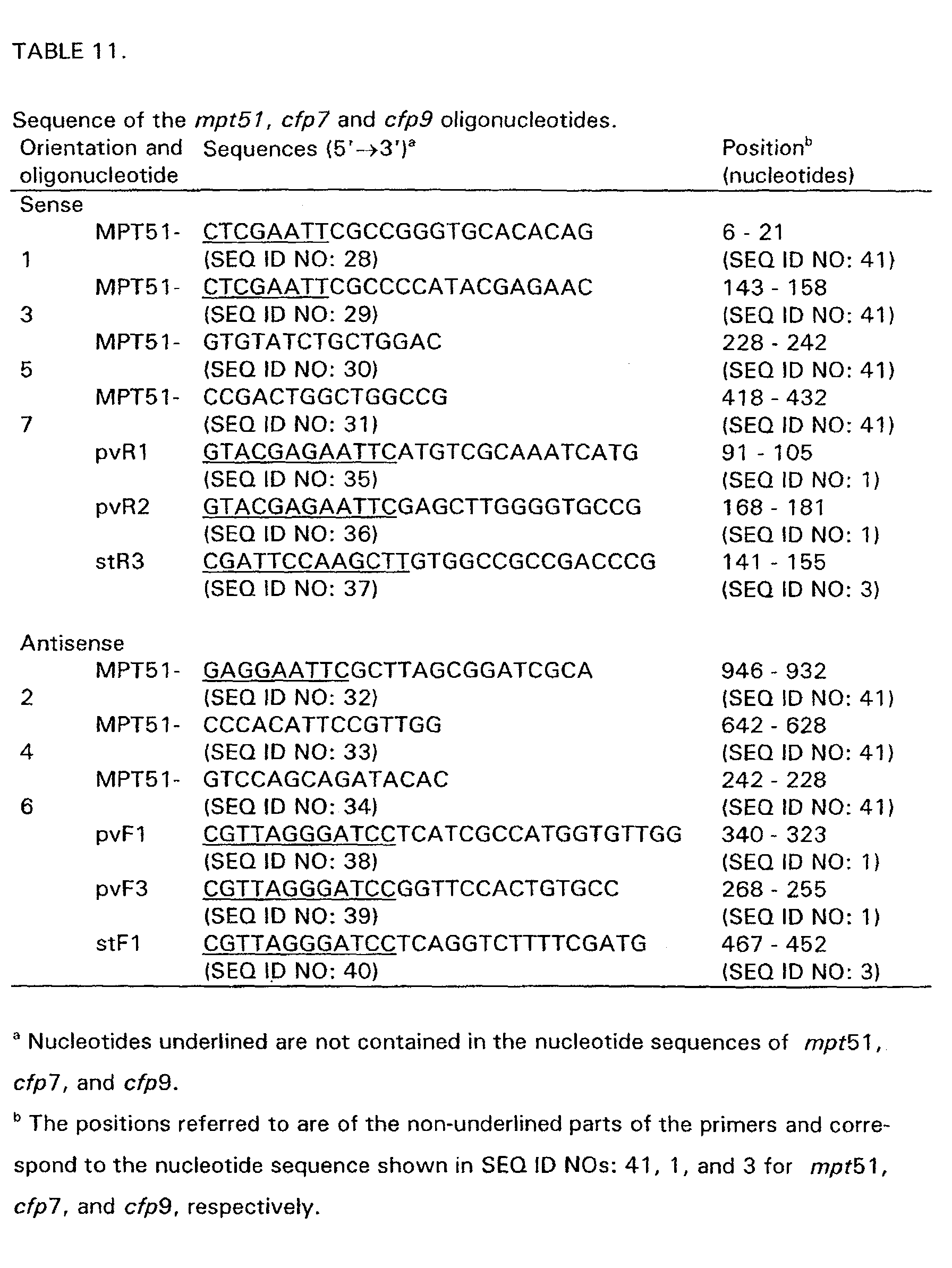
-
-
The Southern blotting was carried out as described previously (Oettinger and Andersen,
1994) with the following modifications: 2 µg of genomic DNA was digested with
Pvull, electrophoresed in an 0.8% agarose gel, and transferred onto a nylon membrane
(Hybond N-plus; Amersham International plc, Little Chalfont, United Kingdom) with a
vacuum transfer device (Milliblot, TM-v; Millipore Corp., Bedford, MA). The
cfp7,
cfp9, mpt51,
rd1-orf2,
rd1-orf3,
rd7-orf4,
rd1-orf5,
rd1-orf8,
rd1-orf9a and
rd1-orf9b
gene fragments were amplified by PCR from the plasmids pRVN01, pRVN02, pTO52,
pTO87, pTO88, pT089, pT090, pT091, pT096 or pTO98 by using the primers shown
in TABLE 11 and TABLE 2 (in Example 2a). The probes were labelled non-radioactively
with an enhanced chemiluminescence kit (ECL; Amersham International plc, Little
Chalfont, United Kingdom). Hybridization and detection was performed according to
the instructions provided by the manufacturer. The results are summarized in TABLES
12 and 13.
-
cfp7, cfp9 and mpt51 were found in the M. tuberculosis complex including BCG and
the environmental mycobacteria; M. avium, M. kansasii, M. marinum, M. intracellular
and M. flavescens. cfp9 was additionally found in M. szulgai and mpt51 in M. xenopi.
-
Furthermore the presence of native MPT51 in culture filtrates from different mycobacterial
strains was investigated with western blots developed with Mab HBT4.
-
There is a strong band at around 26 kDa in
M. tuberculosis H37Rv, Ra, Erdman, M.
bovis AN5,
M. bovis BCG substrain Danish 1331 and
M. africanum. No band was
seen in the region in any other tested mycobacterial strains.
| Interspecies analysis of the rd1-orf2, rd1-orf3, rd1-orf4, rd1-orf5, rd1- orf8, rd1-orf9a and rd1-orf9b genes by Southern blotting. |
| Species and strain | rd1- orf2 | rd1- orf3 | rd1- orf4 | rd1- orf5 | rd1- orf8 | rd1- orf9a | rd1- orf9b |
| 1. M. tub. H37Rv | + | + | + | + | + | + | + |
| 2. M. bovis | + | + | + | + | N.D. | + | + |
| 3. M. bovis BCG Danish 1331 | + | - | - | - | N.D. | - | - |
| 4. M. bovis BCG Japan | + | - | - | - | N.D. | - | - |
| 5. M. avium | - | - | - | - | N.D. | - | - |
| 6. M. kansasii | - | - | - | - | N.D. | - | - |
| 7. M. marinum | + | - | + | - | N.D. | - | - |
| 8. M. scrofu- laceum | + | - | - | - | N.D. | - | - |
| 9. M. intercellu- lare | - | - | - | - | N.D. | - | - |
| 10. M. fortuitum | - | - | - | - | N.D. | - | - |
| 11. M. xenopi | - | - | - | - | N.D. | - | - |
| 12. M. szulgai | + | - | - | - | N.D. | - | - |
| +, positive reaction; -, no reaction, N.D. not determined. |
-
Positive results for rd1-orf2, rd1-orf3, rd1-orf4, rd1-orf5, rd1-orf8, rd1-orf9a and rd1-orf9b
were only obtained when using genomic DNA from M. tuberculosis and M. bovis,
and not from M. bovis BCG or other mycobacteria analyzed except rd1-orf4 which
also was found in M. marinum.
Presence of cfp7a, cfp7b, cfp10a, cfp17, cfp20, cfp21, cfp22, cfp22a, cfp23, cfp25
and cfp25a in different mycobacterial species.
-
Southern blotting was carried out as described for
rd1-orf2,
rd1-orf3,
rd1-orf4,
rd1-orf5,
rd1-orf8,
rd1-orf9a and
rd1-orf9b. The
cfp7a, cfp7b, cfp10a,
cfp17,
cfp20,
cfp21,
cfp22,
cfp22a,
cfp23, cfp25 and
cfp25a gene fragments were amplified by
PCR from the recombinant pMCT6 plasmids encoding the individual genes. The
primers used (same as the primers used for cloning) are described in example 3, 3A
and 3B. The results are summarized in Table 13b.
| Interspecies analysis of the cfp7a, cfp7b, cfp10a, cfp17, cfp20, cfp21, cfp22, cfp22a, cfp23, cfp25, and cfp25a genes by Southern blotting. |
| Species and strain | cfp7a | cfp7b | cfp- 10a | cfp | 17 | cfp 20 | cfp21 | cfp22 | cfp- 22a | cfp 23 | cfp 25 | cfp- 25a |
| 1. M. tub. H37Rv | + | + | + | + | + | + | + | + | + | + | + |
| 2. M. bo vis | + | + | + | + | + | + | + | + | + | + | + |
| 3. M. bovis BCG Danish 1331 | + | + | + | + | + | N.D. | + | + | + | + | + |
| 4. M. bovis BCG Japan | + | + | + | + | + | + | + | + | + | + | + |
| 5. M. avium | + | N.D. | - | + | - | + | + | + | + | + | - |
| 6. M. kansasii | - | N.D. | + | - | - | - | + | - | + | - | - |
| 7. M. marinum | + | + | - | + | + | + | + | + | + | + | + |
| 8. M. scrofu- laceum | - | - | + | - | + | + | - | + | + | + | - |
| 9. M. intercel- lulare | + | + | - | + | - | + | + | - | + | + | - |
| 10. M. fortui- tum | - | N.D. | - | - | - | - | - | - | + | - | - |
| 11. M. xenopi | + | + | + | + | + | + | + | + | + | + | + |
| 12. M. szulgai | + | + | - | + | + | + | + | + | + | + | + |
| +, positive reaction; -, no reaction, N.D. not determined. |
LIST OF REFERENCES
-
Andersen, P. and Heron, I, 1993, J. Immunol. Methods 161: 29-39.
-
Andersen, Å. B. et al., 1992, Infect. Immun. 60: 2317-2323.
-
Andersen P., 1994, Infect. Immun. 62: 2536-44.
-
Andersen P. et al., 1995, J. Immunol. 154: 3359-72
-
Barkholt, V. and Jensen, A. L., 1989, Anal. Biochem. 177: 318-322.
-
Borodovsky, M., and J. McIninch. 1993, Computers Chem. 17: 123-133.
-
van Dyke M. W. et al., 1992. Gene pp. 99-104.
-
Gosselin et al., 1992, J. Immunol. 149: 3477-3481.
-
Harboe, M. et al., 1996, Infect. Immun. 64: 16-22.
-
von Heijne, G., 1984, J. Mol. Biol. 173: 243-251.
-
Hochstrasser, D.F. et al., 1988, Anal.Biochem. 173: 424-435
-
Köhler, G. and Milstein, C., 1975, Nature 256: 495-497.
-
Li, H. et al., 1993, Infect. Immun. 61: 1730-1734.
-
Lindblad E.B. et al., 1997, Infect. Immun. 65: 623-629.
-
Mahairas, G. G. et al., 1996, J. Bacteriol 178: 1274-1282.
-
Maniatis T. et al., 1989, "Molecular cloning: a laboratory manual", 2nd ed., Cold
Spring Harbor Laboratory, Cold Spring Harbor, N.Y.
-
Nagai, S. et al., 1991, Infect. Immun. 59: 372-382.
-
Oettinger, T. and Andersen, Å. B., 1994, Infect. Immun. 62: 2058-2064.
-
Ohara, N. et al., 1995, Scand. J. immunol. 41: 233-442.
-
Pal P. G. and Horwitz M. A., 1992, Infect. Immun. 60: 4781-92.
-
Pearson, W. R. and Lipman D. J., 1988. Proc. Natl. Acad. Sci. USA 85: 2444-2448.
-
Ploug, M. et al., 1989, Anal. Biochem. 181: 33-39.
-
Porath, J. et al., 1985, FEBS Lett. 185: 306-310.
-
Roberts, A.D. et al., 1995, Immunol. 85: 502-508.
-
Sørensen, A.L. et al., 1995, Infect. Immun. 63: 1710-1717.
-
Theisen, M. et al., 1995, Clinical and Diagnostic Laboratory Immunology, 2: 30-34.
-
Valdés-Stauber, N. and Scherer, S., 1994, Appl. Environ. Microbiol. 60: 3809-3814.
-
Valdés-Stauber, N. and Scherer, S., 1996, Appl. Environ. Microbiol. 62: 1283-1286.
-
Williams, N., 1996, Science 272: 27.
-
Young, R.
A. et al., 1985, Proc. Natl. Acad. Sci. USA
82: 2583-2587.








 - where S = G/C and Y = T/C






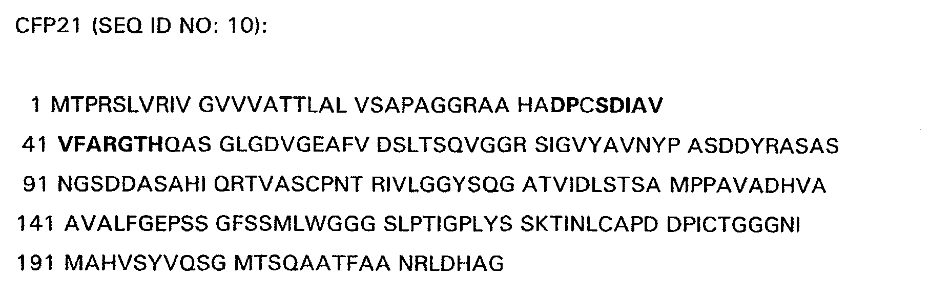
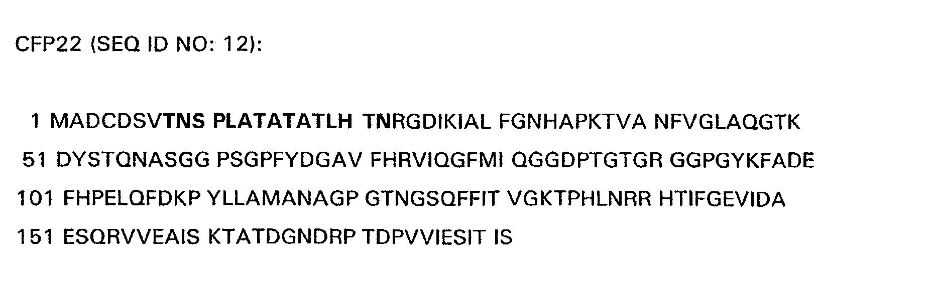




















































 OPBR-48 and OPBR-3 create BglII and HinDIII, respectively. Additionally OPBR-3 deletes the stop codon of MPT59.