EP1484396A1 - Molecules associating to c-terminal domain in receptor cell - Google Patents
Molecules associating to c-terminal domain in receptor cell Download PDFInfo
- Publication number
- EP1484396A1 EP1484396A1 EP03705270A EP03705270A EP1484396A1 EP 1484396 A1 EP1484396 A1 EP 1484396A1 EP 03705270 A EP03705270 A EP 03705270A EP 03705270 A EP03705270 A EP 03705270A EP 1484396 A1 EP1484396 A1 EP 1484396A1
- Authority
- EP
- European Patent Office
- Prior art keywords
- protein
- frount
- receptor
- dna
- cell
- Prior art date
- Legal status (The legal status is an assumption and is not a legal conclusion. Google has not performed a legal analysis and makes no representation as to the accuracy of the status listed.)
- Granted
Links
Images









Classifications
-
- C—CHEMISTRY; METALLURGY
- C07—ORGANIC CHEMISTRY
- C07K—PEPTIDES
- C07K14/00—Peptides having more than 20 amino acids; Gastrins; Somatostatins; Melanotropins; Derivatives thereof
- C07K14/435—Peptides having more than 20 amino acids; Gastrins; Somatostatins; Melanotropins; Derivatives thereof from animals; from humans
- C07K14/52—Cytokines; Lymphokines; Interferons
- C07K14/521—Chemokines
-
- A—HUMAN NECESSITIES
- A61—MEDICAL OR VETERINARY SCIENCE; HYGIENE
- A61P—SPECIFIC THERAPEUTIC ACTIVITY OF CHEMICAL COMPOUNDS OR MEDICINAL PREPARATIONS
- A61P13/00—Drugs for disorders of the urinary system
- A61P13/12—Drugs for disorders of the urinary system of the kidneys
-
- A—HUMAN NECESSITIES
- A61—MEDICAL OR VETERINARY SCIENCE; HYGIENE
- A61P—SPECIFIC THERAPEUTIC ACTIVITY OF CHEMICAL COMPOUNDS OR MEDICINAL PREPARATIONS
- A61P25/00—Drugs for disorders of the nervous system
-
- A—HUMAN NECESSITIES
- A61—MEDICAL OR VETERINARY SCIENCE; HYGIENE
- A61P—SPECIFIC THERAPEUTIC ACTIVITY OF CHEMICAL COMPOUNDS OR MEDICINAL PREPARATIONS
- A61P29/00—Non-central analgesic, antipyretic or antiinflammatory agents, e.g. antirheumatic agents; Non-steroidal antiinflammatory drugs [NSAID]
-
- A—HUMAN NECESSITIES
- A61—MEDICAL OR VETERINARY SCIENCE; HYGIENE
- A61P—SPECIFIC THERAPEUTIC ACTIVITY OF CHEMICAL COMPOUNDS OR MEDICINAL PREPARATIONS
- A61P31/00—Antiinfectives, i.e. antibiotics, antiseptics, chemotherapeutics
-
- A—HUMAN NECESSITIES
- A61—MEDICAL OR VETERINARY SCIENCE; HYGIENE
- A61P—SPECIFIC THERAPEUTIC ACTIVITY OF CHEMICAL COMPOUNDS OR MEDICINAL PREPARATIONS
- A61P37/00—Drugs for immunological or allergic disorders
- A61P37/02—Immunomodulators
-
- A—HUMAN NECESSITIES
- A61—MEDICAL OR VETERINARY SCIENCE; HYGIENE
- A61P—SPECIFIC THERAPEUTIC ACTIVITY OF CHEMICAL COMPOUNDS OR MEDICINAL PREPARATIONS
- A61P37/00—Drugs for immunological or allergic disorders
- A61P37/02—Immunomodulators
- A61P37/04—Immunostimulants
-
- A—HUMAN NECESSITIES
- A61—MEDICAL OR VETERINARY SCIENCE; HYGIENE
- A61P—SPECIFIC THERAPEUTIC ACTIVITY OF CHEMICAL COMPOUNDS OR MEDICINAL PREPARATIONS
- A61P37/00—Drugs for immunological or allergic disorders
- A61P37/02—Immunomodulators
- A61P37/06—Immunosuppressants, e.g. drugs for graft rejection
-
- A—HUMAN NECESSITIES
- A61—MEDICAL OR VETERINARY SCIENCE; HYGIENE
- A61P—SPECIFIC THERAPEUTIC ACTIVITY OF CHEMICAL COMPOUNDS OR MEDICINAL PREPARATIONS
- A61P37/00—Drugs for immunological or allergic disorders
- A61P37/08—Antiallergic agents
-
- A—HUMAN NECESSITIES
- A61—MEDICAL OR VETERINARY SCIENCE; HYGIENE
- A61P—SPECIFIC THERAPEUTIC ACTIVITY OF CHEMICAL COMPOUNDS OR MEDICINAL PREPARATIONS
- A61P9/00—Drugs for disorders of the cardiovascular system
- A61P9/10—Drugs for disorders of the cardiovascular system for treating ischaemic or atherosclerotic diseases, e.g. antianginal drugs, coronary vasodilators, drugs for myocardial infarction, retinopathy, cerebrovascula insufficiency, renal arteriosclerosis
Definitions
- This invention relates to a protein which associates with the intracellular C-terminal domain of a G protein-coupled receptor for chemokine, a DNA encoding the same, and utilization thereof. More specifically, it relates to a novel protein which associates with the intracellular C-terminal domain of receptors CCR2 and CCR5 to thereby control the functions of the receptors CCR2 and CCR5, a DNA encoding the same and utilization thereof in the field of medicine.
- chemokine receptors for example, leukocyte chemotaxis, which is the fundamental function in inflammation and immune responses, is strictly controlled by attractors serving as agonists (Documents 1 to 4). More specifically speaking, a chemokine prototype CCL2 (also known as MCAF and MCP-1) was found as a macrophage attractant mediated by a receptor CCR2 (Documents 5 to 8). Moreover, chemokines CCL3, 4 and 5 are known as agonists for CCR5.
- chemokines there have been identified more than 50 types of chemokines and 20 types of G protein-coupled receptors (GPCRs) as the chemokine-chemokine receptor family. Each chemokine receptor has a strict chemokine-specificity and shows an expression pattern restricted to leukocyte subtype (Documents 9 to 11).
- the receptor CCR2 is a 7-transmembrane G protein-coupled receptor known as occurring in monocytes, macrophages, lymphocytes, endothelial cells, smooth muscular cells and so on and its agonist CCL2 acts as an attractant via interaction with the receptor CCR2 (Document 12). It is considered that the CCL2-CCR2 pathway participates in the causes of atherosclerosis (Documents 13 and 14), chronic glomerulonephritis (Document 15), multiple sclerosis (Documents 16 and 17) and other chronic inflammatory diseases (Documents 18 to 21).
- CCR5 which is known as a chemokine CCL2 (MIP-1 ⁇ ), CCL4 (MIP-1 ⁇ ) or CCL5 (RANTES) receptor
- MIP-1 ⁇ chemokine CCL2
- MIP-1 ⁇ CCL4
- RANTES CCL5
- CCR2 and CCR5 serve as coreceptors for cell entry of human immunodeficiency virus (HIV) (Deng et al., Nature, 381, 661-666 (1996)).
- HAV human immunodeficiency virus
- a large number of points still remain unknown in intracellular signal transduction cascades relating to leukocyte chemotaxis. Accordingly, there is a great worth in studying the mechanism controlling chemokine receptor-mediated leukocyte chemotaxis.
- a receptor having an attractant bonded thereto activates a G protein and thus causes the dissociation of the G protein into ⁇ - and ⁇ -subunits and the formation of a second messenger, thereby initiating actin polymerization and leukocyte chemotaxis (Documents 22 and 23).
- the dissociation of the G protein is followed by the phosphorylation of the receptor by protein kinases such as PKs, Jaks and GRKs (Documents 24 and 25).
- protein kinases such as PKs, Jaks and GRKs
- chemokine receptors CCR2 and CXCR4 both activate monocyte chemotaxis cascades, the Pro-12-C terminal domain of CCR2 never relates to the conservation of a similar domain of CXCR4.
- the present inventors drawn a novel hypothesis concerning the intracellular signal transduction mechanism that, even in the case where phosphorylation does not occur in the intracellular C-terminal domain of a receptor, an unknown molecule associates with the Pro-C terminal domain of a G protein-coupled receptor for each chemokine and thus controls leukocyte chemotaxis depending on the receptor.
- a CCR2-binding protein was searched for as an aspect of the invention.
- the present inventors attempted to compare and discuss the conservation states of corresponding genetic information in biological systems and clarify the mechanisms of intracellular signal transduction and regulation concerning cell migration, thereby developing a novel route for the establishment of technical means contributing to the treatment and diagnosis of diseases in which the CCL2-CCR2 pathway and the CCL3, 4 or 5-CCR5 pathway participate.
- the present invention relates to the following DNAs.
- the present invention provides an intracellular signal transduction-regulating polypeptide associating with the intracellular C-terminal domain of a receptor CCR2, which is one of G protein-coupled receptors for chemokines, and a DNA encoding the same. Further, it provides an antibody, an antisense sequence inhibiting the expression of the DNA encoding the above-described protein and a probe consisting of a sequence complementary to the above-described DNA. Furthermore, it provides a method of identifying a substance which inhibits the association of the above-described protein with the intracellular C-terminal domain of the receptor by using the protein.
- the present inventors have confirmed for the first time the presence of intracellular signal transduction-regulating polypeptide associating with the intracellular C-terminal domain of a receptor CCR2, which is one of G protein-coupled receptors for chemokines, and the DNA encoding the same, i.e., discovery of a novel intracellular signal transduction mechanism.
- This protein can be obtained by searching for a molecule specifically binding to an intracellular domain following the 7-transmembrane part of the receptor with the use of the two-hybrid method, thus obtaining its partial sequence, constructing oligoprimers based on this sequence, and then cloning the full-length cDNA by using the RACE method.
- a cytoplasmic protein directly associating with the intracellular C-terminal domain sequence of the receptor CCR2 (the sequence occurring in the 309- to 360-residues) carried by human myelomonocytic leukemia can be obtained.
- oligoprimers are constructed and a full-length cDNA (clone 1, SEQ ID NO:19) is cloned by the RACE method.
- the full-length cDNA (clone 1) thus obtained is amplified by the PCR method and then inserted into a plasmid vector pcDNA3 (manufactured by Invitorgen) to thereby construct an expression vector.
- this expression vector is transferred into HEK293 cells (ATCC/CRL-157) to give a transformant.
- Expression of FROUNT protein 1 (SEQ ID NO:1) is detected by using the Western blotting method.
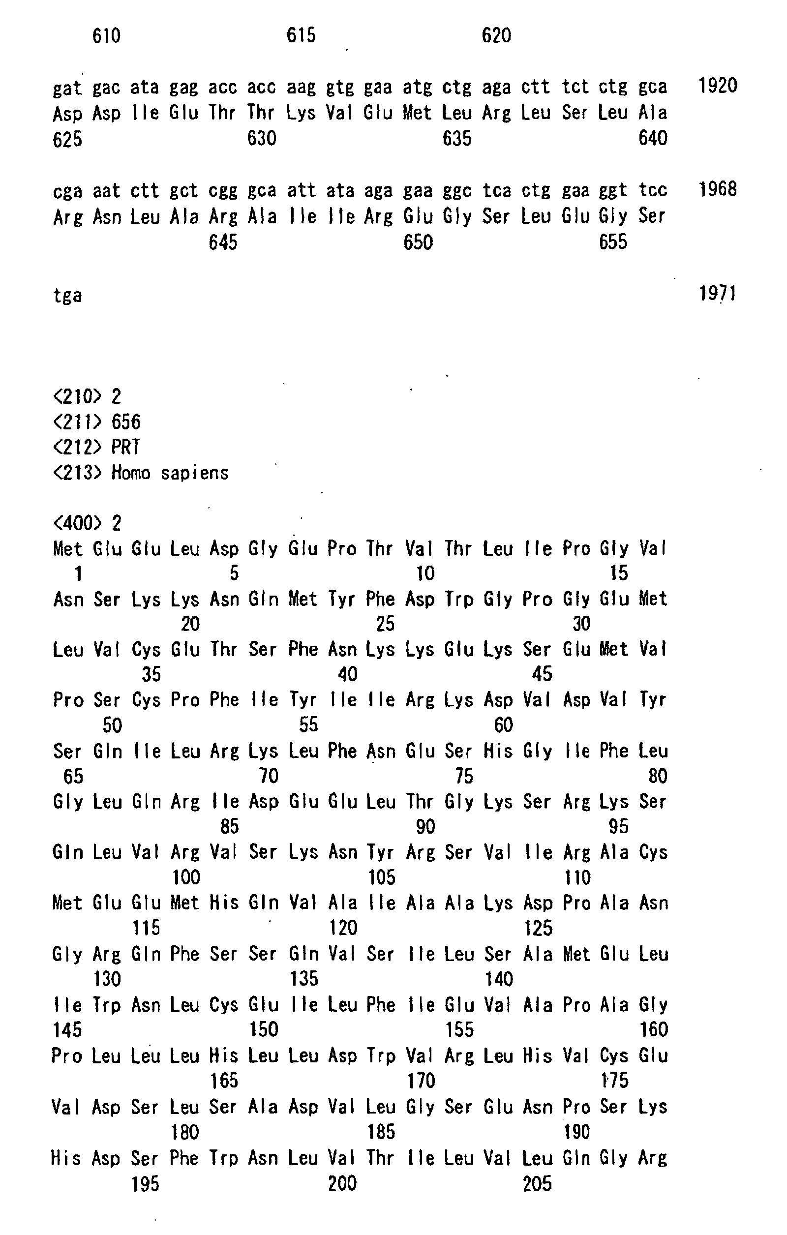
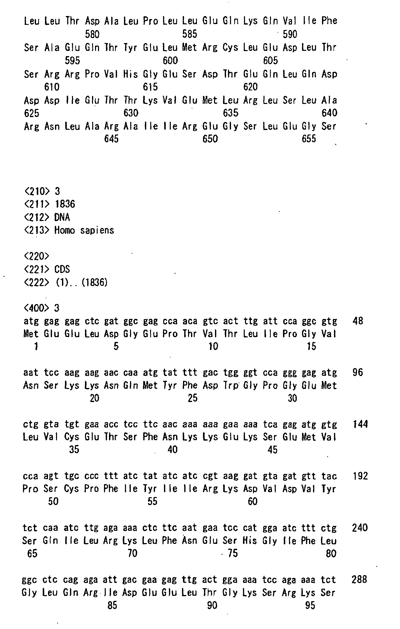
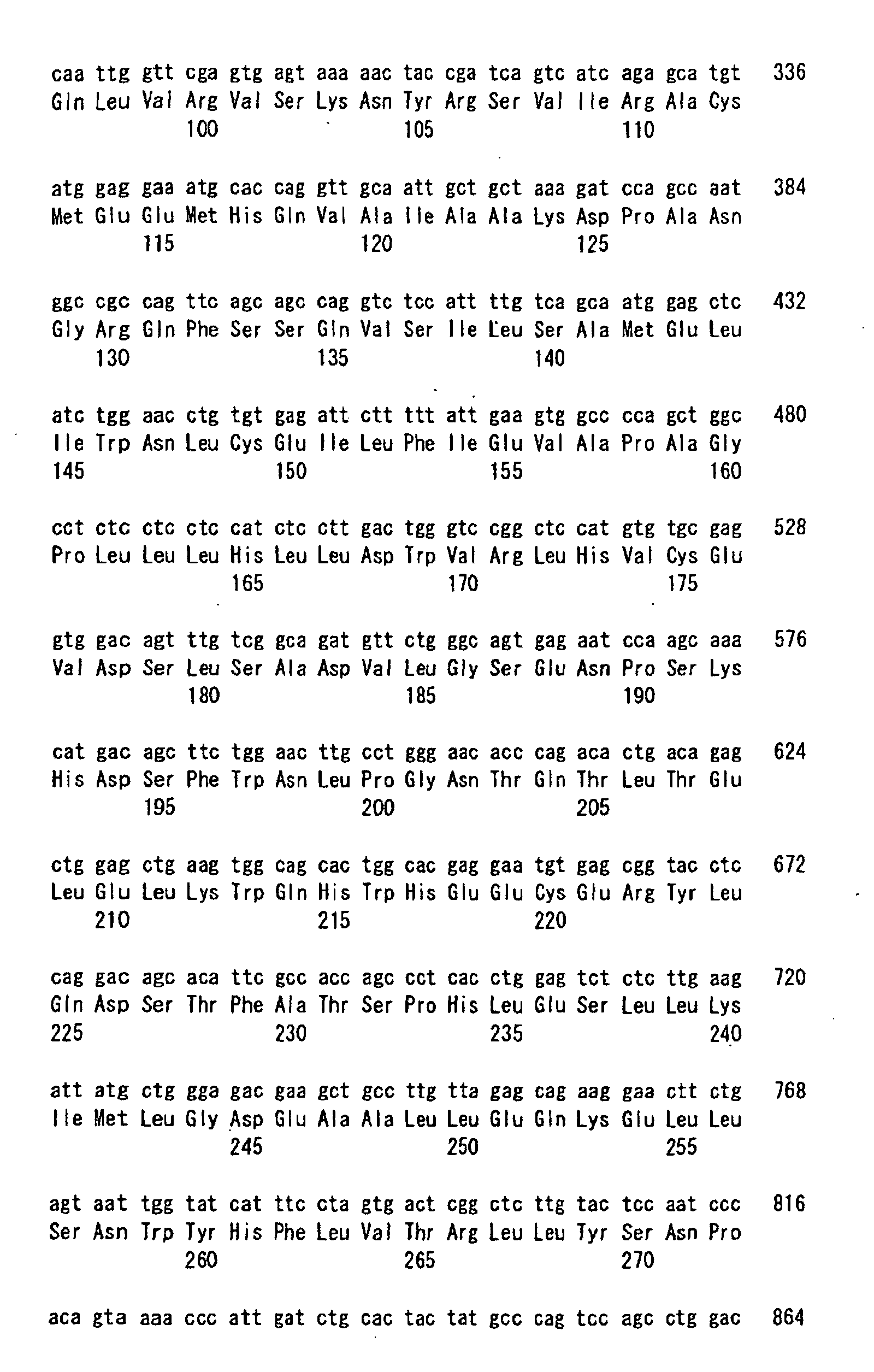
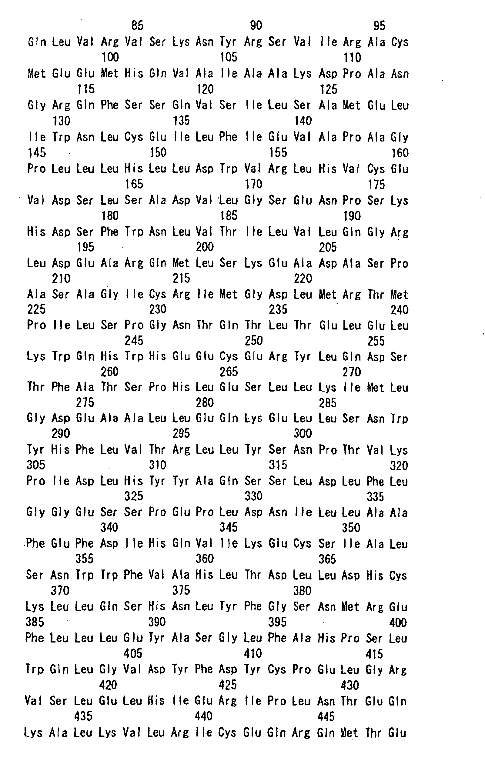
- the thus obtained protein having the amino acid sequence represented by SEQ ID NO:1 and associating with the intracellular C-terminal domain of the receptor CCR2 (the protein originating in clone 1 which will be also called FROUNT protein 1 hereinafter) has a sequence consisting of 656 amino acids and its molecular weight is 79 KDa. Its structural characteristics reside in containing a leucine zipper structure known as playing an important role in the association between proteins, 4 tyrosine-based motives and 4 dileucine motives.
- Fig. 5(a) shows the Kyte-Doolittle hydropathy plot of this protein. When examined by homology searching, it is clarified that none of known human genes or proteins has a sequence similar to the above-described protein.
- the biological properties of the obtained FROUNT protein 1 confirmed by the present inventors are as follows.
- FROUNT protein 1 associates with the intracellular C-terminal domains of the chemokine G protein-coupled receptors CCR2 and CCR5 and thus participates in the mechanism of controlling the G protein switching system. That is to say, it seems that FROUNT protein 1 is a polypeptide which regulates the intracellular signal transduction, acts on the intracellular C-terminal domain of a receptor together with G protein and thus participates in the mechanism of controlling the function of the receptor. Since there has been known neither a chemotactic signal transduction mechanism specific to an individual chemokine receptor nor a protein capable of controlling the mechanism, it can be concluded that the presence such a protein per se has been clarified for the first time by the present inventors.
- FROUNT protein 1 Based on the fact that the DNA sequence of FROUNT protein 1 is conserved even in the nematode and yeast having no leukocyte, there is a high possibility that FROUNT protein 1 and FROUNT-like proteins associating with the intracellular C-terminal domains of cell membrane receptors CCR2 and CCR5 to chemokines would control signal transduction systems from individual receptors not only in other leukocyte chemotactic factors but also in hormones and cell membrane receptors such as neurotransmitter molecules.
- the protein according to the present invention and the DNA encoding the same are primarily an intracellular signal transduction-regulating polypeptide associating with the intracellular C-terminal domain of a G protein-coupled receptor CCR2 for chemokine CCL2 and a DNA encoding the same, and an intracellular signal transduction-regulating polypeptide associating with the intracellular C-terminal domain of a G protein-coupled receptor CCR5 for chemokines CCL's 3, 4 and 5, and a DNA encoding the same.
- the protein according to the present invention and the DNA encoding the same are a polypeptide having the amino acid sequence represented by SEQ ID NO:1 and a DNA encoding the same.
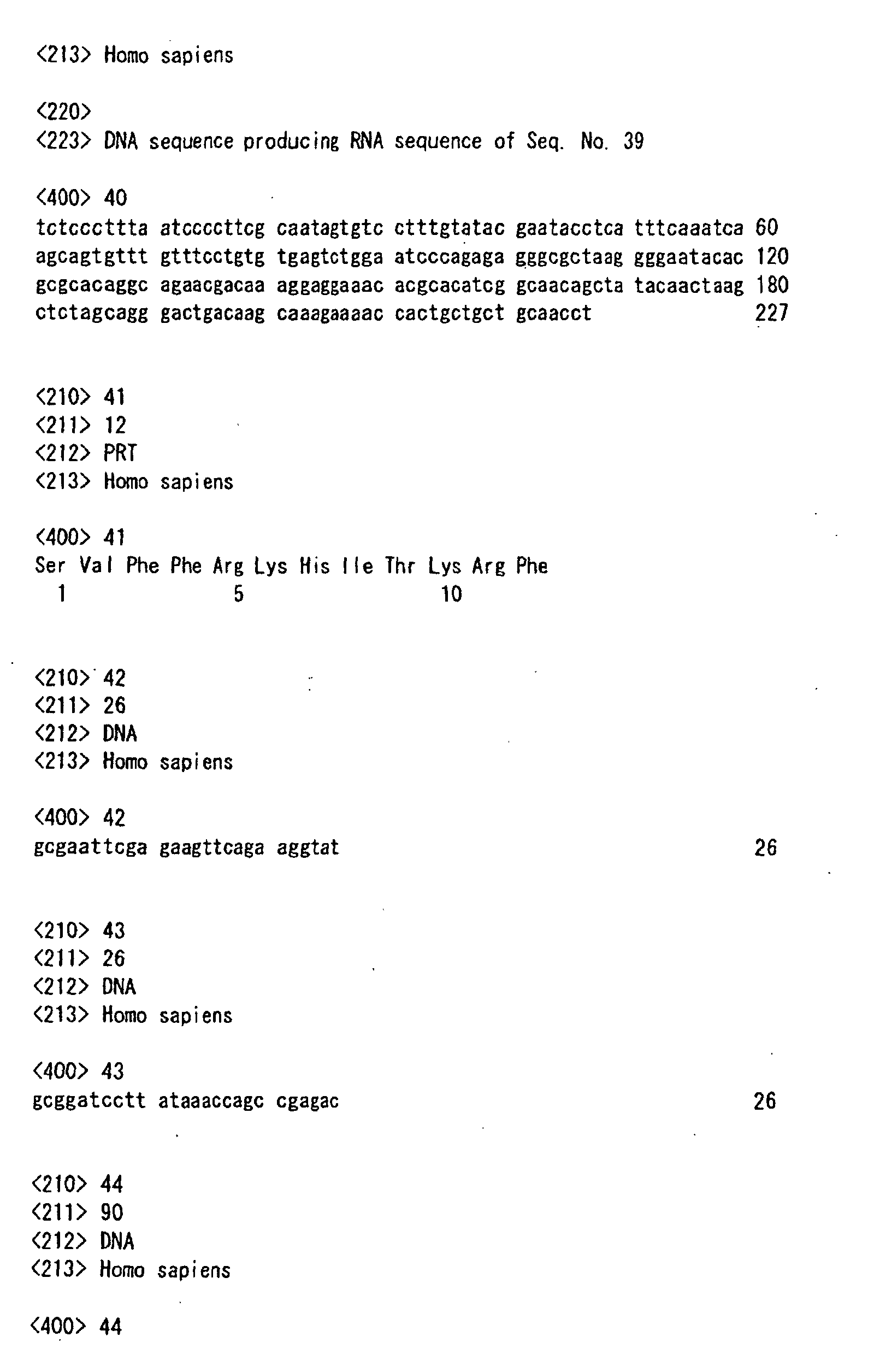
- the DNA has the sequence represented by SEQ ID NO:20.
- clone 1 of FROUNT protein 1 and another clone 2 (SEQ ID NO:20) are obtained.
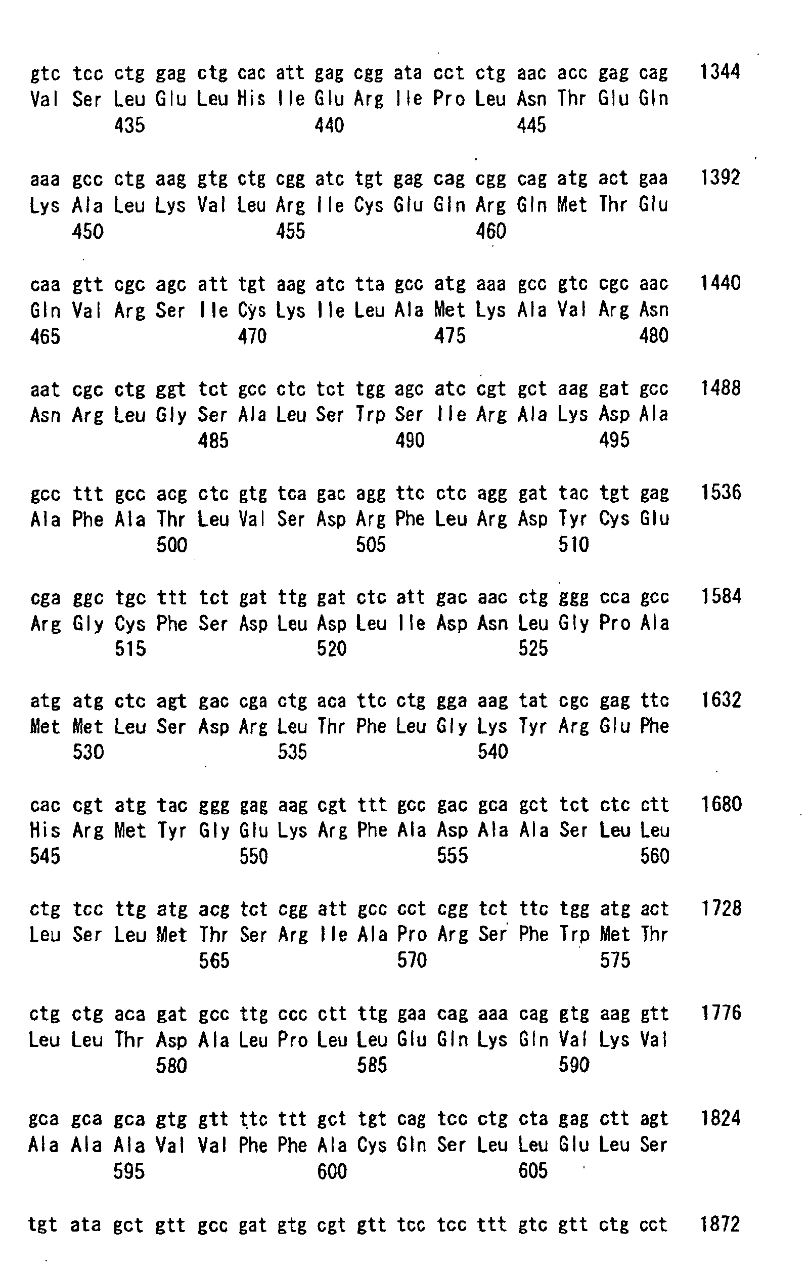
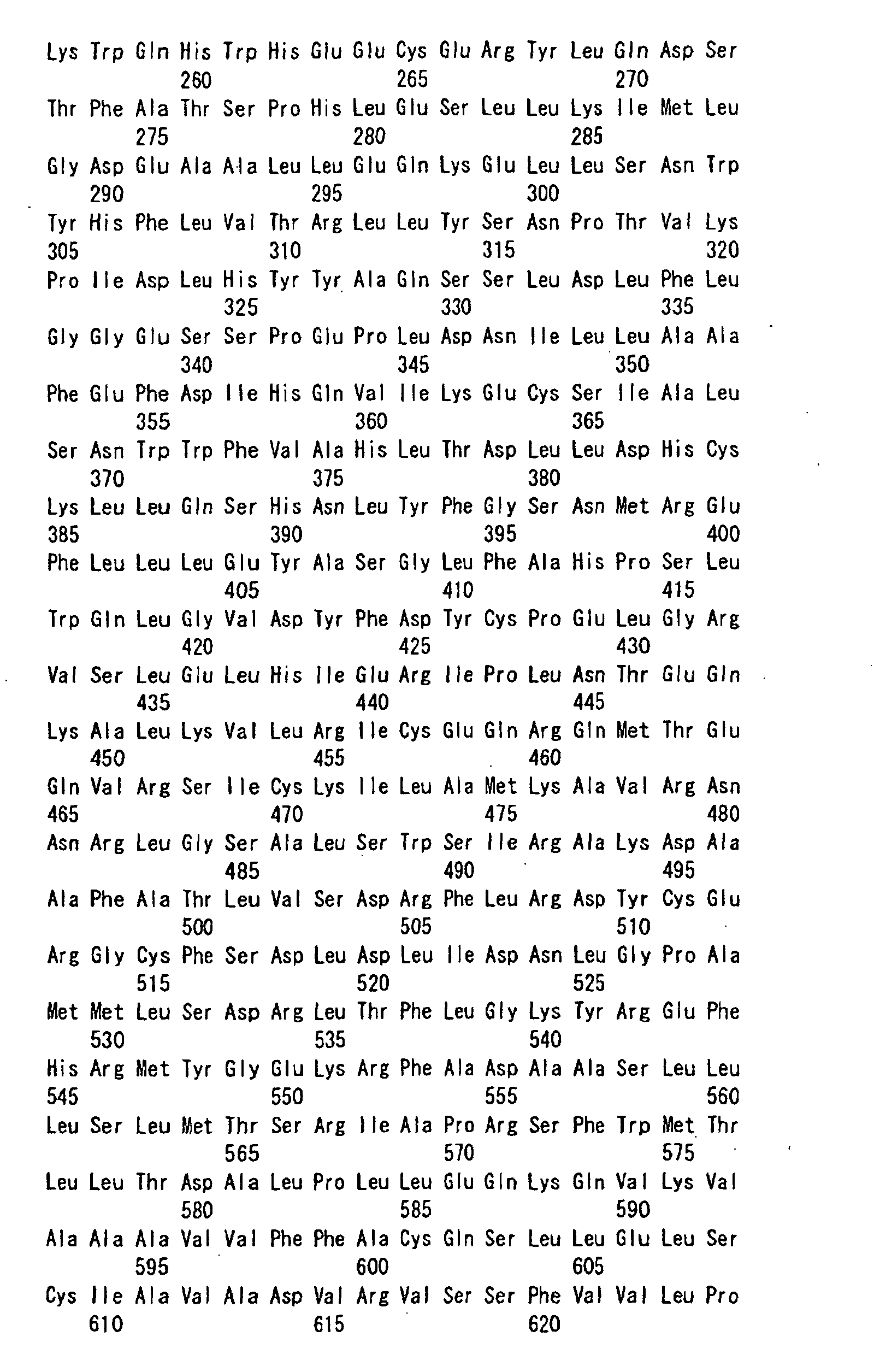
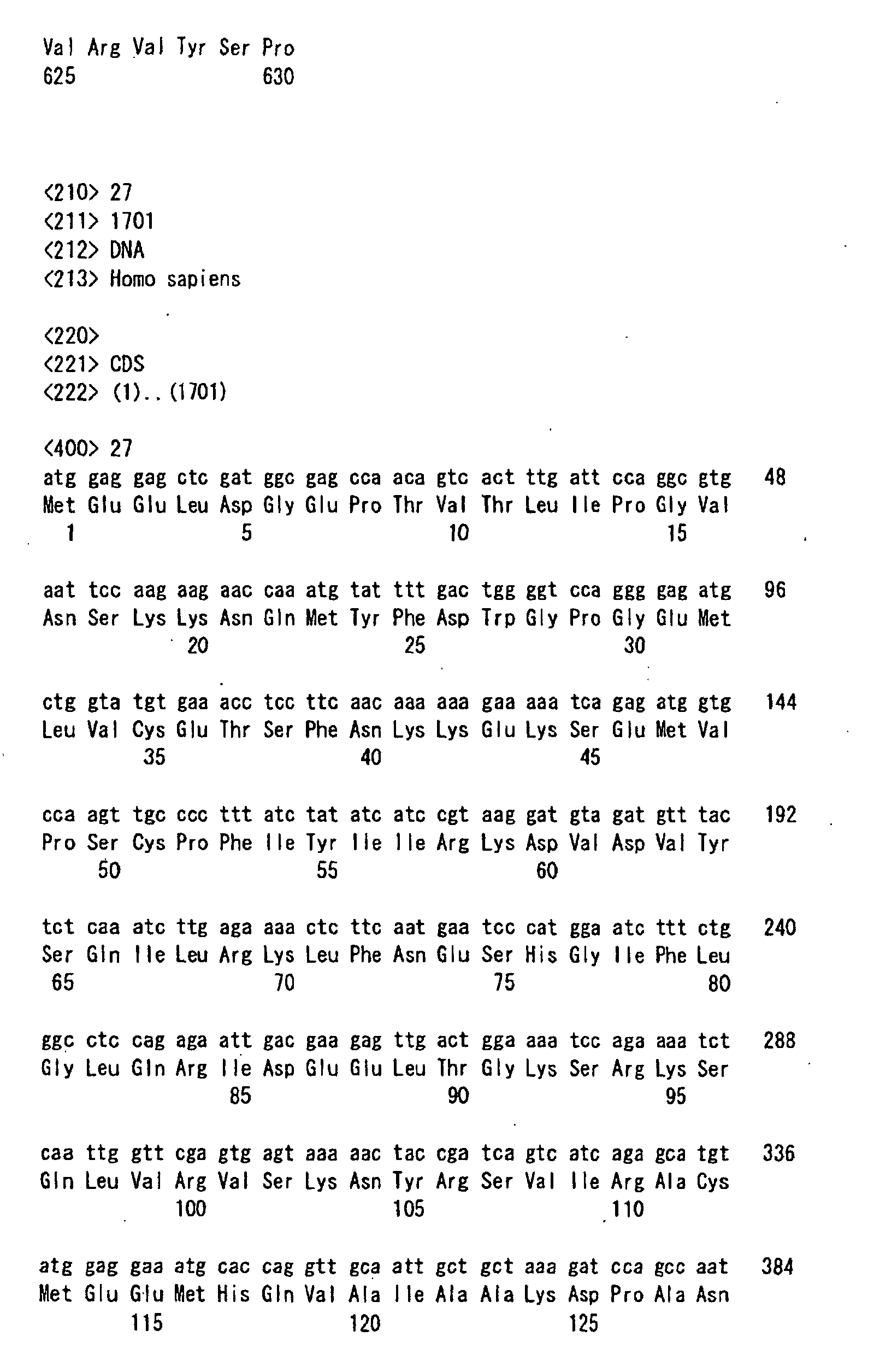
- human FROUNT involves at least two molecular types, i.e., a sequence consisting of 656 amino acid residues ( ⁇ -type, clone 1) and another one consisting of 630 amino acid residues ( ⁇ -type, clone 13) differing from each other exclusively in the C-terminal part.
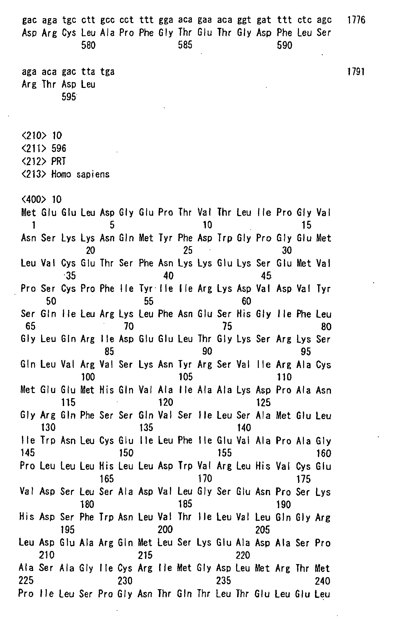
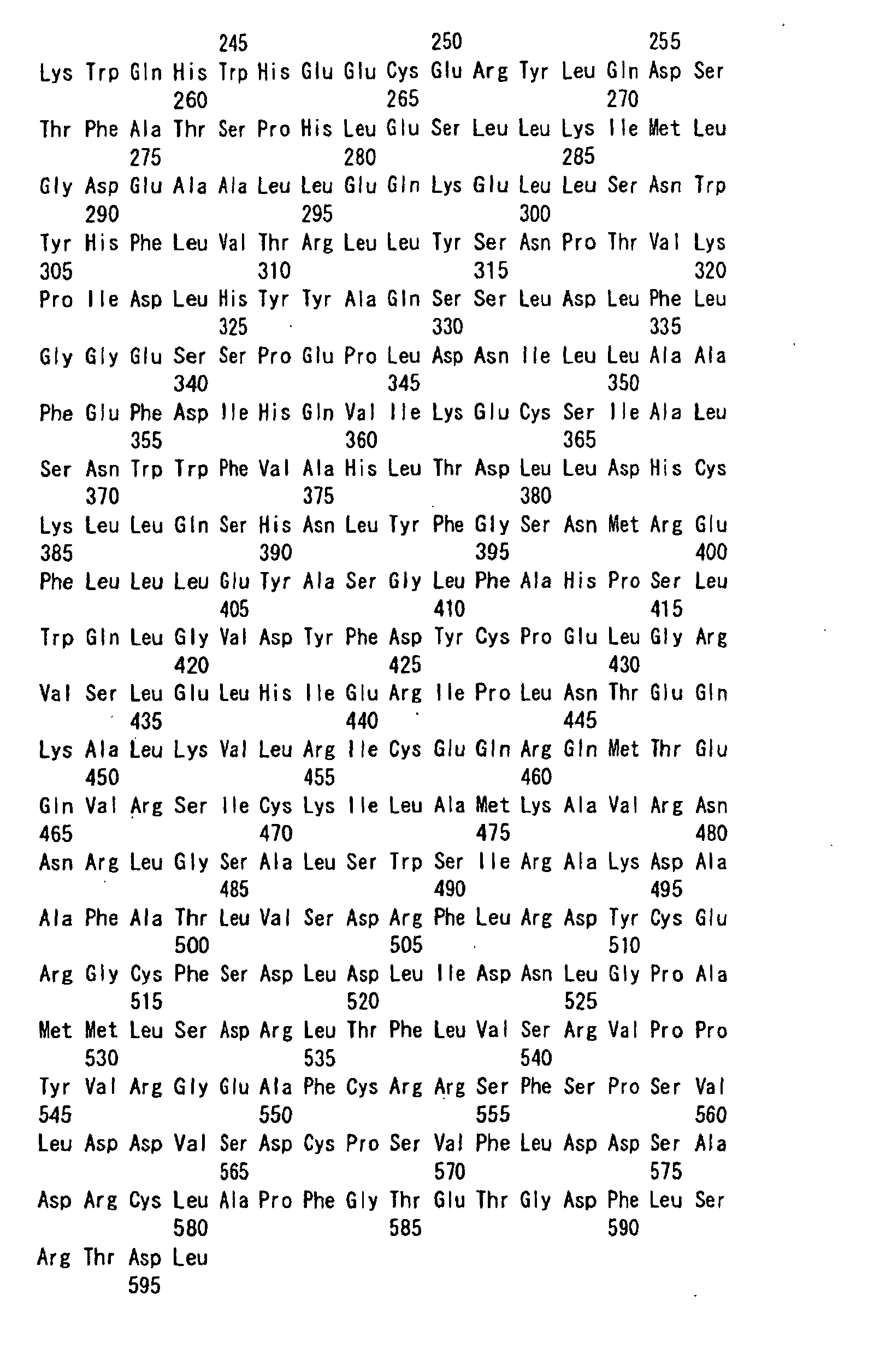
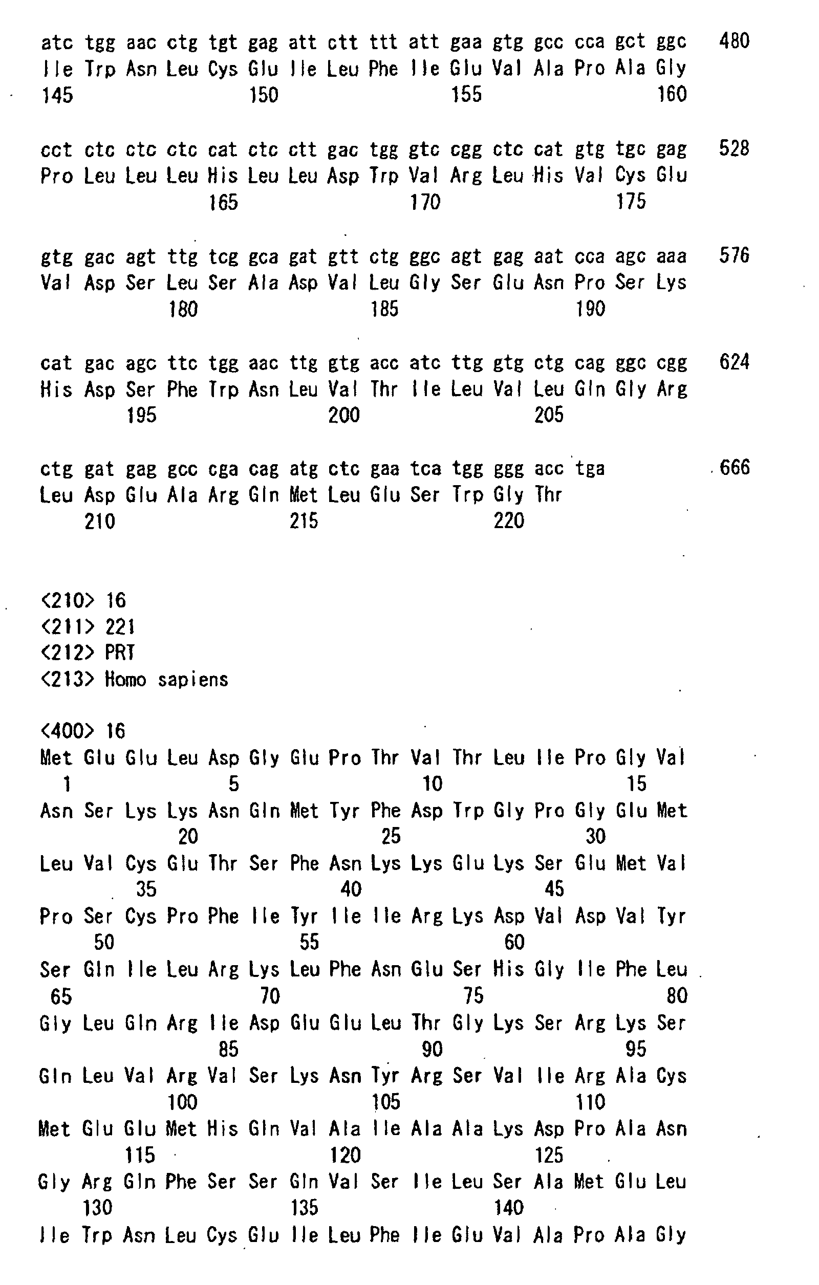
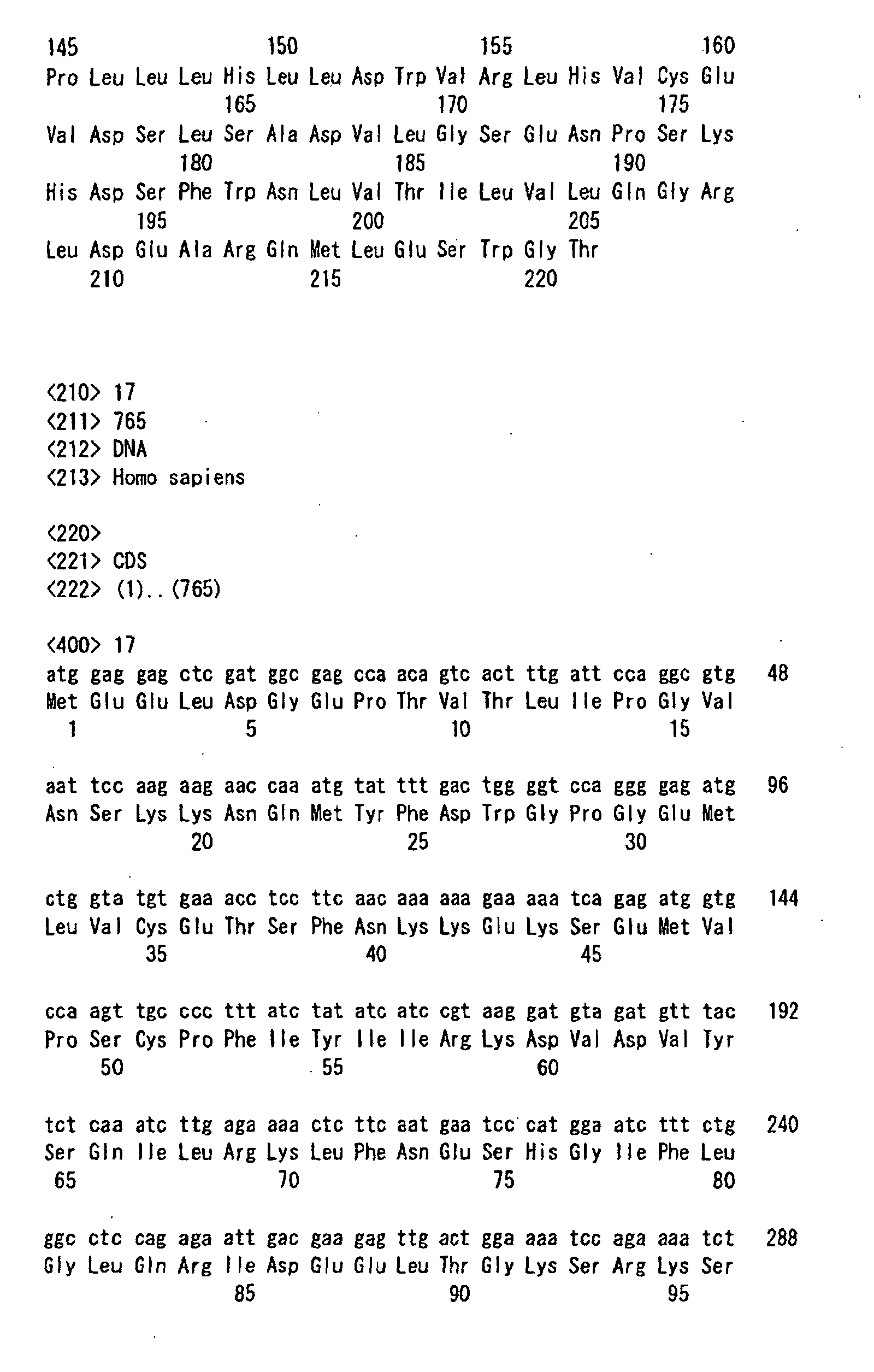
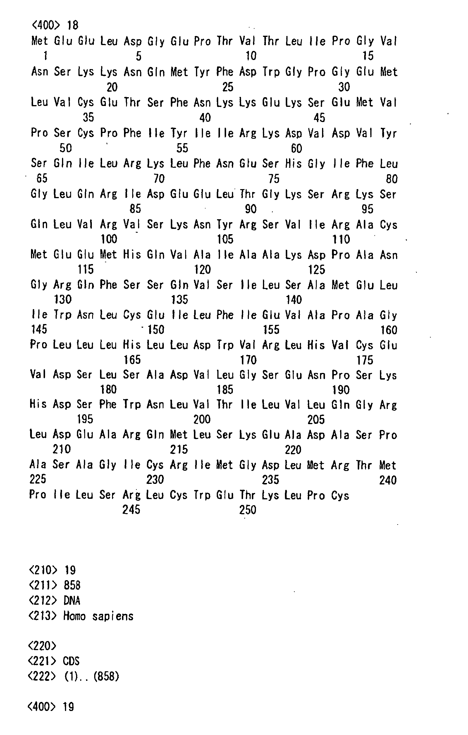
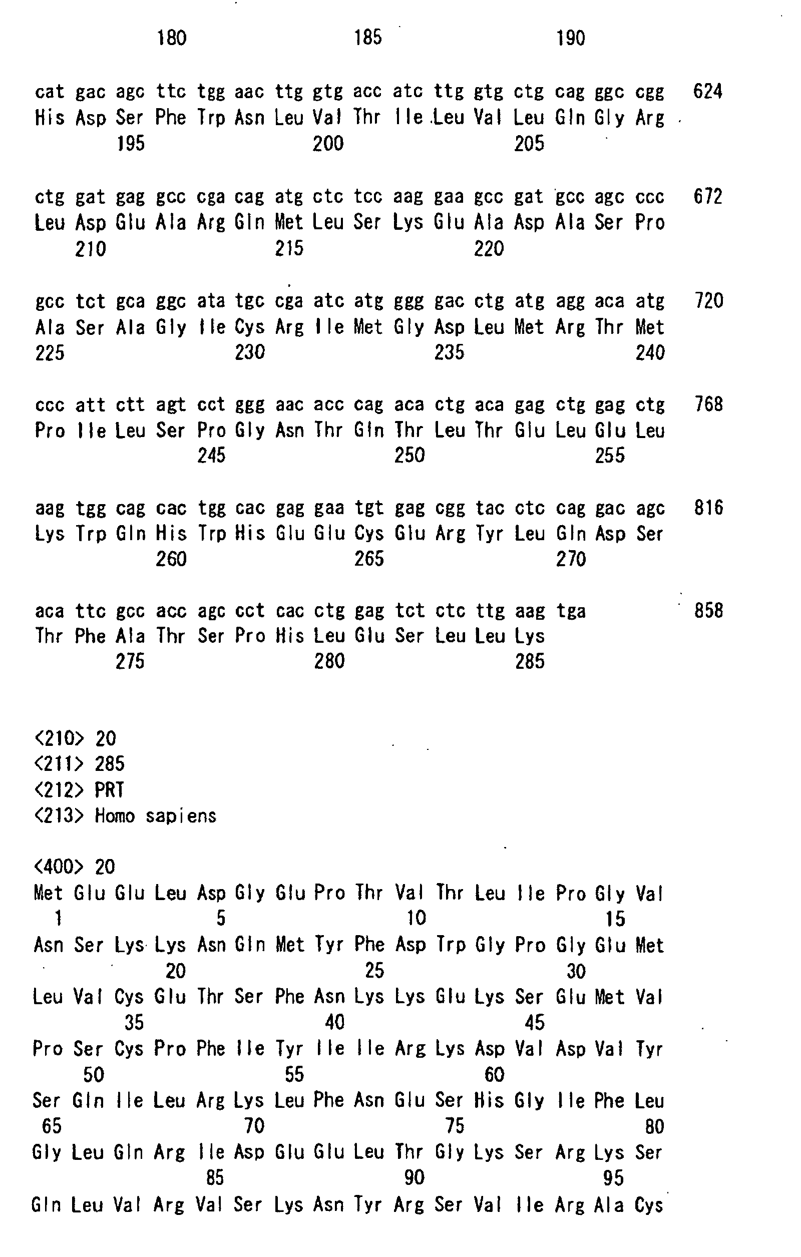
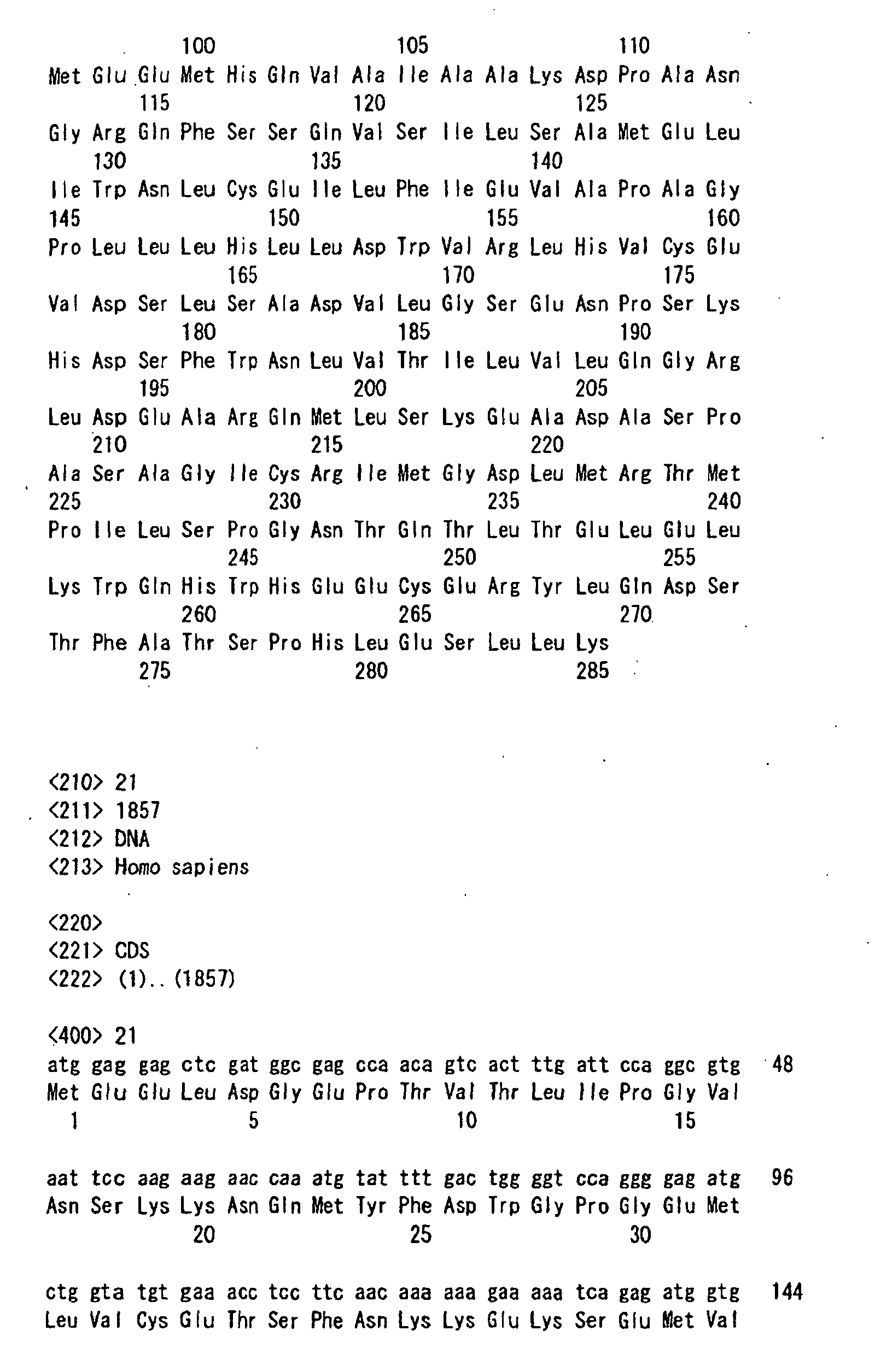
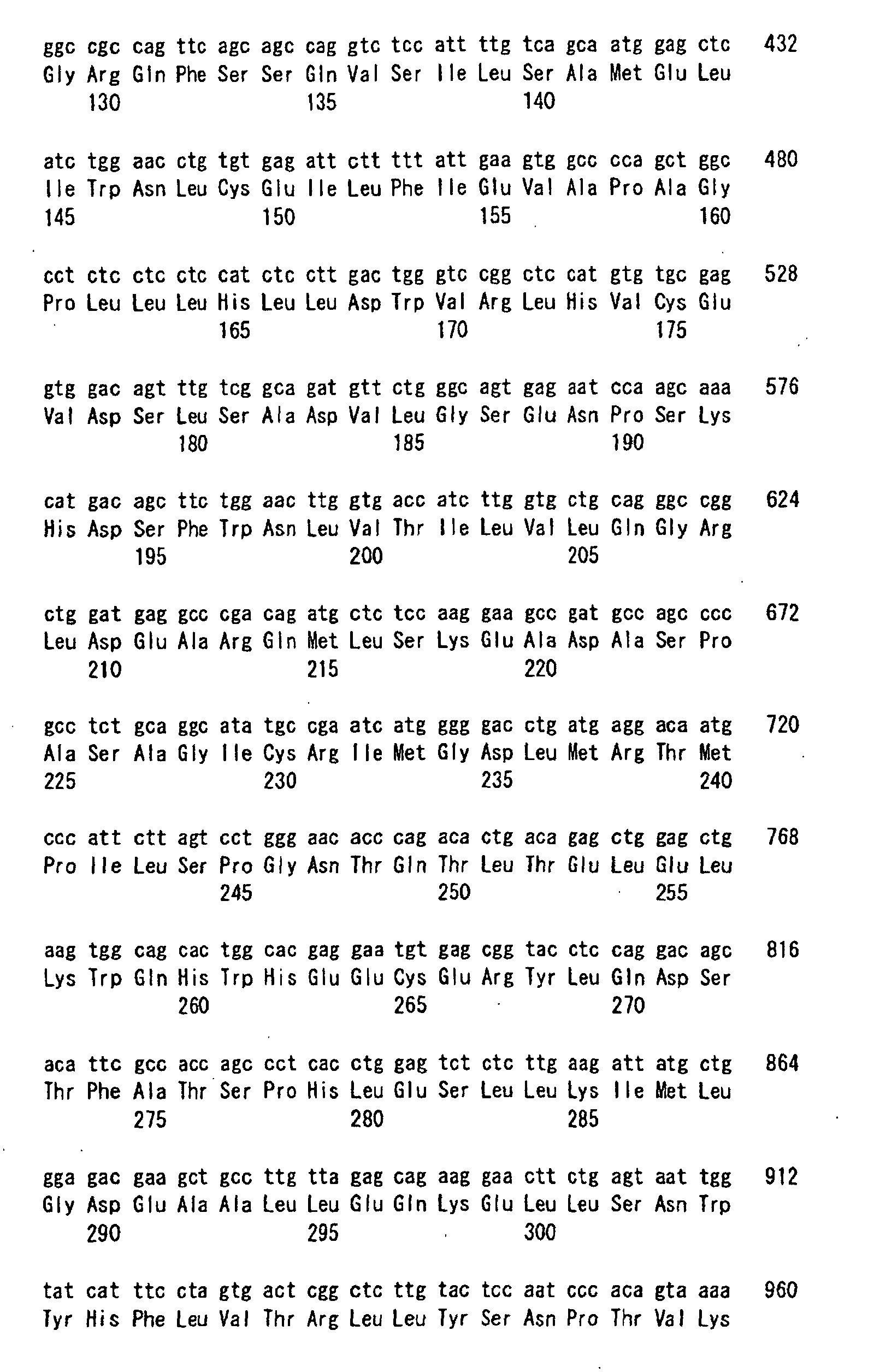
- cloning is carried out by the PCR method and thus at least 18 splicing mutants of the ⁇ - and ⁇ -types represented by SEQ ID NOS:1 to 18 are identified.
- Each of these clones 1 to 18 has an N-terminus starting with ATG and a C-terminus stopping with a termination codon (TAG, TGA, TAA). It is anticipated that these clones respectively have the amino acid sequences represented by SEQ ID NOS:1 to 18.
- Polypeptides corresponding to these clones 2 to 18 will be referred to as FROUNT proteins 2 to 18 hereinafter.
- the clones 2 to 18 are in common to the clone 1 at least in the N-terminal sequence and, moreover, have common sequences in other parts.
- clones 2 to 18 have functions either entirely or partly common to clone 1. It is also considered that clones 2 to 18 are usable as probes for detecting FROUNT proteins 1 to 18. Furthermore, these polypeptides are usable as antigens for constructing antibodies and so on.
- the FROUNT gene clone 1 is encoded as 19 exons in the 17th chromosome (in the neighborhood of D17S785 and D17S1352) similar to CCL's 2, 3 and 5.
- the exons 1 to 16 are the same as those in the FROUNT gene clone 1 but the reading frame in the 17th exon is extended backward compared with the FROUNT gene clone 1. It is thus confirmed that the FROUNT gene clone 13 is encoded as 17 exons and the FROUNT gene clone 13 differs from the FROUNT gene clone 1 exclusively in the C-terminal structure.
- FROUNT proteins 1 to 18 originate in the same genomic DNA (gDNA) and have one of the following functions.
- FROUNT protein 1 has the above function 1 while FROUNT proteins 2 to 18 have at least the above function 2.
- FROUNT protein as used herein means a protein having one of the above functions 1 and 2.
- All of the DNA sequence data obtained by sequencing the DNA molecules in the present invention are obtained by using an automatic DNA sequencer (Model ABI377 manufactured by Applied Biosystems) and the amino acid sequences of the polypeptides according to the present invention are deduced based on the translation of the DNA sequences thus determined.
- an automatic DNA sequencer Model ABI377 manufactured by Applied Biosystems
- amino acid sequences of the polypeptides according to the present invention are deduced based on the translation of the DNA sequences thus determined.
- each of the DNA sequences thus determined by using the automatic DNA sequencer has some errors. Therefore, each DNA sequence thus determined should be regarded as having typically at least 90% identity, still typically at least about 95% to 99.9% identity, to the actual DNA sequence.
- the present invention involves in its scope a DNA consisting of a sequence having at least 90% identity to a DNA represented by any of SEQ ID NOS:19 to 36 and a polypeptide consisting of a sequence having at least 90% identity to a polypeptide represented by any of SEQ ID NOS:1 to 18.
- the problem concerning errors in DNA sequencing as described above also arises in the sequences 39 and 40 (and, moreover, 37 and 38) as will be described hereinafter. In these cases, therefore, the determined DNA sequences and the amino acid sequences deduced therefrom should be regarded as having typically at least 90% identity, still typically at least about 95% to 99.9% identity, to the actual DNA sequences too.
- CCR2- or CCR5-mediated chemotaxis of monocytes and macrophages plays important roles in inflammation reactions and participates in chronic inflammatory diseases such as arteriosclerosis and autoimmune diseases. Furthermore, it is known that these chemokine receptor molecules are essentially required in cell entry of human immunodeficiency virus (HIV). Thus, it is considered that chemokine receptor signal controllers, molecules carrying a part of a FROUNT protein, association-inhibiting molecules or antisenses are usable in preventing and treating these inflammatory diseases and infections such as AIDS (acquired immunodeficiency syndrome) induced by HIV and so on. Namely, these molecules are expected as being useful as novel targets for establishing therapeutic methods.
- HIV human immunodeficiency virus
- FROUNT protein 1 exerts an effect of promoting the CCL2-CCR2 pathway of monocytes and macrophages, while its inhibitor, antibody and antisense exert an inhibitory effect. Therefore, it is expected that FROUNT protein 1 and its gene are usable in treating diseases caused by lowering in the functions of monophages and macrophages in the CCL2-CCR2 pathway, for example, as immunoenhancers, self-defensive reaction promoters or compositions for treating infectious diseases.
- the inhibitor, antibody and antisense thereof are usable in treating diseases caused by hyper-reactions in the CCL2-CCR2 pathway, for example, as compositions for treating atherosclerosis, chronic glomerulonephritis and multiple sclerosis, immunomodulators or antiallergic agents.
- FROUNT-like proteins Since molecules highly homologous with FROUNT protein are observed even in the nematode and yeast having no leukocyte, there is a sufficiently high possibility that FROUNT-like proteins would control signal transduction systems from individual receptors not only in other leukocyte chemotactic molecules but also in hormones and G protein-coupled receptors such as neurotransmitter molecules. Namely, these FROUNT-like proteins are expected as contributing to the provision of clinical targets over a wide range beyond the field in which chemokines act.
- a method of screening a novel agonist and antagonist with the use of, as an indication, the association of a G protein-coupled receptor with a signal-controlling molecule or clusterization of these molecules in response to ligand stimulation is highly useful.
- a compound capable of inhibiting ligand stimulation is screened by stimulating cells, wherein a FROUNT protein fused with a fluorescent protein and receptor(s) CCR2 and/or CCR5 are forcibly expressed, with combinations of individual ligands with various compounds and using clusterization of the FROUNT protein thus induced as an indication. It is found out that this method might be available as a novel system by which a compound controlling signaling of the receptor CCR2 or CCR5 can be easily screened.
- FROUNT protein 1 exerts an effect of promoting the CCL2-CCR2 pathway of monocytes and macrophages, while its inhibitor, antibody and antisense exert an inhibitory effect.
- An association domain peptide has a competitive effect to FROUNT protein 1 to thereby inhibit its activity. Similar results are obtained by directly transferring the association domain peptide into a cell or transferring a gene fragment thereof into a cell and forcibly expressing therein. Accordingly, use can be made of FROUNT protein 1, its gene, an inhibitor, an antibody and an antisense thereof, the association domain peptide and an antibody, a gene encoding the association domain peptide, an antisense thereof, etc. in preventing and treating chronic inflammatory diseases and infectious diseases wherein the CCL2-CCR2 pathway and the CCL3, 4, 5-CCR5 pathways of monocytes and macrophages participate.
- peptides having an association domain sequence among FROUNT proteins 2 to 18, are expected as having a function similar to FROUNT protein 1.
- peptides having no association domain sequence are usable as probes, antigens for acquiring antibodies, and so on. Such a probe or antibody is useful in detecting and quantifying FROUNT protein 1.
- an antibody and a probe selective to each clone enable quantitative understanding of the balance among FROUNT proteins 1 to 18 potentially expressed in cells, thereby contributing to the clarification of the relationship to diseases.
- An expression vector can be obtained by connecting a cloned gene of the FROUNT protein according to the present invention to the downstream of a promoter in a vector appropriate for expression.
- the vector include plasmids originating in Escherichia coli (for example, pBR322, pBR325, pUC12 and pUC13), plasmids originating in Bacillus strains (for example, pUB110, pTP5 and pC194), plasmids originating in yeasts (for example, pSH19 and pSH15), bacteriophages such as ⁇ phage and animal viruses such as retrovirus and vaccinia virus.
- a promoter is further connected to the upstream.
- the promoter usable therefor may be an arbitrary one so long as it is adequate for the host to be used in the gene expression.
- E. coli as the host, for example, use may be made of trp promoter, lac promoter, recA promoter, ⁇ PL promoter, lpp promoter and so on.
- trp promoter lac promoter
- recA promoter ⁇ PL promoter
- lpp promoter lpp promoter and so on.
- SP01 promoter SP02 promoter
- penP promoter and so on.
- PH05 promoter In the case of using a yeast as the host, use may be made of PH05 promoter, PGK promoter, GAP promoter, ADH promoter and so on.
- coli as the host, it is particularly preferable to use trp promoter or ⁇ PL promoter.
- trp promoter In the case of using an animal cell as the host, it is preferable to use an SV40-origin promoter, a retrovirus promoter, etc. and an SV40-origin promoter is still preferable.
- E. coli a Bacillus strain, a yeast, an animal cell and so on.
- E. coli include E. coli K12DH1 (Proc. Natl. Sci. USA 60:160 (1968)), E. coli M103 (Nucleic Acids Research 9:309 (1981)), E. coli JA221 (J. Mol. Biol. 120:517 (1978)), E. coli HB101 (J. Mol. Biol. 41:459 (1969)), E. coli C600 (Genetics 39:440 (1954)) and so on.
- Bacillus strain examples include Bacillus subtilis MI114 (Gene 24:255 (1983) ), Bacillus subtilis 207-21 (J. Biochem. 95:87 (1984)) and so on.
- yeast examples include Saccaromyces Cerevisiae strains AH22R, NA87-11A, DKD-5D and so on.
- animal cell examples include COS-7, Vero, CHO, mouse L cell, human FL cell and so on.
- E. coli is transformed in accordance with, for example, a method described in Proc. Natl. Acad. Sci. USA 69:2110 (1972) or Gene 17:107 (1982).
- a Bacillus strain is transformed in accordance with, for example, a method described in Molecular & General Genetics 168:111 (1979).
- a yeast is transformed in accordance with, for example, a method described in Proc. Natl. Acad. Sci. USA 75:1929 (1978).
- An animal cell is transformed in accordance with, for example, a method described in Virology 52:456 (1973). It is appropriate to culture the obtained transformant in a liquid medium by a method commonly known in the art.
- the FROUNT protein can be separated and purified from the culture by harvesting the microbial cells or animal cells from the culture by a known method, suspending the cells in a buffer solution containing a protein degenerating agent such as guanidine hydrochloride, disrupting the cells by ultrasonication, lysozyme-treatment, freezing-thawing, etc. and then collecting the supernatant by centrifugation.
- a protein degenerating agent such as guanidine hydrochloride
- the FROUNT protein is purified and isolated from the supernatant by using, for example, salting out, precipitation from a solvent, dialysis, ultrafitratin, gel filtration, SDS-polyacrylamide electrophoresis, ion-exchange chromatography, affinity chromatography, reversed-phase high performance liquid chromatography, isoelectric focusing or a combination of these procedures.
- a required DNA can be transferred into a cell by applying a known method having been already established.
- Typical examples of such methods include a method which comprises integrating the DNA into an adenovirus-origin vector or a retrovirus-origin vector and administering as a plasmid and another method of administering as a liposome preparation.
- a liposome is a closed vesicle made of a lipid bilayer membrane and having an aqueous layer therein. It is known that this lipid bimolecular membrane structure is closely similar to a biomembrane.
- the phospholipid to be used in producing the liposome preparation according to the present invention include phosphatidylcholines such as lecithin and lysolecithin, acidic phospholipids such as phosphatidylserine, phosphatidylglycerol, phosphatidylinositol and phosphatidic acid, phospholipids derived therefrom by substituting acyl group by lauroyl group, myristoyl group, oleoyl group, etc., shingophospholipids such as phosphatidyl ethanolamine and sphingomyelin and so on.
- a liposome preparation containing the FROUNT protein gene according to the present invention can be produced by, for example, suspending a thin phospholipid membrane having been purified in a solution containing the FROUNT protein gene and subjecting to ultrasonication.
- the liposome preparation containing the FROUNT protein gene according to the present invention may be in the form of a membrane-fused liposome prepared by fusing with an appropriate virus or the like. In this case, it is preferred that the virus has been inactivated by using, for example, UV rays.
- a membrane-fused liposome fused with Sendai virus hemagglutinating virus of Japan; HVJ
- This membrane-fused liposome can be produced by a method described in J. Biol. Chem. 266(6), 336-3364 (1991).
- an HJV-fused liposome preparation can be prepared by mixing purified HJV having been inactivated by UV-irradiation with a suspension of liposomes containing a FROUNT protein gene vector, gently stirring the mixture and then removing the unbound HJV by the sucrose density gradient centrifugation method.
- the gene transfer efficiency into a cell can be elevated by binding a substance having an affinity for the cell (for example, an antibody, a ligand to a receptor, etc.) to the liposome.
- An antisense which inhibits the expression of the FROUNT protein in a cell includes antisense nucleic acids (RNA or DNA) causing inhibition in the step of transcription, inhibition in the step of RNA processing, inhibition in the step of membrane permeation of RNA and inhibition in the step of translation. Furthermore, a DNA producing an antisense RNA in a cell can be used with the purpose of inhibition in the present invention.
- the antisense nucleic acid molecule according to the present invention include a nucleic acid molecule complementary to the sense nucleic acid encoding FROUNT protein, a nucleic acid molecule complementary to the regulatory domain of genomic DNA, a nucleic acid molecule complementary to an mRNA sequence and so on.
- the full-length of the RNA sequence represented by SEQ ID NO:39 or a part thereof may be cited.
- the RNA sequence represented by SEQ ID NO:39 shows an antisense strand of the site encoding the C-terminal sequence consisting of 57 amino acid residues in FROUNT protein 13. It is confirmed that an antisense nucleic acid derived therefrom brings about reduction in mRNA in cells not only in FROUNT gene 13 but also in FROUNT gene 1.
- the antisense nucleic acid molecule usable herein is complementary to the entire coding domain. Namely, it may be an oligonucleotide which is complementary to a part of the coding or non-coding domain of mRNA or a part of a genomic DNA regulatory domain.
- the length of the antisense oligonucleotide can be selected from among, for example, 5, 10, 15, 20, 30, 40 ad 50 nucleotides.
- the antisense nucleic acid according to the present invention can be constructed by chemical synthesis or enzymatic ligation which has been known in the art.
- the antisense nucleic acid according to the present invention can be administered in the form of an RNA molecule. Alternatively, it may be administered as a DNA molecule capable of expressing the RNA molecule in a cell.
- RNA sequence represented by SEQ ID NO:39 or a part thereof may be administered. It is also possible that the full length of the DNA sequence represented by SEQ ID NO:40, which corresponds to the above sequence, or a part thereof is administered to thereby allow the expression of the corresponding RNA in a cell.
- the administration route of the antisense nucleic acid molecule according to the present invention may be directly injected into a tissue site in the form of such a liposome preparation as described above or a plasmid having the antisense nucleic acid molecule integrated into a known virus vector. It is also possible that the antisense nucleic acid molecule is modified so as to target a specific tissue and then systemically administered. For example, the antisense nucleic acid molecule can be modified so as to target a selected cell or tissue by binding a peptide or an antibody binding to a cell surface receptor or a cell surface antigen.
- the antisense nucleic acid molecule according to the present invention may be transported into a cell by using a vector as described above. To achieve a sufficient antisense molecule concentration in the cell, it is preferable to use a vector structure wherein the antisense nucleic acid molecule is under the control of pol II or III promoter.
- the present invention further involves a ribozyme for lowering the activity of the FROUNT protein in a cell.
- This ribozyme is a catalytic RNA molecule which contains a domain complementary to mRNA and has a ribonuclease activity of cleaving the RNA strand.
- the translation of the FROUNT protein can be inhibited.
- a hammerhead ribozyme (Haseruhoff and Gerluch, Nature 334:585-591 (1988)) can be cited.
- a ribozyme specific to a nucleic acid encoding the FROUNT protein can be designed based on the DNA sequence represented by SEQ ID NO:19 disclosed in the present description (see, for example, Cech et al., USP 5116742 and USP 4987071).
- the ribozyme can be transported into a cell by directly injection.
- the transportation can be also made by integrating the ribozyme in the form of the corresponding DNA into an inactivated retrovirus vector, transforming a cell with it and then expressing the ribozyme RNA in the cell.
- polypeptides represented by SEQ ID NOS:1 to 18, fragments thereof or analogs thereof are usable as immunogens for producing antibodies immunospecific respectively to FROUNT proteins.
- An antibody against a polypeptide according to the present invention can be obtained by administering the polypeptide or an epitope-carrying fragment or analog thereof to an animal (preferably a nonhuman animal such as rabbit, goat or mouse) in a conventional manner.
- an adjuvant or a similar immunostimulant use can be made of an adjuvant or a similar immunostimulant.
- To produce a monoclonal antibody it is possible to employ an arbitrary technique by which an antibody produced by continuous cell culture can be provided. Examples of such techniques include the hybridoma technique (G.
- the antibody against the FROUNT protein according to the present invention can be used in, for example, quantifying or detecting the FROUNT protein. If necessary, the antibody can be labeled with a marker.
- the FROUNT protein according to the present invention in association with the intracellular localization due to internalization of receptors CCR2 and/or CCR5 stimulated with a ligand (clusterization), the FROUNT protein according to the present invention, in particular, the FROUNT protein having a function of associating with the intracellular C-terminal domain of receptor (s) is localized together with the receptor(s) in the cell.
- the FROUNT protein having a function of associating with the intracellular C-terminal domain of receptor (s) is localized together with the receptor(s) in the cell.
- an agonist inhibitor against the receptor (s) CCR2 and/or CCR5 can be identified by forcibly expressing a marker-labeled FROUNT protein in a cell, further expressing the receptor(s) CCR2 and/or CCR5 therein, treating the cell with an agonist to CCR2 and/or CCR5 and a ample which is a candidate for an inhibitor, observing whether or not the clusterization of the receptor(s) is induced, and thus judging whether or not the candidate has an inhibitory effect on the agonist.
- the labeling agent use may be made of any marker so long as it has no cytotoxicity and does not inhibit the activity of the FROUNT protein.
- a green fluorescent protein such as a red fluorescent protein, a blue fluorescent protein and a yellow fluorescent protein which are available from CLONTECH Laboratories, Inc. (USA).
- a red fluorescent protein which is highly distinguishable from the background of the cell.
- Such a fluorescent protein can be easily expressed in the state of being fused with the FROUNT protein by a conventional method.
- FROUNT protein 1 fused with a green fluorescent protein and the intracellular localization of CCR2 protein fused with a red fluorescent protein are loaded and superposed by using a fluorescent microscope.
- the colocalizatoin of the green fluorescence and the red fluorescence can be visualized and quantified as yellow fluorescence.
- the green fluorescent protein is a protein consisting of 238 amino acid residues and emitting green light (509 nm) when irradiated with light at 350 to 490 nm. It requires neither any other protein, substrate nor auxiliary factor for the light emission. Because of being well expressed in various cells as a soluble light-emitting protein, this gene is used as a reporter gene. By substituting serine at the 65-position of this protein by alanine, leucine, cysteine or threonine, its light emission efficiency can be considerably elevated. These derivatives are also usable in the present invention.
- red fluorescent protein yellow fluorescent protein and blue fluorescent protein which are obtained through mutation of the green fluorescent protein gene
- genes and proteins are marketed from, for example, CLONTECH Laboratories, Inc. (USA).
- Known methods for detecting the occurrence of stimulation with a ligand include a method of detecting cell chemotaxis, a method of detecting Ca ++ produced in a cell, and so on. In these methods, however, a large number of cells are needed or the detection procedures are troublesome. According to the method of the present invention, in contrast thereto, judgment can be made merely by using several cells and microscopically observing the cells after the stimulation. Namely, in the case where the marker is localized in the cells, it is judged that the ligand has not been inhibited. In the case where the marker is scattered in the cells, it is judged that the ligand has been inhibited. Since screening can be carried out with the use of a small number of cells, it can be concluded that this method is highly advantageous in treating a large number of cells.
- the new screening method with the use of a FROUNT protein thus established has been further improved to thereby provide a novel screening method which has never been known so far. That is, a method of screening an inhibitor by taking advantage of the phenomenon that clusterization of a receptor and a signal transducing molecule on cell surface is induced in response to stimulation with a ligand.
- FROUNT proteins which couple or associate with the intracellular terminus of receptors. Examples of such molecules include G proteins, etc. It is also known that not only chemokines but also hormones act on receptors to cause clusterization.
- an antagonist which comprises preparing a cell having a receptor undergoing clusterization in response to stimulation with an agonist and a marker-labeled molecule coupling or associating with the intracellular C-terminal domain of the receptor having been forcibly expressed therein, treating the cell with an agonist and a specimen containing a candidate for an antagonist, then observing whether or not the clusterization of the marker is induced in the cell, and a cell therefor.
- Typical examples of the receptor include transmembrane receptors interacting with chemokines or cytokines, in particular, 7-transmembrane receptors, I type cytokine receptors, tyrosine kinase receptors, serine/threonine receptors and so on.
- 7-transmembrane receptors examples of molecules coupling or associating with the intracellular C-terminal domain of the receptors include FROUNT proteins as well as G proteins, GRKs, Arrestins and so on.
- the above-described colocalization can be utilized by labeling both of the receptor and the molecule coupling or associating with the intracellular C-terminal domain of the receptor with proteins emitting lights in different colors.
- a method with the use of a chimeric receptor comprising: forcibly expressing a chimeric receptor, which is obtained by integrating a DNA sequence encoding the full length or a part of a FROUNT protein-association sequence in the intracellular C-terminal domain of the receptor CCR2 (more specifically speaking, the amino acid sequence represented by SEQ ID NO:41) into the intracellular C-terminal domain of the DNA sequence of a desired receptor, in a cell appropriate for the desired receptor; constructing a cell having a labeled FROUNT protein and the chimeric receptor by forcibly expressing a marker-labeled FROUNT protein (for example, a FROUNT protein fused with a green fluorescent protein) in the above-described cell; treating
- the chimeric receptor and the marker-labeled FROUNT protein can be easily expressed in the cell by using plasmids obtained by integrating DNA sequences respectively encoding the same into known expression vectors.
- the above-described colocalization can be utilized by labeling both of the chimeric receptor and the FROUNT protein with proteins emitting lights in different colors.
- the present invention further involves in its scope such plasmids and cells transformed by these plasmids.
- the present invention involves in its scope a method of identifying an intracellular signal transduction pathway inhibitor depending on the binding of a FROUNT protein to a receptor with the use of the characteristics of the FROUNT protein which comprises using the binding activity of the FROUNT protein to the receptor as an indication and screening a substance inhibiting the binding activity.
- an inhibitor can be accurately screened by using an extremely small number of cells for a desired combination of a receptor with an agonist.
- a FROUNT protein can be quantified by using an antibody, while a mutation in a sequence can be examined by applying known procedures, e.g., determining the sequence of a DNA or RNA fragment collected from a specimen and comparing it with a normal sequence, or by screening a DNA sequence contained in a specimen with the use of a probe having a sequence complementary with DNA sequence encoding a normal FROUNT protein and examining whether or not complete hybridization arises.
- a THP1-origin cDNA library fusing with the transcriptional activation domain of yeast Gal4 was prepared by using a pACT2 vector (CLONTECH Laboratories, Inc.,) in accordance with the method of Okayama and Berg (Okayama & Berg, Mol. Cell. Biol. 2:161 (1982); ibid ., 3:280 (1983)). Then this cDNA library was transferred into E. coli DH10B and plasmid DNA was extracted by the alkali method (Birnboim, H.C. & Doly, J., Nucleic Acids Res. 1:1513 (1979)). Thus, a cDNA library consisting of about 2x10 5 clones was prepared with the use of E . coli DH10B as the host.
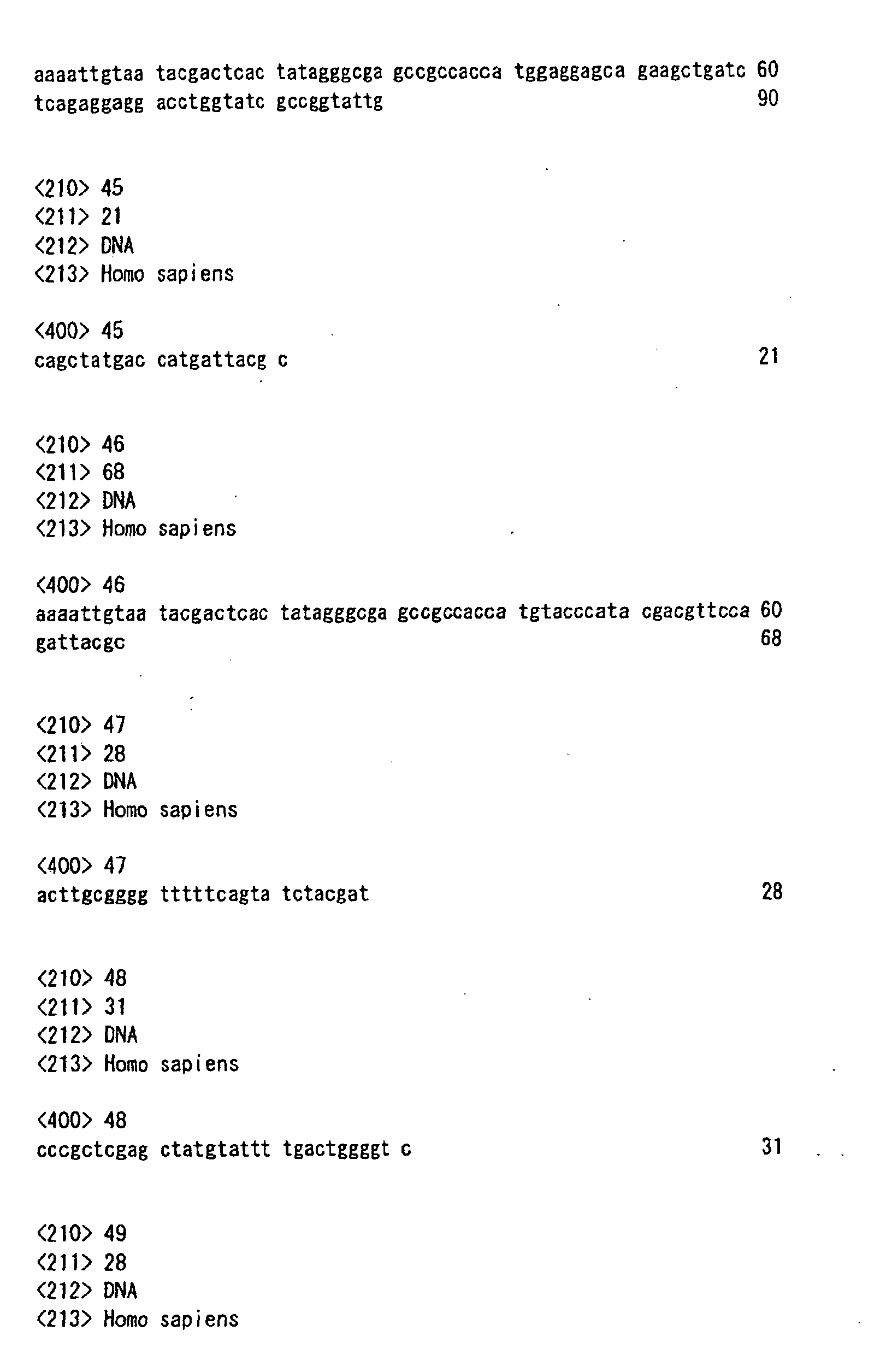
- a bait fragment containing the amino acid residues 309 to 360 in the intracellular C-terminal side of human CCR2b was amplified by a polymerase chain reaction (PCR) method by pCMGS-CCR2b in accordance with J. Sambrook et al., Molecular Cloning: A Laboratory Manual, 2nd ed. (1989) with the use of a forward primer 5'GCGAATTCGAGAAGTTCAGAAGGTAT3' (SEQ ID NO:42) and a reverse primer 5'GCGGATCCTTATAAACCAGCCGAGAC(SEQ ID NO:43).
- PCR polymerase chain reaction
- the thus amplified fragment was treated with restriction enzymes (EcoRI and BmaHI) and then orientationally cloned into pACT2 (a yeast Gal4 DNA binding domain cloning vector containing TRYP gene for selection in a yeast lacking tryptophan biosynthesis (CLONTECH Laboratories, Inc.)).
- the base sequence of the bait fragment of this plasmid DNA was amplified by the PCR method with the use of a primer specific to the adjacent sequence of the vector in accordance with the dideoxynucleotide chain termination method (J. Messing et al., Nucleic Acids Res. 9: 309, (1981)). Then the base sequence was determined by using an automatic DNA sequencer (Model ABI377 manufactured by Applied Biosystems).
- the THP1-origin cDNA library and bait vector as described above were transformed into a yeast strain Y190 in accordance with the lithium acetate/polyethylene glycol transformation protocol (see, Ito et al., J. Bacteriolo. 153:163-168 (1983)).
- a synthetic complete (SC) medium lacking tryptophan, leucine and histidine (SC Trp Leu His) and containing 10 mM of 3-aminotriazole Sigma Chemical Co.
- Yeast cells positive to protein interaction which contained a mixture of a binding domain plasmid with an activation domain plasmid, were re-cultured as an isolated matter in each well of a 96-well microtiter plate. Then, about 10 ⁇ l of each isolated matter was dissolved and an insert in the pACT2 plasmid was amplified by PCR with the use of primers specific to the adjacent sequences of individual vectors. The base sequence of the insert was isolated and determined by the above-described method.
- sequence (SEQ ID NO:38) of a gene (clone 19) encoding 156 amino acids was identified and compared with publicly known sequences by using a "BLAST" program available in public mediated by National Center for Biotechnology Information (NCBI). Thus, it was confirmed that this clone was a novel gene the function of which had never been analyzed.
- the gene (clone 19) encoding 156 amino acids was named FROUNT gene and the isolated plasmid clone was named pACT2-FROUNT.
- FROUNT protein the isolated plasmid clone
- FROUNT protein is not a "self-activating" protein (i.e., requiring the interaction with a second protein domain to form a functional activated complex) and that FROUNT protein has binding ability specific to the receptors CCR2 and CCR5 (Fig. 1).
- Fig. 1 FROUNT protein has binding ability specific to the receptors CCR2 and CCR5 (Fig. 1).
- GAL4 BD and GAL4 AD respectively stand for a transcriptional factor binding domain-fused protein expression vector and an activation domain-fused protein expression vector
- BD or AD vector stands for a control (empty) vector
- FROUNT, p53, SV40 T-antigen, CCR5, CXCR4 and CCR2 stand for transferred genes; each numerical value in parentheses indicates the number of inserted amino acid residues
- -LDH stands for proliferating ability in the deficient medium
- ⁇ -Gal activity stands for ⁇ -galactosidase activity
- + and - stand respectively for the presence and absence of the activity.
- coding sequences of pAS2-1-CCR2b and pAS2-1-p53 were amplified by PCR using a T7 promoter, a 5' primer encoding myc epitope: 5'AAAATTGTAATACGACTCACTATAGGGCGAG CCGCCACCATGGAGGAGCAGAAGCTGATCTCAGAGGAGGACCTGGTATCGCCGGTATTG 3' (SEQ ID NO:44) and a 3' primer originating in p AS2-1: 5'CAGCTATGACCATGATTACGC3' (SEQ ID NO:45) to express an epitope-tagged protein.
- pACT2-FROUNT was amplified by PCR using a T7 promoter, a 5' primer encoding HA epitope: 5'AAAATTGTAATACGACTCACTATAGGGCGAGCCGCCACCATGTACC CATACGACGTTCCAGATTACGC3' (SEQ ID NO:46) and a 3' primer originating in p ACT2: 3'ACTTGCGGGGTTTTTCAGTATCTACGAT5' (SEQ ID NO:47). Then, isotope-labeled recombinant proteins were produced in accordance with user's manual of MATCHMAKER Co-IP Kit (CLONTECH Laboratories, Inc.).
- Fig. 2 which shows the results of SDS-PAGE analysis in coimmunoprecipitation, myc-CCR2, p53 and HA-FNT stand for isotope-labeled myc or HA-fused recombinant protein; I.P. stands for an antibody employed in the coimmunoprecipitation of each sample; HA mAb and Myc mAb stand for antibodies specific for Ha and Myc respectively; HA-peptide and Myc-peptide stand for antigen peptides; and + and - respectively stand for the presence and absence for each sample.
- THP-1-origin cDNA prepared by using the same method as in the preparation of the plasmid cDNA library in EXAMPLE 1 was inserted into a ⁇ phage vector ⁇ ZIP (GIBCO BRL) to give a phage cDNA library using E. coli DH10B as the host.
- the phage cDNA library using E. coli DH10B as the host was sowed on 10 soft agar plates at a density of about 1x10 5 clones per plate.
- the library was dissolved in a 0.5 N NaOH solution and the phage DNA thus exposed and denatured was dried and immobilized on the filter (Maniatis et al., Molecular Cloning, Cold Spring Harbor Laboratory, p. 320 (1982)).
- the above-described FROUNT gene fragment (clone 19) was 32 P-labeled by the Nick translation method (Maniatis et al., ibid ., p.109) and employed as a probe.
- the labeled probe and the filter having the DNA thus immobilized thereon were subjected to association in 5 x SSPE (0.9 M NaCl 50 mM sodium phosphate buffer solution (pH 7.4), 5 mM EDTA) containing the labeled probe, 50% formamide, 5 x Denhardt's, 0.1% SDS, 100 ⁇ g/ml denatured salmon sperm DNA solution (10 ml) at 42°C for 16 hours.
- 5 SSPE 0.9 M NaCl 50 mM sodium phosphate buffer solution (pH 7.4), 5 mM EDTA
- phage DNA was extracted by the method of Davis et al. (Dvais et al., Advanced Bacterial Genetics, Gold Spring Harbor Laboratory (1980)) and the cDNA base sequence of the clone reacting with the probe was determined. Further, a cDNA library for racing was prepared from THP-1 cells in accordance with the protocol of 5' race PCR marathon system (TOYOBO). Using this library, the 5'-terminal base sequence of the obtained gene was clarified.
- human FROUNT gene full-length cDNA was cloned by the PCR method with the use of the THP-1 cell-origin cDNA library as a template. Then the full base sequence was determined by using an ABI377 sequencer (clone 1).
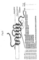
- Fig. 4 shows the amino acid sequence of human FROUNT protein 1 (hFROUNT) wherein the framed part corresponds to FROUNT conserve domain (FCD), the underlined part corresponds to the part having been isolated by the yeast two-hybrid assay as described above (SEQ ID NO:38), and 4 stars respectively indicate 4 leucine residues conserved in the leucine zipper domain.
- Fig. 5 shows the results of the Kyte-Doolittle hydropathy plot of human FROUNT protein 1 wherein the horizontal axis refers to the number of amino acid residues while the longitudinal axis refers to the hydrophobicity of each amino acid residue.
- Fig. 3 shows CCR2 mutants (CR2-1 to CR2-7) and the results of the yeast two-hybrid assay using them as in Fig. 1.
- a cDNA library originating in mouse bone marrow cells was prepared by the above-described method. Then mouse FROUNT gene was isolated as in the human FROUNT protein 1 gene (clone 1/SEQ ID NO:19). By using a software for sequence comparison (Network Protein Sequence, CLUSTALW), it was clarified that the amino acid sequence deduced from the base sequence of the mouse FROUNT gene was highly homologous with human FROUNT protein 1 (Fig. 5(b)). As the results of searching for proteins having high homology with human FROUNT protein 1 in species other than humans and mouse with the use of publicly known database (NCBI, BLAST), FROUNT proteins of Drosophila melanogaster and Caenorhabditis elegans were identified.
- NCBI National Protein Sequence
- Fig. 5(b) shows comparison of FROUNT protein among species and a schematic model of the domain structure of human FROUNT protein 1.
- homology to human FROUNT protein 1 at the amino acid level in FCD, the upstream domain of FCD or the downstream domain of FCD is given in (%).
- Fig. 5(b) shows comparison of FROUNT protein among species and a schematic model of the domain structure of human FROUNT protein 1.
- homology to human FROUNT protein 1 at the amino acid level in FCD, the upstream domain of FCD or the downstream domain of FCD is given in (%).
- hFROUNT is a schematic model of the domain structure of human FROUNT protein 1; 4 Y's represent tyrosine motives; 4 L's represent dileucine motives; FCD represents the FROUNT conserved domain; the underlined part in the CCR2 binding site corresponds to the part isolated by the yeast two-hybrid assay as described above; 4 stars represent 4 leucine residues conserved in the leucine zipper domain; and FNT-D Ab and FNT-A Ab represent each the site employed as an antigen in constructing a specific polyclonal antibody.
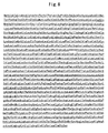
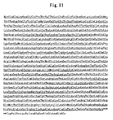
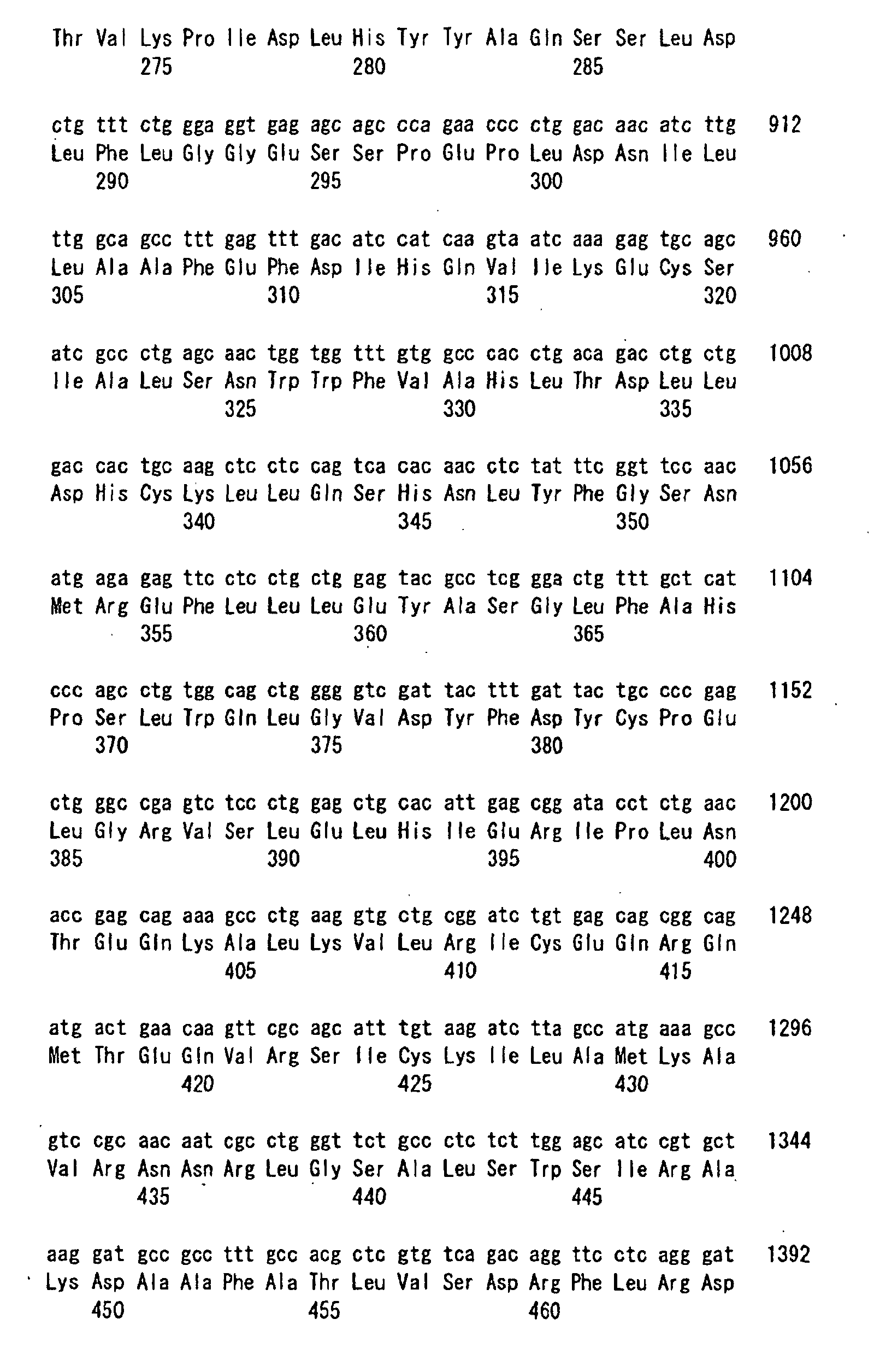
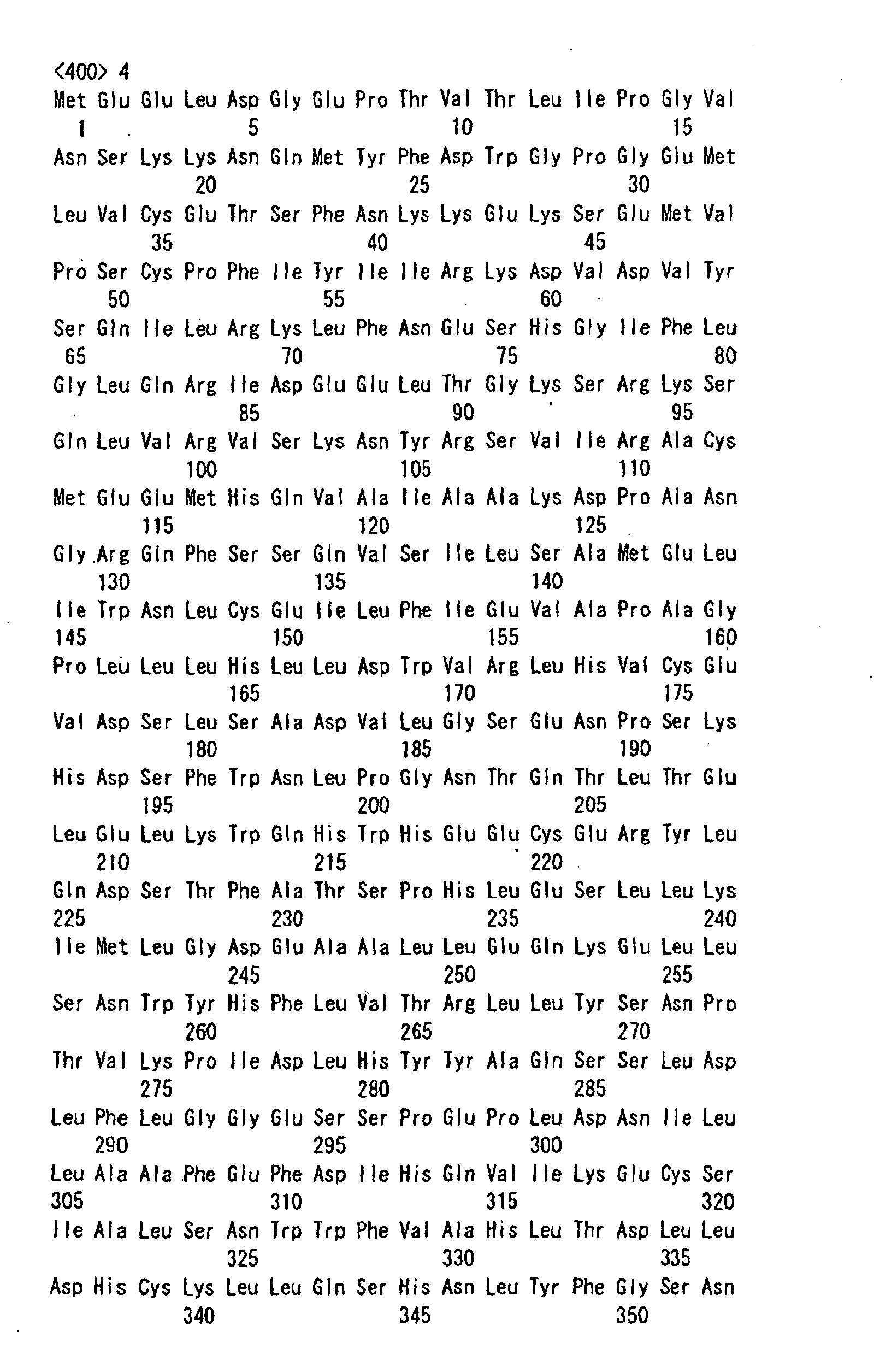
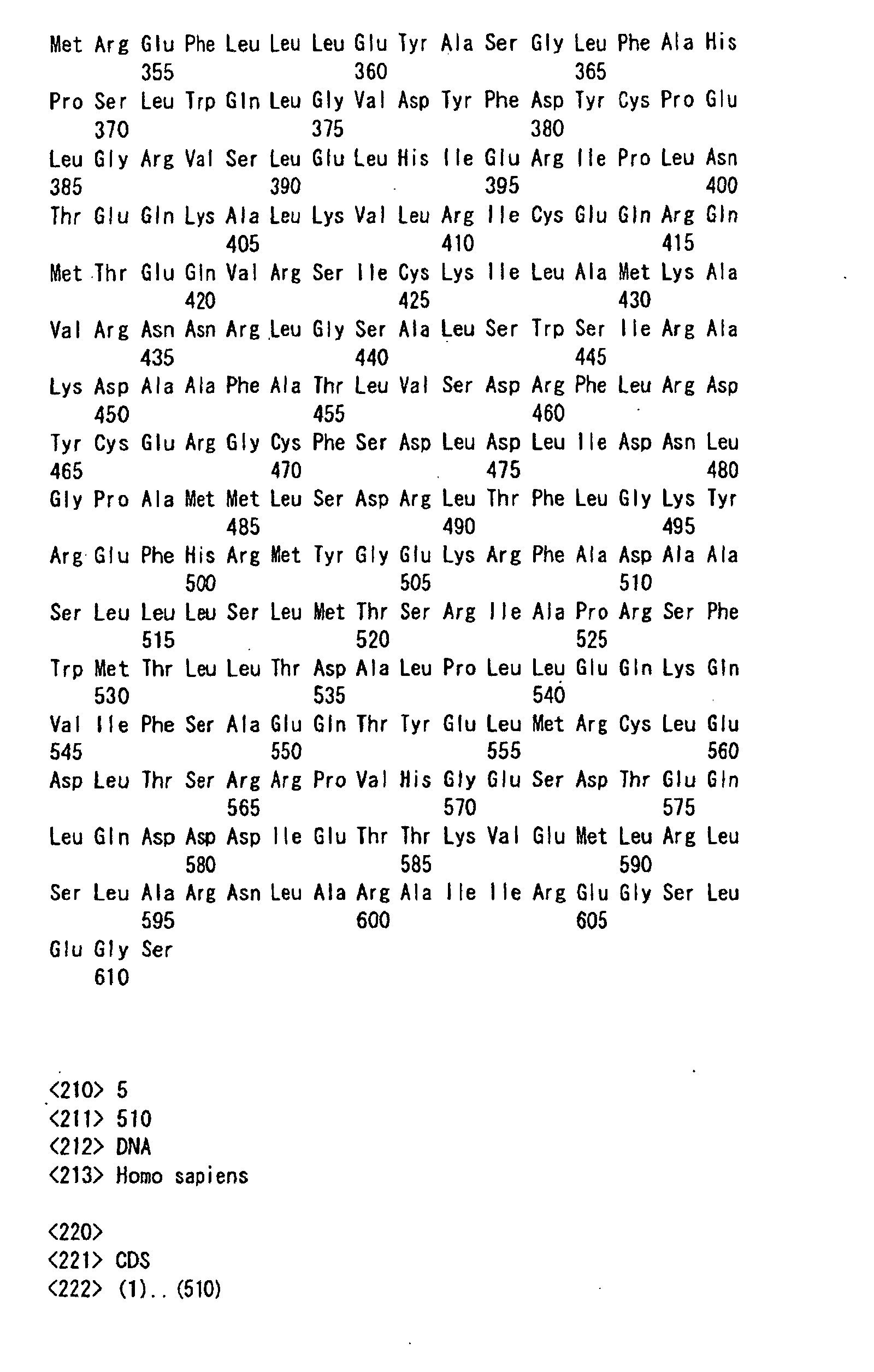
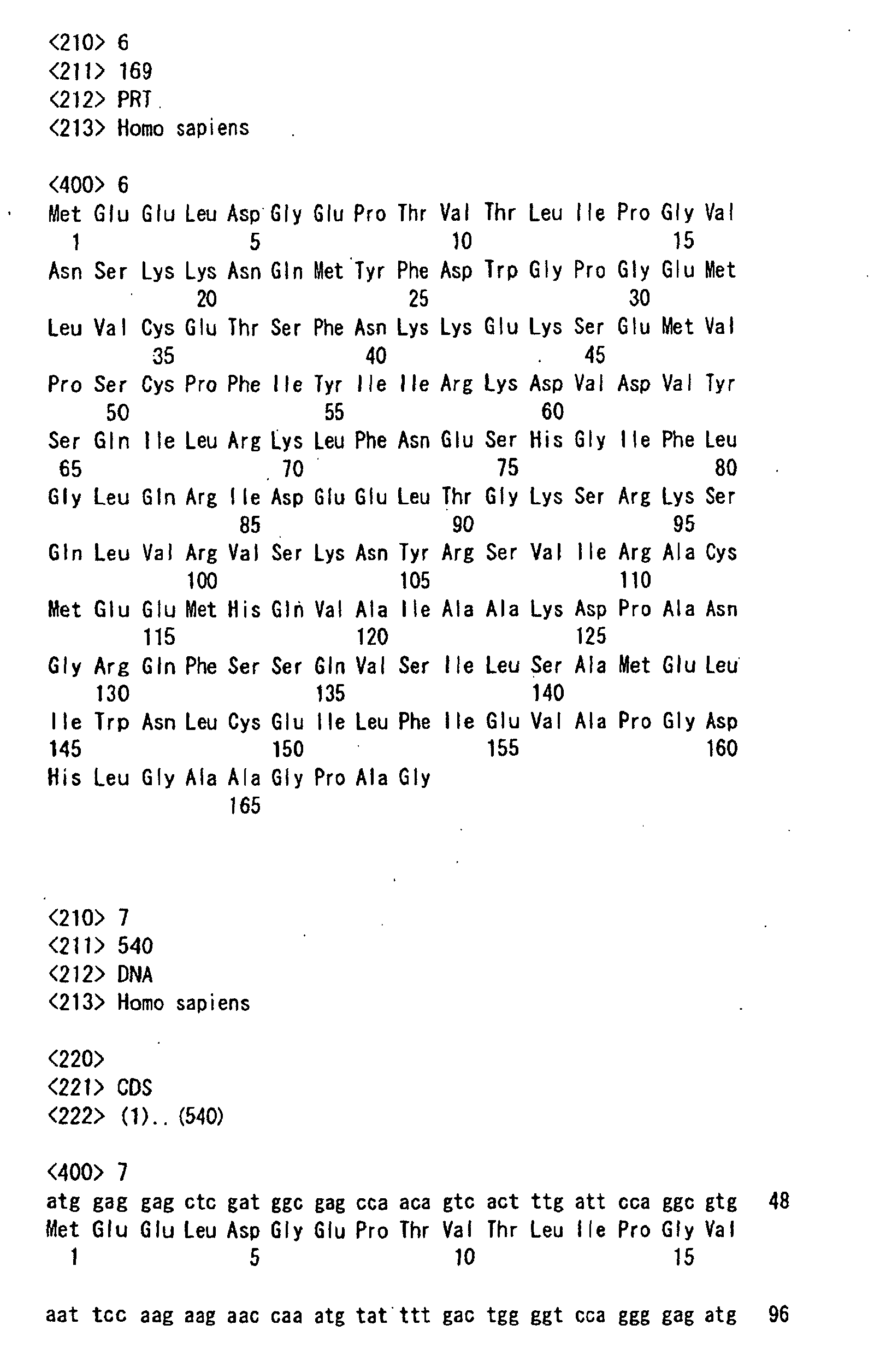
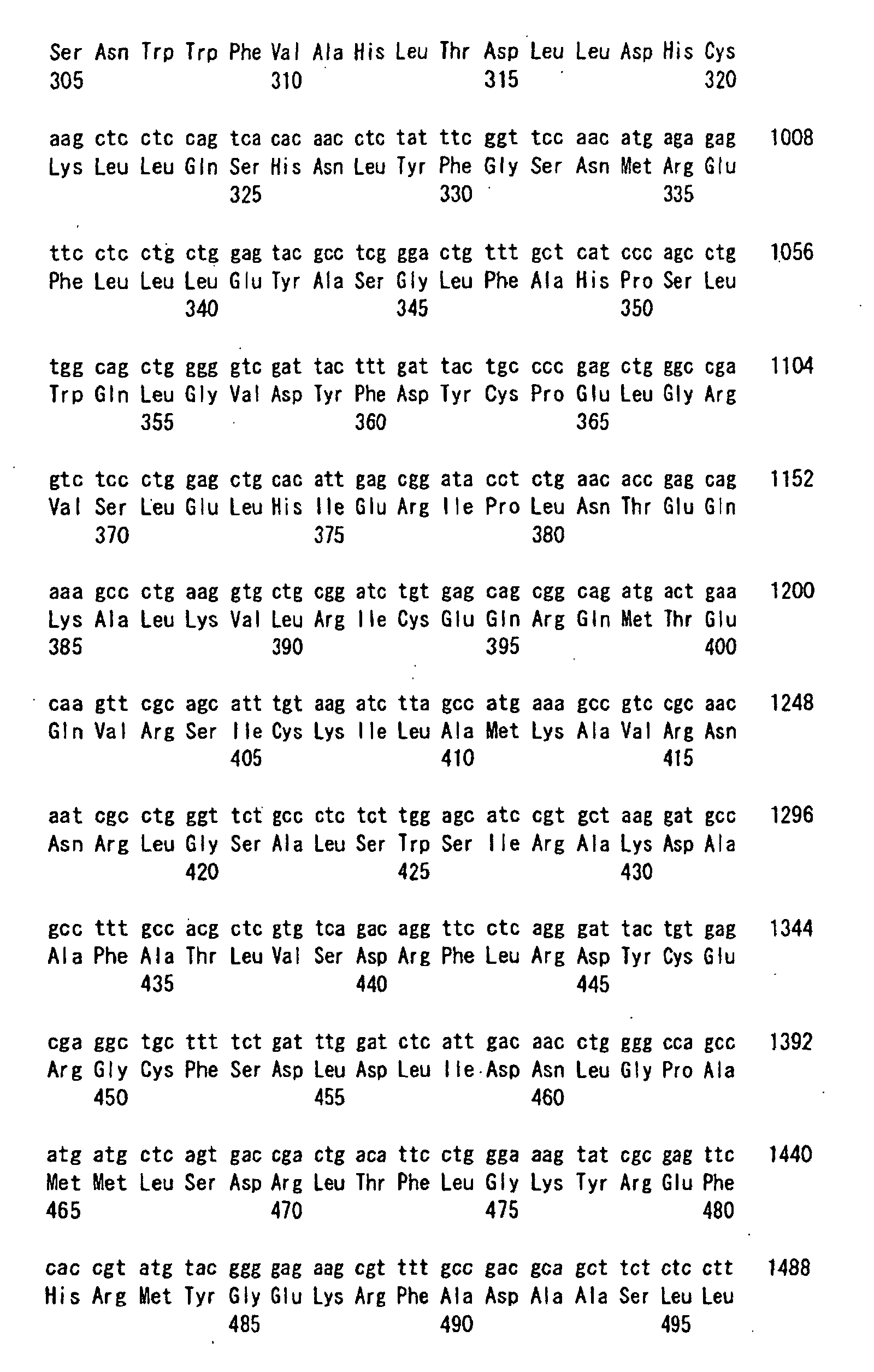
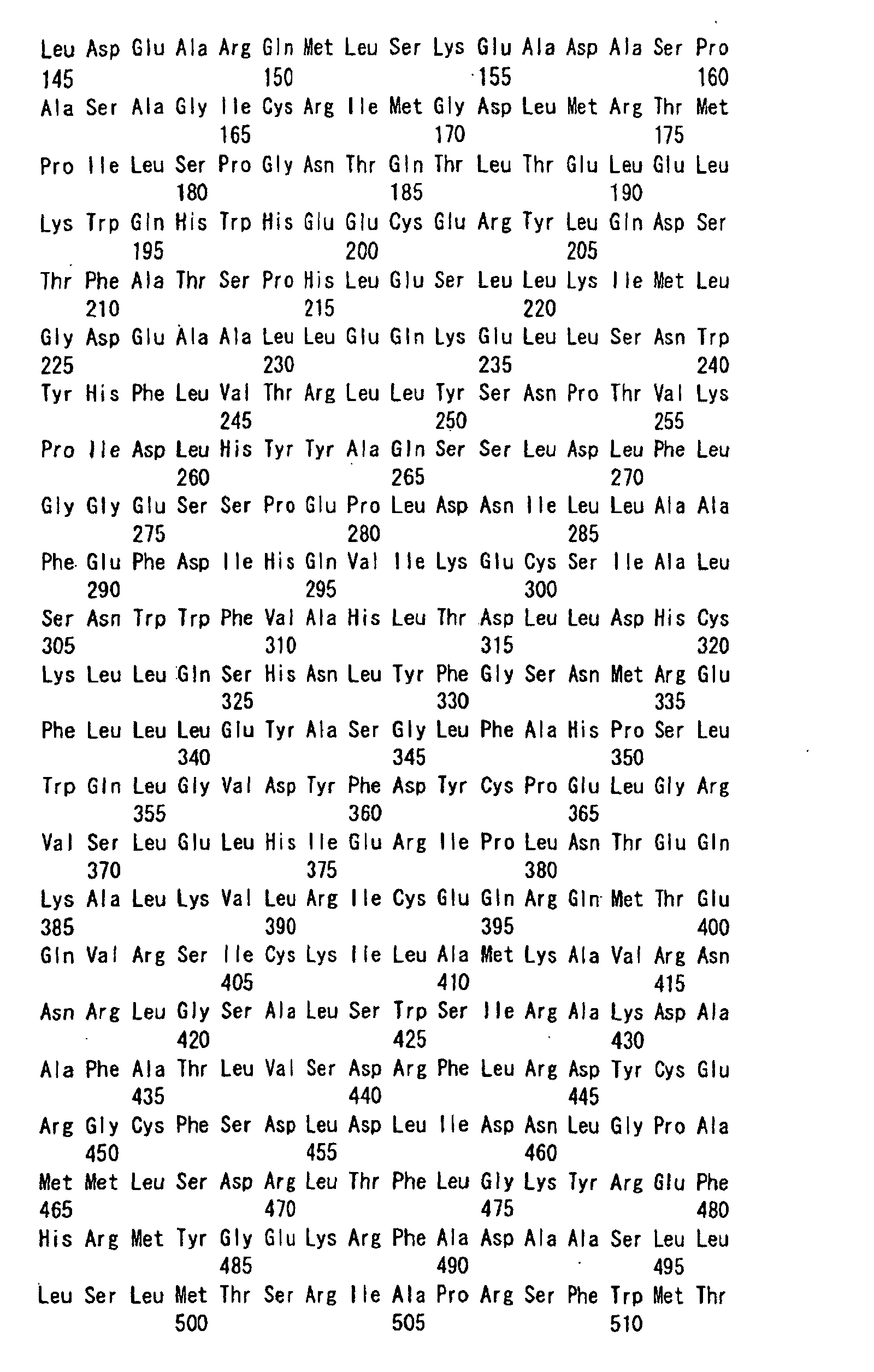
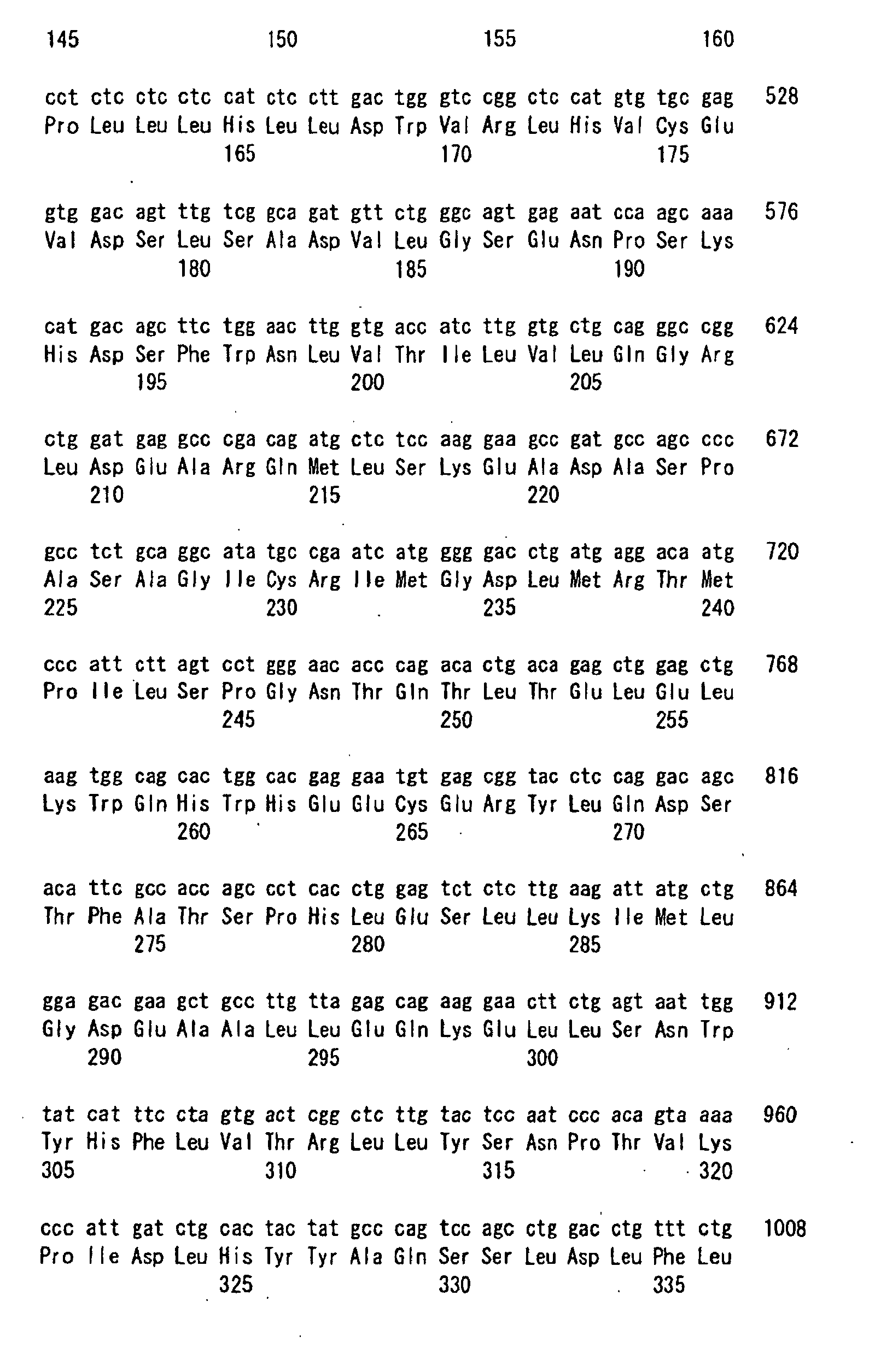
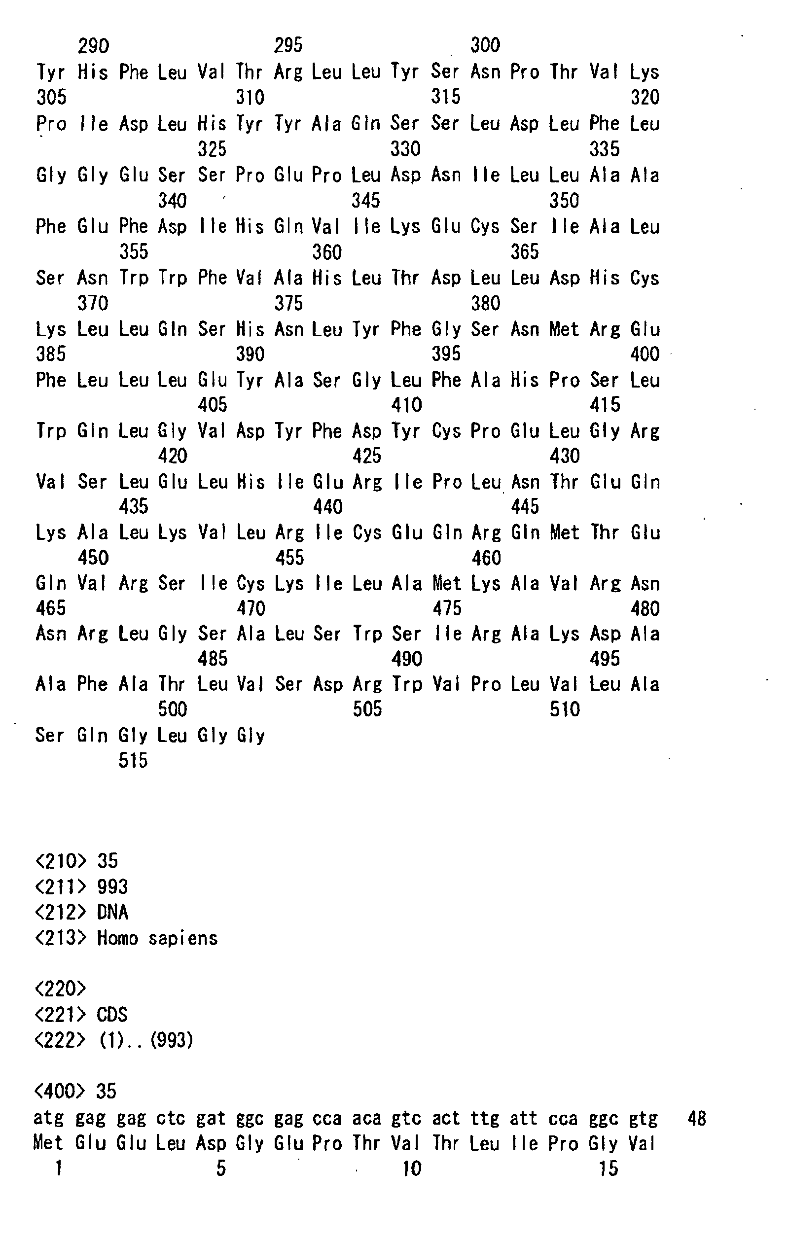
- Human FROUNT clone 2 ( ⁇ -deletion type (Fig. 8)) is a FROUNT mutant having 611 amino acids derived from human FROUNT clone 1 ( ⁇ -type, 656 amino acids in the full-length (Fig. 7)) by deletion of the bases 662 to 792 (135 bp). Human FROUNT clones 3 to 12 also belong to the ⁇ -deletion type.
- Human FROUNT clone 14 ( ⁇ -deletion type (Fig. 10)) is a FROUNT mutant having 611 amino acids derived from human FROUNT clone 13 ( ⁇ -type, 630 amino acids in the full-length (Fig. 9)) by deletion of the bases 470 to 661 (192 bp). Human FROUNT clones 15 and 16 also belong to the ⁇ -deletion type.
- Human FROUNT clone 17 ( ⁇ -insertion type (Fig. 11)) is a FROUNT mutant having 518 amino acids derived from human FROUNT clone 13 ( ⁇ -type, 630 amino acids in the full-length (Fig. 9)) by insertion of 90 bp between the bases 1581 and 1582 followed by frame shifting. Human FROUNT clone 18 also belongs to the ⁇ -insertion type.
- an underlined part corresponds to the amino acid sequence identical with human FORUNT 1, while ***** represents a deletion or insertion site.
- EXAMPLE 6 Detection of FROUNT protein and distribution thereof in cell using green fluorescent protein-fused FROUNT protein 1
- Human FROUNT protein 1 gene (clone 1) was amplified by the PCR method with the use of a 5'-primer: 5'CCCGCTCGAGCTATGTATTTTGACTGGGGTC3' (SEQ ID NO:48) and a 3'-primer: 5'GCGA ATTCTCAGGAACCTTCCAGTGAGC3' (SEQ ID NO:49) and then treated with restriction enzymes (XhoI and EcoRI). After inserting into the XhoI, EcoRI site of pEGFP C1 (CLONTECH Laboratories, Inc.), the base sequence of the insert was determined by using an ABI377 Sequencer. Thus, a plasmid vector pEGFP-FORUNT for expression in animal cells was constructed.
- This plasmid vector was transformed into E. coli DH5 ⁇ in accordance with a method reported in Proc. Natl. Acad. Sci. USA 69:2110 (1972) and a plasmid DNA for transforming animal cells was obtained in accordance with the protocol of Max Prep (Qeagen).
- Human HEK293 cells (ATCC:CRL-157) were grown bymonolayer culture in a DMEM medium containing 5% of fetal bovine serum (Falcon size: 100 mm, 5 plastic dishes). After replacing the medium with a fresh one of the same type, the cells were further cultured in a G418-containing medium after the transformation.
- calcium phosphate gel containing 30 ⁇ g/dish of the plasmid pEGFP-C1 (control) or pEGFP-FROUNT DNA was prepared and added to the cells in accordance with a publicly known method (Graham et al., Virology 52:456 (1973)) to give pEGFP-C1 transformed cells or pEGFP-FROUNT transformed cells respectively. 4 hours thereafter, the above pEGFP-C1 transformed cells or pEGFP-FROUNT transformed cells were treated with glycerol and further cultured in a medium containing 5% of fetal bovine serum. 24 and 48 hours after the transformation, the cells were stripped off from the dish by using trypsin and an EDTA solution.
- the expression of the green fluorescent protein (GFP) in the pEGFP-C1 and pEGFP-FROUNT transformed cells was confirmed with the use of a fluorocytometer EPICS ELITE ESP (Beckman Coulter) or EPICS XL/XL-MCL system 2 (Beckman Coulter). Further, the localization of the FROUNT protein in the cells was observed by a publicly known method with the use of a confocal fluoromicroscope. As a result, it was confirmed that the control eGFP was observed all over the cells including nucleus while the GFP-fused FROUNT protein was localized in the cytoplasm other than nucleus (Fig. 6(a)).
- a plasmid vector for expressing human CCR2 protein in animal cells was constructed by the same procedure and then transferred into the pEGFP-C1 and pEGFP-FROUNT transformed cells as described above. Then the intracellular localization of the green fluorescent protein was observed before and after stimulating with a CCR2 antibody or CCL2 which is a ligand to CCR2.
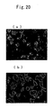
- Fig. 6 presents fluorescent microphotographs of cells having control eGFP (upper) and fluorescent protein-fused FROUNT (lower) transferred therein.
- Figs. 6 (a) and 6(b) respectively show micrographs before and after treating with the CCR2 antibody.
- the Merge image shows the simultaneous visualization of green fluorescence and CCR2 (red fluorescence). Similar results were observed in the case of the CCL2 stimulation (data not shown).
- the site common to FROUNT proteins of the ⁇ - and ⁇ -types in human FROUNT protein 1 gene was amplified by the PCR method with the use of a 5'-primer: 5'CGGGATCCGCCATGTATTTTGACTGGGGTC3' (SEQ ID NO:50) and a 3'-primer: 5'GCGAATTCTCATGACAAAATGGAGACCTGGCTGC3' (SEQ ID NO:51), treated with restriction enzymes (EcoRI and BmaHI) and then orientationally cloned into the EcoRI, BamHI site of pGEX4T3 (Amersham Pharmacia). Then the base sequence of the insert was determined by using an ABI377 sequencer.
- a plasmid vector pGX-FROUNT for expressing glutathione S transferase (GST)-fused protein was constructed.
- This plasmid vector was transformed into E. coli BL21 (DE3) in accordance with a method described in Proc. Natl. Acad. Sci. USA 69:2110 (972)) followed by expression in accordance with a publicly known protocol recommended by Amersham Pharmacia.
- the GST-fused protein was purified by glutathione sepharose affinity chromatography. By SDS PAGE and Coumassie staining, it was judged that the accuracy and purity of this GST-fused FROUNT both exceeded 95%.
- This GST-fused FROUNT protein 1 (100 ⁇ g) was mixed with an adjuvant and employed in subcutaneously immunizing a New Zealand white rabbit in a publicly known time course. After confirming an increase in the titer, a FROUNT protein 1-specific polyclonal antibody was purified from the serum of the animal. The titer of the FROUNT protein 1-specific polyclonal antibody was confirmed by calibrating the ability to bind to the GST-fused FROUNT protein 1 with the use of the Western blotting method. Endogenous FROUNT protein 1 was detected by a publicly known immunostaining method with the use of the above-described specific polyclonal antibody.
- FIG. 12 is a schematic model of the retrovirus vector pEGFPMY wherein Cont stands for a control (empty vector); hFNT stands for an expression vector of human FROUNT protein 1; AS-22 stands for an antisense expression vector of human FROUNT protein; DN-hFNT stands for a partly deficient FROUNT protein expression vector; LTR stands for a long terminal repeat; gag stands for a structural protein; eGFP stands for a green fluorescent protein; and IRES stands for a ribosomal entry site.
- Cont for a control (empty vector)
- hFNT stands for an expression vector of human FROUNT protein 1
- AS-22 stands for an antisense expression vector of human FROUNT protein
- DN-hFNT stands for a partly deficient FROUNT protein expression vector
- LTR stands for a long terminal repeat
- gag stands for a structural protein
- eGFP stands for a green fluorescent protein
- IRES stands for a ribosomal entry site.
- FROUNT mRNA was compared and quantified.
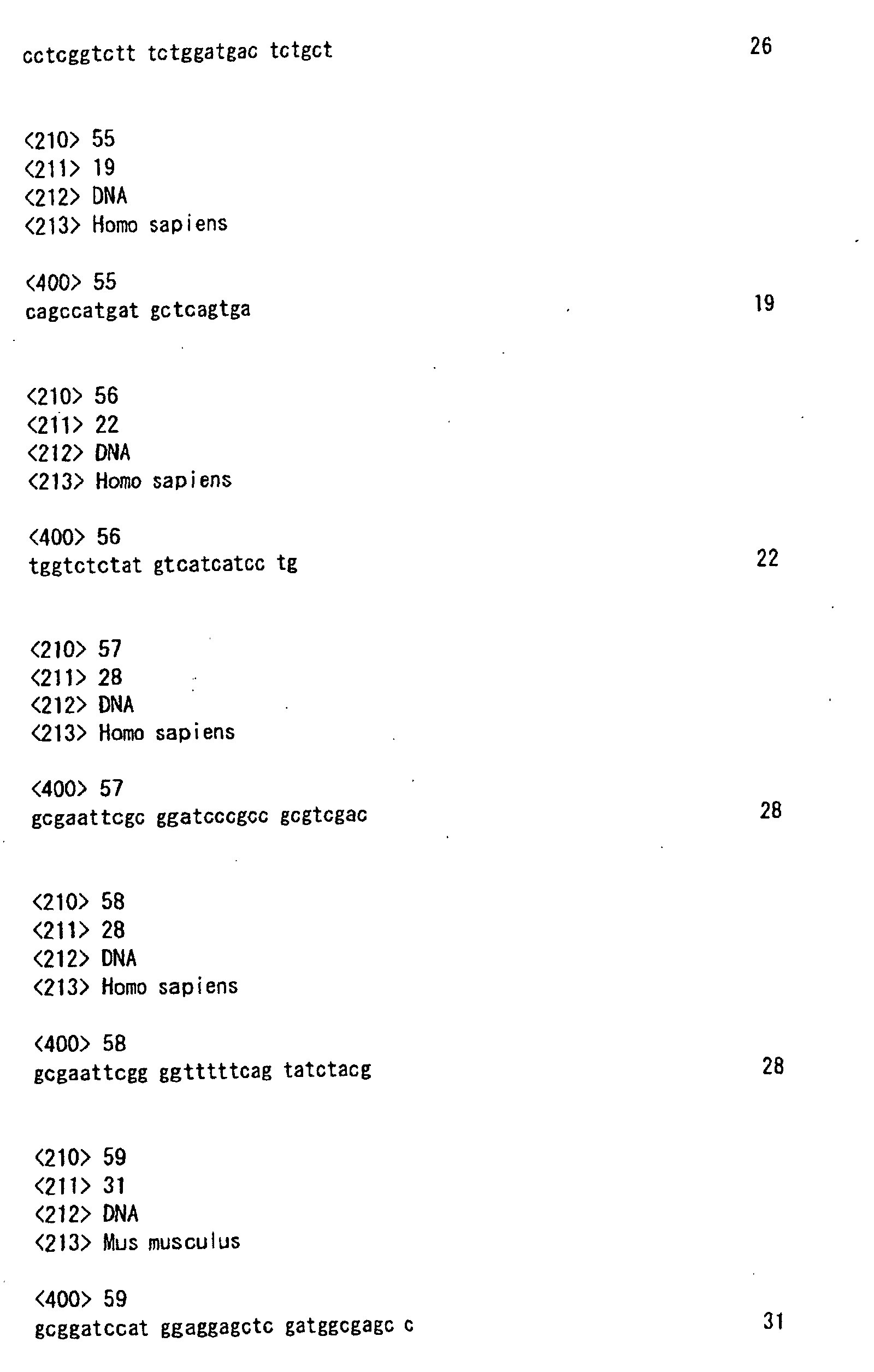
- a cell line wherein the expression of about 90% of FROUNT mRNA was inhibited could be established in association with the transfer of an antisense expression vector (AS-22), which was obtained by amplifying a fragment by the PCR method with the use of a 5'-primer: 5'GCGGATCCTCAAATCAAGCAGTGTTTGTC3' (SEQ ID NO:52) and a 3'-primer: 5'CGGGATCCGCCATGCTTTTGGAACAGAAACAGGTG3' (SEQ ID NO:53), treating with BamH1 and inserting into pMY, into HEK293 cells by the above-described method (Fig. 13).
- AS-22 antisense expression vector
- FIG. 13 shows the results of the quantification of human FROUNT mRNA (upper a) and FROUNT protein (lower b) in Cont, hFNT and As-22 cells and the results of Western blotting using FROUNT antibody.
- Fig. 14 shows confocal fluoromicroscopic observation results of eGFP-expressing cells in Cont and AS-22 cells (virus-transfer cells, upper), expression of FROUNT protein (medium) and overlapped image thereof (lower). Compared with the Cont cells, a remarkable lowering in the FROUNT protein expression was observed in the eGFP-expressing cells (virus-transfer cells).
- FROUNT protein in cells was analyzed by using this FROUNT antisense-transfer cells (AS-22 cells).
- the following vector was constructed by forcibly expressing the C-terminal part alone of FROUNT protein 1 carrying the binding activity to CCR2 identified by the above-described yeast two-hybrid method and the in vitro binding experiment.
- a plasmid vector encoding 156 C-terminal amino acids of FROUNT protein 1 isolated by using the yeast two-hybrid method a fragment of SEQ ID NO:38 was amplified by the PCR method with the use of a 5'-primer: 5' GCGAATTCGCCGGATCCCGCCGCGTCGAC3' (SEQ ID NO:57) and a 3'-primer: 5'GCGAATTCGGGGTTTTTC AGTATCTACG3' (SEQ ID NO:58) and then treated with EcoR1. Then it was inserted inframe into the C-terminus as allowing fusion to thereby construct a plasmid vector DN-FNT (Fig. 12, upper). Then a virus was prepared as described above and a cell line was infected to establish partly deficient FROUNT protein-expressing cells (DN-hFNT cells).
- DN-hFNT cells partly deficient FROUNT protein-expressing cells
- Fig. 15 shows the number of migrating cells (the longitudinal axis) in the cells (Cont, hFNT, AS-22 and DN-hFNT) when stimulated with 33 ng/ml of CCL2 or CXCL12 and Fig. 15 (b) shows the number of migrating cells (the longitudinal axis) in respective cells when stimulated with CCL2 at various concentrations.
- a chemokine reacts with a chemokine receptor and thus elevates intracellular calcium concentration (Ca 2+ ).
- 10 7 gene-transfer THP-1 cells as described above were washed with PBS (Gibco BRL), suspended in 1 ml of buffer solution A (Tyroid's Salt Solution (Gibco BRL)/0.1% BSA) and incubated to a final concentration of 5 ⁇ M Fruo3AM (Dojindo) at room temperature for 1 hour. Then the cells were washed with the buffer solution Aand suspended in 2 ml of the buffer solution A.
- the intracellular calcium concentration (Ca 2+ ) was measured by using a fluorescent spectrophotometer Fluoroscan Ascent system (Labosystems).
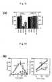
- Fig. 16 shows the results of calcium influx (nM, the longitudinal axis) quantification into various cells stimulated with CCL2 at various concentrations.
- chemokine receptor When a chemokine receptor reacts with a chemokine, a plural number of receptor molecules aggregate and form clusters followed by internalization. The same phenomenon is observed in the reaction between a chemokine reactor and an antibody specific thereto.
- FROUNT protein 1 To examine the participation of FROUNT protein 1 in this clusterization, the above-described gene-transfer HEK293 cells, in which CCR2 was constantly expressed by the above-described method, were bonded to a glass plate. After washing the cells with the medium A at 4°C, a PE-labeled CCR2-specific antibody (R&D System, Inc.) was dissolved in the medium A and incubated at 4°C for 30 min.
- Fig. 17 presents images of the cells stimulated with the CCR2-specific antibody
- Fig. 17(b) shows the results of the confocal microscopic quantification of the clusterization ability of each receptor stimulated with the CCR2- or CXCL2-specific antibody (the longitudinal axis referring to control %).
- an antisense-transfer mouse was constructed in the following method and the phenotype of the mouse was observed.
- a fragment was amplified by the PCR method with the use of a 5'-primer:5'GCGGATCCATGGAGGAGCTCGATGGCG AGCC3'(SEQ ID NO:59) and a 3'-primer: 5'GCGGATCCTCAGGAA CCTTCCAGTGAGC3' (SEQ ID NO:60) and treated with BamHI.
- a 5'-primer 5'GCGGATCCATGGAGGAGCTCGATGGCG AGCC3'(SEQ ID NO:59) and a 3'-primer: 5'GCGGATCCTCAGGAA CCTTCCAGTGAGC3' (SEQ ID NO:60) and treated with BamHI.
- Fig. 18 antisense and sense-expression retrovirus vectors
- FIG. 18 shows schematic models of the retrovirus vectors pEGFPMY, wherein Cont stands for a control (empty) vector; mFNT stands for a mouse FROUNT protein expression vector; AS-mFNT stands for a mouse FROUNT protein antisense expression vector; LTR stands for a long terminal repeat; gag stands for a structural protein; eGFP stands for a green fluorescent protein; and IRES stands for a ribosomal entry site.
- Cont stands for a control (empty) vector
- mFNT stands for a mouse FROUNT protein expression vector
- AS-mFNT stands for a mouse FROUNT protein antisense expression vector
- LTR stands for a long terminal repeat
- gag stands for a structural protein
- eGFP stands for a green fluorescent protein
- IRES stands for a ribosomal entry site.
- bone marrow precursor cells purified from mouse bone marrow cells with the use of an MACS system (Milteny Biotech) were infected with these retroviruses by the centrifugation method.
- Cells infected with each virus vector alone were separated and purified to a purity of 98% or higher by using a cell sorter system with the expression of EGFP encoded by the virus vector as an indication.
- the virus vector-infected cells were intravenously administered. 3 months after the transfer, 50 ⁇ l of peripheral blood was collected from the eye ground and the reconstruction ratio of the bone marrow cells was measured with a fluorocytometer.
- thioglycolate (Difco) was dissolved in PBS at a concentration of 4% and the obtained solution was intraperitoneally administered in 1.5 ml portions to the gene-transfer mice as described above. After 72 hours, 5 ml of PBS cooled to 4°C was further intraperitoneally administered and each animal was well massaged. Then intraperitoneal cells were collected and the cell counts were compared. As a result, it was confirmed that the AS-mFNT mouse showed a remarkable decrease in intraperitoneal cell count compared with the Cont mouse and the mFNT mouse. However, no abnormality in intraperitoneal cell count was observed in these mice before the stimulation with thioglycolate.
- Fig. 19 shows the results of a chemotaxis experiment with the use of thioglycolate on the intraperitoneal cells (a) and the macrophages (b) of each virus vector-transfer mouse, wherein each open bar shows a cell count without stimulation while each solid bar shows a cell count upon thioglycolate stimulation.
- a FROUNT protein expression vector was prepared by inserting the cDNA represented by SEQ ID NO:19 between the EcoRI and NotI sites of a pUC-SR ⁇ expression vector (FEBS 333:61-66 (1993)). In this plasmid vector, the transcription of the FROUNT protein cDNA was controlled by the SR ⁇ promoter (Nature 342:440-443 (1989)).
- Tetrahydrofuran was mixed with phosphatidylserine, phosphatidylcholine and cholesterol at a weight ratio of 1:4:8:2. After distilling off the tetrahydrofuran on a rotary evaporator, the lipid mixture (10 mg) was deposited on the container wall. 96 ⁇ g of HMG 1 nucleic acid (high mobility group 1 nuclear protein) purified from bovine thymus was mixed with a BBS solution (20 ⁇ l) of the FROUNT protein DNA plasmid (300 ⁇ g) at 20°C for 1 hour and then the resultant mixture was added to the above-described lipids.
- HMG 1 nucleic acid high mobility group 1 nuclear protein
- the obtained liposome-DNA-HMG 1 complex suspension was mixed with a portex, ultrasonicated for 3 seconds and then stirred for 30 minutes.
- Purified HVJ Z strain
- UV irradiation 110 erg/mm 2 sec
- BBS was added to give a total volume of 4 ml.
- the obtained mixture was incubated at 4°C for 10 minutes and then gently stirred at 37°C for 30 minutes. Unfused liposomes were removed from the HJV-liposomes by the sucrose density gradient centrifugation method.
- HVJ-liposomes containing the FROUNT protein expression vector (containing 10 g/ml of FROUNT protein expression vector) was obtained from the upper layer of sucrose density gradient.
- a liposome preparation containing the antisense RNA and a liposome preparation containing DNA producing the antisense RNA can be obtained. Such a preparation is injected into a target site via an injection needle.
- the protein according to the present invention which plays a different role from a G protein in the intracellular signal transduction mechanisms of the G protein-coupled receptors CCR2 and CCR5, indicates the presence of a new mechanism participating in the intracellular signal transduction together with the G protein. That is to say, there is a possibility that these new mechanisms in the CCR2 system and the CCR5 system would affect the efficacy and side effects of drugs in addition to the intracellular signal transduction mechanism having been attracted public attention concerning the relationships among receptors, G proteins and effectors so far. Therefore, the discovery of the protein according to the present invention and DNA encoding the same brings about the provision of novel medical targets. Namely, the present invention provides new approaches to the treatment, prevention, diagnosis, etc.
Landscapes
- Health & Medical Sciences (AREA)
- Chemical & Material Sciences (AREA)
- Life Sciences & Earth Sciences (AREA)
- Organic Chemistry (AREA)
- Medicinal Chemistry (AREA)
- General Health & Medical Sciences (AREA)
- Nuclear Medicine, Radiotherapy & Molecular Imaging (AREA)
- General Chemical & Material Sciences (AREA)
- Chemical Kinetics & Catalysis (AREA)
- Bioinformatics & Cheminformatics (AREA)
- Pharmacology & Pharmacy (AREA)
- Engineering & Computer Science (AREA)
- Animal Behavior & Ethology (AREA)
- Immunology (AREA)
- Public Health (AREA)
- Veterinary Medicine (AREA)
- Zoology (AREA)
- Molecular Biology (AREA)
- Proteomics, Peptides & Aminoacids (AREA)
- Urology & Nephrology (AREA)
- Genetics & Genomics (AREA)
- Biophysics (AREA)
- Biochemistry (AREA)
- Gastroenterology & Hepatology (AREA)
- Toxicology (AREA)
- Pulmonology (AREA)
- Cardiology (AREA)
- Pain & Pain Management (AREA)
- Rheumatology (AREA)
- Biomedical Technology (AREA)
- Transplantation (AREA)
- Oncology (AREA)
- Communicable Diseases (AREA)
- Neurosurgery (AREA)
- Neurology (AREA)
- Heart & Thoracic Surgery (AREA)
- Vascular Medicine (AREA)
- Peptides Or Proteins (AREA)
- Medicines That Contain Protein Lipid Enzymes And Other Medicines (AREA)
- Micro-Organisms Or Cultivation Processes Thereof (AREA)
Abstract
Description




























Claims (44)
- A DNA encoding the amino acid sequence of a polypeptide (a FROUNT protein) having an amino acid sequence represented by any of SEQ ID NOS:1 to 18.
- A DNA encoding a FROUNT protein represented by any of SEQ ID NOS:19 to 36.
- A DNA of the sequence having at least 90% identity to the DNA as claimed in claim 1 or 2 and encoding a polypeptide having a function of the FROUNT protein.
- A FROUNT protein having an amino acid sequence represented by any of SEQ ID NOS:1 to 18.
- A polypeptide having an amino acid sequence having at least 90% identity to the amino acid sequence as claimed in claim 4 and having the function of the FROUNT protein.
- An antisense DNA or an antisense RNA inhibiting the expression of a FROUNT protein having an amino acid sequence represented by any of SEQ ID NOS:1 to 18.
- An antisense DNA or an antisense RNA directed against the full length or a part of the DNA as claimed in any of claims 1 to 3.
- An antisense RNA having the full length or a part of the sequence represented by SEQ ID NO:39.
- An antisense RNA having at least 90% identity to the sequence of the RNA as claimed in claim 8 and inhibiting the expression of a FROUNT protein.
- A DNA for producing the RNA as claimed in claim 8 or 9 which consists of the DNA sequence represented by SEQ ID NO: 40 or the full length or a part of a sequence having at least 90% identity to this sequence.
- A ribozyme against an RNA corresponding to a DNA encoding the amino acid sequence represented by SEQ ID NO:1 or the DNA sequence represented by SEQ ID NO:19.
- A plasmid containing the DNA as claimed in any of claims 1 to 3.
- A liposome preparation containing the DNA as claimed in any of claims 1 to 3.
- A plasmid containing the DNA as claimed in claim 7 or 10.
- A liposome preparation containing the DNA or RNA as claimed in any of claims 7 to 10.
- A liposome preparation containing the ribozyme as claimed in claim 11.
- An isolated antibody binding specifically to the polypeptide as claimed in claim 4 or 5.
- A composition for treating chronic inflammatory disease or autoimmune diseases or for treating or preventing infectious diseases which contains as the active ingredient the plasmid or the liposome preparation as claimed in any of claims 14 to 16.
- A composition for treating atherosclerosis, chronic glomerulonephritis or multiple sclerosis, an immunomodulator or an antiallergic agent which contains as the active ingredient the plasmid or the liposome preparation as claimed in any of claims 14 to 16.
- A pharmaceutical composition which contains as the active ingredient the DNA as claimed in any of claims 1 to 3.
- An immunoenhancer, a self-defensive reaction promoter or a composition for treating infectious diseases which contains as the active ingredient the DNA as claimed in any of claims 1 to 3.
- An immunoenhancer, a self-defensive reaction promoter or a composition for treating infectious diseases which contains as the active ingredient the plasmid or the liposome preparation as claimed in claim 12 or 13.
- A method of examining the presence or absence of an abnormality in the CCL2-CCR2 pathway or the CCL3, 4 or 5-CCR5 pathway characterized by comprising comparing the full length or a part of the DNA sequence as claimed in any of claims 1 to 3 with a DNA sequence collected from a specimen and thus judging whether or not the DNA collected from the specimen has an abnormality.
- A probe for examining the presence or absence of an abnormality in the CCL2-CCR2 pathway or the CCL3, 4 or 5-CCR5 pathway which consists of the full length or a part of a sequence complementary to the DNA as claimed in any of claims 1 to 3.
- A method of identifying an agonist inhibitor characterized by comprising preparing a cell in which a receptor undergoing clusterization by stimulation with an agonist and a marker-labeled molecule coupling or associating with the intracellular terminus of the receptor are forcibly expressed, treating the cell with a specimen containing the agonist and a candidate for the agonist inhibitor, observing whether or not the clusterization of the marker is induced in the cell, and thus judging whether or not the candidate has an inhibitory effect on the agonist.
- A method of identifying an inhibitor of an agonist to receptor(s) CCR2 and/or CCR5 characterized by comprising forcibly expressing a marker-labeled FROUNT protein in a cell having the receptor (s) CCR2 and/or CCR5 or expressing the same, treating the cell with an agonist to CCR2 and/or CCR5 and a candidate for the agonist inhibitor, observing whether or not the clusterization of the receptor(s) is induced, and thus judging whether or not the candidate has an inhibitory effect on the agonist.
- Amethod of identifying an agonist inhibitor by using a chimeric receptor cell characterized by comprising preparing a cell having a labeled FROUNT protein and a chimeric receptor by forcibly expressing a chimeric receptor, which is obtained by integrating a DNA sequence encoding the full length or a part of a FROUNT protein-association sequence in the intracellular C-terminal domain of receptor(s) CCR2 and/or CCR5 into the intracellular C-terminal domain of the DNA sequence of a desired receptor, in a cell appropriate for the desired receptor and then forcibly expressing a marker-labeled FROUNT protein in the cell, treating the chimeric receptor cell with an agonist to the receptor and a candidate for an agonist inhibitor, then observing whether or not the clusterization of the receptor is induced and thus judging whether or not the candidate has an inhibitory effect on the agonist.
- The identification method as claimed in claim 27 characterized in that the FROUNT protein-association sequence in the intracellular C-terminal domain of receptor(s) CCR2 and/or CCR5 is the amino acid sequence represented by SEQ ID NO:41.
- The identification method as claimed in claim 26 or 27 characterized in that the marker-labeled FROUNT protein is a FROUNT protein fused with a visible color fluorescent protein.
- The identification method as claimed in claim 29 wherein the visible color fluorescent protein is a green fluorescent protein, a red fluorescent protein, a blue fluorescent protein or a yellow fluorescent protein.
- A DNA encoding the FROUNT protein fused with a visible color fluorescent protein as claimed in claim 29 or 30.
- A plasmid containing the DNA sequence as claimed in claim 31.
- A chimeric receptor DNA obtained by integrating a DNA sequence encoding the full length or a part of a FROUNT protein-association sequence in the intracellular C-terminal domain of receptor CCR2 into the intracellular C-terminal domain of the DNA sequence of a desired receptor.
- The chimeric receptor as claimed in claim 32 characterized in that the FROUNT protein-association sequence in the intracellular C-terminal domain of receptor(s) CCR2 and/or CCR5 is the amino acid sequence represented by SEQ ID NO:41.
- A cell wherein a receptor undergoing clusterization by stimulation with an agonist and a marker-labeled molecule coupling or associating with an intracellular terminus of the receptor are forcibly expressed.
- A cell wherein a marker-labeled FROUNT protein is forcibly expressed and receptor (s) CCR2 and/or CCR5 are further expressed therein.
- A cell having a labeled FROUNT protein and a chimeric receptor prepared by forcibly expressing a chimeric receptor, which is obtained by integrating a DNA sequence encoding the full length or a part of a FROUNT protein-association sequence in the intracellular C-terminal domain of receptor(s) CCR2 and/or CCR5 into the intracellular C-terminal domain of the DNA sequence of a desired receptor, in a cell appropriate for the desired receptor and then forcibly expressing a marker-labeled FROUNT protein in the cell.
- The cell as claimed in claim 36 or 37 characterized in that the FROUNT protein-association sequence in the intracellular C-terminal domain of receptor(s) CCR2 and/or CCR5 is the amino acid sequence represented by SEQ ID NO:41.
- The cell as claimed in claim 36 or 37 characterized in that the marker-labeled FROUNT protein is a FROUNT protein fused with a visible color fluorescent protein.
- The cell as claimed in claim 39 wherein the visible color fluorescent protein is a green fluorescent protein, a red fluorescent protein, a blue fluorescent protein or a yellow fluorescent protein.
- A method of identifying an intracellular signal transduction pathway inhibitor depending on binding of a FROUNT protein to a receptor which comprises using the binding activity of the FROUNT protein to the receptor as an indication and screening a substance inhibiting the binding activity.
- The method of identifying an inhibitor as claimed in claim 25, 26 or 27 characterized in that the identification is made depending on a color change as an indication by using a cell wherein both of the receptor and the protein associating with the C-terminal domain of the receptor are labeled with visible color markers being different from each other in color.
- A cell wherein both of a receptor and a protein associating with the C-terminal domain of the receptor, which are labeled with visible color markers being different from each other in color, are expressed therein.
- Amethod of judging whether or not a specimen contains a cytotoxic substance which comprises treating the cell as claimed in any of claims 35 to 40 and 43 with the specimen, then treating it with an agonist to the receptor carried by the cell and observing whether or not clusterization or colocalization is induced.
Applications Claiming Priority (3)
| Application Number | Priority Date | Filing Date | Title |
|---|---|---|---|
| JP2002042262 | 2002-02-19 | ||
| JP2002042262 | 2002-02-19 | ||
| PCT/JP2003/001699 WO2003070946A1 (en) | 2002-02-19 | 2003-02-18 | Molecules associating to c-terminal domain in receptor cell |
Publications (3)
| Publication Number | Publication Date |
|---|---|
| EP1484396A1 true EP1484396A1 (en) | 2004-12-08 |
| EP1484396A4 EP1484396A4 (en) | 2006-01-04 |
| EP1484396B1 EP1484396B1 (en) | 2015-10-07 |
Family
ID=27750490
Family Applications (1)
| Application Number | Title | Priority Date | Filing Date |
|---|---|---|---|
| EP03705270.1A Expired - Lifetime EP1484396B1 (en) | 2002-02-19 | 2003-02-18 | Molecules associating to c-terminal domain in receptor cell |
Country Status (5)
| Country | Link |
|---|---|
| US (2) | US20060008801A1 (en) |
| EP (1) | EP1484396B1 (en) |
| JP (1) | JP4053989B2 (en) |
| AU (1) | AU2003211407A1 (en) |
| WO (1) | WO2003070946A1 (en) |
Families Citing this family (2)
| Publication number | Priority date | Publication date | Assignee | Title |
|---|---|---|---|---|
| JP5424960B2 (en) * | 2010-03-31 | 2014-02-26 | 独立行政法人科学技術振興機構 | Inhibitor of interaction between CCR2B or CCR5 and front protein |
| JP6836230B2 (en) | 2015-01-09 | 2021-02-24 | 学校法人東京理科大学 | How to predict the prognosis of patients with cancer or inflammatory disease |
Family Cites Families (5)
| Publication number | Priority date | Publication date | Assignee | Title |
|---|---|---|---|---|
| AU2095299A (en) * | 1997-12-31 | 1999-07-19 | Incyte Pharmaceuticals, Inc. | Human signal peptide-containing proteins |
| EP1074617A3 (en) * | 1999-07-29 | 2004-04-21 | Research Association for Biotechnology | Primers for synthesising full-length cDNA and their use |
| CN1333264A (en) * | 2000-07-07 | 2002-01-30 | 上海博德基因开发有限公司 | Novel polypeptide--human cell multiplication related protein 53.68 and polynucleotide for encoding said polypeptide |
| US6743619B1 (en) * | 2001-01-30 | 2004-06-01 | Nuvelo | Nucleic acids and polypeptides |
| US7468253B2 (en) * | 2001-06-07 | 2008-12-23 | Chemocentryx, Inc. | Method for multiple chemokine receptor screening for antagonists using RAM assay |
-
2003
- 2003-02-18 EP EP03705270.1A patent/EP1484396B1/en not_active Expired - Lifetime
- 2003-02-18 JP JP2003569839A patent/JP4053989B2/en not_active Expired - Lifetime
- 2003-02-18 WO PCT/JP2003/001699 patent/WO2003070946A1/en not_active Ceased
- 2003-02-18 AU AU2003211407A patent/AU2003211407A1/en not_active Abandoned
- 2003-02-18 US US10/504,879 patent/US20060008801A1/en not_active Abandoned
-
2008
- 2008-08-13 US US12/191,213 patent/US7759066B2/en not_active Expired - Lifetime
Also Published As
| Publication number | Publication date |
|---|---|
| EP1484396A4 (en) | 2006-01-04 |
| AU2003211407A1 (en) | 2003-09-09 |
| EP1484396B1 (en) | 2015-10-07 |
| JPWO2003070946A1 (en) | 2005-06-09 |
| JP4053989B2 (en) | 2008-02-27 |
| WO2003070946A1 (en) | 2003-08-28 |
| US20060008801A1 (en) | 2006-01-12 |
| US20090081693A1 (en) | 2009-03-26 |
| US7759066B2 (en) | 2010-07-20 |
Similar Documents
| Publication | Publication Date | Title |
|---|---|---|
| JP2002505868A (en) | Human EDG-1c polynucleotides and polypeptides and uses thereof | |
| JPH1128094A (en) | Novel OLRCC15 receptor | |
| JPH10327888A (en) | New g-protein binding receptor(hfgan72x) | |
| JPH10327889A (en) | Novel G-protein coupled receptor (HFGAN72Y) | |
| JP2000083689A (en) | Sialoadhesin family member-2 (saf-2) | |
| JP2000083669A (en) | Human splice variant CXCR4B of CXCR4 chemokine receptor | |
| JPH1128093A (en) | CDNA clone HDPBI30 encoding novel human 7-transmembrane receptor | |
| JP2003524370A (en) | Method for searching for human receptor GPR14 and agonists and antagonists for human and rat GPR14 | |
| JP2001525178A (en) | Method for searching for agonist and antagonist for human 11cb splice variant | |
| US6344342B1 (en) | Human G protein coupled lysophosphatidic acid receptor | |
| US7759066B2 (en) | Molecule associating with intracellular C-terminal domain of receptor | |
| JPH10150993A (en) | Novel G-protein coupled receptor HLTEX11 | |
| JP2002504492A (en) | G-protein coupled receptor Fishboy | |
| CA2417087A1 (en) | Mammalian dna binding membrane-associated protein-encoding gene and uses | |
| JPWO2000029571A1 (en) | Genes encoding novel transmembrane proteins | |
| JP2000083682A (en) | Novel human G-protein binding receptor (HCEPT09) | |
| JP2002517222A (en) | GPR35A receptor | |
| JP4638354B2 (en) | Gene encoding G protein-coupled receptor and gene product thereof | |
| JPH1132784A (en) | CDNA clone HMTMF81 encoding a novel human 7-transmembrane receptor | |
| KR100851255B1 (en) | G protein coupled receptor | |
| US6855812B2 (en) | P-glycoproteins and uses thereof | |
| JP2000125888A (en) | Sialoadhesin family member-3 | |
| JP2002512016A (en) | EDG family gene, human H218 | |
| JP2000078994A (en) | CDNA clone HNFDY20 encoding a novel 7-transmembrane receptor | |
| JPWO2002077226A1 (en) | Prostaglandin EP1 receptor |
Legal Events
| Date | Code | Title | Description |
|---|---|---|---|
| PUAI | Public reference made under article 153(3) epc to a published international application that has entered the european phase |
Free format text: ORIGINAL CODE: 0009012 |
|
| 17P | Request for examination filed |
Effective date: 20040915 |
|
| AK | Designated contracting states |
Kind code of ref document: A1 Designated state(s): AT BE BG CH CY CZ DE DK EE ES FI FR GB GR HU IE IT LI LU MC NL PT SE SI SK TR |
|
| AX | Request for extension of the european patent |
Extension state: AL LT LV MK RO |
|
| RIC1 | Information provided on ipc code assigned before grant |
Ipc: 7C 12R 1/19 B Ipc: 7C 12N 5/10 B Ipc: 7C 12Q 1/02 B Ipc: 7C 07K 16/18 B Ipc: 7C 07K 19/00 B Ipc: 7C 12Q 1/68 B Ipc: 7C 12P 21/02 B Ipc: 7C 12N 15/11 A Ipc: 7C 12R 1/91 B Ipc: 7C 07K 14/47 B |
|
| A4 | Supplementary search report drawn up and despatched |
Effective date: 20051117 |
|
| RAP1 | Party data changed (applicant data changed or rights of an application transferred) |
Owner name: MATSUSHIMA, KOUJI Owner name: ECI, INC. |
|
| RAP1 | Party data changed (applicant data changed or rights of an application transferred) |
Owner name: ECI, INC. |
|
| RAP1 | Party data changed (applicant data changed or rights of an application transferred) |
Owner name: JAPAN SCIENCE AND TECHNOLOGY AGENCY |
|
| GRAP | Despatch of communication of intention to grant a patent |
Free format text: ORIGINAL CODE: EPIDOSNIGR1 |
|
| INTG | Intention to grant announced |
Effective date: 20141105 |
|
| GRAS | Grant fee paid |
Free format text: ORIGINAL CODE: EPIDOSNIGR3 |
|
| GRAP | Despatch of communication of intention to grant a patent |
Free format text: ORIGINAL CODE: EPIDOSNIGR1 |
|
| INTG | Intention to grant announced |
Effective date: 20150518 |
|
| GRAA | (expected) grant |
Free format text: ORIGINAL CODE: 0009210 |
|
| AK | Designated contracting states |
Kind code of ref document: B1 Designated state(s): AT BE BG CH CY CZ DE DK EE ES FI FR GB GR HU IE IT LI LU MC NL PT SE SI SK TR |
|
| REG | Reference to a national code |
Ref country code: GB Ref legal event code: FG4D |
|
| REG | Reference to a national code |
Ref country code: AT Ref legal event code: REF Ref document number: 753793 Country of ref document: AT Kind code of ref document: T Effective date: 20151015 Ref country code: CH Ref legal event code: EP |
|
| REG | Reference to a national code |
Ref country code: IE Ref legal event code: FG4D |
|
| REG | Reference to a national code |
Ref country code: DE Ref legal event code: R096 Ref document number: 60348113 Country of ref document: DE |
|
| REG | Reference to a national code |
Ref country code: CH Ref legal event code: PCOW Free format text: NEW ADDRESS: KAWAGUCHI CENTER BUILDING, 4-1-8, HONCHO KAWAGUCHI-SHI, SAITAMA 332-0012 (JP) Ref country code: CH Ref legal event code: NV Representative=s name: KIRKER AND CIE S.A., CH |
|
| REG | Reference to a national code |
Ref country code: NL Ref legal event code: MP Effective date: 20151007 |
|
| REG | Reference to a national code |
Ref country code: AT Ref legal event code: MK05 Ref document number: 753793 Country of ref document: AT Kind code of ref document: T Effective date: 20151007 |
|
| REG | Reference to a national code |
Ref country code: FR Ref legal event code: PLFP Year of fee payment: 14 |
|
| PG25 | Lapsed in a contracting state [announced via postgrant information from national office to epo] |
Ref country code: ES Free format text: LAPSE BECAUSE OF FAILURE TO SUBMIT A TRANSLATION OF THE DESCRIPTION OR TO PAY THE FEE WITHIN THE PRESCRIBED TIME-LIMIT Effective date: 20151007 Ref country code: NL Free format text: LAPSE BECAUSE OF FAILURE TO SUBMIT A TRANSLATION OF THE DESCRIPTION OR TO PAY THE FEE WITHIN THE PRESCRIBED TIME-LIMIT Effective date: 20151007 Ref country code: IT Free format text: LAPSE BECAUSE OF FAILURE TO SUBMIT A TRANSLATION OF THE DESCRIPTION OR TO PAY THE FEE WITHIN THE PRESCRIBED TIME-LIMIT Effective date: 20151007 |
|
| PG25 | Lapsed in a contracting state [announced via postgrant information from national office to epo] |
Ref country code: SE Free format text: LAPSE BECAUSE OF FAILURE TO SUBMIT A TRANSLATION OF THE DESCRIPTION OR TO PAY THE FEE WITHIN THE PRESCRIBED TIME-LIMIT Effective date: 20151007 Ref country code: GR Free format text: LAPSE BECAUSE OF FAILURE TO SUBMIT A TRANSLATION OF THE DESCRIPTION OR TO PAY THE FEE WITHIN THE PRESCRIBED TIME-LIMIT Effective date: 20160108 Ref country code: BE Free format text: LAPSE BECAUSE OF NON-PAYMENT OF DUE FEES Effective date: 20160229 Ref country code: FI Free format text: LAPSE BECAUSE OF FAILURE TO SUBMIT A TRANSLATION OF THE DESCRIPTION OR TO PAY THE FEE WITHIN THE PRESCRIBED TIME-LIMIT Effective date: 20151007 Ref country code: PT Free format text: LAPSE BECAUSE OF FAILURE TO SUBMIT A TRANSLATION OF THE DESCRIPTION OR TO PAY THE FEE WITHIN THE PRESCRIBED TIME-LIMIT Effective date: 20160208 Ref country code: AT Free format text: LAPSE BECAUSE OF FAILURE TO SUBMIT A TRANSLATION OF THE DESCRIPTION OR TO PAY THE FEE WITHIN THE PRESCRIBED TIME-LIMIT Effective date: 20151007 |
|
| REG | Reference to a national code |
Ref country code: DE Ref legal event code: R097 Ref document number: 60348113 Country of ref document: DE |
|
| PG25 | Lapsed in a contracting state [announced via postgrant information from national office to epo] |
Ref country code: CZ Free format text: LAPSE BECAUSE OF FAILURE TO SUBMIT A TRANSLATION OF THE DESCRIPTION OR TO PAY THE FEE WITHIN THE PRESCRIBED TIME-LIMIT Effective date: 20151007 |
|
| PLBE | No opposition filed within time limit |
Free format text: ORIGINAL CODE: 0009261 |
|
| STAA | Information on the status of an ep patent application or granted ep patent |
Free format text: STATUS: NO OPPOSITION FILED WITHIN TIME LIMIT |
|
| PG25 | Lapsed in a contracting state [announced via postgrant information from national office to epo] |
Ref country code: EE Free format text: LAPSE BECAUSE OF FAILURE TO SUBMIT A TRANSLATION OF THE DESCRIPTION OR TO PAY THE FEE WITHIN THE PRESCRIBED TIME-LIMIT Effective date: 20151007 Ref country code: DK Free format text: LAPSE BECAUSE OF FAILURE TO SUBMIT A TRANSLATION OF THE DESCRIPTION OR TO PAY THE FEE WITHIN THE PRESCRIBED TIME-LIMIT Effective date: 20151007 Ref country code: SK Free format text: LAPSE BECAUSE OF FAILURE TO SUBMIT A TRANSLATION OF THE DESCRIPTION OR TO PAY THE FEE WITHIN THE PRESCRIBED TIME-LIMIT Effective date: 20151007 |
|
| REG | Reference to a national code |
Ref country code: DE Ref legal event code: R119 Ref document number: 60348113 Country of ref document: DE |
|
| 26N | No opposition filed |
Effective date: 20160708 |
|
| PG25 | Lapsed in a contracting state [announced via postgrant information from national office to epo] |
Ref country code: MC Free format text: LAPSE BECAUSE OF FAILURE TO SUBMIT A TRANSLATION OF THE DESCRIPTION OR TO PAY THE FEE WITHIN THE PRESCRIBED TIME-LIMIT Effective date: 20151007 Ref country code: LU Free format text: LAPSE BECAUSE OF FAILURE TO SUBMIT A TRANSLATION OF THE DESCRIPTION OR TO PAY THE FEE WITHIN THE PRESCRIBED TIME-LIMIT Effective date: 20160218 |
|
| PG25 | Lapsed in a contracting state [announced via postgrant information from national office to epo] |
Ref country code: SI Free format text: LAPSE BECAUSE OF FAILURE TO SUBMIT A TRANSLATION OF THE DESCRIPTION OR TO PAY THE FEE WITHIN THE PRESCRIBED TIME-LIMIT Effective date: 20151007 |
|
| REG | Reference to a national code |
Ref country code: IE Ref legal event code: MM4A |
|
| PG25 | Lapsed in a contracting state [announced via postgrant information from national office to epo] |
Ref country code: BE Free format text: LAPSE BECAUSE OF FAILURE TO SUBMIT A TRANSLATION OF THE DESCRIPTION OR TO PAY THE FEE WITHIN THE PRESCRIBED TIME-LIMIT Effective date: 20151007 |
|
| REG | Reference to a national code |
Ref country code: FR Ref legal event code: PLFP Year of fee payment: 15 |
|
| PG25 | Lapsed in a contracting state [announced via postgrant information from national office to epo] |
Ref country code: DE Free format text: LAPSE BECAUSE OF NON-PAYMENT OF DUE FEES Effective date: 20160901 Ref country code: IE Free format text: LAPSE BECAUSE OF NON-PAYMENT OF DUE FEES Effective date: 20160218 |
|
| REG | Reference to a national code |
Ref country code: FR Ref legal event code: PLFP Year of fee payment: 16 |
|
| PG25 | Lapsed in a contracting state [announced via postgrant information from national office to epo] |
Ref country code: CY Free format text: LAPSE BECAUSE OF FAILURE TO SUBMIT A TRANSLATION OF THE DESCRIPTION OR TO PAY THE FEE WITHIN THE PRESCRIBED TIME-LIMIT Effective date: 20151007 Ref country code: HU Free format text: LAPSE BECAUSE OF FAILURE TO SUBMIT A TRANSLATION OF THE DESCRIPTION OR TO PAY THE FEE WITHIN THE PRESCRIBED TIME-LIMIT; INVALID AB INITIO Effective date: 20030218 |
|
| PG25 | Lapsed in a contracting state [announced via postgrant information from national office to epo] |
Ref country code: TR Free format text: LAPSE BECAUSE OF FAILURE TO SUBMIT A TRANSLATION OF THE DESCRIPTION OR TO PAY THE FEE WITHIN THE PRESCRIBED TIME-LIMIT Effective date: 20151007 |
|
| PG25 | Lapsed in a contracting state [announced via postgrant information from national office to epo] |
Ref country code: BG Free format text: LAPSE BECAUSE OF FAILURE TO SUBMIT A TRANSLATION OF THE DESCRIPTION OR TO PAY THE FEE WITHIN THE PRESCRIBED TIME-LIMIT Effective date: 20151007 |
|
| PGFP | Annual fee paid to national office [announced via postgrant information from national office to epo] |
Ref country code: GB Payment date: 20220105 Year of fee payment: 20 Ref country code: CH Payment date: 20220105 Year of fee payment: 20 |
|
| PGFP | Annual fee paid to national office [announced via postgrant information from national office to epo] |
Ref country code: FR Payment date: 20220110 Year of fee payment: 20 |
|
| REG | Reference to a national code |
Ref country code: CH Ref legal event code: PL |
|
| REG | Reference to a national code |
Ref country code: GB Ref legal event code: PE20 Expiry date: 20230217 |
|
| PG25 | Lapsed in a contracting state [announced via postgrant information from national office to epo] |
Ref country code: GB Free format text: LAPSE BECAUSE OF EXPIRATION OF PROTECTION Effective date: 20230217 |