EP1483375B1 - A method for producing recombinant adenovirus viruses using site-specific recombination - Google Patents
A method for producing recombinant adenovirus viruses using site-specific recombination Download PDFInfo
- Publication number
- EP1483375B1 EP1483375B1 EP03708709A EP03708709A EP1483375B1 EP 1483375 B1 EP1483375 B1 EP 1483375B1 EP 03708709 A EP03708709 A EP 03708709A EP 03708709 A EP03708709 A EP 03708709A EP 1483375 B1 EP1483375 B1 EP 1483375B1
- Authority
- EP
- European Patent Office
- Prior art keywords
- site
- specific recombination
- viral
- genomic
- dnas
- Prior art date
- Legal status (The legal status is an assumption and is not a legal conclusion. Google has not performed a legal analysis and makes no representation as to the accuracy of the status listed.)
- Expired - Lifetime
Links
- 238000005215 recombination Methods 0.000 title claims abstract description 72
- 230000006798 recombination Effects 0.000 title claims abstract description 71
- 241000701161 unidentified adenovirus Species 0.000 title claims description 18
- 238000004519 manufacturing process Methods 0.000 title description 11
- 108020004414 DNA Proteins 0.000 claims abstract description 98
- 230000003612 virological effect Effects 0.000 claims abstract description 63
- 238000000034 method Methods 0.000 claims abstract description 61
- 238000000338 in vitro Methods 0.000 claims abstract description 28
- 239000000203 mixture Substances 0.000 claims abstract description 16
- 108010052160 Site-specific recombinase Proteins 0.000 claims abstract description 4
- 239000013598 vector Substances 0.000 claims description 66
- 238000004806 packaging method and process Methods 0.000 claims description 35
- 230000029812 viral genome replication Effects 0.000 claims description 17
- 241000700605 Viruses Species 0.000 abstract description 80
- 239000000463 material Substances 0.000 abstract description 41
- 108090000623 proteins and genes Proteins 0.000 description 21
- 230000014509 gene expression Effects 0.000 description 19
- 238000002360 preparation method Methods 0.000 description 14
- 241000588724 Escherichia coli Species 0.000 description 13
- 108091008146 restriction endonucleases Proteins 0.000 description 13
- 239000013603 viral vector Substances 0.000 description 13
- 239000000243 solution Substances 0.000 description 12
- 102000004169 proteins and genes Human genes 0.000 description 11
- 230000006801 homologous recombination Effects 0.000 description 10
- 238000002744 homologous recombination Methods 0.000 description 10
- 239000013612 plasmid Substances 0.000 description 10
- 239000000047 product Substances 0.000 description 10
- 238000003556 assay Methods 0.000 description 9
- 239000002299 complementary DNA Substances 0.000 description 8
- 239000002245 particle Substances 0.000 description 8
- 239000011541 reaction mixture Substances 0.000 description 8
- 230000010076 replication Effects 0.000 description 7
- 241000701022 Cytomegalovirus Species 0.000 description 6
- 102100034343 Integrase Human genes 0.000 description 6
- 108010061833 Integrases Proteins 0.000 description 6
- 230000015572 biosynthetic process Effects 0.000 description 6
- AIYUHDOJVYHVIT-UHFFFAOYSA-M caesium chloride Chemical compound [Cl-].[Cs+] AIYUHDOJVYHVIT-UHFFFAOYSA-M 0.000 description 6
- 238000010367 cloning Methods 0.000 description 6
- 241001430294 unidentified retrovirus Species 0.000 description 6
- 239000006144 Dulbecco’s modified Eagle's medium Substances 0.000 description 5
- 230000002950 deficient Effects 0.000 description 5
- 230000000694 effects Effects 0.000 description 5
- 150000007523 nucleic acids Chemical group 0.000 description 5
- 238000012546 transfer Methods 0.000 description 5
- 101710187001 DNA terminal protein Proteins 0.000 description 4
- PEDCQBHIVMGVHV-UHFFFAOYSA-N Glycerine Chemical compound OCC(O)CO PEDCQBHIVMGVHV-UHFFFAOYSA-N 0.000 description 4
- FAPWRFPIFSIZLT-UHFFFAOYSA-M Sodium chloride Chemical compound [Na+].[Cl-] FAPWRFPIFSIZLT-UHFFFAOYSA-M 0.000 description 4
- 239000011543 agarose gel Substances 0.000 description 4
- 238000004458 analytical method Methods 0.000 description 4
- 239000013592 cell lysate Substances 0.000 description 4
- 238000006243 chemical reaction Methods 0.000 description 4
- 239000012634 fragment Substances 0.000 description 4
- 238000001415 gene therapy Methods 0.000 description 4
- 238000001727 in vivo Methods 0.000 description 4
- ATHGHQPFGPMSJY-UHFFFAOYSA-N spermidine Chemical compound NCCCCNCCCN ATHGHQPFGPMSJY-UHFFFAOYSA-N 0.000 description 4
- 229920000936 Agarose Polymers 0.000 description 3
- 241000283690 Bos taurus Species 0.000 description 3
- 241000702421 Dependoparvovirus Species 0.000 description 3
- ZRALSGWEFCBTJO-UHFFFAOYSA-N Guanidine Chemical compound NC(N)=N ZRALSGWEFCBTJO-UHFFFAOYSA-N 0.000 description 3
- 108091028043 Nucleic acid sequence Proteins 0.000 description 3
- 239000007983 Tris buffer Substances 0.000 description 3
- 108020005202 Viral DNA Proteins 0.000 description 3
- 239000000872 buffer Substances 0.000 description 3
- 210000000234 capsid Anatomy 0.000 description 3
- 238000007796 conventional method Methods 0.000 description 3
- 230000001605 fetal effect Effects 0.000 description 3
- 230000008014 freezing Effects 0.000 description 3
- 238000007710 freezing Methods 0.000 description 3
- 229960004198 guanidine Drugs 0.000 description 3
- 230000001939 inductive effect Effects 0.000 description 3
- 108020004707 nucleic acids Proteins 0.000 description 3
- 102000039446 nucleic acids Human genes 0.000 description 3
- 239000002244 precipitate Substances 0.000 description 3
- 210000002966 serum Anatomy 0.000 description 3
- 238000010257 thawing Methods 0.000 description 3
- LENZDBCJOHFCAS-UHFFFAOYSA-N tris Chemical compound OCC(N)(CO)CO LENZDBCJOHFCAS-UHFFFAOYSA-N 0.000 description 3
- 108091032973 (ribonucleotides)n+m Proteins 0.000 description 2
- JXCKZXHCJOVIAV-UHFFFAOYSA-N 6-[(5-bromo-4-chloro-1h-indol-3-yl)oxy]-3,4,5-trihydroxyoxane-2-carboxylic acid;cyclohexanamine Chemical compound [NH3+]C1CCCCC1.O1C(C([O-])=O)C(O)C(O)C(O)C1OC1=CNC2=CC=C(Br)C(Cl)=C12 JXCKZXHCJOVIAV-UHFFFAOYSA-N 0.000 description 2
- HEDRZPFGACZZDS-UHFFFAOYSA-N Chloroform Chemical compound ClC(Cl)Cl HEDRZPFGACZZDS-UHFFFAOYSA-N 0.000 description 2
- 238000001712 DNA sequencing Methods 0.000 description 2
- KCXVZYZYPLLWCC-UHFFFAOYSA-N EDTA Chemical compound OC(=O)CN(CC(O)=O)CCN(CC(O)=O)CC(O)=O KCXVZYZYPLLWCC-UHFFFAOYSA-N 0.000 description 2
- 102000004190 Enzymes Human genes 0.000 description 2
- 108090000790 Enzymes Proteins 0.000 description 2
- 241000701959 Escherichia virus Lambda Species 0.000 description 2
- 241000701085 Human alphaherpesvirus 3 Species 0.000 description 2
- 241000701044 Human gammaherpesvirus 4 Species 0.000 description 2
- 102000003960 Ligases Human genes 0.000 description 2
- 108090000364 Ligases Proteins 0.000 description 2
- TWRXJAOTZQYOKJ-UHFFFAOYSA-L Magnesium chloride Chemical compound [Mg+2].[Cl-].[Cl-] TWRXJAOTZQYOKJ-UHFFFAOYSA-L 0.000 description 2
- 101710164463 Preterminal protein Proteins 0.000 description 2
- 108020004511 Recombinant DNA Proteins 0.000 description 2
- 241000725643 Respiratory syncytial virus Species 0.000 description 2
- 240000004808 Saccharomyces cerevisiae Species 0.000 description 2
- 235000014680 Saccharomyces cerevisiae Nutrition 0.000 description 2
- 239000006227 byproduct Substances 0.000 description 2
- 238000012215 gene cloning Methods 0.000 description 2
- 208000015181 infectious disease Diseases 0.000 description 2
- 230000002458 infectious effect Effects 0.000 description 2
- 238000011160 research Methods 0.000 description 2
- 230000001177 retroviral effect Effects 0.000 description 2
- 239000011780 sodium chloride Substances 0.000 description 2
- 229940063673 spermidine Drugs 0.000 description 2
- 239000000758 substrate Substances 0.000 description 2
- 241001515965 unidentified phage Species 0.000 description 2
- 241000701242 Adenoviridae Species 0.000 description 1
- 108020005544 Antisense RNA Proteins 0.000 description 1
- 0 C(C12)C3C1(CC1)*2C1C3 Chemical compound C(C12)C3C1(CC1)*2C1C3 0.000 description 1
- 108091026890 Coding region Proteins 0.000 description 1
- 241000557626 Corvus corax Species 0.000 description 1
- 238000007399 DNA isolation Methods 0.000 description 1
- UPEZCKBFRMILAV-JNEQICEOSA-N Ecdysone Natural products O=C1[C@H]2[C@@](C)([C@@H]3C([C@@]4(O)[C@@](C)([C@H]([C@H]([C@@H](O)CCC(O)(C)C)C)CC4)CC3)=C1)C[C@H](O)[C@H](O)C2 UPEZCKBFRMILAV-JNEQICEOSA-N 0.000 description 1
- 102100031780 Endonuclease Human genes 0.000 description 1
- 108010042407 Endonucleases Proteins 0.000 description 1
- 241000702191 Escherichia virus P1 Species 0.000 description 1
- 101150066002 GFP gene Proteins 0.000 description 1
- 102100021244 Integral membrane protein GPR180 Human genes 0.000 description 1
- KDXKERNSBIXSRK-YFKPBYRVSA-N L-lysine Chemical compound NCCCC[C@H](N)C(O)=O KDXKERNSBIXSRK-YFKPBYRVSA-N 0.000 description 1
- 206010028980 Neoplasm Diseases 0.000 description 1
- 238000012408 PCR amplification Methods 0.000 description 1
- ISWSIDIOOBJBQZ-UHFFFAOYSA-N Phenol Chemical compound OC1=CC=CC=C1 ISWSIDIOOBJBQZ-UHFFFAOYSA-N 0.000 description 1
- 241001112090 Pseudovirus Species 0.000 description 1
- 101710185720 Putative ethidium bromide resistance protein Proteins 0.000 description 1
- 108091034057 RNA (poly(A)) Proteins 0.000 description 1
- 108020005091 Replication Origin Proteins 0.000 description 1
- 241000700584 Simplexvirus Species 0.000 description 1
- 108020004459 Small interfering RNA Proteins 0.000 description 1
- 239000012505 Superdex™ Substances 0.000 description 1
- 239000004098 Tetracycline Substances 0.000 description 1
- 208000036142 Viral infection Diseases 0.000 description 1
- 239000002253 acid Substances 0.000 description 1
- 108700010877 adenoviridae proteins Proteins 0.000 description 1
- 230000002411 adverse Effects 0.000 description 1
- UPEZCKBFRMILAV-UHFFFAOYSA-N alpha-Ecdysone Natural products C1C(O)C(O)CC2(C)C(CCC3(C(C(C(O)CCC(C)(C)O)C)CCC33O)C)C3=CC(=O)C21 UPEZCKBFRMILAV-UHFFFAOYSA-N 0.000 description 1
- 229960000723 ampicillin Drugs 0.000 description 1
- AVKUERGKIZMTKX-NJBDSQKTSA-N ampicillin Chemical compound C1([C@@H](N)C(=O)N[C@H]2[C@H]3SC([C@@H](N3C2=O)C(O)=O)(C)C)=CC=CC=C1 AVKUERGKIZMTKX-NJBDSQKTSA-N 0.000 description 1
- 230000003321 amplification Effects 0.000 description 1
- 239000007853 buffer solution Substances 0.000 description 1
- 201000011510 cancer Diseases 0.000 description 1
- 238000004113 cell culture Methods 0.000 description 1
- 239000003153 chemical reaction reagent Substances 0.000 description 1
- 238000003776 cleavage reaction Methods 0.000 description 1
- 239000003184 complementary RNA Substances 0.000 description 1
- 230000007423 decrease Effects 0.000 description 1
- 238000012217 deletion Methods 0.000 description 1
- 230000037430 deletion Effects 0.000 description 1
- 230000001419 dependent effect Effects 0.000 description 1
- 238000001514 detection method Methods 0.000 description 1
- 238000011161 development Methods 0.000 description 1
- 230000008034 disappearance Effects 0.000 description 1
- 229940079593 drug Drugs 0.000 description 1
- 239000003814 drug Substances 0.000 description 1
- 238000007876 drug discovery Methods 0.000 description 1
- UPEZCKBFRMILAV-JMZLNJERSA-N ecdysone Chemical compound C1[C@@H](O)[C@@H](O)C[C@]2(C)[C@@H](CC[C@@]3([C@@H]([C@@H]([C@H](O)CCC(C)(C)O)C)CC[C@]33O)C)C3=CC(=O)[C@@H]21 UPEZCKBFRMILAV-JMZLNJERSA-N 0.000 description 1
- 239000003623 enhancer Substances 0.000 description 1
- 108700004025 env Genes Proteins 0.000 description 1
- ZMMJGEGLRURXTF-UHFFFAOYSA-N ethidium bromide Chemical compound [Br-].C12=CC(N)=CC=C2C2=CC=C(N)C=C2[N+](CC)=C1C1=CC=CC=C1 ZMMJGEGLRURXTF-UHFFFAOYSA-N 0.000 description 1
- 229960005542 ethidium bromide Drugs 0.000 description 1
- 239000013604 expression vector Substances 0.000 description 1
- 108700004026 gag Genes Proteins 0.000 description 1
- 238000001502 gel electrophoresis Methods 0.000 description 1
- 239000001963 growth medium Substances 0.000 description 1
- 239000005556 hormone Substances 0.000 description 1
- 229940088597 hormone Drugs 0.000 description 1
- 238000003780 insertion Methods 0.000 description 1
- 230000037431 insertion Effects 0.000 description 1
- 238000002955 isolation Methods 0.000 description 1
- 229930027917 kanamycin Natural products 0.000 description 1
- 229960000318 kanamycin Drugs 0.000 description 1
- SBUJHOSQTJFQJX-NOAMYHISSA-N kanamycin Chemical compound O[C@@H]1[C@@H](O)[C@H](O)[C@@H](CN)O[C@@H]1O[C@H]1[C@H](O)[C@@H](O[C@@H]2[C@@H]([C@@H](N)[C@H](O)[C@@H](CO)O2)O)[C@H](N)C[C@@H]1N SBUJHOSQTJFQJX-NOAMYHISSA-N 0.000 description 1
- 229930182823 kanamycin A Natural products 0.000 description 1
- 239000006166 lysate Substances 0.000 description 1
- 229910001629 magnesium chloride Inorganic materials 0.000 description 1
- 239000003550 marker Substances 0.000 description 1
- -1 media Substances 0.000 description 1
- 238000003199 nucleic acid amplification method Methods 0.000 description 1
- 230000003287 optical effect Effects 0.000 description 1
- 239000013600 plasmid vector Substances 0.000 description 1
- 108700004029 pol Genes Proteins 0.000 description 1
- 102000004196 processed proteins & peptides Human genes 0.000 description 1
- 108090000765 processed proteins & peptides Proteins 0.000 description 1
- 239000011535 reaction buffer Substances 0.000 description 1
- 238000012552 review Methods 0.000 description 1
- 229920006395 saturated elastomer Polymers 0.000 description 1
- 230000007017 scission Effects 0.000 description 1
- 238000012163 sequencing technique Methods 0.000 description 1
- 238000001179 sorption measurement Methods 0.000 description 1
- 239000006228 supernatant Substances 0.000 description 1
- 239000000725 suspension Substances 0.000 description 1
- 229960002180 tetracycline Drugs 0.000 description 1
- 229930101283 tetracycline Natural products 0.000 description 1
- 235000019364 tetracycline Nutrition 0.000 description 1
- 150000003522 tetracyclines Chemical class 0.000 description 1
- 238000010396 two-hybrid screening Methods 0.000 description 1
- 238000005199 ultracentrifugation Methods 0.000 description 1
- 241001529453 unidentified herpesvirus Species 0.000 description 1
- 229960005486 vaccine Drugs 0.000 description 1
- 108700026220 vif Genes Proteins 0.000 description 1
- 230000009385 viral infection Effects 0.000 description 1
Images




Classifications
-
- C—CHEMISTRY; METALLURGY
- C12—BIOCHEMISTRY; BEER; SPIRITS; WINE; VINEGAR; MICROBIOLOGY; ENZYMOLOGY; MUTATION OR GENETIC ENGINEERING
- C12N—MICROORGANISMS OR ENZYMES; COMPOSITIONS THEREOF; PROPAGATING, PRESERVING, OR MAINTAINING MICROORGANISMS; MUTATION OR GENETIC ENGINEERING; CULTURE MEDIA
- C12N15/00—Mutation or genetic engineering; DNA or RNA concerning genetic engineering, vectors, e.g. plasmids, or their isolation, preparation or purification; Use of hosts therefor
- C12N15/09—Recombinant DNA-technology
- C12N15/63—Introduction of foreign genetic material using vectors; Vectors; Use of hosts therefor; Regulation of expression
- C12N15/79—Vectors or expression systems specially adapted for eukaryotic hosts
- C12N15/85—Vectors or expression systems specially adapted for eukaryotic hosts for animal cells
- C12N15/86—Viral vectors
-
- A—HUMAN NECESSITIES
- A61—MEDICAL OR VETERINARY SCIENCE; HYGIENE
- A61P—SPECIFIC THERAPEUTIC ACTIVITY OF CHEMICAL COMPOUNDS OR MEDICINAL PREPARATIONS
- A61P43/00—Drugs for specific purposes, not provided for in groups A61P1/00-A61P41/00
-
- C—CHEMISTRY; METALLURGY
- C12—BIOCHEMISTRY; BEER; SPIRITS; WINE; VINEGAR; MICROBIOLOGY; ENZYMOLOGY; MUTATION OR GENETIC ENGINEERING
- C12N—MICROORGANISMS OR ENZYMES; COMPOSITIONS THEREOF; PROPAGATING, PRESERVING, OR MAINTAINING MICROORGANISMS; MUTATION OR GENETIC ENGINEERING; CULTURE MEDIA
- C12N7/00—Viruses; Bacteriophages; Compositions thereof; Preparation or purification thereof
-
- C—CHEMISTRY; METALLURGY
- C12—BIOCHEMISTRY; BEER; SPIRITS; WINE; VINEGAR; MICROBIOLOGY; ENZYMOLOGY; MUTATION OR GENETIC ENGINEERING
- C12N—MICROORGANISMS OR ENZYMES; COMPOSITIONS THEREOF; PROPAGATING, PRESERVING, OR MAINTAINING MICROORGANISMS; MUTATION OR GENETIC ENGINEERING; CULTURE MEDIA
- C12N2710/00—MICROORGANISMS OR ENZYMES; COMPOSITIONS THEREOF; PROPAGATING, PRESERVING, OR MAINTAINING MICROORGANISMS; MUTATION OR GENETIC ENGINEERING; CULTURE MEDIA dsDNA viruses
- C12N2710/00011—Details
- C12N2710/10011—Adenoviridae
- C12N2710/10311—Mastadenovirus, e.g. human or simian adenoviruses
- C12N2710/10341—Use of virus, viral particle or viral elements as a vector
- C12N2710/10343—Use of virus, viral particle or viral elements as a vector viral genome or elements thereof as genetic vector
-
- C—CHEMISTRY; METALLURGY
- C12—BIOCHEMISTRY; BEER; SPIRITS; WINE; VINEGAR; MICROBIOLOGY; ENZYMOLOGY; MUTATION OR GENETIC ENGINEERING
- C12N—MICROORGANISMS OR ENZYMES; COMPOSITIONS THEREOF; PROPAGATING, PRESERVING, OR MAINTAINING MICROORGANISMS; MUTATION OR GENETIC ENGINEERING; CULTURE MEDIA
- C12N2800/00—Nucleic acids vectors
- C12N2800/30—Vector systems comprising sequences for excision in presence of a recombinase, e.g. loxP or FRT
-
- C—CHEMISTRY; METALLURGY
- C12—BIOCHEMISTRY; BEER; SPIRITS; WINE; VINEGAR; MICROBIOLOGY; ENZYMOLOGY; MUTATION OR GENETIC ENGINEERING
- C12N—MICROORGANISMS OR ENZYMES; COMPOSITIONS THEREOF; PROPAGATING, PRESERVING, OR MAINTAINING MICROORGANISMS; MUTATION OR GENETIC ENGINEERING; CULTURE MEDIA
- C12N2830/00—Vector systems having a special element relevant for transcription
- C12N2830/38—Vector systems having a special element relevant for transcription being a stuffer
Definitions
- This invention relates to a method for producing recombinant viruses and vectors for the same.
- a recombinant viral vector means a genetically engineered vector, which is used in, for example, the preparation of a vaccine; analysis of function of a gene, a group of genes, or a genomic domain; production of proteins; and gene therapy.
- Genomic materials which could be integrated into a viral vector, include any genomic materials such as a gene, cDNA, genomic DNA, DNA sequences encoding peptides or proteins, RNA, anti-sense RNA, siRNA, DNA for si RNA, promoter, enhancer etc.
- a virus which is used in the preparation of a vector, includes adenovirus, adeno-associated virus (AAV), retrovirus, Herpes virus and Vacxnia virus etc.
- Retrovirus is known as one of the most widely used viruses in gene therapy [see Clive Patience et al. J Virol, 72:2671-2676, 1998 , Marshall, Science, 269: 1050-1055,1995 ]. Retroviral genome has two LTRs, capsid sequences and 3 (three) coding regions (gag, pol and env), and the method of its preparation and its use in vitro and in vivo has been already disclosed [ WO9908692A1 and EP 453242 ]. The clinical application of retroviral vector, however, has the following disadvantages as follows: it forms a replication-competent retrovirus (RCR) ( Sharon K. Powell et al.
- RCR replication-competent retrovirus
- Adenovirus has a linear genome (sized about 36 kb) and includes replication origin and capsid signals near to the left inverse terminal region (ITR) (103 bp) [ Shenk, Adenoviridae: The Viruses and Their Replication. In: Fields BN, Knipe DM Howley PM, ed. Virology. Philadelphia: Raven publishers, 1996: 2111-2148 ].
- ITR left inverse terminal region
- adenoviral vectors have been made by deleting E1 site of the viral genome, which is essential for the viral replication.
- the viruses, which include foreign genomic materials substituting E1 site are transferred to the packaging cells that provide E1 proteins. Thereby, the viral replication occurs only in the packaging cells.
- Representative packaging cells include, for example, HEK293 cell, 911 cell, and PER. C6 cell, etc. [ Hitt MM et al., Advances in Pharmacology, vol. 40, 137-206 (1997 ), Wang Y. and Huang S., Drug].
- Adenoviral vector has advantages such as safty, affinity to various cells, possibility of infecting dividing cells and higher titer (10 11 pfu/ml). Thus, adenovirus has been used in the preparation of a vector expressing heterogeneous genes.
- Adeno-associated virus (sized about 4700 bp) has ITRs (sized about 145 bp) as replication origins at each terminus. It can be integrated into the genomic DNAs of various types of host cells safely and specifically.
- a recombinant virus In order to use a recombinant virus as a vector, it should be modified not to replicate themselves in the infected cells. Therefore, defective viruses, which have deletions of some essential regions of genome for viral replication, are usually employed in the preparation of the recombinant viral vectors. In case of a retrovirus, for example, gag, pol and/or env genes are deleted, and the regions are replaced with desired genomic materials [ Bender et al., J. Virol. 61 (1987) 1639 ].
- E1, E2 and/or E4 sites are deleted for the preparation of a viral vector [ Levrero et al., Gene 101 (1991) 195 ; Gosh-Choudhury et al., Gene 50 (1986) 161 , Van der Vliet et al., (1975)].
- a pseudo-virus vector which consists of only the essential regions (such as, ITR and capsid sequences) of the replication, has been used also for obtaining recombinant viruses (see WO95/02697 ).
- a carrier vector usually includes a cassette, such as a promoter, restriction enzyme target sites for gene cloning and poly(A) attachment signals, for the expression of foreign genomic materials.
- the carrier vector has nucleic acid sequences inducing homologous recombination.
- a defective adenoviral genome which lacks LITR, packaging signals, or E1 site, is used in this method.
- Frank Graham et al. designed a plasmid type vector, which can replicate in E. coli, in order to resolve the problem of the said method of requiring a further step for selecting defective viral genomes from viruses [ Bett AJ et al., Proc. Natl. Acad. Sci USA, vol 91, 8802-8806 (1994 )].
- the Graham's method however, also has to use the homologous recombination between carrier vectors and defective adenoviral genomic DNAs, and the lower rate of homologous recombination in packaging cells also made this method inefficient.
- the second group includes direct cloning of desired foreign genomic materials to a certain region of adenoviral genome using restriction enzyme and ligase.
- This method resolved the difficulties caused by the structural complexity of the adenoviral genome (sized about 30 kb).
- expression cassette for the desired gene is, firstly, cloned to a carrier vector, which in turn is cloned again into the viral genome using specific restriction enzyme sites within both of the cassette and the viral genome.
- the cloned cassette and the viral genome are introduced into E. coli, and the desired clones are selected.
- the selected clones are introduced to the packaging cells.
- the recombinant viruses could be obtained from the packaging cells [ Mizuguchi H.
- viral genomic DNAs having pTP (preterminal protein) at the region of ITR terminus are extracted from viruses, not from E. coli, in order to increase the productivity of desired recombinant viruses, which in turn are transferred to cell lines after being ligated with the desired genome expression cassette [ Okada T. et al., Nucleic Acids Research, vol. 26, 1947-1950 (1998 )].
- a method employs E. coli, which has relatively higher efficiency of homologous recombination, in order to improve the lower recombination rate in the packaging cells [ Chartier C. et al., Journal of Virology, vol. 70, 4805-4810 (1996 ), He T. et al., Proc. Natl. Acad. Sci USA, Vol 95, 2509-2514 (1998 )].
- the desired genomic materials should be also cloned into carrier vectors, it is possible to obtain relatively higher rate of homologous recombination in the particular E. coli by infecting the cell together with plasmid-type adenoviral genome.
- the desired recombinant viruses could be obtained with higher productivity in comparison to the methods mentioned above.
- the desired genomic materials should be cloned to carrier vectors.
- a particular E. coli cell line for example, BJ5183
- the recombinant DNAs expressed in E. coli should be screened, and the screened recombinant DNAs should be transferred to another E. coli cell line (for example, DH5 ⁇ ) due to the unstableness of the plasmid in the E coli used for the recombination.
- this method is considered as being very complex and not effective.
- US 6143557 discloses recombinational cloning by the use of nucleic acids, vectors and methods, in vitro and in vivo, for moving or exchanging segments of DNA molecules using engineered recombination sites and recombination proteins to provide chimeric DNA molecules that have the desired characteristic(s) and/or DNA segment(s).
- the conventional methods for the preparation of recombinant viruses have disadvantages as follows: i) Desired genomic materials should be cloned to carrier vectors; ii) Since the gene cloning of the desired genomic materials should be carried out in a cell, it requires complex and inefficient cloning procedures; iii) The desired recombinant viruses should be selected from host cells before the mass production. Therefore, according to those methods, it takes much time to obtain recombinant viruses, while the productivity is relatively low. Especially, the disadvantages of ii) and iii) have been regarded as the most important problems to be overcome in the preparation of recombinant viruses, thus there have been continuous efforts to resolve these problems so far. On the other hand, according to the development of applications of recombinant viruses, the requirements for more efficient and rapid method for preparing recombinant viruses are increasing.
- buffers, media, reagents, cells, culture conditions and the like or to some subclass of same, is not intended to be limiting, but should be read to include all such related materials that one of ordinary skill in the art would recognize as being of interest or value in the particular context in which that discussion is presented. For example, it is often possible to substitute one buffer system or culture medium for another, such that a different but known way is used to achieve the same goals as those to which the use of suggested method, material or composition is directed.
- the present invention employs recombinant viral vectors, which are prepared by in vitro site-specific recombination. More preferably, the viruses used in this invention have linear genomic DNAs.
- a method for preparing linear recombinant-adenoviral vectors, wherein desired DNAs are inserted into adenoviral genomic DNAs by in vitro site-specific recombination characterized in that the method comprises:
- a method for preparing recombinant adenoviruses having linear genomic DNA comprising the steps of:
- a vector comprising viral genomic DNAs is cleaved to two or more segments before being applied to in vitro site-specific recombination, in which both of the segments are essential for the viral replication.
- a DNA fragment including desired genomic materials is inserted between the two genomic DNA segments specifically.
- site-specific recombination it is an event occurring naturally in various living organisms. Enzymes for the site-specific recombination contain not only endonuclease activity but also ligase activity, so they recognize a certain part of DNA sequences and replace it with any other corresponding DNA sequences [ Yang W. and Mizuuchi K., Structure, 1997, Vol. 5, 1401-1406(9 )]. Int/att system from bacterio ⁇ phage, Cre/LoxP system from P1 bacterio phage and FLP-FRT system from yeast are well developed site-specific recombination systems.
- Site-specific recombination system has been used in i) expression control of desired genomic materials cloned into recombinant viruses, ii) excising of helper-virus when preparing helper-viral dependent viruses, iii) increasing the efficiency of homologous recombination in the packaging cells, or iv) inducing site-specific recombination between carrier vectors and viral genome in the packaging cells to prepare recombinant viruses
- Philip NG et al. Human Gene therapy, vol. 10., 2667-2672 (1999 ), Hardy S et al., Journal of Viology, vol.
- the present invention is characterized by employing in vitro site-specific recombination in the preparation of recombinant viral vectors. It has unexpected advantages in comparison to the prior arts as follows: i) it is possible to obtain recombinant viral vectors more easily and more rapidly by employing in vitro site-specific recombinant; ii) it does not require any further procedure, such as plaque assay, for selecting desired recombinant viruses from intermediate host cells by digesting viral genomic DNAs to two segments before applying them to site-specific recombination; and iii) it is possible to apply the reaction mixture including recombinant viral vectors to host cells without further processes.
- FIG. 5 shows the results of comparisons between the method of this invention for the preparation of adenovirus (AdHT System) and the method of the prior art (AdEasy system) in view of processes and time-period. From the results, it is clear that the desired recombinant viruses can be obtained even more rapidly by using the method of this invention.
- AdHT System adenovirus
- AdEasy system the method of the prior art
- genomic material since numerous kinds of desired genomic materials are integrated into the sites of viral genomic DNAs accurately in vitro, various genomic materials can be expressed from the same kind of virues in multi-well (e.g. 96-well and 384-well) at the same time.
- viruses such as, adenovirus, retrovirus, adeno-associated virus, bacteriophage, herpes simplex virus, CMV (cytomegalovirus), EBV (Epstein-Barr virus) or VZV (Varicella-zoster virus), etc
- the same kind of genomic material can be expressed from various types of viruses by using an universal common vector (pMaster Vector) containing the desired genomic material.
- the present invention also provides significantly useful tools for studying functional genomics of which demands have been increasing greatly, since the completion of genome project [ Wang Y. and Huang S., Drug Discovery Today, vol. 5, 10-16 (2000 )].
- this invention provides recombinant adenoviruses of which genomic DNAs are in linear form.
- genomic DNAs of viruses are firstly cleaved to two segments, wherein the segments respectively are essential for the viral replication and packaging. Then, using the cleaved segments as templates, the desired genomic materials containing site-specific recombination sites are integrated through site-specific recombination. More specifically, E1 site deleted adenovirus is digested to two segments, and recombination sites are introduced into each of the segments in order to induce in vitro site-specific recombination. Other corresponding site-specific recombination sites are introduced into the desired genomic materials.
- the left segment of adenoviral genome includes ITR and packaging signals
- the right segment includes other genes essential for viral replication. Recombination and viral packaging, however, cannot occur solely with only one of the two cleaved segments (since the right segment lacks signals for particle formation and the left segment is too short to form a viral particle). Thus, only the viral genome containing the desired genomic materials, which are integrated by site-specific recombination, can be used as a template for replication in a packaging cell.
- viral genomic DNAs are cleaved to two segments with restriction enzyme, wherein each of the segments is essential for viral packaging and has a site-specific recombination sites (see FIG. 2 ). More specifically, one or more restriction enzyme recognition sites are firstly cloned to viral genomic DNAs (see FIG. 1 ), and then two or more viral DNA segments can be obtained by restriction enzyme treatment.
- the viral genomic DNAs including restriction enzyme recognition sites could be obtained as a plasmid from E. coli, or be obtained directly from viruses.
- the obtained plasmid (pAdHTS) has a construct as depicted in FIG. 1 .
- pAdHTS SEQ. ID No: 1
- SEQ. ID No: 1 is cleaved to three segments by restriction enzyme treatment. Except for the segment not related to site-specific recombination and viral replication, other two segments are employed in the preparation of recombinant viral vectors (see FIG. 2 ).
- parent viral formation can be prevented.
- the left segment and the right segment of viral DNAs are generated as plasmids respectively, which in turn are made to linear forms, wherein the two segments have site-specific recombination sites, which are different from each other.
- left-segment has ITR and packaging signals
- the right-segment has nucleic acids essential for the viral replication.
- Each of the segments has replication origins (e.g. pBR322 origin or pUC origin etc.) for the replication of DNAs in E. coli, and at least one selection marker (e.g. Ampr, Kanr, Cmr, etc) to permit detection and/or selection.
- replication origins e.g. pBR322 origin or pUC origin etc.
- selection marker e.g. Ampr, Kanr, Cmr, etc
- the viral genomic DNA segments in this invention can be in linear or in circular form. Preferably, they are in linear form.
- linear DNA segments generated by restriction enzyme treatment are integrated into pMaster vectors in vitro, linear formed recombinant viral genome could be obtained as depicted in FIG. 4 .
- restriction enzyme such as PacI
- they are firstly treated with restriction enzyme, such as PacI , to form linear DNA segments and consequently to obtain linear viral genome (see FIG. 4 ), and then are introduced into the packaging cells to induce viral replication.
- a promoter is inserted into at a site of viral genomic DNAs in order to induce the expression of desired genomic materials (see FIG. 1 and FIG. 2 ).
- the promoter not operating in a cleaved DNA segment, induces the expression of genomic materials, when the materials are introduced by site-specific recombination.
- FIG. 6 green fluorescence proteins (GFP) and Gus genes are not expressed in the group of cells that has not been subjected to site-specific recombination.
- a promoter which can be employed in the present invention, includes CMV promoter for continuous gene expression, RSV (Respiratory syncytial virus) promoter, SV (simian virus) promoter, a promoter system which induces gene expression only in specific environment, a promoter system containing ERE which operates in response to corresponding hormone, a promoter system operating in response to ecdysone, and a promoter system operating in response to tetracycline, etc.
- the promoter typically controls the expression, optionally with an operator sequence.
- AttR sites within two viral genomic DNA segments react with attL sites from carrier vectors including foreign genomic materials, and form attB and attP sites. More specifically, when the two viral genomic DNA segments are treated with carrier vectors having desired genomic materials in the presence of ⁇ integrase, IHF as a co-factor and Xis, attB sites are generated as main products. Vector DNA segments having attP sites are generated also as by-products in the reaction mixture.
- integrase/att system from bacteriophage ⁇ the Cre/loxP system from bacteriophage P1
- FLP/FRT system from the Saccharomyces cerevisiae 2 ⁇ circle plasmid
- the mixtures are transferred directly to suitable host cells, which are expected to express recombinant viruses, only the viral genomic DNAs containing attB1-[desired genomic materials]-attB2 can replicate and form viral particles.
- the viral DNA segments and the vector DNAs, which do not react with each other, and the vector DNA segments having attP sites do not form viral particles.
- PCR products show that the desired genomic materials are inserted exactly into suitable sites of viral genome. PCR reaction is carried out for both of the samples collected before- and after-recombination reaction using primers (Adeno-F primer and Adeno-R primer) that are able to amplify certain regions of viral genome.
- Viral replication and packaging are observed after infecting the packaging cells, such as 293 cells, with the reaction mixture including site-specific recombinant viral vectors.
- FIG. 8 the results of site-specific recombination of AdHTP vectors and pMater clones are presented.
- the expression efficiency of GFP was still higher in the method of this invention compared to the method of the prior art (Ad-GFP control).
- the activity assays of Gus proteins indicated that the method of this invention did not have adverse-effects on the activities of the proteins expressed.
- the recombinant viruses produced in this invention are suitable for the expression of desired genomic materials from infected host cells.
- pMaster vector having a desired genomic material flanked with attL sites was prepared (see FIG.2 ). Then, pAdHTS (SEQ. ID No.: 1) having viral genomic DNA flanked with attR sites was prepared using conventional recombination method (see FIG.1 ).
- the pAdHTS vector for this study was generated by deleting E3 site of adenoviral genomic DNAs and by replacing the E1 site with an expression cassette of [CMV promoter]-attR1-[ ⁇ stuffer DNA]-attR2-[poly A attachment signal], which induced expression of the recombinant desired genomic material (see FIG. 1 and FIG.2 ).
- ⁇ stuffer DNA was prepared to contain restriction enzyme recognition sites, such as SwaI, ClaI or PacI , by amplifying PCR primers having the restriction sites and cloning them thereto. Then, the DNA was treated with SwaI and/or ClaI to generate two DNA segments, wherein each of the segments is essential for the viral replication and packaging.
- the att sites had the same nucleic acid sequences as those devised by James L. et al.[see Hartley JL. et al., Genome Research, vol. 10, 1788-1795 (2000 )].
- pMaster vectors flanked with the mutant attL sites were cloned with GFP cDNA and Gus cDNA, respectively, using recombination methods (see FIG. 2 , Gene-X site of pMaster vector).
- the plasmid vectors comprising expression cassettes of attL1-[desired genomic materials]-attL2 were generated.
- DNAs of pAdHTS (1 ⁇ g) were purified using phenol/chloroform after treating them with SwaI and/or ClaI enzyme(s).
- the pMaster-GFP (100 ng) containing GFP cDNA, pMaster-Gus (100 ng) containing Gus cDNA, integrase (100 ng), IHF (10 ng), and Xis (10 ng) were reacted in a buffer (50mM Tris, pH 7.8, 16.5 mM NaCl, 35 mM KCl, 5 mM Spermidine, 0.25 mM EDTA, 3% Glycerol) at 25°C for 1 hr. Then, integrase, IHF and Xis were removed from E.
- PCR was carried out using adeno-F primer and adeno-R primer, thereby the desired sized PCR products (about 32 kb and about 4.5 kb) could be observed where the site-specific recombination was induced (see FIG. 6 ).
- the PCR products were isolated from agarose gel, and DNA sequencing was carried out for the PCR production. As the result, it was determined that the PCR products of Lanes 2, 3 and 4 were ⁇ DNAs resulting from insufficient cleavages of adenoviral genome. And, it was determined that the PCR product of Lane 5 was attB1-GFP-attB2, and that the PCR product of Lane 6 was attB1-Gus-attB2. That is, recombination between two adenoviral genomic segments and one pMaster clone was induced.
- reaction mixture 100 ng of DNA obtained from Example 1 was introduced to 293 packaging cells. Then, the 293 cells were transferred to 24-well (5 ⁇ 10 4 cells/well) with DMEM (5% bovine fetal serum). After 24 hrs, recombination induced reaction mixture (2 micro-litter) and LipofecAmine2000 (0.5 micro-litter) (from Invitrogen Inc.) were transferred to the cells in accordance with the manufacturer's protocol. After 7 days from the transfer, CPE was observed under a microscope, which indicated the formation of viruses in the cells. At that moment, the cells were harvested and were centrifugated. The resulting precipitates were suspended in 100 micro-liter of DMEM 10% bovine fetal serum.
- the cells were treated at -70°C for 1 hr followed by at 37°C for 5 min. These freezing and thawing were repeated 3 times. The lysate was removed by centrifuge to produce viral mixture. Afterward, 293 cells were infected with the viral mixture. Plaque assay was carried out to detect the formation of suitable viruses.
- plaque assay 2 ⁇ 10 5 cells were transferred to 6-well plate. After 24 hrs from the transfer, 10 micro-liter of the viral mixture mixed with 400 micro-liter of DMEM 400 was plated on the cells for 1 hr in order to induce viral adsorption. Plaques of viruses were assayed for 2 weeks, after solidifying the cells with 3 ml of DMEM to which 1% low melting-point agarose (SeaPlaque Agarose; FMC) was added. The titer of viruses was determined as the number of viral plaques per 1 ml of viral mixture.
- X-Gluc (5-bromo-4-chloro-3-indolil-beta-D-gluchronic acid) as substrate of Glu protein was added to the solidified agarose. After 24 hrs, the number of blue colored plaques was calculated. The titer of viruses was 10 6 ⁇ 10 7 PFU per 1 ml.
- the production efficiency of recombinant viruses was determined by the recombinant viruses expressing Gus genes. All the viruses were observed as blue plaques, which suggested that 100% of the viruses obtained in the present invention were recombinant viruses. That is, only the viral genome, including desired genomic materials, replicated and formed viral particles. This had occurred because even though the efficiency of the site-specific recombination was not 100%, the separated DNA segments themselves alone and the by-products of site-specific recombination did not replicate.
- recombinant viruses expressing GFP and Gus were prepared as mentioned above.
- the 293 cells were infected with the recombinant viruses.
- the expression of desired proteins was detected under the fluorescence microscope (for GFP) or by adding X-Gluc substrate (for Gus).
- desired proteins for GFP or Gus
- FIG. 8 only the group of viruses to which site-specific recombinase was added generated recombinant viruses and expressed desired proteins (see FIG. 8 Lanes 4 and 5).
- EXAMPLE 3 Preparation of recombinant viruses using pAd-Left and pAd-Right
- pAd-Left SEQ. ID No.: 4
- pAd-Right SEQ ID No.: 5
- pAdHTS vector was treated with PacI and/or SwaI .
- the left fragment containing LITR and packaging signals were ligated with Amp-Ori DNA from PCR amplification in order to form pAd-Left.
- the right fragment was ligated with Amp-Ori DNA to form pAd-Right.
- PCR primer for Amp-Ori included ampicillin resistant gene of pBR322 DNA, a region for amplification of replication origins, and PacI and/or SwaI linker (s) for cloning restriction enzyme recognition sites.
- the obtained plasmid DNAs of pAd-Left and pAd-Right were generated from DH5 ⁇ (Qiagen, Midiprep kit), which in turn were digested with SwaI and PacI .
- site-specific recombination was induced as disclosed in Examples 1 and 2, so as to produce recombinant viruses.
- linear DNAs of pAd-Left 100 ng of DNA
- linear DNA of pAd-Right 800 ng of DNA
- pMaster-GFP having GFP cDNA (100 ng), integrase (100 ng), IHF (10 ng) and Xis (10 ng) within a buffer for 1 hr.
- the resulting mixture was transferred to 293 packaging cells as disclosed in Example 2. After 7 days, viruses were obtained from cell debris, and plaque assay was carried out.
- AdHTS viruses were digested with restriction enzyme of PacI , which in turn were transferred to 293 packaging cells to induce viral packaging.
- Mass production of AdHTS viruses was carried out by transferring about 5 ⁇ 10 5 of 293 cells to 30 cell culture dishes (150 mm of diameter) together with DMEM (5% bovine fetal serum). After 24 hrs, AdHTS virus was infected to 5 MOI. After 48 hrs from viral infection, cells were harvested from the dishes with cell scraper, and then were centrifugated. Afterward, the precipitates were suspended in a solution of 10 mM of Tris and 1mM of MgCl 2 . The suspension was treated at -70°C for 1 hr followed by at 37°C for 5 min.
- the freezing and thawing were repeated 3 times.
- the cell lysate was centrifugated, and the precipitates were removed to obtain viral mixture.
- CsCl gradient ultra-centrifugation was employed. 1.4 g/ml of CsCl solution was poured into a tube (BeckMan), and then 1.2 g/ml of CsCl solution was added to the solution again. After adding 20 ml of viral mixture to the solution while maintaining the concentration gradient, the solution was centrifugated with ultra-centrifuge at 25,000 rpm for 2 hrs, thereby 1 ⁇ 10 12 of infectious viral particles were isolated from defective viral particles and cell debris. Two different methods were used in order to prepare DNA-terminal protein complex from AdHTS viruses.
- adenoviral solution which had a mixture of 6 M guanidine HCl and the same amount of AdHTS viral solution, was transferred to superdex 16/30HR S-200 column (Amersham-Pharmacia). While developing the solution in 0.4 ml/min of flow rate, optical densities for each of the subsequent 2 ml fractions obtained continuously were detected at 260 nm and 280 nm. AdHTS DNA-terminal protein complex was detected on 1% agarose gel from the collection of peak fractions.
- AdHTS DNA-terminal protein complex obtained from above two methods was digested with SwaI (NEB) and stored at -70°C before being used in other studies.
- EXAMPLE 5 Plaque assays for viruses resulted from site-specific recombination with pMaster-GFP
- plaque assay 10 ul of in vitro reaction mixture of pAdHTS vector from Example 2 and pMaster-GFP were transferred to 293 packaging cells as disclosed in Example 2. After 7 days, plaque assay was carried out for the viruses extracted from the cell lysate. In the same manner, plaque assay for pAd-left/pAd-right of Example 3 and for AdHTS-DNA-TPC of Example 4 were carried out, and the results are represented in Table 1 below. Table 1. The results of plaque assays for viruses resulted from site-specific recombination between each of adenoviral vectors and pMaster-GFP.
- Example 4 45 ng/well of SwaI treated AdHTS-DNA-terminal protein complex in Example 4 were transferred to each well of 96-well PCR plate, which in turn were subjected to site-specific recombination as in Example 1. Briefly, 100 ng of integrase, 10 ng of IHF, 10 ng of Xis, reaction buffer (50mM Tris, pH 7.8, 16.5 mM NaCl, 35 mM KCl, 5 mM spermidine, 0.25 mM EDTA, 3% glycerole), and 5 ng of pMaster-GFP including GFP cDNA were transferred to each well.
- reaction buffer 50mM Tris, pH 7.8, 16.5 mM NaCl, 35 mM KCl, 5 mM spermidine, 0.25 mM EDTA, 3% glycerole
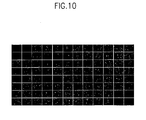
- the cells containing DNAs showed fluorescence. CPE was observed 4-7 days after the transfer, which indicated viral formation. At 10 th day from the transfer, 96-well plate was treated at -70°C for 1 hr followed by at 37°C for 5 min. The freezing and thawing were repeated 3 times to obtain cell lysate. The cell lysate was centrifugated at 2,500 rpm for 10 min. Then, the 293 cells in 96-well plate were transfected with the supernatant to confirm the formation of viruses expressing GFP (see FIG. 10 ).
- this invention employs in vitro site-specific recombination in order to prepare vectors for recombinant viruses.
- desired foreign genomic materials are inserted into suitable sites of viral genomic DNAs, which are digested to two or more segments, rapidly and in site-specific way.
- the reaction mixture of viral genomic DNAs and desired genomic materials can be applied to host cells in vitro directly without further selection of the recombinant viral genomic DNAs.
- this invention it is possible to obtain numerous recombinant viruses at the same time. Furthermore, it can prevent self-replication of viruses almost completely. Therefore, this invention provides a high-throughput system for producing hundreds or thousands of recombinant viruses for various studies of genomes.
Landscapes
- Health & Medical Sciences (AREA)
- Life Sciences & Earth Sciences (AREA)
- Genetics & Genomics (AREA)
- Engineering & Computer Science (AREA)
- Chemical & Material Sciences (AREA)
- Organic Chemistry (AREA)
- Bioinformatics & Cheminformatics (AREA)
- Zoology (AREA)
- Wood Science & Technology (AREA)
- General Engineering & Computer Science (AREA)
- Biotechnology (AREA)
- Biomedical Technology (AREA)
- General Health & Medical Sciences (AREA)
- Biochemistry (AREA)
- Microbiology (AREA)
- Virology (AREA)
- Biophysics (AREA)
- Molecular Biology (AREA)
- Medicinal Chemistry (AREA)
- Physics & Mathematics (AREA)
- Plant Pathology (AREA)
- General Chemical & Material Sciences (AREA)
- Pharmacology & Pharmacy (AREA)
- Chemical Kinetics & Catalysis (AREA)
- Public Health (AREA)
- Veterinary Medicine (AREA)
- Nuclear Medicine, Radiotherapy & Molecular Imaging (AREA)
- Animal Behavior & Ethology (AREA)
- Immunology (AREA)
- Micro-Organisms Or Cultivation Processes Thereof (AREA)
- Preparation Of Compounds By Using Micro-Organisms (AREA)
- Medicines That Contain Protein Lipid Enzymes And Other Medicines (AREA)
- Medicines Containing Material From Animals Or Micro-Organisms (AREA)
- Enzymes And Modification Thereof (AREA)
Abstract
Description
- This invention relates to a method for producing recombinant viruses and vectors for the same.
- A recombinant viral vector means a genetically engineered vector, which is used in, for example, the preparation of a vaccine; analysis of function of a gene, a group of genes, or a genomic domain; production of proteins; and gene therapy. Genomic materials, which could be integrated into a viral vector, include any genomic materials such as a gene, cDNA, genomic DNA, DNA sequences encoding peptides or proteins, RNA, anti-sense RNA, siRNA, DNA for si RNA, promoter, enhancer etc. A virus, which is used in the preparation of a vector, includes adenovirus, adeno-associated virus (AAV), retrovirus, Herpes virus and Vacxnia virus etc.
- Retrovirus is known as one of the most widely used viruses in gene therapy [see Clive Patience et al. J Virol, 72:2671-2676, 1998, Marshall, Science, 269: 1050-1055,1995]. Retroviral genome has two LTRs, capsid sequences and 3 (three) coding regions (gag, pol and env), and the method of its preparation and its use in vitro and in vivo has been already disclosed [
WO9908692A1 EP 453242 - Adenovirus has a linear genome (sized about 36 kb) and includes replication origin and capsid signals near to the left inverse terminal region (ITR) (103 bp) [Shenk, Adenoviridae: The Viruses and Their Replication. In: Fields BN, Knipe DM Howley PM, ed. Virology. Philadelphia: Raven publishers, 1996: 2111-2148]. Usually, adenoviral vectors have been made by deleting E1 site of the viral genome, which is essential for the viral replication. The viruses, which include foreign genomic materials substituting E1 site, are transferred to the packaging cells that provide E1 proteins. Thereby, the viral replication occurs only in the packaging cells. Representative packaging cells include, for example, HEK293 cell, 911 cell, and PER. C6 cell, etc. [Hitt MM et al., Advances in Pharmacology, vol. 40, 137-206 (1997), Wang Y. and Huang S., Drug]. Adenoviral vector has advantages such as safty, affinity to various cells, possibility of infecting dividing cells and higher titer (1011 pfu/ml). Thus, adenovirus has been used in the preparation of a vector expressing heterogeneous genes.
- Adeno-associated virus (sized about 4700 bp) has ITRs (sized about 145 bp) as replication origins at each terminus. It can be integrated into the genomic DNAs of various types of host cells safely and specifically.
- In order to use a recombinant virus as a vector, it should be modified not to replicate themselves in the infected cells. Therefore, defective viruses, which have deletions of some essential regions of genome for viral replication, are usually employed in the preparation of the recombinant viral vectors. In case of a retrovirus, for example, gag, pol and/or env genes are deleted, and the regions are replaced with desired genomic materials [Bender et al., J. Virol. 61 (1987) 1639]. Regarding an adenovirus, E1, E2 and/or E4 sites are deleted for the preparation of a viral vector [Levrero et al., Gene 101 (1991) 195; Gosh-Choudhury et al., Gene 50 (1986) 161, Van der Vliet et al., (1975)]. On the other hand, a pseudo-virus vector, which consists of only the essential regions (such as, ITR and capsid sequences) of the replication, has been used also for obtaining recombinant viruses (see
WO95/02697 - More specifically, conventional methods for the preparation of adenoviruses could be grouped into 3 categories as follows: In the first group, homologous recombination between carrier vectors and adenoviral genomic DNAs is provided, wherein the vectors carry desired foreign genomic materials [Hitt MM et al., Advances in Pharmacology, vol. 40, 137-206 (1997)]. In this method, desired foreign genomic materials are cloned into carrier vectors and then introduced to the packaging cells together with adenoviral vector genomic DNAs in order to induce homologous recombination. A carrier vector usually includes a cassette, such as a promoter, restriction enzyme target sites for gene cloning and poly(A) attachment signals, for the expression of foreign genomic materials. Furthermore, the carrier vector has nucleic acid sequences inducing homologous recombination. In general, a defective adenoviral genome, which lacks LITR, packaging signals, or E1 site, is used in this method. On the other hand, Frank Graham et al. designed a plasmid type vector, which can replicate in E. coli, in order to resolve the problem of the said method of requiring a further step for selecting defective viral genomes from viruses [Bett AJ et al., Proc. Natl. Acad. Sci USA,, vol 91, 8802-8806 (1994)]. The Graham's method, however, also has to use the homologous recombination between carrier vectors and defective adenoviral genomic DNAs, and the lower rate of homologous recombination in packaging cells also made this method inefficient.
- The second group includes direct cloning of desired foreign genomic materials to a certain region of adenoviral genome using restriction enzyme and ligase. This method resolved the difficulties caused by the structural complexity of the adenoviral genome (sized about 30 kb). In this method, expression cassette for the desired gene is, firstly, cloned to a carrier vector, which in turn is cloned again into the viral genome using specific restriction enzyme sites within both of the cassette and the viral genome. Next, the cloned cassette and the viral genome are introduced into E. coli, and the desired clones are selected. Afterward, the selected clones are introduced to the packaging cells. Then, the recombinant viruses could be obtained from the packaging cells [Mizuguchi H. and Kay MA., Human Gene Therapy, vol. 10, 2013-2017 (1999)]. In another method of this group, viral genomic DNAs having pTP (preterminal protein) at the region of ITR terminus are extracted from viruses, not from E. coli, in order to increase the productivity of desired recombinant viruses, which in turn are transferred to cell lines after being ligated with the desired genome expression cassette [Okada T. et al., Nucleic Acids Research, vol. 26, 1947-1950 (1998)].
- In the third group, a method employs E. coli, which has relatively higher efficiency of homologous recombination, in order to improve the lower recombination rate in the packaging cells [Chartier C. et al., Journal of Virology, vol. 70, 4805-4810 (1996), He T. et al., Proc. Natl. Acad. Sci USA, Vol 95, 2509-2514 (1998)]. According to this method, although the desired genomic materials should be also cloned into carrier vectors, it is possible to obtain relatively higher rate of homologous recombination in the particular E. coli by infecting the cell together with plasmid-type adenoviral genome. Thereby, the desired recombinant viruses could be obtained with higher productivity in comparison to the methods mentioned above. In this method, however, the desired genomic materials should be cloned to carrier vectors. And, a particular E. coli cell line (for example, BJ5183) should be used. Furthermore, the recombinant DNAs expressed in E. coli should be screened, and the screened recombinant DNAs should be transferred to another E. coli cell line (for example, DH5 α) due to the unstableness of the plasmid in the E coli used for the recombination. Thus, this method is considered as being very complex and not effective.
- A review of GATEWAY ™ recombinatorial cloning C. elegans ORFs into a versatile vector and from that vector into two different expression vectors used for two-hybrid analysis has been described. [Walhout AJM. et al, methods in Enzymology, vol. 328, 575-592 (2000)].
-
US 6143557 discloses recombinational cloning by the use of nucleic acids, vectors and methods, in vitro and in vivo, for moving or exchanging segments of DNA molecules using engineered recombination sites and recombination proteins to provide chimeric DNA molecules that have the desired characteristic(s) and/or DNA segment(s). - As disclosed above, the conventional methods for the preparation of recombinant viruses have disadvantages as follows: i) Desired genomic materials should be cloned to carrier vectors; ii) Since the gene cloning of the desired genomic materials should be carried out in a cell, it requires complex and inefficient cloning procedures; iii) The desired recombinant viruses should be selected from host cells before the mass production. Therefore, according to those methods, it takes much time to obtain recombinant viruses, while the productivity is relatively low. Especially, the disadvantages of ii) and iii) have been regarded as the most important problems to be overcome in the preparation of recombinant viruses, thus there have been continuous efforts to resolve these problems so far. On the other hand, according to the development of applications of recombinant viruses, the requirements for more efficient and rapid method for preparing recombinant viruses are increasing.
- Thus, we have carried out studies to produce recombinant viruses having desired genes, and we have completed this invention by providing recombinant viral vectors using in vitro site-specific recombination.
- It is important to understand the present invention to note that all technical and scientific terms used herein, unless otherwise defined, are intended to have the same meaning as commonly understood by one of ordinary skill in the art. The techniques used herein are also those that are known to one of ordinary skill in the art, unless stated otherwise.
- Reference to particular buffers, media, reagents, cells, culture conditions and the like, or to some subclass of same, is not intended to be limiting, but should be read to include all such related materials that one of ordinary skill in the art would recognize as being of interest or value in the particular context in which that discussion is presented. For example, it is often possible to substitute one buffer system or culture medium for another, such that a different but known way is used to achieve the same goals as those to which the use of suggested method, material or composition is directed.
- It is an object of this invention to provide more rapid and more efficient method for producing recombinant viruses.
- In order to accomplish this invention, the present invention employs recombinant viral vectors, which are prepared by in vitro site-specific recombination. More preferably, the viruses used in this invention have linear genomic DNAs.
- Therefore, it is another object of the present invention to provide recombinant viral vectors using in vitro site-specific recombination.
- In accordance with the present invention, there is provided a method for preparing linear recombinant-adenoviral vectors, wherein desired DNAs are inserted into adenoviral genomic DNAs by in vitro site-specific recombination, characterized in that the method comprises:
- i) providing adenoviral genomic DNA segments containing att site-specific recombination sites and
base sequences essential to viral replication and packaging, - ii) providing carrier vectors comprising at least one desired DNAs and att site-specific recombination sites; and
- iii) reacting the DNA segments of i) and the carrier vector of ii) in vitro.
- In accordance with another aspect of the present invention, there is provided a method for preparing recombinant adenoviruses having linear genomic DNA, characterized in that the method comprising the steps of:
- i) providing adenoviral genomic DNA segments containing att site-specific recombination sites and base sequences essential to viral replication and packaging;
- ii) providing carrier vectors containing at least one desired DNAs and att site-specific recombination sequences;
- iii) reacting the genomic DNA segments of i) and the carrier vectors of ii) in vitro using site-specific recombinase to produce mixture comprising expressive linear recombinant adenoviral vectors; and
- iv) transfecting the mixture of iii) to host cells.
- In the present invention, a vector comprising viral genomic DNAs is cleaved to two or more segments before being applied to in vitro site-specific recombination, in which both of the segments are essential for the viral replication. A DNA fragment including desired genomic materials is inserted between the two genomic DNA segments specifically. Thus, since un-bound viral genomic DNA segments cannot be expressed by themselves, only the viruses having desired DNA fragment shall be expressed. Therefore, one can obtain desired recombinant viruses from host cells directly without further procedures like selecting the desired recombinant viruses from intermediate host cells.
- Referring to the site-specific recombination, it is an event occurring naturally in various living organisms. Enzymes for the site-specific recombination contain not only endonuclease activity but also ligase activity, so they recognize a certain part of DNA sequences and replace it with any other corresponding DNA sequences [Yang W. and Mizuuchi K., Structure, 1997, Vol. 5, 1401-1406(9)]. Int/att system from bacterio λ phage, Cre/LoxP system from P1 bacterio phage and FLP-FRT system from yeast are well developed site-specific recombination systems.
- Site-specific recombination system has been used in i) expression control of desired genomic materials cloned into recombinant viruses, ii) excising of helper-virus when preparing helper-viral dependent viruses, iii) increasing the efficiency of homologous recombination in the packaging cells, or iv) inducing site-specific recombination between carrier vectors and viral genome in the packaging cells to prepare recombinant viruses [Philip NG et al., Biotechniques, vol. 29. 524-528 (2000), Philip NG et al., Human Gene therapy, vol. 10., 2667-2672 (1999), Hardy S et al., Journal of Viology, vol. 71., 1842-1849 (1997), Parks RJ et al., Proc. Natl. Acad. Sci USA,, vol 93., 13565-13570 (1996)]. However, the said applications of the site-specific recombination disclosed in the prior arts were merely indirect and supplementary uses in vivo or in a cell line, not in vitro.
- Thus, there have been no applications of in vitro site-specific recombination for specific insertion of desired genomic materials into linear viral genomic DNAs in order to obtain recombinant viral vectors. In particular, there have been no applications of using viral genomic DNAs, which are separated into two segments.
- Therefore, the present invention is characterized by employing in vitro site-specific recombination in the preparation of recombinant viral vectors. It has unexpected advantages in comparison to the prior arts as follows: i) it is possible to obtain recombinant viral vectors more easily and more rapidly by employing in vitro site-specific recombinant; ii) it does not require any further procedure, such as plaque assay, for selecting desired recombinant viruses from intermediate host cells by digesting viral genomic DNAs to two segments before applying them to site-specific recombination; and iii) it is possible to apply the reaction mixture including recombinant viral vectors to host cells without further processes.
- Furthermore, according to this invention, homologous recombination in the packaging cells is not required. Nor, particular cell lines for inducing homologous recombination in E. coli are required.
FIG. 5 shows the results of comparisons between the method of this invention for the preparation of adenovirus (AdHT System) and the method of the prior art (AdEasy system) in view of processes and time-period. From the results, it is clear that the desired recombinant viruses can be obtained even more rapidly by using the method of this invention. - Furthermore, according to the present invention, since numerous kinds of desired genomic materials are integrated into the sites of viral genomic DNAs accurately in vitro, various genomic materials can be expressed from the same kind of virues in multi-well (e.g. 96-well and 384-well) at the same time. Likewise, since numerous kinds of viral genomes from various types of viruses [such as, adenovirus, retrovirus, adeno-associated virus, bacteriophage, herpes simplex virus, CMV (cytomegalovirus), EBV (Epstein-Barr virus) or VZV (Varicella-zoster virus), etc] are employed, the same kind of genomic material can be expressed from various types of viruses by using an universal common vector (pMaster Vector) containing the desired genomic material.
- Accordingly, the present invention also provides significantly useful tools for studying functional genomics of which demands have been increasing greatly, since the completion of genome project [Wang Y. and Huang S., Drug Discovery Today, vol. 5, 10-16 (2000)].
- In one embodiment, this invention provides recombinant adenoviruses of which genomic DNAs are in linear form. As depicted in
FIG. 2 , genomic DNAs of viruses are firstly cleaved to two segments, wherein the segments respectively are essential for the viral replication and packaging. Then, using the cleaved segments as templates, the desired genomic materials containing site-specific recombination sites are integrated through site-specific recombination. More specifically, E1 site deleted adenovirus is digested to two segments, and recombination sites are introduced into each of the segments in order to induce in vitro site-specific recombination. Other corresponding site-specific recombination sites are introduced into the desired genomic materials. - Referring to
FIG. 2 , the left segment of adenoviral genome includes ITR and packaging signals, and the right segment includes other genes essential for viral replication. Recombination and viral packaging, however, cannot occur solely with only one of the two cleaved segments (since the right segment lacks signals for particle formation and the left segment is too short to form a viral particle). Thus, only the viral genome containing the desired genomic materials, which are integrated by site-specific recombination, can be used as a template for replication in a packaging cell. - On the other hand, methods disclosed below can be used for obtaining two segments of viral genomic DNAs. In the first method, viral genomic DNAs are cleaved to two segments with restriction enzyme, wherein each of the segments is essential for viral packaging and has a site-specific recombination sites (see
FIG. 2 ). More specifically, one or more restriction enzyme recognition sites are firstly cloned to viral genomic DNAs (seeFIG. 1 ), and then two or more viral DNA segments can be obtained by restriction enzyme treatment. The viral genomic DNAs including restriction enzyme recognition sites could be obtained as a plasmid from E. coli, or be obtained directly from viruses. The obtained plasmid (pAdHTS) has a construct as depicted inFIG. 1 . Next, pAdHTS (SEQ. ID No: 1) is cleaved to three segments by restriction enzyme treatment. Except for the segment not related to site-specific recombination and viral replication, other two segments are employed in the preparation of recombinant viral vectors (seeFIG. 2 ). - In the second method, parent viral formation can be prevented. As disclosed in Example 3, the left segment and the right segment of viral DNAs are generated as plasmids respectively, which in turn are made to linear forms, wherein the two segments have site-specific recombination sites, which are different from each other. Referring to
FIG. 3 , left-segment has ITR and packaging signals, and the right-segment has nucleic acids essential for the viral replication. Each of the segments has replication origins (e.g. pBR322 origin or pUC origin etc.) for the replication of DNAs in E. coli, and at least one selection marker (e.g. Ampr, Kanr, Cmr, etc) to permit detection and/or selection. These segments can be provided from other plasmids, respectively. - The viral genomic DNA segments in this invention can be in linear or in circular form. Preferably, they are in linear form. For example, if linear DNA segments generated by restriction enzyme treatment are integrated into pMaster vectors in vitro, linear formed recombinant viral genome could be obtained as depicted in
FIG. 4 . Thus, in case of circular plasmid DNAs, they are firstly treated with restriction enzyme, such as PacI, to form linear DNA segments and consequently to obtain linear viral genome (seeFIG. 4 ), and then are introduced into the packaging cells to induce viral replication. - In one embodiment, a promoter is inserted into at a site of viral genomic DNAs in order to induce the expression of desired genomic materials (see
FIG. 1 andFIG. 2 ). The promoter, not operating in a cleaved DNA segment, induces the expression of genomic materials, when the materials are introduced by site-specific recombination. InFIG. 6 , green fluorescence proteins (GFP) and Gus genes are not expressed in the group of cells that has not been subjected to site-specific recombination. - A promoter, which can be employed in the present invention, includes CMV promoter for continuous gene expression, RSV (Respiratory syncytial virus) promoter, SV (simian virus) promoter, a promoter system which induces gene expression only in specific environment, a promoter system containing ERE which operates in response to corresponding hormone, a promoter system operating in response to ecdysone, and a promoter system operating in response to tetracycline, etc. The promoter typically controls the expression, optionally with an operator sequence.
- In another embodiment, attR sites within two viral genomic DNA segments react with attL sites from carrier vectors including foreign genomic materials, and form attB and attP sites. More specifically, when the two viral genomic DNA segments are treated with carrier vectors having desired genomic materials in the presence of λ integrase, IHF as a co-factor and Xis, attB sites are generated as main products. Vector DNA segments having attP sites are generated also as by-products in the reaction mixture. Numerous recombination systems from various organisms, for example, integrase/att system from bacteriophage λ, the Cre/loxP system from bacteriophage P1, and the FLP/FRT system from the
Saccharomyces cerevisiae 2 µ circle plasmid, can be employed. - If the mixtures are transferred directly to suitable host cells, which are expected to express recombinant viruses, only the viral genomic DNAs containing attB1-[desired genomic materials]-attB2 can replicate and form viral particles. The viral DNA segments and the vector DNAs, which do not react with each other, and the vector DNA segments having attP sites do not form viral particles.
- Referring to
FIG. 6 , the disappearance of left segments of viral genomic DNAs suggests that the DNA segments having attR sites are recombined. InFIG. 7 , PCR products show that the desired genomic materials are inserted exactly into suitable sites of viral genome. PCR reaction is carried out for both of the samples collected before- and after-recombination reaction using primers (Adeno-F primer and Adeno-R primer) that are able to amplify certain regions of viral genome. - Adeno-F primer: 5'-GGTCTATATA AGCAGAGCTG-3' (SEQ. ID No.: 2)
- Adeno-R primer: 5'-GTATGGCTGA TTATGATCAG-3' (SEQ. ID No.: 3)
- The analysis of PCR products using DNA sequencing shows that viral genome and desired genomic materials are integrated at the target sites and generated [left segment of viral genome-attB1-desired genomic materials-attB2-right segment of viral genome] (see
FIG. 2 ). - Viral replication and packaging are observed after infecting the packaging cells, such as 293 cells, with the reaction mixture including site-specific recombinant viral vectors. These results indicate that the in vitro mixture of this invention can be applied to host cells directly to produce recombinant viruses without further step of selection. That is, according to this invention, without employing intermediate host cells, such as E. coli, for selecting appropriate genomic DNAs, desired recombinant viruses can be produced rapidly.
- In
FIG. 8 , the results of site-specific recombination of AdHTP vectors and pMater clones are presented. The expression efficiency of GFP was still higher in the method of this invention compared to the method of the prior art (Ad-GFP control). The activity assays of Gus proteins indicated that the method of this invention did not have adverse-effects on the activities of the proteins expressed. Thus, the recombinant viruses produced in this invention are suitable for the expression of desired genomic materials from infected host cells. -
-
FIG.1 depicts the construct of pAdHTS vector. -
FIG.2 shows the process of site-specific recombination between pAdHTS vector and pMaster clone in vitro. -
FIG.3 depicts the constructs of pAd-Left and pAd-Right. -
FIG.4 shows the process of site-specific recombination of pAd-Left/pAd-Right and pMaster clone in vitro. -
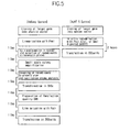
FIG.5 shows processes and time-period for the production of recombinant adenovirus using AdHTS in comparison to those of the methods of prior arts. -
FIG.6 illustrates agarose gel eletrophoretic analysis showing the results of site-specific recombination of pAdHTS vector and pMaster clone in vitro. -
FIG. 7 shows the results of PCR, which indicate site-specific recombination of pAdHTS vectors and pMaster clones. -
FIG. 8 shows the results of site-specific recombination of AdHTS vectors and pMaster clones. The desired genomic materials in pMaster clones are transferred to adenoviral genome to express the desired proteins. -
FIG. 9 shows the results of PCR indicating the site-specific recombination of AdHTS and pMaster-GFP in 96-well. -
FIG.10 shows the results of site-specific recombination of AdHTS and pMaster-GFP in 96-well. GFP gene inserted into pMaster clone is transferred to adenoviral genome to express GFP - Preferred embodiments of this invention are described in the following examples. Other embodiments within the scope of the claims herein will be apparent to those skilled in the art from consideration of the specification or practice of the invention as disclosed herein. It is intended that the specification, together with the examples, be considered exemplary only, with the scope and the spirit of the invention being indicated by the claims which follow the examples. The examples herein are meant to exemplify the various aspects of carrying out the invention and not intended to limit the scope of the invention in any way. The examples do not include detailed descriptions of conventional methods employed, such as in the performance of genomic DNA isolation, PCR, and sequencing procedures. Such methods are well-known to those skilled in the art and are described in numerous publications. In addition, all the publications referred herein are integrated hereto as references.
- pMaster vector having a desired genomic material flanked with attL sites was prepared (see
FIG.2 ). Then, pAdHTS (SEQ. ID No.: 1) having viral genomic DNA flanked with attR sites was prepared using conventional recombination method (seeFIG.1 ). The pAdHTS vector for this study was generated by deleting E3 site of adenoviral genomic DNAs and by replacing the E1 site with an expression cassette of [CMV promoter]-attR1-[λ stuffer DNA]-attR2-[poly A attachment signal], which induced expression of the recombinant desired genomic material (seeFIG. 1 andFIG.2 ). λ stuffer DNA was prepared to contain restriction enzyme recognition sites, such as SwaI, ClaI or PacI, by amplifying PCR primers having the restriction sites and cloning them thereto. Then, the DNA was treated with SwaI and/or ClaI to generate two DNA segments, wherein each of the segments is essential for the viral replication and packaging. The att sites had the same nucleic acid sequences as those devised by James L. et al.[see Hartley JL. et al., Genome Research, vol. 10, 1788-1795 (2000)]. pMaster vectors flanked with the mutant attL sites (attL1 and attL2) were cloned with GFP cDNA and Gus cDNA, respectively, using recombination methods (seeFIG. 2 , Gene-X site of pMaster vector). As the result, the plasmid vectors comprising expression cassettes of attL1-[desired genomic materials]-attL2 were generated. - DNAs of pAdHTS (1 µg) were purified using phenol/chloroform after treating them with SwaI and/or ClaI enzyme(s). The pMaster-GFP (100 ng) containing GFP cDNA, pMaster-Gus (100 ng) containing Gus cDNA, integrase (100 ng), IHF (10 ng), and Xis (10 ng) were reacted in a buffer (50mM Tris, pH 7.8, 16.5 mM NaCl, 35 mM KCl, 5 mM Spermidine, 0.25 mM EDTA, 3% Glycerol) at 25°C for 1 hr. Then, integrase, IHF and Xis were removed from E. coli according to the common method [Kotewicz M et al., Journal of Biological Chemistry, 1980, vol. 255, 2433-2439; Nash HA. and Robertson CA. Journal of Biological Chemistry, vol. 256, 9246-9253 (1981); Cho EH et al., Journal of Bacteriology, vol. 182, 5807-5812 (2000)]. The results of agarose gel electrophoretic analysis indicated that pAdHTS treated with PacI generated three DNA segments (32 kb, 4.5 kb and 1.8 kb) (
FIG. 7 Lane 1). Where site-specific recombination occurred, it was observed that the segment (sized about 1.8 kb) was disappeared (FIG. 7 Lanes 2 and 3). In order to confirm this, PCR was carried out using adeno-F primer and adeno-R primer, thereby the desired sized PCR products (about 32 kb and about 4.5 kb) could be observed where the site-specific recombination was induced (seeFIG. 6 ). - The PCR products were isolated from agarose gel, and DNA sequencing was carried out for the PCR production. As the result, it was determined that the PCR products of
Lanes - 2 micro-liter of the reaction mixture (100 ng of DNA) obtained from Example 1 was introduced to 293 packaging cells. Then, the 293 cells were transferred to 24-well (5×10⁴ cells/well) with DMEM (5% bovine fetal serum). After 24 hrs, recombination induced reaction mixture (2 micro-litter) and LipofecAmine2000 (0.5 micro-litter) (from Invitrogen Inc.) were transferred to the cells in accordance with the manufacturer's protocol. After 7 days from the transfer, CPE was observed under a microscope, which indicated the formation of viruses in the cells. At that moment, the cells were harvested and were centrifugated. The resulting precipitates were suspended in 100 micro-liter of DMEM 10% bovine fetal serum. The cells were treated at -70°C for 1 hr followed by at 37°C for 5 min. These freezing and thawing were repeated 3 times. The lysate was removed by centrifuge to produce viral mixture. Afterward, 293 cells were infected with the viral mixture. Plaque assay was carried out to detect the formation of suitable viruses.
- For plaque assay, 2× 10⁵ cells were transferred to 6-well plate. After 24 hrs from the transfer, 10 micro-liter of the viral mixture mixed with 400 micro-liter of DMEM 400 was plated on the cells for 1 hr in order to induce viral adsorption. Plaques of viruses were assayed for 2 weeks, after solidifying the cells with 3 ml of DMEM to which 1% low melting-point agarose (SeaPlaque Agarose; FMC) was added. The titer of viruses was determined as the number of viral plaques per 1 ml of viral mixture. After 2 weeks, in order to detect product efficiency of the desired recombinant viruses, 500 µg of X-Gluc (5-bromo-4-chloro-3-indolil-beta-D-gluchronic acid) as substrate of Glu protein was added to the solidified agarose. After 24 hrs, the number of blue colored plaques was calculated. The titer of viruses was 106∼107 PFU per 1 ml. The production efficiency of recombinant viruses was determined by the recombinant viruses expressing Gus genes. All the viruses were observed as blue plaques, which suggested that 100% of the viruses obtained in the present invention were recombinant viruses. That is, only the viral genome, including desired genomic materials, replicated and formed viral particles. This had occurred because even though the efficiency of the site-specific recombination was not 100%, the separated DNA segments themselves alone and the by-products of site-specific recombination did not replicate.
- As depicted in
FIG. 8 , recombinant viruses expressing GFP and Gus, respectively, were prepared as mentioned above. The 293 cells were infected with the recombinant viruses. Then, the expression of desired proteins (GFP or Gus) was detected under the fluorescence microscope (for GFP) or by adding X-Gluc substrate (for Gus). As shown inFIG. 8 , only the group of viruses to which site-specific recombinase was added generated recombinant viruses and expressed desired proteins (seeFIG. 8 Lanes 4 and 5). - pAd-Left (SEQ. ID No.: 4) and pAd-Right (SEQ ID No.: 5) were prepared (see
FIG.3 ). Firstly, pAdHTS vector was treated with PacI and/or SwaI. Then, the left fragment containing LITR and packaging signals were ligated with Amp-Ori DNA from PCR amplification in order to form pAd-Left. Likewise, the right fragment (sized about 33 kb) was ligated with Amp-Ori DNA to form pAd-Right. In this study, PCR primer for Amp-Ori included ampicillin resistant gene of pBR322 DNA, a region for amplification of replication origins, and PacI and/or SwaI linker (s) for cloning restriction enzyme recognition sites. The obtained plasmid DNAs of pAd-Left and pAd-Right were generated from DH5α (Qiagen, Midiprep kit), which in turn were digested with SwaI and PacI. Using the respective plasmid DNA, site-specific recombination was induced as disclosed in Examples 1 and 2, so as to produce recombinant viruses. Briefly, linear DNAs of pAd-Left (100 ng of DNA) and linear DNA of pAd-Right (800 ng of DNA) were reacted in the presence of pMaster-GFP having GFP cDNA (100 ng), integrase (100 ng), IHF (10 ng) and Xis (10 ng) within a buffer for 1 hr. The resulting mixture was transferred to 293 packaging cells as disclosed in Example 2. After 7 days, viruses were obtained from cell debris, and plaque assay was carried out. - In order to obtain AdHTS viruses, pAdHTS were digested with restriction enzyme of PacI, which in turn were transferred to 293 packaging cells to induce viral packaging. Mass production of AdHTS viruses was carried out by transferring about 5× 10⁵ of 293 cells to 30 cell culture dishes (150 mm of diameter) together with DMEM (5% bovine fetal serum). After 24 hrs, AdHTS virus was infected to 5 MOI. After 48 hrs from viral infection, cells were harvested from the dishes with cell scraper, and then were centrifugated. Afterward, the precipitates were suspended in a solution of 10 mM of Tris and 1mM of MgCl2. The suspension was treated at -70°C for 1 hr followed by at 37°C for 5 min. The freezing and thawing were repeated 3 times. The cell lysate was centrifugated, and the precipitates were removed to obtain viral mixture. In order to obtain infectious viral particles, CsCl gradient ultra-centrifugation was employed. 1.4 g/ml of CsCl solution was poured into a tube (BeckMan), and then 1.2 g/ml of CsCl solution was added to the solution again. After adding 20 ml of viral mixture to the solution while maintaining the concentration gradient, the solution was centrifugated with ultra-centrifuge at 25,000 rpm for 2 hrs, thereby 1× 10¹² of infectious viral particles were isolated from defective viral particles and cell debris. Two different methods were used in order to prepare DNA-terminal protein complex from AdHTS viruses. In the first method, 6 M guanidine HCl was added to the same amount of AdHTS viral solution, and then 1 mg of ethidium bromide and saturated CSCl/4M guanidin HCl solution were added to make the final volume to 5 ml. Next, the solution was ultra-centrifugated at 650,000 g for 4 hrs. Isolated viral DNA-terminal protein complex band was detected with 365 nm of ultraviolet rays, then the DNAs corresponding to the band were extracted. The extracted DNAs were desalted for use in the following studies. In the second method, adenoviral solution, which had a mixture of 6 M guanidine HCl and the same amount of AdHTS viral solution, was transferred to superdex 16/30HR S-200 column (Amersham-Pharmacia). While developing the solution in 0.4 ml/min of flow rate, optical densities for each of the subsequent 2 ml fractions obtained continuously were detected at 260 nm and 280 nm. AdHTS DNA-terminal protein complex was detected on 1% agarose gel from the collection of peak fractions.
- The AdHTS DNA-terminal protein complex obtained from above two methods was digested with SwaI (NEB) and stored at -70°C before being used in other studies.
- 10 ul of in vitro reaction mixture of pAdHTS vector from Example 2 and pMaster-GFP were transferred to 293 packaging cells as disclosed in Example 2. After 7 days, plaque assay was carried out for the viruses extracted from the cell lysate. In the same manner, plaque assay for pAd-left/pAd-right of Example 3 and for AdHTS-DNA-TPC of Example 4 were carried out, and the results are represented in Table 1 below.
Table 1. The results of plaque assays for viruses resulted from site-specific recombination between each of adenoviral vectors and pMaster-GFP. Mater Clone pMmaster-GFP adenoviral vector No recombination PAdHTS AdHTS-TPCC pAd-left pAd-Right total plaques (white spot) 0 50 74 65 GFP-positive plaques 0 50 74 65 - 45 ng/well of SwaI treated AdHTS-DNA-terminal protein complex in Example 4 were transferred to each well of 96-well PCR plate, which in turn were subjected to site-specific recombination as in Example 1. Briefly, 100 ng of integrase, 10 ng of IHF, 10 ng of Xis, reaction buffer (50mM Tris, pH 7.8, 16.5 mM NaCl, 35 mM KCl, 5 mM spermidine, 0.25 mM EDTA, 3% glycerole), and 5 ng of pMaster-GFP including GFP cDNA were transferred to each well. As a control, 5 ng of pMaster-Gus including Gus cDNA was transferred to 4 (four) wells respectively, and reaction was continued for 1 hr. In order to confirm site-specific recombination, PCR reaction was carried out using Adeno-F primer and Adeno-R primer. As the result, PCR products corresponding to GFP were detected in 88 wells except in the controls (see
FIG. 9 ). Similar to Example 2, 10 ul (DNA 50 ng) of the reaction mixture, which was confirmed as including site-specific recombinant, was transferred to 96-well plate, wherein each of wells contained about 5× 10³ of 292 packaging cells which were incubated for 24 hrs and wherein each of wells was coated with poly-L-lysin. The cells containing DNAs showed fluorescence. CPE was observed 4-7 days after the transfer, which indicated viral formation. At 10th day from the transfer, 96-well plate was treated at -70°C for 1 hr followed by at 37°C for 5 min. The freezing and thawing were repeated 3 times to obtain cell lysate. The cell lysate was centrifugated at 2,500 rpm for 10 min. Then, the 293 cells in 96-well plate were transfected with the supernatant to confirm the formation of viruses expressing GFP (seeFIG. 10 ). In the same manner, the expression efficiency of recombinant viruses was determined for the other adenoviral vectors, such as pAdHTS and pAd-Left/pAd-Right, in 96-well, respectively, and the average efficiency of recombinant viruses is presented on Table 2 below.Table 2. The expression efficieny of recombinant viruses in 96-well Master clone pMaster-GFP adenoviral vector, pAdHTS pAd-Left/pAd-Right AdHTS-TPC expression efficiency of recombinant viruses in 96-well 62.5 47.9 99.0 - As disclosed above, this invention employs in vitro site-specific recombination in order to prepare vectors for recombinant viruses. In this invention, desired foreign genomic materials are inserted into suitable sites of viral genomic DNAs, which are digested to two or more segments, rapidly and in site-specific way. Furthermore, the reaction mixture of viral genomic DNAs and desired genomic materials can be applied to host cells in vitro directly without further selection of the recombinant viral genomic DNAs. Thus, according to this invention, it is possible to obtain numerous recombinant viruses at the same time. Furthermore, it can prevent self-replication of viruses almost completely. Therefore, this invention provides a high-throughput system for producing hundreds or thousands of recombinant viruses for various studies of genomes.
- <110> Neurogenex Co., Ltd.
- <120> A METHOD FOR PRODUCING RECOMBINANT VIRUSES USING SITE-SPECIFIC RECOMBINATION
- <130> PN0022781.00
- <150> KR10-2002-0012633
<151> 2002-03-09 - <160> 5
- <170> KopatentIn 1.71
- <210> 1
<211> 34573
<212> DNA
<213> Artificial Sequence - <220>
<223> pAdHTS vector - <220>
<221> misc_feature
<222> (3181)..(30681)
<223> Ad5 - <220>
<221> promoter
<222> (400)..(930)
<223> CMV promoter - <220>
<221> misc_feature
<222> (33318)..(34112)
<223> kanamycin - <220>
<221> rep_origin
<222> (31716)..(32493)
<223> pBR322 ori - <220>
<221> misc_feature
<222> (31536)..(31638)
<223> RITR - <220>
<221> polyA_site
<222> (1747)..(1868)
<223> SV40 polyA - <220>
<221> misc_feature
<222> (197)..(345)
<223> Ad5 psi - <220>
<221> misc_feature
<222> (1464)..(1588)
<223> attR2 site - <220>
<221> misc_feature
<222> (949)..(1073)
<223> attR1 site - <400> 1






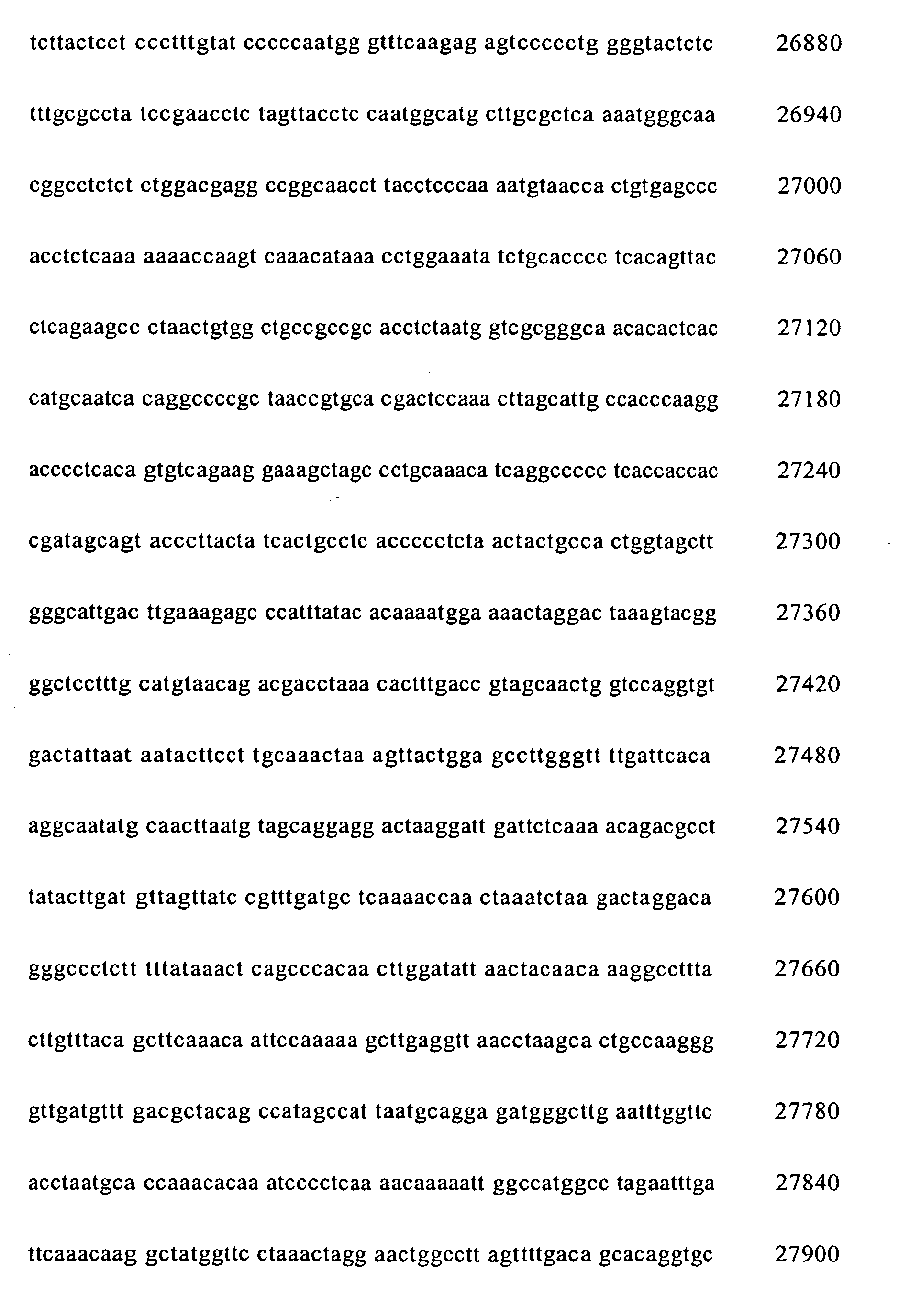


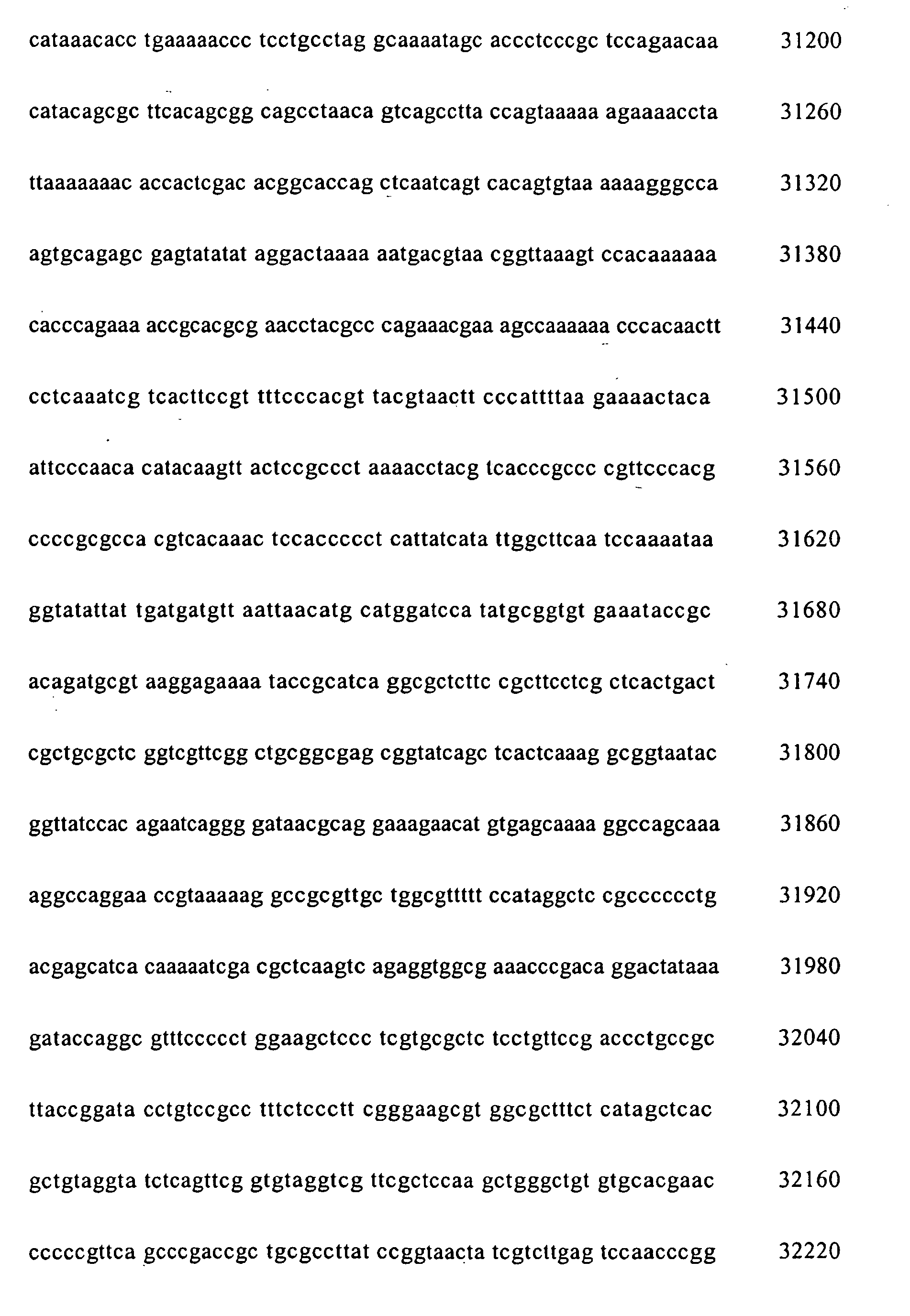


- <210> 2
<211> 20
<212> DNA
<213> Artificial Sequence - <220>
<223> Adeno-F primer - <400> 2
ggtctatata agcagagctg 20 - <210> 3
<211> 20
<212> DNA
<213> Artificial Sequence - <220>
<223> Adeno-R primer - <400> 3
gtatggctga ttatgatcag 20 - <210> 4
<211> 4752
<212> DNA
<213> Artificial Sequence - <220>
<223> pAd-Left - <220>
<221> misc_feature
<222> (1227)..(2087)
<223> R Amp - <220>
<221> promoter
<222> (400)..(930)
<223> F CMV promoter - <220>
<221> rep_origin
<222> (2235)..(2902)
<223> R pBR322 Ori - <220>
<221> misc_feature
<222> (197)..(345)
<223> F Ad5 psi - <220>
<221> misc_feature
<222> (4)..(106)
<223> F LITR - <220>
<221> misc_feature
<222> (949)..(1073)
<223> F attR1 - <400> 4

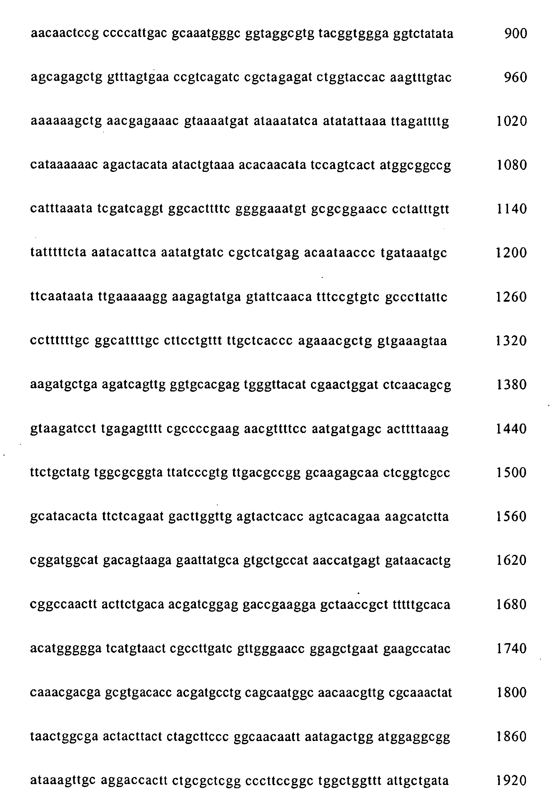


- <210> 5
<211> 33855
<212> DNA
<213> Artificial Sequence - <220>
<223> pAd-Right - <220>
<221> source
<222> (1)..(3657)
<223> F pAd-Easyl - <220>
<221> misc_feature
<222> (2666)..(3526)
<223> F Amp - <220>
<221> rep_origin
<222> (1851)..(2518)
<223> F pBR322 Ori - <220>
<221> misc_feature
<222> (33750)..(33852)
<223> F RITR - <220>
<221> polyA_site
<222> (3961)..(4082)
<223> F SV40 polyA - <220>
<221> misc_feature
<222> (3678)..(3802)
<223> R attR2 - <400> 5


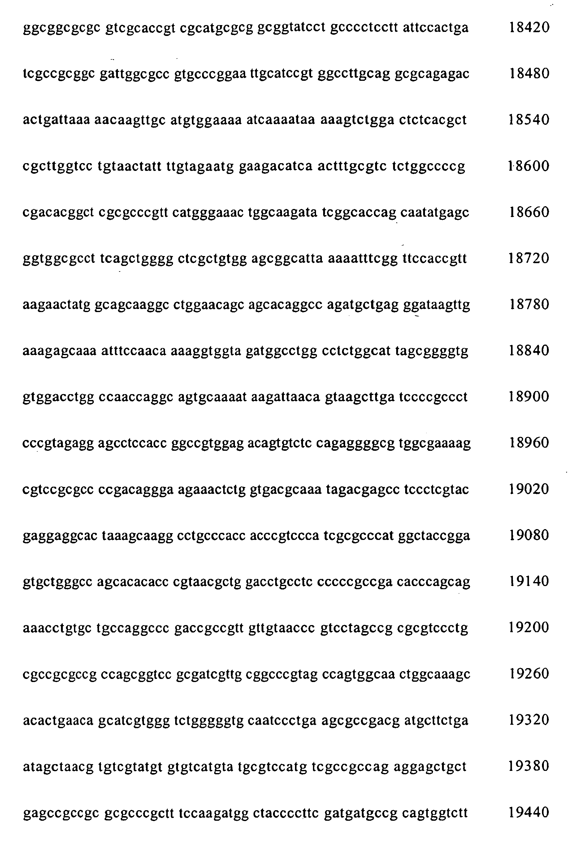


Claims (6)
- A method for preparing linear recombinant adenoviral vectors, wherein desired DNAs are inserted into adenoviral genomic DNAs by in vitro site-specific recombination, characterized in that the method comprises:i) providing adenoviral genomic DNA segments containing att site-specific recombination sites and base sequences essential to viral replication and packaging,ii) providing carrier vectors comprising at least one desired DNAs and att site-specific recombination sites; andiii) reacting the DNA segments of i) and the carrier vector of ii) in vitro.
- The method for preparing linear recombinant adenoviral vectors according to claim 1, wherein the viral genomic DNA segments of i) consists of 2 (two) DNA segments which respectively are essential for viral replication and packaging, and include att site-specific recombination sequences.
- The method for preparing linear recombinant adenoviral vectors according to claim 1, wherein the adenovirus is characterized by being deleted of E1, E2, E3, and/or E4 sites.
- A method for preparing recombinant adenoviruses having linear genomic DNA, characterized in that the method comprising the steps of:i) providing adenoviral genomic DNA segments containing att site-specific recombination sites and base sequences essential to viral replication and packaging;ii) providing carrier vectors containing at least one desired DNAs and att site-specific recombination sequences;iii) reacting the genomic DNA segments of i) and the carrier vectors of ii) in vitro using site-specific recombinase to produce mixture comprising expressive linear recombinant adenoviral vectors; andiv) transfecting the mixture of iii) to host cells.
- The method for preparing recombinant adenoviruses having linear genomic DNAs according to claim 4, wherein the adenoviral genomic DNAs of i) consists of 2 (two) DNA segments which respectively are essential for viral replication and packaging, and include att site-specific recombination sequences.
- The method for preparing recombinant adenoviruses having linear genomic DNAs according to claim 4, wherein the adenovirus is characterized by being deleted of E1, E2, E3, and/or E4 site.
Applications Claiming Priority (3)
| Application Number | Priority Date | Filing Date | Title |
|---|---|---|---|
| KR2002012633 | 2002-03-09 | ||
| KR20020012633 | 2002-03-09 | ||
| PCT/KR2003/000453 WO2003076605A1 (en) | 2002-03-09 | 2003-03-08 | A method for producing recombinant viruses using site-specific recombination |
Publications (3)
| Publication Number | Publication Date |
|---|---|
| EP1483375A1 EP1483375A1 (en) | 2004-12-08 |
| EP1483375A4 EP1483375A4 (en) | 2005-08-10 |
| EP1483375B1 true EP1483375B1 (en) | 2009-01-21 |
Family
ID=27800659
Family Applications (1)
| Application Number | Title | Priority Date | Filing Date |
|---|---|---|---|
| EP03708709A Expired - Lifetime EP1483375B1 (en) | 2002-03-09 | 2003-03-08 | A method for producing recombinant adenovirus viruses using site-specific recombination |
Country Status (9)
| Country | Link |
|---|---|
| US (1) | US20040005710A1 (en) |
| EP (1) | EP1483375B1 (en) |
| JP (2) | JP4558328B2 (en) |
| KR (1) | KR100631428B1 (en) |
| CN (1) | CN100532545C (en) |
| AT (1) | ATE421576T1 (en) |
| AU (1) | AU2003212687A1 (en) |
| DE (1) | DE60325942D1 (en) |
| WO (1) | WO2003076605A1 (en) |
Families Citing this family (14)
| Publication number | Priority date | Publication date | Assignee | Title |
|---|---|---|---|---|
| GB0424356D0 (en) * | 2004-11-03 | 2004-12-08 | Ark Therapeutics Ltd | Baculovirus-based gene libraries |
| WO2007132169A1 (en) * | 2006-05-11 | 2007-11-22 | Crusade Laboratories Limited | Mutant hsv, materials and methods for generation of mutant hsv |
| AU2011292120B2 (en) | 2010-08-16 | 2015-07-30 | Salk Institute For Biological Studies | Adenoviral assembly method |
| EP3483285B1 (en) | 2011-02-09 | 2021-07-14 | Bio-Rad Laboratories, Inc. | Analysis of nucleic acids |
| KR101416600B1 (en) * | 2012-08-17 | 2014-07-08 | (주)프로스테믹스 | Method for isolation of Adenovirus DNA-terminal protein complex using hydrophilic membrane |
| SG11201507393TA (en) | 2013-03-14 | 2015-10-29 | Salk Inst For Biological Studi | Oncolytic adenovirus compositions |
| CN105555972B (en) | 2013-07-25 | 2020-07-31 | 伯乐生命医学产品有限公司 | Genetic assay |
| ES2899913T3 (en) | 2014-09-24 | 2022-03-15 | Salk Inst For Biological Studi | Oncolytic tumor viruses and methods of use |
| WO2016081567A1 (en) * | 2014-11-19 | 2016-05-26 | Orlo James Fiske | Thin optical system and camera |
| EP3390645B1 (en) | 2016-02-23 | 2022-09-14 | Salk Institute for Biological Studies | Exogenous gene expression in therapeutic adenovirus for minimal impact on viral kinetics |
| WO2017147265A1 (en) | 2016-02-23 | 2017-08-31 | Salk Institute For Biological Studies | High throughput assay for measuring adenovirus replication kinetics |
| AU2017375633C1 (en) | 2016-12-12 | 2023-04-27 | Salk Institute For Biological Studies | Tumor-targeting synthetic adenoviruses and uses thereof |
| BR112020019942A2 (en) | 2018-04-09 | 2021-01-26 | Salk Institute For Biological Studies | oncolytic adenovirus compositions with enhanced replication properties |
| CN114457098B (en) * | 2022-01-07 | 2022-12-27 | 广东省农业科学院蔬菜研究所 | Multi-gene vector system and application thereof |
Family Cites Families (3)
| Publication number | Priority date | Publication date | Assignee | Title |
|---|---|---|---|---|
| US6143557A (en) * | 1995-06-07 | 2000-11-07 | Life Technologies, Inc. | Recombination cloning using engineered recombination sites |
| DE69836092T2 (en) * | 1997-10-24 | 2007-05-10 | Invitrogen Corp., Carlsbad | RECOMBINATORY CLONING USING NUCLEAR FACILITIES WITH RECOMBINATION CENTERS |
| ATE391789T1 (en) * | 1999-12-10 | 2008-04-15 | Invitrogen Corp | USE OF A VARIETY OF RECOMBINATION SITE WITH UNIQUE SPECIFICITY IN RECOMBINATORY CLONING |
-
2003
- 2003-03-07 US US10/383,846 patent/US20040005710A1/en not_active Abandoned
- 2003-03-08 WO PCT/KR2003/000453 patent/WO2003076605A1/en not_active Ceased
- 2003-03-08 CN CNB038056380A patent/CN100532545C/en not_active Expired - Fee Related
- 2003-03-08 DE DE60325942T patent/DE60325942D1/en not_active Expired - Lifetime
- 2003-03-08 JP JP2003574812A patent/JP4558328B2/en not_active Expired - Fee Related
- 2003-03-08 EP EP03708709A patent/EP1483375B1/en not_active Expired - Lifetime
- 2003-03-08 KR KR1020047014159A patent/KR100631428B1/en not_active Expired - Fee Related
- 2003-03-08 AT AT03708709T patent/ATE421576T1/en not_active IP Right Cessation
- 2003-03-08 AU AU2003212687A patent/AU2003212687A1/en not_active Abandoned
-
2010
- 2010-04-15 JP JP2010094507A patent/JP2010193896A/en not_active Abandoned
Also Published As
| Publication number | Publication date |
|---|---|
| CN1639325A (en) | 2005-07-13 |
| KR100631428B1 (en) | 2006-10-11 |
| US20040005710A1 (en) | 2004-01-08 |
| AU2003212687A1 (en) | 2003-09-22 |
| EP1483375A1 (en) | 2004-12-08 |
| KR20040111390A (en) | 2004-12-31 |
| DE60325942D1 (en) | 2009-03-12 |
| WO2003076605A1 (en) | 2003-09-18 |
| EP1483375A4 (en) | 2005-08-10 |
| CN100532545C (en) | 2009-08-26 |
| ATE421576T1 (en) | 2009-02-15 |
| JP2010193896A (en) | 2010-09-09 |
| JP4558328B2 (en) | 2010-10-06 |
| JP2005519603A (en) | 2005-07-07 |
Similar Documents
| Publication | Publication Date | Title |
|---|---|---|
| JP2010193896A (en) | Method for producing recombinant virus using site-specific recombination | |
| US5817492A (en) | Recombinant DNA viral vector for transfecting animal cells | |
| US6340595B1 (en) | High throughput screening of gene function using adenoviral libraries for functional genomics applications | |
| US6228646B1 (en) | Helper-free, totally defective adenovirus for gene therapy | |
| ES2260803T3 (en) | PROCEDURE FOR THE PRODUCTION OF RECOMBINANT VIRUSES. | |
| HU222808B1 (en) | Process for the production of recombinant adenoviruses for use in prokaryotic plasmids and pharmaceutical compositions comprising adenoviruses. | |
| AU762973B2 (en) | Methods and compositions for producing viral particles | |
| US20050095231A1 (en) | Modified adenovirus containing a fiber replacement protein | |
| JP2010207231A (en) | Highly productive mechanism for producing recombinant virus by using site-specific recombination | |
| JP7407099B2 (en) | Means to generate adenoviral vectors for cloning large nucleic acids | |
| SK169898A3 (en) | Circular replicative dna molecules, viral vector, cell modified by insertion of viral vector, pharmaceutical composition containing a viral vector, process for the preparation of circular replicative dna and use of viral vector | |
| HUP0203801A2 (en) | Recombinant adenoviruses preparation and adenovirus banks | |
| JP5558694B2 (en) | Method for producing adenovirus vector | |
| KR100537142B1 (en) | In vivo production method of cloned components | |
| Long et al. | Construction of a multi-functional helper-dependent adenovirus based system for cancer gene therapy |
Legal Events
| Date | Code | Title | Description |
|---|---|---|---|
| PUAI | Public reference made under article 153(3) epc to a published international application that has entered the european phase |
Free format text: ORIGINAL CODE: 0009012 |
|
| 17P | Request for examination filed |
Effective date: 20040830 |
|
| AK | Designated contracting states |
Kind code of ref document: A1 Designated state(s): AT BE BG CH CY CZ DE DK EE ES FI FR GB GR HU IE IT LI LU MC NL PT RO SE SI SK TR |
|
| AX | Request for extension of the european patent |
Extension state: AL LT LV MK |
|
| RIN1 | Information on inventor provided before grant (corrected) |
Inventor name: SEEN, DONG-SEUNG Inventor name: JUNG, NEON-C Inventor name: SON, HO-SUN Inventor name: CHO, SUNG-EUN Inventor name: CHOI, EUN-WOOK Inventor name: KIM, KYUNG-JIN |
|
| A4 | Supplementary search report drawn up and despatched |
Effective date: 20050629 |
|
| RIC1 | Information provided on ipc code assigned before grant |
Ipc: 7C 12N 7/01 A Ipc: 7C 12N 15/86 B |
|
| 17Q | First examination report despatched |
Effective date: 20060215 |
|
| GRAP | Despatch of communication of intention to grant a patent |
Free format text: ORIGINAL CODE: EPIDOSNIGR1 |
|
| RTI1 | Title (correction) |
Free format text: A METHOD FOR PRODUCING RECOMBINANT ADENOVIRUS VIRUSES USING SITE-SPECIFIC RECOMBINATION |
|
| GRAS | Grant fee paid |
Free format text: ORIGINAL CODE: EPIDOSNIGR3 |
|
| RAP1 | Party data changed (applicant data changed or rights of an application transferred) |
Owner name: NEWGEX INC. |
|
| GRAA | (expected) grant |
Free format text: ORIGINAL CODE: 0009210 |
|
| AK | Designated contracting states |
Kind code of ref document: B1 Designated state(s): AT BE BG CH CY CZ DE DK EE ES FI FR GB GR HU IE IT LI LU MC NL PT RO SE SI SK TR |
|
| REG | Reference to a national code |
Ref country code: GB Ref legal event code: FG4D |
|
| REG | Reference to a national code |
Ref country code: CH Ref legal event code: EP |
|
| REG | Reference to a national code |
Ref country code: IE Ref legal event code: FG4D |
|
| REF | Corresponds to: |
Ref document number: 60325942 Country of ref document: DE Date of ref document: 20090312 Kind code of ref document: P |
|
| REG | Reference to a national code |
Ref country code: CH Ref legal event code: NV Representative=s name: ISLER & PEDRAZZINI AG |
|
| PG25 | Lapsed in a contracting state [announced via postgrant information from national office to epo] |
Ref country code: NL Free format text: LAPSE BECAUSE OF FAILURE TO SUBMIT A TRANSLATION OF THE DESCRIPTION OR TO PAY THE FEE WITHIN THE PRESCRIBED TIME-LIMIT Effective date: 20090121 |
|
| NLV1 | Nl: lapsed or annulled due to failure to fulfill the requirements of art. 29p and 29m of the patents act | ||
| PG25 | Lapsed in a contracting state [announced via postgrant information from national office to epo] |
Ref country code: FI Free format text: LAPSE BECAUSE OF FAILURE TO SUBMIT A TRANSLATION OF THE DESCRIPTION OR TO PAY THE FEE WITHIN THE PRESCRIBED TIME-LIMIT Effective date: 20090121 Ref country code: ES Free format text: LAPSE BECAUSE OF FAILURE TO SUBMIT A TRANSLATION OF THE DESCRIPTION OR TO PAY THE FEE WITHIN THE PRESCRIBED TIME-LIMIT Effective date: 20090502 Ref country code: SI Free format text: LAPSE BECAUSE OF FAILURE TO SUBMIT A TRANSLATION OF THE DESCRIPTION OR TO PAY THE FEE WITHIN THE PRESCRIBED TIME-LIMIT Effective date: 20090121 |
|
| PG25 | Lapsed in a contracting state [announced via postgrant information from national office to epo] |
Ref country code: AT Free format text: LAPSE BECAUSE OF FAILURE TO SUBMIT A TRANSLATION OF THE DESCRIPTION OR TO PAY THE FEE WITHIN THE PRESCRIBED TIME-LIMIT Effective date: 20090121 Ref country code: SE Free format text: LAPSE BECAUSE OF FAILURE TO SUBMIT A TRANSLATION OF THE DESCRIPTION OR TO PAY THE FEE WITHIN THE PRESCRIBED TIME-LIMIT Effective date: 20090421 Ref country code: PT Free format text: LAPSE BECAUSE OF FAILURE TO SUBMIT A TRANSLATION OF THE DESCRIPTION OR TO PAY THE FEE WITHIN THE PRESCRIBED TIME-LIMIT Effective date: 20090622 |
|
| PG25 | Lapsed in a contracting state [announced via postgrant information from national office to epo] |
Ref country code: BE Free format text: LAPSE BECAUSE OF FAILURE TO SUBMIT A TRANSLATION OF THE DESCRIPTION OR TO PAY THE FEE WITHIN THE PRESCRIBED TIME-LIMIT Effective date: 20090121 |
|
| PG25 | Lapsed in a contracting state [announced via postgrant information from national office to epo] |
Ref country code: MC Free format text: LAPSE BECAUSE OF NON-PAYMENT OF DUE FEES Effective date: 20090331 Ref country code: EE Free format text: LAPSE BECAUSE OF FAILURE TO SUBMIT A TRANSLATION OF THE DESCRIPTION OR TO PAY THE FEE WITHIN THE PRESCRIBED TIME-LIMIT Effective date: 20090121 Ref country code: CZ Free format text: LAPSE BECAUSE OF FAILURE TO SUBMIT A TRANSLATION OF THE DESCRIPTION OR TO PAY THE FEE WITHIN THE PRESCRIBED TIME-LIMIT Effective date: 20090121 Ref country code: DK Free format text: LAPSE BECAUSE OF FAILURE TO SUBMIT A TRANSLATION OF THE DESCRIPTION OR TO PAY THE FEE WITHIN THE PRESCRIBED TIME-LIMIT Effective date: 20090121 |
|
| PLBE | No opposition filed within time limit |
Free format text: ORIGINAL CODE: 0009261 |
|
| STAA | Information on the status of an ep patent application or granted ep patent |
Free format text: STATUS: NO OPPOSITION FILED WITHIN TIME LIMIT |
|
| PG25 | Lapsed in a contracting state [announced via postgrant information from national office to epo] |
Ref country code: SK Free format text: LAPSE BECAUSE OF FAILURE TO SUBMIT A TRANSLATION OF THE DESCRIPTION OR TO PAY THE FEE WITHIN THE PRESCRIBED TIME-LIMIT Effective date: 20090121 Ref country code: RO Free format text: LAPSE BECAUSE OF FAILURE TO SUBMIT A TRANSLATION OF THE DESCRIPTION OR TO PAY THE FEE WITHIN THE PRESCRIBED TIME-LIMIT Effective date: 20090121 |
|
| 26N | No opposition filed |
Effective date: 20091022 |
|
| PG25 | Lapsed in a contracting state [announced via postgrant information from national office to epo] |
Ref country code: BG Free format text: LAPSE BECAUSE OF FAILURE TO SUBMIT A TRANSLATION OF THE DESCRIPTION OR TO PAY THE FEE WITHIN THE PRESCRIBED TIME-LIMIT Effective date: 20090421 Ref country code: IE Free format text: LAPSE BECAUSE OF NON-PAYMENT OF DUE FEES Effective date: 20090308 |
|
| PG25 | Lapsed in a contracting state [announced via postgrant information from national office to epo] |
Ref country code: GR Free format text: LAPSE BECAUSE OF FAILURE TO SUBMIT A TRANSLATION OF THE DESCRIPTION OR TO PAY THE FEE WITHIN THE PRESCRIBED TIME-LIMIT Effective date: 20090422 |
|
| PG25 | Lapsed in a contracting state [announced via postgrant information from national office to epo] |
Ref country code: IT Free format text: LAPSE BECAUSE OF FAILURE TO SUBMIT A TRANSLATION OF THE DESCRIPTION OR TO PAY THE FEE WITHIN THE PRESCRIBED TIME-LIMIT Effective date: 20090121 |
|
| PG25 | Lapsed in a contracting state [announced via postgrant information from national office to epo] |
Ref country code: LU Free format text: LAPSE BECAUSE OF NON-PAYMENT OF DUE FEES Effective date: 20090308 |
|
| PGFP | Annual fee paid to national office [announced via postgrant information from national office to epo] |
Ref country code: FR Payment date: 20110404 Year of fee payment: 9 Ref country code: CH Payment date: 20110324 Year of fee payment: 9 |
|
| PG25 | Lapsed in a contracting state [announced via postgrant information from national office to epo] |
Ref country code: HU Free format text: LAPSE BECAUSE OF FAILURE TO SUBMIT A TRANSLATION OF THE DESCRIPTION OR TO PAY THE FEE WITHIN THE PRESCRIBED TIME-LIMIT Effective date: 20090722 |
|
| PGFP | Annual fee paid to national office [announced via postgrant information from national office to epo] |
Ref country code: DE Payment date: 20110325 Year of fee payment: 9 Ref country code: GB Payment date: 20110321 Year of fee payment: 9 |
|
| PG25 | Lapsed in a contracting state [announced via postgrant information from national office to epo] |
Ref country code: TR Free format text: LAPSE BECAUSE OF FAILURE TO SUBMIT A TRANSLATION OF THE DESCRIPTION OR TO PAY THE FEE WITHIN THE PRESCRIBED TIME-LIMIT Effective date: 20090121 |
|
| PG25 | Lapsed in a contracting state [announced via postgrant information from national office to epo] |
Ref country code: CY Free format text: LAPSE BECAUSE OF FAILURE TO SUBMIT A TRANSLATION OF THE DESCRIPTION OR TO PAY THE FEE WITHIN THE PRESCRIBED TIME-LIMIT Effective date: 20090121 |
|
| REG | Reference to a national code |
Ref country code: CH Ref legal event code: PL |
|
| GBPC | Gb: european patent ceased through non-payment of renewal fee |
Effective date: 20120308 |
|
| REG | Reference to a national code |
Ref country code: FR Ref legal event code: ST Effective date: 20121130 |
|
| PG25 | Lapsed in a contracting state [announced via postgrant information from national office to epo] |
Ref country code: FR Free format text: LAPSE BECAUSE OF NON-PAYMENT OF DUE FEES Effective date: 20120402 Ref country code: CH Free format text: LAPSE BECAUSE OF NON-PAYMENT OF DUE FEES Effective date: 20120331 Ref country code: GB Free format text: LAPSE BECAUSE OF NON-PAYMENT OF DUE FEES Effective date: 20120308 Ref country code: LI Free format text: LAPSE BECAUSE OF NON-PAYMENT OF DUE FEES Effective date: 20120331 |
|
| REG | Reference to a national code |
Ref country code: DE Ref legal event code: R119 Ref document number: 60325942 Country of ref document: DE Effective date: 20121002 |
|
| PG25 | Lapsed in a contracting state [announced via postgrant information from national office to epo] |
Ref country code: DE Free format text: LAPSE BECAUSE OF NON-PAYMENT OF DUE FEES Effective date: 20121002 |