EP1473363A1 - Method of monitoring gene expression - Google Patents
Method of monitoring gene expression Download PDFInfo
- Publication number
- EP1473363A1 EP1473363A1 EP03700514A EP03700514A EP1473363A1 EP 1473363 A1 EP1473363 A1 EP 1473363A1 EP 03700514 A EP03700514 A EP 03700514A EP 03700514 A EP03700514 A EP 03700514A EP 1473363 A1 EP1473363 A1 EP 1473363A1
- Authority
- EP
- European Patent Office
- Prior art keywords
- expression
- nmr
- polyphosphate
- gene
- genes
- Prior art date
- Legal status (The legal status is an assumption and is not a legal conclusion. Google has not performed a legal analysis and makes no representation as to the accuracy of the status listed.)
- Withdrawn
Links
Images


Classifications
-
- C—CHEMISTRY; METALLURGY
- C12—BIOCHEMISTRY; BEER; SPIRITS; WINE; VINEGAR; MICROBIOLOGY; ENZYMOLOGY; MUTATION OR GENETIC ENGINEERING
- C12Q—MEASURING OR TESTING PROCESSES INVOLVING ENZYMES, NUCLEIC ACIDS OR MICROORGANISMS; COMPOSITIONS OR TEST PAPERS THEREFOR; PROCESSES OF PREPARING SUCH COMPOSITIONS; CONDITION-RESPONSIVE CONTROL IN MICROBIOLOGICAL OR ENZYMOLOGICAL PROCESSES
- C12Q1/00—Measuring or testing processes involving enzymes, nucleic acids or microorganisms; Compositions therefor; Processes of preparing such compositions
- C12Q1/68—Measuring or testing processes involving enzymes, nucleic acids or microorganisms; Compositions therefor; Processes of preparing such compositions involving nucleic acids
- C12Q1/6809—Methods for determination or identification of nucleic acids involving differential detection
-
- C—CHEMISTRY; METALLURGY
- C12—BIOCHEMISTRY; BEER; SPIRITS; WINE; VINEGAR; MICROBIOLOGY; ENZYMOLOGY; MUTATION OR GENETIC ENGINEERING
- C12Q—MEASURING OR TESTING PROCESSES INVOLVING ENZYMES, NUCLEIC ACIDS OR MICROORGANISMS; COMPOSITIONS OR TEST PAPERS THEREFOR; PROCESSES OF PREPARING SUCH COMPOSITIONS; CONDITION-RESPONSIVE CONTROL IN MICROBIOLOGICAL OR ENZYMOLOGICAL PROCESSES
- C12Q1/00—Measuring or testing processes involving enzymes, nucleic acids or microorganisms; Compositions therefor; Processes of preparing such compositions
- C12Q1/68—Measuring or testing processes involving enzymes, nucleic acids or microorganisms; Compositions therefor; Processes of preparing such compositions involving nucleic acids
- C12Q1/6897—Measuring or testing processes involving enzymes, nucleic acids or microorganisms; Compositions therefor; Processes of preparing such compositions involving nucleic acids involving reporter genes operably linked to promoters
-
- G—PHYSICS
- G01—MEASURING; TESTING
- G01N—INVESTIGATING OR ANALYSING MATERIALS BY DETERMINING THEIR CHEMICAL OR PHYSICAL PROPERTIES
- G01N24/00—Investigating or analyzing materials by the use of nuclear magnetic resonance, electron paramagnetic resonance or other spin effects
- G01N24/08—Investigating or analyzing materials by the use of nuclear magnetic resonance, electron paramagnetic resonance or other spin effects by using nuclear magnetic resonance
-
- G—PHYSICS
- G01—MEASURING; TESTING
- G01R—MEASURING ELECTRIC VARIABLES; MEASURING MAGNETIC VARIABLES
- G01R33/00—Arrangements or instruments for measuring magnetic variables
- G01R33/20—Arrangements or instruments for measuring magnetic variables involving magnetic resonance
- G01R33/44—Arrangements or instruments for measuring magnetic variables involving magnetic resonance using nuclear magnetic resonance [NMR]
- G01R33/46—NMR spectroscopy
- G01R33/465—NMR spectroscopy applied to biological material, e.g. in vitro testing
Definitions
- the present invention relates to a non-destructive method of monitoring gene expression using NMR and a method for screening various types of agents by using the method of monitoring.
- ⁇ -galactosidase and luciferase which require exogenous substrates, or GFP, which is autofluorescent, have been developed as reporter genes and they have brought useful data at various phases conventionally.
- the applications for these proteins have been generally limited to transparent organisms, and they have not been useful in the case of opaque samples such as deep tissues of animals.
- a method in which transferrin receptor and ⁇ -galactosidase are used as reporter genes for high sensitivity imaging by magnetic resonance (MRI) of exogenous genes has been known, but delivery of specific substrate is necessary in both cases.
- the former requires fusion of microparticles including iron oxide (called MION) with transferring while the latter requires a gadolinium-ion complex (called EgadMe) containing a galactopyranosyl ring.
- MION iron oxide
- EgadMe gadolinium-ion complex
- the subject of the present invention is to provide a technique by which the most important gene expression profiling data for analyzing gene function can be obtained safely and conveniently, i. e. a non-destructive and high-resolution visualization in vivo technique with high versatility, by which expression levels of genes can be monitored in real time, and which can be applied to deep tissues and the like.
- Promoter structure is important in determining the transcription initiation site.
- genes on genomes are transcribed, it is thought that multistep responses that cannot be reproduced in vitro, such as structural changes within the nucleus, chromosome, template DNA and the like occur successively.
- Saccharomyces cerevisiae is a simple unicellular organism having only 6,000 genes, it comprises transcription machinery that is approximately equivalent to that of Man, and it is currently the most attractive model organism to elucidate the detailed pattern of gene expression from the perspective of data expression system.
- a group of genes (PHM genes) relating to the polyphosphate synthetic pathway was identified in Saccharomyces cerevisiae recently for the first time as eukaryotes (Molecular Biology of the Cell, Vol. 11, 4309-4321, 2000).
- PHM1-4 genes encode the subunits of polyphosphate synthetase, which is thought to function in vacuoles or on vacuolar membranes.
- PHM1-4 genes encode the subunits of polyphosphate synthetase, which is thought to function in vacuoles or on vacuolar membranes.
- PHM1-4 genes encode the subunits of polyphosphate synthetase, which is thought to function in vacuoles or on vacuolar membranes.
- PHM1-4 genes encode the subunits of polyphosphate synthetase, which is thought to function in vacuoles or on vacuolar membranes.
- PHM1-4 genes encode the subunits of polyphosphate synthetase, which is thought to function in vacuoles or on vacuolar membranes.
- PHM1-4 genes encode the subunits of polyphosphate synthetase, which is thought to function in vacuoles or on vacuolar membranes.
- PHM1-4 genes encode the subunits of polyphosphate synth
- This quantitation can be achieved by 31 P-NMR directly, or indirectly by measuring the influence on nearby water of iron ions binding to polyphosphate by 1 H-NMR.
- Plasmids were constructed carrying PHM genes downstream of promoters responding to various conditions (e.g. environmental stimuli), and introduced into a PHM deleted strain of Saccharomyces cerevisiae .
- the intracellular expression level of polyphosphate was quantified by 31 P-NMR and 1 H-NMR non-destructively and in real time, showing that the intracellular level of polyphosphate in the yeast cells varies depending on the expression level of the PHM genes. The present invention has thus completed.
- the present invention relates to a method of monitoring the expression of a chosen gene, wherein accumulation of a molecule that varies a NMR signal and can be quantified by NMR, without the requirement to add an exogenous substrate ("1"); the method of monitoring expression of a chosen gene according to "1", wherein the molecule that varies the NMR signal and can be quantified by NMR is a polyphosphate ("2"); the method of monitoring expression of a chosen gene according to "2", wherein the polyphosphate is a polyphosphate generated by expression of a polyphosphate synthetase gene placed downstream of the chosen gene ("3”); the method of monitoring expression of a chosen gene according to "2", wherein the polyphosphate is a polyphosphate generated by expression of the polyphosphate synthetase gene placed downstream of the chosen gene in-frame (“4"); the method of monitoring expression of a chosen gene according to "1” , wherein the molecule that varies the NMR signal and can be quantified by NMR is a type of cyto
- the present invention further relates to a method for screening various types of agents, wherein the method of monitoring expression of a chosen gene according to any one of "1" to "9” is used ("10").
- the present invention includes any methods of monitoring in which expression is quantified by NMR and no exogenous substrate is added to the cells.
- molecules that can be detected by NMR through their effects on the signals can be found from those that absorb radio-frequency fields because of the presences of magnetic moments, which are products of nuclear orbital moments and spin moments, intrinsic to their nucleus.
- poly-phosphates cytochromes and ferritins, both of which contain iron-heme groups, are good examples.
- polyphosphate can accumulate in all organisms in significant amounts and can be quantified directly by 31 P-NMR.
- Polyphosphate synthetase genes for instance PHM genes of Saccharomyces cerevisiae, specifically. PHM 1-4 genes placed downstream of the chosen gene and in-frame with it allow transcription to be quantified in real time.
- the polyphosphate generated by this method has a mean length of up to 50 phosphate groups, within the detectable range by NMR. The 10 mer is thought to show the highest sensitivity for 31 P-NMR. The polymer sizes can be confirmed by staining the cell contents with toluidine blue after polyacrylamide gel-electrophoresis.
- NMR signals derived from polyphosphate can be independently quantified without interference from signals of nucleic acids.
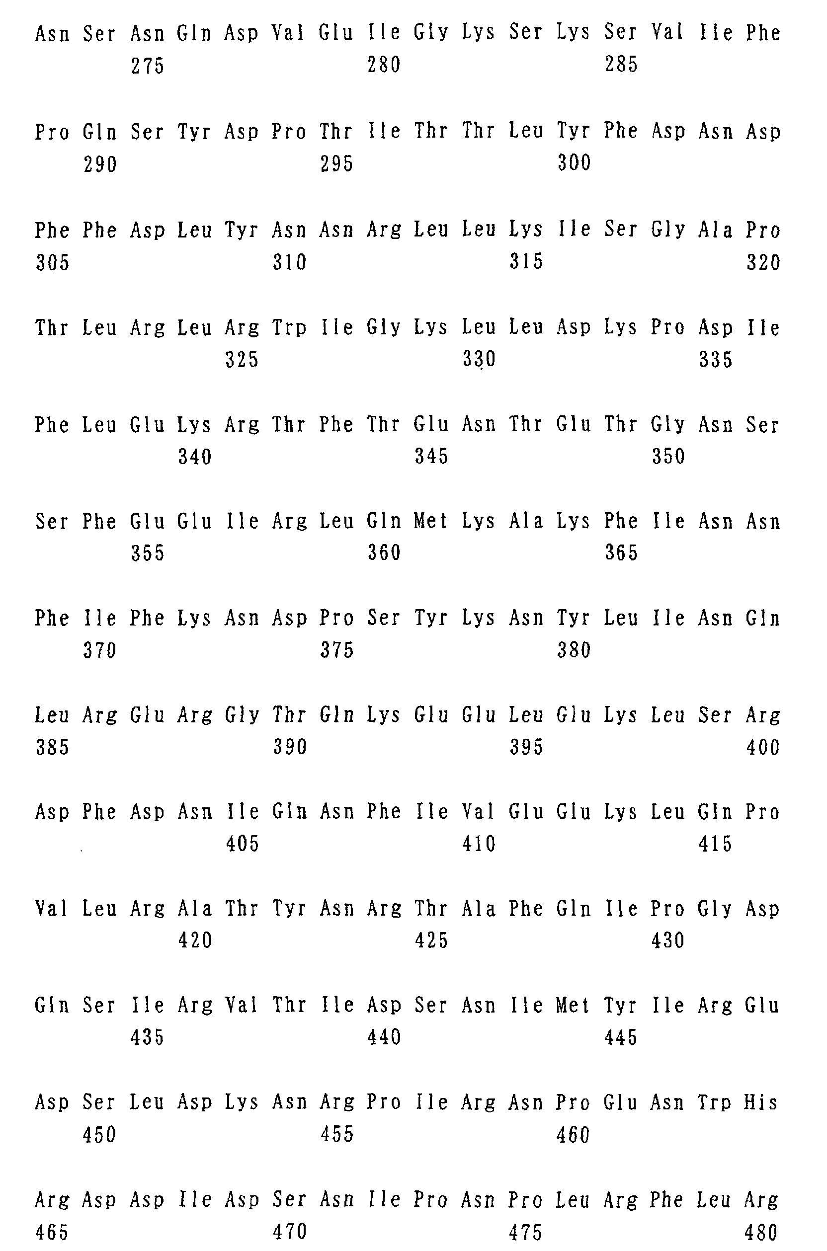
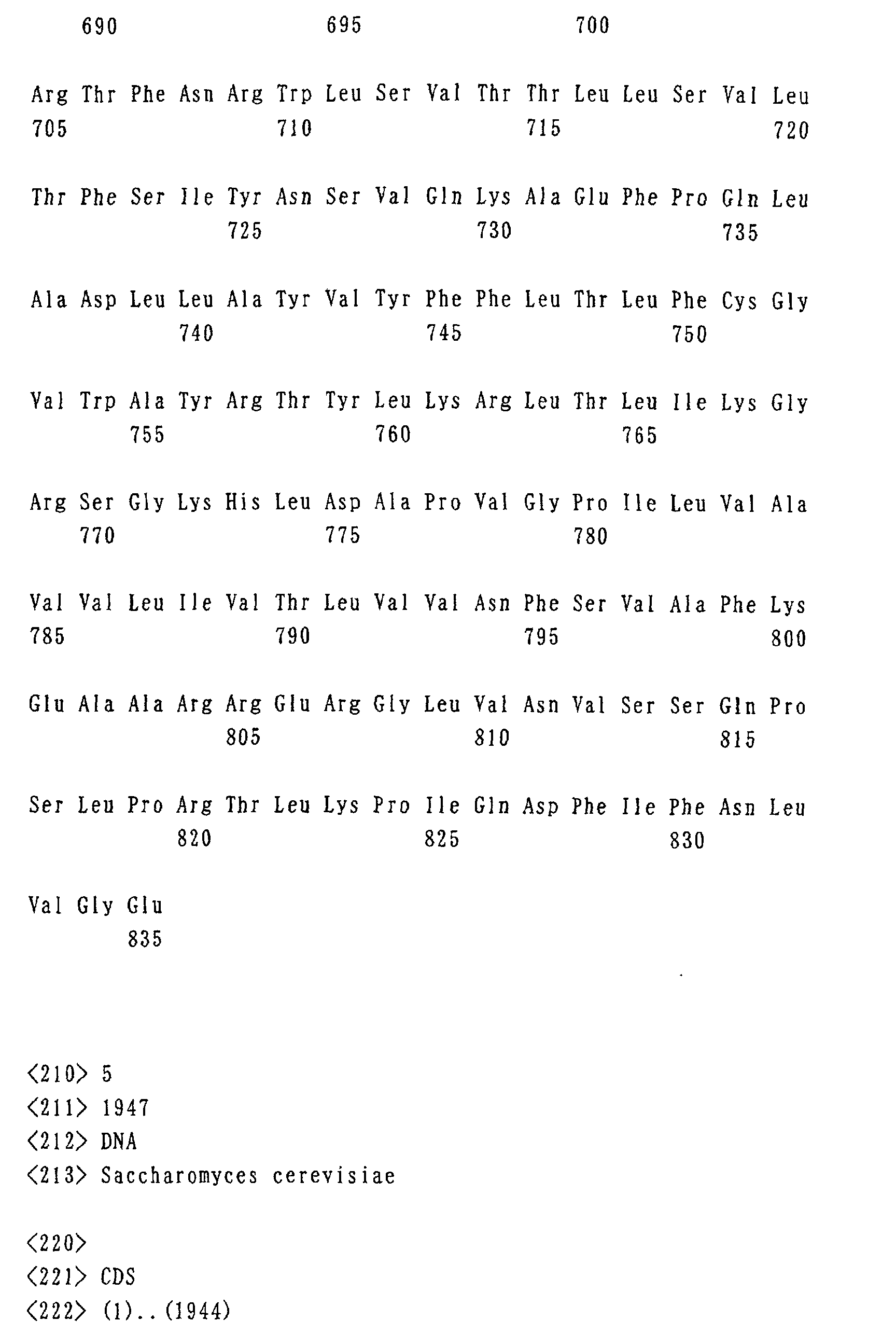
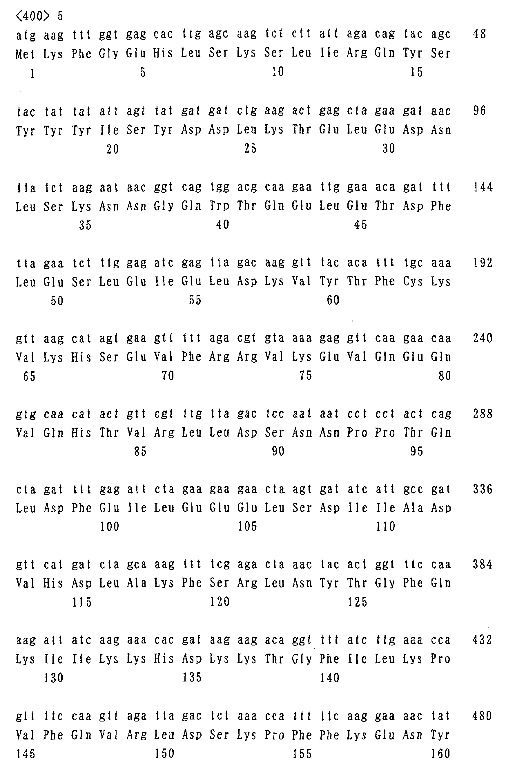
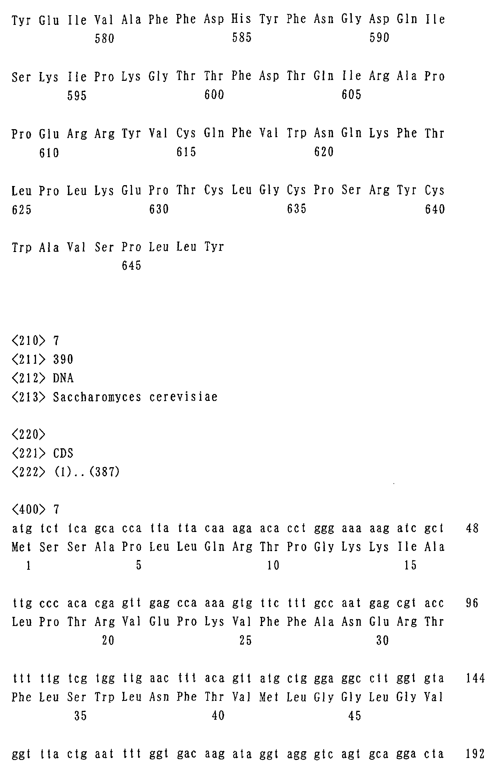
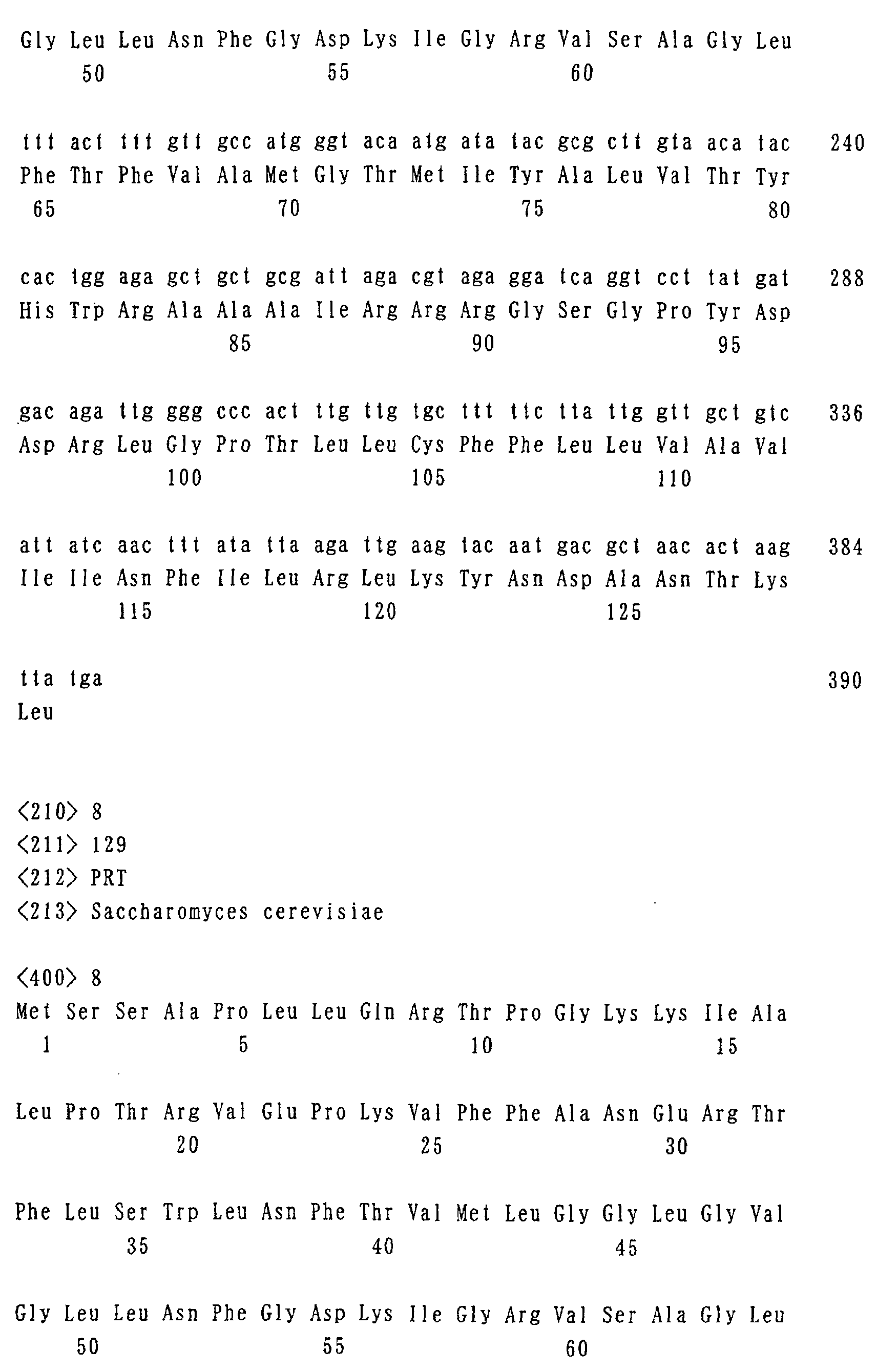
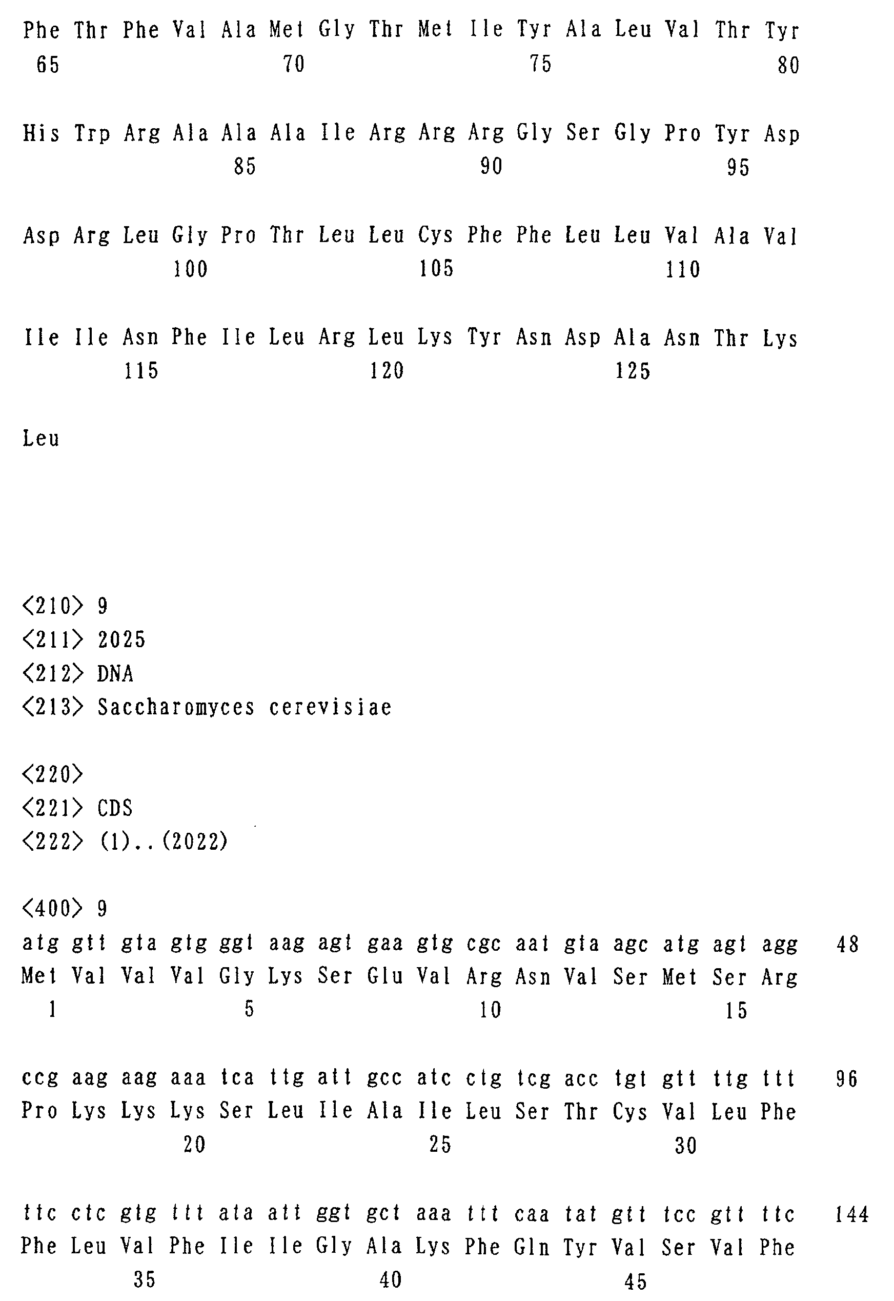
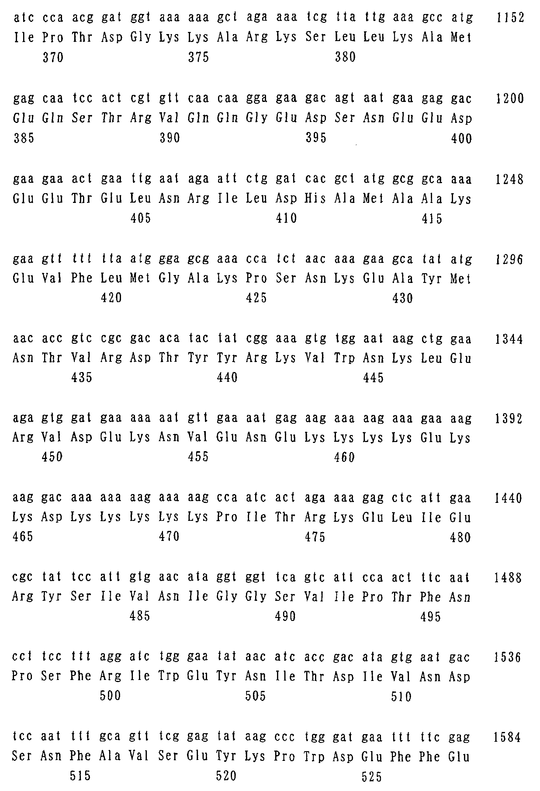
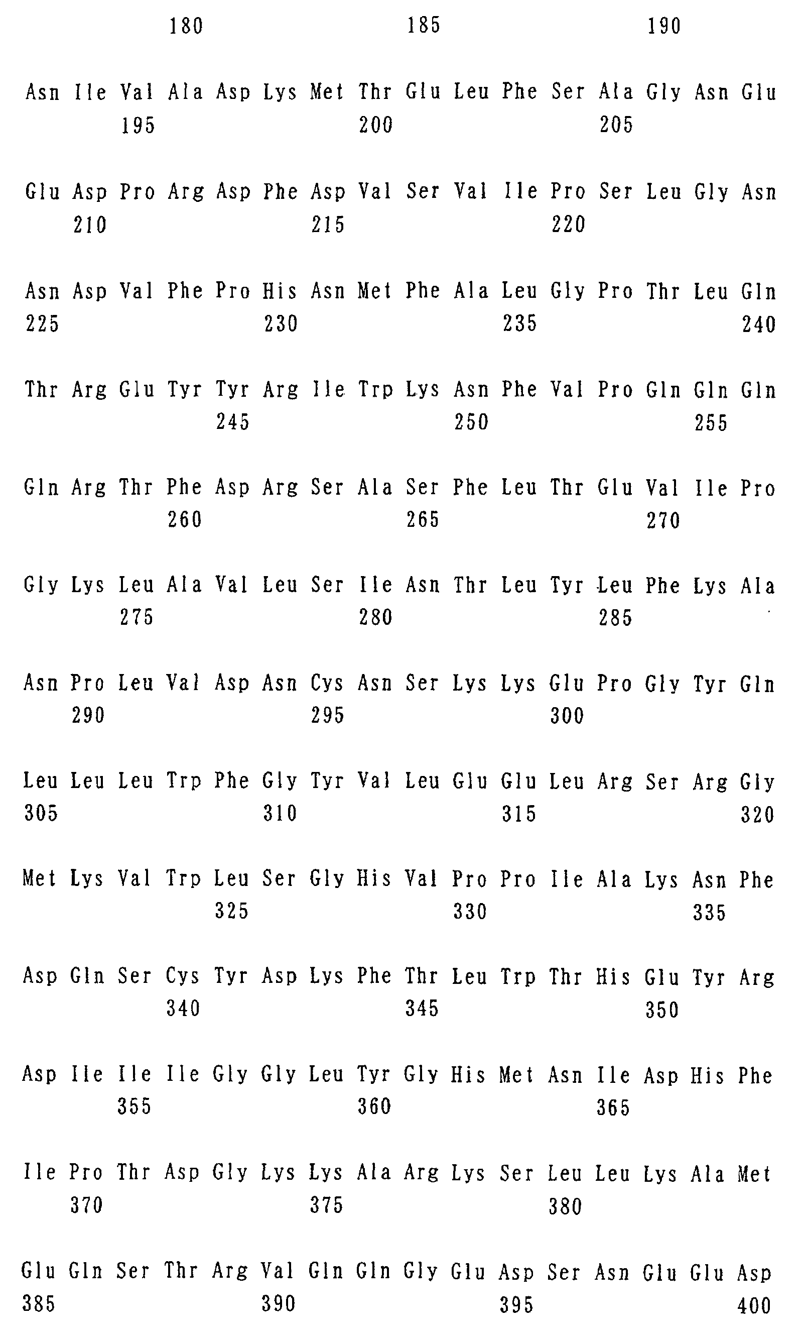
- DNA sequences of PHM 1-5 genes and amino acid sequences of PHM 1-5 are shown by Seq. I.D. Nos. 1-10.
- the polyphosphate synthetases used there is no particular restriction as long as the synthetases can generate polyphosphate intracellularly.
- PPK polyphosphate kinase
- cytochrome b-5 is preferable since its signal is readily measured by NMR.
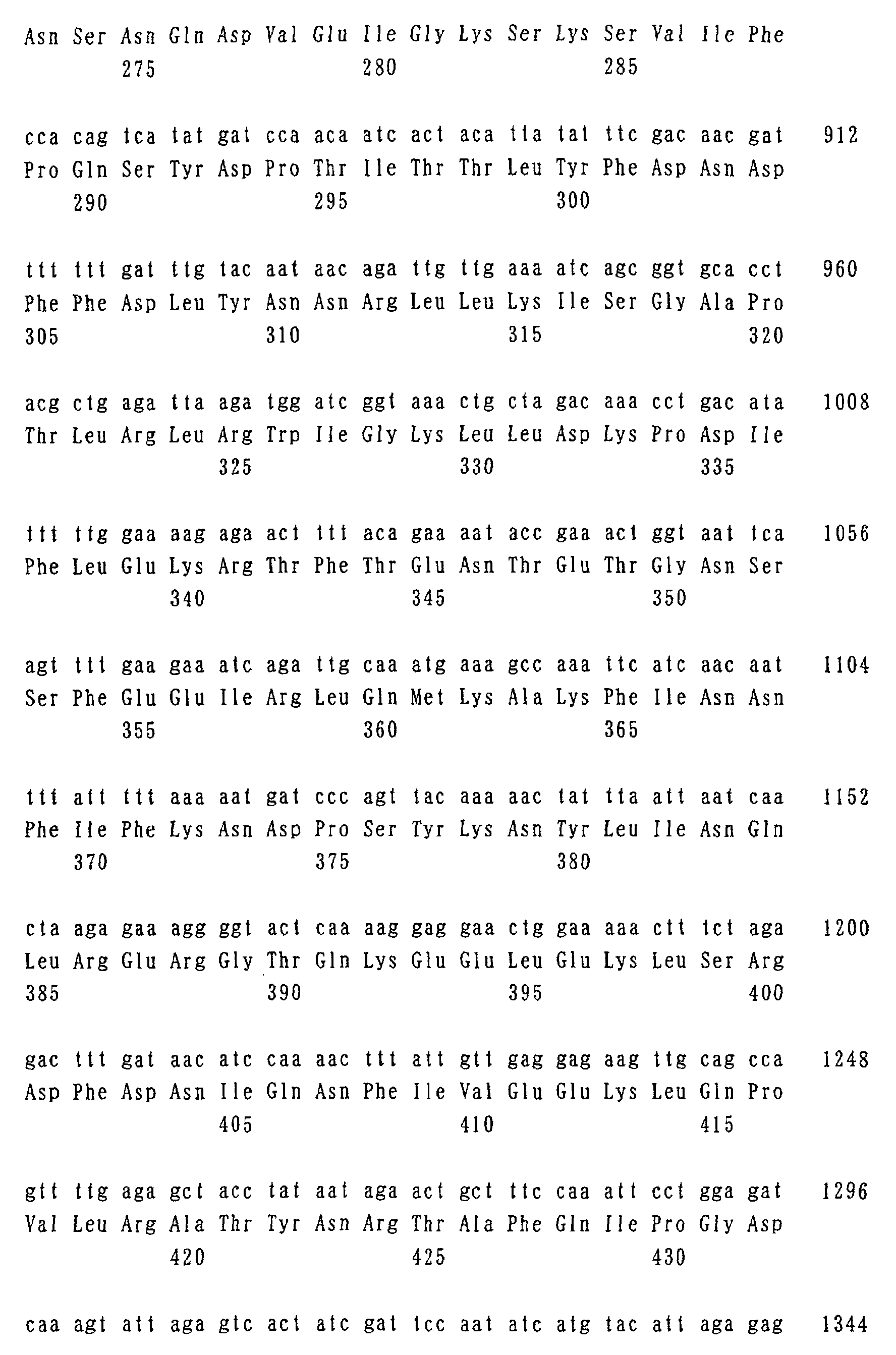
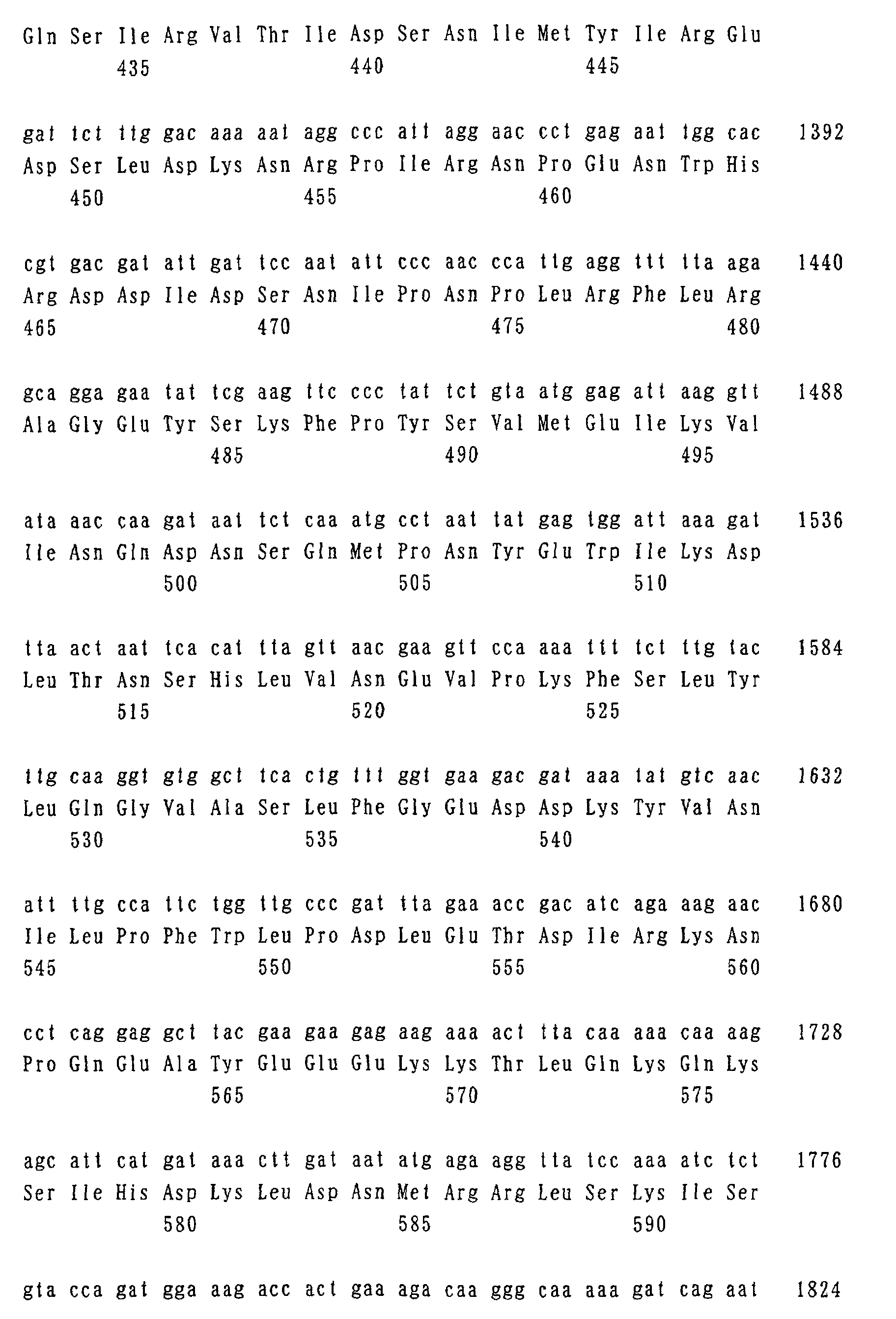
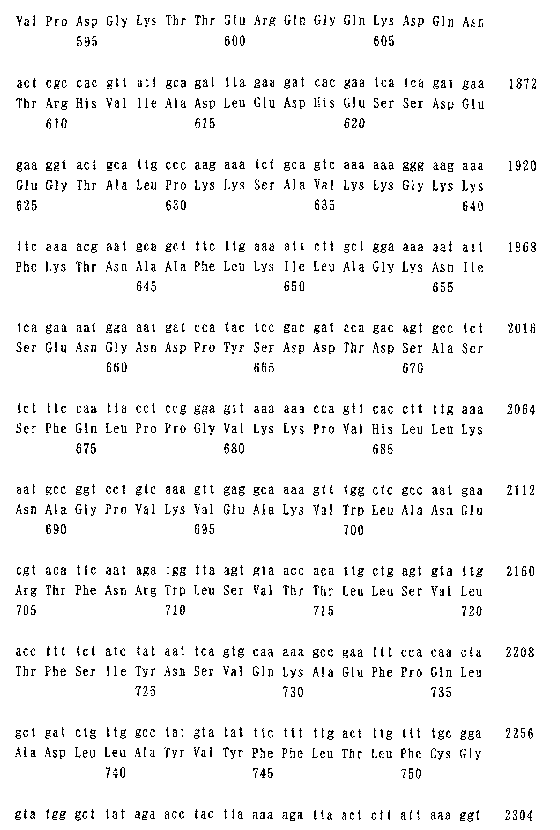
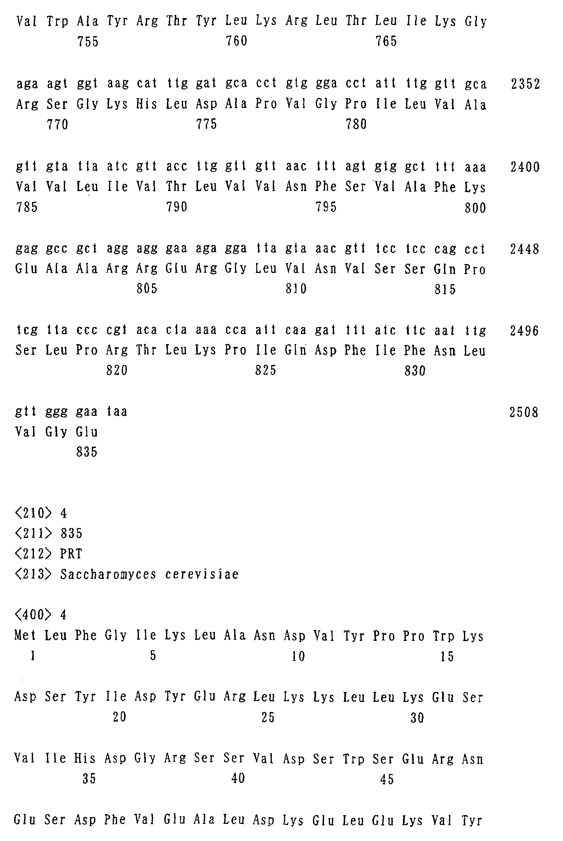
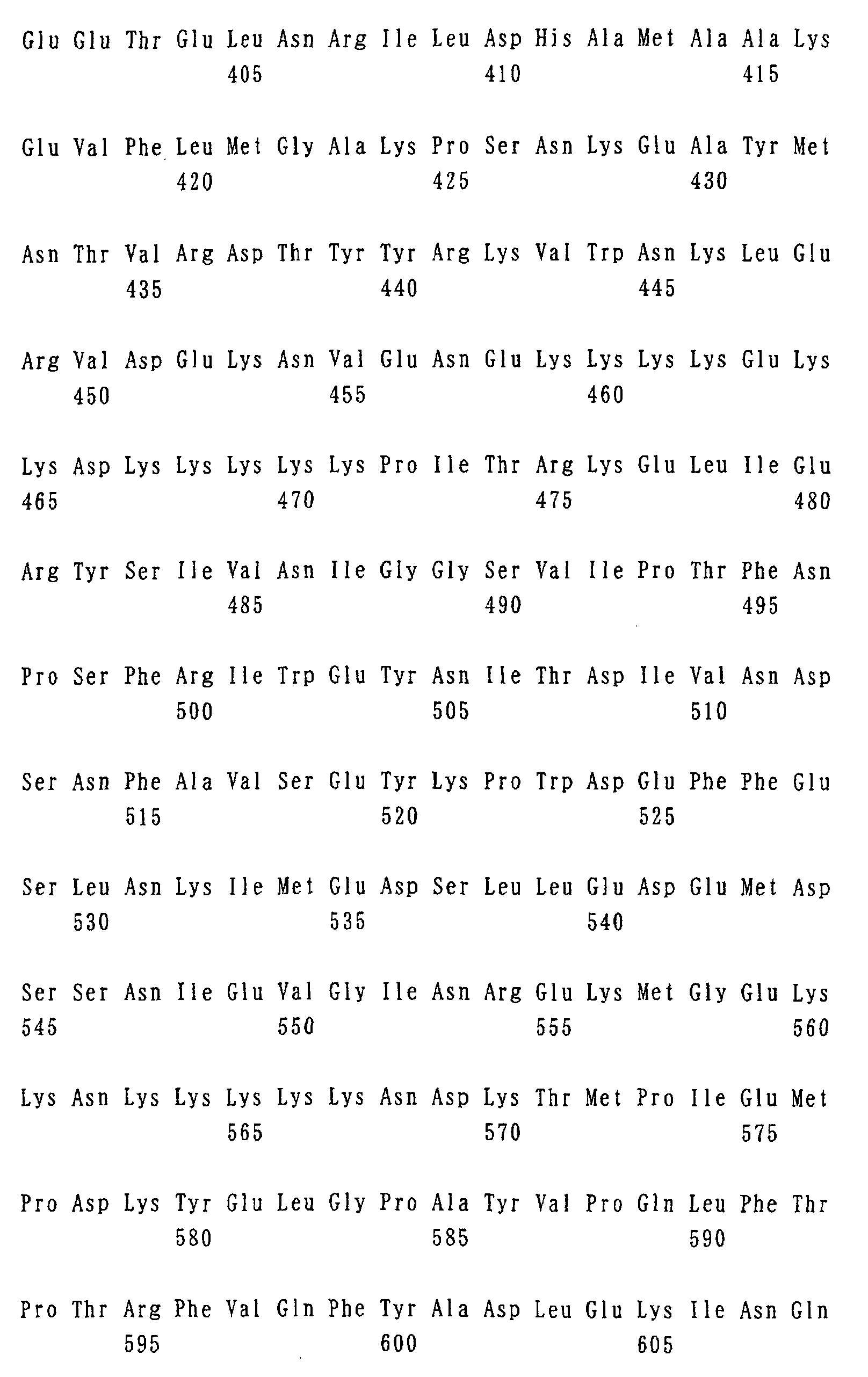
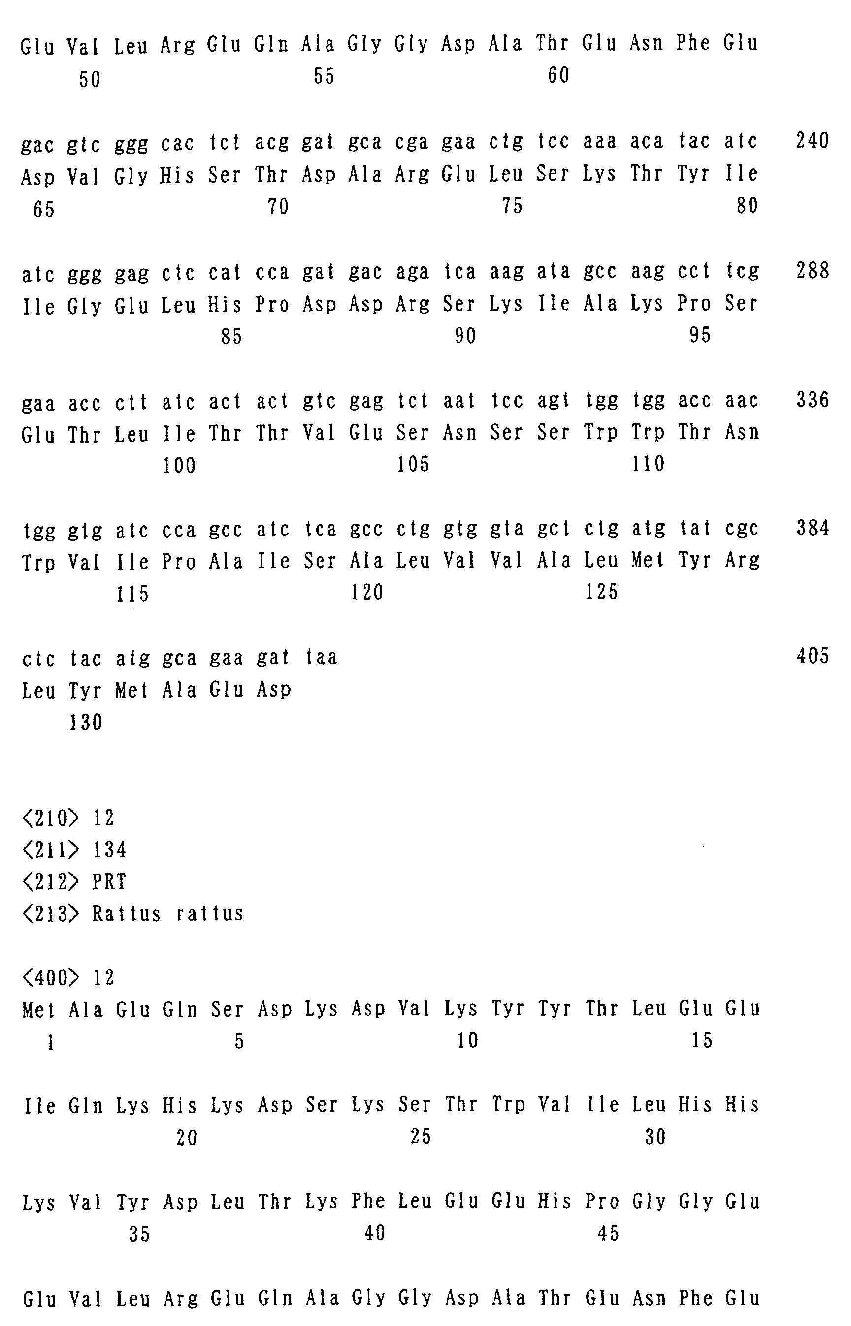
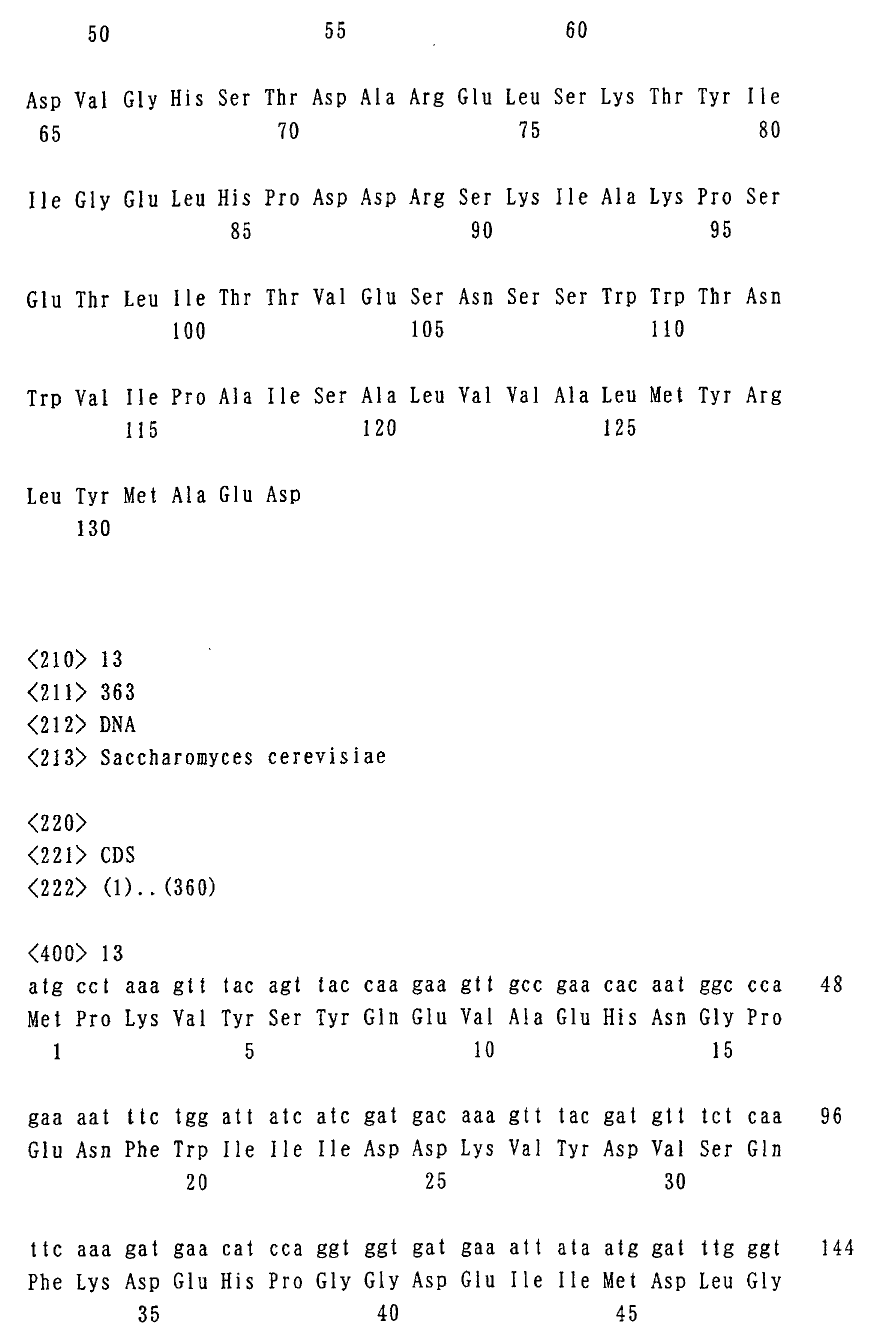
- the DNA sequence of cytochrome b-5 gene from rat, the amino acid sequence of the protein, the DNA sequence of the cytochrome b-5 gene from yeast, and the amino acid sequence of the protein are shown by Seq. I.D. No. 11, Seq. I.D. No. 12, Seq. I.D. No. 13, and Seq. I. D. No. 14, respectively for reference.
- heme proteins having iron atoms such as, ferritin, hemoglobin, catalase, peroxidase and the like can also be used as a reporter gene, placed downstream in-frame with the chosen genes.
- Target genes of general transcription factors such as TFIID subunit (TAF), and the like, i. e. genes which are targeted by general transcription factors and regulated their expression by general transcription factors for instance RPS5 gene, HIS4 gene, TUB2 gene and the like are suitable examples for which expression profiling data may be determined by the invention described.
- TFIID subunit TAF
- the present invention is particularly suitable for real-time non-destructively monitoring of expression of the chosen gene in opaque samples like deep tissues of animals, so as to detect expression level of the chosen genes in cells, tissues or organs.
- the PPK polyphosphate kinase
- the PPK may be placed downstream of the chosen promoters and the amount of polyphosphate accumulated can be quantified by NMR.
- PPK may be used a reporter instead of the aforementioned Saccharomyces cerevisiae PHM genes.
- Escherichia coli PPK can synthesize polyphosphate from ATP in a single step, unlike the synthetic system of eukaryotic cells.
- Other polyphosphate synthetic systems from animals can be used as reporter genes.
- the method for screening various types of agents that regulate the expression of the chosen gene it is not particularly restricted as long as it is a method for screening using the aforementioned method of monitoring gene expression by NMR. For instance, by bringing cells or tissues either expressing or accumulating molecules that vary NMR signals and which are detectable by NMR into contact with a test substance in vitro , quantifying the expression level of the chosen genes by NMR, and comparing/evaluating the expression level with control agents, without test agents, it is possible to screen for substances inhibiting or promoting the expression of the proteins which are translation products of the chosen genes.
- candidate agents therapeutic agents against AIDS, leukemia, viral carcinogenesis such as cancer and the like can be exemplified.
- the NMR apparatus used in the method of monitoring an expression of the chosen gene and the method for screening the various types of agents of the present invention it is not particularly restricted as long as it varies NMR and can quantify the NMR signal to be measured.
- the probe sections will enable high resolution and the bore will be large.
- NMR apparatus with good magnetic field gradient amplifiers will allow the method of the invention to be applied not only to microorganisms but also to small animals and plants.
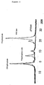
- Wild- type line and phm4 knockout line of Saccharomyces cerevisiae were grown on YPD medium (Bacto yeast extract [1% w/v], Bacto peptone [2% w/v] and glucose [2% w/v]), and phosphate ( 31 P) NMR spectra of the cell population were quantified by using a DRX-500 MHz spectrometer (Bruker). Using literature values, separate signals corresponding to intracellular orthophosphate, and polyphosphate endo and exo groups were identified. The result is shown in Fig. 1. As is apparent from Fig. 1, the most of phosphate was accumulated as polyphoshpate in wild type yeast cells. However, polyphosphate was not present in the phm4 knockout line.
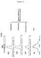
- Fig. 2 After NMR measuring tubes were filled with cells, one-dimensional imaging (one-dimensional profile) by 31 P-NMR was taken. The result is shown in Fig. 2. As is apparent from Fig. 2, the profiles could be obtained by using either signal from polyphosphate endo or exo groups, or from orthophosphate. This indicates that imaging of intracellular polyphosphate is possible. On the other hand, it was revealed that one-dimensional profiles using signals derived from water by the frequently used 1 H (hydrogen nuclei) -MRI technique, cannot distinguish the section filled with cells from the section of suspension.
- Plasmids carrying PHM4 genes downstream from the GAL1 promoter were used to transform the phm4 knockout line. After transformants were selected on a minimal synthetic medium, they were grown in YPG medium (Bacto yeast extract [1% w/v], Bacto peptone [2% w/v] and galactose [2% w/v]), and the expression of PHM4 genes was induced. The amount of polyphosphate after induction was quantified by the same method as that used in Fig. 1. The result is shown in Fig. 3. As is apparent from Fig. 3, a clear signal due to polyphosphate could be observed. Therefore, the activity of GAL1 promoters could be monitored by 31 P-NMR signal of polyphosphate.
- the present invention is a technique by which highly detailed gene expression profiles for analyzing gene function can be obtained safely and conveniently, i.e. a non-destructive and high-resolution versatile visualizing technique, applicable in vivo , and by which the expression level of genes can be monitored in real time, and which can be applied not only to microorganisms and cells but also to deep tissues of mice and the like.
Landscapes
- Chemical & Material Sciences (AREA)
- Life Sciences & Earth Sciences (AREA)
- Organic Chemistry (AREA)
- Health & Medical Sciences (AREA)
- Zoology (AREA)
- Proteomics, Peptides & Aminoacids (AREA)
- Engineering & Computer Science (AREA)
- Wood Science & Technology (AREA)
- Analytical Chemistry (AREA)
- Genetics & Genomics (AREA)
- Microbiology (AREA)
- Molecular Biology (AREA)
- Immunology (AREA)
- Biotechnology (AREA)
- Biophysics (AREA)
- Biochemistry (AREA)
- Bioinformatics & Cheminformatics (AREA)
- General Engineering & Computer Science (AREA)
- General Health & Medical Sciences (AREA)
- Physics & Mathematics (AREA)
- Measuring Or Testing Involving Enzymes Or Micro-Organisms (AREA)
- Investigating Or Analysing Biological Materials (AREA)
Abstract
The present invention provides a technique by which
highly detailed gene expression profiling data for analyzing
the gene function can be obtained safely and conveniently.
It is a versatile non-destructive and high-resolution
visualizing technique by which expression levels of genes
can be monitored in real time, and which can be applied in
vivo and to deep tissues. For instance, plasmids in which
molecules that vary the NMR signal and can be quantified
by PHM genes are incorporated downstream from promoters
responding to the condition of medium, external stimuli and
the like, are introduced into the cells. The level of
polyphosphate in deep tissues and the like can then be
quantified by 31P-NMR, allowing expression profiling of the
promoter, both non-destructively and in real time.
Description
The present invention relates to a non-destructive
method of monitoring gene expression using NMR and a method
for screening various types of agents by using the method
of monitoring.
Understanding at the molecular level what regulates
gene expression in eukaryotes is one of the most important
research projects in contemporary biology. Approximately
20 years have passed since R. Roeder succeeded in
demonstrating in vitro transcription for the first time,
numerous experiments using in vitro transcription systems
have been performed all over the world, and a number of
findings relating to the respective elements including the
purification/cloning of general transcription factor TFIID
by the present inventors have been obtained in the meantime.
However, when these findings are applied to biological
systems, inexplicable results are still frequently observed.
It is presumed that this is not because respective elements
are scattered, but because many elements function together
as a system in vivo. It is thought that in vitro experimental
systems alone cannot model this behavior, and development
of better in vivo experimental systems is essential in order
to appreciate these regulatory systems.
β-galactosidase and luciferase, which require
exogenous substrates, or GFP, which is autofluorescent, have
been developed as reporter genes and they have brought useful
data at various phases conventionally. The applications
for these proteins have been generally limited to transparent
organisms, and they have not been useful in the case of opaque
samples such as deep tissues of animals. On the other hand,
in opaque samples, a method in which transferrin receptor
and β-galactosidase are used as reporter genes for high
sensitivity imaging by magnetic resonance (MRI) of exogenous
genes has been known, but delivery of specific substrate
is necessary in both cases. For instance, the former
requires fusion of microparticles including iron oxide
(called MION) with transferring while the latter requires
a gadolinium-ion complex (called EgadMe) containing a
galactopyranosyl ring. Further, once these substrates are
accumulated in vivo they are not degraded easily, therefore
they are not suitable for monitoring the expression level
of genes in real time. Moreover, it is realistically
impossible to make numerous tissue sections, in order to
examine gene expression of the whole genome (including as
much as 30,000 genes in the case of higher eukaryotes) on
a cellular level at very high resolution, and reconstruct
it on computer. Apart from a few exceptional organisms
having transparent bodies, such as Brachydanio rerio
(zebrafish) and Caenorhabditis elegans, gene expression can
only be examined in detail by preexisting methods such as
in situ hybridization of thin sections. However, as the
expression of most of the genes involved in development of
the central nervous system is switched on or off extremely
rapidly, even analysis with tissue sections is an extremely
difficult task. Accordingly, the technique of whole body
micro-imaging is becoming increasingly important.
Although the genome sequences of many organisms have
now been determined along with the progress of Genome Project,
the expression profile of a gene is crucial in understanding
the gene function. It is not widely known that promoter
region is essential in specifying the temporal and spatial
control of gene transcription, and therefore provides
extremely valuable data for analyzing the function of
proteins encoded downstream. Although it is natural that
several genes relating to the same phenomena are expressed
in the same cells, there are few cases in which these kinds
of data are actually used in discovering the function. This
is because the expression of very few genes has been
determined at a cellular level of detail. Consequently,
it is essential in comprehending the entire biological system
to collect the expression data of as many genes as possible,
and construct the database of expression profile hence.
As previously mentioned, in opaque samples such as
deep tissues of animals, the method of measuring by MRI using
transferrin receptors and β-galactosidase genes is known,
but both reporters require specific substrates, which are
accumulated in vivo and degraded slowly, therefore they are
not suitable for monitoring expression level of gene in real
time, further it is extremely difficult in practice to
distribute substrates to all the cells evenly. Besides,
non-destructive methods of quantifying that can be applied
to deep tissues, µPET (micro-positron emission tomography),
SPECT (single-photon emission computed tomography) and the
like are known, but their spatial resolution is around one
millimetre, and insufficient for visualizing a single cell.
The development of a versatile visualizing technique is
therefore necessary in vivo.
The subject of the present invention is to provide
a technique by which the most important gene expression
profiling data for analyzing gene function can be obtained
safely and conveniently, i. e. a non-destructive and
high-resolution visualization in vivo technique with high
versatility, by which expression levels of genes can be
monitored in real time, and which can be applied to deep
tissues and the like.
Promoter structure is important in determining the
transcription initiation site. When the genes on genomes
are transcribed, it is thought that multistep responses that
cannot be reproduced in vitro, such as structural changes
within the nucleus, chromosome, template DNA and the like
occur successively. In the meantime, though Saccharomyces
cerevisiae is a simple unicellular organism having only 6,000
genes, it comprises transcription machinery that is
approximately equivalent to that of Man, and it is currently
the most attractive model organism to elucidate the detailed
pattern of gene expression from the perspective of data
expression system. A group of genes (PHM genes) relating
to the polyphosphate synthetic pathway was identified in
Saccharomyces cerevisiae recently for the first time as
eukaryotes (Molecular Biology of the Cell, Vol. 11, 4309-4321,
2000).
In Saccharomyces cerevisiae, it is known that along
with the decrease of concentration of phosphate in the medium,
transcription factor PHO4 shifts to a hypophosphorylated
form from a hyperphosphorylated form. As a result, the
intranuclear level of PHO4 increases to activate a set of
target genes relating to phosphate metabolism. Though
polyphosphate synthetases PHM1-4 were isolated as candidates
for target genes of PHO4, analysis showed that these encode
membrane proteins which are very similar to each other, and
the level of polyphosphate decreases in each of the deleted
strains. Accumulation of polyphosphate cannot be seen at
all in phm1Δphm2Δ double mutation line, phm3Δ line and phm4Δ
line, which strongly suggests that PHM1-4 genes encode the
subunits of polyphosphate synthetase, which is thought to
function in vacuoles or on vacuolar membranes. The present
inventors considered that the aforementioned polyphosphate
synthetase (PHM) gene of Saccharomyces cerevisiae might be
able to be used as a reporter gene, by quantifying
polyphosphate, which is known to be significantly
accumulated in all organisms. This quantitation can be
achieved by 31P-NMR directly, or indirectly by measuring the
influence on nearby water of iron ions binding to
polyphosphate by 1H-NMR. Plasmids were constructed
carrying PHM genes downstream of promoters responding to
various conditions (e.g. environmental stimuli), and
introduced into a PHM deleted strain of Saccharomyces
cerevisiae. The intracellular expression level of
polyphosphate was quantified by 31P-NMR and 1H-NMR
non-destructively and in real time, showing that the
intracellular level of polyphosphate in the yeast cells
varies depending on the expression level of the PHM genes.
The present invention has thus completed.
The present invention relates to a method of monitoring
the expression of a chosen gene, wherein accumulation of
a molecule that varies a NMR signal and can be quantified
by NMR, without the requirement to add an exogenous substrate
("1"); the method of monitoring expression of a chosen gene
according to "1", wherein the molecule that varies the NMR
signal and can be quantified by NMR is a polyphosphate ("2");
the method of monitoring expression of a chosen gene according
to "2", wherein the polyphosphate is a polyphosphate
generated by expression of a polyphosphate synthetase gene
placed downstream of the chosen gene ("3"); the method of
monitoring expression of a chosen gene according to "2",
wherein the polyphosphate is a polyphosphate generated by
expression of the polyphosphate synthetase gene placed
downstream of the chosen gene in-frame ("4"); the method
of monitoring expression of a chosen gene according to "1" ,
wherein the molecule that varies the NMR signal and can be
quantified by NMR is a type of cytochrome ("5"); the method
of monitoring expression of a chosen gene according to any
one of "1" to "5", wherein the quantification by NMR is a
non-destructive quantification by NMR ("6"); the method of
monitoring expression of a chosen gene according to any one
of "1" to "6", wherein the expression of the chosen gene
is monitored in real time ("7"); the method of monitoring
expression of a chosen gene according to any one of "1" to
"7", wherein the expression level of the chosen gene within
a cell, a tissue or an organ is detected ("8"); and the
method of monitoring expression of a chosen gene according
to any one of "1" to "8", wherein the chosen gene is a target
gene of a general transcription factor ("9").
The present invention further relates to a method for
screening various types of agents, wherein the method of
monitoring expression of a chosen gene according to any one
of "1" to "9" is used ("10").
The present invention includes any methods of
monitoring in which expression is quantified by NMR and no
exogenous substrate is added to the cells. For this purpose,
molecules that can be detected by NMR through their effects
on the signals can be found from those that absorb
radio-frequency fields because of the presences of magnetic
moments, which are products of nuclear orbital moments and
spin moments, intrinsic to their nucleus. In addition to
poly-phosphates, cytochromes and ferritins, both of which
contain iron-heme groups, are good examples.
As mentioned above, polyphosphate can accumulate in
all organisms in significant amounts and can be quantified
directly by 31P-NMR. Polyphosphate synthetase genes, for
instance PHM genes of Saccharomyces cerevisiae, specifically.
PHM 1-4 genes placed downstream of the chosen gene and
in-frame with it allow transcription to be quantified in
real time. The polyphosphate generated by this method has
a mean length of up to 50 phosphate groups, within the
detectable range by NMR. The 10 mer is thought to show the
highest sensitivity for 31P-NMR. The polymer sizes can be
confirmed by staining the cell contents with toluidine blue
after polyacrylamide gel-electrophoresis. Importantly,
NMR signals derived from polyphosphate can be independently
quantified without interference from signals of nucleic
acids. DNA sequences of PHM 1-5 genes and amino acid
sequences of PHM 1-5 are shown by Seq. I.D. Nos. 1-10. As
for the polyphosphate synthetases used, there is no
particular restriction as long as the synthetases can
generate polyphosphate intracellularly. PPK
(polyphosphate kinase) derived from prokaryotic organisms,
functional homologues, or orthlogues, other than PHM of the
aforementioned Saccharomyces cerevisiae can be used.
As for using cytochromes molecules, in addition to
cytochromes b/c1/c/a3, cytochrome c oxidase, cytochrome P450,
cytochrome b-559, and b-563 can be used but cytochrome b-5
is preferable since its signal is readily measured by NMR.
The DNA sequence of cytochrome b-5 gene from rat, the amino
acid sequence of the protein, the DNA sequence of the
cytochrome b-5 gene from yeast, and the amino acid sequence
of the protein are shown by Seq. I.D. No. 11, Seq. I.D. No.
12, Seq. I.D. No. 13, and Seq. I. D. No. 14, respectively
for reference. Apart from heme proteins having iron atoms
such as, ferritin, hemoglobin, catalase, peroxidase and the
like can also be used as a reporter gene, placed downstream
in-frame with the chosen genes.
As for which genes may be studied by the method of
monitoring described by the present invention, there are
no particular restrictions. Target genes of general
transcription factors such as TFIID subunit (TAF), and the
like, i. e. genes which are targeted by general transcription
factors and regulated their expression by general
transcription factors for instance RPS5 gene, HIS4 gene,
TUB2 gene and the like are suitable examples for which
expression profiling data may be determined by the invention
described.
The present invention is particularly suitable for
real-time non-destructively monitoring of expression of the
chosen gene in opaque samples like deep tissues of animals,
so as to detect expression level of the chosen genes in cells,
tissues or organs. In order to perform monitoring of
expression of the chosen gene according the present invention,
the PPK (polyphosphate kinase) gene from Escherichia coli
may be placed downstream of the chosen promoters and the
amount of polyphosphate accumulated can be quantified by
NMR. Thus PPK may be used a reporter instead of the
aforementioned Saccharomyces cerevisiae PHM genes.
Escherichia coli PPK can synthesize polyphosphate from ATP
in a single step, unlike the synthetic system of eukaryotic
cells. Other polyphosphate synthetic systems from animals
can be used as reporter genes.
As for the method for screening various types of agents
that regulate the expression of the chosen gene, it is not
particularly restricted as long as it is a method for
screening using the aforementioned method of monitoring gene
expression by NMR. For instance, by bringing cells or
tissues either expressing or accumulating molecules that
vary NMR signals and which are detectable by NMR into contact
with a test substance in vitro, quantifying the expression
level of the chosen genes by NMR, and comparing/evaluating
the expression level with control agents, without test agents,
it is possible to screen for substances inhibiting or
promoting the expression of the proteins which are
translation products of the chosen genes. As for such
candidate agents, therapeutic agents against AIDS, leukemia,
viral carcinogenesis such as cancer and the like can be
exemplified.
As for the NMR apparatus used in the method of
monitoring an expression of the chosen gene and the method
for screening the various types of agents of the present
invention, it is not particularly restricted as long as it
varies NMR and can quantify the NMR signal to be measured.
Preferably the probe sections will enable high resolution
and the bore will be large. NMR apparatus with good magnetic
field gradient amplifiers will allow the method of the
invention to be applied not only to microorganisms but also
to small animals and plants.
The present invention will be explained more
specifically with examples below, but the technical scope
of the invention is not limited to these examples.
Wild- type line and phm4 knockout line of Saccharomyces
cerevisiae (S. cerevisiae) were grown on YPD medium (Bacto
yeast extract [1% w/v], Bacto peptone [2% w/v] and glucose
[2% w/v]), and phosphate (31P) NMR spectra of the cell
population were quantified by using a DRX-500 MHz
spectrometer (Bruker). Using literature values, separate
signals corresponding to intracellular orthophosphate, and
polyphosphate endo and exo groups were identified. The
result is shown in Fig. 1. As is apparent from Fig. 1, the
most of phosphate was accumulated as polyphoshpate in wild
type yeast cells. However, polyphosphate was not present
in the phm4 knockout line.
After NMR measuring tubes were filled with cells,
one-dimensional imaging (one-dimensional profile) by
31P-NMR was taken. The result is shown in Fig. 2. As is
apparent from Fig. 2, the profiles could be obtained by using
either signal from polyphosphate endo or exo groups, or from
orthophosphate. This indicates that imaging of
intracellular polyphosphate is possible. On the other hand,
it was revealed that one-dimensional profiles using signals
derived from water by the frequently used 1H (hydrogen nuclei)
-MRI technique, cannot distinguish the section filled with
cells from the section of suspension.
Plasmids carrying PHM4 genes downstream from the GAL1
promoter were used to transform the phm4 knockout line.
After transformants were selected on a minimal synthetic
medium, they were grown in YPG medium (Bacto yeast extract
[1% w/v], Bacto peptone [2% w/v] and galactose [2% w/v]),
and the expression of PHM4 genes was induced. The amount
of polyphosphate after induction was quantified by the same
method as that used in Fig. 1. The result is shown in Fig.
3. As is apparent from Fig. 3, a clear signal due to
polyphosphate could be observed. Therefore, the activity
of GAL1 promoters could be monitored by 31P-NMR signal of
polyphosphate.
Wild type line (Fig. 4, left) and phm4 knockout line
(Fig. 4, right) of Saccharomyces cerevisiae (S. cerevisiae)
were cultured on YPD media with 1 mM of FeCl3, and colonies
were swabbed onto an acrylic plate 3 mm in width and 1 mm
in thickness, placed in NMR tubes and imaging of colonies
from cross section by 1H-NMR was performed. The result is
shown in Fig. 4. These results revealed that equivalent
signals could be detected in both yeast strains by a usual
NMR imaging method (Fig. 4, lower panel). The influence
of iron ions bound to polyphosphate was emphasized by a
modified NMR imaging method; the NMR signals were
significantly attenuated in wild yeast by the iron ions (Fig.
4, upper panel), as a result of imaging the aforementioned
colonies using the specific technique. It is thought to
be possible to delete the signals of wild-type yeast strain
completely by improving the conditions further. The
expression of PHM4 can be monitored very sensitively with
this technique of quantification.
The present invention is a technique by which highly
detailed gene expression profiles for analyzing gene
function can be obtained safely and conveniently, i.e. a
non-destructive and high-resolution versatile visualizing
technique, applicable in vivo, and by which the expression
level of genes can be monitored in real time, and which can
be applied not only to microorganisms and cells but also
to deep tissues of mice and the like.















Claims (10)
- A method of monitoring expression of a chosen gene, wherein accumulation of a molecule that varies a NMR signal and can be quantified by NMR, without the requirement to add an exogenous substrate.
- The method of monitoring an expression of a chosen gene according to claim 1, wherein the molecule that varies the NMR signal and can be quantified by NMR is a polyphosphate.
- The method of monitoring expression of a chosen gene according to claim 2, wherein the polyphosphate is a polyphosphate generated by expression of a polyphosphate synthetase gene placed downstream of the chosen gene.
- The method of monitoring expression of a chosen gene according to claim 2, wherein the polyphosphate is a polyphosphate generated by expression of the polyphosphate synthetase gene placed downstream of the chosen gene in-frame.
- The method of monitoring expression of a chosen gene according to claim 1, wherein the molecule that varies the NMR signal and can be quantified by NMR is a type of cytochrome.
- The method of monitoring expression of a chosen gene according to any one of claims 1 to 5, wherein the quantification by NMR is a non-destructive quantification by NMR.
- The method of monitoring expression of a chosen gene according to any one of claims 1 to 6, wherein the expression of the chosen gene is monitored in real time.
- The method of monitoring expression of a chosen gene according to any one of claims 1 to 7, wherein the expression level of the chosen gene within a cell, a tissue or an organ is detected.
- The method of monitoring expression of a chosen gene according to any one of claims 1 to 8, wherein the chosen gene is a target gene of a general transcription factor.
- A method for screening various types of agents, wherein the method of monitoring expression of a chosen gene according to any one of claims 1 to 9 is used.
Applications Claiming Priority (3)
| Application Number | Priority Date | Filing Date | Title |
|---|---|---|---|
| JP2002002396A JP2003207508A (en) | 2002-01-09 | 2002-01-09 | Gene expression monitoring method |
| JP2002002396 | 2002-01-09 | ||
| PCT/JP2003/000117 WO2003060117A1 (en) | 2002-01-09 | 2003-01-09 | Method of monitoring gene expression |
Publications (2)
| Publication Number | Publication Date |
|---|---|
| EP1473363A1 true EP1473363A1 (en) | 2004-11-03 |
| EP1473363A4 EP1473363A4 (en) | 2005-07-20 |
Family
ID=19190738
Family Applications (1)
| Application Number | Title | Priority Date | Filing Date |
|---|---|---|---|
| EP03700514A Withdrawn EP1473363A4 (en) | 2002-01-09 | 2003-01-09 | METHOD OF MONITORING GENE EXPRESSION |
Country Status (4)
| Country | Link |
|---|---|
| US (1) | US20050244822A1 (en) |
| EP (1) | EP1473363A4 (en) |
| JP (1) | JP2003207508A (en) |
| WO (1) | WO2003060117A1 (en) |
Families Citing this family (1)
| Publication number | Priority date | Publication date | Assignee | Title |
|---|---|---|---|---|
| CN102977206B (en) * | 2012-11-19 | 2014-10-01 | 中国农业科学院生物技术研究所 | Use of cytochrome combined domain protein as secretion assistant factor in improvement of secretory expression amount of foreign gene in pichia pastoris |
Family Cites Families (3)
| Publication number | Priority date | Publication date | Assignee | Title |
|---|---|---|---|---|
| US5703056A (en) * | 1995-03-15 | 1997-12-30 | Sloan-Kettering Institute For Cancer Research | Non-invasive imaging of gene transfer |
| WO1998033809A1 (en) * | 1997-01-31 | 1998-08-06 | The General Hospital Corporation | Compositions and methods for imaging gene expression |
| EP1487499B1 (en) * | 2002-03-07 | 2009-06-10 | Carnegie-Mellon University | Contrast agents for magnetic resonance imaging and methods related thereto |
-
2002
- 2002-01-09 JP JP2002002396A patent/JP2003207508A/en active Pending
-
2003
- 2003-01-09 EP EP03700514A patent/EP1473363A4/en not_active Withdrawn
- 2003-01-09 WO PCT/JP2003/000117 patent/WO2003060117A1/en not_active Ceased
- 2003-01-09 US US10/501,039 patent/US20050244822A1/en not_active Abandoned
Also Published As
| Publication number | Publication date |
|---|---|
| WO2003060117A1 (en) | 2003-07-24 |
| JP2003207508A (en) | 2003-07-25 |
| EP1473363A4 (en) | 2005-07-20 |
| US20050244822A1 (en) | 2005-11-03 |
Similar Documents
| Publication | Publication Date | Title |
|---|---|---|
| Bidlingmaier et al. | The Cbk1p pathway is important for polarized cell growth and cell separation in Saccharomyces cerevisiae | |
| Jeppsson et al. | The maintenance of chromosome structure: positioning and functioning of SMC complexes | |
| Fowlkes et al. | A quantitative spatiotemporal atlas of gene expression in the Drosophila blastoderm | |
| Arora et al. | Antiviral protein Ski8 is a direct partner of Spo11 in meiotic DNA break formation, independent of its cytoplasmic role in RNA metabolism | |
| Schäpe et al. | Influence of lamin A on the mechanical properties of amphibian oocyte nuclei measured by atomic force microscopy | |
| Gregor et al. | Shape and function of the Bicoid morphogen gradient in dipteran species with different sized embryos | |
| CA2185550A1 (en) | An in vivo gene expression system | |
| Anton‐Leberre et al. | Exposure to high static or pulsed magnetic fields does not affect cellular processes in the yeast Saccharomyces cerevisiae | |
| EP2176409B1 (en) | Nucleic acid construct systems capable of diagnosing or treating a cell state | |
| CN104498500B (en) | A kind of aptamer for identifying adriamycin-resistant breast cancer cell and its screening technique and application | |
| CN1726290B (en) | Method for producing genetically modified organisms for screening active substances | |
| KR20210137380A (en) | Phosphatase that modulates thermotolerance for mammalian body temperatures of Cryptococcus neoformans, and method for screening antifungal agents using the same | |
| JP2008507995A (en) | Method for detecting molecular interactions in cells | |
| Yoshida et al. | The Schizosaccharomyces pombe cdt2+ gene, a target of G1-S phase-specific transcription factor complex DSC1, is required for mitotic and premeiotic DNA replication | |
| Hashi et al. | Modifying expression modes of human neurotensin receptor type 1 alters sensing capabilities for agonists in yeast signaling biosensor | |
| US20050244822A1 (en) | Method of monitoring gene expression | |
| Pelet | Nuclear relocation of Kss1 contributes to the specificity of the mating response | |
| Salem et al. | Suppressor analysis of the Saccharomyces cerevisiae gene REC104 reveals a genetic interaction with REC102 | |
| JP2008507265A (en) | Cell lines that inform the cell cycle | |
| Belmont et al. | Inducible control of subcellular RNA localization using a synthetic protein-RNA aptamer interaction | |
| Pastushok et al. | Two Mms2 residues cooperatively interact with ubiquitin and are critical for Lys63 polyubiquitination in vitro and in vivo | |
| Peng et al. | Role of the anillin-like protein in growth of Cryptococcus neoformans at human host temperature | |
| Summers et al. | Use of yeast as a system to study amyloid toxicity | |
| US20140024044A1 (en) | Cell cycle measuring method based on an electrochemical method | |
| Sugihara et al. | Mutation in fission yeast phosphatidylinositol 4-kinase Pik1 is synthetically lethal with defect in telomere protection protein Pot1 |
Legal Events
| Date | Code | Title | Description |
|---|---|---|---|
| PUAI | Public reference made under article 153(3) epc to a published international application that has entered the european phase |
Free format text: ORIGINAL CODE: 0009012 |
|
| 17P | Request for examination filed |
Effective date: 20040805 |
|
| AK | Designated contracting states |
Kind code of ref document: A1 Designated state(s): AT BE BG CH CY CZ DE DK EE ES FI FR GB GR HU IE IT LI LU MC NL PT SE SI SK TR |
|
| A4 | Supplementary search report drawn up and despatched |
Effective date: 20050608 |
|
| 17Q | First examination report despatched |
Effective date: 20050725 |
|
| STAA | Information on the status of an ep patent application or granted ep patent |
Free format text: STATUS: THE APPLICATION IS DEEMED TO BE WITHDRAWN |
|
| 18D | Application deemed to be withdrawn |
Effective date: 20080801 |