EP1453494B1 - Use of a sphingosine derivative for the treatment of microbial infections - Google Patents
Use of a sphingosine derivative for the treatment of microbial infections Download PDFInfo
- Publication number
- EP1453494B1 EP1453494B1 EP02793332A EP02793332A EP1453494B1 EP 1453494 B1 EP1453494 B1 EP 1453494B1 EP 02793332 A EP02793332 A EP 02793332A EP 02793332 A EP02793332 A EP 02793332A EP 1453494 B1 EP1453494 B1 EP 1453494B1
- Authority
- EP
- European Patent Office
- Prior art keywords
- macrophages
- sphingosine
- cells
- infection
- pld
- Prior art date
- Legal status (The legal status is an assumption and is not a legal conclusion. Google has not performed a legal analysis and makes no representation as to the accuracy of the status listed.)
- Expired - Lifetime
Links
- 208000015181 infectious disease Diseases 0.000 title claims abstract description 64
- 238000011282 treatment Methods 0.000 title claims abstract description 23
- 230000000813 microbial effect Effects 0.000 title claims description 7
- 150000003410 sphingosines Chemical class 0.000 title description 15
- 210000002540 macrophage Anatomy 0.000 claims abstract description 128
- WWUZIQQURGPMPG-UHFFFAOYSA-N (-)-D-erythro-Sphingosine Natural products CCCCCCCCCCCCCC=CC(O)C(N)CO WWUZIQQURGPMPG-UHFFFAOYSA-N 0.000 claims abstract description 21
- WWUZIQQURGPMPG-KRWOKUGFSA-N sphingosine Chemical compound CCCCCCCCCCCCC\C=C\[C@@H](O)[C@@H](N)CO WWUZIQQURGPMPG-KRWOKUGFSA-N 0.000 claims abstract description 16
- 238000000034 method Methods 0.000 claims abstract description 15
- 230000003641 microbiacidal effect Effects 0.000 claims abstract description 14
- VDRZDTXJMRRVMF-UONOGXRCSA-N D-erythro-sphingosine Natural products CCCCCCCCCC=C[C@@H](O)[C@@H](N)CO VDRZDTXJMRRVMF-UONOGXRCSA-N 0.000 claims abstract description 13
- 229910019142 PO4 Inorganic materials 0.000 claims abstract description 13
- 239000010452 phosphate Substances 0.000 claims abstract description 13
- -1 sphingosine derivative compounds Chemical class 0.000 claims abstract description 11
- 238000000338 in vitro Methods 0.000 claims abstract description 4
- DUYSYHSSBDVJSM-KRWOKUGFSA-N sphingosine 1-phosphate Chemical compound CCCCCCCCCCCCC\C=C\[C@@H](O)[C@@H](N)COP(O)(O)=O DUYSYHSSBDVJSM-KRWOKUGFSA-N 0.000 claims description 57
- 244000052769 pathogen Species 0.000 claims description 14
- 230000001717 pathogenic effect Effects 0.000 claims description 13
- 230000002519 immonomodulatory effect Effects 0.000 claims description 10
- 238000004519 manufacturing process Methods 0.000 claims description 10
- 239000003814 drug Substances 0.000 claims description 8
- 230000001965 increasing effect Effects 0.000 claims description 4
- 239000001226 triphosphate Substances 0.000 claims description 2
- 235000011178 triphosphate Nutrition 0.000 claims description 2
- 125000002264 triphosphate group Chemical group [H]OP(=O)(O[H])OP(=O)(O[H])OP(=O)(O[H])O* 0.000 claims description 2
- 208000035143 Bacterial infection Diseases 0.000 claims 1
- WWUZIQQURGPMPG-DNWQSSKHSA-N L-threo-Sphingosine Chemical class CCCCCCCCCCCCC\C=C\[C@H](O)[C@@H](N)CO WWUZIQQURGPMPG-DNWQSSKHSA-N 0.000 claims 1
- 208000022362 bacterial infectious disease Diseases 0.000 claims 1
- 102000011420 Phospholipase D Human genes 0.000 abstract description 71
- 108090000553 Phospholipase D Proteins 0.000 abstract description 71
- 241000187479 Mycobacterium tuberculosis Species 0.000 abstract description 60
- 241000894006 Bacteria Species 0.000 abstract description 24
- 230000008782 phagocytosis Effects 0.000 abstract description 13
- 206010057249 Phagocytosis Diseases 0.000 abstract description 12
- 150000001875 compounds Chemical class 0.000 abstract description 10
- 239000008194 pharmaceutical composition Substances 0.000 abstract description 8
- 238000003556 assay Methods 0.000 abstract description 7
- 238000002360 preparation method Methods 0.000 abstract description 5
- 238000006731 degradation reaction Methods 0.000 abstract description 3
- 230000001939 inductive effect Effects 0.000 abstract description 3
- 239000002955 immunomodulating agent Substances 0.000 abstract description 2
- 229940121354 immunomodulator Drugs 0.000 abstract description 2
- 230000001225 therapeutic effect Effects 0.000 abstract description 2
- 238000002405 diagnostic procedure Methods 0.000 abstract 1
- 230000002979 macrophagic effect Effects 0.000 abstract 1
- 229940124561 microbicide Drugs 0.000 abstract 1
- 238000012544 monitoring process Methods 0.000 abstract 1
- 210000004027 cell Anatomy 0.000 description 80
- 230000000694 effects Effects 0.000 description 42
- 210000000680 phagosome Anatomy 0.000 description 34
- LOKCTEFSRHRXRJ-UHFFFAOYSA-I dipotassium trisodium dihydrogen phosphate hydrogen phosphate dichloride Chemical compound P(=O)(O)(O)[O-].[K+].P(=O)(O)([O-])[O-].[Na+].[Na+].[Cl-].[K+].[Cl-].[Na+] LOKCTEFSRHRXRJ-UHFFFAOYSA-I 0.000 description 27
- 239000002953 phosphate buffered saline Substances 0.000 description 27
- LFQSCWFLJHTTHZ-UHFFFAOYSA-N Ethanol Chemical compound CCO LFQSCWFLJHTTHZ-UHFFFAOYSA-N 0.000 description 24
- 241000187480 Mycobacterium smegmatis Species 0.000 description 24
- 238000004458 analytical method Methods 0.000 description 23
- 238000002474 experimental method Methods 0.000 description 21
- 230000004913 activation Effects 0.000 description 20
- 239000002609 medium Substances 0.000 description 17
- 201000008827 tuberculosis Diseases 0.000 description 17
- MHMNJMPURVTYEJ-UHFFFAOYSA-N fluorescein-5-isothiocyanate Chemical compound O1C(=O)C2=CC(N=C=S)=CC=C2C21C1=CC=C(O)C=C1OC1=CC(O)=CC=C21 MHMNJMPURVTYEJ-UHFFFAOYSA-N 0.000 description 15
- 238000011534 incubation Methods 0.000 description 15
- 230000035800 maturation Effects 0.000 description 15
- 230000007246 mechanism Effects 0.000 description 15
- VFNKZQNIXUFLBC-UHFFFAOYSA-N 2',7'-dichlorofluorescein Chemical compound O1C(=O)C2=CC=CC=C2C21C1=CC(Cl)=C(O)C=C1OC1=C2C=C(Cl)C(O)=C1 VFNKZQNIXUFLBC-UHFFFAOYSA-N 0.000 description 14
- 239000000243 solution Substances 0.000 description 13
- 239000000203 mixture Substances 0.000 description 12
- 230000000845 anti-microbial effect Effects 0.000 description 11
- 230000001419 dependent effect Effects 0.000 description 11
- 108090000623 proteins and genes Proteins 0.000 description 11
- IJGRMHOSHXDMSA-UHFFFAOYSA-N Atomic nitrogen Chemical compound N#N IJGRMHOSHXDMSA-UHFFFAOYSA-N 0.000 description 10
- HEDRZPFGACZZDS-UHFFFAOYSA-N Chloroform Chemical compound ClC(Cl)Cl HEDRZPFGACZZDS-UHFFFAOYSA-N 0.000 description 10
- QVGXLLKOCUKJST-UHFFFAOYSA-N atomic oxygen Chemical compound [O] QVGXLLKOCUKJST-UHFFFAOYSA-N 0.000 description 10
- 230000001413 cellular effect Effects 0.000 description 10
- 239000001301 oxygen Substances 0.000 description 10
- 229910052760 oxygen Inorganic materials 0.000 description 10
- 235000018102 proteins Nutrition 0.000 description 10
- 102000004169 proteins and genes Human genes 0.000 description 10
- 239000012048 reactive intermediate Substances 0.000 description 10
- 230000004044 response Effects 0.000 description 10
- 239000000725 suspension Substances 0.000 description 10
- 239000012980 RPMI-1640 medium Substances 0.000 description 9
- 239000007788 liquid Substances 0.000 description 9
- 210000001616 monocyte Anatomy 0.000 description 9
- 102000004190 Enzymes Human genes 0.000 description 8
- 108090000790 Enzymes Proteins 0.000 description 8
- 102100037850 Interferon gamma Human genes 0.000 description 8
- 108010074328 Interferon-gamma Proteins 0.000 description 8
- 241000186359 Mycobacterium Species 0.000 description 8
- 230000001580 bacterial effect Effects 0.000 description 8
- 239000011324 bead Substances 0.000 description 8
- 230000015572 biosynthetic process Effects 0.000 description 8
- 229940088598 enzyme Drugs 0.000 description 8
- 230000003834 intracellular effect Effects 0.000 description 8
- 210000004980 monocyte derived macrophage Anatomy 0.000 description 8
- 208000027531 mycobacterial infectious disease Diseases 0.000 description 8
- 238000000926 separation method Methods 0.000 description 8
- FAPWRFPIFSIZLT-UHFFFAOYSA-M Sodium chloride Chemical compound [Na+].[Cl-] FAPWRFPIFSIZLT-UHFFFAOYSA-M 0.000 description 7
- 229930006000 Sucrose Natural products 0.000 description 7
- CZMRCDWAGMRECN-UGDNZRGBSA-N Sucrose Chemical compound O[C@H]1[C@H](O)[C@@H](CO)O[C@@]1(CO)O[C@@H]1[C@H](O)[C@@H](O)[C@H](O)[C@@H](CO)O1 CZMRCDWAGMRECN-UGDNZRGBSA-N 0.000 description 7
- JCABVIFDXFFRMT-DIPNUNPCSA-N [(2r)-1-[ethoxy(hydroxy)phosphoryl]oxy-3-hexadecanoyloxypropan-2-yl] octadec-9-enoate Chemical compound CCCCCCCCCCCCCCCC(=O)OC[C@H](COP(O)(=O)OCC)OC(=O)CCCCCCCC=CCCCCCCCC JCABVIFDXFFRMT-DIPNUNPCSA-N 0.000 description 7
- 238000000684 flow cytometry Methods 0.000 description 7
- 230000001404 mediated effect Effects 0.000 description 7
- 210000001539 phagocyte Anatomy 0.000 description 7
- 239000005720 sucrose Substances 0.000 description 7
- 241000193830 Bacillus <bacterium> Species 0.000 description 6
- OKKJLVBELUTLKV-UHFFFAOYSA-N Methanol Chemical compound OC OKKJLVBELUTLKV-UHFFFAOYSA-N 0.000 description 6
- 206010062207 Mycobacterial infection Diseases 0.000 description 6
- 239000002253 acid Substances 0.000 description 6
- 238000005119 centrifugation Methods 0.000 description 6
- 230000001332 colony forming effect Effects 0.000 description 6
- RAXXELZNTBOGNW-UHFFFAOYSA-N imidazole Natural products C1=CNC=N1 RAXXELZNTBOGNW-UHFFFAOYSA-N 0.000 description 6
- 230000008569 process Effects 0.000 description 6
- 230000010076 replication Effects 0.000 description 6
- 238000012360 testing method Methods 0.000 description 6
- 230000035899 viability Effects 0.000 description 6
- 230000000844 anti-bacterial effect Effects 0.000 description 5
- 239000000872 buffer Substances 0.000 description 5
- 238000009472 formulation Methods 0.000 description 5
- 230000004927 fusion Effects 0.000 description 5
- 239000000499 gel Substances 0.000 description 5
- 230000004957 immunoregulator effect Effects 0.000 description 5
- 210000003712 lysosome Anatomy 0.000 description 5
- 230000001868 lysosomic effect Effects 0.000 description 5
- 239000012528 membrane Substances 0.000 description 5
- 229910052757 nitrogen Inorganic materials 0.000 description 5
- 210000005259 peripheral blood Anatomy 0.000 description 5
- 239000011886 peripheral blood Substances 0.000 description 5
- 239000012071 phase Substances 0.000 description 5
- 239000001397 quillaja saponaria molina bark Substances 0.000 description 5
- 229930182490 saponin Natural products 0.000 description 5
- 150000007949 saponins Chemical class 0.000 description 5
- 238000001262 western blot Methods 0.000 description 5
- PORPENFLTBBHSG-MGBGTMOVSA-N 1,2-dihexadecanoyl-sn-glycerol-3-phosphate Chemical compound CCCCCCCCCCCCCCCC(=O)OC[C@H](COP(O)(O)=O)OC(=O)CCCCCCCCCCCCCCC PORPENFLTBBHSG-MGBGTMOVSA-N 0.000 description 4
- 108090000258 Cathepsin D Proteins 0.000 description 4
- 102000003908 Cathepsin D Human genes 0.000 description 4
- 241000233866 Fungi Species 0.000 description 4
- ZDXPYRJPNDTMRX-VKHMYHEASA-N L-glutamine Chemical compound OC(=O)[C@@H](N)CCC(N)=O ZDXPYRJPNDTMRX-VKHMYHEASA-N 0.000 description 4
- 229930182816 L-glutamine Natural products 0.000 description 4
- 239000006146 Roswell Park Memorial Institute medium Substances 0.000 description 4
- 239000008186 active pharmaceutical agent Substances 0.000 description 4
- 230000001464 adherent effect Effects 0.000 description 4
- 239000012298 atmosphere Substances 0.000 description 4
- 230000001472 cytotoxic effect Effects 0.000 description 4
- 239000012091 fetal bovine serum Substances 0.000 description 4
- 230000005764 inhibitory process Effects 0.000 description 4
- 239000004816 latex Substances 0.000 description 4
- 229920000126 latex Polymers 0.000 description 4
- 244000005700 microbiome Species 0.000 description 4
- PHEDXBVPIONUQT-RGYGYFBISA-N phorbol 13-acetate 12-myristate Chemical compound C([C@]1(O)C(=O)C(C)=C[C@H]1[C@@]1(O)[C@H](C)[C@H]2OC(=O)CCCCCCCCCCCCC)C(CO)=C[C@H]1[C@H]1[C@]2(OC(C)=O)C1(C)C PHEDXBVPIONUQT-RGYGYFBISA-N 0.000 description 4
- 235000010482 polyoxyethylene sorbitan monooleate Nutrition 0.000 description 4
- 229920000053 polysorbate 80 Polymers 0.000 description 4
- 239000011780 sodium chloride Substances 0.000 description 4
- DAEPDZWVDSPTHF-UHFFFAOYSA-M sodium pyruvate Chemical compound [Na+].CC(=O)C([O-])=O DAEPDZWVDSPTHF-UHFFFAOYSA-M 0.000 description 4
- 230000000638 stimulation Effects 0.000 description 4
- 239000000126 substance Substances 0.000 description 4
- WRIDQFICGBMAFQ-UHFFFAOYSA-N (E)-8-Octadecenoic acid Natural products CCCCCCCCCC=CCCCCCCC(O)=O WRIDQFICGBMAFQ-UHFFFAOYSA-N 0.000 description 3
- LQJBNNIYVWPHFW-UHFFFAOYSA-N 20:1omega9c fatty acid Natural products CCCCCCCCCCC=CCCCCCCCC(O)=O LQJBNNIYVWPHFW-UHFFFAOYSA-N 0.000 description 3
- QSBYPNXLFMSGKH-UHFFFAOYSA-N 9-Heptadecensaeure Natural products CCCCCCCC=CCCCCCCCC(O)=O QSBYPNXLFMSGKH-UHFFFAOYSA-N 0.000 description 3
- QTBSBXVTEAMEQO-UHFFFAOYSA-N Acetic acid Chemical compound CC(O)=O QTBSBXVTEAMEQO-UHFFFAOYSA-N 0.000 description 3
- 108010088751 Albumins Proteins 0.000 description 3
- 102000009027 Albumins Human genes 0.000 description 3
- 108091003079 Bovine Serum Albumin Proteins 0.000 description 3
- 102000016938 Catalase Human genes 0.000 description 3
- 108010053835 Catalase Proteins 0.000 description 3
- WQZGKKKJIJFFOK-GASJEMHNSA-N Glucose Natural products OC[C@H]1OC(O)[C@H](O)[C@@H](O)[C@@H]1O WQZGKKKJIJFFOK-GASJEMHNSA-N 0.000 description 3
- ODKSFYDXXFIFQN-BYPYZUCNSA-N L-arginine Chemical compound OC(=O)[C@@H](N)CCCN=C(N)N ODKSFYDXXFIFQN-BYPYZUCNSA-N 0.000 description 3
- 229930064664 L-arginine Natural products 0.000 description 3
- 235000014852 L-arginine Nutrition 0.000 description 3
- 241001529936 Murinae Species 0.000 description 3
- 239000005642 Oleic acid Substances 0.000 description 3
- ZQPPMHVWECSIRJ-UHFFFAOYSA-N Oleic acid Natural products CCCCCCCCC=CCCCCCCCC(O)=O ZQPPMHVWECSIRJ-UHFFFAOYSA-N 0.000 description 3
- 229930040373 Paraformaldehyde Natural products 0.000 description 3
- 241000700605 Viruses Species 0.000 description 3
- LSUTUUOITDQYNO-UHFFFAOYSA-N calphostin C Chemical compound C=12C3=C4C(CC(C)OC(=O)C=5C=CC=CC=5)=C(OC)C(O)=C(C(C=C5OC)=O)C4=C5C=1C(OC)=CC(=O)C2=C(O)C(OC)=C3CC(C)OC(=O)OC1=CC=C(O)C=C1 LSUTUUOITDQYNO-UHFFFAOYSA-N 0.000 description 3
- 238000006243 chemical reaction Methods 0.000 description 3
- JKNIRLKHOOMGOJ-UHFFFAOYSA-N cladochrome D Natural products COC1=C(CC(C)OC(=O)Oc2ccc(O)cc2)c3c4C(=C(OC)C(=O)c5c(O)cc(OC)c(c45)c6c(OC)cc(O)c(C1=O)c36)CC(C)OC(=O)c7ccc(O)cc7 JKNIRLKHOOMGOJ-UHFFFAOYSA-N 0.000 description 3
- SRJYZPCBWDVSGO-UHFFFAOYSA-N cladochrome E Natural products COC1=CC(O)=C(C(C(OC)=C(CC(C)OC(=O)OC=2C=CC(O)=CC=2)C2=3)=O)C2=C1C1=C(OC)C=C(O)C(C(C=2OC)=O)=C1C=3C=2CC(C)OC(=O)C1=CC=CC=C1 SRJYZPCBWDVSGO-UHFFFAOYSA-N 0.000 description 3
- 238000012258 culturing Methods 0.000 description 3
- 231100000433 cytotoxic Toxicity 0.000 description 3
- 230000007423 decrease Effects 0.000 description 3
- 239000008121 dextrose Substances 0.000 description 3
- BFMYDTVEBKDAKJ-UHFFFAOYSA-L disodium;(2',7'-dibromo-3',6'-dioxido-3-oxospiro[2-benzofuran-1,9'-xanthene]-4'-yl)mercury;hydrate Chemical compound O.[Na+].[Na+].O1C(=O)C2=CC=CC=C2C21C1=CC(Br)=C([O-])C([Hg])=C1OC1=C2C=C(Br)C([O-])=C1 BFMYDTVEBKDAKJ-UHFFFAOYSA-L 0.000 description 3
- 231100000673 dose–response relationship Toxicity 0.000 description 3
- 239000000839 emulsion Substances 0.000 description 3
- GNBHRKFJIUUOQI-UHFFFAOYSA-N fluorescein Chemical compound O1C(=O)C2=CC=CC=C2C21C1=CC=C(O)C=C1OC1=CC(O)=CC=C21 GNBHRKFJIUUOQI-UHFFFAOYSA-N 0.000 description 3
- 238000000799 fluorescence microscopy Methods 0.000 description 3
- 230000012010 growth Effects 0.000 description 3
- 230000006698 induction Effects 0.000 description 3
- QXJSBBXBKPUZAA-UHFFFAOYSA-N isooleic acid Natural products CCCCCCCC=CCCCCCCCCC(O)=O QXJSBBXBKPUZAA-UHFFFAOYSA-N 0.000 description 3
- 150000002632 lipids Chemical class 0.000 description 3
- 239000000463 material Substances 0.000 description 3
- ZQPPMHVWECSIRJ-KTKRTIGZSA-N oleic acid Chemical compound CCCCCCCC\C=C/CCCCCCCC(O)=O ZQPPMHVWECSIRJ-KTKRTIGZSA-N 0.000 description 3
- 229920002866 paraformaldehyde Polymers 0.000 description 3
- 244000045947 parasite Species 0.000 description 3
- 239000002245 particle Substances 0.000 description 3
- 239000000546 pharmaceutical excipient Substances 0.000 description 3
- 150000003904 phospholipids Chemical class 0.000 description 3
- 230000009467 reduction Effects 0.000 description 3
- 238000002415 sodium dodecyl sulfate polyacrylamide gel electrophoresis Methods 0.000 description 3
- 239000008247 solid mixture Substances 0.000 description 3
- 238000007619 statistical method Methods 0.000 description 3
- 238000005406 washing Methods 0.000 description 3
- XLYOFNOQVPJJNP-UHFFFAOYSA-N water Substances O XLYOFNOQVPJJNP-UHFFFAOYSA-N 0.000 description 3
- XDFNWJDGWJVGGN-UHFFFAOYSA-N 2-(2,7-dichloro-3,6-dihydroxy-9h-xanthen-9-yl)benzoic acid Chemical compound OC(=O)C1=CC=CC=C1C1C2=CC(Cl)=C(O)C=C2OC2=CC(O)=C(Cl)C=C21 XDFNWJDGWJVGGN-UHFFFAOYSA-N 0.000 description 2
- JKMHFZQWWAIEOD-UHFFFAOYSA-N 2-[4-(2-hydroxyethyl)piperazin-1-yl]ethanesulfonic acid Chemical compound OCC[NH+]1CCN(CCS([O-])(=O)=O)CC1 JKMHFZQWWAIEOD-UHFFFAOYSA-N 0.000 description 2
- HRPVXLWXLXDGHG-UHFFFAOYSA-N Acrylamide Chemical compound NC(=O)C=C HRPVXLWXLXDGHG-UHFFFAOYSA-N 0.000 description 2
- 102000000989 Complement System Proteins Human genes 0.000 description 2
- 108010069112 Complement System Proteins Proteins 0.000 description 2
- CEAZRRDELHUEMR-URQXQFDESA-N Gentamicin Chemical compound O1[C@H](C(C)NC)CC[C@@H](N)[C@H]1O[C@H]1[C@H](O)[C@@H](O[C@@H]2[C@@H]([C@@H](NC)[C@@](C)(O)CO2)O)[C@H](N)C[C@@H]1N CEAZRRDELHUEMR-URQXQFDESA-N 0.000 description 2
- 229930182566 Gentamicin Natural products 0.000 description 2
- 208000031998 Mycobacterium Infections Diseases 0.000 description 2
- 229920001213 Polysorbate 20 Polymers 0.000 description 2
- 239000000654 additive Substances 0.000 description 2
- 230000015556 catabolic process Effects 0.000 description 2
- 239000003153 chemical reaction reagent Substances 0.000 description 2
- 239000003795 chemical substances by application Substances 0.000 description 2
- 238000010835 comparative analysis Methods 0.000 description 2
- 239000000470 constituent Substances 0.000 description 2
- 235000014113 dietary fatty acids Nutrition 0.000 description 2
- 229940079593 drug Drugs 0.000 description 2
- 239000002158 endotoxin Substances 0.000 description 2
- 230000002255 enzymatic effect Effects 0.000 description 2
- 239000003797 essential amino acid Substances 0.000 description 2
- 235000020776 essential amino acid Nutrition 0.000 description 2
- 239000000194 fatty acid Substances 0.000 description 2
- 229930195729 fatty acid Natural products 0.000 description 2
- 150000004665 fatty acids Chemical class 0.000 description 2
- 229960002518 gentamicin Drugs 0.000 description 2
- 239000008187 granular material Substances 0.000 description 2
- 239000003102 growth factor Substances 0.000 description 2
- 239000001963 growth medium Substances 0.000 description 2
- 230000003301 hydrolyzing effect Effects 0.000 description 2
- 230000036039 immunity Effects 0.000 description 2
- 230000002401 inhibitory effect Effects 0.000 description 2
- 230000003993 interaction Effects 0.000 description 2
- 239000000543 intermediate Substances 0.000 description 2
- 208000032839 leukemia Diseases 0.000 description 2
- 229920006008 lipopolysaccharide Polymers 0.000 description 2
- 239000002502 liposome Substances 0.000 description 2
- 210000004698 lymphocyte Anatomy 0.000 description 2
- 239000006166 lysate Substances 0.000 description 2
- 238000000386 microscopy Methods 0.000 description 2
- 210000005087 mononuclear cell Anatomy 0.000 description 2
- 230000003406 mycobactericidal effect Effects 0.000 description 2
- 230000007935 neutral effect Effects 0.000 description 2
- 230000020477 pH reduction Effects 0.000 description 2
- 230000007918 pathogenicity Effects 0.000 description 2
- WTJKGGKOPKCXLL-RRHRGVEJSA-N phosphatidylcholine Chemical compound CCCCCCCCCCCCCCCC(=O)OC[C@H](COP([O-])(=O)OCC[N+](C)(C)C)OC(=O)CCCCCCCC=CCCCCCCCC WTJKGGKOPKCXLL-RRHRGVEJSA-N 0.000 description 2
- 239000000256 polyoxyethylene sorbitan monolaurate Substances 0.000 description 2
- 235000010486 polyoxyethylene sorbitan monolaurate Nutrition 0.000 description 2
- 239000000047 product Substances 0.000 description 2
- 230000000750 progressive effect Effects 0.000 description 2
- 238000000746 purification Methods 0.000 description 2
- 108020003175 receptors Proteins 0.000 description 2
- 229940054269 sodium pyruvate Drugs 0.000 description 2
- 239000007787 solid Substances 0.000 description 2
- 239000002904 solvent Substances 0.000 description 2
- 239000006228 supernatant Substances 0.000 description 2
- ZIIUUSVHCHPIQD-UHFFFAOYSA-N 2,4,6-trimethyl-N-[3-(trifluoromethyl)phenyl]benzenesulfonamide Chemical compound CC1=CC(C)=CC(C)=C1S(=O)(=O)NC1=CC=CC(C(F)(F)F)=C1 ZIIUUSVHCHPIQD-UHFFFAOYSA-N 0.000 description 1
- ZCYVEMRRCGMTRW-UHFFFAOYSA-N 7553-56-2 Chemical compound [I] ZCYVEMRRCGMTRW-UHFFFAOYSA-N 0.000 description 1
- 208000030507 AIDS Diseases 0.000 description 1
- 229920001817 Agar Polymers 0.000 description 1
- 108090000695 Cytokines Proteins 0.000 description 1
- 102000004127 Cytokines Human genes 0.000 description 1
- KCXVZYZYPLLWCC-UHFFFAOYSA-N EDTA Chemical compound OC(=O)CN(CC(O)=O)CCN(CC(O)=O)CC(O)=O KCXVZYZYPLLWCC-UHFFFAOYSA-N 0.000 description 1
- ULGZDMOVFRHVEP-RWJQBGPGSA-N Erythromycin Chemical compound O([C@@H]1[C@@H](C)C(=O)O[C@@H]([C@@]([C@H](O)[C@@H](C)C(=O)[C@H](C)C[C@@](C)(O)[C@H](O[C@H]2[C@@H]([C@H](C[C@@H](C)O2)N(C)C)O)[C@H]1C)(C)O)CC)[C@H]1C[C@@](C)(OC)[C@@H](O)[C@H](C)O1 ULGZDMOVFRHVEP-RWJQBGPGSA-N 0.000 description 1
- WSFSSNUMVMOOMR-UHFFFAOYSA-N Formaldehyde Chemical compound O=C WSFSSNUMVMOOMR-UHFFFAOYSA-N 0.000 description 1
- 101000946889 Homo sapiens Monocyte differentiation antigen CD14 Proteins 0.000 description 1
- 206010020751 Hypersensitivity Diseases 0.000 description 1
- 108010044467 Isoenzymes Proteins 0.000 description 1
- NHTMVDHEPJAVLT-UHFFFAOYSA-N Isooctane Chemical compound CC(C)CC(C)(C)C NHTMVDHEPJAVLT-UHFFFAOYSA-N 0.000 description 1
- 108010063045 Lactoferrin Proteins 0.000 description 1
- 102000010445 Lactoferrin Human genes 0.000 description 1
- 241000186779 Listeria monocytogenes Species 0.000 description 1
- 208000032376 Lung infection Diseases 0.000 description 1
- 102000014944 Lysosome-Associated Membrane Glycoproteins Human genes 0.000 description 1
- 108010064171 Lysosome-Associated Membrane Glycoproteins Proteins 0.000 description 1
- 102000012750 Membrane Glycoproteins Human genes 0.000 description 1
- 108010090054 Membrane Glycoproteins Proteins 0.000 description 1
- 241001465754 Metazoa Species 0.000 description 1
- 102100035877 Monocyte differentiation antigen CD14 Human genes 0.000 description 1
- 102000016943 Muramidase Human genes 0.000 description 1
- 108010014251 Muramidase Proteins 0.000 description 1
- 241001646725 Mycobacterium tuberculosis H37Rv Species 0.000 description 1
- 102000003896 Myeloperoxidases Human genes 0.000 description 1
- 108090000235 Myeloperoxidases Proteins 0.000 description 1
- 108010062010 N-Acetylmuramoyl-L-alanine Amidase Proteins 0.000 description 1
- 102000008299 Nitric Oxide Synthase Human genes 0.000 description 1
- 108010021487 Nitric Oxide Synthase Proteins 0.000 description 1
- 239000000020 Nitrocellulose Substances 0.000 description 1
- 102000003992 Peroxidases Human genes 0.000 description 1
- 102000015439 Phospholipases Human genes 0.000 description 1
- 108010064785 Phospholipases Proteins 0.000 description 1
- 239000004793 Polystyrene Substances 0.000 description 1
- XBDQKXXYIPTUBI-UHFFFAOYSA-M Propionate Chemical compound CCC([O-])=O XBDQKXXYIPTUBI-UHFFFAOYSA-M 0.000 description 1
- 240000004808 Saccharomyces cerevisiae Species 0.000 description 1
- 210000001744 T-lymphocyte Anatomy 0.000 description 1
- 229920000392 Zymosan Polymers 0.000 description 1
- KXKVLQRXCPHEJC-UHFFFAOYSA-N acetic acid trimethyl ester Natural products COC(C)=O KXKVLQRXCPHEJC-UHFFFAOYSA-N 0.000 description 1
- 239000013543 active substance Substances 0.000 description 1
- 230000000996 additive effect Effects 0.000 description 1
- 239000002671 adjuvant Substances 0.000 description 1
- 239000000443 aerosol Substances 0.000 description 1
- 239000008272 agar Substances 0.000 description 1
- 238000013019 agitation Methods 0.000 description 1
- 230000007815 allergy Effects 0.000 description 1
- 150000001414 amino alcohols Chemical class 0.000 description 1
- 230000001355 anti-mycobacterial effect Effects 0.000 description 1
- 239000004599 antimicrobial Substances 0.000 description 1
- 239000003963 antioxidant agent Substances 0.000 description 1
- 210000003719 b-lymphocyte Anatomy 0.000 description 1
- 244000052616 bacterial pathogen Species 0.000 description 1
- 230000003115 biocidal effect Effects 0.000 description 1
- 210000004369 blood Anatomy 0.000 description 1
- 239000008280 blood Substances 0.000 description 1
- 230000003139 buffering effect Effects 0.000 description 1
- 229930194791 calphostin Natural products 0.000 description 1
- 230000003197 catalytic effect Effects 0.000 description 1
- 125000002091 cationic group Chemical group 0.000 description 1
- 230000008809 cell oxidative stress Effects 0.000 description 1
- 230000019522 cellular metabolic process Effects 0.000 description 1
- 238000012512 characterization method Methods 0.000 description 1
- 239000011248 coating agent Substances 0.000 description 1
- 238000000576 coating method Methods 0.000 description 1
- 230000000295 complement effect Effects 0.000 description 1
- 239000012141 concentrate Substances 0.000 description 1
- 230000021615 conjugation Effects 0.000 description 1
- 210000000805 cytoplasm Anatomy 0.000 description 1
- 230000006378 damage Effects 0.000 description 1
- 238000001514 detection method Methods 0.000 description 1
- 238000009792 diffusion process Methods 0.000 description 1
- JVSWJIKNEAIKJW-UHFFFAOYSA-N dimethyl-hexane Natural products CCCCCC(C)C JVSWJIKNEAIKJW-UHFFFAOYSA-N 0.000 description 1
- 239000001177 diphosphate Substances 0.000 description 1
- XPPKVPWEQAFLFU-UHFFFAOYSA-J diphosphate(4-) Chemical class [O-]P([O-])(=O)OP([O-])([O-])=O XPPKVPWEQAFLFU-UHFFFAOYSA-J 0.000 description 1
- 235000011180 diphosphates Nutrition 0.000 description 1
- 201000010099 disease Diseases 0.000 description 1
- 208000037265 diseases, disorders, signs and symptoms Diseases 0.000 description 1
- 239000003937 drug carrier Substances 0.000 description 1
- 238000001962 electrophoresis Methods 0.000 description 1
- 230000008030 elimination Effects 0.000 description 1
- 238000003379 elimination reaction Methods 0.000 description 1
- 230000007613 environmental effect Effects 0.000 description 1
- 238000011156 evaluation Methods 0.000 description 1
- 230000028023 exocytosis Effects 0.000 description 1
- 238000000605 extraction Methods 0.000 description 1
- 210000002950 fibroblast Anatomy 0.000 description 1
- 238000012921 fluorescence analysis Methods 0.000 description 1
- 230000037406 food intake Effects 0.000 description 1
- 230000008014 freezing Effects 0.000 description 1
- 238000007710 freezing Methods 0.000 description 1
- CEAZRRDELHUEMR-UHFFFAOYSA-N gentamicin Chemical compound O1C(C(C)NC)CCC(N)C1OC1C(O)C(OC2C(C(NC)C(C)(O)CO2)O)C(N)CC1N CEAZRRDELHUEMR-UHFFFAOYSA-N 0.000 description 1
- 239000011521 glass Substances 0.000 description 1
- 238000001033 granulometry Methods 0.000 description 1
- 239000005556 hormone Substances 0.000 description 1
- 229940088597 hormone Drugs 0.000 description 1
- 230000007062 hydrolysis Effects 0.000 description 1
- 238000006460 hydrolysis reaction Methods 0.000 description 1
- 238000003384 imaging method Methods 0.000 description 1
- 230000008105 immune reaction Effects 0.000 description 1
- 238000003119 immunoblot Methods 0.000 description 1
- 230000001524 infective effect Effects 0.000 description 1
- 239000003112 inhibitor Substances 0.000 description 1
- 230000015788 innate immune response Effects 0.000 description 1
- 210000005007 innate immune system Anatomy 0.000 description 1
- 229910052740 iodine Inorganic materials 0.000 description 1
- 239000011630 iodine Substances 0.000 description 1
- CSSYQJWUGATIHM-IKGCZBKSSA-N l-phenylalanyl-l-lysyl-l-cysteinyl-l-arginyl-l-arginyl-l-tryptophyl-l-glutaminyl-l-tryptophyl-l-arginyl-l-methionyl-l-lysyl-l-lysyl-l-leucylglycyl-l-alanyl-l-prolyl-l-seryl-l-isoleucyl-l-threonyl-l-cysteinyl-l-valyl-l-arginyl-l-arginyl-l-alanyl-l-phenylal Chemical compound C([C@H](N)C(=O)N[C@@H](CCCCN)C(=O)N[C@@H](CS)C(=O)N[C@@H](CCCNC(N)=N)C(=O)N[C@@H](CCCNC(N)=N)C(=O)N[C@@H](CC=1C2=CC=CC=C2NC=1)C(=O)N[C@@H](CCC(N)=O)C(=O)N[C@@H](CC=1C2=CC=CC=C2NC=1)C(=O)N[C@@H](CCCNC(N)=N)C(=O)N[C@@H](CCSC)C(=O)N[C@@H](CCCCN)C(=O)N[C@@H](CCCCN)C(=O)N[C@@H](CC(C)C)C(=O)NCC(=O)N[C@@H](C)C(=O)N1CCC[C@H]1C(=O)N[C@@H](CO)C(=O)N[C@@H]([C@@H](C)CC)C(=O)N[C@@H]([C@@H](C)O)C(=O)N[C@@H](CS)C(=O)N[C@@H](C(C)C)C(=O)N[C@@H](CCCNC(N)=N)C(=O)N[C@@H](CCCNC(N)=N)C(=O)N[C@@H](C)C(=O)N[C@@H](CC=1C=CC=CC=1)C(O)=O)C1=CC=CC=C1 CSSYQJWUGATIHM-IKGCZBKSSA-N 0.000 description 1
- 235000021242 lactoferrin Nutrition 0.000 description 1
- 229940078795 lactoferrin Drugs 0.000 description 1
- 210000000265 leukocyte Anatomy 0.000 description 1
- 230000004807 localization Effects 0.000 description 1
- 235000010335 lysozyme Nutrition 0.000 description 1
- 229960000274 lysozyme Drugs 0.000 description 1
- 239000004325 lysozyme Substances 0.000 description 1
- 108091005446 macrophage receptors Proteins 0.000 description 1
- 201000004792 malaria Diseases 0.000 description 1
- 210000004962 mammalian cell Anatomy 0.000 description 1
- 238000005259 measurement Methods 0.000 description 1
- 230000002503 metabolic effect Effects 0.000 description 1
- 230000003278 mimic effect Effects 0.000 description 1
- 210000002864 mononuclear phagocyte Anatomy 0.000 description 1
- 150000004712 monophosphates Chemical class 0.000 description 1
- 230000000877 morphologic effect Effects 0.000 description 1
- 230000009670 mycobacterial growth Effects 0.000 description 1
- 210000000822 natural killer cell Anatomy 0.000 description 1
- 239000002858 neurotransmitter agent Substances 0.000 description 1
- 210000000440 neutrophil Anatomy 0.000 description 1
- 229920001220 nitrocellulos Polymers 0.000 description 1
- 210000003463 organelle Anatomy 0.000 description 1
- 239000007800 oxidant agent Substances 0.000 description 1
- 230000001590 oxidative effect Effects 0.000 description 1
- 230000007170 pathology Effects 0.000 description 1
- 230000037361 pathway Effects 0.000 description 1
- 239000008188 pellet Substances 0.000 description 1
- 210000003819 peripheral blood mononuclear cell Anatomy 0.000 description 1
- 108040007629 peroxidase activity proteins Proteins 0.000 description 1
- 230000002688 persistence Effects 0.000 description 1
- 239000008024 pharmaceutical diluent Substances 0.000 description 1
- 150000008103 phosphatidic acids Chemical class 0.000 description 1
- 238000007747 plating Methods 0.000 description 1
- 229920002401 polyacrylamide Polymers 0.000 description 1
- 229920002223 polystyrene Polymers 0.000 description 1
- 230000003389 potentiating effect Effects 0.000 description 1
- 239000000843 powder Substances 0.000 description 1
- 235000008476 powdered milk Nutrition 0.000 description 1
- 239000003755 preservative agent Substances 0.000 description 1
- 238000002203 pretreatment Methods 0.000 description 1
- 235000004252 protein component Nutrition 0.000 description 1
- 230000035945 sensitivity Effects 0.000 description 1
- 238000013207 serial dilution Methods 0.000 description 1
- YEENEYXBHNNNGV-XEHWZWQGSA-M sodium;3-acetamido-5-[acetyl(methyl)amino]-2,4,6-triiodobenzoate;(2r,3r,4s,5s,6r)-2-[(2r,3s,4s,5r)-3,4-dihydroxy-2,5-bis(hydroxymethyl)oxolan-2-yl]oxy-6-(hydroxymethyl)oxane-3,4,5-triol Chemical compound [Na+].CC(=O)N(C)C1=C(I)C(NC(C)=O)=C(I)C(C([O-])=O)=C1I.O[C@H]1[C@H](O)[C@@H](CO)O[C@]1(CO)O[C@@H]1[C@H](O)[C@@H](O)[C@H](O)[C@@H](CO)O1 YEENEYXBHNNNGV-XEHWZWQGSA-M 0.000 description 1
- 239000007921 spray Substances 0.000 description 1
- 239000003381 stabilizer Substances 0.000 description 1
- 238000010186 staining Methods 0.000 description 1
- 238000007447 staining method Methods 0.000 description 1
- 238000003756 stirring Methods 0.000 description 1
- 238000013517 stratification Methods 0.000 description 1
- 239000000758 substrate Substances 0.000 description 1
- 239000004094 surface-active agent Substances 0.000 description 1
- 230000008685 targeting Effects 0.000 description 1
- 238000010257 thawing Methods 0.000 description 1
- 238000004809 thin layer chromatography Methods 0.000 description 1
- 230000000699 topical effect Effects 0.000 description 1
- 230000026683 transduction Effects 0.000 description 1
- 238000010361 transduction Methods 0.000 description 1
- 238000012546 transfer Methods 0.000 description 1
- 238000005199 ultracentrifugation Methods 0.000 description 1
- 241001148471 unidentified anaerobic bacterium Species 0.000 description 1
- 239000012808 vapor phase Substances 0.000 description 1
- 239000003981 vehicle Substances 0.000 description 1
- 230000001018 virulence Effects 0.000 description 1
Images



Classifications
-
- A—HUMAN NECESSITIES
- A61—MEDICAL OR VETERINARY SCIENCE; HYGIENE
- A61K—PREPARATIONS FOR MEDICAL, DENTAL OR TOILETRY PURPOSES
- A61K31/00—Medicinal preparations containing organic active ingredients
- A61K31/66—Phosphorus compounds
- A61K31/661—Phosphorus acids or esters thereof not having P—C bonds, e.g. fosfosal, dichlorvos, malathion or mevinphos
-
- A—HUMAN NECESSITIES
- A61—MEDICAL OR VETERINARY SCIENCE; HYGIENE
- A61K—PREPARATIONS FOR MEDICAL, DENTAL OR TOILETRY PURPOSES
- A61K31/00—Medicinal preparations containing organic active ingredients
- A61K31/045—Hydroxy compounds, e.g. alcohols; Salts thereof, e.g. alcoholates
- A61K31/047—Hydroxy compounds, e.g. alcohols; Salts thereof, e.g. alcoholates having two or more hydroxy groups, e.g. sorbitol
-
- A—HUMAN NECESSITIES
- A61—MEDICAL OR VETERINARY SCIENCE; HYGIENE
- A61K—PREPARATIONS FOR MEDICAL, DENTAL OR TOILETRY PURPOSES
- A61K31/00—Medicinal preparations containing organic active ingredients
- A61K31/13—Amines
- A61K31/132—Amines having two or more amino groups, e.g. spermidine, putrescine
-
- A—HUMAN NECESSITIES
- A61—MEDICAL OR VETERINARY SCIENCE; HYGIENE
- A61P—SPECIFIC THERAPEUTIC ACTIVITY OF CHEMICAL COMPOUNDS OR MEDICINAL PREPARATIONS
- A61P31/00—Antiinfectives, i.e. antibiotics, antiseptics, chemotherapeutics
- A61P31/04—Antibacterial agents
-
- A—HUMAN NECESSITIES
- A61—MEDICAL OR VETERINARY SCIENCE; HYGIENE
- A61P—SPECIFIC THERAPEUTIC ACTIVITY OF CHEMICAL COMPOUNDS OR MEDICINAL PREPARATIONS
- A61P31/00—Antiinfectives, i.e. antibiotics, antiseptics, chemotherapeutics
- A61P31/04—Antibacterial agents
- A61P31/06—Antibacterial agents for tuberculosis
Definitions
- the present invention relates to the use of immunomodulating sphingosine compounds able to induce, restore or increase the efficiency of immuno-defence mechanisms involved in the early steps of the infection.
- the invention relates to the use of sphingosine compounds for the manufacture of a medicament for the treatment of microbial infections, particularly those derived from Mycobacterium tuberculosis.
- Immuno-defence mechanisms against infective agents include innate and acquired immunity responses developing during the early phases of the infection and later, respectively. Innate immunity is non specific and mediated by the activity of mononuclear, polymorphonuclear phagocytes and Natural Killer cells, whereas acquired immunity is specific and mediated by T and B lymphocytes, which are clonally distributed and characterised by specificity and memory.
- Macrophages or mononuclear phagocytes are fundamental cellular components of innate Immune System (SI). These are able to phagocyte and digest exogenous particles by hydrolytic activity of lysosomial enzymes.
- SI innate Immune System
- Phagocytosis mechanisms are different depending on inert material or microorganisms are involved.
- phagocytosis is non specific and mediated only by electrostatic forces;
- exogenous particle after coating with complement proteins or antibodies, is phagocytosed by specific recognition mechanism mediated by complement protein or antibody receptors.
- the exogenous material firstly, adheres to the plasmatic membrane of macrophage and then the latter is enveloped within the cytoplasm resulting in a vesicle named phagosome.
- O 2 -dependent and O 2- non dependent system two anti-microbial systems are exerted by phagocyte: namely, O 2 -dependent and O 2- non dependent system.
- O 2 -dependent system uses oxygen reactive intermediates (ROI) as O 2 - , OH° and H 2 O 2 , which can act both directly on the pathogens and indirectly through the activation of myeloperoxidase.
- ROI oxygen reactive intermediates
- O 2 -non dependent systems usually act by killing anaerobic bacteria and include a multiplicity of lysosomial enzymes which can be active within the cell or secreted outside, like cationic proteins, lactoferrin and lysozyme.
- cytotoxic defensive L-arginine dependent system against intracellular pathogens there is another important cytotoxic defensive L-arginine dependent system against intracellular pathogens.
- Such a cytotoxic effect substantially is mediated by nitrogen reactive intermediates (RNI), generated by L-arginine substrate through the activation of inducible form of nitric oxide synthase enzyme (iNOS).
- RNI nitrogen reactive intermediates
- iNOS inducible form of nitric oxide synthase enzyme
- the cytotoxic activity of macrophages therefore results from all the processes hitherto described, whose final goal is to make the macrophage able to eliminate the foreign microorganisms (viruses, bacteria, fungi, protozoa, parasites, etc.).
- tuberculosis etiological agent TB is able, through various evasion mechanisms, to escape macrophage microbicidal mechanisms.
- Tuberculosis currently represents one of three most diffused diseases in the world together with acquired immunodeficiency syndrome (AI DS) and malaria.
- AI DS immunodeficiency syndrome
- MTB virulence A key of MTB virulence is the capability thereof to infect monocytes and macrophages and to replicate therein.
- the mycobacterium can use various macrophage receptors.
- MTB is able to exert a range of mechanisms to escape the macrophage response, such as the production of ammonium ions, resulting in an alkaline intralysosomial compartment pH increase decreases the enzymatic activity of this compartment which acts, on the contrary, optimally at acid pHs (pH 5), normally maintained by a ATP-dependent protonic pump.
- MTB inhibits final maturation events of the phagolysosome biosynthesis.
- Molecular characterisation of the MTB containing phagosome shows: reduced levels of membrane glycoproteins associated to lysosomes (LAMPs) and cathepsin D, the persistence of Rab5 molecule, characteristic of the early maturation steps and not complete acidification with respect to beads containing phagosomes.
- Inhibition of phagosome-lysosome fusion represents, in fact, an important evasion strategy of the bactericidal mechanisms because this phenomenon is never found in died bacteria containing phagosomes.
- PLD phospholipase D
- Phospholipase D is a membrane enzyme widely diffused within the mammalian cells whose activity is under the control of hormones, neurotransmitters, growth factors and cytokines. Interaction of extracellular signal molecules with surface receptors activates PLD which catalyses the hydrolysis of phosphatidylcholine (PC) and other phospholipids, generating phosphatidic acid (PA).
- PC phosphatidylcholine
- PA phosphatidic acid
- PLD activation is associated with various leukocyte antimicrobial mechanisms among which there are phagocytosis, generation of ROI and secreted granules. Particularly the PLD activation in phagocytosis of particles opsonized by the complement or antibodies in neutrophiles and macrophages was demonstrated.
- MTB phagocytosis by human macrophages is strictly associated with the PLD activation, in fact it was proved that molecules inhibiting PLD activity provoke a reduction of the phagocytic process and administration of exogenous PLD can restore such a process ( Kusner et al., "ATP-induced killing of virulent Mycobacterium tuberculosis within human macrophages requires phospholipase D", The Journal of Immunology, 2000,164:379-388 ).
- the present invention results from the finding that the administration of sphingosine derivatives increases the bactericidal activity of human macrophages.
- US-A-6 147 118 discloses the use of a sphingoid base to inhibit topical microbial growth of bacteria, yeast or fungi or skin.
- Increase of microbicidal activity in macrophages is associated to an increase of the macrophage PLD activity and production of reactive oxygen and nitrogen intermediates and induction of the maturation of phagolysosomes in macrophages.
- administration of sphingosine derivatives, which substance is a constituent of cellular membrane promotes or restores the endogenous defence mechanisms without the need of using exogenous substances.
- the object of the present invention is to provide new means for the treatment of infections, specifically treatment of the infections derived from pathogens, like Mycobacterium tuberculosis, able to develop resistance to currently available drugs,
- the object of the present invention is the use of a sphingosine compound for the manufacture of a medicament for treatment of microbial infections by immunomodulation, principally infections resulting from bacterial and specifically Mycobacterium tuberculosis.
- Such compounds which are sphingosine derivatives, like D-erythro-sphingosine 1-phosphate (S1P), are characterised in that they are able to stimulate in a dose-dependent effect the macrophage PLD both in presence and absence of infection.
- S1P D-erythro-sphingosine 1-phosphate
- Another objects of the invention are methods to induce or restore or increase the in vitro microbicidal activity of macrophages wherein macrophages are infected by a pathogen, preferably MTB and successively treated with sphingosine derivatives.
- a pathogen preferably MTB and successively treated with sphingosine derivatives.
- Immunomodulating compounds according to the present invention are sphingosine derivatives.
- Sphingosine is a long chain amino alcohol constituent of cellular membranes and together with sphingosine 1-phosphate is a member of a new class of second lipid messengers produced in response to growth factors.
- Sphingosine itself and its derivatives can be both in erythro and threo conformation, (D) and (L) configuration and cis and trans across the double bond.
- Sphingosine derivatives are able to mimic immunomodulating activity, particularly those selected among D-erythro or t.-threo and monophosphates, diphosphates, triphosphates thereof and more particularly D-erythro sphingosine 1-phosphate (S1P).
- sphingosine derivatives induces an increase of microbicidal activity, particularly bactericidal and specifically mycobactericidal of human or animal macrophages which can be detected as a decrease of colony forming pathogen units (CFU).
- CFU colony forming pathogen units
- Such an immunomodulating activity is associated to the induction of phagolysosome maturation and increase of the activity of macrophage phospholipase D in pathogen infected macrophages and increase of the production of oxygen and/or nitrogen reactive intermediates.
- the above sphingosine derivative compounds to be used as immunomodulators and for related uses in the preparation of medicaments with immunoregulating activity, e.g. associated to the above phenomena, are object of the present invention.
- Sphingosine derivatives as immunoregulating compounds according to the present invention can be used advantageously in the treatment of infections resulting from any pathogen as viruses, bacteria, fungi, protozoa and other parasites and particularly Mycobacterium tuberculosis or Listeria monocytogenes.
- compositions can be made comprising a therapeutically active amount of a sphingosine derivative compound together with one or more pharmaceutically acceptable adjuvant and/or excipient.
- compositions can contain the immunoregulating compound as the sole active agent when used in a treatment for simple promotion of native immunodefences or can contain the same in association with other microbicidal medicaments.
- compositions can be formulated according to known methods which include the presence of a pharmaceutically acceptable vesicle both in solid and liquid form. Examples of these vehicles and formulation methods are reported in specialised literature.
- Pharmaceutical compositions suitable for immunoregulating activity must contain an effective amount of the active principle according to the present invention. In the case of liquid compositions such an amount corresponds to concentrations from 0,1 ⁇ M to 1 mM, preferably from 0,5 ⁇ M to 10 ⁇ M. In the case of solid compositions the amount expressed as ⁇ g corresponding to the above concentrations can be used.
- Pharmaceutical compositions are administered to a subject in a such amount that is effective for successful treatment of the related infection. Relative amount can be varied depending on a multiplicity of factors like individual conditions, weight, sex and age.
- Preferred formulations are liquid compositions in the form of solutions, suspensions or emulsions to be administered by oral or parenteral route. Due to the chemical characteristics of sphingosine derivatives the inclusion thereof within liposomes or emulsions is particularly preferred. Administration of liquid compositions as spray or aerosol is particularly preferred for the treatment of infected aerial pathways and particularly lung infections as tuberculosis.
- the immunoregulating compound can be administered typically in mixture with suitable pharmaceutical diluents, excipients or vehicles conveniently selected by taking in consideration the desired administration route.
- inventive compounds are soluble in hydroalcoholic solvents (water/ethanol), such solutions thereof are used as such, if suitable, or are used as starting reagent for the preparation of different pharmaceutical formulations.
- the composition can contain pharmaceutically acceptable optional additives as surfactants, buffers, stabilisers, preservatives and antioxidants.
- the preparation of the pharmaceutical composition includes methods known in the specific field.
- the immunomodulating compounds are mixed in solid or liquid form with suitable excipients, possibly with any desired optional additive, preferably in hydroalcoholic solutions, at concentrations from 0,1 ⁇ M to 1 mM, preferably from 0,5 ⁇ M to 10 ⁇ M or, in the case of solid compositions, at corresponding weight amounts.
- suitable excipients possibly with any desired optional additive, preferably in hydroalcoholic solutions, at concentrations from 0,1 ⁇ M to 1 mM, preferably from 0,5 ⁇ M to 10 ⁇ M or, in the case of solid compositions, at corresponding weight amounts.
- These solutions are then formulated in liquid compositions suitable to be administered, as solutions, suspensions, emulsions, or liposome encapsulated or dried as powders or granules suitable to be formulated in solid compositions.
- Further object of the present invention is an in vitro method to induce or restore or increase the microbicidal activity of phagocytes, preferably human macrophages.
- Macrophages cultured as described in the experimental section and infected by pathogens susceptible to phagocytosis at a pathogen/macrophage infection ratio (MOI) from 1:1 to 10:1 for a period from 1 to 24 hours are treated with solutions containing sphingosine derivatives according to the invention at concentrations from 0,1 ⁇ M to 5 mM, preferably from 0,5 ⁇ M to 50 ⁇ M, particularly 5 ⁇ M over an incubation period from 1 minute to 24 hours, preferably from 15 to 90 minutes.
- Suitable pathogens are viruses, bacteria, fungi, protozoa, parasites, particularly Mycobacterium tuberculosis. In the latter case macrophages are preferably treated with D-erythro sphingosine 1-phosphate.
- Induction or restoration or increase of the microbicidal activity of macrophages can be monitored in terms of the activity of the macrophage PLD as described in figure 3 and example 1.
- the activity of macrophages can be evaluated in terms of generation of oxygen reactive intermediates (ROI), as described in figure 4 and example 3 or viability of phagocytised microorganisms as described in figure 7 and example 6.
- ROI oxygen reactive intermediates
- H37Rv (MTB) M. tuberculosis virulent strain was cultured in Sauton medium at 37°C in unmodified atmosphere, taking care the growth thereof was as surface film. After culturing for about three weeks mycobacteria were washed with PBS (phosphate buffered saline), centrifuges at 14000 rpm for 5 minutes and sonicated for 1-2 minutes. Successively mycobacteria were suspended within the same buffer and then aliquoted and frozen at -80°C.
- PBS phosphate buffered saline
- M . smegmatis non pathogen strain was carried out in ADC enriched Middlebrook 7H9 (DIFCO) liquid medium for three days in incubator at 37°C and 5 % CO 2 containing atmosphere. Finally the bacteria were aliquoted and frozen at -80°C.
- DIFCO Middlebrook 7H9
- FITC fluorescein isothiocyanate
- Mononuclear cells from peripheral blood were isolated from healthy donor buffy-coat by stratification on Lymphoprep (Eurobio, density ⁇ 1,077) lymphocyte separation medium and centrifugation at 1800 rpm for 20 minutes. Interface recovered cells were washed three times in RPMI 1640 (GIBCO). First, second and third centrifugation were carried out at 1600, 1400 and 1000 rpm for 15 minutes, 10 minutes and 10 minutes, respectively. Cells were suspended in RPMI complete medium, i.e. with addition of 10 % FBS, gentamicin (used at 50 ⁇ g/ml) and L-glutamine (used in at 1 %). Monocytes were separated from lymphocytes by adherence in flask.
- Cells were cultured in 24 well plates (Costar) at 1x10 6 cell/ml concentration for 7 days in order to obtain macrophages (M ⁇ ) and incubated in the presence or absence of IFN ⁇ (500 U/ml) depending on the experiment.
- Macrophages suspended at 1x10 6 cell/ml concentration in 24 well plates, were incubated with [ 3 H]-miristic acid at 1 ⁇ Ci/ml/well concentration for 180 minutes at 37°C in RPMI complete medium in order to label fatty acids of plasmatic membrane. After three washes with warm RPMI, cells were incubated with 1 % ethanol for 15 minutes and then infected with M. smegmatis or MTB at 1:1 bacterium/cell multiplicity of infection.
- PLD phospholipase D
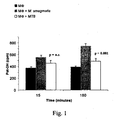
- tuberculosis and assaying the PLD activity at 15 and 180 minute following the exposure to mycobacterium was carried out at the infection multiplicity (MOI) of 1 bacillus for cell and analysis of the PLD activity was performed using as index the formation of phosphatidyl ethanol (Pet-OH) generated by PLD catalysed transphosphatidylation reaction, 1% ethanol being present within the culture medium. Obtained results are represented in figure 1 and it is possible to see that cells exposed both to the pathogenic and non pathogenic mycobacterial strains show PLD activity levels higher than non infected macrophages and the exposure to a non pathogenic bacterium results in macrophage PLD activation significantly higher than MTB. 180 minutes following the infection. This indicates that simple exposure to mycobacteria stimulates a strong and early PLD activation which is inversely proportional to the mycobacterium pathogenicity.
- MOI infection multiplicity
- FIG. 2 shows the formation of phosphatidylethanol, indication of macrophage PLD activity, generated in macrophages 3 hours (panel A) after MTB or M. smegmatis infection.
- MTB infected cells Following the assay of the S1P efficiency on the macrophage PLD, the effect thereof on MTB infected cells was evaluated in order to increase the PLD activation levels of pathogenic mycobacterium infected macrophages.
- Cells were MTB infected under the same conditions as in above experiments and macrophage PLD activity was analysed 24 hours after infection. Results are reported in figure 3B and as can be seen MTB infected and S1P treated macrophages show a Pet-OH production, index of PLD activity, higher than MTB infected non S1P treated ones, indicating that S1P causes increase of PLD activity of MTB infected macrophages.
- dichloro fluorescein is a fluorochrome obtained from reaction of non fluorescent 2,7-dichloro fluorescein diacetate (DCFH) with various oxidants
- analysis thereof is used to detect cellular oxidative stress and amount of oxygen reactive intermediates (ROI) produced by phagocytic cells.
- ROI oxygen reactive intermediates
- this analysis is employed in order to monitor the D-erythro-sphingosine 1-phosphate (S1P) modulated ROI production from macrophages. Macrophages were incubated for 90 minutes with sphingosine 1-phosphate at 0,5 ⁇ M, 5 ⁇ M, 10 ⁇ M and 50 ⁇ M concentrations.
- Fluorescence microscopy was used to detect fluorescein treated M . tuberculosis infected macrophages.
- Cellular suspension containing 10 5 macrophages was spread over microscopy slide, allowed to adhere over 16-18 hours in humid atmosphere within a Petri dish and infected with fluorescein treated H37Rv M. tuberculosis strain at 10:1 bacterium-phagocyte ratio.
- Successively, cells were washed with PBS and fixed with a solution of PBS and 2 % para formaldehyde for 30 minutes. Cells were then washed twice and incubated for 5 minutes with PBS and 0,05 % saponin. Finally after three PBS washes the slide was observed with fluorescence microscope and then photographed.
- a bacterium staining method with fluorescein isothiocyanate (FITC) was provided. Conjugation of H37Rv MTB with fluorochrome was monitored by flow cytometry and the same can be observed in panel A of figure 5 . Then, macrophages were infected with MTB virulent strain, which had been labelled previously with fluorochrome, directly over the slide. Analysis, carried out with fluorescence microscope and shown in panel B, reveals the intracellular localisation of the bacteria, proving that it is possible to infect macrophages with thus previously treated mycobacteria.
- FITC fluorescein isothiocyanate
- Immunoblotting assay was used to analyse the filters with so transferred proteins. After a PBS 0,1 % Tween 20 washing to remove transfer buffer excess and saturation with 5 % dried milk in 0,1% PBS Tween20, filters were again incubated with anti-Cathepsin D antibody (BD Transduction Laboratories) for 2 hours under shaking. After incubation with primary antibody and several washings, filters were incubated with peroxidase conjugated anti IgG antibody (Bio-rad) for 2 hours under stirring. A developing system based on increased chemiluminescence by imaging plates at various exposure times was used.
- This protein in fact represent a successful phagolysosome label whose amount is proportional to the phagolysosome maturation. Results obtained are shown in panel B of figure 6 and it can be observed that the amount of intralysosomial protein in the phagosomial fraction from S1P treated (column 3) is higher than in non treated macrophages (column 2).
- S1P sphingosine 1-phosphate
- THP-1 leukemia line cells Human monocytic THP-1 leukemia line cells were used in this example as a model for macrophage response.
- 5x10 5 /ml THP-1 cells were induced to differentiate by 48 hours incubation in the presence of 20 ng/ml Phorbol Myristate Acetate (PMA).
- PMA Phorbol Myristate Acetate
- Differentiated THP-1 cells (dTHP-1) were then centrifuged three times and suspended in complete medium [RPMI 1640 (Gibco BRL) supplemented with 10% fetal bovine serum (Gibco BRL), 5 mM L-glutamine (Invitrogen), 1 mM sodium pyruvate (Gibco BRL) and non essential amino acids (Euroclone)].
- dTHP-1 were then infected for 3 hours with M. smegmatis at the multiplicity of infection of bacterium/cell 50:1. Following infection cells were washed three times with RPMI 1640 and incubated in complete medium at 37°C for further 1, 12 and 24 hours in the presence or absence of sphingosine-1 phosphate used at the concentration of 5 ⁇ M.
- Inhibiting activity of calphostin C is exploited by targeting PLD catalytic domain ( Sciorra V.A., Hammond S.M., Morris A.J. Potent direct inhibition of mammalian phospholipase D isoenzymes by calphostin c. Biochemistry 2001; 40: 2640-46 ).
- cells were 1) lysed using cold PBS containing 0.1% saponin (Sigma, MA, USA), ii) serially diluted in PBS containing 0.01% Tween-80 (Merck, Darmstadt, Germany) and iii) plated in triplicate in Middlebrook 7H10 medium (Becton Dickinson, MD, USA) supplemented with 10 % OADC formulation (0,5 mg/ml oleic acid, 50 mg/ml albumin, 20 mg/ml dextrose, 0,04 mg/ml catalase and 8,5 mg/ml sodium chloride). M. smegmatis colonies were enumerated after incubation for 3 days at 37°C.
- S-1 P sphingosine 1-phosphate
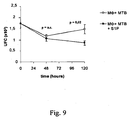
- Sphingosine 1-phosphate inhibits intracellular replication of Mycobacterium smegmatis in THP-1 macrophage cell line.
- Sphingosine 1-phosphate induces antimicrobial activity in such cells though macrophage D phospholipase activation as demonstrated by the increase of mycobacterial replication after ethanol treatment (dose dependent effect) or calphostin C (used at the concentration of 10 nM).
- Mycobacteria were enumerated by assay of colony forming units performed at 1 hour (panel A), 12 hours (panel B), and 24 hours (panel C) after mycobacterial infection.
- Human monocytic THP-1 leukemia line cells were used in this example as a model for macrophage response.
- 5x10 5 /ml THP-1 cells were induced to differentiate by 48 hours incubation in the presence of 20 ng/ml Phorbol Myristate Acetate (PMA).
- PMA Phorbol Myristate Acetate
- THP-1 cells Differentiated THP-1 cells (dTHP-1) were then centrifuged three times and suspended in complete medium [RPMI 1640 (Gibco BRL) supplemented with 10% fetal bovine serum, 5 mM L-glutamine) (Invitrogen), 1 mM sodium pyruvate (Gibco BRL) and non essential amino acids (Euroclone), 5 ⁇ g/ml gentamicin (Invitrogen)].
- dTHP-1 were then infected for 3 hours with H37Rv M. tuberculosis at the multiplicity of infection of 1:10 (panel A) or 1:1 (panel B) bacterium/cell.
- cells were 1) lysed using cold PBS containing 0.1% saponin (Sigma, MA, USA), ii) serially diluted in PBS containing 0.01% Tween-80 (Merck, Darmstadt, Germany) and iii) plated in triplicate on Middlebrook 7H10 medium (Becton Dickinson, MD, USA) supplemented with 10 % OADC formulation (0,5 mg/ml oleic acid, 50 mg/ml albumin, 20 mg/ml dextrose. 0.04 mg/ml catalase and 8,5 mg/ml sodium chloride).
- Mycobacterium tuberculosis colonies were enumerated after incubation for al least 21 days at 37°C.
- S1P sphingosine 1-phosphate
- Panel A refers to a representative experiment using a multiplicity of infection of 1 bacterium every 10 cells.
- Panel B refers to a representative experiment using a multiplicity of infection of 1 bacterium per cell.
- monocyte derived macrophages were infected for 3 hours with MTB at 1:1 macrophage/bacterium infection ratio and then treated with S1 P (5 ⁇ M).
- S1 P 5 ⁇ M
- the plates containing infected cellular cultures were centrifuged at 2000 RPM for 10 minutes and the supernatant was discarded.
- 1 ml PBS and 0,1 % saponin were added to each well and the contents were incubated for 20 minute at 37°C. After incubation cellular lysates were sonicated for 20 seconds in such a way to break any possible bacterial aggregates.
- Lysates thus obtained were diluted in sterile saline 1:100, 1:1000, 1:10000 with addition of 0,01 % Tween 80 and plated on Middlebrook 7H10 (Difco). Plates were incubated at 37°C for about one month and colonies enumerated.
- Peripheral blood mononuclear cells were isolated from human Buffy coat blood preparations by gradient centrifugation and suspended at 5x10 6 /ml in complete medium [RPMI 1640 (Gibco BRL) supplemented with 10% fetal bovine serum (Gibco BRL), 5 mM L-glutamine and Gentamicine (5 ⁇ g/ml)]. Monocytes were then separated by adherence for 1 hour in polystyrene 75 cm 2 flasks (Corning, Cambridge, MA). After incubation, non-adherent cells were removed by three washes with warm RPMI 1640.
- sphingosine 1-phosphate used at concentrations of 0,5 ⁇ M, 5 ⁇ M. and 50 ⁇ M.
- cells were 1) lysed using cold PBS containing 0.1% saponin (Sigma, MA, USA) ii) serially diluted in PBS containing 0.01% Tween-80 (Merck, Darmstadt, Germany) and iii) plated in triplicate on Middlebrook 7H10 medium (Becton Dickinson, MD, USA), supplemented with 10 % OADC formulation (0,5 mg/ml oleic acid, 50 mg/ml albumin, 20 mg/ml dextrose, 0,04 mg/ml catalase and 8,5 mg/ml sodium chloride).
- OADC formulation 0,5 mg/ml oleic acid, 50 mg/ml albumin, 20 mg/ml dextrose, 0,04 mg/ml catalase and 8,5 mg/ml sodium chloride.
- Mycobacterium. tuberculosis colonies were enumerated after incubation for about 21 days at 37°C.
- Sphingosine 1-phosphate used at the concentrations of 0,5, 5 or 50 ⁇ M inhibits intracellular replication of Mycobacterium tuberculosis in human monocyte-derived macrophages (MDM) from peripheral blood of healthy donors. Results are obtained from experiments performed on MDM from 4 different donors and are expressed as the means of the percentages of mycobacterial killing ⁇ standard error at the fifth day after infection. Percentage of mycobacterial killing is calculated by using the following formula: [1-(CFU from infected and S-1P stimulated MDM / CFU from infected unstimulated MDM)] x 100.
Landscapes
- Health & Medical Sciences (AREA)
- Chemical & Material Sciences (AREA)
- Medicinal Chemistry (AREA)
- Pharmacology & Pharmacy (AREA)
- Life Sciences & Earth Sciences (AREA)
- Animal Behavior & Ethology (AREA)
- General Health & Medical Sciences (AREA)
- Public Health (AREA)
- Veterinary Medicine (AREA)
- Epidemiology (AREA)
- Communicable Diseases (AREA)
- Oncology (AREA)
- Chemical Kinetics & Catalysis (AREA)
- General Chemical & Material Sciences (AREA)
- Nuclear Medicine, Radiotherapy & Molecular Imaging (AREA)
- Organic Chemistry (AREA)
- Pulmonology (AREA)
- Acyclic And Carbocyclic Compounds In Medicinal Compositions (AREA)
- Pharmaceuticals Containing Other Organic And Inorganic Compounds (AREA)
- Medicines That Contain Protein Lipid Enzymes And Other Medicines (AREA)
Abstract
Description
- The present invention relates to the use of immunomodulating sphingosine compounds able to induce, restore or increase the efficiency of immuno-defence mechanisms involved in the early steps of the infection.
- Particularly the invention relates to the use of sphingosine compounds for the manufacture of a medicament for the treatment of microbial infections, particularly those derived from Mycobacterium tuberculosis.
- Immuno-defence mechanisms against infective agents include innate and acquired immunity responses developing during the early phases of the infection and later, respectively. Innate immunity is non specific and mediated by the activity of mononuclear, polymorphonuclear phagocytes and Natural Killer cells, whereas acquired immunity is specific and mediated by T and B lymphocytes, which are clonally distributed and characterised by specificity and memory.
- Macrophages or mononuclear phagocytes are fundamental cellular components of innate Immune System (SI). These are able to phagocyte and digest exogenous particles by hydrolytic activity of lysosomial enzymes.
- Phagocytosis mechanisms are different depending on inert material or microorganisms are involved. In the first case, in fact, phagocytosis is non specific and mediated only by electrostatic forces; in the second, on the contrary, exogenous particle, after coating with complement proteins or antibodies, is phagocytosed by specific recognition mechanism mediated by complement protein or antibody receptors. In any case, the exogenous material, firstly, adheres to the plasmatic membrane of macrophage and then the latter is enveloped within the cytoplasm resulting in a vesicle named phagosome. This reaches the middle of the cell where it is fused with lysosomes, vacuolar organelles rich of hydrolytic enzymes active at acid pH, 4,5-5,0, thus generating phagolysosome. During the maturation process of the phagolysosome therefore there is a progressive acidification of the phagosome whose pH decreases from neutral to acid values, namely 5 or less within the phagolysosome. In the latter almost complete degradation of phagocytosed material occurs, which then is ejected form the cell by means of exocytosis process. The steps of adhesion, ingestion, phagosome formation and phagolysosome maturation involve macrophage activation with the increase of cellular metabolism. Substantially, two anti-microbial systems are exerted by phagocyte: namely, O2-dependent and O2-non dependent system. O2-dependent system uses oxygen reactive intermediates (ROI) as O2 -, OH° and H2O2, which can act both directly on the pathogens and indirectly through the activation of myeloperoxidase. On the contrary O2-non dependent systems usually act by killing anaerobic bacteria and include a multiplicity of lysosomial enzymes which can be active within the cell or secreted outside, like cationic proteins, lactoferrin and lysozyme. Among O2-non dependent antimicrobial mechanisms in the macrophage there is another important cytotoxic defensive L-arginine dependent system against intracellular pathogens. Such a cytotoxic effect substantially is mediated by nitrogen reactive intermediates (RNI), generated by L-arginine substrate through the activation of inducible form of nitric oxide synthase enzyme (iNOS). The direct association of mycobactericidal effect of murine macrophages with the activation of L-arginine-dependent cytotoxic system was demonstrated. The cytotoxic activity of macrophages therefore results from all the processes hitherto described, whose final goal is to make the macrophage able to eliminate the foreign microorganisms (viruses, bacteria, fungi, protozoa, parasites, etc.).
- After phagocytosis by macrophages following intracellular growth of pathogens depends on their capability to avoid the destruction by lysosomial enzymes, reactive oxygen and nitrogen intermediates. Within this context Mycobacterium tuberculosis, tuberculosis etiological agent (TB) is able, through various evasion mechanisms, to escape macrophage microbicidal mechanisms.
- Tuberculosis currently represents one of three most diffused diseases in the world together with acquired immunodeficiency syndrome (AI DS) and malaria.
- An important factor contributing to the increase of MTB occurrence in the industrial countries is represented by the diffusion of drug-resistant strains thereby preventing the elimination of the tuberculosis pathology by means of the usual antibiotic treatments.
- A key of MTB virulence is the capability thereof to infect monocytes and macrophages and to replicate therein. Within this context, the mycobacterium can use various macrophage receptors.
- After the macrophage phagocytosis, MTB is able to exert a range of mechanisms to escape the macrophage response, such as the production of ammonium ions, resulting in an alkaline intralysosomial compartment pH increase decreases the enzymatic activity of this compartment which acts, on the contrary, optimally at acid pHs (pH 5), normally maintained by a ATP-dependent protonic pump.
- Other mechanisms are represented by the capability of mycobacterium to inhibit the maturation of phagolysosome. Electronic microscopy studies examined dynamics of phagosome-lysosome fusion and effects thereof on the bacterial replication in human infected macrophages and from this study it results that MTB containing vesicles appears to bud from phagosome and separate therefrom without subsequent fusion with the lysosome.
- Studies about the interaction of MTB containing phagosomes with the lysosomes in murine macrophages proved inhibition of the phagosome-lysosome fusion, confirmed by subsequent studies also on human macrophages. MTB, in fact, inhibits final maturation events of the phagolysosome biosynthesis. Molecular characterisation of the MTB containing phagosome shows: reduced levels of membrane glycoproteins associated to lysosomes (LAMPs) and cathepsin D, the persistence of Rab5 molecule, characteristic of the early maturation steps and not complete acidification with respect to beads containing phagosomes. Inhibition of phagosome-lysosome fusion represents, in fact, an important evasion strategy of the bactericidal mechanisms because this phenomenon is never found in died bacteria containing phagosomes.
- From recent studies about MTB, an important role of a macrophage enzyme in antibacterial response, phospholipase D (PLD), appears. This enzyme is involved in various microbicidal mechanisms of human macrophages and so represents an important component of antibacterial response (Kusner J.D., et al., Activation of phospholipase D is tightly coupled to the phagocytosis of Mycobacterium tuberculosis or opsonized zymosan by human macrophages. J. Exp. Med. 1996; 184:585-595).
- Phospholipase D (PLD) is a membrane enzyme widely diffused within the mammalian cells whose activity is under the control of hormones, neurotransmitters, growth factors and cytokines. Interaction of extracellular signal molecules with surface receptors activates PLD which catalyses the hydrolysis of phosphatidylcholine (PC) and other phospholipids, generating phosphatidic acid (PA).
- PLD activation is associated with various leukocyte antimicrobial mechanisms among which there are phagocytosis, generation of ROI and secreted granules. Particularly the PLD activation in phagocytosis of particles opsonized by the complement or antibodies in neutrophiles and macrophages was demonstrated. MTB phagocytosis by human macrophages is strictly associated with the PLD activation, in fact it was proved that molecules inhibiting PLD activity provoke a reduction of the phagocytic process and administration of exogenous PLD can restore such a process (Kusner et al., "ATP-induced killing of virulent Mycobacterium tuberculosis within human macrophages requires phospholipase D", The Journal of Immunology, 2000,164:379-388).
- Although efforts carried out up to now, new and efficient means for treatment of infections able to induce or restore immuno-defence mechanisms, particularly those mediated by phagocyte activity, are still required. Efficient means are more stringently required for infections derived from pathogens like Mycobacterium tuberculosis able to develop resistance to currently available medicaments.
- However, in view of a practical application in therapeutic field, the promotion of these mechanisms by the administration of exogenous PLD, as suggested by Kusner et al. (supra) results in various problems like immune reactions against protein and allergies. Stability of enzymatic activity when the enzyme is in pharmaceutical formulations represents a further problem. In this context, in the past it was demonstrated that sphingosine 1-phosphate increases phosphatidic acid levels in murine fibroblasts (Desai N.N. et al. "Sphingosine 1-phosphate, a metabolite of sphingosine, increases phosphatidic acid levels by phospholipase D activation" J. Biol. Chem. 1992; 267: 23122-23128). Desai supposes that this PA increase can result from a PLD activation.
- The present invention results from the finding that the administration of sphingosine derivatives increases the bactericidal activity of human macrophages.
-
US-A-6 147 118 discloses the use of a sphingoid base to inhibit topical microbial growth of bacteria, yeast or fungi or skin. - Increase of microbicidal activity in macrophages is associated to an increase of the macrophage PLD activity and production of reactive oxygen and nitrogen intermediates and induction of the maturation of phagolysosomes in macrophages. Advantageously, administration of sphingosine derivatives, which substance is a constituent of cellular membrane, promotes or restores the endogenous defence mechanisms without the need of using exogenous substances.
- The object of the present invention is to provide new means for the treatment of infections, specifically treatment of the infections derived from pathogens, like Mycobacterium tuberculosis, able to develop resistance to currently available drugs,
- Particularly the object of the present invention is the use of a sphingosine compound for the manufacture of a medicament for treatment of microbial infections by immunomodulation, principally infections resulting from bacterial and specifically Mycobacterium tuberculosis.
- Such compounds, which are sphingosine derivatives, like D-erythro-sphingosine 1-phosphate (S1P), are characterised in that they are able to stimulate in a dose-dependent effect the macrophage PLD both in presence and absence of infection.
- Other objects of the invention are methods to induce or restore or increase the in vitro microbicidal activity of macrophages wherein macrophages are infected by a pathogen, preferably MTB and successively treated with sphingosine derivatives.
-
-
Figure 1 shows comparative analysis of PLD activity of IFN-γ-stimulated macrophages after M. smegmatis or MTB infection. Macrophages (MΦ) were infected by M. smegmatis or MTB at MOI of a bacillus for cell. The formation of phosphatidyl ethanol, indicating the activity of macrophage PLD, plotted in ordinate, was measured at 15 and 180 minutes after the infection, and plotted in abscissa. The results are expressed as mean ± DS of triplicate of an experiment representative of experiments carried out on cells from three different donors. The statistical analysis was carried out comparing the results from M. smegmatis and MTB infected cells, respectively. -
Figure 2 shows the formation of phosphatidyl ethanol, indicating the activity of macrophage PLD, plotted in ordinate, of IFN-γ-stimulated macrophages after M. smegmatis or MTB infection. Macrophages were infected by M. smegmatis or MTB at MOI of a bacillus for cell. PLD activity was measured at 3 (A panel) and 24 (panel B) hours after the infection, in the presence (+) or absence (-) of IFN-γ. Results are expressed as mean ± DS of duplicate of experiments carried out on cells from two different donors. The statistical analysis was carried out comparing the results from M. smegmatis and MTB infected cells, respectively. -
Figure 3 shows the formation of phosphatidyl ethanol, indicating the activity of macrophage PLD, plotted in ordinate, at 20, 30, 60, 90 minutes after the treatment (in abscissa) with scalar doses (1, 5, 10 µM) of D-erythro-sphingosine 1-phosphate (S1P) (panel A).
In the panel B the increase of PLD activity (in ordinate) detected in MTB infected, 5 µM S1P treated MTB infected and M. smegmatis infected, respectively, at 24 hours after the infection (in abscissa), in comparison to non infected macrophages, is reported. Data of the latter panel is expressed as mean of increments ± D.S. of experiments carried out on cells from three healthy donors. The statistical analysis was carried out comparing the results from MTB infected and S1P treated infected macrophages, respectively. -
Figure 4 shows the analysis of ROI generation from S1P treated macrophages. Particularly macrophages (MΦ) were treated by S1P and successively ROI generation was analysed by cytofluorometry, namely using dichlorofluorescein (DCF) assay. InFigure 4A , in ordinate, % of DCF positive cells obtained at 30 minutes after the treatment with S1P scalar doses, plotted in abscissa, is reported. The graph offigure 4B , on the contrary, reports in ordinate % of DCF+ cells at 30 and 90 minutes (abscissa) after the treatment with 5 µM S1P. -
Figure 5 reports the picture of fluorescein-isothiocyanate conjugated MTB.
Mycobacterium tuberculosis was conjugated to fluorescein-isothiocyanate (FITC) and visualised by flow cytofluorometry (figure 5A ) and fluorescence microscopy within the macrophages (figure 5B ). -
Figure 6 shows the analysis of phagolysosome maturation within S1 P treated macrophages.
Infigure 6A there is reported the flow cytometric analysis of MTB-FITC infected macrophages at MOI of a bacillus for cell over three hours and 5 µM S1P treated during 30 minutes. For any condition there is reported % of fluorescent macrophages (1,9 %, 63,4 %, 38,9 %).Figure 6B shows the results of Western blotting of MΦ phagosomialfractions 24 hours after latex beads intemalisation. Bands corresponding to 28 kDa refer to intralysosomial Cathepsin D protein of S1P treated (column 3), not treated (column 2), and control macrophages which did not internalise beads (column 4), respectively.Column 1 refers to a positive experiment internal control. -
Figure 7 shows the graphs of the antimicrobial activity of sphingosine 1-phosphate against Mycobacterium smegmatis. Mycobacteria are enumerated by an assay of colony forming units carried out at 1 (panel A), 12 (panel B) and 24 hours (panel C) after mycobacterial infection. -
Figure 8 shows the graphs of the antimicrobial activity of sphingosine 1-phosphate against Micobacterium tuberculosis. Infection was carried out with a multiplicity of infection of 1 bacterium for 1 (panel B) and 10 cells (panel A), respectively. -
Figure 9 shows the effect of the S1P treatment on the viability of mycobacteria measured as colony forming units (CFU).
Macrophages (MΦ) were MTB infected at MOI of 1 bacillus for 1 cell over 3 hours and treated with S1P (5 µM) for 30 minutes. CFUs, plotted in ordinate, were obtained by plating of intracellular mycobacteria in triplicate immediately (time 0), and at 2 and 5 days after the infection (in abscissa). Obtained results are expressed as mean ± DS of triplicate of an experiment representative of two experiments carried out on cells from donors. -
Figure 10 shows a graph of the intracellular replication inhibition of Micobacterium tuberculosis in macrophages derived from peripheral blood from healthy donors using sphingosine 1-phosphate at concentrations of 0,5, 5 and 50 µM, respectively. - Immunomodulating compounds according to the present invention are sphingosine derivatives. Sphingosine is a long chain amino alcohol constituent of cellular membranes and together with sphingosine 1-phosphate is a member of a new class of second lipid messengers produced in response to growth factors. Sphingosine itself and its derivatives can be both in erythro and threo conformation, (D) and (L) configuration and cis and trans across the double bond. Sphingosine derivatives are able to mimic immunomodulating activity, particularly those selected among D-erythro or t.-threo and monophosphates, diphosphates, triphosphates thereof and more particularly D-erythro sphingosine 1-phosphate (S1P).
- Administration of therapeutically effective amounts of sphingosine derivatives induces an increase of microbicidal activity, particularly bactericidal and specifically mycobactericidal of human or animal macrophages which can be detected as a decrease of colony forming pathogen units (CFU). Such an immunomodulating activity is associated to the induction of phagolysosome maturation and increase of the activity of macrophage phospholipase D in pathogen infected macrophages and increase of the production of oxygen and/or nitrogen reactive intermediates.
- Therefore the above sphingosine derivative compounds to be used as immunomodulators and for related uses in the preparation of medicaments with immunoregulating activity, e.g. associated to the above phenomena, are object of the present invention.
- Sphingosine derivatives as immunoregulating compounds according to the present invention can be used advantageously in the treatment of infections resulting from any pathogen as viruses, bacteria, fungi, protozoa and other parasites and particularly Mycobacterium tuberculosis or Listeria monocytogenes.
- Pharmaceutical compositions can be made comprising a therapeutically active amount of a sphingosine derivative compound together with one or more pharmaceutically acceptable adjuvant and/or excipient.
- These compositions can contain the immunoregulating compound as the sole active agent when used in a treatment for simple promotion of native immunodefences or can contain the same in association with other microbicidal medicaments.
- Pharmaceutical compositions can be formulated according to known methods which include the presence of a pharmaceutically acceptable vesicle both in solid and liquid form. Examples of these vehicles and formulation methods are reported in specialised literature. Pharmaceutical compositions suitable for immunoregulating activity must contain an effective amount of the active principle according to the present invention. In the case of liquid compositions such an amount corresponds to concentrations from 0,1 µM to 1 mM, preferably from 0,5 µM to 10 µM. In the case of solid compositions the amount expressed as µg corresponding to the above concentrations can be used. Pharmaceutical compositions are administered to a subject in a such amount that is effective for successful treatment of the related infection. Relative amount can be varied depending on a multiplicity of factors like individual conditions, weight, sex and age. Preferred formulations are liquid compositions in the form of solutions, suspensions or emulsions to be administered by oral or parenteral route. Due to the chemical characteristics of sphingosine derivatives the inclusion thereof within liposomes or emulsions is particularly preferred. Administration of liquid compositions as spray or aerosol is particularly preferred for the treatment of infected aerial pathways and particularly lung infections as tuberculosis. The immunoregulating compound can be administered typically in mixture with suitable pharmaceutical diluents, excipients or vehicles conveniently selected by taking in consideration the desired administration route.
- Because the inventive compounds are soluble in hydroalcoholic solvents (water/ethanol), such solutions thereof are used as such, if suitable, or are used as starting reagent for the preparation of different pharmaceutical formulations. Further, the composition can contain pharmaceutically acceptable optional additives as surfactants, buffers, stabilisers, preservatives and antioxidants.
- The preparation of the pharmaceutical composition includes methods known in the specific field. The immunomodulating compounds are mixed in solid or liquid form with suitable excipients, possibly with any desired optional additive, preferably in hydroalcoholic solutions, at concentrations from 0,1 µM to 1 mM, preferably from 0,5 µM to 10 µM or, in the case of solid compositions, at corresponding weight amounts. These solutions are then formulated in liquid compositions suitable to be administered, as solutions, suspensions, emulsions, or liposome encapsulated or dried as powders or granules suitable to be formulated in solid compositions.
- Further object of the present invention is an in vitro method to induce or restore or increase the microbicidal activity of phagocytes, preferably human macrophages. Macrophages cultured as described in the experimental section and infected by pathogens susceptible to phagocytosis at a pathogen/macrophage infection ratio (MOI) from 1:1 to 10:1 for a period from 1 to 24 hours are treated with solutions containing sphingosine derivatives according to the invention at concentrations from 0,1 µM to 5 mM, preferably from 0,5 µM to 50 µM, particularly 5 µM over an incubation period from 1 minute to 24 hours, preferably from 15 to 90 minutes. Suitable pathogens are viruses, bacteria, fungi, protozoa, parasites, particularly Mycobacterium tuberculosis. In the latter case macrophages are preferably treated with D-erythro sphingosine 1-phosphate.
- Induction or restoration or increase of the microbicidal activity of macrophages can be monitored in terms of the activity of the macrophage PLD as described in
figure 3 and example 1. Alternatively the activity of macrophages can be evaluated in terms of generation of oxygen reactive intermediates (ROI), as described infigure 4 and example 3 or viability of phagocytised microorganisms as described infigure 7 and example 6. - The invention is further illustrated by the following examples which describe reagents, conditions and procedure suitable for the practice of the invention but it is understood that the scope thereof is not limited thereto.
- H37Rv (MTB) M. tuberculosis virulent strain was cultured in Sauton medium at 37°C in unmodified atmosphere, taking care the growth thereof was as surface film. After culturing for about three weeks mycobacteria were washed with PBS (phosphate buffered saline), centrifuges at 14000 rpm for 5 minutes and sonicated for 1-2 minutes. Successively mycobacteria were suspended within the same buffer and then aliquoted and frozen at -80°C.
- Culture of M. smegmatis non pathogen strain was carried out in ADC enriched Middlebrook 7H9 (DIFCO) liquid medium for three days in incubator at 37°C and 5 % CO2 containing atmosphere. Finally the bacteria were aliquoted and frozen at -80°C.
- Before the freezing, serial dilutions of initial bacterial suspension were carried out. Such suspensions then were seeded in plates containing OADC enriched 7H10 agar. The number of colony forming units was evaluated at about 4 weeks and 3 days of MTB and M. smegmatis culturing, respectively. Bacteria concentrations of initial aliquots, both of which were about 109 bacteria/ml, were deduced from the number of such colonies.
- In some experiments MTB aliquots, after thawing at room temperature, centrifuged at 10000 rpm for 10 minutes, suspended in PBS and sonicated for 1-2 minutes, were labelled with fluorescein isothiocyanate (FITC) (Sigma). FITC, pre-suspended at 1 µg/ml concentration, was added to the bacterial suspension, which was incubated at 37°C in the presence of 5 % CO2 for about 90 minutes. At the end of the incubation the bacterial suspension was washed three times with centrifugation at 14000 rpm for 15 minutes and suspended in PBS at required concentration.
- Mononuclear cells from peripheral blood were isolated from healthy donor buffy-coat by stratification on Lymphoprep (Eurobio, density ~ 1,077) lymphocyte separation medium and centrifugation at 1800 rpm for 20 minutes. Interface recovered cells were washed three times in RPMI 1640 (GIBCO). First, second and third centrifugation were carried out at 1600, 1400 and 1000 rpm for 15 minutes, 10 minutes and 10 minutes, respectively. Cells were suspended in RPMI complete medium, i.e. with addition of 10 % FBS, gentamicin (used at 50 µg/ml) and L-glutamine (used in at 1 %). Monocytes were separated from lymphocytes by adherence in flask. To that end, 15 ml of cellular suspension were stratified in 75 cm2 polystirene flasks (Coming) and incubated for 1 hour at 37°C in 5 % CO2 containing atmosphere. Non adherent cells were removed by three wahings with warm RPMI pre-heated at 37°C. Adherent cells corresponding to monocyte/macrophage population were removed from the bottom of the flask after incubation at 4°C with cold PBS for about 30 minutes. As detected by flow cytometric analysis of cell physical parameters, i.e. FCS dimension and SSC granulometry, minimal purity thereof was always higher than 80 %.
- Cells were cultured in 24 well plates (Costar) at 1x10⁶ cell/ml concentration for 7 days in order to obtain macrophages (MΦ) and incubated in the presence or absence of IFNγ (500 U/ml) depending on the experiment.
- Before the macrophage (MΦ) infection the bacteria were thawed at room temperature. Then, three PBS washes were carried out at 10000 rpm for 10 minutes. Being prone to aggregate mycobacteria were transferred in glass tubes and bath sonicated to allow cellular separation. Infection of monocyte derived macrophages was carried out in culturing plates with incubation of the cells for three hours with H37Rv MTB or M. smegmatis at infection ratio of bacterium/cell 1:1. At the end of the infection the plates were centrifuged at 1800 rpm for 10 minutes and supernatant discarded. Then, the cells were suspended in complete medium, treated with D-erythro-sphingosine 1-phosphate (Calbiochem) at concentrations reported time by time in the individual experiment and finally separated from the bottom of the well and analysed.
- A system for the detection of phosphatidyl ethanol (Pet-OH), a stable product from the PLD catalysed transphosphatidylation reaction in the presence of ethanol within the medium, is used for the determination of macrophage (MΦ) PLD activity. Macrophages, suspended at 1x10⁶ cell/ml concentration in 24 well plates, were incubated with [3H]-miristic acid at 1 µCi/ml/well concentration for 180 minutes at 37°C in RPMI complete medium in order to label fatty acids of plasmatic membrane. After three washes with warm RPMI, cells were incubated with 1 % ethanol for 15 minutes and then infected with M. smegmatis or MTB at 1:1 bacterium/cell multiplicity of infection. After 3 hour infection cells were centrifuged at 1800 rpm for 10 minutes, suspended in
Hepes 20 mM (pH 7,3), centrifuged and again suspended inHepes 20 mM (pH 7,3) and methanol at 1:1 ratio. Finally the cells were separated and collected in test tubes to which 2 ml of a solution containing chloroform/methanol 1:1 were added. After agitation of the suspension chloroform (1 ml) and KCI (1 ml) were added and again agitated. Two phases could be seen: a lower chloroform containing phospholipids, neutral lipids and other liposoluble compounds and an upper water phase containing polar substances. Successively the lower chloroform phase was recovered and transferred in fresh tubes, dried under nitrogen and suspended in 20 µl of chloroform. - Separation of Pet-OH from other phospholipids was carried out by monodimensional thin layer chromatography on plates activated at 100°C for 1 hour, just before the use. Methyl acetate/iso-octane/acetic acid/bidistilled water (130/20/30/100 v/v) was used as solvent system. The spots detected with vapor phase iodine were removed from the plate and the gel was transferred in test tubes in the presence of 200 µl of ethanol to enhance lipid solubilisation and 3 ml of scintillation liquid. Test tubes were then transferred in a liquid scintillation counter for the measurement of beta rays emitted from incorporated labelled fatty acids.
- Based on recent studies demonstrating the antimycobacterial effect of phospholipase D (PLD) it was decided to analyse the human macrophage PLD activity after mycobacterium infection. In order to point out a correlation between PLD activation and mycobacterium pathogenicity, PLD activities in macrophages (MΦ) from healthy donors infected with non pathogenic, i.e. Mycobacterium smegmatis, or pathogenic, i.e. Mycobacterium tuberculosis (MTB), were compared. Behaviour of macrophage PLD in the early phases of the mycobacterium infection was monitored by infecting cells with M. smegmatis or M. tuberculosis and assaying the PLD activity at 15 and 180 minute following the exposure to mycobacterium. Infection in our experiments was carried out at the infection multiplicity (MOI) of 1 bacillus for cell and analysis of the PLD activity was performed using as index the formation of phosphatidyl ethanol (Pet-OH) generated by PLD catalysed transphosphatidylation reaction, 1% ethanol being present within the culture medium. Obtained results are represented in
figure 1 and it is possible to see that cells exposed both to the pathogenic and non pathogenic mycobacterial strains show PLD activity levels higher than non infected macrophages and the exposure to a non pathogenic bacterium results in macrophage PLD activation significantly higher than MTB. 180 minutes following the infection. This indicates that simple exposure to mycobacteria stimulates a strong and early PLD activation which is inversely proportional to the mycobacterium pathogenicity. - In order to point out possible correlation between macrophage activation state and PLD stimulation following mycobacterial infection, cells were treated with IFN-γ (500 U/ml), before and after the infection.
Figure 2 shows the formation of phosphatidylethanol, indication of macrophage PLD activity, generated inmacrophages 3 hours (panel A) after MTB or M. smegmatis infection. Such an experiment confirms previously obtained results and points out that IFN-γ pre-treatment of the infected macrophages does not modify the behaviour of the PLD activity, excluding any effect of IFN-γ mediated cellular activation on PLD activity. - In order to analyse the behaviour over the time of the macrophage PLD activity following mycobacterial infection the experiment was repeated and cells analysed 24 hours after the infection. Results are reported in panel B and as can be seen M. smegmatis infection causes a PLD increment higher than MTB. Further it is possible to point out that IFN-γ treatment before and/or after infection does not appear to modify levels of macrophage PLD activity following mycobacterial infection.
- From above experiments it was found out that PLD activity levels of MTB infected macrophages are lower than M. smegmatis infected macrophages. Object of this experiment was to increase levels of the endogenous PLD activity of MTB infected macrophages by means of D-erythro sphingosine 1-phosphate (S1P). In order to test the efficiency of this molecule and find out optimum dose for the macrophage maximum activation the latter were treated by scalar S1P doses and cell PLD activity was analysed after 20, 30, 60, and 90 minutes. As can be seen from
figure 3A in the S1P treated macrophages, maximum activation is obtained after 30 minutes and using aS1P 5 µM concentration. Following the assay of the S1P efficiency on the macrophage PLD, the effect thereof on MTB infected cells was evaluated in order to increase the PLD activation levels of pathogenic mycobacterium infected macrophages. Cells were MTB infected under the same conditions as in above experiments and macrophage PLD activity was analysed 24 hours after infection. Results are reported infigure 3B and as can be seen MTB infected and S1P treated macrophages show a Pet-OH production, index of PLD activity, higher than MTB infected non S1P treated ones, indicating that S1P causes increase of PLD activity of MTB infected macrophages. - Because dichloro fluorescein (DCF) is a fluorochrome obtained from reaction of
non fluorescent 2,7-dichloro fluorescein diacetate (DCFH) with various oxidants, analysis thereof is used to detect cellular oxidative stress and amount of oxygen reactive intermediates (ROI) produced by phagocytic cells. In the used model, this analysis is employed in order to monitor the D-erythro-sphingosine 1-phosphate (S1P) modulated ROI production from macrophages. Macrophages were incubated for 90 minutes with sphingosine 1-phosphate at 0,5 µM, 5 µM, 10 µM and 50 µM concentrations. After theincubation 100 µl of 0,4 µM DCFH (Calbiochem) were added and incubated for at least 30 minutes. Finally, cells were washed with PBS and analysed using FACScan flow cytometer from Becton Dickinson. - It is already known that PLD is involved in antimicrobial activities like generation of oxygen reactive intermediates (ROI) and, in order to test this property, ROI amount was monitored after macrophage PLD modulation by S1P. Measure was carried out using dichlorofluorescein (DCF), fluorescence emitting molecule when bound to these oxidative products of phagocytic cells so that it is detectable by flow cytometry. From panel A of
figure 4 it is observed that non treated cells show low levels of DCF positive response indicating the minimal amount of oxygen reactive intermediates produced by macrophages in standard conditions. When cells are stimulated by lipopolysaccharides, which induce a known increase of ROI generation, an increase of DCF positive cells is observed, resulting in the reliability of the system efficiency for the evaluation of cell produced ROI amount. When cells are treated with S1P scalar doses they show increasing and proportional levels of DCF positive response with maximum at 5-10 µM. In order to analyse over the time the modulation of oxygen reactive intermediate production by S1P the experiment was repeated using 5 µM S1P treated macrophages and measuring DCF positive response after 30 and 90 minutes. Results are reported in panel B offigure 4 and as can be seen the ROI production by macrophages increases progressively over the time after the S1P PLD stimulation in comparison to non LPS stimulated or to activated macrophages. - By virtue of extreme sensitivity of fluorescein isothiocyanate (FITC) to acid pHs this molecule was used to monitor the phagosome acidity and have therefore information about maturation stage of phagolysosome. To this aim, bacteria were labelled with fluorescein isothiocyanate (FITC) as above described. Infection was carried out using FITC labelled bacteria. Then, cells were washed after 3 hours and stimulated for 30 minutes with 5 µM sphingosine 1-phosphate and following 1,5 % paraformaldehyde (PFA) fixated and finally monitored by flow cytofluorometry.
- Fluorescence microscopy was used to detect fluorescein treated M. tuberculosis infected macrophages. Cellular suspension containing 105 macrophages was spread over microscopy slide, allowed to adhere over 16-18 hours in humid atmosphere within a Petri dish and infected with fluorescein treated H37Rv M. tuberculosis strain at 10:1 bacterium-phagocyte ratio. Successively, cells were washed with PBS and fixed with a solution of PBS and 2 % para formaldehyde for 30 minutes. Cells were then washed twice and incubated for 5 minutes with PBS and 0,05 % saponin. Finally after three PBS washes the slide was observed with fluorescence microscope and then photographed.
- In order to obtain a flow cytometric system suitable to monitor mycobacterial infection in macrophages a bacterium staining method with fluorescein isothiocyanate (FITC) was provided. Conjugation of H37Rv MTB with fluorochrome was monitored by flow cytometry and the same can be observed in panel A of
figure 5 . Then, macrophages were infected with MTB virulent strain, which had been labelled previously with fluorochrome, directly over the slide. Analysis, carried out with fluorescence microscope and shown in panel B, reveals the intracellular localisation of the bacteria, proving that it is possible to infect macrophages with thus previously treated mycobacteria. - In order to evaluate the effect of S1 P on a specific antimicrobial activity as the maturation of phagolysosome, the infection with FITC-labelled MTB was carried out and the fluorescence values of FITC-MTB were used as indicators of the phagolysosome maturation. In fact such a process is associated with a progressive reduction of phagolysosomial pH and FITC, used to label the mycobacterium within the macrophage phagosome, is sensitive to acid pH. Based on the known capability of M. tuberculosis in reducing the phagosome-lysosome fusion in order to inhibit lysosomial degradation, an attempt to promote and increase the process through exogenous stimulation of PLD by means of S1P was carried out. In
figure 6 (panel A) it can be seen that the FITC-MTB fluorescence from S1P treated macrophages is reduced from 63.4 % to 38,9 % when compared to S1 P non-treated macrophages. - In order to analyse the expression of an intralysosomial protein within beads containing phagolysosomes the macrophages phagosomial fraction was separated. Particularly, cells were incubated with sterile latex beads (0,8 µM diameter) (Sigma), 1:200 diluted, in culture medium for 1 hour in the presence of 5 % CO2 and then treated with 5 µM S1P.
- Separation of the phagosomes was carried out by centrifugation on sucrose gradient. Cells were washed three times with PBS (twice for 10 minutes) and then again incubated for 24 hours at 37°C. Following the intemalisation and 24 hour incubation period cells were washed with cold PBS and collected using a little scraper in PBS at 4°C. then centrifuged at 2000 rpm for 10 minutes at 4°C and finally washed with homogenising buffer (250 mM sucrose, 3 mM imidazole,
pH 7,4) again at 4°C. Cellular pellet, was suspended in 1 ml homogenising buffer and cells were lysed with an homogenizer. Phagosomes were isolated by centrifugation on sucrose gradient. Cells, suspended in 1 ml of PBS, were added to 1 ml of 40 % sucrose solution and such a suspension was stratified on 2 ml of 62 % sucrose solution in ultracentrifuge tubes. Then 2 ml of 35 %, 25 % and 10 % sucrose solutions were stratified (all the solutions were prepared in 3 mM imidazole,pH 7,4). The tubes were ultra-centrifuged using SW41 rotor at 100000 g at 4°c. The phagosomes were collected at the interface between 10 % and 25 % sucrose solutions and suspended in 12 ml of cold PBS. Phagosomes were centrifuged at 40000 g at 4°C and suspended in 100 µl PBS. - Analysis of intralysosomial protein, cathepsin D, in the phagosomal fractions was carried out by Western blot. Proteins from phagosomal fractions were separated by SDS-PAGE. Polyacrylamide gel in a non continuos buffering system comprising two overlying gels, i.e. "running" and "stacking" gel, respectively, was prepared. The latter is characterised by a lower acrylamide content, ionic strength and pH in order to concentrate the sample in a narrow band before moving on the separation gel.
- Separation was carried out in SDS-PAGE at acrylamide concentration of 10 % so that bands containing molecular weights between 22 and 300 kDa could be separated and containing 0,4 % SDS. Samples were heat denatured for about 10 minutes. Electrophoretic test was carried out at 30-35 mA in an electrophoretic room (Pharmacia). At the end of the electrophoresis the proteins were transferred on nitrocellulose filter using Western blotting technique in electrophoretic room (Pharmacia) at 200 V.
- Immunoblotting assay was used to analyse the filters with so transferred proteins. After a
PBS % Tween 20 washing to remove transfer buffer excess and saturation with 5 % dried milk in 0,1% PBS Tween20, filters were again incubated with anti-Cathepsin D antibody (BD Transduction Laboratories) for 2 hours under shaking. After incubation with primary antibody and several washings, filters were incubated with peroxidase conjugated anti IgG antibody (Bio-rad) for 2 hours under stirring. A developing system based on increased chemiluminescence by imaging plates at various exposure times was used. - In order to confirm the advantageous effect of S1P on the phagolysosome maturation after phagocytosis we used as a model the internalisation of latex beads in S1P stimulated macrophages. Beads are intemalised by cells through phagocytosis in about 60 minutes. Extraction of phagosomes from these cells was carried out by ultracentrifugation on density gradient. Protein component of phagosomial portions containing both just formed and lysosome fused phagosomes was assayed by Western blotting using a Cathepsin D intralysosomial protein specific antibody. This protein in fact represent a successful phagolysosome label whose amount is proportional to the phagolysosome maturation. Results obtained are shown in panel B of
figure 6 and it can be observed that the amount of intralysosomial protein in the phagosomial fraction from S1P treated (column 3) is higher than in non treated macrophages (column 2). - Human monocytic THP-1 leukemia line cells were used in this example as a model for macrophage response. In order to obtain macrophage phenotype, 5x10⁵/ml THP-1 cells were induced to differentiate by 48 hours incubation in the presence of 20 ng/ml Phorbol Myristate Acetate (PMA). Differentiated THP-1 cells (dTHP-1) were then centrifuged three times and suspended in complete medium [RPMI 1640 (Gibco BRL) supplemented with 10% fetal bovine serum (Gibco BRL), 5 mM L-glutamine (Invitrogen), 1 mM sodium pyruvate (Gibco BRL) and non essential amino acids (Euroclone)]. dTHP-1 were then infected for 3 hours with M. smegmatis at the multiplicity of infection of bacterium/cell 50:1. Following infection cells were washed three times with RPMI 1640 and incubated in complete medium at 37°C for further 1, 12 and 24 hours in the presence or absence of sphingosine-1 phosphate used at the concentration of 5 µM.
- In order to assess the role of macrophage PLD activity in sphingosine 1-phosphate induced mycobacterial growth control, following the infection the cells were cultured in the presence of various concentrations of ethanol (0.3%, 0.03%, 0.003%) or 10 nM calphostin C. Ethanol, inducing the formation of phosphatidyl ethanol inactive compound is a specific inhibitor of PLD metabolic activity (Liscovitch, M., Czarny, M., Fiucci, G. and Tang, X., 2000. Phospholipase D: Molecular and cell biology of a novel gene family. Biochem. J. 345:401). Inhibiting activity of calphostin C is exploited by targeting PLD catalytic domain (Sciorra V.A., Hammond S.M., Morris A.J. Potent direct inhibition of mammalian phospholipase D isoenzymes by calphostin c. Biochemistry 2001; 40: 2640-46). Just after the infection and at the indicated time points, cells were 1) lysed using cold PBS containing 0.1% saponin (Sigma, MA, USA), ii) serially diluted in PBS containing 0.01% Tween-80 (Merck, Darmstadt, Germany) and iii) plated in triplicate in Middlebrook 7H10 medium (Becton Dickinson, MD, USA) supplemented with 10 % OADC formulation (0,5 mg/ml oleic acid, 50 mg/ml albumin, 20 mg/ml dextrose, 0,04 mg/ml catalase and 8,5 mg/ml sodium chloride). M. smegmatis colonies were enumerated after incubation for 3 days at 37°C.
- As shown in
figure 7 sphingosine 1-phosphate (S-1 P, used at the concentration of 5 µM) inhibits intracellular replication of Mycobacterium smegmatis in THP-1 macrophage cell line. Sphingosine 1-phosphate induces antimicrobial activity in such cells though macrophage D phospholipase activation as demonstrated by the increase of mycobacterial replication after ethanol treatment (dose dependent effect) or calphostin C (used at the concentration of 10 nM). Mycobacteria were enumerated by assay of colony forming units performed at 1 hour (panel A), 12 hours (panel B), and 24 hours (panel C) after mycobacterial infection. - Human monocytic THP-1 leukemia line cells were used in this example as a model for macrophage response. In order to obtain macrophage phenotype, 5x10⁵/ml THP-1 cells were induced to differentiate by 48 hours incubation in the presence of 20 ng/ml Phorbol Myristate Acetate (PMA). Differentiated THP-1 cells (dTHP-1) were then centrifuged three times and suspended in complete medium [RPMI 1640 (Gibco BRL) supplemented with 10% fetal bovine serum, 5 mM L-glutamine) (Invitrogen), 1 mM sodium pyruvate (Gibco BRL) and non essential amino acids (Euroclone), 5 µg/ml gentamicin (Invitrogen)]. dTHP-1 were then infected for 3 hours with H37Rv M. tuberculosis at the multiplicity of infection of 1:10 (panel A) or 1:1 (panel B) bacterium/cell. After infection cells were washed three times with RPMI 1640 and incubated in complete medium for further 1, 2 and 5 days (panel A) or 1 and 7 days (panel B) in the presence or absence of sphingosine 1-phosphate used at the concentration of 5 µM. Just after the infection and at the indicated time points, cells were 1) lysed using cold PBS containing 0.1% saponin (Sigma, MA, USA), ii) serially diluted in PBS containing 0.01% Tween-80 (Merck, Darmstadt, Germany) and iii) plated in triplicate on Middlebrook 7H10 medium (Becton Dickinson, MD, USA) supplemented with 10 % OADC formulation (0,5 mg/ml oleic acid, 50 mg/ml albumin, 20 mg/ml dextrose. 0.04 mg/ml catalase and 8,5 mg/ml sodium chloride). Mycobacterium tuberculosis colonies were enumerated after incubation for al least 21 days at 37°C.
- As shown in
figure 8 sphingosine 1-phosphate (S1P, used at the concentration of 5 µM) inhibits intracellular replication of Mycobacterium tuberculosis in THP-1 macrophage cell line. Panel A refers to a representative experiment using a multiplicity of infection of 1 bacterium every 10 cells. Panel B refers to a representative experiment using a multiplicity of infection of 1 bacterium per cell. - In order to evaluate the bacterial viability, monocyte derived macrophages were infected for 3 hours with MTB at 1:1 macrophage/bacterium infection ratio and then treated with S1 P (5 µM). At time 0 (just after infection) and after 2 and 5 days the plates containing infected cellular cultures were centrifuged at 2000 RPM for 10 minutes and the supernatant was discarded. Then 1 ml PBS and 0,1 % saponin were added to each well and the contents were incubated for 20 minute at 37°C. After incubation cellular lysates were sonicated for 20 seconds in such a way to break any possible bacterial aggregates. Lysates thus obtained were diluted in sterile saline 1:100, 1:1000, 1:10000 with addition of 0,01
% Tween 80 and plated on Middlebrook 7H10 (Difco). Plates were incubated at 37°C for about one month and colonies enumerated. - Based on the observations about the possible antimicrobial role of PLD and S1P during Mycobacterium tuberculosis infection it was investigated whether S1P macrophage exogenous stimulation and PLD activation were associated to a reduction of mycobacterial viability. To that object, monocytes were infected for three hours with H37Rv MTB at a multiplicity of infection of 1 bacillus for 1 cell. Non phagocytosed bacteria were discarded by washing the cells and the analysis of colony forming units (CFU) was carried out at time 0 (just after infection) and 2 and 5 day after infection.
Figure 9 shows that 5 days after the infection CFU number is reduced after S1P treatment according to a statistically significant manner. - Peripheral blood mononuclear cells were isolated from human Buffy coat blood preparations by gradient centrifugation and suspended at 5x10⁶/ml in complete medium [RPMI 1640 (Gibco BRL) supplemented with 10% fetal bovine serum (Gibco BRL), 5 mM L-glutamine and Gentamicine (5 µg/ml)]. Monocytes were then separated by adherence for 1 hour in polystyrene 75 cm2 flasks (Corning, Cambridge, MA). After incubation, non-adherent cells were removed by three washes with warm RPMI 1640. In each experiment, purification level of recovered monocyte population was analysed in flow cytometry by CD14 staining or morphological parameters analysis (FSC versus SSC) and it was found to be more than 85%. Finally, adherent monocytes were collected after 15 minute incubation at 4°C in the presence of 5 mM EDTA in PBS. Cells then were washed, incubated in complete medium for 5-7 days in 24 well plate at the concentration of 106 cells/ml to get monocyte-derived macrophages. Obtained macrophages were then infected for 3 hours with the virulent strain H37Rv M. tuberculosis at the multiplicity of infection of 1:1 bacterium/cell. After infection cells were washed three times with RPMI 1640 and incubated in complete medium RPMI 1640 for 5 days in the absence or presence of sphingosine 1-phosphate used at concentrations of 0,5 µM, 5 µM. and 50 µM. At the indicated time points, cells were 1) lysed using cold PBS containing 0.1% saponin (Sigma, MA, USA) ii) serially diluted in PBS containing 0.01% Tween-80 (Merck, Darmstadt, Germany) and iii) plated in triplicate on Middlebrook 7H10 medium (Becton Dickinson, MD, USA), supplemented with 10 % OADC formulation (0,5 mg/ml oleic acid, 50 mg/ml albumin, 20 mg/ml dextrose, 0,04 mg/ml catalase and 8,5 mg/ml sodium chloride). Mycobacterium. tuberculosis colonies were enumerated after incubation for about 21 days at 37°C.
- Sphingosine 1-phosphate (S-1 P), used at the concentrations of 0,5, 5 or 50 µM inhibits intracellular replication of Mycobacterium tuberculosis in human monocyte-derived macrophages (MDM) from peripheral blood of healthy donors. Results are obtained from experiments performed on MDM from 4 different donors and are expressed as the means of the percentages of mycobacterial killing ± standard error at the fifth day after infection. Percentage of mycobacterial killing is calculated by using the following formula: [1-(CFU from infected and S-1P stimulated MDM / CFU from infected unstimulated MDM)] x 100.
Claims (6)
- Use of a sphingosine compound for the manufacture of a medicament for treatment of microbial infections by immunomodulation, wherein the immunomodulation comprises increasing the microbicidal activity of macrophages.
- Use according to claim 1 wherein the sphingosine compound is selected from D-erythro or L-threo sphingosine isomers, mono-, di- or tri- phosphates.
- Use according to claim 2 wherein the sphingosine compound is D-erythro sphingosine 1-phosphate (SIP).
- Use according to any preceding claim wherein the microbial infection is a bacterial infection.
- Method to induce or restore or increase in vitro the microbicidal activity of pathogen infected macrophages wherein the macrophages are treated with a sphingosine compound at concentration from 0.1 to 100µM.
- A sphingosine compound for use in the treatment of microbial infections by immunomodulation, wherein the immunomodulation comprises increasing the microbicidal activity of macrophages.
Applications Claiming Priority (3)
| Application Number | Priority Date | Filing Date | Title |
|---|---|---|---|
| ITRM20010688 | 2001-11-21 | ||
| IT2001RM000688 ITRM20010688A1 (en) | 2001-11-21 | 2001-11-21 | IMMUNOREGULATORY COMPOUNDS. |
| PCT/IT2002/000740 WO2003045365A2 (en) | 2001-11-21 | 2002-11-21 | Sphingosine derivative as an immunoregulator compound |
Publications (2)
| Publication Number | Publication Date |
|---|---|
| EP1453494A2 EP1453494A2 (en) | 2004-09-08 |
| EP1453494B1 true EP1453494B1 (en) | 2008-10-29 |
Family
ID=11455889
Family Applications (1)
| Application Number | Title | Priority Date | Filing Date |
|---|---|---|---|
| EP02793332A Expired - Lifetime EP1453494B1 (en) | 2001-11-21 | 2002-11-21 | Use of a sphingosine derivative for the treatment of microbial infections |
Country Status (8)
| Country | Link |
|---|---|
| US (2) | US8377910B2 (en) |
| EP (1) | EP1453494B1 (en) |
| AT (1) | ATE412406T1 (en) |
| AU (1) | AU2002358986A1 (en) |
| DE (1) | DE60229660D1 (en) |
| IT (1) | ITRM20010688A1 (en) |
| OA (1) | OA13138A (en) |
| WO (1) | WO2003045365A2 (en) |
Families Citing this family (5)
| Publication number | Priority date | Publication date | Assignee | Title |
|---|---|---|---|---|
| ITRM20010688A1 (en) | 2001-11-21 | 2003-05-21 | Univ Roma | IMMUNOREGULATORY COMPOUNDS. |
| GB0513431D0 (en) * | 2005-06-30 | 2005-08-10 | Kherion Technology Ltd | Prophylactic compositions and uses |
| US20150258043A1 (en) * | 2012-10-15 | 2015-09-17 | Yeda Research And Development Co. Ltd. | Use of sphingoid long chain bases and their analogs in treating and preventing bacterial infections |
| EP3259990A1 (en) * | 2016-06-20 | 2017-12-27 | Ceva Sante Animale | Methods for controlling ectoparasites in non-human mammals |
| IL260690A (en) | 2018-07-19 | 2018-12-31 | Yeda Res & Dev | Sphingosine analogs and use thereof against bacterial lung infections |
Family Cites Families (24)
| Publication number | Priority date | Publication date | Assignee | Title |
|---|---|---|---|---|
| AU2700788A (en) * | 1987-12-18 | 1989-06-22 | City Of Hope | Inhibition of lipogenesis |
| US5256641A (en) | 1990-11-01 | 1993-10-26 | State Of Oregon | Covalent polar lipid-peptide conjugates for immunological targeting |
| US5260288A (en) * | 1992-04-03 | 1993-11-09 | The Biomembrane Institute | Method for inhibition of cell motility by sphingosine-1-phosphate and derivatives |
| CN1132586C (en) * | 1993-09-09 | 2003-12-31 | 罗拉斯医疗公司 | Immunomodulating compositions from bile |
| US5430169A (en) * | 1994-02-14 | 1995-07-04 | The United States Of America Represented By The Department Of Health And Human Services | Method for preparation of sphingoid bases |
| WO1996023523A1 (en) * | 1995-02-03 | 1996-08-08 | Korea Institute Of Science And Technology | Novel hemostatic composition |
| ES2210767T3 (en) * | 1997-05-02 | 2004-07-01 | Cosmoferm B.V. | ANTIMICROBIAL COMPOSITIONS FOR TOPICAL USE. |
| CA2206047A1 (en) * | 1997-05-23 | 1998-11-23 | Romeo Rang (Deceased) | Immunomodulating compositions for the treatment of viral disorders |
| JPH1180026A (en) | 1997-09-02 | 1999-03-23 | Yoshitomi Pharmaceut Ind Ltd | Novel immunosuppressants, methods of use and methods of identification thereof |
| JP4091250B2 (en) * | 1997-09-11 | 2008-05-28 | タカラバイオ株式会社 | Sphingosine derivatives and pharmaceutical composition |
| DE19810999A1 (en) * | 1998-03-13 | 1999-09-16 | Dermapharm Gmbh | Use of sphingosine compounds to treat inflammatory skin disorders, especially psoriasis, acne and dermatoheliosis |
| US6248329B1 (en) | 1998-06-01 | 2001-06-19 | Ramaswamy Chandrashekar | Parasitic helminth cuticlin nucleic acid molecules and uses thereof |
| FR2823117A1 (en) | 2000-11-14 | 2002-10-11 | Pharmascience Lab | PHARMACEUTICAL OR COSMETIC COMPOSITION AND USE OF AT LEAST ONE ACTIVE COMPOUND FOR INHIBITING MIGRATION OF LANGERHANS CELLS |
| GB0030700D0 (en) * | 2000-12-15 | 2001-01-31 | European Molecular Biology Lab Embl | Membrane signalling networks |
| WO2002060405A1 (en) * | 2001-01-29 | 2002-08-08 | Cosmoferm B.V. | Veterinary dermatologic composition comprising a sphingoid base and/or a sphingoid base derivative |
| EP1383778B1 (en) | 2001-01-30 | 2009-10-21 | University Of Virginia Patent Foundation | Agonists and antagonists of sphingosine-1-phosphate receptors |
| ITRM20010688A1 (en) | 2001-11-21 | 2003-05-21 | Univ Roma | IMMUNOREGULATORY COMPOUNDS. |
| CA2483594C (en) | 2002-05-16 | 2011-02-15 | Novartis Ag | Use of edg receptor binding agents in cancer |
| AU2003273865A1 (en) | 2002-09-13 | 2004-04-30 | Novartis Ag | Amino-propanol derivatives |
| MXPA05003254A (en) | 2002-09-24 | 2005-06-08 | Novartis Ag | Sphingosine-1-phosphate receptor agonists in the treatment of demyelinating disorders. |
| NL1022443C2 (en) * | 2003-01-20 | 2004-07-22 | Tno | Sphingolipids for improving the composition of the intestinal flora. |
| AU2004246904B2 (en) * | 2003-06-18 | 2010-02-04 | Biolab Ltd. | Sphingolipids polyalkylamine conjugates for use in transfection |
| AU2005259501A1 (en) * | 2004-06-29 | 2006-01-12 | Jado Technologies Gmbh | Sphingolipids against pathological processes in lipid rafts |
| GB0513431D0 (en) * | 2005-06-30 | 2005-08-10 | Kherion Technology Ltd | Prophylactic compositions and uses |
-
2001
- 2001-11-21 IT IT2001RM000688 patent/ITRM20010688A1/en unknown
-
2002
- 2002-11-21 US US10/496,497 patent/US8377910B2/en active Active
- 2002-11-21 AU AU2002358986A patent/AU2002358986A1/en not_active Abandoned
- 2002-11-21 AT AT02793332T patent/ATE412406T1/en not_active IP Right Cessation
- 2002-11-21 DE DE60229660T patent/DE60229660D1/en not_active Expired - Fee Related
- 2002-11-21 OA OA1200400144A patent/OA13138A/en unknown
- 2002-11-21 EP EP02793332A patent/EP1453494B1/en not_active Expired - Lifetime
- 2002-11-21 WO PCT/IT2002/000740 patent/WO2003045365A2/en not_active Ceased
-
2013
- 2013-01-17 US US13/743,516 patent/US9078907B2/en not_active Expired - Fee Related
Non-Patent Citations (3)
| Title |
|---|
| GORVEL, J.-P. ET AL.: "Brucella intracellular life: from invasion to intracellular replication", VETERINARY BIOLOGY, vol. 90, 2002, pages 281 - 297 * |
| LEIRIOA, P. ET AL.: "Survival of protozoan intracellular parasites in host cells", EMBO REPORTS, vol. 5, no. 12, 2004, pages 1142 - 1146 * |
| PIZARRO-CERDA, J. ET AL.: "Subversion of phosphoinositide metabolism by intracellular bacterial pathogens", NATURE CELL BIOLOGY, vol. 6, no. 11, November 2004 (2004-11-01), pages 1026 - 1033 * |
Also Published As
| Publication number | Publication date |
|---|---|
| US9078907B2 (en) | 2015-07-14 |
| ITRM20010688A0 (en) | 2001-11-21 |
| ATE412406T1 (en) | 2008-11-15 |
| OA13138A (en) | 2006-12-13 |
| ITRM20010688A1 (en) | 2003-05-21 |
| AU2002358986A8 (en) | 2003-06-10 |
| US20050009757A1 (en) | 2005-01-13 |
| AU2002358986A1 (en) | 2003-06-10 |
| US20130202648A1 (en) | 2013-08-08 |
| EP1453494A2 (en) | 2004-09-08 |
| DE60229660D1 (en) | 2008-12-11 |
| US8377910B2 (en) | 2013-02-19 |
| WO2003045365A2 (en) | 2003-06-05 |
| WO2003045365A3 (en) | 2004-03-25 |
Similar Documents
| Publication | Publication Date | Title |
|---|---|---|
| US9078907B2 (en) | Immunoregulator compounds | |
| Garg et al. | Sphingosine 1–phosphate induces antimicrobial activity both in vitro and in vivo | |
| Crowle et al. | Inhibition by retinoic acid of multiplication of virulent tubercle bacilli in cultured human macrophages | |
| Cockeran et al. | Proinflammatory interactions of pneumolysin with human neutrophils | |
| Vergne et al. | Mycobacterium tuberculosis phagosome maturation arrest: mycobacterial phosphatidylinositol analog phosphatidylinositol mannoside stimulates early endosomal fusion | |
| US6200597B1 (en) | Formulation and use of carotenoids in treatment of cancer | |
| Fettel et al. | Sphingosine‐1‐phosphate (S1P) induces potent anti‐inflammatory effects in vitro and in vivo by S1P receptor 4‐mediated suppression of 5‐lipoxygenase activity | |
| Alipour et al. | Activity and interactions of liposomal antibiotics in presence of polyanions and sputum of patients with cystic fibrosis | |
| Fischer et al. | Mycobacterial lysocardiolipin is exported from phagosomes upon cleavage of cardiolipin by a macrophage-derived lysosomal phospholipase A2 | |
| Seres et al. | The phagocytosis-associated respiratory burst in human monocytes is associated with increased uptake of glutathione | |
| US20210283091A1 (en) | Decreasing immune activity through modulation of postcellular signaling factors | |
| Mo et al. | Insight into the role of macrophages in periodontitis restoration and development | |
| Klapisz et al. | Sphingolipids and cholesterol modulate membrane susceptibility to cytosolic phospholipase A2 | |
| Augustyniak et al. | Innate immune properties of selected human neuropeptides against Moraxella catarrhalis and nontypeable Haemophilus influenzae | |
| Tolson et al. | Uptake of archaeobacterial liposomes and conventional liposomes by phagocytic cells | |
| Weikel et al. | Phorbol esters specifically enhance the cytolytic activity of Entamoeba histolytica | |
| Yopp et al. | Leukotrienes, sphingolipids, and leukocyte trafficking | |
| An et al. | Reduced proinflammatory activity of outer membrane vesicles of Tannerella forsythia treated with quorum sensing inhibitors | |
| Kim et al. | Role of heme oxygenase-1 in potentiation of phagocytic activity of macrophages by taurine chloramine: Implications for the resolution of zymosan A-induced murine peritonitis | |
| Castro et al. | Myriocin, a serine palmitoyltransferase inhibitor, blocks cytokinesis in Leishmania (Viannia) braziliensis promastigotes | |
| Kucherenko et al. | Effect of chloride channel inhibitors on cytosolic Ca2+ levels and Ca2+-activated K+ (Gardos) channel activity in human red blood cells | |
| Chotjumlong et al. | Human β‐defensin‐3 up‐regulates cyclooxygenase‐2 expression and prostaglandin E2 synthesis in human gingival fibroblasts | |
| KONDO et al. | Bactericidal activity of the membrane fraction isolated from phagocytes of mice and its stimulation by melittin | |
| Bermudez et al. | Tumor necrosis factor α stimulates mycobactericidal/mycobacteriostatic activity in human macrophages by a protein kinase C-independent pathway | |
| Plevovaá et al. | Intravenous immunoglobulin as prophylaxis of chemotherapy-induced oral mucositis |
Legal Events
| Date | Code | Title | Description |
|---|---|---|---|
| PUAI | Public reference made under article 153(3) epc to a published international application that has entered the european phase |
Free format text: ORIGINAL CODE: 0009012 |
|
| 17P | Request for examination filed |
Effective date: 20040518 |
|
| AK | Designated contracting states |
Kind code of ref document: A2 Designated state(s): AT BE BG CH CY CZ DE DK EE ES FI FR GB GR IE IT LI LU MC NL PT SE SK TR |
|
| AX | Request for extension of the european patent |
Extension state: AL LT LV MK RO SI |
|
| 17Q | First examination report despatched |
Effective date: 20060816 |
|
| 17Q | First examination report despatched |
Effective date: 20060816 |
|
| RIC1 | Information provided on ipc code assigned before grant |
Ipc: A61K 31/047 20060101AFI20070104BHEP Ipc: A61K 31/132 20060101ALI20070104BHEP |
|
| RTI1 | Title (correction) |
Free format text: SPHINGOSINE DERIVATIVE FOR USE IN TREATMENT OF INFECTIONS OF MYCOBACTERIUM TUBERCULOSIS |
|
| GRAP | Despatch of communication of intention to grant a patent |
Free format text: ORIGINAL CODE: EPIDOSNIGR1 |
|
| RTI1 | Title (correction) |
Free format text: USE OF A SPHINGOSINE DERIVATIVE FOR THE TREATMENT OF INFECTIONS OF MYCOBACTERIUM TUBERCULOSIS |
|
| GRAS | Grant fee paid |
Free format text: ORIGINAL CODE: EPIDOSNIGR3 |
|
| GRAJ | Information related to disapproval of communication of intention to grant by the applicant or resumption of examination proceedings by the epo deleted |
Free format text: ORIGINAL CODE: EPIDOSDIGR1 |
|
| RTI1 | Title (correction) |
Free format text: USE OF A SPHINGOSINE DERIVATIVE FOR THE TREATMENT OF MICROBIAL INFECTIONS |
|
| GRAP | Despatch of communication of intention to grant a patent |
Free format text: ORIGINAL CODE: EPIDOSNIGR1 |
|
| GRAS | Grant fee paid |
Free format text: ORIGINAL CODE: EPIDOSNIGR3 |
|
| GRAA | (expected) grant |
Free format text: ORIGINAL CODE: 0009210 |
|
| AK | Designated contracting states |
Kind code of ref document: B1 Designated state(s): AT BE BG CH CY CZ DE DK EE ES FI FR GB GR IE IT LI LU MC NL PT SE SK TR |
|
| REG | Reference to a national code |
Ref country code: GB Ref legal event code: FG4D |
|
| REG | Reference to a national code |
Ref country code: CH Ref legal event code: EP |
|
| REG | Reference to a national code |
Ref country code: IE Ref legal event code: FG4D |
|
| REF | Corresponds to: |
Ref document number: 60229660 Country of ref document: DE Date of ref document: 20081211 Kind code of ref document: P |
|
| NLV1 | Nl: lapsed or annulled due to failure to fulfill the requirements of art. 29p and 29m of the patents act | ||
| PG25 | Lapsed in a contracting state [announced via postgrant information from national office to epo] |
Ref country code: AT Free format text: LAPSE BECAUSE OF FAILURE TO SUBMIT A TRANSLATION OF THE DESCRIPTION OR TO PAY THE FEE WITHIN THE PRESCRIBED TIME-LIMIT Effective date: 20081029 Ref country code: ES Free format text: LAPSE BECAUSE OF FAILURE TO SUBMIT A TRANSLATION OF THE DESCRIPTION OR TO PAY THE FEE WITHIN THE PRESCRIBED TIME-LIMIT Effective date: 20090209 Ref country code: BG Free format text: LAPSE BECAUSE OF FAILURE TO SUBMIT A TRANSLATION OF THE DESCRIPTION OR TO PAY THE FEE WITHIN THE PRESCRIBED TIME-LIMIT Effective date: 20090129 |
|
| PG25 | Lapsed in a contracting state [announced via postgrant information from national office to epo] |
Ref country code: PT Free format text: LAPSE BECAUSE OF FAILURE TO SUBMIT A TRANSLATION OF THE DESCRIPTION OR TO PAY THE FEE WITHIN THE PRESCRIBED TIME-LIMIT Effective date: 20090330 Ref country code: FI Free format text: LAPSE BECAUSE OF FAILURE TO SUBMIT A TRANSLATION OF THE DESCRIPTION OR TO PAY THE FEE WITHIN THE PRESCRIBED TIME-LIMIT Effective date: 20081029 Ref country code: NL Free format text: LAPSE BECAUSE OF FAILURE TO SUBMIT A TRANSLATION OF THE DESCRIPTION OR TO PAY THE FEE WITHIN THE PRESCRIBED TIME-LIMIT Effective date: 20081029 |
|
| PG25 | Lapsed in a contracting state [announced via postgrant information from national office to epo] |
Ref country code: MC Free format text: LAPSE BECAUSE OF NON-PAYMENT OF DUE FEES Effective date: 20081130 |
|
| REG | Reference to a national code |
Ref country code: CH Ref legal event code: PL |
|
| PG25 | Lapsed in a contracting state [announced via postgrant information from national office to epo] |
Ref country code: BE Free format text: LAPSE BECAUSE OF FAILURE TO SUBMIT A TRANSLATION OF THE DESCRIPTION OR TO PAY THE FEE WITHIN THE PRESCRIBED TIME-LIMIT Effective date: 20081029 Ref country code: DK Free format text: LAPSE BECAUSE OF FAILURE TO SUBMIT A TRANSLATION OF THE DESCRIPTION OR TO PAY THE FEE WITHIN THE PRESCRIBED TIME-LIMIT Effective date: 20081029 Ref country code: EE Free format text: LAPSE BECAUSE OF FAILURE TO SUBMIT A TRANSLATION OF THE DESCRIPTION OR TO PAY THE FEE WITHIN THE PRESCRIBED TIME-LIMIT Effective date: 20081029 |
|
| REG | Reference to a national code |
Ref country code: IE Ref legal event code: MM4A |
|
| PG25 | Lapsed in a contracting state [announced via postgrant information from national office to epo] |
Ref country code: CZ Free format text: LAPSE BECAUSE OF FAILURE TO SUBMIT A TRANSLATION OF THE DESCRIPTION OR TO PAY THE FEE WITHIN THE PRESCRIBED TIME-LIMIT Effective date: 20081029 Ref country code: SE Free format text: LAPSE BECAUSE OF FAILURE TO SUBMIT A TRANSLATION OF THE DESCRIPTION OR TO PAY THE FEE WITHIN THE PRESCRIBED TIME-LIMIT Effective date: 20090129 |
|
| PGFP | Annual fee paid to national office [announced via postgrant information from national office to epo] |
Ref country code: IT Payment date: 20090120 Year of fee payment: 7 |
|
| PLBE | No opposition filed within time limit |
Free format text: ORIGINAL CODE: 0009261 |
|
| STAA | Information on the status of an ep patent application or granted ep patent |
Free format text: STATUS: NO OPPOSITION FILED WITHIN TIME LIMIT |
|
| PG25 | Lapsed in a contracting state [announced via postgrant information from national office to epo] |
Ref country code: SK Free format text: LAPSE BECAUSE OF FAILURE TO SUBMIT A TRANSLATION OF THE DESCRIPTION OR TO PAY THE FEE WITHIN THE PRESCRIBED TIME-LIMIT Effective date: 20081029 |
|
| 26N | No opposition filed |
Effective date: 20090730 |
|
| PG25 | Lapsed in a contracting state [announced via postgrant information from national office to epo] |
Ref country code: IE Free format text: LAPSE BECAUSE OF NON-PAYMENT OF DUE FEES Effective date: 20081121 Ref country code: CH Free format text: LAPSE BECAUSE OF NON-PAYMENT OF DUE FEES Effective date: 20081130 Ref country code: LI Free format text: LAPSE BECAUSE OF NON-PAYMENT OF DUE FEES Effective date: 20081130 Ref country code: DE Free format text: LAPSE BECAUSE OF NON-PAYMENT OF DUE FEES Effective date: 20090603 |
|
| PG25 | Lapsed in a contracting state [announced via postgrant information from national office to epo] |
Ref country code: LU Free format text: LAPSE BECAUSE OF NON-PAYMENT OF DUE FEES Effective date: 20081121 Ref country code: CY Free format text: LAPSE BECAUSE OF FAILURE TO SUBMIT A TRANSLATION OF THE DESCRIPTION OR TO PAY THE FEE WITHIN THE PRESCRIBED TIME-LIMIT Effective date: 20081029 |
|
| PG25 | Lapsed in a contracting state [announced via postgrant information from national office to epo] |
Ref country code: TR Free format text: LAPSE BECAUSE OF FAILURE TO SUBMIT A TRANSLATION OF THE DESCRIPTION OR TO PAY THE FEE WITHIN THE PRESCRIBED TIME-LIMIT Effective date: 20081029 |
|
| PG25 | Lapsed in a contracting state [announced via postgrant information from national office to epo] |
Ref country code: GR Free format text: LAPSE BECAUSE OF FAILURE TO SUBMIT A TRANSLATION OF THE DESCRIPTION OR TO PAY THE FEE WITHIN THE PRESCRIBED TIME-LIMIT Effective date: 20090130 |
|
| PG25 | Lapsed in a contracting state [announced via postgrant information from national office to epo] |
Ref country code: IT Free format text: LAPSE BECAUSE OF NON-PAYMENT OF DUE FEES Effective date: 20091121 |
|
| REG | Reference to a national code |
Ref country code: FR Ref legal event code: ST Effective date: 20111216 |
|
| PG25 | Lapsed in a contracting state [announced via postgrant information from national office to epo] |
Ref country code: FR Free format text: LAPSE BECAUSE OF NON-PAYMENT OF DUE FEES Effective date: 20081229 |
|
| REG | Reference to a national code |
Ref country code: GB Ref legal event code: 732E Free format text: REGISTERED BETWEEN 20140612 AND 20140618 |
|
| PGFP | Annual fee paid to national office [announced via postgrant information from national office to epo] |
Ref country code: GB Payment date: 20210930 Year of fee payment: 20 |
|
| REG | Reference to a national code |
Ref country code: GB Ref legal event code: PE20 Expiry date: 20221120 |
|
| PG25 | Lapsed in a contracting state [announced via postgrant information from national office to epo] |
Ref country code: GB Free format text: LAPSE BECAUSE OF EXPIRATION OF PROTECTION Effective date: 20221120 |