BACKGROUND OF THE INVENTION
Field of the Invention
-
The present invention relates to the crystallization of
DNA recombination/repair protein, the determination of the
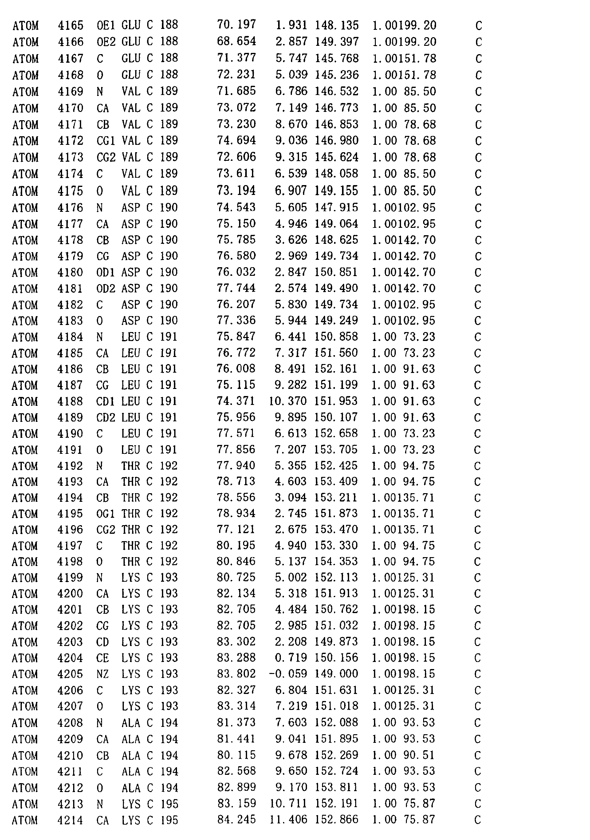
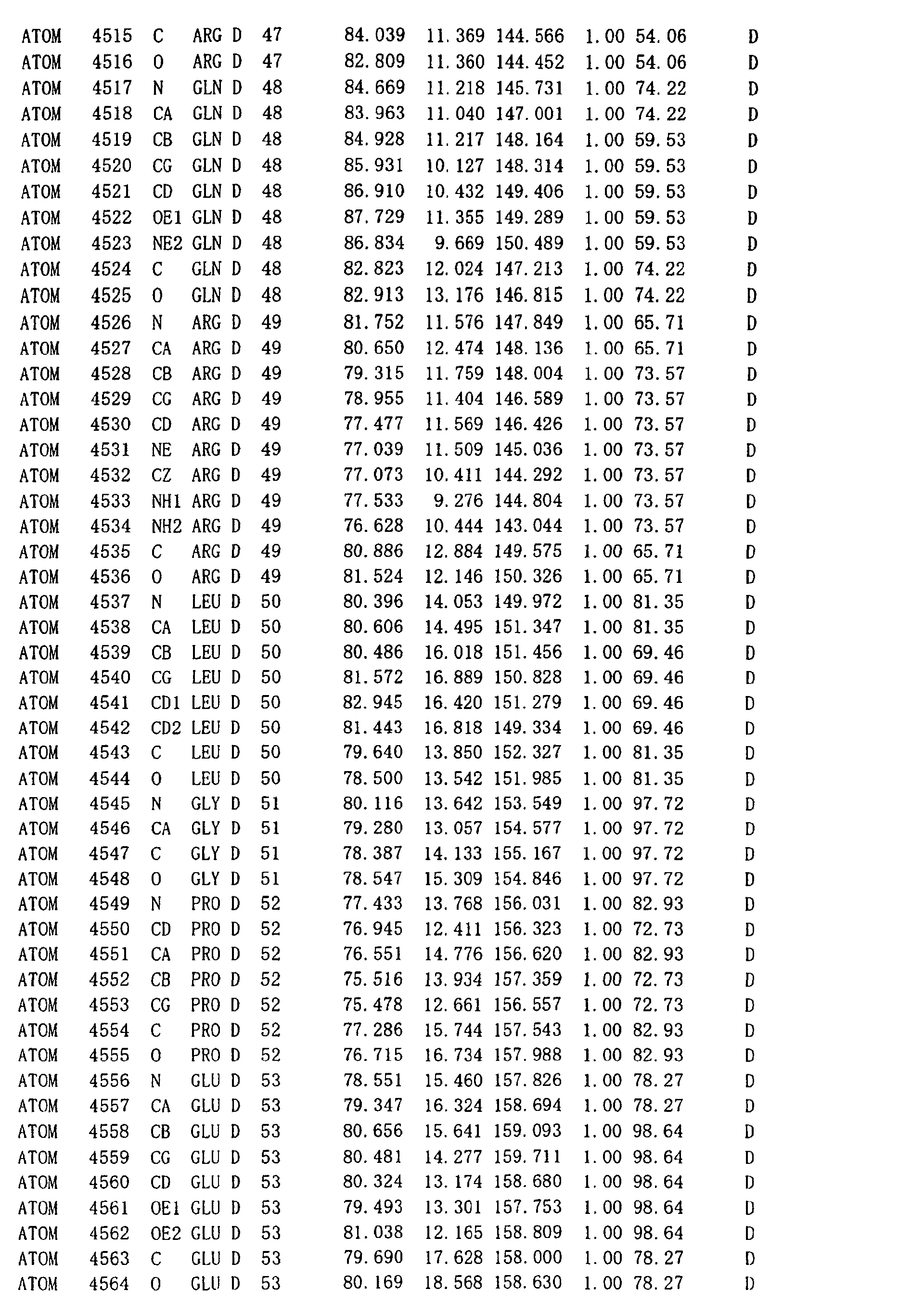
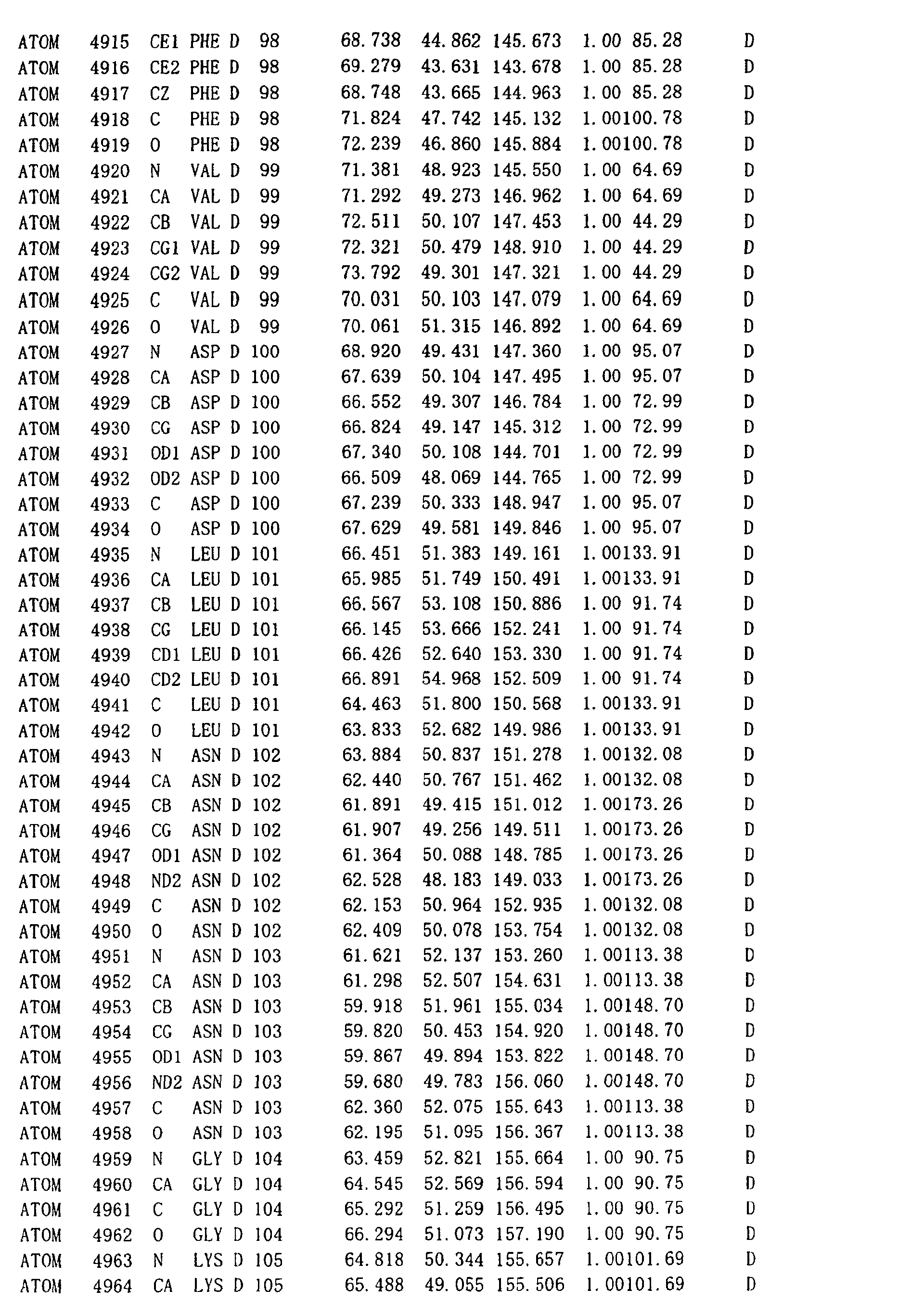
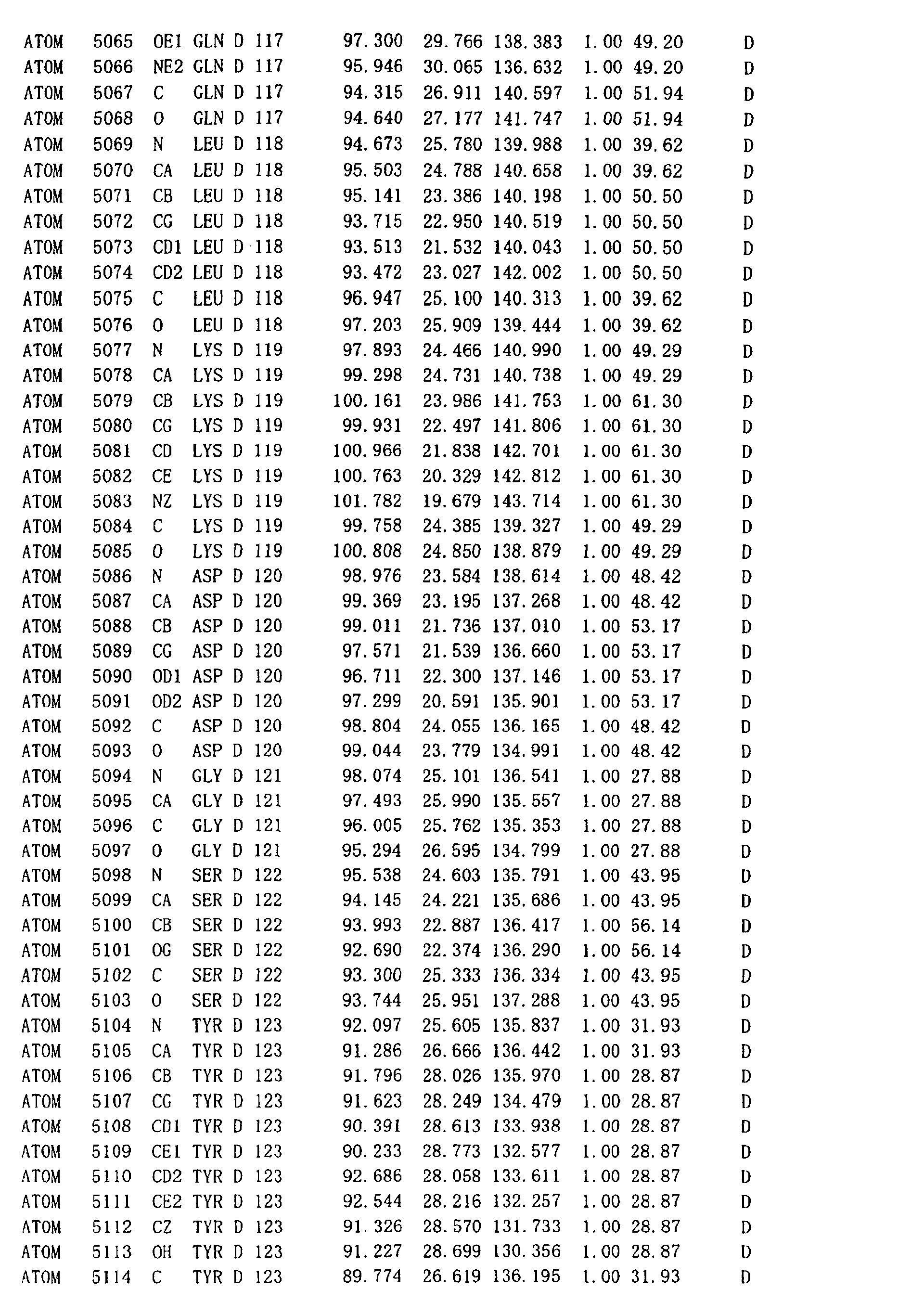
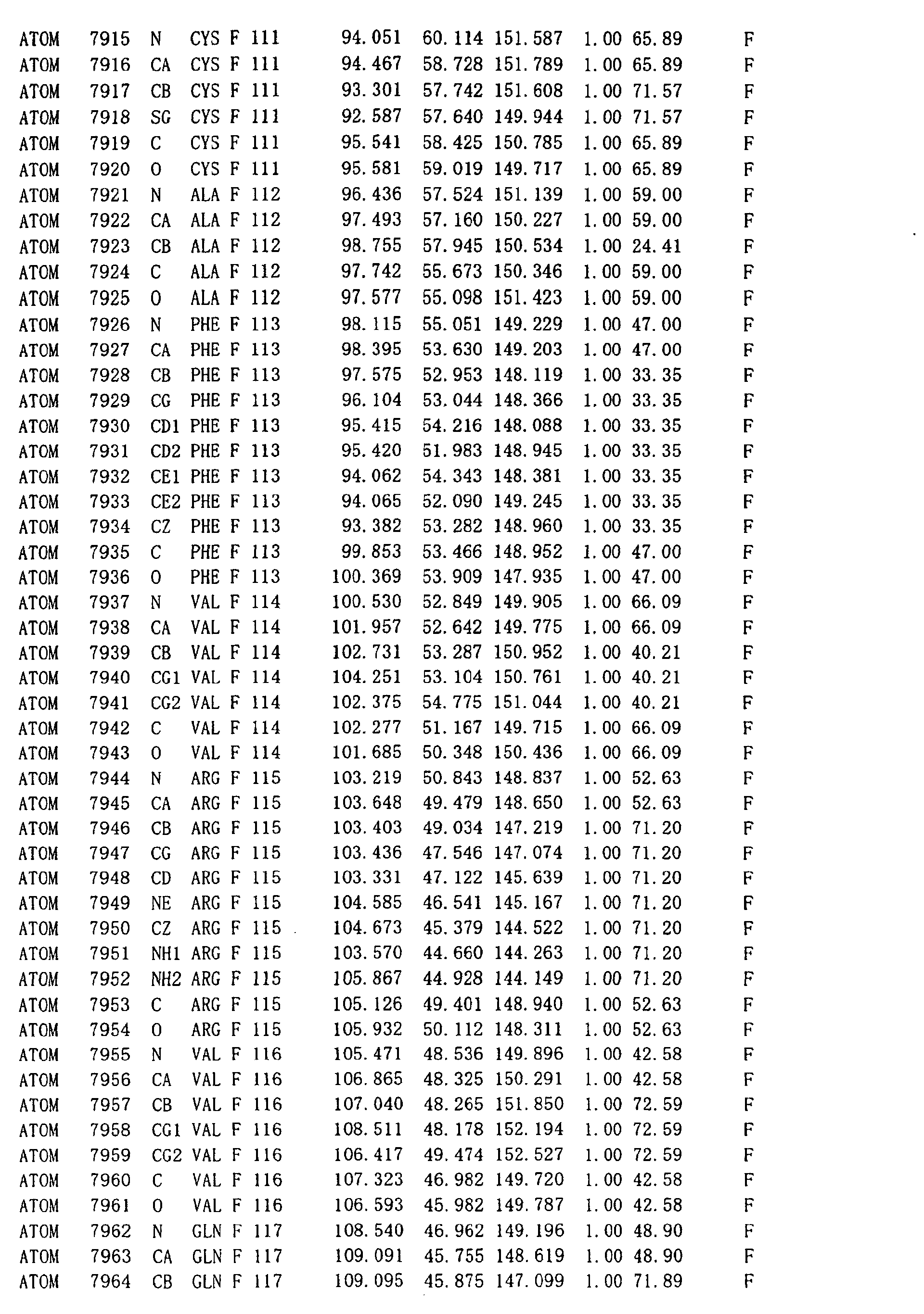
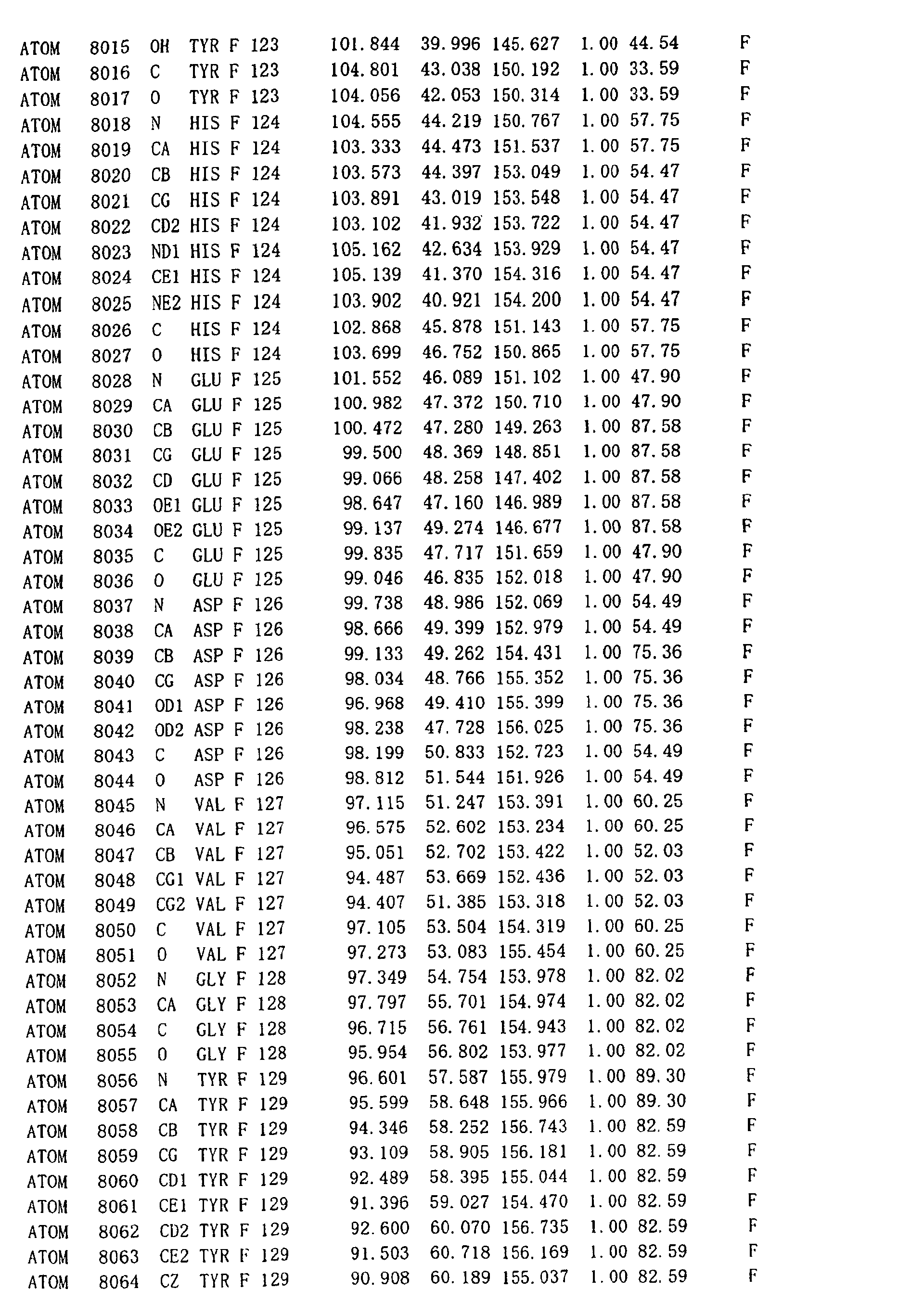
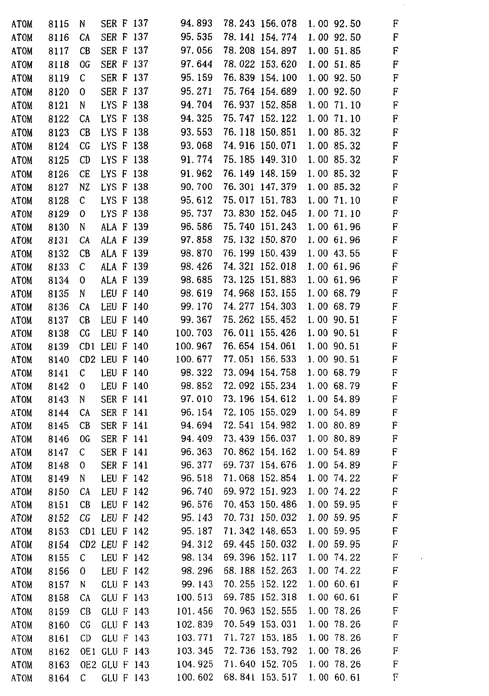
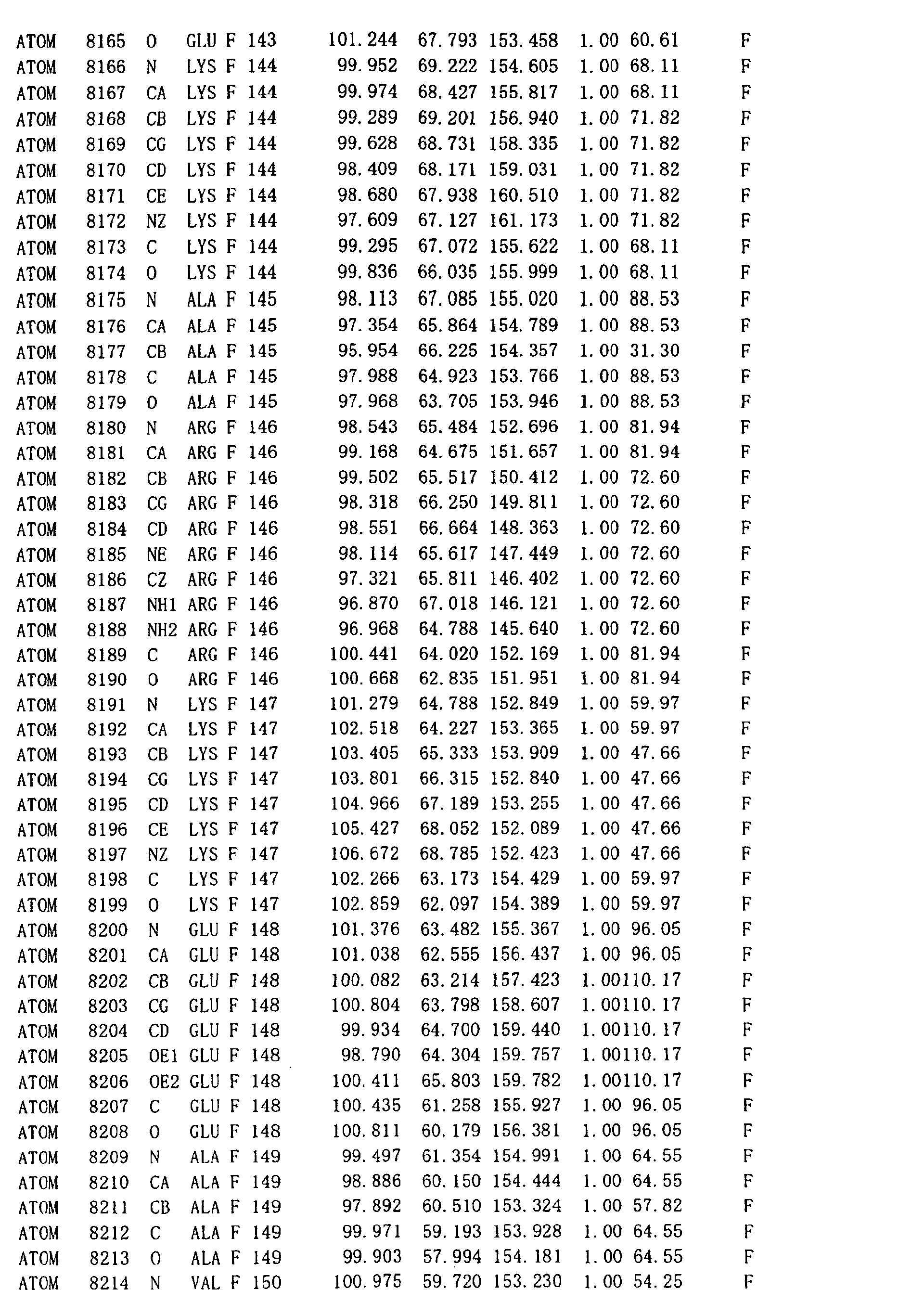
three-dimensional structure of DNA recombination/repair
protein by X-ray crystallography using the crystal, and the
use thereof. More specifically, the invention relates to the
designing and identification of a mutant enzyme based on the
three-dimensional structure, and a rational method for
screening a compound binding to the enzyme.
Description of the Related Art
-
The ability to repair double-stranded DNA breaks that
occur on damaged chromosomal DNA is necessary to maintain the
integrity of the genomic DNA. It has been known that the
double-stranded DNA breaks can result from ionizing radiation,
the administration of DNA-damaging agent, replication errors,
and also from specific enzymes that act during meiosis,
mating-type switching of yeast, and V(D)J recombination of
antibody gene. To repair such chromosomal DNA damage, cells
have acquired homologous recombination and non-homologous DNA
end joining activities. Homologous recombination plays an
important role in the preservation of DNA sequence in chromosomal
DNA. The homologous pairing step of the homologous
recombination, which is the process of searching for sequence
homology in either homologous chromosome or sister chromatids,
is an essential step. Thus, the identification of an enzyme
promoting homologous pairing is one of the most important steps
for understanding homologous recombination.
-
Two homologous pairing enzymes of RecA and RecT are
reported in the case of Escherichia coli. RecA and RecT, which
have no amino acid sequence homology, catalyze homologous
pairing in separate recombinational repair pathways, the RecBCD
and RecF pathways for RecA, and the RecE pathway for RecT. In
eukaryotes, two RecA homologues, Rad51 and Dmc1, have been
identified, and both proteins promote homologous pairing.
Although the Rad51-independent recombination pathway has been
identified, no proteins with significant sequence homology to
RecT have been found in eukaryotes. An electron microscopic
visualization showed that RecT forms a heptameric or octameric
ring structure. Interestingly, the yeast Rad52 protein
(referred to as "SCRad52" hereinafter) and the human Rad52
protein (referred to as "HsRad52" hereinafter) also form ring
structures similar to that of RecT. Stasiak et al. reported
based on their electron microscopicanalysis that HsRad52 shows
a heptameric ring structure (Current Biology, 10, (2000), p.
337-340).
-
RAD52 genes have been identified in eukaryotes including
yeast, chicken, mouse and human. The role of Rad52 in the
Rad51-dependent recombination pathway has been extensively
studied both in vivo and in vitro. The ScRad52 and HsRad52
both bind to single-stranded DNA and promote annealing of
complementary single-stranded DNAs to each other. Inaddition,
it is reported that ScRad52 and HsRad52 together with yeast
and human Rad51 proteins, enhance the joint molecule formation
between circular single-stranded DNA and linear
double-stranded DNA. Therefore, Rad52 protein has been
proposed to be a mediator protein that binds single-stranded
DNA and facilitates the loading of Rad51 onto the recombination
site. In addition to such Rad51-dependent recombination
pathway, the role of Rad52 in Rad51-independent recombination
pathway has also been studied. However its precise molecular
mechanism, particularly the catalytic mechanism of the enzyme
including the DNA-binding mode during the homologous pairing
step has not yet sufficiently been understood.
-
According to JP-T-2001-502545, a complex of eukaryotic
Rad52 protein with nucleic acid, and a method for screening
a biologically active substance involved in the homologous
recombination using the complex are disclosed. According to
themethod disclosed in the publication, however, the eukaryotic
Rad52 protein should be purified and isolated after its
expression by recombinant DNA technology. Thus, itisdifficult
to directly assay the homologous recombination activity of the
Rad52 in eukaryotic cells. Because the assay procedure for
the homologous recombination activity is complicated,
screening of numerous compounds using the complex is extremely
inefficient.
-
Meanwhile, currently, it is known that certain types of
cancer and other genetic diseases are caused by abnormalities
in chromosomal DNA. For example, many oncogenes have been
identified in humans. Most of oncogenes identified so far have
been known to cause amino acid substitution in the resulting
expressed protein due to a mutation in the gene in normal cell
(proto-oncogene), frequently due to point mutation. The
mutated expressed protein exhibits abnormal biological
functions to cause carcinogenesis of cells.
-
For example, Ras is one of the most extensively studied
oncogenes. Through the substitution of specific amino acid
residues in the Ras protein, the Ras protein is activated to
stimulate abnormal proliferation of cells and thereby induce
carcinogenesis in cells.
-
Alternatively, another important oncogene encodes an
intracellular protein p53. The protein p53 is believed to be
a carcinogenesis-suppressor gene. It is revealed that p53
inactivation via mutagenesis induces carcinogenesis of cells.
Thus, attempts have been made for cancer therapy to introduce
a gene expressing the normal p53 protein in cancer cells (US
Patent No. 6017524).
-
Because chromosomal DNA damage and the introduction of
mutation into DNA due to the damage are closely associated with
cellular carcinogenesis through oncogene activation and the
inactivation of carcinogenesis-suppressor gene, as described
above, the preservation of genomic integrity with DNA
recombinational repair, and stable transmission of genomic
information may possibly be useful for the prophylaxis and
therapeutic treatment of cancer. That is, in cells with
significantly reduced DNA recombination/repair protein
activity, it is considered that the enhancement of the activity
will possibly suppress carcinogenesis by repairing chromosomal
DNA damage.
-
On the other hand, the suppression of DNA
recombination/repair protein activity will possibly enhance
anticancer therapies, particularly radiotherapy or
chemotherapy. Because ionizing radiation and radioactive
drugs are generally used to damage DNA to treat cancer cells,
it is suggested that the administration of compounds inhibiting
DNA recombination/repair protein activity enhance the
sensitivity of cancer cells to the radiotherapy and
chemotherapeutic agents (see JP-T- 2001-508868) . In addition,
because homologous recombination of chromosomal DNA is involved
in the integration of virus DNA and the like in gene therapy,
such homologous recombination can be used in a manner to modulate
DNA recombinational repairing activity to enhance the
efficiency of the introduction of foreign gene into a specific
site of chromosomal DNA, and to increase the ultimate use of
gene therapy.
-
Although the role of the DNA recombinational repair
mechanism and its applications are so significant as described
above, it is very difficult to assay the Rad52-specific DNA
recombination activity or to elucidate the physiological role
thereof, because various DNA recombination/repair proteins
exist in cells as described above. Additionally, the
relationship between the structure and functions of Rad52 in
homologous DNA recombinational repair has not yet been
elucidated.
-
To solve the problems described above, it is an object
of the present invention to obtain a single crystal of the Rad52
protein as one of DNA recombination/repair proteins to
demonstrate the three-dimensional structure by X-ray
crystallography and elucidate the relationship between the
structure and the expression mechanism of the DNA
recombinational repairing activity.
-
It is an another objective of the present invention to
identify structural characteristics influencing the DNA
recombinational repairing activity based on the
three-dimensional structure of Rad 52 and to design a mutant
enzyme with various modulations for the enhancement,
suppression or stabilization of the enzyme activity.
-
Further, it is a still additional objective of the present
invention to provide a method for screening a compound capable
of modulating the DNA recombinational repairing activity via
binding to Rad52 and also to provide an inhibitor or an activator
obtained by using the method.
SUMMARY OF THE INVENTION
-
With attention focused on the fact that the N-terminal
region of the Rad52 protein is essential for DNA binding and
the appearance of homologous pairing activity, the inventors
screened the N-terminal region necessary and sufficient for
the enzyme to express the function thereof by forming a mul timer
complex. Further, the inventors crystallized the protein and
carried out the X-ray crystallographic analysis using the
crystal. Consequently, the inventors first revealed that the
N-terminal region of the Rad52 protein formed an undecameric
ring structure and could bind in the periphery of the ring
structure to DNAs, and have finally led to the completion of
the present invention.
-
That is, the present invention relates to the crystal
of the Rad52 protein as a DNA recombination/repair protein,
a method for producing the crystal, the analysis of the
three-dimensional structure using the crystal, and a method
for screening a compound binding to various mutant enzymes and
the enzyme complex using the three-dimensional structure thus
obtained. More specifically, the invention is as follows.
- (1) A polypeptide fragment comprising the amino acid
sequence from position 1 to position 192 in the amino acid
sequence defined in SEQ ID NO: 1, wherein the amino acid residues
at positions 55, 65, 69, 70, 153 and 169 form a DNA binding
site.
- (2) Apolypeptide fragment derived from the polypeptide
fragmentof (1) aspreparedbydeletion, substitutionoraddition
of one or several amino acids, the polypeptide fragment having
a DNA binding activity.
- (3) A DNA recombination/repair protein complex
comprising an assembly of the following protein (a) or (b),
wherein said complex forms an undecameric ring structure:
- (a) a protein consisting of the amino acid sequence
of SEQ ID NO:1:
- (b) a protein derived from the amino acid sequence of
SEQ ID NO:1 as prepared by deletion, addition, insertion or
substitution of one or several amino acids, the protein having
a DNA recombinational repairing activity.
- (4) A mutant DNA recombination/repair protein complex
comprising an assembly of theprotein fromposition 1 to position
212 in the amino acid sequence of SEQ ID NO:1, wherein the complex
forms an undecameric ring structure.
- (5) The mutant DNA recombination/repair protein
complex of (4), wherein the complex has a three-dimensional
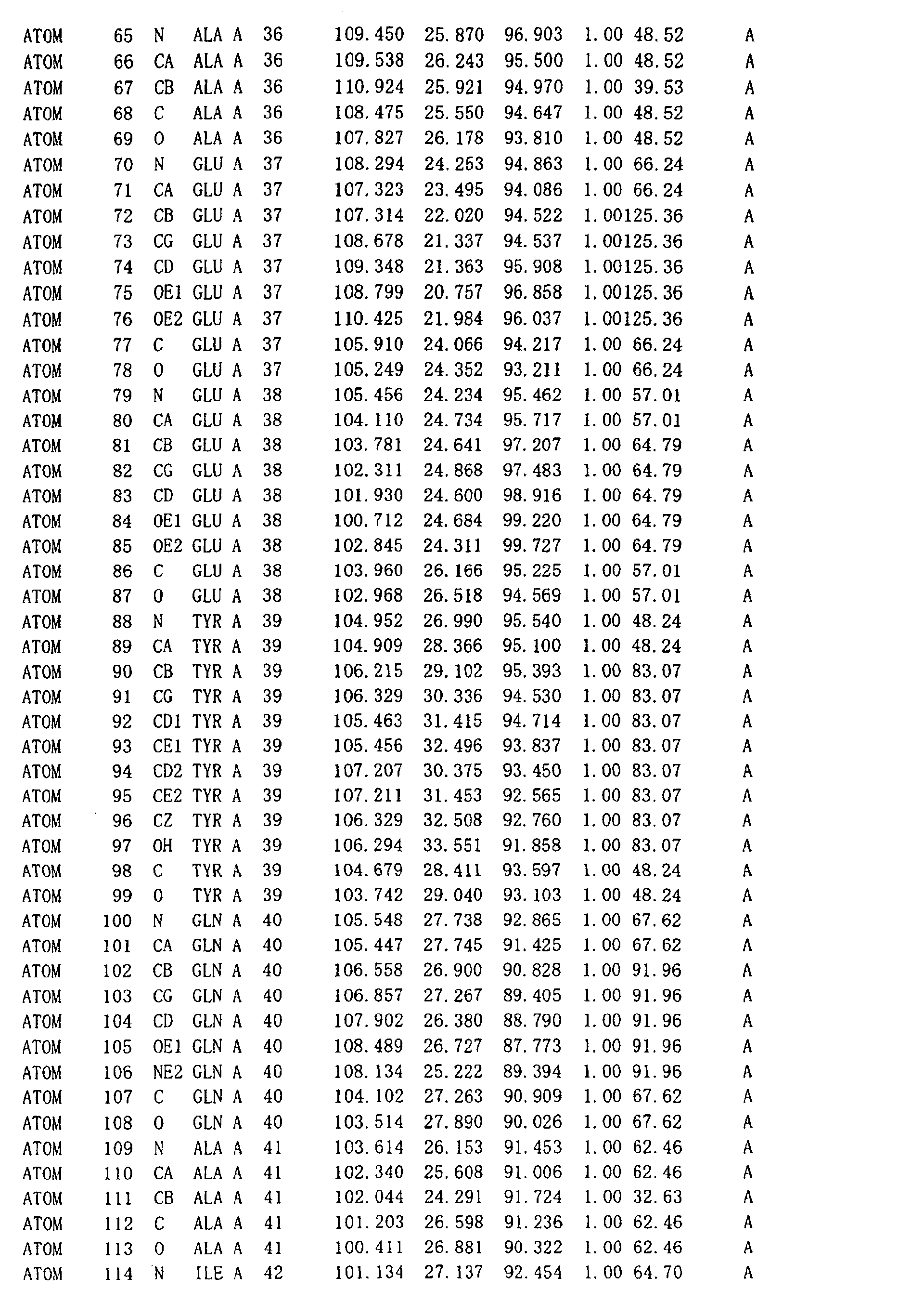
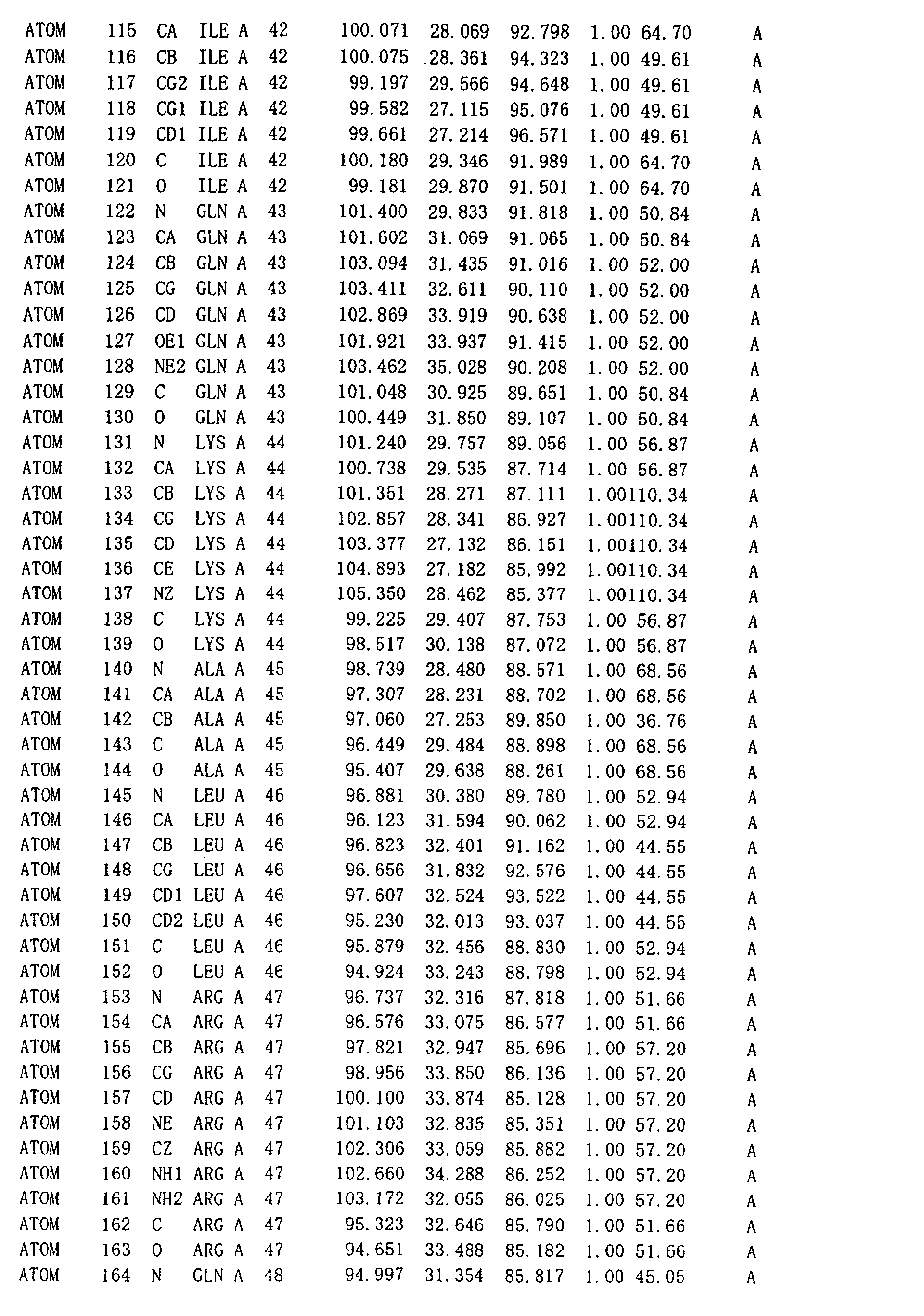
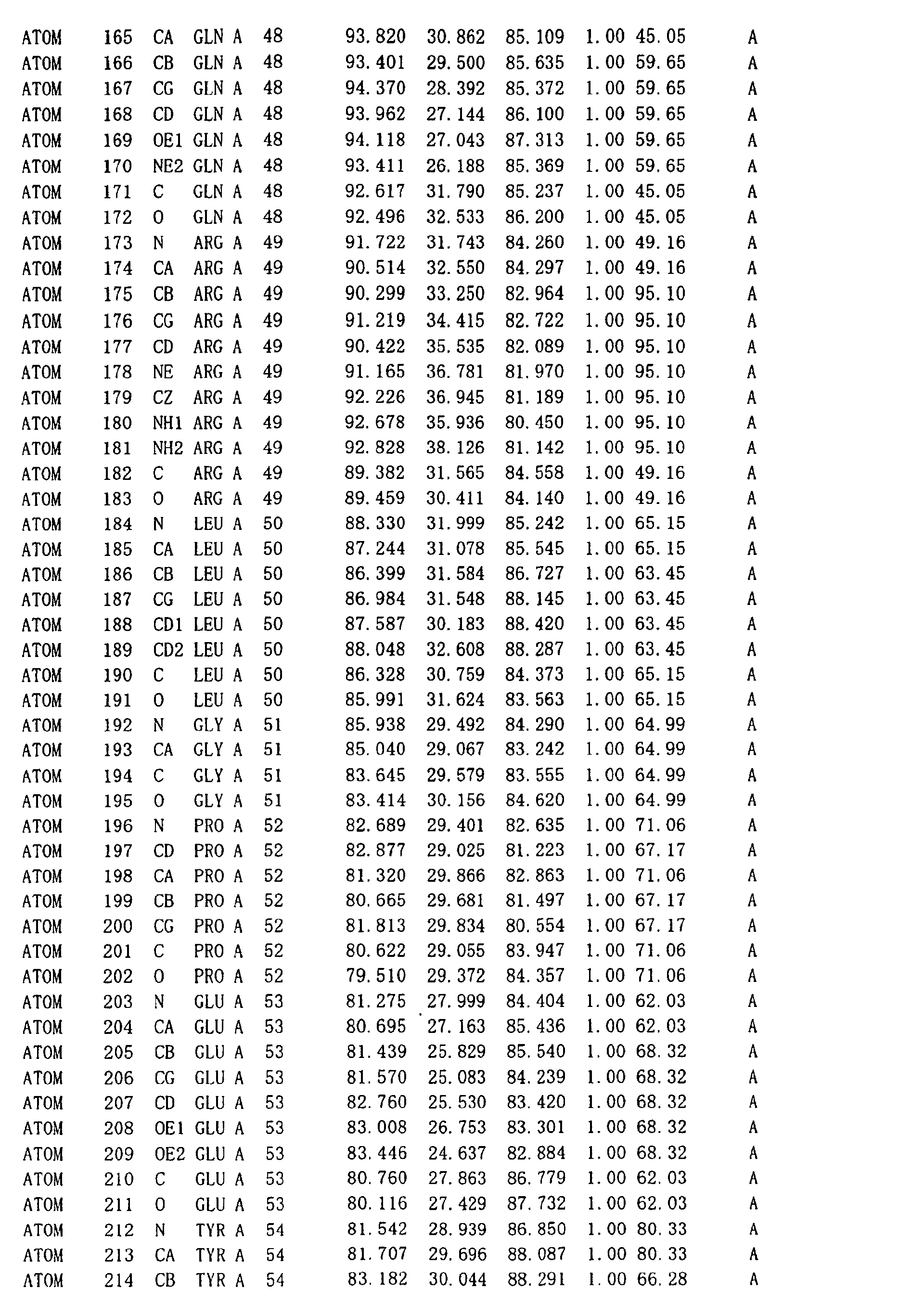
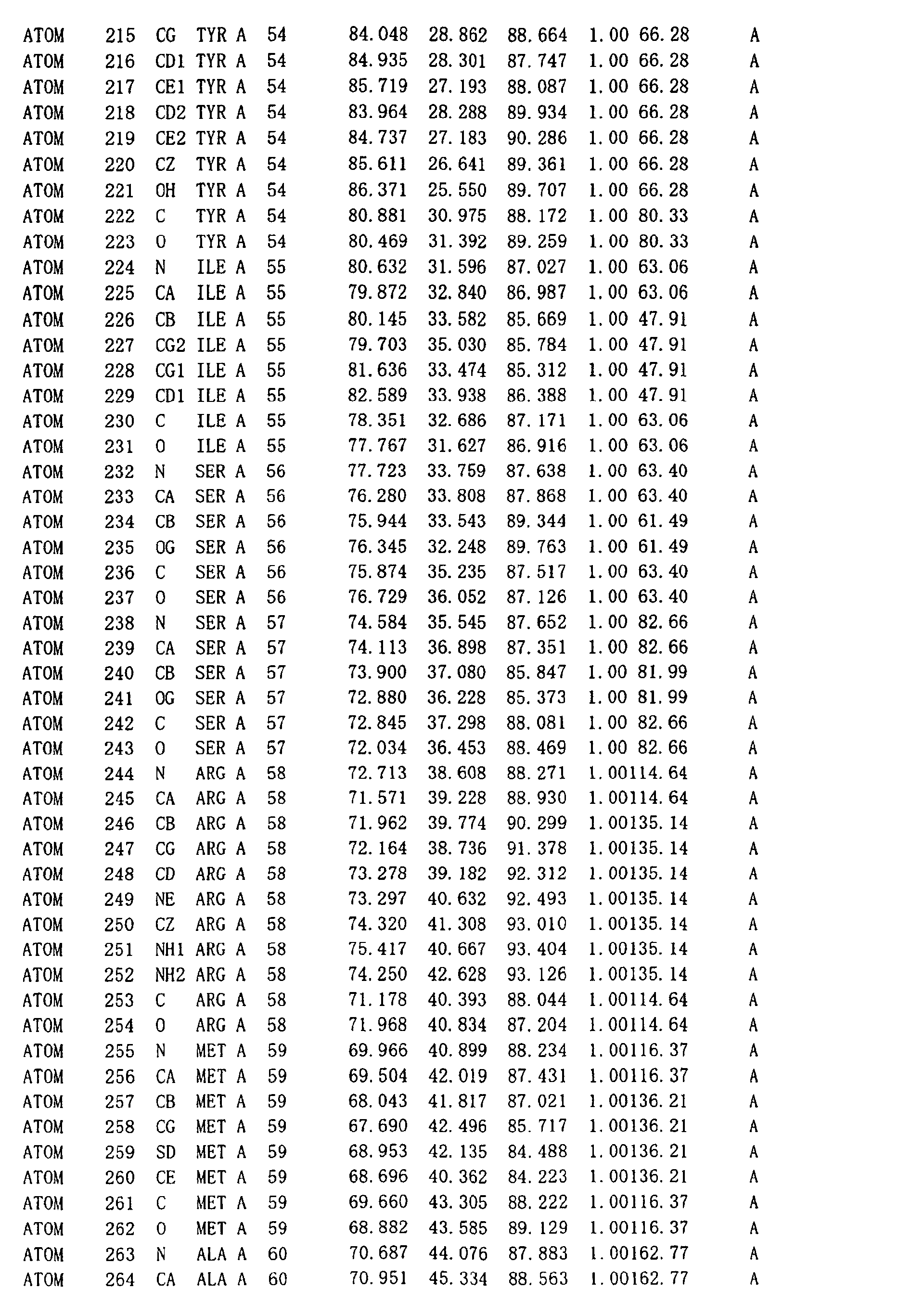
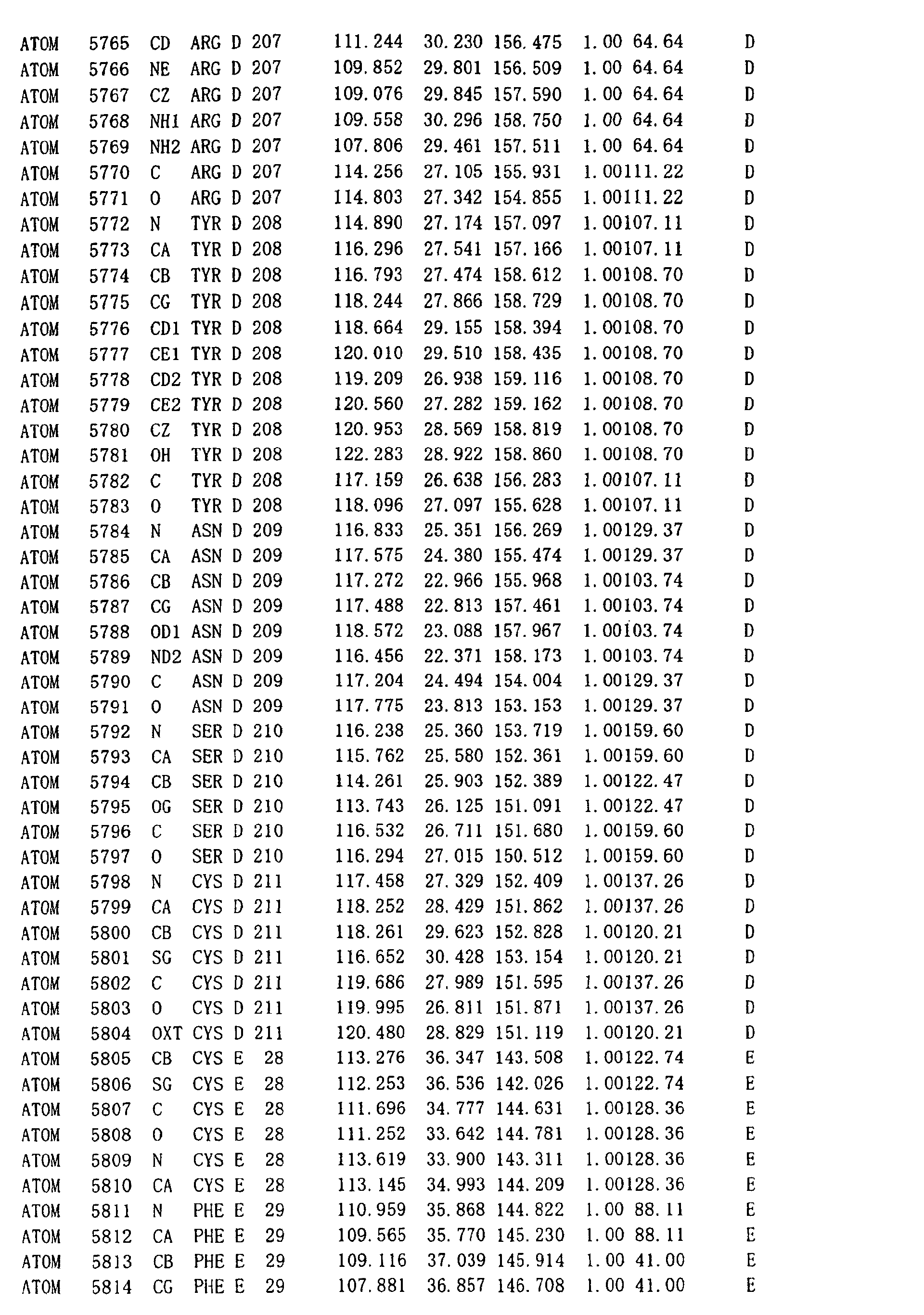
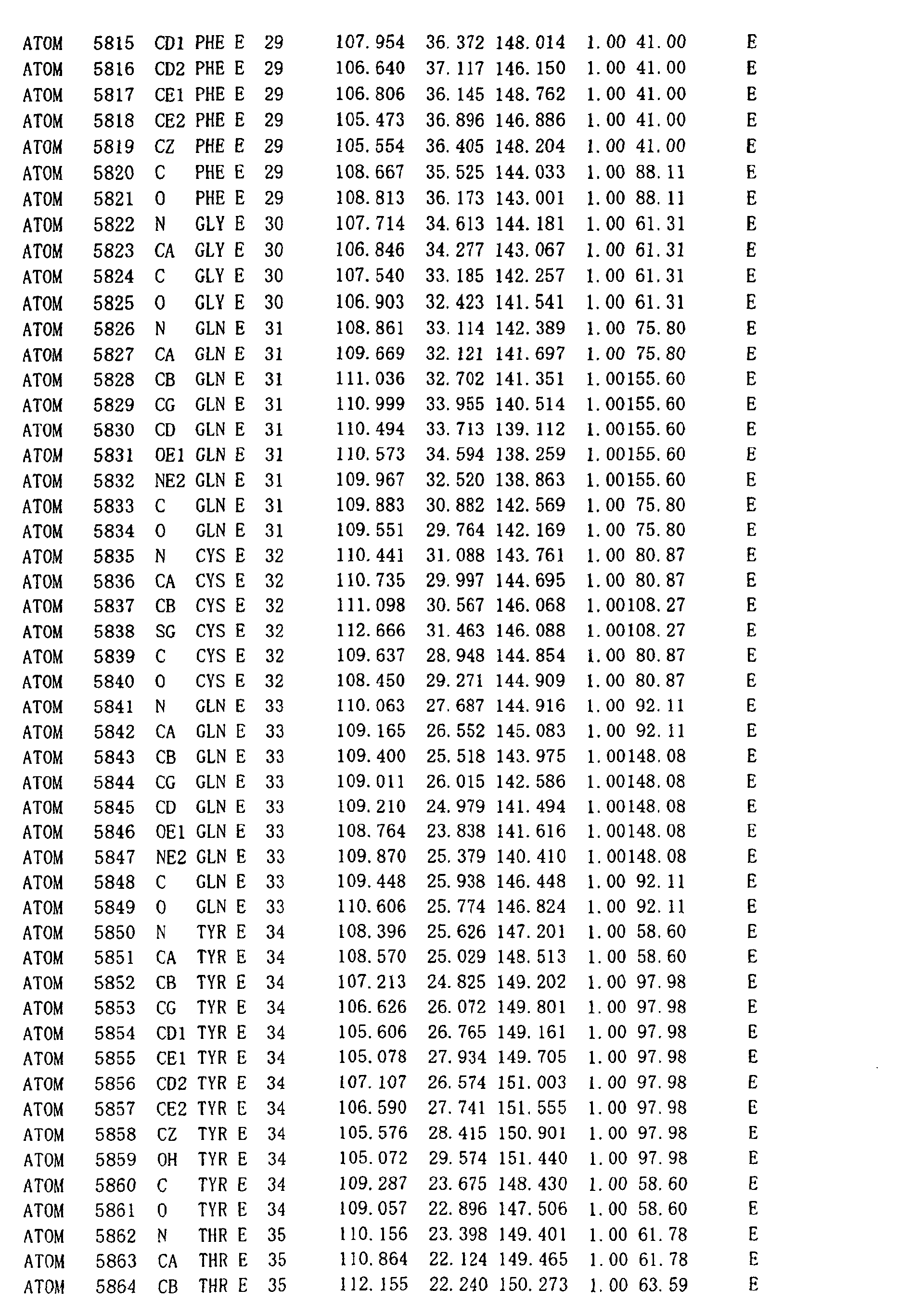
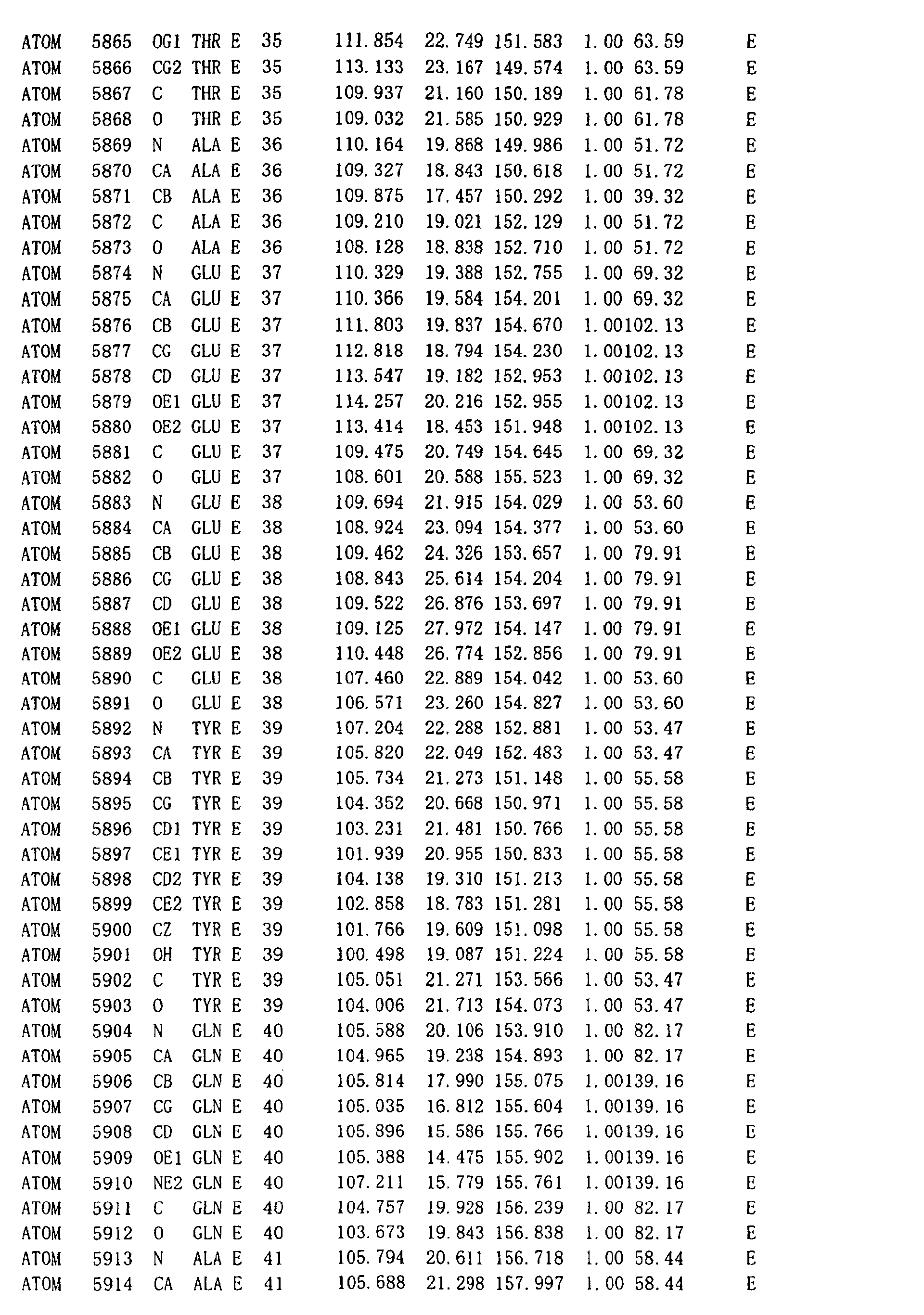
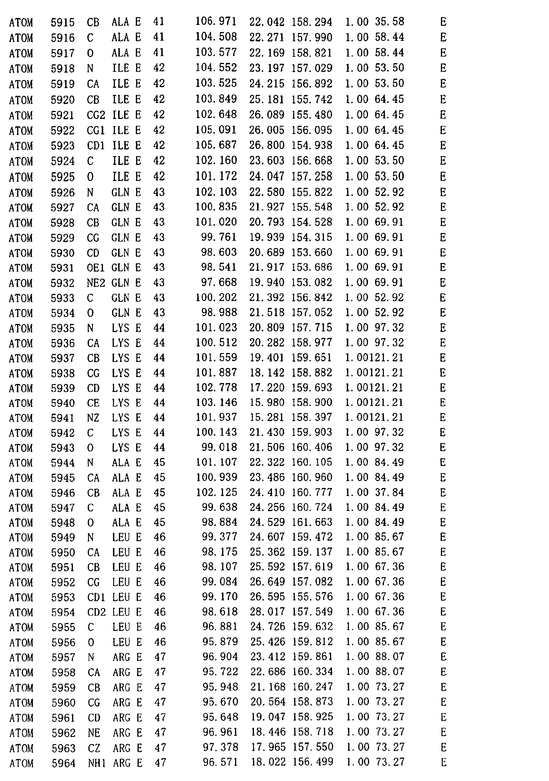
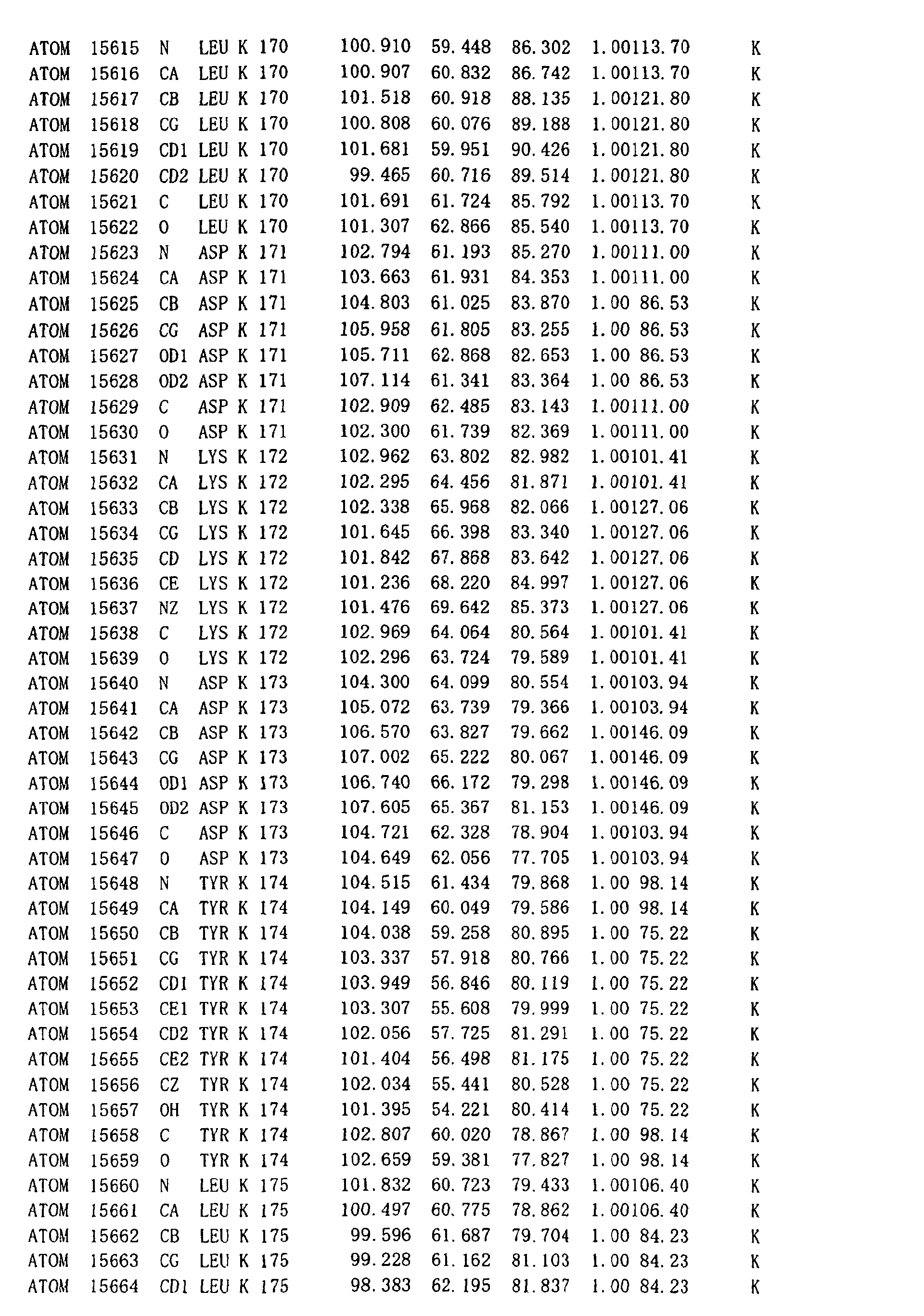
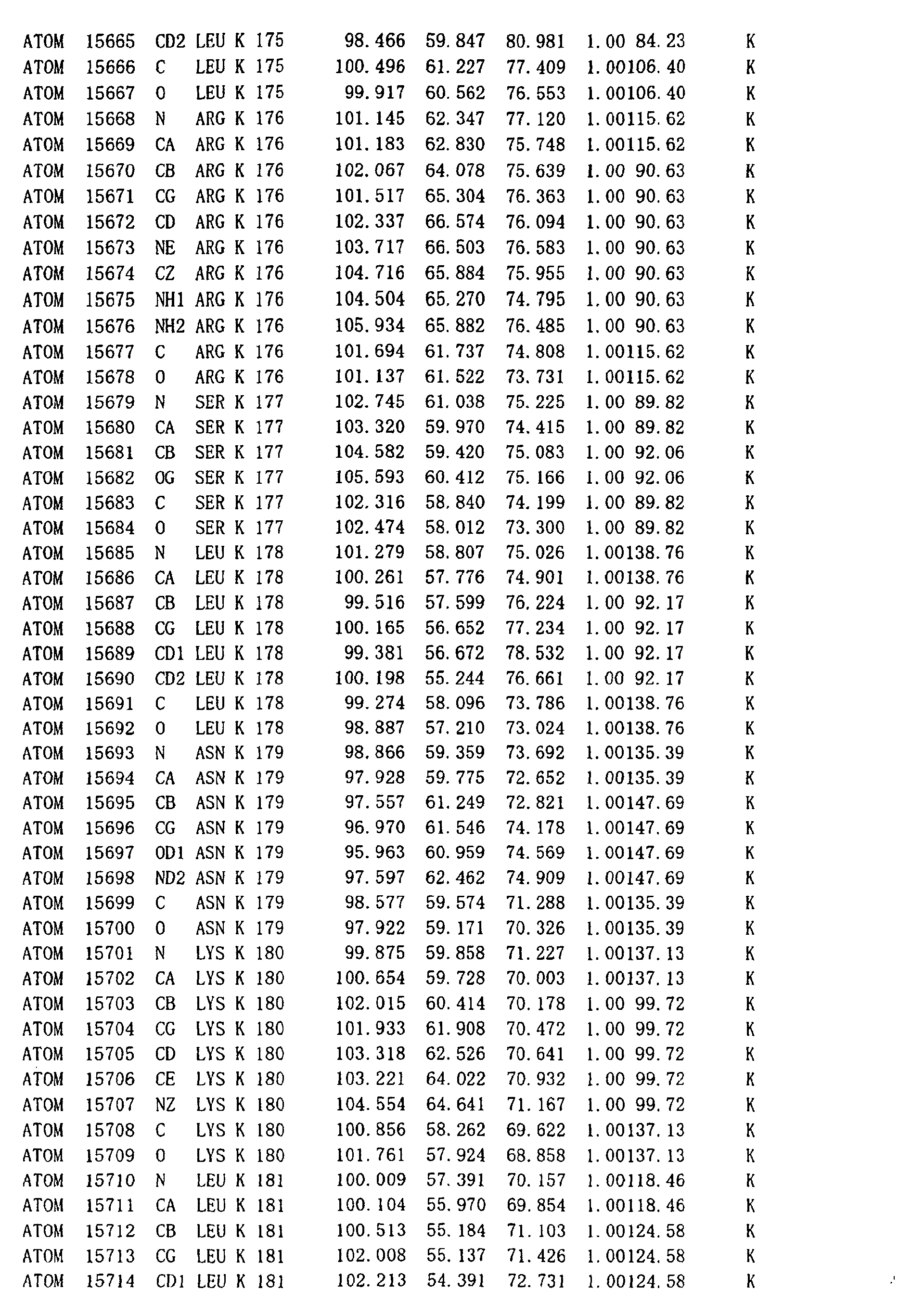
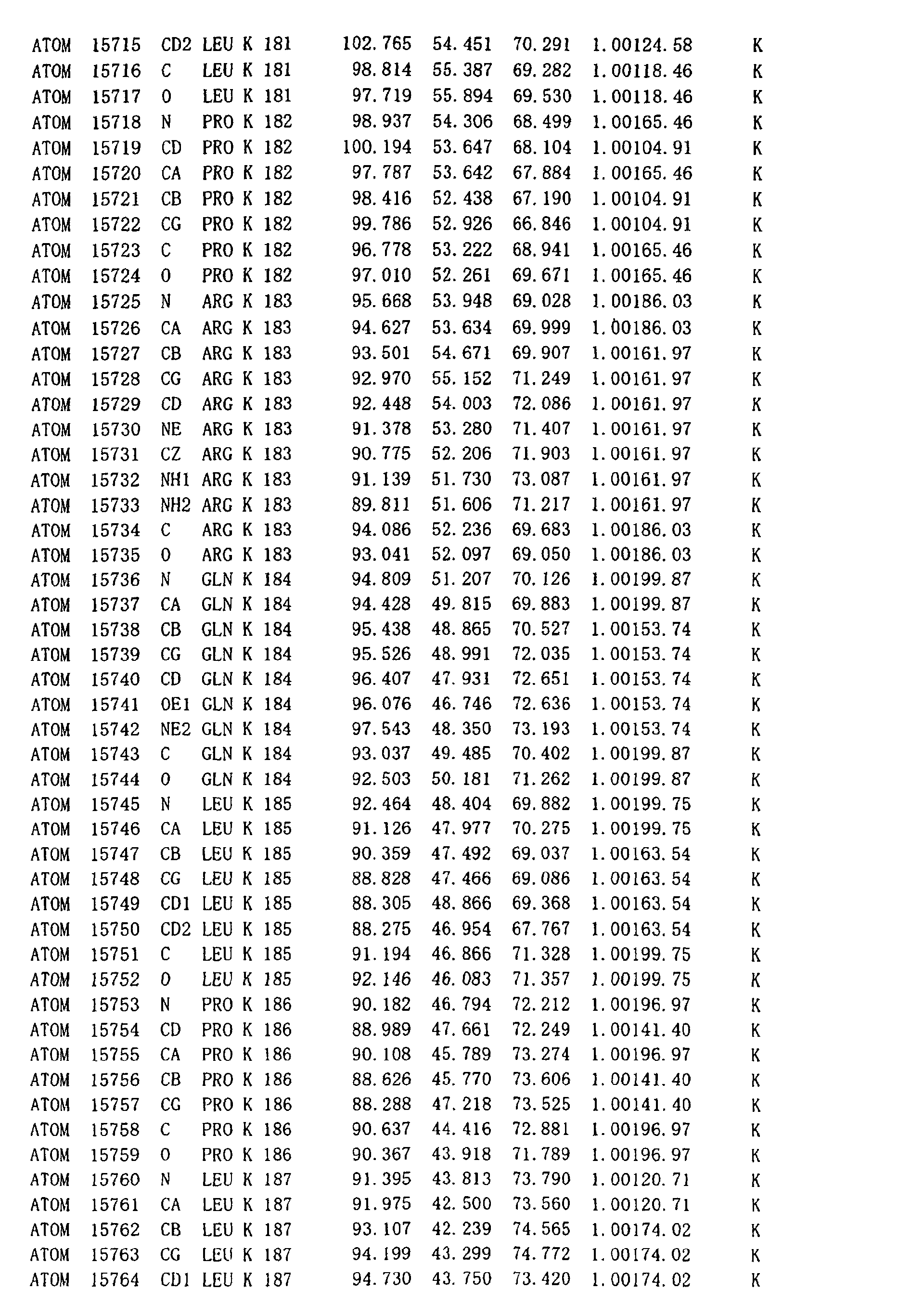
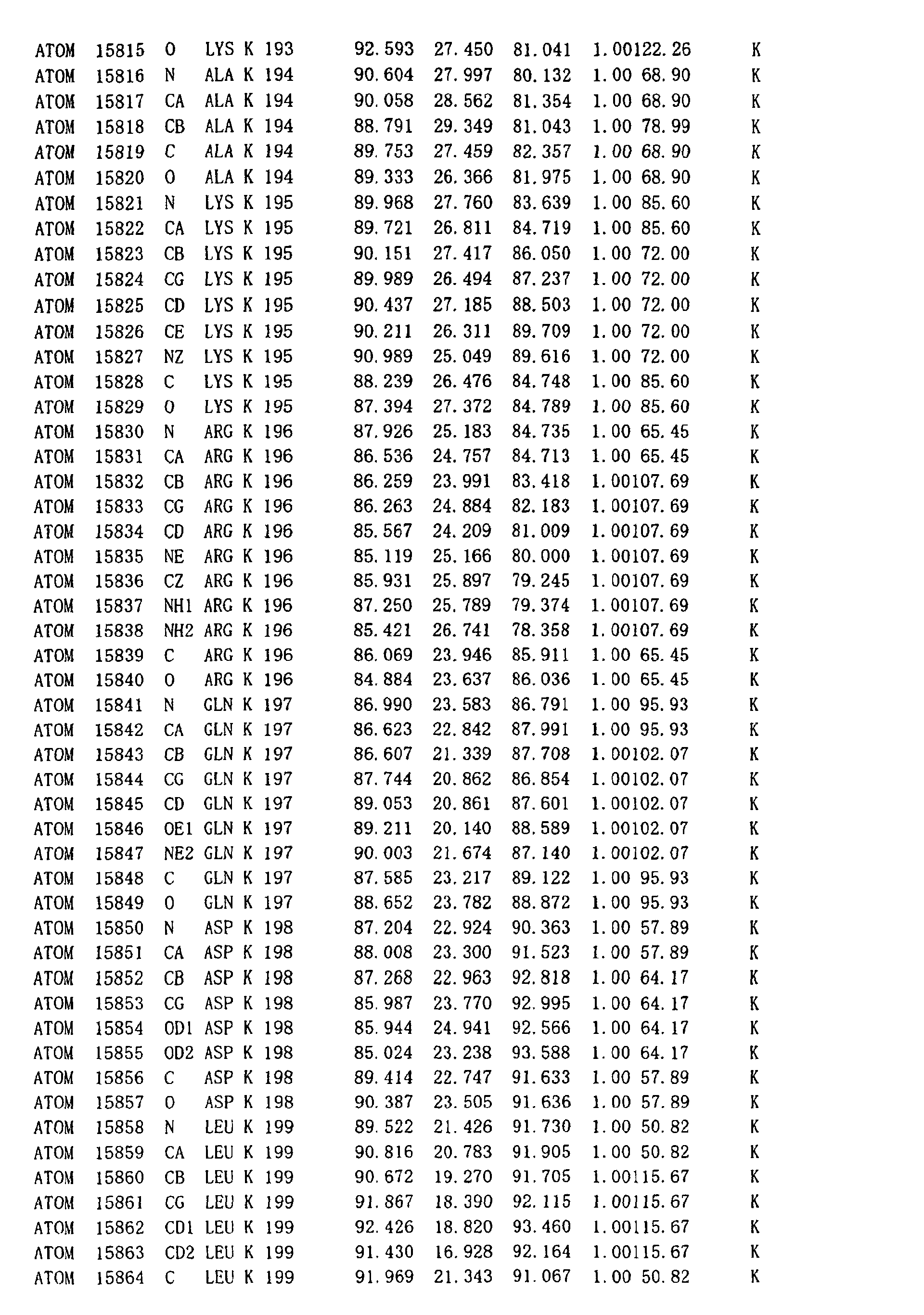
structure substantially defined by the atomic coordinates shown
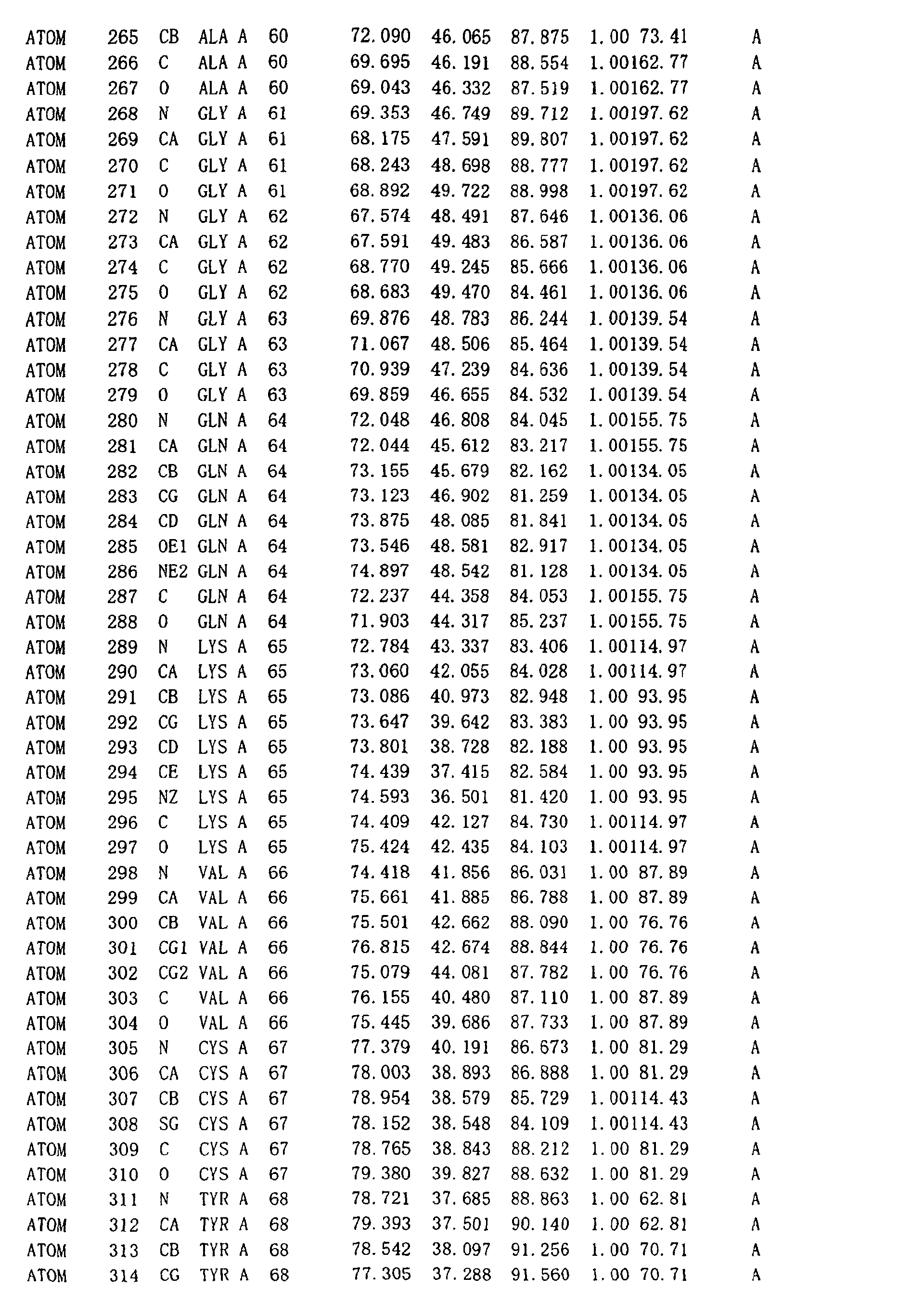
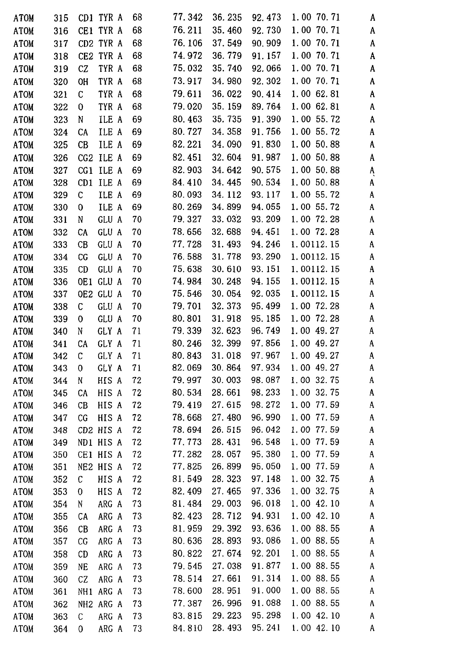
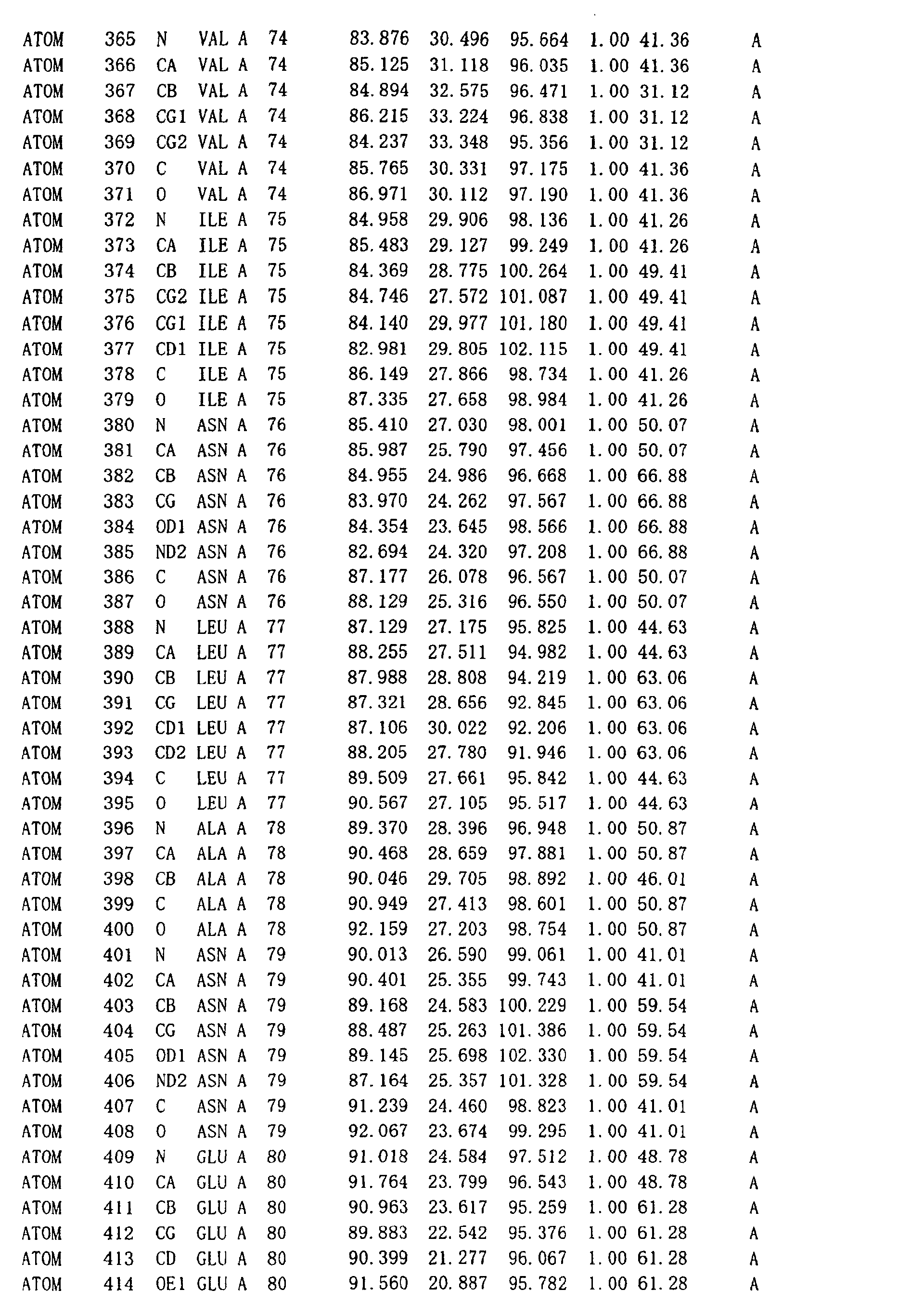
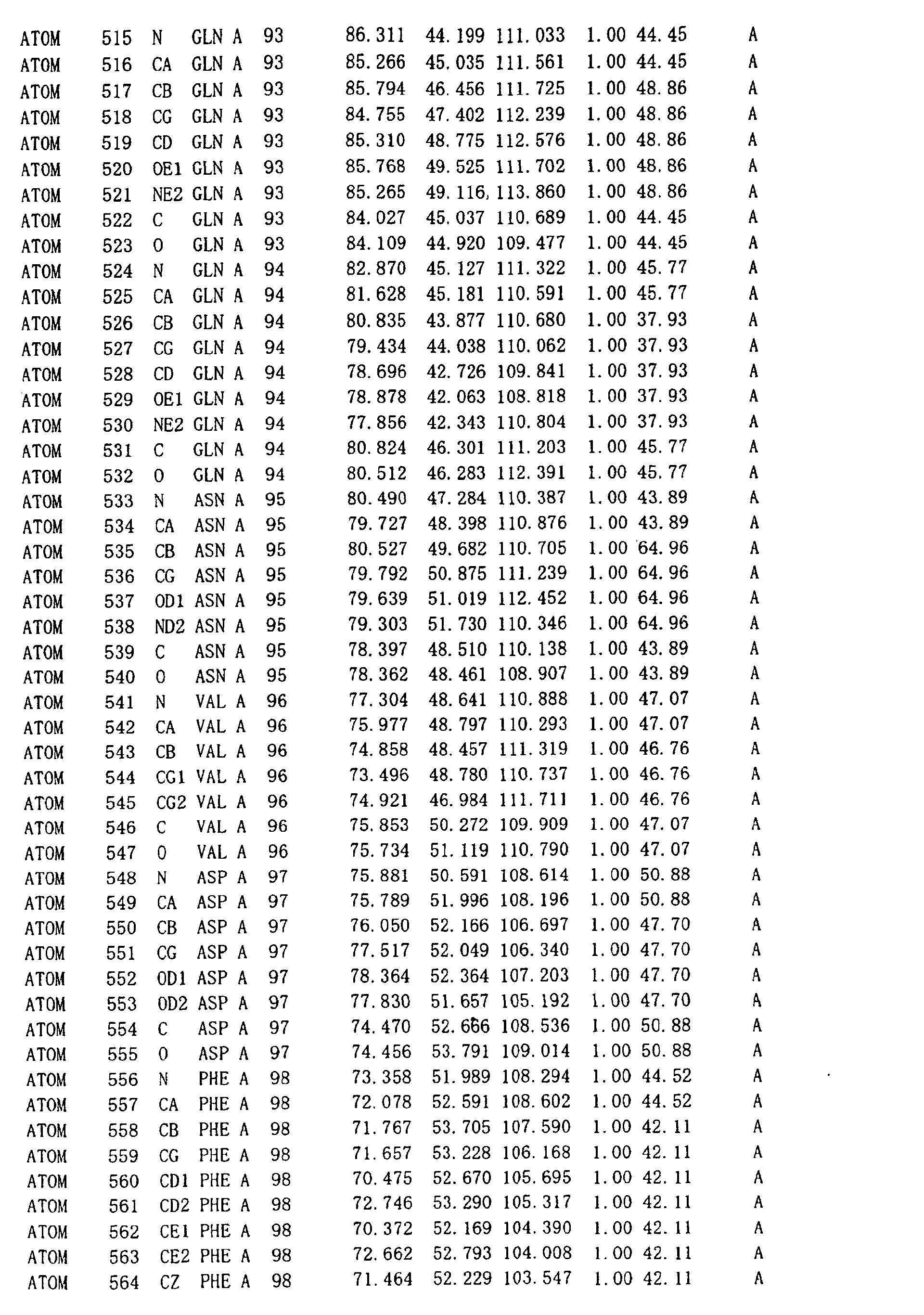
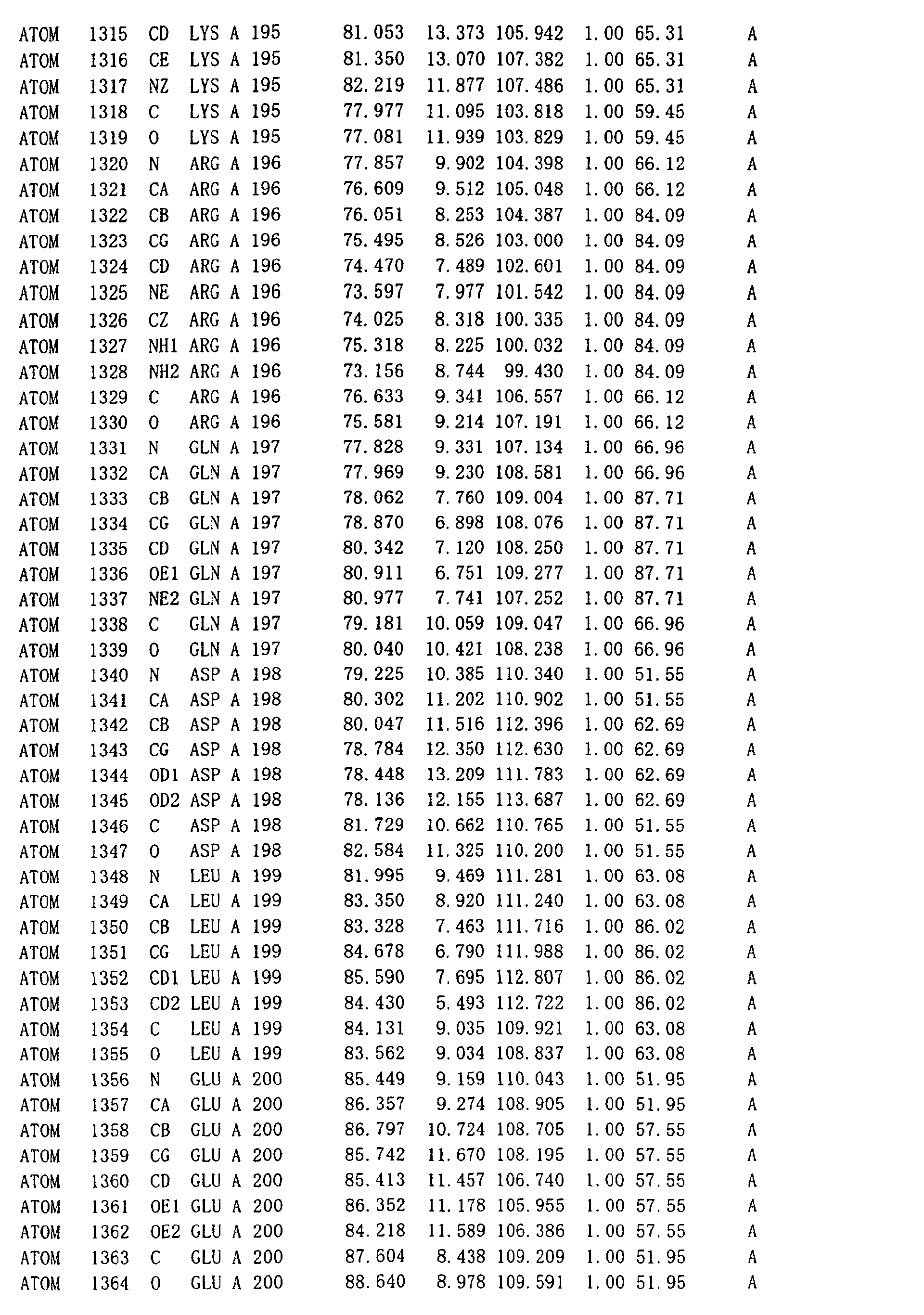
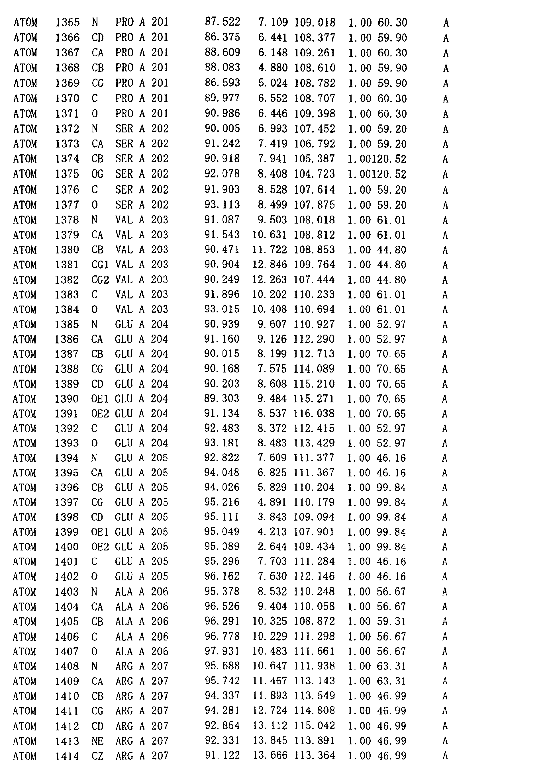
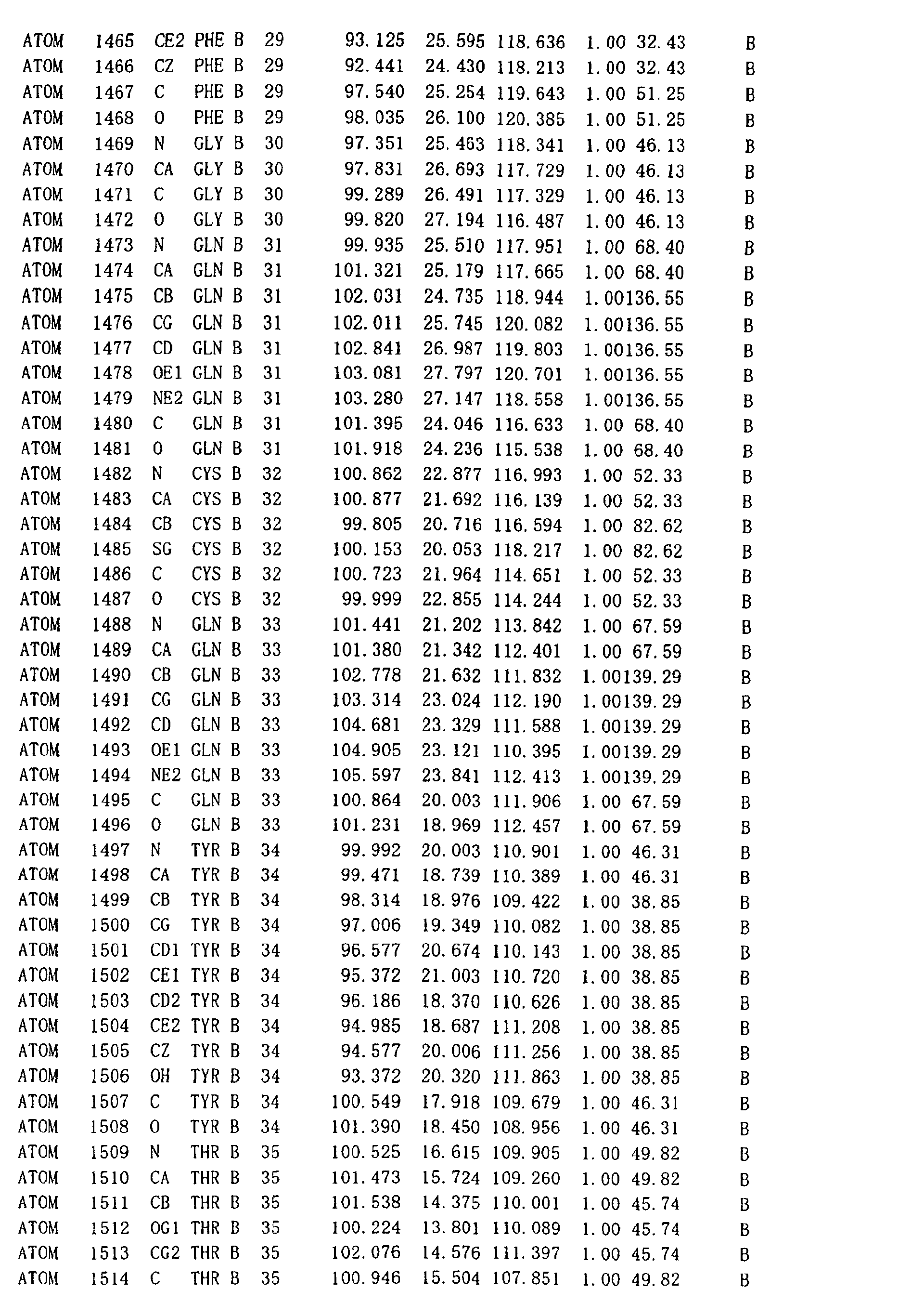
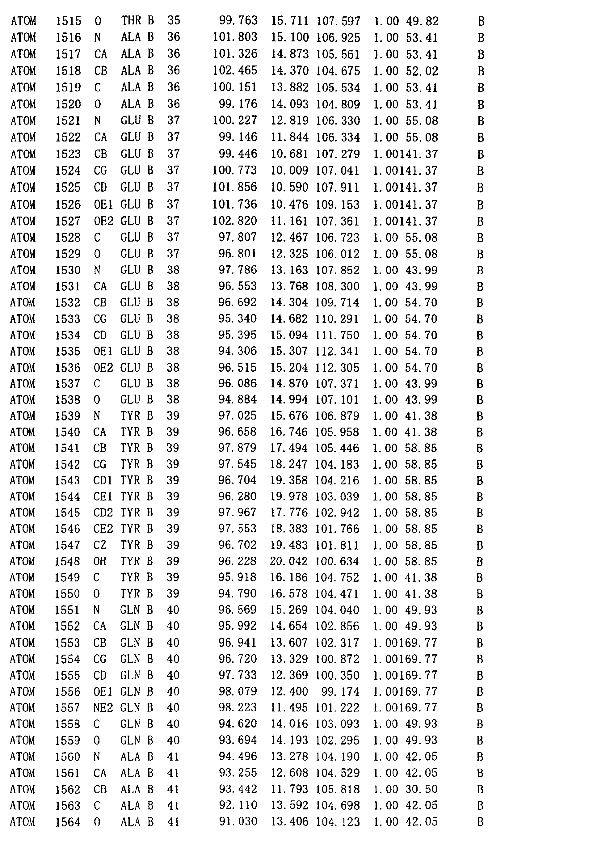
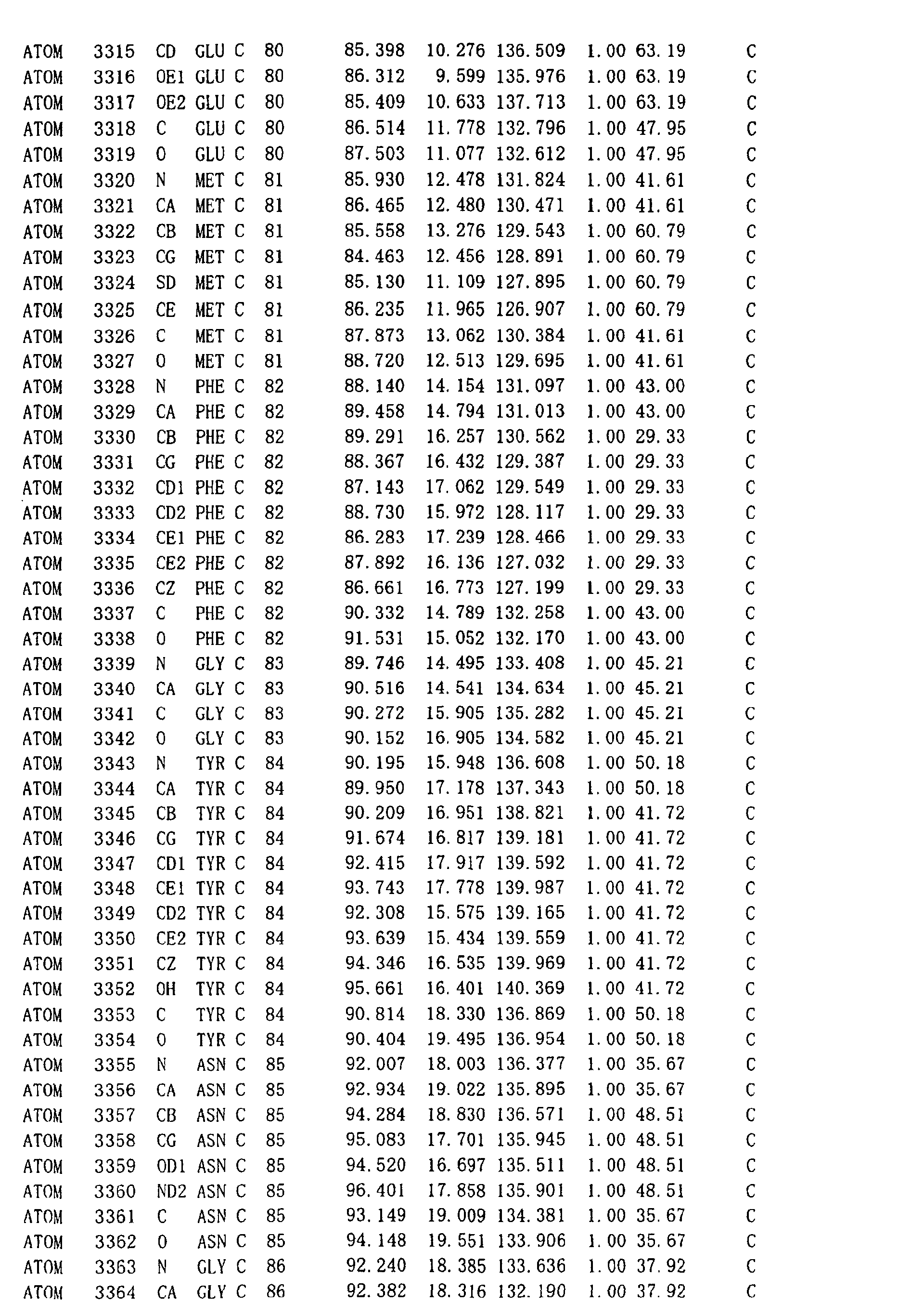
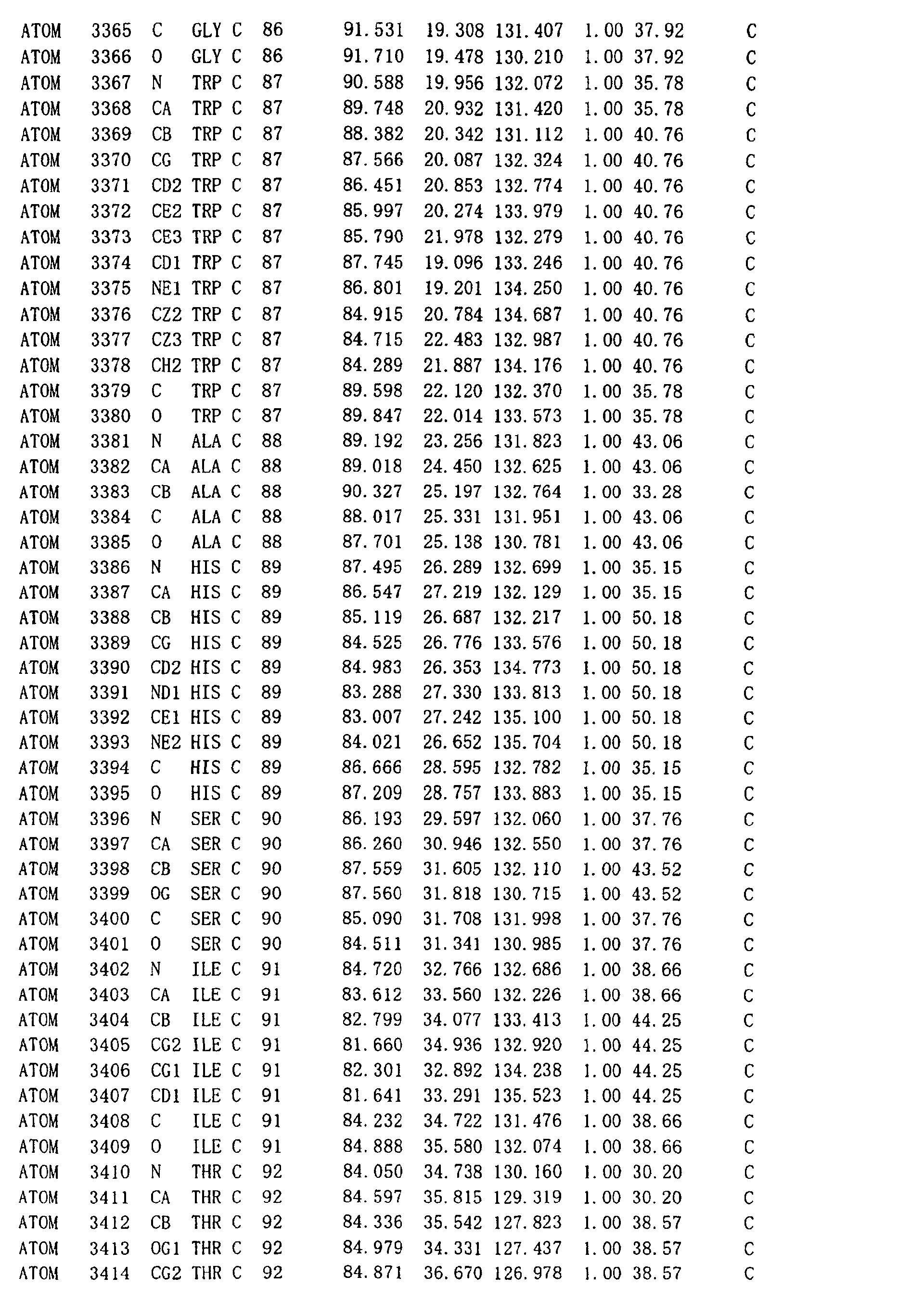
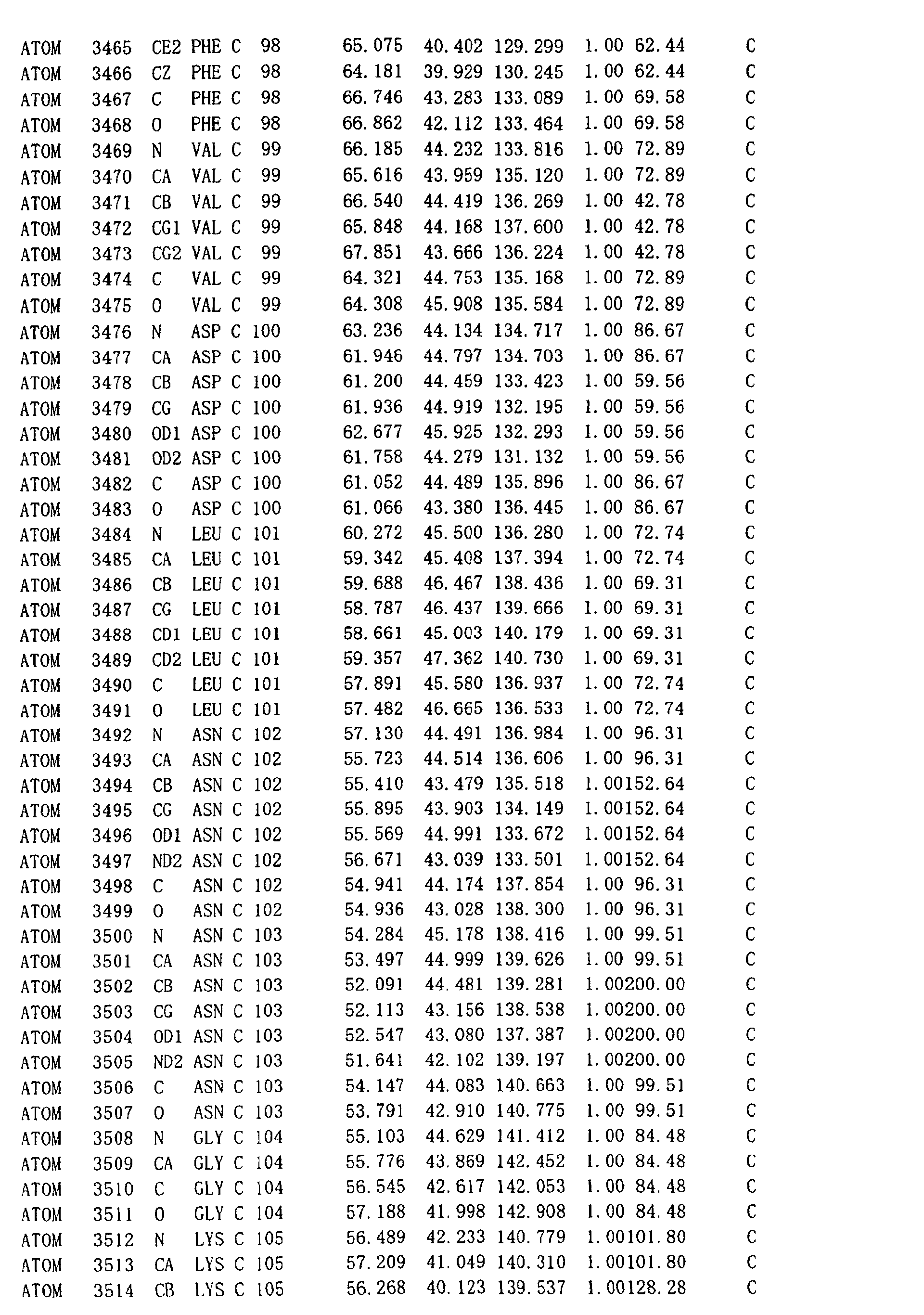
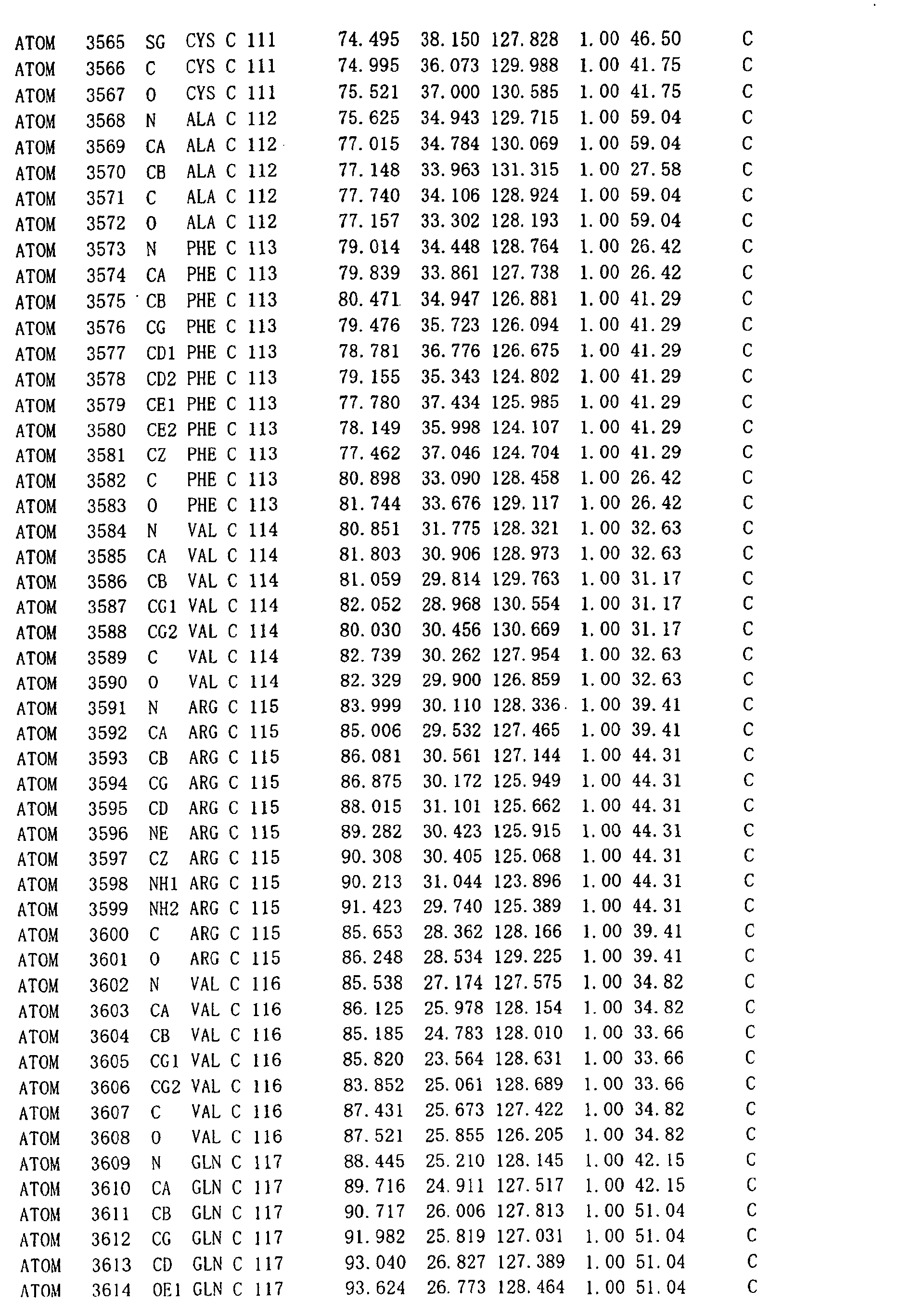
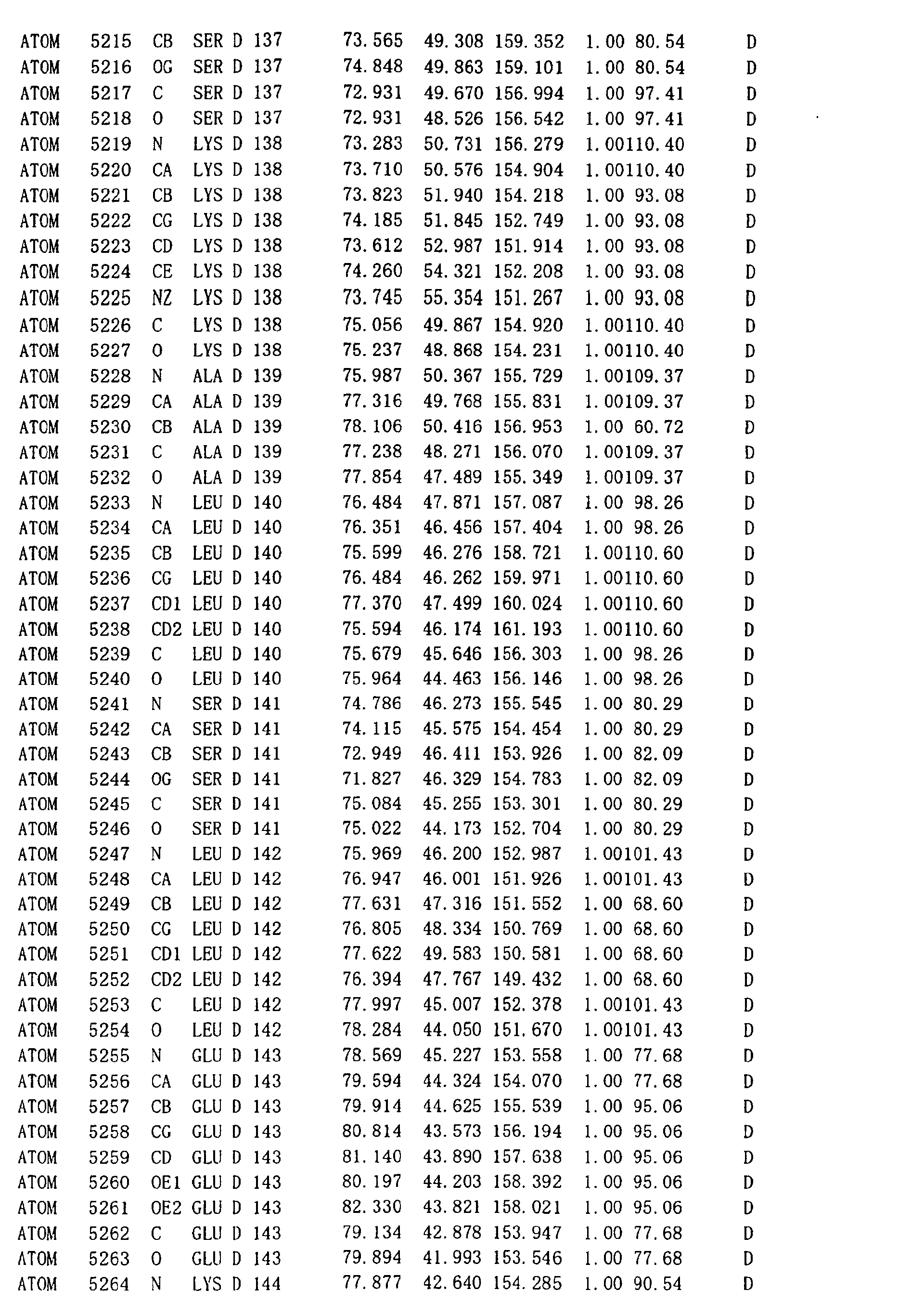
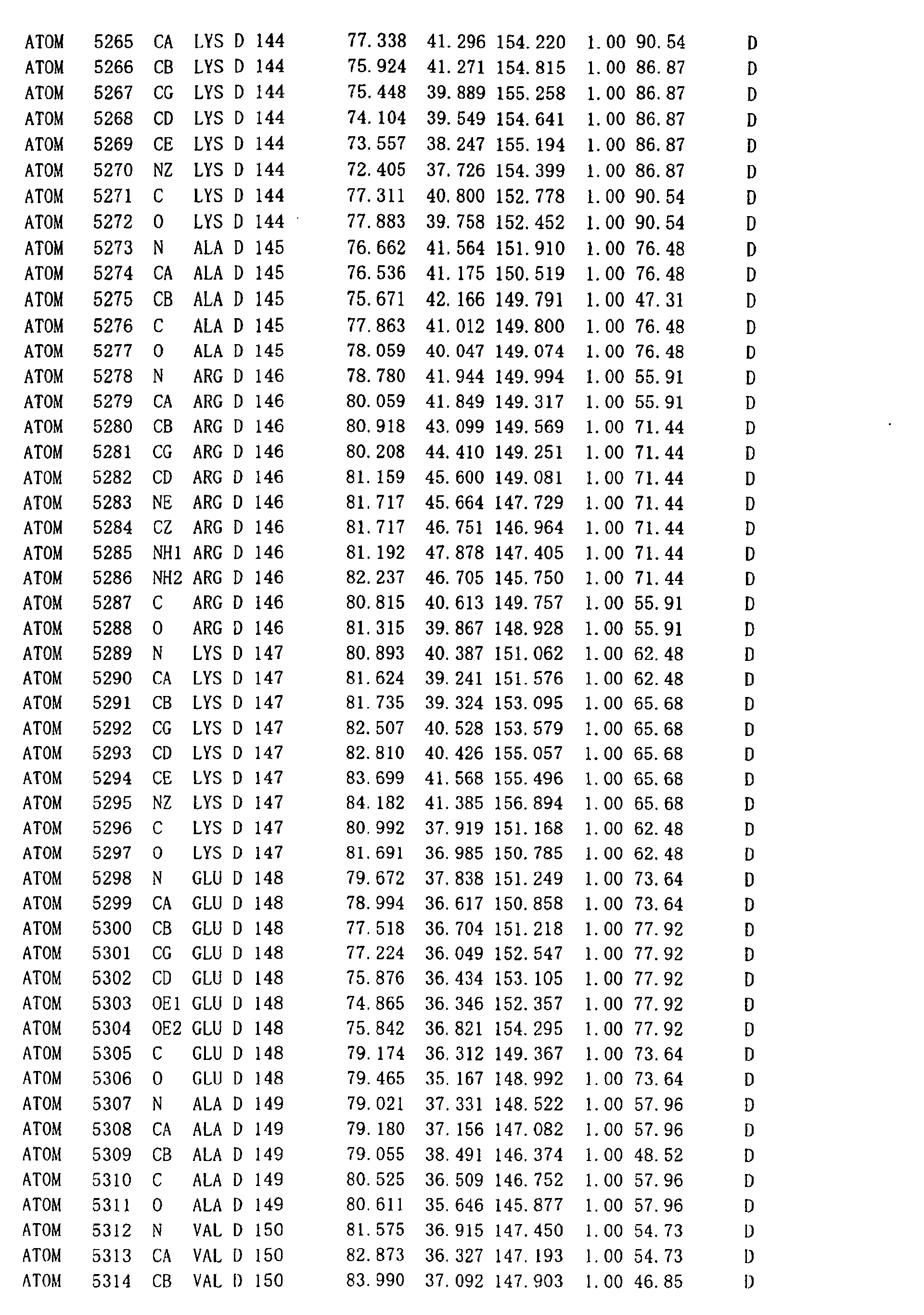
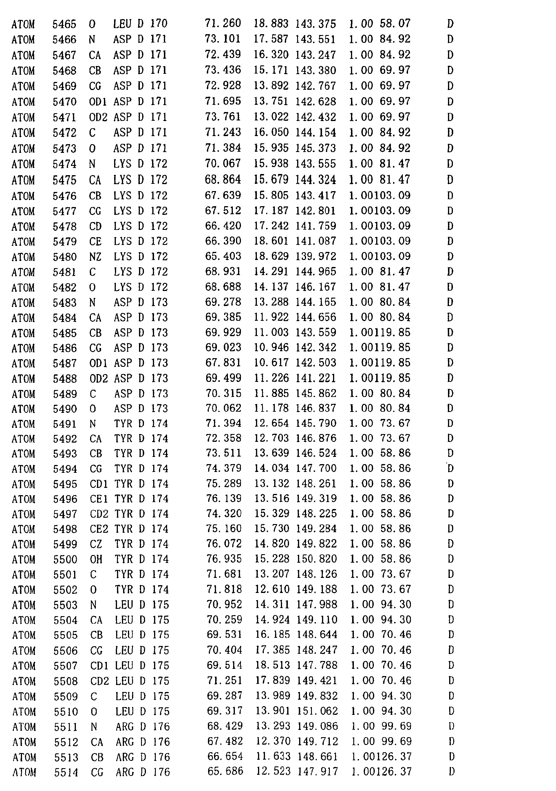
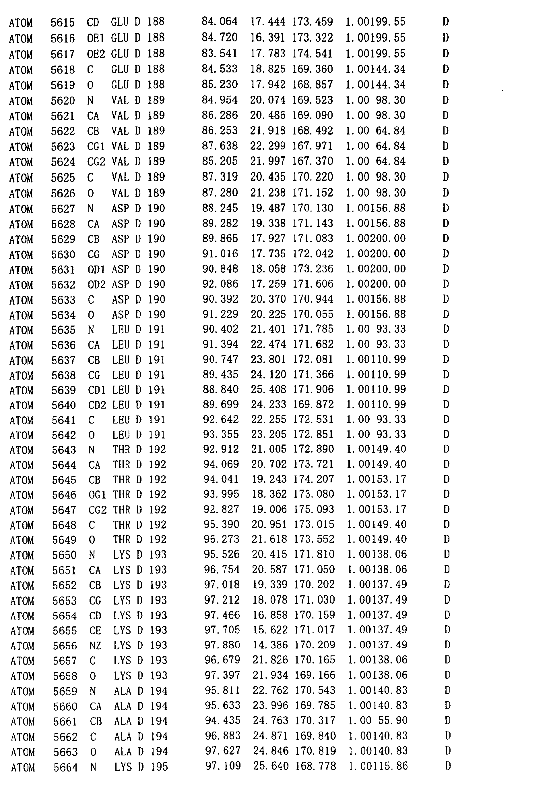
in Table 1.
- (6) A crystal of an undecameric mutant DNA
recombination/repair protein complex comprising an assembly
of the protein comprising the amino acid sequence from position
1 to position 212 in the amino acid sequence of SEQ ID NO:1,
wherein the crystal has a space group of P4 3212.
- (7) A crystal of an undecameric mutant DNA
recombination/repair protein complex comprising an assembly
of the protein comprising the amino acid sequence from position
1 to position 212 in the amino acid sequence of SEQ ID NO:1,
wherein the crystal has unit cell dimensions of a=b=145.6 Å,
and c=247.5 Å.
- (8) The crystal of a mutant DNA recombination/repair
protein complex of (6) or (7), wherein methionine at position
78 in the amino acid sequence of SEQ ID NO:1 is substituted
with selenomethionine.
- (9) A method for preparing a crystal of a DNA
recombination/repair protein complex or a mutant thereof,
comprising the steps of mixing a DNA recombination/repair
protein or a mutant thereof at a concentration of 3 to 4 mg/ml
and single-stranded DNA at a molar ratio of 14:1 to 11:1, and
crystallizing the resulting mixture by vapor diffusion method
using a buffer solution, pH 6.0 to 8.0 containing 10 to 20%
polyethylene glycol (PEG) or polyethylene glycol monomethyl
ester (PEGMME) and 0.05 to 0.2 M ammonium sulfate.
- (10) A method for determining the three-dimensional
structure of a DNA recombination/repair protein complex or a
mutant thereof, comprising the steps of:
- (a) generating a crystal of a protein with DNA
recombinational repairing activity,
- (b) obtaining the X-ray diffraction data using said
crystal by heavy atom isomorphous replacement or
multiwavelength anomalous diffraction, and
- (c) determining a three-dimensional structural
coordinate based on said X-ray diffraction data.
- (11) A computer system comprising a memory with atomic
coordinates defining a DNA recombination/repair protein
complex being arranged in the inside thereof and a processor
in electric communication with the memory, the processor
comprising a process of generating a model with a
three-dimensional structure representing at least a part of
the DNA recombination/repair protein complex or calculating
the conformational energy of the complex.
- (12) A method for identifying a mutant DNA
recombination/repair protein with modulated DNA
recombinational repairing activity, comprising the steps of:
- (a) constructing the three-dimensional structure
representing a DNA recombination/repair protein complex, using
a computer model,
- (b) identifying a site influencing the DNA
recombinational repairing activity on the basis of the
three-dimensional structure model,
- (c) producing a mutant enzyme by introducing a mutation
in said identified site, and
- (d) measuring the activity of said mutant enzyme.
- (13) The method of (12), wherein said three-dimensional
structure model is substantially defined by the atomic
coordinates shown in Table 1.
- (14) The method of (12) or (13), wherein said DNA
recombinational repairing activity is DNA binding activity,
homologous-pairing activity or multimer-forming activity.
- (15) A method for identifying a mutant DNA
recombination/repair protein with modulated DNA
recombinational repairing activity, comprising the steps of:
- (a) identifying a DNA binding region by calculating
the surface charge of all or a part of the DNA
recombination/repair protein complex substantially defined by
the atomic coordinates shown in Table 1,
- (b) selecting basic and hydrophobic amino acid residues
existing in said identified DNA binding region,
- (c) preparing a mutant enzyme by carrying out the
substitution, deletion, addition or chemical modification of
said selected amino acid residues, and
- (d) assaying the DNA binding activity and/or homologous
pairing activity of the mutant enzyme.
- (16) The method for identifying a mutant DNA
recombination/repair protein of (15), wherein the amino acid
residues selected at the step (b) are one or two or more amino
acid residues selected from arginine at position 55, tyrosine
at position 65, histidine at position 69, arginine at position
70, arginine at position 153 and lysine at position 169 in the
amino acid sequence of SEQ ID NO:1.
- (17) A method for producing a mutant DNA
recombination/repair protein, comprising the steps of
identifying a mutant DNA recombination/repair protein by a
method of any one of (12) to (16), and expressing said mutant
DNA recombination/repair protein using the recombinant DNA
encoding the mutant DNA recombination/repair protein.
- (18) A mutant DNA recombination/repair protein with
modulated DNA recombinational repair activity, as obtained by
a method of any one of (12) to (16).
- (19) A method for screening a compound capable of
modulating the repairing activity of a DNA recombination/repair
protein, using a polypeptide fragment or a complex of any one
of (1) to (5) to design or select a compound binding thereto.
- (20) A method for screening a compound capable of
modulating the repairing activity of a DNA recombination/repair
protein, using a computer with the memory of the coordinate
data of the three-dimensional structure of a polypeptide
fragment or a complex of any one of (1) through (5) to design
or select a compound binding thereto.
- (21) The method of (20) , further comprising contacting
said designed or selected compound with a DNA
recombination/repair protein, and determining whether said
compound binds to the DNA recombination/repair protein.
- (22) A compound modulating the activity of a DNA
recombination/repair protein as obtained by a method of any
one of (19) through (21).
- (23) The compound according to (22), wherein said
compound modulating the DNA recombinational repairing activity
is a natural compound or a synthetic compound.
- (24) A method for preparing a pharmaceutical composi tion
containing a compound modulating DNA recombinational repairing
activity, comprising a step of mixing a compound obtained by
a method of any one of (19) through (21) with a pharmaceutically
acceptable carrier.
-
BRIEF DESCRIPTION OF THE DRAWINGS
-
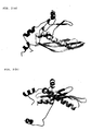
- Fig. 1 shows the three-dimensional structure of the
HsRad521-212 complex as a human-derived mutant DNA
recombination/repair protein in a ribbon model. Figs. 1A and
1B show the structure of the undecamer of the HsRad521-212 as
depicted in a different direction.
- Fig. 2 shows the three-dimensional structure of the
HsRad521-212 monomer as a human-derived mutant DNA
recombination/repair protein in a ribbon model. Figs. 2A and
2B show the structure of the HsRad52-1-212 monomer as depicted
in a different direction.
- Fig. 3 shows the DNA binding site and DNA binding mode
of the HsRad521-212 monomer as a human-derived mutant DNA
recombination/repair protein. Fig. 3A is a stereo
representation of the DNA bind ing sites of the HsRad521-212 , while
Fig. 3B is a steric figure depicting the DNAbinding site between
adjacent two HsRad521-212 molecules forming the complex and Fig.
3C is a model figure depicting the DNA binding mode of the
undecamer of the HsRad521-212 complex.
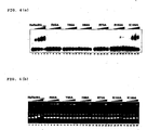
- Fig. 4 shows the assay results of the DNAbinding activities
of the wild type HsRad52-1-212 and various mutants thereof by
gel shift method. Fig. 4A shows the results of detecting the
binding activity to single-stranded DNA by autoradiography.
Fig. 4B shows the results of detecting the binding activity
to double-stranded DNA by ethidium bromidestaining. Fivelanes
for each one protein show the concentration dependency in case
of sequentially increasing the protein concentration.
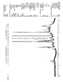
- Fig. 5 shows the proton NMR spectrum of a ligand candidate
compound AK-968-41170131 (Compound A).
- Fig. 6 shows the proton NMR spectrum of a mixture (1:1)
of the ligand candidate compound AK-968-41170131 (Compound A)
and HsRad521-212.
- Fig. 7 shows the comparison of the NMR spectra of the
ligand candidate compound AK-968-41170131 (Compound A) and a
mixture (1:1) of the ligand candidate compound AK-968-41170131
(Compound A) and HsRad521-212 around 7.5 ppm.
- Fig. 8 shows the proton NMR spectrum of a ligand candidate
compound AK-968-41025315 (Compound a).
- Fig. 9 shows the proton NMR spectrum of a mixture (1:1)
of the ligand candidate compound AK-968-41025315 (Compound a)
and HsRad521-212.
- Fig. 10 shows the comparison of the proton NMR spectra
of the ligand candidate compound AK-968-41025315 (Compound a)
and a mixture (1:1) of the ligand candidate compound
AK-968-41025315 (Compound a) and HsRad521-212 around 7.5 ppm.
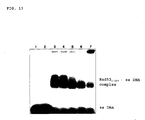
- Fig. 11 shows the gel shift assay results showing that
the ligand candidate compound AK-968-41170131 (Compound A)
inhibits the binding of HsRad52 to single-stranded DNA (ssDNA) .
-
DETAILED DESCRIPTION OF THE INVENTION
-
In accordance with the invention, "DNA recombinational
repairing activity" means an activity for recognizing a site
with chromosomal DNA damage and the like, and recognizing,
breaking and rejoining homologous nucleotide sequences between
DNA molecules via homologous recombination. Because
homologous recombination is very complex, the details of the
reaction mechanism are not yet elucidated. As described above,
it is considered that two pathways, namely Rad51-dependent
pathway and Rad51-independent pathway exist in eukaryotes.
Therefore, DNA repairing activity through these two homologous
recombination reactions is referred to as "DNA recombinational
repairing activity".
-
"DNA recombination/repair protein" means an enzyme having
the DNA recombinational repairing activity and examples thereof
include HsRad52 with the amino acid sequence of SEQ ID NO:1.
HsRad52 is a protein comprising 418 amino acid residues from
humans. The cDNA nucleotide sequence has been revealed (for
example, GenBank Accession No. XM_052347). The RAD52 genes
in eukaryotes are found in chicken, mouse and yeast (S. pombe
and S. cerevisiae). The primary structures of the Rad52
proteins obtained from these cDNA nucleotide sequences are
compared to each other. About 70% homology is known in their
N-terminal regions, particularly the regions up to the position
177 from the N terminus in HsRad52 (Muris, DF, et al., 1994,
Mutat. Res. 315, 295-305).
-
Therefore, the "DNA recombination/repair protein" in
accordance with the invention means HsRad52 comprising the amino
acid sequence of SEQ ID NO:1 or a homolog thereof in eukaryotes.
-
In accordance with the invention, the "mutant" of the
DNA recombination/repair protein means a modified enzyme
comprising an amino acid sequence derived from the amino acid
sequence of SEQ ID NO:1 as prepared by deletion, addition,
insertion or substitution or modification of one or several
amino acids and still having at least a part of the activity
of the intact enzyme. Herein, the term "at least a part of
the activity" typically means a specific activity of at least
10% of the activity of the intact enzyme, preferably at least
50% thereof. If desired, the term sometimes means a specific
activity at less than 10% thereof.
-
According to research works so far made by the present
inventors, the limited digestion of HsRad52 protein and the
analysis of a mutant enzyme deficient in a C-terminal region
show that a core domain responsible for the homologous pairing
activity of HsRad52 exists in the N-terminal region of HsRad52.
A polypeptide HsRad521-237 comprising the amino acids at position
237 from the N terminus is resistant to the limited digestion
withproteinase K. Because the analysis of the D-loop formation
reaction shows that the mutant enzyme HsRad521-237 has a
homologous pairing activity at a level almost equal to that
of the wild type enzyme, it is considered that the N-terminal
region from the N terminus of HsRad52 to position 237 thereof
is responsible for the homologous pairing activity. Further,
the N-terminal region forms a mul timeric higher-order structure
like that of the wild type HsRad52 and has also the DNA binding
activity. These indicate that the HsRad52 protein even when
the protein is deficient in a part of the C-terminal region
forms a complex similar to that formed by the wild type HsRad52
protein and still keeps the DNA recombinational repairing
activity and that such C-terminal region-deficient HsRad52
protein is sufficiently useful for the elucidation of the
relationship between the structure and function of the HsRad52
protein for the homologous recombination mechanism. As such
mutant HsRad52 protein deleted of a part of the C-terminal region,
specifically, various mutants can be utilized. For example,
plural deficient mutants with serial deletion by every five
amino acids from the C terminus of HsRad521-237 are exemplified,
such as HsRad52-1-232, HsRad521-227, HsRad521-222, HsRad521-217,
HsRad521-212, HsRad521-207, HsRad521-202, HsRad521-197, HsRad521-192
and HsRad521-187. Among them, mutant enzymes with amino acid
sequences from the N terminus at least up to position 192 still
retain an activity as DNA recombination/repair protein. Thus,
selecting amutant enzyme most crystallizable, aprotein crystal
for use in X-ray crystallography can be prepared.
(Preparation of crystal of DNA recombination/repair protein
complex)
-
The most general approach for identifying
three-dimensional protein structure is an approach by X-ray
crystallographic analysis. That is, the approach includes
crystallizing protein, allowing monochromatic X-ray to
irradiate the crystal to obtain an X-ray diffraction image,
and analyzing the three-dimensional structure of the protein
based on the resulting X-ray diffraction image (Blundell, T.L.
and Johnson, L. N., PROTEIN CRYSTALLOGRAPHY, p.1-565, (1976)
Academic Press, New York).
-
The crystallization utilizes a protein property of
deposition in crystal under a specific condition, by a procedure
of adding a precipitation agent to a solution of a target protein
or by a procedure of reducing the amount of the solvent via
evaporation or the like to turn the solution state of the protein
to the non-solution state. For the crystallization, highly
purified protein is required. Additionally, physical and
chemical factors are involved in the crystallization conditions,
such as protein concentration, salt concentration, hydrogen
ion concentration (pH), the type of a precipitation agent to
be added, and temperature. Further, methods for adding such
precipitation agent and for adjusting the amount of the solvent,
numerous crystallization methods exist such as batch method,
dialysis method and vapor diffusion method (Blundell, T. L.
and Johnson, L.N., PROTEIN CRYSTALLOGRAPHY, p. 59-82, (1976)
Academic Press, New York) . For example, the vapor diffusion
method functions tomobilize volatile solvents or precipitation
agentsvia diffusionthrough gasphase. Through vapor diffusion
from the protein solution and the solution of a precipitation
agent, the protein concentration and the concentration of the
precipitation agent both increase, leading to the occurrence
of crystallization in a supersaturated zone. Depending on how
liquid droplets of the protein solution are made, there are
several methods such as hanging drop method, sitting dropmethod,
and sandwich drop method. So as to prepare a protein crystal
suitable for X-ray crystallography as described above,
essentially, an individual protein should be obtained as a
protein at high purity and various factors and crystallization
methods should be examined for their optimization. The
preparation of high-purity protein is an important factor for
successful crystallization. Thus, a homogenous protein can
be prepared rapidly by expressing a cloned gene in an efficient
expression system.
-
The crystal of the DNA recombination/repair protein
complex in accordance with the invention can be prepared as
describedbelow. The cDNA sequence encoding the HsRad52 protein
has already been known and can be readily expressed in various
hosts such as bacteria and animal cells using DNA recombination
technology.
-
In one embodiment of the present invention, the HsRad52
protein can be expressed at a large scale in a system using
a recombinant Escherichia (E.) coli. various plasmid vectors
are commercially available for large-scale expression of
exogenous gene in recombinant E. coli. For example, pET system
(Novagen) can be used, which works for introducing a gene to
be expressed downstream strong T7 phage promoter for
transcription with T7RNA polymerase supplied by the host E.
coli. In the system, T7 RNA polymerase gene is integrated in
the chromosome of the host E. coli and is downstream lacUV5
promoter, so that the expression can be controlled by the
induction by IPTG addition. Additionally, introducing various
tag sequences on the N-terminal side or C-terminal side of the
HsRad52 protein enables ready purification us ing a metal chelate
resin (histidine tag) and antibodies (T7 tag, CBD tag, and the
like). From the fused protein purified, only the moiety of
the HsRad52 protein can further be recovered using proteases
such as enterokinase and thrombin.
-
By growing and culturing the recombinant E. coli
expressing the HsRad52 protein in a culture medium containing
selenomethionine or selenocysteine containing selenium as a
heavy metal atom, additionally, a mutant enzyme where the
methionine or cysteine residue in the HsRad52 protein is
substituted with selenomethionine or selenocysteine can be
recovered.
-
For further allowing the resulting enzyme more suitable
for crystallization, such protein is purified to higher purity.
As the purification method, general methods used by a person
skilled in the art forproteinpurification including for example
column chromatography (affinity, hydrophobic, ion exchange,
gel filtration, etc.), salting out, centrifugation, and
electrophoresis can be used singly or in combination.
-
Then, the crystal of the DNA recombination/repair protein
complex is prepared. Essentially, crystallization should be
carried out under conditions so that the HsRad52 protein can
keep the state of the multimer complex formed intracellularly.
Using vapor diffusin process, for example, the crystal of the
DNA recombination/repair protein complex can be obtained under
conditions of pH = 6 to 8, a protein concentration of 3 to 4
mg/ml, and a temperature of 20°C, in case that 10 to 20%
polyethylene glycol (PEG) or polyethylene glycol monomethyl
ester (PEGMME) and 0.05 to 0.2 M ammonium sulfate are used as
precipitation agents. So as to promote the accumulation of
the HsRad52 protein to form the multimer complex, preferably,
the HsRad52 protein and single-stranded DNA are preliminarily
mixed together at a molar ratio of 14:1 to 11:1.
-
In addition to the method for introducing the heavy metal
atom into protein via culturing in the selenium-containing
culture medium, the native type protein crystal is immersed
in a solution of a metal salt or an organic metal compound
containing gold, platinum, iridium, osmium, mercury, lead,
uranium and samarium, to prepare crystals of heavy metal
derivatives. The method is utilized for the application of
heavy atom isomorphous replacement and multiwavelength
anomalous diffraction.
-
The crystal of the HsRad521-212 complex containing the amino
acid sequence from position 1 to position 212 of SEQ ID NO:1
belongs to a tetragonal space group P4 3212 and is of unit cell
dimensions of 145.6 angstroms in the directions of axes a and
b and 247.6 angstroms in the direction of axis c.
(X-ray crystallography of DNA recombination/repair protein
complex)
-
The structure of the thus obtained crystal of the DNA
recombination/repair protein complex is analyzed using X-ray
crystallographic analytical technology known to a person
skilled in the art. For the determination of three-dimensional
structure as in the case of the invention, where the
three-dimensional structure of an analogous protein is unknown
and the structural analysis by molecular substitution method
using the three-dimensional structure is impossible, the heavy
atom isomorphous replacement and multiwavelength anomalous
diffraction (MAD) are applicable. In one embodiment of the
present invention, X-ray crystallographic analysis is MAD.
X-ray used therefor is at a wavelength of 0.3 to 3.0 angstroms
obtained from synchrotron radiation. The MAD is a process of
obtaining X-ray diffraction data at plural wavelength from a
heavy atom isomorphous replacement crystal with an absorption
end close to the incident X-ray energy. Resonance between X-ray
and the electron shell of the heavy atom causes a difference
in X-ray diffraction, to thereby overcome the phase problem
in the structural analysis of protein. The principle of the
method has already been known classically. However, the
appearance of synchrotron radiation with a variable wavelength
first enabled the use for structural protein determination.
Hendrickson, et al. first made a success in the structural
analysis of protein by the method (see Hendrickson W.A. et al.,
Proteins 4, 77-88, 1988 and Hendrickson W.A. et al., EMBO J.
5, 1665-1672, 1990).
-
Synchrotron radiation is a radiation obtained by
accelerating electron charged in accumulate ring with a magnetic
field, and has an energy far higher than those from general
X-ray generators. For example, the third generation Large
Synchrotron Apparatus Spring- 8 (8GeV) at RIKEN HARIMA INSTITUTE
can be used.
-
The data of the measured diffraction image are processed
into reflection intensity data, using a program DENZO (Mac
Science) or a similar image processing program. Based on the
reflection intensity data of the native type crystal and the
reflection intensity data of the heavy atom replacement crystal
at plural wavelengths, the position of the heavy atom bound
to the protein in the crystal is determined using the
Differential Patterson chart. Using subsequently a program
PHASE or CCP4 or a similar diffraction data analysis program,
the heavy atom position parameters are refined to determine
the initial phase. The determined initial phase is modified
into a phase with higher reliability according to a solvent
smoothing method using Program DM (CCP4 package) or a similar
phase improvement program (electron density improvement
program).
-
The three-dimensional structure model of the DNA
recombination/repair protein complex can be constructed from
the electron density map displayed on the three-dimensional
graphics by the Program O by the following procedures. First,
plural regions with characteristic amino acid sequences are
screened on the electron density map. With reference to the
amino acid sequences starting from the regions found, then,
a partial structure of amino acid residues fitting to the
electron density is constructed on three-dimensional graphics
using the Program O.
-
By repeating the procedure sequentially, all the amino
acid residues of the DNA recombination/repair protein complex
are fitted to the corresponding electron densities, so that
an initial three-dimensional structure model for the whole
complex is constructed. The constructed three-dimensional
structure model is used as a starting model structure, to refine
the three-dimensional coordinates describing the
three-dimensional structure according to the refined protocol
of XPLOR as a structure refiningprogram. The status of refining
progress and molecular model can be checked and confirmed by
the following indicators. Reliability factor (R factor) is
an indicator showing the agreement level between the absolute
value of the crystal structure factor obtainedby the diffraction
intensity measurement and the absolute value of the crystal
structure factor as calculated from molecular model. The
reliability factor takes a small value as the refining progresses
normally. Additionally, a method for confirming that the
dihedral angle values of the main chain of a protein are in
an acceptable region by calculating the dihedral angles of the
main chain of the protein and plotting the angles is also used
(Ramachandran Plot).
-
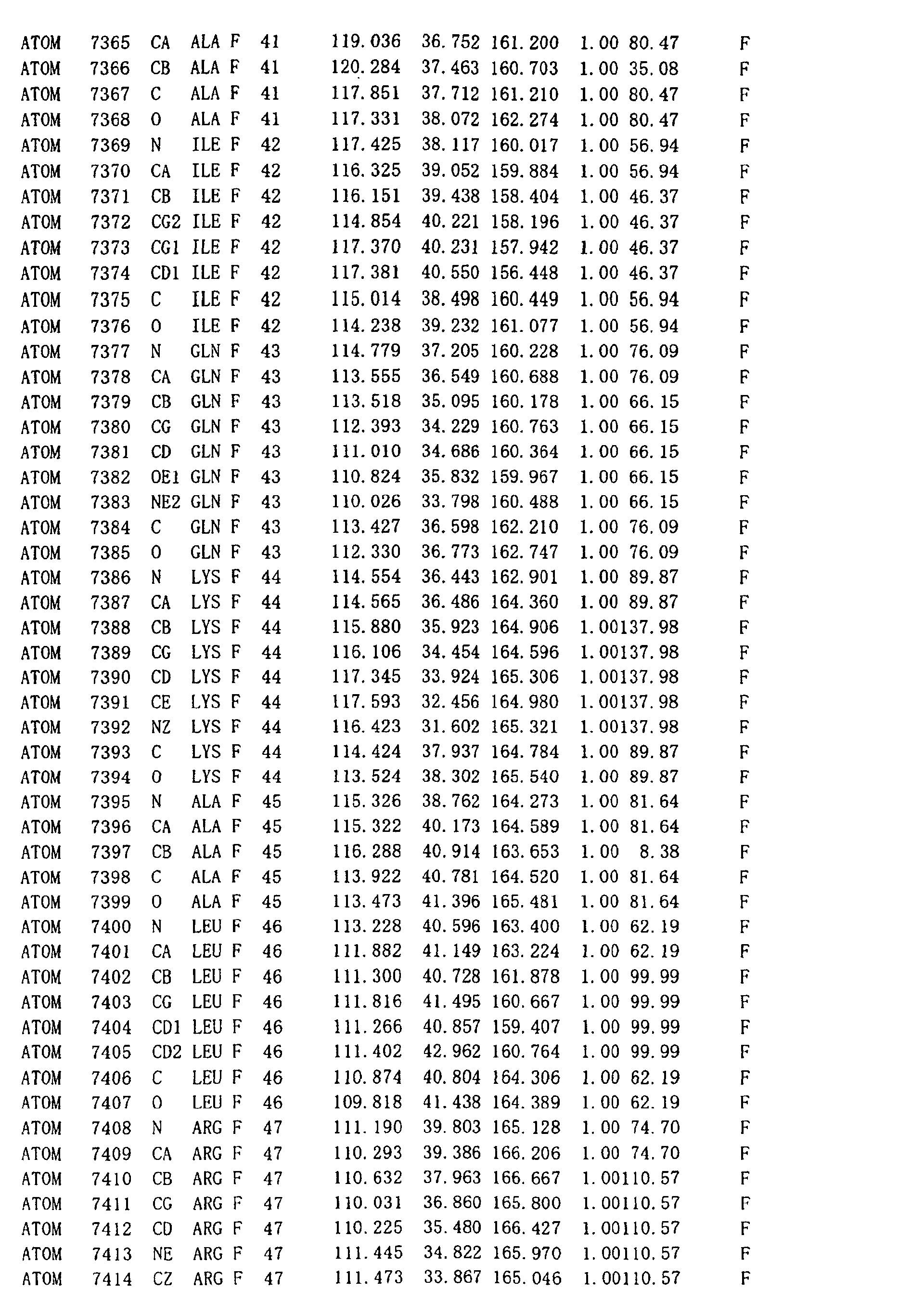
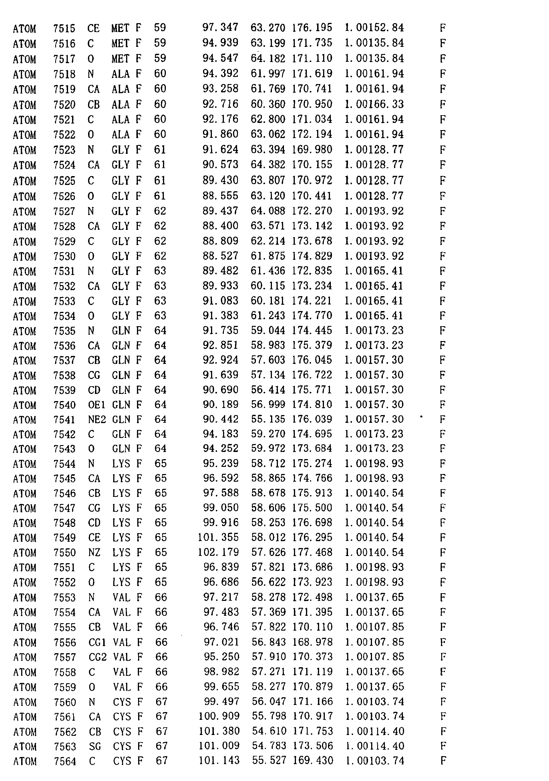
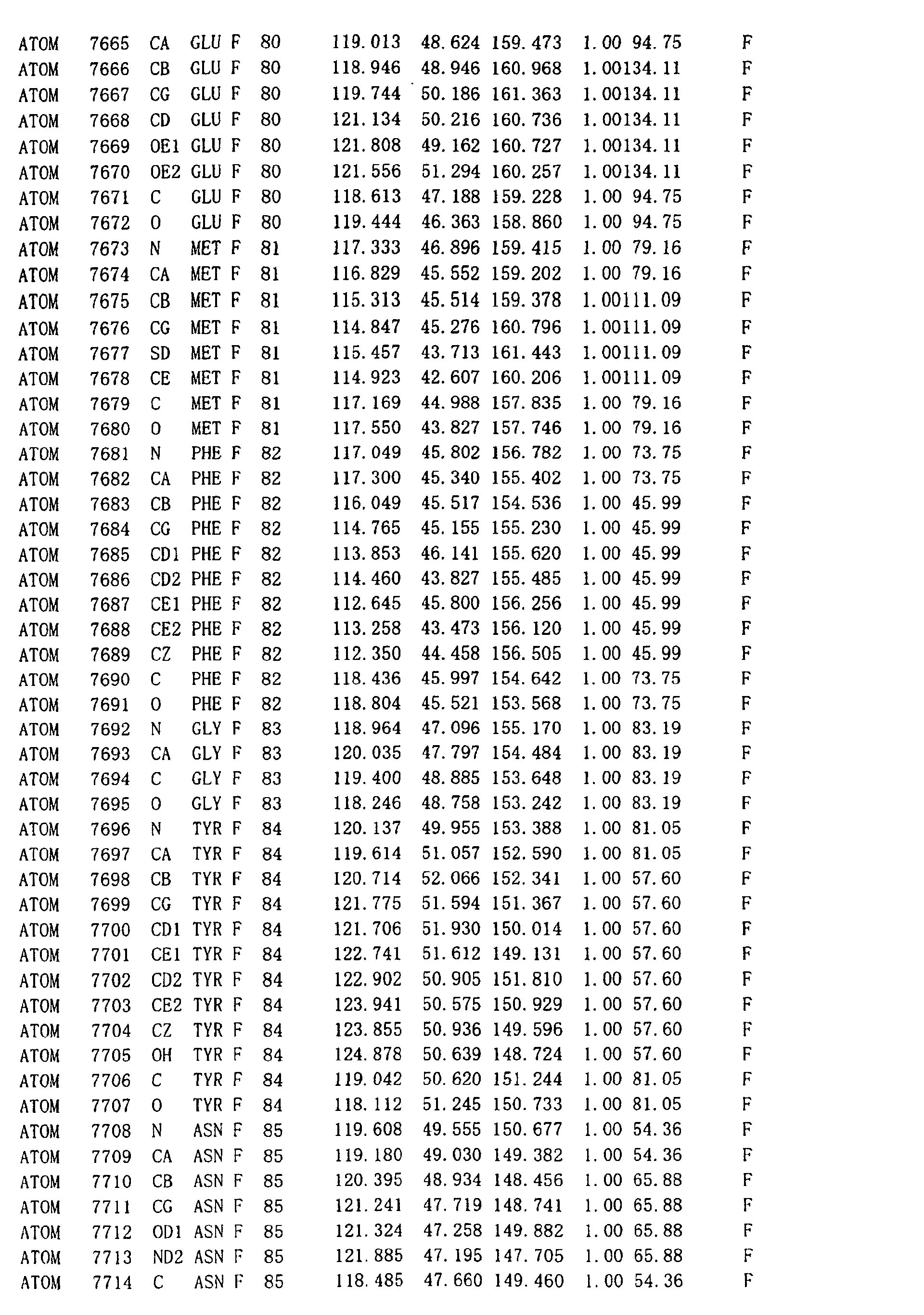
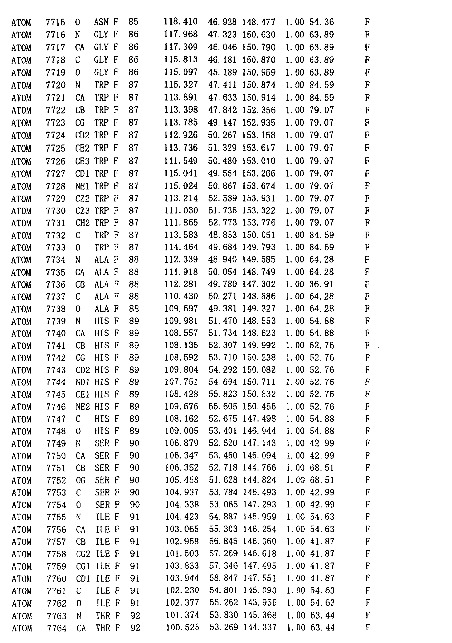
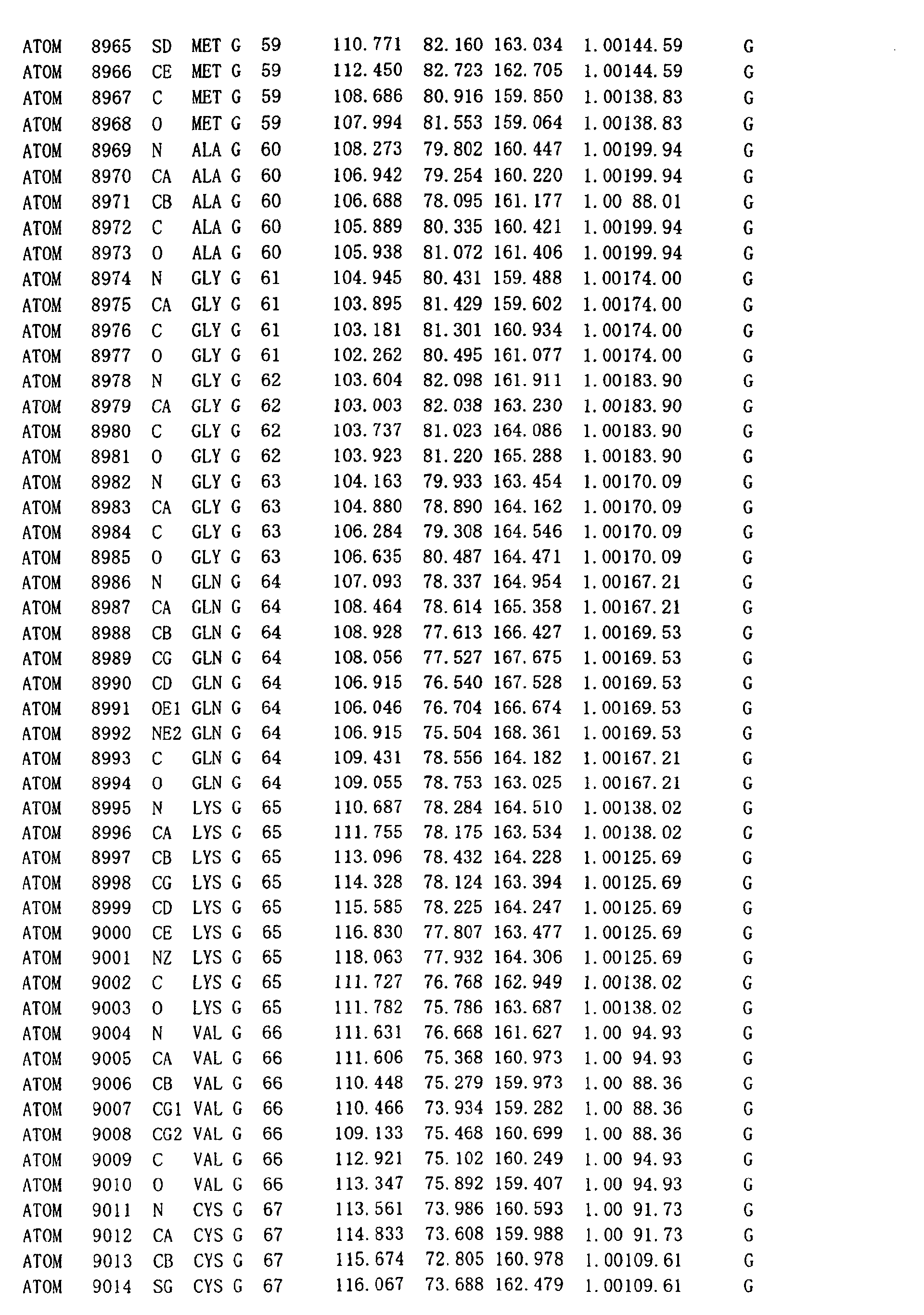
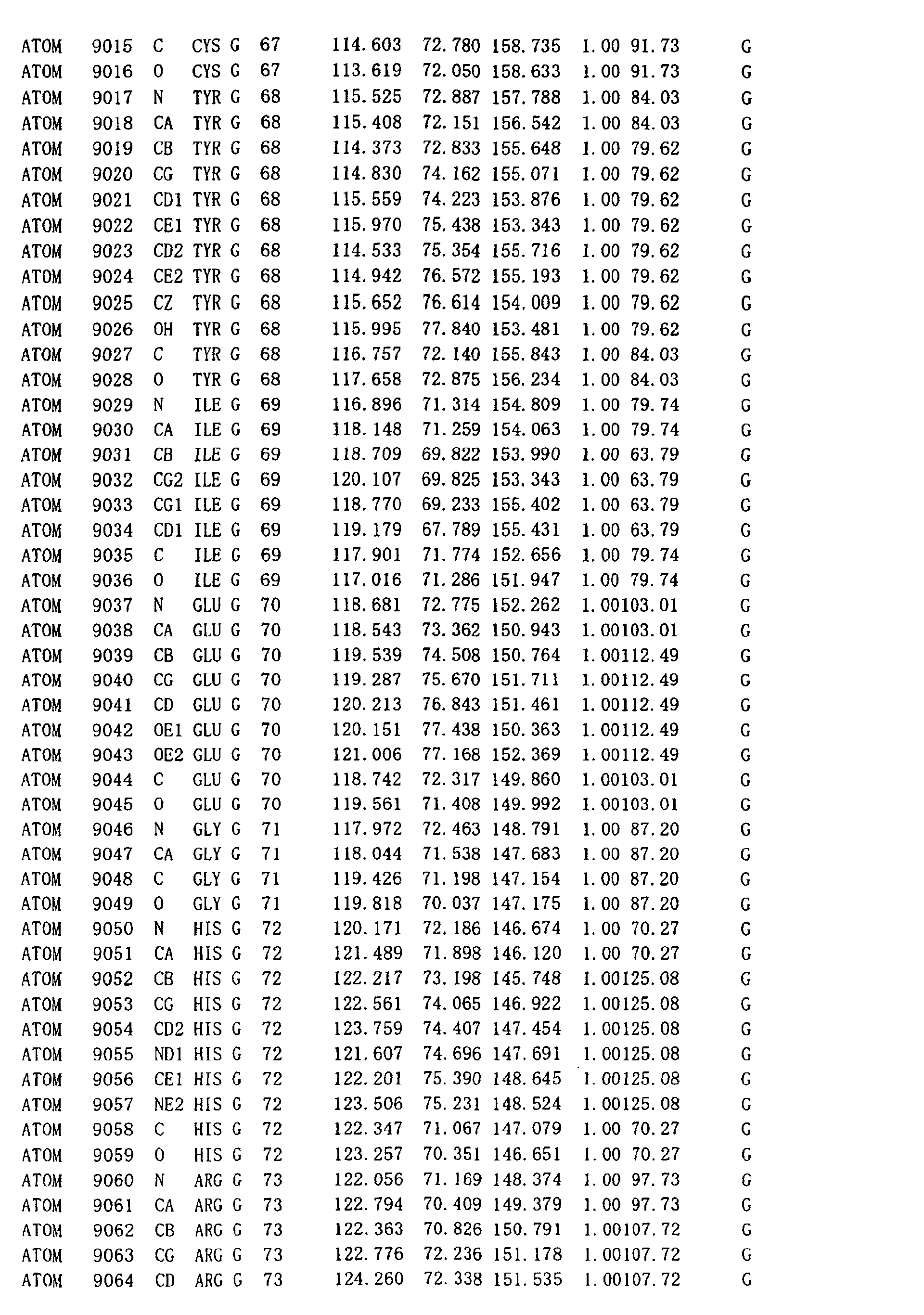
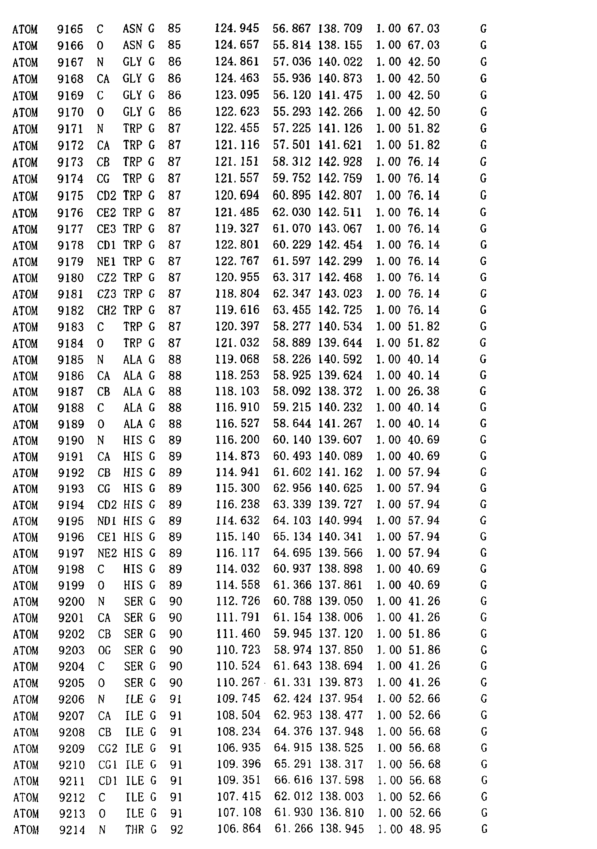
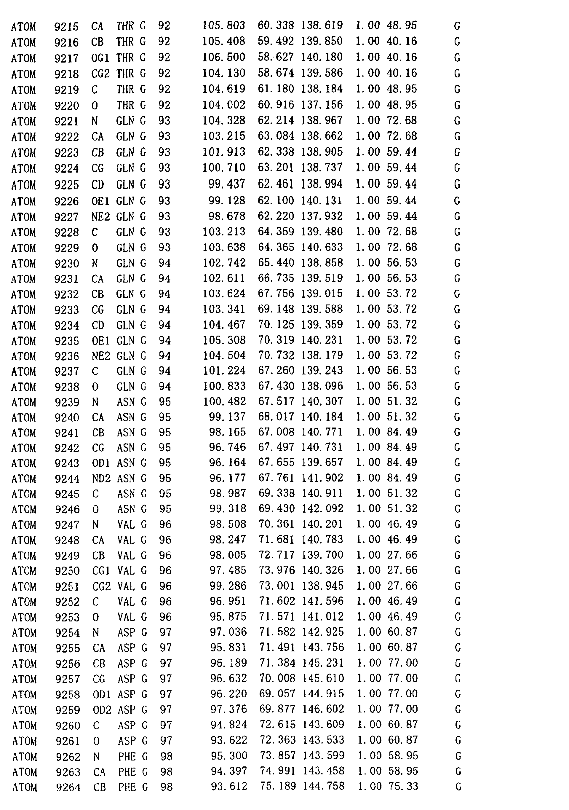
In such manner, the three-dimensional atomic coordinates
(values showing the spatial positional relation among
individual atoms) of the DNA recombination/repair protein
complex of the invention can first be obtained in such manner.
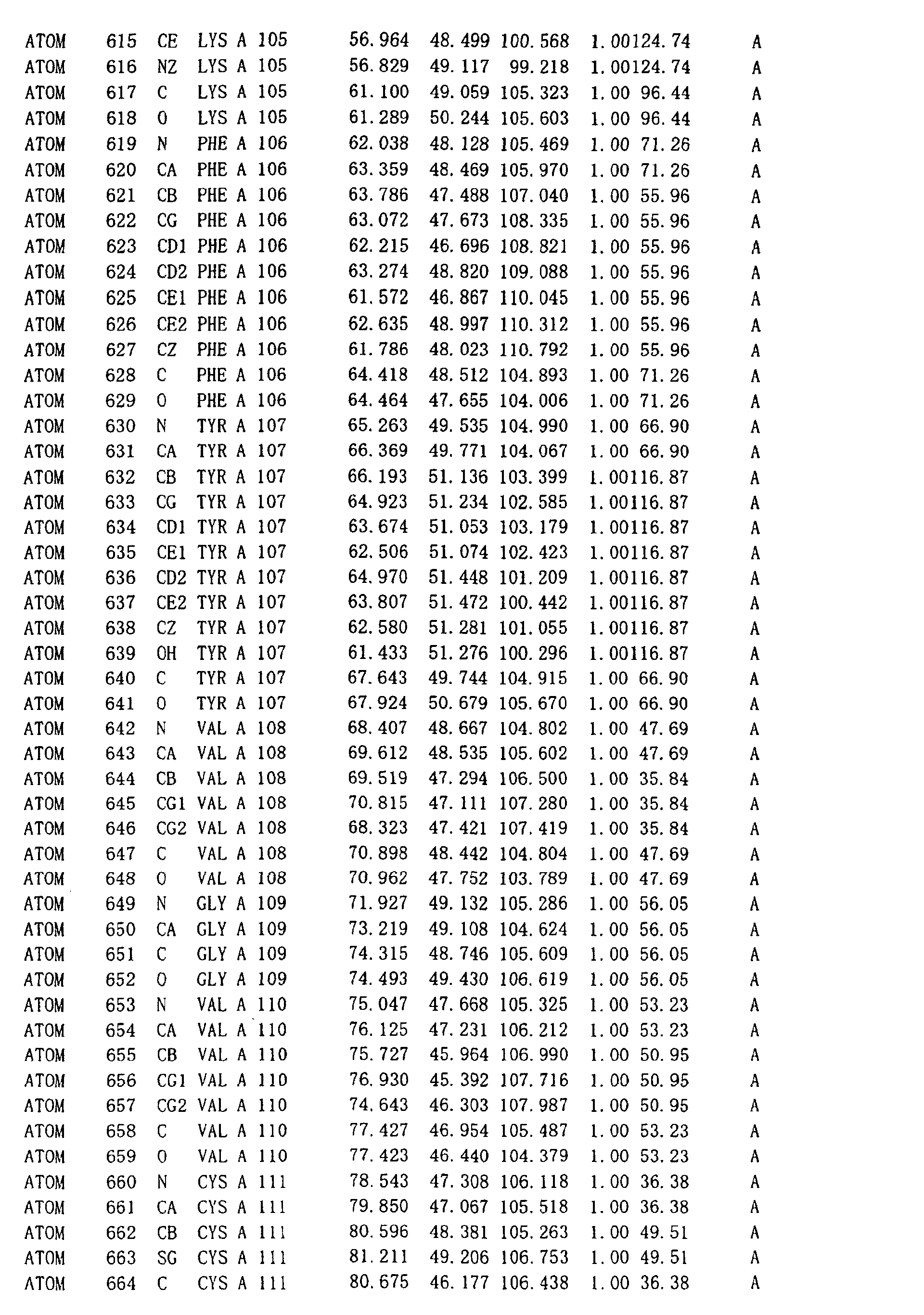
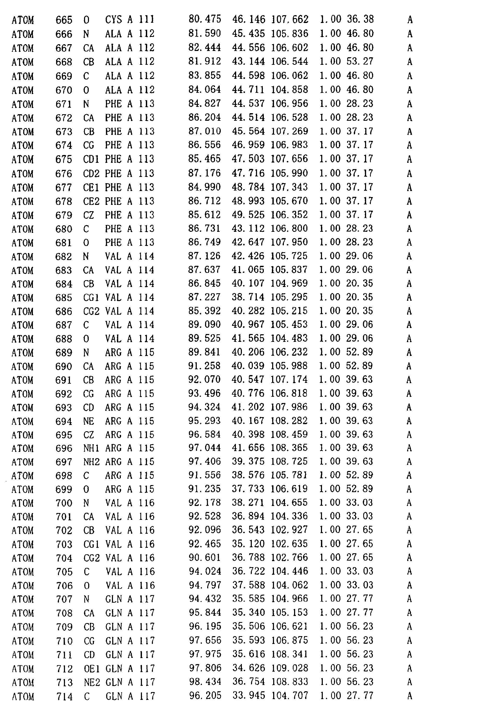
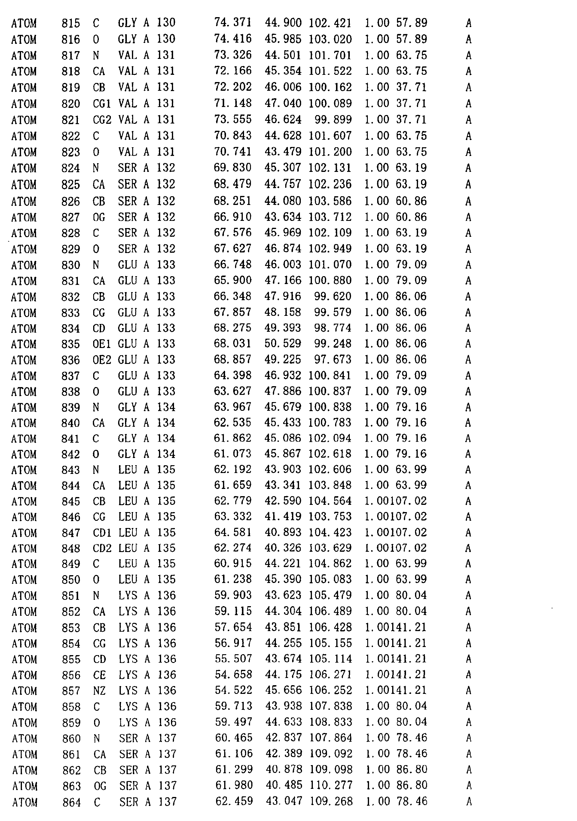
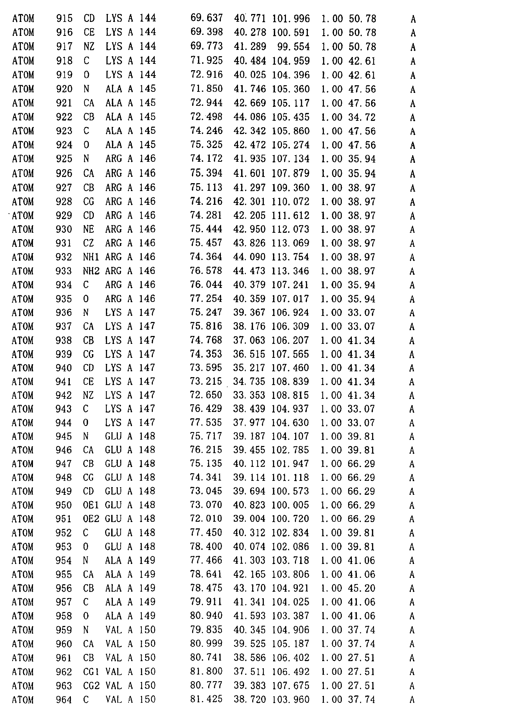
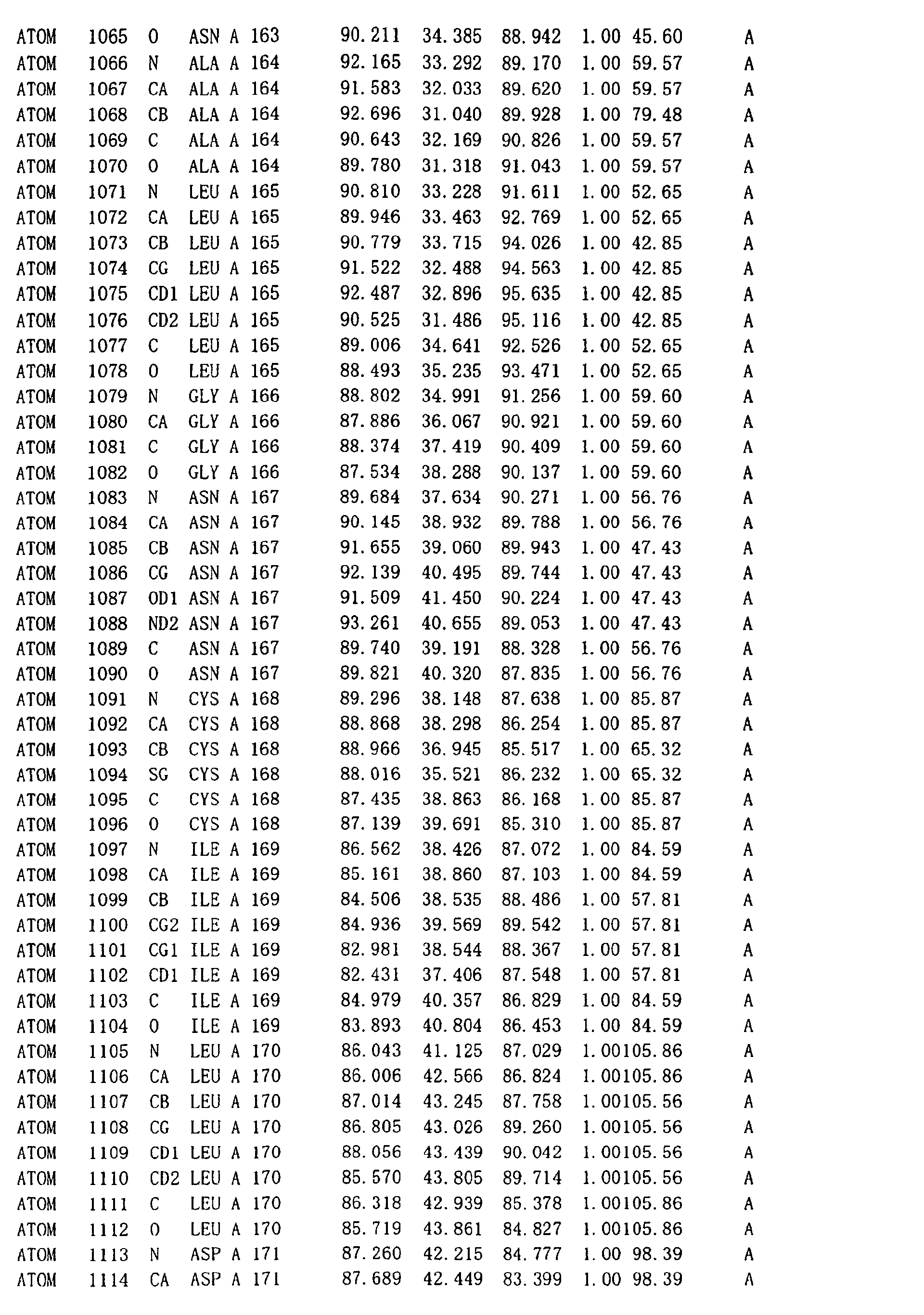
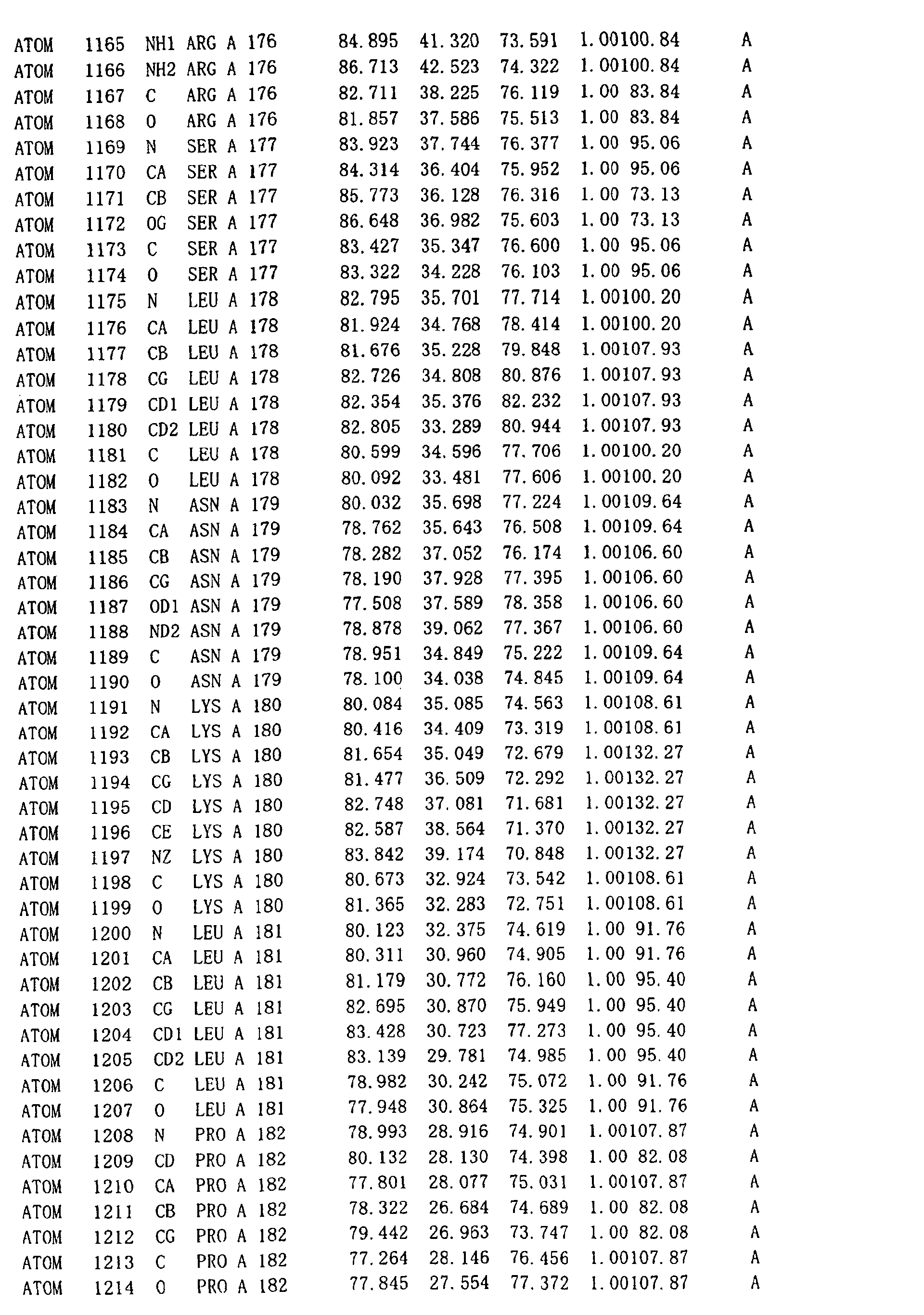
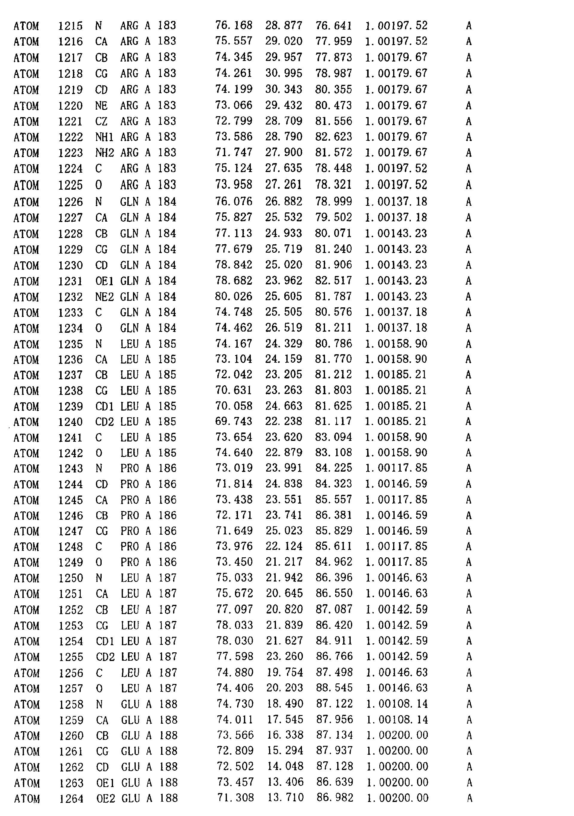
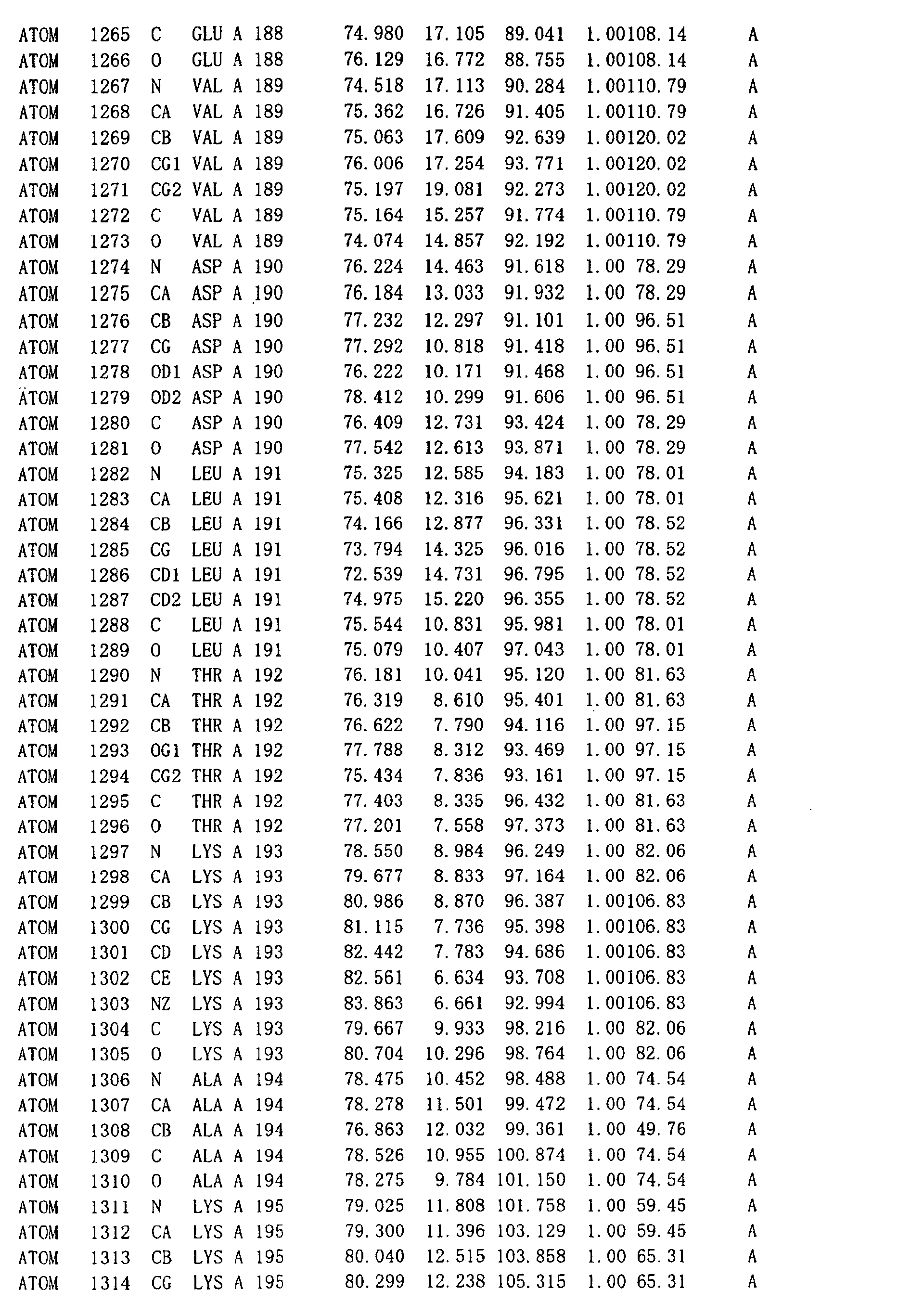
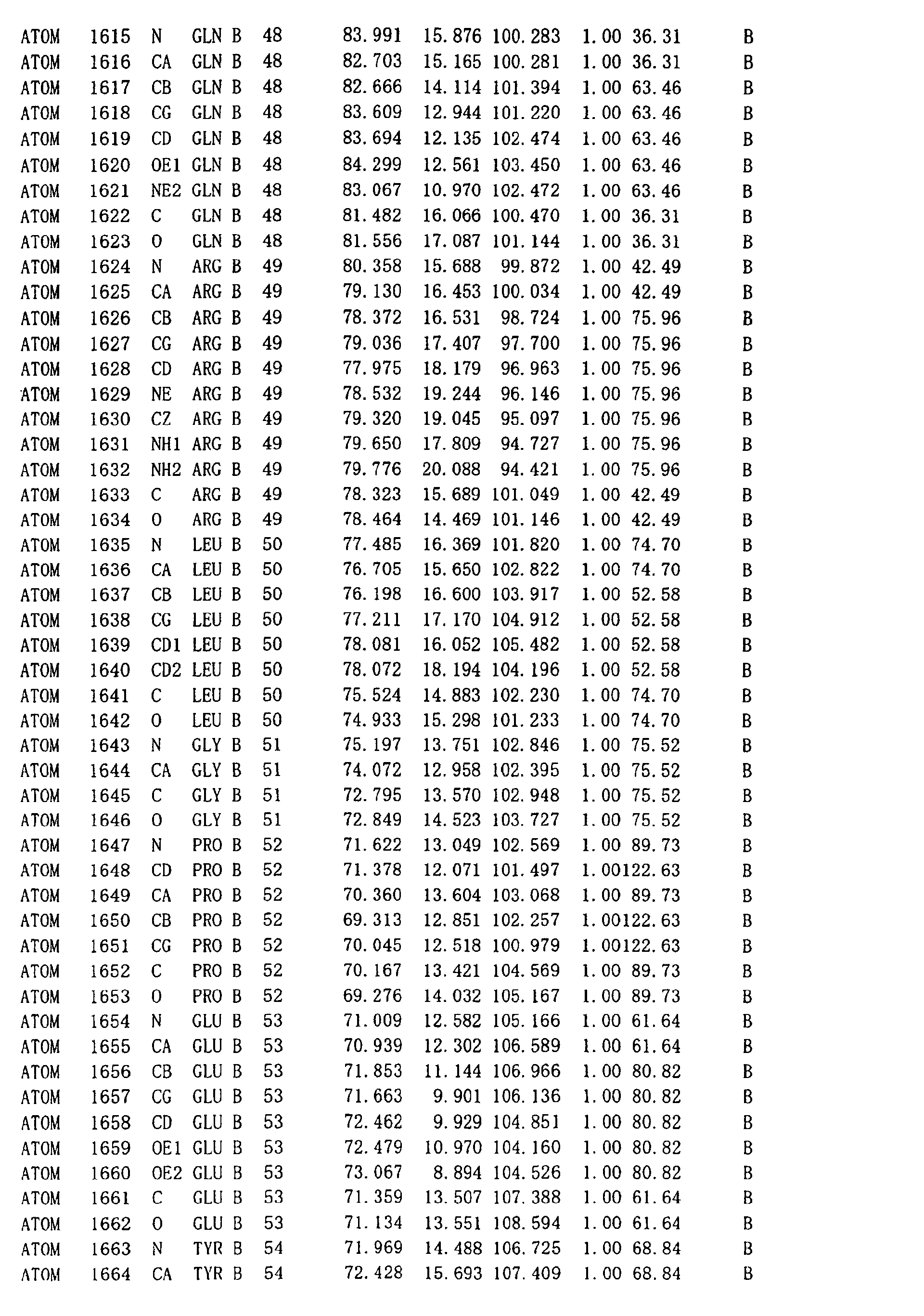
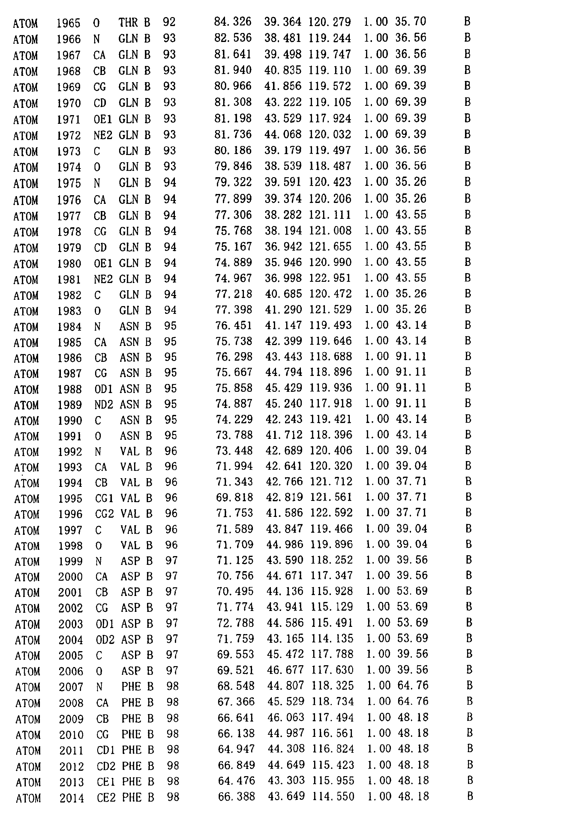
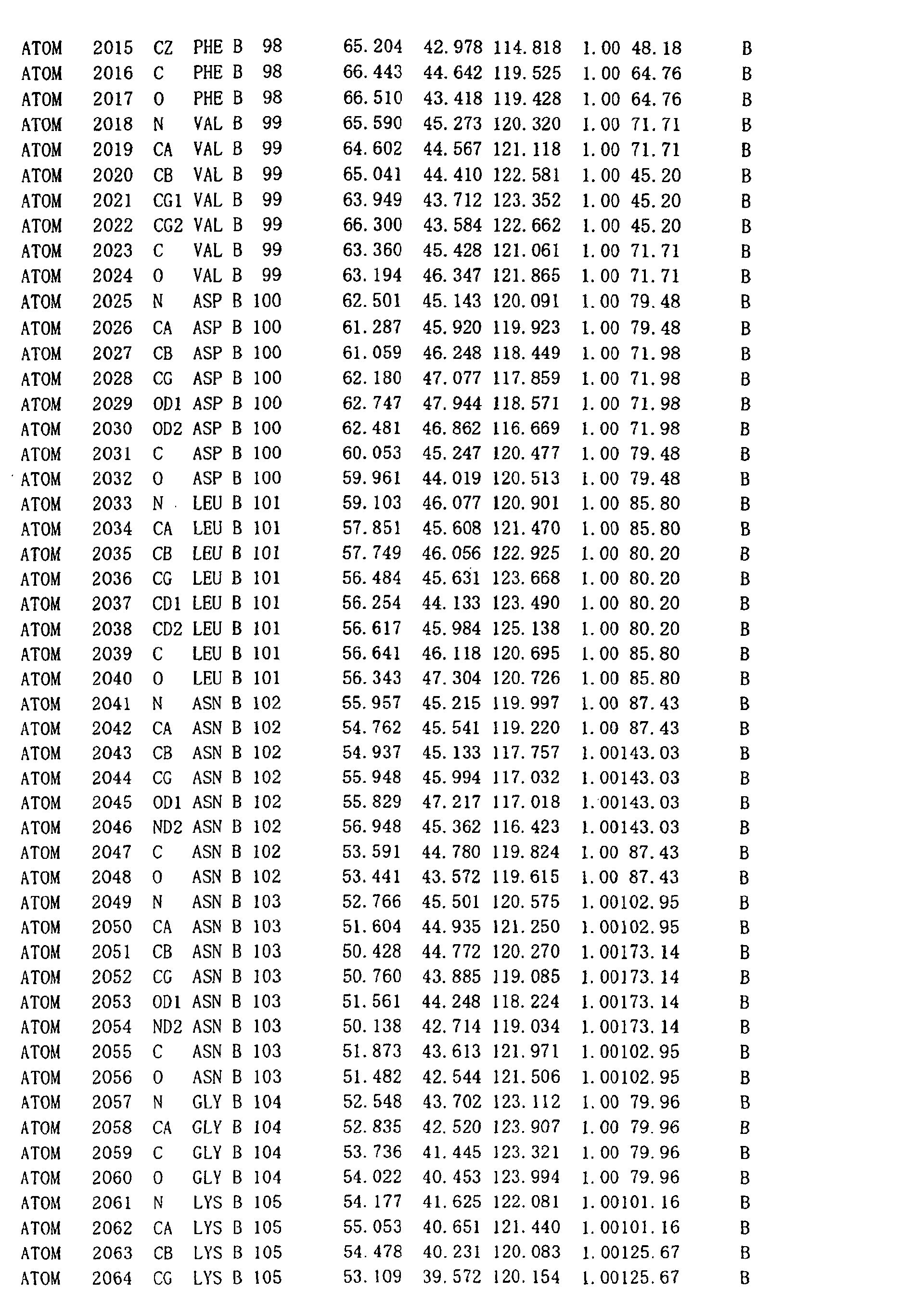
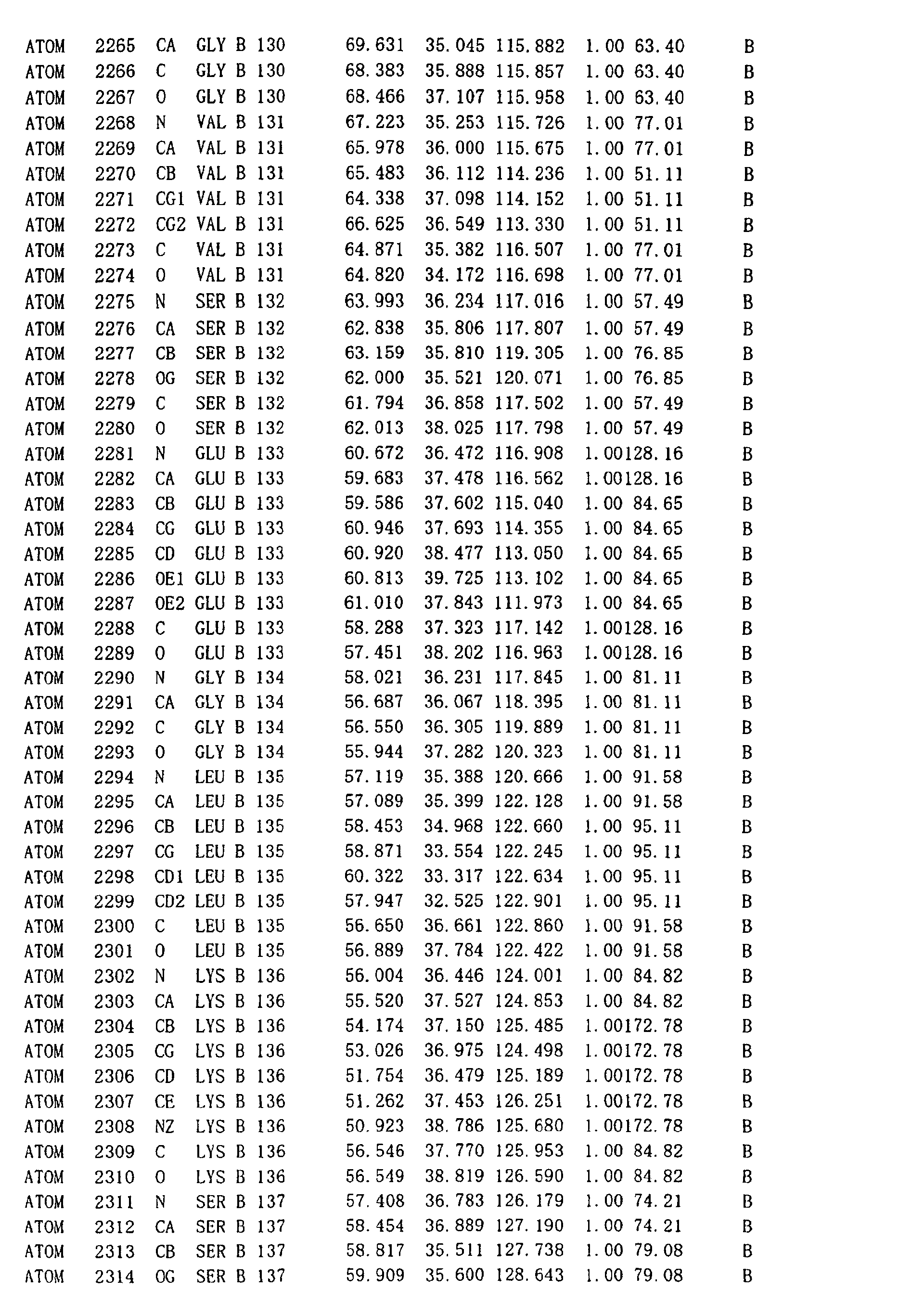
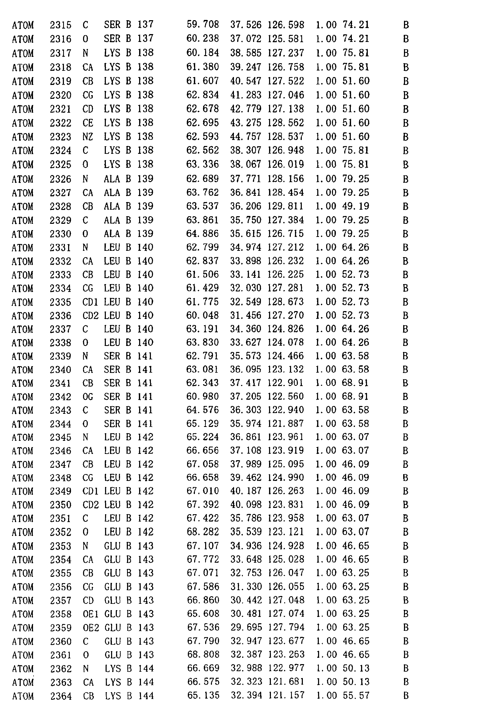
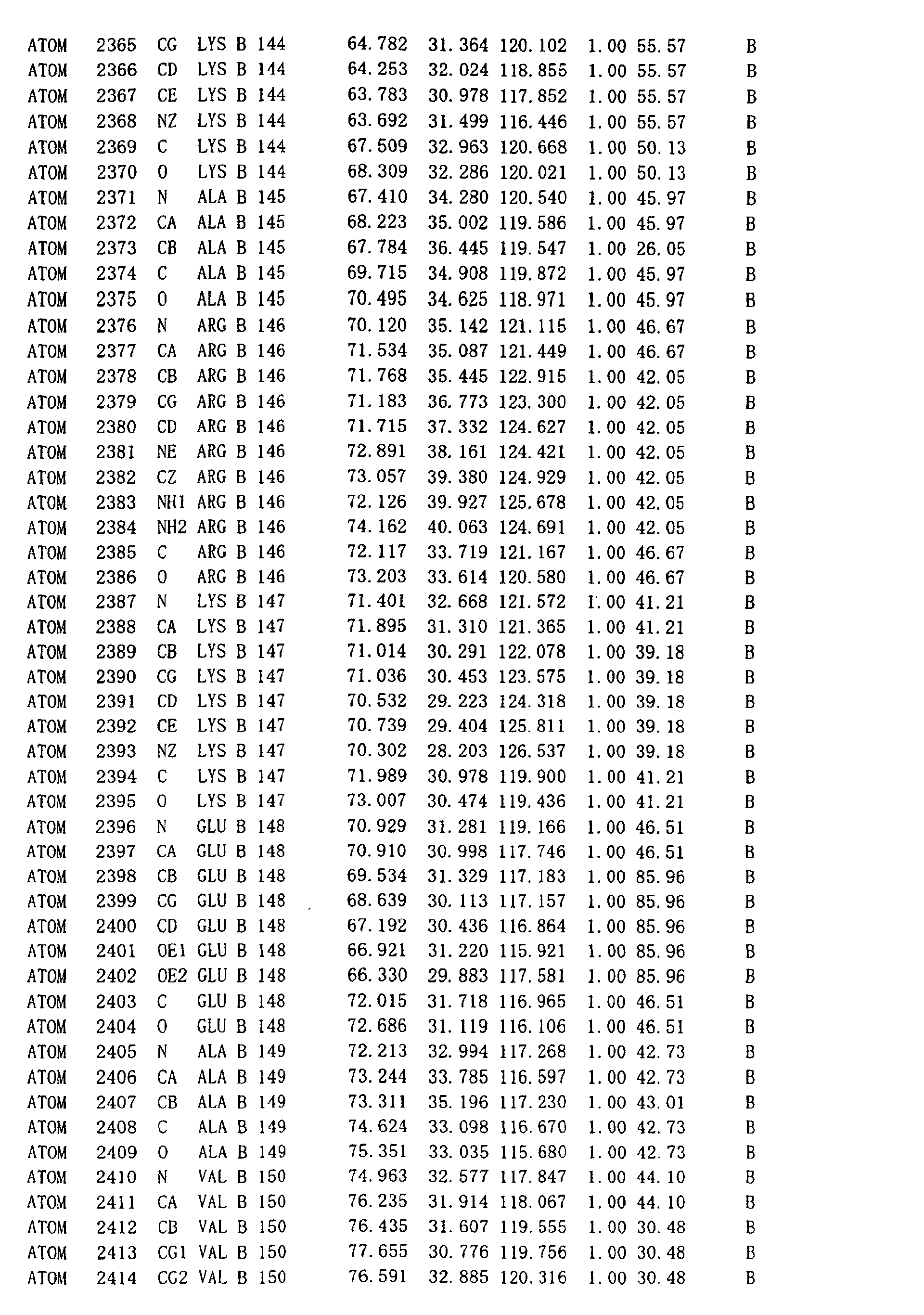
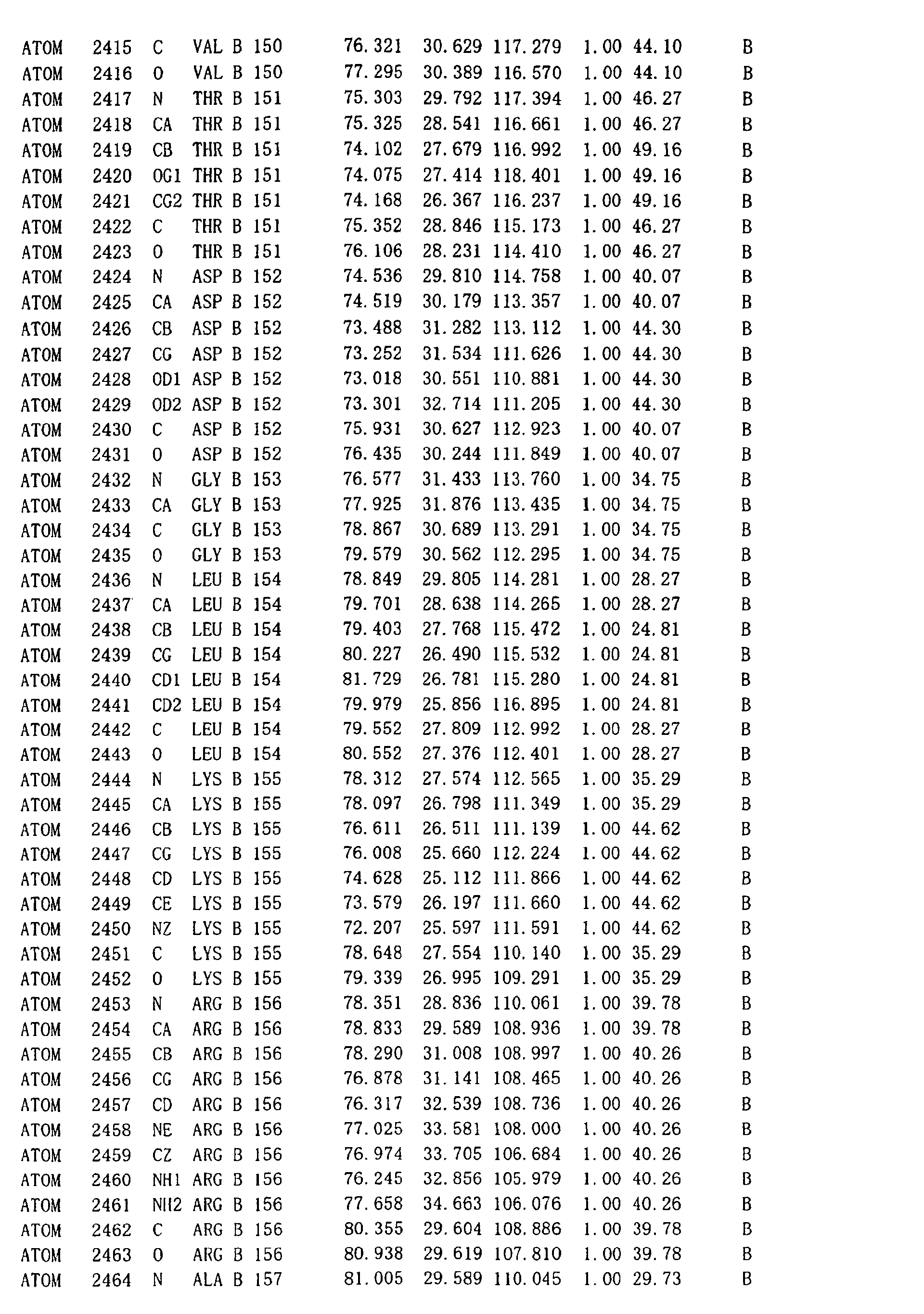
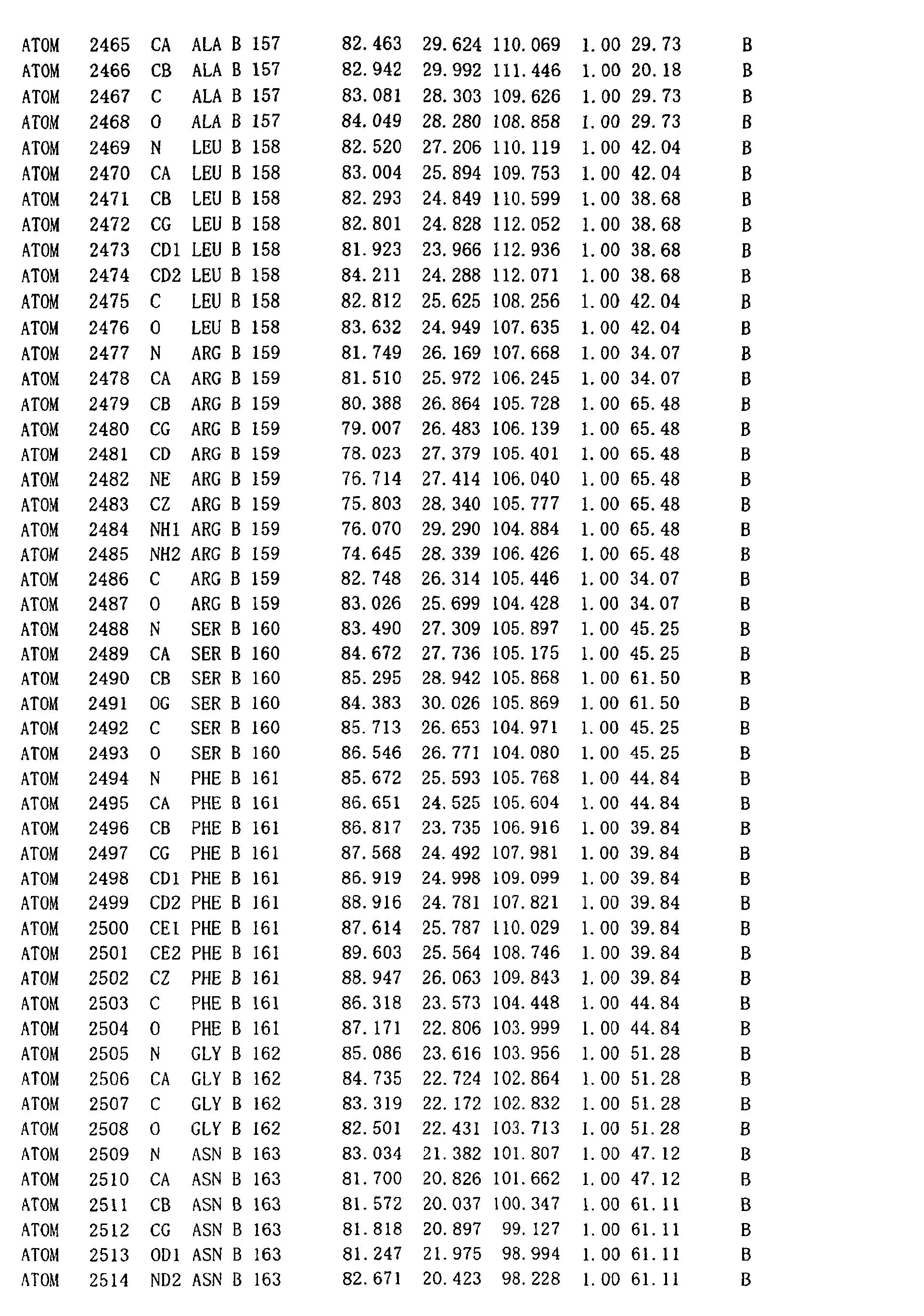
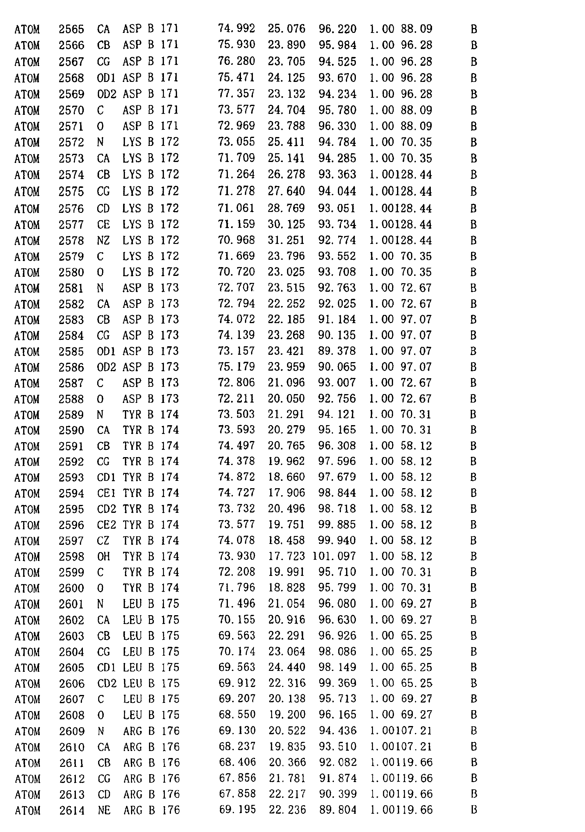
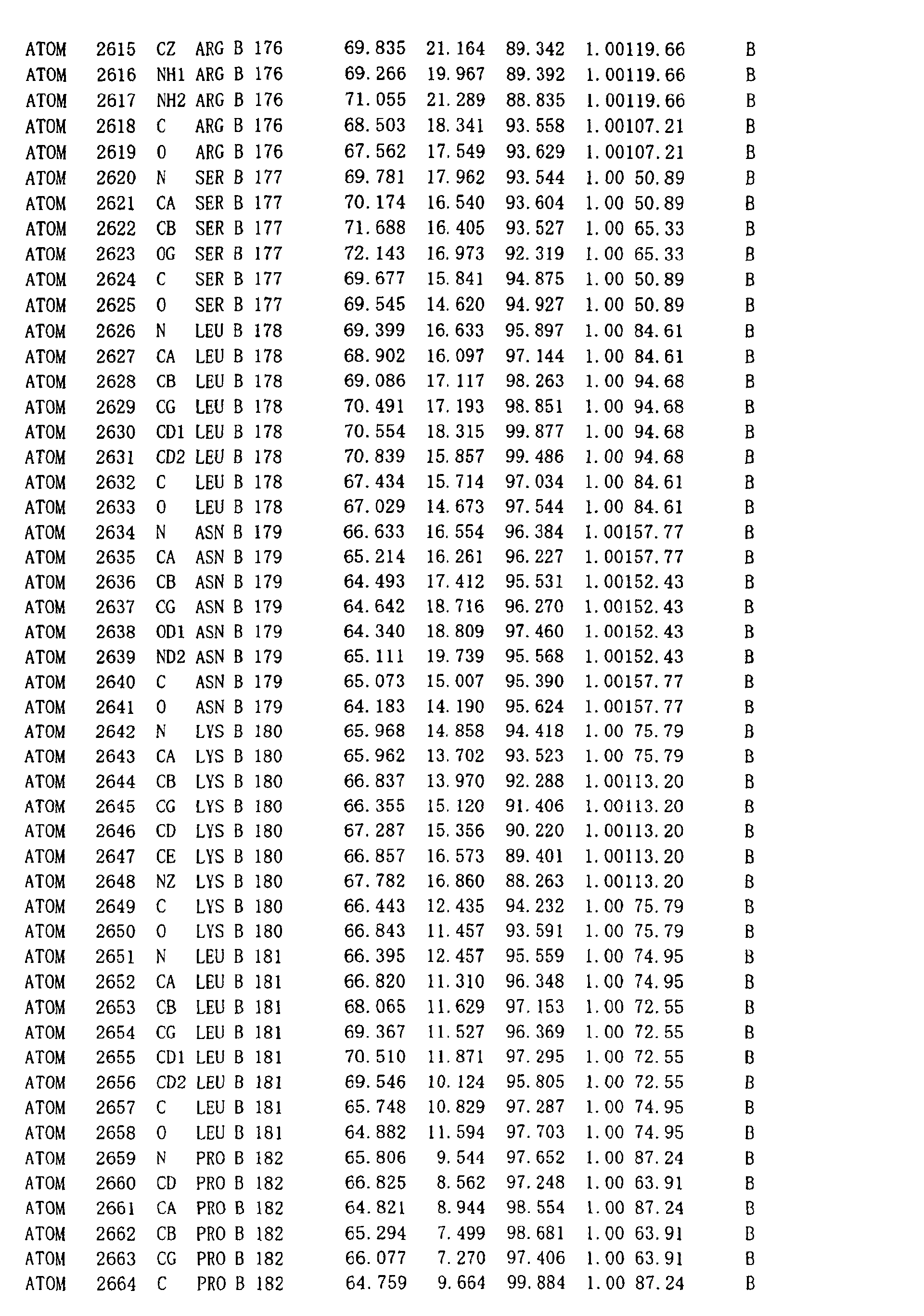
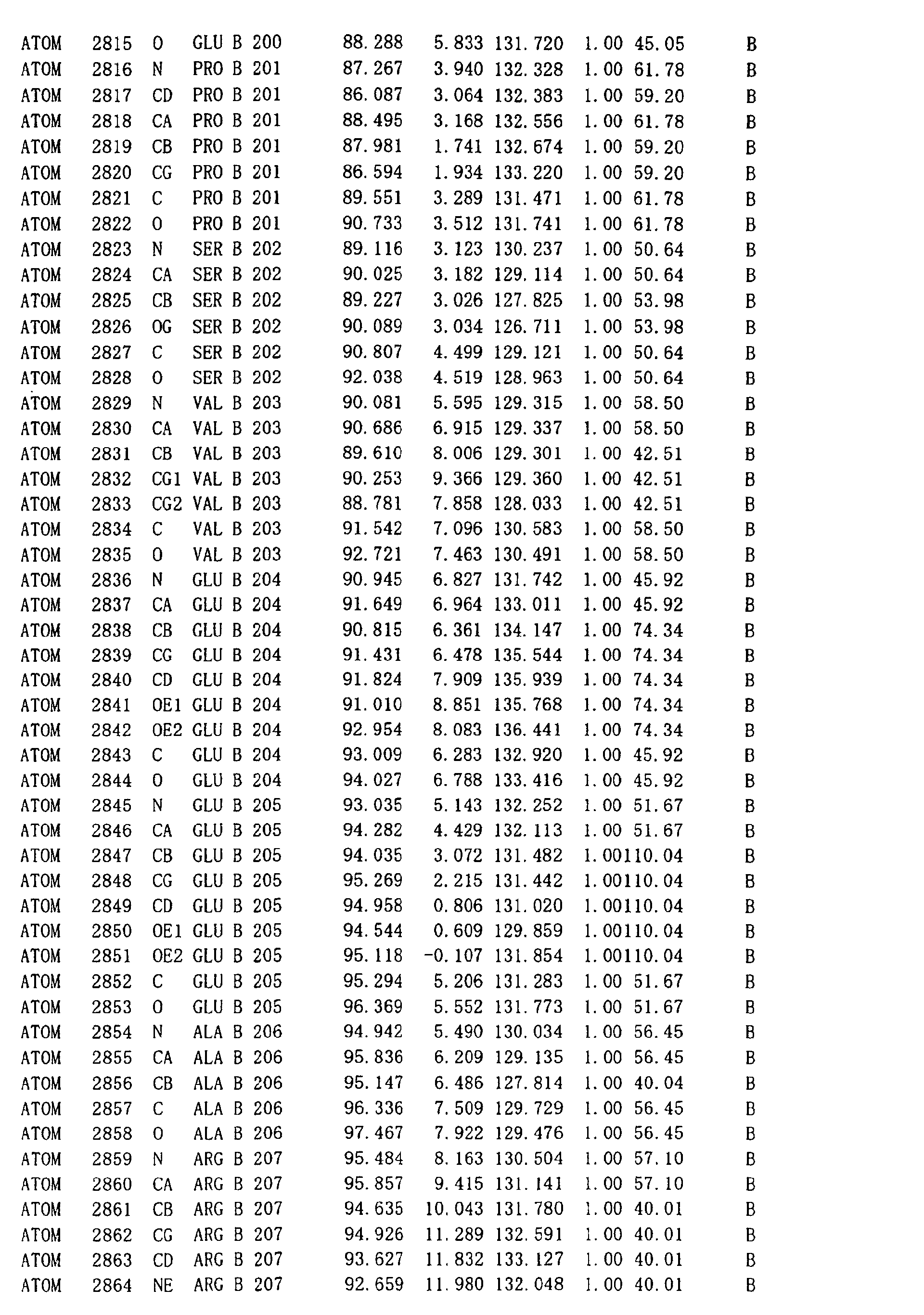
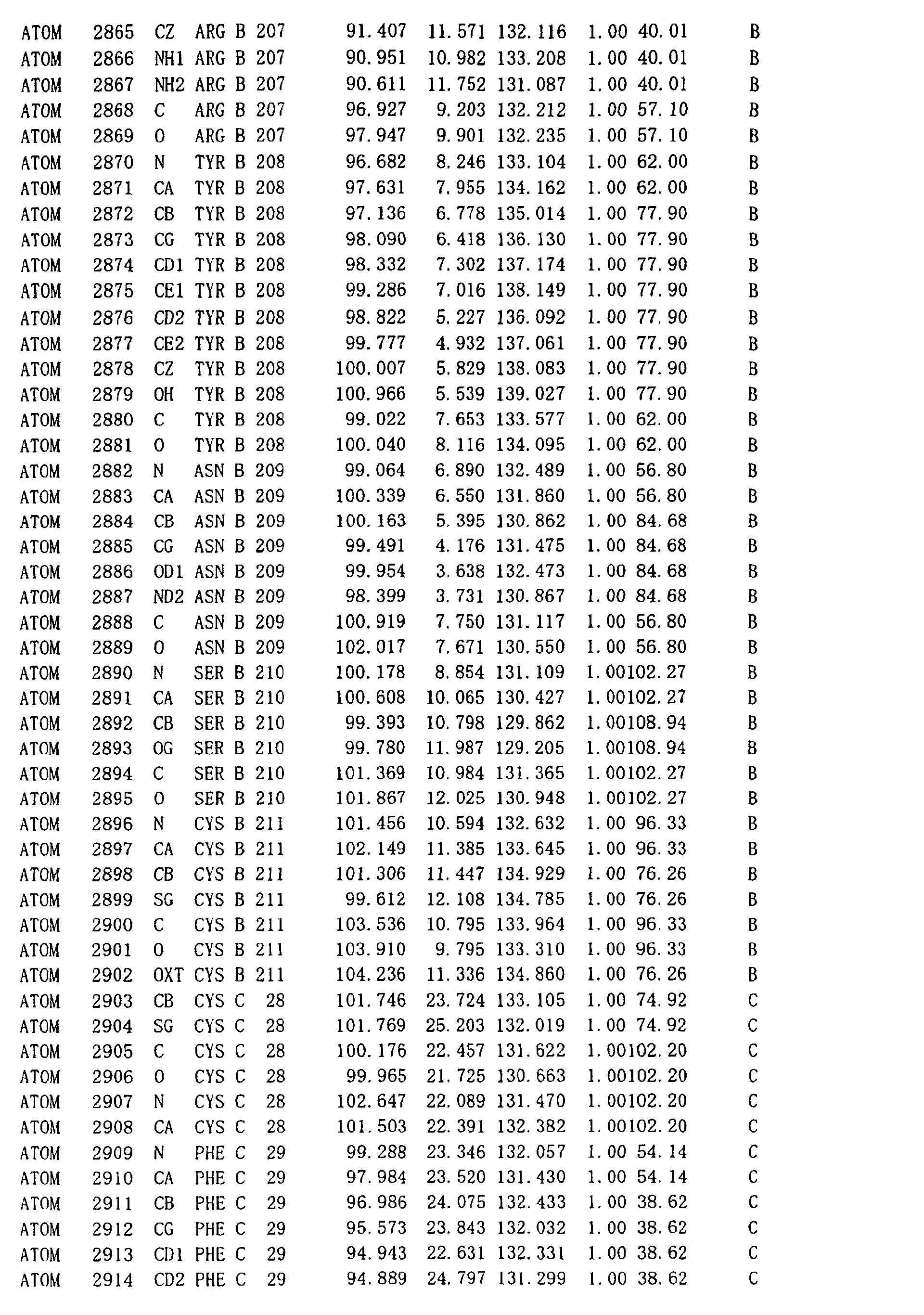
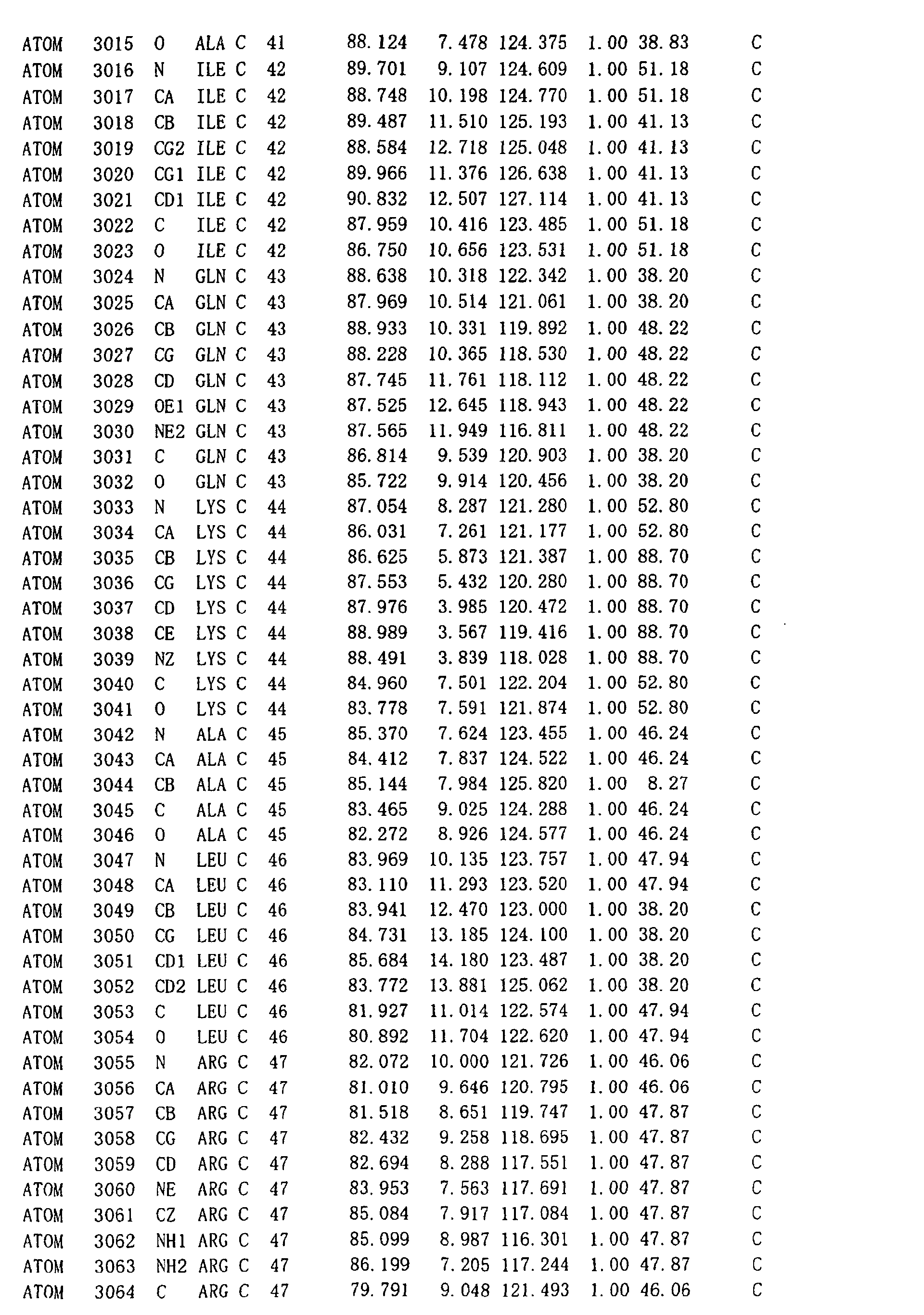
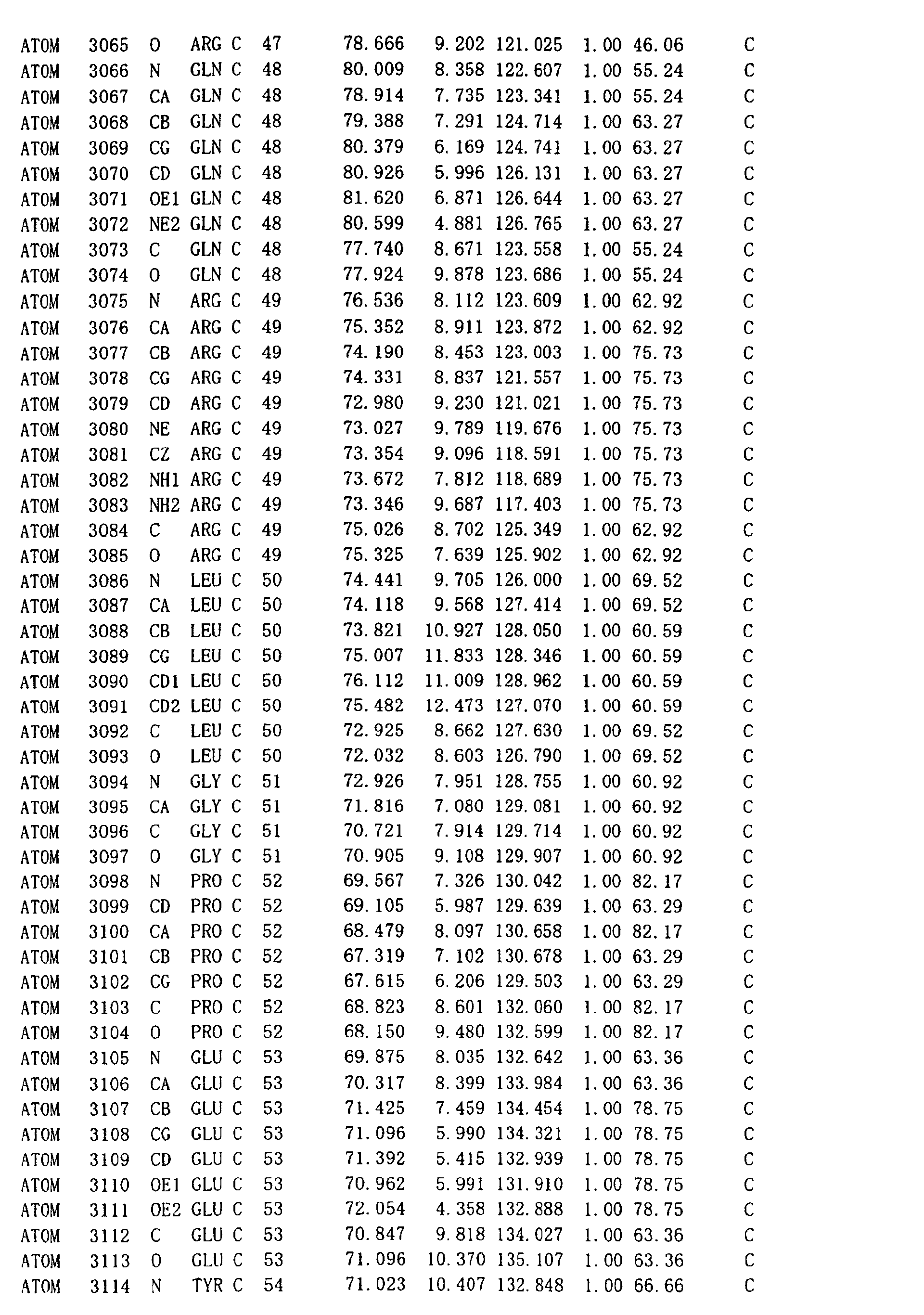
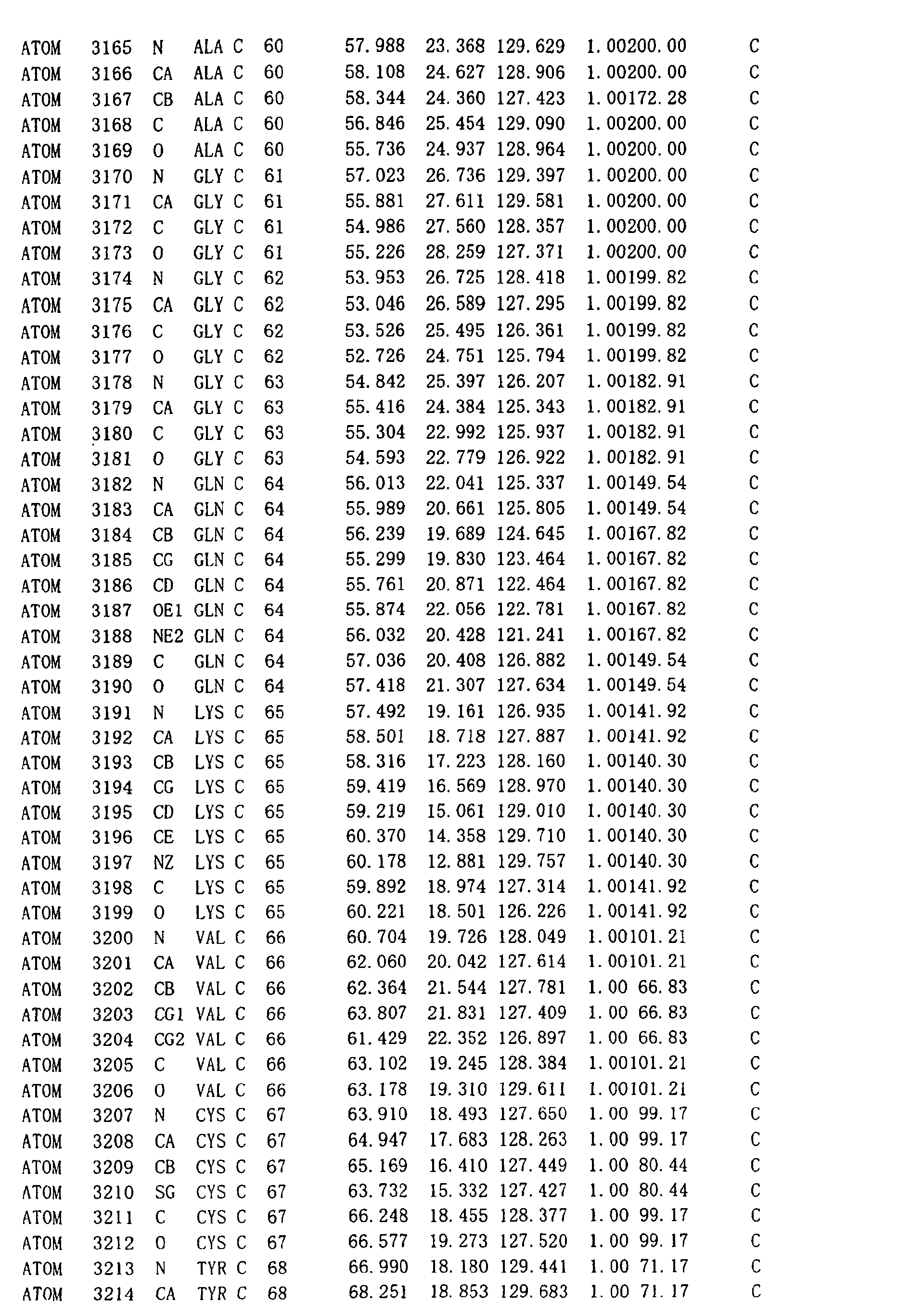
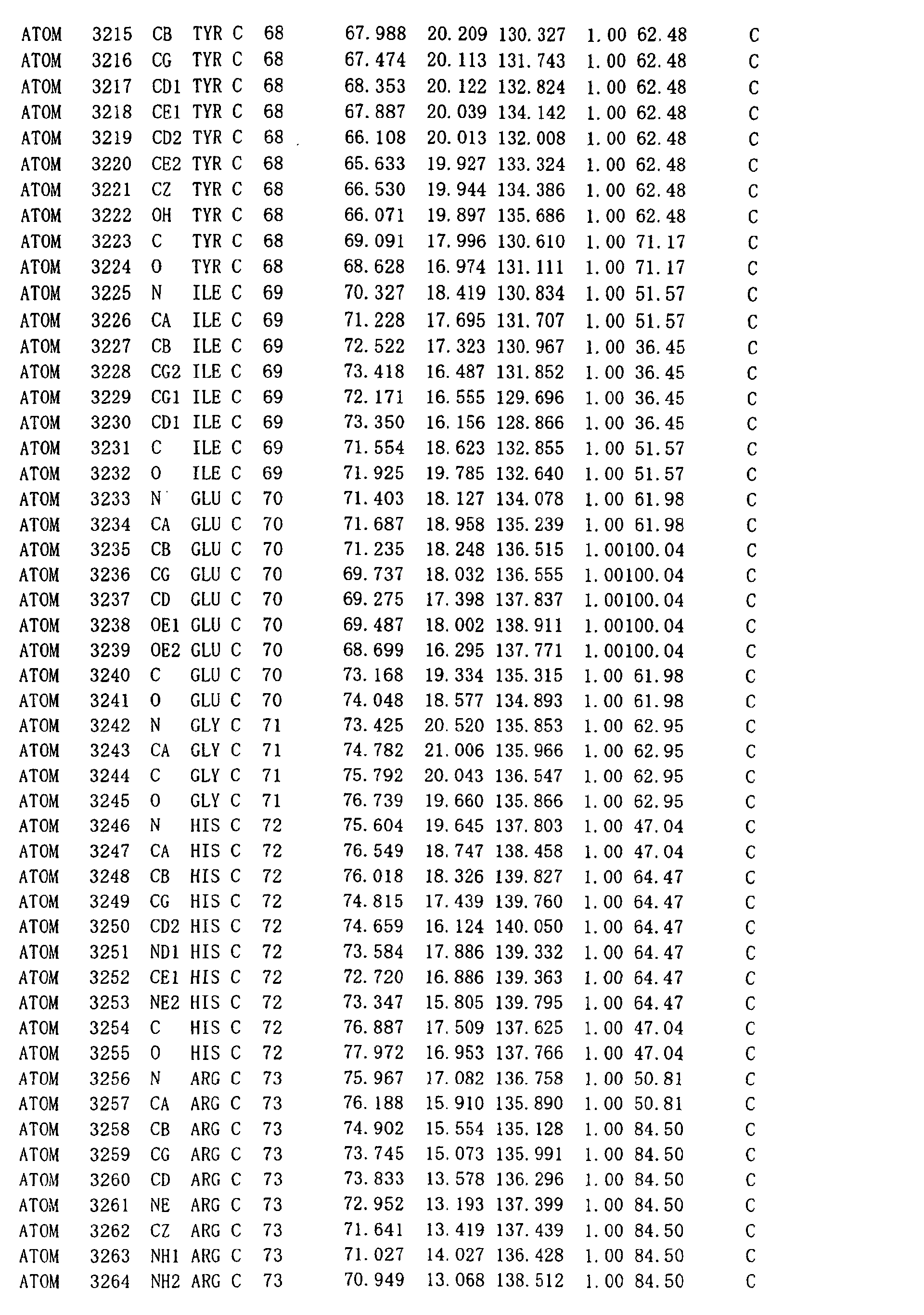
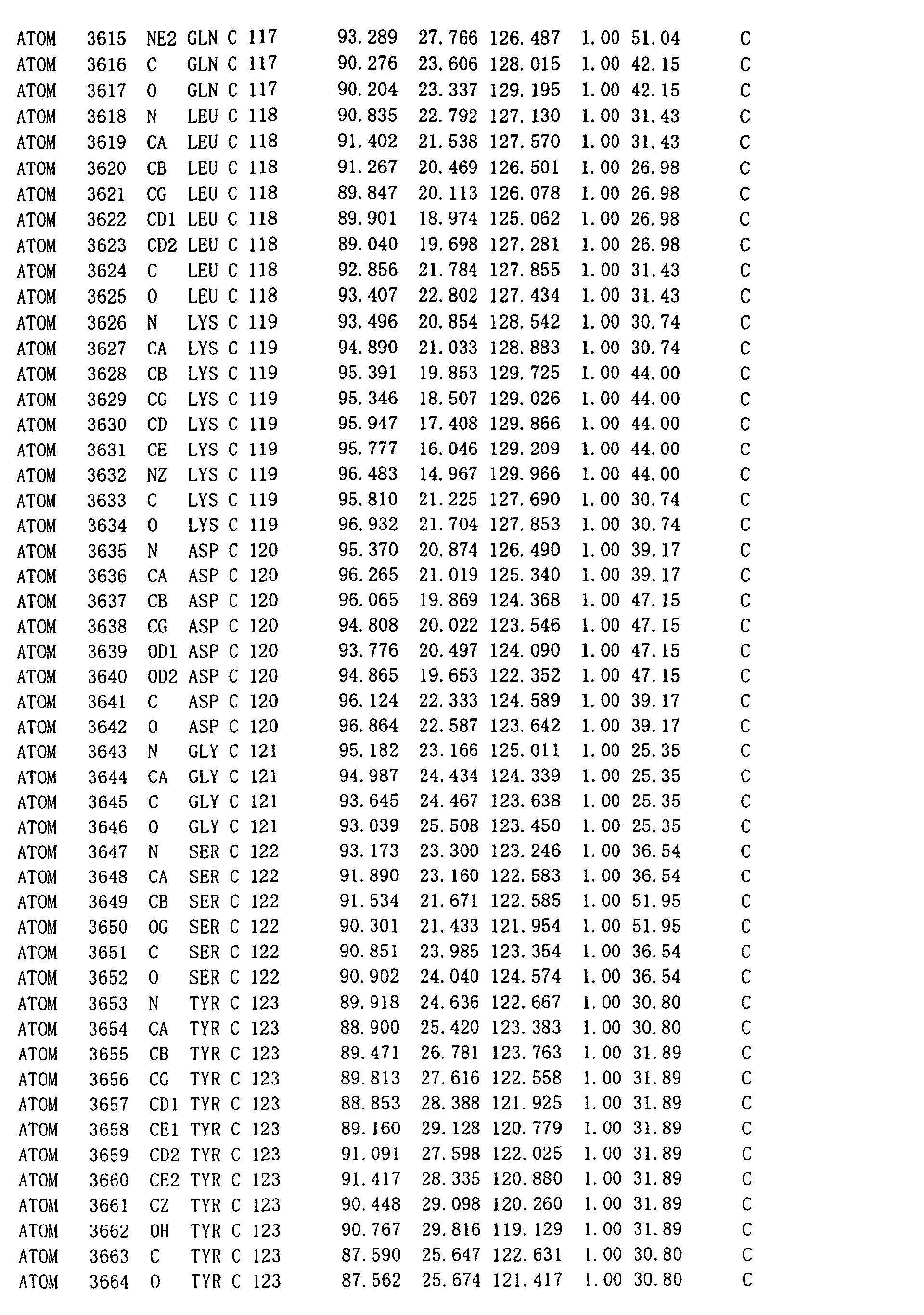
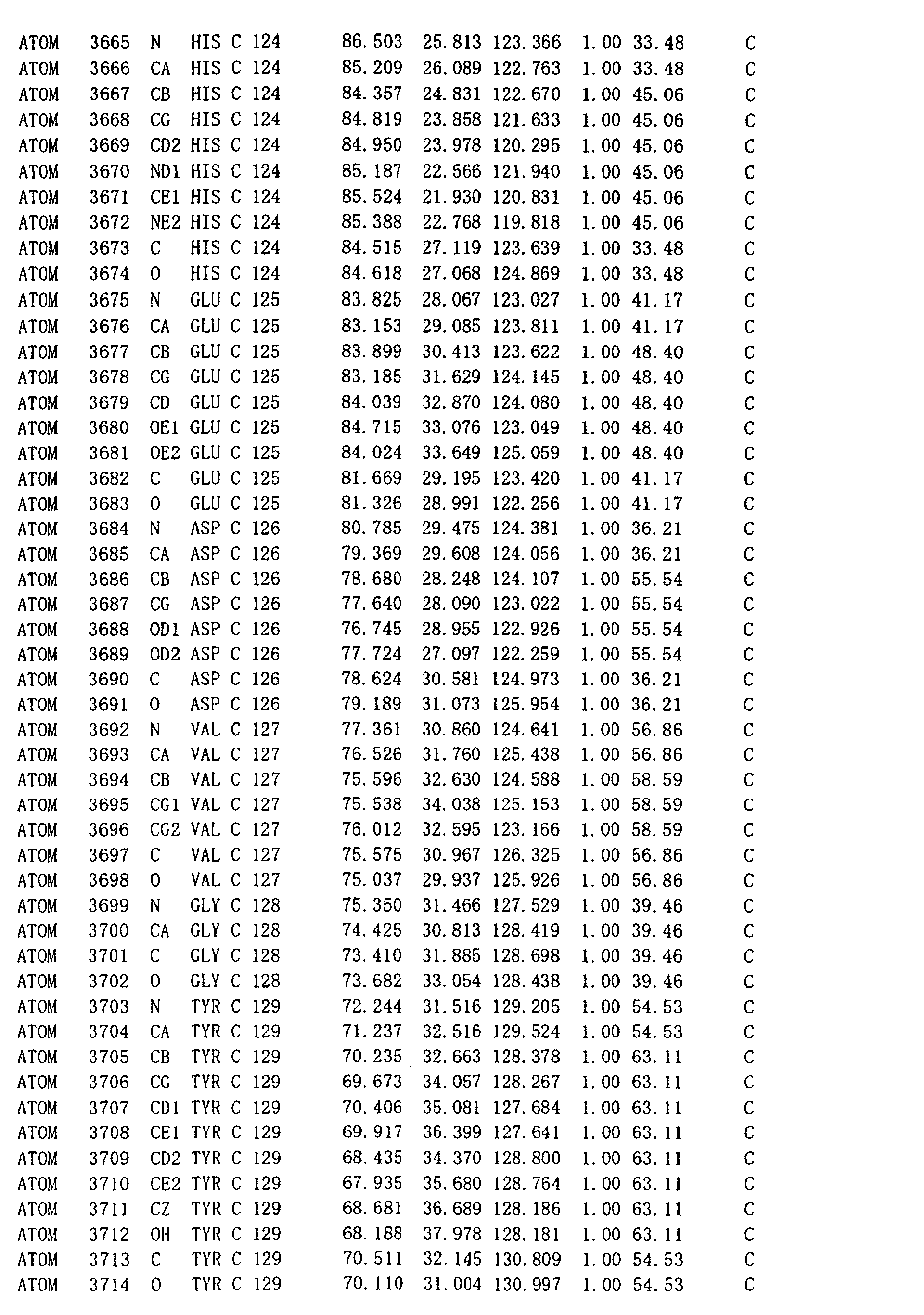
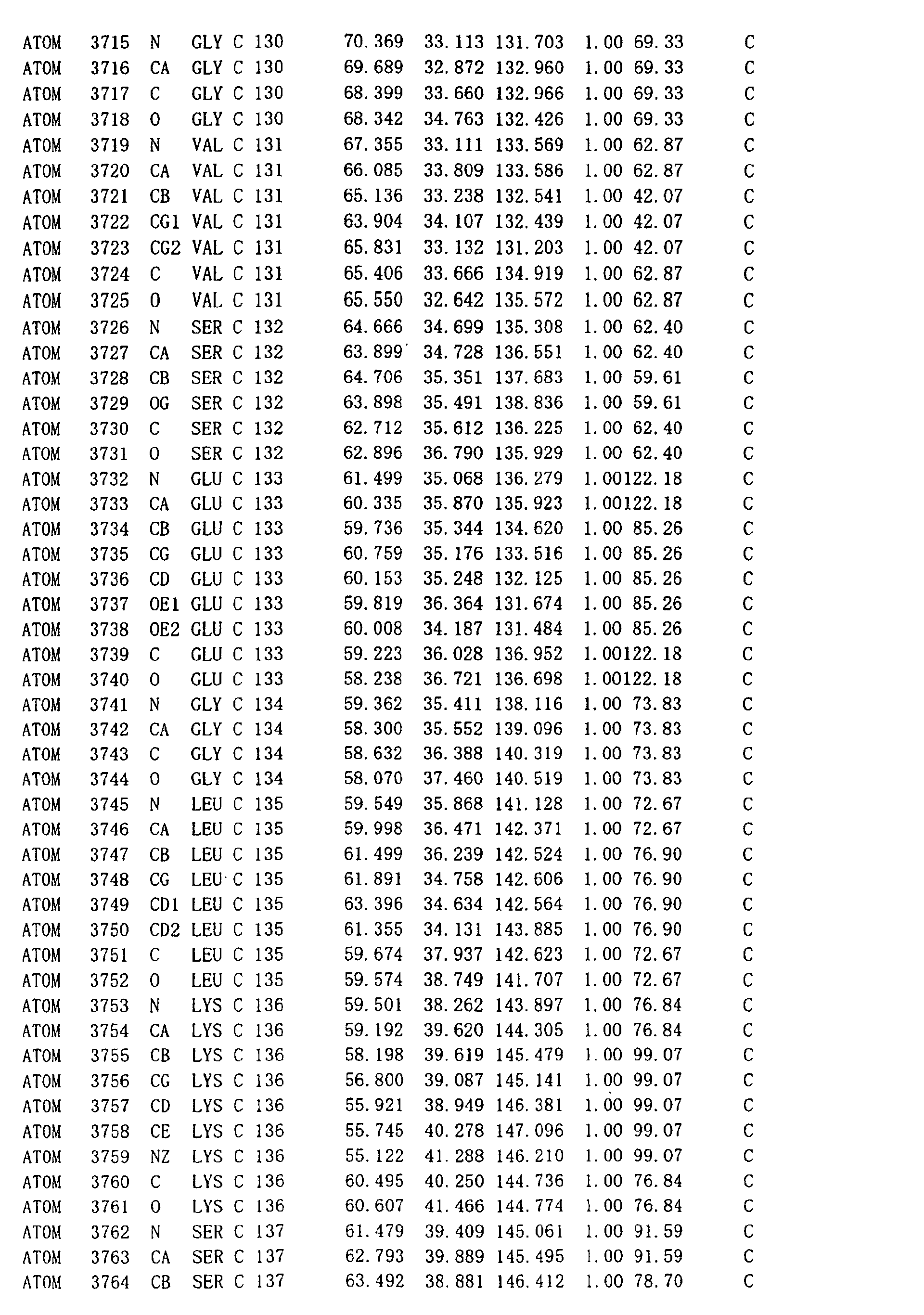
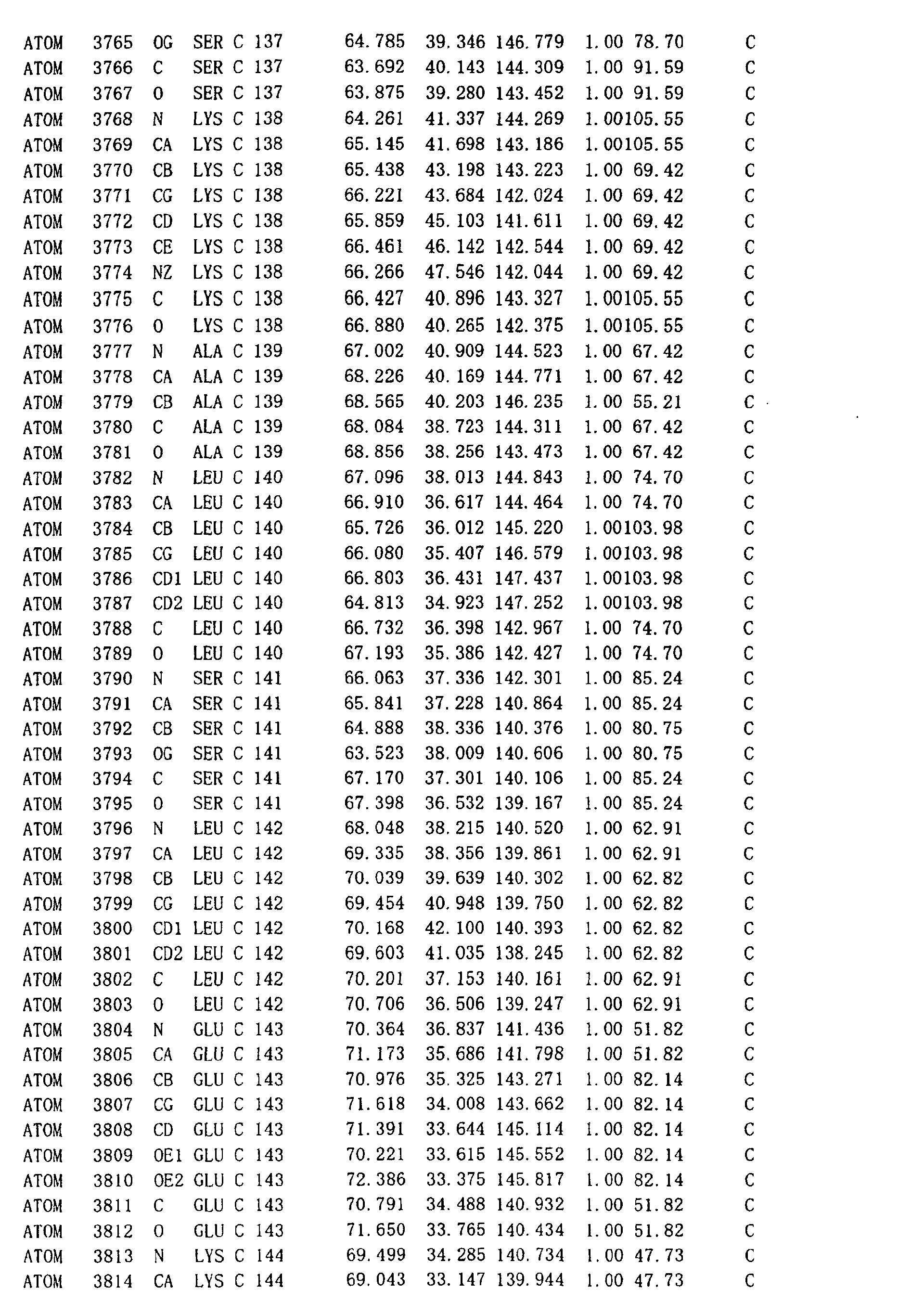
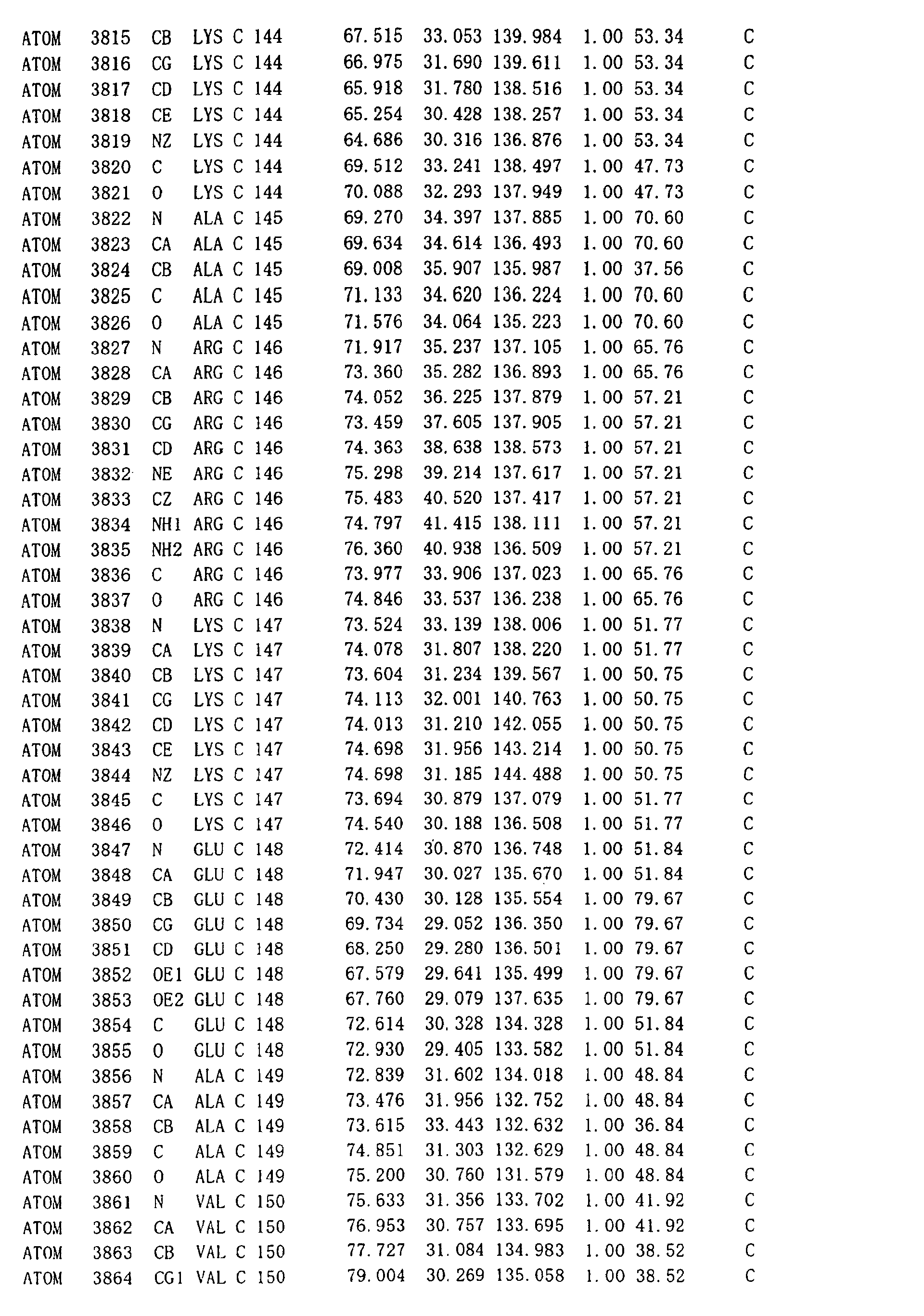
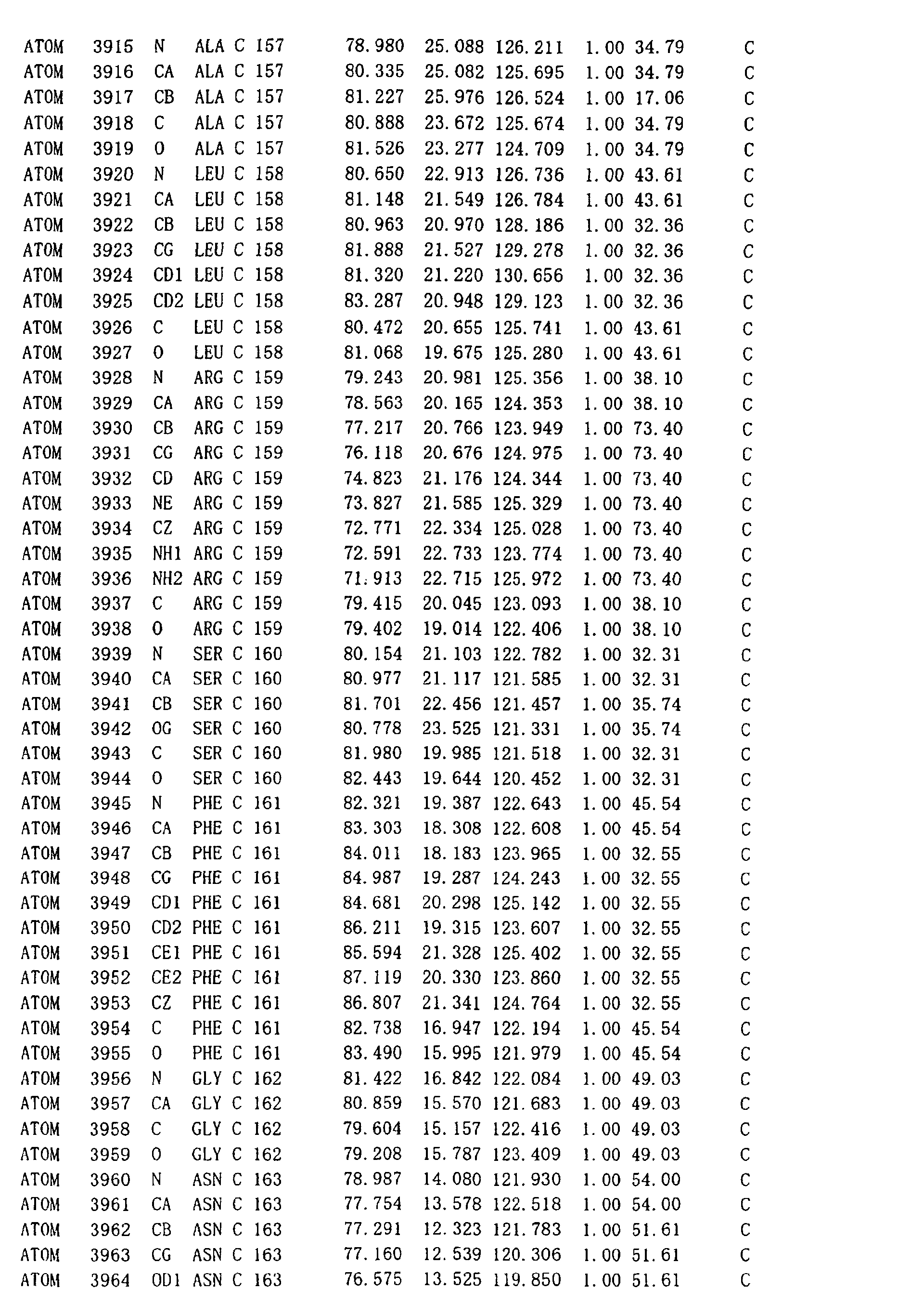
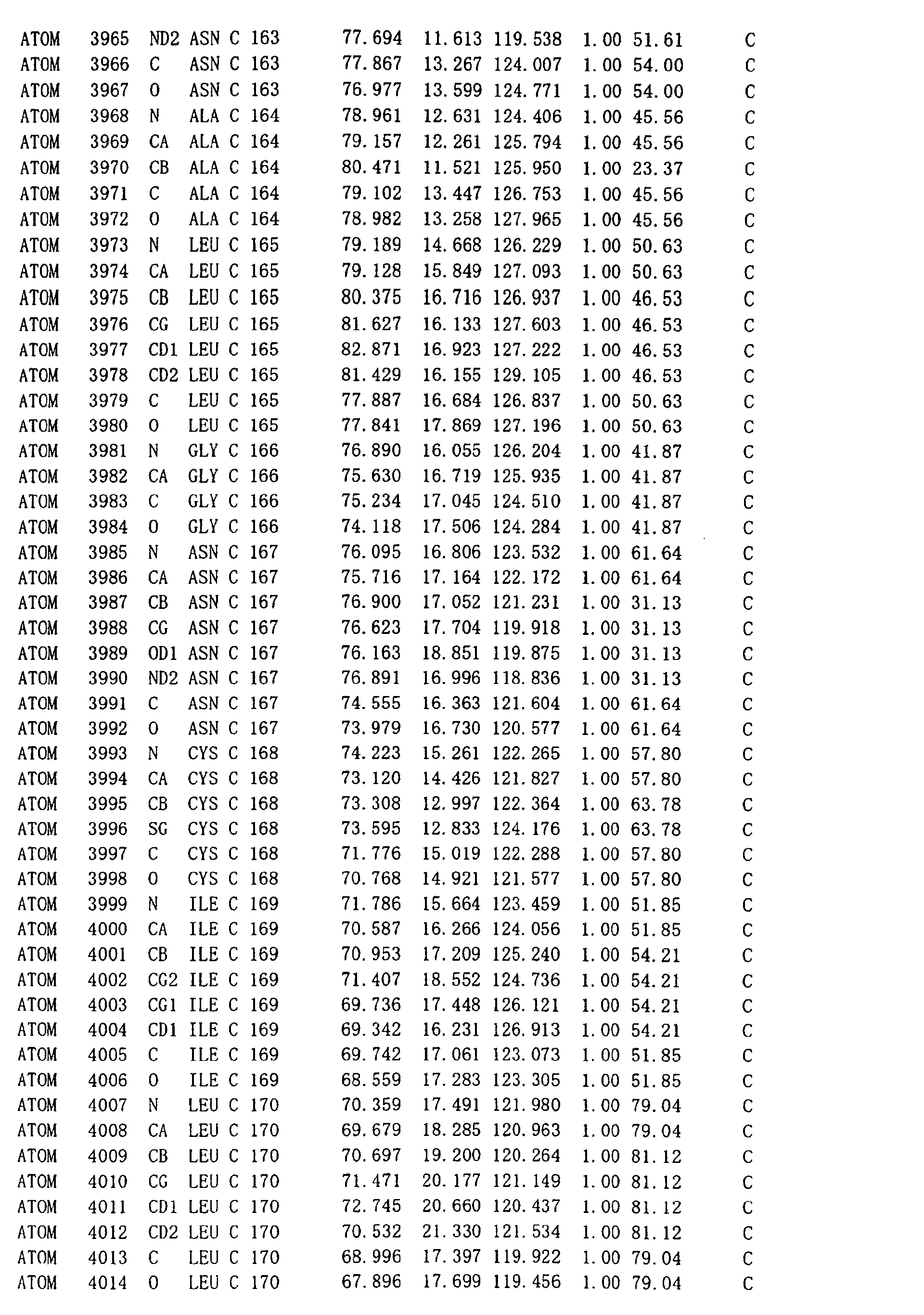
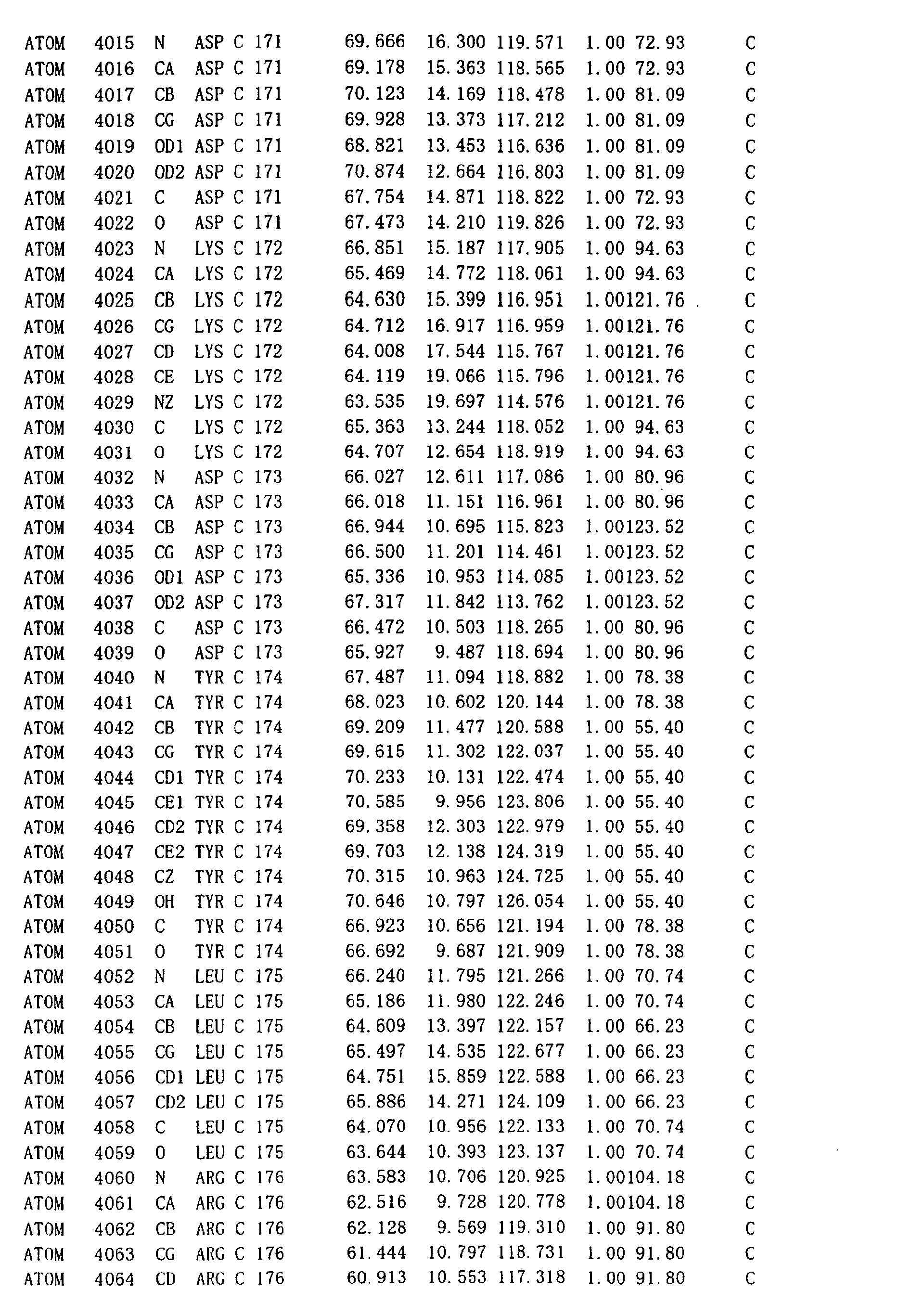
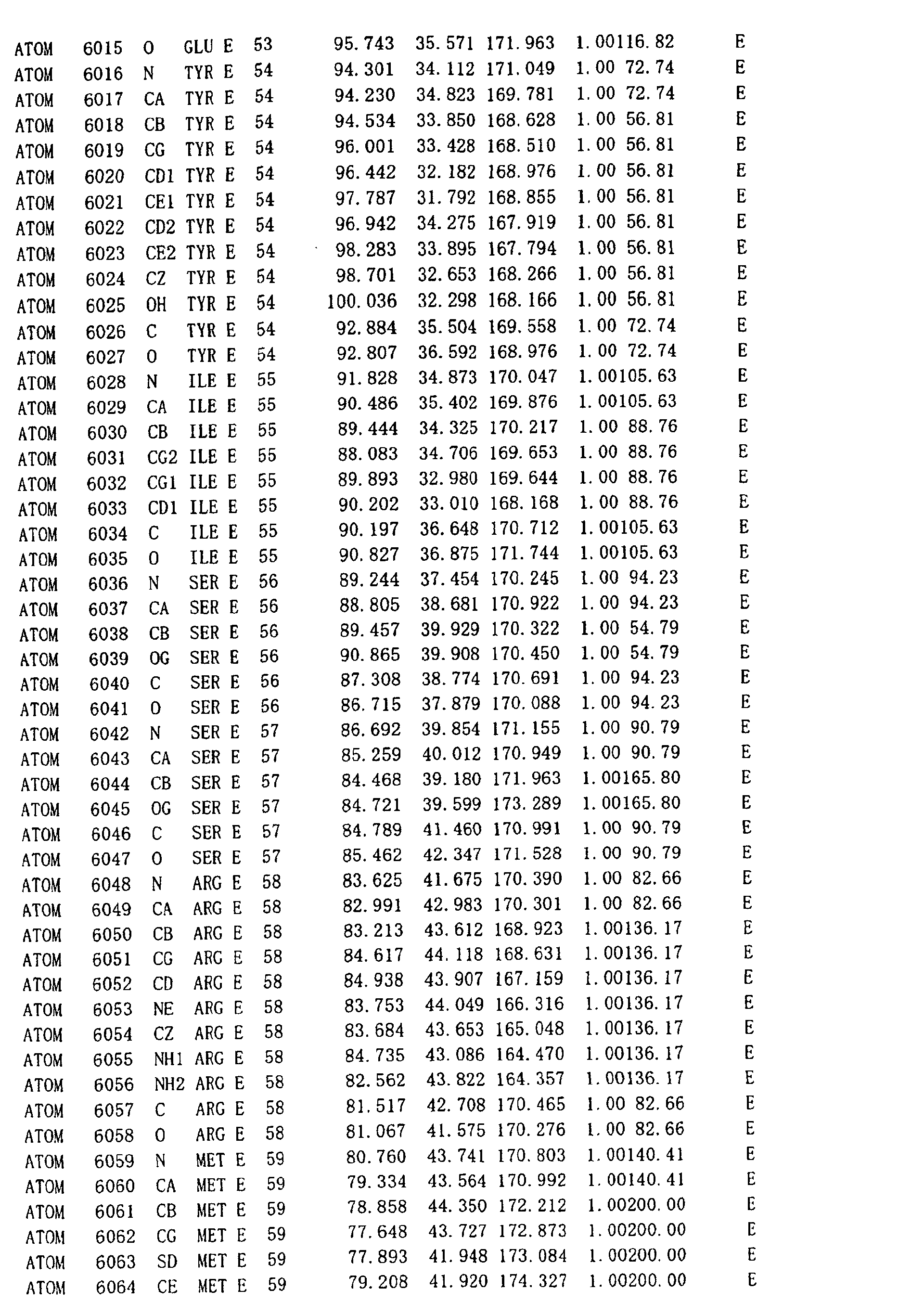
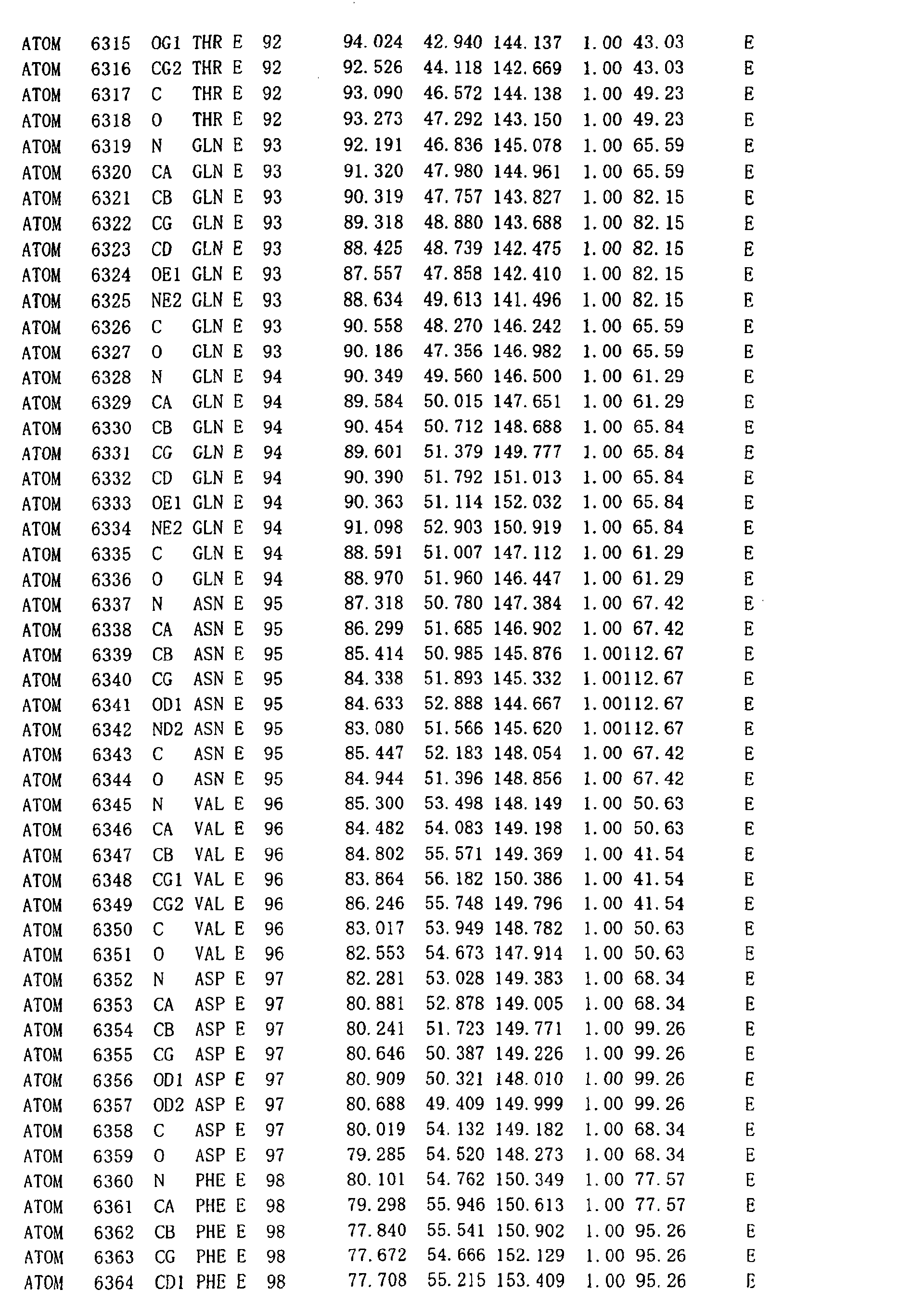
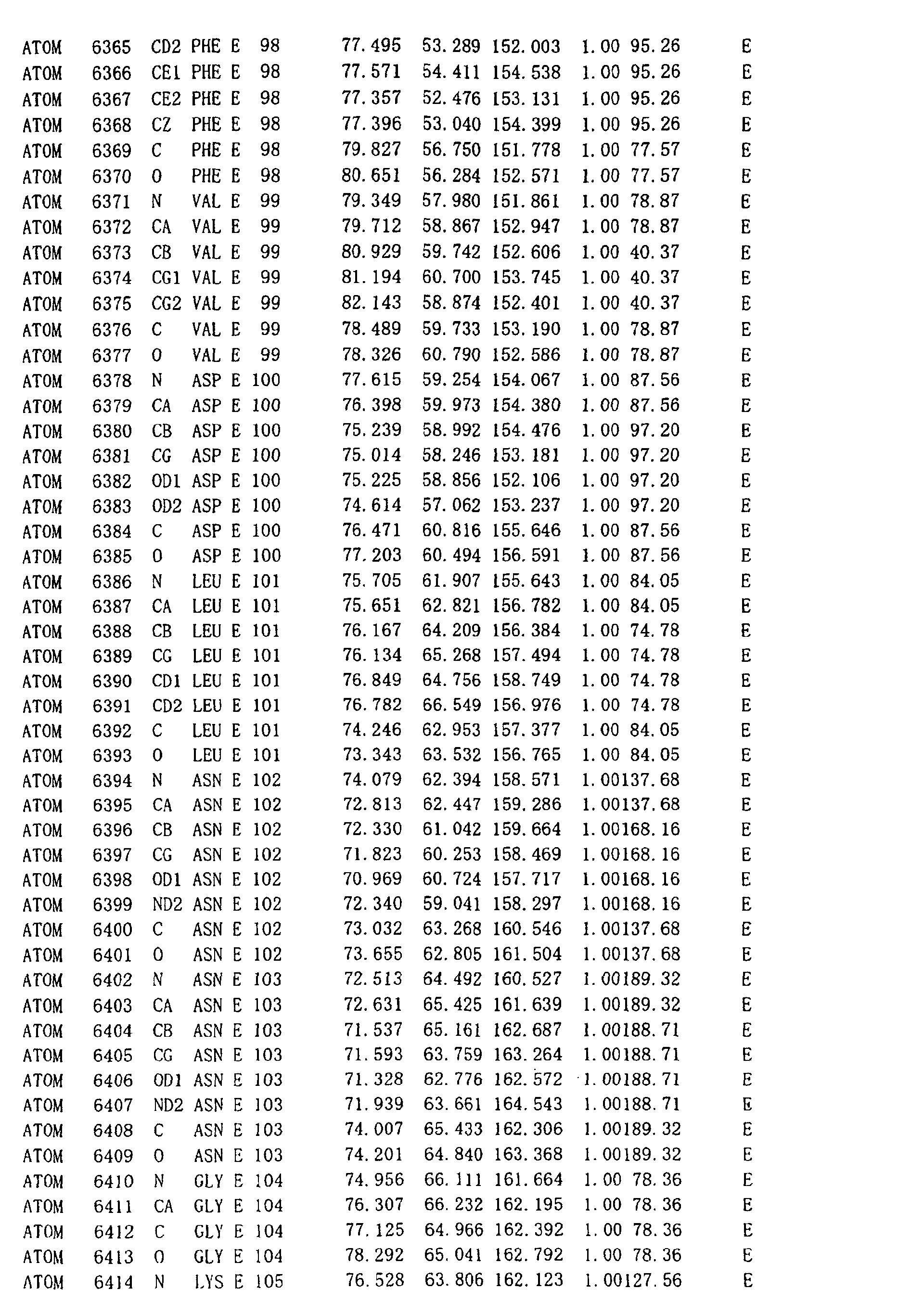
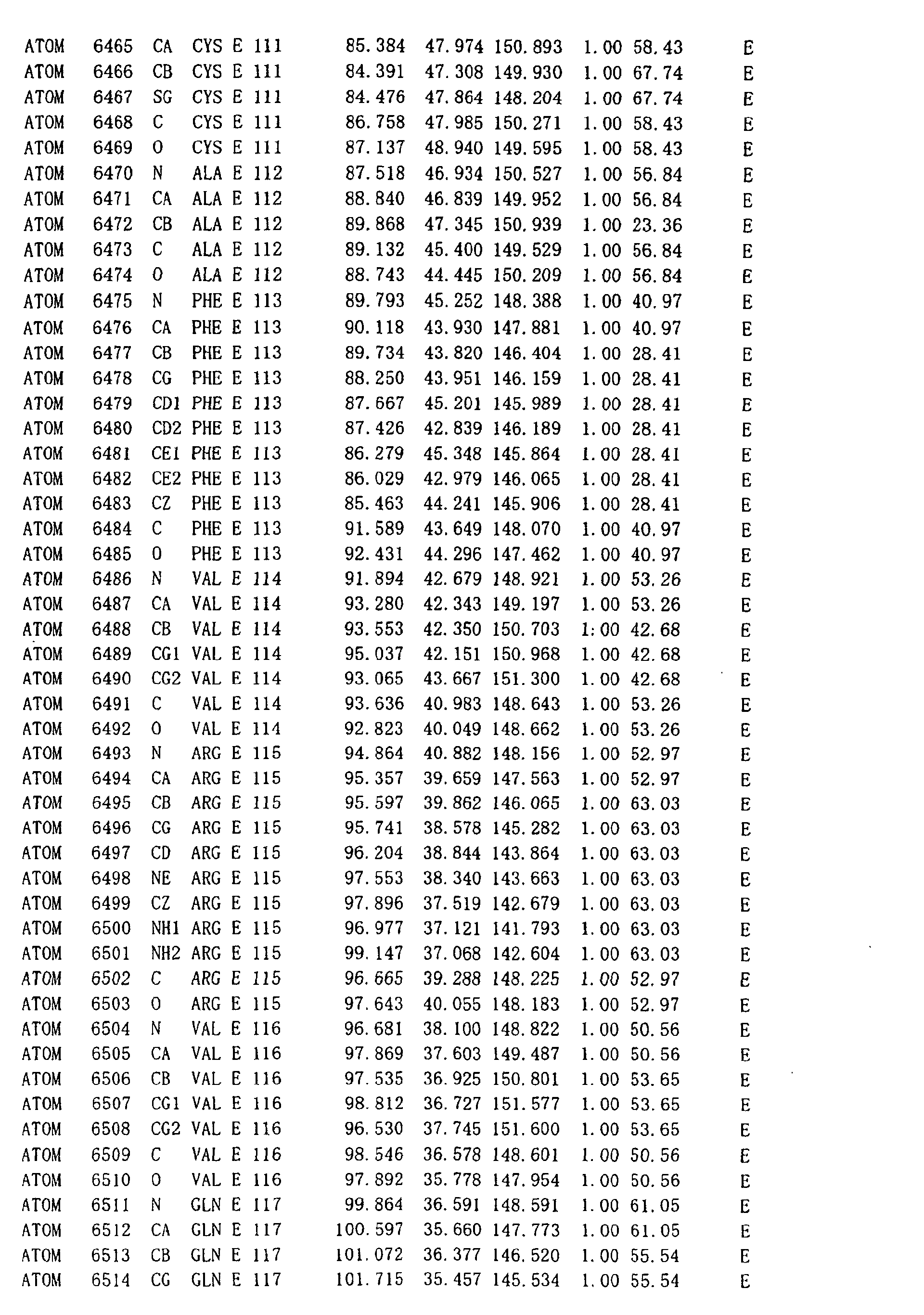
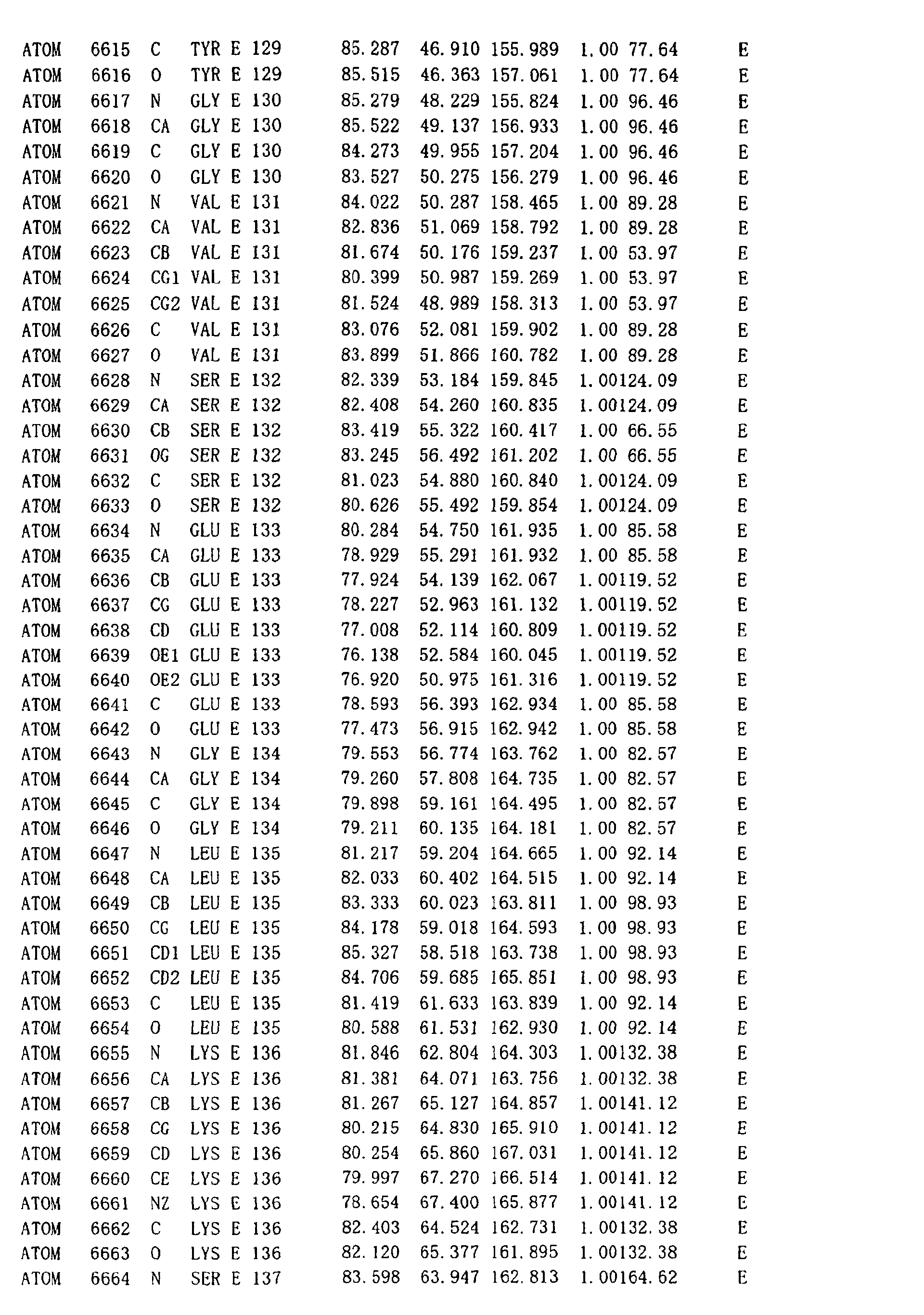
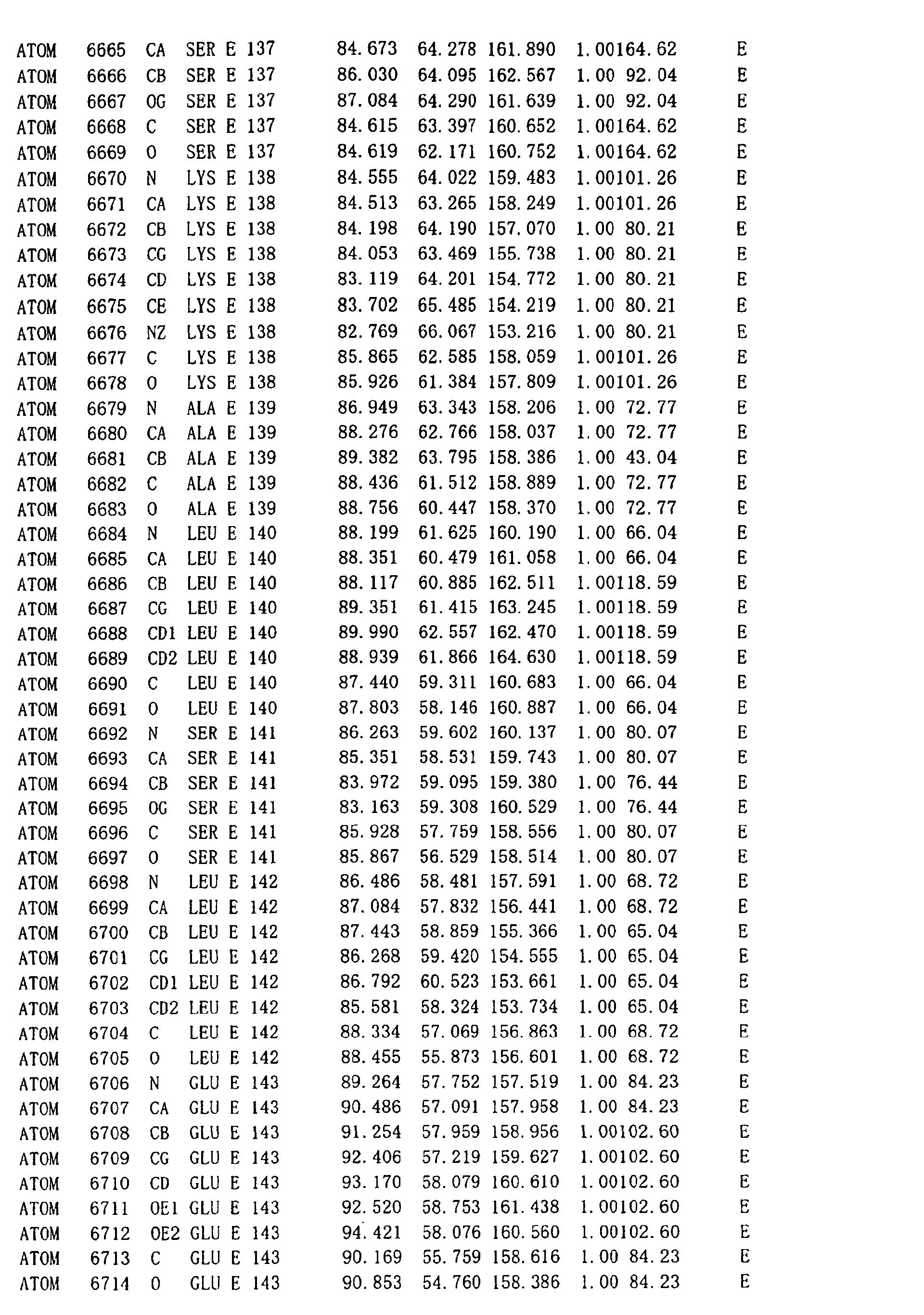
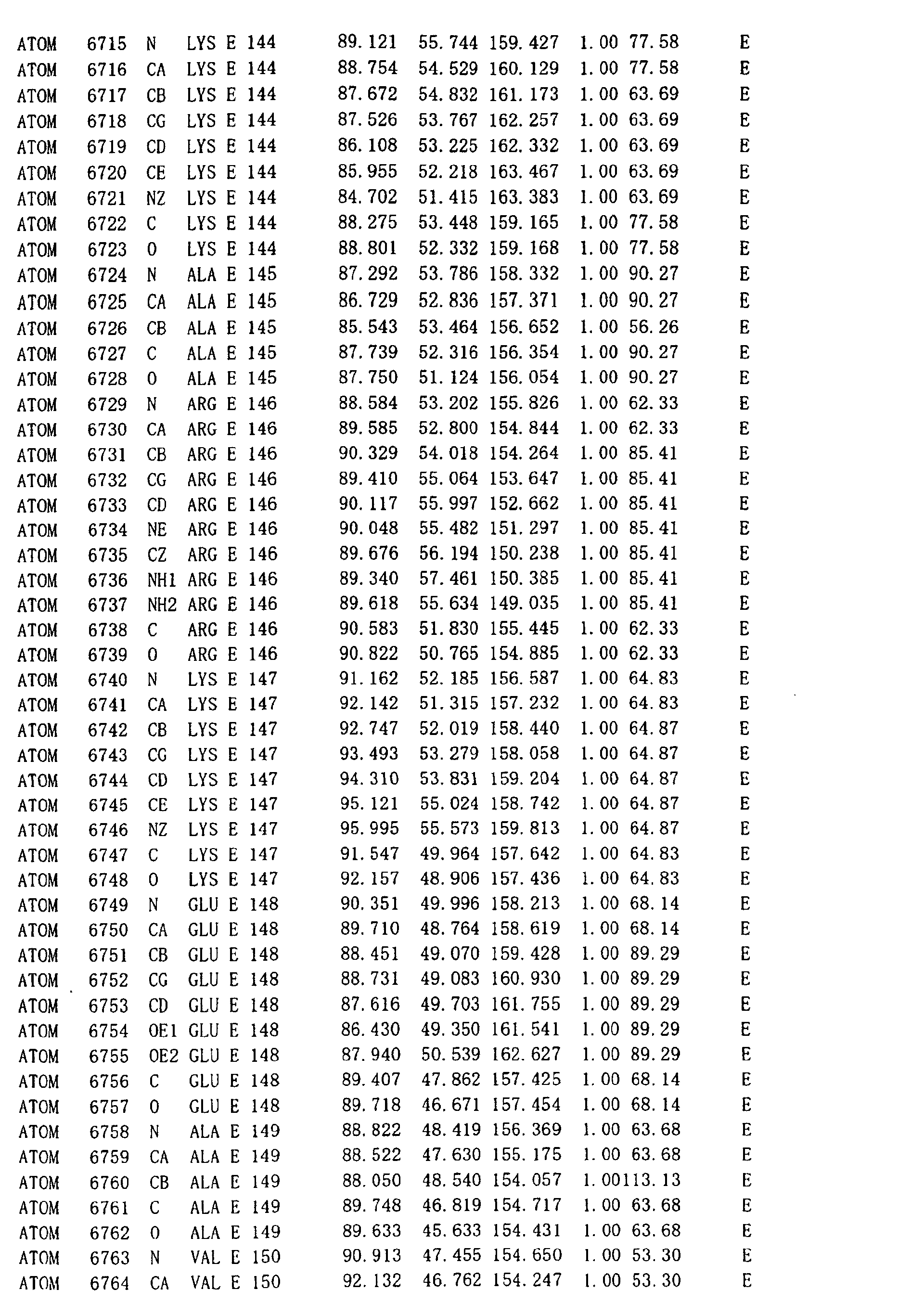
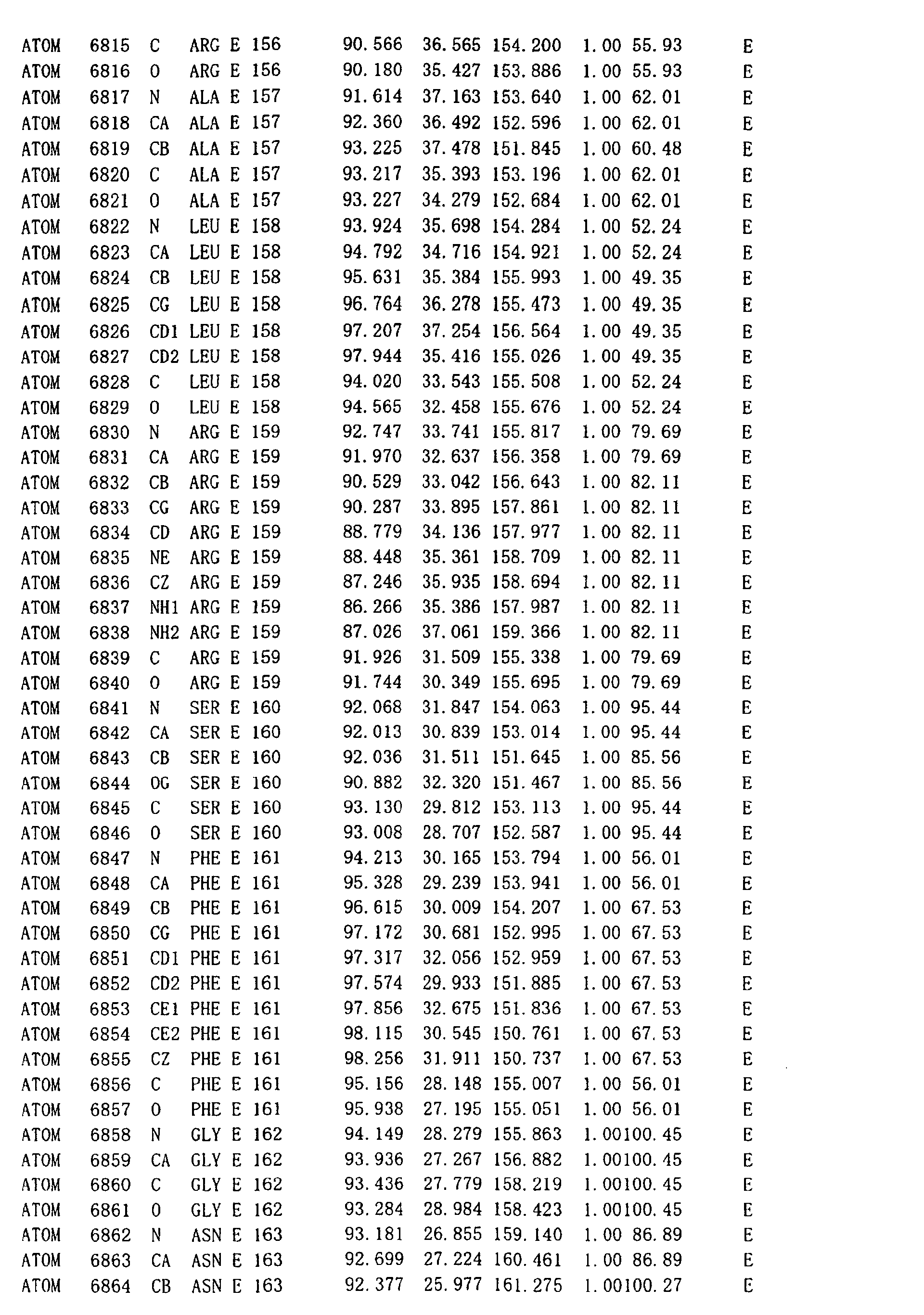
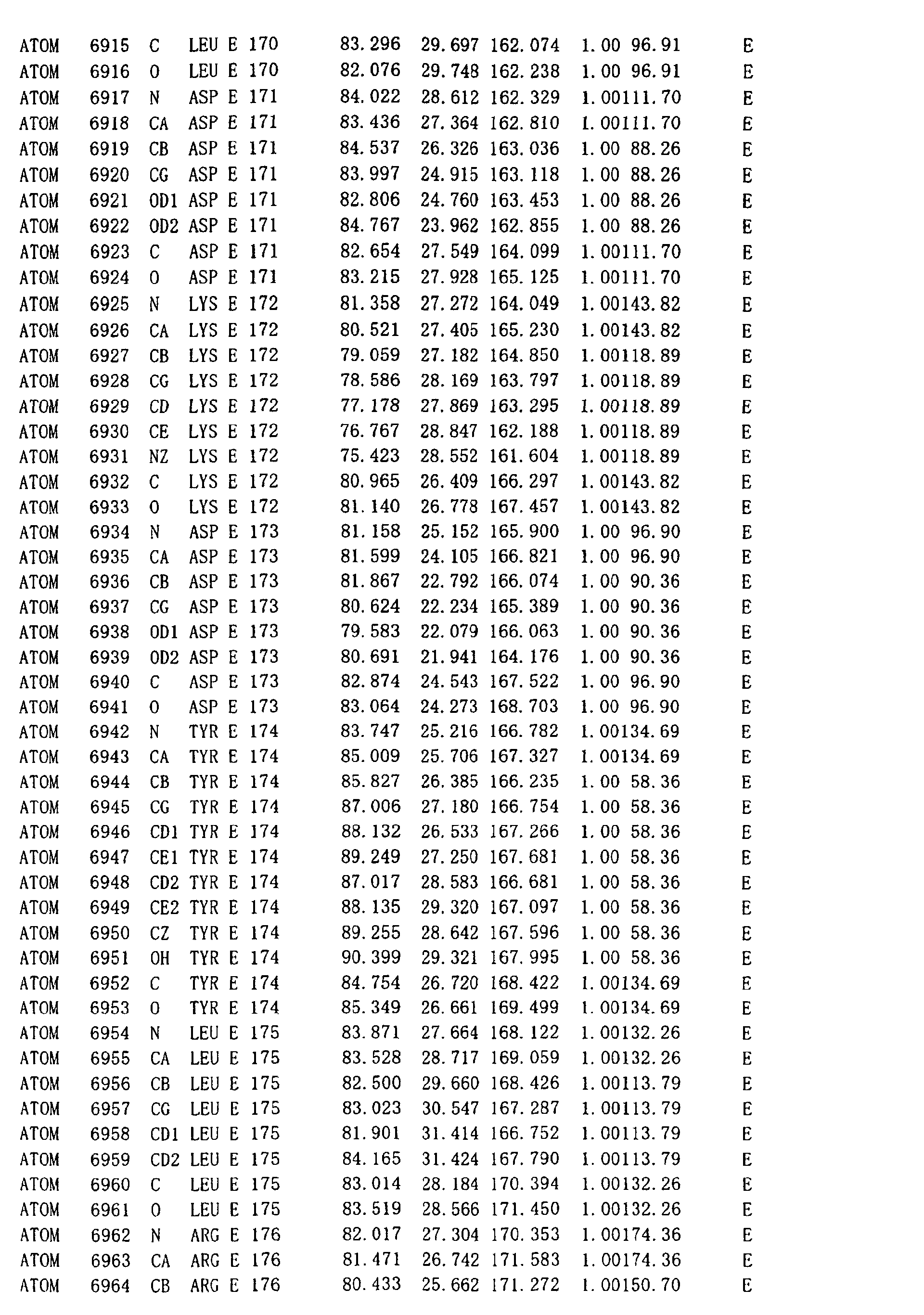
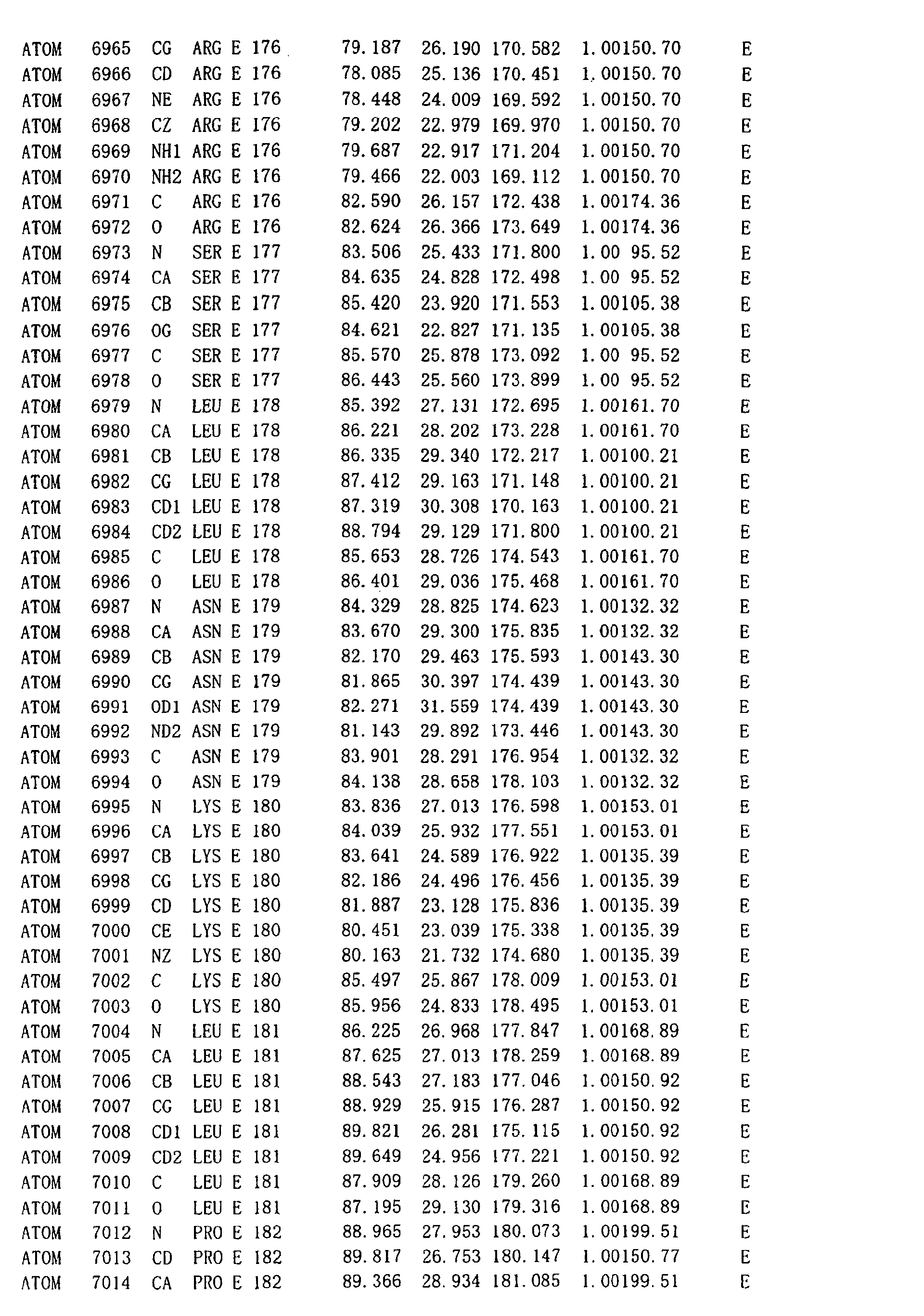
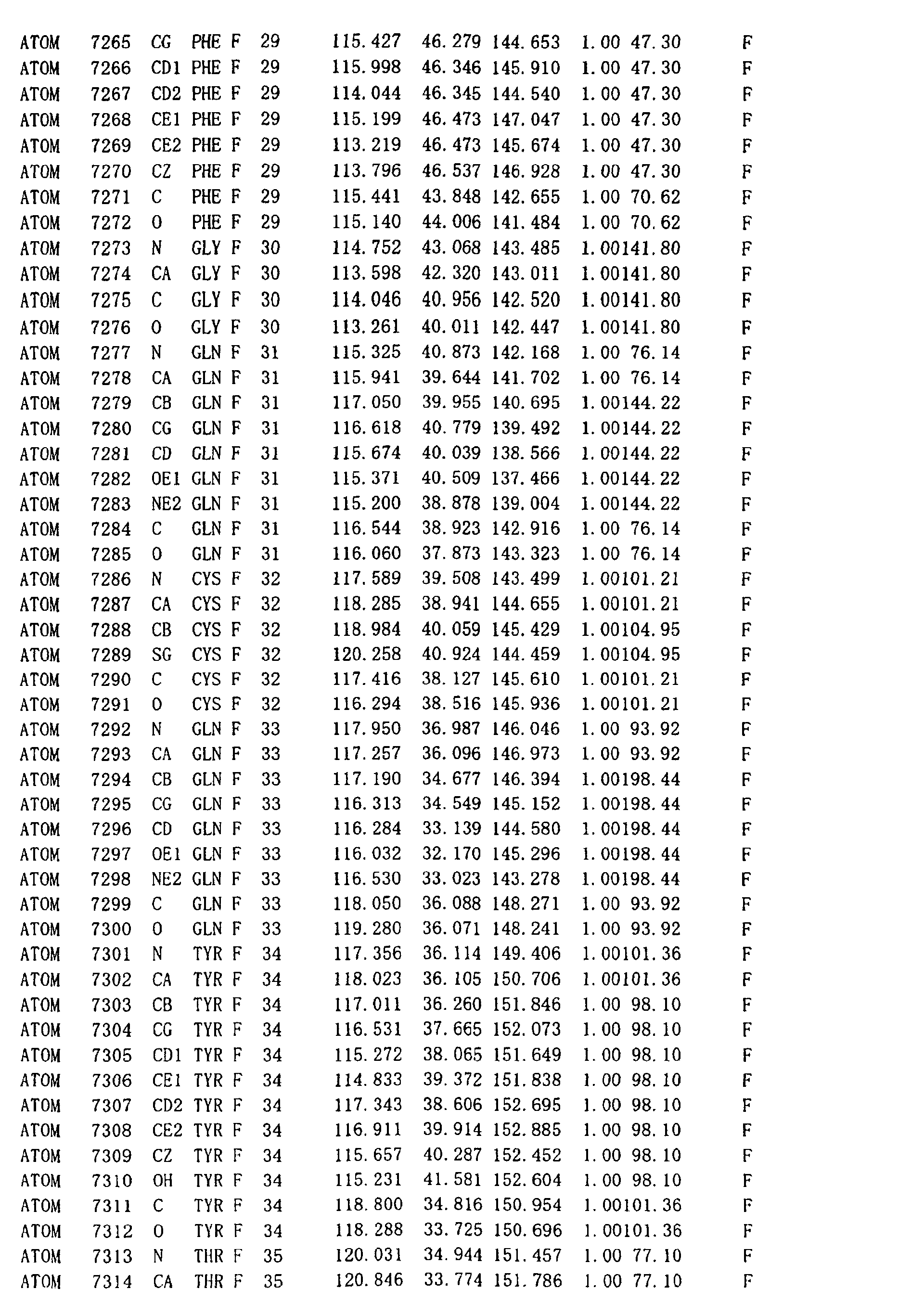
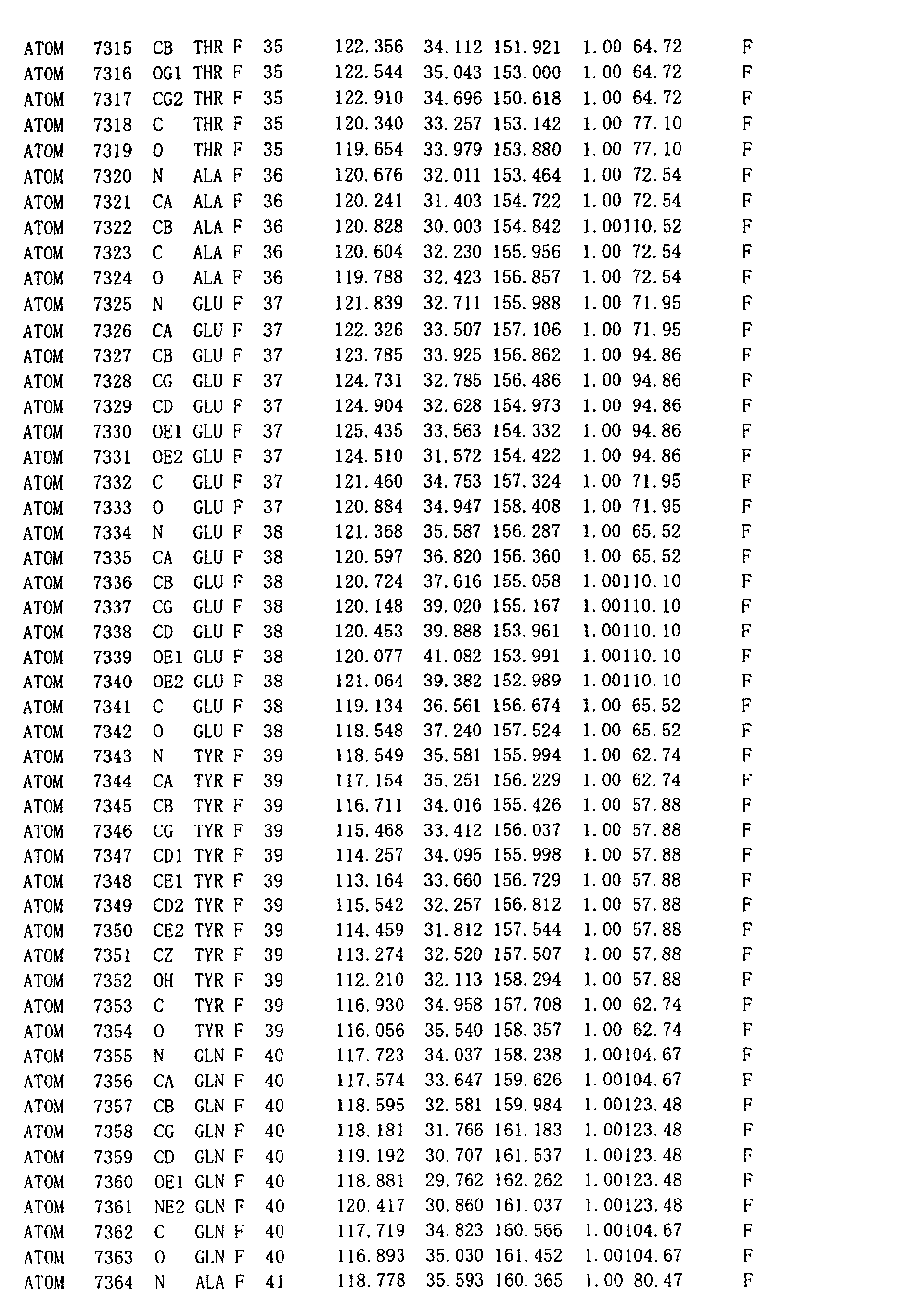
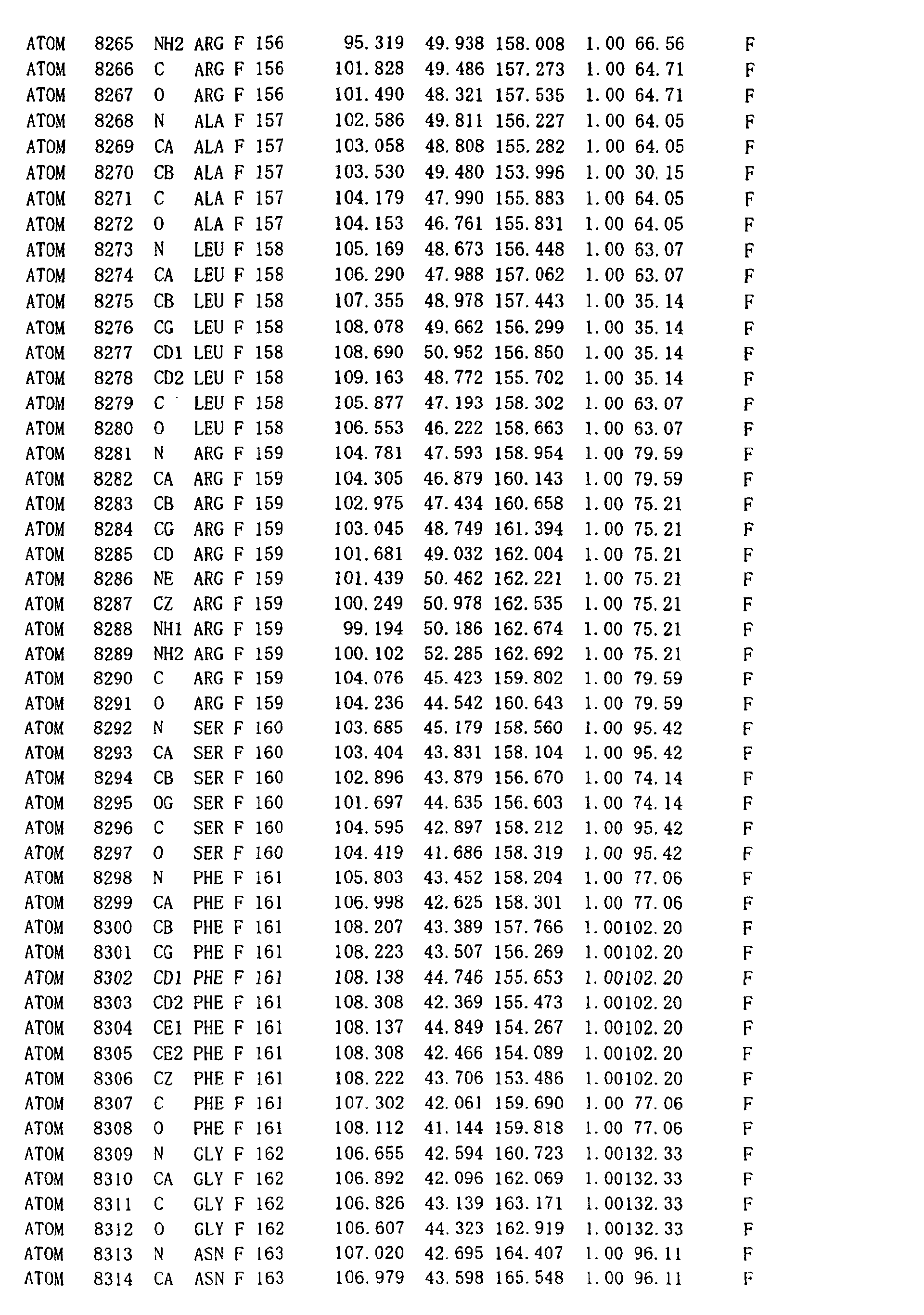
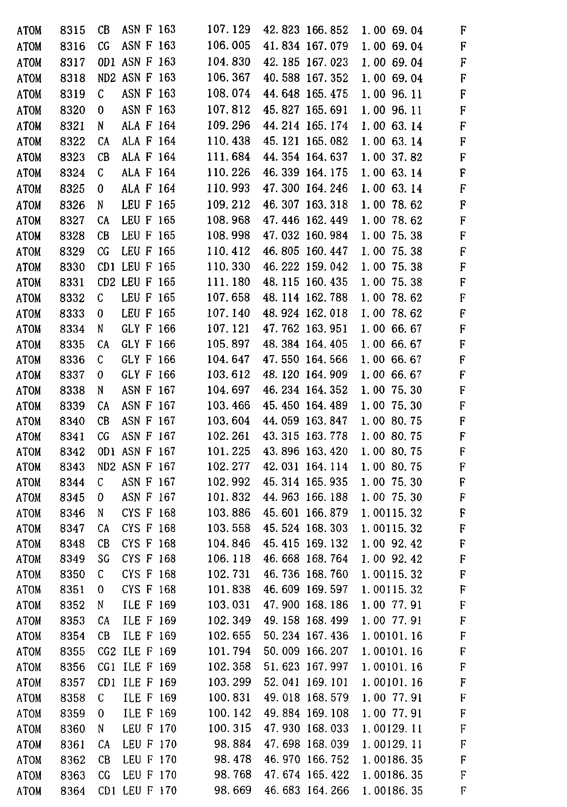
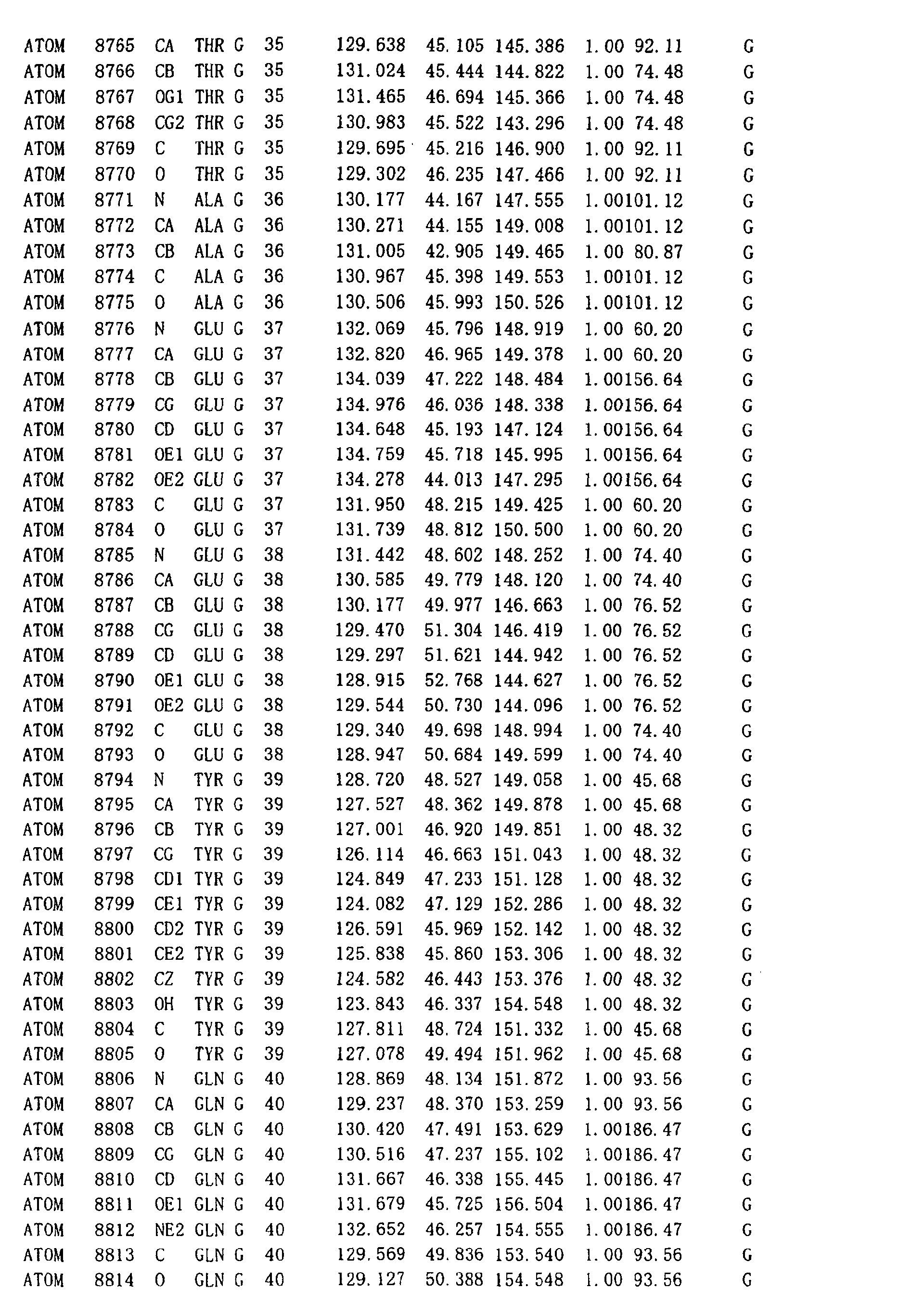
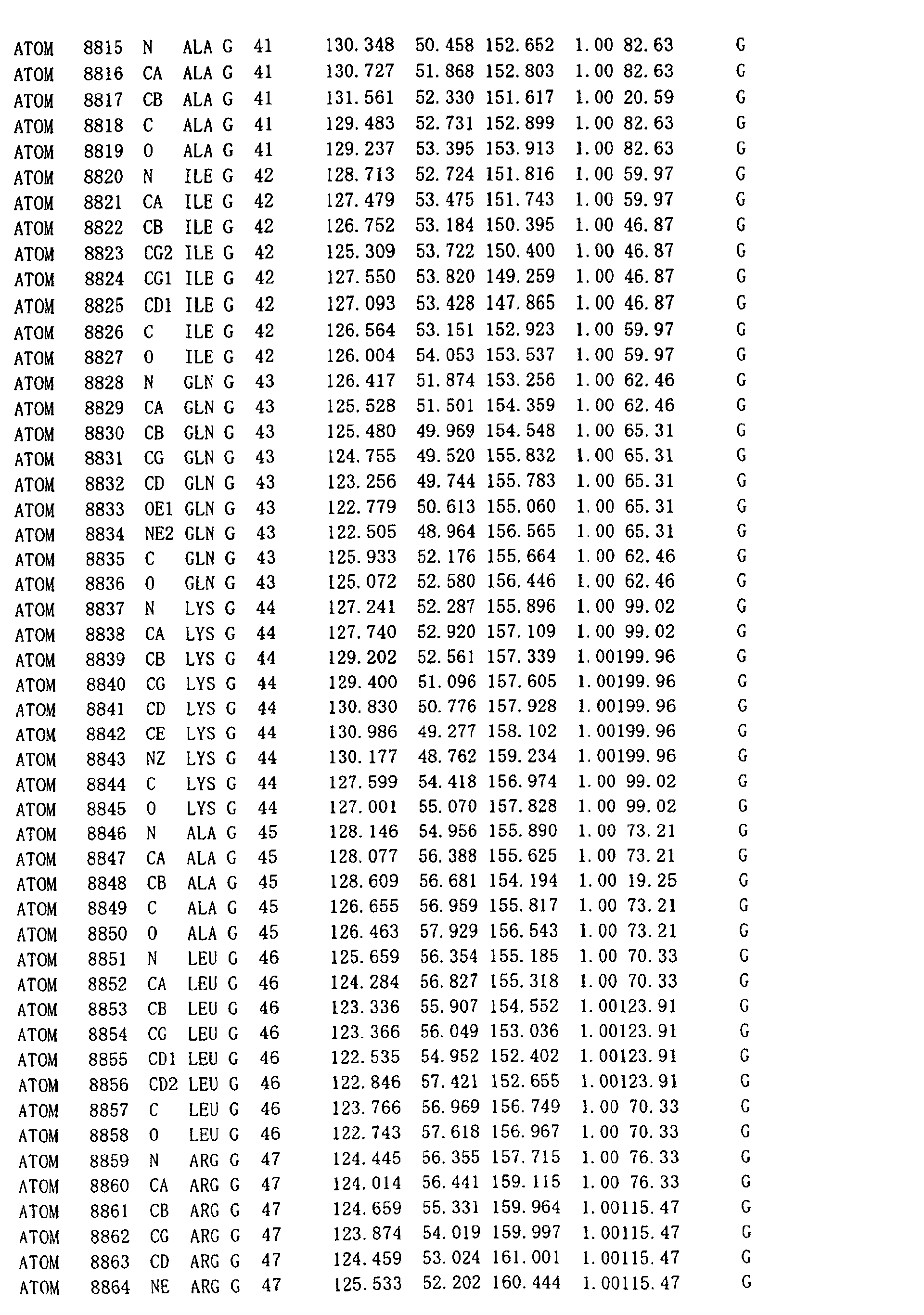
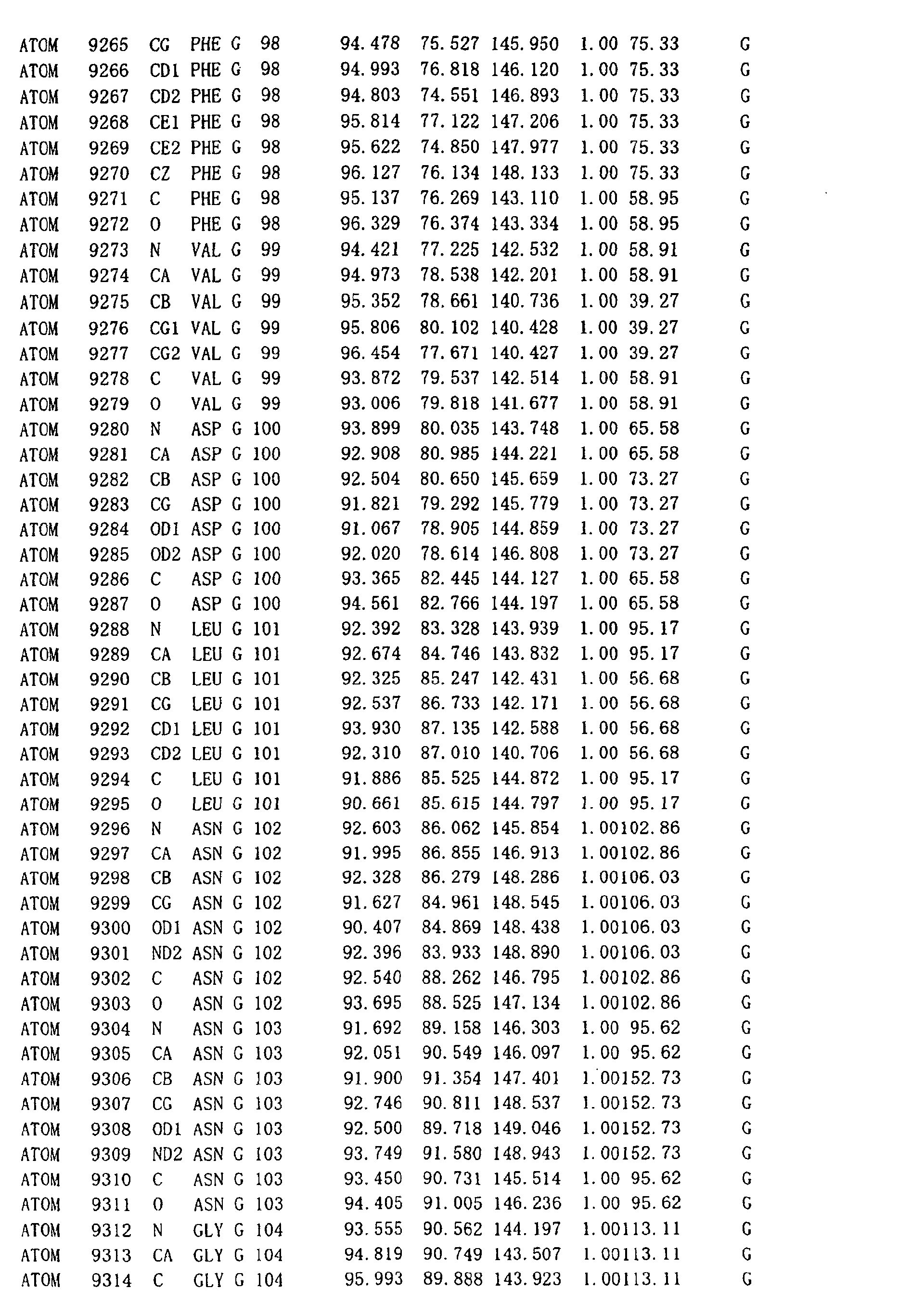
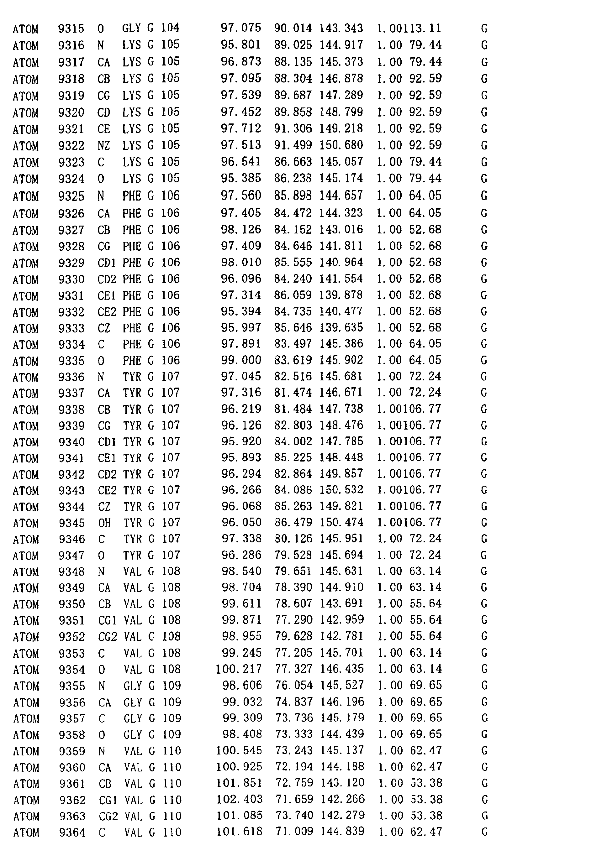
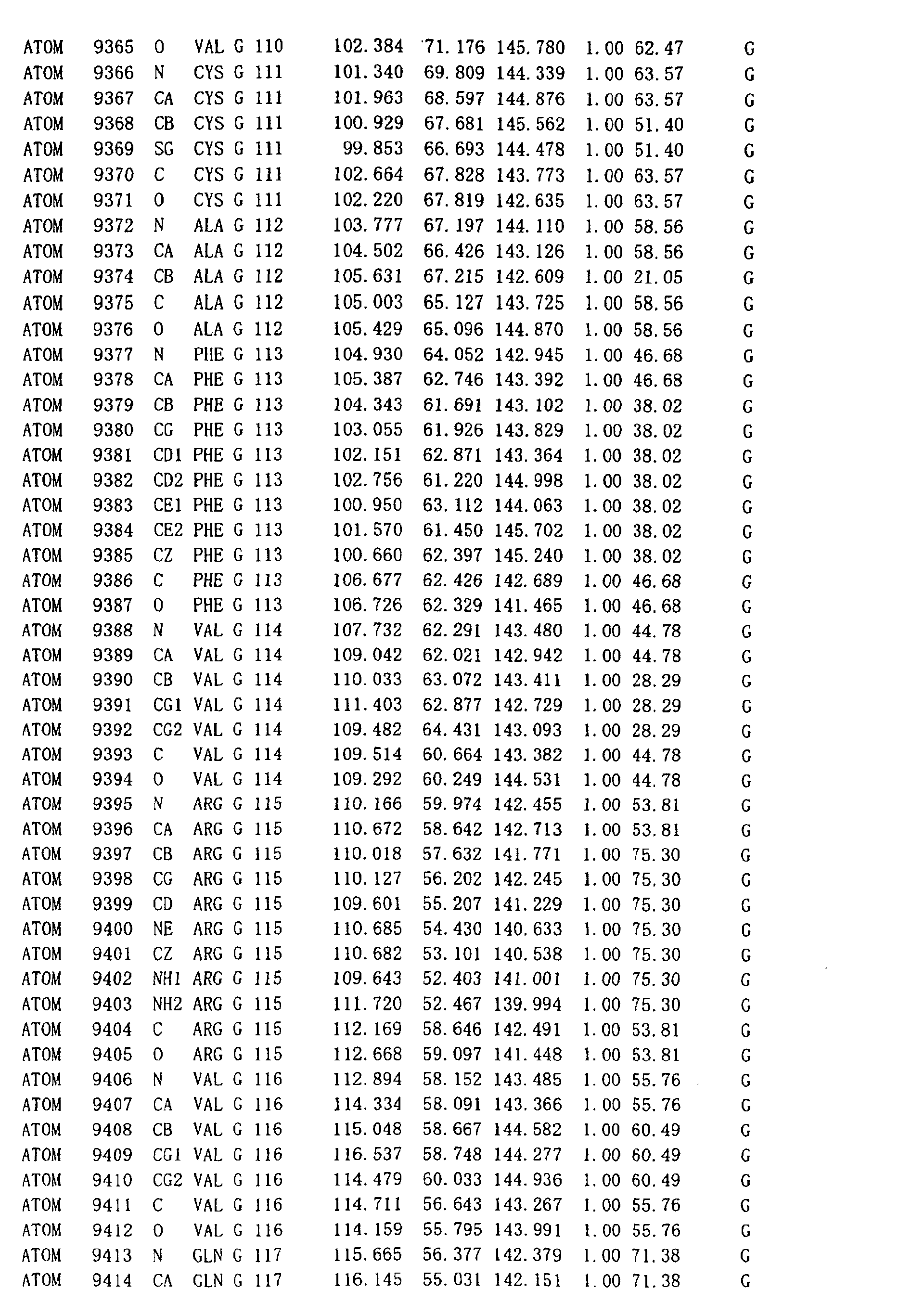
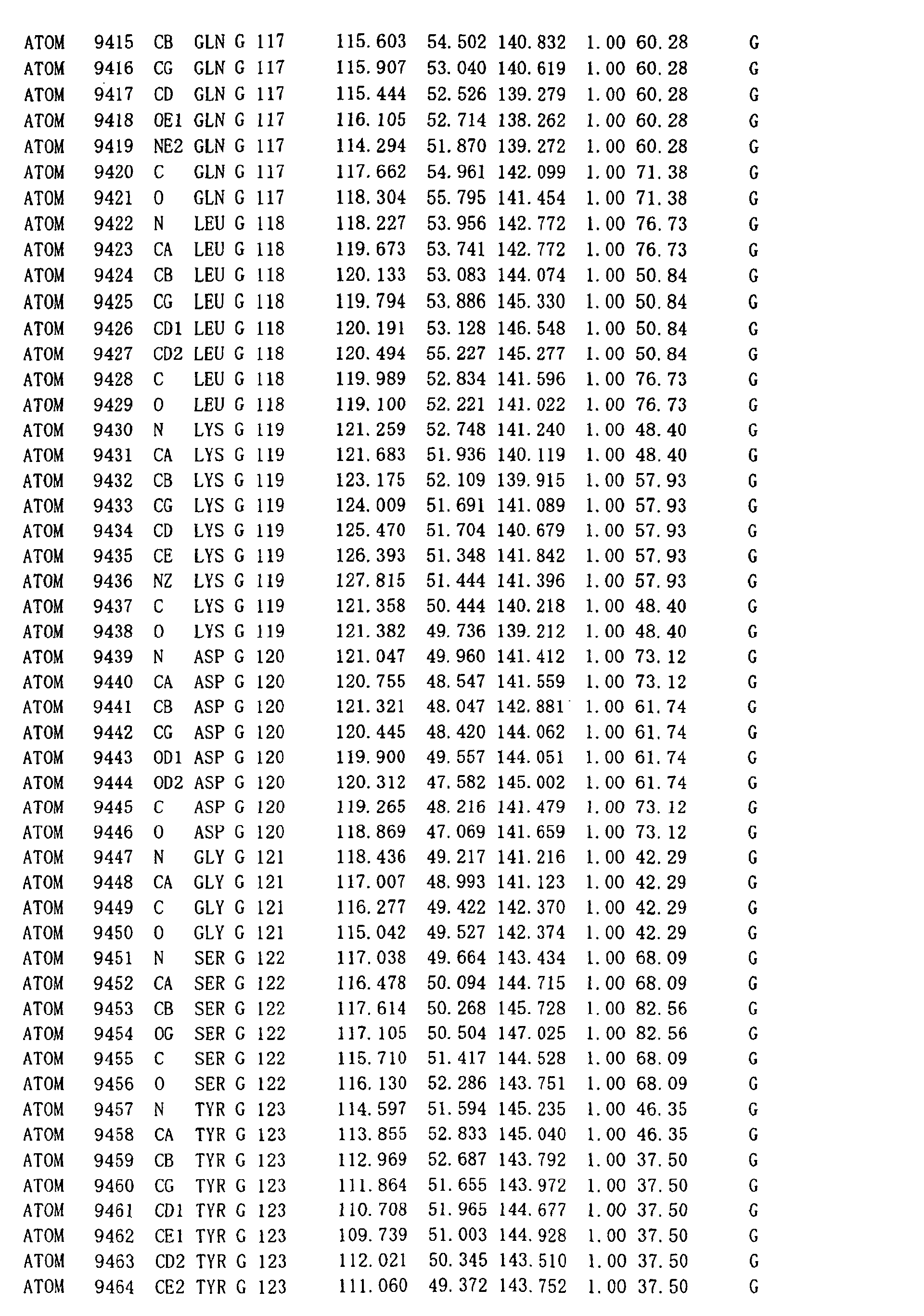
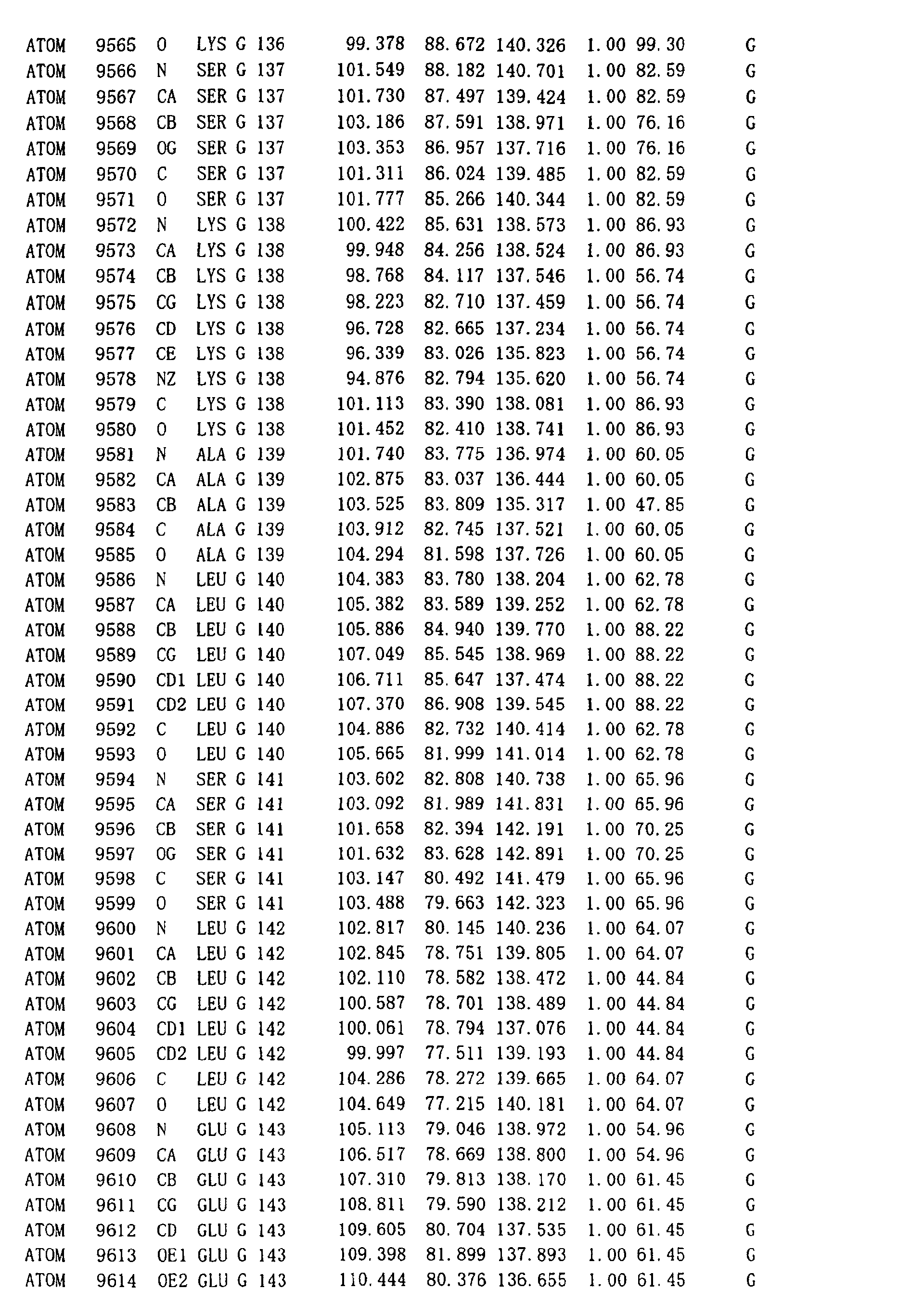
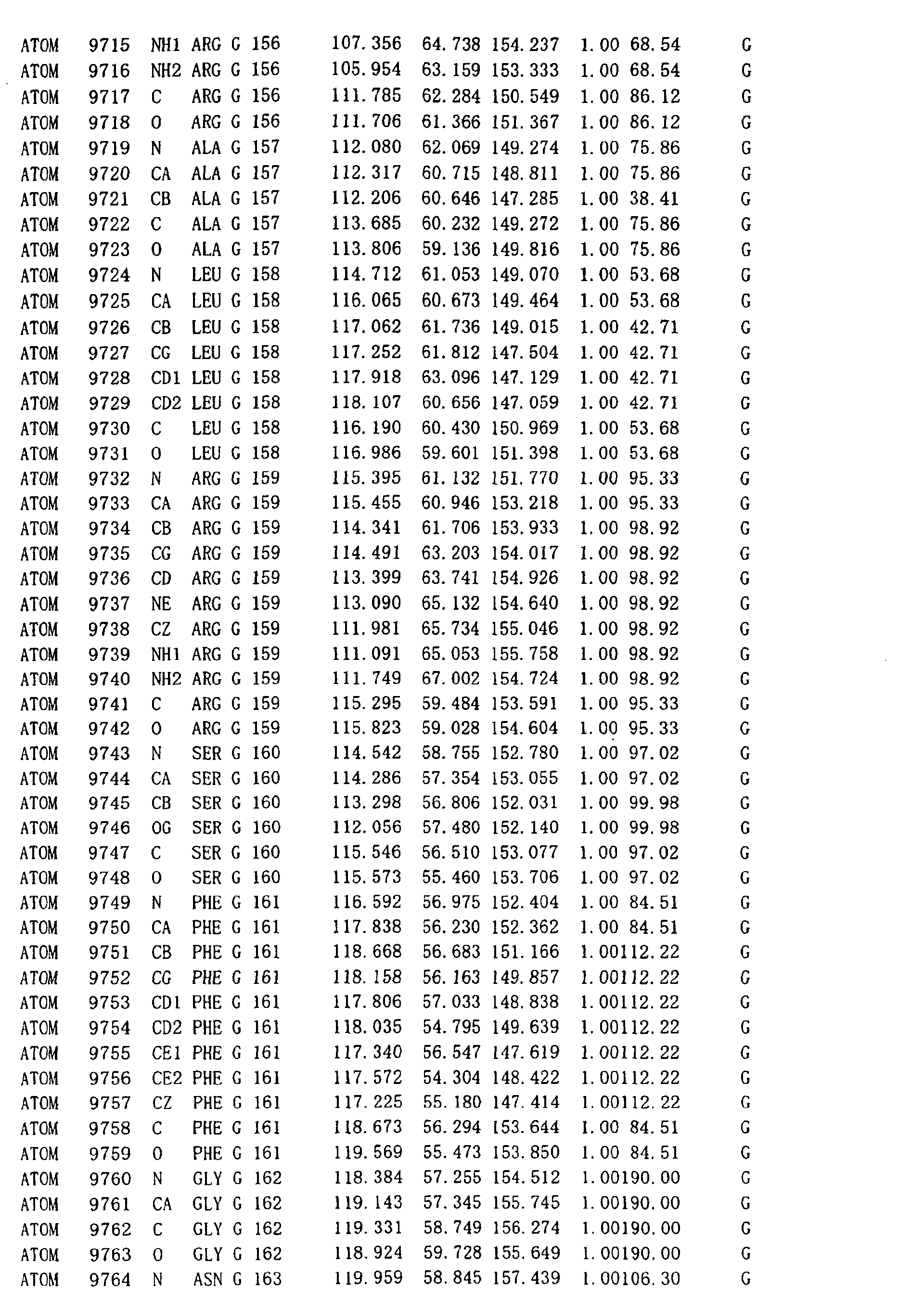
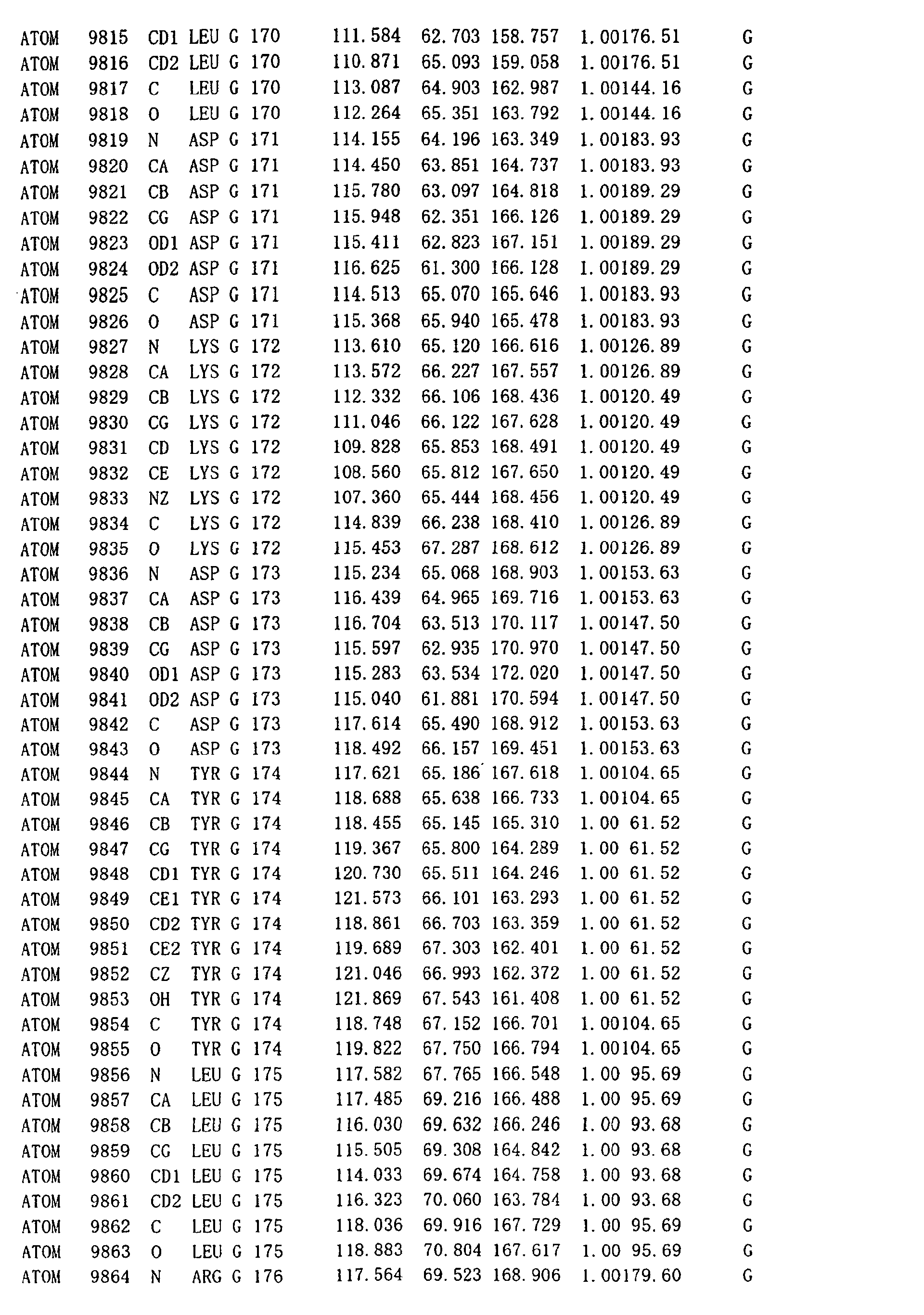
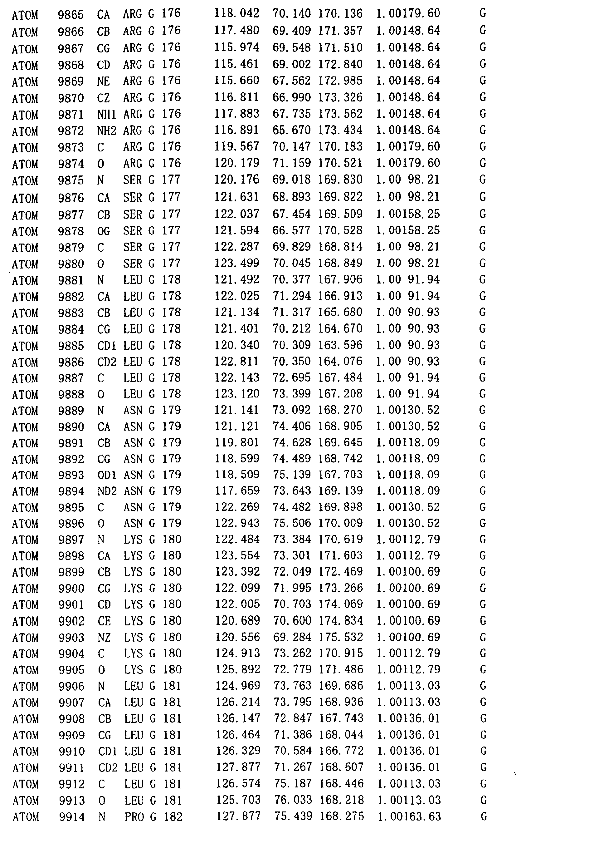
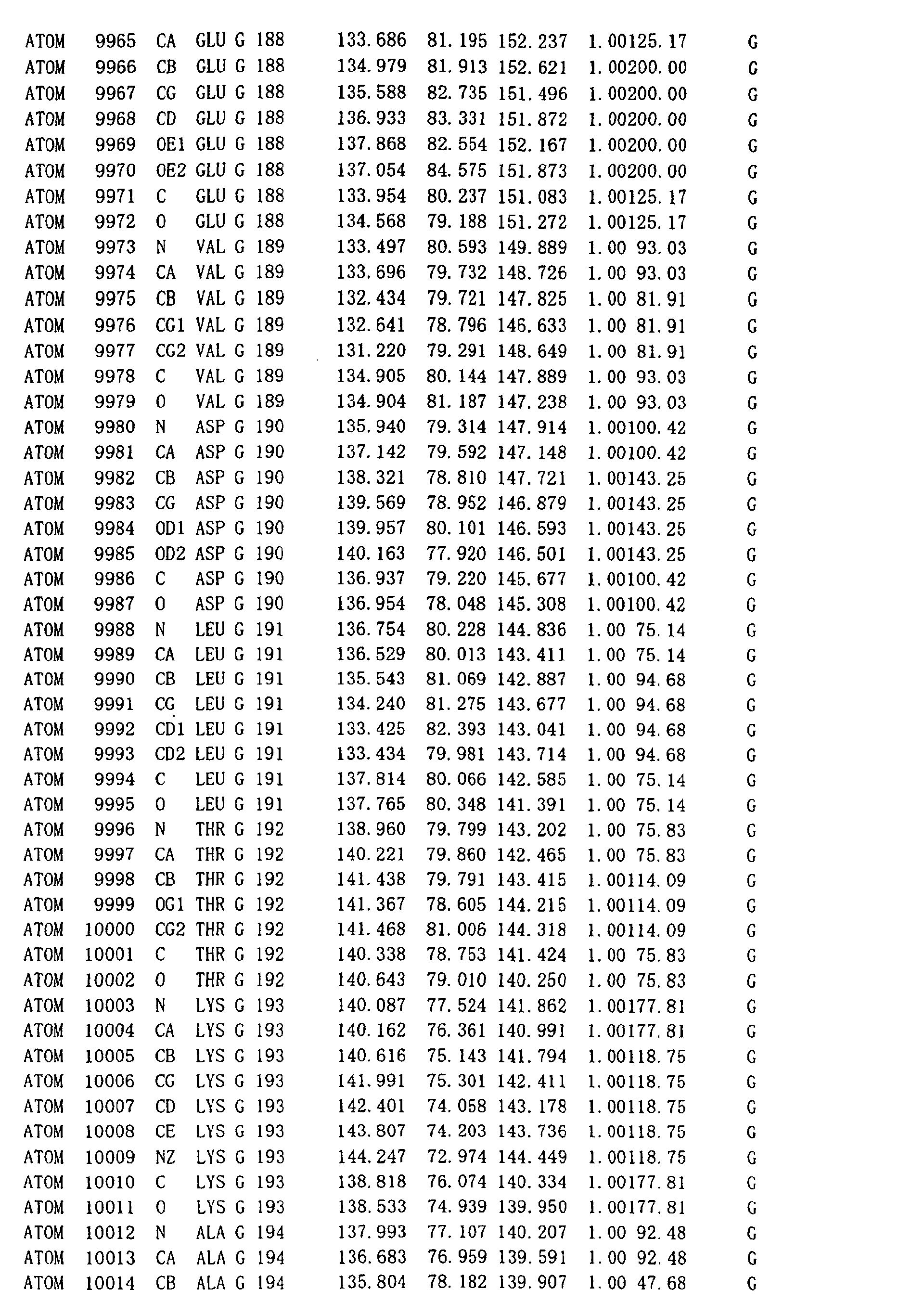
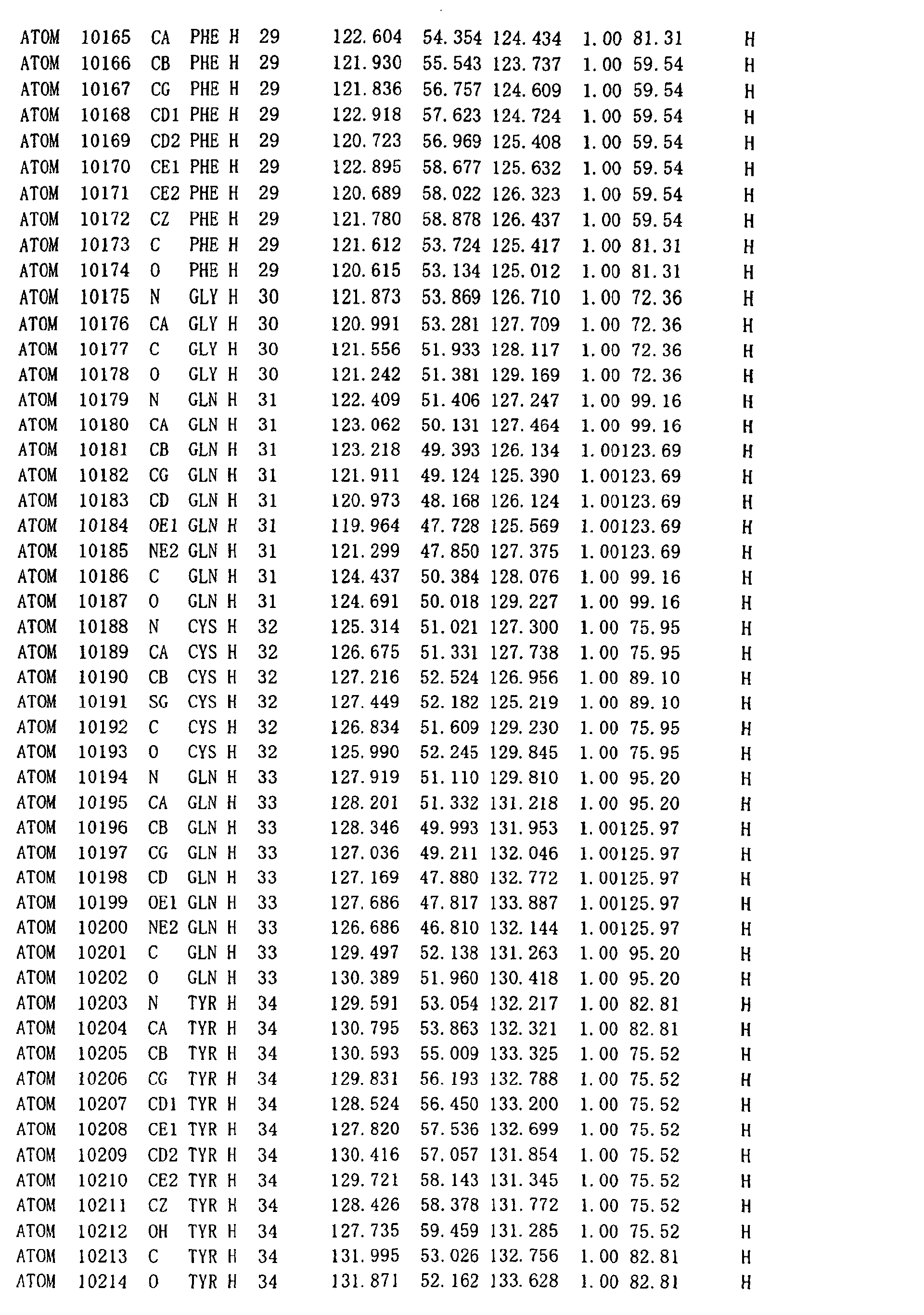
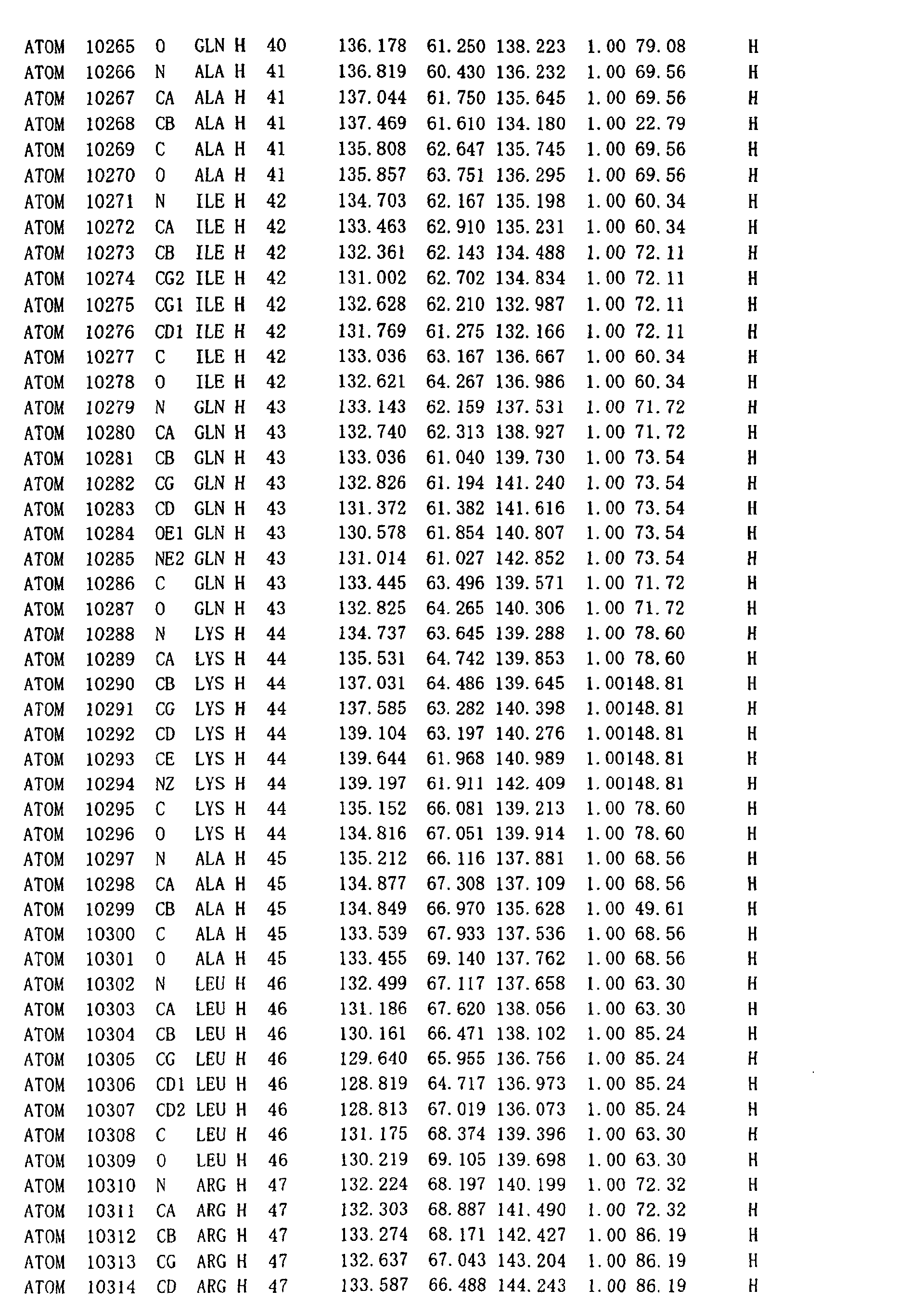
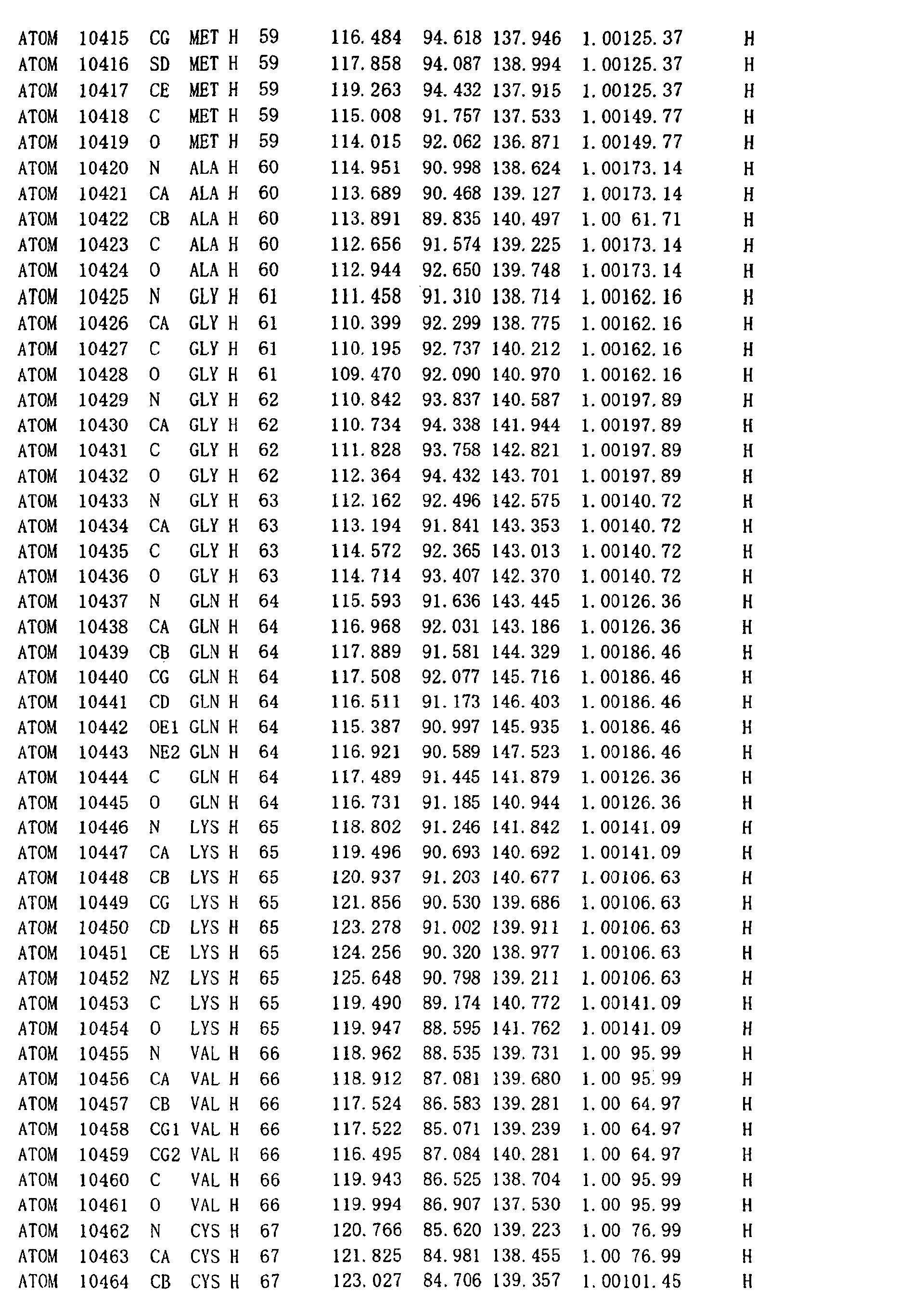
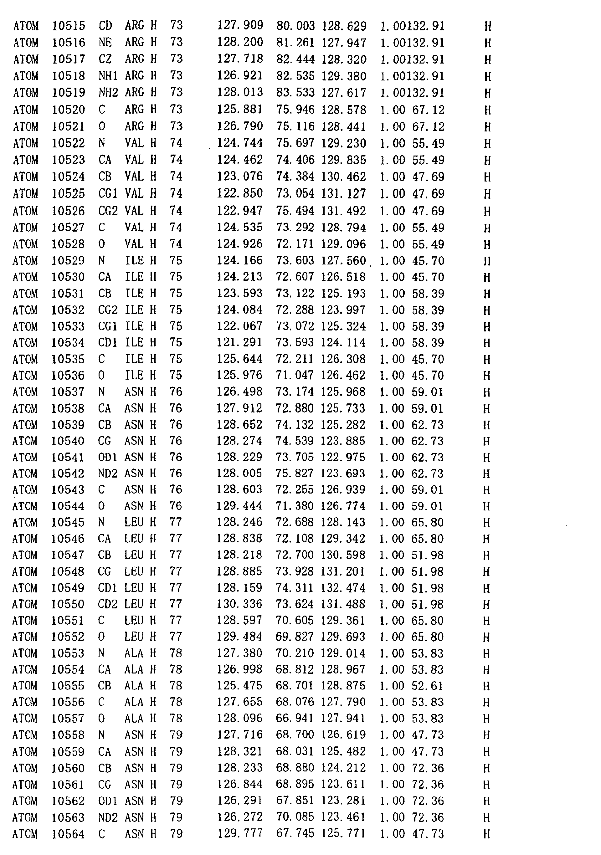
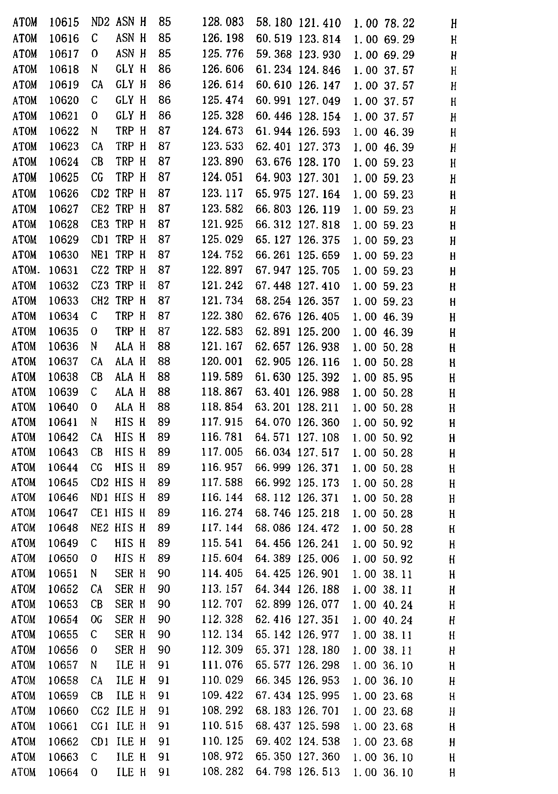
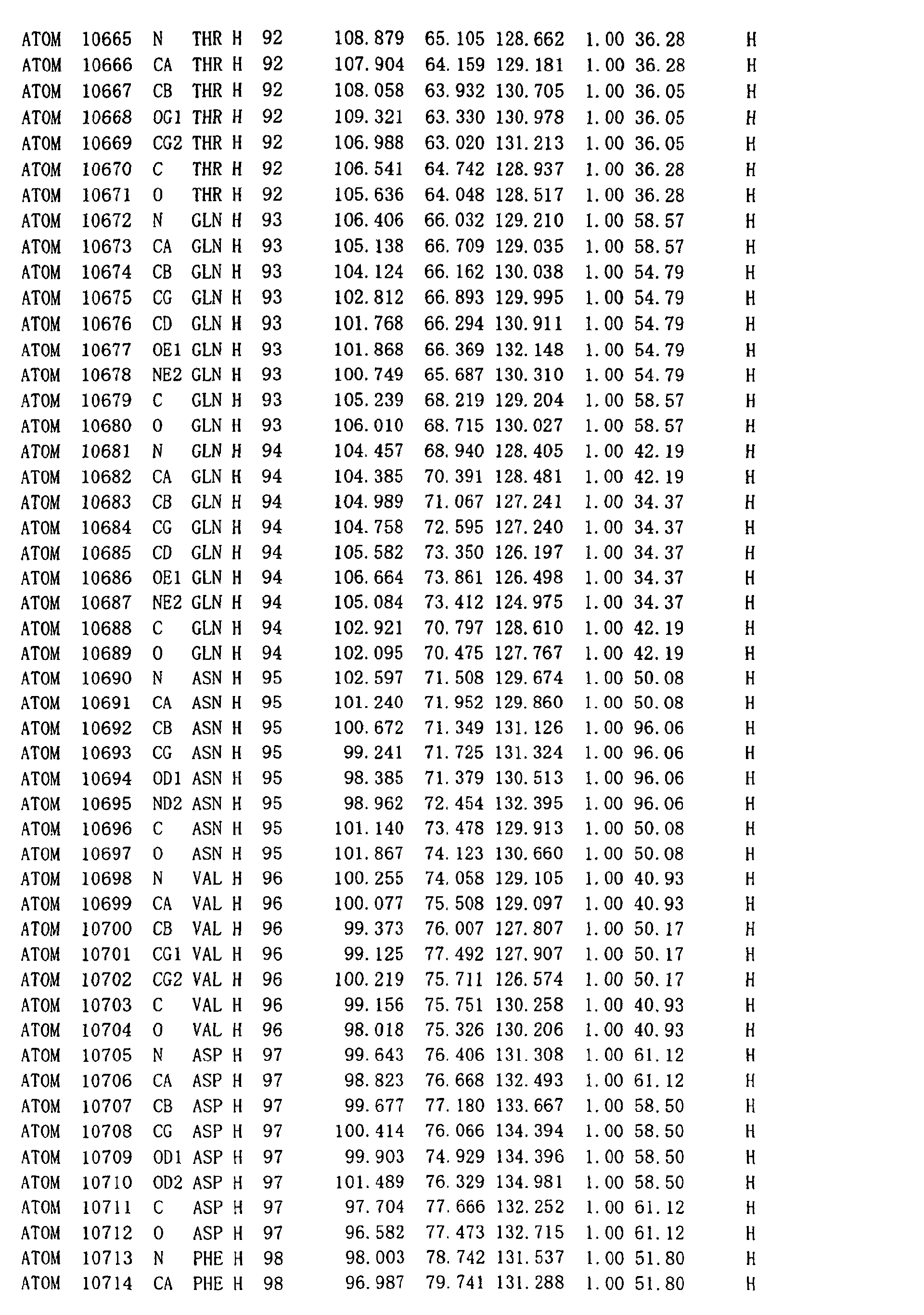
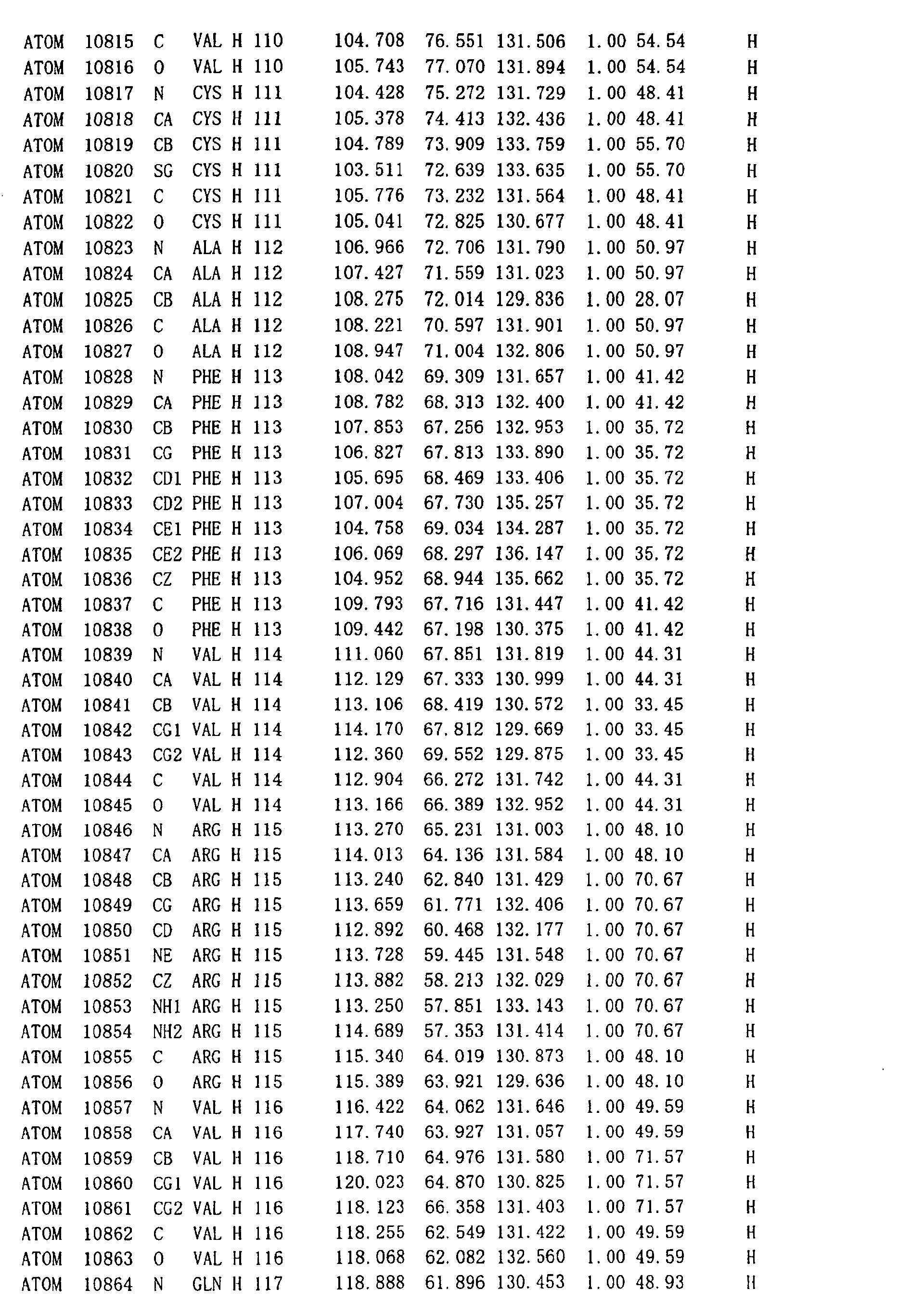
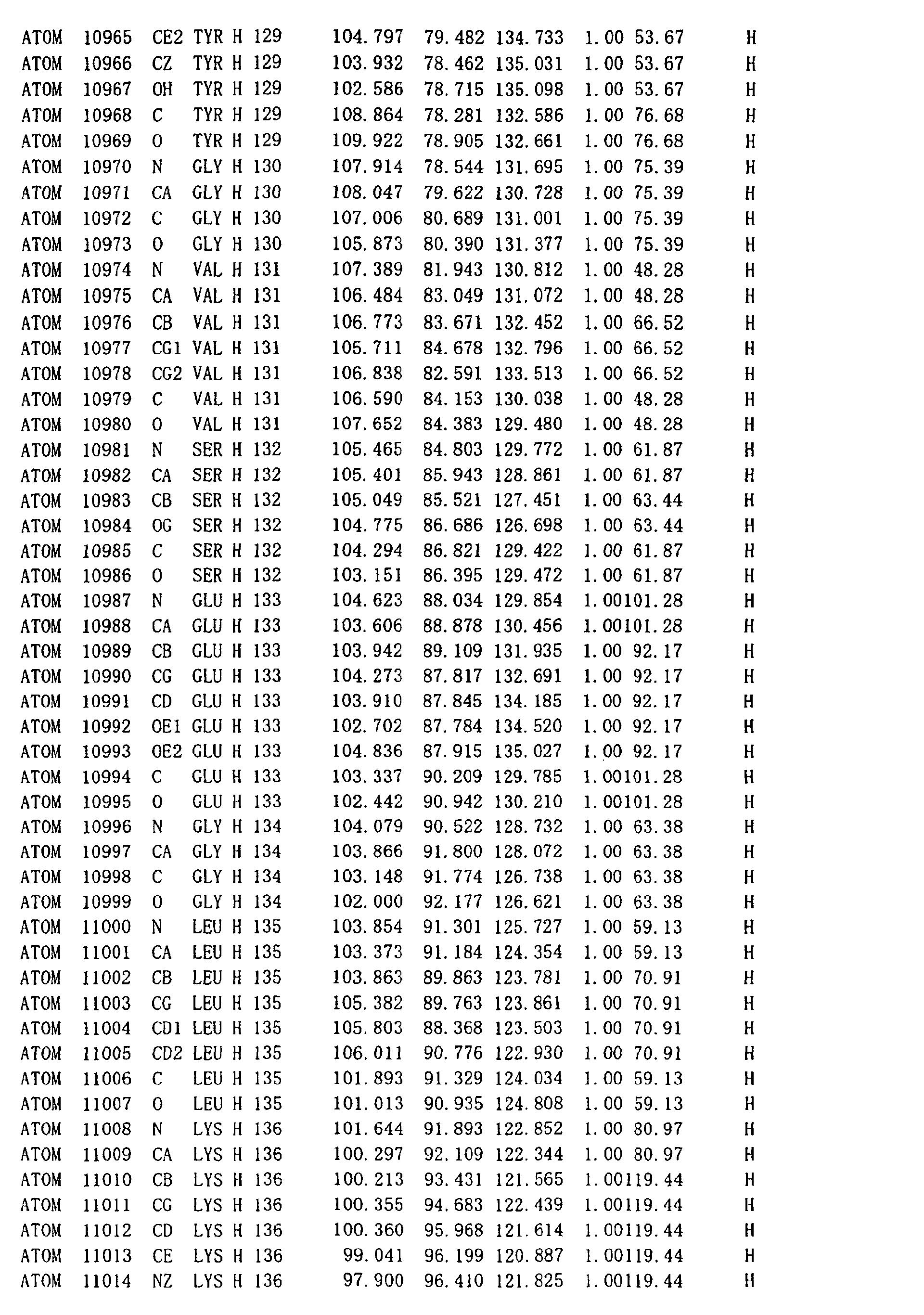
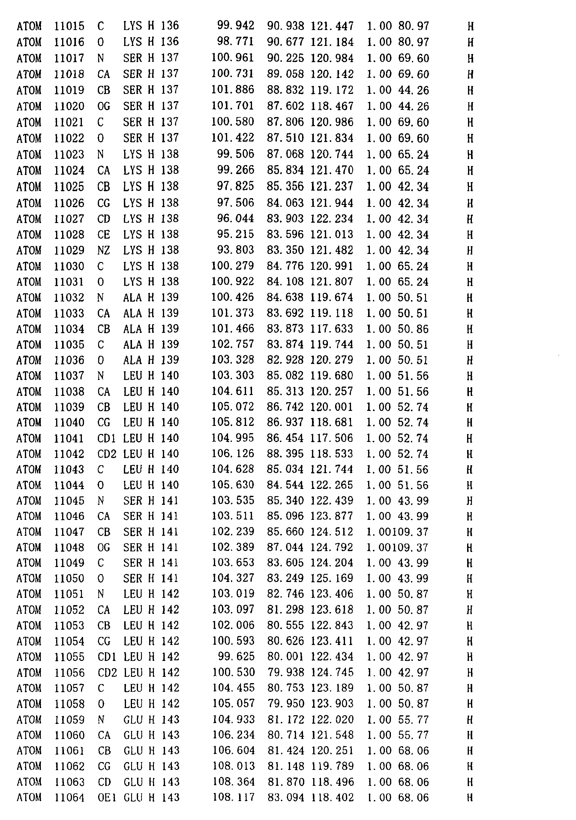
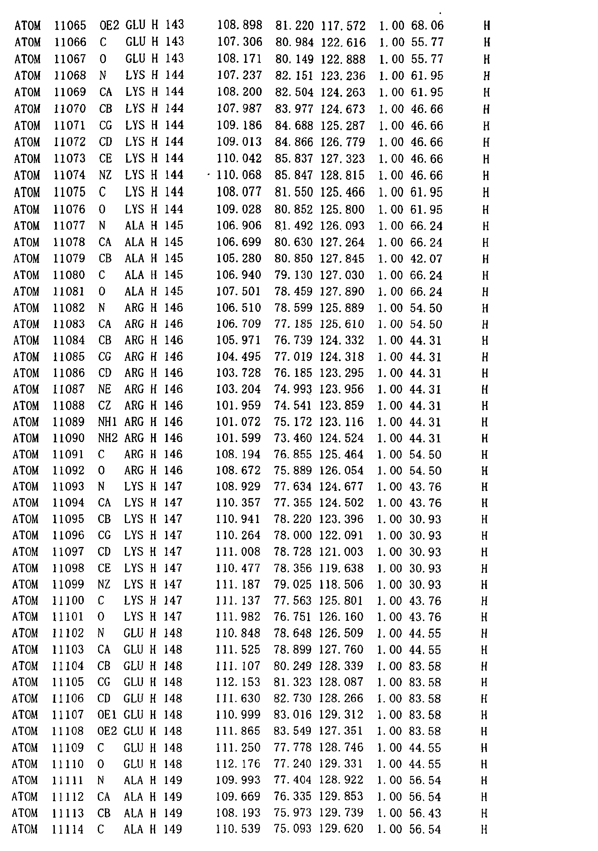
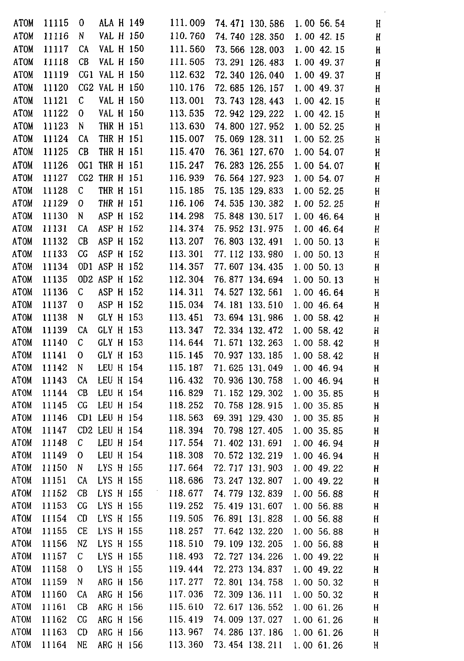
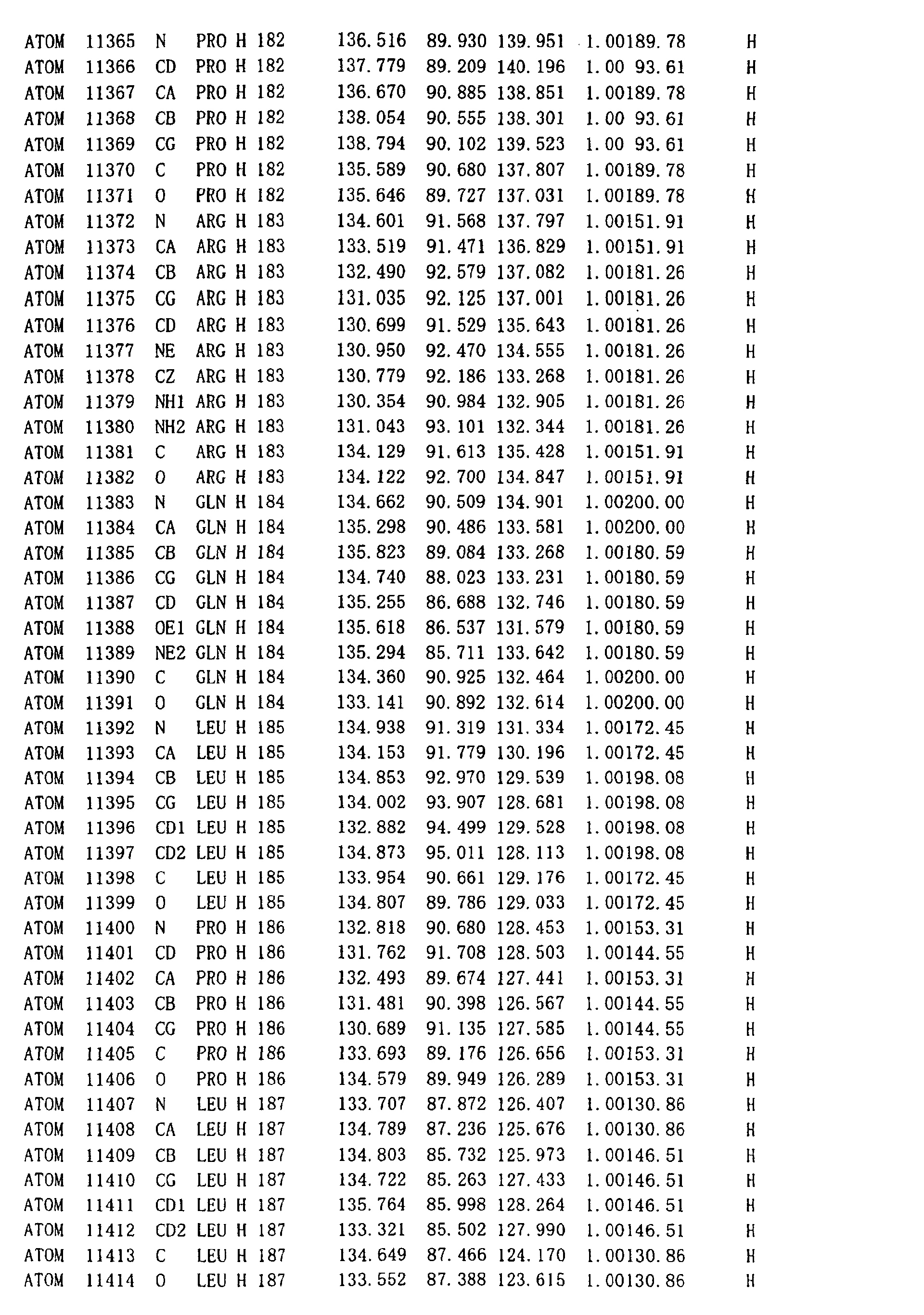
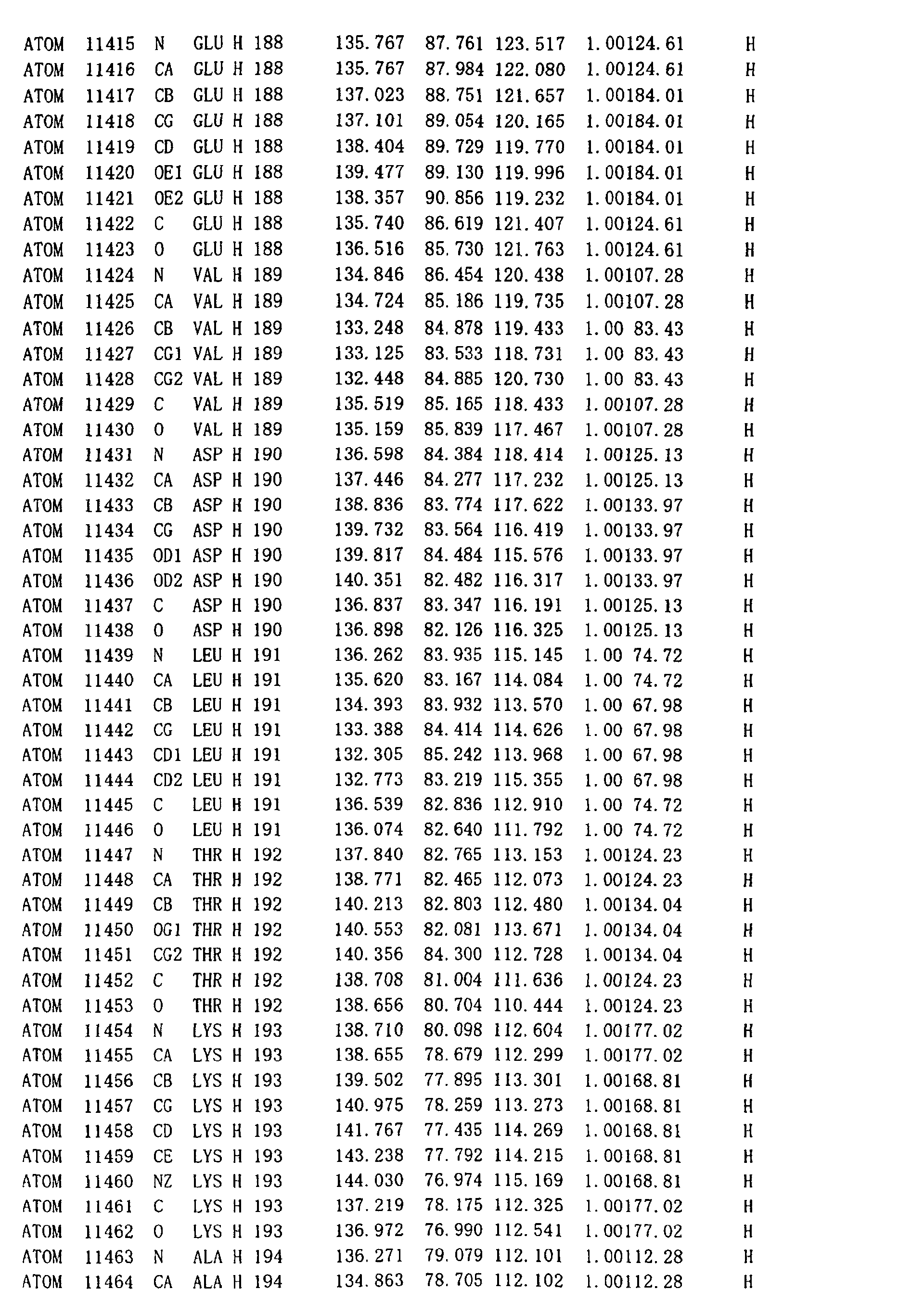
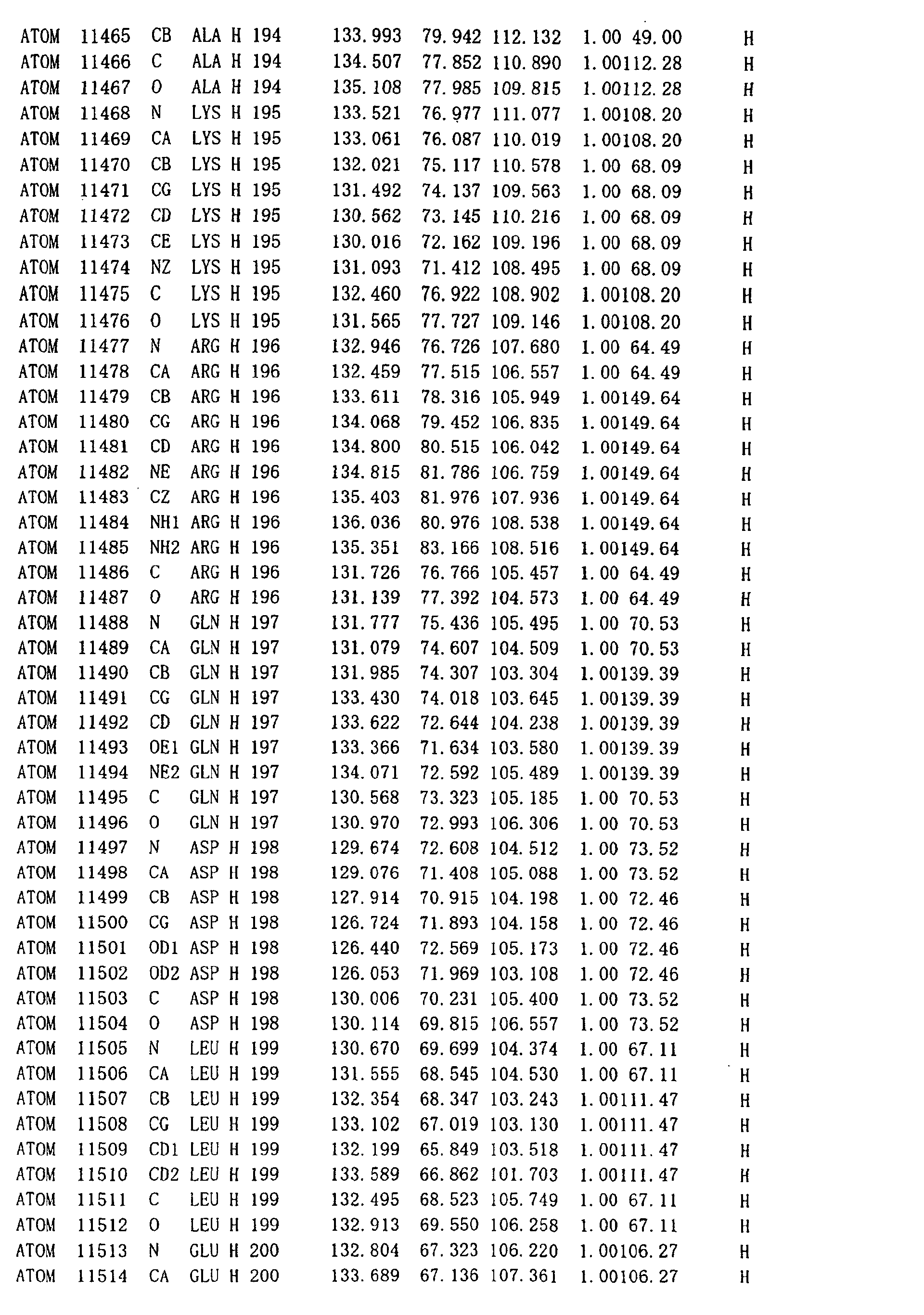
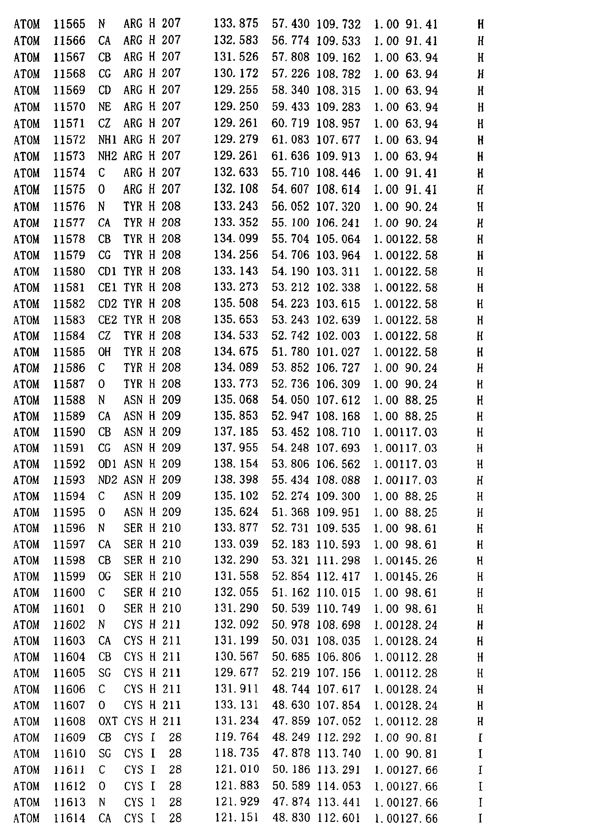
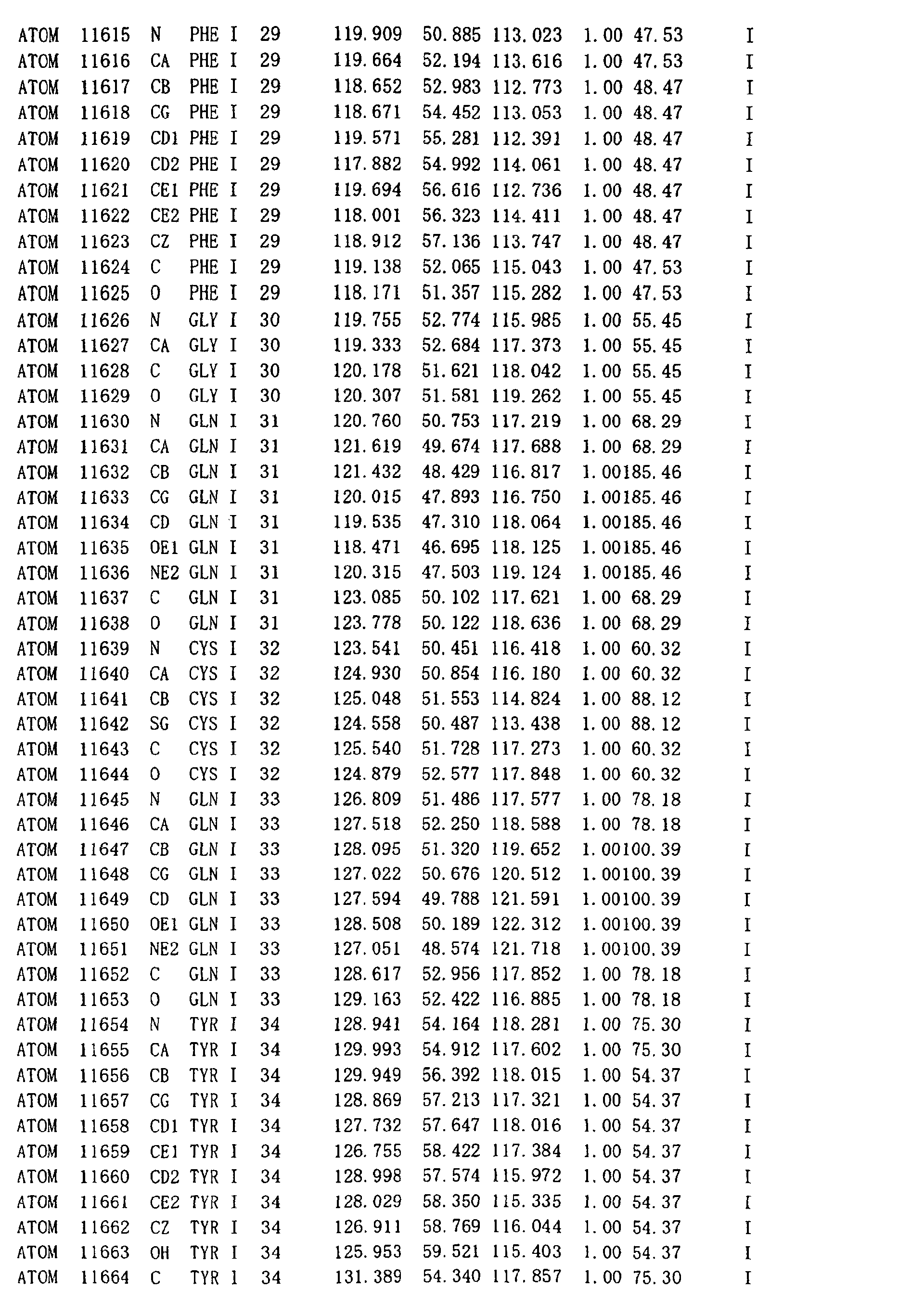
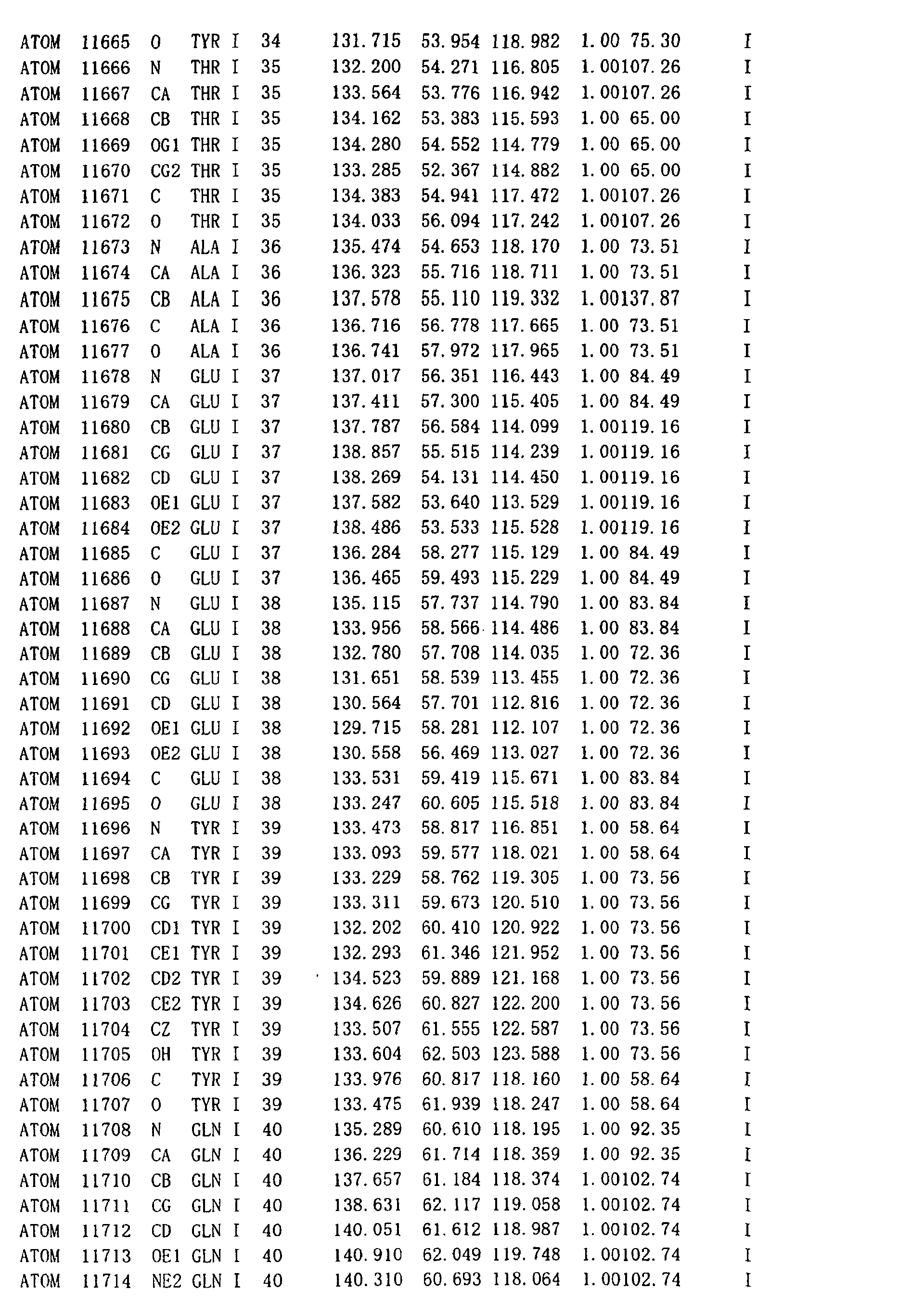
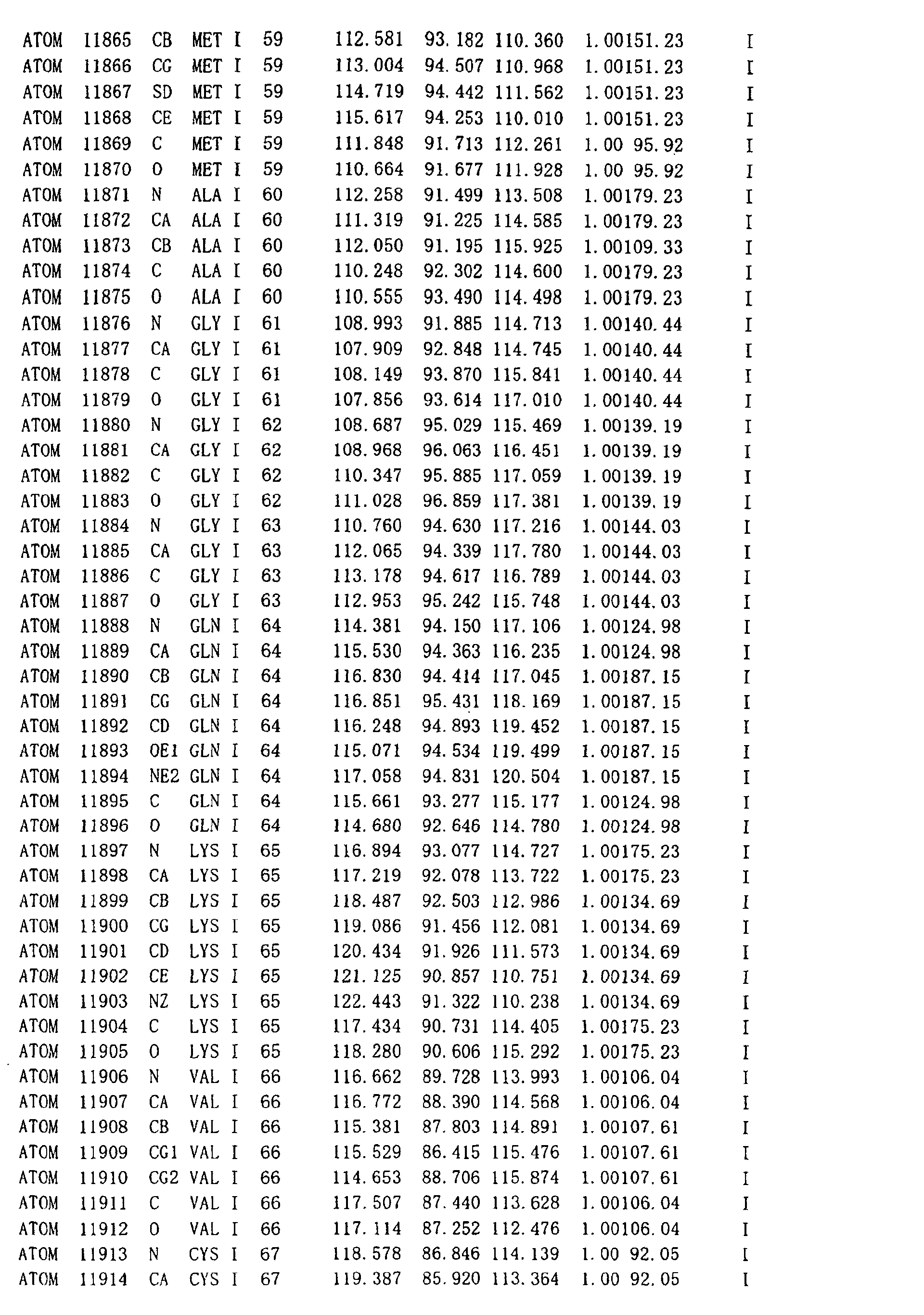
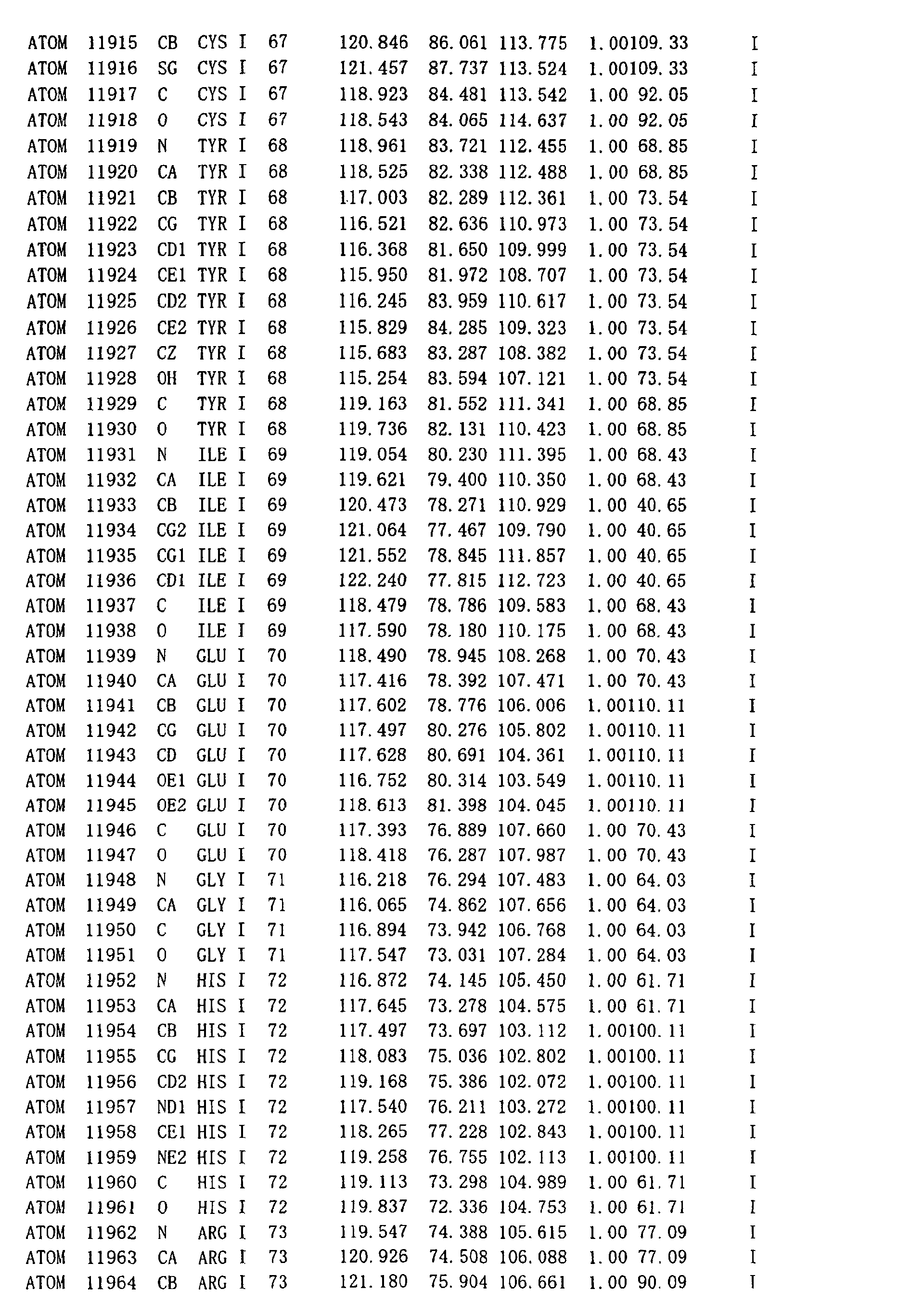
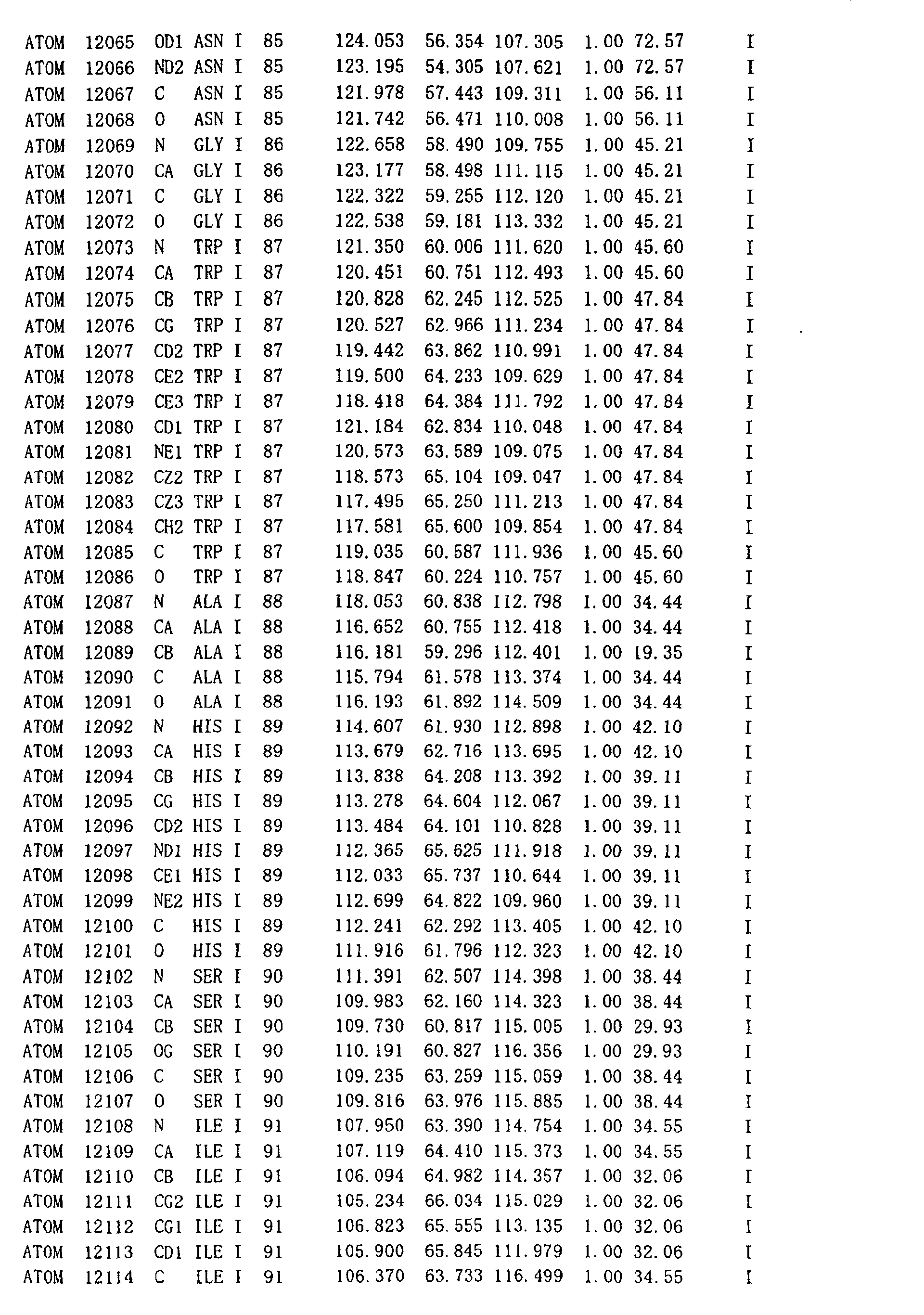
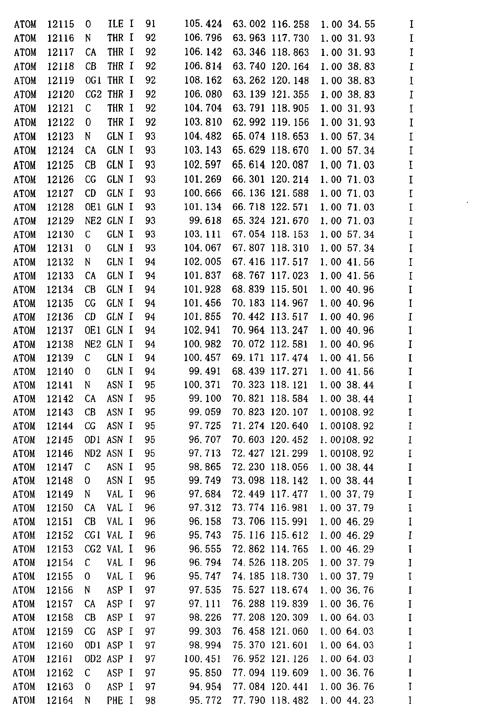
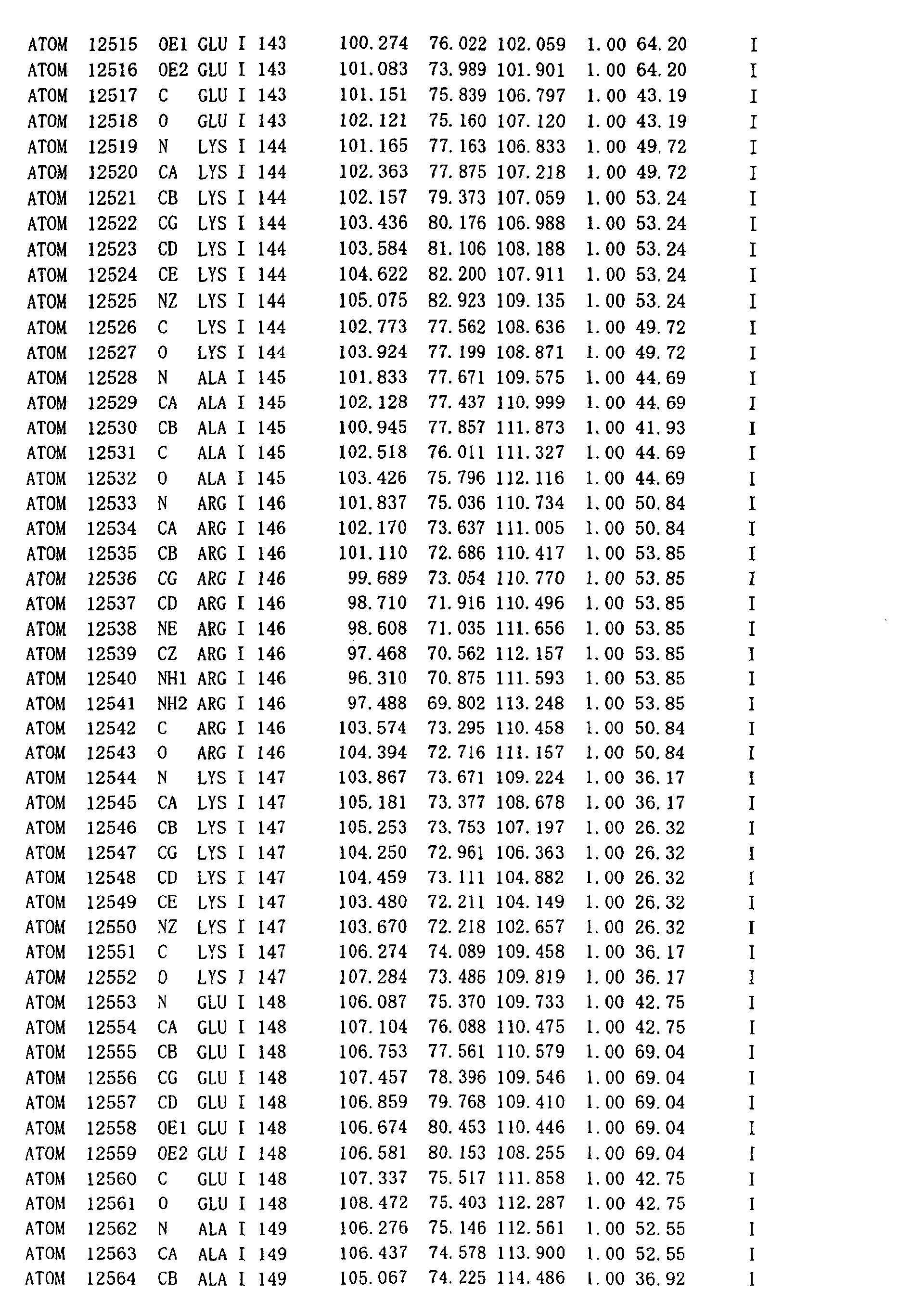
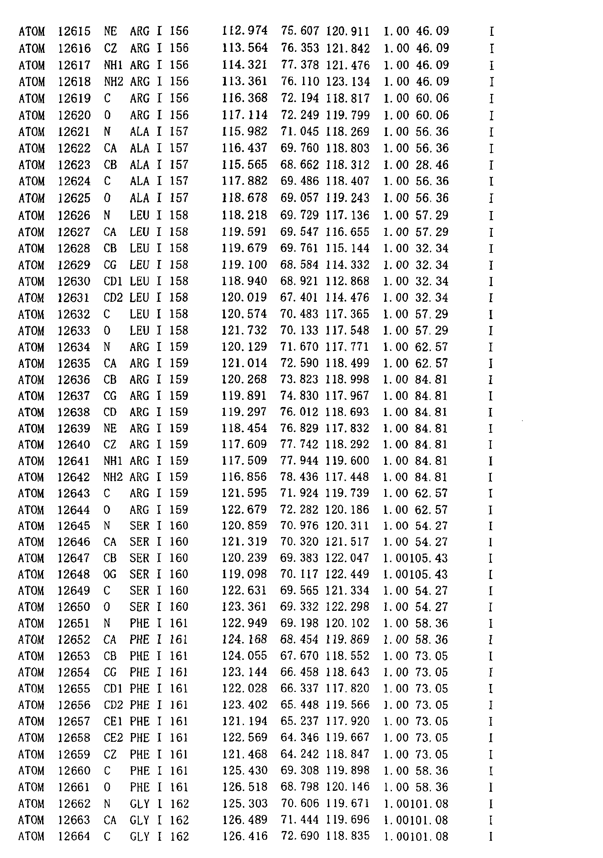
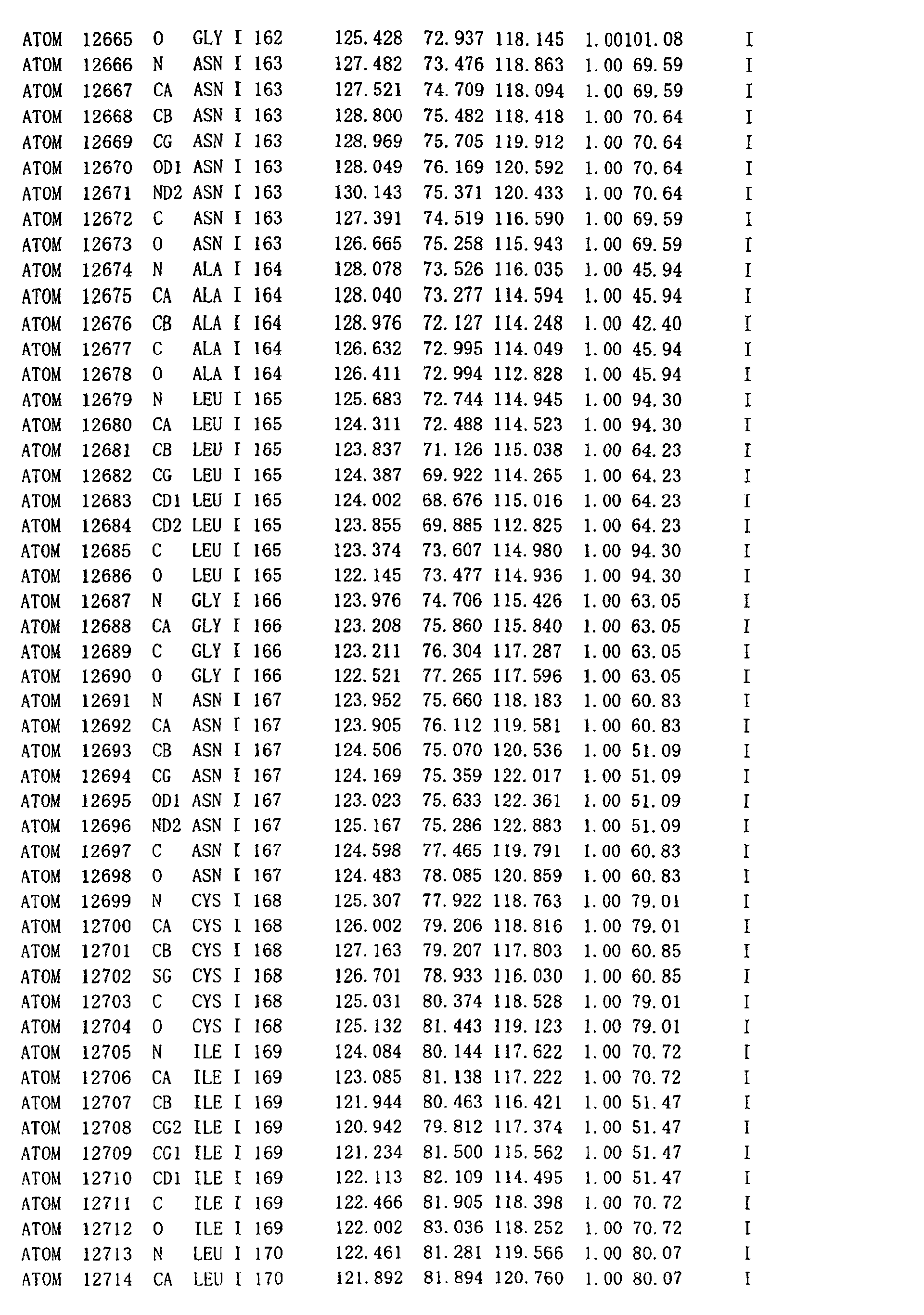
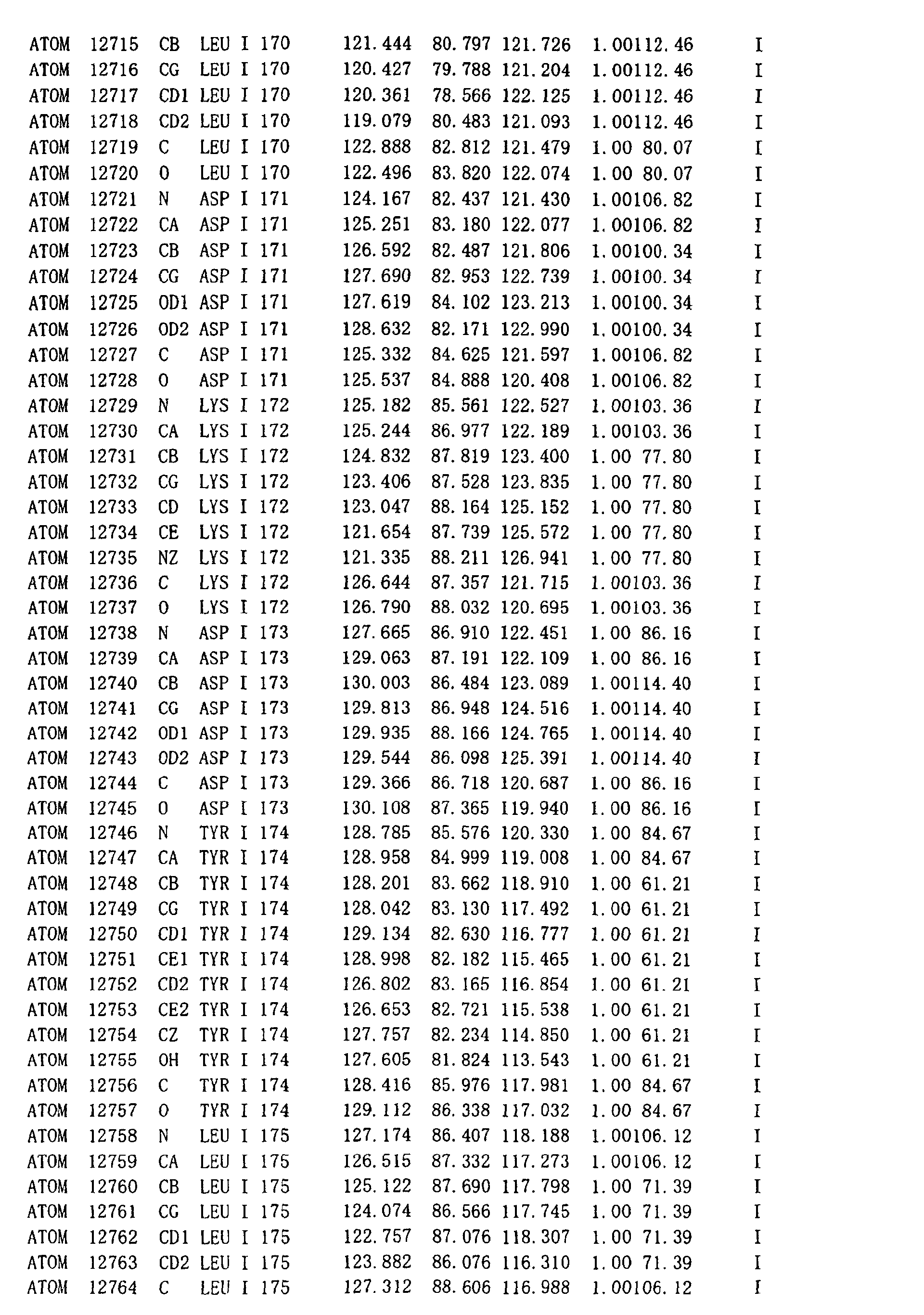
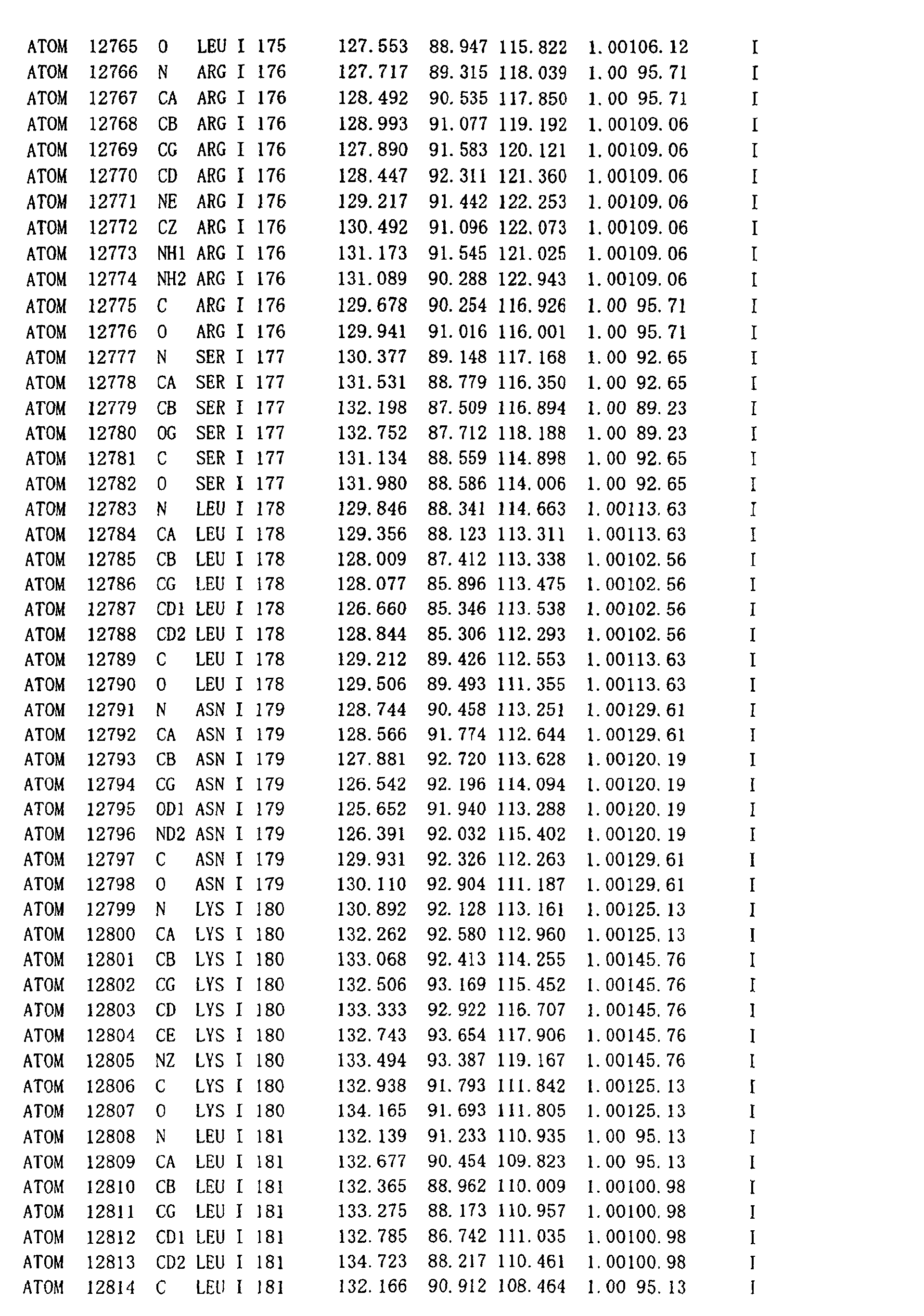
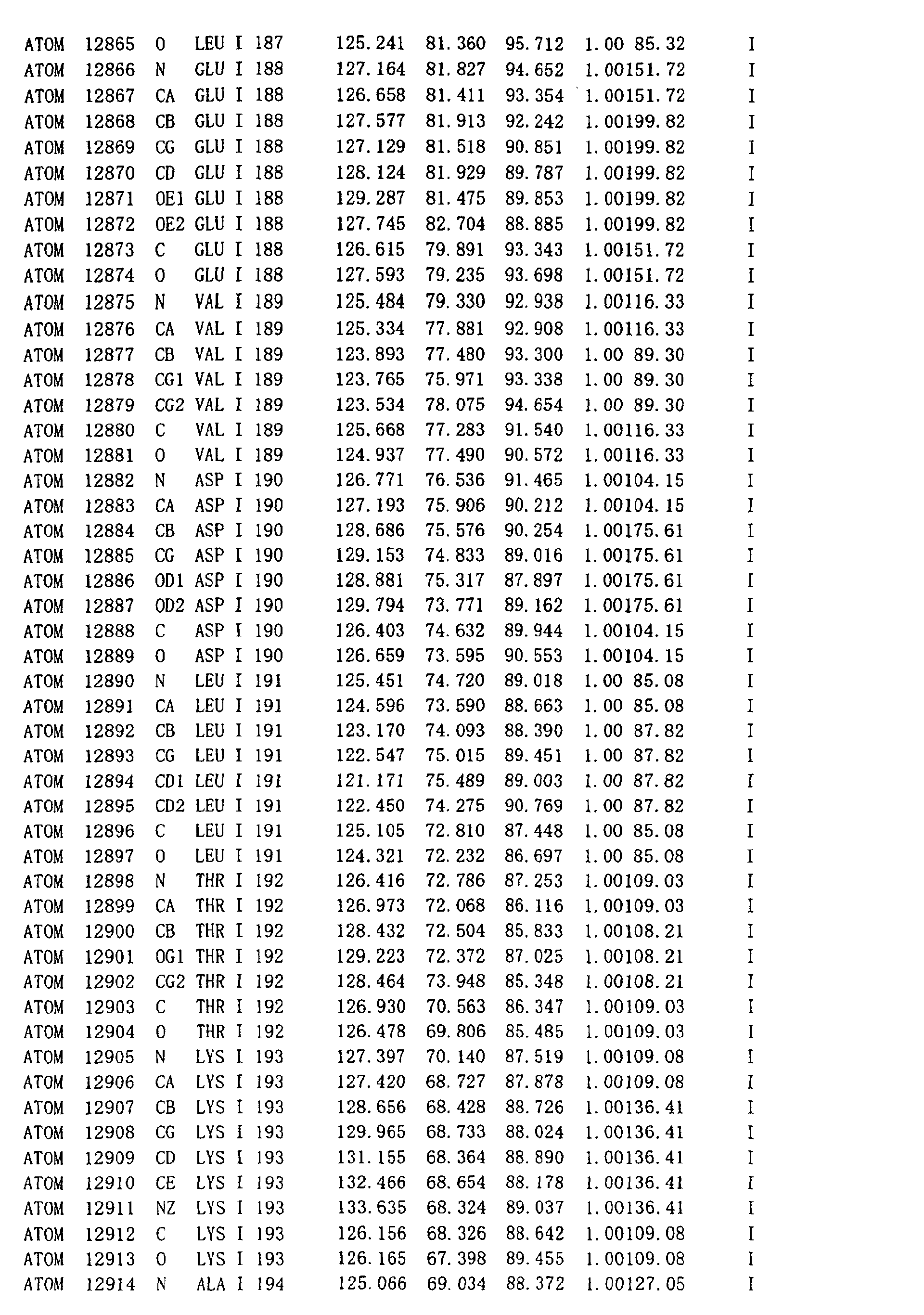
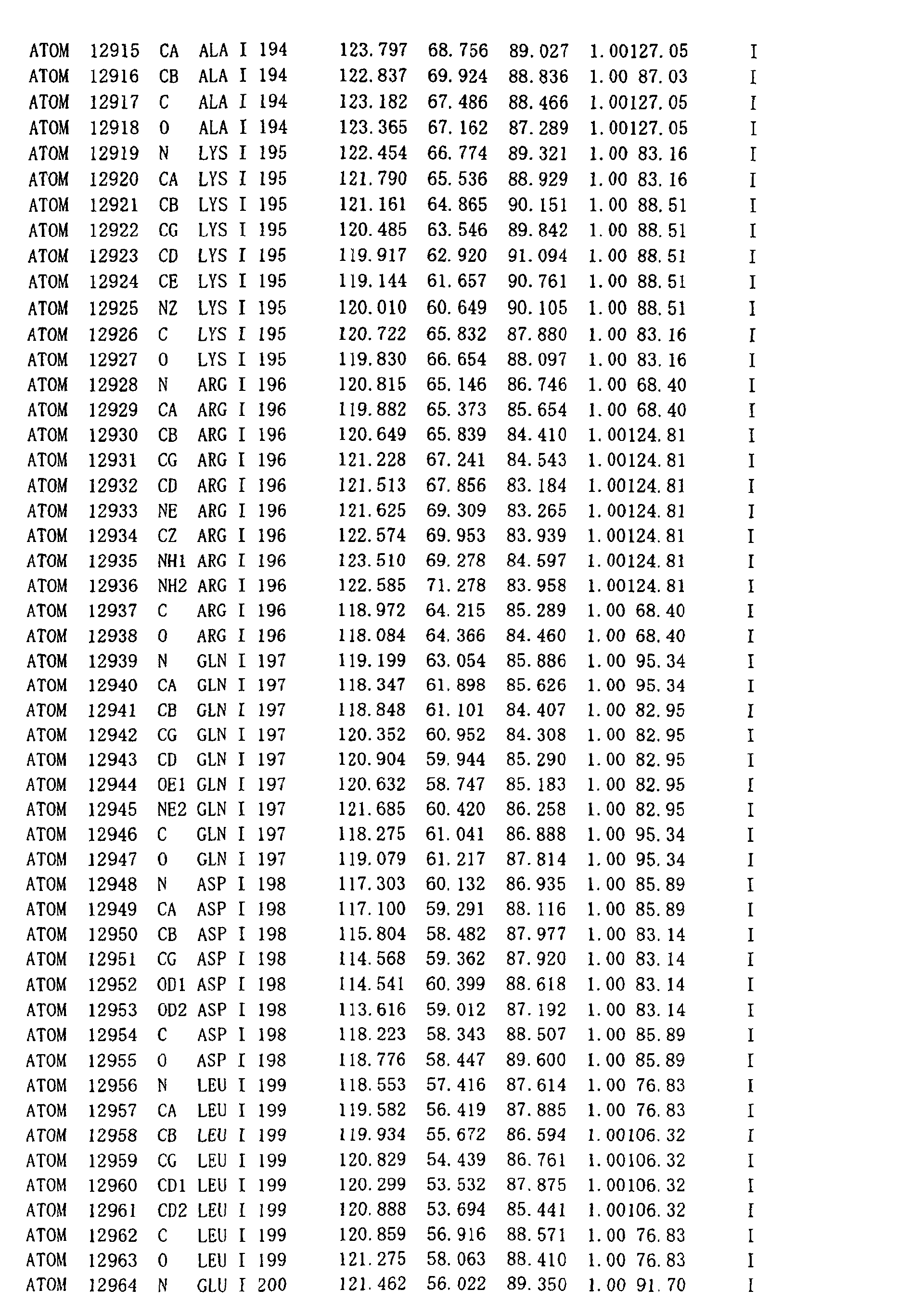
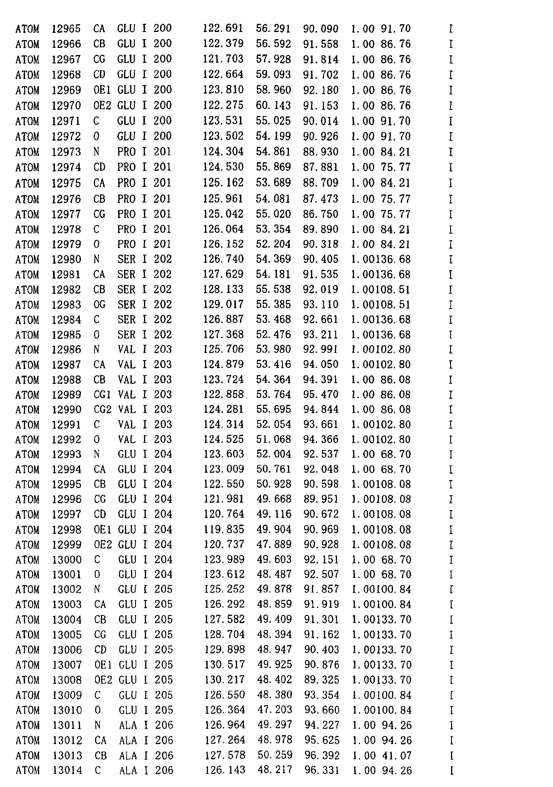
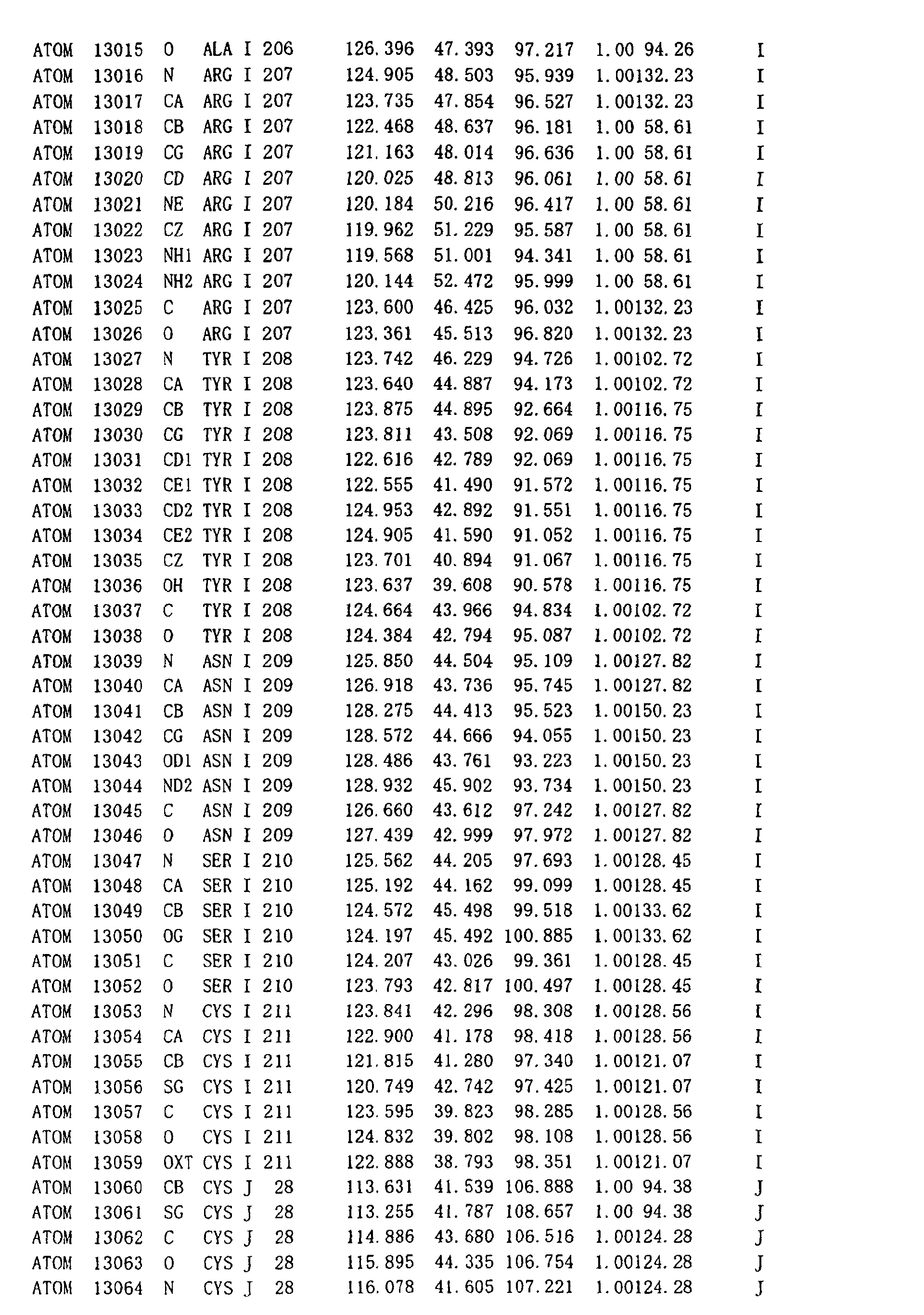
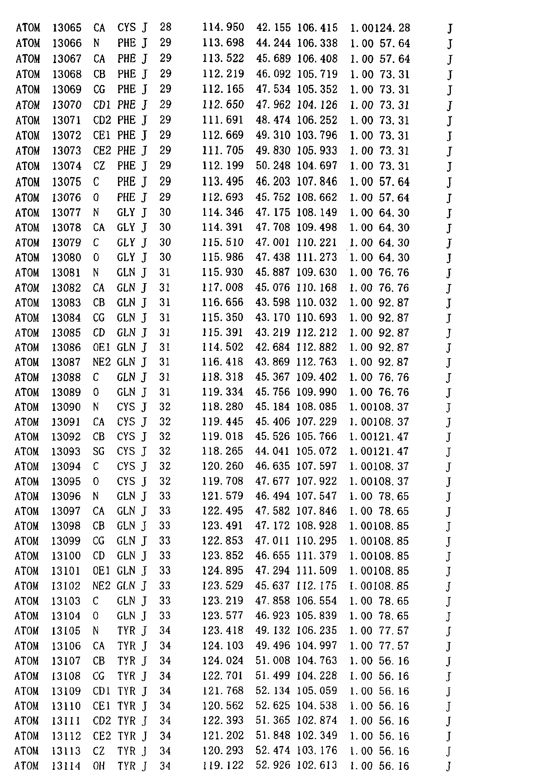
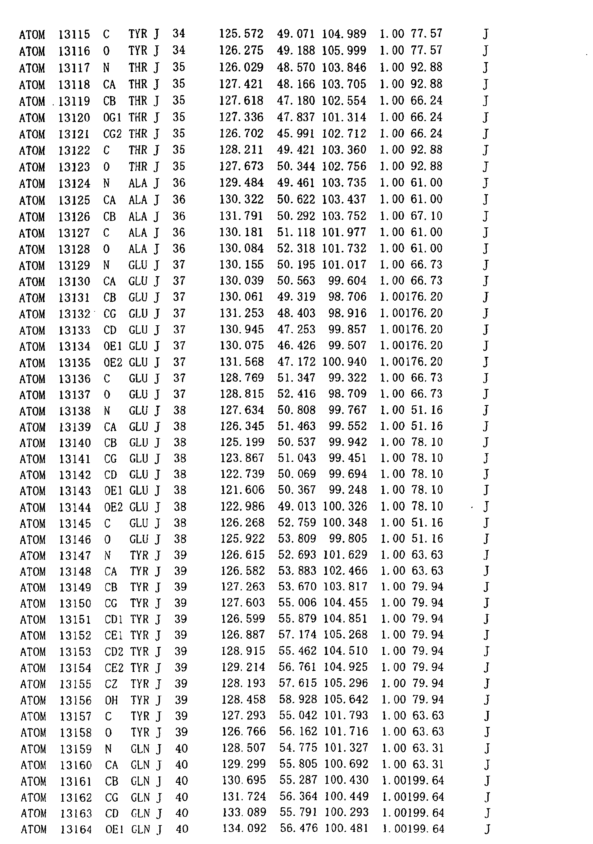
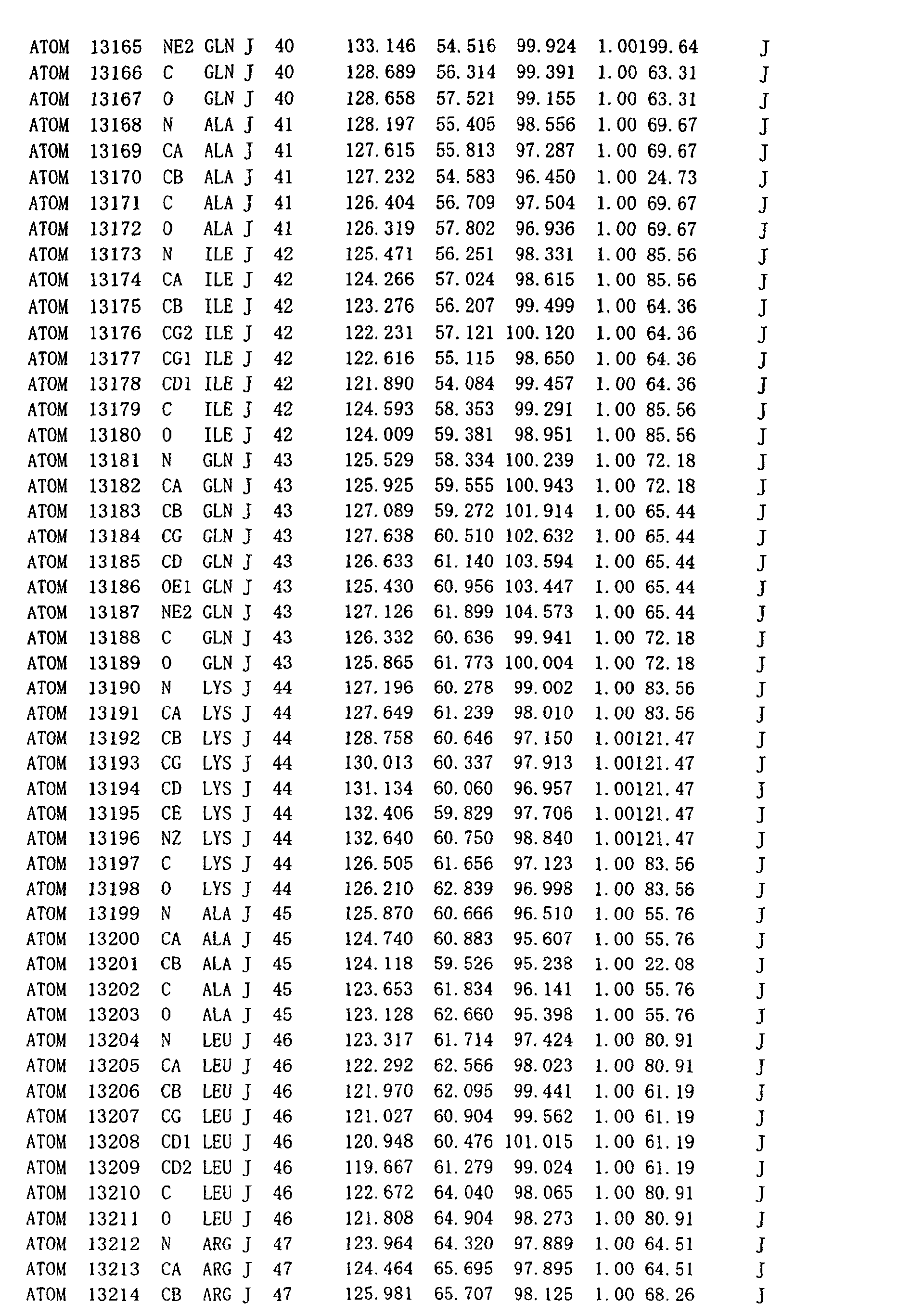
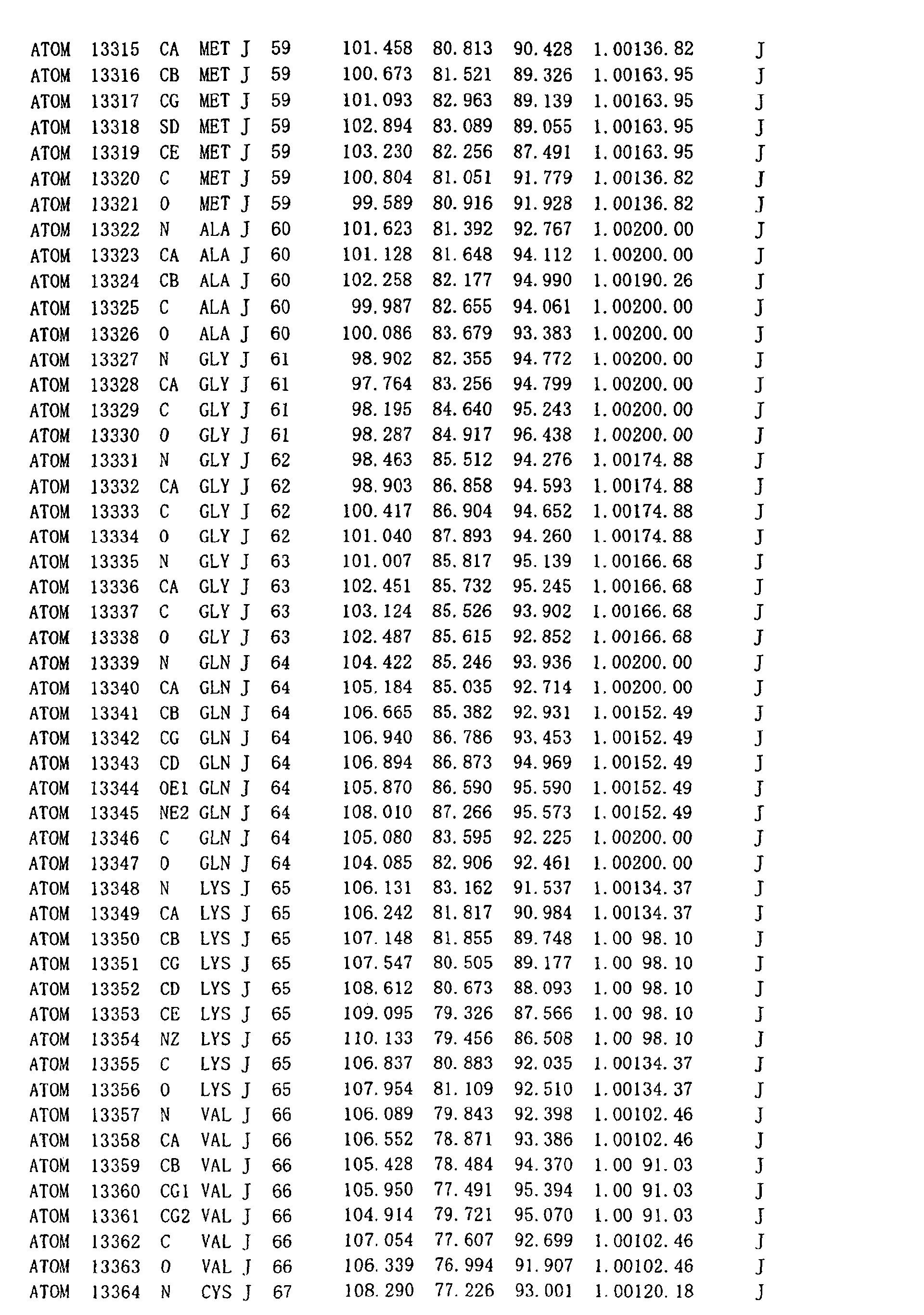
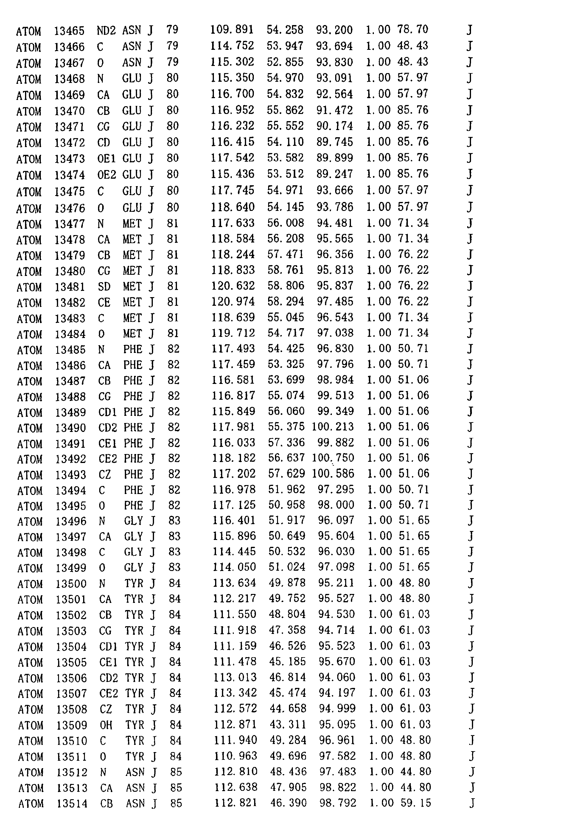
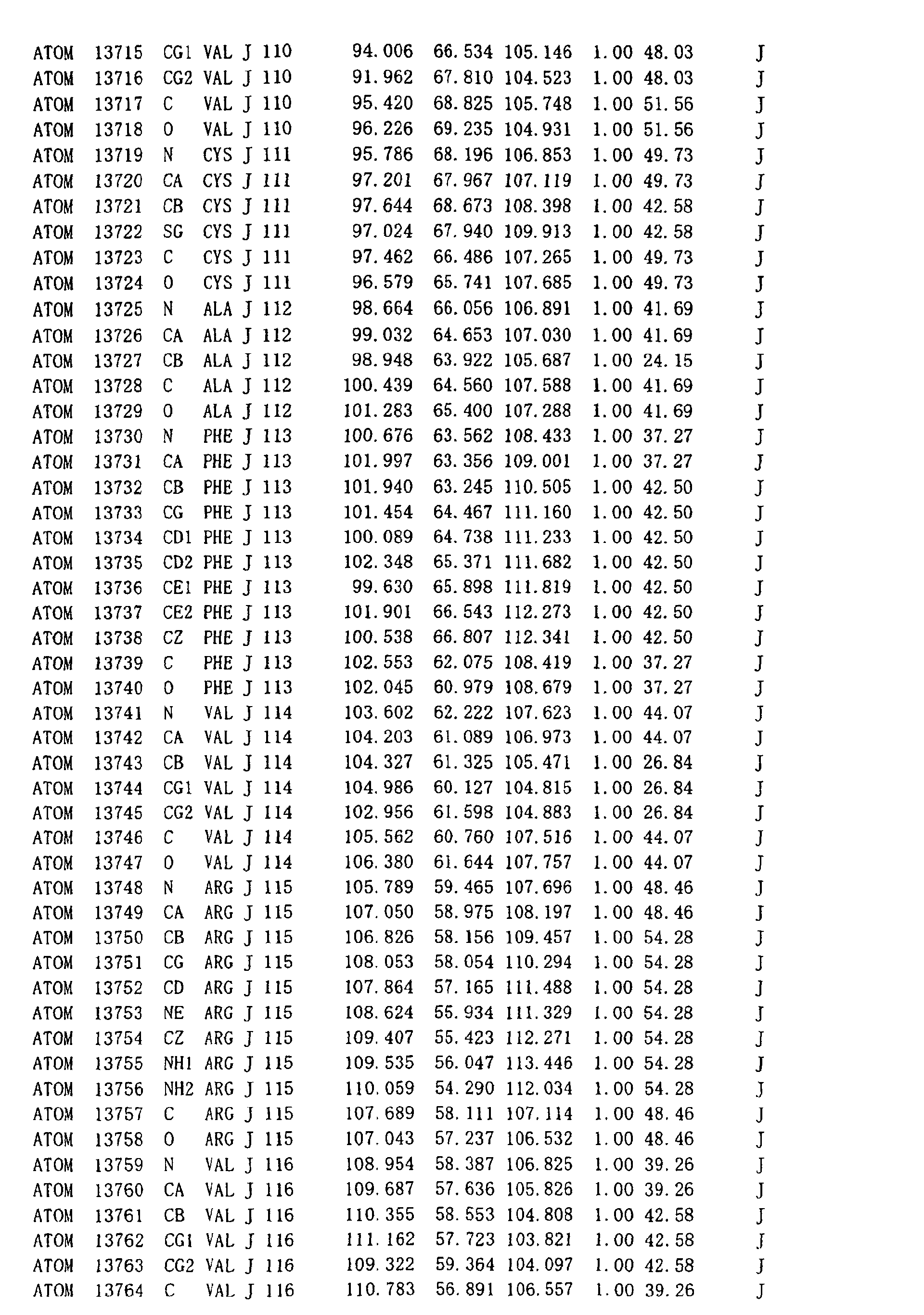
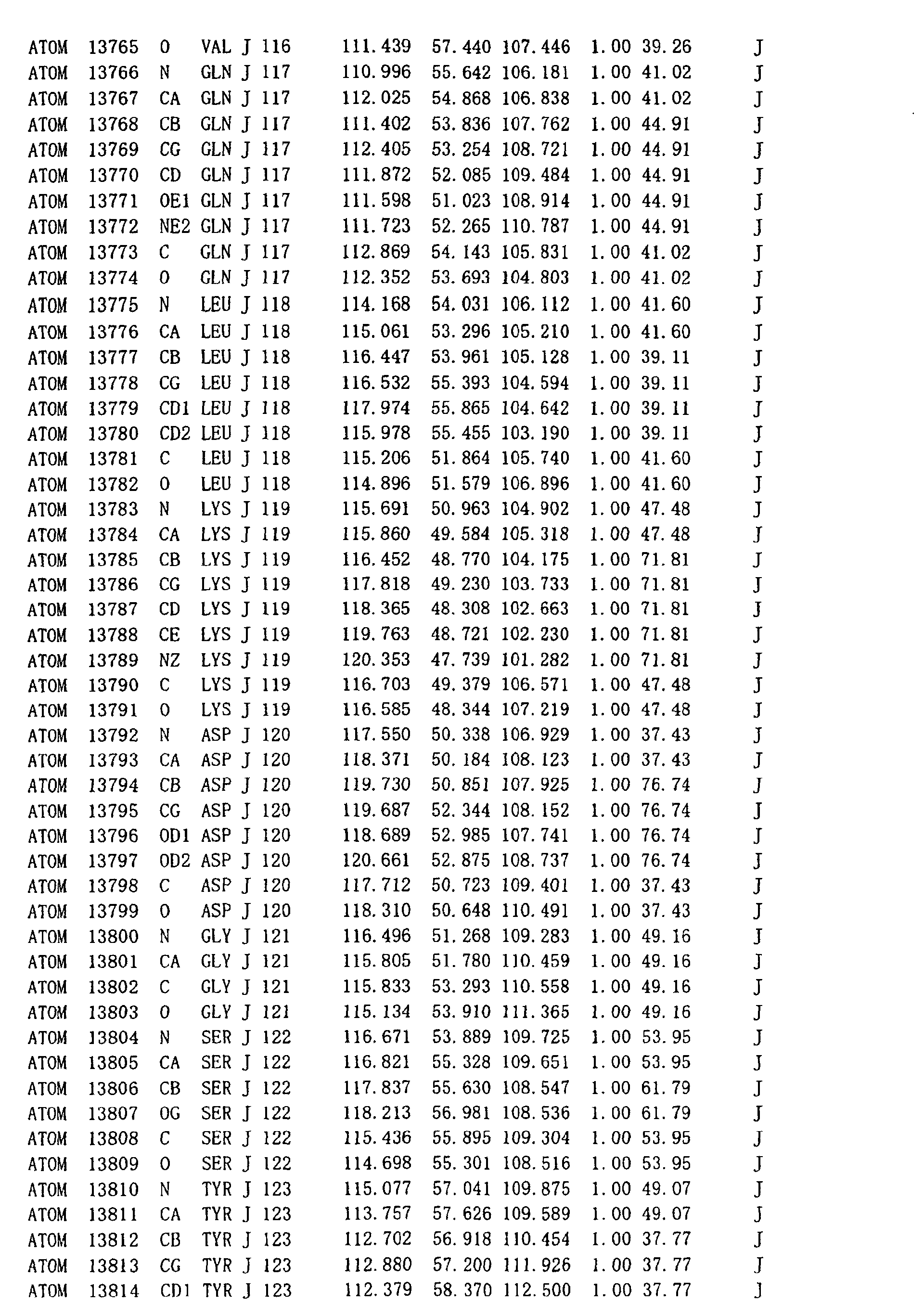
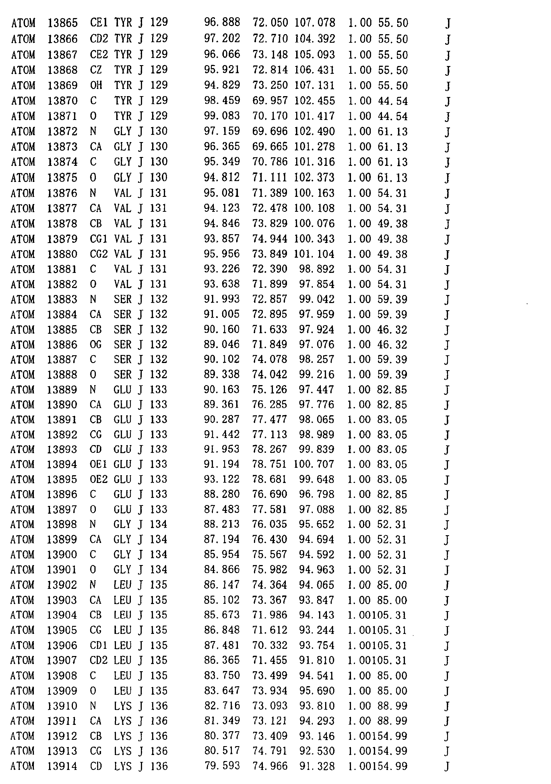
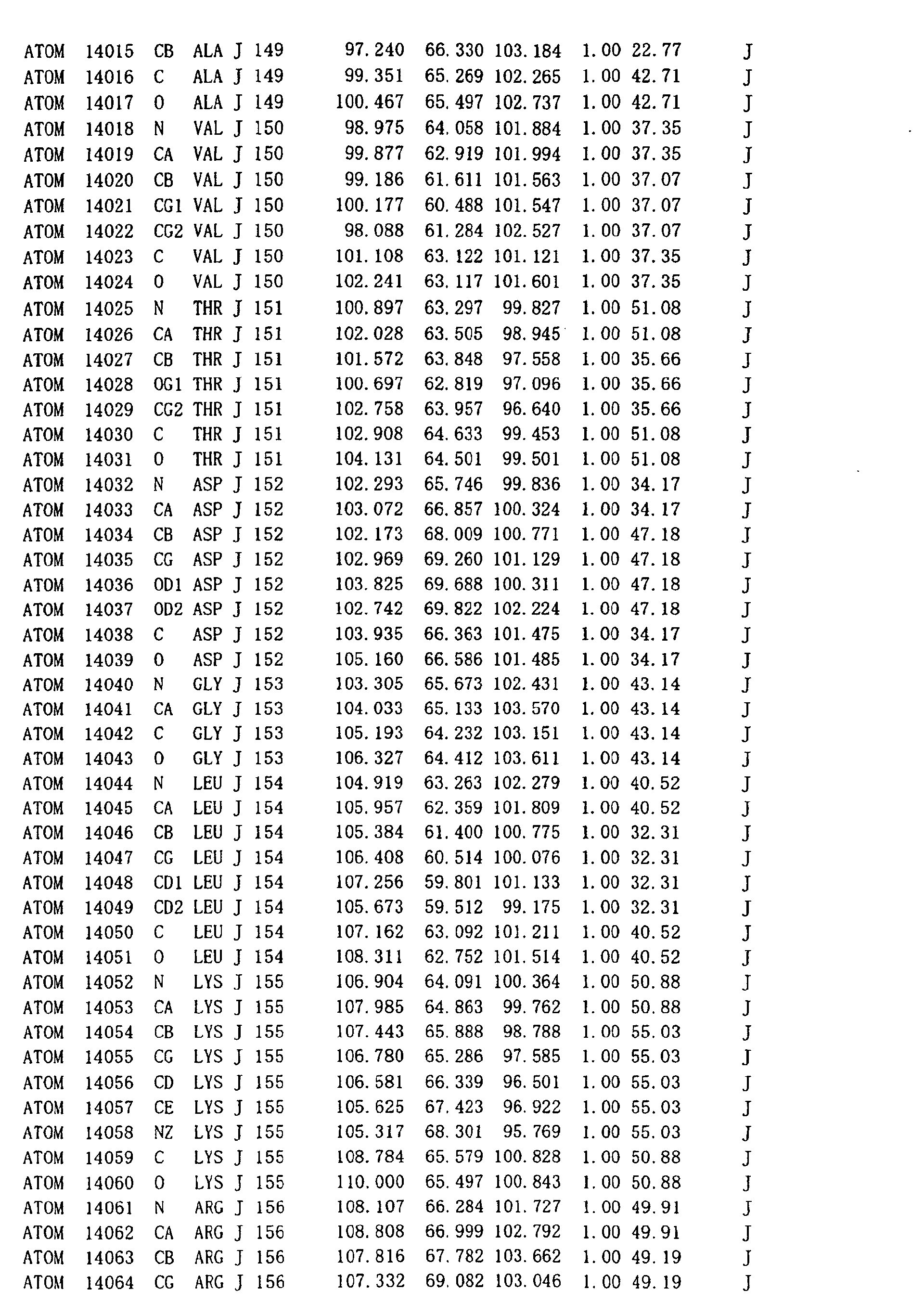
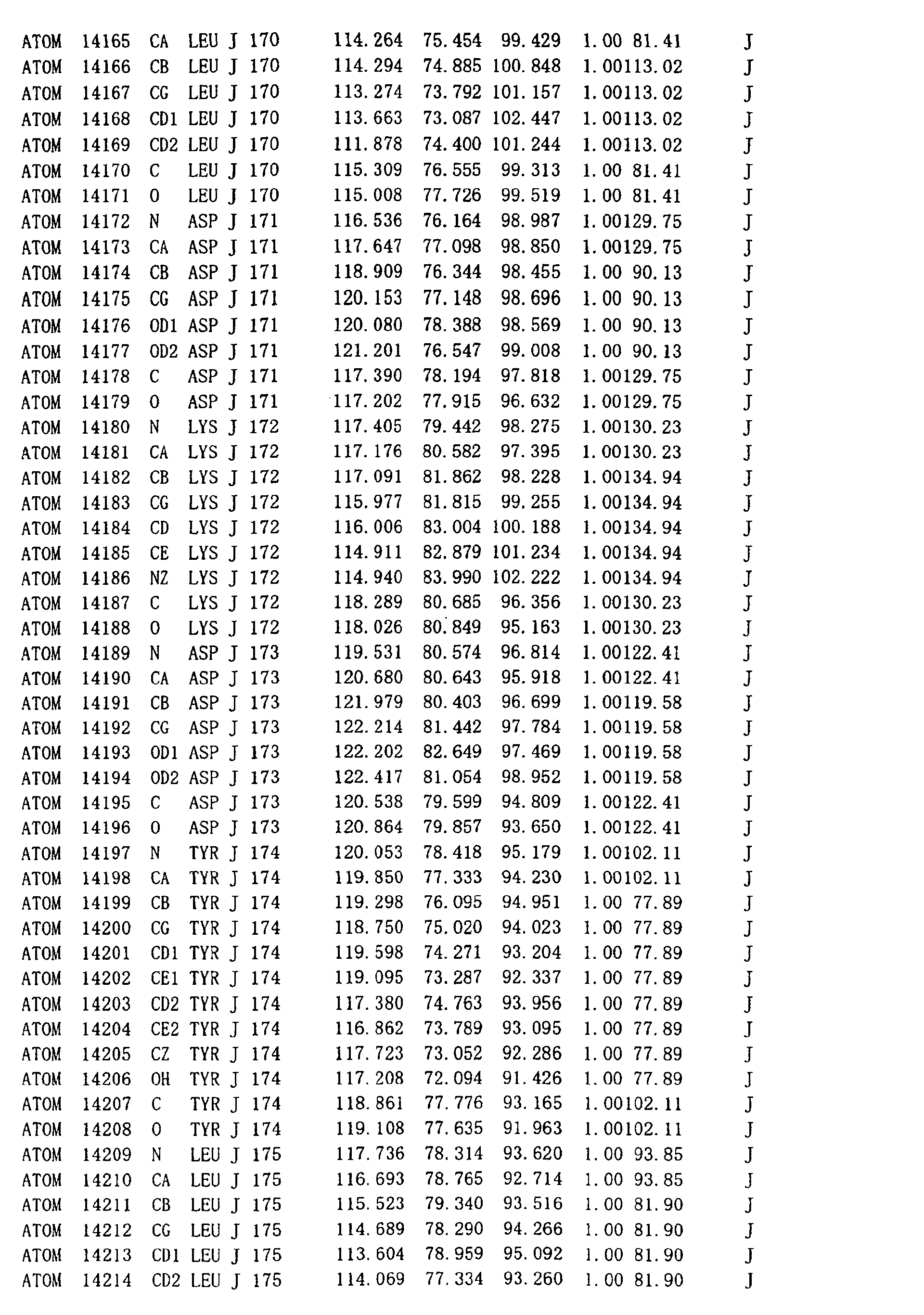
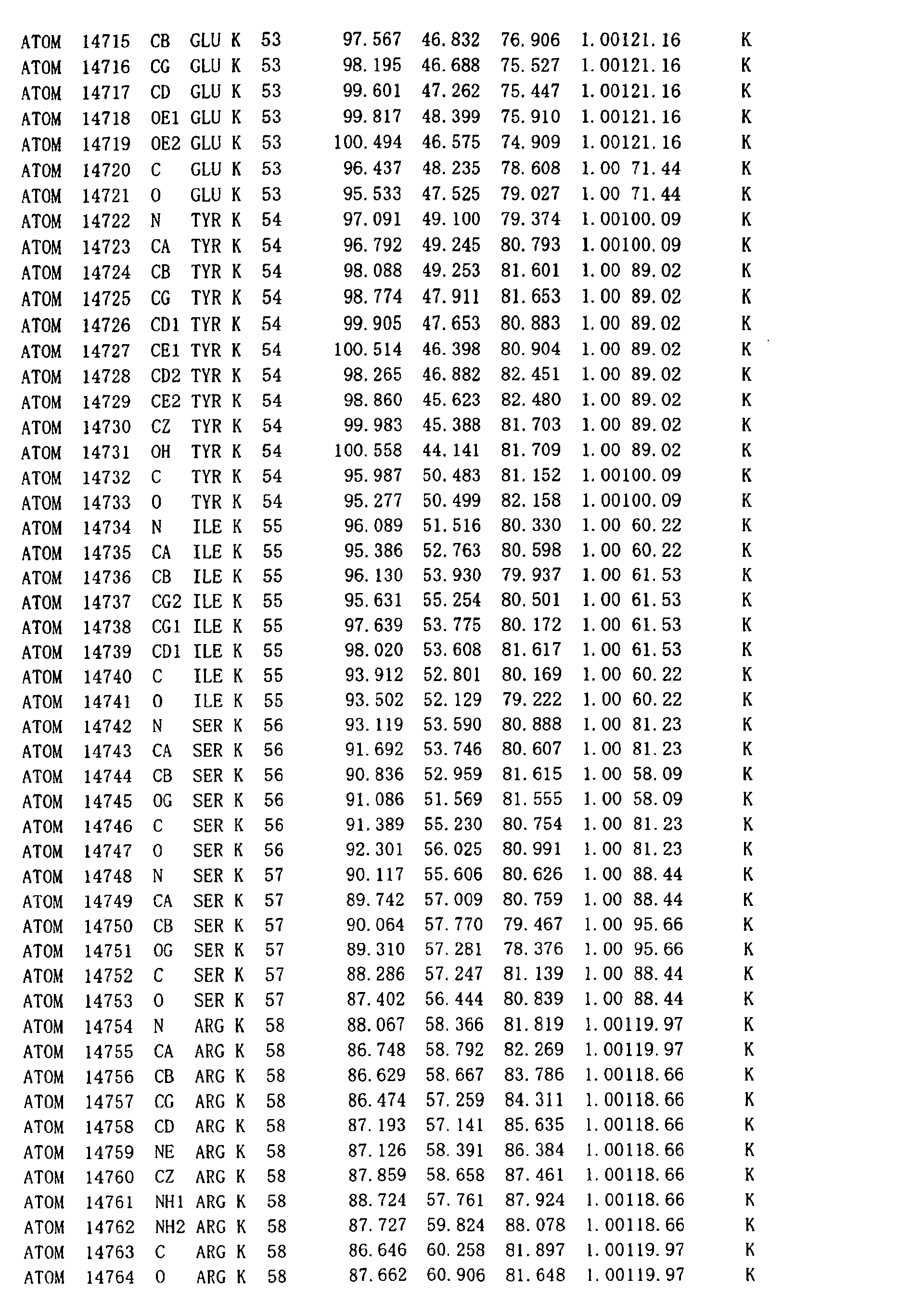
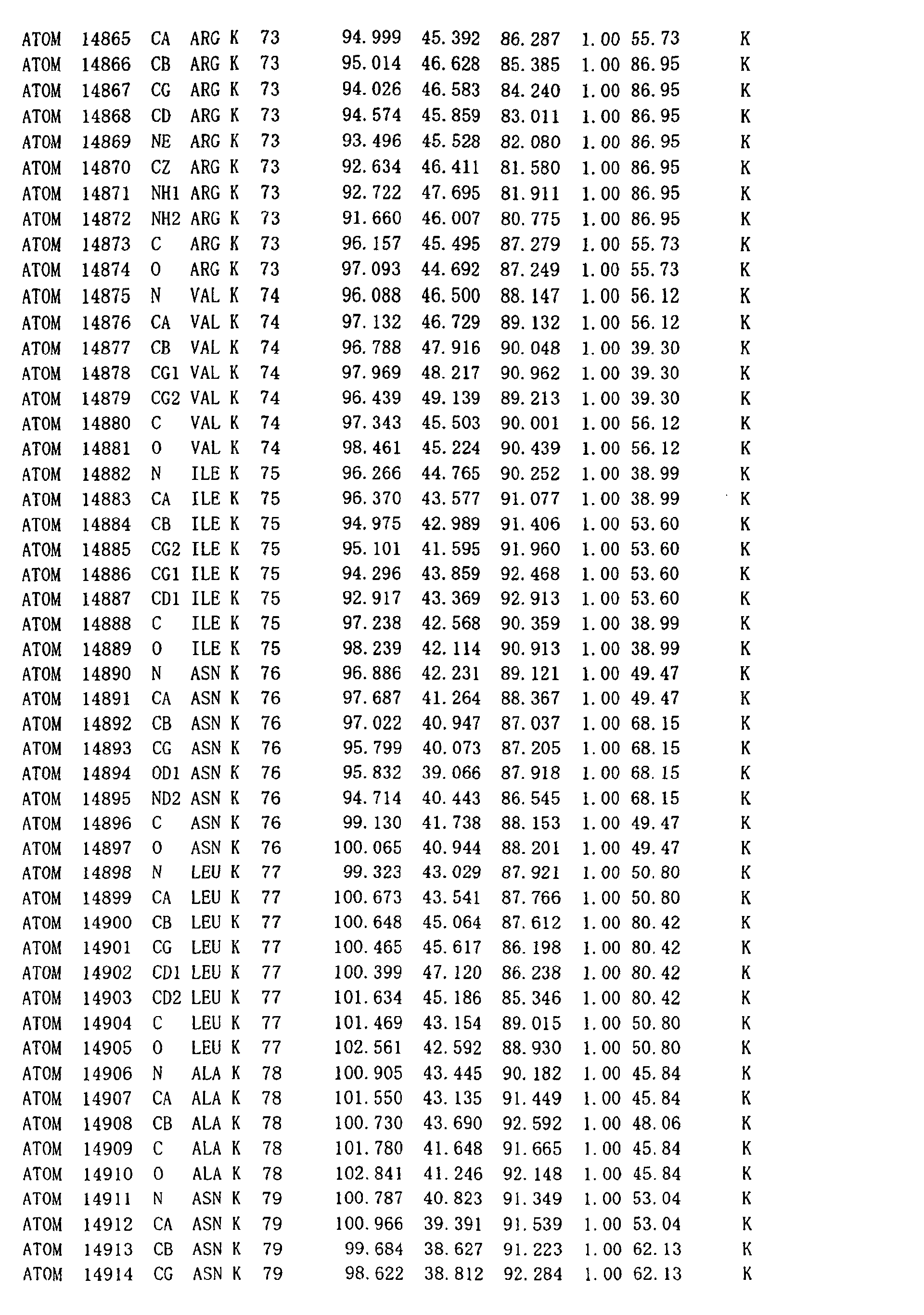
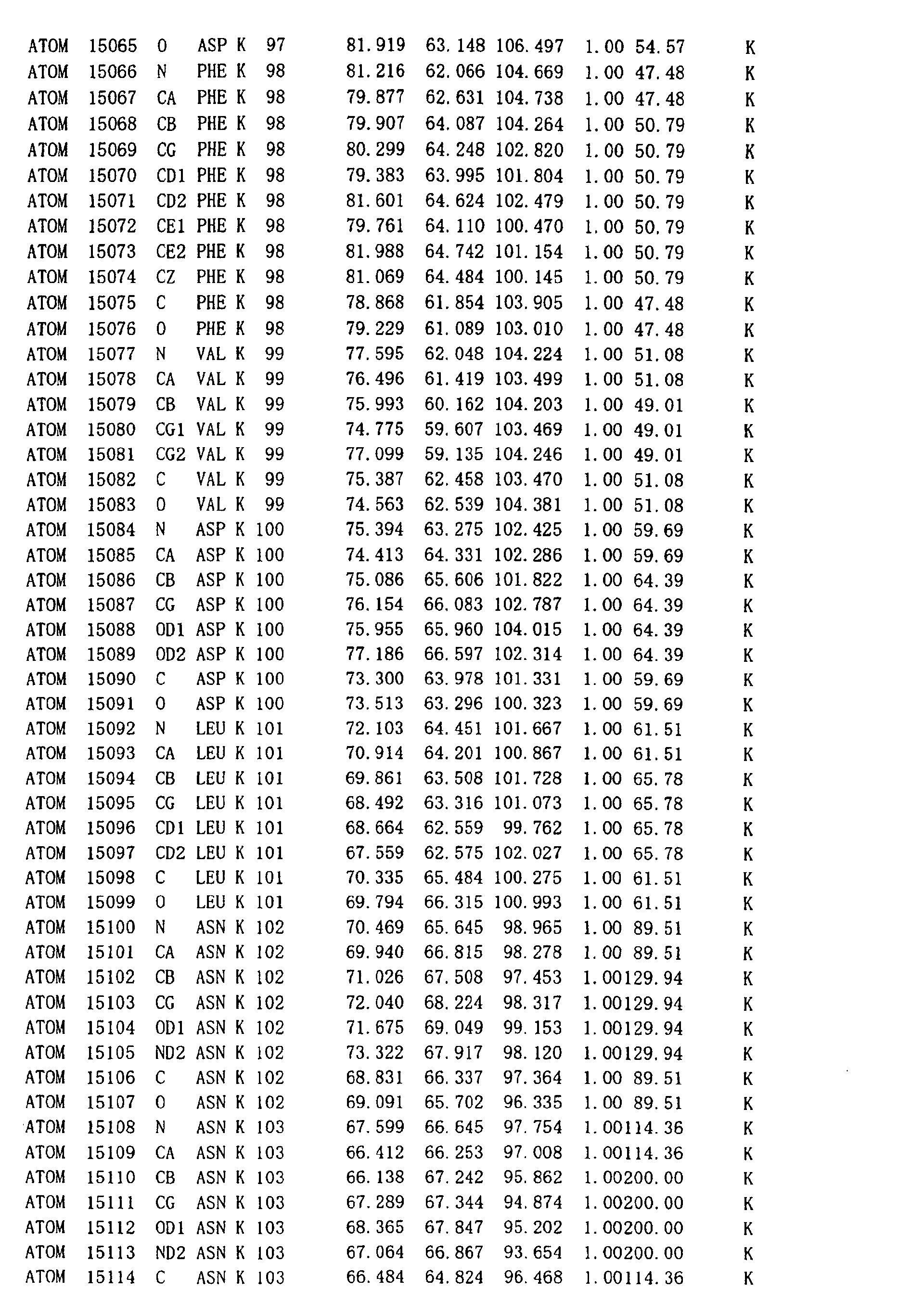
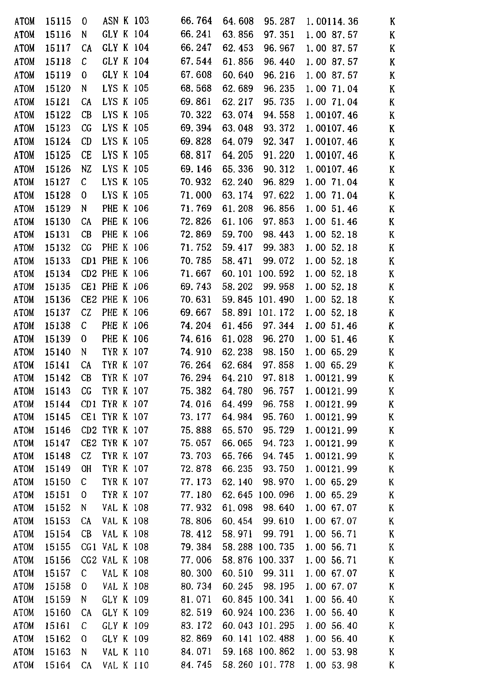
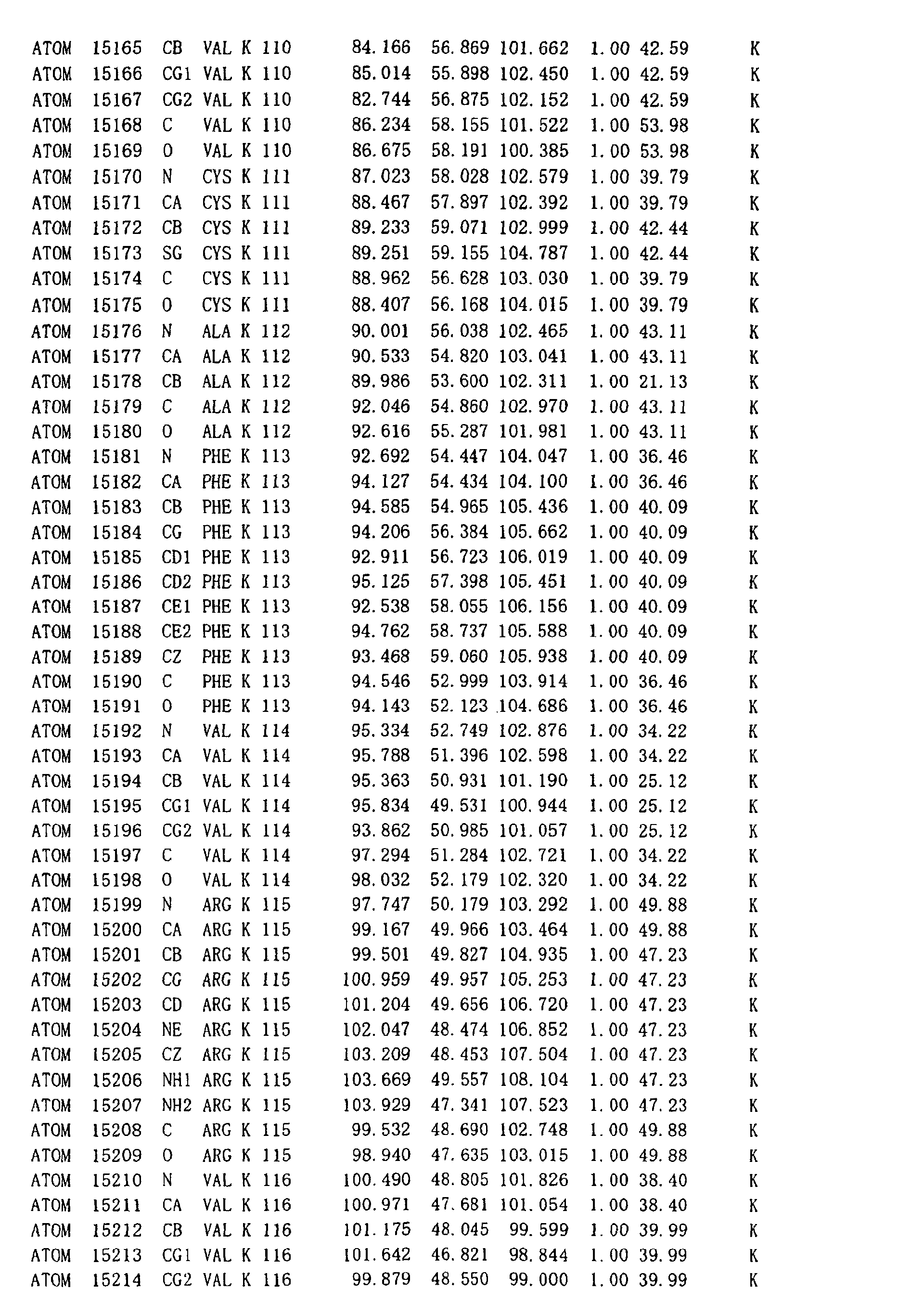
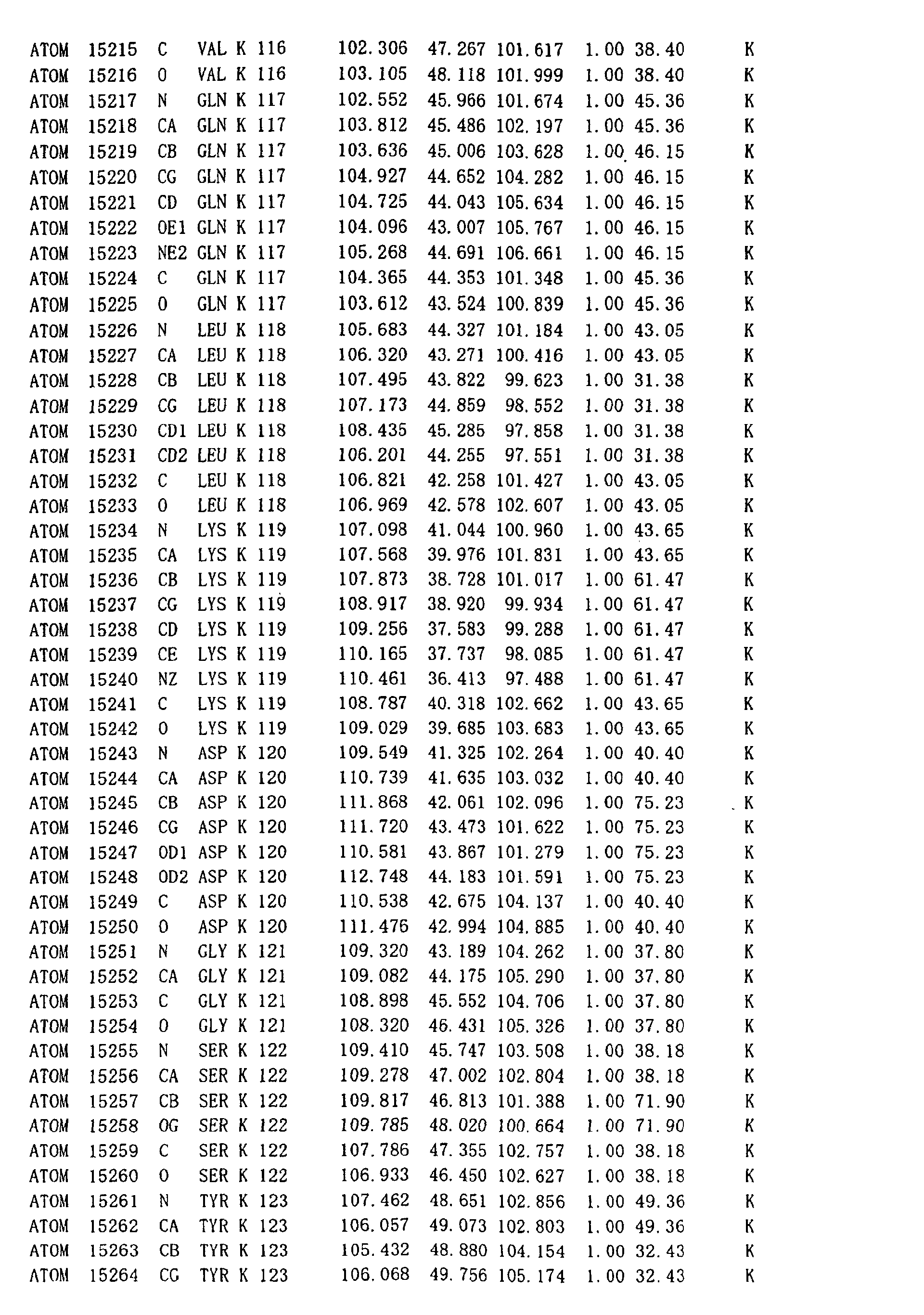
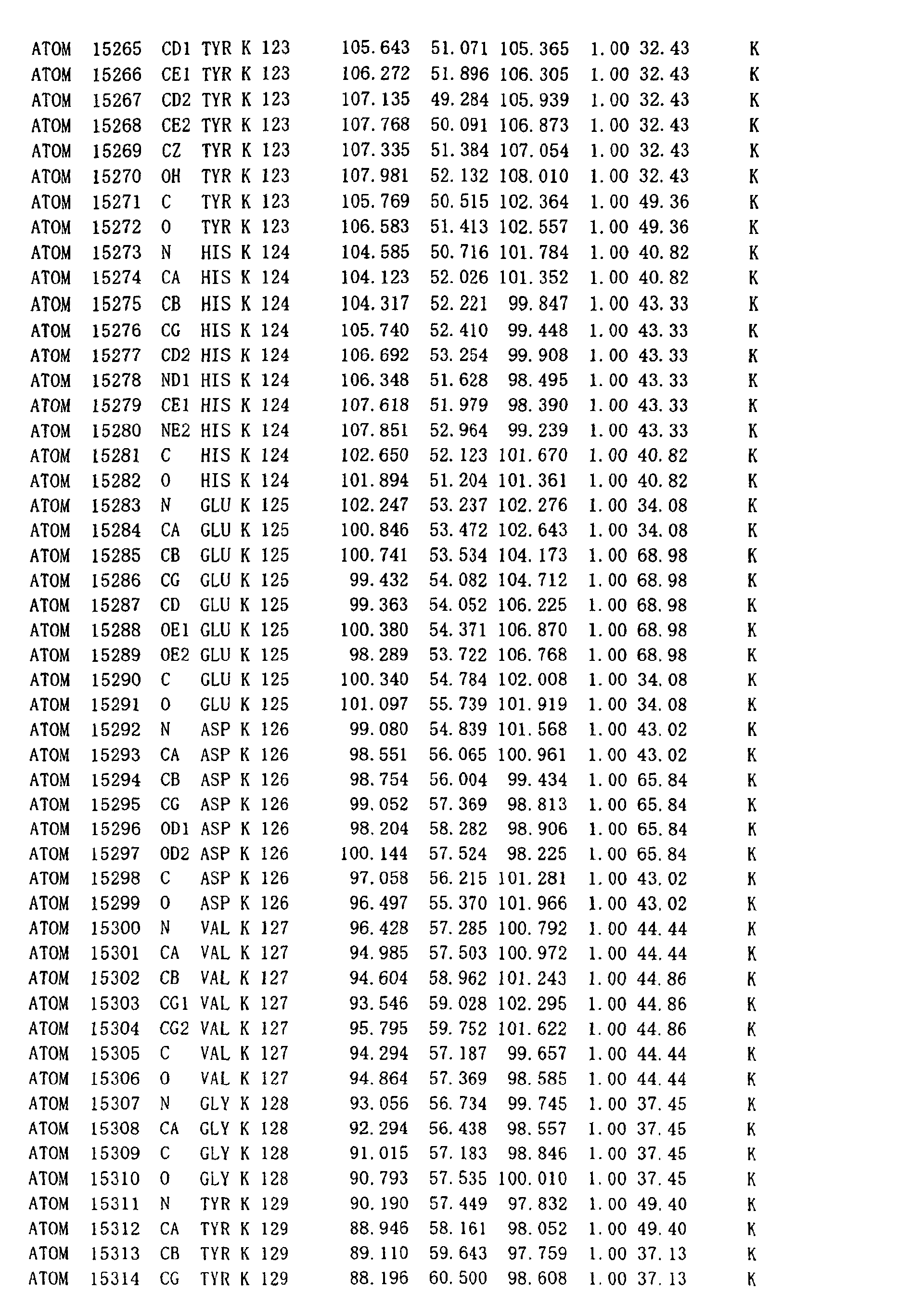
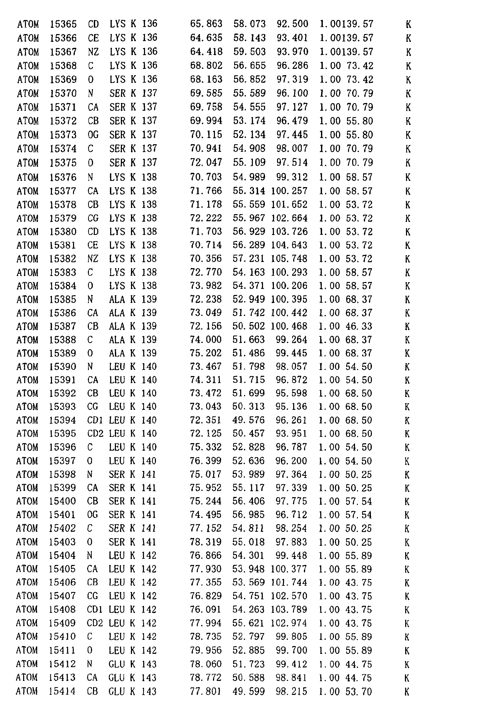
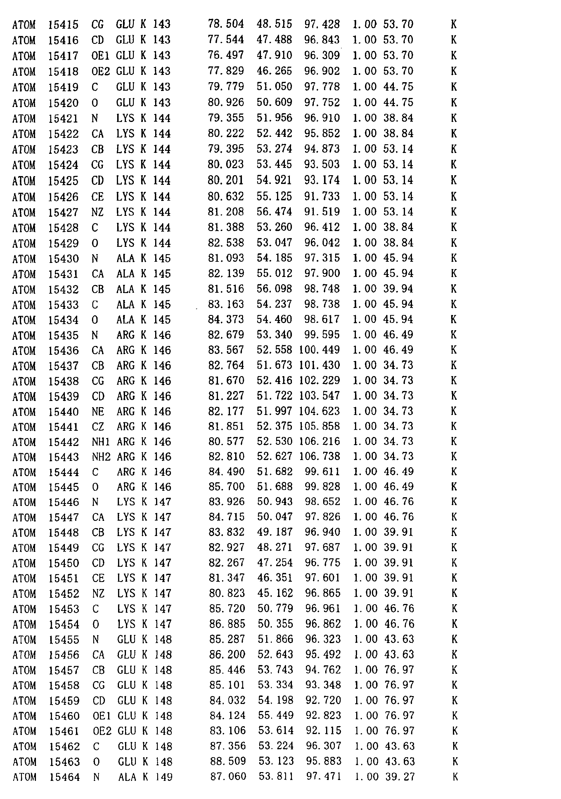
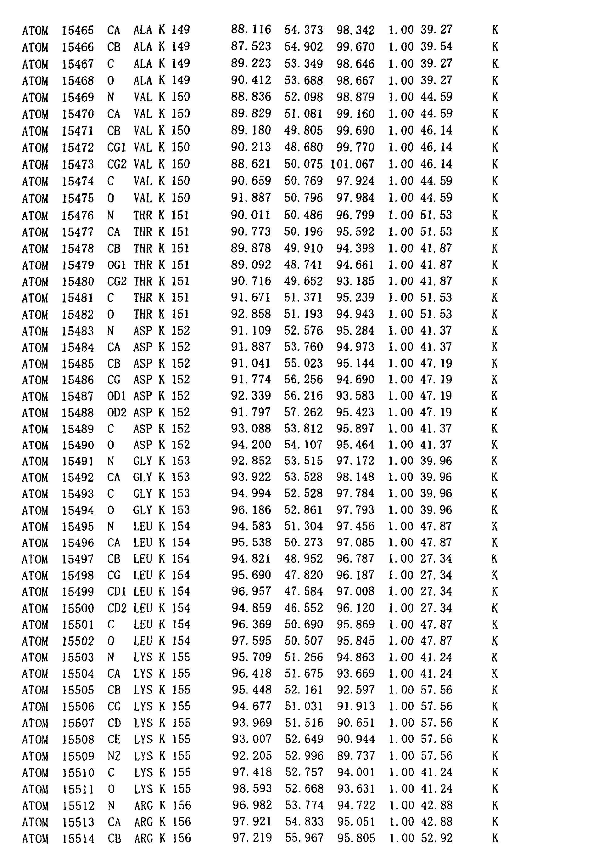
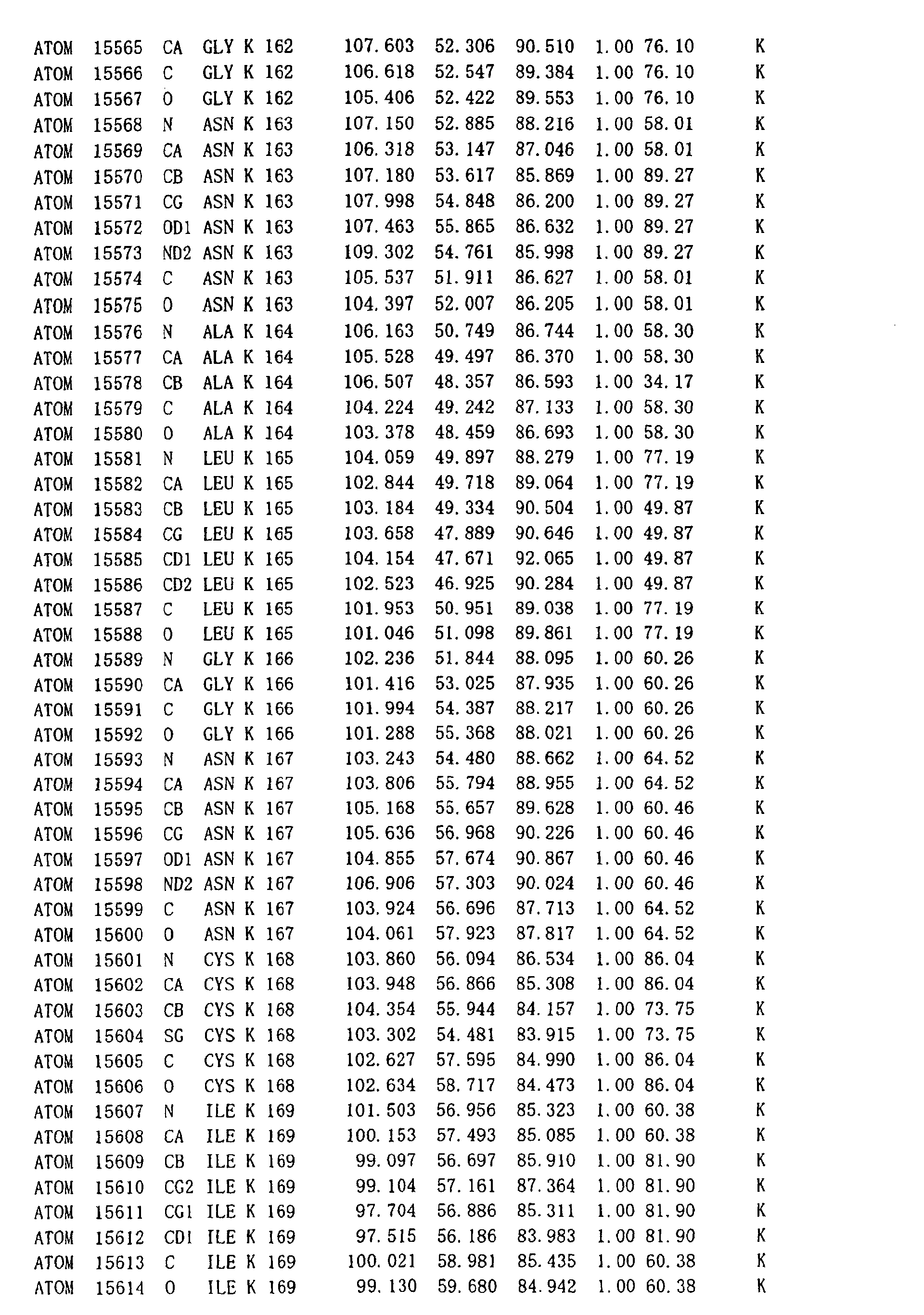
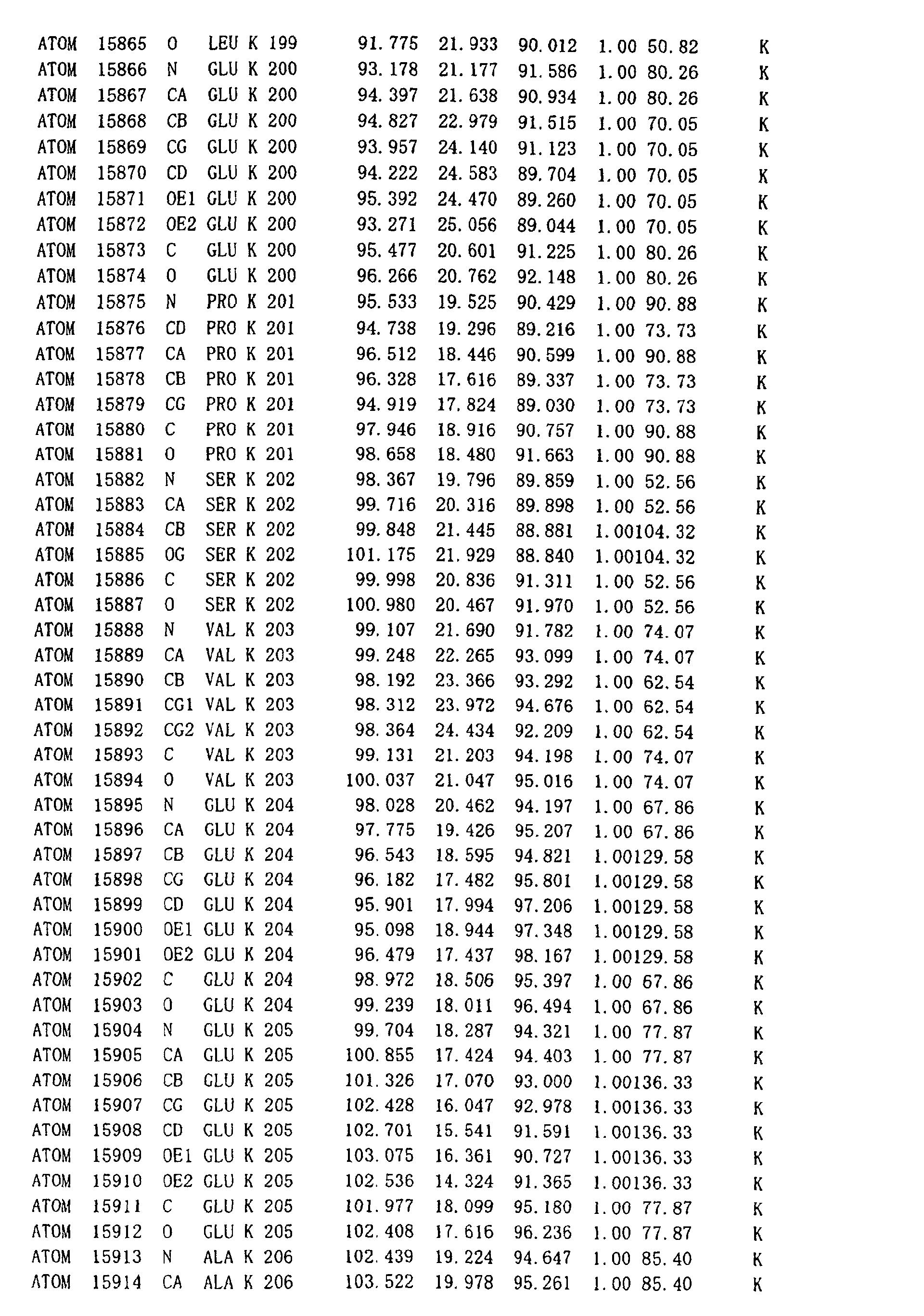
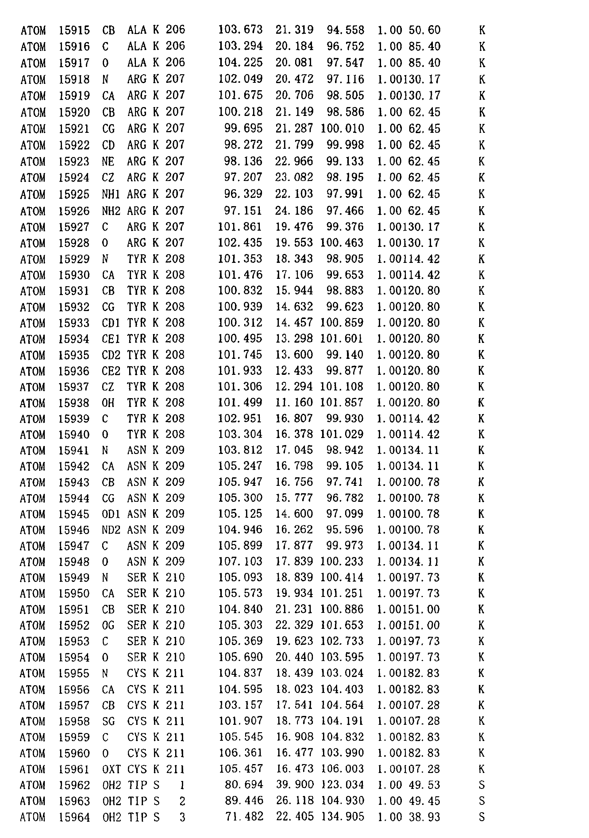
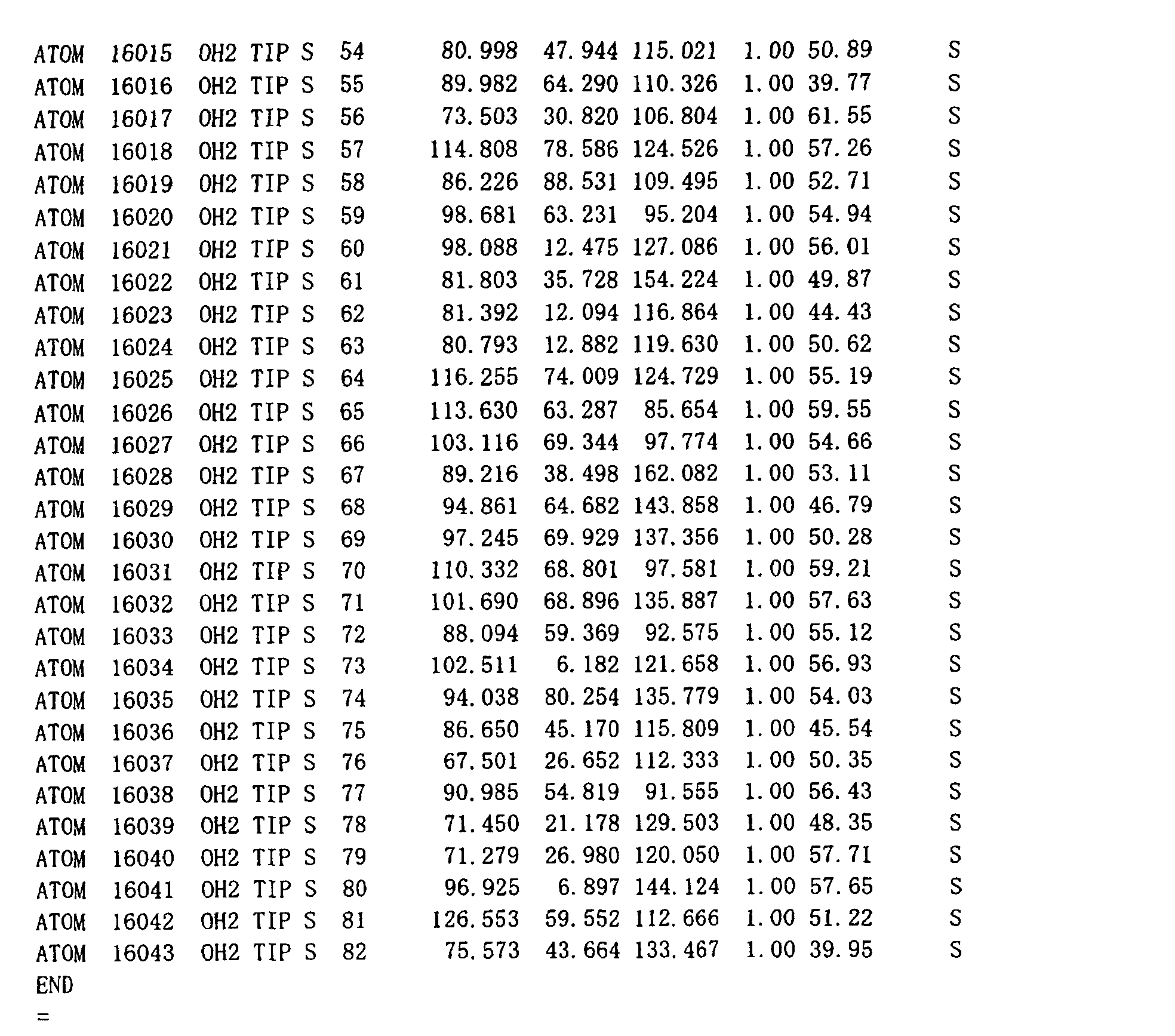
Table 1 shows the resulting atomic coordinates according to
the text format of Protein Data Bank as an expression method
for general use for a person skilled in the art. Herein, the
resulting atomic coordinates has been deposited with the
accession code PDB ID: 1KNO at Protein Data Bank (PDB) , Research
Collaboratory for Structural Bioinformatics (operated by RCSB) .
Table 1 can be referred to within the scope of the claims of
the present specification.
-
Herein, the atomic coordinates are expressed using the
origin of unit lattice in the crystal as the origin in the
three-dimensional space. In case that the atomic coordinates
of the invention are used for computer calculation, new atomic
coordinates obtained as the consequence of mathematic transfer
procedures for translation, rotation and symmetry without
changing relative allocation of each atom in three-dimensional
space are also within the scope of the invention.
(Computer system displaying three-dimensional structure model
of HsRad52 protein complex)
-
By inputting the atomic coordinates obtained from the
crystal of the DNA recombination/repair protein complex in
accordance with the invention into a computer operating a
computer program expressing three-dimensional molecule
structure or the memory medium of the computer, the
three-dimensional structure of the DNA recombination/repair
protein complex can be represented in detail.
-
As the memory medium of computer, any memory medium capable
of introducing the atomic coordinates of the DNA
recombination/repair protein complex onto the program of the
computer is satisfactory, with no specific limitation. The
memory medium includes for example electric temporary memory
medium called memory and semi-permanent memory media such as
flexible disk, hard disk, photodisk, photomagnetic disk and
magnetic tape.
-
A three-dimensional structure model expressing at least
a part of the DNA recombination/repair protein complex can be
generated by a processor in electric communication with the
memory or memory media. As such processor (program), for
example, there can be used QUANTA (Molecular Simulations Co. ,
Ltd.), SYBYL (Molecular Modeling Software, Co., Ltd.), AMBER
(Weiner, S. J., etal., (1984) J. Am. Chem. Soc., 106, 765-784)
orCHARMM (Brooks, B.R. etal., (1983) J. Comp. Chem. 4, 187-217).
-
As a program for calculating the coordination energy of
the complex and minimizing the energy following the step for
generating these molecular models, further, the CHARMM and AMBER
and the like are used.
-
Fig. 1 shows the three-dimensional structure of the DNA
recombination/repair protein complex, namely the HsRad521-212
complex obtained in such manner at a resolution of 2.8 angstroms
in a ribbon model. As apparently shown in Fig. 1, it was found
that the crystal of the HsRad521-212 complex formed a ring
structure through the assembly of the monomer protein of 212
amino acid residues. Because it is supposed that the remaining
amino acid residues at the side of C terminus are not directly
involved in the formation of the complex, even the full-length
HsRad52 in living biological organisms forms the undecameric
complex and is therefore encompassed within the scope of the
invention. The three-dimensional structure of the monomer
composing the complex is shown in Fig. 2. The monomer protein
of 212 amino acid residues has five α-helixes and five β-sheet
structures.
(Use of three-dimensional structure of DNA
recombination/repair protein complex)
-
The atomic coordinates obtained from the crystal of the
DNA recombination/repair protein complex in accordance with
the invention are input in a computer operating a computer
program representing three-dimensional molecule structure or
the memory medium thereof, to do visual examination and/or
calculate the conformational energy, which first enables the
rational designing in three-dimensional space of a mutant DNA
recombination/repair protein with a modification of a desired
characteristic property, for example the improvement of
catalytic activity and the improvement of protein stability.
-
One of typical methods for designing a mutant DNA
recombination/repair protein includes a step of inputting the
three-dimensional atomic coordinates of the DNA
recombination/repair protein complex of the invention in a
computer or the memory medium thereof, displaying the
three-dimensional structure of the protein on the computer
display using an appropriate program and examining the structure
visually.
-
The homologous pairing step as the most important step
of the DNA recombinational repairing mechanism can be speculated
visually to bind DNA in the periphery of the ring structure,
based on the three-dimensional structure of the DNA
recombination/repair protein complex shown in Fig. 1. By
subsequently calculating for example the surface charge of DNA
recombination/repair protein complex, amino acid residues
binding to DNA on the surface of the complex can be estimated.
Because DNA molecule has a negative charge derived from the
phosphate diester bond on the surface, amino acid residues
binding to the DNA molecule are highly possibly basic or
hydrophobic amino acid residues. By calculating the surface
charge of the DNA recombination/repair protein complex, these
can be identified. The identified individual amino acid
residues are replaced to alanine one by one by a method such
as site specific amino acid substitution, to analyze the DNA
binding site by alanine scanning and the like for assessing
the DNA binding activity. As the consequence of such analysis,
amino acid residues speculated as important for DNA binding,
or amino acid residues in a region close thereto are substituted
with other amino acids or are chemically modified, to prepare
mutant enzymes. Herein, the term "region close thereto" means
a region involved in the electrostatic interaction, hydrophobic
interaction, Van der Waals interaction and hydrogen bonding
to the amino acid residues, specifically a region within about
5 angstroms (Å).
-
Fig. 3 shows the DNA binding region speculated by such
method in the form of three-dimensional structure model. Fig.
3A shows the speculative DNA binding site in the HsRad52 monomer
protein. It is shown that the loop region in the vicinity of
arginine at position 55 (R55) to arginine at position 70 (R70)
and the region in the vicinity of arginine at position 153 to
lysine at position 169 are close to each other in terms of the
three-dimensional structure within the same molecule (Fig. 3A)
and is also close to the adjacent molecules (Fig. 3B) . Because
the mutant Rad52 where the any of the basic amino acids is
substituted with alanine loses the DNA binding activity
according to gel shift assay, it is confirmed that these regions
are involved in the DNA binding. Fig. 3C schematically shows
how the HsRad52 complex interacts with DNA on the ring-like
structure thereof. Therefore,it was shown that in the
HsRad521-212 complex shown in Fig. 1 or Fig. 3C, the complex was
bound to DNA on the lobe hanging out of the ring. The invention
includes a polypeptide fragment comprising the amino acid
sequence from the position 1 at least up to the position 192
and having a DNA recombinational repairing activity, which can
form the DNA binding site.
-
A mutant enzyme can be designed by deducing the binding
between the DNA recombination/repair protein complex and DNA
on the basis of the speculation from the coordination energy
calculated by a computer. Generally, a three-dimensional
protein structure when given enables the calculation of the
coordination energy (potential energy). As such energy
calculation system, programs such as ECEPP, CHARMM and AMBER
can widely be used. As factors composing the coordination
energy, for example, binding potential, electrostatic
potential, Van der Waals potential, and hydrogen binding
potential exist. Once the coordination energy is calculated,
the three-dimensional structure can be deduced when a specific
amino acid residue is substituted or the like. The optimized
structure for these mutant complexes to bind to DNA can be
analyzed or deduced.
-
The mutant designed in accordance with the invention may
be prepared by many methods. For example, DNA encoding the
mutant of the invention can be prepared by a method known to
a person skilled in the art such as site specific mutagenesis,
PCR and chemical synthesis or combinations thereof. By
introducing DNA encoding these mutant enzymes into an
appropriate expression vector and introducing further the
expression vector into an appropriate cell to culture the
resulting transformant, the mutant enzymes can be expressed.
-
As such expression vector, various vectors can be used
as long as the vectors can be amplified in host cells. When
the host is Escherichia coli, for example, plasmid vectors with
promoter/operator systems such as tac, λPL and T7 having high
transcription activity and enabling the induction are used.
Specifically, such plasmid vectors include the pKK233-3
(Brosius, J., and Holy, A. (1984) Proc. Natl. Acad. Sci. USA,
81, 6929) and pGEX-1 (Smith, D. B. and Johnson, K. S., (1988)
Gene 67, 31-40) with tac promoter and pET-3 (Rosenberg, A. H.,
et al., (1987) Gene 56, p.125-35) with T7 promoter. In case
that the host is bakers' yeast, for example, there is a shuttle
vector pAM82 (Miyanohara, A., et al., (1983) Proc. Natl. Acad.
Sci. USA, 80,1) usable in both bakers' yeast and E. coli . In
case that the host is an insect cell, baculovirus vector
pBacPAK8/9 (Clontech) and the like are used. In case that the
host is an animal cell such as Chinese hamster ovary cell CHO
or human HeLa cell, vectors such as pMSG (Amersham Pharmacia)
and pcDNA (Invitrogen) are used.
-
By inputting all or a part of the atomic coordinates of
the DNA recombination/repair protein complex provided in
accordance with the invention into a computer operating a
computer program expressing the three-dimensional molecular
structure or the memory medium thereof, a compound binding to
the DNA recombination/repair protein complex to modulate the
DNA recombinational repairing activity can be identified,
screened, assessed or designed. The compound may be any of
natural compounds and synthetic compounds or any of high polymer
or low molecular compounds.
-
The method includes a step of displaying a candidate
compound and the DNA recombination/repair protein complex
according to a computer model and screening a candidate compound
(lead compound) in a form or with a chemical structure well
suitable for a specific site of the DNA recombination/repair
protein complex under visual observation. To prepare or
display the molecular model, programs such as QUANTA and SYBYL
describedabovecanbeused. Further, various computer programs
to estimate the affinity to a specific site of the DNA
recombination/repair protein complex, repulsion and steric
hindrance and the like can be used. Examples of these programs
include GRID (Goodford, P.J., (1985) J. Med. Chem., 28,
p. 849-857) to determine a suitable binding site of a biopolymer
in terms of energy, MCSS (Miranker, A., etal., (1991) Proteins:
Structure, Function and Genetics, 11, p.29-34) to screen
functional binding sites, or AUTODOCK (Goodsell, D.S., et al.,
(1990) PROTEINS: Structure, Function and Genetics, 8, 195-202)
to geometrically analyze the interaction between a polymer and
a ligand for automatic docking, and DOCK (Kuntz, I.D., et al.,
(1982) J. Mol. Biol. 161, 269-288). Via calculations using
such programs, a region in a specific region of the surface
of the DNA recombination/repair protein complex, which can bind
to single-stranded or double-stranded DNA in a stable manner
in terms of energy, can be deduced, to indicate a candidate
compound three-dimensionally complementary to the region.
-
When such candidate compound (lead compound) is found,
continuously, the steric interaction with the candidate
compound can be examined visually, for further optimization
of a computer display imaging the three-dimensional structure
of the DNA recombination/repair protein complex. Using
programs such as QUANTA and SYBYL described above, a model for
optimizing the candidate compound can be constructed. Besides,
various programs useful for the optimization of molecular
modeling can be used, including for example CAVEAT (Lauri, G.,
and Bartlett, P.A., J. (1994) Comput. Aided Mol. Des. 8, 51-66)
andHOOK (Eisen, M. B., etal., (1994) Proteins: Struct., Funct.,
Genet., 19, 199-221).
-
In addition to the programs, further, a candidate compound
can automatically be constructed de novo from the
three-dimensional structure of the DNA recombination/repair
protein complex to output the structure. For them, programs
such as LUDI (Bohm, H. J., (1992) J. Comp. Aid. Molec. Design.)
and LEGEND (Nishibata, Y., et al. , (1991) Tetrahedron 47, 8985)
can be used.
-
The compounds thus designed or selected are contacted
with the DNA recombination/repair protein, to determine whether
or not the compound binds to the DNA recombination/repair protein.
The candidate compound canbe obtainedusing a chemical synthesis
method for general use or from compounds from natural origins.
It can be determined by various methods, whether or not the
compound binds to the DNA recombination/repair protein. For
example, binding to single- stranded DNA or double-stranded DNA,
formation of ternary complex with single-stranded DNA or
double-stranded DNA, and D-loop formation are observed by gel
shift assay. These reactions are essentially based on a
reaction of binding of the DNA recombination/repair protein
to first single- stranded DNA and annealing with double-stranded
DNA containing a nucleotide sequence complementary to the
single-stranded DNA. The complex formed in such manner can
be detected by various detection methods such as radioactive
labeling, using gel electrophoresis.
-
Otherwise, the interaction of the compound with the DNA
recombination/repair protein can be spectrophotometrically
detected, using NMR and mass spectrometry. Generally, NMR
signal sensitively reflects the local molecular environmental
change (ligand binding, structural change and the like). In
case that NMR spectrum is measured by mixing a protein
interacting with a ligand, therefore, it can be confirmed that
the ligand signal gets wider or disappears or the chemical shift
values change. In contrast, the structure of protein bound
to the ligand can also be detected in a specific manner. When
15N-1H HSQC spectrum using a protein uniformly labeled with 15N,
only the NMR signals of the protein can be observed.
-
The compound modulating the DNA recombinational repairing
activity thus obtained may be an inhibitor or activator of the
DNA recombinational repairing reaction specific to Rad52. The
inhibitor can be used for assaying Rad52-specific activity.
For example, mixing the compound with a sample such as cell
extract solution to completely inhibit the homologous
recombination activity of Rad52, subsequently assaying the DNA
recombinational repairing activity, and calculating the
difference between the activity and the DNA recombinational
repairing activity measured under conditions where the
inhibitor is removed, the DNA recombinational repairing
activity specific to Rad52 can be calculated.
-
In case of using the inhibitor as a therapeutic agent
of cancer, for example an enhancer for radiotherapy and
chemotherapy, the inhibitor is mixed with a carrier for general
use, to prepare a pharmaceutical composition. The carrier for
pharmaceutical compositions includes for example but is not
limited to lactose, glucose, D-mannose, starch, crystal
cellulose, calcium carbonate, kaolin, gelatin, hydroxypropyl
alcohol, polyvinylpyrrolidone, ethanol, carboxymethyl
cellulose, carboxymethyl cellulose sodium salt, magnesium
stearate, talc, acetyl cellulose, purified sugar, titanium
oxide, benzoic acid, p-oxybenzoate ester, sodium
dehydroacetate, gum arabic, tragacanth, methylcellulose, egg
yolk, surfactants, simple syrup, citric acid, distilled water,
glycerin, propylene glycol, macrogol, phosphate monohydrogen
sodium, phosphate dihydrogen sodium, sodium phosphate and
sodium chloride. Depending on the intended formulation, for
example, injection and tablet, the carrier is mixed with the
inhibitor identified by the method of the invention.
EXAMPLES
-
The present invention is explained in more detail by
reference to the following Examples, however, they do not
restrict the scope of the present invention.
(Example 1)
Limited proteolysis of HsRad52
-
5 µl of HsRad52 protein (1 to 3 mg/ml) prepared by a known
method (Kagawa et al., J. Biol. Chem., 2001, 276, 35201-8) was
mixed with 5 µl of proteinase K (30 µl/ml ) and the mixture was
incubated at 25°C for 4 to 6 hours. The proteinase K solution
was diluted in 10 mM Tris-HCl solution, pH 7.5. The reaction
mixture was fractionated by 12% SDS polyacrylamide gel
electrophoresis (SDS-PAGE), and the resulting protein bands
were transferred to a PVDF membrane using a semi-wet blotting
apparatus (Bio-Rad). Proteolytic bands were visually detected
by staining with a Coomassie Brilliant Blue staining solution,
followed by destaining of the membrane with methanol. The
protein was eluted from a portion of the PVDFmembrane containing
the protein bands, to determine the N-terminal amino acid
sequence. To measurethe molecular weightby massspectrometry,
the reaction mixture containing proteolyzed HsRad52 was treated
with 1 µl of 2N HCl to stop further proteolysis by proteinase
K. The reaction mixture was diluted with water to 100 fold,
for measurement with a mass spectrometry apparatus. As a
consequence of them, the region from the N terminus to position
237 of HsRad52, namely HsRad521-237 was found resistant to the
limited proteolysis by proteinase K.
(Example 2)
Analysis of HsRad521-237 by deletion
-
Plural deletion mutants of HsRad521-237 as prepared by
deleting sequentially every 5 amino acid residues from the C
terminus thereof, namely HsRad521-232, HsRad521-227, HsRad521-222,
HsRad521-217, HsRad521-212, HsRad521-207, HsRad521-202, HsRad521-197,
HsRad521-192 and HsRad521-187 were subjected to a combination of
both of crystallization and biochemical experiments.
HsRad521-192 and deletion mutants larger than the HsRad521-192
had absolutely the same DNA binding activity as that of
HsRad521-237. Among all of the deletion mutants, the highest
amount of crystal was obtained from HsRad521-212 in a system using
a screening kit (Crystal Screens 1 and 2 manufactured by Hampton
Research Co. , Ltd.). Thus, it was determined from these results
that HsRad521-212 was the most suitable for structural analysis.
(Example 3)
Purification of HsRad52-1-212
-
HsRad521-212 was purified by three steps of affinity
adsorption on Ni-NTA agarose, hexahistidine- tag cleavage and
Heparin-Sepharose column chromatography. A DNA encoding
HsRad521-212 was cloned in pET15-b (Novagen) , an expression vector
transcribed by T7 polymerase. The HsRad521-212 protein was
expressed at a large scale, using E. coli strain JM109 (DE3)
together with tRNAArg3 recognizing the CGG codon and RNAArg4
recognizing the AGA/AGG codon. In case that the expression
plasmid did not contain the tRNA genes, the expression level
of the HsRad521-212 protein was extremely low. Generally, the
HsRad521-212 Protein was purified from a culture of one liter.
The recombinant E. coli was cultured at 30°C, to which IPTG
was added to a final concentration of 1 mM in the logarithmic
growthphase (OD600 = 0.6), to induce the expression of the protein.
Subsequently, the bacteria were cultured for 4 to 6 hours to
collect the bacterial cells, which were disrupted in a buffer
solution A (pH 7.8) containing 50 mM Tris-HCl, 0.8 M KCl, 2
mM 2-mercaptoethanol, 10 mM imidazole, 10% glycerol and a
protease inhibitor (Complete EDTA-free; manufactured by
Boehringer Mannheim Co., Ltd.) on ice by ultrasonication. All
the procedures after the collection of the bacterial cells were
carried out at 4°C. The disrupted bacterial cells were
centrifuged at 27,700 x g for 20 minutes. The resulting
supernatant and 4 ml of Ni-NTA agarose bead (Qiagen) were gently
mixed together by batch process for one hour. The Ni-NTA agarose
bead binding the HsRad521-212 protein was charged in Econo-column
(Bio-Rad) and rinsed using a buffer solution B (pH 7.8)
containing 50 mM Tris-HCl, 0.8 M KCl, 2 mM 2-mercaptoethanol,
50 mM imidazole, and 10% glycerol at a volume 30-fold the volume
of the column at a flow rate of about 0.3 ml/minute. The
HsRad521-212 protein was eluted on a linear concentration gradient
of the imidazole solution from 50 mM to 300 mM at a volume 30-fold
the volume of the column. The peak fraction eluted with about
0.15 M imidazole solution was recovered, followed by addition
of 2 units of thrombin protease (Amersham Pharmacia Biotech)
per 1 mg HsRad521-212 protein, to remove the hexahistidine-tag
sequence. The reaction mixture was immediately dialyzed
against a buffer solution C (pH 7.5) containing 50 mM Tris-HCl,
0.2 M KCl, 0.5 mM EDTA, 2 mM 2-mercaptoethanol and 10% glycerol
for 12 hours or longer. After the hexahistidine- tag sequence
was removed, the HsRad521-212 protein was applied to a 4-ml
heparin-Sepharose column (Amersham Pharmacia Biotech). The
column was rinsed with the buffer solution C of a volume 20-fold
that of the column, to elute the protein on a linear KCl
concentration gradient of 0.2 M to 1 M and of a volume 20-fold
that of the column. The HsRad521-212 protein was eluted with
a sharp peak at about 0.3 M KCl concentration. The peak fraction
of the HsRad521-212 protein at 1 to 2 mg/ml concentration was
dialyzed against a buffer solution D (pH 7.0) containing 20
mM Hepes-KOH, 0.2 M KCl, 0.5 mM EDTA, 2 mM 2-mercaptoethanol
and 50% glycerol and stored at -20°C. Using the protein assay
kit of Bio-Rad, the protein was assayed using BSA (Pierce) as
a standard substance. The HsRad521-212 protein thus purified
was used for assaying various enzyme activities such as assaying
DNA binding activity and homologous pairing activity.
(Example 4)
Purification of protein for crystallization
-
For crystallization, the HsRad521-212 protein was
expressed in E.coli at a large scale by the same method as
described above and was then purified by the same method as
described above, except for the change of only the final buffer
solution for use in dialysis to another one. Specifically,
the HsRad521-212 protein was expressed in E. coli JM109 (DE3)
using an expression vector pET-15b (Novagen). Then, the
HsRad521-212 protein with thehistidine tag sequence was purified
onNi-NTAagarose (QIAGEN) , fromwhich thehistidine tag sequence
was removed by thrombin protease. Subsequently, the protein
was purified by heparin-Sepharose column chromatography
(Amersham Pharmacia Biotech). The peak fraction eluted from
the heparin-Sepharose column was dialyzed against a buffer
solution (pH 7 . 0) containing 10 mM Bis-Tris propane, 0.2 M KCl,
0.1 mM EDTA and 2 mM 2 -mercaptoethanol and was then concentrated
to 3 to 4 mg/ml, using Centricon-30 (Millipore) . To facillitate
the growth of the HsRad521-212 crystal, the HsRad521-212 protein
was mixed with an 8-mer single-stranded DNA (GTTGGTTG, 1.5 to
2 mM) at a molar ratio of 14 :1 to 11: 1 on ice, and was incubated
overnight at 4°C. The following day, the HsRad521-212 crystal
was grown by the hanging drop method at 20°C. The first droplet
was prepared by mixing 1 µl of a solution, pH 6.2 containing
12% polyethylene glycol (PEG) 5000 monomethyl ether (MME), 80
mM ammonium sulfate, 20 mM betaine monohydrate or urea, and
40 mM sodium cacodylate, with 2 µl of a mixture of HsRad521-212
and the single-stranded DNA. The reservoir solution contained
15% PEG 5000 MME, 0.1 M ammonium sulfate and 50 mM sodium
cacodylate (pH6.2). Crystals typically appeared 12 hours or
one day and reached the maximum size (0.1 mm × 0.2 mm × 0.3
mm)within 3 days.
(Example 5)
Data collection
-
The crystal of the HsRad521-212 protein contains the
undecameric HsRad521-212 ring as one of asymmetric units. A
diffraction data of a 2. 8-angstrom (Å) resolution or more could
be obtained. Prior to data collection, the crystal was treated
with the reservoir solution diluted to 3 fold with water.
Subsequently, the crystal solution was dialyzed against a
solution containing 15% ethylene glycol, 5% PEG5000 MME, 33
mM ammonium sulfate and 17 mM sodium cacodylate (pH 6.2)and
further dialyzed against a solution of the same composition
except for 30% ethylene glycol solution, for cryo-protection.
The HsRad521-212 crystals were flash frozen in a stream of nitrogen
gas.
-
Data sets of both the native and selenomethionine-substituted
crystals were collected at the RIKEN beam line I
(BL45XU) at the 8 GeV Super Photon ring (SPring-8) in Harima
Institute Japan. The diffraction data were reduced by a data
processing program DENZO-SCALEPACK.
(Example 6)
Determination and refining of three-dimensional structure
-
For the three-dimensional structural determination,
three data sets from three different crystals (one was native
type, and the other two were heavy atom-substituted materials
with selenomethionine) were used. The HsRad521-212 monomer
protein contained four methionine residues. One of the four
selenium atoms (methionine residue at position 78) was found
by the direct method program, SnB and was continuously used
for phase determination. The position of selenium atom was
further refined by the SHARP program, so that the initial phase
was calculated at 3.5-angstrom resolution. The initial maps
were solvent flattened by the program DM. The phases calculated
from the two selenomethionine data were combined and enlarged
to 2.8 angstroms using the data of the native type. According
to the iterative rounds of model building with the Program O
and the refining protocol with Program XPLOR, the
three-dimensional structure was refined until the complete main
chain tracing was possible. All the refining up to this stage
contained a 300 weight of NCS restriction. By the final refining
including minimization and refining of Group B factor, the NCS
weight was reduced step by step from 300 to 50. The final model
containing 15841 residues of the HsRad521-212 undecamer and 82
water molecules had an R value of 23.5% (28. 5% as free R value) .
On the Ramachandran plot of the final structure, 88.6% of the
residues existed in the optimal region, while 11.4% of the
residues existed in an additional acceptable region. The
graphic display of the three-dimensional structure was carried
out by using programs MOLSCRIPT and SPOCK.
(Example 7)
Introduction of site specific mutagenesis into HsRad52
-
Based on the three-dimensional structure of HsRad521-212
shown in Table 1 and Figs. 1 and 2, basic amino acid residues
with possibility of DNA binding in the periphery of the
undecameric ring structure were selected, to prepare point
mutants of HsRad521-212, where these were individually
substituted with alanine residue. The selected amino acid
residues were arginine at position 55, tyrosine at position
65, histidine at position 69, arginine at position 70, arginine
at position 153 and lysine at position 169. Mutants where
respective amino acid residues were substituted with alanine
are referred to as R55A, Y65A, H69A, R70A, R153A and K169A.
The introduction of site specific mutagenesis was done using
QuikChange kit (manufactured by Stratagene) according to the
instruction manual about the method for use.
-
The single-stranded and double-stranded DNAs binding
activities of the six types of point mutants thus prepared from
HsRad52
1-212 were detected by gel shift assay method. The
substrate DNA used by the method was prepared as described below.
The pGsat4 of double-stranded DNA was prepared by inserting
a 198-bp DNA fragment from human α-satellite into a plasmid
pGEM-T East vector (manufactured by Promega). The 3.2-kb
plasmid DNA was prepared by a gentle lysis method using sarcosyl,
without alkali modification of the host
E. coli, so as to avoid
the irreversible denaturation. As the single-stranded DNA,
a 50-base oligonucleotide purified by HPLC as shown in SEQ ID
NO:2 was purchased from Roche Biochemical.
The 5' terminus of the oligonucleotide was labeled with
radioactivity in the presence of [γ
32P] ATP using T4
polynucleotide kinase (New England Biolabs) and then purified
on a column Chromaspin-10 (manufactured by Clontech).
-
For gel shift assay, a reaction mixtures containing final
concentratins of 50 mM Hepes-KOH (pH 7.5), 40 mM KCl, 1 mM MgCl2,
0.1 mg/ml BSA, 1 mM DTT, 2% glycerol and 0.25 to 2 µM mutants
of HsRad521-212 was used. For single-stranded DNA binding assay,
1 µl of 1 µM 32P-labeled SAT-1 single-stranded DNA (nucleotide)
was mixed with 9 µl of the reaction mixture, which was incubated
at 37°C for 5 minutes. Subsequently, 1 µl of 12% glyceraldehyde
solution was added, to immobilize HsRad52 and the
single-stranded DNA. After incubating for 20 minutes, a 6-fold
volume of staining solution for electrophoresis was added. The
reaction mixture was resolved by 1% agarose gel electrophoresis
in 0.5 × TBE buffer at 3.3 V/cm and ambient temperature. After
electrophoresis, the gel was dried for detection by
autoradiography. The amount of the free single-stranded DNA
was assayed by an image analyzer BAS2500 manufactured by Fuji
Film. The results are shown in Fig. 4A. Single-stranded DNAs
that do not bind to HsRad52 remained on the tip of the gel,
but as the increase of the protein concentration, the protein
bound to the single-stranded DNA shifted toward the side of
a higher molecular weight. Compared with the wild type
HsRad521-212, the single-stranded DNA binding activity of all
of the point mutants was lowered. Particularly, the mutants
R55A, Y65A, H69A, and R70A lost most of the binding activity
to the single-stranded DNA. This indicates that a region close
to arginine residue at position 55 to tyrosine residue at
position 70 is a DNA binding region.
-
Next, for the double-stranded DNA binding assay, 1 µl
of 15 µM pGsat4 double-stranded DNA was added to 9 µl of the
reaction solution, and reacted for 5 minutes, and then
electrophoresed on 1% agarose gel in the same manner as described
above, except for no immobilization of HsRad52 and the
double-stranded DNA. After electrophoresis, the gel was
stained with 0.5 µg/ml ethidium bromide for detection. The
amount of the free double-stranded DNA was assayed by MacBAS
software. The results are shown in Fig. 4B. The
double-stranded DNAs that do not bind to HsRad52 remained on
the tip of the gel, while as the increase of the protein
concentration, the protein eventually bound to the
double-stranded DNA and shifted toward the side of a higher
molecular weight. Compared with the wild type HsRad521-212, the
double-stranded DNA binding activity of all of the point mutants
was lost. This indicates that two proximate regions, one is
a region close to arginine residue at position 55 to tyrosine
residue at position 70 and another is a region close to arginine
at position 153 and lysine at position 169 are DNA binding
regions.
-
The inventors made an additional analysis of the DNA
binding site of HsRad52 in detail using the same method. The
results are described by Kagawa W., et al., Molecular Cell,
Vol.10, 359-371, August, 2002,which is incorporated by
reference to the present specification. In Fig. 5 in the
publication, (A) depicts binding to single-stranded DNA; (B)
depicts binding to double-stranded DNA; (C) through (F) depict
them graphically; (G) depicts the detection of the ternary
complex formation of HsRad52, single-stranded DNA and
double-stranded DNA; and (H) depicts the D-loop formation with
single-stranded DNA and with double-stranded DNA. As
apparently shown with reference to these figures, arginine at
position 55, tyrosine at position 65, lysine at position 152,
arginine at position 153 and arginine at position 156 are all
essential for DNA binding. Among them, arginine at position
55 and lysine at position 152 are needed for binding to
single-stranded DNA, while tyrosine at position 65, arginine
at position 153, and arginine at position 156 are needed for
binding to both single-stranded DNA and double-stranded DNA.
(Example 8)
-
In silico screening for a compound binding to the DNA binding
region of HsRad52
<Data base optimization>
-
As a comparative data base for low molecular (weight)
compound, the low molecular compound catalog data base supplied
from SPECS were used. One entry including plural molecules
is divided per each molecule, to prepare a library from which
overlapping was preliminarily removed, as a population (152323
molecules) for the screening. Based on "Lipinski's Rule of
5", then, target-independent optimization of the low molecular
compound data base was carried out. The screening conditions
herein used are as follows.
- 1. Molecular weight of 100 or more to 500 or less
- 2. Calculated logP value (o/w) of 5 or less (using XLOGP-1
algorithm)
- 3. Number of hydrogen bond acceptor atoms (number of N atoms
and O atoms contained in low molecular compound) of 10 or less
- 4. Number of hydrogen bond donor atoms (number of NH and OH
contained low molecular compound) of 5 or less.
-
-
So as to appropriately carry out docking calculation,
further, the following molecules were excluded.
- 1. Molecule with a number of single bonds possibly rotational
of 21 or more.
- 2. Molecule containing radical
- 3. Molecule containing elements other than H, C, N, O, F, S,
P, Cl, Br and I.
The number of molecules after screening was 103773.-
<Speculation of binding site>
-
A groove comprising a group of residues presumably
essentially involved in the binding to both single-stranded
DNA and double-stranded DNA in HsRad521-212 (arginine at position
55, tyrosine at position 65, lysine at position 152, arginine
at position 153 and arginine at position 156 in the amino acid
sequence of SEQ ID NO:1) was defined as binding site.
<Screening>
(1) Primary screening
-
Using Dock 4.0, the binding site was docked to the
preliminarily optimized low molecular compound library in its
entirety. Based on the energy score of Dock, then, lowmolecular
compounds were ranked.
(2) Secondary screening
-
The 10, 000 molecules highly ranked from the top as obtained
as the consequence of the primary screening and similar 178
compounds thereof contained in the data base (Sybyl Finger Print
tanimoto similarity of 0.8 or more) were docked in details using
AutoDock 3.0.5. Subsequently, compounds within 4 angstroms
from the sphere used during the docking were screened, from
which molecules with abnormally terminated calculation were
excluded. Consequently, the docking structures of 9989
molecules were finally obtained. Among the resulting compounds,
the 1000 compounds with highly ranked binding levels to the
binding site from the top were defined as ligand candidate
compounds.
ΔG (binding free energy change) of the ligand candidate
compounds was -9.43 kcal/mol or more.
(Example 9)
Screening for HsRad52-binding compounds by NMR
-
Among the ligand candidate compounds obtained in Example
8, one candidate compound was in view of the binding free energy
change (ΔG) obtained by AutoDock and solubility. The compound
was dissolved in DMSO-d6 to be adjusted to 455.7 µM. The
HsRad52
1-212 protein prepared in Example 3 was dissolved to 152
µM using buffer solution A (dissolved in heavy water to 10 mM
Tris-dll, (pH 7.5) /200 mM KCl) . Using them, two types of samples
namely the ligand candidate compound alone and the ligand
candidate compound to which the HsRad52 protein was added at
1:1 were prepared.
| Composition of the ligand candidate compound alone in solution |
| DMSO-d6 solution of candidate compound (455.7 µM) | 20 µl |
| Buffer solution A | 400 µl |
| Total volume | 420 µl |
-
The final concentration of the ligand candidate compound is 21.7 µM.
| Composition of mixture solution of the ligand candidate compound and the HsRad52 protein |
| DMSO-d6 solution of candidate compound (455.7 µM) | 10 µl |
| HsRad52 protein solution (152 µM) | 30 µl |
| Buffer solution A | 170 µl |
| Total volume | 210 µl |
-
The final concentrations of the ligand candidate compound and
the HsRad52 protein are 21.7 µM, respectively.
-
One-dimensional 1H-NMR of the individual samples was
measured, to observe whether or not any change existed between
prior to and after the addition of HsRad52 protein.
Consequently, the NMR spectrum of a sample of ID AK968-41170131
with a chemical name of cis-6-[4-(4-isobutyl-phenyl)-5-methyl-thiazol-2-ylcarbamoyl]-3,4-dimethyl-cyclohex-3-enec
arboxylic acid (Compound A, -11.71 kcal/mol) as such ligand
candidate compound is shown in Figs. 5 through 7. Fig. 5 shows
the NMR spectrum of Compound A alone; and Fig. 6 shows the NMR
spectrum of Compound A mixed with the HsRad52 protein. In Fig.
6, a slight increase of the base line due to the mixing with
the protein can be observed, but apparently, Compound A-derived
two signals around 7.5 ppm disappeared. The chart enlarging
the region is shown in Fig. 7. Fig. 7A shows the case of the
Compound A alone; Fig. 7B shows the case of the Compound A mixed
with the HsRad52 protein; and Fig. 7C shows the comparison
between the two A and B . These results indicate that the signals
derived from the four hydrogens in the benzene ring of the
Compound A disappeared due to the interaction with HsRad52,
and that the Compound A bound to HsRad52.
-
Alternatively, Figs. 8 through 10 show the results of
experiments in the same manner using as the ligand candidate
compound a sample of ID AK968-41025315 with a chemical name
of 3,4-dimethyl-6-[N'-(9H-xanthene-9-carbonyl)-hydrazinocarbonyl]-cyclohex-3-enecarboxylic
acid (Compound a,
-13.33 kcal/mol). Fig. 8 shows the NMR spectrum of Compound
a alone; and Fig. 9 shows the NMR spectrum of a mixture solution
of the Compound a and the HsRad52 protein. The chart enlarging
the signal around 7.5 ppm is shown in Fig. 10. These figures
apparently indicate that the NMR signal of the compound a is
never changed even after mixing with HsRad52, possibly
indicating that the compound a may not bind to the HsRad52
protein.
-
Herein, all the NMR spectra were taken in deuterium, using
Bruker DRX500 NMR spectrophotometer at 298 K.
-
Using such method, the plural ligand candidate compounds
obtained in Example 8 were adjusted to various concentrations
to measure the NMR spectra. Compounds detected with the
interaction with HsRad52 are shown below in Table 4. Herein,
the strength of a binding force relatively calculated from the
signal intensity of the NMR spectrum is shown as relative binding
force, provided that the binding force of the Compound A is
defined as 1.
| Ligand binding compounds with suggested binding to HsRad52 by NMR |
| Compound | Sample ID | AutoDock Energy Score | Compound Name | Relative Binding force |
| A | AK-968-41170131 | -11.71 (kcal/mol) | 6-[4-(4-Isobuty 1-phenyl)-5-methyl-thiazol-2-y Icarbamoyl]-3,4 - dimethyl-cyclo hex-3-enecarbox ylic acid | 1 |
| B | AK-968-41170335 | -11.5 | 6-[4-(4-Isoprop yl-phenyl)-5-me thyl-thiazol-2-ylcarbamoyl]-3, 4-dimethyl-cycl ohex-3-enecarbo xylic acid | 0.5 |
| C | AK-968-41172047 | -11.41 | 3-(4-(4-Butyl-p henyl)-5-methyl - thiazol-2-ylca rbamoyl]-7-oxa-bicyclo[2.2.1]h | 0.5 |
| | | | eptane-2-carbox ylic acid |
| D | AK-968-41171476 | -11.38 | 6-[4-(2,4-Dimet hyl-phenyl)-5-m ethyl-thiazol-2 - ylcarbamoyl)-3 ,4-dimethyl-cyc lohex-3-enecarb oxylic acid | 0.2 |
| E | AK-968-41171884 | -11.34 | 3,4-Dimethyl-6-[5-methyl-4-(4-propyl-phenyl)-thiazol-2-ylcar bamoyl]-cyclohe x-3-enecarboxyl ic acid | 0.5 |
-
The results shown in Table 4 suggest that the compounds
interactive with HsRad52 may have the structure represented
by the following general formula (I).
-
Herein, R1 represents a straight chain (linear) or
branched-chain alkyl group with one to 10 carbon atoms; R2 and
R3 are independent and represent hydrogen atom or a lower alkyl
group such as methyl group; R4 and R5 represent an alkyl group
or alkenyl group which may or may not have a substituent, where
these groups together forma five-membered or six-membered ring.
-
More specifically, the compounds are
cis-6-[4-(4-alkylphenyl)-5-methylthiazol-2-ylcarbamoyl]-3,
4-dimethylcyclohex-3-enecarboxylic acid (herein, the alkyl
group is a straight-chain or branched-chain alkyl group with
one to 10 carbon atoms) and 3-[4-(4-alkylphenyl)-5-methylthiazol-2-ylcarbamoyl]-7-oxabicyclo[2.2.1]heptane-2-carboxylic
acid (herein, the alkyl group means a straight-chain
or branched-chain alkyl group with one to 10 carbon atoms).
These compounds may be in the form of salts (sodium salt,
potassium salt, ammonium salt, etc.). Additionally, these
compounds may be in the form of optically active or racemic
compound.
(Example 10)
Measurement of binding activity of ligand candidate compound
to HsRad52 by gel shift assay method
-
To confirm whether or not the ligand candidate compound
AK968-41170131 (Compound A) of which the binding to HsRad52
was suggested in Example 9 actually bound to HsRad52, the
activity of Compound A to inhibit the binding between HsRad52
and SAT-1 single-stranded DNA (SEQ ID NO:2; 50-mer) was measured
as an indicator by the same method as in Example 7. To a solution
containing 50 mM HEPES-KOH (pH 7.5), 40 mM KCl, 1 mM MgCl2,
0.1 mg/ml BSA, 1 mM DTT, 2% glycerol and 1 µM HsRad521-212, where
the concentrations were all expressed as final concentrations,
were mixed 32P-labeled SAT-1 single-stranded DNA to a final each
base concentration of 1 µM and Compound A at various
concentrations at 37°C for 6 minutes. Subsequently, the mixture
was fixed with 0.2% glutaraldehyde for 20 minutes and
electrophoresed on 1% agarose gel. The gel was dried, for
visualization by autoradiography. The results are shown in
Fig. 11. In Fig. 11, Lane 1 shows the single-stranded DNA alone;
Lane 2 shows the mixture of the single-stranded DNA with Compound
A; Lane 3 shows the mixture of the single-stranded DNA with
HsRad521-212 as samples. Lanes 4 through 7 show the mixture of
the single-stranded DNA, HsRad521-212 and the respective amounts
of Compound A in a final concentration of 0.1, 1, 10 and 100
µM. As apparently shown in Fig. 11, the addition of HsRad521-212
shifts the complex with the single-stranded DNA toward the side
of higher molecules. While the concentration of the Compound
A increases (Lanes 4 through 7), the amount of the complex
decreases, i.e. the formation of the complex of the
single-stranded DNA with HsRad521-212 was inhibited. As
speculated from the in silico screening (Example 8) and NMR
screening (Example 9), thus, it was confirmed that the ligand
candidate compound AK968-41170131 (Compound A) bound to the
DNA binding site of HsRad52.
INDUSTRIAL APPLICABILITY
-