Technical Field
The present invention relates to SGRF gene knockout non-human
animals and cells, and to uses thereof.
Background Art
SGRF (Interleukin-Six, G-CSF Related Factor)/IL-23
(hereafter indicated as "SGRF") is a novel cytokine cloned by
the present inventors based on an EST (Patent number EP1072610,
GenBank Accession No. AB030000). Oppmann et al. also isolated
this gene using similar methods, reporting it as a novel
cytokine (p19/IL-23) that exerts its physiological function by
combining with the p40 subunit of IL-12 (Immunity Vol. 13, p715,
2000). However, to date the physiological function of this gene
has been unclear, except for the following: (1) it induces
proliferation of CD4+CD45RBlow T cells in mice, and (2) it induces
IFN-γ production in human memory T cells. Analysis using IL-12
p40 deficient mice (knockout mice) and research using an
antibody against IL-12 p40 has introduced the possibility that
functions conventionally attributed to IL-12 could in fact be
due to SGRF function.
The present inventors sought effective experimental
systems for use in elucidating the functions of the SGRF gene,
which were as yet unknown. Genetically modified animals in
which SGRF gene allele inactivation lead to lack of SGRF
enzymatic activity, or cell lines derived from these genetically
modified animals (genetically modified mouse-derived cell lines),
or ES cell lines can be utilized to elucidate the physiological
function of SGRF at both the cellular and whole individual
levels. Thus, such animals and cell lines can be used to
develop therapeutic drugs to treat diseases associated with the
SGRF gene.
Disclosure of the Invention
The present invention was carried out under such
circumstances. An object of the present invention is to provide
genetically modified animals in which both SGRF gene alleles
have been inactivated, and therefore to provide animals which do
not possess SGRF activity. Another object of the present
invention is to provide cell lines derived from these animals,
ES cells in which expression of the SGRF gene has been
suppressed, as well as screening methods for compounds that
substitute for SGRF function in these animals. Another object
of the present invention is to provide therapeutic drugs for
diseases associated with SGRF, based on observations of SGRF
function obtained using these genetically modified mice.
The present inventors generated SGRF gene-modified
animals in order to achieve the above-described objectives.
SGRF gene exon region was replaced with appropriate drug marker
genes, thereby generating a targeting vector. This targeting
vector was transfected into mouse-derived ES cell lines, and
then cell lines in which homologous recombination had occurred
were selected. ES cells in which one of the SGRF gene alleles
was inactivated were injected into a C57BL/6J mouse-derived
blastocyst, and chimeric mice were obtained. Mice in which one
of the SGRF gene alleles had been inactivated (SGRF/IL-23 +/-)
were obtained by crossing these chimeric mice with C57BL/6J mice.
Mice in which both SGRF gene alleles were inactivated (SGRF/IL-23-/-)
were successfully obtained by crossing SGRF/IL-23+/- mice
with each other.
Such genetically modified animals and ES cells can be
used to predict the side effects of SGRF inhibitors, including,
for example, anti-SGRF antibodies or SGRF antagonists. In
addition, the genetically modified animals and ES cells of this
invention can be used to examine whether or not test proteins
and small molecules have the ability to substitute for SGRF
function, and also to screen for DNAs that encode proteins
comprising the ability to substitute for SGRF function.
The present inventors used the above-described
genetically modified animals to reveal the association of SGRF's
physiological functions with disease. For example, the
genetically modified mice were found to be susceptible to
infection by the pathogenic microbe, Listeria. Therefore, SGRF
may be a therapeutic drug for pathogenic microbes. The
induction of experimental autoimmune encephalomyelitis (EAE)
does not easily occur in genetically modified mice, and had a
low delayed-type hypersensitivity response. Therefore, it is
suggested that SGRF is associated with the development of
autoimmune and inflammatory diseases in humans. Hence,
compounds that inhibit SGRF function are expected to be drugs
for treating autoimmune or inflammatory diseases.
SGRF is expressed in the brain, and SGRF/IL-23-/- mice do
not easily develop EAE, indicating involvement of SGRF in
encephalopathy. Therefore, compounds that inhibit SGRF function
are expected to be drugs for treating encephalopathy.
Thus, the present invention relates to genetically
modified animals in which SGRF gene allele inactivation has
eliminated SGRF enzymatic activity, cell lines derived from
these animals, and ES cells in which expression of the SGRF gene
is suppressed. The present invention also relates to uses for
screening compounds that can substitute for SGRF function using
these animals, and to therapeutic drugs for diseases associated
with SGRF. More specifically, the present invention provides:
The present invention provides genetically modified non-human
animals and ES cells characterized by artificial
suppression of SGRF gene expression.
The SGRF (Interleukin-Six, G-CSF Related Factor) gene
(the protein encoded by this gene is described as "SGRF") of the
present invention has been identified in, for example, humans
(GenBank Accession No. AB030000) and mice (GenBank Accession No.
AF301619). The SGRF gene consists of four exons (GenBanK
Accession No. AB030001). In the present invention, "SGRF gene
expression is artificially suppressed" means that the expression
of the SGRF gene is suppressed due to genetic mutation,
including insertion, deletion, or substitution of nucleotides
into one or both of the SGRF gene alleles. Cases where the
function of the normal SGRF protein is reduced or lost due to
the expression of a mutant SGRF protein are also included in
"suppression of SGRF gene expression". "Suppression" as
described above includes both cases where SGRF gene expression
is completely suppressed, and where expression of only one SGRF
gene allele is suppressed. In the present invention, specific
suppression of SGRF gene expression is preferred. There is no
restriction as to the sites into which mutations can be
introduced, as long as gene expression is suppressed. For
example, mutations can be introduced into exons or promoter
regions.
Animals used for SGRF gene modification in the present
invention are usually animals other than humans, preferably
rodents such as mice, rats, hamsters and rabbits, and more
preferably mice. ES cells used for SGRF gene modification in
the present invention are preferably derived from rodents, and
more preferably from mice. Furthermore, animals generally
referred to as "knockout animals" are also included as
genetically modified animals of the present invention.
In the present invention, methods for artificially
suppressing the SGRF gene in genetically modified ES cells and
genetically modified non-human animals (also referred to as
"genetically modified animals") include deletion of part of or
the entire SGRF gene, or deletion of part of or the entire SGRF
gene expression regulatory region. Inactivation of the SGRF
gene by the insertion of a foreign gene into one or both of the
SGRF gene alleles is preferable. Therefore, in a preferred
embodiment of the present invention, a genetically modified
animal or genetically modified ES cell is characterized by the
insertion of a foreign gene into one or both of the SGRF gene
alleles.
The genetically modified animal of the present invention
can be constructed using genetic engineering techniques well
known to those skilled in the art. For example, a genetically
modified mouse can be constructed as follows: First, DNA
including the exons of the SGRF gene is isolated from a mouse.
A targeting vector is then constructed by inserting an
appropriate marker gene into this DNA fragment. This targeting
vector is introduced into a mouse ES cell line using
electroporation or the like, and a cell line in which homologous
recombination has occurred is selected. The marker gene to be
inserted is preferably a gene that is resistant to an antibiotic,
such as the neomycin-resistant gene. When such an antibioticresistant
gene is inserted, a cell line in which homologous
recombination has occurred can be easily selected by simply
incubating the cells in a medium containing the antibiotic. For
a more effective selection, a gene such as the thymidine kinase
gene can also be inserted into the targeting vector. This
procedure allows exclusion of cell lines in which non-homologous
recombination has occurred. Moreover, a cell line in which one
of the SGRF gene alleles has been inactivated can be efficiently
obtained by selecting homologous recombinants using PCR and
Southern blotting.
When cell lines produced by homologous recombination are
selected, chimeras are preferably constructed using multiple
clones, because gene insertion at sites other than homologous
recombination sites may have destroyed unknown genes. Chimeric
mice can be produced by injecting the obtained ES cell line into
mice blastocysts. Mice in which one SGRF allele is inactivated
can be obtained by crossing these chimeric mice with wild type
mice. By crossing the offspring of these crosses with each
other, mice can be obtained in which both SGRF alleles have been
inactivated. More specifically, it is possible to construct a
genetically modified mouse of the present invention according to
the method described below in the Examples. In addition to mice,
it is also possible to genetically modify other animals in which
ES cells have been established.
An ES cell line in which both SGRF alleles are
inactivated can be created by incubating an ES cell line in
which one locus is inactivated in a selective medium comprising
a high concentration of an antibiotic, upon which the other
locus is inactivated. The above-mentioned ES cell line can also
be constructed by selecting an ES cell line in which one locus
is inactivated, reintroducing the targeting vector into this
cell line, and selecting a cell line in which homologous
recombination has occurred. The marker gene to be inserted into
the targeting vector is preferably different to that used
previously.
The present invention also provides cell lines established
from the genetically modified non-human animals of the present
invention. Cell lines can be established from the genetically
modified animals of the present invention using conventional
methods. For example, a primary culture method for embryonic
cells can be used in the case of rodents (Shin-seikagaku Jikken
Kouza, vol.18, pp 125-129, Tokyo Kagaku Doujin; and "Manuals for
manipulating mouse embryos", pp 262-264, Kindai Shuppan).
The genetically modified animals, cell lines established
from these animals, and ES cell lines of the present invention
can be used for detailed functional analysis of SGRF genes. For
example, the genetically modified animals, cell lines
established from these animals, and ES cell lines of the present
invention can be used for predicting the side effects of SGRF
inhibitors such as anti-SGRF antibodies or SGRF antagonist small
molecules. SGRF inhibitors (antagonists) are not considered to
have lethal side effects since the genetically modified mice
obtained from the present invention grew normally and did not
die, at least during fetal development. SGRF inhibitor side
effects may also be predicted by detailed examination of the
genetically modified animals of the present invention. SGRF
inhibitor side effects in each tissue can be precisely examined
by using cell lines established from tissues of the genetically
modified animals. Moreover, genetically modified animals of the
present invention can be used to screen for compounds that can
substitute for the function of the SGRF protein, as described
below.
In the genetically modified animals of the present
invention, the SGRF gene is inactivated from birth, and thus the
animal can efficiently produce antibodies against SGRF-binding
proteins such as SGRF or IL-12 p40. For example, it is possible
to efficiently produce monoclonal or polyclonal antibodies
against SGRF by immunizing a genetically modified mouse of the
present invention with SGRF together with Freund's complete
adjuvant. In this case, the SGRF used for immunization can be
derived from a rat or a human, as well as a mouse.
Observation of the symptoms of the genetically modified
animals of this invention, and comparison of these symptoms with
the symptoms of diseases of unknown cause, will reveal if a
disease is caused by SGRF malfunction. For example, phenotypes
characteristically appearing in genetically modified mice or
cell lines derived from these mice, can be compared with various
symptoms of human disease. If more than half of the symptoms of
a human disease are observed in the genetically modified mice of
this invention, it can be presumed that the disease may be due
to SGRF malfunction.
As described in the Examples below, the present inventors
revealed SGRF's physiological function and association with
disease by generating and analyzing mice with an inactivated
SGRF gene (SGRF KO mice). These SGRF KO mice are susceptible to
infection with the pathogenic microbe Listeria. Therefore, SGRF
and alternative substances involved in SGRF signaling are
expected to be drugs against pathogenic microbes. Thus, the
present invention provides infection-preventing drugs that
comprise SGRF as an active ingredient.
Induction of experimental autoimmune encephalomyelitis
(EAE) is difficult in SGRF KO mice. SGRF is suspected to be an
important factor in the development of human autoimmune diseases,
including multiple sclerosis (MS), and can be a target for
treating such autoimmune diseases. Thus, SGRF antagonists are
expected to be drugs for treating autoimmune diseases.
Therefore, the present invention provides therapeutic drugs for
autoimmune diseases which comprise an SGRF antagonist as an
active ingredient.
SGRF KO mice have reduced delayed-type hypersensitivity,
as described in the Examples. Thus, suppression of SGRF
function can be a target for treating various inflammatory
diseases, such as asthma and allergies. Therefore, the present
invention also provides therapeutic drugs for inflammatory
diseases which comprise an SGRF antagonist as an active
ingredient.
The present invention also provides therapeutic drugs for
encephalopathy which comprise an SGRF antagonist as an active
ingredient.
SGRF antagonists of the present invention include, for
example, anti-SGRF antibodies and anti-IL-12 p40 antibodies.
SGRF antagonists can be screened, for example, via the
following method:
A recombinant protein can be prepared, for example, as a
recombinant polypeptide by methods well known to those skilled
in the art. The recombinant polypeptide can be prepared by
inserting a DNA (for example, a DNA encoding the SGRF gene) into
an appropriate expression vector, transfecting this vector into
appropriate host cells, collecting transformants thus obtained,
and then purifying the polypeptide after obtaining an extract
using chromatography such as ion exchange chromatography,
reverse phase chromatography, gel filtration chromatography, or
affinity chromatography, wherein antibodies against the mutant
of this invention are fixed onto a column. A number of these
columns may be used in combination.
When SGRF is expressed in a host cell (for example, an
animal cell, E. coli, etc.) as a polypeptide fused with
glutathione S-transferase protein, or as a recombinant
polypeptide combined with a number of histidines, the expressed
recombinant polypeptide can be purified using a glutathione
column or a nickel column.
When E. coli is the host cell, there is no limitation as
to the above-described vector, as long as it comprises an "ori"
and a marker gene, where the "ori" is for amplifying and mass-producing
the vector in E. coli (e.g., JM109, DH5α, HB101, or
XL1Blue), and the marker gene is for selecting the transformed E.
coli (e.g., a drug-resistance gene can be selected using a drug
(e.g., ampicillin, tetracycline, kanamycin, or chloramphenicol)).
For example, M13-series vectors, pUC-series vectors, pBR322,
pBluescript, pCR-Script, and such can be used. In addition to
these vectors, pGEM-T, pDIRECT, pT7, and the like can also be
used for subcloning and excising the cDNA. When using a vector
to produce SGRF, an expression vector is especially useful.
When the expression vector is expressed in E. coli, it should
comprise the above characteristics in order to be amplified in E.
coli. Additionally, when E. coli such as JM109, DH5α, HB101 or
XL1-Blue are used as the host cell, the vector should comprise a
promoter which can efficiently promote expression of the desired
gene in E. coli, e.g., the lacZ promoter (Ward et al. (1989)
Nature 341:544-546; (1992) FASEB J. 6:2422-2427), araB promoter
(Better et al. (1988) Science 240:1041-1043), or T7 promoter.
Other examples of the vectors include pGEX-5X-1 (Pharmacia),
"QIAexpress system" (QIAGEN), pEGFP, and pET.
The vector may comprise a signal sequence for secreting
the polypeptide. The signal sequence for secretion of the
polypeptide into the periplasm of E. coli can be the pelB signal
sequence (Lei, S. P. et al. (1987) J. Bacteriol. 169:4379). For
example, electroporation or the calcium chloride method may be
used to introduce the vector into host cells.
Other vectors for use in producing SGRF, other than those
derived from E. coli, include expression vectors derived from
mammals (e.g., pCDNA3 (Invitrogen), pEGF-BOS (Nucleic Acids Res.
(1990) 18(17):5322), pEF, pCDM8), insect cells (e.g., "Bac-to-Bac
baculovirus expression system" (GIBCO-BRL), pBacPAK8),
plants (e.g., pMH1, pMH2), animal viruses (e.g., pHSV, pMV,
pAdexLcw), retroviruses (e.g., pZIPneo), yeasts (e.g., "Pichia
Expression Kit" (Invitrogen), pNV11, SP-Q01), and Bacillus
subtilis (e.g., pPL608, pKTH50).
In order to express proteins in animal cells such as CHO,
COS, and NIH3T3 cells, the vector must comprise a promoter
necessary for expression in such cells (e.g., the SV40 promoter
(Mulligan et al. (1979) Nature 277:108), MMLV-LTR promoter, EF1α
promoter (Mizushima et al. (1990) Nucleic Acids Res. 18:5322),
CMV promoter, etc.). The vector preferably comprises an
additional marker gene for selecting transformants (for example,
a drug resistance gene can be selected using a drug (e.g.,
neomycin, G418, etc.)). Examples of vectors with such
characteristics include pMAM, pDR2, pBK-RSV, pBK-CMV, pOPRSV,
pOP13, etc.
Systems for in vivo polypeptide production include those
using animals and plants. A DNA encoding SGRF can be introduced
into this animal or plant, where the SGRF is produced in vivo
and then recovered.
Animals for use in the production system described above
include mammals and insects. Mammals such as goats, pigs, sheep,
mice and cattle may be used (Vicki Glaser (1993) SPECTRUM
Biotechnology Applications). Alternatively, the mammals may be
transgenic animals.
For instance, a DNA encoding SGRF may be prepared as a
fusion gene with a gene such as goat β casein gene that encodes a
polypeptide specifically produced in milk. DNA fragments
comprising the fusion gene are injected into goat embryos, which
are then introduced back into female goats. SGRF can be
obtained from milk produced by the transgenic goats born from
those that received the modified embryos, or from their
offspring. Appropriate hormones may be administered to increase
the amount of polypeptide-containing milk produced by the
transgenic goats (Ebert, K. M. et al., (1994) Bio/Technology
12:699-702).
Alternatively, insects such as the silkworm may be used.
Baculoviruses, into which a DNA encoding SGRF has been inserted,
can be used to infect silkworms, and SGRF can then be recovered
from the body fluid and silk (Susumu M. et al., (1985) Nature
315:592-594).
When using plants, tobacco can be used as an example. In
the case of tobacco, a DNA encoding SGRF may be inserted into a
plant expression vector such as pMON 530, which is introduced
into bacteria such as Agrobacterium tumefaciens. These bacteria
are then used to infect tobacco plants such as Nicotiana tabacum,
and desired mutants are recovered from the leaves (Julian K.-C.
Ma et al., Eur. J. Immunol. 24:131-138 (1994)).
SGRF obtained as above may be isolated inside or outside
(e.g., from the medium) of host cells, and purified as a
substantially pure homogeneous polypeptide. The method for
polypeptide isolation and purification is not limited to any
specific method. In fact, any standard method may be used. For
instance, column chromatography, filters, ultrafiltration,
salting out, solvent precipitation, solvent extraction,
distillation, immunoprecipitation, SDS-polyacrylamide gel
electrophoresis, isoelectric point electrophoresis, dialysis,
and recrystallization may be appropriately selected and combined
to isolate and purify the polypeptide.
SGRF of the present invention may be optionally modified
or partially deleted by treatment with an appropriate protein-modifying
enzyme, before or after purification. For example,
trypsin, chymotrypsin, lysylendopeptidase, protein kinase,
glucosidase, and the like can be used as protein-modifying
enzymes.
The above-mentioned anti-SGRF antibody of the present
invention is not restricted, and monoclonal or polyclonal
antibodies may be used. In addition, antiserum obtained by
immunizing an animal such as a rabbit with SGRF, all classes of
polyclonal and monoclonal antibodies, human antibodies, and
humanized antibodies produced by genetic recombination, are all
included in the above anti-SGRF antibody. The above-mentioned
antibodies can be prepared by the following methods. For
example, a polyclonal antibody can be prepared as follows:
Small animals such as rabbits are immunized with SGRF to obtain
serum, a serum fraction which recognizes only SGRF is collected
using SGRF-coupled affinity column chromatography, and
immunoglobulin G or M are then purified from this fraction using
protein A or protein G columns to obtain polyclonal antibodies.
A monoclonal antibody can be prepared as follows: Small animals
such as mice are immunized with SGRF, the spleen is excised from
the animal and gently crushed to separate cells, these cells are
fused with mouse myeloma cells using reagents such as
polyethylene glycol, and clones that produce antibodies against
SGRF are selected from these fused cells (hybridomas). Next,
the obtained hybridomas are transplanted into the peritoneal
cavity of a nude mouse, and ascites are collected from the
mouse. Monoclonal antibodies can be prepared by purifying these
ascites using , for example, ammonium sulfate precipitation,
protein A or protein G column chromatography, DEAE ion exchange
chromatography, or SGRF-coupled affinity column chromatography.
The above-mentioned antibodies can be used for purification and
identification of SGRF, and can be used as drugs to control SGRF
function. Human antibodies or humanized antibodies are
effective when using antibodies as therapeutic drugs for humans,
taking antigenicity into account. Human antibodies or
humanized antibodies can be prepared by methods well known to
those skilled in the art. For example, human antibodies can be
prepared by using SGRF to immunize mice whose immune system has
been replaced with a human immune system. Alternatively, human
antibodies can be prepared by panning from a phage library
comprising human antibodies. Humanized antibodies can be
obtained, for example, using CDR graft methods in which the
antibody gene is cloned from monoclonal antibody-producing
cells, and its antigenic determinant site is grafted into an
existing human antibody.
The present invention also provides a method of screening
for candidate therapeutic drug compounds to treat autoimmune or
inflammatory disease. The screening method of the present
invention includes the following steps: (a) contacting SGRF with
a test compound; (b) measuring SGRF binding activity with a test
compound; and (c) selecting compounds that bind to SGRF.
The screening method of the present invention can also be
used for screening therapeutic drugs for encephalopathy.
Test compounds that can be used for screening are not
restricted. For example, synthetic small molecule compound
libraries, purified proteins, gene library expression products,
synthetic peptide libraries, cell extracts, and cell culture
supernatants can be used. SGRF binding activity with a test
compound can be measured using methods well known to those
skilled in the art.
Screening for proteins that bind to SGRF may be carried
out using, for example, West-Western blotting (Skolnik EY,
Margolis B, Mohammadi M, Lowenstein E, Fischer R, Drepps A,
Ullrich A, and Schlessinger J (1991) Cloning of P13 Kinase-associated
p85 utilizing a novel method for expression/cloning
of target proteins for receptor tyrosine kinases. Cell 65, 83-90).
Specifically, phage vectors (λgt11, ZAP11, etc) are used to
construct a cDNA library from cells or tissues (for example, the
testis, lymph node, or thymus) in which a protein binding to a
protein of the present invention is expected to be expressed.
This library is then expressed on LB-agarose and transferred to
a filter membrane, which is then reacted with purified biotin-labeled
or GST-fused protein of the present invention. The
plaques expressing proteins that bind to a protein of the
present invention can be detected by using streptavidin, anti-GST
antibody, or the like.
Alternatively, screening for SGRF-binding proteins or
genes encoding these proteins can be performed according to "a
two-hybrid system"("MATCHMAKER Two-Hybrid System", "Mammalian
MATCHMAKER Two-Hybrid Assay Kit", "MATCHMAKER One-Hybrid System"
(all products by Clontech), "HybriZAP Two-Hybrid Vector System"
(Stratagene), literature [Dalton S., and Treisman R.,
Characterization of SAP-1, a protein recruited by serum response
factor to the c-fos serum response element. Cell 68, 597-612
(1992)]). The two-hybrid system can be performed as follows:
SGRF is fused to an SRF- or Gal4-binding domain, and expressed
in yeast cells; a cDNA library, which expresses proteins as
fusion proteins with the VP16 or Gal4 transcription activation
domain, is prepared from cells expected to express proteins
binding to SGRF; the library is introduced to the above-mentioned
yeast cells; and library-derived cDNA are isolated
from the positive clones detected (positive clones can be
confirmed by reporter gene activation due to the binding of SGRF
and the protein when expressed in the yeast cells). The protein
encoded by the isolated cDNA can be obtained by introducing and
expressing that cDNA in E. coli.
Screening for SGRF-binding proteins can also be performed
by applying cell extracts or culture supernatants expected to
express SGRF-binding proteins, onto a SGRF-coupled affinity
chromatography column, and purifying proteins that specifically
bind to this column.
Small molecule compounds, proteins (or genes encoding
these proteins), peptides, and such, which bind to SGRF can be
isolated using methods well known to those skilled in the art.
Such methods include, for example, screening for binding-molecules
by contact with synthetic compounds, natural product
banks, or random phage peptide display libraries with an
immobilized SGRF; and high-throughput screening methods using
combinatorial chemistry techniques (Wrighton NC; Farrell FX;
Chang R; Kashyap AK; Barbone FP; Mulcahy LS; Johnson DL; Barrett
RW; Jolliffe LK; Dower WJ., Small peptides as potent mimetics of
the protein hormone erythropoietin, Science (United States) Jul
26, 273 p458-64 (1996), Verdine GL., The combinatorial chemistry
of nature. Nature (England) Nov 7, 384 p11-13 (1996), Hogan JC
Jr., Directed combinatorial chemistry. Nature (England) Nov 7,
384 p17-9 (1996)).
The present invention also provides methods of screening
for compounds that can substitute for SGRF protein function,
where the. methods use genetically modified animals of the
present invention. The above-mentioned screening methods
include the following steps: (a) administrating a test compound
to a genetically modified animal of the present invention; (b)
determining whether the test compound substitutes for SGRF
function; and (c) selecting compounds that can substitute for
SGRF function.
Administration of the test compound to a genetically
modified animal of (a) can be performed orally or parenterally.
Moreover, in determining whether a test compound is substitutes
for SGRF function, for example, the following conditions are
determined to substitute for SGRF function:
Whether resistance to pathogen infection has been
conferred in the above-mentioned (1) can be assessed, for
example, by comparing pathogen infection of mice in which both
SGRF gene alleles are inactivated, with that of normal
individuals in which SGRF is not inactivated (SGRF/IL-23+/+) .
Listeria and such are often used for this type of mouse
infection study. However, infections by pathogenic microbes
other than Listeria, such as pathogenic E. coli, Salmonella, and
Staphylococcus, or viruses such as Vascular Stomatitis Virus
(VSV), Lymphocytic Choriomeningitis Virus (LCMV), and influenza,
or parasites such as Leishmania and Japanese Schistosomiasis,
can be used for comparison. The above-described pathogenic
microbes used for assessment of SGRF involvement in defense
mechanisms are not limited to those that infect mice. Microbes
that can infect other animals, such as HTLV-I, HCV, HBV or HIV,
can also be used for assessment.
A case such as that above in (2) can be assessed by using
well-known immunological methods to induce a disease model in a
genetically modified mice of the present invention, and then
comparing these with normal individuals. For example,
experimental autoimmune encephalomyelitis (EAE) is known to be a
model for human multiple sclerosis. Collagen-induced arthritis
is known as a model for human chronic rheumatoid arthritis, and
Dextran sodium sulfate-induced colitis is known as a model for
colitis. Assessment can also be. performed using a human
systemic erythematodis model or a diabetes model, by breeding
with known lpr/lpr mice, which develop autoimmune disease
naturally, or with NOD mice or the like.
In the above-mentioned (3), assessment can be performed
by using well-known immunological methods to induce a disease
model in a genetically modified mice of the present invention,
and then comparing it with a normal mouse. For example, an
asthma model can be established by re-sensitizing OVA through
the respiratory tract after immunization. Alternatively,
hepatitis can be induced by injecting concanavallin A into
mouse-tail veins. Delayed Type Hypersensitivity (DTH) is a
general inflammation model.
SGRF, antagonists against SGRF, anti-SGRF antibodies, or
compounds that can be isolated using the screening methods of
the present invention, can be used as pharmaceutical agents for
humans and non-human animals by directly administering the
isolated compound to the patient, or by formulating and
administering it according to pharmacologically accepted methods.
For example, the compound(s) may be orally administered as
tablets (sugarcoated as necessary), capsules, elixirs, or
microcapsules. It may also be parenterally administered as
injections of an aseptic solution or suspension with water or
another pharmaceutically acceptable liquid. For example, by
suitably mixing a pharmaceutical composition with a
pharmacologically acceptable carrier or medium, a unit dosage
form such as one generally required for drug implementation can
be formulated. Specific examples of a pharmacologically
acceptable carrier or medium include sterilized water,
physiological saline, vegetable oil, an emulsifier, suspension
agent, surfactant, stabilizer, flavoring agent, excipient,
vehicle, antiseptic, binder, and so on. The amount of active
ingredient in these preparations is adjusted to obtain a
suitable volume within a specified range.
Examples of additives that can be mixed into tablets and
capsules include binders such as gelatin, cornstarch, gum
tragacanth, and gum arabic; excipients such as crystalline
cellulose; swelling agents such as cornstarch, gelatin, and
alginic acid; lubricants such as magnesium stearate; sweeteners
such as sucrose, lactose, and saccharin; and flavoring agents
such as peppermint, Gaultheria adenothrix oil, and cherry
flavoring. When the preparation unit is in the form of a
capsule, liquid carriers such as fats and oils can be included
in addition to the above materials. Aseptic compositions for
injections can be formulated according to ordinary preparation
methods, using vehicles such as distilled water for injection.
Examples of aqueous solutions suitable for injections
include physiological saline, and isotonic liquids containing
glucose or other adjuvants, such as D-sorbitol, D-mannose, D-mannitol,
and sodium chloride. These may be used in combination
with suitable solubilizers, for example, alcohols such as
ethanol; polyalcohols including propylene glycol and
polyethylene glycol; and nonionic surfactants such as
Polysorbate 80 (TM) and HCO-50.
Examples of oleaginous liquids include sesame oil and
soybean oil, and these may be used in combination with
solubilizers such as benzyl benzoate and benzyl alcohol. In
addition, the blend may include buffers such as phosphate buffer
and sodium acetate buffer; analgesics such as procaine
hydrochloride; stabilizers such as benzyl alcohol and phenol;
and antioxidants. The prepared injection liquid is normally
filled into suitable ampules.
Administration to patients can be carried out using
standard methods known to those skilled in the art, such as, by
intra-arterial injection, intravenous injection or subcutaneous
injection; or by intranasal, transbronchial, intramuscular,
percutaneous or oral administration. Dosage varies depending on
a patient's body weight and age, administration method, and so
on; however, an appropriate dosage can be suitably selected by a
person skilled in the art. If the compound can be encoded by a
DNA, gene therapy may also be carried out by incorporating the
DNA into a vector for gene therapy. Dosage and administration
method vary according to a patient's body weight, age, and
symptoms, but can be suitably selected by a person skilled in
the art.
Although compound dosage varies according to symptoms,
orally administered dosage for an adult (with a body weight of
60 kg) typically ranges from about 0.1 to 100 mg per day,
preferably about 1.0 to 50 mg per day, and more preferably about
1.0 to 20 mg per day.
Although parenterally administered dosage varies according
to the target organ, symptoms, administration method, and
subject to be administered, a single dosage in the form of an
injection preparation for an adult of body weight 60 kg, for
example, is normally about 0.01 to 30 mg per day, preferably
about 0.1 to 20 mg per day, and more preferably about 0.1 to 10
mg per day. Administration is preferably carried out by
intravenous injection. When administering to other animals,
doses adjusted per 60 kg body weight or per body surface area
can be administered.
Brief Description of the Drawings
Wild type mice in the figures are represented as Wt.
SGRF/IL-23-/- mice are represented as KO.
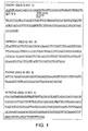
Fig. 1 shows the genomic nucleotide sequence encoding the
mouse SGRF gene.
Fig. 2 shows the continuation of the genomic nucleotide
sequence encoding the mouse SGRF gene in Fig.1.
Fig. 3 depicts a photograph and a drawing, showing SGRF
mRNA expression analysis in mouse tissue and the construction of
SGRF/IL-23
-/- mice.
Fig. 4 shows graphs of antigen-induced cytokine production
and proliferation by lymph nodes cells collected from SGRF/IL-23-
/- and wild type mice. Groups of five mice (male, ten weeks old),
were each immunized with KLH (200 µg) in CFA. Lymph nodes were
harvested five days later, cultured for 48 hours in 300 µg/ml
KLH, and then analyzed for their ability to produce IFN-γ (a),
IL-4 (b), IL-5 (c), or IL-10 (d), or to proliferate (e).
ConcanavalinA (ConA, 2 µg/ml) was used as the control for nonspecific
proliferation. Data are presented as the mean ± SD
values derived from the five mice in each group.
†, P < 0.003; ‡, p< 0.05; and §, p< 0.03 (comparison between
SGRF/IL-23-/- and wild type mice by unpaired t-test).
Fig. 5 depicts graphs representing susceptibility to L.
monocytogenes infection, and DTH reactions, in wild type and
SGRF/IL-23
-/- mice.
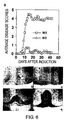
Fig. 6 depicts a graph and a photograph representing the
result of EAE induction in wild type and SGRF/IL-23
-/- mice.
Mice (female, eight weeks old) were immunized with MOG peptide
35-55 in CFA, and subcutaneously injected with pertussis toxin
according to the protocol for EAE induction.
Best Mode for Carrying out the Invention
The present invention will be described below in detail
with reference to Examples, but it is not to be construed as
being limited thereto.
[Example 1] Expression analysis of SGRF mRNA
Expression analysis of SGRF mRNA in mouse tissue was
performed by Northern blotting using SGRF cDNA as a probe.
High expression of mouse SGRF mRNA was confirmed in the thymus,
and expression was detected in the brain and testis (Fig.3a).
RT-PCR analysis also detected mRNA expression in the spleen and
lymph nodes (data not shown).
[Example 2] Construction of the mice
Cloning of the SGRF gene was performed according to the
method described in Patent EP1072610. The mouse SGRF genome was
screened by Genome Systems, Inc. (now Incyte Genomics, Ins. St.
Louis, USA). Two BAC (Bacteria Artificial Chromosome) clones
were obtained. The Clone Addresses and GS Control Numbers
identified by Genome System, Inc. are as follows:
| Clone Address | GS Control Number |
| 225(L12) | 24057 |
| 198(A03) | 24058 |
A large number of BAC clones were produced by known
methods, and BAG DNAs were prepared using a Large-Construction
Kit (QIAGEN).
Restriction digest mapping suggested that these BAG clones
were identical to each other. However, a ~13 Kbp Hind III
fragment was produced by digesting the GS Control Number 24057
clone, and a ~9 Kbp Bgl II fragment was produced by digesting
the GS Control Number 24058 clone. The ~13 Kbp fragment was
subcloned into pBluescript vector (STRATAGENE) at the Hind III
site, and the ~9 Kbp fragment was cloned into a pGEM-T-easy
vector (Promega) at the EcoRV site (converted to Bgl II).
Vectors carrying each DNA fragment were referred to as pBSK-mSGRF
(Hind III) and pGEM-mSGRF (Bgl II) respectively. The
genomic sequence encoding the mouse SGRF (mSGRF) gene was
determined using sequence analysis and is shown in Fig. 1 and 2,
and in SEQ ID NOs: 1 to 7 (note this sequence is that of 129/SV
JII mice).
The above-mentioned sequence data results revealed that
both the mouse SGRF (mSGRF) gene and the human SGRF (hSGRF) gene
(GenBank Accession No. AB030001) comprise four exons. Therefore,
a genetically modified mouse of the present invention can be
constructed as follows: A targeting vector is constructed by
replacing the SGRF exon region with an appropriate drug marker
gene. The targeting vector is then transfected into mouse ES
cell strains using electroporation or the like, and cell strains
that undergo homologous recombination are selected.
Specifically, this was performed as follows:
Forward primer:
FmSGREX2: 5'-GTC GAC TGC AGT CTA GAC TAA GCC GAT GTT GAT GTG TC-3'
(SEQ ID NO: 10)
Reverse primer:
RSGR-A: 5'-GCT GTC TGG AGT ACT GTG CAT CTG C-3' (SEQ ID NO 11)
In this case, a knockout vector was constructed using a
~ 0.9 Kbp fragment (produced by digestion at Sal I and Xho I
sites in the FmSGRFEX2 primer and in Exon 4 of the SGRF gene),
and the above-described ~9 Kbp fragment of the mSGRF promoter
region inserted into pGEM-mSGRF (Bgl II). Cell strains which
did not undergo homologous recombination were eliminated by
inserting a drug resistance gene (neo) between these fragments,
and by adding a diphtheria toxin A fragment (DT-A).
In this Example, as described above, the SGRF gene was
inactivated by replacing Exon 1 and 2 of the SGRF gene with a
drug resistance gene (neo) (Fig. 3 (A)). Since this drug
resistance gene (neo) would probably influence the expression of
neighboring genes, a loxP sequence (5'-ATA ACT TCG TAT AGC ATA
CAT TAT ACG AAG TTA T-3' (SEQ ID NO: 12)) was inserted at either
side of the neo sequence. Thus, the neo can be deleted using a
Cre protein, if a known or unknown gene be located near the SGRF
gene. The targeting vector was digested at a single site using
a restriction enzyme (Not I), and then transfected into ES cells
by electroporation.
Homologous recombinants were constructed by transfecting
AB2.2 cells (ES cells derived from 129SvEv mice) according to
the manual attached to The Mouse Kit (Lexicon Genetics Inc.),
commercially available from TAKARA. Homologous recombinants
were selected by G418. PCR was used to screen for recombinants.
ES cells used for screening were cultured in a 96-well plate.
Each well was washed twice with 200 µl PBS and treated with cell
lysis buffer (5 µl 10x LA buffer (use for TAKARA LA Taq), 5 µl 5%
NP-40, 4 µl proteinase K (TAKARA, 20 mg/l), 35 µl distilled
water) for two hours at 55 degrees Celsius. These cells were
incubated at 95 degrees C for 15 minutes to inactivate
proteinase K, and then used for PCR samples. The PCR reaction
mixture contained 1 µl of an above-mentioned PCR sample, 5 µl 10x
LA buffer, 5 µl MgCl2 (25 mM) , 8 µl dNTP (2.5 mM) , 0.4 µl each
primers (50 µM each), 0.5 µl LA taq (TAKARA), and 29.7 µl
distilled water (total 50 µl). PCR was performed under the
following condition: preheating at 95 degrees C for one minute;
35 cycles of amplification at 95 degrees C for 30 seconds, 64
degrees C for 30 seconds, and 72 degrees C for 80 seconds; and
heat extension at 72 degrees C for seven minutes.
The primers were as below. A ~1.3 Kbp band was amplified
in ES cell samples in which homologous recombination occurred.
Primer Fneo-1 was in the drug resistance gene in the knockout
vector, and primer RSFR-A was in Exon 4 of the SGRF gene, which
was located outside of the knockout vector.
Reverse primer:
RSGR-A: 5'-GCT GTC TGG AGT ACT GTG CAT CTG C-3' (SEQ ID NO: 13)
Forward primer:
FNeo-1: 5'-CCG TGA TAT TGC TGA AGA GCT TGG C-3' (SEQ ID NO: 14)
420 and 760 ES cell clones were analyzed in the two
experiments. Of these, two and six clones respectively were
homologous recombinants. Thus the recombination efficiencies
were 2/420 (0.467%) and 6/760 (0.79%), and the combined total
recombination efficiency for both experiments was 0.68%.
Chimeric mice were obtained by injecting seven ES cell
strains, in which one SGRF gene allele was inactivated, into
blastocysts derived from C57BL/6J mice. Mice comprising cells
in which one SGRF gene allele was inactivated (hereafter
referred to as "SGRF/IL-23+/- mice") were obtained by mating
C57BL/6 mice with the above-described chimeric mice. SGRF/IL-23+/-
mice were obtained in four strains, clone numbers 11, 23,
75, and 86, of the seven cell strains used for the experiment.
Mice derived from clone 11 and clone 86 were used for further
studies. Mice in which both SGRF gene alleles were inactivated
(hereafter referred to as "SGRF/IL-23 -/- mice") were obtained by
mating the SGRF/IL-23+/- mice with each other. Southern blot
analysis was used to confirm the insertion of the mutant gene
into one or both SGRF gene alleles in mice thus obtained (Fig. 3
(c)).
Loss of gene expression was confirmed by a reverse
transcriptase polymerase chain reaction (RT-PCR) using thymus-derived
polyA+ RNA (Fig. 3 (d)). Both heterologous and
homologous SGRF/IL-23 mice showed normal growth and appearance,
and were able to reproduce.
[Example 3] Analysis of SGRF gene-deleted mice
(1) Cytokine production in SGRF/IL-23-/- mice immunized with KLH
The ability of lymph node cells, obtained from keyhole
limpet haemocyanin (KLH)-immunized wild type mice (normal mice,
SGRF/IL-23+/+) and SGRF/IL-23-/- mice, to produce cytokines and to
proliferate was examined in order to investigate the involvement
of SGRF/IL-23 in antigen-induced cytokine production and ability
to proliferate T cells.
To induce a Th1 response, mice were immunized
subcutaneously at the base of their tail using 200 µg KLH in PBS,
in a 1:1 emulsion with complete Freund's adjuvant (CFA),
containing 5 mg/ml Mycobacterium tuberculosis strain H37Ra,
(Difco Laboratories). After five days, inguinal lymph nodes
were removed and the cells were cultured in RPMI1640
supplemented with 10% FCS and 0.3 mg/ml of KLH. To measure
cytokine production, lymph node cells were cultured in 1ml of
medium in 24-well plates until the cells proliferated to 6 x 10⁶
cells/ml. After culturing for 48 hours, culture supernatants
were harvested and cytokine levels were determined using enzyme-linked
immunosorbent assay (ELISA) kits, obtained from R&D
Systems, according to the manufacturer's instructions.
Furthermore, radiolabel incorporation into DNA was measured by
MicroBeta scintillation counting (Perkin Elmer) as follows:
After 48 hours of incubation, 0.25 µCi of [3H]thymidine (Amersham
Pharmacia) was added and incubated for four hours in 96-well
flat-bottomed microplates in which there were 6 x 10⁶ cells/ml in
a total volume of 200 µl. Then, the degree of proliferation was
measured.
IFN-γ production by SGRF/IL-23-deficient lymphocytes was
significantly impaired compared to the production by wild-type
lymphocytes (Fig. 4a), whereas KLH-induced IL-4 and IL-5
production was markedly enhanced (Fig. 4b,c). In contrast, both
IL-10 production and KLH-induced proliferation were similar for
wild type and SGRF/IL-23-deficient lymphocytes (Fig. 4d,e).
These data indicate that in SGRF/IL-23-/- mice, production
of Th1-type cytokines is reduced, and production of Th2-type
cytokines is increased.
(2) Defending the body against pathogenic microbes and DTH
Th1-type responses are important in defenses against
intracellular pathogens such as Listeria, and Th1-type cytokines
such as IFN-γ and TNF-α are essential for this defense (Kaufmann
S H. Immunity to intracellular bacteria. Annu. Rev Immunol 11,
129-163 (1993)).
The survival rate of SGRF/IL-23-/- mice after Listeria
infection, and the number of Listeria present in the liver four
days after Listeria infection, were investigated to examine the
involvement of SGRF/IL-23 in biophylaxis against pathogenic
microbe infection.
Specifically, Listeria (serum type 4b) (2.3x 10⁶
CFU/mouse) were injected intravenously (Suzuki H. et al., A role
for macrophage scavenger receptors in
atherosclerosis and susceptibility to infection. Nature 386,
292-296 (1997)) into either SGRF genotypic mice (male, ten weeks
old) or wild type mice (male, ten weeks old) and the post-infection
survival rates of the mice were examined everyday for
15 days. The results indicated the survival rate of SGRF/IL-23-
/- mice was lower than that of wild type mice (Fig. 5a).
Listeria (3.1x10⁶ CFU/mouse) was also injected into mice
in order to examine post-infection Listeria proliferation. Four
days after infection, and under sterile conditions, the mice
livers were removed and homogenized in sterile PBS. The
homogenized tissues were diluted stepwise with PBS, plated on
brain-heart infusion agar (Difco), and incubated at 37 degrees C
for 16 hours. CFUs were then counted. L. monocytogenes
proliferation in the livers of SGRF/IL-23-/- mice four days after
injection was significantly increased compared with that of
wild-type mice (Fig. 5).
These results suggest that SGRF/IL-23-/- mice, compared
with wild type mice, are susceptible (less resistant) to L.
monocytogenes. Thus, these results suggest that SGRF/IL-23, as
well as IL-12, contributes to the elimination of L.
monocytogenes.
To further examine the role of SGRF/IL-23 on Th1-type
immune responses, the present inventors investigated delayed-type
hypersensitivity (DTH), which is the typical in vivo
manifestation of cell-mediated immunity.
Delayed-type hypersensitivity was induced by using a well
known method (Immunity, Vol.4, p471 (1996)). Specifically, mice
were immunized with methyl BSA (SIGMA) by intradermal injection
of a 50 µl emulsion of 2.5 mg/ml mBSA in CFA (Beckton Dickinson)
at two sites on the abdomen. Five days after immunization, DTH
was induced in the mice by subcutaneous injection of 30 µl of
5.0 mg/ml mBSA into one rear footpad. A comparable volume of
PBS was injected into the other rear footpad as a control.
Measurements of footpad swelling were taken using a micrometer
(Ozaki Inc.) at 24, 48 and 72 hours after injection. Footpad
swelling following methyl BSA injection was measured by
comparison with that caused by PBS injection, and average values
for each group were obtained (n=6).
The results showed that for three days after injection,
specific footpad swelling was suppressed in the SGRF/IL-23-/-
mice (Fig. 5c).
These results indicate that SGRF/IL-23 plays an important
role in Th1-type responses. Therefore, it is predicted that
SGRF is a factor necessary for the defense against infections
caused by pathogenic microbes.
(3) Involvement in autoimmune disease development
The involvement of SGRF/IL-23 in autoimmune diseases such
as multiple sclerosis, chronic rheumatoid arthritis, and
autoimmune colitis can be investigated by inducing disease
models in the genetically modified mice of the present invention
according to known immunological methods, and then making
comparisons with normal mice.
Experimental autoimmune encephalomyelitis (EAE) is a
central nervous system (CNS) autoimmune disease mediated by Th1
responses, and is known as a model for human multiple sclerosis.
The following experiment was performed to investigate the role
of SGRF/IL-23 in autoimmune disease.
EAE was induced in SGRF/IL-23-/- mice and wild type mice by
well known methods (J. Exp. Med. Vol. 186 p1233, 1997).
Specifically, 300 µg of MOG peptide (MEV GWY RSP FSR VVH LYR
NGK/ SEQ ID NO: 15; Sawady Technology) and 0.2 ml of CFA
(including 500 µg of Mycobacterium tuberculosis strain H37Ra
(Difco Laboratories)) was injected subcutaneously into one side
of the mice abdomens on day 0 and day 7. 200 ng of Pertussis
toxin (List Biological Laboratories) was injected into the mice
veins on day 0, and administrated intraperitoneally two days
after the first immunization.
After inducing EAE in the mice, pathology scoring was
conducted according to a known method, and using a scale from 1
to 5: 1, normal; 2, wobbly gait; 3, hind limb paralysis; 4, hind
and fore limb paralysis; 5, death. Each mouse was scored and
average values were collected. The results indicated that
SGRF/IL-23-/- mice showed significant resistance to MOG-induced
EAE compared to wild type mice. Six SGRF/IL-23-/- mice and six
wild type mice were used for the experiment in Fig. 6. Although
EAE developed in two of the six SGRF/IL-23-/- mice, their
pathology scores were low. On the other hand, all wild type
mice developed severe EAE, and four of the six wild type mice
died.
Histological analysis was performed on EAE-induced
SGRF/IL-23-/- mice and wild type mice. Spinal cords from EAE-induced
mice were removed and fixed with 20% formalin neutral
buffered solution (Wako) in order to conduct histological
analysis. Paraffin sections were prepared and stained using
hematoxylin/eosin and Kluver-Barrera staining in the Sapporo
General Pathology Co. Ltd.
Histological analysis of the spinal cords of wild-type
mice injected with MOG peptide showed typical lymphocyte
infiltration (Fig. 6b) and demyelination (Fig. 6d). In contrast,
no histological change was observed in the spinal cords of
SGRF/IL-23-/- mice (Fig. 6c, e).
These results indicate that SGRF/IL-23-/- is an important
component in the development of EAE, and that SGRF could be a
new target for multiple sclerosis (MS) therapy in humans.
(4) Involvement in the bone metabolism
The well known OVX model, in which ovaries are removed
from a female mouse, can be used to determine whether SGRF is
involved in the bone metabolism.
Industrial Application
The present invention provides genetically modified
animals in which both SGRF gene alleles are inactivated and SGRF
activity has been eliminated. The genetically modified animals
and ES cells of the present invention can be used to predict the
side effects of SGRF inhibitors such as anti-SGRF antibodies or
SGRF antagonists. The genetically modified animals and ES cells
of the present invention can also be used to determine whether a
test protein or small molecule comprises the function of
substituting for SGRF function, or whether it can be used to
screen for DNA that encodes a protein comprising the function of
substituting for SGRF function.
Furthermore, since the genetically modified mice (SGRF KO
mouse) are susceptible to infection by the pathogenic microbe
Listeria, SGRF or alternative substances involved in SGRF
signaling are considered to be therapeutic agents against
pathogenic microbes.
Moreover, experimental autoimmune encephalomyelitis is not
easily induced in SGRF KO mice and delayed-hypersensitivity is
reduced. Therefore, antagonists that inhibit SGRF function are
considered to be strong candidates for therapeutic agents for
autoimmune or inflammatory diseases.