EP1445333A2 - Screening of a novel hepatic syndrome and its uses - Google Patents
Screening of a novel hepatic syndrome and its uses Download PDFInfo
- Publication number
- EP1445333A2 EP1445333A2 EP04290341A EP04290341A EP1445333A2 EP 1445333 A2 EP1445333 A2 EP 1445333A2 EP 04290341 A EP04290341 A EP 04290341A EP 04290341 A EP04290341 A EP 04290341A EP 1445333 A2 EP1445333 A2 EP 1445333A2
- Authority
- EP
- European Patent Office
- Prior art keywords
- mutation
- exon
- intrahepatic
- nucleotide
- seq
- Prior art date
- Legal status (The legal status is an assumption and is not a legal conclusion. Google has not performed a legal analysis and makes no representation as to the accuracy of the status listed.)
- Withdrawn
Links
Images


Classifications
-
- C—CHEMISTRY; METALLURGY
- C12—BIOCHEMISTRY; BEER; SPIRITS; WINE; VINEGAR; MICROBIOLOGY; ENZYMOLOGY; MUTATION OR GENETIC ENGINEERING
- C12Q—MEASURING OR TESTING PROCESSES INVOLVING ENZYMES, NUCLEIC ACIDS OR MICROORGANISMS; COMPOSITIONS OR TEST PAPERS THEREFOR; PROCESSES OF PREPARING SUCH COMPOSITIONS; CONDITION-RESPONSIVE CONTROL IN MICROBIOLOGICAL OR ENZYMOLOGICAL PROCESSES
- C12Q1/00—Measuring or testing processes involving enzymes, nucleic acids or microorganisms; Compositions therefor; Processes of preparing such compositions
- C12Q1/68—Measuring or testing processes involving enzymes, nucleic acids or microorganisms; Compositions therefor; Processes of preparing such compositions involving nucleic acids
- C12Q1/6876—Nucleic acid products used in the analysis of nucleic acids, e.g. primers or probes
- C12Q1/6883—Nucleic acid products used in the analysis of nucleic acids, e.g. primers or probes for diseases caused by alterations of genetic material
-
- C—CHEMISTRY; METALLURGY
- C12—BIOCHEMISTRY; BEER; SPIRITS; WINE; VINEGAR; MICROBIOLOGY; ENZYMOLOGY; MUTATION OR GENETIC ENGINEERING
- C12Q—MEASURING OR TESTING PROCESSES INVOLVING ENZYMES, NUCLEIC ACIDS OR MICROORGANISMS; COMPOSITIONS OR TEST PAPERS THEREFOR; PROCESSES OF PREPARING SUCH COMPOSITIONS; CONDITION-RESPONSIVE CONTROL IN MICROBIOLOGICAL OR ENZYMOLOGICAL PROCESSES
- C12Q2600/00—Oligonucleotides characterized by their use
- C12Q2600/156—Polymorphic or mutational markers
Definitions
- the present invention relates to the screening of a hepatic syndrome occurring in young adults and associating (i) intrahepatic hyperechoic foci with or without intrahepatic sludge or microlithiasis and further on a cholesterol microcholelithiasis, intrahepatic cholestasis and (ii) one or more mutations of the MDR3 gene (point mutations and/or SNPs).
- Cholesterol cholelithiasis is characterized by the presence of calculi of cholesterol or of calcium bilirubinate. Cholesterol lithiasis is the most common form of cholelithiasis (80%). It is characterized by the formation of calculi of cholesterol in the gall bladder, due to an excess of bile cholesterol compared to solubilizing molecules, namely bile acids and phospholipids. Its prevalence is estimated, taking all ages into account, to be 10 to 20% of the population in industrialized countries. It is more prevalent in women and increases considerably with age (40% in women over the age of 60).
- Gall bladder lithiasis can be a source of complications: cholecystitis, cancer of the gall bladder, migration of calculi in the common bile duct, a cause of angiocholitis and of pancreatitis.
- Treatment for this disease is based mainly on surgery: cholecystectomy by laparotomy or celioscopy, biliary drainage with peroperative or perendoscopic extraction of the common bile duct calculi.
- Medical treatment which consists of administering chenodeoxycholic acid and/or ursodeoxycholic acid, is reserved for forms of non-complicated cholesterol cholelithiasis of the gall bladder.
- Chronic biliary cholestasis is characterized by a deficiency in passage of the bile from the liver to the extrahepatic bile ducts and the intestine.
- the causes and physiopathology are varied (Poupon et al., 2000, J. Hepatol., 32, 129-140).
- Most chronic biliary cholestasis can progress to biliary cirrhosis and hepatocellular insufficiency requiring a liver transplant.
- cholestasis is observed in biliary atresia, in Alagille syndrome or in progressive familial intrahepatic cholestasis (PFIC).
- PFIC progressive familial intrahepatic cholestasis
- Familial intrahepatic cholestasis are infantile recessive diseases which are characterized by intermittent jaundice, severe cholestasis and cirrhosis, with a fatal outcome during the first ten years of life.
- the PFIC1 locus was found in chromosome 18q21-q22; five mutations were identified: a deletion, a deleted exon and three missense mutations; these missense mutations concern domains which are highly conserved in type V ATPases (E. Jacquemin et al., J. Hepatol., 1999, 31, 377-381).
- Familial cholestasis type 2 exhibits the same clinical signs as in Byler's disease, but it is caused by mutations in the FIC2 gene, which encodes the BSEP (Bile Salt Export Pump) transporter. This gene is the homolog of the murine SPGP gene, the product of which, expressed exclusively in hepatocytes, is involved in bile salt transport.
- PFIC type 2 is characterized by a severe pruritis, normal ⁇ -GT activity and a normal cholesterol level in the serum, high concentrations of bile acids in the serum and very low levels of bile acids in the bile.
- the PIFC2 locus was found in chromosome 2q24: 10 mutations were identified, including deletions and missense mutations involving important domains of the BSEP transporter (E. Jacquemin et al., J. Hepatol., 1999, 31, 377-381).
- cholestasis type 3 (PFIC3) have mutations in the MDR3 gene.
- PFIC type 3 differs from the other PFICs in particular by the fact that it has a high serum ⁇ -glutamyl transferase activity.
- the MDR (Multi Drug Resistance) family of ABC transporters is a multigene family of homologous proteins, some of which (MDR3) are not involved in drug resistance, and are phosphatidylcholine transporters.
- MDR3 gene revealed in 1993, is a phospholipid translocator. It is expressed in hepatocytes and provides phosphatidylcholine translocation, thus leading to secretion of this phospholipid into the bile.
- point mutations may help to provide a test for screening a new syndrome defined by a decrease in the concentration of bile phospholipids and the presence of cholesterol crystals sensitive to treatment with UDCA, combined with a point mutation of the MDR3 gene, they have now find that in view of a more accurate screening of said syndrome, it is preferable to detect further on other point mutations which may be found in other positions than the ones disclosed in WO 02/12556 and also to detect single-nucleotide polymorphisms (SNPs), which may also be found in this syndrome, with an abnormal frequency.
- SNPs single-nucleotide polymorphisms
- the onset of this syndrome is related to the appearance, before microlithiasis, of intrahepatic hyperechoic foci with or without intrahepatic sludge.
- microcalculi which can be detected through examination of the liver by ultrasonography, exhibit the following characteristics:
- LPAC Low Phospholipid Associated Cholelithiasis
- a subject-matter of the present invention is therefore a method for the screening of a hepatic syndrome comprising at least the detection, from a sample of nucleic acid extracted from peripheral blood mononuclear cells, of at least one heterozygous mutation of the MDR3 gene and/or of a homozygous mutation which does not destroy the expression of the protein expressed by said gene with phosphatidylcholine transporter activity, in adult individuals associating cholesterol microcholelithiasis and intrahepatic cholestasis, characterized in that said MDR3 gene mutation is located in at least one of the following exons: 4, 5, 6, 8, 9, 10, 14, 15, 16, 17, 18, 19, 23 and/or 26 at the following nucleotide positions:
- the expression "mutation which does not destroy the expression of the protein having phosphatidylcholine transporter activity” is intended to mean both mutations (point mutation and/or SNPs with abnormal frequency) which lead to the expression of a protein having residual phosphatidyl transporter activity and mutations which decrease the level of expression of the normal protein (mutation in the promoter for example). These various mutations do not induce any hepatobiliary symptoms in children, but the appearance, in young adults, of symptoms associating cholesterol microcholelithiasis and intrahepatic cholestatis, induced by additional factors linked to the host and/or to the environment.
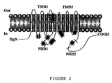
- mutations 9, 10, 14, 15, 17 or 18 are preferably located in exons 9, 10, 14, 15, 17 or 18; indeed, as it emerges from the obtained data, most mutations are localized in the central part of the molecule, close to nucleotide binding domain 1 (NBD1), or in adjacent transmembrane domains and intracellular loops (see figures 1 and 2). More precisely, 80% of mutations are situated in regions encoded by exons 9 to 18, which correspond approximately to 38% of the encoding region (TM5 and 6; 3 rd intracellular loop including NBD1; TM7 and 8).
- All the detected mutations are significantly related to LPAC, when, based on a multivariate analysis (see example 1), able to define a clinical score, said score being ⁇ 2 (1 point if age at the onset of symptoms was below 40 years, 1 point for recurrence after cholecystectomy and 1 point for the presence of intrahepatic spots).
- said method comprises the detection of a MDR3 gene mutation, summed up (in italic) in the following Tables I and II, which include the different possible mutations to be screened in LPAC.
- MDR3 gene point mutations in patient with LPAC syndrome Gene Position Location and nucleotide change Peptide change Protein domain Status 6 495T ⁇ A 523T ⁇ C Phe 165 Ile Thr 175 Ala 1 st intracellular loop between TMA-TM3 Hetero Hetero 9 902T ⁇ C 959C ⁇ T Met 301 Thr Ser 320 Phe TM5 Hetero Homo 10 1007-1015insT 1007-1015delT 355 stop 341 stop TM6 TM6 Hetero Hetero 12 1327insA 447 stop Close to NBD1 1 Hetero 14 1584G ⁇ C Glu 528 Asp Close to NBD1 1 Hetero 15 1772T ⁇ A Leu 591 Gln 3 rd intracellular loop Homo 17 1973G ⁇ A Try 6
- Said Table 11 indicates the most frequent allele to total number of allele ratio for each tested SNPs in the different groups of patients. Nucleotide changes in SNPs are underlined.
- SNPs single-nucleotide polymorphisms
- the method according to the invention comprises:
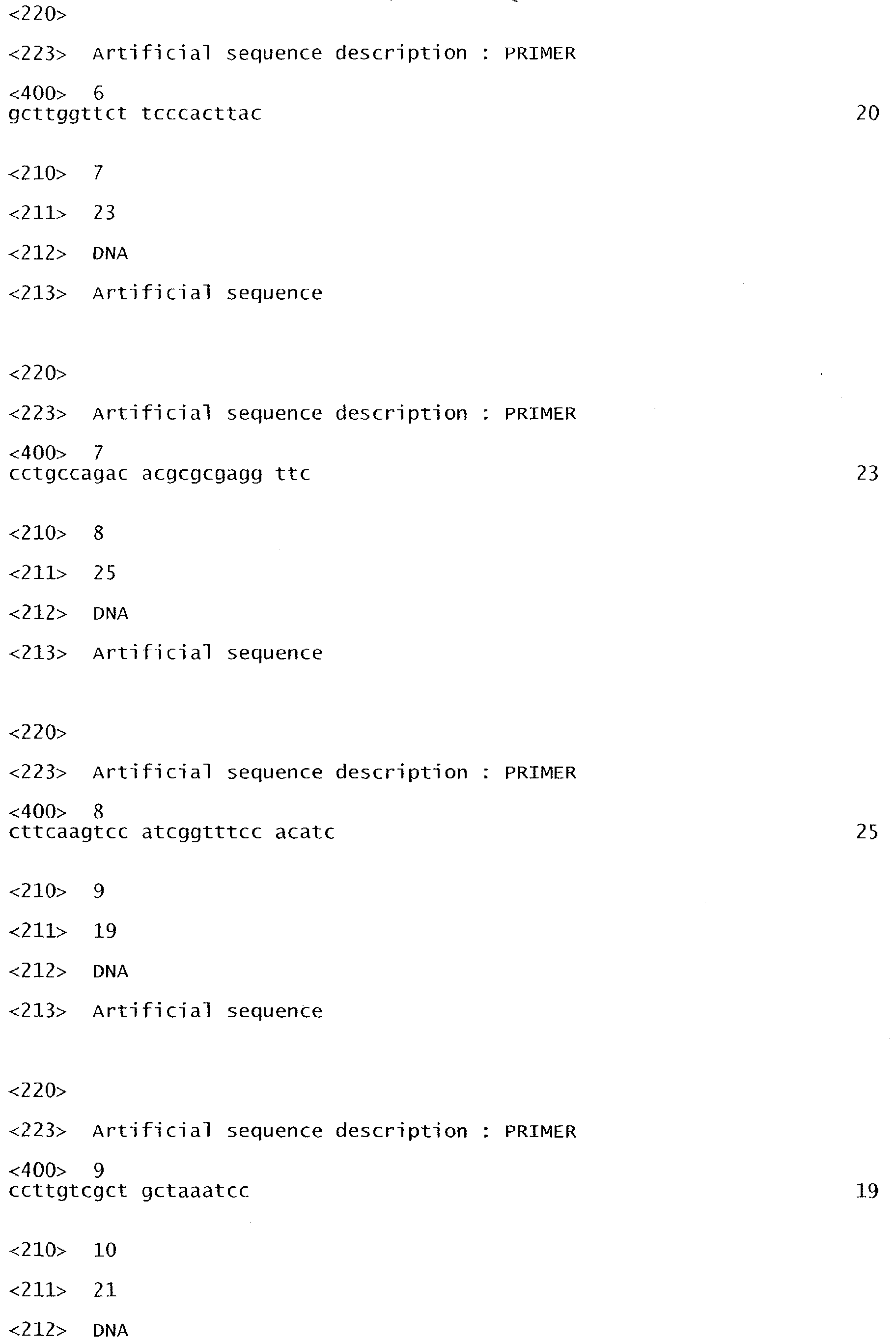
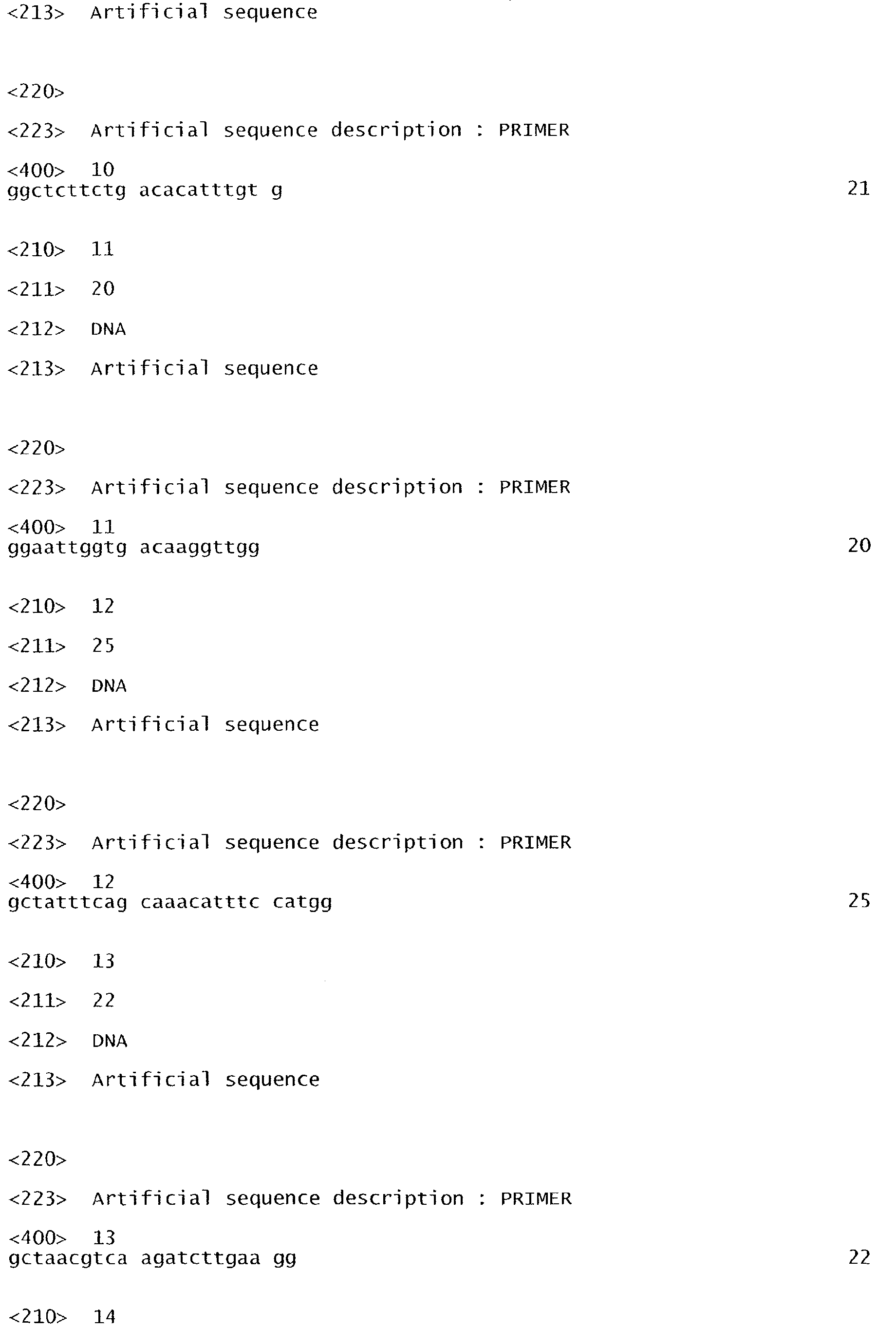
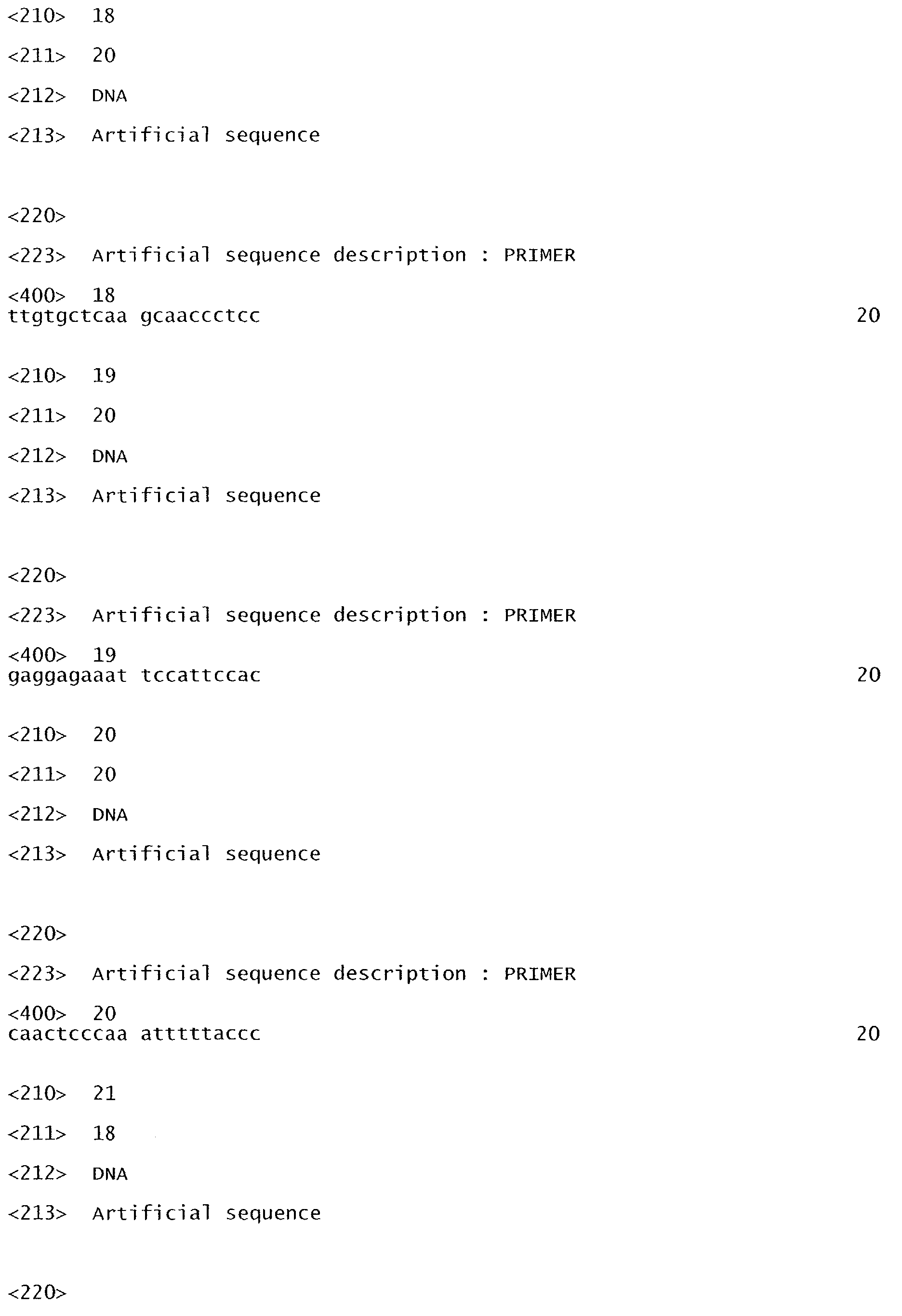
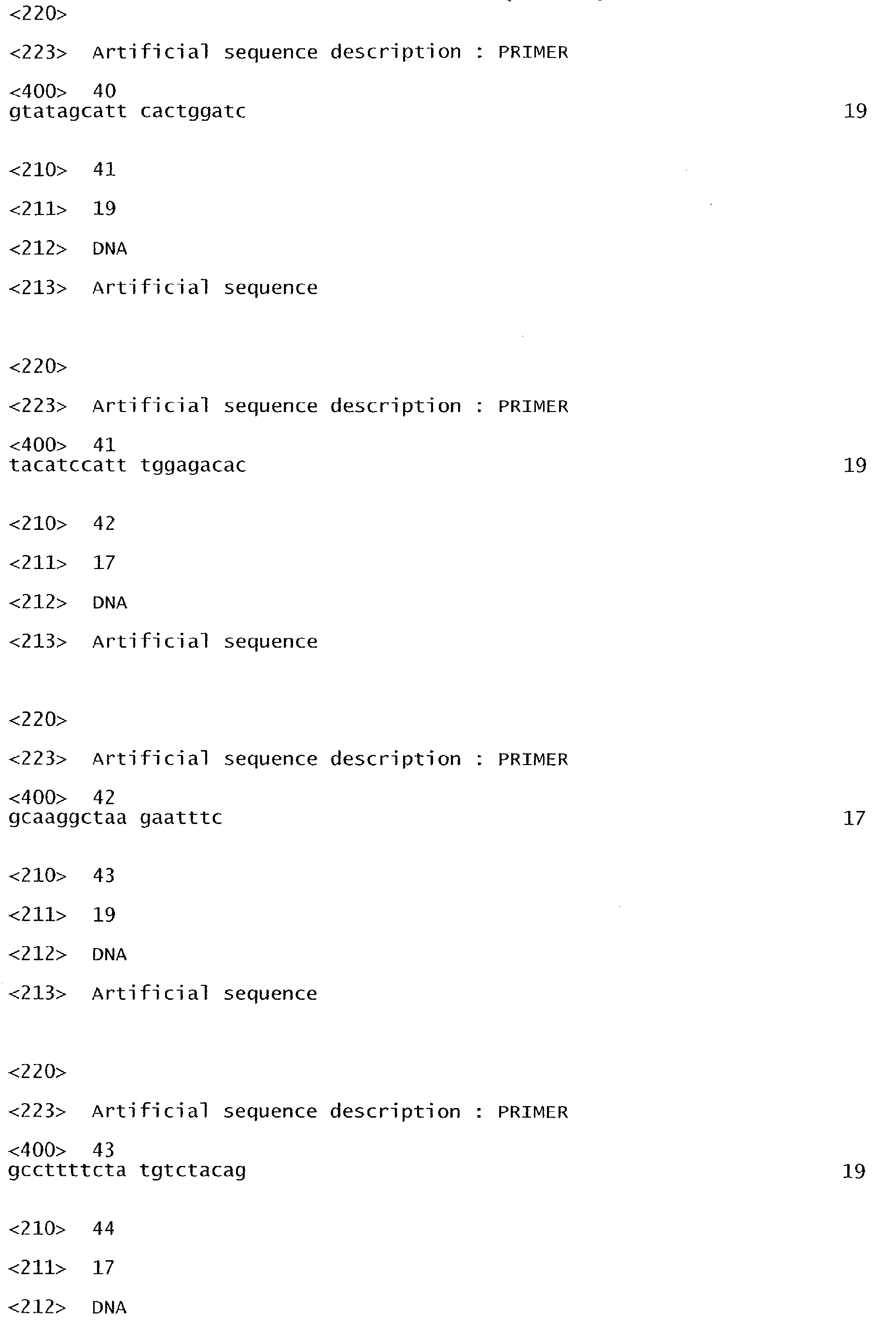
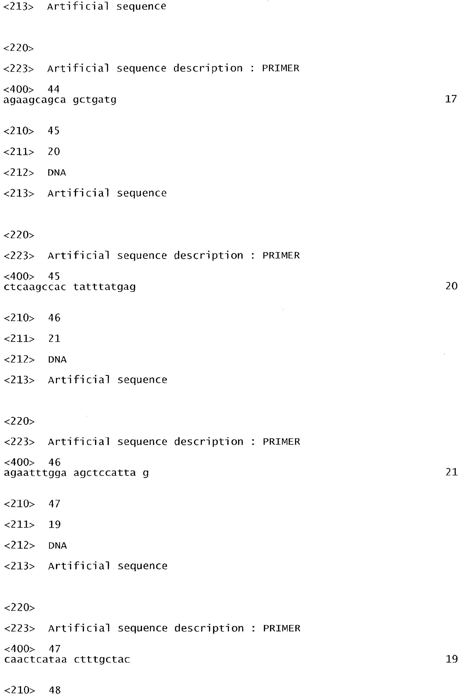
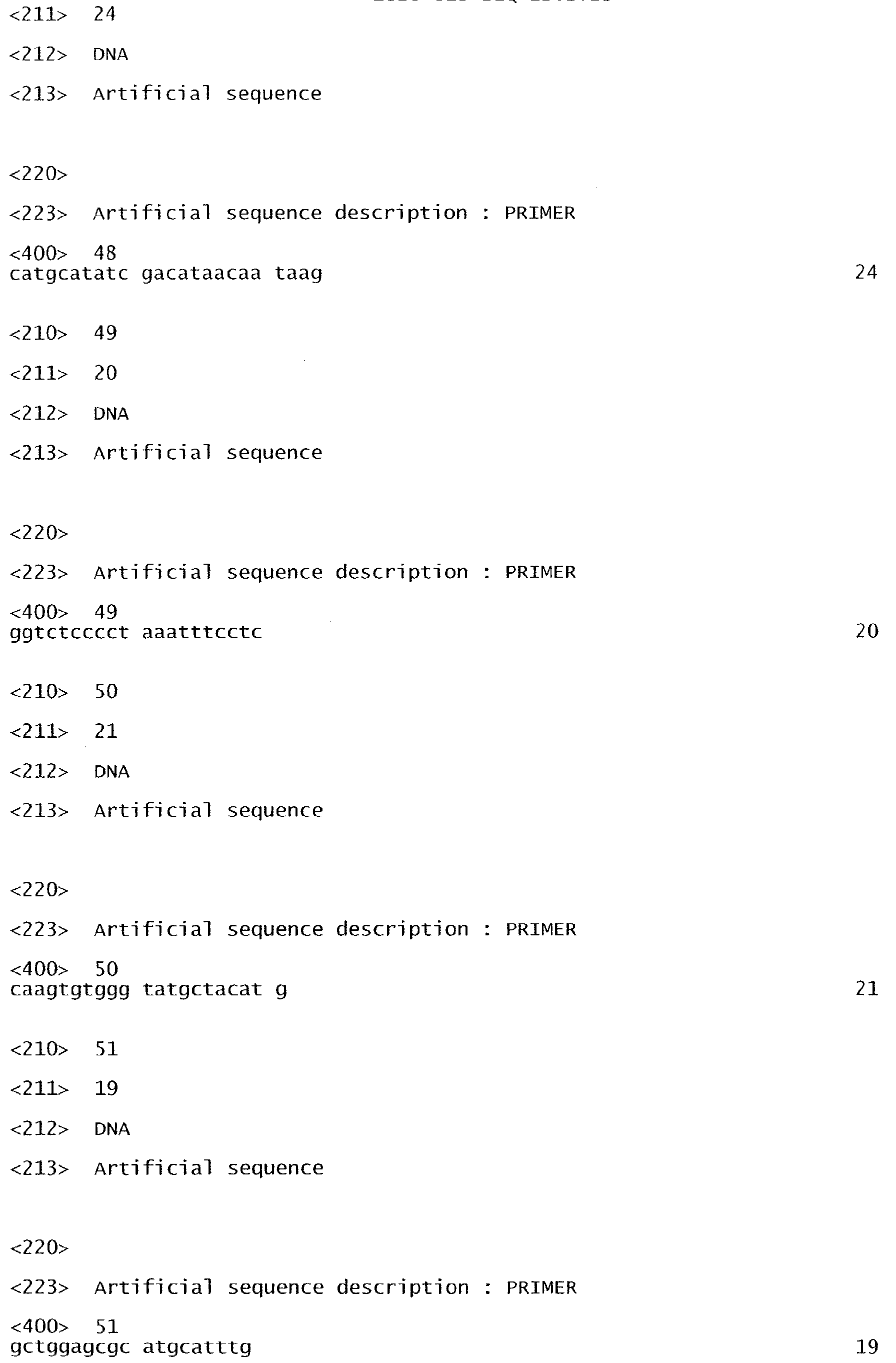
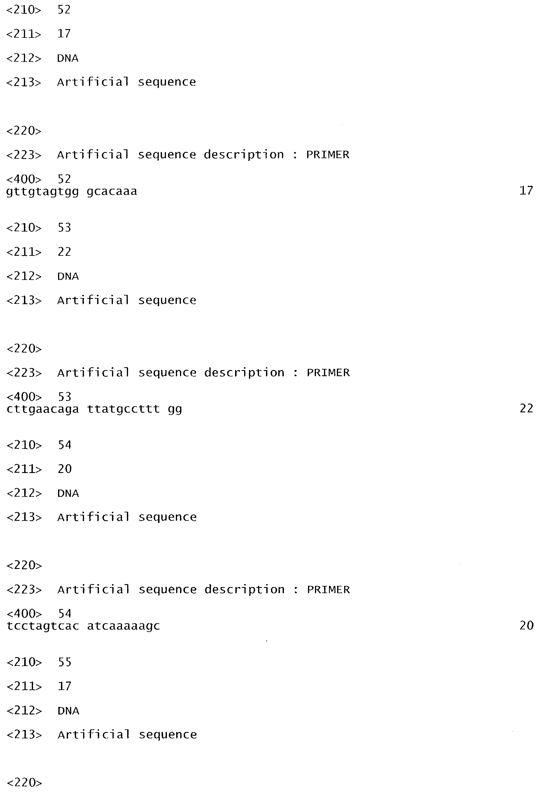
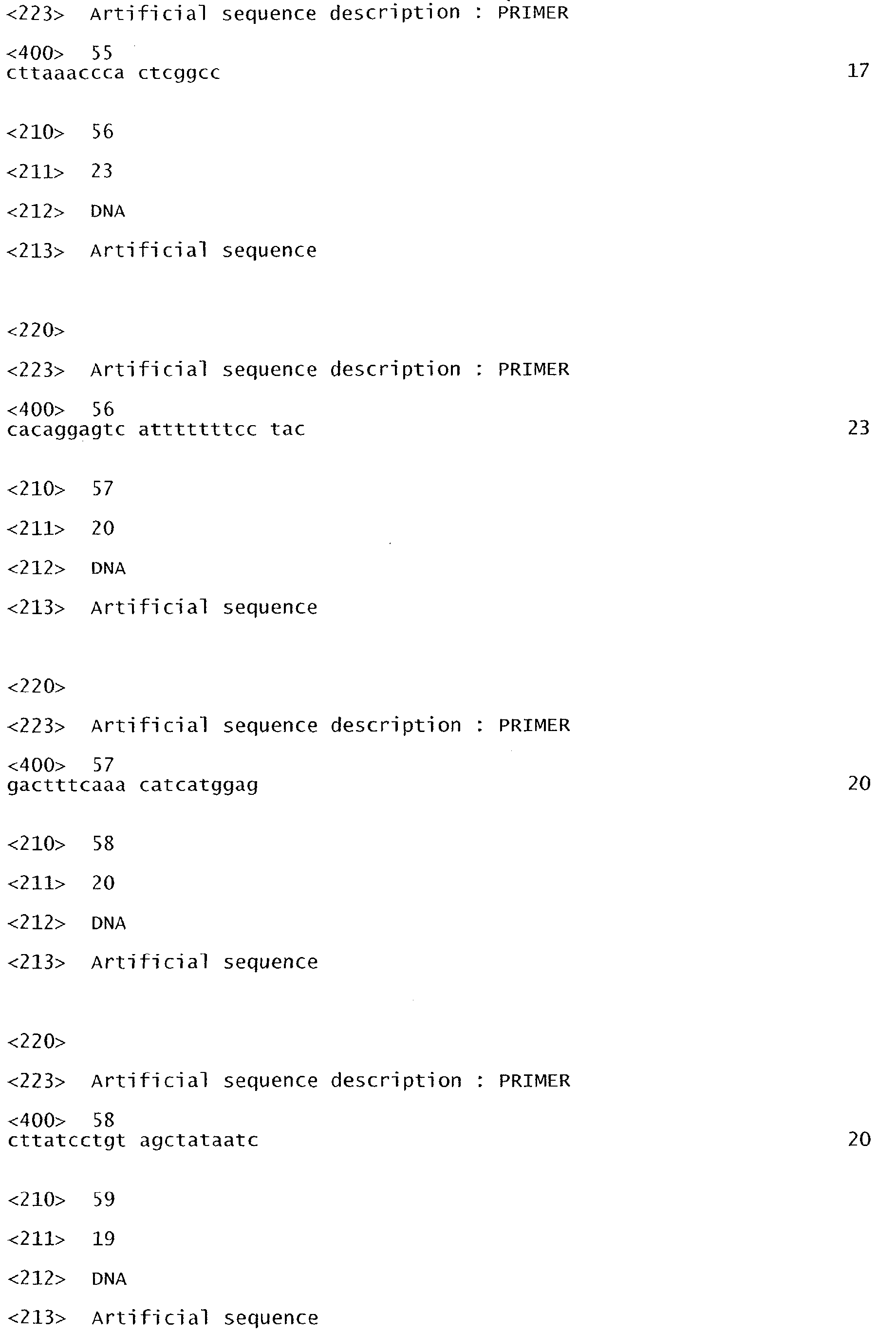
- Said primers may be used in combination with at least two primers described in WO 02/12556, listed as SEQ ID NO:1 to SEQ ID NO:13 in the attached sequence listing.
- said nucleic acid fragment on which the amplification is performed is selected from the group which consists in mRNA, cDNA and genomic DNA.
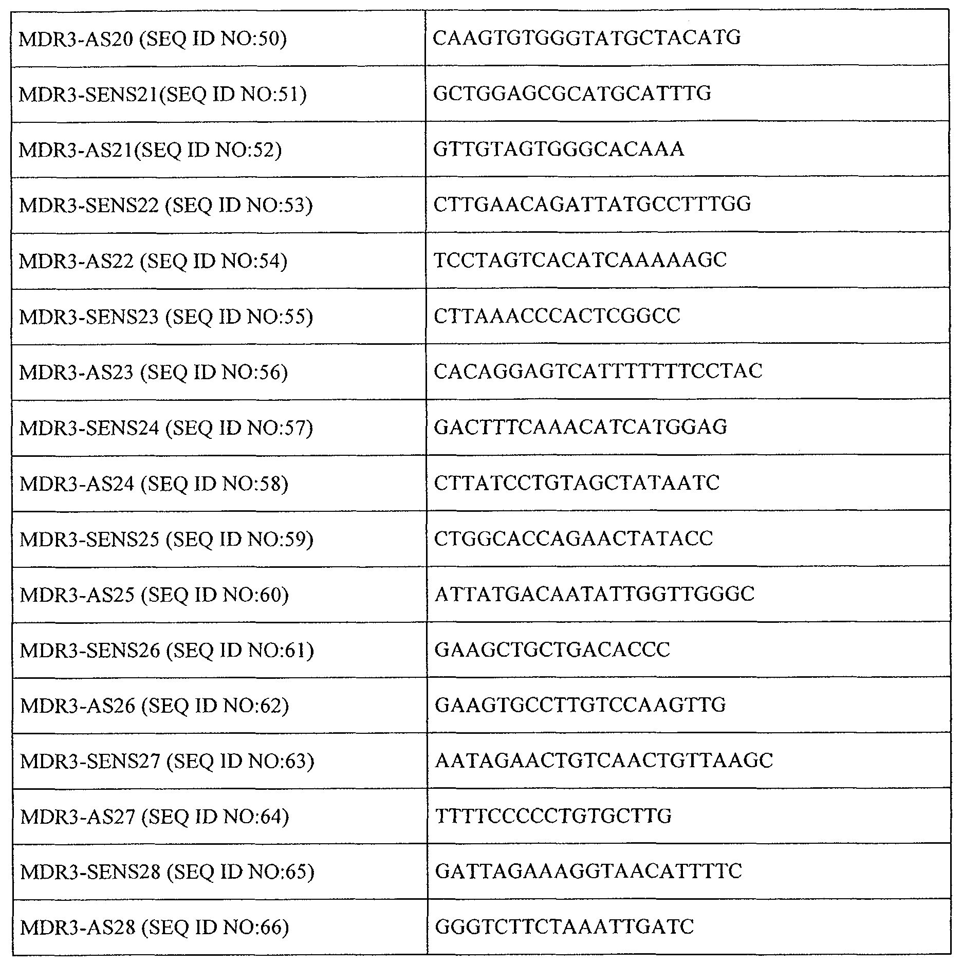
- the first amplification step consists of direct PCR amplification of at least one of exons 6, 9, 10, 14, 15, 17, 18, 19, 23, and 26 and further of at least one of exons 4, 5, 8 and/or 16 of the genomic DNA with the primers, located in the flanking intronic regions, as specified in the hereabove Table III (SEQ ID NO:15 to 66).
- the second step of detection of the presence of at least one of said mutations using the amplified fragment is carried out, as appropriate, using one of the following methods:
- the mutations are detected by digestion with the enzyme recognizing said restriction site, compared with a control which does not exhibit said mutation.
- the fragment amplified by PCR is sequenced according to conventional techniques; by way of example, mention may be made of the dideoxynucleotide technique.
- the mutation to be detected corresponds to the insertion or to the deletion of a small sequence
- said mutation can be detected using techniques of the PCR/PCR/LDR type, which are based on detecting the ligation of allele-specific primers using a thermostable ligase (Favis et al., Nature Biotech., 2000, 18, 561-564).
- Such a method may advantageously be used to evaluate the risk of appearance of a syndrome associating intrahepatic cholestasis and cholesterol microcholelithiasis in families at risk of cholesterol lithiasis and/or having unexplained hepatic biological abnormalities.
- said evaluation method can advantageously also comprise searching for intrahepatic microcrystals of cholesterol, measuring the cholesterol saturation index of the bile, determining the cholesterol/bile phospholipids ratio, assaying bile phospholipids, testing against ursodeoxycholic acid and analysing the clinical relation between said mutation and said hepatic abnormalities by establishing a clinical score based on multivariate analysis on the basis of the following parameters age, recurrence of cholecystectomy and intrahepatic spots.
- the diagnosis may be considered to be positive for the abovementioned syndrome when one of the abovementioned mutations is screened, associated with the presence of intrahepatic hyperechoic foci with or without intrahepatic sludge or microlithiasis and further on cholesterol microcholelithiasis, a very low level of bile phospholipids and a cholesterol saturation index of the bile of greater than 1.
- Another subject-matter of the present invention is also the use of the various primers as defined above, for the screening of a syndrome associating (i) intrahepatic hyperechoic foci with or without intrahepatic sludge or microlithiasis and further on cholesterol microcholelithiasis, intrahepatic cholestasis and (ii) at least one mutation of the MDR3 gene, as specified hereabove, optionally associated with at least one mutation as described in WO 02/12556.
- the invention includes the use of the primers selected from the group consisting of the sequences SEQ ID NO:15 to SEQ ID NO:66, optionally in combination with primers selected from the group consisting of the sequences the primers of SEQ ID NO:1 to SEQ ID NO:13 for the screening of a syndrome associating (i) intrahepatic hyperechoic foci with or without intrahepatic sludge or microlithiasis and further on cholesterol microcholelithiasis, intrahepatic cholestasis and (ii) at least one mutation of the MDR3 gene.
- Another subject-matter of the present invention is also a kit for the screening of a syndrome associating (i) intrahepatic hyperechoic foci with or without intrahepatic sludge or microlithiasis and further on cholesterol microcholelithiasis, intrahepatic cholestasis and (ii) at least one mutation of the MDR3 gene, characterized in that it comprises at least two primers as defined above, capable of detecting a mutation of the human MDR3 gene, as defined above.
- the invention includes a kit for the screening of a syndrome associating (i) intrahepatic hyperechoic foci with or without intrahepatic sludge or microlithiasis and further on cholesterol microcholelithiasis, intrahepatic cholestasis and (ii) at least one mutation of the MDR3 gene, characterized in that it comprises at least two primers selected from the group consisting of the sequences SEQ ID NO:15 to SEQ ID NO:66, optionally in combination with two primers selected from the group consisting of the sequences SEQ ID NO:1 to SEQ ID NO:13.
- Another subject-matter of the present invention is also a kit for evaluating the risk of appearance of a syndrome associating (i) intrahepatic hyperechoic foci with or without intrahepatic sludge or microlithiasis and further on cholesterol microcholelithiasis, intrahepatic cholestasis and (ii) at least one mutation of the MDR3 gene, in a population at risk, characterized in that it comprises, besides the primers capable of detecting the mutation of the MDR3 gene as defined above, reagents for assaying cholesterol and bile phospholipids.
- the invention includes a kit for evaluating the risk of appearance of a syndrome associating (i) intrahepatic hyperechoic foci with or without intrahepatic sludge or microlithiasis and further on cholesterol microcholelithiasis, intrahepatic cholestasis and (ii) at least one mutation of the MDR3 gene, in a population at risk, characterized in that it comprises, besides two primers selected from the group consisting of the sequences SEQ ID NO:15 to SEQ ID NO:66, optionally in combination with two primers selected from the group consisting of the sequences SEQ ID NO:1 to SEQ ID NO:13, reagents for assaying cholesterol and bile phospholipids.
- the reagents are enzymatic reagents.
- Example 1 Materials and methods
- biliary diseases such as primary biliary cirrhosis or primary sclerosing cholangitis
- fibrocystic liver disease Caroli's disease or congenital hepatic fibrosis
- Osler-Weber-Rendu disease cystic fibrosis
- protoporphyria total parenteral nutrition
- obesity BMI ⁇ 30 kg/m 2
- biliary infection infestation with parasites (including HIV infection) or malabsorption (including ileal resection) diagnosed on the basis of clinical examination and appropriate investigations and none received somatostatin synthetic analogs or hypo-cholesterolemic agent therapy.
- None of these patients had a medical history of progressive familial intrahepatic cholestasis during childhood and patients suffering from black pigment stones, hemolysis or cirrhosis were also excluded.
- the clinical feature characteristic of the syndrome were collected for the 60 patients with cholelithiasis as summed up in the following Table IV.
- Genomic DNA was obtained from peripheral white blood cells using standard procedures. Specific primers to amplify exons and splice junctions, using the Massachusetts Institute of Technology web site (http://www-genome.wi.mit.edu/cgi-bin/primer/primer3-www.cgi) were designed. The sequences of these intronic primers are those described in the here above Table III.
- Each PCR reaction contained 200 ng genomic DNA, with each primer at a concentration of 0.4 ⁇ M, 0.08 ⁇ M deoxynucleoside triphosphates (Pharmacia, Piscataway, N.J.), 1.5 mM magnesium chloride and 1.25 U Taq polymerase.
- PCR products were purified on sephadex column and sequenced using Big Dye Terminator Chemistry (Applied Biosystems). Identification and localization of MDR3 gene mutations and SNPs was assessed by sequence comparisons using Phred Phrap Consed Software or Seqscape software (version 1; Applied Biosystems).
- a clinical score indicative of the presence of a MDR3 mutation was defined as follows: 1 point if age at the onset of symptoms was less than 40 years, 1 point for recurrence after cholecystectomy and 1 point for the presence of intrahepatic spots.
- DCMs disease-causing mutations
- DCMs included three 1bp-insertions and one 1bp-deletion, resulting in a frameshift predicted to cause premature messenger RNA termination, a loss of protein function and ten single-nucleotide substitutions including one null mutation.
- Affected patients with heterozygous mutations showed 1 bp-insertion, 1 bp-deletion, nonsense mutations or missense mutations, whereas affected patients with homozygous mutations showed only missense mutations.
- Most mutations (DCMs) were localized as mentioned here above in the central part of the molecule, close to nucleotide binding domain 1 (NBD1), or in adjacent transmembrane domains and intracellular loops (see Figure 2). Eighty percent of mutations were indeed situated in regions encoded by exons 9 to 18, which corresponded approximately to 38% of the encoding region (TM 5 and 6; 3 rd intracellular loop including NBD1, TM 7 and 8).
- SNPs are the most frequent type of genetic variations, are usually not associated with a disease, and are detected in both patients and controls.
- SNPs novel single-nucleotide polymorphisms in the coding region of the MDR3 gene have also been identified (see Table II here above).
- the allele frequency of the most frequent variant e.g. 504 T ⁇ C, AAC
- Other SNPs were more rare and were detected at similar frequencies in the different patient groups and in control subjects.
- the mutation screening described in the invention method was unable to detect major DNA rearragements, and nor did the analysis include the promoter or other potential regulation regions of the gene.
- the unexpected low frequency of the 504 C > T variant (Leu 168 Leu) in the subgroup of patients with LPAC syndrome but without an MDR3 gene point mutation suggests that some of them may have been hemizygous for this region, so that a large deletion in exon 6 could be implicated in the disease.
- Such synonymous single-nucleotide polymorphisms located in coding regions although seemingly translationally silent, could also have a profound influence on alternative splicing and potentially lead to exon skipping or aberrant splicing.
- Abcb 11 previously called the bile salt export pump, BSEP
- Abcc 2 previously referred to as multidrug resistance related protein 2, or MRP2
- MRP2 multidrug resistance related protein 2
- results strongly support the role of an MDR3 gene defect in LPAC syndrome and replace it in the context of MDR3 gene-associated liver diseases (see figure 4).
Landscapes
- Chemical & Material Sciences (AREA)
- Life Sciences & Earth Sciences (AREA)
- Proteomics, Peptides & Aminoacids (AREA)
- Health & Medical Sciences (AREA)
- Organic Chemistry (AREA)
- Wood Science & Technology (AREA)
- Analytical Chemistry (AREA)
- Zoology (AREA)
- Genetics & Genomics (AREA)
- Engineering & Computer Science (AREA)
- Pathology (AREA)
- Immunology (AREA)
- Microbiology (AREA)
- Molecular Biology (AREA)
- Biotechnology (AREA)
- Biophysics (AREA)
- Physics & Mathematics (AREA)
- Biochemistry (AREA)
- Bioinformatics & Cheminformatics (AREA)
- General Engineering & Computer Science (AREA)
- General Health & Medical Sciences (AREA)
- Measuring Or Testing Involving Enzymes Or Micro-Organisms (AREA)
Abstract
Description
- either a heterozygous deletion of a nucleotide (1712deIT) beginning at amino acid 571, leading to a reading frame shift introducing a stop codon, 15 codons downstream: this mutation, which leads to the expression of a truncated, nonactive protein, was detected in pregnant women from a family suffering from PFIC3 (Jacquemin et al., The Lancet, 1999, mentioned above);
- or a missense mutation in exon 14 (A546D): this mutation was detected in pregnant women with no family history of PFIC (Dixon et al., mentioned above).
- cholesterol microcholelithiasis, intrahepatic cholestasis and
- at least one point mutation of the MDR3 gene located in
exon 6 and/or inexon 9 and/or inexon 12; more specifically the detected mutations were positioned as follows: a mutation inexon 6 at nucleotide 523, a mutation inexon 9 at nucleotide 959 and a mutation inexon 12 at nucleotide 1327.
- a heterozygous missense mutation in
exon 6 at nucleotide 523: A/G (ACG/GCG), leading to the amino acid mutation T175V; this amino acid is located in a conserved amino acid sequence required for the ATPase activity of the MDR3 protein, - a homozygous missense mutation in
exon 9 at nucleotide 959: C/T(TCC/TTC), leading to the amino acid mutation S320F, which is located at the end of transmembrane domain 5 (TM5), and - a heterozygous mutation in exon 12: insertion of an adenine at nucleotide 1327 (1327insA) in the first nucleotide-binding domain (NBD1); this mutation causes a reading frame shift at codon 443 and the appearance of a stop codon (nt 1339-1341), leading to the expression of a 446 amino acid truncated protein.
- they are observed in the form of multiple calculi which are small in size (<2 mm), present in the intrahepatic bile ducts,
- they are sensitive to treatment with ursodeoxycholic acid (UDCA); this treatment makes it possible to obtain rapid disappearance of the symptoms, unlike cholecystectomy, which leads to a recurrence of the disease. This recurrence can, however, be prevented by administering UDCA, and
- they differ from gall bladder calculi by their small size, their location and their sensitivity to treatment with UDCA.
- a mutation in exon 4 at
nucleotide 175, - a mutation in
exon 6 at nucleotide 495 or 504, or a a heterozygous missense mutation inexon 6 at nucleotide 523:T/C (TCG/CCG), leading to the amino acid mutation T175A; this amino acid is located in a conserved amino acid sequence required for the ATPase activity of the MDR3 protein, - a mutation in
exon 8 at nucleotide 711, - a mutation in
exon 9 at nucleotide 902, - a mutation in
exon 10 at nucleotides 1007-1015 (insertion or deletion) - a mutation in
exon 14 at nucleotide 1584, - a mutation in exon 15 at nucleotide 1772,
- a mutation in exon 16 at nucleotide 1954,
- a mutation in exon 17 at nucleotide 1973,
- a mutation in
exon 18 at nucleotides 2270-2273 (insertion) - a mutation in exon 19 at nucleotide 2363,
- a mutation in exon 23 at nucleotide 2800 and
- a mutation in exon 26 at nucleotide 3481.
| MDR3 gene point mutations in patient with LPAC syndrome | |||||
| Gene Position | Location and nucleotide change | Peptide change | | Status | |
| 6 | 495T→A 523T→C | Phe 165 Ile | 1st intracellular loop between TMA-TM3 | Hetero | |
| 9 | 902T→C 959C→T | Met 301 Thr | TM5 | Hetero | |
| 10 | 1007-1015insT 1007-1015delT | 355 stop 341 stop | TM6 TM6 | Hetero | |
| 12 | | 447 stop | Close to NBD11 | Hetero | |
| 14 | 1584G→C | Glu 528 Asp | Close to NBD11 | Hetero | |
| 15 | 1772T→A | Leu 591 Gln | 3rd intracellular loop | Homo | |
| 17 | 1973G→A | Try 658 | 3rd intracellular loop | Hetero | |
| 18 | 2270-2273insT | 793 Stop | 4th intracellular loop between TM8-TM9 | Hetero | |
| 19 | 2363G→T | Arg 788 Glu | 4th intracellular loop between TM8-TM9 | Hetero | |
| 23 | 2800G→T | Ala 934 | 5th intracellular loop between TM10-TM11 | Homo | |
| 26 | 3481C→T | Pro 1161 Se | Close to NBD2 | Hetero | |
| Note : The A of ATG of the initiator Met codon was denoted as "nucleotide +1" |
| Characterization of MDR3 gene SNPs and determination of the main allele frequency in patients with and without LPAC syndrome and in control subjects. | |||||
| SNP Localization | Exon 4 | | | | Exon 16 |
| Nucleotide Change | 175 C→T | 342 T→C | 504 C→T | 711 A→T | 1954 A→G |
| Amino Acid | Leu 59 | Thr 114 | Asn 168 | Ile 237 | Arg 652 Gly |
| Most frequent allele | CTG | ACT | AAC | ATA | AGG |
| Mutation | 21/24 | 24/24 | 16/30 | 21/24 | 23/24 |
| LPAC Phenotype (Score ≥ 2) | (87.5%) | (100%) | (53.3%) | (87.5%) | (95.8%) |
| No mutation | 27/28 | 27/28 | 3/28 | 27/28 | 27/28 |
| LPAC phenotype (Score ≥ 2) | (96.4%) | (96.4%) | (10.7%) | (96.4%) | (96.4%) |
| No mutation | 52/56 | 56/56 | 29/56 | 51/56 | 51/56 |
| No LPAC phenotype (Score < 2) | (92.8%) | (100%) | (51.8%) | (91.1%) | (91.1%) |
| Control subjects | 54/66 | 65/66 | 36/66 | 50/66 | 61/66 |
| (81.8%) | (98.5%) | (54.5%) | (89.3%) | (92.4%) | |
| Bonferroni Adjusted | 0.05 | 0.99 | 0.001 | 0.14 | 0.99 |
| P-value (5 comparisons) |
- a first step of amplification of a nucleic acid fragment, in which at least one of said mutations is liable to be observed, from the nucleic acid extracted from peripheral blood mononuclear cells, using at least one primer selected from the group consisting of the primers represented in the herafter Table III under the numbers SEQ NO:15 to SEQ ID NO:66, and
- a second step of detection of the presence of at least one of said mutations, using said amplification fragment(s) obtained.


- sequencing
- enzyme restriction or
- techniques of the PCR/PCR/LDR reaction.
- Figure 1 illustrates the location of the various mutations on the cDNA encoding the MDR3 protein. Exons (Ex) 6 to 12 are represented by white boxes, the black boxes correspond to transmembrane domains (TM) 2 to 6 and the hashed box represents the nucleotide-binding domain (NBD1). The location of the various mutations is represented by arrows. ICL1 corresponds to the intracellular loop of the MDR3 protein, which is essential for the ATPase activity of this protein.
- Figure 2 illustrates the localization of the mutations in the different domains of the MDR3 protein (disease-causing mutations or DCMs). The gray boxes correspond to the transmembrane domains (TMD) of the protein. TMD1 and 2 correspond to the two symmetrical regions containing the TM domains 1-6 and 7-12, respectively. NBD1 and 2 correspond to the nucleotide binding domains and the black stars correspond to the mutations that have been identified in patients presenting LPAC. The numbers in the stars indicate the number of patients having the corresponding mutation.
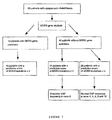
- Figure 3 illustrates the study profile of example 1.
- Figure 4 illustrates MDR3 gene mutations in LPAC and other human liver diseases: genotype-phenotype relationship.
| Patient characteristics | ||
| Patients (n) | 60 | |
| Age (years ± SD) | 38.1 ± 14.4 | |
| Gender (M/F) | 15/45 | |
| (n) | (%) | |
| Age at onset of symptoms < 40 years | 37/60 | 62 |
| | 60/60 | 100 |
| Recurrence after | 28/60 | 47 |
| Cholecystectomy | 54/60 | 90 |
| Intrahepatic hyperechoic foci | 32/60 | 53 |
| Medical history of ICP | 15/46 | 33 |
| Increased serum GGT activity | 22/60 | 37 |
| Familial history of cholelithiasis | 21/50 | 42 |
| Prophylactic UDCA treatment | 25/39 | 64 |
| UDCA denotes ursodeoxycholic acid. | ||
| ICP denotes intrahepatic cholestasis of pregnancy. |
| Adjusted odds-ratios (OR) for the presence of a mutation at the MDR3 locus in patients with cholelithiasis. | ||||
| Clinical Criteria | OR | CI 95% | P-value | Adjusted OR |
| Familial history of cholelithiasis in 1st degree relatives Increased serum GGT activity at the | 5,4 | [1.2, 29.4] | 0.01 | - |
| 1.1 | [0.3,4.1] | 1 | - | |
| time of genotype determination | 4.9 | [1.1, 24.0] | 0.02 | - |
| History of ICP Intrahepatic hyperchoic foci | 12.4 | [2.4, 126.0] | 0.0005 | 6.1 |
| Recurrence after cholecystectomy | 18.9 | [3.6, 193.7] | <0.0001 | 8.5 |
| Age at onset of symptoms < 40 | 7.8 | [1.5, 77.8] | 0.008 | 3.0 |
| Gender (Male vs Female) | 0.8 | [0.1, 3.4] | 1 | - |







Claims (10)
- A method for the screening of a hepatic syndrome comprising at least the detection, from a sample of nucleic acid extracted from peripheral blood mononuclear cells, of at least one heterozygous mutation of the MDR3 gene and/or of a homozygous mutation which does not destroy the expression of the protein expressed by said gene with phosphatidylcholine transporter activity, in adult individuals associating cholesterol microcholelithiasis and intrahepatic cholestasis, characterized in that said MDR3 gene mutation is located in at least one of the following exons: 4, 5, 6, 8, 9, 10, 14, 15, 16, 17, 18, 19, 23 and/or 26 at the following nucleotide positions:a mutation in exon 4 at nucleotide 175,a mutation in exon 6 at nucleotide 495 or 504, or a a heterozygous missense mutation in exon 6 at nucleotide 523:T/C (TCG/CCG), leading to the amino acid mutation T175A; this amino acid is located in a conserved amino acid sequence required for the ATPase activity of the MDR3 protein,a mutation in exon 8 at nucleotide 711,a mutation in exon 9 at nucleotide 902,a mutation in exon 10 at nucleotides 1007-1015 (insertion or deletion)a mutation in exon 14 at nucleotide 1584,a mutation in exon 15 at nucleotide 1772,a mutation in exon 16 at nucleotide 1954,a mutation in exon 17 at nucleotide 1973,a mutation in exon 18 at nucleotides 2270-2273 (insertion)a mutation in exon 19 at nucleotide 2363,a mutation in exon 23 at nucleotide 2800 anda mutation in exon 26 at nucleotide 3481.
- The method as claimed in claim 1, characterized in that said MDR3 gene mutation is preferably located in exons 9, 10, 14, 15, 17 or 18.
- The method as claimed in claim 1 or in claim 2, characterized in that it comprises:a first step of amplification of a nucleic acid fragment, in which at least one of said mutations is liable to be observed, from the nucleic acid extracted from peripheral blood mononuclear cells, using at least one primer selected from the group consisting of the primers having the sequences SEQ ID NO:15 to SEQ ID NO:66, anda second step of detection of the presence of at least one of said mutations, using said amplification fragment(s) obtained.
- The method as claimed in claim 3, wherein the first amplification step consists of direct PCR amplification of at least one of exons 6, 9, 10, 14, 15, 17, 18, 19, 23, and 26 and further of at least one of exons 4, 5, 8 and/or 16 of the genomic DNA with the primers, located in the flanking intronic regions, as specified in SEQ ID NO:15 to 66.
- The method as claimed in claim 3 characterized in that the second step of detection of the presence of at least one of said mutations using the amplified fragment is carried out, as appropriate, using one of the following methods: sequencing, enzyme restriction or techniques of the PCR/PCR/LDR type.
- The method as claimed in any one of claims 3 to 5 characterized in that the detection step further comprises in said first step of amplification the use of at least two primers selected from the group consisting of the sequences SEQ ID NO:1 to SEQ ID NO:13.
- The use of the method as claimed in claim 1, for evaluating the risk of appearance of a syndrome associating intrahepatic hyperechoic foci with or without intrahepatic sludge or microlithiasis and further on intrahepatic cholestasis and cholesterol microcholelithiasis in families at risk from cholesterol lithiasis and/or having unexplained hepatic biological abnormalities.
- The use of the primers selected from the group consisting of the sequences SEQ ID NO:15 to SEQ ID NO:66, optionally in combination with primers selected from the group consisting of the sequences the primers of SEQ ID NO:1 to SEQ ID NO:13 for the screening of a syndrome associating (i) intrahepatic hyperechoic foci with or without intrahepatic sludge or microlithiasis and further on cholesterol microcholelithiasis, intrahepatic cholestasis and (ii) at least one mutation of the MDR3 gene.
- A kit for the screening of a syndrome associating (i) intrahepatic hyperechoic foci with or without intrahepatic sludge or microlithiasis and further on cholesterol microcholelithiasis, intrahepatic cholestasis and (ii) at least one mutation of the MDR3 gene, characterized in that it comprises at least two primers selected from the group consisting of the sequences SEQ ID NO:15 to SEQ ID NO:66, optionally in combination with two primers selected from the group consisting of the sequences SEQ ID NO:1 to SEQ ID NO: 13.
- A kit for evaluating the risk of appearance of a syndrome associating (i) intrahepatic hyperechoic foci with or without intrahepatic sludge or microlithiasis and further on cholesterol microcholelithiasis, intrahepatic cholestasis and (ii) at least one mutation of the MDR3 gene, in a population at risk, characterized in that it comprises, besides two primers selected from the group consisting of the sequences SEQ ID NO:15 to SEQ ID NO:66, optionally in combination with two primers selected from the group consisting of the sequences SEQ ID NO:1 to SEQ ID NO:13, reagents for assaying cholesterol and bile phospholipids.
Applications Claiming Priority (2)
| Application Number | Priority Date | Filing Date | Title |
|---|---|---|---|
| US360705 | 1999-07-26 | ||
| US10/360,705 US7262011B2 (en) | 2000-08-08 | 2003-02-10 | Screening of a novel hepatic syndrome and its uses |
Publications (2)
| Publication Number | Publication Date |
|---|---|
| EP1445333A2 true EP1445333A2 (en) | 2004-08-11 |
| EP1445333A3 EP1445333A3 (en) | 2004-09-29 |
Family
ID=32655661
Family Applications (1)
| Application Number | Title | Priority Date | Filing Date |
|---|---|---|---|
| EP04290341A Withdrawn EP1445333A3 (en) | 2003-02-10 | 2004-02-10 | Screening of a novel hepatic syndrome and its uses |
Country Status (1)
| Country | Link |
|---|---|
| EP (1) | EP1445333A3 (en) |
Family Cites Families (1)
| Publication number | Priority date | Publication date | Assignee | Title |
|---|---|---|---|---|
| FR2812886B1 (en) * | 2000-08-08 | 2002-11-08 | Assist Publ Hopitaux De Paris | SCREENING FOR A NEW HEPATIC SYNDROME AND ITS APPLICATIONS |
-
2004
- 2004-02-10 EP EP04290341A patent/EP1445333A3/en not_active Withdrawn
Also Published As
| Publication number | Publication date |
|---|---|
| EP1445333A3 (en) | 2004-09-29 |
Similar Documents
| Publication | Publication Date | Title |
|---|---|---|
| US7888022B2 (en) | Screening of a novel hepatic syndrome and its uses | |
| Rosmorduc et al. | ABCB4 gene mutation—associated cholelithiasis in adults | |
| Sullivan et al. | Prevalence of disease-causing mutations in families with autosomal dominant retinitis pigmentosa: a screen of known genes in 200 families | |
| Bento et al. | Heterogeneity in gene loci associated with type 2 diabetes on human chromosome 20q13. 1 | |
| KR20220027690A (en) | Method for providing information for metabolic syndrome disease and kits using the same | |
| Robles-Osorio et al. | Genetic heterogeneity of autosomal dominant hypercholesterolemia in Mexico | |
| US20140342919A1 (en) | Markers related to age-related macular degeneration and uses therefor | |
| Hilal et al. | Screening of CYP1B1 and MYOC in Moroccan families with primary congenital glaucoma: three novel mutations in CYP1B1 | |
| US20100209923A1 (en) | Probe for diagnosis of marfan syndrome and a method for screening using the probe | |
| Chograni et al. | RGS6: a novel gene associated with congenital cataract, mental retardation, and microcephaly in a Tunisian family | |
| Pulkkinen et al. | Detection of novel LAMC2 mutations in Herlitz junctional epidermolysis bullosa | |
| US8192928B2 (en) | Mutations in the macrophage scavenger receptor 1 gene alter risk of prostate cancer, asthma, and cardiovascular disease | |
| CN108753945B (en) | SNP (single nucleotide polymorphism) locus related to obesity and/or hypertriglyceridemia of Chinese children and application thereof | |
| Wang et al. | Frasier syndrome comes full circle: genetic studies performed in an original patient | |
| US20190316199A1 (en) | Test method for evaluating the risk of anti-thyroid drug-induced agranulocytosis, and evaluation kit | |
| KR102650359B1 (en) | SNP for drug hypersensitivity and diagnosis method using the same | |
| KR102565803B1 (en) | Method for providing information for hypertension and kits using the same | |
| JP2000316578A (en) | Method for detecting diabetes risk factors | |
| KR100924371B1 (en) | How to Detect and Predict Bronchodilatory Responses to Beta Agonists | |
| US20150031569A1 (en) | Mutations of the GPR179 Gene in Congenital Stationary Night Blindness | |
| JP4056212B2 (en) | GEFS + related genes and GEFS + diagnostic methods | |
| JP2006230241A (en) | Diagnosis and treatment of pulmonary hypertension using genes related to pulmonary hypertension | |
| JP2001149082A (en) | Cd36 mutant gene and method for determining disease caused by abnormal lipid metabolism and diagnostic kit therefor | |
| US20090220954A1 (en) | Methods of diagnosing cardiovascular disease | |
| US7598029B2 (en) | Method for identifying HIV-1 protease inhibitors with reduced metabolic affects |
Legal Events
| Date | Code | Title | Description |
|---|---|---|---|
| PUAI | Public reference made under article 153(3) epc to a published international application that has entered the european phase |
Free format text: ORIGINAL CODE: 0009012 |
|
| AK | Designated contracting states |
Kind code of ref document: A2 Designated state(s): AT BE BG CH CY CZ DE DK EE ES FI FR GB GR HU IE IT LI LU MC NL PT RO SE SI SK TR |
|
| AX | Request for extension of the european patent |
Extension state: AL LT LV MK |
|
| PUAL | Search report despatched |
Free format text: ORIGINAL CODE: 0009013 |
|
| AK | Designated contracting states |
Kind code of ref document: A3 Designated state(s): AT BE BG CH CY CZ DE DK EE ES FI FR GB GR HU IE IT LI LU MC NL PT RO SE SI SK TR |
|
| AX | Request for extension of the european patent |
Extension state: AL LT LV MK |
|
| 17P | Request for examination filed |
Effective date: 20050218 |
|
| AKX | Designation fees paid |
Designated state(s): AT BE BG CH CY CZ DE DK EE ES FI FR GB GR HU IE IT LI LU MC NL PT RO SE SI SK TR |
|
| 17Q | First examination report despatched |
Effective date: 20100210 |
|
| STAA | Information on the status of an ep patent application or granted ep patent |
Free format text: STATUS: THE APPLICATION IS DEEMED TO BE WITHDRAWN |
|
| 18D | Application deemed to be withdrawn |
Effective date: 20111115 |