EP1443118A1 - Primers for detecting Fusobacterium nucleatum by PCR methods and methods for detection - Google Patents
Primers for detecting Fusobacterium nucleatum by PCR methods and methods for detection Download PDFInfo
- Publication number
- EP1443118A1 EP1443118A1 EP03028521A EP03028521A EP1443118A1 EP 1443118 A1 EP1443118 A1 EP 1443118A1 EP 03028521 A EP03028521 A EP 03028521A EP 03028521 A EP03028521 A EP 03028521A EP 1443118 A1 EP1443118 A1 EP 1443118A1
- Authority
- EP
- European Patent Office
- Prior art keywords
- nucleotides
- primer
- seq
- nucleotide sequence
- depicted
- Prior art date
- Legal status (The legal status is an assumption and is not a legal conclusion. Google has not performed a legal analysis and makes no representation as to the accuracy of the status listed.)
- Withdrawn
Links
- 238000000034 method Methods 0.000 title claims abstract description 55
- 241000605986 Fusobacterium nucleatum Species 0.000 title claims abstract description 20
- 238000001514 detection method Methods 0.000 title description 15
- 239000002773 nucleotide Substances 0.000 claims abstract description 164
- 125000003729 nucleotide group Chemical group 0.000 claims abstract description 164
- 230000000295 complement effect Effects 0.000 claims abstract description 26
- 230000003321 amplification Effects 0.000 claims description 33
- 238000003199 nucleic acid amplification method Methods 0.000 claims description 33
- 108020004414 DNA Proteins 0.000 claims description 31
- 239000000523 sample Substances 0.000 claims description 30
- YBJHBAHKTGYVGT-ZKWXMUAHSA-N (+)-Biotin Chemical compound N1C(=O)N[C@@H]2[C@H](CCCCC(=O)O)SC[C@@H]21 YBJHBAHKTGYVGT-ZKWXMUAHSA-N 0.000 claims description 6
- DZBUGLKDJFMEHC-UHFFFAOYSA-N acridine Chemical compound C1=CC=CC2=CC3=CC=CC=C3N=C21 DZBUGLKDJFMEHC-UHFFFAOYSA-N 0.000 claims description 6
- MHMNJMPURVTYEJ-UHFFFAOYSA-N fluorescein-5-isothiocyanate Chemical compound O1C(=O)C2=CC(N=C=S)=CC=C2C21C1=CC=C(O)C=C1OC1=CC(O)=CC=C21 MHMNJMPURVTYEJ-UHFFFAOYSA-N 0.000 claims description 5
- 102000002260 Alkaline Phosphatase Human genes 0.000 claims description 3
- 108020004774 Alkaline Phosphatase Proteins 0.000 claims description 3
- SHIBSTMRCDJXLN-UHFFFAOYSA-N Digoxigenin Natural products C1CC(C2C(C3(C)CCC(O)CC3CC2)CC2O)(O)C2(C)C1C1=CC(=O)OC1 SHIBSTMRCDJXLN-UHFFFAOYSA-N 0.000 claims description 3
- 108060001084 Luciferase Proteins 0.000 claims description 3
- 239000005089 Luciferase Substances 0.000 claims description 3
- 108020001027 Ribosomal DNA Proteins 0.000 claims description 3
- 229960002685 biotin Drugs 0.000 claims description 3
- 235000020958 biotin Nutrition 0.000 claims description 3
- 239000011616 biotin Substances 0.000 claims description 3
- QONQRTHLHBTMGP-UHFFFAOYSA-N digitoxigenin Natural products CC12CCC(C3(CCC(O)CC3CC3)C)C3C11OC1CC2C1=CC(=O)OC1 QONQRTHLHBTMGP-UHFFFAOYSA-N 0.000 claims description 3
- SHIBSTMRCDJXLN-KCZCNTNESA-N digoxigenin Chemical compound C1([C@@H]2[C@@]3([C@@](CC2)(O)[C@H]2[C@@H]([C@@]4(C)CC[C@H](O)C[C@H]4CC2)C[C@H]3O)C)=CC(=O)OC1 SHIBSTMRCDJXLN-KCZCNTNESA-N 0.000 claims description 3
- -1 dinitrophenyl Chemical group 0.000 claims description 3
- 230000004544 DNA amplification Effects 0.000 claims 5
- 238000002372 labelling Methods 0.000 claims 2
- 239000000126 substance Substances 0.000 claims 2
- 239000012472 biological sample Substances 0.000 abstract description 19
- 241000894006 Bacteria Species 0.000 abstract description 18
- 206010006326 Breath odour Diseases 0.000 abstract description 3
- 208000032139 Halitosis Diseases 0.000 abstract description 3
- 201000010099 disease Diseases 0.000 abstract description 2
- 208000037265 diseases, disorders, signs and symptoms Diseases 0.000 abstract description 2
- 239000013615 primer Substances 0.000 description 99
- 244000005700 microbiome Species 0.000 description 42
- 238000003752 polymerase chain reaction Methods 0.000 description 36
- 239000000047 product Substances 0.000 description 21
- 238000001962 electrophoresis Methods 0.000 description 15
- 210000003296 saliva Anatomy 0.000 description 12
- 238000006243 chemical reaction Methods 0.000 description 11
- 238000007857 nested PCR Methods 0.000 description 11
- 108020004465 16S ribosomal RNA Proteins 0.000 description 10
- 108091034117 Oligonucleotide Proteins 0.000 description 10
- 239000000243 solution Substances 0.000 description 9
- 241000605909 Fusobacterium Species 0.000 description 8
- LOKCTEFSRHRXRJ-UHFFFAOYSA-I dipotassium trisodium dihydrogen phosphate hydrogen phosphate dichloride Chemical compound P(=O)(O)(O)[O-].[K+].P(=O)(O)([O-])[O-].[Na+].[Na+].[Cl-].[K+].[Cl-].[Na+] LOKCTEFSRHRXRJ-UHFFFAOYSA-I 0.000 description 8
- 239000002953 phosphate buffered saline Substances 0.000 description 8
- 238000007400 DNA extraction Methods 0.000 description 7
- 102000053602 DNA Human genes 0.000 description 6
- 210000002200 mouth mucosa Anatomy 0.000 description 6
- 238000012360 testing method Methods 0.000 description 6
- 238000000018 DNA microarray Methods 0.000 description 5
- 210000004027 cell Anatomy 0.000 description 5
- 239000000758 substrate Substances 0.000 description 5
- 241000605952 Fusobacterium necrophorum Species 0.000 description 4
- 241000605994 Fusobacterium periodonticum Species 0.000 description 4
- 241000605975 Fusobacterium varium Species 0.000 description 4
- VYPSYNLAJGMNEJ-UHFFFAOYSA-N Silicium dioxide Chemical compound O=[Si]=O VYPSYNLAJGMNEJ-UHFFFAOYSA-N 0.000 description 4
- 108010006785 Taq Polymerase Proteins 0.000 description 4
- JLCPHMBAVCMARE-UHFFFAOYSA-N [3-[[3-[[3-[[3-[[3-[[3-[[3-[[3-[[3-[[3-[[3-[[5-(2-amino-6-oxo-1H-purin-9-yl)-3-[[3-[[3-[[3-[[3-[[3-[[5-(2-amino-6-oxo-1H-purin-9-yl)-3-[[5-(2-amino-6-oxo-1H-purin-9-yl)-3-hydroxyoxolan-2-yl]methoxy-hydroxyphosphoryl]oxyoxolan-2-yl]methoxy-hydroxyphosphoryl]oxy-5-(5-methyl-2,4-dioxopyrimidin-1-yl)oxolan-2-yl]methoxy-hydroxyphosphoryl]oxy-5-(6-aminopurin-9-yl)oxolan-2-yl]methoxy-hydroxyphosphoryl]oxy-5-(6-aminopurin-9-yl)oxolan-2-yl]methoxy-hydroxyphosphoryl]oxy-5-(6-aminopurin-9-yl)oxolan-2-yl]methoxy-hydroxyphosphoryl]oxy-5-(6-aminopurin-9-yl)oxolan-2-yl]methoxy-hydroxyphosphoryl]oxyoxolan-2-yl]methoxy-hydroxyphosphoryl]oxy-5-(5-methyl-2,4-dioxopyrimidin-1-yl)oxolan-2-yl]methoxy-hydroxyphosphoryl]oxy-5-(4-amino-2-oxopyrimidin-1-yl)oxolan-2-yl]methoxy-hydroxyphosphoryl]oxy-5-(5-methyl-2,4-dioxopyrimidin-1-yl)oxolan-2-yl]methoxy-hydroxyphosphoryl]oxy-5-(5-methyl-2,4-dioxopyrimidin-1-yl)oxolan-2-yl]methoxy-hydroxyphosphoryl]oxy-5-(6-aminopurin-9-yl)oxolan-2-yl]methoxy-hydroxyphosphoryl]oxy-5-(6-aminopurin-9-yl)oxolan-2-yl]methoxy-hydroxyphosphoryl]oxy-5-(4-amino-2-oxopyrimidin-1-yl)oxolan-2-yl]methoxy-hydroxyphosphoryl]oxy-5-(4-amino-2-oxopyrimidin-1-yl)oxolan-2-yl]methoxy-hydroxyphosphoryl]oxy-5-(4-amino-2-oxopyrimidin-1-yl)oxolan-2-yl]methoxy-hydroxyphosphoryl]oxy-5-(6-aminopurin-9-yl)oxolan-2-yl]methoxy-hydroxyphosphoryl]oxy-5-(4-amino-2-oxopyrimidin-1-yl)oxolan-2-yl]methyl [5-(6-aminopurin-9-yl)-2-(hydroxymethyl)oxolan-3-yl] hydrogen phosphate Polymers Cc1cn(C2CC(OP(O)(=O)OCC3OC(CC3OP(O)(=O)OCC3OC(CC3O)n3cnc4c3nc(N)[nH]c4=O)n3cnc4c3nc(N)[nH]c4=O)C(COP(O)(=O)OC3CC(OC3COP(O)(=O)OC3CC(OC3COP(O)(=O)OC3CC(OC3COP(O)(=O)OC3CC(OC3COP(O)(=O)OC3CC(OC3COP(O)(=O)OC3CC(OC3COP(O)(=O)OC3CC(OC3COP(O)(=O)OC3CC(OC3COP(O)(=O)OC3CC(OC3COP(O)(=O)OC3CC(OC3COP(O)(=O)OC3CC(OC3COP(O)(=O)OC3CC(OC3COP(O)(=O)OC3CC(OC3COP(O)(=O)OC3CC(OC3COP(O)(=O)OC3CC(OC3COP(O)(=O)OC3CC(OC3COP(O)(=O)OC3CC(OC3CO)n3cnc4c(N)ncnc34)n3ccc(N)nc3=O)n3cnc4c(N)ncnc34)n3ccc(N)nc3=O)n3ccc(N)nc3=O)n3ccc(N)nc3=O)n3cnc4c(N)ncnc34)n3cnc4c(N)ncnc34)n3cc(C)c(=O)[nH]c3=O)n3cc(C)c(=O)[nH]c3=O)n3ccc(N)nc3=O)n3cc(C)c(=O)[nH]c3=O)n3cnc4c3nc(N)[nH]c4=O)n3cnc4c(N)ncnc34)n3cnc4c(N)ncnc34)n3cnc4c(N)ncnc34)n3cnc4c(N)ncnc34)O2)c(=O)[nH]c1=O JLCPHMBAVCMARE-UHFFFAOYSA-N 0.000 description 4
- 238000004925 denaturation Methods 0.000 description 4
- 230000036425 denaturation Effects 0.000 description 4
- 210000002950 fibroblast Anatomy 0.000 description 4
- 239000003550 marker Substances 0.000 description 4
- 239000000741 silica gel Substances 0.000 description 4
- 229910002027 silica gel Inorganic materials 0.000 description 4
- 239000006228 supernatant Substances 0.000 description 4
- 241001303074 Fusobacterium naviforme Species 0.000 description 3
- 241000605978 Fusobacterium russii Species 0.000 description 3
- 108020004682 Single-Stranded DNA Proteins 0.000 description 3
- 238000000137 annealing Methods 0.000 description 3
- 239000008280 blood Substances 0.000 description 3
- 210000004369 blood Anatomy 0.000 description 3
- 239000007853 buffer solution Substances 0.000 description 3
- 210000001175 cerebrospinal fluid Anatomy 0.000 description 3
- 238000007796 conventional method Methods 0.000 description 3
- 239000012634 fragment Substances 0.000 description 3
- 239000011521 glass Substances 0.000 description 3
- 238000004128 high performance liquid chromatography Methods 0.000 description 3
- 238000004519 manufacturing process Methods 0.000 description 3
- 239000000463 material Substances 0.000 description 3
- 239000012528 membrane Substances 0.000 description 3
- 108020004707 nucleic acids Proteins 0.000 description 3
- 102000039446 nucleic acids Human genes 0.000 description 3
- 150000007523 nucleic acids Chemical class 0.000 description 3
- 238000003753 real-time PCR Methods 0.000 description 3
- 108091008146 restriction endonucleases Proteins 0.000 description 3
- 125000006850 spacer group Chemical group 0.000 description 3
- 238000010186 staining Methods 0.000 description 3
- 238000010561 standard procedure Methods 0.000 description 3
- 102000004190 Enzymes Human genes 0.000 description 2
- 108090000790 Enzymes Proteins 0.000 description 2
- TWRXJAOTZQYOKJ-UHFFFAOYSA-L Magnesium chloride Chemical compound [Mg+2].[Cl-].[Cl-] TWRXJAOTZQYOKJ-UHFFFAOYSA-L 0.000 description 2
- 108091028043 Nucleic acid sequence Proteins 0.000 description 2
- NINIDFKCEFEMDL-UHFFFAOYSA-N Sulfur Chemical compound [S] NINIDFKCEFEMDL-UHFFFAOYSA-N 0.000 description 2
- 239000011543 agarose gel Substances 0.000 description 2
- 150000001413 amino acids Chemical class 0.000 description 2
- 239000011324 bead Substances 0.000 description 2
- 239000003153 chemical reaction reagent Substances 0.000 description 2
- 238000012364 cultivation method Methods 0.000 description 2
- 230000001419 dependent effect Effects 0.000 description 2
- XBDQKXXYIPTUBI-UHFFFAOYSA-N dimethylselenoniopropionate Natural products CCC(O)=O XBDQKXXYIPTUBI-UHFFFAOYSA-N 0.000 description 2
- ZMMJGEGLRURXTF-UHFFFAOYSA-N ethidium bromide Chemical compound [Br-].C12=CC(N)=CC=C2C2=CC=C(N)C=C2[N+](CC)=C1C1=CC=CC=C1 ZMMJGEGLRURXTF-UHFFFAOYSA-N 0.000 description 2
- 229960005542 ethidium bromide Drugs 0.000 description 2
- 238000000605 extraction Methods 0.000 description 2
- 210000004379 membrane Anatomy 0.000 description 2
- 210000000214 mouth Anatomy 0.000 description 2
- BASFCYQUMIYNBI-UHFFFAOYSA-N platinum Chemical compound [Pt] BASFCYQUMIYNBI-UHFFFAOYSA-N 0.000 description 2
- 108091033319 polynucleotide Proteins 0.000 description 2
- 102000040430 polynucleotide Human genes 0.000 description 2
- 239000002157 polynucleotide Substances 0.000 description 2
- 108090000623 proteins and genes Proteins 0.000 description 2
- 229910052717 sulfur Inorganic materials 0.000 description 2
- 239000011593 sulfur Substances 0.000 description 2
- 239000000725 suspension Substances 0.000 description 2
- 238000003786 synthesis reaction Methods 0.000 description 2
- 210000001519 tissue Anatomy 0.000 description 2
- 229910021642 ultra pure water Inorganic materials 0.000 description 2
- 239000012498 ultrapure water Substances 0.000 description 2
- 229920000936 Agarose Polymers 0.000 description 1
- 108091003079 Bovine Serum Albumin Proteins 0.000 description 1
- 108020004635 Complementary DNA Proteins 0.000 description 1
- 239000003155 DNA primer Substances 0.000 description 1
- AHCYMLUZIRLXAA-SHYZEUOFSA-N Deoxyuridine 5'-triphosphate Chemical compound O1[C@H](COP(O)(=O)OP(O)(=O)OP(O)(O)=O)[C@@H](O)C[C@@H]1N1C(=O)NC(=O)C=C1 AHCYMLUZIRLXAA-SHYZEUOFSA-N 0.000 description 1
- KCXVZYZYPLLWCC-UHFFFAOYSA-N EDTA Chemical compound OC(=O)CN(CC(O)=O)CCN(CC(O)=O)CC(O)=O KCXVZYZYPLLWCC-UHFFFAOYSA-N 0.000 description 1
- LFQSCWFLJHTTHZ-UHFFFAOYSA-N Ethanol Chemical compound CCO LFQSCWFLJHTTHZ-UHFFFAOYSA-N 0.000 description 1
- 238000003794 Gram staining Methods 0.000 description 1
- 239000004677 Nylon Substances 0.000 description 1
- 238000012408 PCR amplification Methods 0.000 description 1
- 208000005888 Periodontal Pocket Diseases 0.000 description 1
- 241000605862 Porphyromonas gingivalis Species 0.000 description 1
- 241001135221 Prevotella intermedia Species 0.000 description 1
- 230000001580 bacterial effect Effects 0.000 description 1
- 239000012620 biological material Substances 0.000 description 1
- 230000015572 biosynthetic process Effects 0.000 description 1
- 238000009835 boiling Methods 0.000 description 1
- 210000005178 buccal mucosa Anatomy 0.000 description 1
- 150000001720 carbohydrates Chemical class 0.000 description 1
- 230000015556 catabolic process Effects 0.000 description 1
- 239000007795 chemical reaction product Substances 0.000 description 1
- 230000000052 comparative effect Effects 0.000 description 1
- NHVNXKFIZYSCEB-XLPZGREQSA-N dTTP Chemical compound O=C1NC(=O)C(C)=CN1[C@@H]1O[C@H](COP(O)(=O)OP(O)(=O)OP(O)(O)=O)[C@@H](O)C1 NHVNXKFIZYSCEB-XLPZGREQSA-N 0.000 description 1
- 230000007547 defect Effects 0.000 description 1
- 239000005547 deoxyribonucleotide Substances 0.000 description 1
- 238000010790 dilution Methods 0.000 description 1
- 239000012895 dilution Substances 0.000 description 1
- 230000000694 effects Effects 0.000 description 1
- 210000003608 fece Anatomy 0.000 description 1
- 239000012091 fetal bovine serum Substances 0.000 description 1
- 239000012530 fluid Substances 0.000 description 1
- PCHJSUWPFVWCPO-UHFFFAOYSA-N gold Chemical compound [Au] PCHJSUWPFVWCPO-UHFFFAOYSA-N 0.000 description 1
- 239000010931 gold Substances 0.000 description 1
- 229910052737 gold Inorganic materials 0.000 description 1
- 238000010438 heat treatment Methods 0.000 description 1
- 238000011835 investigation Methods 0.000 description 1
- 229910001629 magnesium chloride Inorganic materials 0.000 description 1
- 239000002609 medium Substances 0.000 description 1
- 239000003068 molecular probe Substances 0.000 description 1
- 230000000877 morphologic effect Effects 0.000 description 1
- 210000004877 mucosa Anatomy 0.000 description 1
- 229930014626 natural product Natural products 0.000 description 1
- 238000007826 nucleic acid assay Methods 0.000 description 1
- 229920001778 nylon Polymers 0.000 description 1
- 244000052769 pathogen Species 0.000 description 1
- 208000028169 periodontal disease Diseases 0.000 description 1
- 230000003239 periodontal effect Effects 0.000 description 1
- 239000002504 physiological saline solution Substances 0.000 description 1
- 229910052697 platinum Inorganic materials 0.000 description 1
- 238000001556 precipitation Methods 0.000 description 1
- 238000002360 preparation method Methods 0.000 description 1
- 108090000765 processed proteins & peptides Proteins 0.000 description 1
- 230000002062 proliferating effect Effects 0.000 description 1
- 235000019260 propionic acid Nutrition 0.000 description 1
- 238000000746 purification Methods 0.000 description 1
- IUVKMZGDUIUOCP-BTNSXGMBSA-N quinbolone Chemical compound O([C@H]1CC[C@H]2[C@H]3[C@@H]([C@]4(C=CC(=O)C=C4CC3)C)CC[C@@]21C)C1=CCCC1 IUVKMZGDUIUOCP-BTNSXGMBSA-N 0.000 description 1
- 238000005185 salting out Methods 0.000 description 1
- 238000005070 sampling Methods 0.000 description 1
- 239000006152 selective media Substances 0.000 description 1
- 229910052710 silicon Inorganic materials 0.000 description 1
- 239000010703 silicon Substances 0.000 description 1
- 241000894007 species Species 0.000 description 1
- 239000004094 surface-active agent Substances 0.000 description 1
- 150000003568 thioethers Chemical class 0.000 description 1
- 239000001226 triphosphate Substances 0.000 description 1
- 235000011178 triphosphate Nutrition 0.000 description 1
- PIEPQKCYPFFYMG-UHFFFAOYSA-N tris acetate Chemical compound CC(O)=O.OCC(N)(CO)CO PIEPQKCYPFFYMG-UHFFFAOYSA-N 0.000 description 1
- 210000002700 urine Anatomy 0.000 description 1
Images


Classifications
-
- C—CHEMISTRY; METALLURGY
- C12—BIOCHEMISTRY; BEER; SPIRITS; WINE; VINEGAR; MICROBIOLOGY; ENZYMOLOGY; MUTATION OR GENETIC ENGINEERING
- C12Q—MEASURING OR TESTING PROCESSES INVOLVING ENZYMES, NUCLEIC ACIDS OR MICROORGANISMS; COMPOSITIONS OR TEST PAPERS THEREFOR; PROCESSES OF PREPARING SUCH COMPOSITIONS; CONDITION-RESPONSIVE CONTROL IN MICROBIOLOGICAL OR ENZYMOLOGICAL PROCESSES
- C12Q1/00—Measuring or testing processes involving enzymes, nucleic acids or microorganisms; Compositions therefor; Processes of preparing such compositions
- C12Q1/68—Measuring or testing processes involving enzymes, nucleic acids or microorganisms; Compositions therefor; Processes of preparing such compositions involving nucleic acids
- C12Q1/6876—Nucleic acid products used in the analysis of nucleic acids, e.g. primers or probes
- C12Q1/6888—Nucleic acid products used in the analysis of nucleic acids, e.g. primers or probes for detection or identification of organisms
- C12Q1/689—Nucleic acid products used in the analysis of nucleic acids, e.g. primers or probes for detection or identification of organisms for bacteria
Definitions
- the present invention relates to a method for specifically detecting or quantifying Fusobacterium nucleatum, which is a suppurative disease-related bacteria and also is a halitosis-causing bacteria.
- microorganisms such as bacterium
- cultivation such as proliferative cultivation or separate cultivation, and it is required to make single colonies grow, whereby taking several days or several weeks.
- the isolated microorganisms is further proliferated in order to conduct morphological observation, cell staining such as Gram staining, investigation of various biochemical characteristics including propionic acid production test, saccharide catabolism test and the like.
- cell staining such as Gram staining
- investigation of various biochemical characteristics including propionic acid production test, saccharide catabolism test and the like.
- a PCR (polymerase chain reaction) method has recently been employed for the identification of microorganisms.
- the PCR method permits a rapid and high sensitive identification of microorganisms.
- the PCR method needs to use primers, and detection or identification of a target microorganism is dependent on designs of the primers.
- Fusobacterium naviforme Fusobacterium periodonticum, Fusobacterium necrophorum, Fusobacterium varium, Fusobacterium russii, Fusobacterium nucleatum (hereinafter referred to as Fn microorganism) and the like.
- the Fn microorganism which is also known as a halitosis-causing bacterium, decomposes a sulfur-containing amino acid and a peptide containing such a sulfur-containing amino acid which are present in an oral food debris or detached mucosa to produce volatile sulfide compounds (hereinafter referred to as VSC).
- VSC volatile sulfide compounds
- the VSC production ability of any bacteria of the Fusobacterium genus other than the Fn microorganism is not as high as that of the Fn microorganism.
- the Fn microorganism is suggested to be related with periodontal diseases since it has a specific ability of being coaggregated with periodontal pathogens such as Porphyromonas gingivalis and Prevotella intermedia.
- the Fn microorganism is a particularly important strain among the bacteria of the Fusobacterium genus. Nevertheless, the Fn microorganism is very difficult to be isolated for the culture, since it has a remarkably analogous relationship with other bacteria of the Fusobacterium genus, and no selective medium capable of specifically detecting the Fn microorganism has been developed.
- the present invention provides a method for detecting and/or quantifying Fusobacterium nucleatum by a PCR method employing the following primers (1) and (2):
- the present invention provides a simple and rapid detection method which is capable of distinguishing an Fn microorganism specifically among the bacteria of the Fusobacterium genus and which also capable of distinguishing from human-derived DNAs contaminating a biological sample.
- Inventors of the present invention investigated the ribosomal DNA of the Fn microorganism to find that the 16S rDNAs are remarkably analogous between closely related species among the Fusobacterium genus, and therefore it is difficult to detect or identify the Fn microorganism by a PCR method simply using primers prepared so as to have a nucleotide sequence in a region of 16S rDNA.
- 23S ribosomal DNA (hereinafter referred to as 23S rDNA) is analogous with the human gene DNA, and therefore it is not desirable for a case of analyzing a biological sample such as a supragingival plaque, subgingival plaque, tongue plaque, saliva, gingival crevice fluide, blood, cerebrospinal fluid, suppurative focus or other tissues.
- a PCR method is conducted in a combination use of "a forward primer with a consecution of 10 or more nucleotides” which has a nucleotide sequence contained in the 16S rDNA and/or an adjacent spacer (a region represented by SEQ ID No. 1) and "a reverse primer with a consecution of 10 or more nucleotides” which has a nucleotide sequence contained in the 23S rDNA (a sequence complementary to a region depicted as SEQ ID No. 2).
- a forward primer with a consecution of 10 or more nucleotides which has a nucleotide sequence contained in the 16S rDNA and/or an adjacent spacer (a region represented by SEQ ID No. 1)
- a reverse primer with a consecution of 10 or more nucleotides which has a nucleotide sequence contained in the 23S rDNA (a sequence complementary to a region depicted as SEQ ID No. 2).
- a forward primer comprising a nucleotide sequence which is depicted as a portion of SEQ ID No.1 and has at least 10 nucleotides in length has, as its partial or entire sequence, a nucleotide sequence which relates to SEQ ID No.1 and has at least 10 nucleotides in length.
- the forward primer has, at its 3' terminal, a nucleotide sequence having at least 10 nucleotides and being same as a part of SEQ ID No. 1, and its 5' terminal may be extended by an additional sequence not relating to any part of SEQ ID No. 1.
- the forward primer is preferably designed so as to adapt to the following restriction: namely, it is preferable for a length of the primer to be in a range of 10 nucleotides or more and 40 nucleotides or less, more preferably 12 nucleotides or more and 30 nucleotides or less, and even more preferably 15 nucleotides or more and 25 nucleotides or less.
- the primer may be designed so as to have a value of Tm within a preferable range of 40 °C or more and 65 °C or less, and more preferably 50 °C or more and 60 °C or less.
- GC content is preferably in a range of 25 % or more and 70 % or less, and more preferably 40 % or more and 60 % or less.
- Preferable examples of the forward primer include:
- a reverse primer comprising a nucleotide sequence which is depicted as a portion of a sequence complementary to SEQ ID No.2 and has at least ten nucleotides in length has, as its partial or entire sequence, a nucleotide sequence which relates to SEQ ID No.2 and has at least 10 nucleotides in length.
- the reverse primer has, at its 3' terminal, a nucleotide sequence having at least 10 nucleotides and being complementary to a part of SEQ ID No. 2, and its 5' terminal may be extended by an additional sequence not relating to any part of SEQ ID No. 2.
- the reverse primer is preferably designed so as to adapt to the following restriction: namely, it is preferable for a length of the primer to be in a range of 10 nucleotides or more and 40 nucleotides or less, more preferably 12 nucleotides or more and 30 nucleotides or less, and even more preferably 15 nucleotides or more and 25 nucleotides or less.
- the primer may be designed so as to have a value of Tm within a preferable range of 40 °C or more and 65 °C or less, and more preferably 50 °C or more and 60 °C or less.
- GC content is preferably in a range of 25 % or more and 70 % or less, and more preferably 40 % or more and 60 % or less.
- Preferable examples of the reverse primer include:
- a primer set using a combination of either the following primers (A) and (B) or the following primers (B) and (C) in a viewpoint of specifically detecting or identifying the Fn microorganism from a biological sample.
- nucleotide sequence of nucleotides 1 to 39 of SEQ ID No. 3, which is one of nucleotide sequences of the primer (A) is found as a preferable region for distinguishing the DNAs between ordinary bacteria and human.
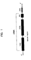
- This nucleotide sequence was revealed to be present at a site consisting of 5 nucleotides at the 3' terminal of the 16S rDNA and the 5' terminal side of a spacer region which is formed between the 16S rDNA and the 23S rDNA(See FIG. 1).
- oligonucleotide primers for the amplification by a PCR method were designed and synthesized.
- these primers are used in the PCR of a biological sample containing the Fn microorganism, the amplification products specific for the Fn microorganism are exclusively obtained.
- a sample usable for detection or identification of the Fn microorganism there may be exemplified oral cavity-related biological samples such as a supragingival plaque, subgingival plaque, tongue plaque, saliva, gingival crevice fluide and the like.
- oral cavity-related biological samples such as a supragingival plaque, subgingival plaque, tongue plaque, saliva, gingival crevice fluide and the like.
- other biological samples such as blood, cerebrospinal fluid, suppurative focus or other tissues.
- the oligonucleotides to be used as primers may be synthesized chemically or may be derived from a natural products.
- an oligonucleotide which has a nucleotide sequence, as its partial or entire sequence, being depicted as a portion or a whole of a sequence complementary to a region between nucleotides 863 to 883 of SEQ ID No.3 and having at least ten nucleotides in length and which is defined as a nucleotide sequence of the primer (B) is used as a reverse primer.
- the chain length of the each primer is preferably 10 nucleotides or more and 40 nucleotides or less, more preferably 12 nucleotides or more and 30 nucleotides or less, and even more preferably 15 nucleotides or more and 25 nucleotides or less.
- the primer (A) there may be used for example: the primer (C) having a nucleotide sequence consisting of the following 18 nucleotides AACGTGCGGATGGATCAC; a nucleotide sequence of nucleotides 1 to 22 of SEQ ID No.3, namely AACGTGCGGATGGATCACCTCC; a nucleotide sequence of nucleotides 7 to 26 of SEQ ID No.3, namely CGGATGGATCACCTCCTTTC; a nucleotide sequence of nucleotides 12 to 31 of SEQ ID No.3, namely GGATCACCTCCTTTCTAAGG; and, a nucleotide sequence of nucleotide 16 to 33 of SEQ ID No.3, namely CACCTCCTTTCTAAGGAG.
- the amplification product of the nucleotide sequence represented by SEQ ID No. 3 can specifically be obtained.
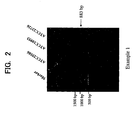
- the length of a major amplification product is calculated to be 883 bp, and a band of about 900 bp indicating the amplification product can be observed in an electrophoresis image (see FIG. 2).
- the primer (D) has a nucleotide sequence, as its partial or entire sequence, being depicted as a portion or a whole of a sequence complementary to a region between nucleotides 124 to 156 of SEQ ID No.3 and having at least ten nucleotides in length.
- a nucleotide sequence of the primer (D) is present in a site called as a spacer region between the 16S rDNA and the 23S rDNA.
- the nucleotide sequence of the primer (D) may be consist of 29 nucleotides, and preferably in a range from 15 nucleotides to 25 nucleotides, and there may be used a primer (E) having a nucleotide sequence consisting of 20 nucleotides, as the primer (D).
- the primer (E) consists of a nucleotide sequence depicted as a sequence complementary to a region between nucleotides 124 to 143 of SEQ ID No.3.
- the semi-nested PCR method comprises two stages of amplification.
- an amplification product containing a target region is obtained.
- the resultant amplification product is used as a template to conduct the second stage of amplification.
- either one of the forward and the reverse primers (namely, inner primers) to be used is capable of positioning at a site inner than a site where either one of the primers (namely, outer primers) used in the first stage positions, whereby excluding the amplification products derived from the DNA in a region other than the target region.
- the first stage of amplification in the PCR is conducted using the primers (A) and (B) as outer primers. Then, using a part of the sample thus amplified in the first stage, the second stage of amplification is initiated. In the second stage of amplification, the PCR is conducted using the primers (A) and (D) as inner primers. Since a major amplification product in such a case is dependent on the primers employed in the second amplification stage, it corresponds to a part of the nucleotide sequence represented by SEQ ID No. 3.
- oligonucleotides other than the primer (A) or (B) may be used.
- an oligonucleotide designed based on the nucleotide sequence of the 16S rDNA such as CGTCACACCACGAGAGTTGG (namely, nucleotides 1384 to 1403 of SEQ ID No. 1) and the like can be used instead of the primer (A).
- an oligonucleotide having a nucleotide sequence complementary to a nucleotide sequence designed based on the 23S rDNA such as CCATTTGGGTGATGGC (namely, nucleotide 597 to 612 of SEQ ID No.2 ) can be used as the outer primer.
- the primers according to the present invention can be applied also to a nested PCR method.
- the nested PCR method is different from the semi-nested PCR method in that it uses an inner primer set in which respective inner primers to be used for the both sides are capable of positioning at inner sites on the target region rather than the outer primers.
- primers (C) and (B) In addition to the primers (C) and (B), the followings are exemplified as the outer primers which can be used in the nested PCR.
- Forward primer an oligonucleotide which has a nucleotide sequence, as its partial or entire sequence, being depicted as a portion of a region between nucleotides 1 to 123 of SEQ ID No.3 wherein the portion related to the region of SEQ ID No.3 has at least ten nucleotides in length, or being depicted as a portion of SEQ ID No. 4 wherein the portion related to SEQ ID No. 4 has at least ten nucleotides in length.
- Reverse primer an oligonucleotide which has a nucleotide sequence, as its partial or entire sequence, being depicted as a portion of the 23S rDNA(SEQ ID No.2).
- the first stage of amplification can be conducted using, as outer primers, a primer with a sequence of CGTCACACCACGAGAGTTGG (nucleotides 1384 to 1403 of SEQ ID No. 1) designed based on the nucleotide sequence of the 16S rDNA and, a primer with a complementary nucleotide sequence corresponding to AAGAAGGGTAACCGACTT (nucleotides 1256 to 1273 of SEQ ID No. 2) among the nucleotide sequence of the 23S rDNA.
- a primer with a sequence of CGTCACACCACGAGAGTTGG (nucleotides 1384 to 1403 of SEQ ID No. 1) designed based on the nucleotide sequence of the 16S rDNA and, a primer with a complementary nucleotide sequence corresponding to AAGAAGGGTAACCGACTT (nucleotides 1256 to 1273 of SEQ ID No. 2) among the nucleotide sequence of the 23S
- the primer set of (A) and (B) or the primer set of (B) and (D), all of which are inventive primers is preferably used as the inner primers for the purpose of specifically detecting or identifying an Fn microorganism.
- the PCR method is a method to amplifying genes commonly practicedby one skilled in the art, and fundamental techniques are described below briefly.
- a double-stranded DNA used as a template is separated into single-stranded DNAs by heating (the thermal denaturation). Then the annealing process is carried out so as to sandwich a target region of the each single-stranded DNA by hybridized sites in such manner that oligonucleotides referred to as primers are hybridized with sites of the 5' terminal of the respective single-stranded DNAs which have separated by the aforementioned thermal denaturation process and have a complementary relationship with respect to the separated counter part.
- dNTP deoxyribonucleotide triphosphates
- the nucleotides are added to the 3' terminal of the primer in accordance with the nucleotide sequence of the template, resulting in the extension of the DNA chain (extension reaction).
- extension reaction By repeating this reaction, a large amount of the DNA fragment containing the target region can be obtained.
- a dUTP may be used as the substrate instead of the dTTP among the dNTPs.
- the PCR method preferably employs the following temperature condition in a single cycle: a thermal denaturation for converting a double-stranded DNA into a single strand is carried out at 90 to 98 °C; an annealing reaction for hybridizing the primer with the template DNA is carried out at 37 to 65 °C; and, an extension reaction for allowing a Taq DNA polymerase to exert the effect is carried out at 50 to 75 °C.
- a target sequence can be amplified.
- the amplification product is separated for example by an electrophoresis, subjected to a nucleic acid staining for example with ethidium bromide, and then can give a judgement that the microorganism to be detected is present in the sample if the chain length of the amplified polynucleotide sequence is equal to the chain length of the target sequence.
- a high performance liquid chromatography is also effective for detecting the amplified polynucleotide sequence.
- an additional sequence extends from the 5' terminal of an essential part of the nucoleotide sequence
- essential for the primer (A) is a sequence which is depicted as a portion or a whole of a region between nucleotides 1 to 39 of SEQ ID No. 3 and has at least ten nucleotides in length
- essential for the primer (B) is a sequence which is depicted as a portion or a whole of a sequence complementary to a region between nucleotides 863 to 883 of SEQ ID No.3 and has at least ten nucleotides in length
- the additional portion in the primer chain length is imparted to an amplification product.
- the primers may be labeled with one or more selected from biotin, digoxigenin, FITC (fluorescein isothiocyanate), acridine, dinitrophenyl, alkaline phosphatase, luciferase and [ 32 P]dNTP.
- While various biological sample may be suggested as a sample to be subjected to the invention, those which may be exemplified are saliva, tongue plaque, supragingival plaque, subgingival plaque, oral mucosa, gingival crevice fluid, blood, cerebrospinal fluid, suppurative focus sample, feces, urine or cultured materials of these biological materials.
- a biological sample thus obtained can be subjected to a PCR directly or after a DNA extraction.
- the DNA extraction may be conducted by a conventional method.
- the DNA extraction may employ one or more of an alkaline extraction or surfactant extraction, enzyme treatment, boiling and the like, and may also employ a commercial kit.
- a sample containing the DNA extracted froman organism's body may further be purified by a conventional method.
- the DNA purification may employ one or more of a high performance liquid chromatography or alcohol precipitation, salting-out, silica gel column, silica gel membrane and the like, and may also employ a commercial kit.
- the primers and the amplification products are also useful as probes for detecting an Fn microorganism.
- the amplification products may be converted into DNA fragments having suitable lengths using restriction enzymes and the like.
- restriction enzymes are one or more of RcoR I, Mse I, Ban II, Alu I, Mbo I, Fok I, Taq I, Mbo II, Hinf I and Ava II.
- a DNA fragment obtained by a restriction enzyme treatment is preferably purified by means of an electrophoresis, high performance liquid chromatography, silica gel column, silica gel membrane, nylon membrane and the like.
- the probe can be labeled with a label.
- the probe may be labeled with one or more of biotin, digoxigenin, FITC(fluoresceinisothiocyanate),acridine,dinitrophenyl, alkaline phosphatase, luciferase or [ 32 P]dNTP.
- the method for producing a DNA chip may be any conventional method. For example, it may employ a probe alignment type in which a probe is aligned on a chip substrate, or a probe synthesis type in which a DNA extension reaction is performed directly on a substrate such as a glass or silicon to form a probe DNA. Alternatively, a commercial DNA chip producing device may be employed, and the reading may be effected using a DNA chip reader.
- a kit for detecting and identifying a microorganism comprises proves and/or primers according to the present invention. It may also contain other components such as a Taq DNA polymerase, other enzymes, substrates such as dNTPs and the like, as well as buffer solutions.
- Fusobacterium nucleatum ATCC25586, ATCC10953, ATCC23726 Three types of standard microorganisms of Fusobacterium nucleatum (Fusobacterium nucleatum ATCC25586, ATCC10953, ATCC23726) were incubated anaerobically in a modified FMmedium (available from NISSUI PHARMACEUTICAL CO., LTD.) at 37 °C for 3 to 5 days, and the colonies formed were taken at one supatula-full amount of platinum loop and processed using a commercial kit (QIAGEN K.K., DNA mini-kit) to obtain a DNA extract.
- a modified FMmedium available from NISSUI PHARMACEUTICAL CO., LTD.
- 1 ⁇ l of the DNA extract was combined with 1.5 ⁇ l of 50 mM MgCl 2 solution, each 2 ⁇ l of 2 mM dNTP solutions (Applied Biosystems) , 1 ⁇ l of a 12.5 pmol/ ⁇ l forward primer solution, 1 ⁇ l of a 12.5 pmol/ ⁇ l reverse primer solution, 0.5 ⁇ l of a 5 U/ ⁇ l Taq DNA polymerase solution (Applied Biosystems) , 5 ⁇ l of an Ampli Taq Gold buffer solution (Applied Biosystems) and a sterilized ultrapure water at a volume to obtain the total volume of 50 ⁇ l, thereby obtaining a reaction solution.
- the procedure was conducted using a GeneAmp 9700 system of Applied Biosystems.
- the presence or absence of the amplification products was determined by a conventional agarose electrophoresis.
- the agarose gel for the electrophoresis was provided, and placed in a running device which had previously been filled with a TAE (tris-acetate, ethylenediamine tetraacetic acid) buffer solution, and then the PCR reaction product was applied onto the sample groove. After running for 15 minutes at 100 V, the agarose gel was soaked in an ethidium bromide solution for 30 minutes to stain the nucleic acid. After the staining, the bands of the amplification products were identified using an UV transilluminater. The results of the electrophoresis are shown in FIG. 2.
- Lane 1 represents a marker
- Lane 2 represents ATCC25586 strain
- Lane 3 represents ATCC10953 strain
- Lane 4 represents ATCC23726 strain. All of the three microorganisms of Fusobacterium nucleatum exhibited the band representing the amplification product of about 900 bp.
- Fusobacterium nucleatum (ATCC25586 strain)exhibited the band representing the amplification product of about 1500 bp.
- Test bacteria and test cells subjected are 5 bacteria other than the Fn microorganism, namely, F.naviforme (ATCC25832), F.periodonticum (ATCC33693), F.necrophorum (ATCC25286), F.varium (ATCC8501), F.russii (ACTT25533) as well as normal human gingival fibroblast (ATCC CRL1292).
- the normal human gingival fibroblast was incubated for 72 hours at 37 °C under an atmosphere of 5% CO 2 in a modified EAGLE medium (available from Dulbecco) supplemented with 10% fetal bovine serum, and then recovered, washed with a physiological saline, and then nucleic acid was isolated. Otherwise, the bacteria strains were processed similarly to Example 1.
- Lane 1 represents a marker
- Lane 2 represents F.naviforme
- Lane 3 represents F.periodonticum
- Lane 4 represents F.necrophorum
- Lane 5 represents F.varium
- Lane 6 represents F.russii
- Lane 7 represents the normal human gingival fibroblast. No lane exhibited a band showing the amplification product.
- Saliva, tongue plaque, subgingival plaque and oral mucosa were taken as biological samples from 5 subjects. The sampling method is as described below.
- a lingual brush was used to rub the tongue to collect the tongue plaque.
- a 10 mg aliquot of the tongue plaque was combined with 1 ml of PBS, suspended and centrifuged at 5000 g for 5 minutes. The supernatant was discarded, and the residue was combined with 1 ml of new PBS, resuspended, and centrifuged again at 5000 g for 5 minutes. This procedure was repeated three times, and then the sample after being washed was subjected to the DNA extraction.
- Paper points (available from United Dental Manufacturers Inc) were inserted into a single periodontal pocket to collect the subgingival plaque. These paper points were shaken under vortex vigorously for 1 minute in 1 ml of PBS to recover the deposited materials. The paper points were removed and the suspension was centrifuged at 5000 g for 5 minutes. The supernatant was discarded, and the residue was combined with 1 ml of PBS to resuspend the sample, which was centrifuged at 5000 g for 5 minutes. This procedure was repeated three times, and the sample after being washed was subjected to the DNA extraction.
- a seed swab was used to collect a buccal mucosa.
- the collected material was suspended in 1 ml of PBS.
- the suspension was centrifuged at 5000 g for 5 minutes.
- the supernatant was discarded, and the residue was combined with 1 ml of PBS to resuspend the sample, which was centrifuged at 5000 g for 5 minutes. This procedure was repeated three times and the sample after being washed was subjected to the DNA extraction.
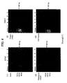
- FIG. 4 (1) shows the results of the detection from the saliva
- FIG. 4(2) shows the results of the detection from the tongue plaque
- FIG. 4(3) shows the results of the detection from the subgingival plaque
- FIG. 4(4) shows the results of the detection from the oral mucosa.
- Lane 1 represents a marker
- Lane 2 represents Subject A
- Lane 3 represents Subject B
- Lane 4 represents Subject C
- Lane 5 represents Subject D
- Lane 6 represents Subject E.
- About 900 bp band was observed in 3 out of 5 subjects with the saliva, 4 out of 5 with the tongue plaque, all of 5 with the subgingival plaque and 1 out of 5 with the oral mucosa.
- Example 3 Using the subgingival plaque samples subjected in Example 3, a quantitative PCR was conducted.
- As a nucleic acid assay reagent 5 ⁇ l of an SYBR Green (available from Molecular Probe) which had been subjected to a 3000-fold dilution was used. 1 ⁇ l of the primers described below which had been diluted to 12.5 pM with a sterilized ultrapure water was used.
- Example 3 AS a biological sample, the saliva in Example 3 was employed. Both of the PCRs in the 1'st and 2' nd stages involved 40 cycles of the amplification. Otherwise, the procedure similar to that in Example 1 was conducted.
- the results of the electrophoresis are shown in FIG. 5.
- Lane 1 represents a marker
- Lane 2 represents Subject A
- Lane 3 represents Subject B
- Lane 4 represents Subject C
- Lane 5 represents Subject D
- Lane 6 represents Subject E.
- a band showing the amplification product was observed in 3 out of 5 subjects.
- Example 1 A probe obtained in Example 1 was labeled fluorescently by a standard method, and adsorbed on a glass plate. The plate was reacted with the DNA obtained from the saliva sample in Example 3. The results are shown in Table 2. Subject Fn microorganism detection A Detected B Detected C Detected D Not detected E Not detected
- Example 1 A probe obtained in Example 1 was labeled fluorescently by a standard method, and adsorbed on glass beads. The beads were reacted with the DNA obtained from the saliva sample in Example 3, and then allowed to develop a color by a standard method. The results are shown in Table 3. Subject Fn microorganism detection A Detected B Detected C Detected D Not detected E Not detected
Landscapes
- Chemical & Material Sciences (AREA)
- Life Sciences & Earth Sciences (AREA)
- Analytical Chemistry (AREA)
- Proteomics, Peptides & Aminoacids (AREA)
- Organic Chemistry (AREA)
- Zoology (AREA)
- Wood Science & Technology (AREA)
- Health & Medical Sciences (AREA)
- Engineering & Computer Science (AREA)
- Microbiology (AREA)
- Immunology (AREA)
- Molecular Biology (AREA)
- Biotechnology (AREA)
- Biophysics (AREA)
- Physics & Mathematics (AREA)
- Biochemistry (AREA)
- Bioinformatics & Cheminformatics (AREA)
- General Engineering & Computer Science (AREA)
- General Health & Medical Sciences (AREA)
- Genetics & Genomics (AREA)
- Measuring Or Testing Involving Enzymes Or Micro-Organisms (AREA)
Abstract
Description
- The present invention relates to a method for specifically detecting or quantifying Fusobacterium nucleatum, which is a suppurative disease-related bacteria and also is a halitosis-causing bacteria.
- In general, detection of microorganisms such as bacterium has been conducted by cultivation such as proliferative cultivation or separate cultivation, and it is required to make single colonies grow, whereby taking several days or several weeks. Moreover, the isolated microorganisms is further proliferated in order to conduct morphological observation, cell staining such as Gram staining, investigation of various biochemical characteristics including propionic acid production test, saccharide catabolism test and the like. Thus, the identification of microorganisms employing the cultivation method has involved facility-dependency, time-consuming and less cost-efficient.
- As a method to overcome defects of the cultivation method, a PCR (polymerase chain reaction) method has recently been employed for the identification of microorganisms. The PCR method permits a rapid and high sensitive identification of microorganisms. However, the PCR method needs to use primers, and detection or identification of a target microorganism is dependent on designs of the primers.
- On the other hand, as bacteria of the Fusobacterium genus which can be isolated from a human oral cavity, there has been known Fusobacterium naviforme, Fusobacterium periodonticum, Fusobacterium necrophorum, Fusobacterium varium, Fusobacterium russii, Fusobacterium nucleatum (hereinafter referred to as Fn microorganism) and the like.
- The Fn microorganism, which is also known as a halitosis-causing bacterium, decomposes a sulfur-containing amino acid and a peptide containing such a sulfur-containing amino acid which are present in an oral food debris or detached mucosa to produce volatile sulfide compounds (hereinafter referred to as VSC). To the contrary, the VSC production ability of any bacteria of the Fusobacterium genus other than the Fn microorganism is not as high as that of the Fn microorganism. In addition, the Fn microorganism is suggested to be related with periodontal diseases since it has a specific ability of being coaggregated with periodontal pathogens such as Porphyromonas gingivalis and Prevotella intermedia.
- Accordingly, the Fn microorganism is a particularly important strain among the bacteria of the Fusobacterium genus. Nevertheless, the Fn microorganism is very difficult to be isolated for the culture, since it has a remarkably analogous relationship with other bacteria of the Fusobacterium genus, and no selective medium capable of specifically detecting the Fn microorganism has been developed.
- As an attempt to detect and identify an Fn microorganism, a method for detecting a bacterium was reported in which the 16S ribosomal DNA (hereinafter referred to as 16S rDNA) of a test microorganism was extracted and a PCR is conducted using, as primers, the oligonucleotides consisting of several ten nucleotide sequences complementary to parts of this DNA (see Conrads, G. et al., J. Endodontics. 25:433-438, 1997). Nevertheless, the conventional PCR method has not been able to detect the Fn microorganism specifically.
- The present invention provides a method for detecting and/or quantifying Fusobacterium nucleatum by a PCR method employing the following primers (1) and (2):
- (1) a forward primer, comprising a nucleotide sequence which is depicted as a portion of SEQ ID No. 1 and has at least ten nucleotides in length;
- (2) a reverse primer, comprising a nucleotide sequence which is depicted as a portion of a sequence complementary to SEQ ID No.2 and has at least ten nucleotides in length.
-
- The present invention provides a simple and rapid detection method which is capable of distinguishing an Fn microorganism specifically among the bacteria of the Fusobacterium genus and which also capable of distinguishing from human-derived DNAs contaminating a biological sample.
- Inventors of the present invention investigated the ribosomal DNA of the Fn microorganism to find that the 16S rDNAs are remarkably analogous between closely related species among the Fusobacterium genus, and therefore it is difficult to detect or identify the Fn microorganism by a PCR method simply using primers prepared so as to have a nucleotide sequence in a region of 16S rDNA. On the other hand, a 23S ribosomal DNA (hereinafter referred to as 23S rDNA) is analogous with the human gene DNA, and therefore it is not desirable for a case of analyzing a biological sample such as a supragingival plaque, subgingival plaque, tongue plaque, saliva, gingival crevice fluide, blood, cerebrospinal fluid, suppurative focus or other tissues.
- Hence, in the present invention, a PCR method is conducted in a combination use of "a forward primer with a consecution of 10 or more nucleotides" which has a nucleotide sequence contained in the 16S rDNA and/or an adjacent spacer (a region represented by SEQ ID No. 1) and "a reverse primer with a consecution of 10 or more nucleotides" which has a nucleotide sequence contained in the 23S rDNA (a sequence complementary to a region depicted as SEQ ID No. 2). Thus, such a combination use makes it possible to achieve a simple and rapid detection, which is capable of specifically distinguishing the Fn microorganism and also capable of distinguishing from human-derived DNAs contaminating a biological sample.
- Brief descriptions of the accompanying drawings are as follows:
- FIG. 1 shows the structure of the DNA encoding an rRNA;
- FIG. 2 shows the results of the electrophoresis of a PCR amplification product of Fusobacterium nucleatum in Example 1;
- FIG.3 shows the results of the electrophoresis of amplification products of the bacteria of the Fusobacterium genus other than Fusobacterium nucleatum and also of human-derived cells;
- FIG. 4 shows the results of PCRs of biological samples including (1) saliva, (2) tongue plaque, (3) subgingival plaque and (4) oral mucosa; and,
- FIG. 5 shows the results of a semi-nested PCR of a biological sample.
-
- In the present invention, a forward primer comprising a nucleotide sequence which is depicted as a portion of SEQ ID No.1 and has at least 10 nucleotides in length has, as its partial or entire sequence, a nucleotide sequence which relates to SEQ ID No.1 and has at least 10 nucleotides in length.
- More specifically, the forward primer has, at its 3' terminal, a nucleotide sequence having at least 10 nucleotides and being same as a part of SEQ ID No. 1, and its 5' terminal may be extended by an additional sequence not relating to any part of SEQ ID No. 1.
- It is not limited to specific ones with proviso that it is capable of hybridizing with a complementary strand of SEQ IDNo. 1 in a condition of the PCR method described hereaf ter, and not capable of hybridizing with a DNA derived from a normal human gingival fibroblast cell.
- The forward primer is preferably designed so as to adapt to the following restriction: namely, it is preferable for a length of the primer to be in a range of 10 nucleotides or more and 40 nucleotides or less, more preferably 12 nucleotides or more and 30 nucleotides or less, and even more preferably 15 nucleotides or more and 25 nucleotides or less. The primer may be designed so as to have a value of Tm within a preferable range of 40 °C or more and 65 °C or less, and more preferably 50 °C or more and 60 °C or less. GC content is preferably in a range of 25 % or more and 70 % or less, and more preferably 40 % or more and 60 % or less.
- Preferable examples of the forward primer include:
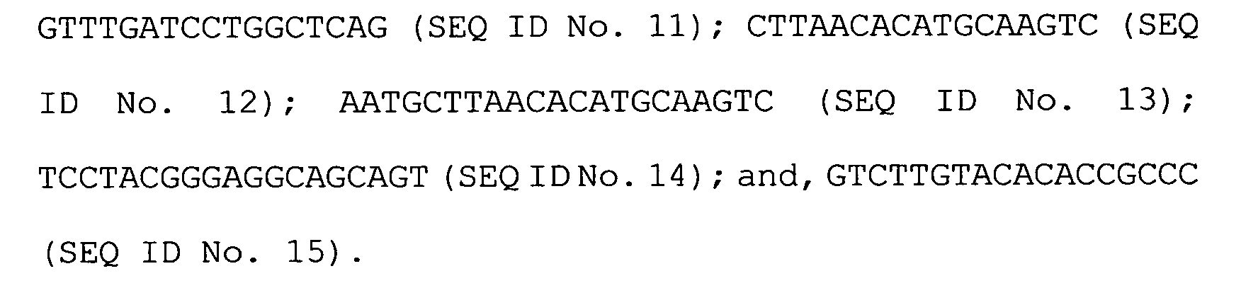
- A reverse primer, comprising a nucleotide sequence which is depicted as a portion of a sequence complementary to SEQ ID No.2 and has at least ten nucleotides in length has, as its partial or entire sequence, a nucleotide sequence which relates to SEQ ID No.2 and has at least 10 nucleotides in length.
- More specifically, the reverse primer has, at its 3' terminal, a nucleotide sequence having at least 10 nucleotides and being complementary to a part of SEQ ID No. 2, and its 5' terminal may be extended by an additional sequence not relating to any part of SEQ ID No. 2.
- It is not limited to specific ones with proviso that it is capable of hybridizing with a DNA strand represented by SEQ ID No. 2 in a condition of the PCR method described hereafter, and not capable of hybridizing with the 23S rDNA derived from any of Fusobacterium periodonticum (ATCC33693), Fusobacterium necrophorum (ATCC25286), Fusobacterium varium (ATCC8501).
- The reverse primer is preferably designed so as to adapt to the following restriction: namely, it is preferable for a length of the primer to be in a range of 10 nucleotides or more and 40 nucleotides or less, more preferably 12 nucleotides or more and 30 nucleotides or less, and even more preferably 15 nucleotides or more and 25 nucleotides or less.
- The primer may be designed so as to have a value of Tm within a preferable range of 40 °C or more and 65 °C or less, and more preferably 50 °C or more and 60 °C or less. GC content is preferably in a range of 25 % or more and 70 % or less, and more preferably 40 % or more and 60 % or less.
- Preferable examples of the reverse primer include:

- In particular, it is preferable to employ a primer set using a combination of either the following primers (A) and (B) or the following primers (B) and (C) in a viewpoint of specifically detecting or identifying the Fn microorganism from a biological sample.
- (A) a primer, comprising a nucleotide sequence which
is depicted as a portion or a whole of a region between
nucleotides 1 to 39 of SEQ ID No.3 and has at least ten nucleotides in length; - (B) a primer, comprising a nucleotide sequence which is depicted as a portion or a whole of a sequence complementary to a region between nucleotides 863 to 883 of SEQ ID No.3 and has at least ten nucleotides in length;
- (C) a primer, consisting of a nucleotide sequence which
is depicted as a region between
nucleotides 1 to 18 of SEQ ID No.3. -
- Since a PCR method is conducted using a biological sample containing not only a Fn microorganism-derived DNA but also a human-derived DNA, a nucleotide sequence of
nucleotides 1 to 39 of SEQ ID No. 3, which is one of nucleotide sequences of the primer (A) , is found as a preferable region for distinguishing the DNAs between ordinary bacteria and human. This nucleotide sequence was revealed to be present at a site consisting of 5 nucleotides at the 3' terminal of the 16S rDNA and the 5' terminal side of a spacer region which is formed between the 16S rDNA and the 23S rDNA(See FIG. 1). - Subsequently, the above-mentioned oligonucleotide primers for the amplification by a PCR method were designed and synthesized. When these primers are used in the PCR of a biological sample containing the Fn microorganism, the amplification products specific for the Fn microorganism are exclusively obtained.
- As a sample usable for detection or identification of the Fn microorganism, there may be exemplified oral cavity-related biological samples such as a supragingival plaque, subgingival plaque, tongue plaque, saliva, gingival crevice fluide and the like. In addition, there may also be used other biological samples such as blood, cerebrospinal fluid, suppurative focus or other tissues. The oligonucleotides to be used as primers may be synthesized chemically or may be derived from a natural products.
- The primers (A), (B) and (C), which are preferably used in a PCR method according to the present invention, are discussed below more in detail. In conducting a PCR, an oligonucleotide which has a nucleotide sequence, as its partial or entire sequence, being depicted as a portion or a whole of a region between
nucleotides 1 to 39 of SEQ ID No.3 and having at least ten nucleotides in length and which is defined as a nucleotide sequence of the primer (A), is used as a forward primer, while an oligonucleotide which has a nucleotide sequence, as its partial or entire sequence, being depicted as a portion or a whole of a sequence complementary to a region between nucleotides 863 to 883 of SEQ ID No.3 and having at least ten nucleotides in length and which is defined as a nucleotide sequence of the primer (B), is used as a reverse primer. The chain length of the each primer is preferably 10 nucleotides or more and 40 nucleotides or less, more preferably 12 nucleotides or more and 30 nucleotides or less, and even more preferably 15 nucleotides or more and 25 nucleotides or less. - As the primer (A), there may be used for example: the primer (C) having a nucleotide sequence consisting of the following 18 nucleotides AACGTGCGGATGGATCAC; a nucleotide sequence of
nucleotides 1 to 22 of SEQ ID No.3, namely AACGTGCGGATGGATCACCTCC; a nucleotide sequence of nucleotides 7 to 26 of SEQ ID No.3, namely CGGATGGATCACCTCCTTTC; a nucleotide sequence of nucleotides 12 to 31 of SEQ ID No.3, namely GGATCACCTCCTTTCTAAGG; and, a nucleotide sequence of nucleotide 16 to 33 of SEQ ID No.3, namely CACCTCCTTTCTAAGGAG. - In a case of conducting a PCR using one kind of the primer (B) such as a primer consisting of a nucleotide sequence complementary to a region between nucleotides 863 to 883 of SEQ ID No. 3 and the primer (C), the amplification product of the nucleotide sequence represented by SEQ ID No. 3 can specifically be obtained. The length of a major amplification product is calculated to be 883 bp, and a band of about 900 bp indicating the amplification product can be observed in an electrophoresis image (see FIG. 2).
- When the cell count of the Fn microorganism in a sample is extremely small, or when a further clarified electrophoresis image is intended, then a semi-nested PCR method is preferably conducted with the use of the three primers (A), (B) and (D). The primer (D) has a nucleotide sequence, as its partial or entire sequence, being depicted as a portion or a whole of a sequence complementary to a region between nucleotides 124 to 156 of SEQ ID No.3 and having at least ten nucleotides in length. A nucleotide sequence of the primer (D) is present in a site called as a spacer region between the 16S rDNA and the 23S rDNA. The nucleotide sequence of the primer (D) may be consist of 29 nucleotides, and preferably in a range from 15 nucleotides to 25 nucleotides, and there may be used a primer (E) having a nucleotide sequence consisting of 20 nucleotides, as the primer (D). The primer (E) consists of a nucleotide sequence depicted as a sequence complementary to a region between nucleotides 124 to 143 of SEQ ID No.3.
- The semi-nested PCR method comprises two stages of amplification. In the first stage of amplification, an amplification product containing a target region is obtained. Then, the resultant amplification product is used as a template to conduct the second stage of amplification. In this second stage, either one of the forward and the reverse primers (namely, inner primers) to be used is capable of positioning at a site inner than a site where either one of the primers (namely, outer primers) used in the first stage positions, whereby excluding the amplification products derived from the DNA in a region other than the target region.
- In a case of using the primers (A), (B) and (D), the first stage of amplification in the PCR is conducted using the primers (A) and (B) as outer primers. Then, using a part of the sample thus amplified in the first stage, the second stage of amplification is initiated. In the second stage of amplification, the PCR is conducted using the primers (A) and (D) as inner primers. Since a major amplification product in such a case is dependent on the primers employed in the second amplification stage, it corresponds to a part of the nucleotide sequence represented by SEQ ID No. 3.
- As to the outer primers usable in the first amplification stage, oligonucleotides other than the primer (A) or (B) may be used. For example, an oligonucleotide designed based on the nucleotide sequence of the 16S rDNA such as CGTCACACCACGAGAGTTGG (namely, nucleotides 1384 to 1403 of SEQ ID No. 1) and the like can be used instead of the primer (A). Also instead of the primer (B), an oligonucleotide having a nucleotide sequence complementary to a nucleotide sequence designed based on the 23S rDNA such as CCATTTGGGTGATGGC (namely, nucleotide 597 to 612 of SEQ ID No.2 ) can be used as the outer primer.
- The primers according to the present invention can be applied also to a nested PCR method. The nested PCR method is different from the semi-nested PCR method in that it uses an inner primer set in which respective inner primers to be used for the both sides are capable of positioning at inner sites on the target region rather than the outer primers.
- In addition to the primers (C) and (B), the followings are exemplified as the outer primers which can be used in the nested PCR.
- Forward primer: an oligonucleotide which has a nucleotide sequence, as its partial or entire sequence, being depicted as a portion of a region between
nucleotides 1 to 123 of SEQ ID No.3 wherein the portion related to the region of SEQ ID No.3 has at least ten nucleotides in length, or being depicted as a portion of SEQ ID No. 4 wherein the portion related to SEQ ID No. 4 has at least ten nucleotides in length. - Reverse primer: an oligonucleotide which has a nucleotide sequence, as its partial or entire sequence, being depicted as a portion of the 23S rDNA(SEQ ID No.2).
- For example, the first stage of amplification can be conducted using, as outer primers, a primer with a sequence of CGTCACACCACGAGAGTTGG (nucleotides 1384 to 1403 of SEQ ID No. 1) designed based on the nucleotide sequence of the 16S rDNA and, a primer with a complementary nucleotide sequence corresponding to AAGAAGGGTAACCGACTT (nucleotides 1256 to 1273 of SEQ ID No. 2) among the nucleotide sequence of the 23S rDNA. In such a case, the primer set of (A) and (B) or the primer set of (B) and (D), all of which are inventive primers, is preferably used as the inner primers for the purpose of specifically detecting or identifying an Fn microorganism.
- The PCR method is a method to amplifying genes commonly practicedby one skilled in the art, and fundamental techniques are described below briefly.
- A double-stranded DNA used as a template is separated into single-stranded DNAs by heating (the thermal denaturation). Then the annealing process is carried out so as to sandwich a target region of the each single-stranded DNA by hybridized sites in such manner that oligonucleotides referred to as primers are hybridized with sites of the 5' terminal of the respective single-stranded DNAs which have separated by the aforementioned thermal denaturation process and have a complementary relationship with respect to the separated counter part. When a Taq DNA polymerase is made to act in the presence of 4 kinds of dNTP (deoxyribonucleotide triphosphates) as substrates, the nucleotides are added to the 3' terminal of the primer in accordance with the nucleotide sequence of the template, resulting in the extension of the DNA chain (extension reaction). By repeating this reaction, a large amount of the DNA fragment containing the target region can be obtained. For the reaction of primer-synthesis or chain-extension, a dUTP may be used as the substrate instead of the dTTP among the dNTPs.
- In the present invention, the PCR method preferably employs the following temperature condition in a single cycle: a thermal denaturation for converting a double-stranded DNA into a single strand is carried out at 90 to 98 °C; an annealing reaction for hybridizing the primer with the template DNA is carried out at 37 to 65 °C; and, an extension reaction for allowing a Taq DNA polymerase to exert the effect is carried out at 50 to 75 °C. By repeating this cycle for several ten times, a target sequence can be amplified. After the PCR, the amplification product is separated for example by an electrophoresis, subjected to a nucleic acid staining for example with ethidium bromide, and then can give a judgement that the microorganism to be detected is present in the sample if the chain length of the amplified polynucleotide sequence is equal to the chain length of the target sequence. For detecting the amplified polynucleotide sequence, a high performance liquid chromatography is also effective.
- When an additional sequence extends from the 5' terminal of an essential part of the nucoleotide sequence ( For example, essential for the primer (A) is a sequence which is depicted as a portion or a whole of a region between
nucleotides 1 to 39 of SEQ ID No. 3 and has at least ten nucleotides in length, or essential for the primer (B) is a sequence which is depicted as a portion or a whole of a sequence complementary to a region between nucleotides 863 to 883 of SEQ ID No.3 and has at least ten nucleotides in length), the additional portion in the primer chain length is imparted to an amplification product. To the contrary, the use of one having a shorter 5' terminal results in an amplification product which is shorter by such a deleted portion. The primers may be labeled with one or more selected from biotin, digoxigenin, FITC (fluorescein isothiocyanate), acridine, dinitrophenyl, alkaline phosphatase, luciferase and [32P]dNTP. - While various biological sample may be suggested as a sample to be subjected to the invention, those which may be exemplified are saliva, tongue plaque, supragingival plaque, subgingival plaque, oral mucosa, gingival crevice fluid, blood, cerebrospinal fluid, suppurative focus sample, feces, urine or cultured materials of these biological materials.
- A biological sample thus obtained can be subjected to a PCR directly or after a DNA extraction. The DNA extraction may be conducted by a conventional method. For example, the DNA extraction may employ one or more of an alkaline extraction or surfactant extraction, enzyme treatment, boiling and the like, and may also employ a commercial kit.
- A sample containing the DNA extracted froman organism's body may further be purified by a conventional method. For example, the DNA purification may employ one or more of a high performance liquid chromatography or alcohol precipitation, salting-out, silica gel column, silica gel membrane and the like, and may also employ a commercial kit.
- The primers and the amplification products are also useful as probes for detecting an Fn microorganism. The amplification products may be converted into DNA fragments having suitable lengths using restriction enzymes and the like. Such restriction enzymes are one or more of RcoR I, Mse I, Ban II, Alu I, Mbo I, Fok I, Taq I, Mbo II, Hinf I and Ava II. A DNA fragment obtained by a restriction enzyme treatment is preferably purified by means of an electrophoresis, high performance liquid chromatography, silica gel column, silica gel membrane, nylon membrane and the like.
- Any of these probes can be labeled with a label. The probe may be labeled with one or more of biotin, digoxigenin, FITC(fluoresceinisothiocyanate),acridine,dinitrophenyl, alkaline phosphatase, luciferase or [32P]dNTP.
- Any of these probe can be bound to a support and employed as a DNA chip. The method for producing a DNA chip may be any conventional method. For example, it may employ a probe alignment type in which a probe is aligned on a chip substrate, or a probe synthesis type in which a DNA extension reaction is performed directly on a substrate such as a glass or silicon to form a probe DNA. Alternatively, a commercial DNA chip producing device may be employed, and the reading may be effected using a DNA chip reader.
- A kit for detecting and identifying a microorganism comprises proves and/or primers according to the present invention. It may also contain other components such as a Taq DNA polymerase, other enzymes, substrates such as dNTPs and the like, as well as buffer solutions.
- The present invention is further described in the following Examples which are not intended to restrict the invention.
- Using the following primers, a standard microorganism of Fusobacterium nucleatum was detected.


- Three types of standard microorganisms of Fusobacterium nucleatum (Fusobacterium nucleatum ATCC25586, ATCC10953, ATCC23726) were incubated anaerobically in a modified FMmedium (available from NISSUI PHARMACEUTICAL CO., LTD.) at 37 °C for 3 to 5 days, and the colonies formed were taken at one supatula-full amount of platinum loop and processed using a commercial kit (QIAGEN K.K., DNA mini-kit) to obtain a DNA extract. 1 µl of the DNA extract was combined with 1.5 µl of 50 mM MgCl2 solution, each 2 µl of 2 mM dNTP solutions (Applied Biosystems) , 1 µl of a 12.5 pmol/µl forward primer solution, 1 µl of a 12.5 pmol/µl reverse primer solution, 0.5 µl of a 5 U/µl Taq DNA polymerase solution (Applied Biosystems) , 5 µl of an Ampli Taq Gold buffer solution (Applied Biosystems) and a sterilized ultrapure water at a volume to obtain the total volume of 50 µl, thereby obtaining a reaction solution.
- Conditions of the PCR reaction was as described below, and the cycle of the PCR employing such conditions was repeated 40 times.
Thermal denaturation: 94 °C, 30 seconds.
Annealing: 55 °C, 30 seconds.
Extension reaction: 72 °C, 30 seconds. - The procedure was conducted using a GeneAmp 9700 system of Applied Biosystems. The presence or absence of the amplification products was determined by a conventional agarose electrophoresis. The agarose gel for the electrophoresis was provided, and placed in a running device which had previously been filled with a TAE (tris-acetate, ethylenediamine tetraacetic acid) buffer solution, and then the PCR reaction product was applied onto the sample groove. After running for 15 minutes at 100 V, the agarose gel was soaked in an ethidium bromide solution for 30 minutes to stain the nucleic acid. After the staining, the bands of the amplification products were identified using an UV transilluminater. The results of the electrophoresis are shown in FIG. 2. In the figure,
Lane 1 represents a marker,Lane 2 represents ATCC25586 strain,Lane 3 represents ATCC10953 strain andLane 4 represents ATCC23726 strain. All of the three microorganisms of Fusobacterium nucleatum exhibited the band representing the amplification product of about 900 bp. - Using the primers shown below, the test was performed similarly to Example 1.
- The primers employed in this Example are shown below.


- Fusobacterium nucleatum (ATCC25586 strain)exhibited the band representing the amplification product of about 1500 bp.
- Test bacteria and test cells subjected are 5 bacteria other than the Fn microorganism, namely, F.naviforme (ATCC25832), F.periodonticum (ATCC33693), F.necrophorum (ATCC25286), F.varium (ATCC8501), F.russii (ACTT25533) as well as normal human gingival fibroblast (ATCC CRL1292).
- The normal human gingival fibroblast was incubated for 72 hours at 37 °C under an atmosphere of 5% CO2 in a modified EAGLE medium (available from Dulbecco) supplemented with 10% fetal bovine serum, and then recovered, washed with a physiological saline, and then nucleic acid was isolated. Otherwise, the bacteria strains were processed similarly to Example 1.
- The results of the electrophoresis are shown in FIG. 3. In the electrophoresis images,
Lane 1 represents a marker,Lane 2 represents F.naviforme,Lane 3 represents F.periodonticum,Lane 4 represents F.necrophorum,Lane 5 represents F.varium, Lane 6 represents F.russii, and Lane 7 represents the normal human gingival fibroblast. No lane exhibited a band showing the amplification product. - Saliva, tongue plaque, subgingival plaque and oral mucosa were taken as biological samples from 5 subjects. The sampling method is as described below.
- 1 ml of saliva was taken and centrifuged at 5000 g for 5 minutes. The supernatant was discarded, and the residue was combined with 1 ml of PBS (phosphate buffered saline) and resuspended. This procedure was repeated three times and the sample after being washed was subjected to the DNA extraction.
- A lingual brush was used to rub the tongue to collect the tongue plaque. A 10 mg aliquot of the tongue plaque was combined with 1 ml of PBS, suspended and centrifuged at 5000 g for 5 minutes. The supernatant was discarded, and the residue was combined with 1 ml of new PBS, resuspended, and centrifuged again at 5000 g for 5 minutes. This procedure was repeated three times, and then the sample after being washed was subjected to the DNA extraction.
- 2 Paper points (available from United Dental Manufacturers Inc) were inserted into a single periodontal pocket to collect the subgingival plaque. These paper points were shaken under vortex vigorously for 1 minute in 1 ml of PBS to recover the deposited materials. The paper points were removed and the suspension was centrifuged at 5000 g for 5 minutes. The supernatant was discarded, and the residue was combined with 1 ml of PBS to resuspend the sample, which was centrifuged at 5000 g for 5 minutes. This procedure was repeated three times, and the sample after being washed was subjected to the DNA extraction.
- A seed swab was used to collect a buccal mucosa. The collected material was suspended in 1 ml of PBS. The suspension was centrifuged at 5000 g for 5 minutes. The supernatant was discarded, and the residue was combined with 1 ml of PBS to resuspend the sample, which was centrifuged at 5000 g for 5 minutes. This procedure was repeated three times and the sample after being washed was subjected to the DNA extraction.
- Otherwise, the procedure similar to that in Example 1 was employed. The results of the electrophoresis are shown in Figures 4 (1) to 4 (4). FIG. 4 (1) shows the results of the detection from the saliva, FIG. 4(2) shows the results of the detection from the tongue plaque, FIG. 4(3) shows the results of the detection from the subgingival plaque and FIG. 4(4) shows the results of the detection from the oral mucosa. In each figure which shows the electrophoresis images of respective collected samples,
Lane 1 represents a marker,Lane 2 represents Subject A,Lane 3 represents Subject B,Lane 4 represents Subject C,Lane 5 represents Subject D and Lane 6 represents Subject E. About 900 bp band was observed in 3 out of 5 subjects with the saliva, 4 out of 5 with the tongue plaque, all of 5 with the subgingival plaque and 1 out of 5 with the oral mucosa. - Using the subgingival plaque samples subjected in Example 3, a quantitative PCR was conducted. As a nucleic acid assay reagent, 5 µl of an SYBR Green (available from Molecular Probe) which had been subjected to a 3000-fold dilution was used. 1 µl of the primers described below which had been diluted to 12.5 pM with a sterilized ultrapure water was used.


- Other reagents were similar to the reaction solution components in Example 1. They were mixed to prepare a reaction solution, which was then subjected to a quantitative PCR using an ABI prism 7000 system of Applied Biosystems. The results are shown in Table 1.
Subject Bacterial count (copies/collection site) A 1.9E + 04 B 5.8E + 06 C 2.5E + 04 D 3.1E + 02 E 8.0E + 01 - Using the primers shown below, a semi-nested PCR consisting of a 2-stage PCR was conducted.
- Primers for 1'st stage PCR:


- Primers for 2'nd stage PCR:


- AS a biological sample, the saliva in Example 3 was employed. Both of the PCRs in the 1'st and 2' nd stages involved 40 cycles of the amplification. Otherwise, the procedure similar to that in Example 1 was conducted. The results of the electrophoresis are shown in FIG. 5. In the figure which shows the electrophoresis images of respective collected samples,
Lane 1 represents a marker,Lane 2 represents Subject A,Lane 3 represents Subject B,Lane 4 represents Subject C,Lane 5 represents Subject D and Lane 6 represents Subject E. A band showing the amplification product was observed in 3 out of 5 subjects. - Using the primers shown below, a nested PCR consisting of a 2-stage PCR was conducted. The results were similar to those in Example 5.
- Primers for 1'st stage PCR:


- Primers for 2'nd stage PCR:


- A probe obtained in Example 1 was labeled fluorescently by a standard method, and adsorbed on a glass plate. The plate was reacted with the DNA obtained from the saliva sample in Example 3. The results are shown in Table 2.
Subject Fn microorganism detection A Detected B Detected C Detected D Not detected E Not detected - A probe obtained in Example 1 was labeled fluorescently by a standard method, and adsorbed on glass beads. The beads were reacted with the DNA obtained from the saliva sample in Example 3, and then allowed to develop a color by a standard method. The results are shown in Table 3.
Subject Fn microorganism detection A Detected B Detected C Detected D Not detected E Not detected 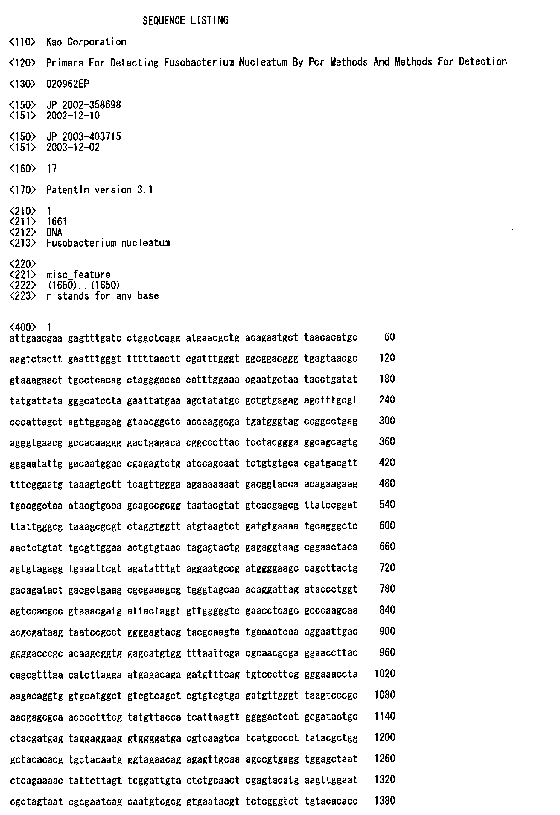
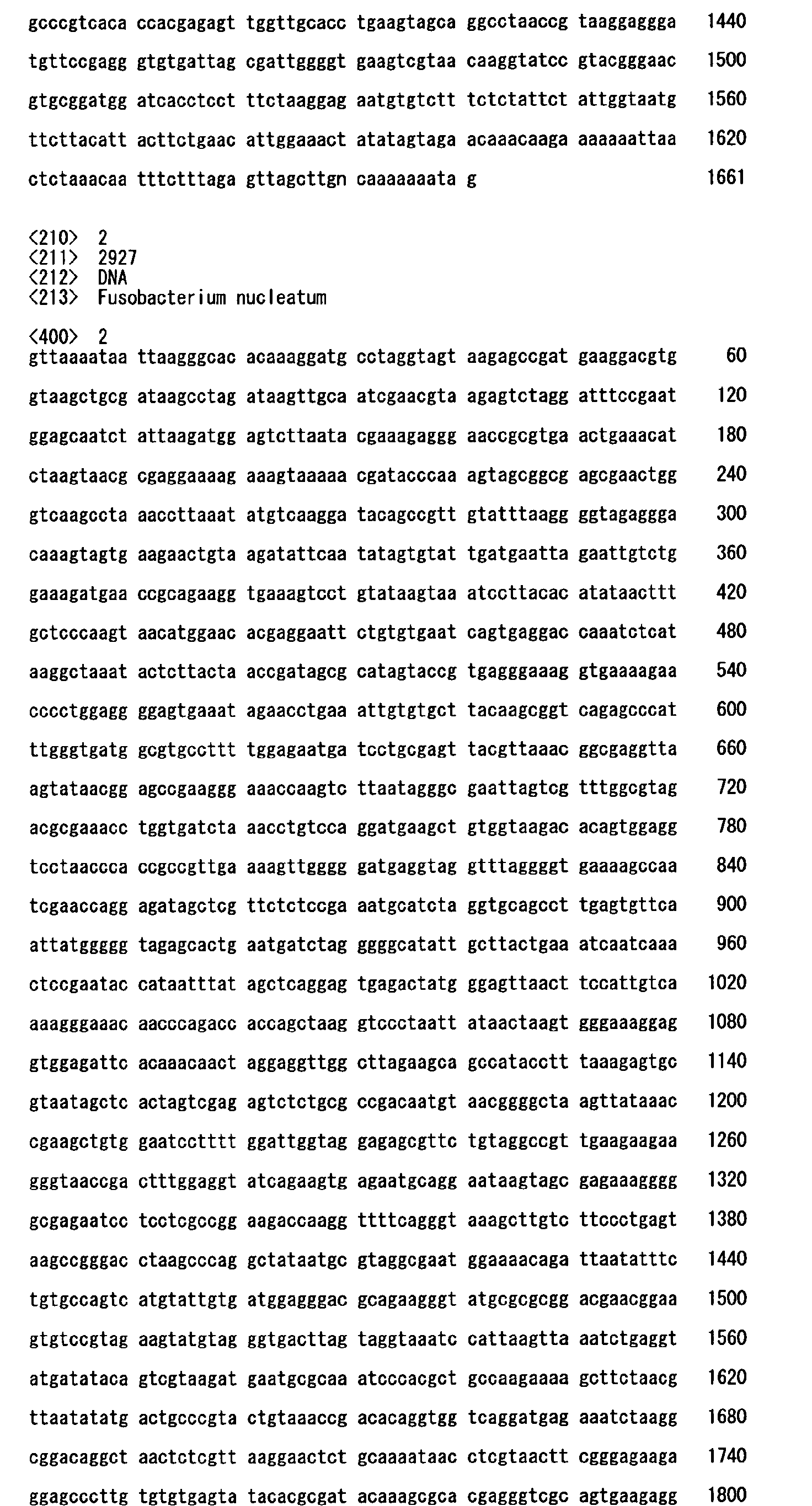
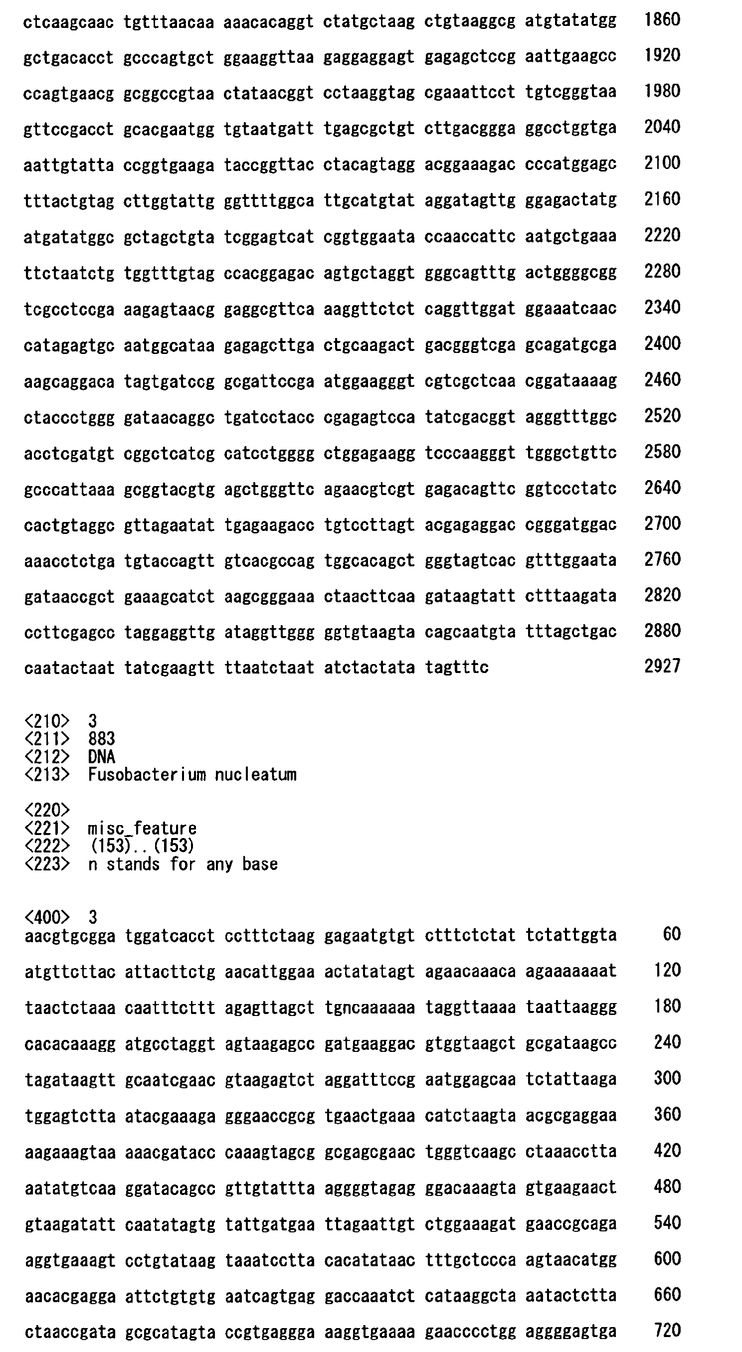
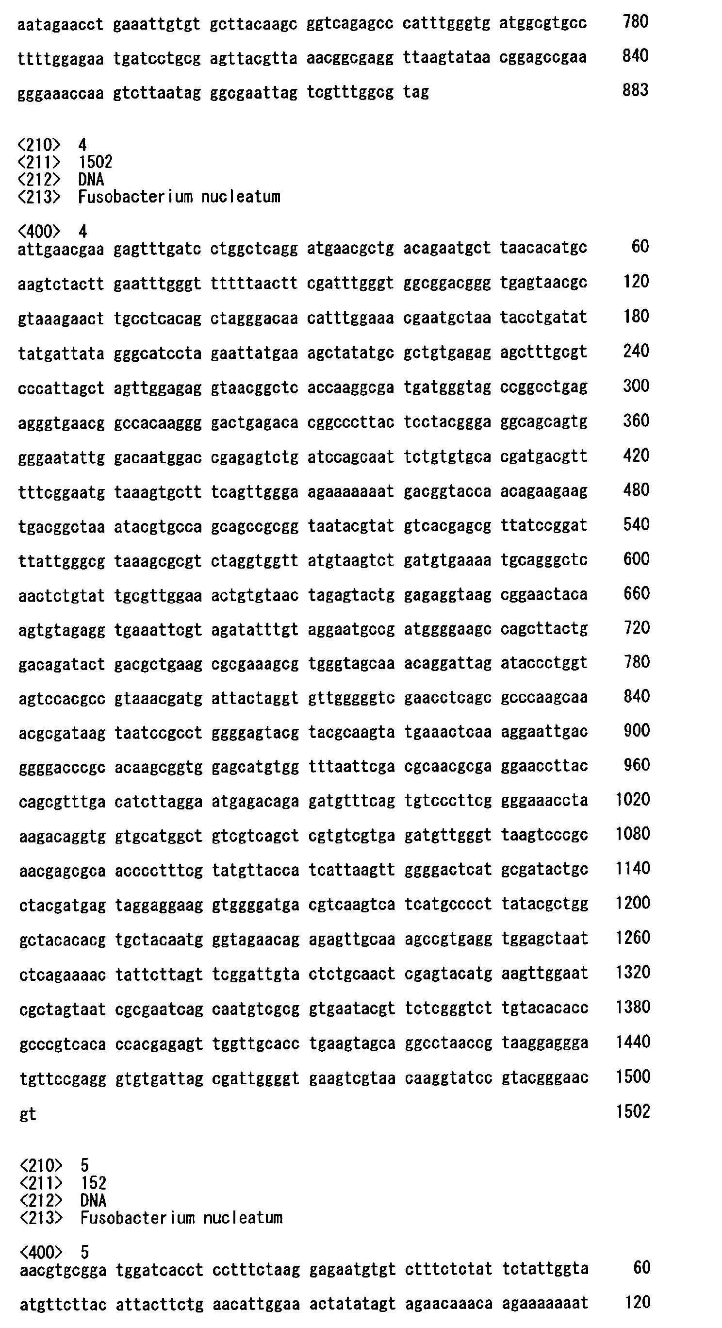


Claims (10)
- A method for detecting and/or quantifying Fusobacterium nucleatum by a PCR method, characterized in that the method employs the following primers (1) and (2):(1) a forward primer, comprising a nucleotide sequence which is depicted as a portion of SEQ ID No. 1 and has at least ten nucleotides in length;(2) a reverse primer, comprising a nucleotide sequence which is depicted as a portion of a sequence complementary to SEQ ID No.2 and has at least ten nucleotides in length.
- A primer set for detecting and/or quantifying Fusobacterium nucleatum by a PCR method, characterized in that the primer set comprises the following primers (A) and (B):(A) a primer, comprising a nucleotide sequence which is depicted as a portion or a whole of a region between nucleotides 1 to 39 of SEQ ID No.3 and has at least ten nucleotides in length;(B) a primer, comprising a nucleotide sequence which is depicted as a portion or a whole of a sequence complementary to a region between nucleotides 863 to 883 of SEQ ID No.3 and has at least ten nucleotides in length.
- A primer set for detecting and/or quantifying Fusobacterium nucleatum by a PCR method, characterized in that the primer set comprises the following primers (C) and (B) :(C) a primer, consisting of a nucleotide sequence which is depicted as a region between nucleotides 1 to 18 of SEQ ID No.3;(B) a primer, comprising a nucleotide sequence which is depicted as a portion or a whole of a sequence complementary to a region between nucleotides 863 to 883 of SEQ ID No.3 and has at least ten nucleotides in length.
- A gene amplification product specific for Fusobacterium nucleatum obtained by conducting a PCR method using a primer set, characterized in that the primer set comprises either the following primers (A) and (B) or the following primers (B) and (C) together with a ribosomal DNA of Fusobacterium nucleatum as a template:(A) a primer, comprising a nucleotide sequence which is depicted as a portion or a whole of a region between nucleotides 1 to 39 of SEQ ID No.3 and has at least ten nucleotides in length;(B) a primer, comprising a nucleotide sequence which is depicted as a portion or a whole of a sequence complementary to a region between nucleotides 863 to 883 of SEQ ID No.3 and has at least ten nucleotides in length;(C) a primer, consisting of a nucleotide sequence which is depicted as a region between nucleotides 1 to 18 of SEQ ID No.3.
- The gene amplification product according to claim 4 wherein the gene amplification product is a probe labeled with a labeling substance.
- The gene amplification product according to claim 5, wherein the labeling substance is at least one selected from the group consisting of biotin, digoxigenin, FITC, acridine, dinitrophenyl, luciferase, alkaline phosphatase and [32P]dNTP.
- A method for detecting and/or quantifying Fusobacterium nucleatum, characterized in that the method comprises:a first step of conducting a PCR method using primers containing the following primers (A) and (B),(A) a primer, comprising a nucleotide sequence which is depicted as a portion or a whole of a region between nucleotides 1 to 39 of SEQ ID No.3 and has at least ten nucleotides in length;(B) a primer, comprising a nucleotide sequence which is depicted as a portion or a whole of a sequence complementary to a region between nucleotides 863 to 883 of SEQ ID No.3 and has at least ten nucleotides in length; and,a second step of conducting a PCR method using primers containing the primer (A) and the following primer (D) or (E) together with an amplification product obtained in the first step as a template,(D) a primer, comprising a nucleotide sequence which is depicted as a portion or a whole of a sequence complementary to a region between nucleotides 124 to 156 of SEQ ID No.3 and has at least ten nucleotides in length;(E) a primer, consisting of a nucleotide sequence which is depicted as a sequence complementary to a region between nucleotides 124 to 143 of SEQ ID No.3.
- A method for detecting and/or quantifying Fusobacterium nucleatum, characterized in that the method comprises:a first step of conducting a PCR method using primers containing the following primers (F)and (B),(F) a primer, comprising a nucleotide sequence which is depicted as a portion of SEQ ID No. 4 and has at least ten nucleotides in length,(B) a primer, comprising a nucleotide sequence which is depicted as a portion or a whole of a sequence complementary to a region between nucleotides 863 to 883 of SEQ ID No.3 and has at least ten nucleotides in length; and,a second step of conducting a PCR method using primers containing the following primers (A) and (D) together with an amplification product obtained in the first step as a template.(A) a primer, comprising a nucleotide sequence which is depicted as a portion or a whole of a region between nucleotides 1 to 39 of SEQ ID No.3 and has at least ten nucleotides in length,(D) a primer, comprising a nucleotide sequence which is depicted as a portion or a whole of a sequence complementary to a region between nucleotides 124 to 156 of SEQ ID No.3 and has at least ten nucleotides in length.
- A gene amplification product specific for Fusobacterium nucleatum obtained by conducting a PCR method, characterized in that the method uses the following primer(A) and the following primer (D) or (E) together with a DNA containing a nucleotide sequence of SEQ ID No. 5 as a template.(A) a primer, comprising a nucleotide sequence which is depicted as a portion or a whole of a region between nucleotides 1 to 39 of SEQ ID No.3 and has at least ten nucleotides in length;(D) a primer, comprising a nucleotide sequence which is depicted as a portion or a whole of a sequence complementary to a region between nucleotides 124 to 156 of SEQ ID No.3 and has at least ten nucleotides in length;(E) a primer, consisting of a nucleotide sequence which is depicted as a sequence complementary to a region between nucleotides 124 to 143 of SEQ ID No.3.
- A probe having a nucleotide sequence depicted as SEQ ID No. 5.
Applications Claiming Priority (4)
| Application Number | Priority Date | Filing Date | Title |
|---|---|---|---|
| JP2002358698 | 2002-12-10 | ||
| JP2002358698 | 2002-12-10 | ||
| JP2003403715A JP2004201679A (en) | 2002-12-10 | 2003-12-02 | Primer for detecting Fusobacterium nucleatum by PCR and method for detecting the same |
| JP2003403715 | 2003-12-02 |
Publications (1)
| Publication Number | Publication Date |
|---|---|
| EP1443118A1 true EP1443118A1 (en) | 2004-08-04 |
Family
ID=32658560
Family Applications (1)
| Application Number | Title | Priority Date | Filing Date |
|---|---|---|---|
| EP03028521A Withdrawn EP1443118A1 (en) | 2002-12-10 | 2003-12-10 | Primers for detecting Fusobacterium nucleatum by PCR methods and methods for detection |
Country Status (3)
| Country | Link |
|---|---|
| US (1) | US20040197849A1 (en) |
| EP (1) | EP1443118A1 (en) |
| JP (1) | JP2004201679A (en) |
Cited By (1)
| Publication number | Priority date | Publication date | Assignee | Title |
|---|---|---|---|---|
| CN101270381B (en) * | 2008-05-14 | 2011-04-13 | 肖水清 | Multiple PCR fast detecting method for oral cavity pathogen |
Families Citing this family (14)
| Publication number | Priority date | Publication date | Assignee | Title |
|---|---|---|---|---|
| WO2004060278A2 (en) | 2002-12-06 | 2004-07-22 | Isis Pharmaceuticals, Inc. | Methods for rapid identification of pathogens in humans and animals |
| US20030027135A1 (en) | 2001-03-02 | 2003-02-06 | Ecker David J. | Method for rapid detection and identification of bioagents |
| US7666588B2 (en) | 2001-03-02 | 2010-02-23 | Ibis Biosciences, Inc. | Methods for rapid forensic analysis of mitochondrial DNA and characterization of mitochondrial DNA heteroplasmy |
| US7718354B2 (en) | 2001-03-02 | 2010-05-18 | Ibis Biosciences, Inc. | Methods for rapid identification of pathogens in humans and animals |
| US20040121309A1 (en) | 2002-12-06 | 2004-06-24 | Ecker David J. | Methods for rapid detection and identification of bioagents in blood, bodily fluids, and bodily tissues |
| US8046171B2 (en) | 2003-04-18 | 2011-10-25 | Ibis Biosciences, Inc. | Methods and apparatus for genetic evaluation |
| US7666592B2 (en) | 2004-02-18 | 2010-02-23 | Ibis Biosciences, Inc. | Methods for concurrent identification and quantification of an unknown bioagent |
| US8119336B2 (en) | 2004-03-03 | 2012-02-21 | Ibis Biosciences, Inc. | Compositions for use in identification of alphaviruses |
| ES2641832T3 (en) | 2004-05-24 | 2017-11-14 | Ibis Biosciences, Inc. | Mass spectrometry with selective ion filtration by setting digital thresholds |
| US7811753B2 (en) | 2004-07-14 | 2010-10-12 | Ibis Biosciences, Inc. | Methods for repairing degraded DNA |
| JP6579874B2 (en) * | 2015-03-31 | 2019-09-25 | 大日本印刷株式会社 | Oligonucleotide set for periodontal pathogen detection and method for detecting periodontal pathogen |
| CN108103149A (en) * | 2018-03-02 | 2018-06-01 | 四川知几未来健康管理有限公司 | Detection Nucleic acid combinations, kit, chip and its application of a kind of Fusobacterium nucleatum |
| CN111197097A (en) * | 2020-01-18 | 2020-05-26 | 新乡学院 | Method for detecting fusobacterium nucleatum by using PCR-ELISA |
| CN112980979A (en) * | 2021-04-15 | 2021-06-18 | 上海市临床检验中心 | Fusobacterium nucleatum fluorescent quantitative detection kit and preparation method and detection method thereof |
Citations (2)
| Publication number | Priority date | Publication date | Assignee | Title |
|---|---|---|---|---|
| US5693467A (en) * | 1995-05-19 | 1997-12-02 | The American Type Culture Collection | Mycoplasma polymerase chain reaction testing system using a set of mixed and single sequence primers |
| US6261769B1 (en) * | 1998-03-31 | 2001-07-17 | The United States Of America As Represented By The Secretary Of Agriculture | Intergenic spacer target sequence for detecting and distinguishing Chlamydial species or strains |
-
2003
- 2003-12-02 JP JP2003403715A patent/JP2004201679A/en active Pending
- 2003-12-09 US US10/729,961 patent/US20040197849A1/en not_active Abandoned
- 2003-12-10 EP EP03028521A patent/EP1443118A1/en not_active Withdrawn
Patent Citations (2)
| Publication number | Priority date | Publication date | Assignee | Title |
|---|---|---|---|---|
| US5693467A (en) * | 1995-05-19 | 1997-12-02 | The American Type Culture Collection | Mycoplasma polymerase chain reaction testing system using a set of mixed and single sequence primers |
| US6261769B1 (en) * | 1998-03-31 | 2001-07-17 | The United States Of America As Represented By The Secretary Of Agriculture | Intergenic spacer target sequence for detecting and distinguishing Chlamydial species or strains |
Non-Patent Citations (7)
| Title |
|---|
| BARRY T ET AL: "THE 16S/23S RIBOSOMAL SPACER REGION AS A TARGET FOR DNA PROBES TO IDENTIFY EUBACTERIA", PCR METHODS & APPLICATIONS, COLD SPRING HARBOR LABORATORY PRESS, US, vol. 1, 1991, pages 51 - 56, XP000609831, ISSN: 1054-9803 * |
| DATABASE EMBL [online] 2 September 2002 (2002-09-02), "Fusobacterium nucleatum susp. nucleatum ATCC 25586, section 97 of 197 of the complete genome", XP002271994, Database accession no. AE010555 * |
| KAPATRAL ET AL.: "Genome Sequence and Analysis of the Oral Bacterium Fusobacterium nucleatum Strain ATCC 25586", J BACTERIOL, vol. 184, no. 7, April 2002 (2002-04-01), pages 2005 - 2018 * |
| KÜLEKCI ET AL.: "PCR analysis of Actinobacillus actinomycetemcomitans, Porphyromonas gingivalis, Treponema denticola and Fusobacterium nucleatum in middle ear effusion.", ANAEROBE, vol. 7, 2001, pages 241 - 246, XP002271993 * |
| MULLIS ET AL.: "The Polymerase Chain Reaction", 1994, BIRKHAUSER, US, BOSTON, XP002273071 * |
| NEWTON & GRAHAM: "PCR", 1994, SPEKTRUM AKADEM. VERLAG, HEIDELBERG, XP002273072 * |
| SONG Y-L ET AL: "Rapid identification of 11 human intestinal Lactobacillus species by multiplex PCR assays using group- and species-specific primers derived from the 16S-23S rRNA intergenic spacer region and its flanking 23S rRNA", FEMS MICROBIOLOGY LETTERS, AMSTERDAM, NL, vol. 187, no. 2, 15 June 2000 (2000-06-15), pages 167 - 173, XP002225666, ISSN: 0378-1097 * |
Cited By (1)
| Publication number | Priority date | Publication date | Assignee | Title |
|---|---|---|---|---|
| CN101270381B (en) * | 2008-05-14 | 2011-04-13 | 肖水清 | Multiple PCR fast detecting method for oral cavity pathogen |
Also Published As
| Publication number | Publication date |
|---|---|
| JP2004201679A (en) | 2004-07-22 |
| US20040197849A1 (en) | 2004-10-07 |
Similar Documents
| Publication | Publication Date | Title |
|---|---|---|
| US20040197849A1 (en) | Primers for detecting fusobacterium nucleatum by PCR methods and methods for detection | |
| JP2008154581A (en) | Primer for bacterial genome amplification reaction | |
| Lew et al. | Detection of Pseudomonas pseudomallei by PCR and hybridization | |
| US7381547B2 (en) | Methods and compositions to detect bacteria using multiplex PCR | |
| EP0910666B1 (en) | Quantitation of rna transcripts using genomic dna as the internal amplification competitor | |
| KR101990163B1 (en) | Method for detecting toxin-producing clostridium difficile | |
| JP7575460B2 (en) | Polynucleotide, polynucleotide set, method for detecting Porphyromonas gingivalis, method for evaluating possibility of suffering from periodontal disease, kit for detecting Porphyromonas gingivalis, and kit for evaluating possibility of suffering from periodontal disease | |
| JP5562007B2 (en) | Method for detecting fungi belonging to the genus Geosmithia | |
| JP4022045B2 (en) | Gene for detecting bacteria and detection method using the same | |
| KR101961763B1 (en) | Composition for detecting of the green mold in mushroom and detecting method using the same | |
| JPH05276999A (en) | Oligonucleotide for detecting bacterium belonging to genus campylobacter, method for detecting bacterium belonging to genus campylobacter and reagent kit for detection | |
| KR102612101B1 (en) | Primer set for detecting Rhodococcus fascians | |
| JPWO2000009683A1 (en) | Genes for detecting bacteria and detection methods using the same | |
| US5432055A (en) | Detection of Porphyromonas gingivalis | |
| JP2005229839A (en) | Method for detecting and identifying lactic acid bacterium | |
| KR101425149B1 (en) | Improved method for diagnosing Mycobacterium tuberculosis using one-tube nested real-time PCR | |
| EP1975257B1 (en) | A method for measuring the number of oral lactobacillus, and a PCR primers-probe set used for the same | |
| US7754870B2 (en) | Method and compositions for detecting Helicobacter pylori | |
| JP4766878B2 (en) | Detection, identification and discrimination of eubacteria using hybridization assays | |
| KR20230083317A (en) | Cystoisospora suis detection method | |
| KR102737931B1 (en) | Specific primer set for detecting Cutibacterium acnes and uses thereof | |
| KR102737930B1 (en) | Specific primer set for detecting Staphylococcus epidermidis and uses thereof | |
| KR101925827B1 (en) | Composition for detecting aggregatibacteractinomycetemcomitans and uses thereof | |
| Kolman | Molecular Anthropology: Progress and Perspectives on Ancient DNA Technology | |
| EP0857219A1 (en) | Microsatellite sequences for canine genotyping |
Legal Events
| Date | Code | Title | Description |
|---|---|---|---|
| PUAI | Public reference made under article 153(3) epc to a published international application that has entered the european phase |
Free format text: ORIGINAL CODE: 0009012 |
|
| AK | Designated contracting states |
Kind code of ref document: A1 Designated state(s): AT BE BG CH CY CZ DE DK EE ES FI FR GB GR HU IE IT LI LU MC NL PT RO SE SI SK TR |
|
| AX | Request for extension of the european patent |
Extension state: AL LT LV MK |
|
| 17P | Request for examination filed |
Effective date: 20041207 |
|
| AKX | Designation fees paid |
Designated state(s): DE FR GB |
|
| 17Q | First examination report despatched |
Effective date: 20050425 |
|
| RBV | Designated contracting states (corrected) |
Designated state(s): DE FR GB |
|
| GRAP | Despatch of communication of intention to grant a patent |
Free format text: ORIGINAL CODE: EPIDOSNIGR1 |
|
| STAA | Information on the status of an ep patent application or granted ep patent |
Free format text: STATUS: THE APPLICATION IS DEEMED TO BE WITHDRAWN |
|
| 18D | Application deemed to be withdrawn |
Effective date: 20071211 |