Field of the invention
-
The present application is directed to a method for performing a bisulfite reaction to
determine methylation positions in a nucleic acid, i.e. methylated and non-methylated
cytosines, whereby the nucleic acid is incubated in a solution comprising the nucleic acid
for a time period of 1.5 to 3.5 hours at a temperature between 70 to 90 °C, whereby the
concentration of bisulfite in the solution is between 3 M to 6.25 M and whereby the pH
value of the solution is between 5.0 to 6.0 whereby the nucleic acid, i.e. the cytosine bases in
the nucleic acid, is deaminated. Then the solution comprising the deaminated nucleic acid
is desulfonated and preferably desalted. The application is further related to kit with a
solution comprising bisulfite with a certain pH and uses thereof as well as a kit comprising
the solution.
Background of the invention
-
Genes constitute only a small proportion of the total mammalian genome, and the precise
control of their expression in the presence of an overwhelming background of noncoding
deoxyribonucleic acid (DNA) presents a substantial problem for their regulation.
Noncoding DNA, containing introns, repetitive elements, and potentially active
transposable elements requires effective mechanisms for its long term silencing. Mammals
appear to have taken advantage of the possibilities afforded by cytosine methylation to
provide a heritable mechanism for altering DNA-protein interactions to assist in such
silencing. DNA methylation is essential for the development of mammals and plays a
potential role during aging and cancer. The involvement of methylation in the regulation of
gene expression and as an epigenetic modification marking imprinted genes is well
established. In mammals, methylation occurs only at cytosine residues and more
specifically only on cytosine residues adjacent to a guanosine residue, i.e. at the sequence
CG. The detection and mapping of DNA methylation sites are essential steps towards
understanding the molecular signals which indicate whether a given sequence is
methylated.
-
This is currently accomplished by the so-called bisulfite method described by Frommer,
M., et al., Proc. Natl. Acad. Sci. USA 89 (1992) 1827-1831, for the detection of 5-methyl-cytosines.
The bisulfite method of mapping 5-methylcytosine uses the effect that sodium
hydrogen sulfite reacts with cytosine but not or only poorly with 5-methyl-cytosine.
Cytosine reacts with bisulfite to form a sulfonated cytosine reaction intermediate being
prone to deamination resulting in a sulfonated uracil which can be desulfonated to uracil
under alkaline conditions (see Fig. 1). It is common knowledge that uracil has the base
pairing behavior of thymine different to the educt cytosine whereas 5-methylcytosine has
the base pairing behavior of cytosine. This makes the discrimination of methylated or non-methylated
cytosines possible by e.g. bisulfite genomic sequencing (Grigg, G., and Clark, S.,
Bioessays 16 (1994) 431-436; Grigg, G.W., DNA Seq. 6 (1996) 189-198) or methylation
specific PCR (MSP) disclosed in US 5,786,146. Basic studies on the reaction of uracil and
cytosine derivatives with bisulfite have been performed by Shapiro et al., JACS 92 (1970)
422-424.
-
There are various documents addressing specific aspects of the bisulfite reaction.
-
Hayatsu, H., et al., Biochemistry 9 (1970) 2858-28659 reacted uracil, cytosine or their
derivatives with 1 M bisulfite, at a pH value around 6, at 37°C for 24 hours. Hayatsu, H., et
al., J. Am. Chem. Soc. 92 (1970) 724-726 describe the reaction of cytosine with 3 M bisulfite
at a pH value around 6 at a temperature of 80 °C for 30 min. Slae and Shapiro, J. Org.
Chem. 43 (1978) 4197-4200 describe the deamination of cytidine with 1 M bisulfite around
neutral pH at various temperatures whereby the reaction time is not described. There were
no investigations of the deamination of cytosine or methyl-cytosine in nucleic acids in
these documents.
-
Paulin, R., et al., Nud. Acids Res. 26 (1998) 5009-5010 investigate the effects of urea on the
efficiency of bisulfite-mediated sequencing of 5-methylcytosine in DNA. The DNA is
reacted with 3.44 M bisulfite in the presence of 5.36 M urea and 0.5 mM hydroquinone, at
a pH value of 5.0 at a temperature of 55 °C for 15 hours.
-
Raizis, A.M., et al., Anal. Biochem. 226 (1995) 161-1666 disclose a bisulfite method for 5-methylcytosine
mapping that minimizes template degradation. They investigate a method
minimizing template degradation using 5 M bisulfite solutions in the presence of 100 mM
hydroquinone at a pH value of 5 at 50°C. A maximum yield of PCR product was observed
after 4 hours. Other conditions as increased pH and lower temperatures were also
investigated.
-
Grunau, C., et al., Nucleic Acids Res 29 (2001) E65-5, page 1 to 7, perform a systematic
investigation of critical experimental parameters of the bisulfite reaction. They investigate
bisulfite solutions of 3.87 to 4.26 and 5.2 to 5.69 M at a pH value of 5. Temperatures that
were tested are 15, 35, 55, 80, 85 and 95 °C for 1, 4 and 18 hours. DNA degradation is a
problem in these investigations.
-
Wang, R.Y., et al., Nucleic Acids Res. 8 (1980) 4777-4790 disclose the use of a 3 M bisulfite
solution at a pH value of 5.5 at a temperature of 37°C for various time periods in the
bisulfite treatment of DNA. Feil, R., et al., Nucleic Acids Res 22 (1994) 695-696 disclose the
use of a 3.5 M bisulfite solution at a pH value of 5 at a temperature of 0°C for 24 hours in
the bisulfite treatment of DNA. Clark, S.J., et al., Nucleic Acids Res 22 (1994) 2990-2997,
disclose the use of a 3 to 4 M bisulfite solution at a pH value of 4.8 to 5.8 at a temperature
of 37 to 72 °C for 8 to 16 hours in the bisulfite treatment of DNA. Tasheva, E.S., and Roufa,
D.J., Mol. Cell. Biol. 14 (1994) 5636-5644 disclose the use of a 1 M bisulfite solution at a
pH value of 5 at a temperature of 50 °C for 48 hours in the bisulfite treatment of fragments
of genomic DNA. Grigg, G.W., DNA Seq 6 (1996) 189-198 discloses the use of a 3.1 M
bisulfite solution at a pH value of 5 at a temperature of 50 °C for 16 hours in the bisulfite
treatment of DNA. Komiyama, M., and Oshima, S., Tetrahedron Letters 35 (1994) 8185-8188
disclose the use of a 1 M bisulfite solution at a pH value of 5 at a temperature of 37 °C
for 4 hours in the bisulfite treatment of DNA whereby diethylenetriamine is present.
-
Olek, A., et al., Nucleic Acids Res. 24 (1996) 5064-5066 disclose a method for bisulfite base
sequencing whereby bisulfite treatment and subsequent PCR steps are performed on
material embedded in agarose beads. A 5 M bisulfite solution at a pH value of 5 at a
temperature of 50 °C is used for 4 hours in the bisulfite treatment of DNA.
-
A review of DNA methylation analysis can be found in Oakeley, E. J., Pharmacol. Ther. 84
( 1999) 389-400.
-
Different additional components in the bisulfite mixture are disclosed by WO 01/98528,
WO02/31186 or by Paulin, R., et al., Nucleic Acids Res 26 (1998) 5009-5010.
-
Kits for performing bisulfite treatments are commercially available from Intergen, now
distributed by Serologicals Corporation, Norcross, GA, USA, e.g. CpGenome™ DNA
modification kit (http://www.serologicals.com/products/int_prod/index.html).
-
All prior art methods for the bisulfite treatment have disadvantages. Therefore, the
problem to be solved by the present invention was to provide a method which overcomes
the disadvantages of the prior art methods.
Summary of the invention
-
The present invention provides a method for the conversion of cytosine bases in a nucleic
acid to uracil bases whereby 5-methyl-cytosine bases are not significantly converted,
comprising the steps of
- a) incubating a solution comprising the nucleic acid for a time period of 1.5 to 3.5 hours
at a temperature between 70 to 90 °C, whereby the concentration of bisulfite in the
solution is between 3 M to 6.25 M and whereby the pH value of the solution is between
5.0 to 6.0 whereby the nucleic acid is deaminated, and
- b) incubating the solution comprising the deaminated nucleic acid under alkaline
conditions whereby the deaminated nucleic acid is desulfonated.
-
-
Further, the invention provides a solution with a pH value between 5.0 and 6.0 and
comprising bisulfite in a concentration between 3 M to 6.25 M, uses thereof and kits
comprising this solution.
-
As known to the expert skilled in the art and according to the invention, the term
"bisulfite" is used interchangeably for "hydrogensulfite".
-
According to the invention the term a "bisulfite reaction", "bisulfite treatment" or
"bisulfite method" shall mean a reaction for the conversion of cytosine bases in a nucleic
acid to uracil bases in the presence of bisulfite ions whereby 5-methyl-cytosine bases are
not significantly converted. This reaction for the detection of methylated cytosines is
described in detail by Frommer et al., supra and Grigg and Clark, supra. The bisulfite
reaction contains a deamination step and a desulfonation step which can be conducted
separately or simultaneously (see Figure 1; Grigg and Clark, supra). The statement that 5-methyl-cytosine
bases are not significantly converted shall only take the fact into account
that it cannot be excluded that a small percentage of 5-methyl-cytosine bases is converted
to uracil although it is intended to convert only and exclusively the (non-methylated)
cytosine bases (Frommer et al., supra).
-
The method according to the invention allows a relatively short reaction time of the
bisulfite reaction giving the possibility to perform a DNA assay within one working day.
One parameter to speed the reaction is the temperature. To decrease the DNA degradation
process, a low pH value is of advantage. By the use of a 5 M bisulfite solution of a pH value
of 5.5 at a temperature of approximately 80°C, a reaction time of e.g. between 120 and 180
min is possible. Additives for stabilization of the bisulfite reagent like hydroquinone are
possible.
Detailed description of the invention
-
The invention is related to a method for the conversion of cytosine bases in a nucleic acid
to uracil bases whereby 5-methyl-cytosine bases are not significantly converted,
comprising the steps of
- a) incubating a solution comprising the nucleic acid for a time period of 1.5 to 3.5
hours at a temperature between 70 to 90 °C, whereby the concentration of
bisulfite in the solution is between 3 M to 6.25 M and whereby the pH value of
the solution is between 5.0 to 6.0 whereby the nucleic acid is deaminated, and
- b) incubating the solution comprising the deaminated nucleic acid under alkaline
conditions whereby the deaminated nucleic acid is desulfonated.
-
-
In a preferred embodiment of the invention, the method may further comprise the step of
desalting the solution comprising the deaminated and desulfonated nucleic acid. This can
be achieved e.g. by ultrafiltration, gel filtration, precipiation as known to the expert skilled
in the art or by binding to magnetic glass particles as described in WO 96/41811.
-
In a preferred embodiment of the invention, the temperature in the method according to
the invention is between 75 to 85 °C. In another preferred embodiment of the invention,
the concentration of bisulfite is between 3.2 M to 6 M, preferably between 4.75 M to 5.5 M.
In another preferred embodiment of the invention, the pH value of the solution is between
5.25 to 5.75. In another preferred embodiment of the invention, the time period is between
1.75 to 3 hours. In another preferred embodiment of the invention, the time period is
between 2 to 3 hours, preferably 2 to 2.5 hours. The reaction is also possible in a time
period between 0.75 and 3.5 hours. In the most preferred embodiment of the invention, in
step a) the temperature is 80 °C, the concentration of bisulfite is 5 M, the pH value of the
solution is 5.5 and the time period is preferably 2 to 2.5 or 3 hours, most preferred 2 hours.
-
The method is preferably performed in solution, however, it is also feasible that the method
according to the invention is performed while the nucleic acid is in a solid phase bound
form, i.e. it is bound to a solid phase under suitable conditions. The solid phase may be a
silicon oxide, preferably in the form of glass fleeces or fibers or magnetic glass particles as
described in WO96/41811, WO 00/32762 and WO 01/37291. The principal method of
performing a bisulfite treatment while the nucleic acid is bound to a solid phase is e.g.
described in the European patent application with the number EP 02019097.1.
-
The expert skilled in the art knows how to perform the bisulfite reaction, e.g. by referring to
Frommer et al., supra, Grigg and Clark, supra or Grunau et al., supra who disclose the
principal parameters of the bisulfite reaction.
-
In an embodiment of the invention, the nucleic acid is deoxyribonucleic acid (DNA), in
particular genomic DNA or nucleic acid, i.e. the DNA or nucleic acid which is found in the
organism's genome and is passed on to offspring as information necessary for survival. The
phrase is used to distinguish between other types of DNA, such as found within plasmids.
The source of the nucleic acid may be eukaryotic or prokarytic, preferably from vertebrates,
particularly from mammalians, most preferred from animals or humans.
-
In an embodiment of the invention the nucleic acid is obtained from a biological sample
using the solid phases as described above and methods known to the expert in the field. The
biological sample comprises cells from multicellular organisms as e.g. human and animal
cells such as leucocytes, and immunologically active low and high molecular chemical
compounds such as haptens, antigens, antibodies and nucleic acids, blood plasma, cerebral
fluid, sputum, stool, biopsy specimens, bone marrow, oral rinses, blood serum, tissues,
urine or mixtures thereof. In a preferred embodiment of the invention the biological
sample is a fluid from the human or animal body. The biological sample can be blood,
blood plasma, blood serum, tissue or urine. The biological sample comprising the nucleic
acids is lysed to create a mixture of biological compounds comprising nucleic acids and
other components. Procedures for lysing biological samples are known by the expert and
can be chemical, enzymatic or physical in nature. A combination of these procedures is
applicable as well. For instance, lysis can be performed using ultrasound, high pressure,
shear forces, alkali, detergents or chaotropic saline solutions, or proteases or lipases. For the
lysis procedure to obtain nucleic acids, special reference is made to Sambrook, J., et al., in
"Molecular Cloning: A Laboratory Manual" (1989), eds. J. Sambrook, E. F. Fritsch and T.
Maniatis, Cold Spring Harbour Laboratory Press, Cold Spring Harbour, NY; and Ausubel,
F., et al., in "Current protocols in molecular biology" (1994), eds. F. Ausubel, R. Brent and
K. R.E., Wiley & Sons, New York. Then the nucleic acids are isolated from the lysis mixture
using the methods known to the expert skilled in the art, e.g. using solid phases as magnetic
glass particles (WO96/41811), and can then be subjected to the methods according to the
invention, i.e. the bisulfite treatment according to the invention. Chaotropic agents are also
used to lyse cells to prepare a mixture between nucleic acids and other biological substances
(see e.g. Sambrook et al. (1989) or EP 0 389 063). Afterwards a material comprising glass or
silica may be added and a purification effect results from the behavior of DNA or RNA to
bind to material with a glass surface under these conditions i.e. in the presence of certain
concentrations of a chaotropic agent, higher concentrations of organic solvents or under
acidic conditions. Sequence specific capturing can also be used for this purpose.
-
In a preferred embodiment of the invention, the nucleic acid is amplified after the steps of
the method according to the invention with the polymerase chain reaction (PCR: EP 0 201
184; EP-A-0 200 362; US 4,683,202). The amplification method may also be the Ligase
Chain Reaction (LCR: Wu, D. Y., and Wallace, R. B., Genomics 4 (1989) 560-569; and
Barany, F., Proc. Natl. Acad. Sci. USA 88 (1991) 189-193), Polymerase Ligase Chain
Reaction (Barany, F., PCR Methods Appl. 1 (1991) 5-16), Gap-LCR (PCT Patent
Publication No. WO 90/01069), Repair Chain Reaction (European Patent Publication No.
EP-A 0 439 182), 3SR (Kwoh, D. Y., et al., Proc. Natl. Acad. Sci. USA 86 (1989) 1173-1177;
Guatelli, J. C., et al., Proc. Natl. Acad. Sci. USA 87 (1990) 1874-1878; PCT Patent
Publication No. WO 92/0880A), and NASBA (U.S. Pat. No. US 5,130,238). Further, there
are strand displacement amplification (SDA), transciption mediated amplification (TMA),
and Qβ-amplification (for a review see e.g. Whelen, A.C., and Persing, D.H., Annu. Rev.
Microbiol. 50 (1996) 349-373; Abramson, R. D., and Myers, T.W., Curr. Opin. Biotechnol.
4 (1993) 41-47). Particularly preferred amplification methods according to the invention
are the methylation specific PCR method (MSP) disclosed in US 5,786,146 which combines
bisulfite treatment and allele-specific PCR (see e.g. US 5,137,806, US 5,595,890, US
5,639,611).
-
In a preferred embodiment, the method may further comprise the step of detecting the
amplified nucleic acid. The amplified nucleic acid may be determined or detected by
standard analytical methods known to the person skilled in the art and described e.g. by
Sambrook, J., et al., in "Molecular Cloning: A Laboratory Manual" (1989), eds. J.
Sambrook, E. F. Fritsch and T. Maniatis, Cold Spring Harbor Laboratory Press, Cold
Spring Harbor, NY; Lottspeich and Zorbas, in "Bioanalytik" (1998), eds. L. a. Zorbas,
Spektrum Akademischer Verlag, Heidelberg, Berlin, Germany; or by Ausubel, F., et al., in
"Current protocols in molecular biology" (1994), eds. F. Ausubel, R. Brent and K. R.E.,
Wiley & Sons Verlag, New York. There may be also further purification steps before the
target nucleic acid is detected e.g. a precipitation step. The detection methods may include
but are not limited to the binding or intercalating of specific dyes as ethidium bromide
which intercalates into the double-stranded DNA and changes its fluorescence thereafter.
The purified nucleic acids may also be separated by electrophoretic methods optionally
after a restriction digest and visualized thereafter. There are also probe-based assays which
exploit the oligonucleotide hybridisation to specific sequences and subsequent detection of
the hybrid. It is also possible to sequence the target nucleic acid after further steps known
to the expert in the field. Other methods apply a diversity of nucleic acid sequences to a
silicon chip to which specific probes are bound and yield a signal when a complementary
sequences bind.
-
In a particularly preferred embodiment of the invention, the nucleic acid is detected by
measuring the intensity of fluorescence light during amplification. This method entails the
monitoring of real time fluorescence. A particularly preferred method exploiting
simultaneous amplification and detection by measuring the intensity of fluorescent light is
the TaqMan® method disclosed in WO 92/02638 and the corresponding US patents US
5,210,015, US 5,804,375, US 5,487,972. This method exploits the exonuclease activity of a
polymerase to generate a signal. In detail, the nucleic acid is detected by a process
comprising contacting the sample with an oligonucleotide containing a sequence
complementary to a region of the target nucleic acid and a labeled oligonucleotide
containing a sequence complementary to a second region of the same target nucleic acid
strand, but not including the nucleic acid sequence defined by the first oligonucleotide, to
create a mixture of duplexes during hybridization conditions, wherein the duplexes
comprise the target nucleic acid annealed to the first oligonucleotide and to the labeled
oligonucleotide such that the 3'-end of the first oligonucleotide is adjacent to the 5'-end of
the labeled oligonucleotide. Then this mixture is treated with a template-dependent nucleic
acid polymerase having a 5' to 3' nuclease activity under conditions sufficient to permit the
5' to 3' nuclease activity of the polymerase to cleave the annealed, labeled oligonucleotide
and release labeled fragments. The signal generated by the hydrolysis of the labeled
oligonucleotide is detected and/ or measured. TaqMan® technology eliminates the need for
a solid phase bound reaction complex to be formed and made detectable. In more general
terms, the amplification and/ or detection reaction of the method according to the
invention is a homogeneous solution-phase assay. Further preferred method are the
formats used in the LightCycler® instrument (see e.g. US 6,174,670). Particularly preferred
is the use of bisulfite treatment, amplification with or without methylation specific primers
in the presence of a methylation-specific probe and real-time fluorescence detection as
described in US 6,331,393.
-
In a preferred embodiment of the present invention, the method is automated, i.e. the
method carries out an automatable process as e.g. described in WO 99/16781. Automatable
process means that the steps of the process are suitable to be carried out with an apparatus
or machine capable of operating with little or no external control or influence by a human
being. Automated method means that the steps of the automatable method are carried out
with an apparatus or machine capable of operating with little or no external control or
influence by a human being. Only the preparation steps for the method may have to be
done by hand, e.g. the storage containers have to filled up and put into place, the choice of
the samples has to be done by a human being and further steps known to the expert in the
field, e.g. the operation of the controlling computer. The apparatus or machine may e.g.
add automatically liquids, mix the samples or carry out incubation steps at specific
temperatures. Typically, such a machine or apparatus is a robot controlled by a computer
which carries out a program in which the single steps and commands are specified. In a
preferred embodiment of the invention, the method is in a high-throughput format, i.e. the
automated methods is carried out in a high-throughput format which means that the
methods and the used machine or apparatus are optimized for a high-throughput of
samples in a short time.
-
Preferably, the method according to the invention is used in diagnostics, for diagnostic
analysis or for bioanalytics, or for the screening of tissue or fluids from the human or even
animal body for the presence of a certain methylation pattern. Further, the method
according to the invention is used to enhance the speed, accuracy or sensitivity of the
detection of methylation sites in nucleic acids.
-
In another embodiment of the invention, a solution with a pH value between 5.0 and 6.0
and comprising bisulfite in a concentration between 3 M to 6.25 M is used in a reaction
wherein cytosine bases in a nucleic acid are converted to uracil bases in the presence of
bisulfite ions whereby 5-methyl-cytosine bases are not significantly converted. Preferably,
the pH value of the solution is between 5.25 and 5.75 and the concentration of bisulfite is
between 3.2 M to 6 M, preferably between 4.75 M to 5.5 M. In the most preferred
embodiment, the pH value of the solution is 5.5 and the concentration of bisulfite is 5 M.
The solution may also contain hydroquinone for stabilisation. The solution according to
the invention is preferably an aqueous solution.
-
In another embodiment of the invention a kit comprising a solution with a pH value
between 5.0 and 6.0 and comprising bisulfite in a concentration between 3 M to 6.25 M is
provided. Preferably, the pH value of the solution is between 5.25 and 5.75 and the
concentration of bisulfite is between 3.2 M to 6 M, preferably 4.75 M to 5.5 M. The
solution may optionally contain hydroquinone. In the most preferred embodiment, the pH
value of the solution is 5.5 and the concentration of bisulfite is 5 M. Such kits known in the
art further comprise plastics ware which may be used during the bisulfite procedure as e.g.
microtiter-plates in the 96 or 384 well format or reaction tubes manufactured e.g. by
Eppendorf, Hamburg, Germany. The kit may further comprise a washing solution which is
suitable for the washing step of the solid phase, in particular, the glass fleece or membrane
or the magnetic glass particles. Often the washing solution is provided as a stock solution
which has to be diluted before the use. The kit may further comprise an eluent, i.e. a
solution or a buffer (e.g. TE, 10 mM Tris, 1 mM EDTA, pH 8.0) or pure water to elute the
DNA or RNA bound to the solid phase. Further, additional reagents may be present which
contain buffers suitable for use in the present invention. Preferably, the kit according to the
invention is used for a reaction wherein cytosine bases in a nucleic acid are converted to
uracil bases in the presence of bisulfite ions whereby 5-methyl-cytosine bases are not
significantly converted.
-
In another embodiment of the invention, a solution is provided with a pH value between
5.0 and 6.0 and comprising bisulfite in a concentration between 3 M to 6.25 M. The
solution optionally contains hydroquinone or other radical scavengers. Preferably, the pH
value of the solution is between 5.25 and 5.75 and wherein the concentration of bisulfite is
between 3.2 M to 6 M, preferably 4.75 M to 5.5 M. In the most preferred embodiment, the
pH value of the solution is 5.5 and wherein the concentration of bisulfite is 5 M.
-
The following examples, references, sequence listing and figures are provided to aid the
understanding of the present invention, the true scope of which is set forth in the appended
claims. It is understood that modifications can be made in the procedures set forth without
departing from the spirit of the invention.
Description of the Figures
-
-
Fig. 1:
- The steps of the bisulfite method
-
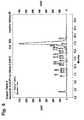
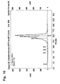
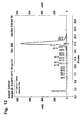
Fig. 2 to 12:
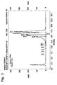
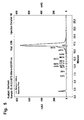
- HPLC profiles of the reaction mixtures after certain time periods as
indicated in the examples
1 Examples
1.1 Comparison optimized conditions with standard conditions using a model system
(oligonucleotide) and analysis by HPLC
1.1.1 Method:
1.1.1.1 Composition of reagents
-
|
Bisulfite reagent pH = 5,0/50°C
|
1,9g Na2S2O5 |
| ("Standard") |
2ml Millipore water |
| |
0,7ml 2M NaOH |
| |
0,5ml 1M hydroquinone (optional) |
| |
addition of Millipore water to a volume of 4 ml |
|
Bisulfite reagent pH = 5,5/80°C
|
1,9g Na2S2O5 |
| |
1ml Millipore water |
| |
2ml 2M NaOH |
| |
0,5ml 1M hydroquinone (optional) |
| |
addition of Millipore water to a volume of 4 ml |
-
Hydroquinone can be added optionally; it is not necessary if the reagent is prepared freshly
for the experiment.
Sequences:
-
The oligonucleotides are synthesized using standard automated solid-phase synthesis
procedure applying phosphoramidite chemistry.
GSTP1 sequence:
-
C or C
Me in the center position of T
10:
1.1.1.2 Reaction conditions:
-
Ca. 5 nmole of a single stranded oligonucleotide or 5 nmole of each strand of a double
stranded oligonucleotide are dissolved in 20µl of Millipore water, then 200µl of the bisulfite
reagent are added. Thereafter the reaction tube is placed into a thermomixer (50°C or 80°C;
600rpm). After t = x hours the reaction is stopped by addition of 500µl of 2,5M NaOH
(desulfonation). After 30min at r.t. the reaction mixture is desalted over a Sephadex G25
column. The oligonucleotide containing fraction is evaporated and dissolved in 200µl of
Millipore water to be analyzed by HPLC.
1.1.1.3 Evaluation
-
|
Analytical HPLC
|
column |
Dionex DNA Pac PA-100 SEL |
| |
buffer A |
| |
0,01M NaOH, 0,2 M NaCl |
| |
buffer B |
| |
0,01M NaOH, 1M NaCl |
| |
gradient |
50-100% B in 25min |
-
Data evaluation: HPLC chromatograms are compared by HPLC-area% of the product
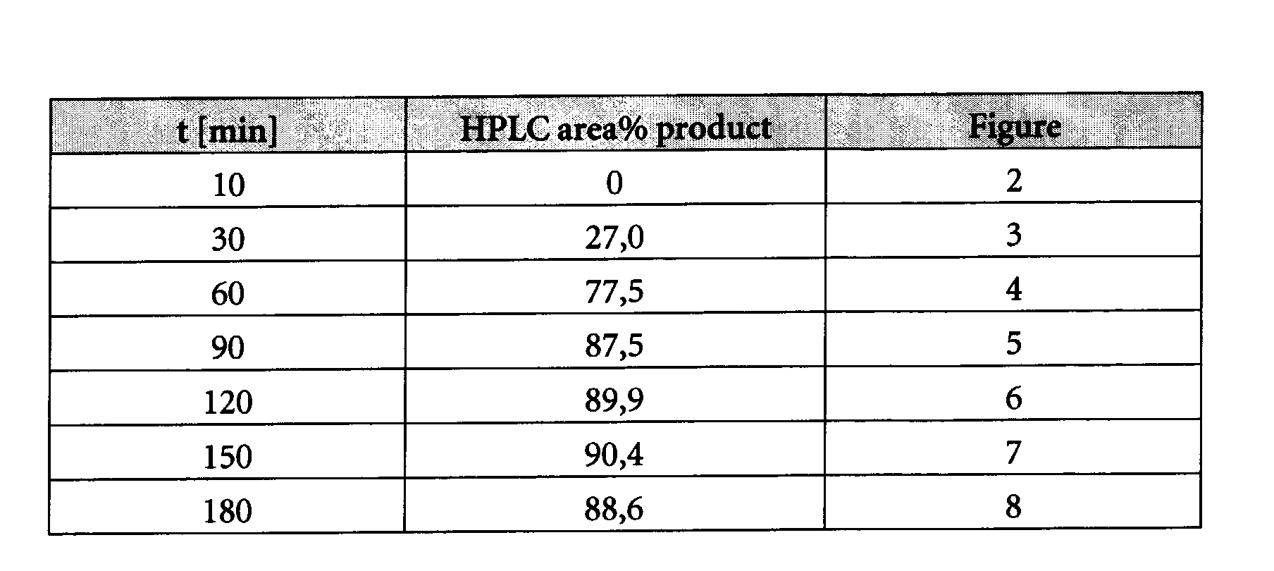
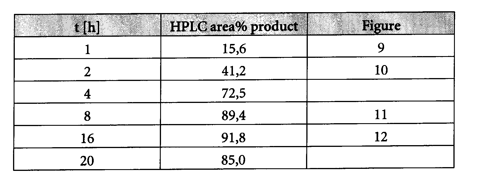
peak at t = x and are shown in Figs. 2 to 12
1.1.2 Results
1.1.2.1 Reaction kinetics GSTP1 ds at T = 80°C, pH = 5,5
-
A double determination was performed according to the standard protocol described above
with the GSTP1 sequences SEQ ID NO:1 which is the sense strand and SEQ ID NO:2 which is
the antisense strand, the mean average values are calculated.
1.1.2.2 Reaction kinetics GSTP1 ds at T = 50°C, pH = 5,0 ("standard
conditions")
-
A double determination was performed according to the standard protocol described above
with the GSTP1 sequences SEQ ID NO:1 which is the sense strand and SEQ ID NO:2 which
is the antisense strand, the mean average values are calculated.
1.1.3 Conclusion
-
Conditions according to the invention lead to a similar product yield after 2 h reaction time
as standard conditions after 8-16 h.
1.2 Comparison of certain conditions of the bisulfite method using PCR of bisulfite
treated genomic DNA
1.2.1 General
-
The fact that the bisulfite reaction has worked and converted non-methylated cytosines to
uracil can also be demonstrated by a polymerase chain reaction whereby primers are used
which are specific to a region of the nucleic acid sequence wherein non-methylated
cytosines have been converted to uracils, i.e. the base adenine in the primer is opposite to
the uracil being the bisulfite reaction product from non-methylated cytosines. In case of
incomplete conversion, the primer could not hybridize to this region as there would be
cytosines not matching the adenine bases in the primer. This would have the effect that no
PCR product would be obtained.
-
An improved method to perform rapid polymerase chain reactions is disclosed e.g. in US
6,174,670 and is used in the LightCycler® instrument (Roche, Mannheim, Germany). In
this method, two labeled probes can come into close proximity in an amplificate dependent
manner so that the two labels can perform a fluorescence energy transfer (FRET). The
amount of the amplificate thereby correlates with the intensity of the emitted light of a
certain wavelength. This specific PCR method can therefore be used to analyze whether a
complete conversion of non-methylated cytosines was obtained, by e.g. analyzing the
promoter region of the glutathion-S-transferase π gene (see e.g. US 5,552,277, Genbank
accession code M24485 and Morrow et al. (1989) Gene 75, 3-11) using suitable probes and
primers. However, the expert skilled in the art knows that other methods can be used for
this evaluation as well. Fluorescence measurements are normalized by dividing by an initial
fluorescence measurement, i.e., the background fluorescence, obtained during a cycle early
in the reaction while the fluorescence measurements between cycles appear to be relatively
constant. The cycle number chosen for the initial fluorescence measurement is the same for
all reactions compared, so that all measurements represent increases relative to the same
reaction cycle. In the early cycles of a polymerase chain reaction amplification, the number
of target molecules can be described by the geometric equation Ni = No x (1 + E)i, where No
= the number of target molecules at the start of the reaction, Ni = the number of target
molecules at the completion of the i-th cycle, E = the efficiency of the amplification (0 ≤ E
≤ 1). During this geometric growth phase of the amplification, the number of cycles
required to reach a particular threshold value (CT value or crossing point) is inversely
proportional to the logarithm of (1 + E). Thus, the CT value represents a measure of the
reaction efficiency that allows comparisons between reactions. A decrease in the CT value,
which means that the reaction reached the threshold value in fewer cycles, indicates an
increase in reaction efficiency. As the increase in amplification product is monitored by
measuring the increase in reaction fluorescence, the CT is defined herein as the number of
amplification cycles carried out until the fluorescence exceeded an arbitrary fluorescence
level (AFL). The AFL was chosen close to the baseline fluorescence level, but above the
range of random fluctuations in the measured fluorescence, so that the reaction kinetics
were measured during the geometric growth phase of the amplification. Accumulation of
amplified product in later cycles inhibits the reaction and eventually leads to a reaction
plateau. An AFL of 1.5 was chosen for all reactions. Because a PCR amplification consists of
discrete cycles and the fluorescence measurements are carried out once per cycle, the
measured fluorescence typically increases from below the AFL to above the AFL in a single
cycle. To improve the precision of the measurements, an "exact" number of cycles to reach
the AFL threshold, referred to herein as the CT value or crossing point, was calculated by
interpolating fluorescence measurements between cycles.
1.2.2 Methods
1.2.2.1 Denaturation of DNA
-
100 µl of methylated DNA (Intergen, distributed by Serologicals Corporation, Norcross,
GA, USA; Cat S 7821) dilution (100ng/ assay spiked in 1000ng human DNA background,
Roche Cat.1691112; 4 replicates per method), and 12 µl 2 M NaOH are mixed and
incubated for 15 min at 37 ° C.
1.2.2.2 Deamination of DNA
-
112 µl of the denatured DNA are mixed with 200 µl bisulfite reagent (2.5M sodium
disulfite, 125 mM hydroquinone, pH 5.1) and incubated for 20 h at 50 °C ("Standard
method")
or
112 µl of the denatured DNA are mixed with 200 µl bisulfite reagent (2.5M sodium
disulfite, 125 mM hydroquinone, pH 5.5) and incubated for 2h at 80°C ("BIS-METHOD").
1.2.2.3 Processing using magnetic glass particles (MGPs)
-
312 µl of the deaminated DNA (from both methods respectively) are mixed with 600 µl
binding buffer (MagNAPure DNA Isolation Kit I, Roche Cat. Nr. 3 003 990) and 75µl
magnetic glass particle solution (MagNAPure DNA Isolation Kit I) and incubated for
15min/ room temperature with continuous mixing in order to bind the nucleic acid to the
MGPS according to the method described in the European patent applications with the
numbers EP02019097.1 or EP02028114.3. Thereafter, the magnetic glass particles (MGPs)
are washed three times with 1 ml 70% ethanol. Bound free separation is done in a magnetic
separator (Roche Cat.1641794). Thereafter, desulfonation takes place by adding 250 µl 38%
EtOH / 100mM NaCl / 200mM NaOH to the DNA bound to the MGPs; the mixture is
incubated for 5min at room temperature with mixing. Thereafter the MGPs are washed
two times with 90% Ethanol. To get rid of ethanol rests the MGPs were heated for
15min./60°C in a thermomixer with open lid. Thereafter the DNA is eluted with 50µl
10mM Tris/0.1mM EDTA pH 7.5 (15min./60°C). 10µl of the eluted DNA is used for
subsequent PCR analysis.
1.2.2.4 Detection of the bisulfite treated DNA by using a specific PCR on
the LightCycler® instrument (hyprobe-format)
1.2.2.4.1 Composition of Mastermix
-
LightCycler® FastStart DNA Master HybridizationProbe 1x (Roche 2239272), 2mM
MgCl2, forward Primer 0.5 µM, reversed Primer 0.5 µM, donor probe 250 nM, acceptor
probe 250 nM, template 10 µl, total PCR volume 20 µl.
1.2.2.4.2 PCR-conditions
-
Denaturation 10min/95°C
- 55 cycles
- 95°C/10s
65°C/10s - signal acquisition
72°C/10s Ramp time 20°C/s
-
Samples were run in parallel in the same run on the LightCycler® instrument.
1.2.2.5 Results:
-
| Sample-Nr.* |
BIS-Method |
Ct-value |
Median Ct-value |
| 1 |
"standard" |
30.08 |
| 2 |
"standard" |
30.07 |
| 3 |
"standard" |
30.13 |
| 4 |
"standard" |
30.13 |
30.10 |
| 5 |
"BIS Method" |
29.11 |
| 6 |
"BIS Method" |
30.14 |
| 7 |
"BIS Method" |
30.14 |
| 8 |
"BIS Method" |
29.58 |
29.74 |
-
The crossing points show that the "BIS Method" according to the invention is slightly more
sensitive as the "standard" method.
1.2.3 Example: Variation of temperature and time of the bisulfite method according to
the invention
-
The following experiments were performed using the experimental setup of the example
under 1.2.1 whereby the temperature and the incubation time were varied and the
indicated ct-values measured.
| Sample | Incubation time [min] | Ct-value | Temperature | Median Ct-value |
| 1 | 180 | 28.56 | 80 °C | 28.35 |
| 2 | 180 | 28.15 | 80 °C |
| 3 | 180 | 28.03 | 80 °C |
| 4 | 180 | 28.64 | 80 °C |
| 5 | 150 | 28.86 | 80 °C | 28.57 |
| 6 | 150 | 28.64 | 80 °C |
| 7 | 150 | 28.08 | 80 °C |
| 8 | 150 | 28.71 | 80 °C |
| 9 | 120 | 28.94 | 80 °C | 28.94 |
| 10 | 120 | 29.02 | 80 °C |
| 11 | 120 | 28.77 | 80 °C |
| 12 | 120 | 29.04 | 80 °C |
| 13 | 90 | 29.76 | 80 °C | 29.67 |
| 14 | 90 | 29.76 | 80 °C |
| 15 | 90 | 29.60 | 80 °C |
| 16 | 90 | 29.57 | 80 °C |
| 17 | 60 | 30.02 | 95 °C | 30.86 |
| 18 | 60 | 29.86 | 95 °C |
| 19 | 60 | 33.54 | 95 °C |
| 20 | 60 | 30.01 | 95 °C |
-
This experiment shows that the extension of incubation time to 3 hours is not critical,
whereas shortening of the incubation time to 90min. results in a small loss of sensitivity. A
higher loss of sensitivity resulted when the incubation time was shortened to 60 min but
incubation temperature was elevated to 95°C.
List of References
-
- Abramson, R. D., and Myers, T.W., Curr. Opin. Biotechnol. 4 (1993) 41-47
- Ausubel, F., et al., in "Current protocols in molecular biology" (1994), eds. F. Ausubel, R.
Brent and K. R.E., Wiley & Sons, New York
- Barany, F., PCR Methods Appl. 1 (1991) 5-16
- Barany, F., Proc. Natl. Acad. Sci. USA 88 (1991) 189-193
- Clark, S.J., et al., Nucleic Acids Res 22 (1994) 2990-2997
- EP 0 201 184
- EP 0 389 063
- EP-A 0 439 182
- EP-A-0 200 362
- European Patent Application No. 02019097.1
- Feil, R., et al., Nucleic Acids Res 22 (1994) 695-696
- Frommer, M., et al., Proc. Natl. Acad. Sci. USA 89 (1992) 1827-1831
- Grigg, G., and Clark, S., Bioessays 16 (1994) 431-436
- Grigg, G.W., DNA Seq. 6 (1996) 189-198
- Grunau, C., et al., Nucleic Acids Res 29 (2001) E65-5
- Hayatsu, H., et al., Biochemistry 9 (1970) 2858-28659
- Hayatsu, H., et al., J. Am. Chem. Soc. 92 (1970) 724-726
- Komiyama, M., and Oshima, S., Tetrahedron Letters 35 (1994) 8185-8188
- Kwoh, D. Y., et al., Proc. Natl. Acad. Sci. USA 86 (1989) 1173-1177
- Lottspeich and Zorbas, in "Bioanalytik" (1998), eds. L. a. Zorbas, Spektrum Akademischer
Verlag, Heidelberg, Berlin, Germany
- Oakeley, E. J., Pharmacol. Ther. 84 (1999) 389-400
- Olek, A., et al., Nucleic Acids Res 24 (1996) 5064-5066
- Paulin, R., et al., Nucl. Acids Res. 26 (1998) 5009-5010
- Raizis, A.M., et al., Anal Biochem 226 (1995) 161-1666
- Sambrook, J., et al., in "Molecular Cloning: A Laboratory Manual" (1989), eds. J.
Sambrook, E. F. Fritsch and T. Maniatis, Cold Spring Harbor Laboratory Press,
Cold Spring Harbor, NY
- Shapiro et al., JACS 92 (1970) 422-424
- Slae and Shapiro, J. Org. Chem. 43 (1978) 4197-4200
- Tasheva, E.S., and Roufa, D.J., Mol. Cell. Biol. 14 (1994) 5636-5644
- US 4,683,202
- US 5,130,238
- US 5,137,806
- US 5,210,015
- US 5,487,972
- US 5,595,890
- US 5,639,611
- US 5,786,146
- US 5,804,375
- US 6,174,670
- US 6,331,393
- Wang, R.Y., et al., Nucl. Acids Res. 8 (1980) 4777-4790
- Whelen, A.C., and Persing, D.H., Annu. Rev. Microbiol. 50 (1996) 349-373
- WO 00/32762
- WO 01/37291
- WO 01/98528
- WO 02/31186
- WO 90/01069
- WO 92/02638
- WO 92/0880A
- WO 96/41811
- WO 99/16781
- Wu, D. Y., and Wallace, R. B., Genomics 4 (1989) 560-569
-








 C or CMe in the center position of T10: