Technical Field
-
The present invention relates to a method of purifying
a recombinant fused protein and a method of producing a target
protein using the same. More specifically, the present
invention relates to a method of purifying a recombinant fused
protein with the use of specific binding between cohesin and
dockerin and a method of producing a target protein using the
same.
Background Art
-
As a method of purifying proteins, gel filtration
separating proteins with different molecular sizes by using
molecular sieving property of a cross-linked polymer gel,
ion-exchange chromatography separating proteins with the use
of electrostatic binding between proteins and dissociation
groups and hydrophobic chromatography with the use of binding
property of proteins to hydrophobic groups and the like have
been used. These methods are based on the physical or chemical
differences of protein molecules, and proteins can be separated
and purified by these methods no relating to the specific
affinity of the protein's function. Accordingly, when a protein
is purified by these methods, several methods need to be
combined.
-
In view of this, a method of separating and purifying
proteins with the use of specific binding between, for example,
a hormone and its receptor, or an antigen and an antibody has
been developed recently. By the recent development of genetic
engineering techniques, techniques of a separation method and
a purification method with the use of these specific bindings
have been improved. For example, a method of easily purifying
a given protein is performed by preparing an expression vector
having a nucleotide sequence encoding the given protein fused
with a nucleotide sequence encoding a fragment of a protein
with a high affinity for a certain ligand (hereinafter referred
to as 'affinity peptide'), introducing the vector into a given
host to have a recombinant protein expressed to obtain the
recombinant protein fused with the affinity peptide, and using
a support having the ligand for the fused affinity peptide
immobilized thereon. At present, this method is known to be
very useful in isolating a recombinant protein easily.
-
Specifically, amethod using polyhistidine as an affinity
peptide has been disclosed in Japanese patent number 2,686,090,
and a method using glutathione S-transferase (hereinafter
referred to as 'GST') has been disclosed in JP-A-7-184663. In
addition, it has been believed that only a given protein can
be specifically eluted with leaving an affinity peptide bound
to a ligand of a support by reacting with a specific reagent
or an enzyme in a purification step with the support having
a ligand bound thereto if only a specific chemical or enzymatic
cleavage site is inserted between the affinity peptide and the
given recombinant protein. In fact, a method based on this
idea has been conducted by Itakura et al., (Itakura et al.,
Science, vol. 198, pp1056-1063, (1977)). This method is also
known to be very useful.
-
Recently, tens of thousand kinds of nucleotide sequences,
which had not been discovered until now, were known by genome
information. However, it is not clear that what function the
protein encoded by the gene has in a living cell only with the
data of the nucleotide sequence. Therefore, it is necessary
that a gene be translated to a protein by recombinant DNA
techniques and its property be examined. In order to examine
the properties of the expressed recombinant proteins, a large
amount of isolated recombinant protein is needed. There exist
tens of thousand kinds of unknown genes, and also tens of thousand
kinds of recombinant proteins produced therefrom. Therefore,
it takes an enormous amount of time to investigate purification
methods for these one by one, and it is extremely difficult
to obtain an enormous amount of tens of thousand kinds of isolated
unknown recombinant proteins promptly.
-
For this reason, as mentioned above, it is very useful
to develop a technique by which an affinity peptide is fused
with a recombinant protein by genetic engineering to have it
expressed in a form of a recombinant protein fused with it,
and a method of preparing a support immobilized a ligand
corresponding to the affinity peptide thereon and using it for
affinity purification.
-
Among the recombinant proteins fused with affinity
peptides by genetic engineering techniques, some recombinant
proteins which should be expressed to be originally
water-soluble became insoluble by the effect of the fused
affinity peptide. Particularly, among the recombinant
proteins fused with polyhistidine which has been used as an
affinity peptide until now, some became insoluble in a certain
host, and some were expressed as a recombinant protein lost
the original activities. Also, since GST has a high molecular
weight of about 26 kDa, similar problems occur with the
recombinant proteins fused with GST.
-
Most important factors in the above-mentioned affinity
purification techniques include a high affinity between the
fused affinity peptide and the corresponding ligand and ease
of dissociation under mild conditions.
-
As a candidate of an affinity peptide which can be used
for affinity purification techniques, a functional domain of
a protein and its ligand molecule have been also investigated.
One of them is a calcium ion binding motif, however, there is
no applicable one to affinity purification so far with respect
to the binding strength between the calcium ion binding motif
and its ligand molecule and the ease of dissociation under mild
conditions. This motif has been used only for confirmation
of expression of recombinant proteins so far, in other words,
it has been used only as a peptide for labeling. In addition,
it is difficult to achieve techniques for obtaining a large
amount of protein for a ligand binding to a calcium ion binding
motif complementarily. Therefore, a method of purifying a
recombinant protein with the use of this motif has not been
established.
-
As a method of detecting or quantifying an affinity
purified recombinant protein as mentioned above, a method using
an antibody especially binding to an affinity peptide included
in a recombinant protein, and a method using a ligand especially
binding to an affinity peptide are known. Specifically, in
a case where an antibody especially binding to a recombinant
protein is used, a method using a monoclonal antibody or a
polyclonal antibody is known. In a case where an affinity
peptide and a ligand are used, a method using polyhistidine
and nickel-nitrilotriacetic acid (Ni-NTA), and a method using
a calmodulin-binding peptide (CBP) and calmodulin are known.
(Stofco-Hahn et al., FEES Lett., Vol. 302, pp274-278 (1992);
Carr et al., J. Biol. Chem., Vol. 266, pp14188-14192 (1991);
Studier et al., Methods Enzymol., Vol. 185, pp60-89 (1990);
Lowenstein, Cell, Vol. 70, 431-442 (1992))
-
However, in a case where an antibody against a recombinant
protein for detecting or quantifying the recombinant protein
is used, the following problems have occurred. For example,
in a case where a polyclonal antibody is used as an antibody,
a large amount of affinity peptide as an antigen is needed to
produce the antibody. Also, it is difficult to prepare an
anti-affinity peptide polyclonal antibody of uniform quality
constantly because of individual differences of animals used
for producing the antibody in the production yield or the
characteristics of antibodies. In a case where a monoclonal
antibody is used as an antibody, a hybridoma which produces
a monoclonal antibody of uniform quality should be selected
and maintained, which needs techniques, experiences and several
months. And it is difficult to stably supply the monoclonal
antibody over the long term.
-
In addition, in a case where a ligand with a high affinity
to an affinity peptide is used for detecting or quantifying
a recombinant protein, a target protein expressed in a form
of a recombinant protein wherein the target protein has been
fused with an affinity peptide may become insoluble, therefore,
the target protein cannot be sometimes detected or quantified
practically.
-
Accordingly, in an affinity purification technique with
the use of specific binding, development of a technique by which
a high affinity and easy dissociation of the binding is achieved
without causing insolubilization or inactivation, and a
technique by which easy detection and quantification of the
target protein after affinity purification is achieved has been
awaited.
Disclosure of the invention
-
The present inventors proceeded with a variety of studies
to solve the foregoing problems, and focused attention on a
protein called cohesin, which has a calcium binding motif and
is a part of the structure of cellulose binding protein called
CipA derived from Clostridium josui (hereinafter referred to
as 'C. josui'), which has been reported by Ohmiya et al., (Ohmiya
et al., Miedai Seibutsushigen Kiyo, vol. 19, pp71-96 (1997)),
and a protein called dockerin which binds complementarily to
the cohesin via calcium ion.
-
As a result, it was found that a target protein can be
isolated or detected easily by expressing the target protein
in a form of a recombinant fused protein wherein the target
protein has been fused with dockerin, and by using a cohesin
domain as its specific ligand. The present invention has been
thus worked out.
-
The present invention provides a method of purifying a
recombinant fused protein characterized in that a recombinant
fused protein, wherein a target protein has been fused with
dockerin, is treated with a support having a cohesin domain
immobilized thereon.
-
The present invention further provides a method of
producing a target protein characterized by obtaining a
recombinant fused protein having the target protein bound to
dockerin with the use of an expression vector having genes
encoding the target protein and the dockerin inserted thereinto,
subsequently purifying the recombinant fused protein by binding
to a support having a cohesin domain immobilized thereon, and
then eliminating the dockerin from the recombinant fused
protein.
-
The present invention further provides a method of
detecting and quantifying a recombinant fused protein
characterized by binding a recombinant fused protein having
a target protein fused with dockerin to a labeled cohesin domain,
and then measuring the labeled amount after eliminating the
non-binding labeled cohesin domain from the measurement system.
-
The present invention further provides a method of
detecting and quantifying a cohesin domain characterized by
binding a cohesin domain to a labeled dockerin, and then
measuring the labeled amount after eliminating the non-binding
labeled dockerin.
-
The present invention further provides a method of
purifying a cohesin domain characterized in that a solution
containing a cohesin domain is treated by adsorption with an
anion exchange support to have the cohesin domain adsorbed to
the anion exchange support, and subsequently the cohesin domain
is eluted by increasing salt concentration.
Brief Description of the Drawings
-
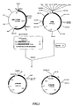
- Fig 1 is a drawing to show the flow of the construction
of pYNG/Cj.
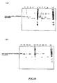
- Fig 2 is a drawing to show detection of cohesin expressed
by Cj1 recombinant virus and Cj2 recombinant virus by a cellulose
binding assay.
- (A) Cohesin expressed by Cj1 recombinant virus
| Description of reference numerals and signs M; Marker |
| 1; Clone No.1 | 5; Clone No.6 |
| 2; Clone No.2 | 6; Clone No.7 |
| 3; Clone No.3 (selected clone) | 7; Blank |
| 4; Clone No.4 | 8; CJ2 Clone No.1 |
- (B) Cohesin expressed by Cj2 recombinant virus
| Description of reference numerals and signs M; Marker |
| 1; Clone No.1 | 5; Clone No.5 |
| 2; Clone No.2 | 6; Clone No.6 |
| 3; Clone No.3 (selected clone) | 7; Blank |
| 4; Clone No.4 | 8; Blank |
- Fig. 3 is a drawing to show the structure of a vector
for addition of a C-terminal dockerin (pYNG/EK-CT-DOCK)
- Fig. 4 is a drawing to show the structure of a vector
for addition of an N-terminal dockerin (pYNG/EK-NT-DOCK)
- Fig. 5 is a drawing to show SDS-PAGE of cultured cells
infected with a recombinant virus for expressing GFP fused with
a C-terminal dockerin. (A) shows Western blotting using an
anti-GFP antibody and (B) shows CBB staining.
- Fig. 6 is a drawing to show purification of GFP fused
with a C-terminal dockerin from CBinD 100 Resin having cohesin
bound thereto. (A) shows silver staining and (B) shows Western
blotting using an anti-GFP antibody.
Description of reference numerals and signs
- 1: Supernatant of the culture medium that cohesin has
been expressed
- 2: Remaining fraction 1 after binding the supernatant
of the above-mentioned 1 to CBinD 100 Resin
- 3: Remaining fraction 2 after binding the supernatant
of the above-mentioned 1 to CBinD 100 Resin
- 4: Remaining fraction 1 after applying the supernatant
of the culture medium that GFP fused with a C-terminal dockerin
has been expressed to cohesin binding CBinD 100 Resin
- 5: Remaining fraction 2 after applying the supernatant
of the culture medium that GFP fused with a C-terminal dockerin
has been expressed to the cohesin binding CBinD 100 Resin
- 6: Fraction 1 after eluting GFP fused with a C-terminal
dockerin with a buffer containing EDTA
- 7: Fraction 2 after eluting GFP fused with a C-terminal
dockerin with a buffer containing EDTA
- 8: Fraction 3 after eluting GFP fused with a C-terminal
dockerin with a buffer containing EDTA
- 9: Fraction after eliminating cohesin domain from the
cohesin binding CBinD 100 Resin with ethylene glycol.
- 10: CBinD 100 Resin after treatment of the above-mentioned
9
- 11: Supernatant of the culture medium that GFP fused with
a C-terminal dockerin has been expressed
- Fig. 7 is a drawing to show the flow of the construction
of pYNG/Cj6.
- Fig. 8 is a drawing to show detection of a cohesin domain
using GFP fused with a C-terminal dockerin
Description of reference numerals and signs
- 1: Detection of cohesin containing one cohesin domain
using GFP fused with a C-terminal dockerin
- 2: Detection of cohesin containing two cohesin domains
using GFP fused with a C-terminal dockerin
- 3: Detection of cohesin containing six cohesin domains
using GFP fused with a C-terminal dockerin
- Figs. 9(A) and (B) are drawings to show purification of
GFP fused with a C-terminal dockerin using CNBr-activated
Sepharose 4 Fast Flow to which a cohesin protein (containing
one, two or six cohesin domains) is covalently bound.
Description of reference numerals and signs
- 1: Body fluid of larvae in which GFP fusedwith a C-terminal
dockerin has been expressed
- 2: The remaining fraction after bound to Cj1 resin
- 3: Fraction 1 of GFP fused with a C-terminal dockerin
eluted from Cj1 resin with EDTA
- 4: Fraction 2 of GFP fused with a C-terminal dockerin
eluted from Cj1 resin with EDTA
- 5: Body fluid of larvae in which GFP fused with a C-terminal
dockerin has been expressed
- 6: Remaining fraction after bound to Cj2 resin
- 7: Fraction 1 of GFP fused with a C-terminal dockerin
eluted from Cj2 resin with EDTA
- 8: Fraction 2 of GFP fused with a C-terminal dockerin
eluted from Cj2 resin with EDTA
- 9: Body fluid of larvae in which GFP fused with a C-terminal
dockerin has been expressed
- 10: Remaining fraction after bound to Cj6 resin
- 11: Fraction 1 of GFP fused with a C-terminal dockerin
eluted from Cj6 resin with EDTA
- 12: Fraction 2 of GFP fused with a C-terminal dockerin
eluted from Cj6 resin with EDTA
M: Marker
←: GFP fused with a C-terminal dockerin
- Figs. 10(A) and (B) are drawings to show purification
of GFP fused with an N-terminal or C-terminal dockerin using
CNBr-activated Sepharose 4 Fast Flow to which a cohesin is
covalently bound and to show cleavage of tag with enterokinase.
Description of reference numerals and signs
- 1: Fraction 1-1 of GFP fused with a C-terminal dockerin
eluted with EDTA
- 2: Fraction 1-1 cleaved by enterokinase at 4°C for 48
hours
- 3: Fraction 1-2 of GFP fused with a C-terminal dockerin
eluted with EDTA
- 4: Fraction 1-2 cleaved by enterokinase at 4°C for 48
hours
- 5: resin after Fraction 1 was eluted
- 6: Fraction 2-1 of GFP fused with a C-terminal dockerin
eluted with EGTA
- 7: Fraction 2-1 cleaved by enterokinase at 4°C for 48
hours
- 8: Fraction 2-2 of GFP fused with a C-terminal dockerin
eluted with EGTA
- 9: Fraction 2-2 cleaved by enterokinase at 4°C for 48
hours
- 10: resin after Fraction 2 has been eluted
- 11: Fraction 3-1 of GFP fused with an N-terminal dockerin
eluted with EDTA
- 12: Fraction 3-1 cleaved by enterokinase at 4°C for 48
hours
- 13: Fraction 3-2 of GFP fused with an N-terminal dockerin
eluted with EDTA
- 14: Fraction 3-2 cleaved by enterokinase at 4°C for 48
hours
- 15: resin after Fraction 3 was eluted
- 16: Fraction 4-1 of GFP fused with an N-terminal dockerin
eluted with EGTA
- 17: Fraction 4-1 cleaved by enterokinase at 4°C for 48
hours
- 18: Fraction 4-2 of GFP fused with an N-terminal dockerin
eluted with EGTA
- 19: Fraction 4-2 cleaved with enterokinase at 4°C for
48 hours
- 20: resin after Fraction 4 was eluted
←: GFP after tag was cleaved
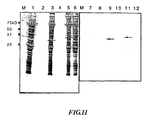
- Fig. 11 is a drawing to show detection of a protein fused
with dockerin by a biotinylated cohesin protein.
Description of reference numerals and signs
- 1: Homogenized pupae solution in which GFP fused with
a C-terminal dockerin has been expressed
- 2: Purified product of GFP fused with a C-terminal dockerin
- 3: Homogenized pupae solution in which GFP fused with
an N-terminal dockerin has been expressed
- 4: Purified product of GFP fused with an N-terminal
dockerin
- 5: Homogenized pupae solution in which mouse
interferon-beta fused with a C-terminal dockerin has been
expressed
- 6: Homogenized pupae solution in which mouse
interferon-beta fused with an N-terminal dockerin has been
expressed
- 7: Homogenized pupae solution in which GFP fused with
a C-terminal dockerin has been expressed
- 8: Purified product of GFP fused with a C-terminal dockerin
has been expressed
- 9: Homogenized pupae solution in which GFP fused with
an N-terminal dockerin has been expressed
- 10: Purified product of GFP fused with an N-terminal
dockerin has been expressed
- 11: Homogenized pupae solution in which mouse
interferon-beta fused with a C-terminal dockerin has been
expressed
- 12: Homogenized pupae solution in which mouse
interferon-beta fused with an N-terminal dockerin has been
expressed
←: GFP fused with a dockerin
-
Best Mode for Carrying Out the Invention
-
A recombinant fused protein wherein a target protein has
been fused with dockerin used in the present invention can be
obtained by inserting a gene encoding the target protein
(hereinafter referred to as 'target protein gene') and a gene
encoding dockerin (hereinafter referred to as 'dockerin gene')
into an expression vector, and by introducing the expression
vector into a given host to express the protein.
-
More specifically, the recombinant fused protein can be
obtained by inserting a dockerin gene as an affinity peptide
into the 5'-upstream region or the 3'-downstream region of a
target protein gene, which is inserted into the downstream of
the promoter of a given expression vector, thereby constructing
an expression vector for producing the recombinant fused protein
of the dockerin and the given protein, and then by introducing
the expression vector into a host to produce the protein.
-
The dockerin genes used for constructing the
above-mentioned expression vector include, for example, a gene
cloned by a conventional method, which is from a cellulase
complex derived from C. josui reported by Ohmiya et al. (Ohmiya
et al., Miedai Seibutsushigen Kiyo, vol. 19, pp71-96 (1997)).
Without being limited thereto, a dockerin gene reported by Ohmiya
et al., (Ohmiya, Ket al., J. Bacteriology, vol. 180; pp4303-4308
(1998)) or by Fierobe et al., (Fierobe H. P et al., Biochemistry,
vol. 38; pp12822-12832 (1999)) may be used. There is no
particular restriction on the target protein genes, and
therefore a gene encoding various known proteins may be used.
-
When the above-mentioned expression vector is constructed,
a gene encoding a chemical or enzymatic cleavage site
(hereinafter referred to as 'cleavage gene') may be inserted
between the target protein region and the dockerin region of
the recombinant fused protein in order to eliminate the dockerin
easily. In a case where an enzymatic cleavage is used for a
peptide sequence of the cleavage site, a peptide sequence which
is recognized and cleaved by an adequate enzyme such as
endoprotease and pro-hormone convertase may be used (Meth.
Enzymol., vol. XIX, Perlman and Lorand edition, New York:
Academic press (1970); Meth. Enzymol., vol. XLV, Lorand edition,
New York: Academic press (1976)). More specific examples of
the peptide sequence of cleavage site include a nucleotide
sequence encoding an enterokinase cleavage site (DDDDK). In
a case where a chemical cleavage is used for a peptide sequence
of the cleavage site, a peptide sequence which is recognized
and cleaved by a chemical reagent such as an organic acid and
an inorganic acid, hydroxylamine, N-bromosuccinimide and
cyanogenbromide may be used (Witcop, Advances in Protein
Chemistry, Anfinseen et al. edition, pp231-321, New York:
Academic Press (1961)). More specific examples of the peptide
sequence of cleavage site include a nucleotide sequence encoding
a cyanogenbromide cleavage site (MX; X is a given amino acid).
-
There is no particular restriction on the expression
vectors into which the target protein gene and the dockerin
gene, and if necessary, the cleavage gene are inserted. The
expression vector may be selected depending on the host to be
used. For example, in a case where E. coli bacteria is used
as a host, pTRC99A (Amersham Biosciences) or the like is used.
In a case where a cultured insect cell or an insect body is
used as a host, it is preferable to use ABv (Katakura Industries
Co., Ltd.) or the like.
-
A host for expressing the above-mentioned expression
vector may be selected depending on the expression vector to
be used. A recombinant fused protein can be expressed in the
vector by a conventional method.
-
The obtained recombinant fused protein as mentioned above
is treated with a support having a cohesin domain immobilized
thereon (hereinafter referred to as 'immobilized cohesin
support') and purified.
-
As a cohesin domain used in this step, any cohesin domains
can be used as long as they bind especially to the above-mentioned
dockerin. Preparation of the cohesin domain can be performed
in accordance with the preparation of the recombinant fused
protein. For example, the gene of the cohesin domain, which
isfromCipAderivedfromC. josui, is cloned, the gene is inserted
into a given expression vector to construct a vector expressing
the cohesin domain, then the expression vector is introduced
into a host, and thus the cohesin domain can be produced. When
the cohesin domain is prepared, a gene encoding a binding site
(hereinafter referred to as 'binding gene') for binding the
cohesin domain to the support may, if necessary, be inserted
into the vector. There is no particular restriction on the
binding gene as long as it binds a cohesin domain to a support.
For example, in a case where the above-mentioned CipA is used
as a cohesin domain, a cellulose binding domain (CBD) of CipA
may be used as a binding gene.
-
The thus obtained cohesin domain can be purified and
isolated by chromatography with the use of an anion exchange
resin. Specifically, a fraction containing the cohesin domain
is adsorbed to an anion exchange resin by adsorptive treatment
with an anion exchange support, subsequently the cohesin domain
is eluted by increasing salt concentration, and thus the cohesin
domain can be purified and isolated. The anion exchange support
used for chromatography includes DEAE anion exchanger, QAE anion
exchanger, Q ion exchanger and the like. As a specific brand
name, Q-Sepharose anion exchanger (Amersham Biosciences) is
given.
-
The purified cohesin domain can be detected by using a
labeled dockerin, which will be described below. A green
fluorescent protein (GFP) is used for labeling the dockerin.
The recombinant fused protein of GFP and dockerin is produced
by a conventional method, and the GFP can be detected with an
anti-GFP antibody, or the fluorescence of GFP can be detected
and quantified with a fluorescence detector.
-
Immobilization of the cohesin domain is performed by
binding the cohesin domain to a support according to a
conventional method. The cohesin domain can be directly bound
to a support. However, a fused protein coupledwith, for example,
a cellulose binding domain (CBD) or the like is produced by
a conventional method, with which the cohesin domain can be
bound to a support. In a case where a hydrogen bond is used
as a bonding mechanism for binding a cohesin domain to a support,
a cellulose support such as CBinD 100 Resin (Novagen) can be
used as a support for immobilization when the above-mentioned
fused protein in which a cellulose binding domain has been
coupled with a cohesin domain is used because it can be adsorbed
to the cellulose via the cellulose binding domain coupled with
the cohesin domain. In a case where a covalent bond is used
as a bonding mechanism for binding a cohesin domain to a support,
an activated support such as CNBr-activated Sepharose 4 Fast
Flow, CNBr-activated Sepharose 4B, EAH Sepharose 4B and ECH
Sepharose 4B, and Epoxy-activated Sepharose 6B (Amersham
Biosciences) can be used because the cohesin domain can be bound
to the activated support with the use of a cross-linking agent
or the like.
-
Examples of the specific methods of binding a recombinant
fused protein to an immobilized cohesin support include, for
example, a method of adding calcium ion to a sample containing
a recombinant fused protein, then applying the mixture to an
immobilized cohesin support. The support having a recombinant
fused protein bound thereto as mentioned above is washed with
an appropriate buffer, subsequently washed with an appropriate
buffer containing a metal chelate such as EDTA, and thereby
the recombinant fused protein is eluted.
-
This is because dockerin has 60 amino-acid residues
(molecular weight; about 7 kDa), which is very small, and the
dissociation constant of the dockerin and cohesin domain is
4.7×10⁻⁷, which is also very small. In addition, the dockerin
is bound to the cohesin domain in the presence of calcium, and
this complex has a property of dis sociating easily by eliminating
calcium ion with a metal chelate.
-
Accordingly, the recombinant fused protein can be
separated from other substances because the dockerin region
is strongly bound to the immobilized cohesin support. In other
words, the dockerin-cohesin complex is formed on the support
by the above-mentioned strong binding, the support is thoroughly
washed with an appropriate buffer to eliminate other substances,
and then an appropriate buffer containing a metal chelate is
applied to eliminate calcium that is needed for binding the
dockerin region of the recombinant fused protein and the cohesin
domain region of the solid phase. Thus, only the recombinant
fused protein is eluted, and thereby it can be purified.
-
The buffers used in the purified step include Good buffer
(Good, N.E. and Izawa, S, Method. Enzymol., vol. 24, Part B,
pp53-68 (1972)): (hereinafter referred to as 'Good buffer'),
phosphate buffer and the like. The metal chelates include
ethylenediamine-N,N,N',N'-tetraacetic acid (hereinafter
referred to as 'EDTA') and -ethylenedioxybis
(ethylamine)-N,N,N',N'-tetraacetic acid (hereinafter
referred to as 'EGTA') and the like.
-
Thus, the recombinant fused protein wherein the target
protein has been fused with the dockerin is purified. The
purified recombinant fused protein can be detected and
quantified by using an antibody against a target protein or
a labeled cohesin domain with the use of the binding property
of a dockerin and a cohesin domain. In a case where an antibody
is used for the detection and quantification, an antibody against
a target protein or dockerin is produced and labeled according
to a conventional method, and the resultant antibody may be
used. In a case where a labeled cohesin domain is used, a cohesin
domain prepared as mentioned above may be labeled according
to a conventional method. As a label used for labeling the
cohesin domain, the one which gives a signal to be detectable
directly or indirectly is preferable, including enzymes,
fluorescent proteins, radioactive labeled molecules,
fluorescent agents, dyes, particles, chemiluminescent
materials, enzyme substances or cofactors, enzyme inhibitors
and magnetic particles and the like. Biotin is particularly
preferable. The cohesin domain may be labeled with these labels
according to a conventional method. Specifically, a
biotinylated cohesin domain by using biotin as a label can be
detected and quantified with the streptavidin, which has been
labeled with alkaline phosphatase or horseradish peroxidase.
-
A method using a labeled cohesin domain is preferable
to a method using an antibody against a target protein in that
it can be applied regardless of the target protein.
-
The target protein can be obtained by eliminating the
dockerin region from the purified recombinant fused protein
as mentioned above.
-
The methods of eliminating the dockerin region from the
recombinant fused protein include a method of cleaving a chemical
or enzymatic cleavage site provided between the target protein
region and the dockerin region of the recombinant fused protein.
In other words, the recombinant fused protein, in which only
the dockerin region can be easily eliminated, can be obtained
by using an expression vector in which a gene encoding a chemical
or enzymatic cleavage site has been inserted between the target
protein gene and the dockerin gene in advance.
-
Accordingly, with respect to a recombinant protein, for
example, only the target protein can be isolated and obtained
by binding a recombinant fused protein to an immobilized cohesin
support, subsequently washing it with an appropriate buffer
such as Good buffer and phosphate buffer, then treating it with
a specific reagent or enzyme, for example, in a case where the
cleavage site is an enterokinase cleavage site, treating it
with enterokinase to leave the dockerin bound to the cohesin
domain of the immobilized cohesin support.
-
The present invention is based on the facts that dockerin
has 60 amino-acid residues (molecular weight; about 7 kDa),
which is very small, the dissociation constant of the dockerin
and cohesin domain is 4.7×10⁻⁷, which is also very small, and
the complex of dockerin and cohesin domain has a property of
dissociating easily by eliminating calcium ion with a metal
chelate.
-
More specifically, dockerin has a small molecular weight,
and a recombinant fused protein wherein the target protein has
been fused is unlikely to become insoluble. Also, the binding
strength between the dockerin and the cohesin domain is strong
enough to perform affinity purification. Furthermore, the
dockerin and the cohesin domain binding can be separated easily
by eliminating calcium ion with a metal chelate. These
properties enable to provide a method of isolating a recombinant
fused protein easily under mild purification conditions without
making the target protein insoluble.
-
Since the dockerin and the cohesin domain, which are used
in the above-mentioned method, can be easily obtained as a
recombinant protein, it is possible to stably supply them as
an affinity peptide and a ligand of uniform quality over a long
term.
-
In a case where a chemical or enzymatic cleavage site
is provided between the target protein and the dockerin of the
recombinant fused protein, only the target protein can be
obtained with a simple method.
-
Conventionally, in order to detect and quantify a target
protein, an antibody against the target protein has to be
prepared for each protein. If a recombinant fused protein
containing dockerin and a target protein is used, the target
protein can be detected and quantified easily with a labeled
cohesin domain.
-
Furthermore, detection and quantification of a
recombinant fused protein wherein a target protein has been
fused with dockerin with a labeled cohesin domain according
to the above-mentioned method will enable to measure or detect
the expression amount of the target protein and localization
or pharmacokinetics in the cells and tissues.
Examples
-
The present invention will be illustrated in more detail
with reference to the following examples, however, the present
invention is not limited thereto.
Example 1
Acquisition of cohesin gene:
-
The cohesin gene was amplified by PCR under the conditions
mentioned below by using pMK-2 Cj CipA vector (given by Associate
Professor Sakka, Laboratory of Applied Microbiology, Faculty
of Bioscience, Mie University), which was obtained by cloning
the C. josui CipA gene (Ohmiya, K et al., J. Bacteriology, vol.
180; pp4303-4308 (1998)) into pMK vector, as a template.
(1) Design of PCR primers
-
The nucleotide sequences and the combinations of primers
used for the amplification of the cohesin gene are as follows.
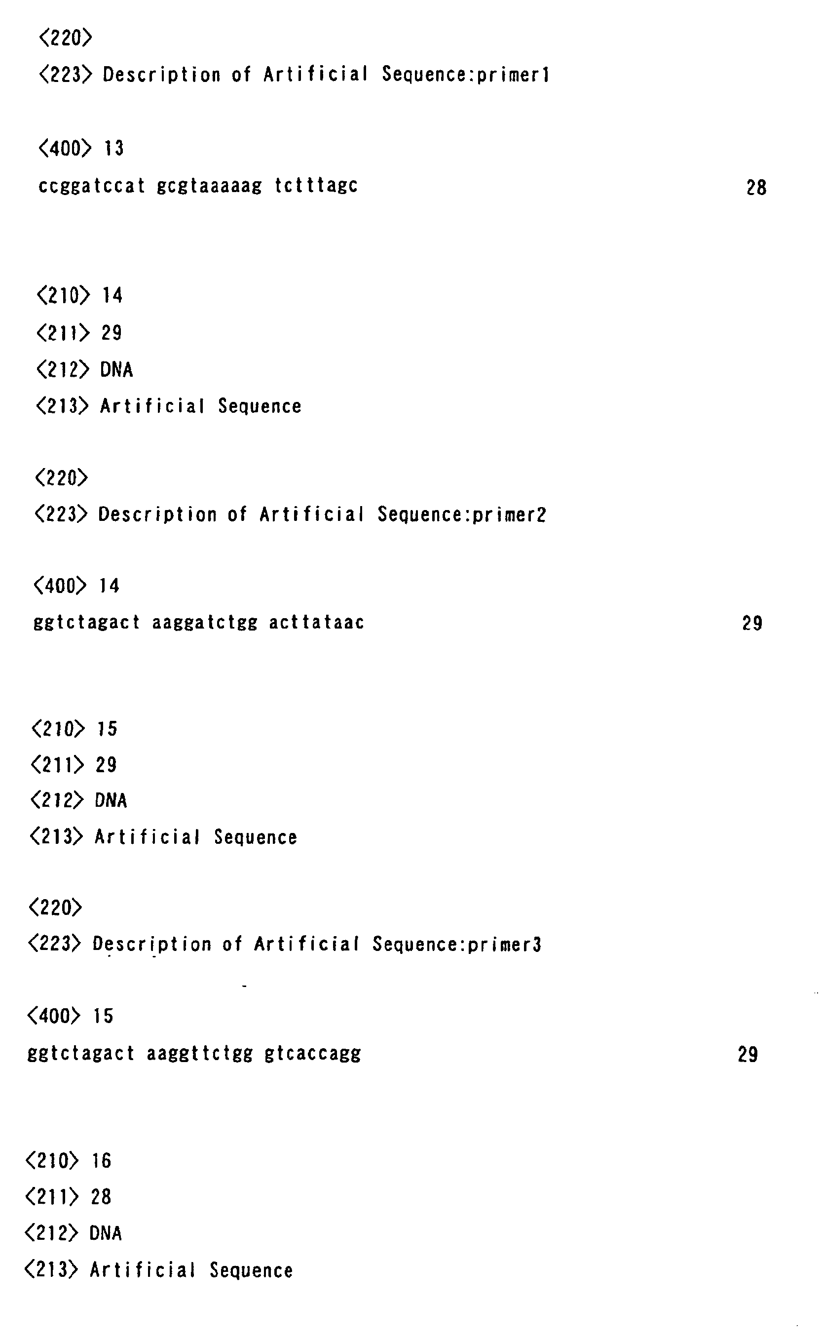
- Primer 1: for addition of a restriction enzyme BamHI site
(for Cj1 or Cj2 amplification)
- Primer 2: for addition of a restriction enzyme XbaI site
(for Cj1 amplification)
- Primer 3: for addition of a restriction enzyme XbaI site
(for Cj2 amplification)
-
(2) Preparation of PCR sample
-
| pMK/C. josui CipA |
100 pg |
| 10 x Ex-Taq buffer (Takara Shuzo Co., Ltd.) |
5 µl |
| Primer |
| 1 |
1 ng |
| Primer 2 or Primer 3 |
1 ng |
| dNTP mix (Takara Shuzo Co., Ltd.) |
4 µl |
| Ex-Taq (Takara Shuzo Co., Ltd.) |
0.5 µl |
| Distilled water Added to make the total volume |
50 µl |
(3) PCR conditions
-
- Step 1: 94.0°C 2 minutes
- Step 2: 94.0°C 30 seconds
- Step 3: 55.0°C 45 seconds
- Step 4: 72.0°C 60 seconds
- Step 5: Repeat Steps 2 to 4 for 30 times
- Step 6: 72.0°C 3 minutes
-
Example 2
-
Construction of transfer vector for cohesin expression:
(1) Preparation of cohesin gene
-
Restriction enzymes BamHI (8 to 24U) and XbaI (8 to 24U)
(both from Takara Shuzo Co., Ltd.) were added to 20 µl of the
C. josui CipA cohesin gene PCR product, which had a restriction
enzyme BamHI recognition sequence at 5' end, one (Cj1) or two
(Cj2) of cohesin domains and a restriction enzyme XbaI
recognition sequence at 3' end. The restriction enzyme
digestion was performed and the end sequences were exposed.
Then, the restriction enzyme-treated cohesin gene fragment was
purified by using a Qiagen spin column (QIAquick: Qiagen).
(2) Insertion into transfer vector
-
After digesting the restriction enzyme recognition sites
in the multi-cloning site of transfer vector pYNG for recombinant
baculoviral preparation (Katakura Industries Co., Ltd.) with
BglII and XbaI (both from Takara Shuzo Co., Ltd.), the digested
ends were dephosphorylated with alkaline phosphatase (Takara
Shuzo Co., Ltd.). Solution I (5 µl) of Ligation Kit (Takara
Shuzo Co., Ltd.) was added to 0.1 µg (2 µl) of the linearized
transfer vector and 0.1 µg (3 µl) of the restriction
enzyme-treated cohesin gene fragment (containing one or two
cohesin domains). After the reaction at 16°C for 1 hour, E.
coli JM109 (Toyobo Co., Ltd.) was transformed with the whole
reaction solution. Ampicillin resistant transformants were
selected and plasmids were purified.
-
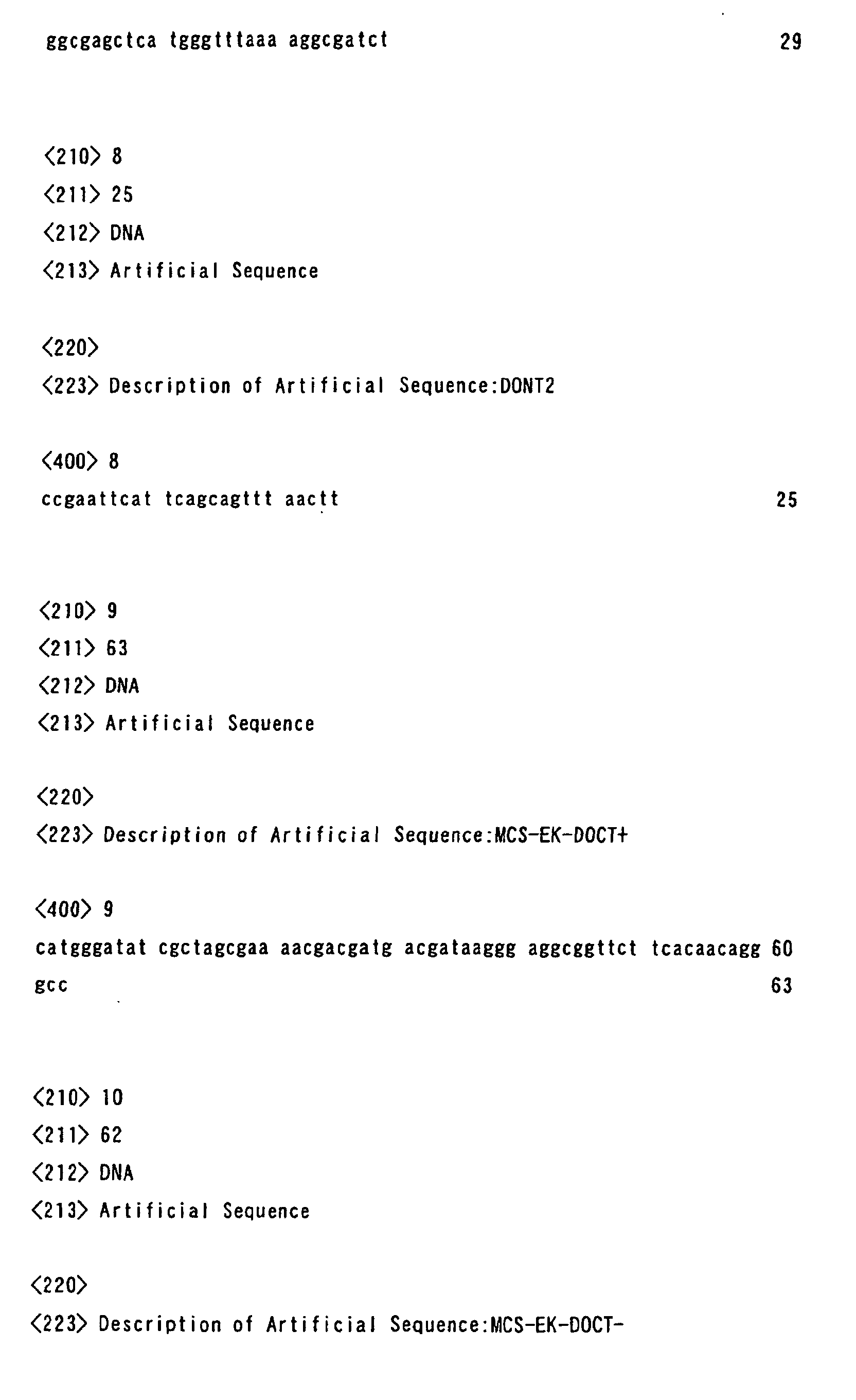
Then, the following primers were used to confirm the
nucleotide sequence of several clones.
-
-
-
After the reaction with the above primers by using DNA
Sequence Kit (Applied Biosystems Inc.), the analysis was
performed by using an ABI Genetic Analyzer (Applied Biosystems
Inc.). A clone harboring the cohesin gene fragment without
mutations was selected and referred to as pYNG/Cj1 (containing
one cohesin domain) or pYNG/Cj2 (containing two cohesin domains)
(Fig. 1).
Example 3
-
Preparation of cohesin-expressing recombinant virus (1):
(1) Cotransfection
-
Firstly, using a 35 mm petri dish for cell culture, Bm-N
cells were prepared in a confluent monolayer of about 1 x 10⁶
cells in static culture. The linearized ABv DNA (0.1 µg)
(Katakura Industries Co., Ltd.) and either of 0.5 µg of the
transfer vector pYNG/Cj1 or pYNG/Cj2 obtained as above were
mixed with 100 µl of TC-100 (Nosan Corporation) in a 1.5 ml
tube and left for 15 minutes at room temperature. The cationic
lipid reagent (Lipofectin: GIBCO-BRL) (5 µl) was added to 100
µl of TC-100 and left for 15 minutes at room temperature. Then,
after these two solutions were mixed and left for an additional
15 minutes at room temperature, 800 µl of TC-100 was added.
This mixture was added to the Bm-N cells prepared in a confluent
monolayer on the 35 mm petri dish for cell culture. After the
cultivation for 20 hours at 25°C, the DNA solution was removed,
2 ml of TC-100 medium containing 10% FBS (Sigma) was added,
then it was statically cultured for 7 days at 25°C. The
supernatant of this culture medium was used as a recombinant
virus stock solution.
(2) Screening
-
An isolation of ABv from the obtained virus stock solution
as above was performed according to the method reported by King
et al. (King, L A and Possee, R. D., A Laboratory Guide London:
Chapman and Hall (1992)). First, the Bm-N cells were adjusted
to 1 x 10³ cells/50 µl per well and cultured with TC-100 medium
(Nosan Corporation) containing 10% FBS (Sigma) in a 96-well
plate. The recombinant virus stock solution was diluted to
10-6, 10-7, 10-8, 10-9, 10-10 (5 serial dilutions) with TC-100 medium
containing 10% FBS and the virus was infected by adding 50 µl
of each dilution of the virus stock solution to one well of
each plate. The plates were sealed to prevent drying and
statically cultured at 25°C. The selection of recombinant virus
was performed by confirming that a polyhedrin was not formed
on day 7 after infection under microscope. And the cellulose
binding activity of the recombinant cohesin secreted in the
culture medium was analyzed by SDS-PAGE and silver staining,
and clones showing activity were selected as a recombinant
cohesin-expressing recombinant virus clone (Cj1-3, Cj2-3) (Fig.
2).
(3) Preparation of recombinant cohesin protein
-
The recombinant virus was inoculated to the Sf9 cells
(Invitrogen) cultured in the EXCELL 420 medium (JRH Bioscience)
at an M.O.I of 5 and the supernatant of the culture medium on
day 4 after inoculation was used as a recombinant cohesin sample.
(4) Cellulose binding assay
-
The cellulose binding assay was performed as follows.
Cellulose CBinD 100 Resin (100 mg) (Novagen) was added to 1
ml of the medium in which cohesin had been expressed, and stirred
for 60 minutes at room temperature. After stirring, the CBinD
100 Resin was collected by centrifugation at 6,000 rpm for 10
minutes at 4°C. The supernatant was discarded, 1 ml of the
washing buffer (Washing buffer: Novagen) was added and the
precipitate was suspended. After stirring for 10 minutes at
room temperature, it was further centrifuged at 6,000 rpm for
10 minutes at 4°C and the CBinD 100 Resin was collected. This
procedure was further repeated twice and the CBinD 100 Resin
was washed. Ethylene glycol (100%, 100 µl) was added to the
CBinD 100 Resin collected by centrifugation and after suspension,
it was left for 10 minutes at room temperature. The supernatant
(80 µl) was collected after centrifugation at 6,000 rpm for
10 minutes at 4°C and used as an eluted fraction. The sample
for electrophoresis was prepared by adding 40 µl of the SDS-PAGE
sample buffer (Laemmli, Nature, vol. 227, pp680-685 (1970))
and boiling for 5 minutes, and SDS-PAGE was performed.
Example 4
Acquisition of dockerin gene:
-
The dockerin gene (sequence Dockerin) was amplified by
PCR under the conditions mentioned below by using the vector
(given by Associate Professor Sakka, Laboratory of Applied
Microbiology, Faculty of Bioscience, Mie University), which
was obtained by cloning the dockerin gene of the C. josui CelB
(DDBJ/EMBL/Genbank Database accession number D16670.1) into
pET-28a(+) vector as a template.
(1) Design of PCR primers
-
The nucleotide sequences and the combinations of primers
used for the amplification of the dockerin gene were as follows.
-
Primers for addition of an N-terminal dockerin
- DONT 1: (for addition of a restriction enzyme SacI
recognition sequence)
- DONT 2: (for addition of a restriction enzyme EcoRI
recognition sequence)
-
-
Primers for addition of a C-terminal dockerin
- DOCT 1: (for addition of a restriction enzyme NcoI and
Rco81I recognition sequences)
- DOCT 2: (for addition of a restriction enzyme NheI
recognition sequence)
-
(2) PCR conditions
-
- Step 1: 94.0°C 2 minutes
- Step 2: 94.0°C 60 seconds
- Step 3: 42.0°C 90 seconds
- Step 4: 72.0°C 60 seconds
- Step 5: Repeat Steps 2 to 4 for 30 times
- Step 6: 72.0°C 3 minutes
-
-
As a result of the amplification by PCR, a C. Josui CelB
dockerin gene fragment of about 200 bp added with restriction
enzyme recognition sequences on both ends was obtained.
Example 5
-
Construction of transfer vector for addition of
C-terminal dockerin:
(1) Preparation of dockerin gene
-
Restriction enzymes NcoI (8 to 24U) and NheI (8 to 24U)
(both from Takara Shuzo Co., Ltd.) were added to 20 µl of the
C. josui CelB dockerin gene PCR product, which had been added
with restriction enzyme NcoI and Eco81I recognition sequences
at 5' end and a restriction enzyme NheI recognition sequence
at 3' end. The restriction enzyme digestion was performed and
the end sequences were exposed. Then, the restriction
enzyme-treated dockerin gene fragment was purified by using
a Qiagen spin column (QIAquick: Qiagen).
(2) Insertion into transfer vector
-
After digesting the restriction enzyme recognition sites
in the multi-cloning site of transfer vector pYNG for recombinant
baculoviral preparation (Katakura Industries Co., Ltd.) with
NcoI and XbaI (both from Takara Shuzo Co., Ltd.), the digested
ends were dephosphorylated with alkaline phosphatase (Takara
Shuzo Co., Ltd.). Solution I (5 µl) of Ligation Kit (Takara
Shuzo Co., Ltd.) was added to 0.1 µg (2 µl) of the linearized
transfer vector and 0.1 µg (3 µl) of the restriction
enzyme-treated dockerin gene fragment. After the reaction at
16°C for 1 hour, E. coli JM109 (Toyobo Co., Ltd.) was transformed
with the whole reaction solution. Ampicillin resistant
transformants were selected and plasmids were purified.
-
Then, several clones were selected and, after the reaction
using DNA Sequence Kit (Applied Biosystems Inc.) with yng. for
mentioned above as a primer was performed, the nucleotide
sequence was analyzed by using an ABI Genetic Analyzer (Applied
Biosystems Inc.). A clone inserted the dockerin gene fragment
without mutations was referred to as pYNG/CT-DOCK vector.
(3) Introduction of enterokinase recognition site and
multicloning site.
-
The oligonucleotides having an enterokinase recognition
site and a multicloning site were synthesized.
MCS-EK-DOCT+:
MCS-EK-DOCT-:
-
The solution containing 1 µM of each oligonucleotide and
50 mM sodium chloride was prepared and the two oligonucleotides
were hybridized by treating at 94°C for 5 minutes, 55°C for 15
minutes and 37°C for 15 minutes (hereinafter referred to as
'EK-DOCK'). Then, Solution I (4 µl) of Ligation Kit (Takara
Shuzo Co., Ltd.) was added to 0.1 µg (3 µl) of the linearized
transfer vector obtained by treating pYNG/CT-DOCK with
restriction enzymes NcoI and Eco81I and 1 µl of the EK-DOCK
fragment and reacted at 16°C for 1 hour. Then, E. coli JM109
(Toyobo Co., Ltd.) was transformed with the whole reaction
solution and plasmids were purified from the obtained ampicillin
resistant transformants. Several clones were selected and,
after the reaction using DNA Sequence Kit (Applied Biosystems
Inc.) with yng.for mentioned above as a primer was performed,
the nucleotide sequence was analyzed by using an ABI Genetic
Analyzer (Applied Biosystems Inc.). A clone that was confirmed
to have the nucleotide sequences of the enterokinase recognition
site and the multicloning site (BglII, SacI, SmaI, EcoRI, NcoI,
EcoRV, and NheI) was referred to as pYNG/EK-CT-DOCK vector for
addition of dockerin (Fig. 3).
Example 6
Construction of transfer vector for addition of
N-terminal dockerin
(1) Preparation of dockerin gene
-
Restriction enzymes SacI (8 to 24U) and EcoRI (8 to 24U)
(both from Takara Shuzo Co., Ltd.) were added to 20 µl of the
C. josui CelB dockerin gene PCR product, which had been added
with a restriction enzyme SacI recognition sequence at 5' end
and a restriction enzyme EcoRI recognition sequence at 3' end.
The restriction enzyme digestion was performed and the end
sequences were exposed. Then, the restriction enzyme-treated
dockerin gene fragment was purified by using a Qiagen spin column
(QIAquick: Qiagen).
(2) Insertion into transfer vector
-
After digesting the restriction enzyme recognition sites
in the multi-cloning site of transfer vector pYNG30Ksig for
recombinant baculoviral preparation (Katakura Industries Co.,
Ltd.) with SacI and EcoRI (both from Takara Shuzo Co., Ltd.)
to make it linear, the digested ends were dephosphorylated with
alkaline phosphatase (Takara Shuzo Co., Ltd.). Solution I (5
µl) of Ligation Kit (Takara Shuzo Co., Ltd.) was added to 0.1
µg (2 µl) of the linearized transfer vector and 0.1 µg (3 µl)
of the restriction enzyme-treated dockerin gene fragment.
After the reaction at 16°C for 1 hour, E. coli JM109 (Toyobo
Co., Ltd.) was transformed with the whole reaction solution.
Ampicillin resistant transformants were selected and plasmids
were purified.
-
Then, several clones were selected and, after the reaction
using DNA Sequence Kit (Applied Biosystems Inc.) with yng.for
mentioned above as a primer was performed, the nucleotide
sequence was analyzed by using an ABI Genetic Analyzer (Applied
Biosystems Inc.). A clone inserted the dockerin gene fragment
without mutations was referred to as pYNG/NT-DOCK vector.
(3) Introduction of enterokinase recognition site and
multicloning site.
-
The oligonucleotides having an enterokinase recognition
site and a multicloning site were synthesized.
MCS-EK-DONT+:
MCS-EK-DONT-:
-
The solution containing 1 µM of each oligonucleotide and
50 mM sodium chloride was prepared and the two oligonucleotides
were hybridized by treating at 94°C for 5 minutes, 55°C for 15
minutes and 37°C for 15 minutes (EK-DONT). Then, Solution I
(4 µl) of Ligation Kit (Takara Shuzo Co., Ltd.) was added to
0.1 µg (3 µl) of the linearized transfer vector obtained by
treating pYNG/NT-DOCK with restriction enzymes EcoRI and XbaI
and 1 µl of the EK-DONT fragment and reacted at 16°C for 1 hour.
Then, E. coli JM109 was transformed with the whole reaction
solution and plasmids were purified from the obtained ampicillin
resistant transformants.
-
Then, several clones were selected and, after the reaction
using DNA Sequence Kit (Applied Biosystems Inc.) with yng.for
mentioned above as a primer was performed, the nucleotide
sequence was analyzed by using an ABI Genetic Analyzer (Applied
Biosystems Inc.). A clone that was confirmed to have the
nucleotide sequences of enterokinase recognition sequence and
the multicloning site (BglII, SmaI, EcoRI, NcoI and XbaI) was
referred to as pYNG/EK-NT-DOCK vector for addition of dockerin
(Fig. 4).
Example 7
Expression of green fluorescent protein (GFP) fused with
C-terminal dockerin
(1) Design of PCR primers
-
Using vector pEGFP-N1 (Clontech) harboring the GFP gene
as a template, introduction of restriction enzyme recognition
sequences and deletion of the termination codon were performed
by PCR. For the preparation of the GFP gene fragment to be
inserted into pYNG/EK-CT-DOCK, the following primers were
synthesized.
yng.for:
-
Primer 5: (for addition of a restriction enzyme EcoRV
recognition sequence)
(2) Preparation of PCR sample
-
| pEGFP-N1 |
100 pg |
| 10 x Ex-Taq buffer (Takara Shuzo Co., Ltd.) |
5 µl |
| yng.for |
1 ng |
| Primer 5 |
1 ng |
| dNTP mix (Takara Shuzo Co., Ltd.) |
4 µl |
| Ex-Taq (Takara Shuzo Co., Ltd.) |
0.5 µl |
-
Distilled water Added to make the total volume 50 µl
(3) PCR conditions
-
The same operation was performed as in the acquisition
of the cohesin gene in Example 1.
(4) Preparation of GFP gene
-
Restriction enzymes BglII (8 to 24U) and EcoRV (8 to 24U)
(both from Takara Shuzo Co., Ltd.) were added to 20 µl of the
GFP gene PCR product, which had been added with a restriction
enzyme BglII recognition sequence at 5' end and a restriction
enzyme EcoRV recognition sequence at 3' end. The restriction
enzyme digestion was performed and the end sequences were exposed.
Then, the restriction enzyme-treated GFP gene fragment was
purified by using a Qiagen spin column (QIAquick: Qiagen).
(5) Insertion into transfer vector
-
After digesting the restriction enzyme recognition sites
in the multi-cloning site of transfer vector pYNG/EK-CT-DOCK
for addition of a C-terminal dockerin with BglII and EcoRV (both
from Takara Shuzo Co., Ltd.) to make it linear, the digested
ends were dephosphorylated with alkaline phosphatase (Takara
Shuzo Co., Ltd.). Solution I (3 µl) of Ligation Kit (Takara
Shuzo Co., Ltd.) was added to 0.1 µg (2 µl) of the linearized
transfer vector and 0.1 µg (2 µl) of the restriction
enzyme-treated GFP gene fragment. After the reaction at 16°C
for 1 hour, E. coli JM109 (Toyobo Co., Ltd.) was transformed
with the whole reaction solution. Ampicillin resistant
transformants were selected and plasmids were purified.
-
Then, several clones were selected and it was confirmed
that the GFP gene fragment was inserted, that the reading frame
was correct and that the GFP gene had no mutations. The
above-mentioned yng.for and yng.rev primers were used for
nucleotide sequence confirmation. After the primer was reacted
using DNA Sequence Kit (Applied Biosystems Inc.), the nucleotide
sequences were analyzed by using an ABI Genetic Analyzer (Applied
Biosystems Inc.).
Example 8
Expression of mouse interferon (mIFN) fused with
C-terminal dockerin
(1) Design of PCR primers
-
Using the mIFN gene (Taniguchi, T et al., J. Biol. Chem.,
vol. 258, pp9522-9529 (1983); given by Professor Munekawa, Kyoto
Institute of Technology) as a template, the introduction of
restriction enzyme recognition sequences and deletion of the
termination codon were performed by PCR. For the preparation
of mIFN gene fragment to be inserted into pYNG/EK-CT-DOCK, the
following primers were synthesized.
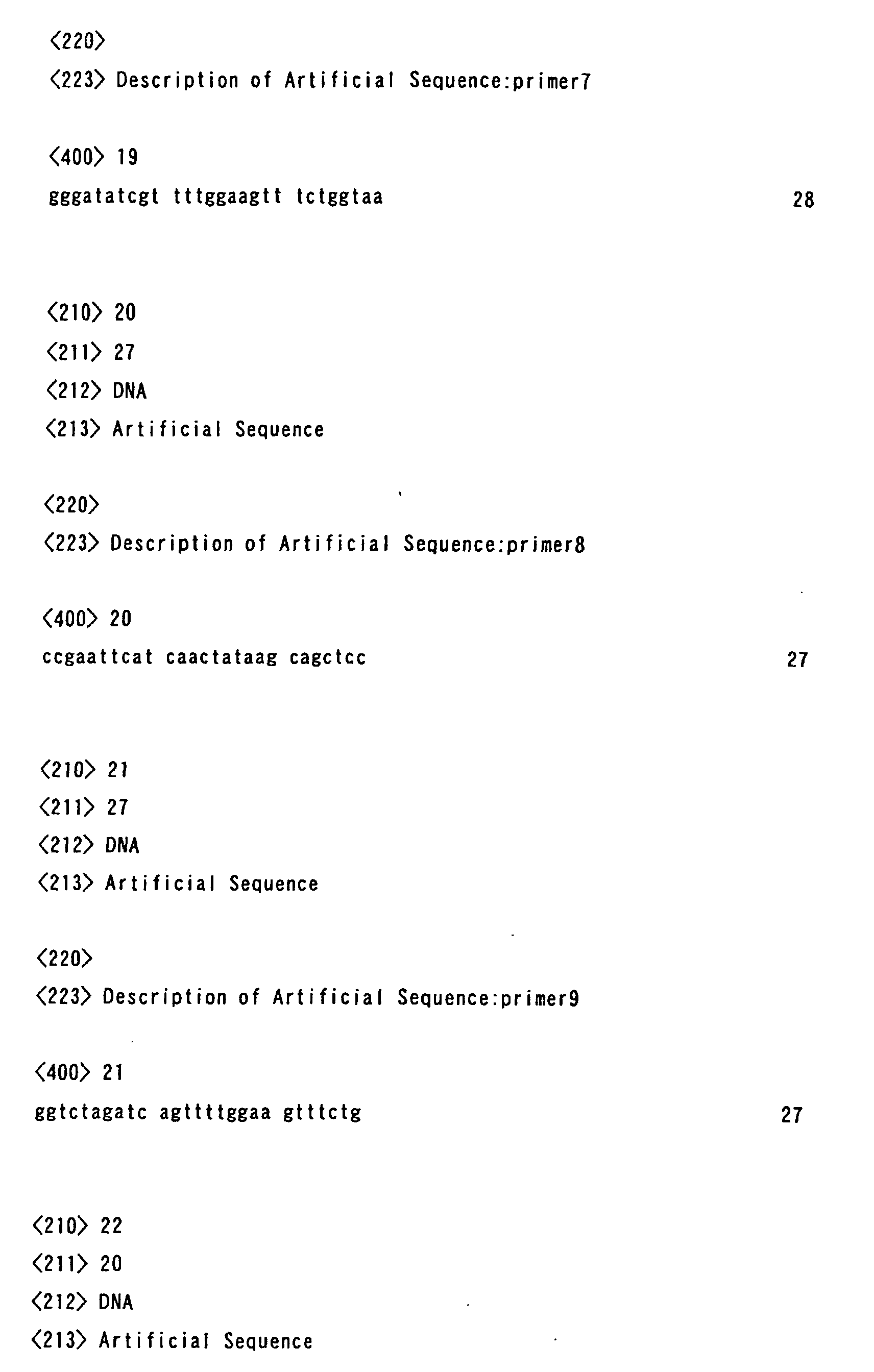
Primer 6: (for addition of a restriction enzyme EcoRI
recognition sequence)
-
Primer 7: (for addition of a restriction enzyme EcoRV
recognition sequence)
-
(2) Preparation of PCR sample
-
| mIFN gene |
100 pg |
| 10 x Ex-Taq buffer (Takara Shuzo Co., Ltd.) |
5 µl |
| Primer |
| 6 |
1 ng |
| Primer 7 |
1 ng |
| dNTP mix (Takara Shuzo Co., Ltd.) |
4 µl |
| Ex-Taq (Takara Shuzo Co., Ltd.) |
0.5 µl |
-
Distilled water Added to make the total volume 50 µl
(3) PCR conditions
-
The same operation was performed as in the acquisition
of the cohesin gene.
(4) Preparation of mIFN gene
-
Restriction enzymes EcoRI (8 to 24U) and EcoRV (8 to 24U)
(both from Takara Shuzo Co., Ltd.) were added to 20 µl of the
mIFN gene PCR product, which had been added with a restriction
enzyme EcoRI recognition sequence at 5' end and a restriction
enzyme EcoRV recognition sequence at 3' end. The restriction
enzyme digestionwas performed and the end sequences were exposed.
Then, the restriction enzyme-treated mIFN gene fragment was
purified by using a Qiagen spin column (QIAquick: Qiagen).
(5) Insertion into transfer vector
-
After digesting the restriction enzyme recognition sites
in the multi-cloning site of transfer vector pYNG/EK-CT-DOCK
for addition of a C-terminal dockerin with EcoRI and EcoRV (both
from Takara Shuzo Co., Ltd.) to make it linear, the digested
ends were dephosphorylated with alkaline phosphatase (Takara
Shuzo Co., Ltd.). Solution I (3 µl) of Ligation Kit (Takara
Shuzo Co., Ltd.) was added to 0.1 µg (2 µl) of the linearized
transfer vector and 0.1 µg (2 µl) of the restriction
enzyme-treated mIFN gene fragment. After the reaction at 16°C
for 1 hour, E. coli JM109 (Toyobo Co., Ltd.) was transformed
with the whole reaction solution. Ampicillin resistant
transformants were selected and plasmids were purified.
-
Then, several clones were selected and it was confirmed
that the mIFN gene fragment was inserted, that the reading frame
was correct and that the mIFN gene had no mutations. The
above-mentioned yng.for and yng.rev primers were used for
nucleotide sequence confirmation. After the reaction using DNA
Sequence Kit (Applied Biosystems Inc.), the nucleotide
sequences were analyzed by using an ABI Genetic Analyzer (Applied
Biosystems Inc.).
Example 9
Expression of mouse interferon (mIFN) fused with
N-terminal dockerin
(1) Design of PCR primers
-
Using the mIFN gene (given by Professor Munekawa, Kyoto
Institute of Technology) as a template, the introduction of
restriction enzyme recognition sequences was performed by PCR.
For the preparation of the mIFN gene fragment to be inserted
into pYNG/EK-NT-DOCK, the following primers were synthesized.
Primer 8: (for addition of a restriction enzyme EcoRI
recognition sequence)
-
Primer 9: (for addition of a restriction enzyme XbaI
recognition sequence)
-
(2) Preparation of PCR sample
-
| mIFN gene |
100 pg |
| 10 x Ex-Taq buffer (Takara Shuzo Co., Ltd.) |
5 µl |
| Primer |
| 8 |
1 ng |
| Primer 9 |
1 ng |
| dNTP mix (Takara Shuzo Co., Ltd.) |
4 µl |
| Ex-Taq (Takara Shuzo Co., Ltd.) |
0.5 µl |
-
Distilled water Added to make the total volume 50 µl
(3) PCR conditions
-
The same operation was performed as in the acquisition
of the cohesin gene.
(4) Preparation of mIFN gene
-
Restriction enzymes EcoRI (8 to 24U) and XbaI (8 to 24U)
(both from Takara Shuzo Co., Ltd.) were added to 20 µl of the
mIFN gene PCR product, which had been added with a restriction
enzyme EcoRI recognition sequence at 5' end and a restriction
enzyme XbaI recognition sequence at 3' end. The restriction
enzyme digestion was performed and the end sequences were exposed.
Then, restriction enzyme-treated mIFN gene fragment was
purified by using a Qiagen spin column (QIAquick: Qiagen).
(5) Insertion into transfer vector
-
After digesting the restriction enzyme recognition sites
in the multi-cloning site of transfer vector pYNG/EK-NT-DOCK
for addition of an N-terminal dockerin with EcoRI and XbaI (both
from Takara Shuzo Co., Ltd.) to make it linear, the digested
ends were dephosphorylated with alkaline phosphatase (Takara
Shuzo Co., Ltd.). Solution I (3 µl) of Ligation Kit (Takara
Shuzo Co., Ltd.) was added to 0.1 µg (2 µl) of the linearized
transfer vector and 0.1 µg (2 µl) of the restriction
enzyme-treated mIFN gene fragment. After the reaction at 16°C
for 1 hour, E. coli JM109 (Toyobo Co., Ltd.) was transformed
with the whole reaction solution. Ampicillin resistant
transformants were selected and plasmids were purified.
-
Then, several clones were selected and it was confirmed
that the mIFN gene fragment was inserted, that the reading frame
was correct and that the mIFN gene had no mutations. The
above-mentioned yng.for and yng.rev primers were used for
nucleotide sequence confirmation. After the reaction using DNA
Sequence Kit (Applied Biosystems Inc.), the nucleotide
sequences were analyzed by using an ABI Genetic Analyzer (Applied
Biosystems Inc.).
Example 10
Preparation of recombinant viruses expressing GFP and
mIFN fused with C-terminal dockerin
(1) Cotransfection
-
Firstly, using a 35 mm petri dish for cell culture, the
Bm-N cells were prepared in a confluent monolayer of about 1
x 106 cells in static culture. The linearized ABv DNA (0.1 µg)
(Katakura Industries Co., Ltd.) and either of 0.5 µg of the
transfer vector of GFP gene fused with the C-terminal dockerin
pYNG/EK-CT-DOC/GFP, the transfer vector of mIFN gene fused with
the C-terminal dockerin pYNG/EK-CT-DOC/mIFN, or the transfer
vector of mIFN gene fused with the N-terminal dockerin
pYNG/EK-NT-DOC/mIFN were mixed with 100 µl of TC-100 (Nosan
Corporation) in a 1.5 ml tube and left for 15 minutes at room
temperature. The cationic lipid reagent (Lipofectin:
GIBCO-BRL) (5 µl) was added to 100 µl of TC-100 and left for
15 minutes at room temperature. Then, after these two solutions
were mixed and left for an additional 15 minutes at room
temperature, 800 µl of TC-100 was added. This mixture was added
to the Bm-N cells prepared in a confluent monolayer on the 35
mm petri dish for cell culture. After the cultivation for 20
hours at 25°C, the DNA solution was removed, 2 ml of TC-100
medium containing 10% FBS (Sigma) was added, then it was
statically cultured for 7 days at 25°C. The supernatant of this
culture medium was used as a recombinant virus stock solution.
(2) Screening
-
The isolation of ABv from the obtained virus stock solution
as above was performed according to the method reported by King
et al. (King, L. A and Possee, R. D., A Laboratory Guide London:
Chapman and Hall (1992)). First, the Bm-N cells were adjusted
to 1 x 10³ cells/50 µl per well and cultured with TC-100 medium
(Nosan Corporation) containing 10% FBS (Sigma) in a 96-well
plate. The recombinant virus stock solution was diluted to
10-6, 10-7, 10-8, 10-9, 10-10 (5 serial dilutions) with TC-100 medium
containing 10% FBS and the virus was infected by adding 50 µl
of each dilution of the virus stock solution to one well of
each plate. The plates were sealed to prevent drying and
statically cultured at 25°C. The selection of recombinant virus
was performed by confirming that a polyhedrin was not formed
on day 7 after infection under microscope.
(3) Preparation of recombinant protein fused with dockerin
-
The recombinant virus prepared as above was inoculated
to the Sf9 cells cultured in the EXCELL 420 medium (JRH
Bioscience) at an M.O.I of 5. The supernatant of the culture
medium on day 4 after inoculation was used as a sample of
recombinant protein fused with the dockerin.
Example 11
-
Detection of recombinant protein fusedwith dockerin (1):
(1) Detection of GFP by SDS-PAGE and antibody
-
The detection of GFP was performed by Western blot analysis
using an anti-GFP antibody (Boehringer Mannheim) after
separating the above samples by SDS-PAGE according to a
conventional method.
(2) Results
-
As shown in Fig. 5, in the Sf9 cells (Invitrogen) inoculated
with the recombinant virus of GFP gene fused with the C-terminal
dockerin, GFP was observed in the supernatant and, further,
it could be confirmed that this GFP was expressed in a soluble
form.
Example 12
-
Detection of sample of recombinant protein fused with
dockerin (2):
(1) Detection of mIFN by anti-virus activity
-
The detection of mIFN activity was performed using the
mouse L929 cells as follows. Firstly, after the subculture
cells in the logarithmic growth phase were counted using an
erythrocytometer, they were prepared at the cell density of
4 to 6 x 10⁶ cells/ml with MEM medium containing 5% of FBS (Sigma)
and 100 µl of this solution was added to each well of a 96-well
plate. After 24 hours, samples were diluted to 10-4 to 10-5 with
the medium and further diluted 2-fold using a transfer plate
and transferred by overlaying the transfer plate on the culture
plate. This was cultured in an incubator at 5% of CO2
concentration for 12 to 24 hours at 37°C. After the cultivation,
mouse encephalomyocarditis virus was diluted to be 1000 to 3000
PFU/ml with the medium and, further, the medium in the culture
plate was discarded and the virus solution was added to the
wells of columns 1 to 10 at 100 µl/well. The medium without
virus was added to the wells of columns 11 and 12 at 100 µl/well.
After the cultivation for 24 to 36 hours, the degree of cell
degeneration was examined under microscope. When the rate of
cell degeneration of the virus-infected control group reached
almost 100%, the medium was discarded and the virus was
inactivated by UV irradiation for 5 minutes. Then, the cells
were immersed in 10% formalin solution for 10 minutes and fixed.
-
After the fixation, the formalin solution was discarded
and the naphthol blue black staining solution was added with
50 µl/well and the staining was performed for 30 minutes. Then,
the staining solution was discarded and washed out well with
tap water. After the plate was air dried, the stained cells
were eluted with 100 mM sodium hydroxide. The eluted plate
was measured for absorbance at 600 nm using a microplate reader
(Bio-Rad) and titers of the samples were obtained by comparing
with the reference. The international standard mIFN from the
US NIH was used as a reference.
(2) Results
-
Activities of 9.22 x 10⁶ unit/ml in the supernatant of
the culture medium inoculated with the recombinant virus of
mIFN gene fused with the C-terminal dockerin and 3.51 x 10⁷
unit/ml in the supernatant of the culture medium inoculated
with the recombinant virus of mIFN gene fused with the N-terminal
dockerin were detected. Therefore, it could be confirmed that
mIFN was expressed in a state of retaining its activity.
Example 13
-
Purification of recombinant GFP fused with C-terminal
dockerin:
(1) Preparation of column and elution of GFP fused with
C-terminal dockerin
-
First, the preparation of the column having the
recombinant cohesin adsorbed thereto was performed as follows.
That is, 1 ml of the recombinant cohesin sample prepared in
Example 3 was added to 50 mg of cellulose binging support resin
(CBinD 100 Resin: Novagen) and mixed for 1 hour at room
temperature using a rotating shaker. Then, the mixture of the
CBinD 100 Resin and the recombinant cohesin sample was
transferred to an Econocolumn (1.0 cm in diameter, 5 cm in length:
Bio-Rad) and the CBinD 100 Resin having the recombinant cohesin
adsorbed thereto was precipitated. After the CBinD 100 Resin
having the recombinant cohesin adsorbed thereto and the
supernatant of the culture medium were separated into two phases,
the cock in the bottom of the Econocolumn was opened and the
supernatant of the culture was eluted. When the surface of
the supernatant in the Econocolumn reached the upper surface
of the CBinD 100 Resin having the recombinant cohesin adsorbed
thereto, 20 ml of 50 mM Tris-HCl (pH 7.2) (hereinafter referred
to as 'Washing Buffer') was applied from the top of the column
and the CBinD 100 Resin having the recombinant cohesin adsorbed
thereto was washed.
-
The preparation of sample for applying to the
above-mentioned column was performed by adding 1 M calcium
chloride aqueous solution to the sample of GFP fused with the
C-terminal dockerin prepared in Example 10 to be at the final
concentration of 10 mM of calcium chloride and mixing them.
Then, 5 ml of the sample for applying to the column was applied
to the column in which the CBinD 100 Resin having the recombinant
cohesin adsorbed thereto was packed. After the sample was
applied, the column was washed by adding 20 ml of Washing Buffer
thereto. After the column was washed, GFP fused with the
C-terminal dockerin was eluted by applying 30 ml of 50 mM Tris-HCl
(pH 7.2) containing 50 mM EDTA (hereinafter referred to as
'Elution Buffer').
-
After the elution, 2 ml of ethylene glycol was applied
to the column and the CBinD 100 Resin having the recombinant
cohesin protein adsorbed thereto was suspended, and then the
suspension was collected to a microtube. After the suspension
was mixed for 1 hour at room temperature using a rotation shaker,
it was centrifuged and the supernatant was sampled. The
sampling was performed at each step of the above-mentioned
examples, and the analyses were performed by silver staining
of SDS-PAGE and Western blotting using an anti-GFP antibody
(Boehringer Mannheim) (Fig. 6).
(2) Detection of GFP fused with C-terminal dockerin by silver
staining of SDS-PAGE and Western blot analysis using anti-GFP
antibody:
-
The samples fractionated in each purification step in
Example 13 (1) were separated by SDS-PAGE (Laemmli, Nature,
vol.227, pp680-685 (1970)). A SDS-PAGE gel of 12.5% was used
and electrophoresed under constant current (20 mA) with the
molecular weight markers. After the electrophoresis was over,
the protein was confirmed by using a silver staining kit (Ginsen
Kit Wako; Wako Pure Chemical Industries, Ltd.) and the purity
at each purification step was confirmed. In the analysis by
the Western blotting method, after separated by SDS-PAGE, the
protein was transferred onto PVDF membrane (Advantec) from the
gel by using a blotting apparatus (ATTO Corporation). After
the transfer, blocking treatment of the PVDF membrane having
the protein adsorbed thereto was performed with T-TBS containing
3% BSA (Wako Pure Chemical Industries, Ltd.). Then, Western
blotting was performed by using an anti-GFP mouse antibody
(Boehringer Mannheim) as a primary antibody and an anti-mouse
IgG rabbit antibody (Wako Pure Chemical Industries, Ltd.) as
a secondary antibody. Signal detection was performed by
exposure of X-ray film (Fuji Photo Film Co., Ltd.) using ECL
reagent (Amersham Biosciences). The measurement of the signal
intensity obtained on the X-ray film in the above step was
performed by inputting the image to a computer as an image data,
and analyzing and quantifying the input data by using NIH image
ver. 1.55 software. Thereby the expression amount of
recombinant GFP was obtained.
(3) Results
-
In lane 11 in Fig. 6, GFP fused with the C-terminal dockerin
was confirmed in the sample of GFP fused with the C-terminal
dockerin. More specifically, from the results of Western blot
analysis, when the samples of GFP fused with the C-terminal
dockerin were added to the CBinD 100 Resin having the recombinant
cohesin adsorbed thereto, the band was not detected in lanes
4 and 5 (the fractions eluted with Washing Buffer) in Fig. 6,
but it was observed in the fraction added with Elution Buffer
(lane 6). Also, the corresponding band was observed at the
same position from the results of silver staining. The results
of the silver stained gels were input to a computer as an image
data, and analysis was performed by using NIH image ver. 1.55
software, which revealed that the refined purity of GFP fused
with the C-terminal dockerin reached 90% by single column
operation.
Example 14
Construction of transfer vector for expressing cohesin
containing six cohesin domains (Cj6):
-
After digesting pYNG/Cj1 constructed in Example 2 by SacI
and EcoRI (both from Takara Shuzo Co., Ltd.), the digested ends
were dephosphorylated with alkaline phosphatase (Takara Shuzo
Co., Ltd.). Subsequently, from pMK-2C Cj CipA vector, which
was the same as used in Example 1, a gene fragment containing
six cohesin domains was obtained by digestion with SacI and
EcoRI (both from Takara Shuzo Co., Ltd.). Solution I (5 µl)
of Ligation Kit (Takara Shuzo Co., Ltd.) was added to 0.1 µg
(3 µl) of the gene fragment and 0.1 µg (2 µl) of the linearized
pYNG/Cj1. After the reaction at 16°C for 1 hour, E. coli JM109
(Toyobo Co., Ltd.) was transformed with the whole reaction
solution. Ampicillin resistant transformants were selected
and plasmids were purified.
-
Then the above-mentioned yng.for and yng. rev primers were
used for nucleotide sequence confirmation of several clones
in the same manner as Example 2. After the reaction of the
primers using DNA Sequence Kit (Applied Biosystems Inc.), the
nucleotide sequences were analyzed by using an ABI Genetic
Analyzer (Applied Biosystems Inc.). A clone in which insertion
of six cohesin domains was confirmed was referred to as pYNG/Cj6
(Fig. 7).
Example 15
Preparation of cohesin-expressingrecombinant virus (2):
-
Cotransfection and screening were performed according
to the method of Example 3, except that pYNG/Cj6 prepared as
above was used as a transfer vector, and the clones showing
the cellulose binding activity were selected as a recombinant
cohesin-expressing recombinant virus clone (Cj6-1).
Example 16
Preparation of recombinant cohesin protein
(1) Production of recombinant cohesin protein by silkworm larvae
-
The recombinant viruses prepared in Examples 3 and 15
(Cj1-3, Cj2-3 and Cj6-1) were inoculated subcutaneously to the
day-1 silkworm larvae of the fifth instar. The larvae were
raised with artificial feed (Morus: Katakura Industries Co.,
Ltd.) under the conditions of 45% humidity and at 25°C. Then
the body fluid was collected 6 days after inoculation. The
collected body fluid was centrifuged at 12, 500g for 60 minutes,
then the supernatant was collected to remove contaminated tissue
fragments or the like. The thus obtained supernatant was
diluted 10-fold with 50 mM potassium phosphate buffer (pH 7.0;
hereinafter referred to as 'Buffer A'), thereby the body fluid
containing the recombinant cohesin protein was prepared.
(2) Purification of recombinant cohesin protein expressed in
body fluid of silkworm
-
Q sepharose anion exchanger (Q sepharose Fast Flow;
Amersham Biosciences) was equilibrated with Buffer A, then it
was transferred to an Econocolumn (2.5 cm in diameter, 5 cm
in length: Bio-Rad), and the body fluid containing the
recombinant cohesin protein was added thereto to have it adsorbed.
Subsequently, after it was washed with Buffer A containing 0.1
M sodium chloride, the recombinant cohesin protein was eluted
by increasing the salt concentration to 0.5 M. The fraction
containing the thus obtained recombinant cohesin protein was
referred to as the purified recombinant cohesin protein
fraction.
(3) Detection of cohesin protein (cohesin domain)
-
After the samples containing the purified recombinant
cohesin protein were separated by SDS-PAGE, the separated
protein was transferred onto PVDF membrane (Advantec) from the
gel by using a blotting apparatus (ATTO Corporation). After
the transfer, blocking treatment of the PVDF membrane having
the protein adsorbed thereto was performed with 50 mM Tris-HCl
buffer containing 5% skim milk (hereinafter referred to as
'Blocking Buffer 1'). The PVDF membrane after blocking
treatment was treated with 100-fold dilution of the samples
of the recombinant protein fused with the dockerin prepared
in Example 10 (supernatant of the culture medium of Sf9 cells
containing GFP fused with the C-terminal dockerin) with Blocking
Buffer 1 at room temperature for 2 hours. With the PVDF membrane
after the treatment, Western blot analysis was performed by
using an anti-GFP mouse antibody (Boehringer Mannheim) as a
primary antibody and an anti-mouse IgG rabbit antibody (Wako
Pure Chemical Industries, Ltd.) as a secondary antibody. Signal
detection was performed by exposure of X-ray film (Fuji Photo
Film Co., Ltd.) using ECL reagent (Amersham Biosciences).
(4) Results
-
Almost no nonspecific reaction was observed by detection
using GFP fused with the C-terminal dockerin, and all the
recombinant cohesin proteins having one, two and six cohesin
domains could be detected (Fig. 8).
Example 17
Expression of green fluorescent protein (GFP) fused with
N-terminal dockerin tag
(1) Design of PCR primers
-
Using vector pEGFP-N1 (Clontech) harboring the GFP gene
as a template, introduction of restriction enzyme recognition
sequences and deletion of the termination codon were performed
by PCR. For the preparation of the GFP gene fragment to be
inserted into pYNG/EK-NT-DOCK, the following primers were
synthesized.
-
Primer 4: (for addition of a restriction enzyme BglII
recognition sequence):
yng.rev
(2) Preparation of PCR sample
-
| pEGFP-N1 |
100 pg |
| 10 x Ex-Taq buffer (Takara Shuzo Co., Ltd.) |
5 µl |
| Primer |
| 4 |
1 ng |
| primer yng.rev |
1 ng |
| dNTP mix (Takara Shuzo Co., Ltd.) |
4 µl |
| Ex-Taq (Takara Shuzo Co., Ltd.) |
0.5 µl |
-
Distilled water Added to make the total volume 50 µl
(3) PCR conditions
-
The same operation was performed as in the acquisition
of the cohesin gene in Example 1.
(4) Preparation of GFP gene
-
Restriction enzymes BglII (8 to 24U) and EcoRV (8 to 24U)
(both from Takara Shuzo Co., Ltd.) were added to 20 µl of the
GFP gene PCR product, which had been added with a restriction
enzyme BglII recognition sequence at 5' end and a restriction
enzyme EcoRV recognition sequence at 3' end. Restriction enzyme
digestion was performed and the end sequences were exposed.
Then, the restriction enzyme-treated GFP gene fragment was
purified by using a Qiagen spin column (QIAquick: Qiagen).
(5) Insertion into transfer vector
-
After digesting the restriction enzyme recognition sites
in the multi-cloning site of transfer vector pYNG/EK-NT-DOCK
for addition of an N-terminal dockerin with BglII and EcoRV
(both from Takara Shuzo Co., Ltd.) to make it linear, the digested
ends were dephosphorylated with alkaline phosphatase (Takara
Shuzo Co., Ltd.). Solution I (3 µl) of Ligation Kit (Takara
Shuzo Co., Ltd.) was added to 0.1 µg (2 µl) of the linearized
transfer vector and 0.1 µg (2 µl) of the restriction
enzyme-treated GFP gene fragment. After the reaction at 16°C
for 1 hour, E. coli JM109 (Toyobo Co., Ltd.) was transformed
with the whole reaction solution. Ampicillin resistant
transformants were selected and plasmids were purified.
-
Then, several clones were selected and it was confirmed
that the GFP gene fragment was inserted, that the reading frame
was correct and that the GFP gene had no mutations. The
above-mentioned yng.for and yng.rev primers were used for
nucleotide sequence confirmation. After the primer was reacted
using DNA Sequence Kit (Applied Biosystems Inc.), the nucleotide
sequences were analyzed by using an ABI Genetic Analyzer (Applied
Biosystems Inc.).
Example 18
Preparation of recombinant virus expressing GFP fused
with N-terminal dockerin:
-
Cotransfection and screening were performed according
to the same method as Example 3, and a recombinant virus
expressing GFP fused with an N-terminal dockerin was prepared.
Except that the transfer vector prepared in Example 17 was used
as a transfer vector.
Example 19
Immobilization of cohesin protein (cohesin domain) on
support
(1) Preparation of support having cohesin protein (cohesin
domain) immobilized thereon.
-
A coupling resin (6 g) for ligand immobilization
(CNBr-activated Sepharose 4 Fast Flow; Amersham Biosciences)
was swollen in 2 L of 1 mM HCl, then washed. The coupling support
for ligand immobilization was divided equally into three
portions of 5.3 g (wet weight). Then, 14 mg (protein mass)
of each fraction containing the recombinant cohesin protein
having one, two or six cohesin domains purified in Example 16
was added thereto, and coupling was performed at room temperature
for 2 hours. After the coupling was completed, blocking
treatment of the support was performed with 1 M aminoethanol
at room temperature for 2 hours. Subsequently, the resin was
washed alternately with 100 mM Tris-HCl buffer (pH 8.0)
containing 0.5 M sodium chloride and 100 mM acetic acid
containing 0.5 M sodium chloride for three times each. The
support having the cohesin domains immobilized thereon by
covalent bond (hereinafter, the resin having one cohesin domain
is referred to as 'Cj1 resin', the resin having two cohesin
domains as 'Cj2 resin', and the resin having six cohesin domains
as 'Cj6 'rersin) was equilibrated with 50 mM Tris-HCl buffer
containing 500 mM sodium chloride (pH 7.2; hereinafter referred
to as 'Buffer B'), then stored in Buffer B containing 10 mM
calcium chloride.
(2) Measurement of protein concentration
-
The concentrations of the proteins covalently bound to
the Cj1 resin, Cj2 resin and Cj6 resin, which were prepared
as above, were determined by measuring absorbance according
to a conventional method, using bovine serum albumin as a
standard protein solution and Micro BCA Protein Assay Kit
(Pierce). The absorbance was measured at 550 nm by using
Microplate Reader Model 450 (Bio-Rad).
(3) Results
-
The protein concentrations of the above-mentioned
supports were 14 mg, 14 mg and 11 mg respectively. Therefore,
the support wherein the cohesin protein having one, two or six
cohesin domains had been immobilized on the coupling resin for
ligand immobilization by covalent bond could be prepared.
Example 20
-
Purification of protein fused with dockerin with resin
having cohesin protein (cohesin domain) immobilized thereon
by covalent bond
(1) Preparation of protein fused with dockerin
-
The recombinant virus expressing GFP fused with the
C-terminal dockerin tag, which was prepared in Example 10 was
inoculated subcutaneously to the day-1 silkworm larvae of the
fifth instar. The larvae were raised with artificial feed
(Morus: Katakura Industries Co., Ltd.) under the conditions
of 45% humidity and at 25°C. Then the body fluid was collected
6 days after inoculation. The collected body fluid was
centrifuged at 12,500g for 60 minutes, then the supernatant
was collected to remove contaminated tissue fragments or the
like. The thus obtained supernatant was diluted 10-fold with
Buffer B containing 10 mM calcium chloride, and thereby the
body fluid containing the recombinant protein fused with the
dockerin was prepared.
(2) Purification of protein fused with dockerin
-
The supports having the cohesin protein (cohesin domain)
immobilized thereon by covalent bond, which was prepared in
Example 19 (Cj1 resin, Cj2 resin and Cj6 resin) were packed
respectively in the Econocolumns (Bio-Rad) with a diameter of
1.5 cm up to the height of 4.0 cm. After the Econocolumn Flow
Adaptor was attached thereto, 170 ml of the body fluid containing
the recombinant protein fused with the dockerin prepared in
the above-mentioned (1) was applied to each column. After the
resins were washed with the same volume of Buffer B, they were
eluted with Buffer B containing 50 mM EDTA.
-
Detection of GFP was performed by analysis of silver
staining or Western blot with an anti-GFP antibody (Boehringer
Mannheim) after the samples were separated by SDS-PAGE according
to a conventional method.
(4) Results
-
In all the Cj1 resin, Cj2 resin and Cj6 resin, adsorption
of GFP fused with the C-terminal dockerin tag and elution with
Buffer B containing 50 mM EDTA could be confirmed. In a case
of Cj1 support, the purity of GFP fused with the C-terminal
dockerin tag, which was 3.1% in the body fluid of larvae,
increased to 85.7% after the purification operation (Fig. 9).
Example 21
-
Cleavage of protein fused with dockerin by enterokinase.
(1) Preparation of protein fused with dockerin
-
The recombinant virus expressing GFP fused with the
C-terminal or N-terminal dockerin tag prepared in Examples 10
or 18 was inoculated subcutaneously to the silkworm pupae 1
day after pupation. The pupae were stored under the conditions
of 45% humidity and at 25°C and collected 7 days after inoculation.
The 10 ml of sodium phosphate buffer (pH 7.4) containing 10%
glycerol was added per 2 pupae, which was treated with a
homogenizer (HG30: Hitachi Ltd.) for 1 minute, and with an
ultrasonic homogenizer (USP-600: Shimadzu Corporation) for 2
minutes. After the mixture was centrifuged at low speed of
3, 000 rpm for 10 minutes, the obtained supernatant was filtered.
Thereby homogenized pupae solution containing the protein fused
with the dockerin was prepared.
(2) Purification of protein fused with dockerin
-
The resin (Cj2 resin) (100 µl) having the cohesin domain
immobilized thereon by covalent bond prepared in Example 19
and 1, 150 µl of Buffer B were added to 250 µl of the homogenized
pupae solution, and the mixture was rotated at 4°C for 2 hours
by using a revolution mixer RVM-100 (Iwaki Co., Ltd.) to have
it adsorbed. The obtained Cj2 resin was washed three times
by centrifugation with 1 ml of Buffer B for each washing, then
eluted with 200 µl of Buffer B containing 50 mM EDTA or 2 mM
EGTA at 4°C. In addition, Buffer B was added to the remaining
resin to obtain another eluate, which was combined to the eluate
in the previous step.
(3) Cleavage of tag by enterokinase
-
To 700 µl of the obtained eluate, 100 unit of enterokinase
(Biozyme Laboratories, Ltd.) was added, which was incubated
at 4°C for 48 hours. The confirmation of cleavage of the tag
with enterokinase was performed by analysis of silver staining
or Western blot with an anti-GFP antibody (Boehringer Mannheim)
after the samples were separated by SDS-PAGE according to a
conventional method.
(4) Results
-
Similar elution effects were observed with 2 mM EGTA and
50 mM EDTA. In addition, both GFP fused with the N-terminal
dockerin tag and GFP fused with the C-terminal dockerin tag
were cleaved by enterokinase (Fig. 10).
Example 22
Labeling of cohesin protein by biotinylation
(1) Biotinylation of cohesin protein
-
The fraction containing the cohesin protein with one
cohesin domain, which was purified in Example 16, was
concentrated to 1 mg/ml of protein mass by using Centriprep
YM-10 (Millipore). Biotinylation of the cohesin protein was
performed by using ECL protein biotinylation system (Amersham
Biosciences). More specifically, the solvent of 2.5 ml of the
concentrated cohesin protein solution was replaced with sodium
hydrogen carbonate buffer, subsequently biotinylation reagent
was added thereto according to a conventional method, and then
the mixture was shaken at room temperature for 1 hour.
Thereafter, themixturewas added to SephadexG25 column included
in Kit, then the fraction containing biotinylated cohesin
protein having one cohesin domain was obtained by flowing PBS.
(2) Detection of recombinant protein fused with dockerin by
biotinylated cohesin
-
The homogenized pupae solution and the purified eluate
of GFP fused with the C-terminal or N-terminal dockerin tag
was prepared according to the same method as Example 21. The
recombinant virus expressing mIFN fused with the C-terminal
or N-terminal dockerin tag, which was prepared in Example 10
were inoculated, collected, prepared according to the same
method as Example 21, and the homogenized pupae solution was
obtained. The homogenized pupae solutions and the purified
eluate were separated by SDS-PAGE according to a conventional
method, and then the protein was silver stained, transferred
onto PVDF membrane (Advantec) from the gel by using or blotting
apparatus (ATTO Corporation). After the transfer, blocking
treatment of the PVDF membrane having the protein adsorbed
thereto was performed with Tris buffer containing 1% skim milk
and 0.05% Tween 20 (hereinafter referred to as 'Blocking Buffer
2'). The PVDF membrane after the blocking treatment was treated
in the 1,000-fold dilution of the fraction containing the
biotinylated cohesin with Blocking Buffer 2 at room temperature
for 1 hour. The PVDF membrane treated with the biotinylated
cohesin was analyzed by Western blotting using streptavidin
(Antigenix America Inc.) labeled with horseradish peroxidase.
Signal detection was performed by developing color with DAB
(Wako Pure Chemical Industries, Ltd.) and hydrogen peroxide.
(3) Results
-
By using the biotinylated cohesin, GFP fused with the
C-terminal or N-terminal dockerin tag and mIFN fused with the
C-terminal or N-terminal dockerin tag could be specifically
detected at high sensitivity (Fig. 11).
Industrial Applicability
-
In the purification method of the present invention, a
recombinant fused protein can be separated and purified with
the use of specific binding of dockerin and cohesin regardless
of the properties of target proteins by producing the recombinant
fused protein wherein the target protein has been fused with
dockerin by genetic engineering techniques.
-
Accordingly, the purification method of the present
invention enables to separate and purify a recombinant fused
protein wherein a given target protein has been fused with
dockerin. Particularly, if are combinant fused protein in which
a chemical or enzymatic cleavage site has been inserted between
the target protein and the dockerin is used, only the target
protein can be easily purified and separated. Therefore, the
purification method of the present invention is extremely
advantageous as a protein purification and separation method
by genetic engineering.