The invention relates to pH-sensitive liposome systems, compositions comprising
said pH sensitive liposome systems and the use of such compositions as contrast
agents and medicaments.
It is well known that in the presence of pathology, physiological parameters such as
pH may be altered. The pH is often reduced during cancer diseases in solid tumours,
during cardiovascular diseases like stroke, during osteoporosis, inflammation and
during certain autoimmune diseases. The lower pH values in tumour tissue have
been attributed to the extensive production of lactic acid within tumour cells and its
extrusion to the interstitial fluid where the acid is accumulated due to an impaired
clearance. The reduced interstitial pH in tumour tissue has formed the basis for the
development of in vivo pH measurement and methods for the detection of tumours.
Diagnostic agents, e.g. contrast agents encapsulated in pH sensitive liposomes were
suggested for in vivo pH measurement and detection of tumour tissue. Such contrast
agents are for example paramagnetic contrast agents used in MR imaging.
WO-A1-99/52505 describes the encapsulation of GdDTPA-BMA, a low molecular
weight paramagnetic contrast agent used in magnetic resonance imaging (MRI), in
pH sensitive liposomes composed of dipalmitoylphosphatidylethanolamine (DPPE)
and palmitic acid (PA). In vitro investigations of said system showed that at
physiological pH, the T1-relaxivity (r1) of said system was significantly lowered
compared to that of non-liposomal GdDTPA-BMA, which was explained by an
exchange limited relaxation process. Lowering the pH gave a sharp and 6-7 fold
increase in r1 due to liposome aggregation and subsequent leakage of encapsulated
GdDTPA-BMA. However, said liposome system is rapidly destabilised in human
blood and leaky at both - high and low pH. Hence, said liposome system is not
suitable for in vivo diagnostic application.
D. Collins et al., Biochem. Biophys. Acta 1025 (1990), 234 - 242 describe a pH
sensitive liposome system that is stable in human plasma. The liposome system is
composed of unsaturated dioleoylphosphatidylethanolamine (DOPE) and
dipalmitoylsuccinylglycerol (DPSG) or dioleoylsuccinylglycerol (DOSG). The pH
sensitivity of the system was tested by encapsulation of calcein and monitoring of
acid-induced calcein release. The potential of said system for in vivo drug delivery is
discussed.
In contrary to pH sensitive liposomes used for drug delivery, pH sensitive liposomes
that are used for the encapsulation of paramagnetic contrast agents like GdDTPA-BMA
must fulfil certain additional criteria. As a change in r1 is caused by an
increased water access to said contrast agents the liposome membrane must be rigid
enough to reduce bulk water access to the contrast agent at physiological pH. At
lower pH, the increased access of bulk water may be due to, for instance, an increase
of water permeability of the liposome membrane or by liposome destabilisation and
subsequent leakage of the contrast agent.
pH sensitive liposomes showing a high water permeability of their membrane
already at physiological pH are not suitable for the preparation of pH sensitive
paramagnetic MR contrast agents for in vivo applications. K.-E. Løkling et al.,
Magn. Reson. Imaging 19 (2001), 731-738 describe that GdDTPA-BMA
encapsulated in unsaturated DOPE/OA liposomes is not a suitable pH sensitive
contrast agent for MR imaging due to the high water permeability of the liposome
membrane at normal physiological pH. Thus, upon lowering the pH, only a poor
pH/r1- dependence was observed.
Accordingly, there was a need for pH sensitive MR contrast agents that are suitable
for in vivo applications.
Surprisingly, we have found a pH sensitive liposome system comprising
wherein in (a), both acyl-residues are the same or different and are saturated acyl
residues that contain at least 14 C atoms or,
one acyl residue is a saturated acyl residue that contains at least 14 C atoms and the
other acyl residue is a monounsaturated acyl residue that contain at least 14 C atoms
and,
wherein in (b), both acyl-residues are the same or different and are saturated acyl
residues that contain at least 14 C atoms or,
one acyl residue is a saturated acyl residue that contains at least 14 C atoms and the
other acyl residue is a monounsaturated acyl residue that contain at least 14 C atoms,
that could be used for the preparation of pH sensitive MR contrast agents that are
suitable for
in vivo applications.
Said pH sensitive liposome system is for instance suitable for the encapsulation of
paramagnetic MR contrast generating species. As such, pH sensitive liposomes are
obtained which are stable and remain pH sensitive in human blood. Said liposomes
are suitable for in vivo detection of low pH areas (i.e. areas with pH lower than
normal physiological pH) and for the in vivo detection of tumours or other diseases
like cardiovascular diseases, e.g. stroke, osteoporosis or inflammation in human or
non-human animal subjects.
The liposome system according to the invention comprises compounds (a) and (b)
each compound comprising two acyl residues that contain at least 14 C atoms,
preferably 14 to 22 C atoms and more preferably 14 to 18 C atoms.
In one embodiment, the acyl residues in compound (a) are both saturated acyl
residues that contain at least 14 C atoms and said acyl residues may be the same like
for instance in dipalmitoyl-glycero-3-phosphatidyethanolamine or different like for
instance in myristoyl- palmitoyl-glycero-3-phosphoethanolamine, preferably they are
the same. In a preferred embodiment, said residues contain 14 to 22 C atoms, more
preferably 14 to 20 C atoms. Preferred compounds (a) are dimyristoyl-glycero-3-phosphoethanolamine,
dipentadecanoyl-glycero-3-phosphoethanolamine,
dipalmitoyl-glycero-3-phospho-ethanolamine, diheptadecanoyl-glycero-3-phosphoethanolamine,
distearoyl-glycero-3-phospho-ethanolamine, dinonadecanoyl-glycero-3-phosphoethanolamine
and diarachidoyl-glycero-3-phosphoethanolamine. More
preferred compounds (a) are dimyristoyl-glycero-3-phosphoethanolamine,
dipalmitoyl-glycero-3-phospho-ethanolamine and distearoyl-glycero-3-phosphoethanolamine.
In another embodiment, the acyl residues in compound (a) are one saturated acyl
residue that contains at least 14 C atoms, like for instance myristoyl, palmitoyl or
stearoyl and one monounsaturated acyl residue that contains at least 14 C atoms, like
for instance myristoleoyl, palmitoleoyl, oleoyl, elaidoyl or eicosenoyl. In a preferred
embodiment, said residues contain 14 to 22 C atoms, more preferably 14 to 18 C
atoms. Preferred compounds (a) are myristoyl-myristoleoyl-glycero-3-phosphoethanolamine,
myristoyl-myristelaidoyl-glycero-3-phosphoethanolamine, myristoylpalmitoleoyl-glycero-3-phosphoethanolamine,
myristoyl-palmitelaidoyl-glycero-3-phosphoethanolamine,
myristoyl-oleoyl-glycero-3-phosphoethanolamine, myristoylelaidoyl-glycero-3-phosphoethanolamine,
palmitoyl-myristoleoyl-glycero-3-phosphoethanolamine,
palmitoyl-myristelaidoyl-glycero-3-phosphoethanolamine,
palinitoyl-palmitoleoyl-glycero-3-phosphoethanolamine, palmitoyl-palmitelaidoylglycero-3-phosphoethanolamine,
palmitoyl-oleoyl-glycero-3-phosphoethanolamine,
palmitoyl-elaidoyl-glycero-3-phosphoethanolamine, palmitoyl-eicosenoyl-glycero-3-phosphoethanolamine,
stearoyl-myristoleoyl-glycero-3-phosphoethanolamine,
stearoyl-myristelaidoyl-glycero-3-phosphoethanolamine, stearoyl-palmitoleoylglycero-3-phosphoethanolamine,
stearoyl-palmitelaidoyl-glycero-3-phosphoethanolamine,
stearoyl-oleoyl-glycero-3-phosphoethanolamine, stearoyl-elaidoylglycero-3-phosphoethanolamine,
stearoyl-eicosenoyl-glycero-3-phosphoethanolamine,
arachidoyl-palmitoleoyl-glycero-3-phosphoethanolamine,
arachidoyl-palmitelaidoyl-glycero-3-phosphoethanolamine, arachidoyl-oleoylglycero-3-phosphoethanolamine,
arachidoyl-elaidoyl-glycero-3-phosphoethanolamine
and arachidoyl-eicosenoyl -glycero-3-phosphoethanolamine. More
preferred compounds (a) are palmitoyl-palmitoleoyl-glycero-3-phosphoethanolamine,
palmitoyl-palmitelaidoyl-glycero-3-phosphoethanolamine,
palmitoyl-oleoyl-glycero-3-phosphoethanolamine, palmitoyl-elaidoyl-glycero-3-phosphoethanolamine,
stearoyl-palmitoleoyl-glycero-3-phosphoethanolamine,
stearoyl-palmitelaidoyl -glycero-3-phosphoethanolamine, stearoyl-oleoyl -glycero-3-phosphoethanolamine,
stearoyl-elaidoyl -glycero-3-phosphoethanolamine and
stearoyl-eicosenoyl -glycero-3-phosphoethanolamine. Preferably, the acyl residues in
compound (a) are both saturated acyl residues that contain at least 14 C atoms. More
preferably, said acyl residues are the same.
In a particularly preferred embodiment, compound (a) in the system according to the
invention is partially substituted by the corresponding polyethylene glycol (PEG)-modified
compound, i.e. the system comprises 1-acyl-2-acyl-glycero-3-phosphoethanolamine
(a), 1-acyl-2-acyl-glycero-3-phosphoethanolamine-PEG (a*)
and 1-acyl-2-acylsuccinylglycerol or 1-acyl-3-acylsuccinylglycerol (b).
It was found that the pH sensitivity (maximum r1 - minimum r1) decreased as a
function of mole% (a*) in the liposome system. However, the blood circulation time
can be prolonged when compounds (a*) are present in the system. Hence a
compromise is necessary between long blood residence time and suitable pH
sensitivity of the system. The amount of (a*) should be optimised to ensure that both
the blood residence time and the pH sensitivity are suitable. Preferably, (a*) is
present in the system in an amount of from 0.5 to 5 mol%, more preferably in an
amount of from 1 to 3 mol%, most preferably in an amount of 1.25 to 2 mol%.
The chain length of the PEG-moiety is not critical, conveniently, commercially
available PEG with a chain length of from 350 to 10000, preferably from 1000 to
5000 and more preferably form 1500 to 3000 is used. PEG as well as PEG-derivatives
such as monomethoxy-PEG (mPEG) are suitable in the context of the
invention. Suitably, the PEG moiety is covalently attached to the terminal amine
group of (a), e.g. via an amide linkage or a carbamate linkage. Preferred compounds
(a*) are the PEG-substituted compounds mentioned on page 3, line 23 to page 4, line
2 and the PEG-substituted compounds mentioned on page 4, lines 4 to page 5, line 2.
The pH sensitive liposome system according to the invention further comprises 1-acyl-2-acylsuccinylglycerol
or 1-acyl-3-acylsuccinylglycerol as the compound (b).
Preferably, the system according to the invention comprises 1-acyl-2-acylsuccinylglycerol
as the compound (b). If not further specified, exemplified
compounds (b) mean both, 1-acyl-2-acylsuccinylglycerol and 1-acyl-3-acylsuccinylglycerol.
In one embodiment, the acyl residues in compound (b) are both saturated acyl
residues that contain at least 14 C atoms and said acyl residues may be the same, like
for instance in dipalmitoylsuccinylglycerol or different, like for instance in
palmitoyl-stearoylsuccinylglycerol, preferably they are the same. In a preferred
embodiment, said residues contain 14 to 22 C atoms, more preferably 14 to 18 C
atoms. Preferred compounds (b) are dimyristoylsuccinylglycerol,
dipentadecanoylsuccinylglycerol, dipalmitoylsuccinylglycerol, diheptadecanoylsuccinylglycerol,
distearoylsuccinylglycerol, dinonadecanoylsuccinylglycerol and
diarachidonylsuccinylglycerol. More preferred compounds (b) are
dimyristoylsuccinylglycerol, dipalmitoylsuccinylglycerol and distearylsuccinylglycerol.
In another embodiment, the acyl residues in compound (b) are one saturated acyl
residue that contains at least 14 C atoms, like for instance myristoyl, palmitoyl or
stearoyl and one monounsaturated acyl residue that contains at least 14 C atoms, like
for instance myristoleoyl, palmitoleoyl, oleoyl, elaidoyl or eicosenoyl. In a preferred
embodiment, said residues contain 14 to 22 C atoms, more preferably 14 to 18 C
atoms. Preferred compounds (b) are myristoyl-myristoleoyl-succinylglycerol,
myristoyl-myristelaidoyl-succinylglycerol, myristoyl-palmitoleoyl-succinylglycerol,
myristoyl-palmitelaidoyl-succinylglycerol, myristoyl-oleoyl-succinylglycerol,
myristoyl-elaidoyl-succinylglycerol, palmitoyl-myristoleoyl-succinylglycerol,
palmitoyl-myristelaidoyl-succinylglycerol, palmitoyl-palmitoleoyl-succinylglycerol,
palmitoyl-palmitelaidoyl-succinylglycerol, palmitoyl-oleoyl-succinylglycerol,
palmitoyl-elaidoyl-succinylglycerol, palmitoyl-eicosenoyl-succinylglycerol,
stearoyl-myristoleoyl-succinylglycerol, stearoyl-myristelaidoyl-succinylglycerol,
stearoyl-palmitoleoyl-succinylglycerol, stearoyl-palmitelaidoyl-succinylglycerol,
stearoyl-oleoyl-succinylglycerol, stearoyl-elaidoyl-succinylglycerol, stearoyl-eicosenoyl-succinylglycerol,
arachidoyl-palmitoleoyl-succinylglycerol, arachidoyl-palmitelaidoyl-succinylglycerol,
arachidoyl-oleoyl-succinylglycerol, arachidoyl-elaidoyl-succinylglycerol
and arachidoyl-eicosenoyl-succinylglycerol. More
preferred compounds (b) are palmitoyl-palmitoleoyl-succinylglycerol, palmitoyl-palmitelaidoyl-succinylglycerol,
palmitoyl-oleoyl-succinylglycerol, palmitoyl-elaidoyl-succinylglycerol,
stearoyl-palmitoleoyl-succinylglycerol, stearoyl-palmitelaidoyl-succinylglycerol,
stearoyl-oleoyl-succinylglycerol, stearoyl-elaidoyl-succinylglycerol
and stearoyl-eicosenoyl -succinylglycerol.
Particularly preferred pH sensitive liposome systems according to the invention
comprise dimyristoyl-glycero-3-phosphoethanolamine and 1,2-dimyristoylsuccinyl-glycerol,
dipalmitoyl-glycero-3-phospho-ethanolamine and 1,2-dimyristoylsuccinyl-glycerol,
distearoyl-glycero-3-phosphoethanolamine and 1,2-dimyristoylsuccinyl-glycerol,
dimyristoyl-glycero-3-phosphoethanolamine and 1,2-dipalmitoylsuccinyl-glycerol,
dipalmitoyl-glycero-3-phospho-ethanolamine and 1,2-dipalmitoylsuccinyl-glycerol,
distearoyl-glycero-3-phosphoethanolamine and 1,2-dipalmitoylsuccinyl-glycerol,
dimyristoyl-glycero-3-phosphoethanolamine and 1,2-distearoylsuccinyl-glycerol,
dipalmitoyl-glycero-3-phospho-ethanolamine and 1,2-distearoylsuccinyl-glycerol,
distearoyl-glycero-3-phosphoethanolamine and 1,2-distearoylsuccinyl-glycerol.
Further particularly preferred pH sensitive liposome systems according to the
invention comprise dimyristoyl-glycero-3-phosphoethanolamine, dimyristoyl-glycero-3-phosphoethanolamine-PEG
and 1,2-dimyristoylsuccinyl-glycerol,
dipalmitoyl-glycero-3-phospho-ethanolamine, dipalmitoyl-glycero-3-phosphoethanolamine-PEG
and 1,2-dimyristoylsuccinyl-glycerol, distearoyl-glycero-3-phosphoethanolamine,
distearoyl-glycero-3-phosphoethanolamine-PEG and 1,2-dimyristoylsuccinyl-glycerol,
dimyristoyl-glycero-3-phosphoethanolamine,
dimyristoyl-glycero-3-phosphoethanolamine-PEG and 1,2-dipalmitoylsuccinyl-glycerol,
dipalmitoyl-glycero-3-phospho-ethanolamine, dipalmitoyl-glycero-3-phospho-ethanolamine-PEG
and 1,2-dipalmitoylsuccinyl-glycerol, distearoyl-glycero-3-phosphoethanolamine,
distearoyl-glycero-3-phosphoethanolamine-PEG
and 1,2-dipalmitoylsuccinyl-glycerol, dimyristoyl-glycero-3-phosphoethanolamine,
dimyristoyl-glycero-3-phosphoethanolamine-PEG and 1,2-distearoylsuccinyl-glycerol,
dipalmitoyl-glycero-3-phospho-ethanolamine, dipalmitoyl-glycero-3-phospho-ethanolamine-PEG
and 1,2-distearoylsuccinyl-glycerol, distearoyl-glycero-3-phosphoethanolamine,
distearoyl-glycero-3-phosphoethanolamine-PEG and 1,2-distearoylsuccinyl-glycerol.
In the pH sensitive liposome system according to the invention, compound (a) or the
mixture of compounds (a) and (a*) is preferably used in excess compared to
compound (b).
The pH sensitive liposome system according to the invention is stable in blood,
blood plasma and in the presence of blood proteins at normal physiological pH (pH
7.4 at 37 °C) and higher pH values. Upon exposure of the pH sensitive liposome
system to a low pH environment, i.e. lower than the normal physiological pH,
liposomal destabilisation and leakage of the encapsulated material occurs.
Another aspect of the invention is a method for the preparation of the inventive pH
sensitive liposomes. Several methods for the preparation of liposomes are known in
the art and said methods may also be used for the preparation of the pH sensitive
liposomes according to the invention (see for example D. D. Lasic et al., Preparation
of liposomes. In D.D. Lasic (ed), Liposomes from physics to applications,
Amsterdam, Elsevier Science Publishers B.V., The Netherlands, 1993, page 63 -
107.) Methods known to the skilled artisan include for example the thin film
hydration method and the reverse phase evaporation method.
In a preferred embodiment, the pH sensitive liposomes according to the invention are
prepared by the thin film hydration method. Briefly, a chloroform/methanol solution
of the compounds (a) and (b) is rotary evaporated to dryness and the resulting film is
further dried under vacuum (see for example D.D. Lasic, Preparation of liposomes.
In D.D. Lasic (ed), Liposomes from physics to applications, Amsterdam, Elsevier
Science Publishers B.V., The Netherlands, 1993, p. 67-73).
The pH sensitive liposomes according to the invention may be unilamellar or
multilamellar liposomes. Unilamellar liposomes are preferred for the encapsulation
of contrast generating species useful in MR imaging or spectroscopy, as described
later in this application.
Methods for the preparation of compounds (a), (a*) and (b) are known in the art.
Compounds (a) may be synthesised as described in J.T. Mason et al., Biochemistry
29 (1990), 590-598. Briefly, mono-acid or mixed-acid diacylphosphatidylcholines
(for synthesis see for example J.T. Mason et al., Anal. Biochem. 113 (1981a), 96) are
converted to the corresponding phosphoethanolamines by phospholipase D mediated
transphosphatidylation.
Compounds (a*) may be synthesised by covalently attaching a PEG moiety (for
instance carboxy-PEG) to the terminal amine group of (a), e.g. via an amide linkage
or a carbamate linkage. The linkage of carboxy-PEG to the terminal amine group of
phosphatidylaethanolamines is described in US 4,426,330. The carbamate linkage is
for instance described in K. Ng et al., Int. J. Pharm. 193:157-166 (200).
Compounds (b) may be synthesised as described in D. Collins et al., Biochem.
Biophys. Acta 1025 (1990), 234 - 242. Briefly, 1,2- or 1,3-diacylglycerols (for the
synthesis of mono-acid or mixed acid diacylglycerols see for example D.R. Kodali et
al., Chem. Phys. Lipids 61(2), 1992, 169-173) are combined with succinate
anhydride and triethylamine in chloroform. The mixture is incubated at 37 °C
overnight and then subjected to water extraction to remove hydrophilic contaminants
and triethylamine.
Yet another aspect of the invention are compositions comprising the pH sensitive
liposome system according to the invention and at least one contrast generating
species useful in MR imaging or spectroscopy, or optical imaging. Such
compositions could be used as contrast agents in MR imaging or spectroscopy and
optical imaging.
The term "contrast generating species" is used herein to refer to a chemical entity or
a chemical composition which generate a contrast effect when used in imaging
modalities.
Suitable contrast generating species in light imaging or optical imaging are dyes like
for instance fluorescent dyes, near infrared dyes or UV dyes. Compositions
comprising the inventive pH sensitive liposome systems and dyes can be used as pH
sensitive contrast agents in optical imaging or light imaging. For instance, dyes are
encapsulated in the inventive pH sensitive liposome system and the composition
obtained is administered to a human or non-human animal subject. Upon exposure to
an environment with lower pH (e.g. tumour tissue), the pH sensitive liposome
system becomes leaky and the dye will be released. Said release will be detectable
by in vivo light imaging or optical imaging, thus allowing the detection of e.g.
tumour tissue.
In a preferred embodiment, the inventive compositions comprise dyes which are
encapsulated in the inventive pH sensitive system and which - upon release at lower
pH - change signal intensity. Preferably, water-soluble fluorescent dyes like
carboxyfluorescin or calcein are encapsulated in the inventive pH sensitive liposome
systems in relatively high concentrations. At such high concentrations, said dyes are
self quenching, i.e. the fluorescence is very low compared to a diluted dye sample.
When the dye is released from the pH sensitive liposome system at lower pH and
diluted, it will fluoresce intensely and said increase in signal intensity will be
detectable in optical imaging.
In another preferred embodiment, the inventive compositions comprise dyes which
are encapsulated in the inventive pH sensitive system and which - upon release at
lower pH - change spectral characteristics. For example, the spectral characteristics
of the dye arsenazo III change markedly in the presence or absence of calcium ions.
Calcium ions bind to the dye and cause a spectral shift from 560 nm to 606/660 nm.
When said dyes are encapsulated in the inventive pH sensitive system and the
surrounding pH is as such that the liposomes are intact, calcium ions can not
penetrate the liposome membrane and thus can not interact with the dye. When the
pH sensitive liposome system becomes leaky at lower pH and the dye is released, it
will bind to calcium ions, e.g. naturally present in tissue fluids, and produce a
spectral shift that will be detectable in optical imaging.
Suitable contrast generating species in MR imaging or spectroscopy are
paramagnetic compounds, superparamagnetic compounds like superparamagnetic
iron oxide particles, ferrimagnetic or ferromagnetic compounds. Further suitable
contrast generating species in MR imaging or spectroscopy compounds containing
other non zero spin nuclei than hydrogen, e.g. 19F, 13C, 29Si, 31P and certain noble
gases like 129Xe or 3He. Said compounds may either contain said non-zero nuclei in
an abundant form or they may be isotopically enriched with said nuclei.
Alternatively, such compounds may be used in their hyperpolarised form, i.e. the
nuclear polarisation of the non zero spin nuclei present in said compounds is
enhanced.
Preferred contrast generating species in MR imaging spectroscopy are paramagnetic
compounds such as stable free radicals, compounds comprising transition metals and
compounds comprising lanthanide metals. Such compounds are preferably transition
metal chelates, transition metal salts and lanthanide metal chelates, e.g. manganese
salts, gadolinium chelates or dysprosium chelates. Preferred compounds are
gadolinium chelates like GdDTPA, GdDTPA-BMA, GdDOTA, GdHPDO3A, Pr-,
Dy-, Eu- and Tm-chelates, Mn-chelates and manganese salts like MnCl2. Preferably,
compositions comprising the inventive pH sensitive liposome systems and
paramagnetic compounds are used as pH sensitive contrast agents in MR imaging.
For instance, Gd-chelates are encapsulated in the inventive pH sensitive liposome
system and the composition obtained is administered to a human or non-human
animal subject. At normal physiological pH, the T1 relaxivity of the composition is
low and, as a result, the contrast enhancement is also low. Upon exposure to an
environment with lower pH (e.g. tumour tissue), the pH sensitive liposome system
becomes leaky and water can access the Gd-chelate, leading to a strong increase in
T1 relaxivity and thus leading to a strong contrast enhancement.
Paramagnetic compounds have either an effect on the relaxation times/relaxivity
(e.g. Gd-chelates) or an effect on the chemical shift (e.g. Eu-chelates). Compounds
having an effect on the chemical shift may be used as shift reagents in MR
spectroscopy.
In a particularly preferred embodiment, compositions according to the invention
comprise the inventive pH sensitive liposome system and Gd-chelates, preferably
Gd-chelates that show extended blood retention like those disclosed in WO-A-96/23526,
particularly gadofosveset, which binds to albumin and shows an enhanced
r1 due to reduced molecular tumbling.
Such compositions can be prepared by hydrating the pH sensitive liposome system
according to the invention with an aqueous solution containing the Gd-chelate (see
for example K.-E. Løkling et al. Magn. Reson. Imaging 19 (2001), 731-738). Extra-liposomal
Gd-chelate may be removed by dialyses. In a preferred embodiment, a
relatively concentrated aqueous solution of Gd-chelate is used, preferably a solution
that has the same osmolality as the blood of the human or non-human animal subject
the composition is administered to. The compositions may be prepared from
unilamellar or multilamellar pH sensitive liposomes, preferably from unilamellar pH
sensitive liposomes. The larger the size of the unilamellar liposomes, the lower the
"start-relaxivity" (i.e. the relaxivity at physiological and higher pH) and hence the
larger the difference in relaxivity between low and physiological/high pH. On the
other hand, small liposomes show prolonged circulation time in blood in vivo. Thus,
those two effects have to be balanced when preparing such compositions. The
circulation time of large unilamellar liposomes can be prolonged by PEG-incorporation.
Yet another aspect of the invention is a method for the identification of a tumour,
inflammation or cardiac infarct tissue in a human or non-human animal subject
comprising
Contrast agents comprising a composition comprising the pH sensitive liposome
system according to the invention and at least one contrast generating species useful
in MR imaging or spectroscopy or optical imaging are preferably formulated in
conventional pharmaceutical or veterinary parenteral administration forms, e.g.
suspensions, dispersions, etc., for example in an aqueous carrier medium such as
water for injections. If an aqueous carrier medium is used, the osmolality of the
carrier medium is preferably the same as the osmolality of the intraliposomal
solution. Said contrast agents may further contain pharmaceutically acceptable
diluents and excipients and formulation aids, for example stabilisers, antioxidants,
osmolality adjusting agents, buffers or pH-adjusting agents. The most preferred
formulation for said contrast agents is a sterile solution or suspension for
intravascular administration or for direct injection into area of interest. Where said
contrast agents are formulated in a ready-to-use form for parenteral administration,
the carrier medium is preferably isotonic or somewhat hypertonic.
The dosage of said contrast agents depends upon the clinical indication, the contrast
generating species and the means by which contrast enhancement occurs.
The contrast agents may be administered in different ways with parenteral
administration being the preferred embodiment. The contrast agents may be
administered into the vasculature or directly into an organ or muscle tissue.
Intravenous administration is particularly preferred.
Yet another aspect of the invention is the use of a composition comprising the pH
sensitive liposome system according to the invention and at least one contrast
generating species useful in MR imaging or spectroscopy or optical imaging for the
manufacture of a contrast agent for the identification of a tumour, inflammation or
cardiac infarct tissue in a human or non-human animal subject by MR imaging,
optical imaging or MR spectroscopy. Further, it is possible to map pH in tumours
and use the information obtained to characterise tumours according to their pH. This
is particularly useful in choosing the adequate treatment of a tumour. It is for
instance known that radiation therapy is minor efficient in tumours with a low pH
than in tumours with a higher pH.
Yet another aspect of the invention are compositions comprising the pH sensitive
liposome system according to the invention and at least one therapeutic agent.
Preferred therapeutic agents are agents useful in tumour therapy and therapeutic
agents useful in the treatment of inflammation.
The encapsulation of therapeutic agents in the pH sensitive liposome system
according to the invention and the administration of the compositions obtained to a
patient has several advantages: circulation lifetime of the therapeutic agents is
enhanced by encapsulation into the inventive system which is stable at normal
physiological pH. The therapeutic agent may be encapsulated in a high concentration
and released at exactly that site in the body where therapy should take place.
Thus, a therapeutic agent used in tumour therapy could be encapsulated in the pH
sensitive liposome system according to the invention. Upon administration to the
patient, the liposomes will circulate in the blood system. Tumours are known to have
abnormal capillary permeability, i.e. the capillary walls are "leaky". Prolonged
circulation of the liposomes in the blood stream will result in an extravasation of the
liposomes into the tumour interstitium where the pH is lower than the normal
physiological pH. The pH sensitive liposome system according to the invention
becomes leaky and the therapeutic agent encapsulated therein will be released
exactly at the site in the body where it should make its impact.
Another aspect of the invention are compositions comprising the pH sensitive
liposome system according to the invention and at least one therapeutic agent for use
as a medicament.
Yet another aspect of the invention is the use of the pH sensitive liposome system
according to the invention and at least one therapeutic agent for the manufacture of a
medicament for the treatment of tumours or inflammation.
Yet another aspect of the invention are compositions comprising the pH sensitive
liposome system according to the invention, at least one contrast generating species
useful in MR imaging or spectroscopy, optical imaging, X-ray, PET or ultrasound
imaging and at least one therapeutic agent. Such compositions can be used for
monitoring the delivery of therapeutic agents in vivo.
Another aspect of the invention are compositions comprising the pH sensitive
liposome system according to the invention, at least one contrast generating species
useful in MR imaging or spectroscopy, optical imaging, X-ray, PET or ultrasound
imaging and at least one therapeutic agent for use as medicaments.
Examples:
Example 1
Preparation of a pH-sensitive liposome system according to the invention:
1.a Preparation of lipid thin film
Blends of 1-acyl-2-acyl-glycero-3-phosphoethanolamine and 1-acyl-2-acylsuccinyl-glycerol
or 1-acyl-3-acylsuccinylglycerol were used and liposomes were prepared by
the thin film hydration method. Briefly, a 10/1 (v/v) chloroform/methanol solution of
the lipids was rotary evaporated to dryness and the resulting film was further dried
under vacuum over night. Liposome systems according to the invention comprising
PEG-modified lipids (a*) were synthesised in the same way.
1.b Preparation of pH-sensitive paramagnetic liposomes
The product of 1.a was hydrated with an aqueous solution containing 250 mM
GdDTPA-BMA and 50 mM TRIS/HCl buffer (pH 8.4), giving a total lipid
concentration of 25 mg/ml. The liposomes were sized down by ultrasonic processing
(W-385 sonicator, Heat systems ― Ultrasonics inc., Farmingdale, NY, USA) and/or
sequential extrusion (Lipex Extruder, Lipex Biomembranes Inc., Vancouver, B.C.,
Canada) through polycarbonate filters (Millipore Co., Bedford, MA, USA) with
various pore diameters. The lipid hydration, sonication and/or extrusion procedures
were performed well above the gel-to-liquid crystalline phase transition temperature
(Tm) of the lipid membrane. Untrapped metal chelate was removed by dialysis
(Spectra/For membrane tubing MW cutoff 50000, Spectrum Medical Industries Inc.,
Houston, TX, USA) against isosmotic and isoprotic glucose solution.
1.c Physicochemical characterisation
The osmolality was determined by freezing point depression osmometry (Fiske one-ten
osmometer, Fiske Associates, Norwood, MA, USA). For the size distribution
measurements, liposomes were diluted with isosmotic and isoprotic glucose solution.
The intensity-weighted hydrodynamic liposome diameter was determined by photon
correlation spectroscopy at a scattering angle of 90° at 25 °C (Malvern PS/MW
4700, Malvern Instruments Ltd., Malvern, England). The width of the particle size
distribution was expressed by the polydispersity index. The effective Gd
concentration (Ceff) in the liposome preparations, defined as the Gd concentration in
the total sample volume, was determined by inductively coupled plasma atomic
emission spectrophotometry (ICP-AES, Perkin Elmer Optima 3300 DV, Perkin
Elmer Inc., Norwalk, CT, USA). For the determination of Ceff, liposomes were
diluted with a 0.2% aqueous solution of Triton X-100. Yttrium (III) was added to the
samples and standard solutions to perform simultaneous internal standardisation. The
Gd concentration in the samples was determined using a multipoint standard
calibration curve. pH measurements were performed with a calomel reference pH
electrode (Cole-Parmer Instrument Co., Vernon Hills, IL, USA) attached to a pH
meter (Orion 250A, Orion Research Inc., Beverly, MA, USA).
Example 2
pH - r1 dependence of DPPE/DPSG (4:1 mole ratio) liposomal GdDTPA-BMA
in isosmotic buffer solutions
DPPE (dipalmitoyl-glycero-3-phosphatidylethanolamine) / DPSG (dipalmitoylsuccinylglycerol)
liposomes (4:1 mole ratio) containing GdDTPA-BMA were
prepared according to the method described in Example 1. The mean hydrodynamic
diameter of the liposomes was measured to 130 nm as described in Example 1. The
T
1-relaxation times were measured at 37 °C and 0.47 Tesla (Minispec PC-120b,
Bruker GmbH, Rheinstetten, Germany) in different isosmotic buffer solutions (0.05
M 2-(
N-morpholino) ethanesulfonic acid (MES) buffers). The buffered liposome
dispersions were incubated at 37 °C prior to the relaxation measurements. The
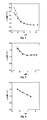
investigated pH-range was 4.4 - 8.3. Table 1 and Fig. 1 show the pH-r
1 dependence
of DPPE/DPSG liposomal Gd-DTPA-BMA at 37 °C in MES buffers after 20
minutes (▪) and 2.0 (▴) hours incubation.
| | r1 (s-1 mM-1) |
| pH | 20 minutes incubation | 2.0 hours incubation |
| 4.4 | 1.03 | 1.41 |
| 4.6 | 0.89 | 1.16 |
| 5.0 | 0.73 | 0.90 |
| 5.5 | 0.38 | 0.54 |
| 6.0 | 0.29 | 0.32 |
| 6.5 | 0.27 | 0.29 |
| 7.1 | 0.26 | 0.27 |
| 7.6 | 0.25 | 0.25 |
| 8.3 | 0.24 | 0.25 |
Table 1/ Fig. 1 shows that r1 of DPPE/DPSG liposomal Gd-DTPA-BMA in
isosmotic MES-buffers was relatively constant between pH 8.3 and 6.0, but
increased significantly by lowering the pH from 6.0 to 4.4.
Example 3
pH - r1 dependence of DPPE/DPSG (4:1 mole ratio) liposomal GdDTPA-BMA
in buffered human whole blood.
DPPE/DPSG liposomes (4:1 mole ratio) containing GdDTPA-BMA were prepared
according to the method described in Example 1. The mean hydrodynamic diameter
of the liposomes was measured to 130 nm as described in Example 1. The T
1-relaxation
times were measured at 37 °C and 0.47 Tesla (Minispec PC-120b, Bruker
GmbH, Rheinstetten, Germany) in different buffer solutions (0.25 M MES and 0.25
M
N-2-hydroxyethylpiperazine-
N'-2-ethanesulfonic acid (HEPES) buffers)
containing 50 volume % human whole blood. The buffered liposome dispersions
were incubated at 37 °C prior to the relaxation measurements. The investigated pH-range
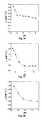
was 5.7 - 8.2. Table 2 and Fig. 2 show the pH-r
1 dependence of DPPE/DPSG
liposomal Gd-DTPA-BMA at 37 °C in MES/HEPES buffered whole blood (50%)
after 20 minutes (▪) and 2.0 (▴) hours incubation. The r
1 values are corrected for
the content of solid materials present in whole blood.
| | r1 (s-1 mM-1) |
| pH | 20 minutes incubation | 2.0 hours incubation |
| 5.7 | 1.80 | 2.21 |
| 5.8 | 1.71 | 2.07 |
| 6.0 | 1.39 | 1.61 |
| 6.4 | 0.79 | 0.88 |
| 6.9 | 0.37 | 0.42 |
| 7.0 | 0.30 | 0.36 |
| 7.5 | 0.21 | 0.31 |
| 7.9 | 0.21 | 0.32 |
| 8.2 | 0.20 | 0.35 |
Table 2/Fig. 2 shows that the r1 of DPPE/DPSG liposomal Gd-DTPA-BMA in
buffered human whole blood was relatively constant between pH 8.2 and 7.5, but
increased significantly by lowering the pH from 7.0 to 5.5. The onset of the r1
increase when lowering the pH was at a higher pH value compared to the results in
Table 1/Fig. 1, and the relative r1-response was higher.
Example 4
pH - r1 dependence of DPPE/DPSG (4:1 mole ratio) liposomal GdDTPA-BMA
preincubated in human whole blood.
DPPE/DPSG liposomes (4:1 mole ratio) containing GdDTPA-BMA were prepared
according to the method described in Example 1. The mean hydrodynamic diameter
of the liposomes was measured to 130 nm as described in Example 1. The liposomes
were incubated for 20 minutes at 37 °C in human whole blood without pH
adjustment. The pH in the vials were then adjusted by adding 0.25 M MES and 0.25
M HEPES buffers and one vial with glucose, and subsequently incubated further at
37 °C. The vials contained 50 volume % whole blood and the investigated pH-range
was 5.7 - 7.5. T
1-relaxation times were measured at 37 °C and 0.47 Tesla (Minispec
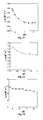
PC-120b, Bruker GmbH, Rheinstetten, Germany). Table 3 and Fig. 3 show the pH-r
1
dependence of DPPE/DPSG liposomal Gd-DTPA-BMA at 37 °C after preincubation
in human whole blood and subsequently further incubation with MES/HEPES
buffers (and one vial with glucose) for 20 minutes (▪) and 60 (▴)minutes. The r
1
values are corrected for the content of solid materials present in whole blood.
| | r1 (s-1 mM-1) |
| pH | 20 minutes incubation | 60 minutes incubation |
| 5.7 | 1.43 | 1.68 |
| 6.0 | 1.10 | 1.22 |
| 6.4 | 0.70 | 0.78 |
| 6.9 | 0.45 | 0.49 |
| 7.5 | 0.40 | 0.47 |
| glucose | 0.41 | 0.48 |
Table 3/Fig. 3 shows that the r1 of DPPE/DPSG liposomal Gd-DTPA-BMA
preincubated in human whole blood was relatively constant between pH 7.5 and 6.9,
but increased significantly by lowering the pH from6.9 to 5.7. The onset of the r1
increase when lowering the pH was at a slightly lower pH value compared to the
results in Table 2/Fig. 2, and the relative r1-response was lower.
Example 5
pH - r1 dependence of DMPE/DPSG (9:1 mole ratio) liposomal GdDTPA-BMA
in isosmotic buffer solutions
DMPE (dimyristoyl-glycero-3-phosphatidylethanolamine) / DPSG (dipalmitoylsuccinylglycerol)
liposomes (9:1 mole ratio) containing GdDTPA-BMA were
prepared according to the method described in Example 1. The mean hydrodynamic
diameter of the liposomes was measured to 122 nm as described in Example 1. The
T
1-relaxation times were measured at 37 °C and 0.47 Tesla (Minispec PC-120b,
Bruker GmbH, Rheinstetten, Germany) in different isosmotic buffer solutions (0.05
M 2-(
N-morpholino) ethanesulfonic acid (MES) buffers). The buffered liposome
dispersions were incubated at 37 °C prior to the relaxation measurements. The
investigated pH-range was 4.4 - 8.3. Table 4 and Fig. 4 show the pH-r
1 dependence
of DMPE/DPSG liposomal Gd-DTPA-BMA at 37 °C in MES buffers after 20
minutes (▪) and 2.0 (▴) hours incubation.
| | r1 (s-1 mM-1) |
| pH | 20 minutes incubation | 2.0 hours incubation |
| 4.4 | 2,33 | 2,99 |
| 4.6 | 2,10 | 2,63 |
| 5.0 | 1,61 | 1,83 |
| 5.5 | 1,15 | 1,24 |
| 6.0 | 0,95 | 1,02 |
| 6.5 | 0,85 | 0,87 |
| 7.1 | 0,78 | 0,79 |
| 7.6 | 0,75 | 0,77 |
| 8.3 | 0,73 | 0,77 |
DMPE (dimyristoyl-glycero-3-phosphatidylethanolamine) / DPSG (dipalmitoylsuccinylglycerol)
liposomes (9:1 mole ratio) containing GdDTPA-BMA were
prepared according to the method described in Example 1. The mean hydrodynamic
diameter of the liposomes was measured to 122 nm as described in Example 1. The
T
1-relaxation times were measured at 37 °C and 0.47 Tesla (Minispec PC-120b,
Bruker GmbH, Rheinstetten, Germany) in different buffer solutions (0.25 M MES
and 0.25 M
N-2-hydroxyethylpiperazine-
N'-2-ethanesulfonic acid (HEPES) buffers)
containing 50 volume % human whole blood. The buffered liposome dispersions
were incubated at 37 °C prior to the relaxation measurements. The investigated pH-range
was 5.7 - 8.2. Table 5 and Fig. 5 show the pH-r
1 dependence of DMPE/DPSG
liposomal Gd-DTPA-BMA at 37 °C in MES/HEPES buffered whole blood (50%)
after 20 minutes (▪) and 2.0 (▴) hours incubation. The r
1 values are corrected for
the content of solid materials present in whole blood.
| | r1 (s-1 mM-1) |
| pH | 20 minutes incubation | 2.0 hours incubation |
| 5.7 | 1,60 | 1,73 |
| 5.8 | 1,56 | 1,64 |
| 6.0 | 1,40 | 1,46 |
| 6.4 | 1,18 | 1,23 |
| 6.9 | 1,10 | 1,13 |
| 7.0 | 1,09 | 1,10 |
| 7.5 | 1,11 | 1,15 |
| 7.9 | 1,12 | 1,14 |
| 8.2 | 1,11 | 1,15 |
DMPE (dimyristoyl-glycero-3-phosphatidylethanolamine) / DPSG (dipalmitoylsuccinylglycerol)
liposomes (9:1 mole ratio) containing GdDTPA-BMA were
prepared according to the method described in Example 1. The mean hydrodynamic
diameter of the liposomes was measured to 122 nm as described in Example 1.The
liposomes were incubated for 20 minutes at 37 °C in human whole blood without pH
adjustment. The pH in the vials were then adjusted by adding 0.25 M MES and 0.25
M HEPES buffers and one vial with glucose, and subsequently incubated further at
37 °C. The vials contained 50 volume % whole blood and the investigated pH-range
was 5.7 - 7.5. T
1-relaxation times were measured at 37 °C and 0.47 Tesla (Minispec
PC-120b, Bruker GmbH, Rheinstetten, Germany). Table 6 and Fig. 6 show the pH-r
1
dependence of DMPE/DPSG liposomal Gd-DTPA-BMA at 37 °C after
preincubation in human whole blood and subsequently further incubation with
MES/HEPES buffers (and one vial with glucose) for 20 minutes (▪) and 60 (▴)
minutes. The r
1 values are corrected for the content of solid materials present in
whole blood.
| | r1 (s-1 mM-1) |
| pH | 20 minutes incubation | 60 minutes incubation |
| 5.7 | 1,57 | 1,54 |
| 6.0 | 1,45 | 1,41 |
| 6.4 | 1,32 | 1,28 |
| 6.9 | 1,14 | 1,13 |
| 7.4 glucose | 1,03 | 0,92 |
DMPE (dimyristoyl-glycero-3-phosphatidylethanolamine) / DPSG (dipalmitoylsuccinylglycerol)
liposomes (7:3 mole ratio) containing GdDTPA-BMA were
prepared according to the method described in Example 1. The mean hydrodynamic
diameter of the liposomes was measured to 115 nm as described in Example 1. The
T
1-relaxation times were measured at 37 °C and 0.47 Tesla (Minispec PC-120b,
Bruker GmbH, Rheinstetten, Germany) in different isosmotic buffer solutions (0.05
M 2-(
N-morpholino) ethanesulfonic acid (MES) buffers). The buffered liposome
dispersions were incubated at 37 °C prior to the relaxation measurements. The
investigated pH-range was 4.4 - 8.3. Table 7 and Fig. 7 show the pH-r
1 dependence
of DMPE/DPSG liposomal Gd-DTPA-BMA at 37 °C in MES buffers after 20
minutes (▪) and 2.0 (▴) hours incubation.
| | r1 (s-1 mM-1) |
| pH | 20 minutes incubation | 2.0 hours incubation |
| 4.4 | 2,99 | 3,29 |
| 4.6 | 2,37 | 2,66 |
| 5.0 | 1,51 | 1,61 |
| 5.5 | 1,26 | 1,29 |
| 6.0 | 1,24 | 1,25 |
| 6.5 | 1,17 | 1,15 |
| 7.1 | 1,13 | 1,12 |
| 7.6 | 1,08 | 1,10 |
| 8.3 | 1,11 | 1,22 |
DMPE (dimyristoyl-glycero-3-phosphatidylethanolamine) / DPSG (dipalmitoylsuccinylglycerol)
liposomes (7:3 mole ratio) containing GdDTPA-BMA were
prepared according to the method described in Example 1. The mean hydrodynamic
diameter of the liposomes was measured to 115 nm as described in Example 1. The
T
1-relaxation times were measured at 37 °C and 0.47 Tesla (Minispec PC-120b,
Broker GmbH, Rheinstetten, Germany) in different buffer solutions (0.25 M MES
and 0.25 M
N-2-hydroxyethylpiperazine-
N'-2-ethanesulfonic acid (HEPES) buffers)
containing 50 volume % human whole blood. The buffered liposome dispersions
were incubated at 37 °C prior to the relaxation measurements. The investigated pH-range
was 5.7 - 8.2. Table 8 and Fig. 8 show the pH-r
1 dependence of DMPE/DPSG
liposomal Gd-DTPA-BMA at 37 °C in MES/HEPES buffered whole blood (50%)
after 20 minutes (▪) and 2.0 (▴) hours incubation. The r
1 values are corrected for
the content of solid materials present in whole blood.
| | r1 (s-1 mM-1) |
| pH | 20 minutes incubation | 2.0 hours incubation |
| 5.7 | 1,60 | 1,80 |
| 5.8 | 1,56 | 1,68 |
| 6.0 | 1,40 | 1,40 |
| 6.4 | 1,32 | 1,13 |
| 6.9 | 1,30 | 1,06 |
| 7.0 | 1,32 | 1,14 |
| 7.5 | 1,33 | 1,45 |
| 7.9 | 1,33 | 1,35 |
| 8.2 | 1,30 | 1,33 |
DMPE (dimyristoyl-glycero-3-phosphatidylethanolamine) / DPSG (dipalmitoylsuccinylglycerol)
liposomes (7:3 mole ratio) containing GdDTPA-BMA were
prepared according to the method described in Example 1. The mean hydrodynamic
diameter of the liposomes was measured to 115 nm as described in Example 1.The
liposomes were incubated for 20 minutes at 37 °C in human whole blood without pH
adjustment. The pH in the vials were then adjusted by adding 0.25 M MES and 0.25
M HEPES buffers and one vial with glucose, and subsequently incubated further at
37 °C. The vials contained 50 volume % whole blood and the investigated pH-range
was 5.7 - 7.5. T
1-relaxation times were measured at 37 °C and 0.47 Tesla (Minispec
PC-120b, Bruker GmbH, Rheinstetten, Germany). Table 9 and Fig. 9 show the pH-r
1
dependence of DMPE/DPSG liposomal Gd-DTPA-BMA at 37 °C after
preincubation in human whole blood and subsequently further incubation with
MES/HEPES buffers (and one vial with glucose) for 20 minutes (▪) and 60 (▴)
minutes. The r
1 values are corrected for the content of solid materials present in
whole blood.
| | r1 (s-1 mM-1) |
| pH | 20 minutes incubation | 60 minutes incubation |
| 5.7 | 1,61 | 1,60 |
| 6.0 | 1,30 | 1,33 |
| 6.4 | 1,26 | 1,20 |
| 6.9 | 1,16 | 1,14 |
| 7.4 glucose | 1,06 | 1,09 |
DPPE (dipalmitoyl-glycero-3-phosphatidylethanolamine) / DPSG (dipalmitoylsuccinylglycerol)
liposomes (9:1 mole ratio) containing GdDTPA-BMA were
prepared according to the method described in Example 1. The mean hydrodynamic
diameter of the liposomes was measured to 116 nm as described in Example 1. The
T
1-relaxation times were measured at 37 °C and 0.47 Tesla (Minispec PC-120b,
Bruker GmbH, Rheinstetten, Germany) in different isosmotic buffer solutions (0.05
M 2-(
N-morpholino) ethanesulfonic acid (MES) buffers). The buffered liposome
dispersions were incubated at 37 °C prior to the relaxation measurements. The
investigated pH-range was 4.4 - 8.0. Table 10 and Fig. 10 show the pH-r
1
dependence of DPPE/DPSG liposomal Gd-DTPA-BMA at 37 °C in MES buffers
after 20 minutes (▪) and 2.0 (▴) hours incubation.
| | r1 (s-1 mM-1) |
| pH | 20 minutes incubation | 2.0 hours incubation |
| 4,4 | 1,58 | 1,96 |
| 4,6 | 1,52 | 1,86 |
| 5,0 | 1,34 | 1,59 |
| 5,5 | 0,89 | 1,06 |
| 6,0 | 0,59 | 0,82 |
| 6,5 | 0,49 | 0,54 |
| 6,9 | 0,46 | 0,48 |
| 7,5 | 0,45 | 0,46 |
| 8,0 | 0,44 | 0,44 |
DPPE (dipalmitoyl-glycero-3-phosphatidylethanolamine) / DPSG (dipalmitoylsuccinylglycerol)
liposomes (9:1 mole ratio) containing GdDTPA-BMA were
prepared according to the method described in Example 1. The mean hydrodynamic
diameter of the liposomes was measured to 116 nm as described in Example 1. The
T
1-relaxation times were measured at 37 °C and 0.47 Tesla (Minispec PC-120b,
Bruker GmbH, Rheinstetten, Germany) in different buffer solutions (0.25 M MES
and 0.25 M
N-2-hydroxyethylpiperazine-
N'-2-ethanesulfonic acid (HEPES) buffers)
containing 50 volume % human whole blood. The buffered liposome dispersions
were incubated at 37 °C prior to the relaxation measurements. The investigated pH-range
was 5.7 - 8.2. Table 11 and Fig. 11 show the pH-r
1 dependence of
DPPE/DPSG liposomal Gd-DTPA-BMA at 37 °C in MES/HEPES buffered whole
blood (50%) after 20 minutes (▪) and 2.0 (▴) hours incubation. The r
1 values are
corrected for the content of solid materials present in whole blood.
| | r1 (s-1 mM-1) |
| pH | 20 minutes incubation | 2.0 hours incubation |
| 5.7 | 2,28 | 2,74 |
| 5.8 | 2,20 | 2,73 |
| 6.0 | 1,95 | 2,32 |
| 6.4 | 1,49 | 1,69 |
| 6.9 | 0,98 | 1,08 |
| 7.0 | 0,91 | 1,04 |
| 7.5 | 0,71 | 0,76 |
| 7.9 | 0,69 | 0,73 |
| 8.2 | 0,65 | 0,71 |
DPPE (dipalmitoyl-glycero-3-phosphatidylethanolamine) / DPSG (dipalmitoylsuccinylglycerol)
liposomes (9:1 mole ratio) containing GdDTPA-BMA were
prepared according to the method described in Example 1. The mean hydrodynamic
diameter of the liposomes was measured to 116 nm as described in Example 1. The
liposomes were incubated for 20 minutes at 37 °C in human whole blood without pH
adjustment. The pH in the vials were then adjusted by adding 0.25 M MES and 0.25
M HEPES buffers and one vial with glucose, and subsequently incubated further at
37 °C. The vials contained 50 volume % whole blood and the investigated pH-range
was 5.7 - 7.5. T
1-relaxation times were measured at 37 °C and 0.47 Tesla (Minispec
PC-120b, Bruker GmbH, Rheinstetten, Germany). Table 12 and Fig. 12 show the
pH-r
1 dependence of DPPE/DPSG liposomal Gd-DTPA-BMA at 37 °C after
preincubation in human whole blood and subsequently further incubation with
MES/HEPES buffers (and one vial with glucose) for 20 minutes (▪) and 60 (▴)
minutes. The r
1 values are corrected for the content of solid materials present in
whole blood.
| | r1 (s-1 mM-1) |
| pH | 20 minutes incubation | 60 minutes incubation |
| 5.7 | 1,95 | 2,15 |
| 6.0 | 1,49 | 1,69 |
| 6.4 | 1,15 | 1,30 |
| 6.9 | 0,95 | 0,98 |
| 7.5 | 0,88 | 0,91 |
| 7.4 glucose | 1,95 | 2,14 |
DPPE (dipalmitoyl-glycero-3-phosphatidylethanolamine) / DPSG (dipalmitoylsuccinylglycerol)
liposomes (7:3 mole ratio) containing GdDTPA-BMA were
prepared according to the method described in Example 1. The mean hydrodynamic
diameter of the liposomes was measured to 110 nm as described in Example 1. The
T
1-relaxation times were measured at 37 °C and 0.47 Tesla (Minispec PC-120b,
Bruker GmbH, Rheinstetten, Germany) in different isosmotic buffer solutions (0.05
M 2-(
N-morpholino) ethanesulfonic acid (MES) buffers). The buffered liposome
dispersions were incubated at 37 °C prior to the relaxation measurements. The
investigated pH-range was 4.4 - 8.0. Table 13 and Fig. 13 show the pH-r
1
dependence of DPPE/DPSG liposomal Gd-DTPA-BMA at 37 °C in MES buffers
after 20 minutes (▪) and 2.0 (▴) hours incubation.
| | r1 (s-1 mM-1) |
| pH | 20 minutes incubation | 2.0 hours incubation |
| 4,4 | 2,37 | 2,72 |
| 4,6 | 2,10 | 2,48 |
| 5,0 | 1,36 | 1,48 |
| 5,5 | 0,93 | 1,13 |
| 6,0 | 0,85 | 0,81 |
| 6,5 | 0,86 | 0,85 |
| 6,9 | 0,83 | 0,82 |
| 7,5 | 0,81 | 0,81 |
| 8,0 | 0,78 | 0,78 |
DPPE (dipalmitoyl-glycero-3-phosphatidylethanolamine) / DPSG (dipalmitoylsuccinylglycerol)
liposomes (7:3 mole ratio) containing GdDTPA-BMA were
prepared according to the method described in Example 1. The mean hydrodynamic
diameter of the liposomes was measured to 110 nm as described in Example 1. The
T
1-relaxation times were measured at 37 °C and 0.47 Tesla (Minispec PC-120b,
Bruker GmbH, Rheinstetten, Germany) in different buffer solutions (0.25 M MES
and 0.25 M
N-2-hydroxyethylpiperazine-
N'-2-ethanesulfonic acid (HEPES) buffers)
containing 50 volume % human whole blood. The buffered liposome dispersions
were incubated at 37 °C prior to the relaxation measurements. The investigated pH-range
was 5.7 - 8.2. Table 14 and Fig. 14 show the pH-r
1 dependence of
DPPE/DPSG liposomal Gd-DTPA-BMA at 37 °C in MES/HEPES buffered whole
blood (50%) after 20 minutes (▪) and 2.0 (▴) hours incubation. The r
1 values are
corrected for the content of solid materials present in whole blood.
| | r1 (s-1 mM-1) |
| pH | 20 minutes incubation | 2.0 hours incubation |
| 5.7 | 2,38 | 2,66 |
| 5.8 | 2,30 | 2,59 |
| 6.0 | 2,11 | 2,15 |
| 6.4 | 1,30 | 1,37 |
| 6.9 | 0,65 | 0,80 |
| 7.0 | 0,57 | 0,75 |
| 7.5 | 0,55 | 0,72 |
| 7.9 | 0,56 | 0,87 |
| 8.2 | 0,60 | 1,00 |
DPPE (dipalmitoyl-glycero-3-phosphatidylethanolamine) / DPSG (dipalmitoylsuccinylglycerol)
liposomes (7:3 mole ratio) containing GdDTPA-BMA were
prepared according to the method described in Example 1. The mean hydrodynamic
diameter of the liposomes was measured to 110 nm as described in Example 1. The
liposomes were incubated for 20 minutes at 37 °C in human whole blood without pH
adjustment. The pH in the vials were then adjusted by adding 0.25 M MES and 0.25
M HEPES buffers and one vial with glucose, and subsequently incubated further at
37 °C. The vials contained 50 volume % whole blood and the investigated pH-range
was 5.7 - 7.5. T
1-relaxation times were measured at 37 °C and 0.47 Tesla (Minispec
PC-120b, Bruker GmbH, Rheinstetten, Germany). Table 15 and Fig. 15 show the
pH-r
1 dependence of DPPE/DPSG liposomal Gd-DTPA-BMA at 37 °C after
preincubation in human whole blood and subsequently further incubation with
MES/HEPES buffers (and one vial with glucose) for 20 minutes (▪) and 60 (▴)
minutes. The r
1 values are corrected for the content of solid materials present in
whole blood.
| | r1 (s-1 mM-1) |
| pH | 20 minutes incubation | 60 minutes incubation |
| 5.7 | 2,00 | 2,16 |
| 6.0 | 1,45 | 1,59 |
| 6.4 | 0,92 | 0,99 |
| 6.9 | 0,67 | 0,74 |
| 7.4 glucose | 0,59 | 0,62 |
DSPE (distearoyl-glycero-3-phosphatidylethanolamine) / DPSG (dipalmitoylsuccinylglycerol)
liposomes (9:1 mole ratio) containing GdDTPA-BMA were
prepared according to the method described in Example 1. The mean hydrodynamic
diameter of the liposomes was measured to 128 nm as described in Example 1. The
T
1-relaxation times were measured at 37 °C and 0.47 Tesla (Minispec PC-120b,
Bruker GmbH, Rheinstetten, Germany) in different isosmotic buffer solutions (0.05
M 2-(
N-morpholino) ethanesulfonic acid (MES) buffers). The buffered liposome
dispersions were incubated at 37 °C prior to the relaxation measurements. The
investigated pH-range was 4.4 - 8.3. Table 16 and Fig. 16 show the pH-r
1
dependence of DSPE/DPSG liposomal Gd-DTPA-BMA at 37 °C in MES buffers
after 20 minutes (▪) and 2.0 (▴) hours incubation.
| | r1 (s-1 mM-1) |
| pH | 20 minutes incubation | 2.0 hours incubation |
| 4.4 | 0,85 | 1,12 |
| 4.6 | 0,86 | 1,09 |
| 5.0 | 0,66 | 0,93 |
| 5.5 | 0,38 | 0,59 |
| 6.0 | 0,16 | 0,20 |
| 6.5 | 0,15 | 0,16 |
| 7.1 | 0,14 | 0,15 |
| 7.6 | 0,13 | 0,15 |
| 8.3 | 0,13 | 0,14 |
DSPE (distearoyl-glycero-3-phosphatidylethanolamine) / DPSG (dipalmitoylsuccinylglycerol)
liposomes (9:1 mole ratio) containing GdDTPA-BMA were
prepared according to the method described in Example 1. The mean hydrodynamic
diameter of the liposomes was measured to 128 nm as described in Example 1.The
T
1-relaxation times were measured at 37 °C and 0.47 Tesla (Minispec PC-120b,
Bruker GmbH, Rheinstetten, Germany) in different buffer solutions (0.25 M MES
and 0.25 M
N-2-hydroxyethylpiperazine-
N'-2-ethanesulfonic acid (HEPES) buffers)
containing 50 volume % human whole blood. The buffered liposome dispersions
were incubated at 37 °C prior to the relaxation measurements. The investigated pH-range
was 5.7 - 8.2. Table 17 and Fig. 17 show the pH-r
1 dependence of
DSPE/DPSG liposomal Gd-DTPA-BMA at 37 °C in MES/HEPES buffered whole
blood (50%) after 20 minutes (▪) and 2.0 (▴) hours incubation. The r
1 values are
corrected for the content of solid materials present in whole blood.
| | r1 (s-1 mM-1) |
| pH | 20 minutes incubation | 2.0 hours incubation |
| 5.7 | 1,21 | 2,28 |
| 5.8 | 1,13 | 1,98 |
| 6.0 | 0,99 | 1,71 |
| 6.4 | 0,74 | 1,16 |
| 6.9 | 0,45 | 0,65 |
| 7.0 | 0,42 | 0,60 |
| 7.5 | 0,29 | 0,38 |
| 7.9 | 0,29 | 0,38 |
| 8.2 | 0,28 | 0,38 |
DSPE (distearoyl-glycero-3-phosphatidylethanolamine) / DPSG (dipalmitoylsuccinylglycerol)
liposomes (9:1 mole ratio) containing GdDTPA-BMA were
prepared according to the method described in Example 1. The mean hydrodynamic
diameter of the liposomes was measured to 128 nm as described in Example 1. The
liposomes were incubated for 20 minutes at 37 °C in human whole blood without pH
adjustment. The pH in the vials were then adjusted by adding 0.25 M MES and 0.25
M HEPES buffers and one vial with glucose, and subsequently incubated further at
37 °C. The vials contained 50 volume % whole blood and the investigated pH-range
was 5.7 - 7.5. T
1-relaxation times were measured at 37 °C and 0.47 Tesla (Minispec
PC-120b, Bruker GmbH, Rheinstetten, Germany). Table 18 and Fig. 18 show the
pH-r
1 dependence of DSPE/DPSG liposomal Gd-DTPA-BMA at 37 °C after
preincubation in human whole blood and subsequently further incubation with
MES/HEPES buffers (and one vial with glucose) for 20 minutes (▪) and 60 (▴)
minutes. The r
1 values are corrected for the content of solid materials present in
whole blood.
| | r1 (s-1 mM-1) |
| pH | 20 minutes incubation | 60 minutes incubation |
| 5.7 | 1,15 | 1,41 |
| 6.0 | 0,96 | 1,14 |
| 6.4 | 0,77 | 0,86 |
| 6.9 | 0,65 | 0,69 |
| 7.4 glucose | 0,60 | 0,65 |
DSPE (distearoyl-glycero-3-phosphatidylethanolamine) / DPSG (dipalmitoylsuccinylglycerol)
liposomes (7:3 mole ratio) containing GdDTPA-BMA were
prepared according to the method described in Example 1. The mean hydrodynamic
diameter of the liposomes was measured to 113 nm as described in Example 1. The
T
1-relaxation times were measured at 37 °C and 0.47 Tesla (Minispec PC-120b,
Bruker GmbH, Rheinstetten, Germany) in different isosmotic buffer solutions (0.05
M 2-(
N-morpholino) ethanesulfonic acid (MES) buffers). The buffered liposome
dispersions were incubated at 37 °C prior to the relaxation measurements. The
investigated pH-range was 4.4 - 8.3. Table 19 and Fig. 19 show the pH-r
1
dependence of DSPE/DPSG liposomal Gd-DTPA-BMA at 37 °C in MES buffers
after 20 minutes (▪) and 2.0 (▴) hours incubation.
| | r1 (s-1 mM-1) |
| pH | 20 minutes incubation | 2.0 hours incubation |
| 4.4 | 2,84 | 3,08 |
| 4.6 | 2,52 | 2,66 |
| 5.0 | 1,94 | 2,19 |
| 5.5 | 0,89 | 1,13 |
| 6.0 | 0,88 | 0,90 |
| 6.5 | 0,88 | 0,89 |
| 7.1 | 0,87 | 0,85 |
| 7.6 | 0,86 | 0,87 |
| 8.3 | 0,87 | 0,87 |
DSPE (distearoyl-glycero-3-phosphatidylethanolamine) / DPSG (dipalmitoylsuccinylglycerol)
liposomes (7:3 mole ratio) containing GdDTPA-BMA were
prepared according to the method described in Example 1. The mean hydrodynamic
diameter of the liposomes was measured to 113 nm as described in Example 1.The
T
1-relaxation times were measured at 37 °C and 0.47 Tesla (Minispec PC-120b,
Bruker GmbH, Rheinstetten, Germany) in different buffer solutions (0.25 M MES
and 0.25 M
N-2-hydroxyethylpiperazine-
N'-2-ethanesulfonic acid (HEPES) buffers)
containing 50 volume % human whole blood. The buffered liposome dispersions
were incubated at 37 °C prior to the relaxation measurements. The investigated pH-range
was 5.7 - 8.2. Table 20 and Fig. 20 show the pH-r
1 dependence of
DSPE/DPSG liposomal Gd-DTPA-BMA at 37 °C in MES/HEPES buffered whole
blood (50%) after 20 minutes (▪) and 2.0 (▴) hours incubation. The r
1 values are
corrected for the content of solid materials present in whole blood.
| | r1 (s-1 mM-1) |
| pH | 20 minutes incubation | 2.0 hours incubation |
| 5.7 | 2,24 | 2,94 |
| 5.8 | 2,21 | 2,86 |
| 6.0 | 1,93 | 2,49 |
| 6.4 | 1,22 | 1,54 |
| 6.9 | 0,79 | 0,98 |
| 7.0 | 0,64 | 0,76 |
| 7.5 | 0,52 | 0,83 |
| 7.9 | 0,57 | 0,97 |
| 8.2 | 0,57 | 1,02 |
DSPE (distearoyl-glycero-3-phosphatidylethanolamine) / DPSG (dipalmitoylsuccinylglycerol)
liposomes (7:3 mole ratio) containing GdDTPA-BMA were
prepared according to the method described in Example 1. The mean hydrodynamic
diameter of the liposomes was measured to 113 nm as described in Example 1. The
liposomes were incubated for 20 minutes at 37 °C in human whole blood without pH
adjustment. The pH in the vials were then adjusted by adding 0.25 M MES and 0.25
M HEPES buffers and one vial with glucose, and subsequently incubated further at
37 °C. The vials contained 50 volume % whole blood and the investigated pH-range
was 5.7 - 7.5. T
1-relaxation times were measured at 37 °C and 0.47 Tesla (Minispec
PC-120b, Bruker GmbH, Rheinstetten, Germany). Table 21 and Fig. 21 show the
pH-r
1 dependence of DSPE/DPSG liposomal Gd-DTPA-BMA at 37 °C after
preincubation in human whole blood and subsequently further incubation with
MES/HEPES buffers (and one vial with glucose) for 20 minutes (▪) and 60 (▴)
minutes. The r
1 values are corrected for the content of solid materials present in
whole blood.
| | r1 (s-1 mM-1) |
| pH | 20 minutes incubation | 60 minutes incubation |
| 5.7 | 1,97 | 2,43 |
| 6.0 | 1,47 | 1,73 |
| 6.4 | 0,92 | 1,08 |
| 6.9 | 0,70 | 0,78 |
| 7.4 glucose | 0,58 | 0,62 |
DPPE/DPSG liposomes (4:1 mole ratio) containing GdDTPA-BMA were prepared
according to the method described in Example 1. The mean hydrodynamic diameter
of the liposomes was measured to 115 nm as described in Example 1. The MR
contrast efficacy of the liposomal system was investigated in human whole blood in
two gel phantoms at 1.0 T on a Siemens Magnetom Harmony system (Siemens AG,
Erlangen, Germany). Each phantom was composed of eight glass vials placed
equidistantly from each other and from the centre of a circular glass container. The
latter was filled with an agar gel (2 % w/w) doped with GdDTPA-BMA to give a T1
of about 500 ms at 1.0 T. Imaging was performed in a quadrature head coil.
Quantitative T1 maps were obtained from spoiled 2D gradient echo images
(TR/TE/flip = 2.1 ms/ 1.7 ms/ 8°) prepared with a nonselective 180° inversion pulse.
The time between the inversion pulse and the acquisition (TI) was varied between
220 ms and 5788 ms, with a total of 11 TI values used. The system gain was kept
constant between consecutive scans so that the MR signal was directly comparable
between scans. Other scan parameters were: voxel size: 1.17 x 1.17 x 9.0 mm3,
number of averages: 20. The inter-scan interval was 6 s to allow for almost complete
recovery of the longitudinal magnetisation between scans. The resulting
magnetisation recovery curve obtained from the 11 magnetisation prepared scans
were fit to the equation:
SI(TI) = A · 1-2exp(-TI · R 1) + B
where ∥ denotes the absolute value, A and B are scaling constants, TI is the inversion
time and R1 is the T1 relaxation rate (s-1). A Lovenberg-Marquard nonlinear least
squares fit routine was used and parametric R1 maps were generated on a pixel-by-pixel
basis from the raw inversion recovery datasets. The resulting R1 value of each
liposome containing glass vial was measured from a constant diameter region of
interest (ROI) placed in each vial in the image. The Ceff in the samples was
approximately 1 mM Gd after dilution (see below for details).
Effect of Incubation in Buffered Human Whole Blood
The liposomes were diluted with MES/HEPES (250 mM) buffered whole
blood in the pH range 5.6-7.9. The volume ratios between liposome dispersion,
buffer and human whole blood were 1/5/5, respectively. The samples were incubated
at 37 °C for 20 minutes. The liposome dispersions were then transferred to the
respective vials in the first phantom and equilibrated to room temperature prior to the
MR imaging. Fig. 22 shows a quantitative R1 map of the phantom. Fig. 24 shows the
change in R1 as a function of pH as measured from the ROI's placed in the
parametric R1 map (Data points: ▪).
Effect of Pre-incubation in Human Whole Blood
The liposomes were diluted with whole blood and incubated at 37 °C for 20
minutes, followed by pH adjustment with 250 mM MES/HEPES buffers and further
incubation for 20 minutes. Isotonic NaCl- and glucose-solution was added to two of
the vials, respectively, instead of buffer, as controls at pH 7.4 (unaltered pH). The
investigated pH range was 5.6 - 7.4. The volume ratios between liposome
dispersion, human whole blood and buffer were 1/5/5, respectively. The dispersions
were transferred to the respective vials in the second phantom and equilibrated to
room temperature prior to the MR imaging. Fig. 23 shows a quantitative R1 map of
the phantom. Fig. 24 shows the change in R1 as a function of pH as measured from
the ROI's placed in the parametric R1 map (Data points: ▴).
Example 24
Cryo-transmission electron microscopy (Cryo-TEM) of DPPE/DPSG liposomal
GdDTPA-BMA.
DPPE/DPSG liposomes (4:1 mole ratio) containing GdDTPA-BMA were
prepared according to the method described in Example 1. The mean hydrodynamic
diameter of the liposomes was measured to 111 nm as described in Example 1. The
DPPE/DPSG liposomal dispersion was diluted 3/1 and 1/1 (v/v) with isoosmotic
buffer solutions (50 mM MES buffers) at pH 4.4 and 7.4, respectively. Different
dilution factors were used at the two pH values in order to obtain micrographs with
similar material content. The buffered liposomal dispersion at pH 4.4 was incubated
at 37 °C for 30 minutes prior to the Cryo-TEM procedure, while the corresponding
sample at pH 7.4 was assessed directly after dilution.
The Cryo-TEM preparation procedure consisted, in short, of the following: The
dispersions described above were equilibrated at 25 °C and 98-99 % relative
humidity within a climate chamber. A droplet (∼2 µl) of sample dispersion was
deposited on glow discharge treated 300 mesh copper grids (Agar Scientific Ltd,
Stansted Essex, England) covered with a perforated polymer film coated with
evaporated carbon on both sides. Excess liquid was removed with filter paper,
followed by immediate plunging of the grid into liquid ethane held just above its
freezing point. The vitrified sample was then transferred under protection against
atmospheric conditions to a Zeiss EM 902A electron microscope (LEO Electron
Microscopy, Oberkochen, Germany). The temperature of the specimen was kept
below -165 °C during the whole process and all observations were made in zero loss
brightfield mode and at an accelerating voltage of 80 kV. A minimum/micro-dose
focussing system (MDF) was used to minimise radiation damage. The images were
acquired by employing a cooled slow-scan CCD camera (Proscan GmbH, Scheuring,
Germany), and analySIS software (Soft Imaging System GmbH, Münster,
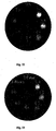
Germany). Figure 25 shows cryo-TEM micrographs of the DPPE/DPSG system at
pH 7.4 (Fig. 25a) and after 30 minutes incubation at pH 4.4 and 37 °C (Fig. 25b).
Well-separated unilamellar liposomes were mainly observed at pH 7.4, but small
bilayer fragments were also present (Fig. 25a). Aggregated structures were present at
pH 4.4, along with large bilayers (Fig. 25b). The white spots in the micrographs are
irradiation-damaged areas.
Example 25
pH - r1 dependence of DPPE/DPSG/DPPE-PEG2000 (4:1:0.025 mole ratio)
liposomal GdDTPA-BMA in isosmotic buffer solutions
DPPE (dipalmitoyl-glycero-3-phosphatidylethanolamine) / DPSG (dipalmitoylsuccinylglycerol)
/ DPPE-PEG2000 (dipalmitoyl-glycero-3-phosphatidylethanolamine-polyethyleneglycol
2000) liposomes (4:1:0.025 mole ratio) containing
GdDTPA-BMA were prepared according to the method described in Example 1. The
mean hydrodynamic diameter of the liposomes was measured to 111 nm as described
in Example 1. The T
1-relaxation times were measured at 37 °C and 0.47 Tesla
(Minispec PC-120b, Bruker GmbH, Rheinstetten, Germany) in different isosmotic
buffer solutions (0.05 M 2-(
N-morpholino) ethanesulfonic acid (MES) buffers). The
buffered liposome dispersions were incubated at 37 °C prior to the relaxation
measurements. The investigated pH-range was 4.4 - 8.3. Table 22 and Fig. 26 show
the pH-r
1 dependence of DPPE/DPSG/DPPE-PEG2000 liposomal Gd-DTPA-BMA
at 37 °C in MES buffers after 20 minutes (▪) incubation.
| | r1 (s-1 mM-1) |
| pH | 20 minutes incubation |
| 4.4 | 0,83 |
| 4.6 | 0,71 |
| 5.0 | 0,53 |
| 5.5 | 0,53 |
| 6.0 | 0,51 |
| 6.5 | 0,50 |
| 7.1 | 0,48 |
| 7.6 | 0,46 |
| 8.3 | 0,44 |
DPPE (dipalmitoyl-glycero-3-phosphatidylethanolamine) / DPSG (dipalmitoylsuccinylglycerol)
/ DPPE-PEG2000 (dipalmitoyl-glycero-3-phosphatidylethanolamine-polyethyleneglycol
2000) liposomes (4:1:0.025 mole ratio) containing
GdDTPA-BMA were prepared according to the method described in Example 1. The
mean hydrodynamic diameter of the liposomes was measured to 111 nm as described
in Example 1. The T
1-relaxation times were measured at 37 °C and 0.47 Tesla
(Minispec PC-120b, Bruker GmbH, Rheinstetten, Germany) in different buffer
solutions (0.25 M MES and 0.25 M
N-2-hydroxyethylpiperazine-
N'-2-ethanesulfonic
acid (HEPES) buffers) containing 50 volume % human whole blood. The buffered
liposome dispersions were incubated at 37 °C prior to the relaxation measurements.
The investigated pH-range was 5.7 - 8.2. Table 23 and Fig. 27 show the pH-r
1
dependence of DPPE/DPSG/DPPE-PEG2000 liposomal Gd-DTPA-BMA at 37 °C in
MES/HEPES buffered whole blood (50%) after 20 minutes (▪) incubation. The r
1
values are corrected for the content of solid materials present in whole blood.
| | r1 (s-1 mM-1) |
| pH | 20 minutes incubation |
| 5.7 | 2,04 |
| 5.8 | 1,89 |
| 6.0 | 1,48 |
| 6.4 | 0,79 |
| 6.9 | 0,47 |
| 7.0 | 0,42 |
| 7.5 | 0,40 |
| 7.9 | 0,42 |
| 8.2 | 0,42 |
DPPE (dipalmitoyl-glycero-3-phosphatidylethanolamine) / DPSG (dipalmitoylsuccinylglycerol)
/ DPPE-PEG2000 (dipalmitoyl-glycero-3-phosphatidylethanolamine-polyethyleneglycol
2000) liposomes (4:1:0.025 mole ratio) containing
GdDTPA-BMA were prepared according to the method described in Example 1. The
mean hydrodynamic diameter of the liposomes was measured to 111 nm as described
in Example 1. The liposomes were incubated for 20 minutes at 37 °C in human
whole blood without pH adjustment. The pH in the vials were then adjusted by
adding 0.25 M MES and 0.25 M HEPES buffers and one vial with glucose, and
subsequently incubated further at 37 °C. The vials contained 50 volume % whole
blood and the investigated pH-range was 5.7 - 7.5. T
1-relaxation times were
measured at 37 °C and 0.47 Tesla (Minispec PC-120b, Bruker GmbH, Rheinstetten,
Germany). Table 24 and Fig. 28 show the pH-r
1 dependence of DPPE/DPSG/DPPE-PEG2000
liposomal Gd-DTPA-BMA at 37 °C after preincubation in human whole
blood and subsequently further incubation with MES/HEPES buffers (and one vial
with glucose) for 20 minutes (▪). The r
1 values are corrected for the content of solid
materials present in whole blood.
| | r1 (s-1 mM-1) |
| pH | 20 minutes incubation |
| 5.7 | 1,46 |
| 6.0 | 1,01 |
| 6.4 | 0,63 |
| 6.9 | 0,52 |
| glucose | 0,47 |
DPPE (dipalmitoyl-glycero-3-phosphatidylethanolamine) / DPSG (dipalmitoylsuccinylglycerol)
/ DPPE-PEG2000 (dipalmitoyl-glycero-3-phosphatidylethanolamine-polyethyleneglycol
2000) liposomes (4:1:0.076 mole ratio) containing
GdDTPA-BMA were prepared according to the method described in Example 1. The
mean hydrodynamic diameter of the liposomes was measured to 106 nm as described
in Example 1. The T
1-relaxation times were measured at 37 °C and 0.47 Tesla
(Minispec PC-120b, Bruker GmbH, Rheinstetten, Germany) in different buffer
solutions (0.25 M MES and 0.25 M
N-2-hydroxyethylpiperazine-
N'-2-ethanesulfonic
acid (HEPES) buffers) containing 50 volume % human whole blood. The buffered
liposome dispersions were incubated at 37 °C prior to the relaxation measurements.
The investigated pH-range was 5.7 - 8.2. Table 25 and Fig. 29 show the pH-r
1
dependence of DPPE/DPSG/DPPE-PEG2000 liposomal Gd-DTPA-BMA at 37 °C in
MES/HEPES buffered whole blood (50%) after 20 minutes (▪) incubation. The r
1
values are corrected for the content of solid materials present in whole blood.
| | r1 (s-1 mM-1) |
| pH | 20 minutes incubation |
| 5.7 | 1,61 |
| 5.8 | 1,40 |
| 6.0 | 1,03 |
| 6.4 | 0,77 |
| 6.9 | 0,62 |
| 7.0 | 0,55 |
| 7.5 | 0,47 |
| 7.9 | 0,46 |
| 8.2 | 0,43 |
DPPE (dipalmitoyl-glycero-3-phosphatidylethanolamine) / DPSG (dipalmitoylsuccinylglycerol)
/ DPPE-PEG2000 (dipalmitoyl-glycero-3-phosphatidylethanolamine-polyethyleneglycol
2000) liposomes (4:1:0.076 mole ratio) containing
GdDTPA-BMA were prepared according to the method described in Example 1. The
mean hydrodynamic diameter of the liposomes was measured to 106 nm as described
in Example 1. The liposomes were incubated for 20 minutes at 37 °C in human
whole blood without pH adjustment. The pH in the vials were then adjusted by
adding 0.25 M MES and 0.25 M HEPES buffers and one vial with glucose, and
subsequently incubated further at 37 °C. The vials contained 50 volume % whole
blood and the investigated pH-range was 5.7 - 7.5. T
1-relaxation times were
measured at 37 °C and 0.47 Tesla (Minispec PC-120b, Bruker GmbH, Rheinstetten,
Germany). Table 26 and Fig. 30 show the pH-r
1 dependence of DPPE/DPSG/DPPE-PEG2000
liposomal Gd-DTPA-BMA at 37 °C after preincubation in human whole
blood and subsequently further incubation with MES/HEPES buffers (and one vial
with glucose) for 20 minutes (▪). The r
1 values are corrected for the content of solid
materials present in whole blood.
| | r1 (s-1 mM-1) |
| pH | 20 minutes incubation |
| 5.7 | 1,17 |
| 6.0 | 0,86 |
| 6.4 | 0,68 |
| 6.9 | 0,60 |
| glucose | 0,54 |
DPPE (dipalmitoyl-glycero-3-phosphatidylethanolamine) / DPSG (dipalmitoylsuccinylglycerol)
/ DPPE-PEG2000 (dipalmitoyl-glycero-3-phosphatidylethanolamine-polyethyleneglycol
2000) liposomes (4:1:0.13 mole ratio) containing
GdDTPA-BMA were prepared according to the method described in Example 1. The
mean hydrodynamic diameter of the liposomes was measured to 112 nm as described
in Example 1. The T
1-relaxation times were measured at 37 °C and 0.47 Tesla
(Minispec PC-120b, Bruker GmbH, Rheinstetten, Germany) in different isosmotic
buffer solutions (0.05 M 2-(
N-morpholino) ethanesulfonic acid (MES) buffers). The
buffered liposome dispersions were incubated at 37 °C prior to the relaxation
measurements. The investigated pH-range was 4.4 - 8.3. Table 27 and Fig. 31 show
the pH-r
1 dependence of DPPE/DPSG/DPPE-PEG2000 liposomal Gd-DTPA-BMA
at 37 °C in MES buffers after 20 minutes (▪) incubation.
| | r1 (s-1 mM-1) |
| pH | 20 minutes incubation |
| 4.4 | 0,67 |
| 4.6 | 0,70 |
| 5.0 | 0,68 |
| 5.5 | 0,66 |
| 6.0 | 0,66 |
| 6.5 | 0,62 |
| 7.1 | 0,62 |
| 7.6 | 0,60 |
| 8.3 | 0,57 |
DPPE (dipalmitoyl-glycero-3-phosphatidylethanolamine) / DPSG (dipalmitoylsuccinylglycerol)
/ DPPE-PEG2000 (dipalmitoyl-glycero-3-phosphatidylethanolamine-polyethyleneglycol
2000) liposomes (4:1:0.13 mole ratio) containing
GdDTPA-BMA were prepared according to the method described in Example 1. The
mean hydrodynamic diameter of the liposomes was measured to 112 nm as described
in Example 1. The T
1-relaxation times were measured at 37 °C and 0.47 Tesla
(Minispec PC-120b, Bruker GmbH, Rheinstetten, Germany) in different buffer
solutions (0.25 M MES and 0.25 M
N-2-hydroxyethylpiperazine-
N'-2-ethanesulfonic
acid (HEPES) buffers) containing 50 volume % human whole blood. The buffered
liposome dispersions were incubated at 37 °C prior to the relaxation measurements.
The investigated pH-range was 5.7 - 8.2. Table 28 and Fig. 32 show the pH-r
1
dependence of DPPE/DPSG/DPPE-PEG2000 liposomal Gd-DTPA-BMA at 37 °C in
MES/HEPES buffered whole blood (50%) after 20 minutes (▪) incubation. The r
1
values are corrected for the content of solid materials present in whole blood.
| | r1 (s-1 mM-1) |
| pH | 20 minutes incubation |
| 5.7 | 1,22 |
| 5.8 | 1,10 |
| 6.0 | 0,88 |
| 6.4 | 0,68 |
| 6.9 | 0,49 |
| 7.0 | 0,45 |
| 7.5 | 0,42 |
| 7.9 | 0,44 |
| 8.2 | 0,45 |
DPPE (dipalmitoyl-glycero-3-phosphatidylethanolamine) / DPSG (dipalmitoylsuccinylglycerol)
/ DPPE-PEG2000 (dipalmitoyl-glycero-3-phosphatidylethanolamine-polyethyleneglycol
2000) liposomes (4:1:0.13 mole ratio) containing
GdDTPA-BMA were prepared according to the method described in Example 1. The
mean hydrodynamic diameter of the liposomes was measured to 112 nm as described
in Example 1. The liposomes were incubated for 20 minutes at 37 °C in human
whole blood without pH adjustment. The pH in the vials were then adjusted by
adding 0.25 M MES and 0.25 M HEPES buffers and one vial with glucose, and
subsequently incubated further at 37 °C. The vials contained 50 volume % whole
blood and the investigated pH-range was 5.7 - 7.5. T
1-relaxation times were
measured at 37 °C and 0.47 Tesla (Minispec PC-120b, Bruker GmbH, Rheinstetten,
Germany). Table 29 and Fig. 33 show the pH-r
1 dependence of DPPE/DPSG/DPPE-PEG2000
liposomal Gd-DTPA-BMA at 37 °C after preincubation in human whole
blood and subsequently further incubation with MES/HEPES buffers (and one vial
with glucose) for 20 minutes (▪). The r
1 values are corrected for the content of solid
materials present in whole blood.
| | r1 (s-1 mM-1) |
| pH | 20 minutes incubation |
| 5.7 | 0,76 |
| 6.0 | 0,63 |
| 6.4 | 0,55 |
| 6.9 | 0,54 |
| glucose | 0,48 |
DPPE (dipalmitoyl-glycero-3-phosphatidylethanolamine) / DPSG (dipalmitoylsuccinylglycerol)
liposomes (4:1 mole ratio) containing Gadofosveset Trisodium
(synthesised as described in WO-A-96/23526) were prepared according to the
method described in Example 1. The hydration was performed with an aqueous
solution containing 20 mM Gadofosveset Trisodium and 50 mM TRIS/HCl buffer
(pH 8.4). The mean hydrodynamic diameter of the liposomes was measured to 117
nm as described in Example 1. The T
1-relaxation times were measured at 37 °C and
0.47 Tesla (Minispec PC-120b, Broker GmbH, Rheinstetten, Germany) in different
isosmotic buffer solutions (0.05 M 2-(
N-morpholino) ethanesulfonic acid (MES)
buffers). The buffered liposome dispersions were incubated at 37 °C prior to the
relaxation measurements. The investigated pH-range was 4.4 - 8.3. Table 30 and
Fig. 34 show the pH-r
1 dependence of DPPE/DPSG liposomal Gadofosveset
Trisodium at 37 °C in MES buffers after 20 minutes (▪) incubation.
| | r1 (s-1 mM-1) |
| pH | 20 minutes incubation |
| 4.4 | 5.76 |
| 4.6 | 5.78 |
| 5.0 | 5.29 |
| 5.5 | 5.23 |
| 6.0 | 5.23 |
| 6.5 | 5.25 |
| 7.1 | 4.87 |
| 7.6 | 4.79 |
| 8.3 | 4.51 |
DPPE (dipalmitoyl-glycero-3-phosphatidylethanolamine) / DPSG (dipalmitoylsuccinylglycerol)
liposomes (4:1 mole ratio) containing Gadofosveset Trisodium
were prepared according to the method described in Example 1. The hydration was
performed with an aqueous solution containing 20 mM Gadofosveset Trisodium and
50 mM TRIS/HCl buffer (pH 8.4). The mean hydrodynamic diameter of the
liposomes was measured to 117 nm as described in Example 1. The T
1-relaxation
times were measured at 37 °C and 0.47 Tesla (Minispec PC-120b, Bruker GmbH,
Rheinstetten, Germany) in different buffer solutions (0.25 M MES and 0.25 M
N-2-hydroxyethylpiperazine-
N'-2-ethanesulfonic
acid (HEPES) buffers) containing 50
volume % human whole blood. The buffered liposome dispersions were incubated at
37 °C prior to the relaxation measurements. The investigated pH-range was 5.7 -
8.2. Table 31 and Fig. 35 show the pH-r
1 dependence of DPPE/DPSG liposomal
Gadofosveset Trisodium at 37 °C in MES/HEPES buffered whole blood (50%) after
20 minutes (▪) incubation. The r
1 values are corrected for the content of solid
materials present in whole blood.
| | r1 (s-1 mM-1) |
| pH | 20 minutes incubation |
| 5.7 | 14,82 |
| 5.8 | 13,87 |
| 6.0 | 11,58 |
| 6.4 | 8,29 |
| 6.9 | 5,155 |
| 7.0 | 4,53 |
| 7.5 | 3,32 |
| 7.9 | 3,23 |
| 8.2 | 3,17 |
DPPE (dipalmitoyl-glycero-3-phosphatidylethanolamine) / DPSG (dipalmitoylsuccinylglycerol)
liposomes (4:1 mole ratio) containing Gadofosveset Trisodium
were prepared according to the method described in Example 1. The hydration was
performed with an aqueous solution containing 20 mM Gadofosveset Trisodium and
50 mM TRIS/HCl buffer (pH 8.4). The mean hydrodynamic diameter of the
liposomes was measured to 117 nm as described in Example 1. The liposomes were
incubated for 20 minutes at 37 °C in human whole blood without pH adjustment.
The pH in the vials were then adjusted by adding 0.25 M MES and 0.25 M HEPES
buffers and one vial with glucose, and subsequently incubated further at 37 °C. The
vials contained 50 volume % whole blood and the investigated pH-range was 5.7 -
7.5. T
1-relaxation times were measured at 37 °C and 0.47 Tesla (Minispec PC-120b,
Bruker GmbH, Rheinstetten, Germany). Table 32 and Fig. 36 show the pH-r
1
dependence of DPPE/DPSG liposomal Gadofosveset Trisodium at 37 °C after
preincubation in human whole blood and subsequently further incubation with
MES/HEPES buffers (and one vial with glucose) for 20 minutes (▪). The r
1 values
are corrected for the content of solid materials present in whole blood.
| | r1 (s-1 mM-1) |
| pH | 20 minutes incubation |
| 5.7 | 13,77 |
| 6.0 | 10,69 |
| 6.4 | 7,58 |
| 6.9 | 4,89 |
| glucose | 4,23 |
DPPE (dipalmitoyl-glycero-3-phosphatidylethanolamine) / DPSG (dipalmitoylsuccinylglycerol)
liposomes (4:1 mole ratio) containing GdDTPA-BMA were
prepared according to the method described in Example 1. The mean hydrodynamic
diameter of the liposomes was measured to 113 nm as described in Example 1. The
liposome formulation was intravenous injected to male Bkl:SD rats (B&K Universal,
Sweden) at a single dosage of 30 µmol Gd(III)/kg body weight. The injection rate
was 1.2 ml/minute. The rats were anaesthetised to surgical level by subcutaneous
administration of a 1:1 mixture of fentanyl/fluanisone (Hypnorm®, Janssen
Pharmaceutica BV, Beerse, Belgium) and midazolam (Dormicum®, F. Hoffinann-La
Roche AG, Basel, Switzerland) at time points varying from five minutes to four
hours after injection (n=3). The rats were killed by section of the vena cava and
blood was withdrawn into heparin-coated tubes. Liver, spleen and lungs were
excised and freeze-stored for later ICP-AES analysis. Table 33 shows the tissue
uptake in percent of administered dose as a function of time after administration of
DPPE/DPSG liposomal GdDTPA-BMA. Data are given as the arithmetic mean ±
SEM (n=3).
| Time post
Injection (min) | Tissue uptake (% of administered dose) |
| | Blood | Liver | Lungs | Spleen | |
| 5 | 26.6±3.7 | 39.5±10.1 | 2.7±0.1 | 5.6±2.7 |
| 15 | 16.1±2.0 | 53.8±5.8 | 2.9±0.3 | 16.4±2.1 |
| 30 | 3.5±0.6 | 73.4±2.6 | 1.1±0.2 | 14.7±2.5 |
| 60 | 0.8±0.3 | 57.6±3.1 | 0.5±0.2 | 9.7±3.3 |
| 120 | 0.4±0.1 | 54.8±8.3 | 0.5±0.2 | 11.0±0.5 |
DPPE (dipalmitoyl-glycero-3-phosphatidylethanolamine) / DPSG (dipalmitoylsuccinylglycerol)
/ DPPE-PEG2000 (dipalmitoyl-glycero-3-phosphatidylethanolamine-polyethyleneglycol
2000) liposomes (4:1:0.076 mole ratio) containing
GdDTPA-BMA were prepared according to the method described in Example 1. The
mean hydrodynamic diameter of the liposomes was measured to 111 nm as described
in Example 1. The liposome formulation was intravenous injected to male Bkl:SD
rats (B&K Universal, Sweden) at a single dosage of 30 µmol Gd(III)/kg body
weight. The injection rate was 1.2 ml/minute. The rats were anaesthetised to surgical
level by subcutaneous administration of a 1:1 mixture of fentanyl/fluanisone
(Hypnorm®, Janssen Pharmaceutica BV, Beerse, Belgium) and midazolam
(Dormicum®, F. Hoffmann-La Roche AG, Basel, Switzerland) at time points varying
from five minutes to four hours after injection (n=3). The rats were killed by section
of the vena cava and blood was withdrawn into heparin-coated tubes. Liver, spleen
and lungs were excised and freeze-stored for later ICP-AES analysis. Table 34
shows the tissue uptake in percent of administered dose as a function of time after
administration of DPPE/DPSG/DPPE-PEG2000 liposomal GdDTPA-BMA. Data are
given as the arithmetic mean ± SEM (n=3).
| Time post Injection (min) | Tissue uptake (% of administered dose) |
| | Blood | Liver | Lungs | Spleen | |
| 5 | 67.1±7.0 | 19.8±10.3 | 0.8±0.3 | 0.6±0.3 |
| 30 | 42.7±2.1 | 23.7±1.0 | 0.5±0.1 | 0.6±0.0 |
| 60 | 27.1±2.4 | 39.2±2.1 | 0.4±0.1 | 2.1±0.1 |
| 120 | 4.2±0.4 | 49.5±2.7 | 0.1±0.0 | 2.4±0.1 |
| 240 | 0.6±0.1 | 49.1±2.8 | 0.1±0.1 | 2.4±0.1 |