TECHNICAL FIELD
The present invention relates to a method for
screening for an agent which reprograms a somatic cell nucleus
which includes the steps of exposing a somatic cell to a
component derived from embryonic stem cells, detecting an
activity of the component which reprograms the somatic cells,
and selecting the component having the reprogramming activity,
a reprogramming agent obtained by the method, and a method
for reprogramming somatic cells using the agent. The present
invention also relates to a method for reprogramming somatic
cells by using the agent, and then differentiating the
reprogrammed cells to obtain cells, tissues or organs.
Further, the present invention relates to a method for
producing undifferentiated fusion cells of embryonic stem
(ES) cells and somatic cells and differentiating the cells
to obtain cells, tissues or organs, and the cells, tissues
or organs obtained by the method.
BACKGROUND ART
An embryonic stem (ES) cell is an undifferentiated
totipotent cell which is induced from an embryo in an early
stage and grows rapidly and has similar properties as those
of an embryonic tumor cell. ES cells were first established
by culturing an inner cell mass (ICM) of a mouse blastocyst
on a feeder cell layer of mouse fibroblasts. ES cells have
infinite lifetime under the conditions that undifferentiated
states thereof are maintained in the presence of the feeder
cell layer and/or leukemia inhibiting factor (LIF)
[R.Williams et al., Nature 336 :684-687(1988)]. Further, ES
cells are known to have a high in vitro differentiating
capability and can be differentiated into various types of
cells by only culturing as an aggregate mass. ES cells are
established from embryos at a stage before implantation and
have pluripotency to be differentiated into various cell
types derived from 3 germ layers, i.e., ectoderm, mesoderm
and endoderm [M. J. Evans and M. H. Kaufman, Nature 292:
154-156 (1981); G. R. Martin, Proc. Natl. Acad. Sci. USA.
78: 7634-7638 (1981)]. More specifically, ES cells are
capable of differentiating into any mature cell of an adult,
and, for example, ES cells can be differentiated into both
somatic cells and germ cells of a chimera animal by being
introduced into a normal embryo at an early stage to form
a chimeric embryo [R. L. Brinster, J. Exp. Med. 140: 1949-1956
(1974) : A. Bradley et al., Nature 309: 255-256 (1984)]. By
mating chimera animals having cells derived from the ES cells
introduced into germ cells such as testis, ovary, and the
like, offspring composed of only cells derived from ES cells
can be obtained. This means that animals can be produced
with an artificially controllable genetic predisposition.
With such an animal, it is possible to research a mechanism
of growth and differentiation not only in vitro but also
at an individual level. Unlike embryonic tumor cells, many
of ES cells are normal cells with the normal diploid karyotype
maintained, have a high rate of chimera formation, and a
high probability of differentiation into cells of germ line
[A. Bredley et al., Nature 309: 255-256 (1986)]. Thus, the
scope of use of the ES cells is spreading outside the field
of embryology.
For example, ES cells are the particularly useful
in research on cells and on genes which control cell
differentiation. For example, for functional analysis of
genes having a known sequence, mouse ES cells have been used
for a production of a mouse strain with a disrupted gene,
introduced by genetic modification. The use of undifferentiated
ES cells may be efficient and effective in
functional analysis work after human genome analysis. Since
ES cells can be differentiated into a wide variety of cell
types in vitro, ES cells have been used for research on cell
differentiationmechanisms in embryogenesis. It is becoming
possible to induce the ES cells to differentiate into
clinically advantageous cells, such as hematopoietic cells,
cardiac muscle cells, and neurons of certain types by adding
growth factors or forming germ layers [M. Wiles et al.,
Development 111: 259-267 (1991); W. Miller-Hance et al.,
J. Biol. Chem. 268: 25244-25252 (1993); V. A. Maltsev et
al., Mech. Dev. 44: 41-50 (1993); G. Bain et al., Dev. Biol.
168: 342-357 (1995)]. Attempts to induce mouse SE cells to
differentiate into advantageous cells have succeeded in the
production of hematopoietic cells, cardiac muscle cells,
specific neurons, and blood vessels [T. Nakano et al., Science
265:1098-1101(1994); R.Pacacios et al., Proc. Natl. Acad.
Sci. USA 92: 7530-7534 (1995): V. A. Maltsev et al., Mech.
Dev. 44: 41-50 (1993); S. H. Lee et al., Nat. Biotechnol.
18: 675-679 (1999); H. Kawasaki et al., Neuron 28:31-40
(2000); S.-I. Nishikawa, Development 125: 1747-1757 (1998);
M. Hirashima et al., Blood 93:1253-1263(1999)].
Currently, ES cells are established for the following
animals: hamster [Doetshman T. et al., Dev. Biol. 127: 224-227
(1988)], pig [Evans M. J. et al., Theriogenology 33: 125128
(1990): Piedrahita J. A. et al., Theriogenology 34: 879-891
(1990); Notarianni E. et al., J. Reprod. Fert. 40: 51-56
(1990); Talbot N. C. et al., Cell. Dev. Biol. 29A: 546-554
(1993)]; sheep [Notarianni E. et al., J. Reprod. Fert. Suppl.
43: 255-260 (1991)]; bovine [Evans M. J. et al.,
Theriogenology 33: 125-128(1990); Saito S. et al., Roux.
Arch. Dev. Biol. 201: 134-141(1992)]; mink [Sukoyan M. A.
et al., Mol. Reorod. Dev. 33: 418-431 (1993)]; rabbit
[Japanese National Phase PCT Laid-Open Publication
No. 2000-508919]; and primates such as rhesus monkey,
marmoset and the like [Thomson J. A. et al., Proc. Natl.
Acad. Sci. USA 92: 7844-7848 (1995); Thomson J. A. et al.,
Biol. Reprod. 55: 254-259 (1996)]. Human ES cells are also
established, and they show differentiating capability
similar to those of mouse ES cells [J. A. Thomson et al.,
Science 282: 1145-1147 (1998); J. A. Thomson et al., Dev.
Biol. 38: 133-165 (1998); B. E. Reubinoff et al., Nat.
Biotechnol. 18: 399-404 (2000)]. It is expected that, by
applying the enormous knowledge accumulating for differentiation
induction and adjustment achieved by using mouse
ES cells, human ES cells will become an infinite material
for various cells and/or tissues for transplantation therapy
for diseases including myocardial infarct, Parkinson's
disease, diabetes, and leukemia and will solve the problem
of a shortage of donors for transplantation therapy. In
June 23, 2000, three research teams of Australia, the United
States, and Germany reported in International Symposium on
Stem Cell that they had succeeded in producing neuron and
muscle cells from human ES cells for the first time. Further,
a recent method for differentiating human ES cells into
hematopoietic cell has been reported. However, even in the
case where ES cells are used in transplantation therapy,
the problem that immune rejection reaction occurs as in
existing organ transplantation still remains.
It is now known that, by introducing a somatic cell nucleus
into enucleated egg cells, the somatic cell nucleus is
reprogrammed to be totipotent in mammal. In this way, clone
sheep, bovine, mouse, pig and the like have been produced
[Wilmut I. et al., Nature 385:810-813(1997); Kato Y. et al. ,
Science 282: 2095-2098 (1998); Wakayama T. et al., Nature
394: 369-374 (1998); Onishi A. et al. , Science 289: 1188-1190
(2000); Polejaeva I. A. et al., Nature 407: 86-90 (2000)].
By utilizing this technique, it is considered that it is
possible to reprogram the nucleus of a somatic cell derived
from a host which is to receive a transplant by using egg
cells and producing a totipotent cell to produce a
transplantation graft which does not cause immune rejection
reaction. Further, with such a method of cell culturing,
shortage of donors can be overcome. However, this method
raises a problem with respect to ethical issues since it
requires the use of egg cells.
In production of an animal clone using a somatic cell
nucleus, the ratio of clones which survive to become adults
is very low. The loss of embryos which occurs before
transplantation may be partially due to lack of a nucleus
- cytoplasm interaction [Kato Y. et al., Science 282:
2095-2098 (1998); Wakayama T. et al., Nature 394: 369-374
(1998)]. Further, a number of cloned fetuses are lost during
pregnancy or immediately after birth. One of the reasons
for these failures during the development stage is considered
to be lack of effective reprogramming of the somatic cell
nucleus. Allele specific methylation patterns in somatic
cells have been examined for the H19 and IGF2r gene loci.
Normally, the allele-specific methylation is maintained in
development after fertilization but not during germ cell
development [Tremblay K. D. et al., Nature Genet., 9:
407-413(1995); Stoger R. et al., Cell 73:61-71(1993)]. In
embryonic germ (EG) cell - thymocyte fusion cells, somatic
cell methylation pattern of some imprinted genes including
Igf2r is inhibited and both of alleles are not methylated
[Tada M. et al., EMBO J. 16: 6510-6520 (1997)]. As a result,
when EG cells are used, abnormal reprogramming of somatic
cells may occur.
Many of the mechanisms related to epigenetic
reprogramming of a somatic cell nucleus which guides normal
embryo development are still to be examined. Recently, it
was demonstrated that variation in the ATRX gene, which is
a member of the SWI2/SNF2 helicase/ATPase family, changes
the methylation profile of a sequence repeat in mammals
[Gibbons R. J. et al., Nat. Genet. 24: 368-371 (2000)]. As
a result, the possibility that demethylation occurs as a
result of chromosome reconstruction has been suggested. It
has also been reported that the maternal activity of ISWI
which is a nucleosome dependent ATPase may function as a
chromosome remodeller during a process of reprogramming the
nucleus in cloned somatic cells of a frog [Kikyo N. et al.,
Science 289: 2360-2362 (2000)]. Nuclei extracted from
Xenopus XTC-2 epithelial cells incubated for a short time
period in an egg from Xenopus are reconstructed. Component
TBP, which is an important part of a basic transcription
complex, is lost.
It is considered that, once the reprogramming agent
which guides normal reprogramming of the somatic cell nucleus
is screened for, epigenetic operation will be possible by
utilizing such agents. By screening such agents, cloning
from adult somatic cells or production of tissue-specific
stem cells without using an embryo of a mammal will also
be possible. It is considered that such techniques will allow
the production of donor cells for many clinical applications
which require transplantation of cells or tissues.
DISCLOSURE OF THE INVENTION
The objective of the present invention is to screen
for an agent which can reprogram somatic cells. Further,
the objective of the present invention is to provide cells,
tissues and organs which can be used in the treatment of
a number of diseases which requires transplantation of cells,
tissues, and organs.
The present inventors fusedES cells and somatic cells
to produce tetraploid cells and demonstrated that the
resulting cells can proliferated in vivo or in vitro, the
somatic cell nucleus is reprogrammed, andit has pluripotency.
Based on these results, it is shown that ES cells produce
an agent which performs normal reprogramming of the ES cell
nucleus. It is considered that components included in ES
cells act on somatic cells, the activity thereof is detected,
and thus a reprogramming agent can be screened for. Further,
it is considered that, by exposing somatic cells to a component
including a reprogramming agent or an isolated reprogramming
agent, the somatic cells can be reprogrammed.
Further, the present invention focuses on the fact
that, since the tetraploid cells have proliferating capability
and pluripotency, differentiation of such cells
can produce cells, tissues or organs which can be used for
transplantation. Particularly, it is considered that, in
the future, by removing chromosomes derived from ES cells
from the tetraploid cells to have undifferentiated cells
which only have chromosomes derived from somatic cells, which
are an ideal material for establishing cells, tissues, and
organs which may be donor for treating various diseases.
The present invention specifically relates to:
The present invention relates to a method for
screening for an agent which reprograms somatic cell nuclei.
The method can be achieved by exposing appropriate somatic
cells to a component derived from ES cells, detecting an
activity of the component which reprograms the somatic cells,
and selecting the component having the reprogramming activity.
The somatic cells used herein may be, for example, lymphocytes,
spleen cells, or cells derived from testis. The somatic cells
are not limited to these cells and may be any cell as long
as it has normal chromosomes, can be stably proliferated,
and can be changed into undifferentiated cells. Particularly,
it is preferable that the somatic cells are derived
from the same species from which the ES cells which produce
the component are derived (for example, in the case where
the component derived from ES cells is derived from human,
the somatic cells should be derived from human). It is also
possible to use cell lines which have been already established.
As used herein, the term "reprogramming agent" or
"agent which reprograms" refers to an agent which acts on
cells to cause the cells to be in the undifferentiated state.
As indicated in the examples below, ES cells cannot reprogram
imprints in the nuclei of somatic cells, and can reprogram
the epigenetic state of the nuclei of somatic cells so that
germ cells can be developed. Therefore, it is clear that
ES cells have an agent capable of reprogramming. Examples
of an ES cell-derived component which is applied to somatic
cells include, but are not limited to, components contained
in ES cells, including cytoplasmic components, nuclear
components, individual RNAs andproteins, and the like. When
cytoplasmic or nuclear components including miscellaneous
molecules are applied, the components may be fractioned to
some degree with a commonly used technique (e.g., chromatography,
etc. ) , and each fraction may be applied to somatic
cells. If a specific fraction is revealed to contain a
reprogramming agent, the fraction can be further purified
so that a single molecule is eventually specified and such
amolecule can be used. Alternatively, a fraction containing
a reprogramming agent can be used without any purification
to reprogram somatic cells. It may be considered that a single
molecule achieves reprogramming. Alternatively, it may be
considered that a plurality of molecules interact one another
to alter somatic cells into the undifferentiated state.
Therefore, the "reprogramming agent" of the present invention
includes an agent consisting of a single molecule, an agent
consisting of a plurality of molecules, and a composition
comprising the single molecule or the plurality of molecules.
A reprogramming agent of the present invention can
be screened for as follows . Components derived from ES cells
are caused to act on somatic cells by means of contact,
injection, or the like. The action is detected based on the
expression of the Oct4-GFP marker gene, the activation of
the X chromosome, or the like, as an indicator for reprogramming.
A component having reprogramming activity is
selected.
A "reprogramming agent contained in an ES cell" of
the present invention can be obtained by a screening method
as described above. There is a possibility that such a
component is contained in cells other than ES cells. However,
once a reprogramming agent is identified from an ES cell
by the above-described method, such a reprogramming agent
can be obtained or produced from other materials based on
the identified reprogramming agent. For example, if a
reprogramming agent obtained by the above-described method
is RNA, the RNA can be sequenced and RNA having the same
sequence can be synthesized using a well-known technique.
Alternatively, if a reprogramming agent is a protein,
antibodies for the protein are produced and the ability of
the antibodies to the protein can be utilized to obtain the
reprogramming agent from materials which contain the agent.
Alternatively, the amino acid sequence of the protein is
partially determined; a probe hybridizable to a gene encoding
the partial amino acid sequence is produced; and cDNA and
genomic DNA encoding the protein can be obtained by a
hybridization technique. Such a gene can be amplified by
PCR, though a primer needs to be prepared. A gene encoding
a reprogramming agent obtained by any of the above-described
methods can be used to produce the reprogramming agent by
a well-known gene recombinant technique. Therefore, a
"reprogramming agent contained in an ES cell" of the present
invention is not necessarily obtained from ES cells.
Therefore, the reprogramming agent includes all agents
capable of reprogramming a somatic cell.
In a method for producing a cell, a tissue, or an
organ from a somatic cell, an ES cell and/or an undifferentiated
fusion cell of a somatic cell of the present
invention, the cell is differentiated by a method which is
not particularly limited as long as the cell is differentiated
into a cell, a tissue or an organ, while the karyotype of
the cell is substantially retained. For example, as shown
in the examples, by introducing a cell into a blastocyst,
subcutaneously injecting a cell into an animal (e. g. , a mouse,
etc.) to form a teratoma, or the like, the cell can be
differentiated into a cell, a tissue, and an organ. A desired
cell, tissue, or organ can be isolated from the differentiated
blastocyst or teratoma. A desired cell, tissue, or organ
may be induced in vitro from a cell by adding a cell growth
factor, a growth factor, or the like which is required for
obtaining a cell of the type of interest. To date there have
been reports for induction of blood vessel, neuron, muscle
cell, hematopoietic cell, skin, bone, liver, pancreas, or
the like from ES cells. These techniques can be applied when
a cell, tissue, or organ corresponding to an implantation
recipient is produced from a fusion cell according to the
present invention.
When an ES cell is used in a method for producing
a cell, a tissue, or an organ from an undifferentiated fusion
cell according to the present invention, the ES cell can
be established from an appropriate individual, or previously
established ES cells derived from various organisms are
preferably utilized. For example, examples of such a ES cell
include, but are not limited to, ES cells of mouse, hamster,
pig, sheep, bovine, mink, rabbit, primate (e.g., rhesus monkey,
marmoset, human, etc.), and the like. Preferably, ES cells
derived from the sample species as that of somatic cells
of interest are employed.
Examples of somatic cells used in the method of the
present invention for producing cells, tissues or organs
from undifferentiated fusion cells according to the present
invention, include, but are not particularly limited to,
lymphocytes, spleen cells, testis-derived cells, and the
like. Such somatic cells also include any somatic cell having
a normal chromosome, which can be stably grown as a fusion
cell and can be altered into an undifferentiated cell having
pluripotency when it is fused with an ES cell. When cells,
tissues or organs produced by the method are intended to
be used for implantation, somatic cells obtained from
transplantation individuals are preferably used.
As used herein, the term "fusion cell" refers to an
undifferentiated cell which is produced by fusing an ES cell
with a somatic cell as described above, can be stable grown,
and has pluripotency. When chromosomes derived from a host
ES cell are successfully removed from a fusion cell, the
fusion cell can become a diploid undifferentiated cell which
has somatic cell-derived chromosomes. The resultant cell
is a preferable donor for more ideal treatment of various
diseases. Examples of techniques for removing chromosomes
derived from ES cells include irradiation, chemical treatment,
methods using genetic manipulation, and the like. For
example, by treating ES cells with irradiation or chemicals
before fusion with somatic cells, it is possible to destroy
only chromosomes derived from the ES cell after fusion. An
exemplary chemical used in removal of chromosomes may be
bromodeoxyuridine (BrdU). Chromosomes are treatedwith BrdU
as follows: initially, ES cells are treated with this
chemical; and UV irradiation is performed after fusing the
ES cells with somatic cells. By irradiation, only
chromosomes derived from the ES cell treated with BrdU are
removed. A technique for removing chromosomes derived from
an ES cell from a fusion cell by genetic manipulation may
be conceived as follows. Initially, a LoxP sequence is
randomly introduced into the genome of an ES cell. After
fusing the ES cell with a somatic cell, the Cre protein is
forcedly expressed so that only chromosomes derived from
the ES cell are removed.
In the present invention, a method of fusing ES cells
and somatic cells when producing cells, tissues, or organs
from undifferentiated fusion cells is not particularly
limited as long as ES cells and somatic cells are fused by
contacting each other and form fusion cells. For example,
as described in the examples, ES cells and somatic cells
are mixed in a certain ratio, for example, in the case of
producing fusion cells of ES cells and thymocyte, at 1:5,
and then washed. The cells are suspended in an appropriate
buffer such as mannitol buffer, and electrically fused.
Besides such a high-voltage pulse cell fusion method utilizing
structural changes in cell membrane by electrical stimulation
(electroporation) [for example, EMBO J. 1: 841-845 (1982)],
a cell fusion method using a chemical cell fusion acceleration
substance such as Sendai virus, lysolecithin, glycerol, oleic
acid ester, polyethyleneglycerol and the like is also known.
The fusion method may be any fusion method as long as the
cells formed by fusion of ES cells and somatic cells can
stably proliferate as fusion cells and nuclei derived from
somatic cells is reprogrammed such that the resulting cells
are undifferentiated cells having pluripotency.
In the case where the cells, tissues, or organs of
the present invention are used for transplantation, the cells,
tissues, or organs may be used alone or may be used in
combination with existing immunosuppression methods, such
as immunosuppressants, surgical operations, or irradiation.
Major immunosuppressants are adrenocorticosteroid, cyclosporine,
FK506 and the like. Surgical operations may be,
for example, extraction of lymph node, extraction of spleen,
extraction of thymus, thoracic duct drainage, and the like.
Irradiation maybe total body irradiation and transplantation
graft irradiation. By combining these methods appropriately,
the rejection reaction in the recipient against the
transplantation graft can be more efficiently suppressed.
BRIEF DESCRIPTION OF THE DRAWINGS
Figure 1 shows pictures showing the result of PCR
analysis demonstrating DNA rearrangement of Tcrβ, Tcrδ, Tcrγ
and IgH genes derived from thymocyte in ES fusion cells.
Pictures (a) to (d) respectively show the results of PCR
analysis using primer sets specific to the following regions:
(a) D-J region of Tcrβ; (b) D-J region of IgH; (c) V-J region
of Tcrδ; and (d) V-J region of Tcry. DNA samples used are
as follows: T; derived from thymocytes from a (Rosa26 ×
Oct4-GFP) F1 mouse, ES; derived from ES cells, M; marker
mixture of λ/HindIII DNA and an 100bp ladder DNA, 1 to 7;
derived from ES hybrid clones.
Figure 2 shows pictures showing reactivation of the
X chromosome derived from thymocyte in ES fusion cells. (a)
Results of R differential staining analysis at the time of
replication of the X chromosome in ES fusion cells. In ES
fusion cells, three X chromosomes (two X chromosomes from
the female derived thymocyte, and one X chromosome from the
male derived ES cell) are detected to be red and green, and
are shown to be active. In (a), three X chromosomes replicated
at the same time, and are shown enlarged in (c) (arrows).
X chromosomes in female somatic cells (shown by arrows in
(b)), and Y chromosome (in the middle of (a)) are uniformly
stained red, and shown to be inactive. (d) Xist RNA is
detected as spots of red signals on active X chromosome of
male ES cell, while inactive X chromosome of female thymocyte
is stained entirely giving a large red signal. In two ES
hybrid cell lines (ES×T1 and ES×T2) examined, three spot red
signals were detected for each nucleus.
Figure 3 shows photomicrographs showing reactivation
of ES fusion cells. GFP fluorescence images and bright
field images are shown of thymus (a, b), and ovary (c, d)
of (Rosa26 × Oct4-GFP) F1 mouse used for production of ES
fusion cells. (e) Bright field images of colonies two days
after fusion with the arrow indicating a GFP-positive colony
as shown in (f). (f) GFP fluorescence image two days after
fusion. The small GFP-positive colony amongst non-expressed
ES cell colony. The picture of the positive colony is shown
enlarged in an upper portion. (g) Bright-field image of (h).
(h) Picture of a GFP-positive cell expanded from the colony
after selection in G418.
Figure 4 shows a diagram and pictures showing the
development capability of ES fusion cells in vivo. (a)
Schematic view showing a method of producing ES fusion cells
and chimeric embryos, (b) Picture shows results of
β-galactosidase active staining of E7.5 chimeric embryos
having ES fusion cells. The cells derived from fusion cells
are shown blue. (c) Picture showing results of histological
analysis of a section of a E7.5 chimeric embryo which is
sectioned along a longitudinal axis. (d, e) pictures showing
chimeric embryo sections at higher magnification. Ect;
ectoderm , Mes; mesoderm, and End; endoderm.
Figure 5 shows diagrams related to analysis of
methylation of H19 genes and Igf2r genes in ES hybrid and
ES × EG fusion cells and pictures showing analysis results.
(a): Analysis results for H19 gene. (b) and (c): Analysis
results for Igf2r gene. Arrows indicate methylated DNA
fractions, and ○ represent unmethylated DNA fractions. A
summary of experimentation methods is illustrated in (c).
Abbreviations are as follows: T; thymocyte, ES/T; mixture
of ES and thymocyte DNA at 1:1, ES/EG; mixture of ES and
EG DNA at 1:1, ES×T; ES hybrid clone of ES cell and Rosa
26 thymocyte.
Figure 6 shows schematic views of teratoma formation
and production of chimeric embryos and photomicrographs of
chimeric embryos and teratoma.
BEST MODE FOR CARRYING OUT THE INVENTION
Hereinafter, the present invention will be described
by way of examples. The present invention is not limited
to the examples.
1. Preparation of chimeric embryos
(1) ES cell lines and EG cell lines
As ES cell lines, ES cell line TMAS-5 [Isolation,
Culture, and Manipulation of embryonic stem cells
(pp. 254-290), in "Manipulating the mouse embryo: A
Laboratory Manual 2nd Edition" edited by Hogan, Beddington,
Castantini and Lacy (Cold Spring Harbor Laboratory Press,
USA)(1994)], and G418-resistant ES cell line NR-2 carrying
a neo/lacZ reporter gene, which was derived from Rosa26
blastocyst [Friedrich G. and Soriano P., Genes Dev.
5 : 1513-1523 (1991)], which were established from E3.5 male
129/Sv blastocysts, were used. As EG cell lines, EG cell
line TMA-58G [Tada M. et al. , EMBO J. 16: 6510-6520 (1997) ],
which was established from E12. 5 female PGC [Tada T. et al.,
Dev. Gene. Evol. 207: 551-561 (1998)], and blstoydine
hydrochloride (BS) -resistant EG cell line (TMA-58G bsr ) , which
was produced by transfecting a drug-resistant gene pSV2bsr
into TMA-58G cells, were used. These cells were maintained
on mouse G418-resistant primordial embryonic fibroblast
(PEF) feeder cells (prepared using a typical technique from
primary cultured fibroblasts in 12.5 day-old embryos of
Rosa26), whichwere inactivatedwithmitomycin C in Dulbecco' s
Modified Eagle's medium (DMEM) ) supplemented with ES medium
(15% fetal bovine serum, 10-4 M2-mercaptoethanol, and
1000 units /mL recombinant leukemia inhibiting agent (LIF;
ESGRO)). TMA-58Gbsr cells were cultured in ES medium
containing 3 to 4 µG/mL BS. ES cell lines and EG cell lines,
which were used in the cell fusion experiments below, were
within passage number 10.
(2) Preparation of hybrid clones by cell fusion
(2)-1. ES fusion cells
Thymus cells derived from the following 3 types of
6 to 8 week-old mice:
The Rosa26 mouse was identified by X-gal staining
the tip of the tails thereof. The Oct4-GFP mouse was confirmed
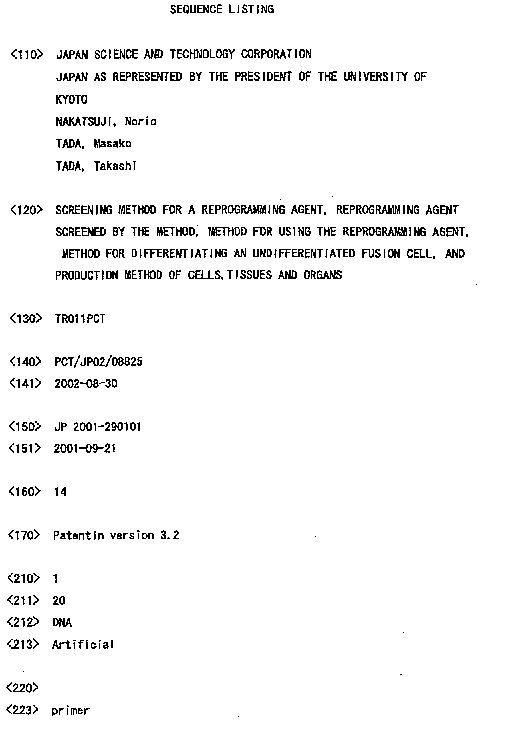
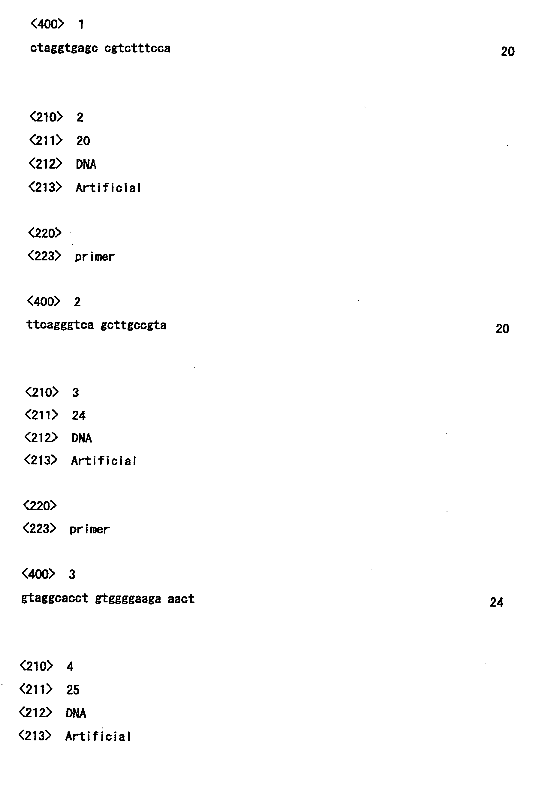
byPCR analysis of DNA from the tail thereof using the following
primer: OCGOFU35, 5'-CTAGGTGAGCCGTCTTTCCA-3' (SEQ ID
NO.: 1), and EGFPUS23, 5'-TTCAGGGTCAGCTTGCC GTA-3' (SEQ ID
NO.: 2). A thymus obtained from the transgenic mouse was
passed through a 18-gauge needle several times to obtain
a suspension of single cells. As an ES cell, a TMAS-5 cell
was used and mixed with the above-described 3 types of
thymocytes at a ratio of 1: 5 (ES cell: thymocyte ) , followed
by washing in PBS three times. The cells were suspended in
0. 3 M mannitol buffered solution at a concentration of 1×10⁶
cells/mL. A glass slide having a 1-mm electrode gap and
Electro Cell Manipulator 2000 (BTX) were used to conduct
electric fusion (E=2. 5 to 3.0 KV/cm) to prepare fusion cells.
The fusion cells were cultured in ES medium for a day.
InactivatedG418-resistant PEFs were screened for in ES medium
containing 250 µg/mL G418 in 7 to 10 days. Fusion cell clones
were collected and spread (passage number 1) in ES medium
supplemented with G418, and were cultured for 3 to 4 days.
ES hybrid clones were subcultured in new medium every two
days.
(2)-2, ES×EG fusion cell
ES×EG fusion cells were produced as follows. NR2
ES cells and TMA-58G bsr EG cells were mixed at a ratio of 1:1,
and were suspended in 0.3 M mannitol buffered solution at
a concentration of 1×10⁶ cells/mL. ES×EG fusion cells were
screened for in ES medium containing 250 µg/mL G418 and 3
to 4 µg/mL BS in 7 to 10 days.
ES hybrid clones could be maintained at a proportion
(2.8×10⁻⁴) similar to that of EG hybrid clones produced by
the present inventors in previous research [Tada M. et al.,
EMBO J. 16:6510-6520(1997)]. All types of fusion cells were
similar to that of the parent ES cell, and no morphological
change was found during culturing. Cytogenetic analysis
using G-banding demonstrated a complete set of chromosomes
including three X chromosomes and one Y chromosome in all
of the 13 ES fusion cells and 2 ES×EG hybrid clones which
were used in experimentation. Further, ES hybrid and ES×EG
hybrid cell lines at passage number 2 to 4 were used in molecular
analysis.
(3) Confirmation of fusion
In order to confirm the fusion between ES cells and
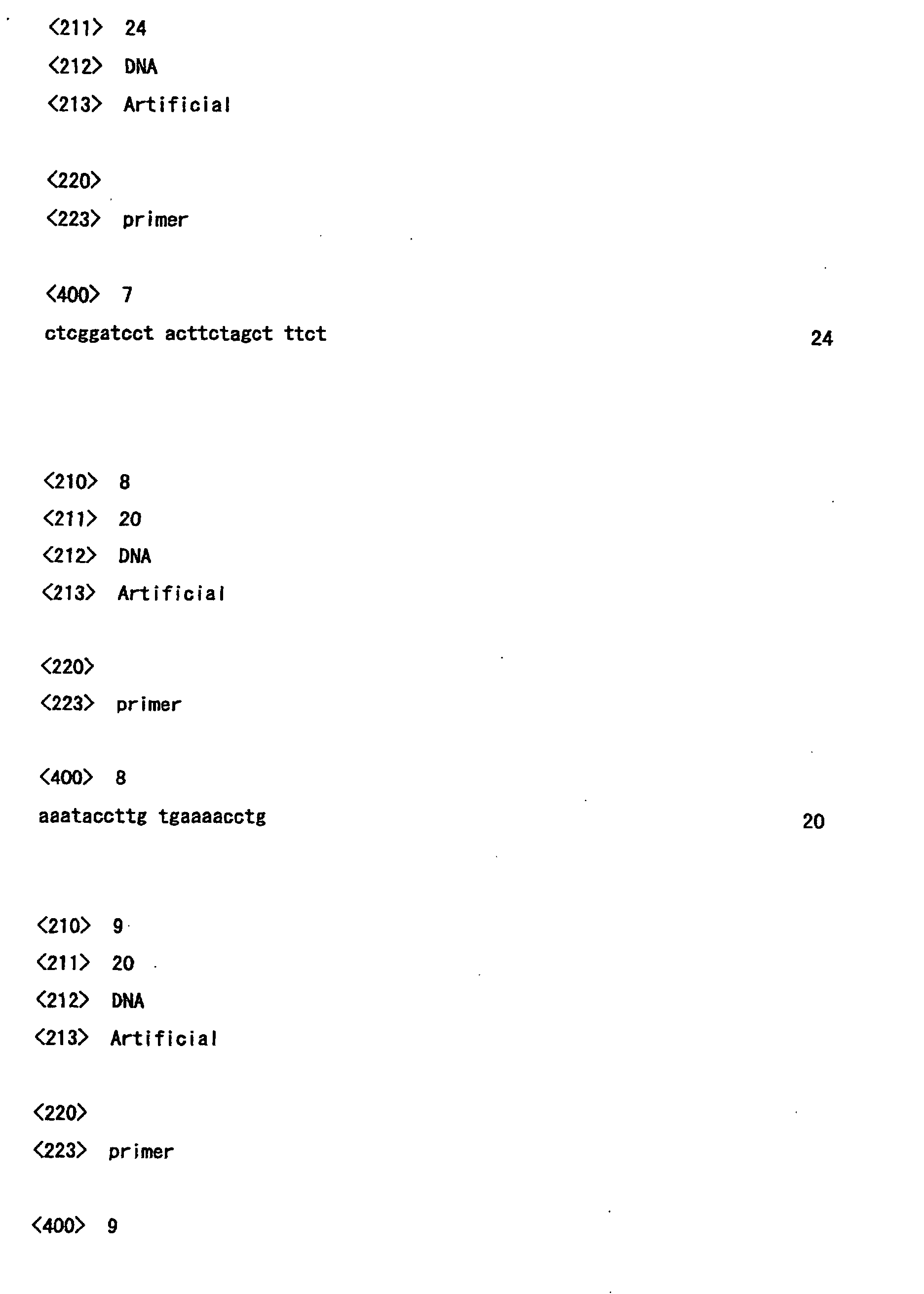
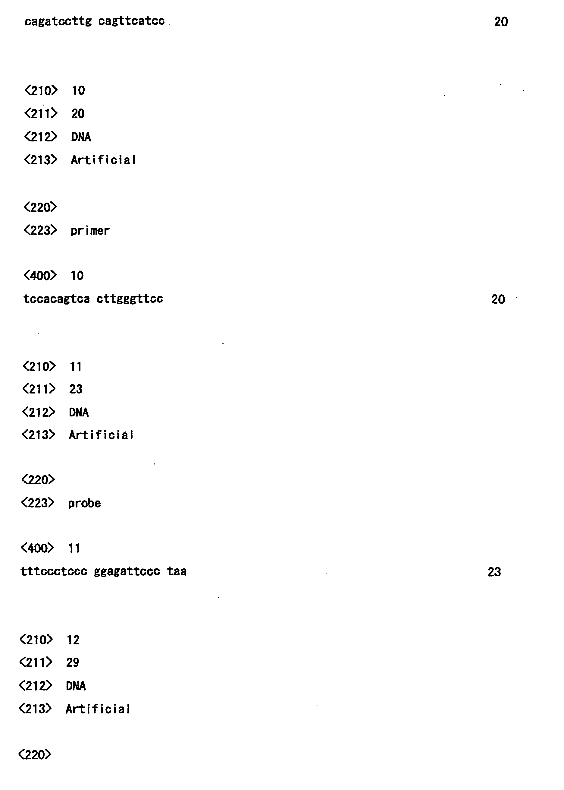
differentiated cells, 4 primer sets specific, respectively,
to the D-J region of T cell receptor
(Tcr) β, the D-J region
of immunoglobulin
(Ig) H, and the V-J regions of
Tcrδ and
Tcrγ were used to perform PCR amplification using DNA extracted
fromthymocytes andES fusion cells as templates. For genomic
DNA (0.5 µg) from an adult thymus, an ES cell and an ES hybrid
clone, PCR amplification was performed to detect the
rearrangement of each gene using the following primer sets:
PCR products were subjected to electrophoresis on
1. 2% agarose gel, followed by staining with ethidium bromide.
The specificity of the PCR product was confirmed by Southern
hybridization using: biotinylated Jβ2-specific oligoprobe
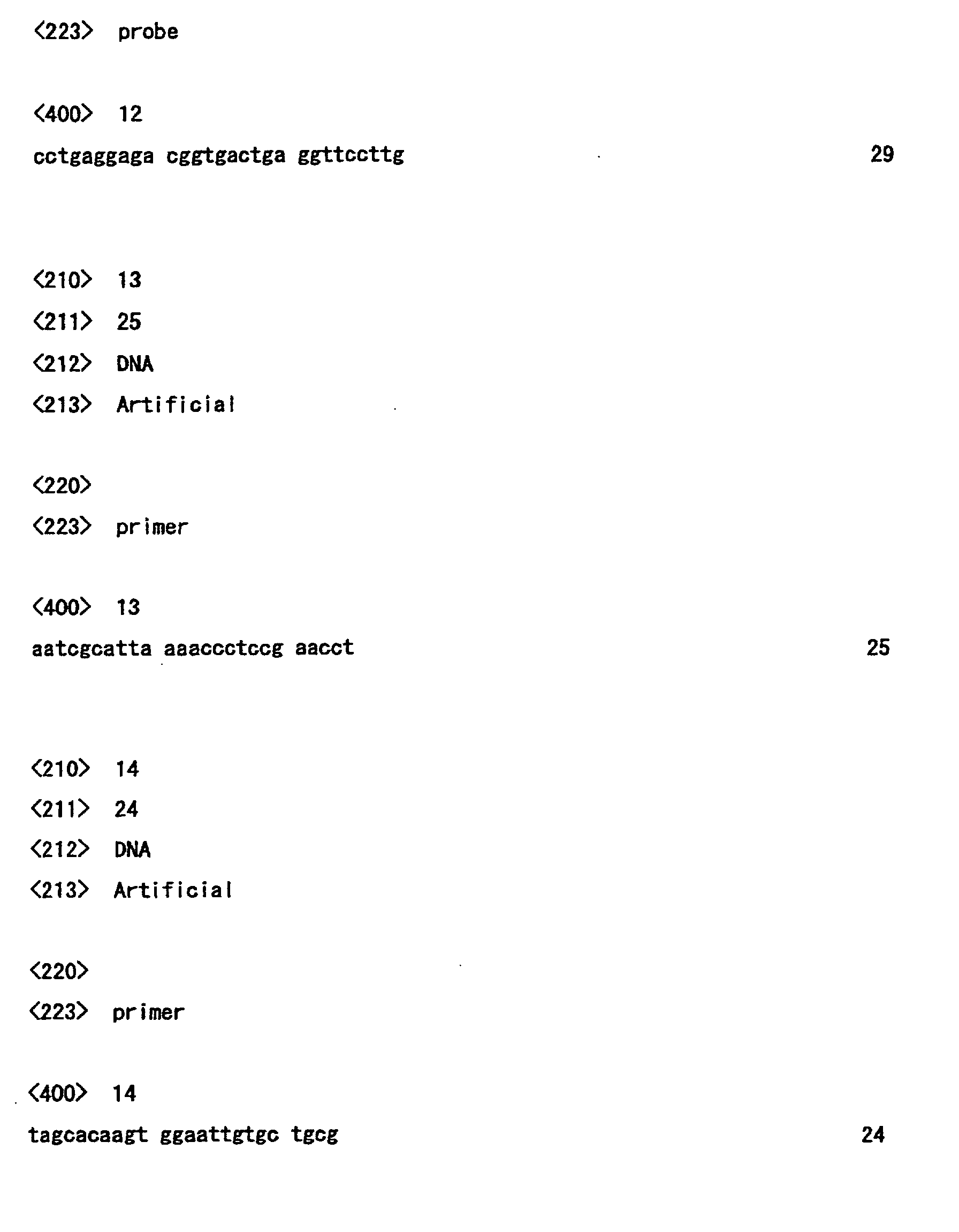
(5'-TTTCCCTCCCGGAGATTCCCTAA-3' (SEO ID NO.: 11) [Levin S.D.
et al., EMBO J. 12:1671-1680(1993)])for Tcrβ; and biotynylated
JH4 oligoprobe
(5'-CCTGAGGAGACGGTGACTGAGGTTCCTTG-3' (SEQ ID NO.: 12)
[Ehlich A. et al., Cell 72:695-704(1993)]) for IgH.
The rearrangement of DNA is clear evidence indicating
that a thymocyte has differentiated into a lymphoid cell
[Fowlkes, B.J. and Pardoll D.M., Adv. Immunol.
44:207-264(1989)]. Rearrangement specific to Tcrβ D-Jβ2.1,
2.2, 2.3, 2.4, 2.5 or 2.6 was observed in 45% of the hybrid
clones (Figure 1a). Further, in several clones, similar
rearrangement was observed in the D-J region of IgH
(Figure 1b), and the V-J regions of Tcrδ and Tcrγ (Figures 1c
and 1d, respectively). Of the 31 ES hybrid clones studied,
a total of 17 clones (55%) underwent rearrangement, which
was, at least, researched. In these cases, it is considered
that the ES cell were fused after differentiation of a
thymocyte nucleus into a lymphoid cell.
(4) X chromosome activity
In female somatic cells, one of the two X chromosomes
is randomly inactivated due to dosage compensation of an
X-linked gene. Inactivation of the X chromosome occurs in
early cell division to induce the delay of transition of
DNA replication into the late S phase, epigenetic changes
including hypermethylation of DNA and low acetylation of
histone H4 are known to occur. In cloned embryos obtained
by nuclear transplantation of a somatic cell nucleus into
an oocyte, the inactivated X chromosome of female somatic
cells is reactivated [Eggan K. et al., Science 290:
1578-1581(2000)]. Therefore, activation of both X
chromosomes serves as an indicator of the occurrence of
reprogramming of a nucleus. In order to analyze the activity
of X chromosomes, the present inventors studied using
replication differential staining method [Sugawara O. et
al., Chromosoma 88:133-138(1983)] (Figure 2a).
(4)-1. Timing of replication of X chromosome
Chromosome preparations of ES fusion cells and ES×EG
fusion cells described in the above-described section (2)
were produced by culturing the cells along with 150 µg/mL
5-bromo-2-deoxy uridine (BrdU) for 7 hours, where the cells
were cultured for the last hour in the presence of 0.3 µg/mL
colcemide. The cells were then subjected to hyposmotic
treatment with 0.075M KCl at room temperature for 8 minutes.
Thereafter, the cells were fixed by immersing in methanol:
acetic acid (3:1) solution three times, followed by air
drying. The cells were stained by freshly prepared acridine
orange solution. The slide was observed under a fluorescence
microscope with a standard B filter. After continued
incorporation of BrdU at the late stage of the S period and
acridine orange staining, active X chromosomes and autosomes
were observed as red and green banded factors. Inactive X
chromosomes were uniformly dark-red stained in female somatic
cells due to the delay of replication (Figure 2b). Among
all 32 cells (4n=80) whose karyotypes were determined, 6
fusion cell clones of an XY female ES cell and an XX female
thymocyte carried three X chromosomes which were simultaneously
replicated (Figure 2c).
(4)-2. Xist RNA FISH
Probes prepared by nick translation of a mixture of
Xist cDNA clones, which contained a series of exons 1 to
7, using cy3-dUTP (Amersham Pharmacia) [Sado T. et al.,
Development 128:1275-1286(2001)] were used and hybridized
with chromosome preparations obtained as in 4-1. Hybridization
and subsequent washing were performed as
previously described [Lawrence J.B. et al., Cell 57:
493-502(1989)]. The result was in agreement with the result
obtained in (4)-1, i.e., , Xist ( inactive X specific transcript )
RNA was not stably accumulated (spotted) on three X
chromosomes in two ES hybrid cell lines tested by RNA FISH
(fluorescence in situ hybridization) (Figure 2d). Xist
accumulation was also instable on active X chromosomes of
male ES cells, and was stable on inactive X chromosomes of
female thymocytes (colored signal). Somatic cell nucleus-derived
inactive X chromosomes acquired several
properties of active X chromosomes after hybridization and
had replication and Xist expression patterns similar to those
observed in undifferentiated cells. Changes in replication
timing and Xist RNA accumulation of X chromosomes of somatic
cells in ES fusion cells shown in the above-described (4)-1
and (4) - 2 suggest that somatic cell nuclei were reprogrammed
after cell fusion.
(5) Reprogramming of somatic cell nucleus
A mouse line having an Oct4-GFP transgene was used
to visualize the reprogramming of somatic cell nuclei
(Figure 3). Expression of Oct4 was specifically observed
in germ cells, embryos before implantation, and ectoblasts
of early embryos before implantation. Therefore, the
activity of Oct4 can be used as an ideal marker for
identification of totipotency and/or pluripotent cells. The
expression pattern of Oct4-GFP is known to be comparable
to the expression pattern of endogenous Oct4 [Yoshimizu T.
et al., Develop Growth Differ. 41:675-684(1999)]. Expression
of Oct4-GFP was examined for the thymus and ovary
of Oct4-GFP transgenic mice. GFP was detected in the growing
ovary, but not in the thymus (Figures 3a to d).
Thymus cells of Oct4-GFP transgenic mice were fused
with ES cells, followed by culturing without screening.
Expression of GFP was examined every 12 hours. Expression
of GFP in living ES fusion cells on a culture dish was
investigated under a dissecting microscope (Leica) with a
GFP exciting source and a GFP filter. After 48 hours, a GFP
positive colony consisting of 16 cells was observed at the
periphery of a larger non-expressing colony (Figures 3e,
f). Subsequently, several other GFP positive colonies were
observed on the same culture plate before reaching confluency.
No GFP positive cells was observed among non-fusion thymocytes
cultured under the same conditions. In order to investigate
whether or not somatic cell nuclei can be reprogrammed in
all ES fusion cells, thymocytes of G418 selection-resistant
(Rosa26×Oct4-GFP) F1 mouse were used. After screening, 36
of the resultant 37 clones expressed GFP (97%). The
expression was stably maintained even after subculturing
over some passages (Figures 3g, h), indicating that the
thymocyte nucleus was reprogrammed in most of the ES fusion
cells. The Oct4-GFP transgene, which was suppressed in
thymocytes before cell fusion, was reactivated in the ES
fusion cells. This further supports the result of (4) that
after cell fusion, somatic cell nuclei were reprogrammed.
(6) Introduction into blastocysts
Normal diploid blastocysts were used to produce
chimeric embryos with ES fusion cells. Diploid blastocysts
were obtained from the uteruses of Day 3.5 pregnancy ICR
females mated with ICR males. Hybrid clones with the
above-described (Rosa26×Oct4-GFP) F1 mouse-derived
(differentiated) thymocytes and hybrid clones with Rosa26
mouse-derived thymocytes were used as tetraploid fusion cells.
The tetraploid fusion cells were microinjected into the
blastocoele pores of well expanded blastocysts (Figure 4a).
These blastocysts were transferred into the uteruses of
pseudopregnant ICR females. Chimeric embryos were removed
from the E7.5 uteruses, followed by removal of the Reichert
membrane, and β-galactosidase staining and histological
analysis.
(6)-1. β-galactosidase active staining
By β-galactosidase active staining, the relative
contribution of a fusion cell to each chimera was confirmed.
Cultured cells were washed with PBS, and fixed using PBS
containing 1% formaldehyde, 0.2% glutaraldehyde, 0.02% NP40
and 1 mMMgCl2, at 4°C for 5 minutes. The same fixing solution
was used to fix embryos and mouse tails at 4°C for 3 to 4
hours. The samples were washed with PBS, f ollowed by staining
with a reaction mixture containing 1 mg/mL
4-Cl-5-Br-indolyl-β-galactosidase (X-gal) with dimethyl
formamide, 5 mM potassium ferricyanide, 5 mM potassium
ferricyanide, and 2 mM MgCl2 in PBS at room temperature for
24 to 48 hours.
(6)-2. Histological analysis
E7.5 embryos stained with X-gal were dehydrated with
an ascending alcohol series, and embedded in JB-4 plastic
resin (polyscience, Warrington, PA). Ultrathin sections
(2-4 µm thick) were negative stained with 0.25% eosin Y.
Four week old teratoma fixed and embedded in paraffin were
cut into 8-µm thick sections. The serial sections were
stained with hematoxylin and eosin.
As a result, 8 of 20 E7.5 embryos were positive,
indicating the limited contribution of the fusion cell
(Figures 4b, c). Detailed analysis revealed derivatives
from the fusion cell in the embryonic ectoderm, embryonic
mesoderm, and the internal organ endoderm (Figures 4d, e).
As described above, the ES fusion cell had a developmental
capability of differentiating into three primordial
germinative layers (ectoderm, mesoderm, and endoderm) in
early embryos before implantation.
(7) Methylation of DNA
Next, DNA of thymocytes, ES cells and prepared hybrid
clones was investigated by Southern blot hybridization as
to whether or not the reprogramming of a thymocyte nucleus
has an influence on the methylation of an imprinted genetic
locus. Southern blot hybridization was performed by
separating genomic DNA digested with a restriction enzyme
into fractions using 0.8% agarose, transferring the fractions
onto a Hybond N+membrane (Amersham) by alkali blotting, and
hybridizing DNA with a 32P-d CTP-labeled probe.
(7)-1. H19 genetic locus
It is considered that a paternal methylated region
is incorporated upstream of the maternal H19 genetic locus
to be expressed, so that primordial methylation imprinting
is maintained [Tremblay K.D. et al., Nature Genet.
9:407-413(1995)]. DNA samples of thymocytes, ES cells, and
prepared hybrid clones were digested with BamHI and a
methylation sensitive restriction enzyme HhaI. A 3.8-kb
SacI probe and a 2.7-kb BamHI probe were used to detect 10-kb
nd2.7-kb paternal methylated fragments and 7.0-kb and 1.8-kb
maternal unmethylated fragments in DNAs from both the
thymocyte and the ES cell. The same pattern was found in
the hybrid clone. There was no difference in the relative
intensity (RI) of bands between the methylated (RI=0.60)
and unmethylated fragment (RI=0.40) bands (Figure 5a).
Similar results were observed using BamHI probes, in which
2.7-kb paternal methylated fragments and 1.8-kb and 0.8-kb
maternal unmethylated fragments were identified. For all
samples, methylated (RI=0.55) and unmethylated (RI=0.45)
bands were similarly detected (Figure 5a).
(7)-2. Igf2r genetic locus
Probes for analysis of methylation of Igf2r region
2 were produced by PCR using the following primers:
5'-AATCGCATTAAAACCCTCCGAACCT-3' (SEQ ID NO.: 13) and
5'-TAGCACAAGTGGAATTGTGCTGCG-3' (SEQ ID NO.: 14) [Stoger R.
et al., Cell 73:61-71(1993)]. ACpG island, which is anintron
of the Ifg2r gene and is known to be imprinted with methylation,
is methylated only on an expressed allele [Stoger R. et al.,
Cell 73:61-71(1993)]. As in the above-described section
(7)-1, each DNA sample was digested with PvuII and a
methylation sensitive restriction enzyme MluI. With the
330-bp Igf2r CpG island probe, a 2.9-kb maternal methylated
fragment and a 2.0-kb paternal unmethylated fragment were
detected in DNAs from both the thymocyte and the ES cell
(Figure 5b). The same pattern was also found in the hybrid
clone. There was no difference in relative intensity (RI)
between methylated (RI=0.55) and unmethylated (RI=0.45)
bands (Figure 5a).
According to sections (7)-1 and (7)-2, it was
demonstrated that the primordial methylation of the H19
upstream region and the Igf2r intron region of the genome
of a thymocyte are not affected by fusion with an ES cell.
This results differs from previous observation of a hybrid
clone of a thymocyte and an EG cell derived from a germ cell
PGC of an E12.5 mouse in which the maternal specific
methylation of Igf2r disappeared [Tada M. et al., EMBO J.
16: 6510-6520(1997)]. The maintenance of the somatic cell
methylation pattern in ES fusion cells suggests that ES cells
and EG cells have different control mechanism for regulating
the DNA methylation of imprinted genes. While the maternal
allele specific methylation of Igf2rwas observed in ES cells,
it was not observed in EG cells and a control 1:1 mixture
of ES cell DNA and EG cell DNA at a ratio of about 1:3
(methylation/RI=0.27, unmethylation/RI=0.73). In the
ES×EG hybrids, methylation bands disappeared (Figure 5c),
indicating that demethylation activity in EG cells is dominant
over the maintenance of methylation imprinting in ES cells.
2. Production of teratoma
Fusion cells of TMAS-5ES cells and Rosa26-derived
thymocytes, which were produced with a method similar to
that for production of chimeric embryos in section 1, were
used as tetraploid fusion cells. About 100 million to 500
million tetraploid fusion cells were subcutaneously injected
into the posterior limb inguinal region of a SCID mouse (CLEA
Japan KK). Four weeks after subcutaneous injection,
teratoma was collected. By X-gal staining, it was determined
that the teratoma was derived from the fusion cell.
Thereafter, the teratoma was fixed in Bouin's fixative,
followed by Haematoxyline-Eosin (HE) staining to stain both
the nucleus and cytoplasm. As a result of HE staining, muscles,
cartilages, epithelial cells, and neurons were observed
(Figure 6).
INDUSTRIAL APPLICABILITY
According to the present invention, an experimentation
systemwhich can be operated so as to enable research
on molecular mechanisms related to reprogramming is provided.
This utilizes the capability of reprogramming at least a
part of a somatic cell nucleus of ES cells in vitro, which
is observed for the first time by producing fusion cells
of ES cells and somatic cells. Unlike EG cells, ES cells
cannot reprogram paternal implementation. Results of
methylation analysis of Igf2r in ES × EG fusion cells suggest
that EG cells have stronger additional dominance factors
involved in epigenetic reprogramming. Actually, ES cells
and EG cells respectively reflect original characteristics
of the cells. Therefore, both the ES cells and EG cells can
be advantageous materials for epigenetic reprogramming and
screening for a factor involved in demethylation of early
stage germ cells and germ line PGC.
By using a method for screening for an agent which
reprograms somatic cell nuclei according to the invention,
which is established based on the above-described observation,
a reprogramming agent is obtained. Somatic cells are exposed
to the reprogramming agent so that the somatic cells can
be undifferentiated. Such undifferentiated cells derived
from somatic cells have the same karyotype as that of the
original somatic cell, and can be an ideal material for
establishing cells, tissues, and organs which can be used
as donors for treating various types of diseases.
The present inventors produced a tetraploid composed
of fused ES cells and somatic cells and demonstrate that
the cells can be proliferated
in vivo or
in vitro, the somatic
cell nucleus is reprogrammed, and it has pluripotency. The
tetraploids can be used in production of cells, tissues,
and organs which may be used as donors for treating various
diseases as ES cells. Furthermore, if chromosomes derived
from host ES cells are successfully removed from such
tetraploids , the cells become diploid undifferentiated cells
which have only chromosomes derived from somatic cells.
These cells may be more ideal donors for treating various
diseases.