FIELD OF THE INVENTION
The present invention relates to a novel hematopoietic
stem cell proliferation regulators produced by a bone marrow
stromal cell which is considered to form a hematopoietic
microenvironment (niche) and regulate the proliferation and
the differentiation of hematopoietic stem cells or
hematopoietic precursor cells via hematopoietic factors and
adhesion molecules, as well as a gene (polynucleotide) encoding
the same.
BACKGROUND OF THE INVENTION
Hematopoietic stem cells present in a bone marrow and
mesencymal stem cells serve to maintain an adult body life by
being differentiated over their entire life span into respective
blood cells, bones, cartilages, fat cells and the like the supply
of which is done by them continuously. Recently, these
tissue-specific stem cells were reported to be capable of being
differentiated also into the cells of multiple organs (nerve,
liver, lung, intestinal tracts, cardiac muscles, muscles and
the like) under a certain condition, and it is considered to
be very important to understand their proliferation mechanisms
also in view of the application to a transplantation therapy
and a regenerative medicine.
A large number of attempts have been made heretofore to
accomplish an in vitro amplification of a hematopoietic stem
cell, and some of them were successful to some extent. In bone
marrow stromal cells as considered to form a hematopoietic
microenvironment (niche) and regulate proliferation and
differentiation of hematopoietic precursor cells via
hematopoietic factors and an adhesion molecules. Those
reported as the hematopoietic factors produced by the stromal
cell are cytokines such as IL-3 and IL-6, SCF, receptor-type
tyrosine kinase ligands such as an Flt-3 ligand (Gibson, F.M.
et al. , Br J Haematol 1995 ) , a differentiation-inhibiting factor
Notch ligand jagged-1 (Varnum-Finney, B. et al., Blood 1998).
However, most of the membrane-binding factors and
adhesion molecules required for the hematopoiesis described
above are considered to be unidentified. While it is impossible
to culture a hematopoietic stem cell for a prolonged period
only by combining currently known factors with each other, such
a culture is possible for a certain time period in the presence
of stromal cells which are hematopoiesis-supporting cells,
suggesting that the stromal cells may express
hematopoiesis-related proteins which have not been identified.
SUMMARY OF THE INVENTION
Accordingly, an objective of the invention is to solve
the above-mentioned problems by analyzing hematopoietic cell
proliferation-regulating factors produced by stromal cells,
constructing a cDNA library and isolating novel
hematopoiesis-related genes whereby enabling a future
application to an in vitro amplification of the hematopoietic
stem cells, a transplantation therapy against hematopoietic
malignancies regenerative therapy care using stem cells and
a gene therapy.
We made an effort to accomplish the above-mentioned
objective and finally discovered, in a bone marrow stromal cells,
novel genes encoding proteins having a hematopoietic stem cell
proliferation-regulating activity, whereby establishing the
invention.
Thus, the invention relates to a gene encoding a protein
(a) or (b):
The invention also relates to a gene comprising a DNA
(a) or (b):
As used herein, the term "hematopoietic stem cell
proliferation-regulating activity" includes an ability of
amplifying or increasing the proliferation of a hematopoietic
stem cell and an ability of rather inhibiting the proliferation
of the hematopoietic stem cell, and means any function for
exerting some effect on the proliferation of the hematopoietic
stem cell. The term "hematopoietic stem cell" means not only
a hematopoietic stem cell in its narrow sense but also myelocytic,
erythroblastic, megakaryocytic and lymphocytic precursor cells
which have been differentiated once and can be detected for
example in the Cobblestone area forming cell (CAFC) described
below.
The invention also relates to a protein encoded by such
a gene or DNA. Since the 6 proteins which have been identified
for the first time by the invention are derived from a stromal
cell and each have a hematopoietic stem cell
proliferation-regulating activity as shown in Examples
described below, they are designated as SDHF (Stromal cell
Derived Hematopoietic Factor)-1, SDHF-2, SDHF-3, SDHF-4,
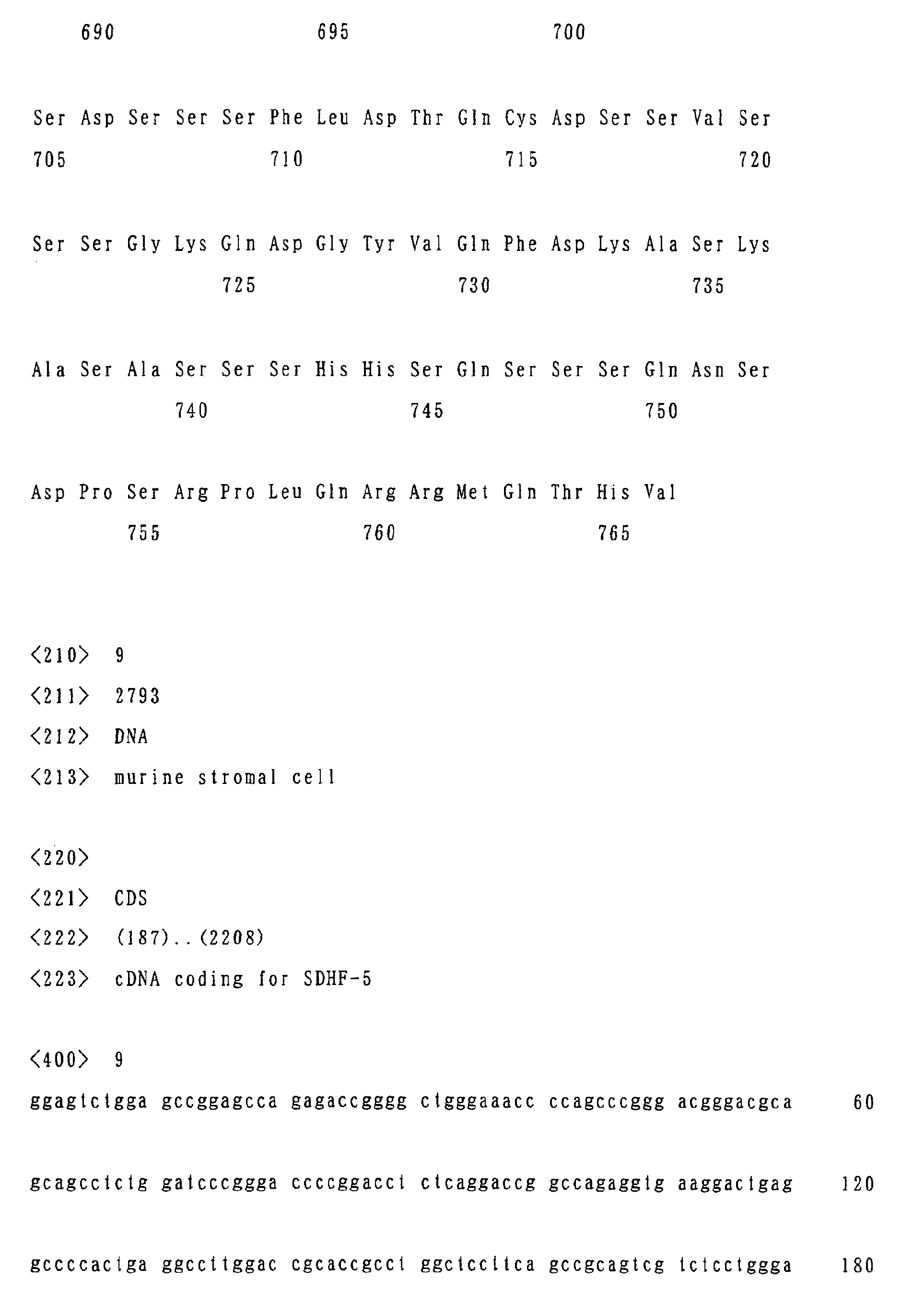
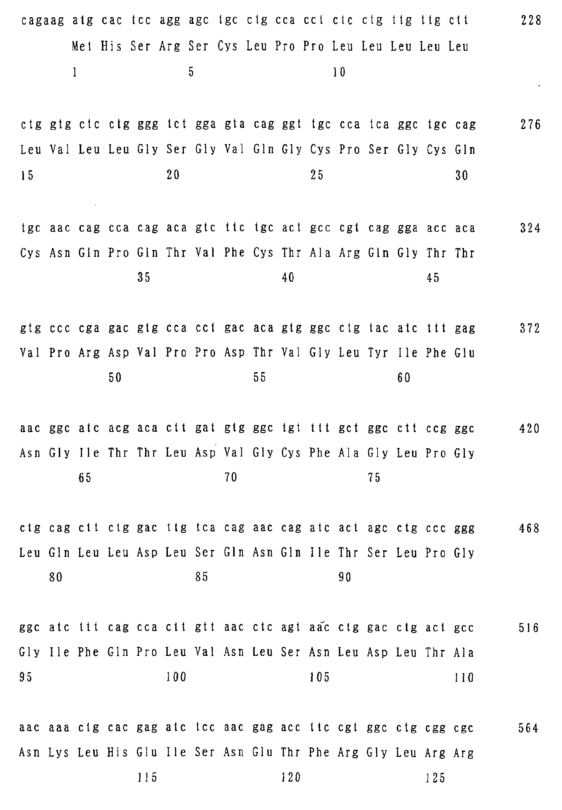
SDHF-5 and SDHF-6. The base sequences encoding these proteins
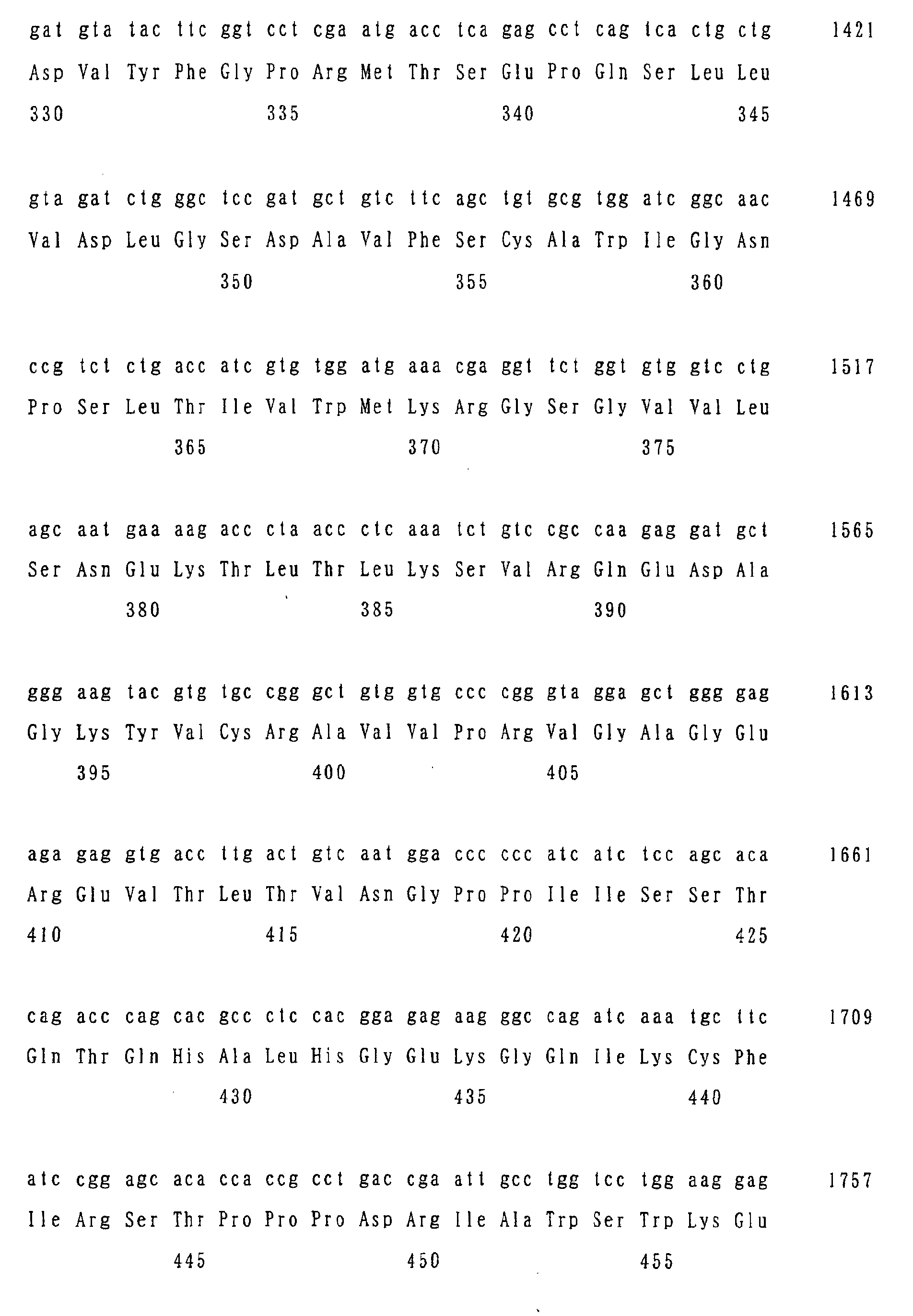
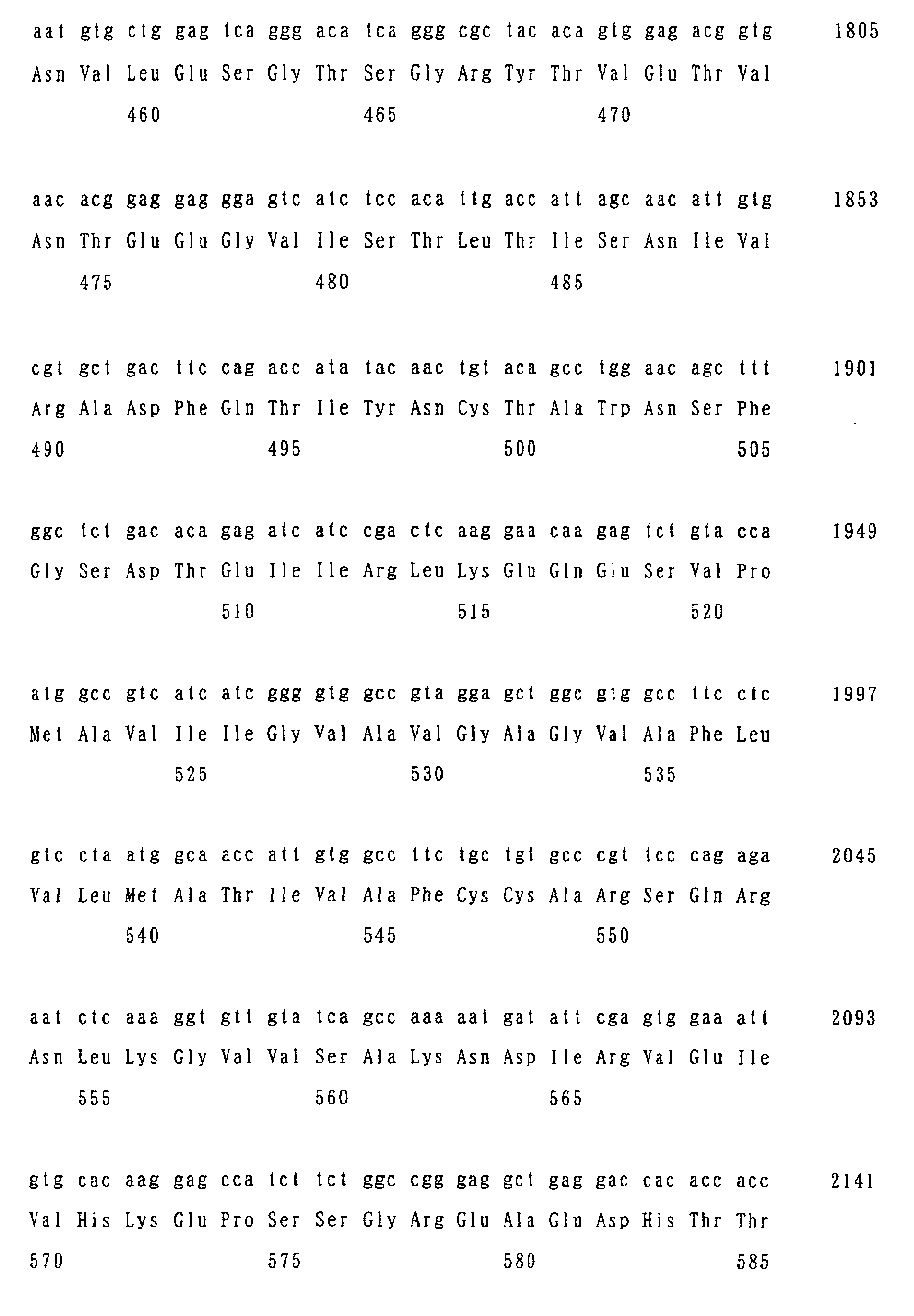
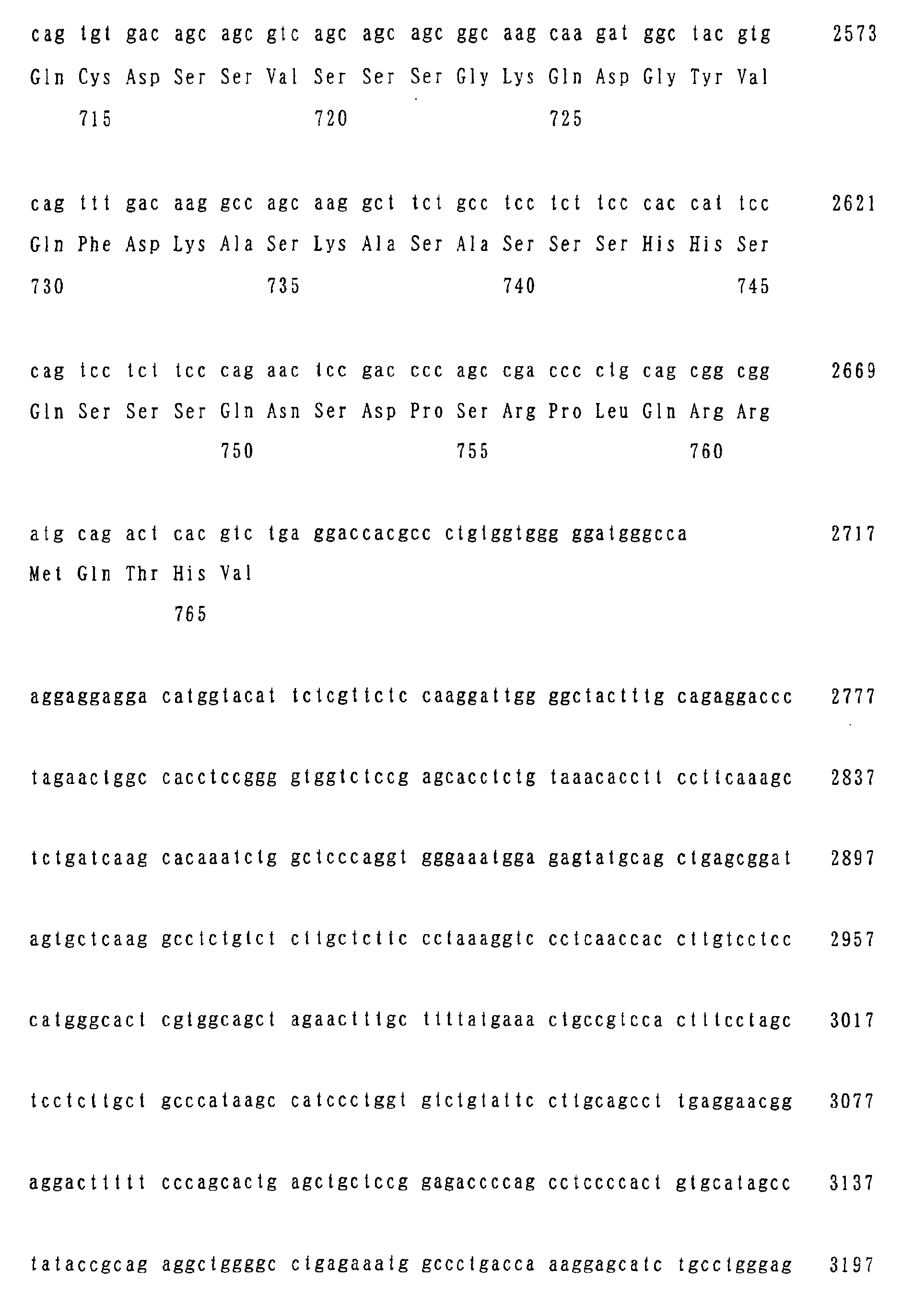
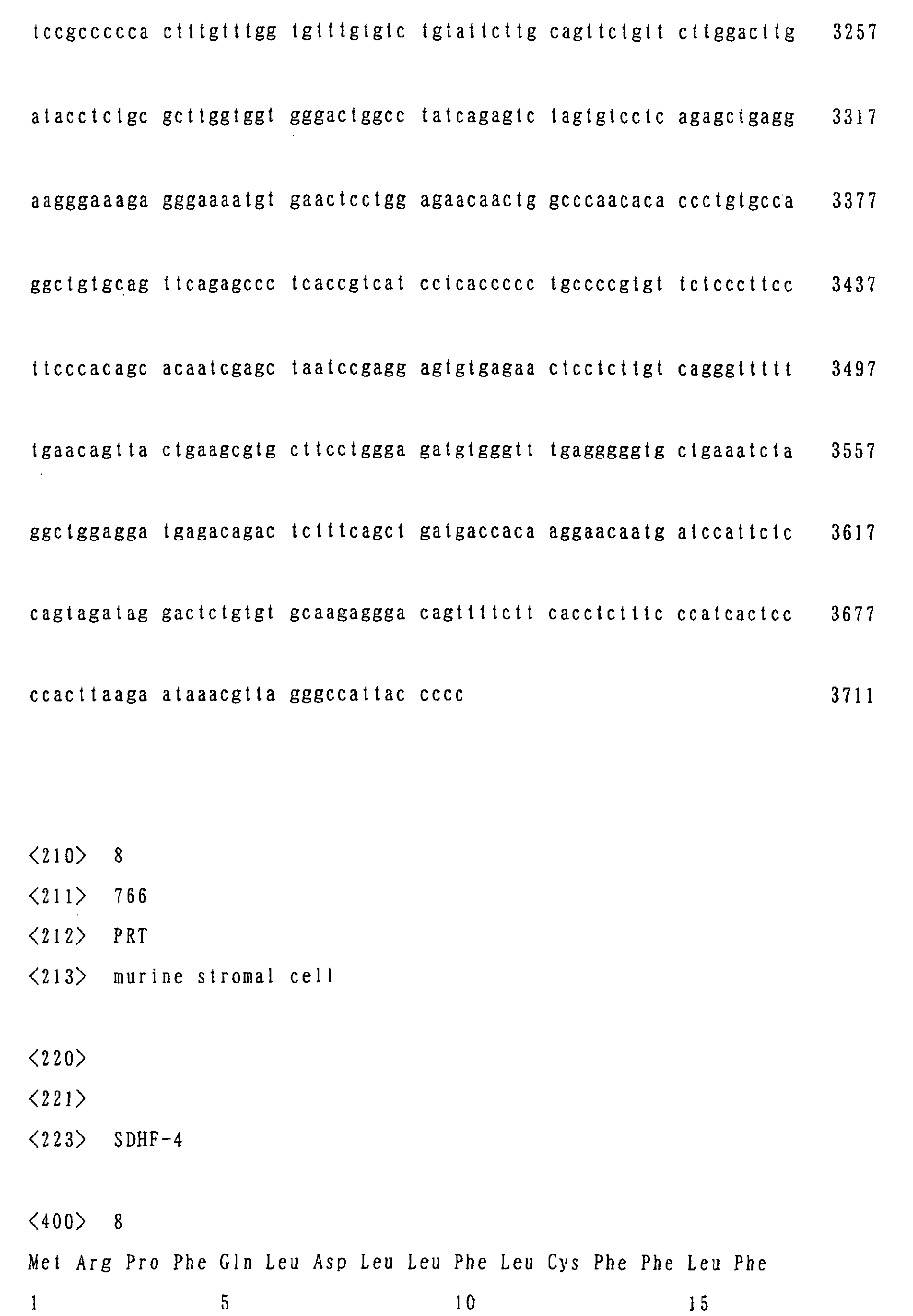
and the corresponding amino acid sequences are represented by
SEQ ID Nos. 1 and 2, SEQ ID Nos. 3 and 4, SEQ ID Nos. 5 and
6, SEQ ID Nos. 7 and 8, SEQ ID Nos. 9 and 10 and SEQ ID Nos.
11 and 12.
The invention further relates to a recombinant expression
vehicle comprising at least any one of the genes described above,
a transformant obtained by the transformation with said
expression vehicle, and a method for producing any of the
proteins described above comprising the culture of said
transformant.
The invention further relates to a method for regulating
the hematopoietic stem cell-proliferating activity of a stromal
cell comprising the transformation of said stromal cell with
at least any one of the genes described above. In said
transformed stromal cell, a gene of the invention is expressed
and its hematopoietic stem cell proliferation-regulating
activity results in an amplification or increase or reduction
in the hematopoietic stem cell-proliferating activity of said
stromal cell. The transformation can be conducted for example
by a method known to those skilled in the art using a recombinant
expression vehicle described above.
The base sequence encoding the amino acid sequence
represented by SEQ ID No. 13 has already been reported (1999,
Genomics 61 (1), pp37-43), and referred to as an ISLR whose
function has never been known heretofore, but it has proven
by us for the first time here to have a hematopoietic stem cell
proliferation-regulating activity as shown in the EXAMPLES
described below.
Accordingly, the invention relates to a method for
modifying the hematopoietic stem cell proliferation-regulating
activity of a stromal cell consisting of a transformation of
said stromal cell with a gene encoding a protein (a) or (b):
The invention further relates to a composition for
regulating a hematopoietic stem cell proliferation comprising
as an active ingredient at least one protein selected from SDHF-1
to SDHF-6 and ISLR described above.
As used herein the term "hematopoietic stem
cell-proliferating activity" means a function for exerting some
effect on the proliferation of a hematopoietic stem cell, and
the term "regulation" means any change in said activity such
as an amplification or increase, or rather inhibition and the
like.
The invention further relates to various antibodies such
as polyclonal antibodies and monoclonal antibodies directed
to respective proteins according to the invention such as SDHF-1,
- 2, -3, -4, -5 and -6 described above.
Figure 23 shows schematic views of the molecular
structures of SDHF-1 to SDHF-6 according to the invention.
BRIEF DESCRIPTION OF THE DRAWINGS
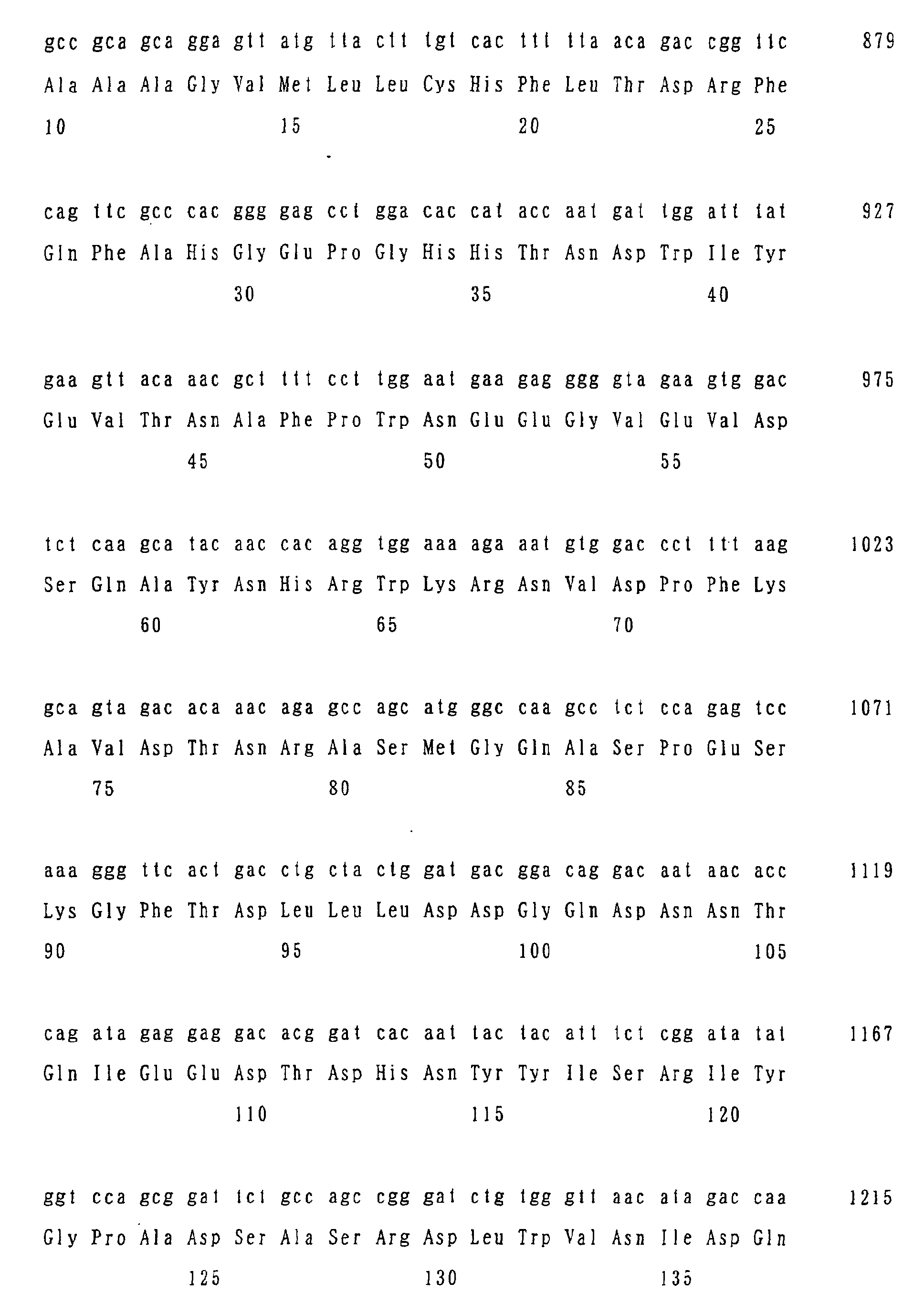
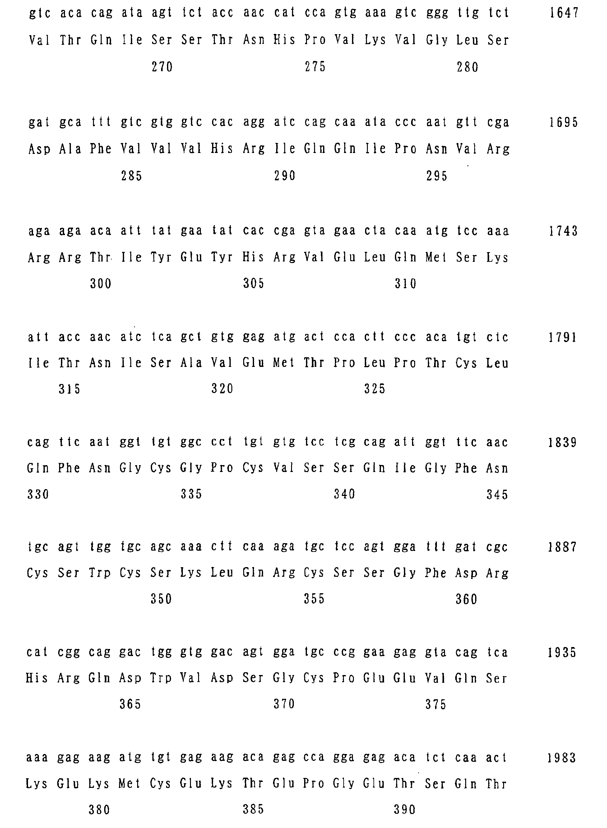
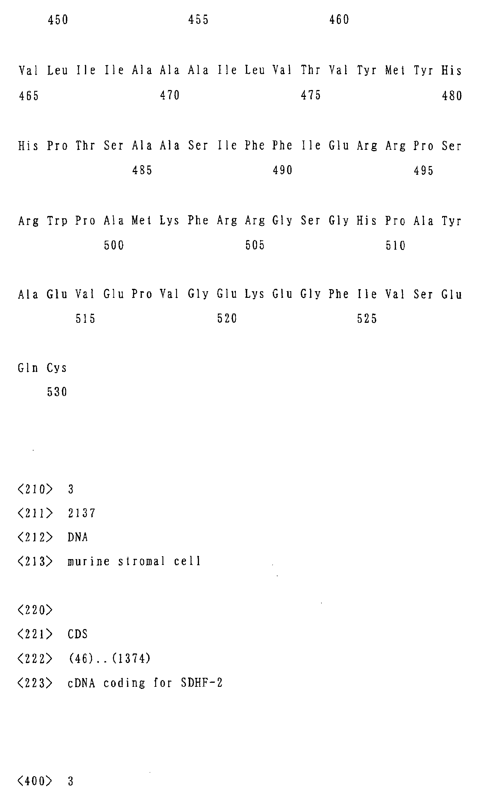
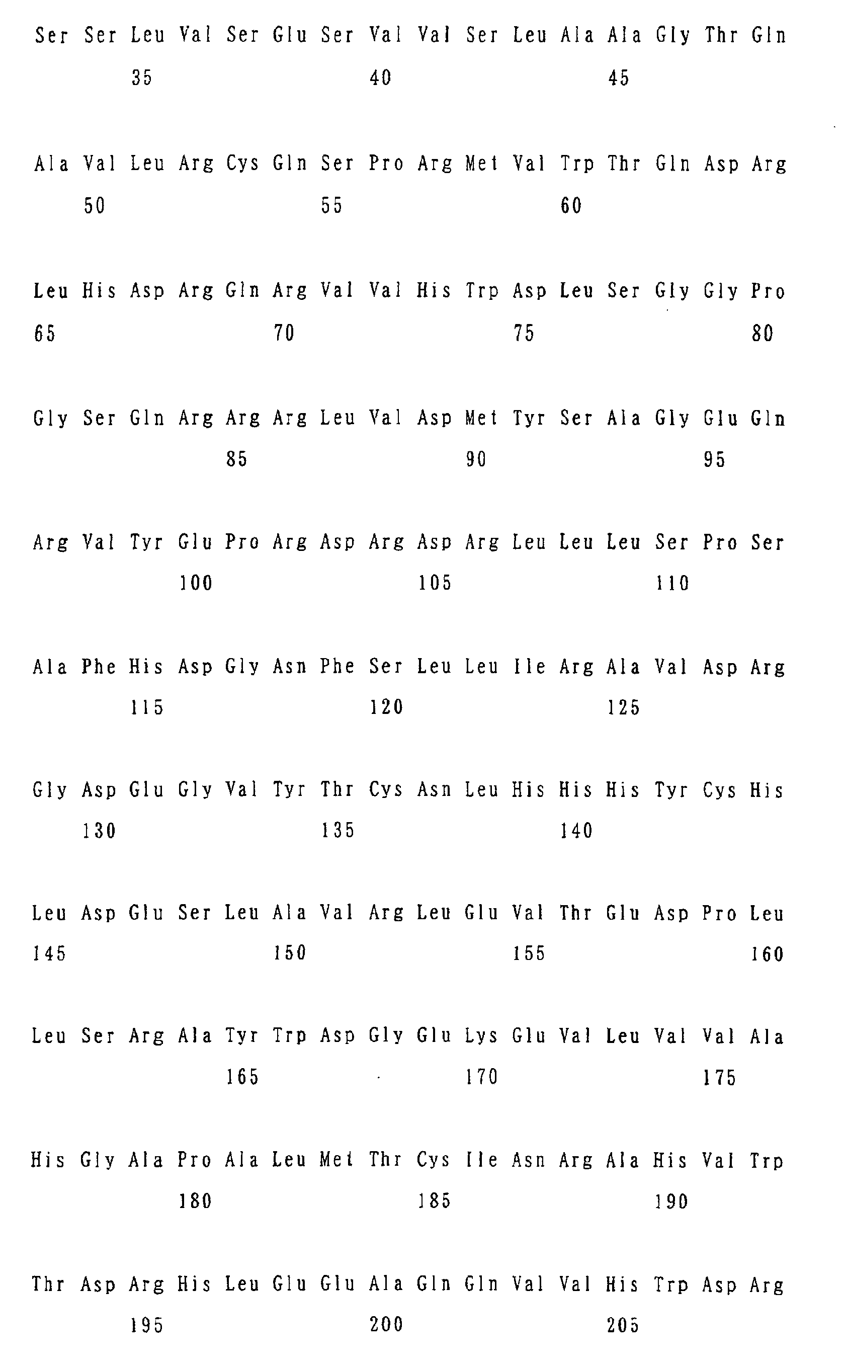
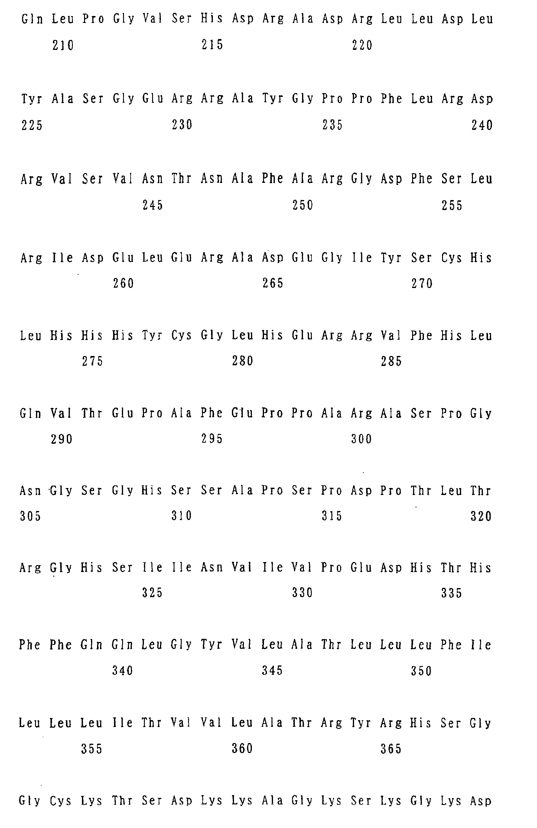
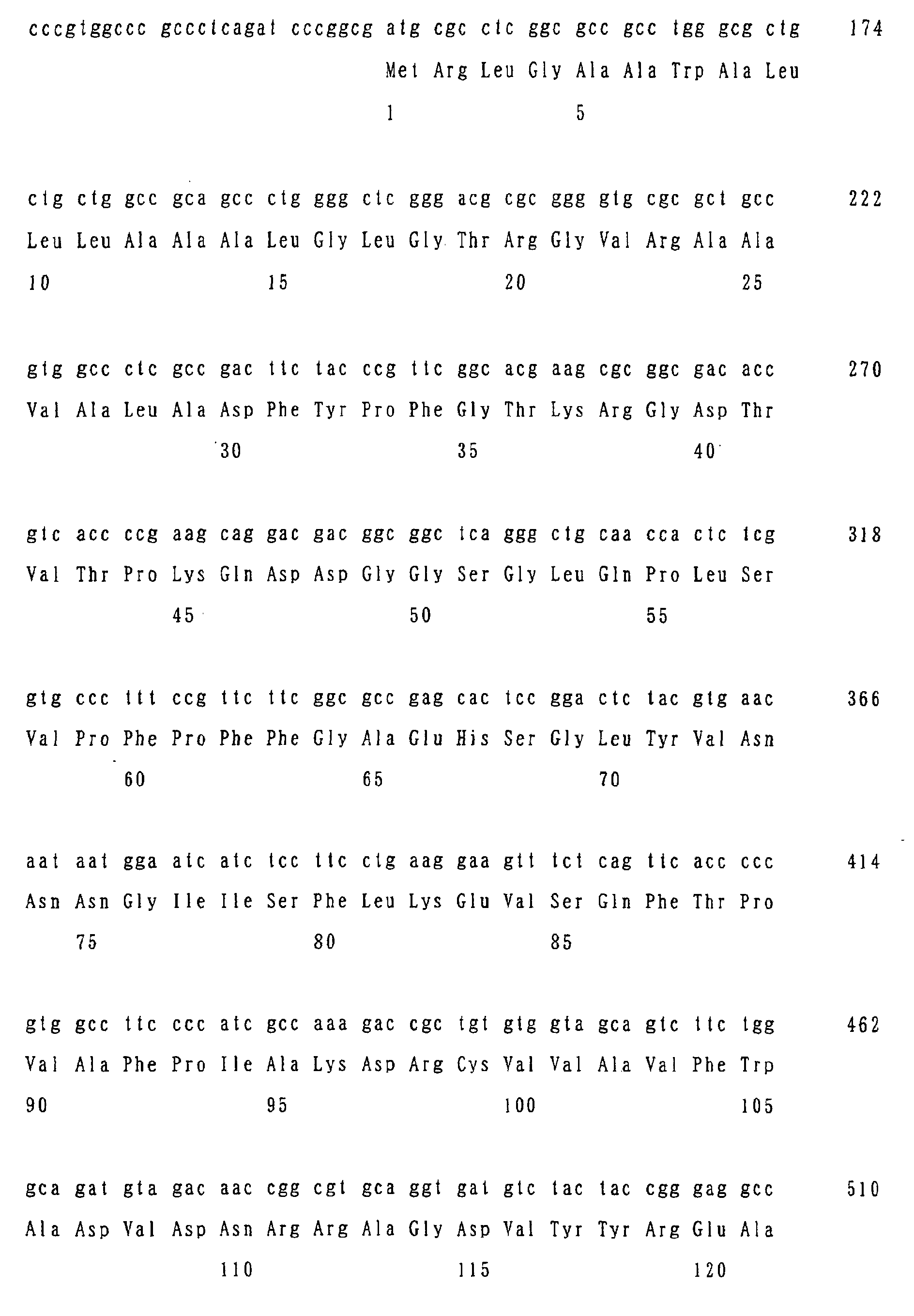
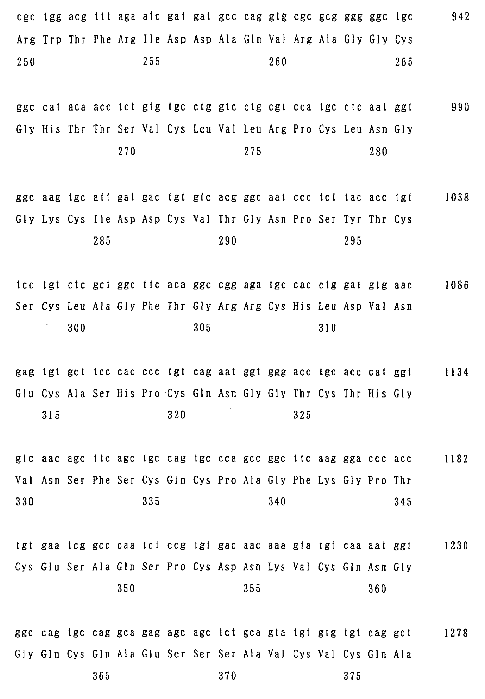
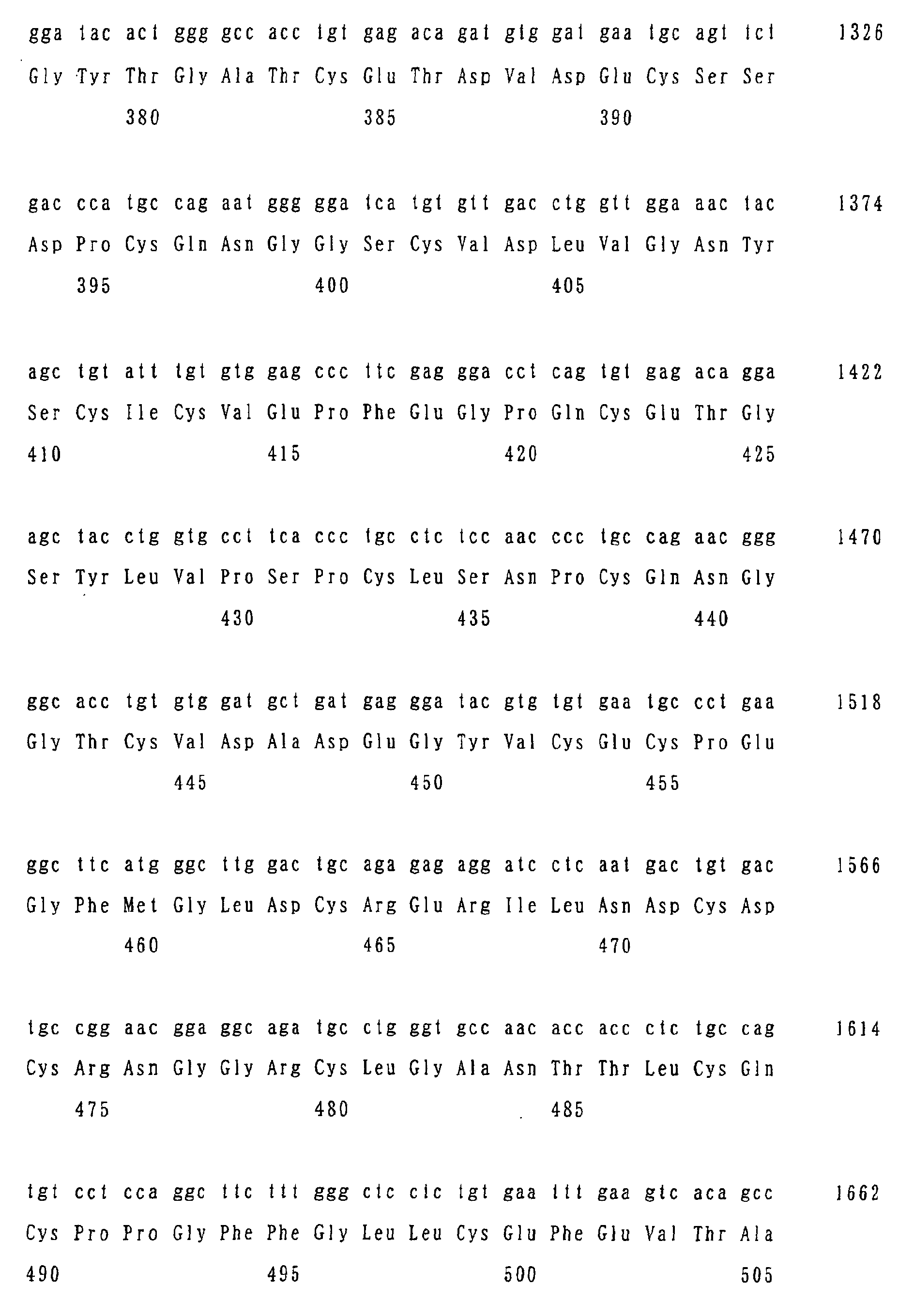
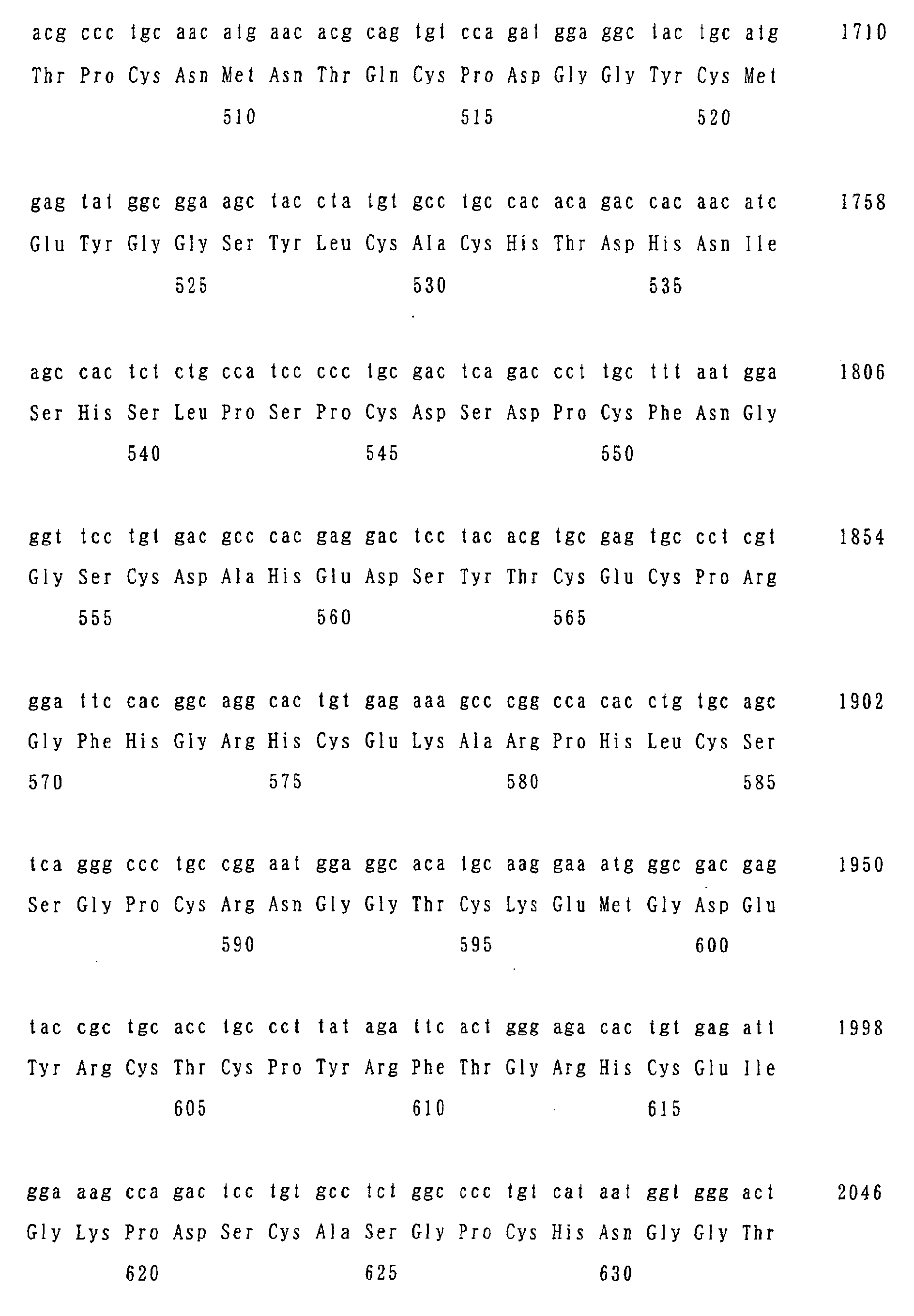
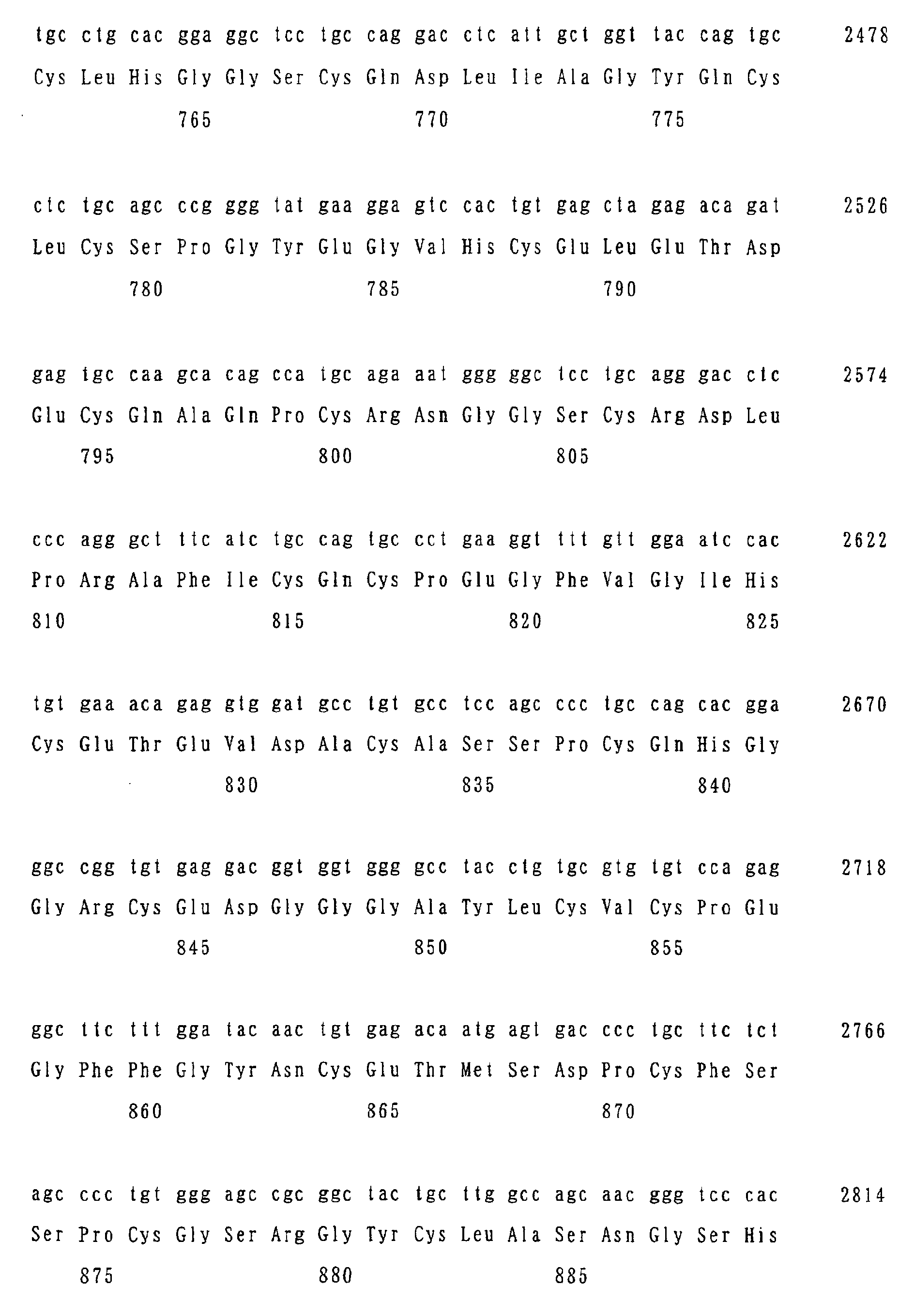
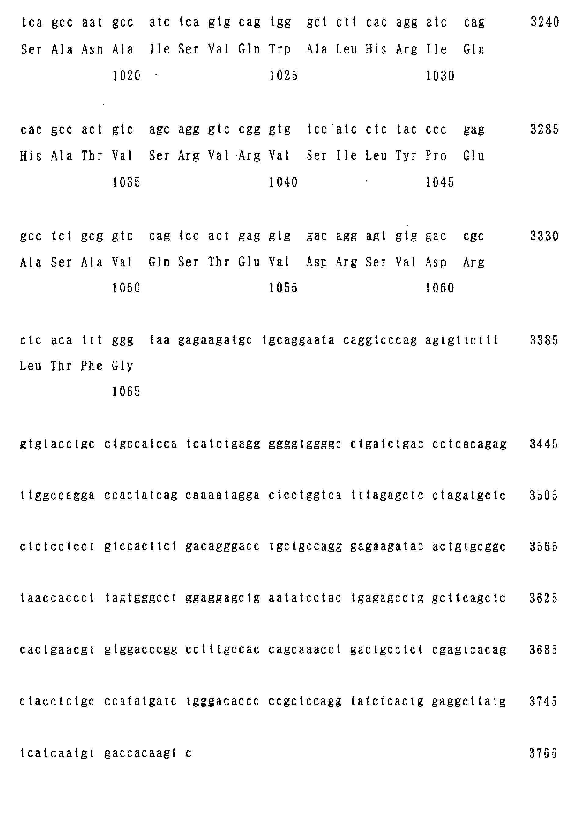
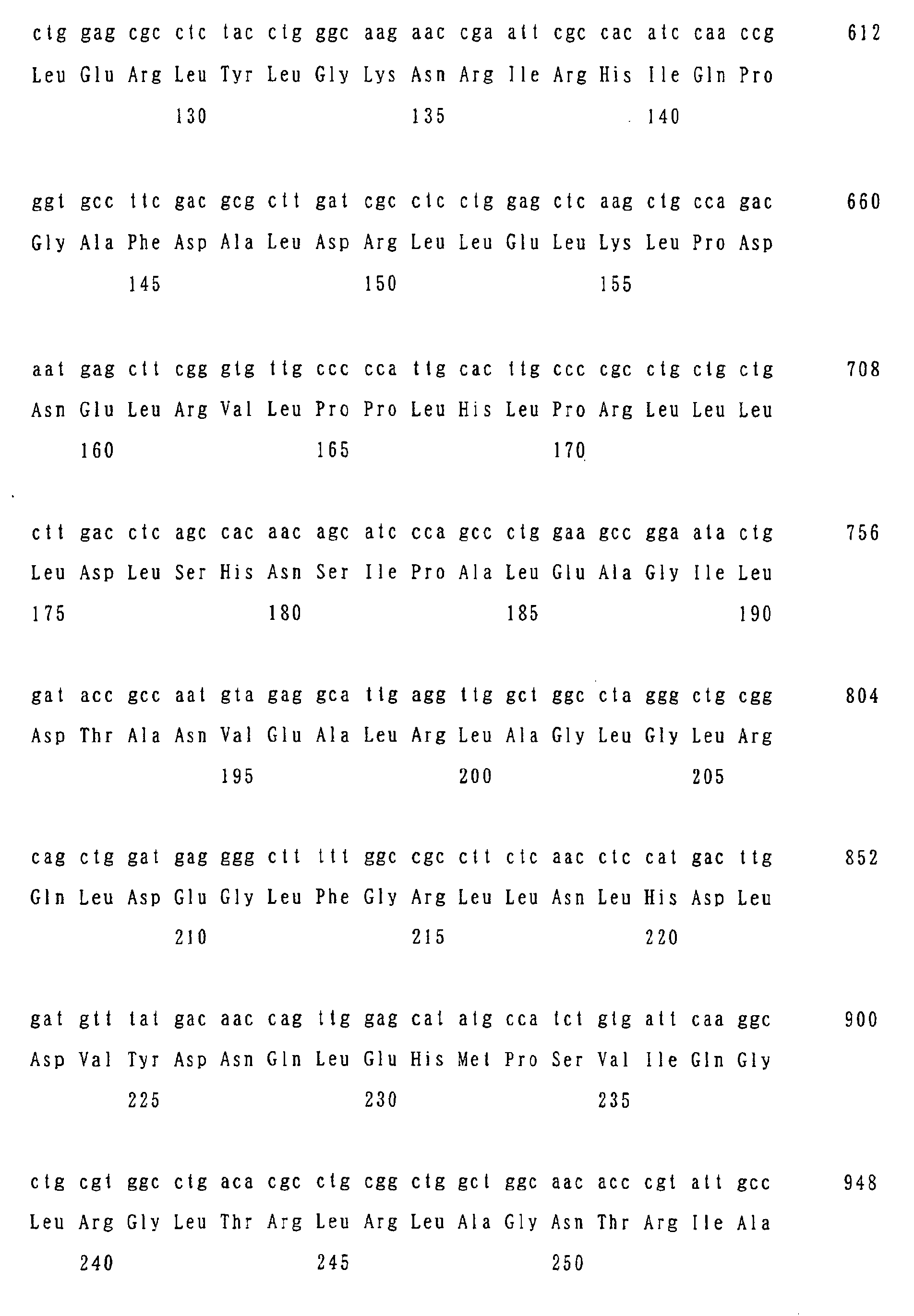
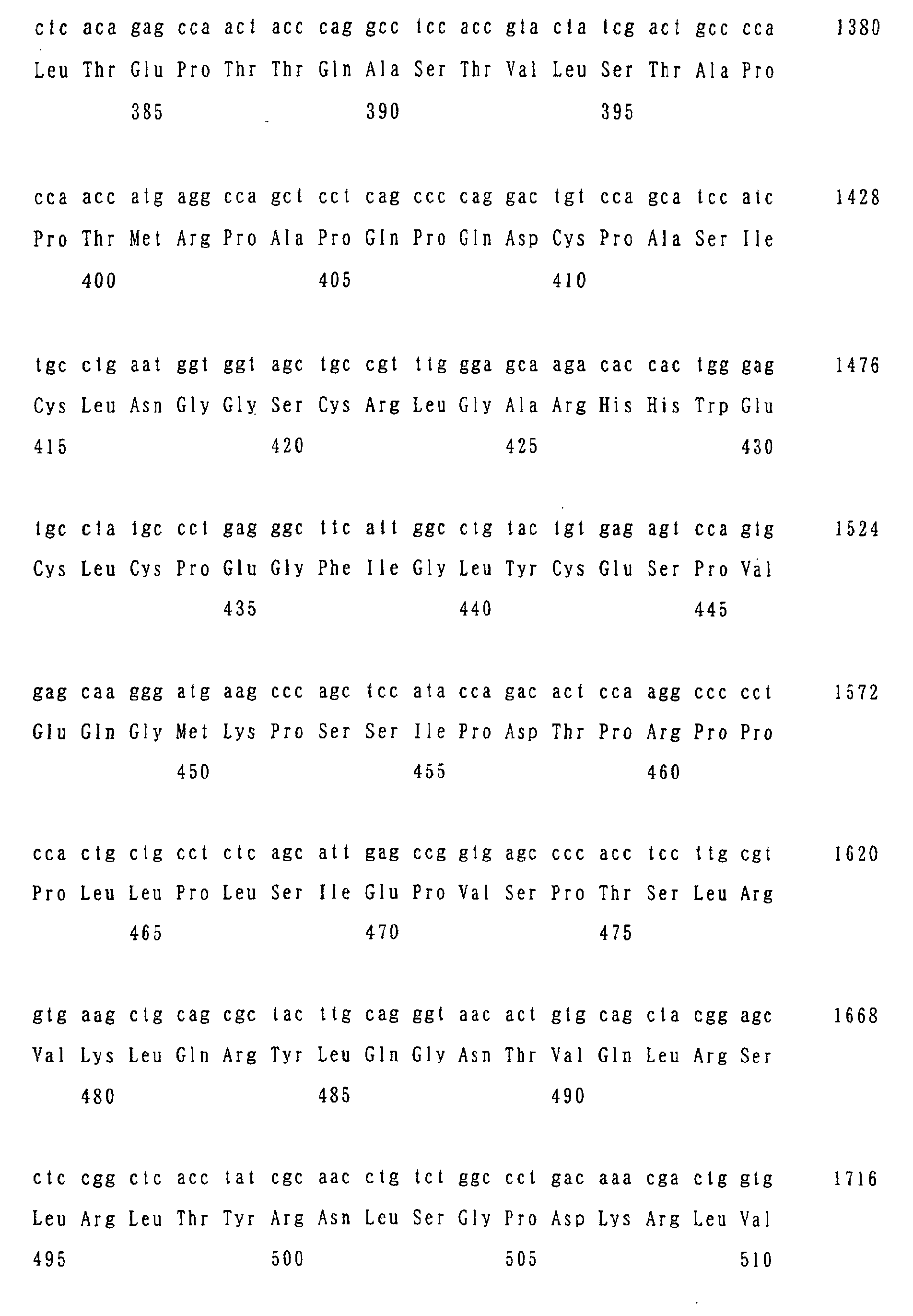
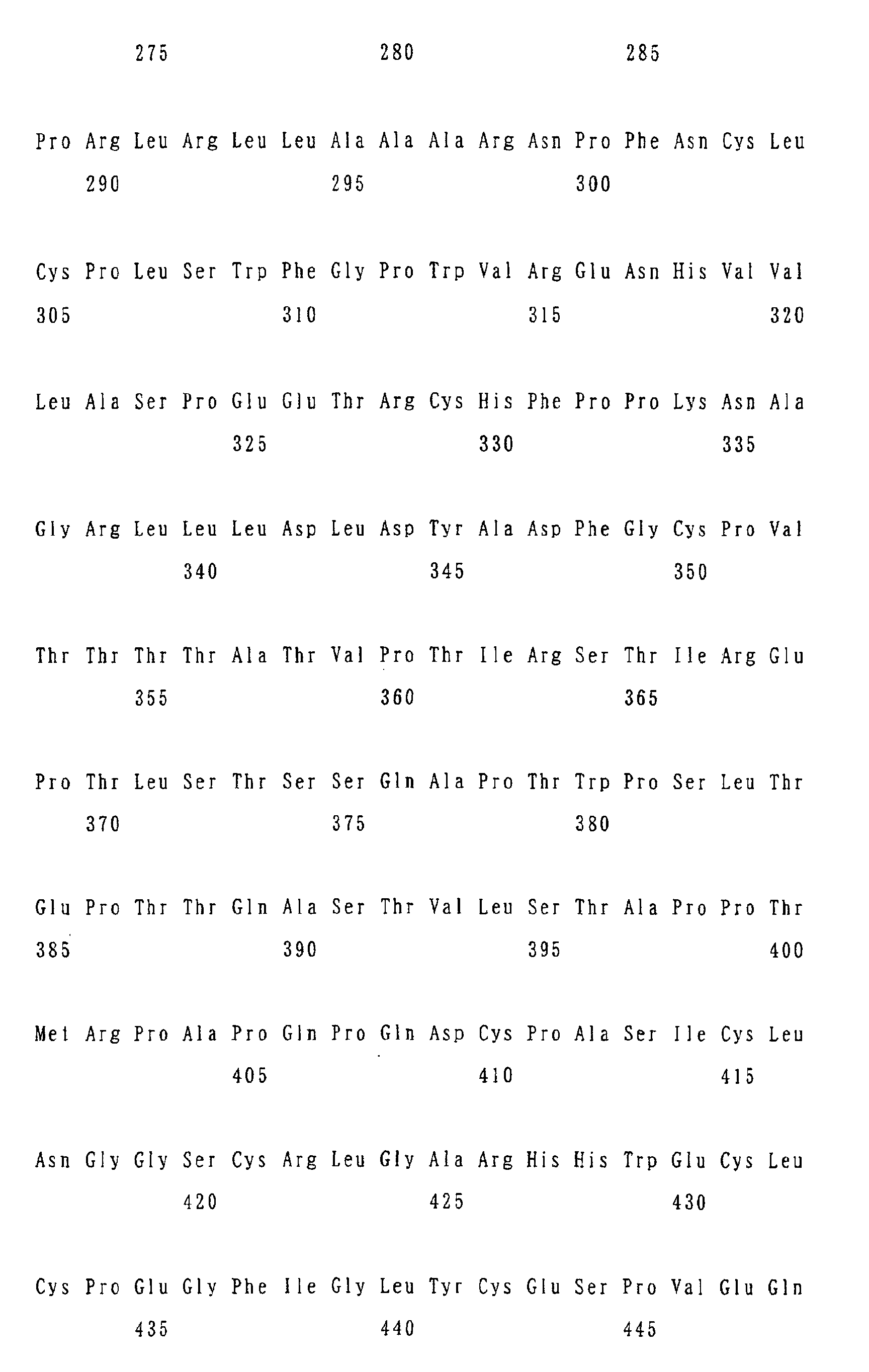
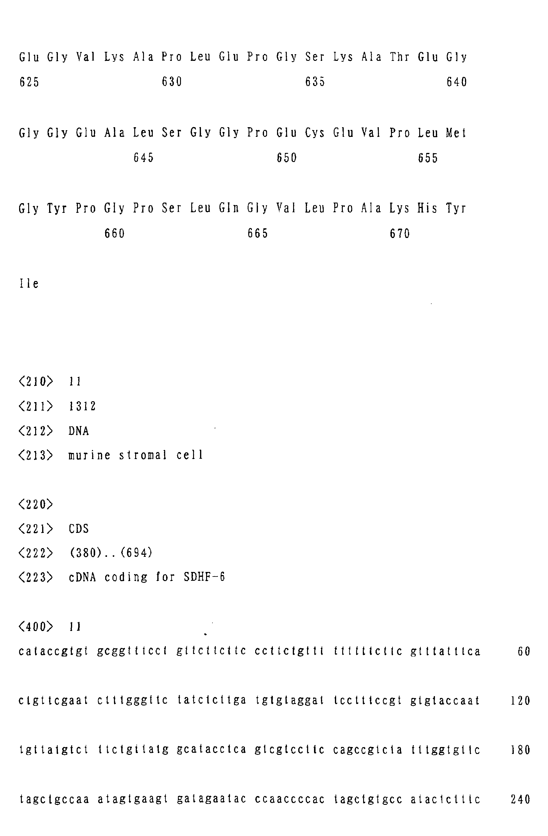
Figure 1 shows the amino acid sequence of SDHF-1.
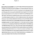
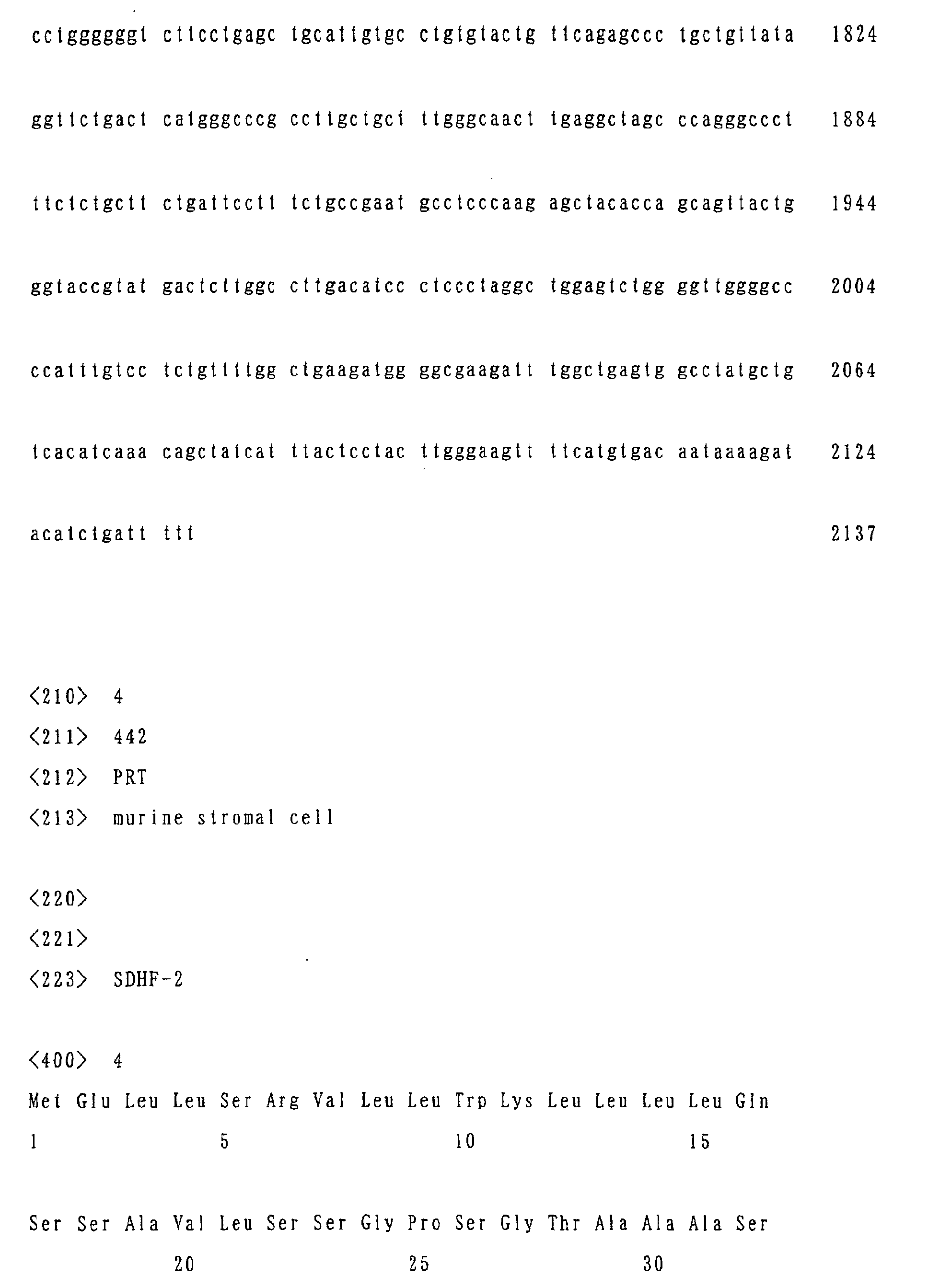
Figure 2 shows the amino acid sequence of SDHF-2.
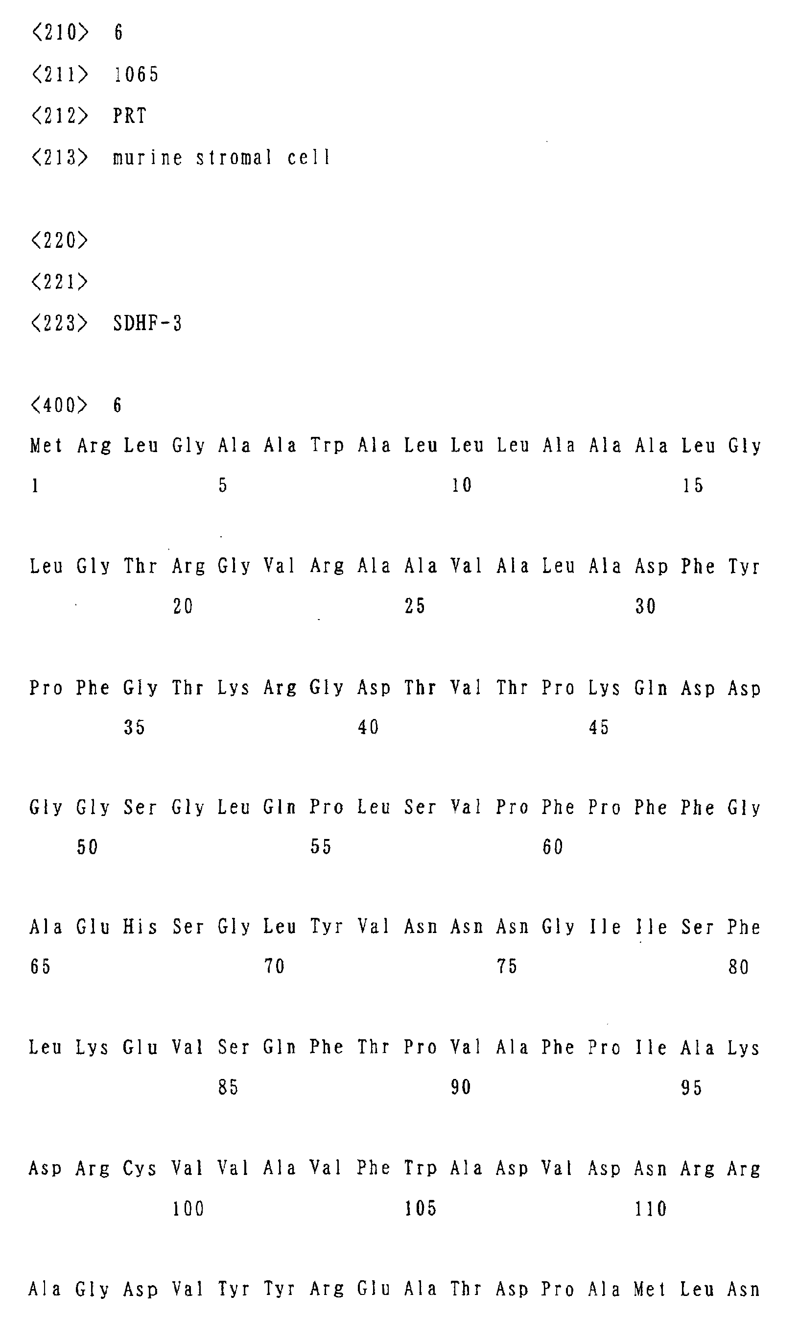
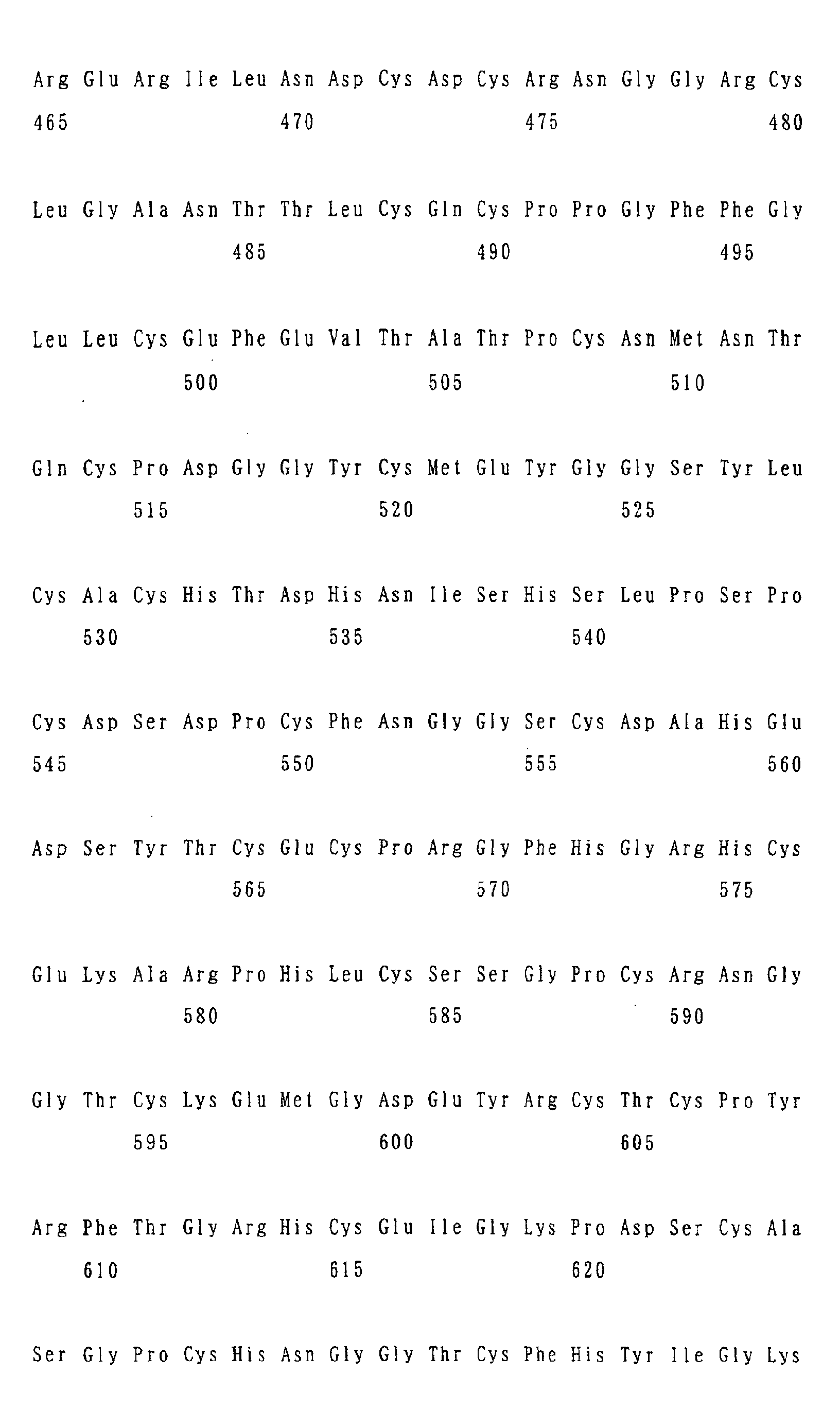
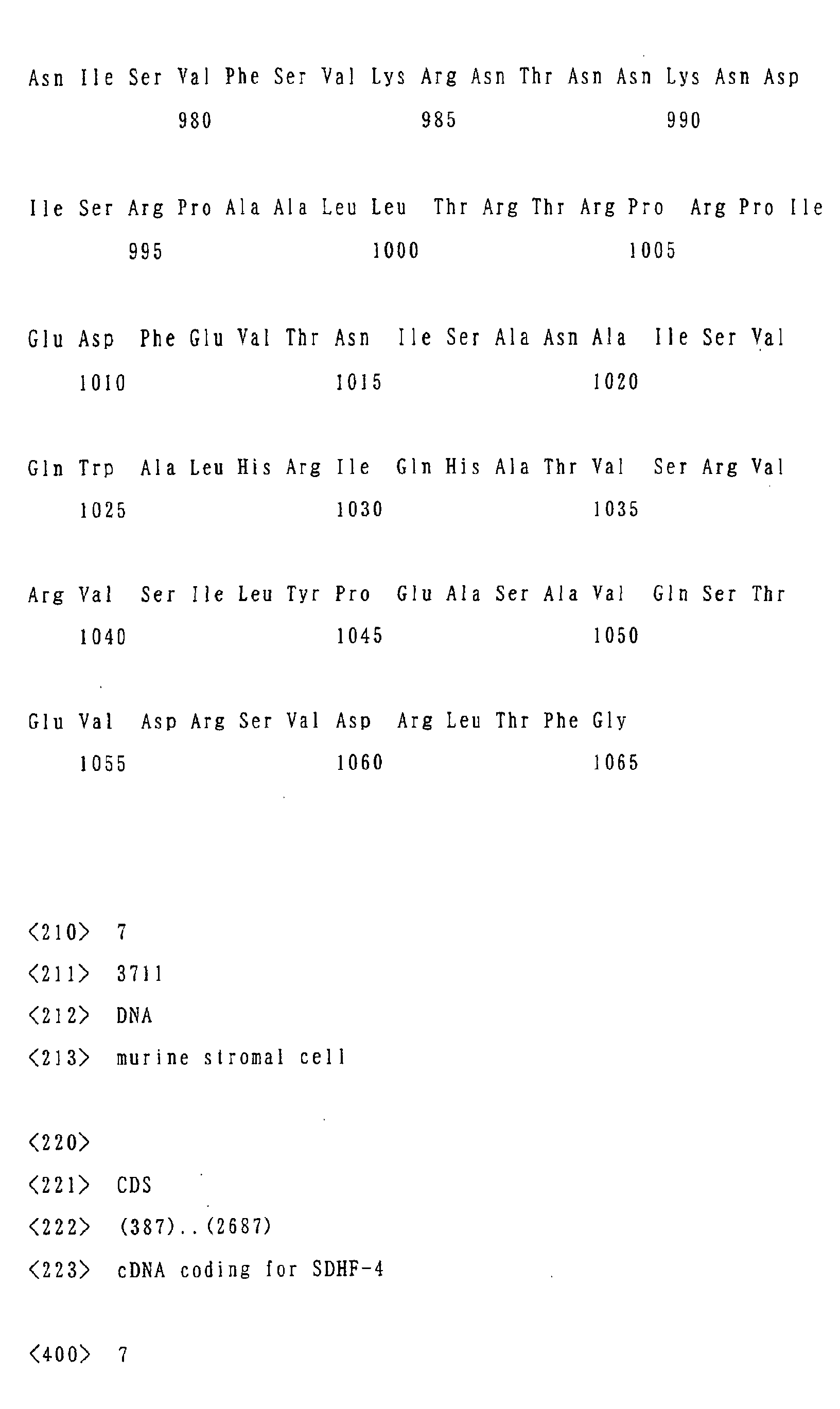
Figure 3 shows the amino acid sequence of SDHF-3.
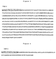
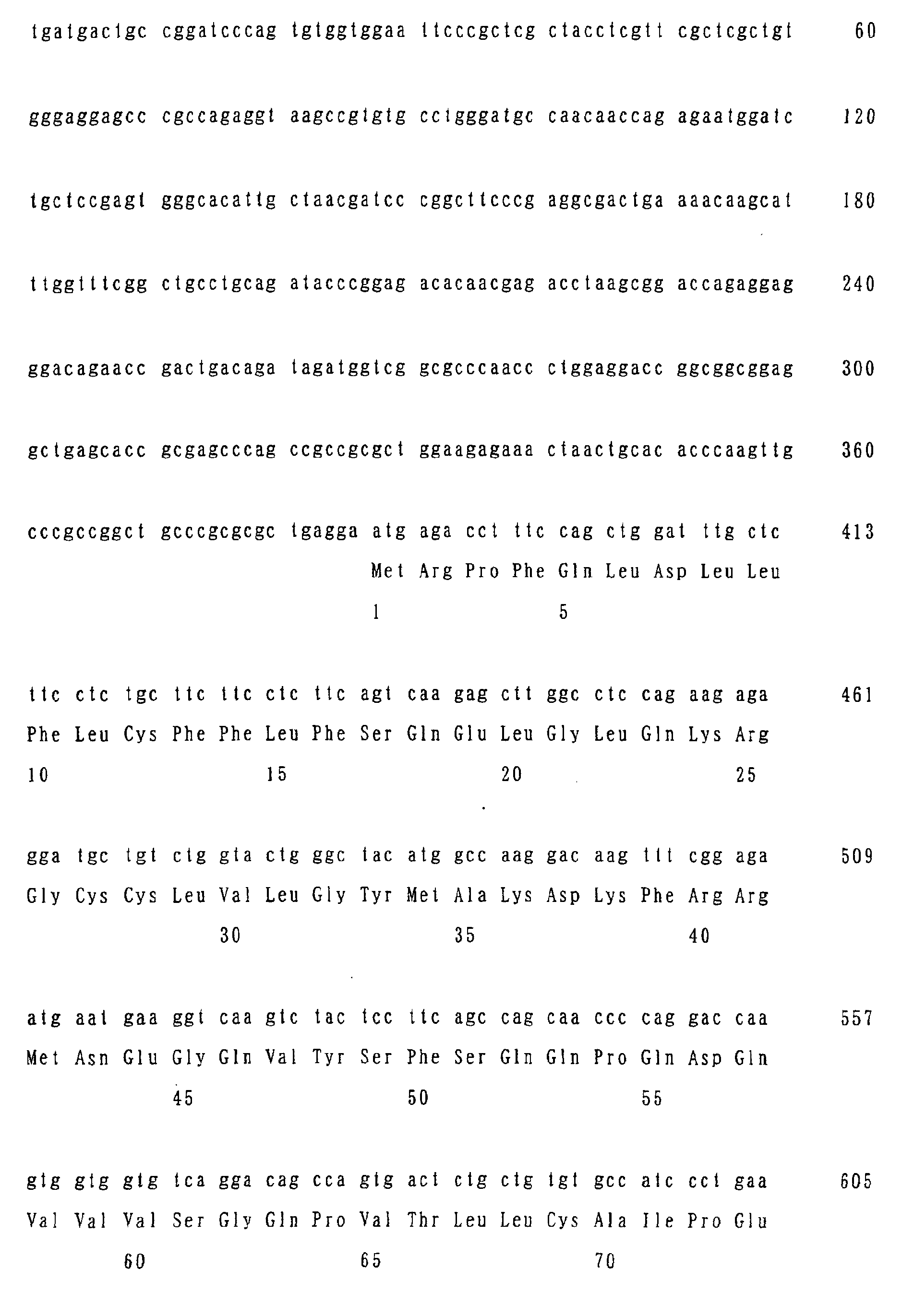
Figure 4 shows the amino acid sequence of SDHF-4.
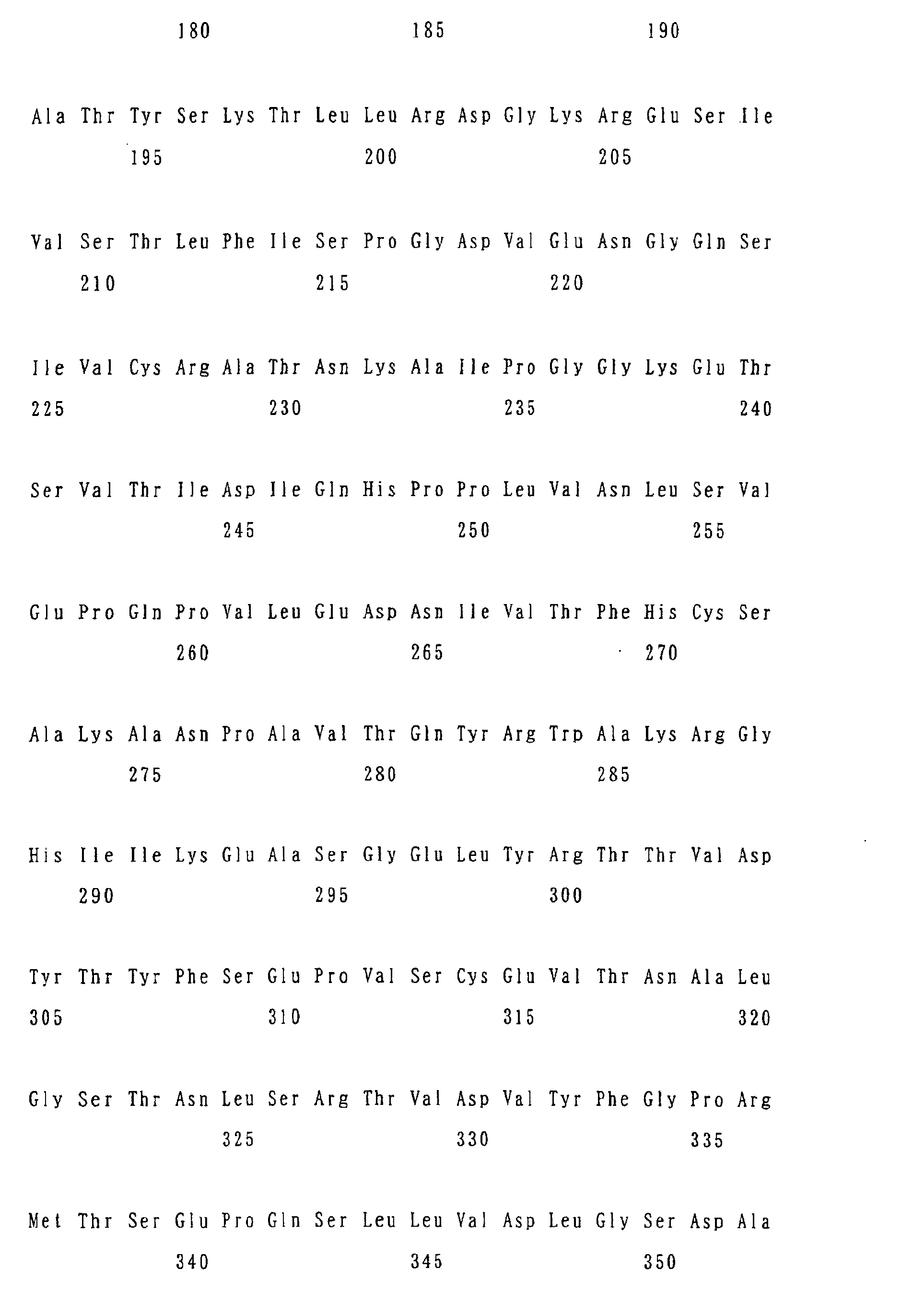
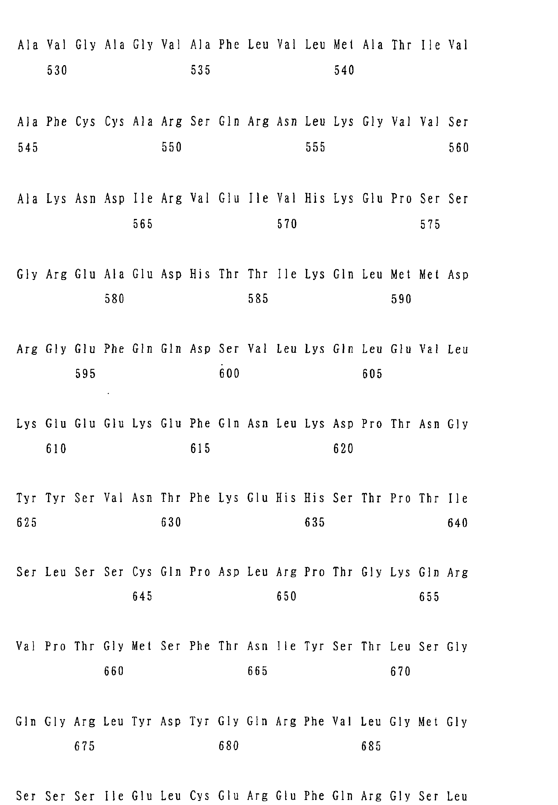
Figure 5 shows the amino acid sequence of SDHF-5.
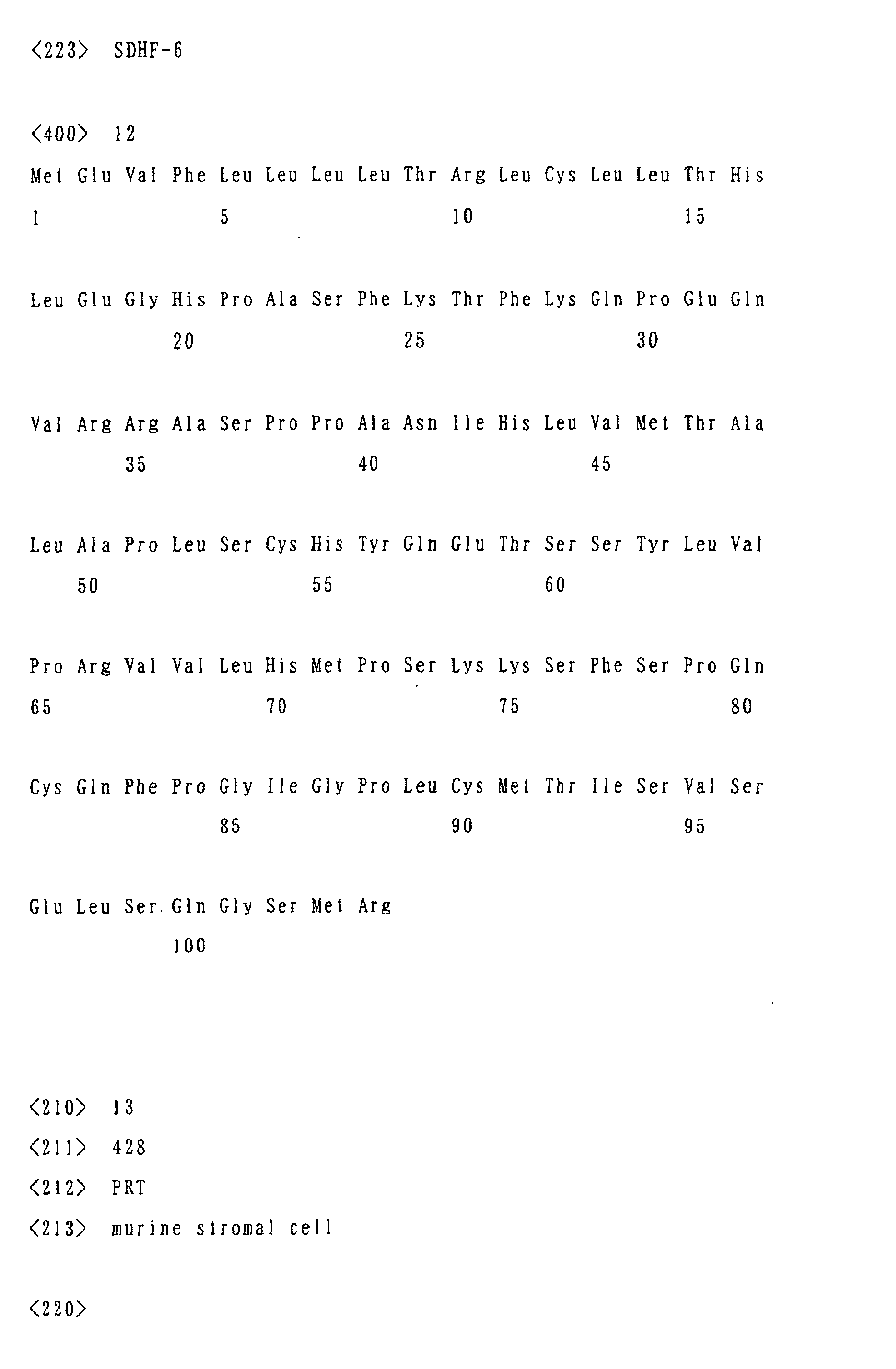
Figure 6 shows the amino acid sequence of SDHF-6.
Figure 7 shows the results obtained by a microarray method .
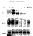
Figure 8 shows the results obtained by a northern blotting.
Figure 9 shows the results obtained by a western analysis.
Figure 10 shows the results obtained by an immunostaining
method.
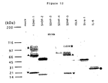
Figure 11 shows the results of a CAFC analysis.
Figure 12 shows the results of the post-translational
modification of the proteins of genes SDHF-1 and SDHF-5 of the
invention detected by the western analysis. The symbol "*"
indicates a mature protein whose molecular weight became larger
as a result of the addition of a sugar chain, the symbol "▴"
indicates a protein on the way of the production before the
addition of the sugar chain, the symbol "▾" indicates a residual
protein after a cleavage, and the symbol "" indicates a
secretory protein.
Figure 13 shows the results of the detection by the
methionine labeling of a secretory protein in a cell culture
supernatant. The symbol "" indicates a secretory protein,
and "→" indicates a protein secreted in the culture supernatant
after a cleavage.
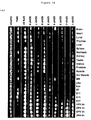
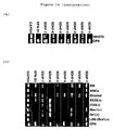
Figure 14 shows a photograph of the electrophoresis
showing the results of the assay of the expression site of a
gene of the invention by an RT-PCR method.
Figure 15 shows the results of an assay of the stem
cell-supporting function of a gene of the invention by an LTC-IC
method.
Figure 16 shows a photograph of the electrophoresis of
an SDHF-4 extracellular region/recombinant protein as being
purified by a protein A column.
Figure 17 is a graph showing that the SDHF-4 extracellular
region/recombinant protein has an ability of maintaining the
non-differentiated condition of a mouse hematopoietic stem
cell.
Figure 18 shows a microscopic photograph (magnification:
x 40) showing that SDHF-6, when expressed highly in an OP9 cell
using a retrovirus expression vector pMX-puro, has an ability
of inducing the differentiation into a fat cell efficiently
after about four-week culture.
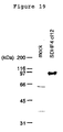
Figure 19 shows a photograph of a western analysis showing
the results of the experiment in which an antibody directed
to a partial peptide of SDHF-4 was employed to allow SDHF-4
to be expressed in a CHO-kl cell using a pSSRα-bsr vector.
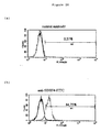
Figure 20 shows the results of a FACS calibur (Becton
Dickinson) analysis of a CHO-k1 cell allowed to express SDHF-4
after labelling the purified antibody described above with FITC.
Figure 21 shows the results of a sorting of a cell
expressing SDHF-4 using a FACS Vantage (Becton Dickinson) after
staining amyelocyte of a C57BL/6J mouse using the same antibody.
Figure 22 shows a microscopic photograph of the
above-mentioned cell selected by the sorting (magnification:
x 200).
Figure 23 shows a schematic view of the molecular
structures of SDHF-1 to SDHF-6.
BEST MODE FOR CARRYING OUT THE INVENTION
A gene or DNA of the invention can be prepared for example
by a method shown in the EXAMPLES described below based on a
cDNA library prepared from an mRNA derived from a stromal cell
such as a mouse stromal cell line OP9 which has been stimulated
by a leukemia-inhibiting factor (LIF) during culture.
Alternatively, it can be prepared also by a chemical
synthesis using a procedure known in the art based on the sequence
information disclosed in this specification. Those skilled
in the art may accomplish the deletion, substitution or addition
of one or several amino acids in a certain amino acid sequence
by means of a method known per se in the art.
In the invention, a specific gene or DNA can be hybridized
under a stringent condition with regard to various parameters
such as salt concentrations at a suitable temperature in a buffer
solution known to those skilled in the art.
An example of the DNA which hybridizes under such a
stringent condition with a gene or DNA of the invention and
which encodes a protein having a hematopoietic stem cell
proliferation-regulating activity is a DNA whose homology with
such a gene is 70% or more, preferably 90% or more.
A DNA obtained by binding a gene or DNA of the invention
appropriately to any of various sequences known to those skilled
in the art in a gene recombination procedure such as regulatory
factors including promoters and enhancers, restricted enzyme
sites, selection marker (marker enzyme) gene and the like is
also encompassed by the invention.
An expression vehicle such as a recombinant plasmid vector
having a gene, DNA or a DNA molecule of the invention described
above or a recombinant virus vector employing a retrovirus and
the like, and a transformant of a microorganism such as
Escherichia coli and an eukaryotic cell such as a mouse COS-7
cell or a mouse stromal cell OP9, which contains such an
expression vehicle and has thus been transformed, are also
encompassed by the invention. These DNA molecules, expression
vehicles and transformants can readily be prepared by a method
known in the art. In addition, a protein of the invention can
be readily produced by those skilled in the art by culturing
an above-mentioned transformant by a known method and purifying
the culture product.
A hematopoietic stem cell proliferation regulation
activating composition of the invention comprising a protein
described above as an active ingredient may also contain, as
appropriate, other cytokines known as hematopoietic factors,
as well as suitable carriers and auxiliary agents known in the
art. The amounts and the ratios of the protein of the invention
as an active ingredient and other components may be selected
by those skilled in the art depending on the purpose of use.
The antibodies such as polyclonal and monoclonal
antibodies of the invention directed to the respective proteins
of the invention such as SDHF-1, -2, -3, -4, -5 and -6 can be
produced by any known method. For example, a polyclonal
antibody can readily be produced by inoculating each protein
described above or its partial peptide as an immunogen to any
experimental animal such as a rabbit. A monoclonal antibody
can readily be obtained by subjecting an antibody-producing
cell prepared similarly using such an immunogen to a cell fusion
with a suitable parent cell (immortalized cell line) by a method
known in the art to yield a hybridoma followed by screening
the hybridoma for a clone which reacts specifically with each
protein. The partial peptide corresponds to any partial amino
acid sequence in the respective protein, and the number of the
amino acids is not limited particularly but preferably is at
least 10, and usually is within the range from 10 to 20. In
view of the reactivity with each protein, it is preferable to
select a partial peptide employed as an immunogen from the
extracellular region of the respective protein.
Such an antibody can be used in a procedure for isolating
or purifying various adult cells such as bone marrow stem cells,
mesencymal stem cells and nervous stem cell as well as stem
cell-supporting cells (stromal cells) such as a bone marrow
hematopoiesis-supporting cells. Such an isolation or
purification may be conducted by various means known to those
skilled in the art, such as an affinity column purification
or a FACS sorting.
EXAMPLES
The invention is further described referring to the
following EXAMPLES, which are not intended to restrict the
technical scope of the invention.
cDNA library construction and Signal Sequence Trap (SST) method
A mouse stromal cell line OP9 is incubated in Alpha-MEM
medium supplemented with 20% fetal bovine serum. A PLAT-E cell
(2000, Morita et al., Gene Therapy vol. 7, 1063-6) was incubated
in DMEM supplemented with 10% fetal bovine serum. The
extraction of polyA RNA from the OP9 cell was conducted using
an Invitrogen FAST TRACK 2.0 mRNA Isolation Kit in accordance
with the protocol attached thereto. As a starting material
for a cDNA library, 5 µg of polyA RNA extracted from 1 x 10⁸
OP9 cells stimulated for 60 minutes at 37°C with 10 ng/ml of
mouse leukemia inhibitory factor (LIF) was employed. The cDNA
library was constructed using a SUPERSCRIPT II (Invitrogen)
using random hexamers as primers. The resultant cDNA library
was combined with a BstXI adapter primer (Invitrogen) and
integrated into the BstXI site of a retrovirus vector pMX-SST
vector (1999, Kojima et al., Nat Biotech vol. 17, p. 487) to
construct a plasmid library.
The cDNA library obtained was introduced into a PLAT-E
cell using FuGene 6 (Roche), and then recovered in the form
of a retrovirus in the supernatant. The resultant retrovirus
cDNA library was infected to a Ba/F3 cell which is an interleukin
3 (IL-3)-dependent mouse pro-B cell. The-pMX-SST vector has
a signal peptide-deficient constantly activated c-Mpl gene,
and the cMpl gene is activated when a library-derived gene has
a signal peptide and consequently the Ba/F3 cell is immortalized
in the absence of IL-3 (1999, Kojima et al., Nat Biotech vol.
17, p. 487-90). The retrovirus library-infected Ba/F3 cell
was deprived of IL-3, and incubated in a 96-well plate at the
density of 1000 cell/well, and then the proliferated cells were
isolated and the genome DNA was extracted. Since a signal
peptide-carrying gene has been inserted into the genome DNA
of the Ba/F3 cell via the retrovirus, it was recovered by a
genome DNA PCR (LA-Taq, TAKARA, 98°C for 20 sec. 68°C for 300
sec.: 35 cycles) using the primers (GGGGGTGGACCATCCTCTA,
CGCGCAGCTGTAAACGGTAG) designed based on the sequence of the
pMX-SST vector.
Gene sequencing
Since a signal peptide-carrying clone was obtained as
a DNA fragment by the PCR described above, its DNA sequence
was determined using an ABI 373A sequencer. The resultant PCR
fragment was purified by a PCR purification kit (Quiagen) to
be deplete the primers, and the sequences were determined by
a dye termination method in accordance with the protocol by
ABI. The resultant sequence information was analyzed by an
NCBI BLAST program (http://www.ncbi.nlm.nih.gov/BLAST/)
whereby judging whether the sequences were known or unknown.
A part of an unknown sequence was determined by a 3' RACE method
using primers designed based on the sequences obtained. The
3' RACE method is a strategy for identifying an unknown gene
to the extent of the polyA region by a PCR method using the
primers designed on the basis of the above-mentioned PCR clone
sequences as well as oligo dT primers. The DNA sequence of
any resultant PCR fragment was determined as described above
using an ABI 373A sequencer.
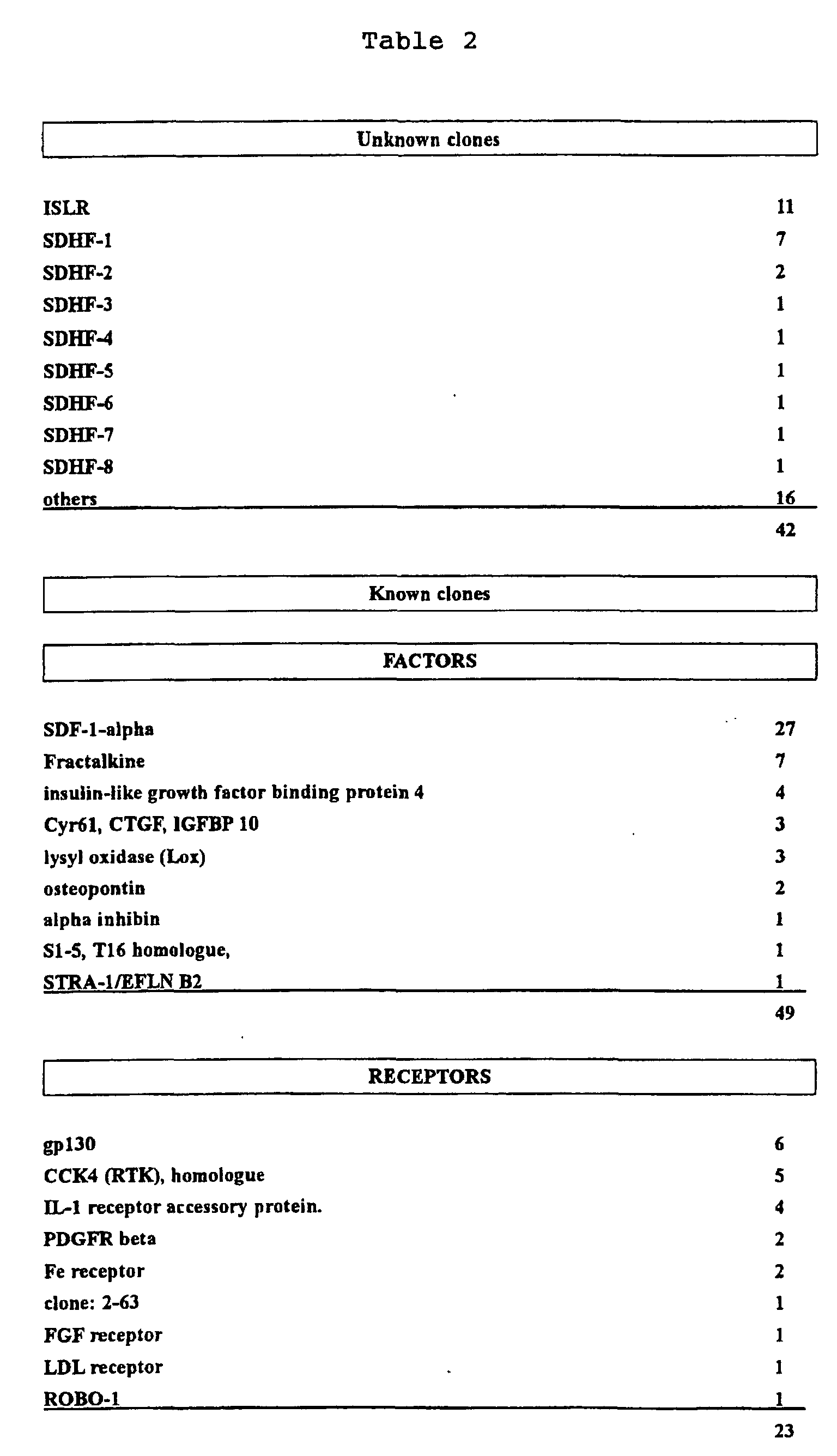
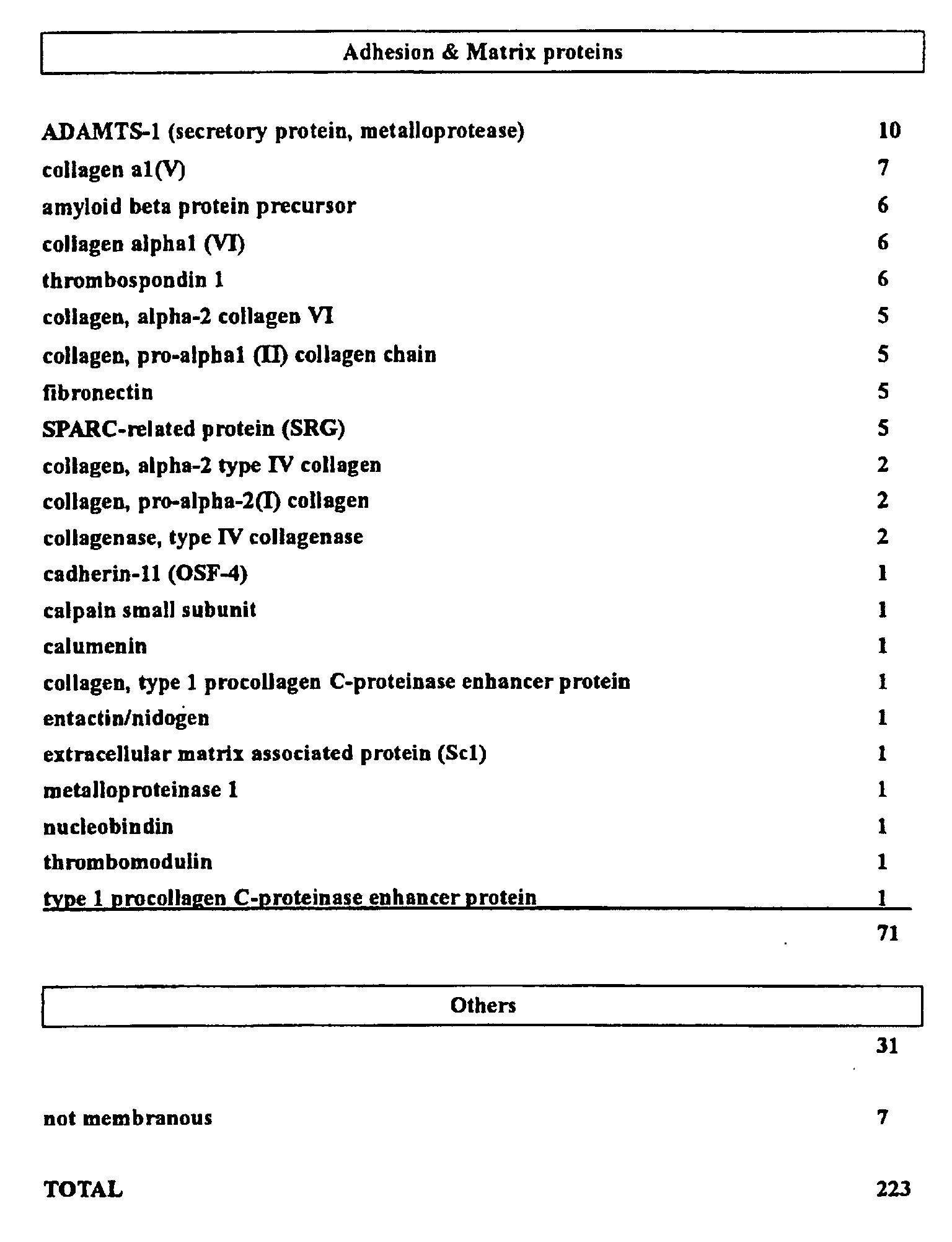
As a result, the genes of 234 clones in total including
known and unknown ones were isolated by the SST method described
above. They included 181 known genes, 42 unknown genes and
11 genes which could not be analyzed. The known genes included
49 proliferation factors, 23 receptors, 71 adhesion molecules
and 38 others.
Among the unknown genes, 6 SDHF (stromal cell derived
hematopoietic factor) genes of the invention and ISLR were
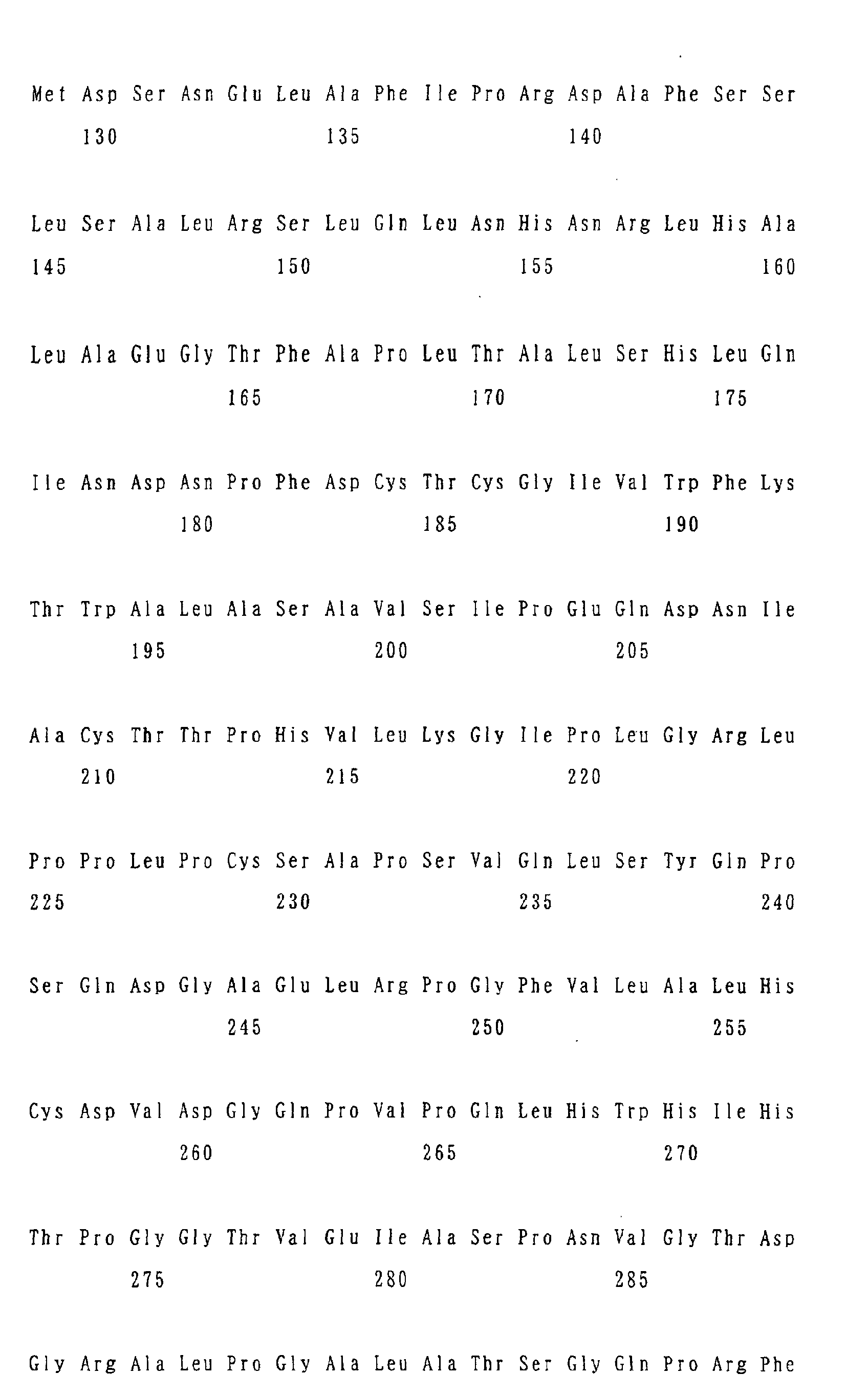
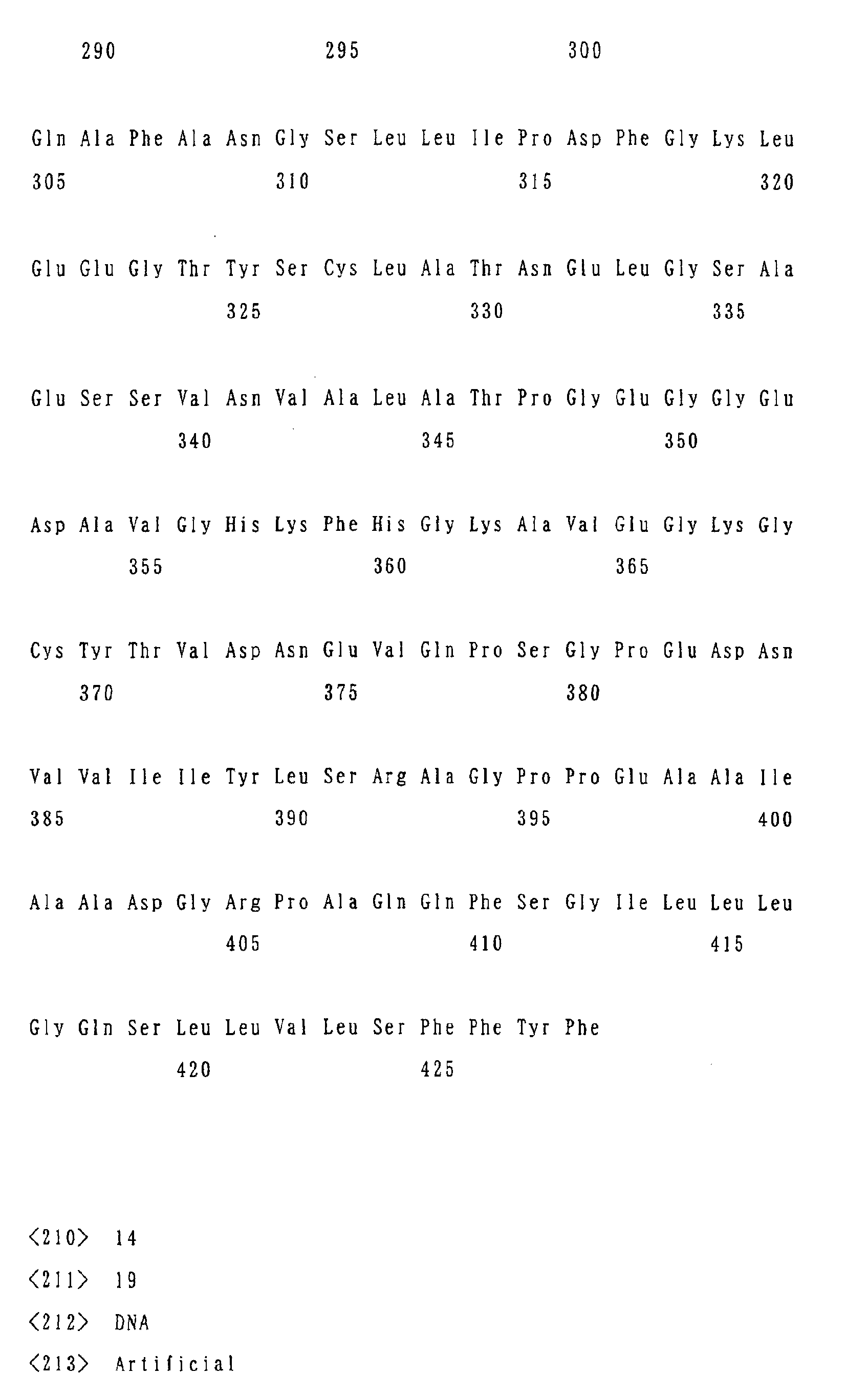
included. As shown in Figures 1 to 6 and in SEQ ID No. 13,
the N terminal of the amino acid sequence encoded by 6 SDHF
and ISLR genes contained a signal peptide (region surrounded
by a rectangular frame). The results obtained as described
above are summarized in Table 1 and Table 2.
| SDF-1-alpha | 27 |
| ISLR (immunoglobulin superfamily containing leucine-rich repeat) | 11 |
| ADAMTS-1 (secretory protein, metalloprotease) | 10 |
| SDHF-1 | 7 |
| collagen al(V) | 7 |
| Fractalkine | 7 |
| amyloid beta protein precursor | 6 |
| collagen alpha1 (VI) | 6 |
| gp130 | 6 |
| thrombospondin 1 | 6 |
| CCK4 (RTK), homologue | 5 |
| collagen, alpha-2 collagen VI | 5 |
| collagen, pro-alpha1 (II) collagen chain | 5 |
| fibronectin | 5 |
| SPARC-related protein (SRG) | 5 |
| syndecan | 5 |
| amyloid precursor-like protein 2 | 4 |
| BiP, immunoglobulin heavy chain binding protein | 4 |
| insulin-like growth factor binding protein 4 | 4 |
| interleukin 1 receptor accessory protein. | 4 |
| protein disulfide isomerase (ERp59) | 4 |
| Cyr61, CTGF, IGFBP10 | 3 |
| lysyl oxidase (Lox) | 3 |
| SDHF-2 | 2 |
| collagen, alpha-2 type IV collagen | 2 |
| collagen, pro-alpha-2(I) collagen | 2 |
| collagenase, type IV collagenase | 2 |
| osteopontin | 2 |
| P2B/LAMP-1 | 2 |
| PDGFR beta | 2 |
| vimentin | 2 |
| SDHF-3 | 1 |
| SDHF-4 | 1 |
| SDHF-5 | 1 |
| SDHF-6 | 1 |
| SDHF-7 | 1 |
| SDHF-8 | 1 |
All PCR fragments obtained by the signal sequence trap
method were spotted onto glass slides (procedure heretofore
being conducted by HOKKAIDO SYSTEM SCIENCE), and the change
in the expression was investigated using a fluorescence-labeled
cDNA probe prepared from the cells adjusted under the condition
before and after the stimulation of the OP9 cells with 10 ng/ml
LIF at 37°C for 60 minutes. The probe was subjected to a reverse
transcription using a SuperScript II employing oligo dT primers
from the polyA RNA extracted as described above, and then
purified using a Microcon 30 (Millipore) labeled by means of
the integration of a Fluorolink Cy-dUTP. The results obtained
are shown in Figure 7.
Northern blotting
The OP9 cells were stimulated with 10 ng/ml of LIF at
37°C for 60 minutes, and then after 0 minutes, 30 minutes, 1
hour, 3 hours, 8 hours and 24 hours, the total RNA was extracted
using a Trizol reagent (Invitrogen) in accordance with the
protocol. The total RNA thus extracted was examined for the
concentration, and then a 10 µg aliquot was subjected to an
electrophoresis on a 1% agarose gel, transferred onto a Hybond
N (Amersham) , and subjected to a northern blotting by a standard
method using the respective gene DNA fragment as a probe. As
a result, an increase in the expression of each of SDHF-1, SDHF-2
and SDHF-5 genes was observed as evident from Figure 8. On
the contrary, SDHF-6 gene exhibited a reduction in the
expression.
Western blotting
From the sequence of the entire length of a gene thus
obtained, an amino acid-encoding region (open reading frame:
ORF) was determined, and its carboxyl terminal was tagged with
a FLAG tag sequence (DYKDDDDK) by a PCR method. All of these
cDNAs were subcloned into an expression vector pUC-CAGGS which
was then expressed transiently in COS-7 cells using FuGene 6
(Roche). The western blotting was conducted as reported
previously (1995, Ueno, JBC Vol. 270, pp. 20135-42). The cells
were solubilized with a Triton lysis buffer (0.5% (v/v) , Triton
X-100, 50mM Tris-HCl, pH 7.4, 2mM PMSF, 10 U/ml aprotinin, 1mM
EDTA) and then a 50 µg lysate was subjected to an
SDS-polyacrylamide gel electrophoresis (PAGE), transferred to
an Immobilon-P membrane, and then detected using an anti-FLAG
antibody, M2 (Sigma). As a result, any of the genes of the
invention was expressed in the COS7 cells and exhibited the
bands in the western blotting.
Immunostaining
Genes tagged with a FLAG peptide were expressed
transiently in the COS-7 cells using a FuGene reagent (see above),
fixed in a 3.7% formaldehyde, subjected to a permeabilization
with a 1% NP40, and stained with an anti-FLAG antibody (secondary
antibody: Texas Red-labeled anti-mouse antibody, Jackson).
The nucleus was stained with DAPI (FUNAKOSHI). The results
are shown in Figure 10.
Cobblestone Area Forming Cell Assay: CAFC assay
A gene obtained as described above in the OP9 cells was
transduced into a retrovirus vector (pMX-puro), which were
inoculated to a 6-well dish at the density of 2.5 x 10⁵, and
incubated overnight and then inoculated with 1 x 10⁵ of the
bone marrow cells obtained from the femoral bones of 5 to 8-week
old C57BL/6 mice, and the number of the CAFCs formed was counted
after 6 days.
Results of CAFC analysis
Hematopoietic cells proliferating underneath a stromal
cell layer upon co-culturing the stromal cells and the bone
marrow cells are referred to as a cobblestone area forming cell
(CAFC). An increased number of the CAFCs formed is largely
means that the stromal cell amplifies the immature hematopoietic
cells. Nevertheless, an increased CAFC does not mean an
amplified hematopoietic stem cell in an exact sense, since the
cells consisting the CAFC include not only the hematopoietic
stem cell but also the cells which have been differentiated
once, such as myelocytic, erythroblastic, megakaryocytic and
lymphocytic precursor cells. The gene described above was
introduced into the OP9 cells of a mouse myelic stromal cell
line having a hematopoiesis-supporting ability using a
retrovirus vector pMX-puro, and the cells imparted with a drug
resistance by 5 µg/ml puromycin was examined for their
CAFC-forming ability. As a result, ISLR exhibited the highest
level, and slightly higher levels were also exhibited by SDHF -1,
SDHF-2, SDHF-4 and SDHF-5, as shown in Figure 11. These findings
suggest the hematopoietic precursor cell-amplifying ability
of these genes. On the contrary, SDHF-3 exhibited the
inhibition of the CAFC-constituting ability, and was considered
to have a hematopoietic stem cell proliferation-inhibiting
ability.
Detection of secretory protein in cell culture supernatant by
methionine labeling
1 x 10⁵ COS-7 cells were inoculated into a 6 cm dish,
which was then incubated overnight. The genes were introduced
by a FuGene method (Roche). The carboxyl terminal of any of
these genes is tagged with a FLAG tag, and a protein expressed
can be detected by a western analysis using an anti-FLAG antibody
as described above. After incubation for 36 hours, the
supernatant was removed, and the cells were washed twice with
PBS, combined with 0.5 ml of DMEM (Methionine, Sigma)
supplemented with 0.5 mCi [35S] methionine (ICN, TRAN-S), and
incubated for 4 hours. The cell components were removed from
the culture supernatant by centrifugation, and the proteins
were concentrated using a Micron 10 (Millipore). After running
in a 15% SDS-PAGE, the labeled proteins were detected using
a BAS2000 (FUJI FILM).
As a result, SDHF-1 and SDHF-5 exhibited the protein
secretion to the culture supernatant as shown in Figure 12 and
Figure 13. Since these genes are membrane proteins structurally
and the secretory proteins are smaller by about 20 kDa than
the mature protein detected by the western analysis, these
secretory proteins were considered to have undergone the
cleavage of the transmembrane region of the carboxyl terminals.
Since an SCF is known to be a proliferation factor of the type
which is secreted as a result of the cleavage of the transmembrane
region of the carboxyl terminal and such findings were not shown
by an adhesion protein, the proteins encoded by the genes of
the invention are suggested strongly to be the proteins of the
type which serve as humoral factors to transmit the signals
to other cells.
Assay of expression site by RT-PCR
Based on the DNA sequences of respective SDHF genes, the
primers were designed and a PCR (LA-Tag, TAKARA) was conducted
using Multiple Tissue cDNA (MTC) panels (Clontech) as templates.
The cells in a bone marrow were sorted by a FACS Vantage (Becton
Dickinson) using fluorescence-labeled monoclonal antibodies
(anti-B220, anti-CD3, anti-Gr1, anti-MAC1, anti-Scal
antibodies, Phamingen), subjected to an RNA extraction with
a Trizol reagent (Invitrogen) followed by an RT reaction using
random hexamers, and then subjected to the PCR similarly.
As a result, all SDHF genes exhibited the expression in
the OP9 cells as shown in Figure 14. SDHF-4 exhibited the
expression localized especially in the brain and the bone marrow,
in which the expression was localized in the stromal cells
interestingly.
Assay of stem cell-supporting ability by long term
culture-initiating cells assay (LTC-IC method)
Since the CAFC method described above only reflects the
hematopoiesis-supporting function over a relatively short
period, an LTC-IC method was conducted for the evaluation of
the stem cell-supporting function over a prolonged period. A
gene isolated as described above was expressed highly in the
OP9 cells using a retrovirus expression vector pMX-puro whereby
conducting an LTC-IC method.
First, a hematopoietic stem cell was purified. Thus,
the bone marrow cells were obtained from the femoral bones of
6 to 8-week old C57BL/6 mice, and the mononuclear cell were
separated by centrifugation by a Ficoll method (specific
gravity: 1.100). After washing with PBS, the cells were reacted
with a biotin-labeled primary antibody (anti-Lineage antibody
cocktail: anti-CD3, anti-CD4, anti-CD8, anti-B220, anti-Ter119,
anti-Gr1, anti-Mac1 antibodies, all from Phamingen) followed
by streptoavidin-labeled magnetic beads each at 4°C for 15
minutes. A MACS method was employed to recover the
column-passing fraction (LIN(-) cells), which was washed by
centrifugation, reacted further with a PE-labeled anti-Scal
antibody, FITC-labeled anti-c-kit antibody,
Per-CP-Cy5.5-labeled streptoavidin (all from Phamingen) at 4°C
for 15 minutes, subjected to a FACS Vantage (Becton Dickinson)
to obtain LIN (-), Scal (+) and c-kit (+) fractions (the cells
thus obtained were defined as KSL cells). cDNAs obtained by
adding FLAG peptides to the carboxyl terminals of SDHF-1 to
6 were transduced to the OP9 cells using a retrovirus vector
(pMX-puro) to establish stably expressing cell lines, 2.5 x
105 of which were inoculated to a 6 -well dish, which were incubated
overnight, inoculated with 100 KSL cells, which were then
incubated for 3 weeks. Thereafter, the cells were recovered
and inoculated, after 2-fold dilution in 8-well pairs, to a
96-well dish, each well of which had been inoculated with 3000
OP9 cells which had been irradiated with γ-ray at 20 Gy. After
5 weeks, the cells were recovered from each well, inoculated
to a methyl cellulose medium (IMDM, 1% methyl cellulose, 15%
fetal bovine serum, 1% bovine serum albumin, 3 U/ml human
erythropoietin, 10 ng/ml human IL-6, 10 ng/ml mouse IL-3, 100
ng/ml mouse SCF, 10 µg/ml bovine insulin), and then evaluated
after 12 days on the basis of the presence or absence of the
blood cell colonies (CFU-C). The frequency of the stem cells
were analyzed by an L-Calc software (Stemcell).
As a result, a marked hematopoiesis-supporting function
was exhibited by the SDHF-4 gene as evident from Figure 15.
The hematopoiesis-supporting function of this gene was
revealed.
Mouse hematopoietic stem cell-amplifying effect of SDHF-4
extracellular region/recombinant protein
The extracellular transmembrane region of SDHF-4 was
assumed (amino acids 524 to 540) using a PSORT II program
(http://psort.ims.u-tokyo.ac.jp/form2.html), and a human
immunogloblin Fc region and a fusion protein (SDHF-4-Δ TM-Fc)
were formed in the extracellular region (amino acids 1 to 523),
and subcloned into an expression vector pSSRα, which were stably
introduced into CHO-k1 cells. A culture in a serum-free medium
CD-CHO (Invitrogen) followed by the purification of the
supernatant on a Protein A column yielded fusion recombinant
proteins (Figure 16). Mouse stem cells were cultured similarly
to the LTC-IC method as the mouse bone marrow LIN (-), Scal
(+) and c-kit (+) fractions (KSL cells), each 100 cells of which
were incubated in a Stem Span medium (StemCell) supplemented
with SCF (50 ng/ml), IL-3 (10 ng/ml) , IL-6 (10 ng/ml) and SDHF-4-Δ
TM-Fc (100 ng/ml). After 7 days, the cells were recovered,
and examined for the expression of LIN, Scal and c-kit using
a FACS Calibur.
As a result, the LSK cells incubated in the StemSpan medium
supplemented with SCF, IL-3 and IL-6 exhibited not only a LIN
(-) cell ratio which was higher in the presence rather than
the absence of 100 ng/ml of SDHF-4-Δ TM-Fc but also increased
ratios of LIN (-), Scal (+) and c-kit (+) fractions. These
findings suggest that the SDHF-4 extracellular
region/recombinant protein has an ability of maintaining the
non-differentiated state of the mouse hematopoietic stem cells.
Fat cell differentiation assay and Oil Red O staining
Each of the cell into which an SDHF-6 gene had been
transduced via a retrovirus vector (pMX-puro) and the cell into
which only the vector had been transduced was incubated
continuously in a confluent state without any subculture over
a period of 4 weeks or longer in the presence of Alpha-MEM +
20% fetal bovine serum. The cells were fixed with 10% formalin
and then stained with an Oil Red O.
As a result, the OP9 cells over expressing SDHF-6 by using
the retroviral vector pMX-puro were revealed to differentiate
into fat cells with a high efficiency after the incubation for
about 4 weeks. This gene was considered to be involved in the
differentiation into the fat cells via the effect on the
mesencymal cells rather than on the hematopoietic stem cells.
Preparation of polyclonal antibody directed to SDHF-4 and
isolation of myelic hematopoiesis-supporting cell therewith
Based on the amino acid sequence of SDHF-4, a synthetic
peptide (GYMAKDKFRRMNEGQVY (corresponding to the amino acids
32 to 48)) was designed, and immunized to a rabbit to prepare
a polyclonal antibody.
This peptide was conjugatal with an epoxy-activated
Sepharose 6B (Pharmacia) to prepare an antigen column, which
was used to purificate a polyclonal antibody, as an affinity
chromatography. A western analysis using this antibody while
comparing the cell expressing SDHF-4 obtained by transducing
a CHO-k1 cell with a pSSRα-bsr vector (SDHF4-c112) with the
cell into which only the vector had been introduced (mock)
revealed that an SDHF-4 gene product could be recognized in
a very specific manner (Figure 19). 1 mg of the resultant
purified antibody was labeled with FITC using a LinKit
Fluoro-Link (ISL) and the same cell was analyzed by a FACS Calibur
(Becton Dickinson), and the results indicated that this antibody
can be utilized also in a flow cytometry (Figure 20: ○-line:
mock, ▪-line: SDHF4-cll2). Using this antibody, the bone
marrow cells of a C57BL/6J mouse were stained, and subjected
to a FACS Vantage (Becton Dickinson) whereby sorting and
recovering the cells expressing SDHF-4 (Figure 21), which
deposited onto a culture dish and were revealed morphologically
to be mesencymal cells corresponding to bone marrow
hematopoiesis-supporting cells (Figure 22).
INDUSTRIAL APPLICABILITY
According to the invention, a stromal cell-derived novel
hematopoiesis-related gene can be isolated successfully, and
was revealed to have a hematopoietic stem cell
proliferation-regulating activity. As a result, a wide range
of the application, including an in vitro amplification of
hematopoietic stem cells, a transplantation therapy against
a malignant tumor of a hematopoietic tissue, a regenerative
medicine using the stem cell, a gene therapy, immunotherapy,
cell transplantation, treatments of a neuropathy (Alzheimer's
disease, cerebral infarction, degenerative neuropathy and the
like), hepatic disease (cirrhosis), pulmonary disease,
myocardial disease, diabetes, bone disease, chronic renal
failure and the like, became possible.