EP1420254A2 - Amyloid quantification - Google Patents
Amyloid quantification Download PDFInfo
- Publication number
- EP1420254A2 EP1420254A2 EP03024739A EP03024739A EP1420254A2 EP 1420254 A2 EP1420254 A2 EP 1420254A2 EP 03024739 A EP03024739 A EP 03024739A EP 03024739 A EP03024739 A EP 03024739A EP 1420254 A2 EP1420254 A2 EP 1420254A2
- Authority
- EP
- European Patent Office
- Prior art keywords
- beta amyloid
- amyloid
- labeled
- plaques
- beta
- Prior art date
- Legal status (The legal status is an assumption and is not a legal conclusion. Google has not performed a legal analysis and makes no representation as to the accuracy of the status listed.)
- Withdrawn
Links
Images





Classifications
-
- G—PHYSICS
- G01—MEASURING; TESTING
- G01N—INVESTIGATING OR ANALYSING MATERIALS BY DETERMINING THEIR CHEMICAL OR PHYSICAL PROPERTIES
- G01N33/00—Investigating or analysing materials by specific methods not covered by groups G01N1/00 - G01N31/00
- G01N33/48—Biological material, e.g. blood, urine; Haemocytometers
- G01N33/50—Chemical analysis of biological material, e.g. blood, urine; Testing involving biospecific ligand binding methods; Immunological testing
- G01N33/68—Chemical analysis of biological material, e.g. blood, urine; Testing involving biospecific ligand binding methods; Immunological testing involving proteins, peptides or amino acids
- G01N33/6803—General methods of protein analysis not limited to specific proteins or families of proteins
- G01N33/6848—Methods of protein analysis involving mass spectrometry
-
- G—PHYSICS
- G01—MEASURING; TESTING
- G01N—INVESTIGATING OR ANALYSING MATERIALS BY DETERMINING THEIR CHEMICAL OR PHYSICAL PROPERTIES
- G01N33/00—Investigating or analysing materials by specific methods not covered by groups G01N1/00 - G01N31/00
- G01N33/48—Biological material, e.g. blood, urine; Haemocytometers
- G01N33/50—Chemical analysis of biological material, e.g. blood, urine; Testing involving biospecific ligand binding methods; Immunological testing
- G01N33/68—Chemical analysis of biological material, e.g. blood, urine; Testing involving biospecific ligand binding methods; Immunological testing involving proteins, peptides or amino acids
- G01N33/6803—General methods of protein analysis not limited to specific proteins or families of proteins
- G01N33/6827—Total protein determination, e.g. albumin in urine
-
- G—PHYSICS
- G01—MEASURING; TESTING
- G01N—INVESTIGATING OR ANALYSING MATERIALS BY DETERMINING THEIR CHEMICAL OR PHYSICAL PROPERTIES
- G01N33/00—Investigating or analysing materials by specific methods not covered by groups G01N1/00 - G01N31/00
- G01N33/48—Biological material, e.g. blood, urine; Haemocytometers
- G01N33/50—Chemical analysis of biological material, e.g. blood, urine; Testing involving biospecific ligand binding methods; Immunological testing
- G01N33/68—Chemical analysis of biological material, e.g. blood, urine; Testing involving biospecific ligand binding methods; Immunological testing involving proteins, peptides or amino acids
- G01N33/6803—General methods of protein analysis not limited to specific proteins or families of proteins
- G01N33/6848—Methods of protein analysis involving mass spectrometry
- G01N33/6851—Methods of protein analysis involving laser desorption ionisation mass spectrometry
Definitions
- the present invention relates to an in vitro method for the quantification of beta-amyloid peptide (A ⁇ ) in mammalian tissue samples and body fluids comprising isotope dilution and mass spectrometry.
- a ⁇ beta-amyloid peptide
- AD Alzheimer's disease
- AD Alzheimer's disease
- Neuritic plaques contain extracellular deposits of amyloid ⁇ -protein (A ⁇ ), which occurs predominantly in a filamentous form. These extracellular deposits are star-shaped masses of amyloid fibrils, which are surrounded and penetrated by dystrophic neurites. These neurites show ultrastructural abnormalities like enlarged lysosomes, numerous mitochondria and (sometimes) paired helical filaments (aggregated forms of tau-protein). Closely associated with these plaques are microglia and reactive astrocytes.
- a ⁇ amyloid ⁇ -protein
- microglia usually can be found within or adjacent to the central amyloid core, whereas astrocytes often occur in a ring outside of the plaques. This is indicative for an immunoreactive response of the brain.
- a ⁇ fibrils have the capacity to fold into what are called “beta-pleated” sheet fibrils, and can experimentally be stained with intercalating agents, such as Congo Red or Thioflavine S.
- a cross-section of a neuritic plaque reveals a diameter between 10 and 120 ⁇ m.
- a ⁇ A ⁇ ⁇ ⁇ ⁇ ⁇ ⁇ ⁇ ⁇ ⁇ ⁇ ⁇ ⁇ ⁇ ⁇ ⁇ ⁇ ⁇ ⁇ ⁇ ⁇ ⁇ ⁇ ⁇ ⁇ ⁇ ⁇ ⁇ ⁇ ⁇ ⁇ ⁇ ⁇ ⁇ ⁇ ⁇ ⁇ ⁇ ⁇ ⁇ ⁇ ⁇ ⁇ ⁇ ⁇ ⁇ ⁇ ⁇ ⁇ ⁇ ⁇ ⁇ ⁇ ⁇ ⁇ ⁇ 1-40.
- a ⁇ 1-42 the longer form with 42 amino-acids
- a ⁇ 1-40 40 amino acids
- a reliable pre-mortem diagnosis is a prerequisite for any clinical trial addressing the disease modifying effect of a drug.
- Disease markers could be the amyloid peptide or derivatives thereof taken from serum, CSF or as a biopsy from brain.
- a method which unambiguously allows to compare human brain specimen with specimens from transgenic animals would be important to proof the validity of any drug trial performed with these animals.
- a ⁇ A ⁇ ⁇ ⁇ ⁇ ⁇ ⁇ ⁇ ⁇ ⁇ ⁇ ⁇ ⁇ ⁇ ⁇ ⁇ ⁇ ⁇ ⁇ ⁇ ⁇ ⁇ ⁇ ⁇ ⁇ ⁇ ⁇ ⁇ ⁇ ⁇ ⁇ ⁇ ⁇ ⁇ ⁇ ⁇ ⁇ ⁇ ⁇ ⁇ ⁇ ⁇ ⁇ ⁇ ⁇ ⁇ ⁇ ⁇ ⁇ ⁇ ⁇ ⁇ ⁇ ⁇ ⁇ ⁇ ⁇ ⁇ ⁇ ⁇ ⁇ ⁇ ⁇ ⁇ ⁇ ⁇ ⁇ ⁇ ⁇ ⁇ ⁇ ⁇ ⁇ ⁇ ⁇ ⁇ ⁇ ⁇ ⁇ ⁇ ⁇ ⁇ ⁇ ⁇ ⁇ ⁇ ⁇ ⁇ ⁇ ⁇ ⁇ ⁇ ⁇ ⁇ ⁇ ⁇ ⁇ ⁇ ⁇ ⁇ ⁇ ⁇ ⁇ ⁇ ⁇ ⁇ ⁇ ⁇ ⁇ ⁇ ⁇ ⁇ ⁇ ⁇ ⁇ ⁇ ⁇ ⁇ ⁇ ⁇ ⁇ ⁇ ⁇ ⁇ ⁇ ⁇
- the present invention therefore solves the problem of quantifying A ⁇ by providing a method which affords a more accurate beta amyloid quantification as well as the quantification of the different forms of A ⁇ by combining methods comprising isotope dilution and mass spectrometry.
- the present invention provides an analytical method which affords the quantification of beta amyloid peptide in mammalian tissue samples and body fluid.
- the method of the invention comprises isotope dilution and mass spectrometrical determination of the beta amyloid content of a biological sample, thereby providing more accurate results of the beta amyloid content than methods known in the art, e.g. antibody-based procedures.
- the methods of the invention take account of A ⁇ microheterogeneities by specifically quantitating the different forms of A ⁇ .
- the present invention provides a method for the quantification of beta amyloid peptide comprising the steps of:
- the present invention provides for a more accurate quantification of A ⁇ in mammalian tissue samples and body fluid, also taking into account the amount of different A ⁇ forms.
- any source which contains beta amyloid may be used.
- Sources of beta amyloid comprise tissue samples, e.g. homogenized brain samples, and body fluid.
- Preferred sources of beta amyloid are amyloid deposits obtained from tissue samples, serum and CSF.
- Amyloid deposits obtained from tissue samples comprise dense (neuritic or senile) plaques, diffuse plaques, and amyloid deposits in small arterioles and venules, causing a microvascular angiopathy.
- the amyloid deposits mainly comprise aggregated beta amyloid, besides minor amounts of other components. Most preferred are amyloid plaques obtained from brain tissue.
- Amyloid deposits containing aggregated beta amyloid may be obtained from tissue samples by methods comprising general biochemical protein purification methods and methods for specific excision of structures from tissues comprising laser dissection microscopy.
- amyloid deposits are excised from tissue samples by laser dissection microscopy.
- the amyloid deposits are excised from tissue slices, more preferably, they are excised from brain slices.
- the laser dissection microscopy method comprises the steps of cold ablation and laser pressure catapulting (Schütze et al (1998), Identification of expressed genes by laser-mediated manipulation of single cells, Nature Biotechnology 16: 737-742; Simone et al (1998), Laser-capture microdissection: opening the microscopic frontier to molecular analysis, TIG 14: 272-276).
- Laser dissection microscopy can be used to capture any specific phenotypes or phenotypic tissue changes identifiable by light microscopy. As an example, this technique could help in detecting differences in gene expression between normal cells or tissues and pathological material by separate microdissection and analysis (e.g. by microarray) of the isolated specimen.
- the excised amyloid deposits or plaques only represent a fraction of the whole, spherical plaque (Fig. 8). Therefore, the amount of A ⁇ determined in an excised plaque has to be balanced by a correction factor in order to arrive at the determination of the amount of A ⁇ present in a whole, spherical plaque.
- the plaques may be transferred to a vessel by an electrostatic effect.
- the tissue samples or body fluid may be a mammalian tissue sample or body fluid. More preferred are human and mouse tissue samples and body fluids.
- the beta amyloid may be present in the source of beta amyloid in aggregated or in soluble form. While A ⁇ in plaques is known to incorporate into amyloid fibrils, soluble nonfibrillar forms of A ⁇ do exist in vivo. Teller et al. (Teller, J. K.; et al., Nat Med 1996, 2, 93-95) detected soluble A ⁇ species in aqueous extracts of brains from Down's syndrome subjects and normal aged controls; the samples were obtained at autopsy from fetuses and from subjects ranging in age from 4 days to 61 years old. The amount of soluble A ⁇ was several-fold greater in the Down's syndrome subjects, and it increased with age.
- the presence of beta amyloid in the tissue sample or body fluid may be determined by methods comprising protein biochemistry, histochemistry and immunochemistry.
- the presence of the aggregated beta amyloid in a tissue sample is determined by histochemical methods comprising staining with Congo Red or Thioflavin S, or by immunohistochemical methods.
- the presence of the aggregated beta amyloid in a tissue sample is determined by double staining with histochemical and immunohistochemical methods.
- the presence of the aggregated beta amyloid in a tissue sample is determined by staining first with Congo Red followed by immunohistochemistry.
- the presence of beta amlyoid in body fluid is determined by Western blotting of a body fluid sample.
- the beta amyloid peptide which is to be quantified by the methods of the present invention may be the more soluble form of A ⁇ , A ⁇ 1-40, which is normally produced in larger amounts by the cells. Additionally, it may be the A ⁇ 1-42 form, ending at amino acid 42, which in contrast is the more hydrophobic form of A ⁇ found in neuritic plaques.
- the A ⁇ 1-40 is usually colocalized with A ⁇ 1-42 in plaques.
- beta amyloid to be quantified by the methods of the present invention comprise A ⁇ 1-38, A ⁇ 1-39, A ⁇ 1-43, as well as the N-terminal truncated forms A ⁇ 3-40, A ⁇ 3-42, A ⁇ 4-42, A ⁇ 6-42, A ⁇ 7-42, A ⁇ 8-42, A ⁇ 9-42, A ⁇ 11-42 (J Näslund, A Schierhorn, U Hellman, L Lannfelt, AD Roses, LO Tjernberg, J Silberring, SE Gandy, B Winblad, PG gard, C Nordstedt, and L Terenius (1994), Relative Abundance of Alzheimer A ⁇ Amyloid Peptide Variants in Alzheimer Disease and Normal Aging, PNAS 91: 8378-8382).
- the terms beta amyloid and A ⁇ are used equivalently in the present invention.
- the aggregated amyloid beta may be amyloid fibrils folding into "beta-pleated" sheet fibrils where amyloid fibrils are classified by the following criteria comprising (1) demonstration of Congo red binding and the display of green birefringence when viewed between crossed polarizers; (2) electron microscopic demonstration of fine nonbranching fibers, 6-10 nm in diameter; (3) presence of characteristic-structure; and (4) an x-ray fiber diffraction pattern resembling that of the cross- pattern seen in silk fibroin.
- a ⁇ labeled with a stable isotope is added as a standard to the source of A ⁇ before the start of the dissolution and/or isolation procedure.
- a ⁇ labeled with a stable isotope is added directly to the homogenized tissue sample, directly to the excised amyloid deposit or directly to the body fluid sample.
- the advantage of the method of the present invention is the fact that the beta amyloid standard labeled with a stable isotope can be spiked at the very beginning into the source of beta amyloid, e.g., into excised amyloid deposits or into a sample of body fluid.
- the unlabeled A ⁇ to be quantified and the labeled A ⁇ standard are chemically identical except for mass difference in identical atoms, they behave identically in the required dissolution and/or isolation procedure of the aggregated amyloid or of soluble amyloid which may be bound by other proteins, which results in equal losses of the analyte and the standard.
- the A ⁇ labeled with a stable isotope and added as a standard represents the same A ⁇ form as the one which is to be quantified in the source of A ⁇ . Therefore, the A ⁇ labeled with a stable isotope may be selected from the group comprising A ⁇ 1-38, A ⁇ 1-39, A ⁇ 1-40, A ⁇ 1-42, A ⁇ 1-43, A ⁇ 3-40, A ⁇ 3-42, A ⁇ 4-42, A ⁇ 6-42, A ⁇ 7-42, A ⁇ 8-42, A ⁇ 9-42, and A ⁇ 11-42.
- the A ⁇ standard is labeled with at least one stable isotope selected from the group comprising 2 H, 13 C, 15 N, and 18 O.
- the A ⁇ standard is labeled with 15 N or 13 C. More preferably, the A ⁇ standard is labeled with 15 N.
- the A ⁇ standard is labeled with as many stable isotopes as necessary for the separation of the isotope patterns in the mass spectra.
- the A ⁇ standard labeled with a stable isotope is added in a defined amount.
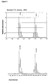
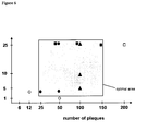
- the labeled A ⁇ standard is added in an amount in the same range as the effective amount of A ⁇ present in the source of beta amyloid. This amount may be determined in preliminary experiments, e.g. to find this amount for the quantification of A ⁇ in amyloid deposits, different numbers of plaques spiked with different amounts of A ⁇ standard have been analyzed (Fig. 6) in three successive experiments. Using 15 plaques or below, only the spiked 15 N-labeled A ⁇ standard could be detected while results obtained from 200 plaques were found to exceed the instrumental linear range. In a third experiment, hundred plaques were spiked with 5, 10, and 25 pmoles of 15 N-amyloid standard (Fig. 3).
- the A ⁇ labeled with a stable isotope used as a standard in the method of the present invention may be produced recombinantly.
- Methods for the preparation of expression constructs and for the recombinant production of polypeptides and proteins are known in the art and are summarized in Ausubel, Current Protocols in Molecular Biology / Protein science, Green Publishing Associates and Wiley Interscience, N.Y.(1994). Methods for the recombinant production of natural A ⁇ are described in the art, e.g. in EP0641861.
- the labeled A ⁇ may be produced by feeding recombinant E. coli with 15 N ammonium chloride.
- Other sources for stable isotopes comprise 13 C-labeled glucose and extracts of algae grown on 15 N-labeled substrates.
- the A ⁇ labeled with a stable isotope used as a standard in the method of the present invention may be produced by chemical synthesis.
- Methods for the synthetic production of polypeptides and proteins are known in the art, e.g. solid phase synthesis of polypeptides, and are summarized in Ausubel, Current Protocols in Protein science, Green Publishing Associates and Wiley Interscience, N.Y.(1994).
- the demonstration that amyloid fibrils formed in vitro using synthetic A ⁇ peptides are identical to those isolated from senile plaques (Kirschner, D. A.; Inouye, H.; Duffy, L. K.; Sinclair, A.; Lind, M.; Selkoe, D. J.
- total A ⁇ comprising labeled and unlabeled A ⁇ may be isolated from body fluid, preferably from serum or CSF, by protein chemical methods comprising immunoprecipitation and immunoaffinity chromatography.
- the beta amyloid in step (c) is isolated from body fluid by methods comprising protein chemistry and immunochemistry.
- the aggregated A ⁇ has to be dissolved.
- the aggregated A ⁇ is dissolved by methods comprising dissolution with solubilizing agents, and optionally mechanical solubilisation in the presence of the labeled A ⁇ standard.
- the solubilizing agents of the present invention may be all agents which have the capacity to dissolve aggregated A ⁇ , e.g. hexafluoropropanol, acid, e.g. formic acid, urea-SDS.
- the mechanical solubilisation may comprise sonication.
- the dissolution procedure of the aggregated A ⁇ takes place in the presence of the added labeled A ⁇ standard thereby guaranteeing equal losses of the A ⁇ standard and the A ⁇ to be quantified.
- the beta amyloid in step (c) is isolated from amyloid deposits by methods comprising dissolution with solubilizing agents and optionally by sonication.
- the isolated A ⁇ is subsequently prepared for analysis by mass spectrometry.
- the preparation for the analysis by mass spectrometry comprises methods which lead to an amelioration of the ionisation of the A ⁇ to be analysed.
- Methods leading to an amelioration of ionisation comprise fragmentation by methods comprising chemical fragmentation and enzymatic digestion, and chemical reactions with flight enhancers.
- Chemical reactions with flight enhancers comprise the charge derivatization of the peptides' free N-termini in order to enhance sensitivity and promote the formation of fragment ions by post-source decay MALDI mass spectrometry (J. Stults et al. (1993) Anal. Chem. 65, 1703-1708; B. Spengler et al. (1997) Int. J.
- the isolated A ⁇ may be directly reacted with flight enhancers for preparation for analysis by mass spectrometry. Alternatively, the isolated A ⁇ may be reacted with flight enhancers after fragmentation. Chemical fragmentation may be carried out by the use of cyanogen bromide or acid hydrolysis.
- the enzymatic digestion may be carried out with a protease selected from the group comprising endoproteinase Lys-C, trypsin, endoproteinase Glu-C, and pepsin.
- the isolated A ⁇ may be dried and redissolved in a buffer before digestion with a protease.
- the fragmentation of the dissolved A ⁇ leads to a better limit of detection in the mass spectrometrical analysis, e.g., cleavage of the amyloid peptide by endoproteinase Lys-C results in the detection of two of the three generated fragments with a 100 fold higher sensitivity in the mass spectrometer.
- the isolated beta amyloid in step (d) is prepared for analysis by mass spectrometry by methods comprising chemical reactions with flight enhancers, chemical fragmentation and enzymatic digestion.
- the dissolved and optionally fragmented A ⁇ may be desalted. Desalting of the sample (e.g., by ZipTip) may increase the sensitivity of the mass spectrometrical analysis by a factor of 10.
- the isolated and optionally fragmented A ⁇ is then analysed by mass spectrometry.
- Ionization techniques used for biological materials today comprise ESI (electrosprayionization) and MALDI (matrix assisted laser desorption ionization).
- the mass spectrometrical analysis used is a MALDI-TOF (time of flight) mass spectrometrical analysis.
- the spectrum of a MALDI-TOF-MS analysis consists primarily of the intact, singly charged molecule ions. Larger molecules, like proteins, may also yield multiply charged ions and, depending on their concentrations, singly charged multimers.
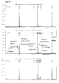
- the peak pattern of the natural 14 N A ⁇ is base-line separated from its artificial 15 N homologue in mass-spectrometry, thereby allowing the discrimination of the natural and the standard A ⁇ in the mass spectra.
- the amount of A ⁇ that was present in the source of aggregated A ⁇ may be determined by different approaches to the analysis of the mass spectra: a) by comparing the heights of the two dominant peaks of the 15 N-labeled amyloid standard and of the A ⁇ from the source of A ⁇ , b) by comparing the heights of all the peaks of the separated peak patterns, c) by comparing the areas under the two dominant peaks, and d) by comparing the sum of the areas under all the peaks of the two different peak patterns.
- the amount of A ⁇ present in the source of A ⁇ can then be calculated. In cases where the amount of A ⁇ that is present in a three-dimensional amyloid deposit, e.g. in a plaque, has to be determined a correction factor has to be included in the calculation.
- a method for the quantification of beta amyloid peptide comprising
- the present invention further provides the use of the method of the present invention for the determination of the A ⁇ content in amyloid deposits, e.g., plaques, obtained from tissue samples.
- a method of quantifying amyloid deposition before death is needed both as a diagnostic tool in mild or clinically confusing cases as well as in monitoring the effectiveness of therapies targeted at preventing A ⁇ deposition.
- a ⁇ content in body fluid containing soluble A ⁇ e.g. serum or CSF.
- body fluid containing soluble A ⁇ e.g. serum or CSF.
- amyloid hypothesis which includes soluble forms of A ⁇ among the neurotoxic species responsible for the pathology of AD.
- the method can be used for the quantification of amino terminal and carboxy terminal beta amyloid microheterogeneities. This is accomplished by spiking the source of beta amyloid with the specific form of beta amyloid the amount of which should be determined.
- AD brains blood vessels, dense plaques, diffuse plaques
- mass spectroscopy e.g. 1-40, 1-42, 1-39, 1-43
- individual protein compositions can be analyzed in these different structures.
- mice have an APPS SWe /PS2 background and were 20 months old (Richards J.G., Messer J., Goepfert F., Ozmen L., Brockhaus M., Bohrmann B., Malherbe P., Jacobsen H., Huber G.S., Bluethmann H., Kew J.N.C., Kemp J.A. Ouagazzal A.M. and Higgins G.A. (2001), Double transgenic mice overexpressing hAPP SWe and hPS2mut show age-dependent cognitive deficits and amyloid deposits in discrete brain regions, Soc. Neurosci. Abstr., Vol. 27, Program No. 546.7, 2001.).
- the human brain tissue employed in the performed experiments was obtained from a single donor after informed consent of patient was obtained.
- the post mortem tissue was collected from a single Alzheimer's patient only two hours after death and instantly frozen (-80°C) to conserve the native tissue structures.
- the patient had a ApoE4/E4 genetic background and the sample was taken from cortical areas.
- mice were killed either by cervical dislocation or by decapitation after anesthesia with halothane.
- the skullpan was opened with scissors, the brain was removed and divided into hemispheres before freezing in dry ice. All animal experiments were performed in full accordance with the guidelines issued by the responsible Swiss Veterinary Office.
- the brain tissue was cut into slices with a thickness of 10 ⁇ m with a kryostat microtome (LEICA CM3050 S). These slices were placed on a glass coverslip and were stored for further processing (e.g. staining) at -20°C.
- Brain tissue sections were rehydrated for 5 minutes in PBS.
- Cell nuclei were stained with Mayers Hematoxylin solution (Fluka, Hematoxylin Mayer-solution, 51275) by incubation in a glass cuvette for 10 minutes at room temperature. This solution stains the nuclei during 10 minutes. After washing and rinsing the probes with tap water for 5 minutes, they were treated with alkaline sodium chloride solution for 20 minutes.
- This solution has to be prepared first by adding NaOH (1/100 of the volume of NaCl; Sigma Diagnostics, sodium hydroxide solution, HT60-2) to the sodium chloride (Sigma Diagnostics, sodium chloride solution, HT60-1) solution.
- Congo Red like Thioflavine S, is a stain for beta-pleated sheet secondary structures of proteins, like those present in amyloid fibrils and can be used to visualize dense plaques that are characteristic for Alzheimer's Disease (Carter et al (1997), A Model for Structure-Dependent Binding of Congo Red to Alzheimer ⁇ -Amyloid Fibrils, Neurobiology of aging 19: 37-40).
- the Congo Red (CR) staining revealed in humans as in mice dense packed plaques, consisting of the A ⁇ peptide.
- human and transgenic mouse brain sections differed in several aspects when staining them with CR. Stained brain sections of 20 months old mice revealed a generally higher number of CR-reactive spots compared to an area of the same size in the neocortex of the human brain sample.
- the stained amyloid plaques in mice were distributed homogeneously over cortical areas, whereas in the used human case only few areas with few congophilic plaques were found. Human plaques were slightly bigger than mouse plaques from the employed transgenic model.
- Brain tissue slices were rehydrated for 5 minutes in phosphate buffered saline (PBS). After removal of the PBS, an aqueous Thioflavine S (Sigma, Thioflavine S, T1892) solution (1%) was applied for 3-5 minutes. After that, the probes were allowed to differentiate for 3-5 min in 70% ethanol (Merck, Ethanol, 100983). The last, mounting was done with glycerol-H 2 O (3:1) (FLUKA, Glycerol anhydrous, 49770).
- PBS phosphate buffered saline
- Brain tissue slices were rehydrated for 5 minutes in PBS. After the removal of the PBS, 1ml 70% acetone at 4°C (Fluka, Acetone, 00570) was applied to the brain tissue slices for approximately 80 seconds. The sections then were washed twice for 2 minutes with 1ml PBS and then, non specific binding sites were blocked for 15 minutes with 500 ⁇ l PBS containing 1 % BSA (Roche, Bovine Serum Albumine Fraction 5, 775869), 1 % Ovalbumine (Fluka, Albumine from hen egg white, 05440) and 1% Normal Goat Serum (BBInternational, Normal Goat Serum, NGS5). After finishing the blocking step, the samples were treated with 200 ⁇ l of the primary antibody (F.
- the PBS employed during immunostainig had to contain protease inhibitor (1 Tab/ 50ml, Roche Diagnostics, Protease inhibitor cocktail tablets, 1836145). The slides were air-dryed and stored until further usage at -20°C.
- a ⁇ depositions The most sensitive method to stain A ⁇ depositions is the labeling with specific monoclonal or polyclonal antibodies against the peptide. Staining characteristics of brain sections were also different according to the used antibody. Human and transgenic mouse brain samples did again not reveal the same appearance when stained with antibodies. Human and mouse plaques were, in addition to the previously described difference in size and number, different with regard to diffuse depositions of A ⁇ that are exclusively detected by immunohistochemistry.
- Brains from transgenic mice appear to be overloaded with the human A ⁇ peptide and hence, large brain regions of the employed transgenic mice were covered with large, diffuse patches of various sizes that were not found in human brain samples. The certainty that these depositions really were amyloid depositions, was obtained by analyzing such regions by Western blotting. The fact that large brain regions nearby this diffuse immunoreactivity were totally free from any fluorescent signal further confirmed this observation.
- the process for double labeling is the combination of Congo Red staining (or Thioflavine S staining) and the immunolabeling.
- Congo Red staining or Thioflavine S staining
- the immunolabeling After testing the two possible sequences of the staining methods, best results were obtained by Congo Red staining first, then followed by immunostaining.
- the staining procedures were sequentially performed as described above by performing the acetone treatment right after the rehydration with PBS.
- LDM laser dissection microscopy
- P.A.L.M. Robot-MicroBeam
- a pulsed nitrogen laser (337nm), which can be varied with respect to its focus (centering the laser beam into the focal plane of the objective) and with respect to the energy of the laser.
- the laser is adjusted for cold ablation using a maximum setting of 30 energy pulses per minute with a pulse duration of 3ns.
- a pulsed laser avoids the convection of excessive heat that might damage the biological specimen
- a computerized system which allows the control of a motorized xy-stage for automated positioning of the laser beam and the storage of up to 50 dissection positions.
- the sample can be observed and investigated through the microscope or, alternatively using a CCD camera, on a monitor.
- the laser dissection microscopy comprises the steps of cold ablation and laser pressure catapulting necessitating two different settings of the laser beam:
- the excision process is a restricted ablative photodecomposition process without heating (Srinivasan R., (1986) Ablation of polymers and biological tissue by ultraviolett lasers, Science 234: 559 - 565).
- the brain slices first were dried at 37°C for ⁇ 30 minutes.
- the microscope was set at a 20-fold magnification and the focus was set up at ⁇ 35 points and the energy at ⁇ 44 points (arbitrary units). After excision of the desired structure, the position is stored by the simple push of a button.
- the excised samples were collected.
- This step is technically mediated by a single laser pulse of increased energy (+20 points), which has its focal plane slightly below the sample (-2.2 points, ⁇ 1-2 ⁇ m).
- a single laser pulse provokes a rapid gas expansion that transports the excised material directly into the cap of a common microfuge tube, which is held and centered above the line of laser fire by a special LPC-collector device. By this procedure, the excised material was accumulated in the cap.
- the excized material cut out from a brain section does not represent the entire plaque, but only a disk out of the three-dimensionally shaped amyloid deposition (Fig. 8). This fact has then to be taken into account when determining the amount of A ⁇ present in a three-dimensionally shaped amyloid deposit.
- Pictures of an immunostained transgenic mouse brain section before and after excision of plaques by LDM are shown in Fig. 2A and Fig. 2B, respectively.
- the cap was filled with ⁇ 25 ⁇ l sample buffer (for mouse tissue: Invitrogen, NuPAGE SDS Sample Buffer 4x, NP0003) containing 8M urea (BioRad, Urea, 161-0731) or ⁇ 25 ⁇ l formic acid (for human tissue: Fluka, formic acid, 06440). Following collection of excised tissue, the liquid was spun down at 15'500 x g. The cap with the human samples was additionally washed out two times with 30 ⁇ l HCOOH and spun down. The human material was left in HCOOH overnight. Then it was vortexed, sonified 3 x for 3min in a bath with vortexing in between.
- sample buffer for mouse tissue: Invitrogen, NuPAGE SDS Sample Buffer 4x, NP0003
- 8M urea BioRad, Urea, 161-0731
- ⁇ 25 ⁇ l formic acid for human tissue: Fluka, formic acid, 06440
- the purified A ⁇ antibody rather than the BAP-2 ascites, was employed to stain A ⁇ -positive plaques.
- the digestion pattern of the A ⁇ peptides obtained from amyloid plaques was compared to the proteolytic fragments obtained from 15 N-labeled biosynthetic A ⁇ 1-42.
- 15 N-labeled biosynthetic A ⁇ 1-42 was produced according to Doebeli et al. (Doebeli et al., Biotechnology 13, 1995, 988 - 993) using 15 N-labeled amino acids.
- the A ⁇ standard was analyzed using the same work-up procedure as for plaque-derived A ⁇ .
- Relative quantification by mass spectrometry is most accurate when the amount of the spiked protein (e.g. A ⁇ ) standard equals the effective amount of protein present in plaques.
- a ⁇ the amount of the spiked protein
- different numbers of plaques spiked with different amounts of A ⁇ -standards have been analyzed in three successive experiments (Fig. 6). Using 15 plaques or below, only the spiked 15 N-labeled A ⁇ -standard could be detected while results obtained from 200 plaques were found to exceed the instrumental linear range.
- hundred plaques were spiked with 5, 10, and 25 pmoles of 15 N-amyloid standard (Fig. 3).
- the effective amount of (dissolvable) A ⁇ present in 100 plaques can be calculated and consequently the amount of A ⁇ in a single excised plaque can be determined.
- Each peptide i.e. an A ⁇ fragment
- has a characteristic distribution of naturally occurring isotopes i.e. 12C / 13C, 14N / 15N.
- the mass spectrometric analysis of a fragment therefore reveals a characteristic set of peaks which, by comparison to the theoretically calculated isotopic distribution, clearly identifies a certain peptide.
- the ratio of volume of the disc to the total volume of the sphere of a plaque (V disc : r 2 ⁇ h and V sphere : 4/3 ⁇ r 3 ) is the 'correction' factor to extrapolate the amount of A ⁇ determined in a disc to the entire amount in the sphere.
- Table 2 the 'correction' factors for different plaque sizes, assuming a slice-thickness of 10 ⁇ m, are listed: Correction factors for different plaques sizes radius of plaque 'correction' factor (V sphere / V disc ) 10 ⁇ m 1.34 30 ⁇ m 4 50 ⁇ m 6.67
- sample buffer Invitrogen, NuPAGE SDS Sample Buffer 4x, NP0003 containing 8M urea (BioRad, urea, 161-0731)
- sample buffer Invitrogen, NuPAGE SDS Sample Buffer 4x, NP0003 containing 8M urea (BioRad, urea, 161-0731)
- 2 ⁇ l of reducing agent Invitrogen, NuPAGE Sample reducing agent 10x, NP0004
- vortexed heated at 50°C for 10min, again vortexed and heated to 50°C for 10min and centrifuged at 11900 x g for 5min.
- the blotted membrane was heated for 3min with microwaves (900W) in boiling PBS to enhance the binding, and the unspecific binding sites were blocked with 50ml 5% skim milk (FLUKA, Skim Milk Powder, 70166) in PBS containing 0.05% Tween20 (Fluka, Tween20, 93773), PBS-T, for at least 30min.
- the WO-2 antibody (provided by the lab of K. Beyreuther, Center for Molecular Biology, University of Heidelberg; providing a strong signal without any background) diluted in PBS-T was added and incubated overnight at 4°C.
- the membrane was first washed with PBS-T, then soaked (3x10min) in fresh PBS-T and the bound antibody detected by 50ml of a horseradish peroxidase linked secondary antibody (Amersham, NA931/NA934) diluted 1:10'000 in PBS-T. The same washing and soaking cycle with PBS-T was applied to the membrane as before. Visualization was performed by ECL detection system (Amersham, ECL Western blotting detection reagents, RPN2106) according to the manufacturer's instruction by exposing the membrane to an autoradiography film (Kodak, BIOMAX ML, 243 012 06).
- the minimum number of mouse plaques for A ⁇ detection by Western blot was determined to be one single plaque (Fig. 7).
- the intensities of the detected bands varied from plaque to plaque.
- several plaques were loaded on a gel and compared with band intensities of standard concentrations of synthetic A ⁇ .
- the majority of mouse plaques contained amounts of A ⁇ that were within a range of 0.05-0.2ng.
- the radius of a dense mouse plaque (spherical) is between 10 and 30 ⁇ m. With an estimated amount of 0.05-0.2ng A ⁇ in an excised plaque (disc), a total amount of 0.07ng to 0.8ng A ⁇ in an entire mouse plaque (spherical) can be calculated.
- the amount of A ⁇ in a single human plaque was estimated. Whereas 9 human dense plaques contained more than 0.1ng A ⁇ , 4 human dense plaques were below this mark. Therefore, the amount of A ⁇ in a single human plaque is around 0.01ng.
- the radius of a human plaque is between 10 and 50 ⁇ m, the amount of A ⁇ between 0.01 and 0.05ng.
- the total amount of A ⁇ in an entire human plaque (spherical) can be calculated to be in the range between 0.013 and 0.33ng.
- the method comprising MS and isotope dilution detected a ⁇ 4-5 fold higher amount of A ⁇ present in a single mouse plaque. This is due to the fact that a huge proportion of the analyte is lost during the procedure. In the mass-spectrometry method the loss of 14 N and 15 N A ⁇ is assumed to occur at the same rate, and the proportion of these two isotope species reflects the situation at the beginning of the procedure. In Western blot (or ELISA) no internal standards can be used and the loss of analyte will be an unknown factor.
Landscapes
- Life Sciences & Earth Sciences (AREA)
- Health & Medical Sciences (AREA)
- Engineering & Computer Science (AREA)
- Molecular Biology (AREA)
- Physics & Mathematics (AREA)
- Urology & Nephrology (AREA)
- Immunology (AREA)
- Hematology (AREA)
- Bioinformatics & Computational Biology (AREA)
- Chemical & Material Sciences (AREA)
- Biomedical Technology (AREA)
- Bioinformatics & Cheminformatics (AREA)
- Food Science & Technology (AREA)
- Analytical Chemistry (AREA)
- Biotechnology (AREA)
- Microbiology (AREA)
- Proteomics, Peptides & Aminoacids (AREA)
- Biophysics (AREA)
- Medicinal Chemistry (AREA)
- Cell Biology (AREA)
- Biochemistry (AREA)
- General Health & Medical Sciences (AREA)
- General Physics & Mathematics (AREA)
- Pathology (AREA)
- Spectroscopy & Molecular Physics (AREA)
- Optics & Photonics (AREA)
- Other Investigation Or Analysis Of Materials By Electrical Means (AREA)
- Investigating Or Analysing Biological Materials (AREA)
Abstract
Description
| Comparison of histopathological observations in human and transgenic mouse brain samples | |||
| Human Brain Sample | Transgenic Mice | Conclusions | |
| Congo Red Staining (CR) | - occasionally areas with few CR-reactive spots - small number of CR-positive plaques - staining of vessels (microangiopathy) | - CR-positive plaques homogenously distributed over cortical areas - large number of CR-positive plaques; many are small - few are big like in the human case - there is size variation | - human plaques are bigger than transgenic mouse plaques - transgenic mouse brain sections contain more CR-positive plaques - plaques in humans seem to be less dense packed with Aβ fibrils - microangiopathy found in humans but not in the employed transgenic mouse model |
| Thioflavine S Staining | - number and size of marked spots comparable to CR-staining | - number and size of marked spots comparable to CR-staining | - conclusions as CR staining |
| antibody / double labeling | - large number of Aβ-depositions - diffuse depositions - double labeling shows two types of depositions: Dense and diffuse plaques with intermediate stages | - brain is overloaded with amyloid depositions - heterogeneous patches of diffuse Aβ depositions - clear difference between dense and diffuse depositions | - in humans, the transition from diffuse to dense plaques is a continuum, whereas in mice, basically two different types can be distinguished - small number of CR-positive spots in humans together with frequent diffuse Aβ depositions |
The kryostate was cleaned and plastic gloves were worn throughout the preparation of the brain samples to avoid contamination. The addition of goat serum to block unspecific binding sites in the brain section was omitted in the staining procedure. Additionally, the purified Aβ antibody rather than the BAP-2 ascites, (which was withdrawn with a syringe through the skin) was employed to stain Aβ-positive plaques.
| Correction factors for different plaques sizes | |
| radius of plaque | 'correction' factor (Vsphere / Vdisc) |
| 10µm | 1.34 |
| | 4 |
| 50µm | 6.67 |
Claims (19)
- A method for the quantification of beta amyloid peptide comprising(a) providing a source of beta amyloid(b) adding a defined amount of beta amyloid peptide labeled with a stable isotope to the source of (a)(c) isolating unlabeled and labeled beta amyloid(d) preparing the isolated beta amyloid for analysis by mass spectrometry(e) analysing the prepared beta amyloid peptide by mass spectrometry, and(f) determining the amount of beta amyloid that was present in the source of beta amyloid.
- The method according to claim 1, wherein the source of beta amyloid in step (a) are amyloid deposits obtained from a tissue sample.
- The method according to claim 2, wherein the amyloid deposits are obtained from a tissue sample by excision by laser dissection microscopy.
- The method according to claim 1, wherein the source of beta amyloid in step (a) is body fluid.
- The method according to claims 1 to 4, wherein the labeled beta amyloid added in step (b) is a beta amyloid which is recombinantly produced and labeled with at least one stable isotope.
- The method according to claims 1 to 5, wherein the labeled beta amyloid added in step (b) is a beta amyloid which is synthetically produced and labeled with at least one stable isotope.
- The method according to claims 1 to 6, wherein the beta amyloid added in step (b) is labeled with a stable isotope selected from the group comprising 15N, 13C, 18O and 2H.
- The method according to claims 1, 4 to 7, wherein the beta amyloid in step (c) is isolated from body fluid by methods comprising protein chemistry and immunochemistry.
- The method according to claims 2 to 3, 5 to 7, wherein the beta amyloid in step (c) is isolated from amyloid deposits by methods comprising dissolution with solubilizing agents.
- The method according to claims 1 to 9, wherein the isolated beta amyloid in step (d) is prepared for analysis by mass spectrometry by methods comprising chemical reactions with flight enhancers, chemical fragmentation and enzymatic digestion.
- The method according to claim 10, wherein the isolated beta amyloid in step (d) is prepared for analysis by mass spectrometry by enzymatic digestion with a protease selected from the group comprising endoproteinase Lys-C, trypsin, and endoproteinase Glu-C.
- The method according to claims 1 to 11, wherein the prepared beta amyloid in step (e) is desalted before analysis by mass spectrometry.
- The method according to claims 1 to 12, wherein the prepared beta amyloid in step (e) is analysed by MALDI-TOF mass spectrometry.
- A method for the quantification of beta amyloid peptide comprising(a) providing excised amyloid deposits from mammalian brain samples containing aggregated beta amyloid(b) adding a defined amount of beta amyloid peptide labeled with a stable isotope(c) dissolving the excised aggregated beta amyloid in the presence of the labeled beta amyloid(d) digesting the dissolved beta amyloid with a protease(e) analysing the digested beta amyloid peptide mixture by mass spectrometry, and(f) determining the amount of beta amyloid that was present in the source of aggregated beta amyloid with the help of the base-line separation resulting from the presence of the natural and the stable isotopes in the beta amyloid.
- Use of the methods according to claims 1 to 3, 5 to 7, 9 to 14, for the determination of the beta amyloid content in amyloid deposits containing aggregated beta amyloid obtained from tissue samples.
- Use of the methods according to claims 1, 4 to 8, 10 to 13, for the determination of the beta amyloid content in body fluid containing soluble beta amyloid.
- Use of the methods according to claims 1 to 14 for the quantification of amino terminal microheterogenous forms of beta amyloid.
- Use of the methods according to claims 1 to 14 for the quantification of carboxy terminal microheterogenous forms of beta amyloid.
- The method substantially as herein before described especially with reference to the foregoing Examples.
Priority Applications (1)
| Application Number | Priority Date | Filing Date | Title |
|---|---|---|---|
| EP03024739A EP1420254A3 (en) | 2002-11-06 | 2003-10-29 | Amyloid quantification |
Applications Claiming Priority (3)
| Application Number | Priority Date | Filing Date | Title |
|---|---|---|---|
| EP02024924 | 2002-11-06 | ||
| EP02024924 | 2002-11-06 | ||
| EP03024739A EP1420254A3 (en) | 2002-11-06 | 2003-10-29 | Amyloid quantification |
Publications (2)
| Publication Number | Publication Date |
|---|---|
| EP1420254A2 true EP1420254A2 (en) | 2004-05-19 |
| EP1420254A3 EP1420254A3 (en) | 2004-05-26 |
Family
ID=32178673
Family Applications (1)
| Application Number | Title | Priority Date | Filing Date |
|---|---|---|---|
| EP03024739A Withdrawn EP1420254A3 (en) | 2002-11-06 | 2003-10-29 | Amyloid quantification |
Country Status (1)
| Country | Link |
|---|---|
| EP (1) | EP1420254A3 (en) |
Cited By (5)
| Publication number | Priority date | Publication date | Assignee | Title |
|---|---|---|---|---|
| EP1739080A1 (en) * | 2005-06-30 | 2007-01-03 | F. Hoffmann-La Roche Ag | Ionization Modifier for Mass Spectrometry |
| CN102713622A (en) * | 2010-01-28 | 2012-10-03 | 松下电器产业株式会社 | β-amyloid assay method |
| EP2582380A4 (en) * | 2010-06-16 | 2014-03-05 | Abbvie Inc | Comparison of protein samples |
| US9405884B2 (en) | 2010-06-16 | 2016-08-02 | Abbvie Inc. | Methods and systems for the analysis of protein samples |
| US10481167B2 (en) | 2008-12-05 | 2019-11-19 | C2N Diagnostics | Methods for measuring concentrations of biomolecules |
Family Cites Families (1)
| Publication number | Priority date | Publication date | Assignee | Title |
|---|---|---|---|---|
| US20020119490A1 (en) * | 2000-12-26 | 2002-08-29 | Aebersold Ruedi H. | Methods for rapid and quantitative proteome analysis |
-
2003
- 2003-10-29 EP EP03024739A patent/EP1420254A3/en not_active Withdrawn
Cited By (7)
| Publication number | Priority date | Publication date | Assignee | Title |
|---|---|---|---|---|
| EP1739080A1 (en) * | 2005-06-30 | 2007-01-03 | F. Hoffmann-La Roche Ag | Ionization Modifier for Mass Spectrometry |
| US10481167B2 (en) | 2008-12-05 | 2019-11-19 | C2N Diagnostics | Methods for measuring concentrations of biomolecules |
| CN102713622A (en) * | 2010-01-28 | 2012-10-03 | 松下电器产业株式会社 | β-amyloid assay method |
| CN102713622B (en) * | 2010-01-28 | 2015-02-11 | 松下健康医疗器械株式会社 | β-amyloid assay method |
| EP2582380A4 (en) * | 2010-06-16 | 2014-03-05 | Abbvie Inc | Comparison of protein samples |
| US9170262B2 (en) | 2010-06-16 | 2015-10-27 | Abbvie, Inc. | Comparison of protein samples |
| US9405884B2 (en) | 2010-06-16 | 2016-08-02 | Abbvie Inc. | Methods and systems for the analysis of protein samples |
Also Published As
| Publication number | Publication date |
|---|---|
| EP1420254A3 (en) | 2004-05-26 |
Similar Documents
| Publication | Publication Date | Title |
|---|---|---|
| US20040096907A1 (en) | Quantification of beta amyloid | |
| US7674599B2 (en) | Methods of using antibodies to detect alpha-synuclein in fluid samples | |
| Güntert et al. | High sensitivity analysis of amyloid-beta peptide composition in amyloid deposits from human and PS2APP mouse brain | |
| CN110187120B (en) | Methods and compositions for the detection and treatment of preeclampsia | |
| D'Andrea et al. | Consistent immunohistochemical detection of intracellular β-amyloid42 in pyramidal neurons of Alzheimer's disease entorhinal cortex | |
| US9738709B2 (en) | Methylated peptides derived from tau protein and their antibodies for diagnosis and therapy of alzheimer's disease | |
| EP2336161A2 (en) | Amyloid-beta(1-42) oligomers, derivatives thereof, antibodies for the same, method for production and use thereof. | |
| US20210270847A1 (en) | Protein and peptide biomarkers for traumatic injury to the central nervous system | |
| JPWO2009051259A1 (en) | Biomarker for liver disease diagnosis | |
| Yu et al. | Evidence for the involvement of apoptosis-inducing factor–mediated caspase-independent neuronal death in Alzheimer disease | |
| WO2014195728A2 (en) | Methods and compositions relating to neurodegenerative diseases | |
| EP3633372A1 (en) | Biomarker for alzheimer's disease | |
| Rüfenacht et al. | Quantification of the Aβ peptide in Alzheimer's plaques by laser dissection microscopy combined with mass spectrometry | |
| US20150018223A1 (en) | Methods of diagnosing tau-associated neurodegenerative diseases | |
| Toyama et al. | Integrated spatial multi-omics study of postmortem brains of Alzheimer’s disease | |
| EP1420254A2 (en) | Amyloid quantification | |
| EP4363847B1 (en) | Integrated method for urinary prostate specific antigen n-glycosylation profiling by capillary electrophoresis | |
| Söderberg et al. | Analysis of single Alzheimer solid plaque cores by laser capture microscopy and nanoelectrospray/tandem mass spectrometry | |
| JP2007319127A (en) | Diagnostic method for Alzheimer's disease | |
| WO2018111099A1 (en) | Biomarkers and treatments for cerebral amyloid angiopathy (caa) | |
| JP7658575B2 (en) | Method for detecting misfolded proteins in biological samples | |
| EP2211181A1 (en) | Diagnostic method for transmissible spongiform encephalopathies | |
| JP6555973B2 (en) | How to check sweat gland morphology | |
| Maftei | Molecular characterization of neuroprotective β-amyloid interacting peptides and autoantibodies relevant to Alzheimer’s disease | |
| Schmidt et al. | Tissue Processing Prior to Protein Analysis and Amyloid-ß Quantitation |
Legal Events
| Date | Code | Title | Description |
|---|---|---|---|
| PUAI | Public reference made under article 153(3) epc to a published international application that has entered the european phase |
Free format text: ORIGINAL CODE: 0009012 |
|
| PUAL | Search report despatched |
Free format text: ORIGINAL CODE: 0009013 |
|
| AK | Designated contracting states |
Kind code of ref document: A2 Designated state(s): AT BE BG CH CY CZ DE DK EE ES FI FR GB GR HU IE IT LI LU MC NL PT RO SE SI SK TR |
|
| AX | Request for extension of the european patent |
Extension state: AL LT LV MK |
|
| AK | Designated contracting states |
Kind code of ref document: A3 Designated state(s): AT BE BG CH CY CZ DE DK EE ES FI FR GB GR HU IE IT LI LU MC NL PT RO SE SI SK TR |
|
| AX | Request for extension of the european patent |
Extension state: AL LT LV MK |
|
| 17P | Request for examination filed |
Effective date: 20041126 |
|
| AKX | Designation fees paid |
Designated state(s): CH DE FR GB LI |
|
| STAA | Information on the status of an ep patent application or granted ep patent |
Free format text: STATUS: THE APPLICATION IS DEEMED TO BE WITHDRAWN |
|
| 18D | Application deemed to be withdrawn |
Effective date: 20061121 |