EP1415002B1 - Focussed antibody technology - Google Patents
Focussed antibody technology Download PDFInfo
- Publication number
- EP1415002B1 EP1415002B1 EP02788114A EP02788114A EP1415002B1 EP 1415002 B1 EP1415002 B1 EP 1415002B1 EP 02788114 A EP02788114 A EP 02788114A EP 02788114 A EP02788114 A EP 02788114A EP 1415002 B1 EP1415002 B1 EP 1415002B1
- Authority
- EP
- European Patent Office
- Prior art keywords
- micro
- patient
- organism
- cells
- sequences
- Prior art date
- Legal status (The legal status is an assumption and is not a legal conclusion. Google has not performed a legal analysis and makes no representation as to the accuracy of the status listed.)
- Expired - Lifetime
Links
Images

Classifications
-
- G—PHYSICS
- G01—MEASURING; TESTING
- G01N—INVESTIGATING OR ANALYSING MATERIALS BY DETERMINING THEIR CHEMICAL OR PHYSICAL PROPERTIES
- G01N33/00—Investigating or analysing materials by specific methods not covered by groups G01N1/00 - G01N31/00
- G01N33/48—Biological material, e.g. blood, urine; Haemocytometers
- G01N33/50—Chemical analysis of biological material, e.g. blood, urine; Testing involving biospecific ligand binding methods; Immunological testing
- G01N33/68—Chemical analysis of biological material, e.g. blood, urine; Testing involving biospecific ligand binding methods; Immunological testing involving proteins, peptides or amino acids
- G01N33/6854—Immunoglobulins
- G01N33/6857—Antibody fragments
-
- C—CHEMISTRY; METALLURGY
- C07—ORGANIC CHEMISTRY
- C07K—PEPTIDES
- C07K16/00—Immunoglobulins [IG], e.g. monoclonal or polyclonal antibodies
- C07K16/005—Immunoglobulins [IG], e.g. monoclonal or polyclonal antibodies constructed by phage libraries
-
- G—PHYSICS
- G01—MEASURING; TESTING
- G01N—INVESTIGATING OR ANALYSING MATERIALS BY DETERMINING THEIR CHEMICAL OR PHYSICAL PROPERTIES
- G01N33/00—Investigating or analysing materials by specific methods not covered by groups G01N1/00 - G01N31/00
- G01N33/48—Biological material, e.g. blood, urine; Haemocytometers
- G01N33/50—Chemical analysis of biological material, e.g. blood, urine; Testing involving biospecific ligand binding methods; Immunological testing
- G01N33/5005—Chemical analysis of biological material, e.g. blood, urine; Testing involving biospecific ligand binding methods; Immunological testing involving human or animal cells
- G01N33/5008—Chemical analysis of biological material, e.g. blood, urine; Testing involving biospecific ligand binding methods; Immunological testing involving human or animal cells for testing or evaluating the effect of chemical or biological compounds, e.g. drugs, cosmetics
- G01N33/5044—Chemical analysis of biological material, e.g. blood, urine; Testing involving biospecific ligand binding methods; Immunological testing involving human or animal cells for testing or evaluating the effect of chemical or biological compounds, e.g. drugs, cosmetics involving specific cell types
- G01N33/5047—Cells of the immune system
- G01N33/5052—Cells of the immune system involving B-cells
-
- G—PHYSICS
- G01—MEASURING; TESTING
- G01N—INVESTIGATING OR ANALYSING MATERIALS BY DETERMINING THEIR CHEMICAL OR PHYSICAL PROPERTIES
- G01N33/00—Investigating or analysing materials by specific methods not covered by groups G01N1/00 - G01N31/00
- G01N33/48—Biological material, e.g. blood, urine; Haemocytometers
- G01N33/50—Chemical analysis of biological material, e.g. blood, urine; Testing involving biospecific ligand binding methods; Immunological testing
- G01N33/68—Chemical analysis of biological material, e.g. blood, urine; Testing involving biospecific ligand binding methods; Immunological testing involving proteins, peptides or amino acids
- G01N33/6803—General methods of protein analysis not limited to specific proteins or families of proteins
- G01N33/6818—Sequencing of polypeptides
-
- A—HUMAN NECESSITIES
- A61—MEDICAL OR VETERINARY SCIENCE; HYGIENE
- A61K—PREPARATIONS FOR MEDICAL, DENTAL OR TOILETRY PURPOSES
- A61K39/00—Medicinal preparations containing antigens or antibodies
- A61K2039/505—Medicinal preparations containing antigens or antibodies comprising antibodies
-
- C—CHEMISTRY; METALLURGY
- C07—ORGANIC CHEMISTRY
- C07K—PEPTIDES
- C07K2317/00—Immunoglobulins specific features
- C07K2317/50—Immunoglobulins specific features characterized by immunoglobulin fragments
- C07K2317/56—Immunoglobulins specific features characterized by immunoglobulin fragments variable (Fv) region, i.e. VH and/or VL
- C07K2317/565—Complementarity determining region [CDR]
Definitions
- the present invention is concerned with methods of identifying antibody specific for and which confer immunity against infection by micro-organisms such as bacteria, viruses and yeasts. Also provided are methods of manufacture of medicaments, methods of treatment of patients producing databases, databases produced by same, methods of preparing reports regarding same, methods of determining the efficacy of vaccines, databases produced by same, and methods of preparing reports regarding same.
- Antibodies capable of binding antigens displayed by the infectious agent are produced and bind to and allow killing of the microorganism through complement activation, recruitment of macrophage and through direct interaction with microbe itself.
- the therapeutic efficacy of antibodies capable of binding a given antigen varies and this is reflected in the fact that antibody production by the immune system matures during the course of an infection and becomes focussed in the case of a patient successfully fighting off an infection.
- the antibody response is elicited by the B cell repertoire where individual B cells each produce structurally diverse antibody molecules.
- the actual size of this B cell/antibody repertoire is unknown, but it is estimated that the random clonal frequency of reactivity for a given antigen may be as high as 1 in 100,000 in cultured B cells (Nobrega A et al., Eur J Immunol. 1998 Apr;28(4):1204-15; PMID: 9565360).
- antibodies capable of binding the pathogen are selected for by changes in the B cell population resulting in key antibodies being produced in large numbers. The mechanisms for these changes include clonal expansion, isotype switching, and somatic mutation of immunoglobulin variable regions.
- B cells responsible for generating antibodies which are able to bind pathogen multiply, thus skewing the B cell repertoire and changing the proportions of B cells.
- the frequency of circulatory B cells producing pathogen-reactive antibodies was as high as 1 in 1,403 (no pathogen reactivity could be found in non-infected individuals; Migot F et al., Clin Exp Immunol. 1995 Dec;102(3):529-34; PMID: 8536368).
- vaccination is achieved by isolating and purifying an immunogen from the infecting pathogen and using it as the basis, either in a modified or unmodified form, for a vaccine which is administered to patients to stimulate antibody production.
- Passive immunotherapy can be achieved by the administration of antibody to a patient and is particularly effective in treating immunocompromised patients who are unable to respond to vaccination, and to patients who need immediate therapy and cannot wait for vaccination to take effect.
- Passive immunotherapy is typically achieved by administering to a patient a monoclonal antibody specific to an immunogen produced by a pathogen.
- an immunogen must first be isolated and purified, administered to test animals, and cells producing antibody specific against the immunogen cloned. The range of antibodies produced by the clones can then be tested for their therapeutic efficacy and a single monoclonal antibody selected which is then administered to patients to effect passive immunotherapy.
- epitopes are only produced by pathogens in vivo and are not synthesised in vitro.
- Various host-specific expression systems are well known, for example in gonorrhea (causative organism Neisseria gonorrhoeae) specific antigens are expressed upon infection of a host - such antigens fall into the general class of cryptantigens.
- highly labile antigens also pose problems to prior art systems since during use they simply degrade and the epitope they display is lost With this large range of epitopes/antigens whose identification and/or in vitro use is of great difficulty or impossible the present invention provides a solution and allows the production and isolation of antibodies specific against them.
- the prior art typically has to attempt to achieve an equivalent of affinity maturation of antibodies by first synthesising a set of candidate antibodies specific to an antigen, testing them for their binding characteristics and then modifying the sequences of the candidates in order to optimise the binding.
- affinity maturation i.e. optimising antibody binding
- the antibodies of the present invention are obtained from patients who have either been infected by a pathogen displaying the antigen or who have been vaccinated with an antigen, they have by their very nature and definition already undergone affinity maturation, as is most clearly demonstrated by the sequences of their CDR3 regions.
- the present invention seeks to overcome the prior art disadvantages.
- a method for identifying candidate sequences of at least the CDR3 region of antibodies specific against at least one antigen produced by a micro-organism during an infection or against a vaccine comprising the steps of:
- Examples of patients used as a source of B cells in such a method are humans and other mammals such as mice, rabbits, rats, baboons, monkeys and apes.
- step (i) may comprise the steps of:
- sequences occurring in total at a frequency of at least 1 percent in the set of sequences determined at step (i) may be the candidate CDR3 sequences or a sequence having at least 80% homology, for example 85, 90, 95, 96, 97, 98 or 99% homology therewith. Sequence homology is as determined using the BLAST2 program (Tatusova TA et al., FEMS Microbiol Lett. 1999 May 15;174(2):247-50; PMID: 10339815) at the National Center for Biotechnology Information, USA (www.ncbi.nlm.nih.gov) with default parameters.
- the sequences occurring in total at a frequency of at least 1 percent in the set of sequences determined at step (i) may be the candidate CDR3 sequences or a sequence having 1 or 2 amino acid changes therefrom.
- a first sequence may occur at a frequency of 0.7 percent, and first, second, third and fourth sequences each having a single amino acid change therefrom each occur at a frequency of 0.1% - the total occurrence is therefore 1.1% and the dominant antibody sequence (occurring at a frequency of 0.5%) is therefore a candidate CDR3 sequence.
- the correlation step (ii) may also correlate the occurrence of the candidate sequences against their occurrence in patients who have not been infected with the micro-organism or administered the vaccine, sequences only being determined to be candidate sequences if they or a sequence having one or two amino acid changes from them occur in total at a frequency of less than 1 percent in the non-infected/vaccinated patients.
- the at least one antigen produced by the micro-organism may of course be an immunogen.
- the present invention provides unique opportunities for understanding antibody responses to infection which were not previously available, and provides novel diagnostic and therapeutic opportunities.
- the methods of the present invention bypass the need to isolate an antigen from the micro-organism, and instead determine the identity of antibodies specific against one or more antigens produced by the micro-organism
- the methods of the present invention allow the identification of therapeutically effective antibodies and can allow the identification of the most important parts of antibody sequences. This is particularly evident when the B cells of a given patient are sampled at different time points during the course of an infection by a micro-organism - as the patient's immune system becomes focussed on producing therapeutically effective antibodies against the infecting micro-organism, a focussing of antibody sequences is observed, and the development of variable and non-variable regions within the CDR parts of the VH and VL sequences is observed.
- the methods of the present invention can allow the identification of antibodies best suited to the treatment of infection in particular groups of patients, such as age, sex and racial groups. Similarly by comparing the antibody sequences of patients who have recovered from infection with those that have not recovered from infection, ineffective antibody sequences (which might be produced by both sets of patients) can be identified.
- the B cells isolated from the at least one patient can be peripheral B-cell lymphocytes (PBLs), and can be isolated from a blood sample from the at least one patient.
- PBLs peripheral B-cell lymphocytes
- B-cells can also be isolated from other sources and the present invention extends to their use.
- B-cells can be isolated from spleen.
- Antibody CDR sequences are determined as detailed in the "Experiments” section using standard techniques and definitions of their start and end.
- the antibody sequences are determined from patients with specified infections. These sequences can be derived from B cell immunoglobulin mRNA and hence reflect expressed antibodies and repertoires therein.
- the sequences determined according to the present invention need not be the sequences of whole antibody molecules - they comprise at least the VH and VL sequences which specify the regions responsible for antigen binding.
- putative amino acid sequences for the VH and VL regions can be determined using standard algorithms and software packages (e.g. see www.mrc-Imb.cam.ac.uk/pubseq/, the Staden package and Gap4 programs). These can be further characterised to determine the CDR (Complementarity Determining Region) parts of the VH and VL sequences, particularly CDR1, CDR2 and CDR3. Methods for determining the putative amino acid sequences and identifying CDR regions are well known and detailed below.
- B cells can be used which have been isolated from the at least one patient at a plurality of time points during infection of the at least one patient by the micro-organism (or post-vaccination), correlation step (ii) correlating the time point during infection of the at least one patient by the micro-organism at which the B cells are isolated.
- B cells can be used which have been isolated from at least two patients, at least one of whom has recovered from infection by the micro-organism, and at least one of whom has not recovered from infection by the micro-organism, correlation step (ii) correlating the recovery of the at least two patients from infection by the micro-organism.
- B cells can be used which have been isolated from at least two patients, the patients being infected by different micro-organisms producing the at least one antigen, correlation step (ii) correlating the sequenced at least the VH and VL coding regions of the B cells to identify a set of candidate sequences for antibodies, each of which is specific against at least one shared antigen produced by the different micro-organisms or is specific against different antigens produced by the different micro-organisms.
- the sequencing of the VH and VL regions can also be used to identify the surrounding antibody framework, and this can be used to determine the antibody isotype. This can then be used in the correlation step to determine whether specific antibody isotypes are particularly useful.
- VH or VL sequences demonstrate skewing of the antibody repertoire over the course of infection, thus revealing the identity of matured, immunity-conferring VH and VL sequences. This has typically been done using 5,000 to 15,000 sequences for each VH or VL. VH and VL repertoires from healthy individuals can also be used for comparison purposes.
- therapy any treatment which is designed to cure, alleviate, remove or lessen the symptoms of, or prevent or reduce the possibility of contracting any disorder or malfunction of the human or animal body
- the invention is suitable for identifying antibody sequence useful in treating the above infections, as well as any other infections such as viral infections where antibody has been shown to be important (including but not limited to HIV, influenza virus, hepatitis viruses A, B, C, and D, haemorrhagic viruses such as Ebola, as well as Dengue and Yellow Fever viruses) as well as parasitic infections such as malaria (P. falciparum).
- HIV HIV
- influenza virus hepatitis viruses A, B, C, and D
- haemorrhagic viruses such as Ebola
- Dengue and Yellow Fever viruses dengue and Yellow Fever viruses
- parasitic infections such as malaria (P. falciparum).
- the present invention is useful in a wide range of applications, e.g. antibody therapy, and vaccine studies.
- the present invention can be used to determine the sequences of antibodies conferring immunity by looking for over-represented VH and VL sequences in patients who have overcome infection. These protective antibodies can be re-synthesised at the genetic level, over-expressed in E. coli (or other expression systems) and purified. The resultant purified recombinant antibody can then be administered to patients as a passive immunotherapy. Antibodies can also be ordered from commercial suppliers such as Operon Technologies Inc., USA (www.operon.com) by simply supplying them with the sequence of the antibody to be manufactured.
- Therapeutically useful sequences can be identified in a number of ways which can be used in the correlation step (ii):
- Vaccination protects against infection by priming the immune system with pathogen-derived antigen(s). Vaccination is effected by a single or repeated exposures to the pathogen-derived antigen(s) and allows antibody maturation and B cell clonal expansion without the deleterious effects of the full-blown infectious process. T cell involvement is also of great importance in effecting vaccination of patients.
- the present invention can be used to monitor the immunisation process with experimental vaccines. Subjects are given the experimental vaccine and VH and VL sequences are amplified from the patient and the antibody repertoire analysed as described above. Qualitative and quantitative assessment of the vaccination process is possible:
- Also provided to the present invention is a method of manufacture of a medicament for the treatment of an infection by a micro-organism which produces at least one antigen, comprising the steps of:
- Such a medicament may of course incorporate a pharmaceutically acceptable carrier, diluent or excipient (Remington's Pharmaceutical Sciences and US Pharmacopoeia, 1984, Mack Publishing Company, Easton, PA, USA; United States Pharmacopoeia, ISBN: 1889788031).
- a pharmaceutically acceptable carrier diluent or excipient
- Also provided according to the present invention is a method of treatment of an infection of a patient by a micro-organism which produces at least one antigen, comprising the steps of:
- Also provided according to the present invention is a method of producing a database which identifies candidate sequences for antibodies specific against at least one antigen produced by a micro-organism, comprising the steps of:
- Also provided according to the present invention is a method for determining the efficacy of a vaccine, comprising the steps of:
- step (i) above may comprise the steps of:
- Correlation step (ii) may comprise correlating said sequenced at least a CDR3 region of the VH and VL coding regions of said B cells with sequenced at least a CDR3 region of the VH and VL coding regions of B cells isolated from at least one patient who has been infected with a micro-organism against which vaccination with said vaccine is intended to stimulate a protective immune response.
- step (ii) additional information may be used in the correlation of step (ii), including:
- the antibody sequences determined from patients who have been administered the vaccine can be compared with antibody sequences isolated from patients who have been infected with the micro-organism against which the vaccine is intended to stimulate a protective immune response in patients.
- the comparison can be made using antibody sequences determined from patients who have recovered from infection by the micro-organism.
- the method may determine the efficacy of the vaccine in stimulating a protective immune response against the micro-organism against which vaccination with the vaccine is intended to stimulate a protective immune response.
- Also provided according to the present invention is a method of vaccinating a patient against infection by a micro-organism, comprising the steps of:
- Also provided according to the present invention is a method of generating a report which identifies the efficacy of a vaccine, comprising the steps of:
- the antibody may be a whole antibody or an antigen binding fragment thereof and may in general belong to any immunoglobulin class. Thus, for example, it may be an IgM or an IgG antibody.
- the antibody or fragment may be of animal, for example, mammalian origin and may be for example of murine, rat, sheep or human origin. It may be a natural antibody or a fragment thereof, or, if desired, a recombinant antibody fragment, i.e. an antibody or antibody fragment which has been produced using recombinant DNA techniques.
- Particular recombinant antibodies or antibody fragments include, (1) those having an antigen binding site at least part of which is derived from a different antibody, for example those in which the hypervariable or complementarity determining regions of one antibody have been grafted into the variable framework regions of a second, different antibody (as described in, for example, EP 239400); (2) recombinant antibodies or fragments wherein non-Fv sequences have been substituted by non-Fv sequences from other, different antibodies (as described in, for example, EP 171496, EP 173494 and EP 194276); or (3) recombinant antibodies or fragments possessing substantially the structure of a natural immunoglobulin but wherein the hinge region has a different number of cysteine residues from that found in the natural immunoglobulin but wherein one or more cysteine residues in a surface pocket of the recombinant antibody or fragment is in the place of another amino acid residue present in the natural immunoglobulin (as described in, for example, WO 89/01782 and
- the antibody or antibody fragment may be of polyclonal or monoclonal origin. It may be specific for at least one epitope.
- Antigen binding antibody fragments include, for example, fragments derived by proteolytic cleavage of a whole antibody, such as F(ab')2, Fab' or Fab fragments, or fragments obtained by recombinant DNA techniques, for example Fv fragments (as described, for example, in WO 89/02465).
- the antibodies according to the invention may be prepared using well-known immunological techniques.
- any suitable host may be injected with the protein and the blood collected to yield the desired polyclonal antibody after appropriate purification and/or concentration (for example by affinity chromatography using the immobilised protein as the affinity medium).
- splenocytes or lymphocytes may be recovered from the protein-injected host and immortalised using for example the method of Kohler et al. (1976, Eur. J. Immunol., 6 : 511), the resulting cells being segregated to obtain a single genetic line producing monoclonal antibodies.
- Antibody fragments may be produced using conventional techniques, for example, by enzymatic digestion with pepsin or papain. Where it is desired to produce recombinant antibodies according to the invention these may be produced using, for example, the methods described in EP 171469, EP 173494, EP 194276 and EP 239400.
- Antibodies according to the invention may be labelled with a detectable label or may be conjugated with an effector molecule, for example a drug eg. an antibacterial agent or a toxin or an enzyme, using conventional procedures and the invention extends to such labelled antibodies or antibody conjugates.
- an effector molecule for example a drug eg. an antibacterial agent or a toxin or an enzyme
- antibody in its various grammatical forms is used herein to refer to immunoglobulin molecules and immunologically active portions of immunoglobulin molecules, i.e., molecules that contain an antibody combining site or paratope. Such molecules are also referred to as "antigen binding fragments" of immunoglobulin molecules.
- Illustrative antibody molecules are intact immunoglobulin molecules, substantially intact immunoglobulin molecules and those portions of an immunoglobulin molecule that contain the paratope, including those portions known in the art as Fab, Fab', F(ab')2, scFv and F(v).
- antibody combining site refers to that structural portion of an antibody molecule comprised of a heavy and light chain variable and hypervariable regions that specifically binds (immunoreacts with) antigen.
- human patients with a given microbial infection(s) were selected as donors of immunised B cells.
- the criteria for selection were:
- PBLs Peripheral B-cell lymphocytes
- PBS Peripheral B-cell lymphocytes
- RNA was prepared from PBLs using the QuickPrep mRNA purification kit (Pharmacia) exactly according to the manufacturers instructions.
- the isolated mRNA was used to prepare cDNA via a reverse transcriptase reaction (Promega cDNA synthesis kit). For this 2 ⁇ g of mRNA was re-suspended in 16 ⁇ L nuclease-free water and heated to 65 °C for 3 minutes (to denature secondary structure) and then immediately chilled on ice for 1 minute.
- the mRNA was then added to the following cocktail: 8 ⁇ L 25 mM MgCl 2 , 4 ⁇ L dNTP mix (10 mM with respect to each ribonucleotide triphosphate), 1 ⁇ L RNAsin 40 u ⁇ L -1 stock solution, 1.2 ⁇ L AMV reverse transcriptase (25 u ⁇ L -1 stock solution), 6 ⁇ L cDNA 10 pmol ⁇ L 1 primer (see Figure 1 - cDNA synthesis). The mixture was incubated at 42 °C for 1 hour and then incubated at 100 °C for 3 minutes to stop the reaction.
- DNA coding for antigen binding regions was then amplified by PCR using the cDNA prepared from patients PBLs.
- the cDNA was used in the following PCR reaction to produce either heavy chain or light chain-derived antibody variable region DNA (see Figure 1): 2.5 ⁇ L cDNA, 33 ⁇ L water, 4 ⁇ L dNTP mix (25 mM with respect to each deoxynucleotide triphosphate), 5 ⁇ L Taq reaction buffer (Perkin Elmer), 2.5 ⁇ L of an equimolar primer mix (final concentration of 20 pmol with respect to each primer) and 0.5 ⁇ L Taq DNA polymerase ( 1 u ⁇ L -1 , Perkin Elmer Corp).
- PCR reaction conditions were 94 °C for 1 minute, 57 °C for 1 minute and 72 °C for 2 minutes for a total of 30 cycles, with an extended denaturation (94°C for 5 minutes) prior to cycle 1 and an additional extension step after the end of cycle 30 (72 °C for 10 minutes).
- PCR was performed using a Perkin Elmer 9700 GeneAmp PCR machine.
- the PCR resulted in two fragment types -derived from VH and VL regions respectively, depending on the PCR primer sets used (for details of primers sets for antibody genes and a PCR schematic see Table 1 and Figure 1, respectively).
- VH and VL gene fragments were cloned into cloning vector pGEM-T easy (Promega Corporation) to facilitate DNA sequencing.
- cloning vector pGEM-T easy Promega Corporation
- 3 ⁇ g PCR product was prepared for restriction using QIAquick PCR purification spin columns (Qiagen, UK) according to the manufacturers instructions. DNA was eluted from the spin column in 40 ⁇ L buffer EB. Purified PCR product (2 ⁇ L) was mixed with 1 ⁇ L pGEM-T easy vector, 6 ⁇ L water and 1 ⁇ L DNA ligase and the mixture ligated for 1 h at room temperature. Ligations were then transformed into electrocompetent E.
- coli TG1 cells (Stratagene) by electroporation, and plated out onto agar plates containing Ampicillin 100 ⁇ g ml -1 IPTG (100 ⁇ M) and X-gal (0.006 % w/v). Colonies were allowed to grow overnight at 37 °C and then stored at 4 °C. Recombinant colonies are identified as white colonies on this media.
- DNA was first prepared from VH and VL recombinant E. coli clones. For this, individual colonies were each transferred to 1.2 ml LB broth supplemented with Ampicillin 100 ⁇ g ml -1 using a 96 well plate format. Cultures were the grown at 37°C for 24 hours. Bacterial cells were harvested by centrifugation at 4,000 x g, 30 minutes and the supernatants discarded. Plasmid DNA was prepared using Wizard SV 96 plasmid purification kits (Promega Corporation) essentially following the manufacturer's instructions. Yields of plasmid DNA were typically in the order of 5 ⁇ g per 1.2 ml starter culture.
- DNA sequencing reactions were performed using the DYEnamic ET dye terminator cycle sequencing kit (Amersham Pharmacia Biotech). Purified plasmid DNA (0.5 ⁇ g) was mixed with 8 ⁇ L DYEnamic ET terminator reagent premix and 1 ⁇ L M13 forward or reverse primer (5 ⁇ M) in a total reaction volume of 20 ⁇ L. Thermal cycling was then performed using a GeneAmp PCR system 9700 (Perkin Elmer) with the following parameters: 95 °C, 20 seconds; 50 °C, 15 seconds; 60 °C, 1 minute; 30 cycles). Reactions were performed using 96 well format non-skirted ELISA plates (AB Gene). Unincorporated dye terminator were removed using precipitation.
- ethanol samples were mixed with 2 ⁇ L 7.5 M ammonium acetate and 55 ⁇ L of 100 % ethanol and centrifuged at 3000 g for 30 minutes. DNA pellets were washed with 70 % ethanol and re-suspended in 20 ⁇ L loading solution. Reactions were sequenced using a MegaBACE 1000 DNA sequencer (Amersham Pharmacia Biotech) following the manufacturers instructions (2 kV injection voltage for 30 s with electrophoresis at 6 kV for 200 minutes). Chromatograms are exported using the .scf file format for finishing and archiving.
- DNA sequences of VH and VL were determined in both forward and reverse strands (using M13 forward and reverse primers respectively) and compared in order to highlight discrepancies and maintain a high degree of accuracy. This was done using the Staden suite of programs (Staden, R. (1996) The Staden Sequence Analysis Package. Molecular Biotechnology 5, 233-241). First, sequence .scf files were entered into the PREGAP program where vector sequence was stripped off and the quality of the sequence was assessed. Poor quality sequences where the DNA sequence was ambiguous were rejected and re-sequenced. Forward and reverse strands were matched using the GAP program to highlight and resolve areas containing sequencing artefacts. The orientation of the VH or VL sequence was noted and reverse complemented where necessary to produce only forward reading frame orientations for translation. VH and VL gene sequences were then translated into amino acid sequences. Each sequence was given a unique identifier (name).
- the database was constructed using SQL Server Database software (Microsoft). This allowed the database to be queried using SQL and allowed CDR1, CDR2 and CDR3 sequences to be extracted from any range of database VH or VL sequences in FASTA format Extracted CDRs were then subject to multiple alignment using CLUSTAL X in such a way so that identical or very similar CDRs are grouped together in blocks (Thompson, J.D., Higgins, D.G. and Gibson, T.J. (1994) CLUSTAL W: improving the sensitivity of progressive multiple sequence alignment through sequence weighting, positions-specific gap penalties and weight matrix choice. Nucleic Acids Research, 22:4673-4680).
- VH and VL sequences were identified and their gene sequences resynthesised using synthetic oligonucleotides. This was important as it also allowed the human-derived gene sequence to be codon-optimised for E. coli in order to improve protein expression.
- Dominant VH and VL sequences were spliced together using a spacer (linker) sequence ( Figure 2). This gene cassette, termed a scFv, was resynthesised to include terminal Nde I and Not I restriction sites for cloning into expression vector pET29b (Novagen).
- ScFv DNA was cut with Nde I (cuts in VH) or Not I (cuts in VL) using the following reaction: 40 ⁇ L DNA (4 ⁇ g), 10 ⁇ L of Restriction enzyme buffer D (Promega), 47 ⁇ L water and 2 ⁇ L Nde I and Not I enzyme (10 u ⁇ L -1 ; Promega) with digestion for 4 h at 37 °C. DNA was then fractionated on 0.7% agarose TAE gels and the digested DNA excised from the gel and purified from the agarose slice using the Geneclean II kit (Bio 101) exactly according to the manufacturers instruction. pET29b vector DNA was cut with Nde I and Not I as described above.
- 1 ⁇ g vector was mixed with 1 ⁇ g restricted VL DNA resuspended in 8.5 ⁇ L water and ligated by addition of 1 ⁇ L 10 x ligation buffer (Boehringer Mannheim) and 0.5 ⁇ L DNA ligase (3 u ⁇ L -1 ; Boehringer Mannheim), followed by ligation overnight at 14 °C.
- the entire ligation was transformed into E. coli TG2 cells (Stratagene, UK) by electroporation. Transformants were then plated out onto agar plates containing Tetracycline (25 ⁇ g ml -1 - selective for recA ::Tn10 on the chromosome of TG2) and Kanamycin (50 ⁇ g ml -1 - selective for pET29). Recombinant plasmid DNA prepared from individual clones was checked for scFv sequence by digestion with Nde I and Not I as described above.
- the lysate was centrifuged in an Oakridge tube (24 300 x g, 4 °C, 30 minutes). The pellet was resuspended in 20 ml of ice cold lysis buffer on ice using a Silverson lab blender. The lysate was split into 8 x Oakridge tubes and each aliquot was diluted to 30 ml (total 120 ml) in ice-cold Lysis Buffer. Inclusion bodies were pelleted (24 300 x g , 4 °C, 30 minutes) and the supernatant was discarded.
- each of the 8 pellets was resuspended in 30 ml of lysis buffer, and the inclusion bodies were harvested by centrifugation again (24 300 x g, 4 °C, 30 minutes). This wash/centrifugation step was performed 5 x times in total to clean the inclusion body fraction. Each pellet was resuspended in 15 ml of ice-cold water and the inclusion bodies were stored at -20 °C.
- NLS N-lauryl sarcosine
- CuCl 2 was added to a concentration of 100 ⁇ M. This serves as a catalyst for oxidation.
- the refolding reaction was transferred to 4 °C and stirred vigorously for 2 days to promote aeration.
- the refolding reaction was vacuum filtered through a 0.44 ⁇ m vacucup bottle top filter unit (90 mm diameter, Gellman Sciences) and the filtrate transferred to a Pellicon Labscale TFF system fitted with PLGC10 membrane unit (Millipore). The reaction was concentrated to 25 ml using tangential flow, discarding the permeate (the scFv is localised to the retentate).
- the solution was diafiltered against 40 x turn-over volumes (1 L) of 10 mM ammonium acetate (AAT) pH 9.0. Finally, the volume of the antibody was diluted to 50 ml using 10 mM AAT pH 9.0. The buffer exchanged antibody was stored for 2 hours at 4 °C. The typical protein content was 1 - 2 mg ml -1 with a yield of up to 50 mg per litre.
- scFv purification a 10 ml bed volume of Ni NTA superflow agarose Qiagen was prepared according to the manufacturers instructions using a 10 ml glass column (Sigma) without flow adaptors. The column was equilibrated with 50 ml Buffer B (6M urea, 0.1 M NaH 2 PO 4 , 10 mM Tris HCl pH 8.0). Refolded scFv (as described above) was diluted 1/5 in Buffer B and applied directly to the column at ml min -1 . 50 ml buffer B was applied to the column (flow rate 5 ml min -1 ) followed by 50 ml Buffer C (same composition as buffer B but pH 6.3). The purified scFv was eluted from the column by slowly applying Buffer B supplemented with 250 mM high grade imidazole until the A 280 ⁇ 0.05.
- Buffer B Buffer B
- VH variable heavy chain
- CDR3 third complementarity determining region
- M3 is another library constructed from the same patient as in M2 but taken ten days later than the M2 sample.
- P1 is a library from a Cystic Fibrosis (CF) patient colonized long term with Pseudomonas aeruginosa.
- C1 is a library from a patient infected with systemic Candida albicans infection.
- Each library has a small number of dominant antibodies.
- the most common antibody in each library have a frequency of 10% (SEQ ID NO: 27) (RNNWPPT), 41% (SEQ ID NO: 28) (RSGSHYDAFNI), 32% (SEQ ID NO: 29) (VTGCFDH), 7% (SEQ ID NO: 30) (LHGFGHGFRI) and 30% (SEQ ID NO: 31) (GEIFDY) (M1, M2, M3, P1, C1 respectively).
- M1 has two major VH chains accounting for 18% of the library.
- M2 has four dominant VH chains that account for 80% of the library.
- M3 has four VH chains making-up 79% of the library.
- P1 has 3 VH chains accounting for 18% of the library.
- the C1 library has six VH chains which together account for 72% of the clones in the library. Various of these VH chains have been tested in mouse models and shown to have therapeutic activity against the relevant agent.
- M2 and M3 library are comparisons of the VH repertoire over a period of time for an individual with an MRSA septicaemia.
- the most dominant VH chain (SEQ ID NO: 28) (RSGSHYDAFNI) is at a frequency of 41 % at the end of the infectious period (M2); this has dropped to 22% after 10 days (M3).
- Other common antibodies show similar drops 19% to 7% (SEQ ID NO: 32) (LLGGSSGRYMDV), 10% to 2% (SEQ ID NO: 33) (HRGGPQCHNLNCYTLDF) and 7% to 3% (SEQ ID NO: 34) (EAESYAERIGFDY).
- Other antibodies show a rise in levels over the same time period.
- VH chains that show a reduction in the post-infectious period would be considered potential therapeutic agents.
- Those antibodies that show a steep rise following clearing of the infection maybe of less importance as therapeutics.
- Analysis of the VH chain expression over the course of an infection and beyond is important in helping to identify neutralizing antibodies.
- M1 and M2/M3 are libraries from two different patients with the same infection. So far it has been possible to identify six different CDR3 sequences that are present in both MRSA patients.
- One such antibody (SEQ ID NO: 33) (LLGGSSGRYMDV) makes up 4% of the repertoire from the M1 library and 19% and 7% of the M2 and M3 libraries respectively. This antibody is identical along the whole length of the VH region in both patients.
- VH chains are also found to be present in both patients' libraries and have an identical sequence along the rest of the VH region (SEQ ID NOs: 35-38) (YDVVVTGKYAFDI, RRYHDLTTNNWFDP, NVGSGSYYTGGHWFDP, HRGGPQCHNLNCYTLDF).
- the fifth CDR3 shared by both patients (SEQ ID NO: 39) (ARRYGTSNYCFDH) is found at a frequency of 2% and 3% in libraries M1 and M2 respectively.
- the VH chains with this CDR3 are identical along most of the VH chain except for one important region, the second complementarity determining region (CDR2).
- the CDR2 is another region of the mature antibody that comes into contact with the antigen and plays an important role in its activity. Identification of antibodies with the same CDR3 but different sequences outside of this region will yield antibodies with similar target antigens although other properties may vary.
- results of the experiments are given in Table 2 (below) and show focussing of the VH repertoire of blood culture positive patients, a number of VH CDR3 sequences being very common.
- the SEQ column is the SEQ ID NO of the CDR 3 sequence.
- V01 VRE colonised patient V02 VRE blood culture positive V03 VRE blood culture positive V04 VRE blood culture positive M01 MRSA blood culture positive M02 MRSA blood culture positive M03 MRSA blood culture positive M04 MRSA blood culture positive M05 MRSA blood culture positive P01 P.aeruginosa CF patient
- M02, M03, M04 are three samples from the same patient over a time course
- V02 SEQ ID NO: 62
- V03 SEQ ID NO: 68
- V04 SEQ ID NO: 67
- M01 SEQ ID NO:40
- M02 SEQ ID NO: 45
- M04 SEQ ID NO: 45
- M05 SEQ ID NO: 53
- V01 and P01 remained relatively diverse.
- V01 has no notable focussing and the most common VH CDR3 from P01 (SEQ ID NO: 56) accounts for only 3% of the library.
- VH CDR3 On the whole libraries from patients with the same infection share more VH CDR3's than patients with different infections.
- the list of common antibodies shows that six different VH CDR3 sequences (SEQ ID NOs: 59,60,61,66,67 and 68) are shared by two different VRE blood culture positive patients. One of these six antibodies is found at a very low level ( ⁇ 1%) in P01 library and another at ⁇ 1% in M04.
- VH CDR3 sequences from the MRSA libraries show a similar result.
- One VH CDR3 sequence (SEQ ID NO: 46) is found in all three of the MRSA patients.
- Three other VH CDR3 sequences (SEQ ID NOs: 47, 48 and 50) appear in two different libraries. None of these VH CDR3 sequences appear in libraries from patients with other diseases.
- VH from three VRE blood culture positive patients (libraries V02, V03 and V04), and from on patient colonised with VRE (V01) were sequenced.
- the CDR3 sequences were compared.
- VH CDR3 sequences were obtained from libraries V02, V03 and V04, non of the clones above were present in library V01 (colonised patient).
- the CDR3 (SEQ ID NO: 68) was also present in ⁇ 1% of library M04 and SEQ ID NO: 59 was also present in ⁇ 1% of library P01.
- Six CDR3 sequences are present in at ⁇ 0.9% in more than one library,
- the MRSA VH CDR3 sequences are derived from three patients.
- One patient has three separate libraries (M02, M03 and M04) corresponding to three blood samples taken 1, 11 and 32 days post infection.
- Three CDR3 sequences show a reduction over time post-infection.
- SEQ ID NO: 45 drops from 38% to 12%
- SEQ ID NO: 46 drops from 23% to 8%
- SEQ ID NO: 48 drops from 10% to less than 1%.
- the VH chains with these CDR3 are expected to be strongly associated with the MRSA infection.
- Another of the common CDR3 sequences SEQ ID NO: 47 peaks at 11 days at 24% and then drops to 8%, this common antibody may also be expected to be associated with the MRSA infection.
Landscapes
- Health & Medical Sciences (AREA)
- Life Sciences & Earth Sciences (AREA)
- Immunology (AREA)
- Engineering & Computer Science (AREA)
- Chemical & Material Sciences (AREA)
- Molecular Biology (AREA)
- Biomedical Technology (AREA)
- Urology & Nephrology (AREA)
- Hematology (AREA)
- Medicinal Chemistry (AREA)
- Physics & Mathematics (AREA)
- Cell Biology (AREA)
- General Health & Medical Sciences (AREA)
- Biochemistry (AREA)
- Pathology (AREA)
- Analytical Chemistry (AREA)
- Food Science & Technology (AREA)
- Biotechnology (AREA)
- General Physics & Mathematics (AREA)
- Microbiology (AREA)
- Proteomics, Peptides & Aminoacids (AREA)
- Organic Chemistry (AREA)
- Bioinformatics & Cheminformatics (AREA)
- Biophysics (AREA)
- Genetics & Genomics (AREA)
- Virology (AREA)
- Toxicology (AREA)
- Tropical Medicine & Parasitology (AREA)
- Bioinformatics & Computational Biology (AREA)
- Medicines Containing Antibodies Or Antigens For Use As Internal Diagnostic Agents (AREA)
- Peptides Or Proteins (AREA)
Abstract
Description
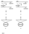
- Figure 1
- shows the general principles for the isolation and DNA
sequencing of VH and VL antibody gene fragments from B
cells.
Reference numeral 10 indicates primary PCR,reference numeral 20 the cloning into a DNA sequencing vector using the T-tailing principle - random orientation, andreference number 30 the determining of the nucleotide sequence of forward and reverse strands using M13 forward and reverse primers; and - Figure 2
- shows a schematic depiction of resynthesised recombinant antibody gene cassette. VH and VL regions are linked with a glycine serine-rich linker. Each variable domain contains three Complementarity Determining Regions (CDRs) which participate in antigen binding.
- Start
- Approx. residue 24
- Residue before
- always a Cys
- Residue after
- always a Trp. Typically Trp-Tyr-Gln, but also, Trp-Leu-Gln, Trp-Phe-Gln, Trp-Tyr-Leu
- Length
- 10 to 17 residues
- Start
- always 16 residues after the end of CDR-L1
- Residues before
- generally Ile-Tyr, but also, Val-Tyr, Ile-Lys, Ile-Phe
- Length
- always 7 residues (except NEW (7FAB) which has a deletion in this region)
- Start
- always 33 residues after end of CDR-L2 (except NEW (7FAB) which has the deletion at the end of CDR-L2)
- Residue before
- always Cys
- Residues after
- always Phe-Gly-XXX-Gly (SEQ ID NO: 69)
- Length
- 7 to 11 residues
- Start
- Approx. residue 26 (always 4 after a Cys)
- Residues before
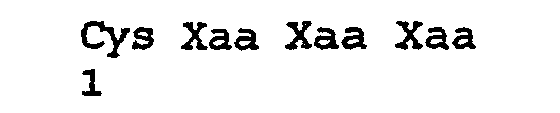
- always Cys-XXX-XXX-XXX (SEQ ID NO: 70)
- Residues after
- always a Trp. Typically Trp-Val, but also, Trp-Ile, Trp-Ala
- Length
- 10 to 12 residues
- Start
- always 15 residues after the end of CDR-H1
- Residues before
- typically Leu-Glu-Trp-Ile-Gly (SEQ ID NO: 71), but a number of variations
- Residues after
- Lys/Arg-Leu/IIe/Val/Phe/Thr/Ala-Thr/Ser/IIe/Ala
- Length
- 16 to 19 residues;
- Start
- always 33 residues after end of CDR-H2 (always 2 after a Cys)
- Residues before
- always Cys-XXX-XXX (typically Cys-Ala-Arg)
- Residues after
- always Trp-Gly-XXX-Gly (SEQ ID NO: 72)
- Length
- 3 to 25 residues
| V01 | VRE colonised patient |
| V02 | VRE blood culture positive |
| V03 | VRE blood culture positive |
| V04 | VRE blood culture positive |
| M01 | MRSA blood culture positive |
| M02 | MRSA blood culture positive |
| M03 | MRSA blood culture positive |
| M04 | MRSA blood culture positive |
| M05 | MRSA blood culture positive |
| P01 | P.aeruginosa CF patient |
| V02 | (SEQ ID NO: 62) | 9% |
| V03 | (SEQ ID NO: 68) | 19% |
| V04 | (SEQ ID NO: 67) | 42% |
| M01 | (SEQ ID NO:40) | 13% |
| M02 | (SEQ ID NO: 45) | 38% |
| M03 | (SEQ ID NO:67) | 24% |
| M04 | (SEQ ID NO: 45) | 12% |
| M05 | (SEQ ID NO: 53) | 4% |

| SEQ | V01 | V02 | V03 | V04 | M01 | M02 | M03 | M04 | M05 P01 | |
| 40 | 0 | 0 | 0 | 0 | 13 | 0 | 0 | 0 | 0 | 0 |
| 41 | 0 | 0 | 0 | 0 | 11 | 0 | 0 | 0 | 0 | 0 |
| 42 | 0 | 0 | 0 | 0 | 11 | 0 | 0 | 0 | 0 | 0 |
| 43 | 0 | 0 | 0 | 0 | 5 | 0 | 0 | 0 | 0 | 0 |
| 44 | 0 | 0 | 0 | 0 | 5 | 0 | 0 | 0 | 0 | 0 |
| 45 | 0 | 0 | 0 | 0 | 0 | 38 | 15 | 12 | 0 | 0 |
| 46 0 | 0 | 0 | 0 | 0 | 1 | 23 | 8 | 8 | <1 | 0 |
| 47 | 0 | 0 | 0 | 0 | 1 | 13 | 24 | 8 | 0 | 0 |
| 48 | 0 | 0 | 0 | 0 | 0 | 10 | 1 | <1 | <1 | 0 |
| 49 | 0 | 0 | 0 | 0 | 0 | 3 | 3 | 4 | 0 | 0 |
| 50 | 0 | 0 | 0 | 0 | 1 | 2 | 1 | 5 | 0 | 0 |
| 51 | 0 | 0 | 0 | 0 | 0 | 0 | 0 | 5 | 0 | 0 |
| 52 | 0 | 0 | 0 | 0 | 0 | 0 | 0 | 5 | 0 | 0 |
| 53 | 0 | 0 | 0 | 0 | 0 | 0 | 0 | 0 | 4 | 0 |
| 54 | 0 | 0 | 0 | 0 | 0 | 0 | 0 | 0 | 3 | 0 |
| 55 | 0 | 0 | 0 | 0 | 0 | 0 | 0 | 0 | 3 | 0 |
| 56 | 0 | 0 | 0 | 0 | 0 | 1 | 0 | 1 | 0 | 3 |
| 57 | 0 | 0 | 0 | 0 | 0 | 0 | 0 | 0 | 0 | 3 |
| 58 | 0 | 0 | 0 | 0 | 0 | 0 | 0 | 0 | 0 | 2 |
| 59 | 0 | 0 | 3 | 2 | 0 | 0 | 0 | 0 | 0 | <1 |
| 60 | 0 | 0 | 2 | 9 | 0 | 0 | 0 | 0 | 0 | 0 |
| 61 | 0 | 0 | 2 | 7 | 0 | 0 | 0 | 0 | 0 | 0 |
| 62 | 0 | 9 | 0 | 0 | 0 | 0 | 0 | 0 | 0 | 0 |
| 63 | 0 | 0 | 0 | 12 | 0 | 0 | 0 | 0 | 0 | 0 |
| 64 | 0 | 0 | 4 | 0 | 0 | 0 | 0 | 0 | 0 | 0 |
| 65 | 0 | 5 | 0 | 0 | 0 | 0 | 0 | 0 | 0 | 0 |
| 66 | 0 | 0 | 2 | 5 | 0 | 0 | 0 | 0 | 0 | 0 |
| 67 | 0 | 0 | 1 | 42 | 0 | 0 | 0 | 0 | 0 | 0 |
| 68 | 0 | 0 | 19 | 12 | 0 | 0 | 0 | <1 | 0 | 0 |
<151> 2001-12-19
<211> 24
<212> DNA
<213> Artificial Sequence
<223> PCR Primer

<211> 24
<212> DNA
<213> Artificial Sequence
<223> PCR Primer

<211> 24
<212> DNA
<213> Artificial Sequence
<223> PCR Primer

<211> 24
<212> DNA
<213> Artificial Sequence
<223> PCR Primer

<211> 24
<212> DNA
<213> Artificial Sequence
<223> PCR Primer

<211> 24
<212> DNA
<213> Artificial Sequence
<223> PCR Primer

<211> 23
<212> DNA
<213> Artificial Sequence
<223> PCR Primer

<211> 23
<212> DNA
<213> Artificial Sequence
<223> PCR Primer

<211> 23
<212> DNA
<213> Artificial Sequence
<223> PCR Primer

<211> 23
<212> DNA
<213> Artificial Sequence
<223> PCR Primer

<211> 23
<212> DNA
<213> Artificial Sequence
<223> PCR Primer

<211> 23
<212> DNA
<213> Artificial Sequence
<223> PCR Primer

<211> 23
<212> DNA
<213> Artificial Sequence
<223> PCR Primer

<211> 24
<212> DNA
<213> Artificial Sequence
<223> PCR Primer

<211> 24
<212> DNA
<213> Artificial Sequence
<223> PCR Primer

<211> 24
<212> DNA
<213> Artificial Sequence
<223> PCR Primer

<211> 24
<212> DNA
<213> Artificial Sequence
<223> PCR Primer

<211> 24
<212> DNA
<213> Artificial Sequence
<223> PCR Primer

<211> 23
<212> DNA
<213> Artificial Sequence
<223> PCR Primer

<211> 23
<212> DNA
<213> Artificial Sequence
<223> PCR Primer

<211> 23
<212> DNA
<213> Artificial Sequence
<223> PCR Primer

<211> 23
<212> DNA
<213> Artificial Sequence
<223> PCR Primer

<211> 23
<212> DNA
<213> Artificial Sequence

<211> 23
<212> DNA
<213> Artificial Sequence
<223> PCR primer

<211> 17
<212> DNA
<213> Artificial Sequence
<223> PCR Primer

<211> 16
<212> DNA
<213> Artificial Sequence
<223> PCR Primer

<211> 7
<212> PRT
<213> Homo sapiens

<211> 11
<212> PRT
<213> Homo sapiens

<211> 7
<212> PRT
<213> Homo sapiens

<211> 10
<212> PRT
<213> Homo sapiens

<211> 6
<212> PRT
<213> Homo sapiens

<211> 12
<212> PRT
<213> Homo sapiens

<211> 17
<212> PRT
<213> Homo sapiens

<211> 13
<212> PRT
<213> Homo sapiens

<211> 13
<212> PRT
<213> Homo sapiens

<211> 14
<212> PRT
<213> Homo sapiens

<211> 16
<212> PRT
<213> Homo sapiens

<211> 17
<212> PRT
<213> Homo sapiens

<211> 13
<212> PRT
<213> Homo sapiens

<211> 15
<212> PRT
<213> Homo sapiens

<211> 9
<212> PRT
<213> Homo sapiens

<211> 13
<212> PRT
<213> Homo sapiens

<211> 13
<212> PRT
<213> Homo sapiens

<211> 16
<212> PRT
<213> Homo sapiens

<211> 12
<212> PRT
<213> Homo sapiens

<211> 12
<212> PRT
<213> Homo sapiens

<211> 13
<212> PRT
<213> Homo sapiens

<211> 17
<212> PRT
<213> Homo sapiens

<211> 13
<212> PRT
<213> Homo sapiens

<211> 16
<212> PRT
<213> Homo sapiens

<211> 11
<212> PRT
<213> Homo sapiens

<211> 24
<212> PRT
<213> Homo sapiens

<211> 14
<212> PRT
<213> Homo sapiens

<211> 14
<212> PRT
<213> Homo sapiens

<211> 10
<212> PRT
<213> Homo sapiens

<211> 9
<212> PRT
<213> Homo sapiens

<211> 18
<212> PRT
<213> Homo sapiens

<211> 11
<212> PRT
<213> Homo sapiens

<211> 11
<212> PRT
<213> Homo sapiens

<211> 11
<212> PRT
<213> Homo sapiens

<211> 11
<212> PRT
<213> Homo sapiens

<211> 7
<212> PRT
<213> Homo sapiens

<212> PRT
<213> Homo sapiens

<211> 15
<212> PRT
<213> Homo sapiens

<211> 13
<212> PRT
<213> Homo sapiens

<211> 9
<212> PRT
<213> Homo sapiens

<211> 13
<212> PRT
<213> Homo sapiens

<211> 15
<212> PRT
<213> Homo sapiens

<211> 4
<212> PRT
<213> Artificial Sequence
<223> Used for defining CDR region
<221> MISC_FEATURE
<222> (3).. (3)
<223> Any amino acid

<211> 4
<212> PRT
<213> Artificial Sequence
<223> Used for defining CDR region
<221> MISC_FEATURE
<222> (2)..(4)
<223> Any amino acid
<211> 5
<212> PRT
<213> Artificial Sequence
<223> Used for defining CDR region

<211> 4
<212> PRT
<213> Artificial Sequence
<223> Used for defining CDR region
<221> MISC_FEATORE
<222> (3)..(3)
<223> Any amino acid

Claims (23)
- A method for identifying candidate sequences of at least the CDR3 region of antibodies specific against at least one antigen produced by a micro-organism during an infection or against a vaccine, comprising the steps of:(i) with B cells isolated from at least one patient who has been infected by said micro-organism or administered said vaccine, sequencing at least the CDR3 region of the VH and/or VL coding regions of said B cells; and(ii) correlating said sequenced at least the CDR3 regions of the VH and VL coding regions of said B cells to identify a set of candidate sequences for at least a CDR3 region of antibodies specific against said at least one antigen produced by said micro-organism or against said vaccine, each of said set of candidate CDR3 sequences or a sequence having at least 60% homology therewith occurring in total at a frequency of at least 1 percent in the set of sequences determined at step (i).
- A method according to claim 1, said B cells being selected from the group consisting of peripheral B-cell lymphocytes and B cells from the spleen.
- A method according to claim 2, said peripheral B-cell lymphocytes being isolated from blood from said at least one patient.
- A method according to claim 1, said at least one antigen being an immunogen.
- A method according to claim 1, said at least one patient displaying a pronounced antibody response in response to infection by said micro-organism.
- A method according to claim 1, said at least one patient having recovered from infection by said micro-organism.
- A method according to claim 1, said correlation step (ii) comprising determining putative amino acid sequences from said sequenced at least the VH and VL CDR3 coding regions, and correlating said putative amino acid sequences.
- A method according to claim 7, said correlation step (ii) comprising identifying the Complementarity Determining Regions comprised in said at least the VH and VL regions and correlating said Complementarity Determining Regions.
- A method according to claim 8, said Complementarity Determining Regions being selected from the group consisting of CDR1, CDR2 and CDR3.
- A method according to claim 1, said correlation step (ii) additionally correlating at least one of the group consisting of: the micro-organism infecting said at least one patient, the site of infection of said at least one patient by said micro-organism, the time point at which said B cells are isolated during infection of said at least one patient by said micro-organism, the age of said at least one patient, the sex of said at least one patient, and the race of said at least one patient.
- A method according to claim 1, said B cells having been isolated from said at least one patient at a plurality of time points during infection of said at least one patient by said micro-organism, said correlation step (ii) correlating the time point during infection of said at least one patient by said micro-organism at which said B cells are isolated.
- A method according to claim 1, said B cells having been isolated from at least two patients, at least one of whom has recovered from infection by said micro-organism, and at least one of whom has not recovered from infection by said micro-organism, said correlation step (ii) correlating the recovery of said at least two patients from infection by said micro-organism.
- A method according to claim 1, said B cells having been isolated from at least two patients, said patients being infected by different micro-organisms producing said at least one antigen, said correlation step (ii) correlating said sequenced at least the VH and VL coding regions of said B cells to identify a set of candidate sequences for antibodies, each of which is specific against at least one shared antigen produced by said different micro-organisms or is specific against different antigens produced by said different micro-organisms.
- A method of manufacture of a medicament for the treatment of an infection by a micro-organism which produces at least one antigen, comprising the steps of:(i) performing a method according to any one of the preceding claims; and(ii) synthesising at least one antibody comprising a candidate sequences specific against said at least one antigen produced by said micro-organism.
- A method of manufacture of a medicament according to claim 14, comprising the step of mixing said synthesised at least one antibody with a pharmaceutically acceptable carrier, diluant or excipient.
- A method of producing a database which identifies candidate sequences for antibodies specific against at least one antigen produced by a micro-organism, comprising the steps of:(i) performing a method according to any one of claims 1-13; and(ii) storing the data produced by said method in said database.
- A method of generating a report which identifies candidate sequences for antibodies specific against at least one antigen produced by a micro-organism, comprising the steps of:(i) performing a method according to any one of claims 1-13; and(ii) producing a report comprising the data produced by said method.
- A method for determining the efficacy of a vaccine, comprising the steps of:(i) with B cells isolated from at least one patient who has been administered said vaccine, sequencing at least the CDR3 region of the VH and/or VL coding regions of said B cells; and(ii) correlating said sequenced at least the CDR3 region of the VH and/or VL coding regions of said B cells to identify a set of candidate sequences for at least the CDR3 region of antibodies specific against said vaccine, each of said set of CDR3 candidate sequences or a sequence having at least 60% homology therewith occurring in total at a frequency of at least 1 percent in the set of sequences determined at step (i).
- A method according to claim 18, correlation step (ii) comprising correlating said sequenced at least a CDR3 region of the VH and VL coding regions of said B cells with sequenced at least a CDR3 region of the VH and VL coding regions of B cells isolated from at least one patient who has been infected with a micro-organism against which vaccination with said vaccine is intended to stimulate a protective immune response.
- A method according to claim 18, said correlation step (ii) additionally correlating at least one of the group consisting of: the time since administration of said vaccine to said at least one patient, the age of said at least one patient, the sex of said at least one patient, the race of said at least one patient, and the Complementarity Determining Regions of said sequenced at least the VH and VL coding regions.
- A method according to claim 18, said method determining the efficacy of said vaccine in stimulating a protective immune response against the micro-organism against which vaccination with said vaccine is intended to stimulate a protective immune response.
- A method of producing a database which identifies the efficacy of a vaccine, comprising the steps of:(i) performing a method according to any one of claims 18-21; and(ii) storing the data produced by said method in said database.
- A method of generating a report which identifies the efficacy of a vaccine, comprising the steps of:(i) performing a method according to any one of claims 18-21; and(ii) producing a report comprising the data produced by said method.
Priority Applications (1)
| Application Number | Priority Date | Filing Date | Title |
|---|---|---|---|
| SI200230106T SI1415002T1 (en) | 2001-12-19 | 2002-12-16 |
Applications Claiming Priority (3)
| Application Number | Priority Date | Filing Date | Title |
|---|---|---|---|
| GB0130267 | 2001-12-19 | ||
| GBGB0130267.8A GB0130267D0 (en) | 2001-12-19 | 2001-12-19 | Focussed antibody technology |
| PCT/GB2002/005690 WO2003052416A2 (en) | 2001-12-19 | 2002-12-16 | Focussed antibody technology |
Publications (2)
| Publication Number | Publication Date |
|---|---|
| EP1415002A2 EP1415002A2 (en) | 2004-05-06 |
| EP1415002B1 true EP1415002B1 (en) | 2005-02-02 |
Family
ID=9927861
Family Applications (1)
| Application Number | Title | Priority Date | Filing Date |
|---|---|---|---|
| EP02788114A Expired - Lifetime EP1415002B1 (en) | 2001-12-19 | 2002-12-16 | Focussed antibody technology |
Country Status (10)
| Country | Link |
|---|---|
| US (1) | US20060233812A1 (en) |
| EP (1) | EP1415002B1 (en) |
| AT (1) | ATE288502T1 (en) |
| AU (1) | AU2002352394A1 (en) |
| CA (1) | CA2471570A1 (en) |
| DE (1) | DE60202877T2 (en) |
| ES (1) | ES2236605T3 (en) |
| GB (1) | GB0130267D0 (en) |
| PT (1) | PT1415002E (en) |
| WO (1) | WO2003052416A2 (en) |
Families Citing this family (51)
| Publication number | Priority date | Publication date | Assignee | Title |
|---|---|---|---|---|
| GB0309126D0 (en) * | 2003-04-17 | 2003-05-28 | Neutec Pharma Plc | Clostridium difficile focussed antibodies |
| US20100069614A1 (en) | 2008-06-27 | 2010-03-18 | Merus B.V. | Antibody producing non-human mammals |
| GB0414886D0 (en) * | 2004-07-02 | 2004-08-04 | Neutec Pharma Plc | Treatment of bacterial infections |
| EP2088432A1 (en) * | 2008-02-11 | 2009-08-12 | MorphoSys AG | Methods for identification of an antibody or a target |
| US8628927B2 (en) | 2008-11-07 | 2014-01-14 | Sequenta, Inc. | Monitoring health and disease status using clonotype profiles |
| US9506119B2 (en) | 2008-11-07 | 2016-11-29 | Adaptive Biotechnologies Corp. | Method of sequence determination using sequence tags |
| US8748103B2 (en) | 2008-11-07 | 2014-06-10 | Sequenta, Inc. | Monitoring health and disease status using clonotype profiles |
| US8691510B2 (en) | 2008-11-07 | 2014-04-08 | Sequenta, Inc. | Sequence analysis of complex amplicons |
| EP2364368B1 (en) | 2008-11-07 | 2014-01-15 | Sequenta, Inc. | Methods of monitoring conditions by sequence analysis |
| US9365901B2 (en) | 2008-11-07 | 2016-06-14 | Adaptive Biotechnologies Corp. | Monitoring immunoglobulin heavy chain evolution in B-cell acute lymphoblastic leukemia |
| US9528160B2 (en) | 2008-11-07 | 2016-12-27 | Adaptive Biotechnolgies Corp. | Rare clonotypes and uses thereof |
| DK3059337T3 (en) * | 2009-01-15 | 2019-07-22 | Adaptive Biotechnologies Corp | Adaptive immunity profiling and methods for producing monoclonal antibodies |
| US9079942B2 (en) * | 2009-02-09 | 2015-07-14 | Epitomics, Inc. | CDR-anchored amplification method |
| WO2010136598A1 (en) | 2009-05-29 | 2010-12-02 | Morphosys Ag | A collection and methods for its use |
| US20100317539A1 (en) * | 2009-06-12 | 2010-12-16 | Guo-Liang Yu | Library of Engineered-Antibody Producing Cells |
| RU2014144463A (en) | 2009-06-25 | 2015-06-20 | Фред Хатчинсон Кансэр Рисёч Сентер | METHOD FOR ADAPTIVE IMMUNITY MEASUREMENT |
| US8293483B2 (en) | 2009-09-11 | 2012-10-23 | Epitomics, Inc. | Method for identifying lineage-related antibodies |
| US9796788B2 (en) | 2010-02-08 | 2017-10-24 | Regeneron Pharmaceuticals, Inc. | Mice expressing a limited immunoglobulin light chain repertoire |
| US20130045492A1 (en) | 2010-02-08 | 2013-02-21 | Regeneron Pharmaceuticals, Inc. | Methods For Making Fully Human Bispecific Antibodies Using A Common Light Chain |
| HRP20151188T4 (en) | 2010-02-08 | 2021-10-01 | Regeneron Pharmaceuticals, Inc. | Common light chain mouse |
| EP2572203B1 (en) | 2010-05-17 | 2017-10-25 | The Board of Regents of The University of Texas System | Determination of antigen-specific antibody sequences in blood circulation |
| HRP20181811T1 (en) | 2010-11-19 | 2018-12-28 | Morphosys Ag | COLLECTION OF THE ANTIBODIES AND ITS USES |
| CN108220297A (en) | 2011-03-09 | 2018-06-29 | 细胞信号科技公司 | For generating the method for monoclonal antibody and reagent |
| PT3865581T (en) | 2011-08-05 | 2024-10-04 | Regeneron Pharma | Humanized universal light chain mice |
| US10385475B2 (en) | 2011-09-12 | 2019-08-20 | Adaptive Biotechnologies Corp. | Random array sequencing of low-complexity libraries |
| CA2791109C (en) * | 2011-09-26 | 2021-02-16 | Merus B.V. | Generation of binding molecules |
| US9279159B2 (en) | 2011-10-21 | 2016-03-08 | Adaptive Biotechnologies Corporation | Quantification of adaptive immune cell genomes in a complex mixture of cells |
| EP2783214A2 (en) | 2011-11-23 | 2014-10-01 | The Board of Regents of The University of Texas System | Proteomic identification of antibodies |
| EP3388535B1 (en) | 2011-12-09 | 2021-03-24 | Adaptive Biotechnologies Corporation | Diagnosis of lymphoid malignancies and minimal residual disease detection |
| US9499865B2 (en) | 2011-12-13 | 2016-11-22 | Adaptive Biotechnologies Corp. | Detection and measurement of tissue-infiltrating lymphocytes |
| DK2823060T3 (en) | 2012-03-05 | 2018-05-28 | Adaptive Biotechnologies Corp | Determination of associated immune receptor chains from frequency-matched subunits |
| DK2838918T3 (en) | 2012-04-20 | 2019-08-12 | Merus Nv | METHODS AND METHODS FOR PREPARING HETERODIMER IG-LIKE MOLECULES |
| EP3091089B1 (en) | 2012-05-08 | 2019-01-23 | Adaptive Biotechnologies Corporation | Method for measuring and calibrating amplification bias in multiplexed pcr reactions |
| EP2861760B1 (en) | 2012-06-15 | 2020-01-01 | The Board of Regents of The University of Texas System | High throughput sequencing of multiple transcripts of a single cell |
| US20160002731A1 (en) | 2012-10-01 | 2016-01-07 | Adaptive Biotechnologies Corporation | Immunocompetence assessment by adaptive immune receptor diversity and clonality characterization |
| US10150996B2 (en) | 2012-10-19 | 2018-12-11 | Adaptive Biotechnologies Corp. | Quantification of adaptive immune cell genomes in a complex mixture of cells |
| US9708657B2 (en) | 2013-07-01 | 2017-07-18 | Adaptive Biotechnologies Corp. | Method for generating clonotype profiles using sequence tags |
| WO2015134787A2 (en) | 2014-03-05 | 2015-09-11 | Adaptive Biotechnologies Corporation | Methods using randomer-containing synthetic molecules |
| CN106255410B (en) | 2014-03-21 | 2020-01-10 | 瑞泽恩制药公司 | Non-human animals producing single domain binding proteins |
| US10066265B2 (en) | 2014-04-01 | 2018-09-04 | Adaptive Biotechnologies Corp. | Determining antigen-specific t-cells |
| US11390921B2 (en) | 2014-04-01 | 2022-07-19 | Adaptive Biotechnologies Corporation | Determining WT-1 specific T cells and WT-1 specific T cell receptors (TCRs) |
| CA2966201A1 (en) | 2014-10-29 | 2016-05-06 | Adaptive Biotechnologies Corp. | Highly-multiplexed simultaneous detection of nucleic acids encoding paired adaptive immune receptor heterodimers from many samples |
| US10246701B2 (en) | 2014-11-14 | 2019-04-02 | Adaptive Biotechnologies Corp. | Multiplexed digital quantitation of rearranged lymphoid receptors in a complex mixture |
| EP3224384A4 (en) | 2014-11-25 | 2018-04-18 | Adaptive Biotechnologies Corp. | Characterization of adaptive immune response to vaccination or infection using immune repertoire sequencing |
| US11047008B2 (en) | 2015-02-24 | 2021-06-29 | Adaptive Biotechnologies Corporation | Methods for diagnosing infectious disease and determining HLA status using immune repertoire sequencing |
| US11111314B2 (en) | 2015-03-19 | 2021-09-07 | Regeneron Pharmaceuticals, Inc. | Non-human animals that select for light chain variable regions that bind antigen |
| US10513733B2 (en) | 2015-03-23 | 2019-12-24 | Board Of Regents, The University Of Texas System | High throughout sequencing of paired VH and VL transcripts from B cells secreting antigen-specific antibodies |
| WO2016161273A1 (en) | 2015-04-01 | 2016-10-06 | Adaptive Biotechnologies Corp. | Method of identifying human compatible t cell receptors specific for an antigenic target |
| US10428325B1 (en) | 2016-09-21 | 2019-10-01 | Adaptive Biotechnologies Corporation | Identification of antigen-specific B cell receptors |
| WO2019084538A1 (en) | 2017-10-27 | 2019-05-02 | Board Of Regents, The University Of Texas System | Tumor specific antibodies and t-cell receptors and methods of identifying the same |
| US11254980B1 (en) | 2017-11-29 | 2022-02-22 | Adaptive Biotechnologies Corporation | Methods of profiling targeted polynucleotides while mitigating sequencing depth requirements |
Family Cites Families (5)
| Publication number | Priority date | Publication date | Assignee | Title |
|---|---|---|---|---|
| CA2006878A1 (en) * | 1988-12-29 | 1990-06-29 | John D. Rodwell | Molecular recognition units |
| US6291158B1 (en) * | 1989-05-16 | 2001-09-18 | Scripps Research Institute | Method for tapping the immunological repertoire |
| DE19739685A1 (en) * | 1997-09-10 | 1999-03-11 | Eichel Streiber Christoph Von | Monoclonal antibodies for the therapy and prophylaxis of diseases caused by Clostridium difficile |
| DE60124313T2 (en) * | 2001-01-15 | 2007-05-31 | Keryos S.P.A. | DNA VACCINES OF HYPERVARIABLES VH-CDR3 IDIOTYPIC DETERMINANTS |
| GB0309126D0 (en) * | 2003-04-17 | 2003-05-28 | Neutec Pharma Plc | Clostridium difficile focussed antibodies |
-
2001
- 2001-12-19 GB GBGB0130267.8A patent/GB0130267D0/en not_active Ceased
-
2002
- 2002-12-16 ES ES02788114T patent/ES2236605T3/en not_active Expired - Lifetime
- 2002-12-16 DE DE60202877T patent/DE60202877T2/en not_active Expired - Lifetime
- 2002-12-16 CA CA002471570A patent/CA2471570A1/en not_active Abandoned
- 2002-12-16 US US10/499,104 patent/US20060233812A1/en not_active Abandoned
- 2002-12-16 AT AT02788114T patent/ATE288502T1/en active
- 2002-12-16 EP EP02788114A patent/EP1415002B1/en not_active Expired - Lifetime
- 2002-12-16 PT PT02788114T patent/PT1415002E/en unknown
- 2002-12-16 AU AU2002352394A patent/AU2002352394A1/en not_active Abandoned
- 2002-12-16 WO PCT/GB2002/005690 patent/WO2003052416A2/en not_active Ceased
Also Published As
| Publication number | Publication date |
|---|---|
| CA2471570A1 (en) | 2003-06-26 |
| ES2236605T3 (en) | 2005-07-16 |
| DE60202877T2 (en) | 2006-04-13 |
| AU2002352394A1 (en) | 2003-06-30 |
| EP1415002A2 (en) | 2004-05-06 |
| DE60202877D1 (en) | 2005-03-10 |
| ATE288502T1 (en) | 2005-02-15 |
| US20060233812A1 (en) | 2006-10-19 |
| WO2003052416A2 (en) | 2003-06-26 |
| PT1415002E (en) | 2005-05-31 |
| WO2003052416A3 (en) | 2003-10-16 |
| GB0130267D0 (en) | 2002-02-06 |
Similar Documents
| Publication | Publication Date | Title |
|---|---|---|
| US20060233812A1 (en) | Focussed antibody technology | |
| RU2765306C2 (en) | Antibody against b7-h3, its antigen-binding fragment and their medical use | |
| CA2367901C (en) | Monoclonal antibodies, antigens and diagnosis and therapy of malignant diseases | |
| CN112794899B (en) | A fully human monoclonal neutralizing antibody against novel coronavirus and its application | |
| US20130267688A1 (en) | Novel antibody structures derived from human germline sequences | |
| EP1613655B1 (en) | Clostridium difficile focussed antibodies | |
| KR20130135028A (en) | Rapid isolation of monoclonal antibodies from animals | |
| AU2012315616C1 (en) | Identification of antigen-specific adaptive immune responses using Arm-PCR and high-throughput sequencing | |
| AU2012315616B8 (en) | Identification of antigen-specific adaptive immune responses using Arm-PCR and high-throughput sequencing | |
| CN113416249B (en) | Compositions against tetanus toxin | |
| CN120988117B (en) | Antibodies against Staphylococcus aureus enterotoxin B and their applications | |
| TWI775422B (en) | Recombinant antibodies, kits comprising the same, and uses thereof | |
| WO2021207531A2 (en) | Monoclonal antibodies and uses thereof | |
| CN119039430B (en) | Protective monoclonal antibody targeting Mycobacterium tuberculosis MctB and its preparation method and application | |
| CN115433273B (en) | Anti-novel coronavirus rabbit recombinant monoclonal antibody with neutralization activity and application thereof | |
| CN121108321A (en) | A monoclonal antibody against the novel coronavirus and its application | |
| CN117683130A (en) | A neutralizing antibody specifically targeting Yersinia pestis F1 antigen | |
| CN116836268A (en) | Antibodies against novel coronavirus SARS-CoV-2 and uses thereof | |
| CN115433279A (en) | Preparation and application of targeting CD123 rabbit recombinant monoclonal antibody | |
| HK40000441A (en) | Identification of antigen-specific adaptive immune responses using arm-pcr and high-throughput sequencing | |
| CN113621032A (en) | A kind of polypeptide with SEQ ID NO.2 sequence and antibody with strong ADCC effect and application |
Legal Events
| Date | Code | Title | Description |
|---|---|---|---|
| PUAI | Public reference made under article 153(3) epc to a published international application that has entered the european phase |
Free format text: ORIGINAL CODE: 0009012 |
|
| 17P | Request for examination filed |
Effective date: 20040213 |
|
| AK | Designated contracting states |
Kind code of ref document: A2 Designated state(s): AT BE BG CH CY CZ DE DK EE ES FI FR GB GR IE IT LI LU MC NL PT SE SI SK TR |
|
| AX | Request for extension of the european patent |
Extension state: AL LT LV MK RO |
|
| GRAP | Despatch of communication of intention to grant a patent |
Free format text: ORIGINAL CODE: EPIDOSNIGR1 |
|
| GRAS | Grant fee paid |
Free format text: ORIGINAL CODE: EPIDOSNIGR3 |
|
| GRAA | (expected) grant |
Free format text: ORIGINAL CODE: 0009210 |
|
| AK | Designated contracting states |
Kind code of ref document: B1 Designated state(s): AT BE BG CH CY CZ DE DK EE ES FI FR GB GR IE IT LI LU MC NL PT SE SI SK TR |
|
| AX | Request for extension of the european patent |
Extension state: AL LT LV MK RO |
|
| REG | Reference to a national code |
Ref country code: GB Ref legal event code: FG4D |
|
| REG | Reference to a national code |
Ref country code: CH Ref legal event code: EP |
|
| REG | Reference to a national code |
Ref country code: IE Ref legal event code: FG4D |
|
| REF | Corresponds to: |
Ref document number: 60202877 Country of ref document: DE Date of ref document: 20050310 Kind code of ref document: P |
|
| REG | Reference to a national code |
Ref country code: CH Ref legal event code: NV Representative=s name: ROTTMANN, ZIMMERMANN + PARTNER AG |
|
| REG | Reference to a national code |
Ref country code: DK Ref legal event code: T3 |
|
| REG | Reference to a national code |
Ref country code: GR Ref legal event code: EP Ref document number: 20050401252 Country of ref document: GR |
|
| REG | Reference to a national code |
Ref country code: SE Ref legal event code: TRGR |
|
| REG | Reference to a national code |
Ref country code: PT Ref legal event code: SC4A Free format text: AVAILABILITY OF NATIONAL TRANSLATION Effective date: 20050328 |
|
| REG | Reference to a national code |
Ref country code: ES Ref legal event code: FG2A Ref document number: 2236605 Country of ref document: ES Kind code of ref document: T3 |
|
| PLBI | Opposition filed |
Free format text: ORIGINAL CODE: 0009260 |
|
| PLAX | Notice of opposition and request to file observation + time limit sent |
Free format text: ORIGINAL CODE: EPIDOSNOBS2 |
|
| PLAB | Opposition data, opponent's data or that of the opponent's representative modified |
Free format text: ORIGINAL CODE: 0009299OPPO |
|
| 26 | Opposition filed |
Opponent name: XOMA(US)LLC Effective date: 20051102 Opponent name: CAMBRIDGE ANTIBODY TECHNOLOGY LIMITED MILSTEINBUIL Effective date: 20051102 |
|
| R26 | Opposition filed (corrected) |
Opponent name: CAMBRIDGE ANTIBODY TECHNOLOGY LIMITED MILSTEIN BU Effective date: 20051102 Opponent name: XOMA(US)LLC Effective date: 20051102 |
|
| ET | Fr: translation filed | ||
| NLR1 | Nl: opposition has been filed with the epo |
Opponent name: CAMBRIDGE ANTIBODY TECHNOLOGY LIMITED MILSTEIN BUI Opponent name: XOMA(US)LLC |
|
| NLR1 | Nl: opposition has been filed with the epo |
Opponent name: XOMA(US)LLC Opponent name: CAMBRIDGE ANTIBODY TECHNOLOGY LIMITED MILSTEIN BUI |
|
| PLAF | Information modified related to communication of a notice of opposition and request to file observations + time limit |
Free format text: ORIGINAL CODE: EPIDOSCOBS2 |
|
| PLBB | Reply of patent proprietor to notice(s) of opposition received |
Free format text: ORIGINAL CODE: EPIDOSNOBS3 |
|
| PLAB | Opposition data, opponent's data or that of the opponent's representative modified |
Free format text: ORIGINAL CODE: 0009299OPPO |
|
| R26 | Opposition filed (corrected) |
Opponent name: CAMBRIDGE ANTIBODY TECHNOLOGY LIMITED MILSTEIN BUI Effective date: 20051102 Opponent name: XOMA(US)LLC Effective date: 20051102 |
|
| NLR1 | Nl: opposition has been filed with the epo |
Opponent name: XOMA (US)LLC Opponent name: CAMBRIDGE ANTIBODY TECHNOLOGY LIMITED MILSTEIN BUI |
|
| RAP2 | Party data changed (patent owner data changed or rights of a patent transferred) |
Owner name: NEUTEC PHARMA LIMITED |
|
| PLCK | Communication despatched that opposition was rejected |
Free format text: ORIGINAL CODE: EPIDOSNREJ1 |
|
| NLT2 | Nl: modifications (of names), taken from the european patent patent bulletin |
Owner name: NEUTEC PHARMA LIMITED Effective date: 20080402 |
|
| PLBN | Opposition rejected |
Free format text: ORIGINAL CODE: 0009273 |
|
| STAA | Information on the status of an ep patent application or granted ep patent |
Free format text: STATUS: OPPOSITION REJECTED |
|
| 27O | Opposition rejected |
Effective date: 20080614 |
|
| NLR2 | Nl: decision of opposition |
Effective date: 20080614 |
|
| PG25 | Lapsed in a contracting state [announced via postgrant information from national office to epo] |
Ref country code: CY Free format text: LAPSE BECAUSE OF NON-PAYMENT OF DUE FEES Effective date: 20081216 |
|
| PGFP | Annual fee paid to national office [announced via postgrant information from national office to epo] |
Ref country code: AT Payment date: 20101129 Year of fee payment: 9 Ref country code: DK Payment date: 20101210 Year of fee payment: 9 Ref country code: EE Payment date: 20101103 Year of fee payment: 9 Ref country code: FR Payment date: 20101224 Year of fee payment: 9 Ref country code: IE Payment date: 20101214 Year of fee payment: 9 Ref country code: MC Payment date: 20101129 Year of fee payment: 9 |
|
| PGFP | Annual fee paid to national office [announced via postgrant information from national office to epo] |
Ref country code: BG Payment date: 20101117 Year of fee payment: 9 Ref country code: CH Payment date: 20101215 Year of fee payment: 9 Ref country code: CZ Payment date: 20101202 Year of fee payment: 9 Ref country code: FI Payment date: 20101214 Year of fee payment: 9 Ref country code: LU Payment date: 20110118 Year of fee payment: 9 Ref country code: PT Payment date: 20101203 Year of fee payment: 9 Ref country code: SI Payment date: 20101109 Year of fee payment: 9 Ref country code: SK Payment date: 20101203 Year of fee payment: 9 |
|
| PGFP | Annual fee paid to national office [announced via postgrant information from national office to epo] |
Ref country code: TR Payment date: 20101122 Year of fee payment: 9 Ref country code: NL Payment date: 20101210 Year of fee payment: 9 Ref country code: SE Payment date: 20101213 Year of fee payment: 9 Ref country code: IT Payment date: 20101218 Year of fee payment: 9 Ref country code: GR Payment date: 20101115 Year of fee payment: 9 Ref country code: GB Payment date: 20101215 Year of fee payment: 9 |
|
| PGFP | Annual fee paid to national office [announced via postgrant information from national office to epo] |
Ref country code: DE Payment date: 20101208 Year of fee payment: 9 Ref country code: CY Payment date: 20101210 Year of fee payment: 9 |
|
| PGFP | Annual fee paid to national office [announced via postgrant information from national office to epo] |
Ref country code: ES Payment date: 20110120 Year of fee payment: 9 Ref country code: BE Payment date: 20110217 Year of fee payment: 9 |
|
| REG | Reference to a national code |
Ref country code: CH Ref legal event code: PFA Owner name: NEUTEC PHARMA PLC Free format text: NEUTEC PHARMA PLC#CLINICAL SCIENCES BUILDING, MANCHESTER ROYAL INFIRMARY, OXFORD ROAD#MANCHESTER, LANCASHIRE M13 9WL (GB) -TRANSFER TO- NEUTEC PHARMA PLC#CLINICAL SCIENCES BUILDING, MANCHESTER ROYAL INFIRMARY, OXFORD ROAD#MANCHESTER, LANCASHIRE M13 9WL (GB) |
|
| REG | Reference to a national code |
Ref country code: PT Ref legal event code: MM4A Free format text: LAPSE DUE TO NON-PAYMENT OF FEES Effective date: 20120618 |
|
| BERE | Be: lapsed |
Owner name: *NEUTEC PHARMA P.L.C. Effective date: 20111231 |
|
| REG | Reference to a national code |
Ref country code: NL Ref legal event code: V1 Effective date: 20120701 |
|
| REG | Reference to a national code |
Ref country code: DK Ref legal event code: EBP |
|
| PG25 | Lapsed in a contracting state [announced via postgrant information from national office to epo] |
Ref country code: CZ Free format text: LAPSE BECAUSE OF NON-PAYMENT OF DUE FEES Effective date: 20111216 Ref country code: MC Free format text: LAPSE BECAUSE OF NON-PAYMENT OF DUE FEES Effective date: 20111231 |
|
| REG | Reference to a national code |
Ref country code: SE Ref legal event code: EUG Ref country code: CH Ref legal event code: PL |
|
| REG | Reference to a national code |
Ref country code: EE Ref legal event code: MM4A Ref document number: E000061 Country of ref document: EE Effective date: 20111231 |
|
| GBPC | Gb: european patent ceased through non-payment of renewal fee |
Effective date: 20111216 |
|
| PG25 | Lapsed in a contracting state [announced via postgrant information from national office to epo] |
Ref country code: PT Free format text: LAPSE BECAUSE OF NON-PAYMENT OF DUE FEES Effective date: 20120618 Ref country code: FI Free format text: LAPSE BECAUSE OF NON-PAYMENT OF DUE FEES Effective date: 20111216 |
|
| REG | Reference to a national code |
Ref country code: SK Ref legal event code: MM4A Ref document number: E 115 Country of ref document: SK Effective date: 20111216 Ref country code: GR Ref legal event code: ML Ref document number: 20050401252 Country of ref document: GR Effective date: 20120704 |
|
| REG | Reference to a national code |
Ref country code: FR Ref legal event code: ST Effective date: 20120831 |
|
| REG | Reference to a national code |
Ref country code: IE Ref legal event code: MM4A |
|
| PG25 | Lapsed in a contracting state [announced via postgrant information from national office to epo] |
Ref country code: CY Free format text: LAPSE BECAUSE OF NON-PAYMENT OF DUE FEES Effective date: 20111216 |
|
| REG | Reference to a national code |
Ref country code: SI Ref legal event code: KO00 Effective date: 20120807 |
|
| REG | Reference to a national code |
Ref country code: DE Ref legal event code: R119 Ref document number: 60202877 Country of ref document: DE Effective date: 20120703 |
|
| PG25 | Lapsed in a contracting state [announced via postgrant information from national office to epo] |
Ref country code: GB Free format text: LAPSE BECAUSE OF NON-PAYMENT OF DUE FEES Effective date: 20111216 Ref country code: DE Free format text: LAPSE BECAUSE OF NON-PAYMENT OF DUE FEES Effective date: 20120703 Ref country code: IE Free format text: LAPSE BECAUSE OF NON-PAYMENT OF DUE FEES Effective date: 20111216 Ref country code: BE Free format text: LAPSE BECAUSE OF NON-PAYMENT OF DUE FEES Effective date: 20111231 Ref country code: EE Free format text: LAPSE BECAUSE OF NON-PAYMENT OF DUE FEES Effective date: 20111231 Ref country code: CH Free format text: LAPSE BECAUSE OF NON-PAYMENT OF DUE FEES Effective date: 20111231 Ref country code: SE Free format text: LAPSE BECAUSE OF NON-PAYMENT OF DUE FEES Effective date: 20111217 Ref country code: LI Free format text: LAPSE BECAUSE OF NON-PAYMENT OF DUE FEES Effective date: 20111231 |
|
| PG25 | Lapsed in a contracting state [announced via postgrant information from national office to epo] |
Ref country code: SI Free format text: LAPSE BECAUSE OF NON-PAYMENT OF DUE FEES Effective date: 20111217 Ref country code: IT Free format text: LAPSE BECAUSE OF NON-PAYMENT OF DUE FEES Effective date: 20111216 Ref country code: SK Free format text: LAPSE BECAUSE OF NON-PAYMENT OF DUE FEES Effective date: 20111216 Ref country code: GR Free format text: LAPSE BECAUSE OF NON-PAYMENT OF DUE FEES Effective date: 20120704 |
|
| REG | Reference to a national code |
Ref country code: AT Ref legal event code: MM01 Ref document number: 288502 Country of ref document: AT Kind code of ref document: T Effective date: 20111216 |
|
| PG25 | Lapsed in a contracting state [announced via postgrant information from national office to epo] |
Ref country code: AT Free format text: LAPSE BECAUSE OF NON-PAYMENT OF DUE FEES Effective date: 20111216 Ref country code: DK Free format text: LAPSE BECAUSE OF NON-PAYMENT OF DUE FEES Effective date: 20120102 Ref country code: NL Free format text: LAPSE BECAUSE OF NON-PAYMENT OF DUE FEES Effective date: 20120701 |
|
| PG25 | Lapsed in a contracting state [announced via postgrant information from national office to epo] |
Ref country code: FR Free format text: LAPSE BECAUSE OF NON-PAYMENT OF DUE FEES Effective date: 20120102 |
|
| PG25 | Lapsed in a contracting state [announced via postgrant information from national office to epo] |
Ref country code: LU Free format text: LAPSE BECAUSE OF NON-PAYMENT OF DUE FEES Effective date: 20111216 |
|
| REG | Reference to a national code |
Ref country code: ES Ref legal event code: FD2A Effective date: 20130704 |
|
| PG25 | Lapsed in a contracting state [announced via postgrant information from national office to epo] |
Ref country code: BG Free format text: LAPSE BECAUSE OF NON-PAYMENT OF DUE FEES Effective date: 20121231 Ref country code: ES Free format text: LAPSE BECAUSE OF NON-PAYMENT OF DUE FEES Effective date: 20111217 |
|
| PG25 | Lapsed in a contracting state [announced via postgrant information from national office to epo] |
Ref country code: TR Free format text: LAPSE BECAUSE OF NON-PAYMENT OF DUE FEES Effective date: 20111216 |