EP1412538B1 - Methods for the simultaneous detection of hcv antigens and hcv antibodies - Google Patents
Methods for the simultaneous detection of hcv antigens and hcv antibodies Download PDFInfo
- Publication number
- EP1412538B1 EP1412538B1 EP02746647A EP02746647A EP1412538B1 EP 1412538 B1 EP1412538 B1 EP 1412538B1 EP 02746647 A EP02746647 A EP 02746647A EP 02746647 A EP02746647 A EP 02746647A EP 1412538 B1 EP1412538 B1 EP 1412538B1
- Authority
- EP
- European Patent Office
- Prior art keywords
- hcv
- antibody
- core
- antigen
- seq
- Prior art date
- Legal status (The legal status is an assumption and is not a legal conclusion. Google has not performed a legal analysis and makes no representation as to the accuracy of the status listed.)
- Expired - Lifetime
Links
- 239000000427 antigen Substances 0.000 title claims description 138
- 108091007433 antigens Proteins 0.000 title claims description 135
- 102000036639 antigens Human genes 0.000 title claims description 135
- 238000000034 method Methods 0.000 title claims description 35
- 238000001514 detection method Methods 0.000 title description 41
- 239000011859 microparticle Substances 0.000 claims description 149
- 150000001413 amino acids Chemical group 0.000 claims description 79
- 239000007790 solid phase Substances 0.000 claims description 57
- 238000012360 testing method Methods 0.000 claims description 57
- 108700039791 Hepatitis C virus nucleocapsid Proteins 0.000 claims description 52
- 101710132601 Capsid protein Proteins 0.000 claims description 34
- DZBUGLKDJFMEHC-UHFFFAOYSA-O acridine;hydron Chemical compound C1=CC=CC2=CC3=CC=CC=C3[NH+]=C21 DZBUGLKDJFMEHC-UHFFFAOYSA-O 0.000 claims description 22
- 150000001875 compounds Chemical class 0.000 claims description 22
- 108010008281 Recombinant Fusion Proteins Proteins 0.000 claims description 20
- 102000007056 Recombinant Fusion Proteins Human genes 0.000 claims description 20
- 239000002773 nucleotide Substances 0.000 claims description 14
- 125000003729 nucleotide group Chemical group 0.000 claims description 14
- 230000003612 virological effect Effects 0.000 claims description 12
- 238000006467 substitution reaction Methods 0.000 claims description 11
- 230000015572 biosynthetic process Effects 0.000 claims description 7
- 239000013598 vector Substances 0.000 claims description 6
- 241000711549 Hepacivirus C Species 0.000 description 203
- 238000003556 assay Methods 0.000 description 105
- 239000000872 buffer Substances 0.000 description 54
- FAPWRFPIFSIZLT-UHFFFAOYSA-M Sodium chloride Chemical compound [Na+].[Cl-] FAPWRFPIFSIZLT-UHFFFAOYSA-M 0.000 description 51
- 108090000765 processed proteins & peptides Proteins 0.000 description 42
- 239000000523 sample Substances 0.000 description 42
- 239000012634 fragment Substances 0.000 description 31
- 210000002966 serum Anatomy 0.000 description 30
- KCXVZYZYPLLWCC-UHFFFAOYSA-N EDTA Chemical compound OC(=O)CN(CC(O)=O)CCN(CC(O)=O)CC(O)=O KCXVZYZYPLLWCC-UHFFFAOYSA-N 0.000 description 29
- 229960001484 edetic acid Drugs 0.000 description 29
- 230000035945 sensitivity Effects 0.000 description 29
- 239000011324 bead Substances 0.000 description 26
- 125000003275 alpha amino acid group Chemical group 0.000 description 25
- 239000003085 diluting agent Substances 0.000 description 25
- 239000011780 sodium chloride Substances 0.000 description 25
- 239000002953 phosphate buffered saline Substances 0.000 description 23
- 238000002360 preparation method Methods 0.000 description 23
- LOKCTEFSRHRXRJ-UHFFFAOYSA-I dipotassium trisodium dihydrogen phosphate hydrogen phosphate dichloride Chemical compound P(=O)(O)(O)[O-].[K+].P(=O)(O)([O-])[O-].[Na+].[Na+].[Cl-].[K+].[Cl-].[Na+] LOKCTEFSRHRXRJ-UHFFFAOYSA-I 0.000 description 22
- 239000000243 solution Substances 0.000 description 22
- DBMJMQXJHONAFJ-UHFFFAOYSA-M Sodium laurylsulphate Chemical compound [Na+].CCCCCCCCCCCCOS([O-])(=O)=O DBMJMQXJHONAFJ-UHFFFAOYSA-M 0.000 description 21
- 238000002820 assay format Methods 0.000 description 21
- 235000019333 sodium laurylsulphate Nutrition 0.000 description 21
- 108091032973 (ribonucleotides)n+m Proteins 0.000 description 20
- XSQUKJJJFZCRTK-UHFFFAOYSA-N Urea Chemical compound NC(N)=O XSQUKJJJFZCRTK-UHFFFAOYSA-N 0.000 description 20
- 210000002381 plasma Anatomy 0.000 description 20
- 108020004414 DNA Proteins 0.000 description 19
- 229920001213 Polysorbate 20 Polymers 0.000 description 19
- 239000000256 polyoxyethylene sorbitan monolaurate Substances 0.000 description 19
- 235000010486 polyoxyethylene sorbitan monolaurate Nutrition 0.000 description 19
- 108090000623 proteins and genes Proteins 0.000 description 19
- 108091003079 Bovine Serum Albumin Proteins 0.000 description 18
- 229930006000 Sucrose Natural products 0.000 description 17
- CZMRCDWAGMRECN-UGDNZRGBSA-N Sucrose Chemical compound O[C@H]1[C@H](O)[C@@H](CO)O[C@@]1(CO)O[C@@H]1[C@H](O)[C@@H](O)[C@H](O)[C@@H](CO)O1 CZMRCDWAGMRECN-UGDNZRGBSA-N 0.000 description 17
- 229940098773 bovine serum albumin Drugs 0.000 description 17
- 239000005720 sucrose Substances 0.000 description 17
- 239000011248 coating agent Substances 0.000 description 16
- 238000000576 coating method Methods 0.000 description 16
- 101710144111 Non-structural protein 3 Proteins 0.000 description 15
- 238000007792 addition Methods 0.000 description 15
- 208000015181 infectious disease Diseases 0.000 description 15
- 101710159910 Movement protein Proteins 0.000 description 14
- 101710144117 Non-structural protein 4 Proteins 0.000 description 14
- 101710144121 Non-structural protein 5 Proteins 0.000 description 14
- 210000004369 blood Anatomy 0.000 description 14
- 235000018102 proteins Nutrition 0.000 description 14
- 102000004169 proteins and genes Human genes 0.000 description 14
- 229920004890 Triton X-100 Polymers 0.000 description 13
- 239000008280 blood Substances 0.000 description 13
- 241000699666 Mus <mouse, genus> Species 0.000 description 12
- 238000002156 mixing Methods 0.000 description 12
- 239000013504 Triton X-100 Substances 0.000 description 11
- 230000003321 amplification Effects 0.000 description 11
- 238000003199 nucleic acid amplification method Methods 0.000 description 11
- 239000008363 phosphate buffer Substances 0.000 description 11
- 239000013615 primer Substances 0.000 description 11
- 102000004196 processed proteins & peptides Human genes 0.000 description 11
- 239000011541 reaction mixture Substances 0.000 description 11
- 239000007787 solid Substances 0.000 description 11
- 239000003155 DNA primer Substances 0.000 description 10
- 239000007987 MES buffer Substances 0.000 description 10
- 239000004202 carbamide Substances 0.000 description 10
- 238000006243 chemical reaction Methods 0.000 description 10
- 238000012217 deletion Methods 0.000 description 10
- 230000037430 deletion Effects 0.000 description 10
- 239000003599 detergent Substances 0.000 description 10
- 230000009977 dual effect Effects 0.000 description 10
- 239000013612 plasmid Substances 0.000 description 10
- 239000013641 positive control Substances 0.000 description 10
- LWIHDJKSTIGBAC-UHFFFAOYSA-K tripotassium phosphate Chemical compound [K+].[K+].[K+].[O-]P([O-])([O-])=O LWIHDJKSTIGBAC-UHFFFAOYSA-K 0.000 description 10
- 241000588724 Escherichia coli Species 0.000 description 9
- 108091034117 Oligonucleotide Proteins 0.000 description 9
- -1 acridinium) Chemical class 0.000 description 9
- 244000309466 calf Species 0.000 description 9
- 239000005018 casein Substances 0.000 description 9
- BECPQYXYKAMYBN-UHFFFAOYSA-N casein, tech. Chemical compound NCCCCC(C(O)=O)N=C(O)C(CC(O)=O)N=C(O)C(CCC(O)=N)N=C(O)C(CC(C)C)N=C(O)C(CCC(O)=O)N=C(O)C(CC(O)=O)N=C(O)C(CCC(O)=O)N=C(O)C(C(C)O)N=C(O)C(CCC(O)=N)N=C(O)C(CCC(O)=N)N=C(O)C(CCC(O)=N)N=C(O)C(CCC(O)=O)N=C(O)C(CCC(O)=O)N=C(O)C(COP(O)(O)=O)N=C(O)C(CCC(O)=N)N=C(O)C(N)CC1=CC=CC=C1 BECPQYXYKAMYBN-UHFFFAOYSA-N 0.000 description 9
- 235000021240 caseins Nutrition 0.000 description 9
- 210000004027 cell Anatomy 0.000 description 9
- 230000000694 effects Effects 0.000 description 9
- 238000011534 incubation Methods 0.000 description 9
- 239000011159 matrix material Substances 0.000 description 9
- 239000013642 negative control Substances 0.000 description 9
- 239000011534 wash buffer Substances 0.000 description 9
- LMDZBCPBFSXMTL-UHFFFAOYSA-N 1-ethyl-3-(3-dimethylaminopropyl)carbodiimide Chemical compound CCN=C=NCCCN(C)C LMDZBCPBFSXMTL-UHFFFAOYSA-N 0.000 description 8
- PXIPVTKHYLBLMZ-UHFFFAOYSA-N Sodium azide Chemical compound [Na+].[N-]=[N+]=[N-] PXIPVTKHYLBLMZ-UHFFFAOYSA-N 0.000 description 8
- 239000000203 mixture Substances 0.000 description 8
- 239000002245 particle Substances 0.000 description 8
- 239000006228 supernatant Substances 0.000 description 8
- XLYOFNOQVPJJNP-UHFFFAOYSA-N water Substances O XLYOFNOQVPJJNP-UHFFFAOYSA-N 0.000 description 8
- 230000001745 anti-biotin effect Effects 0.000 description 7
- 150000001540 azides Chemical class 0.000 description 7
- 239000003153 chemical reaction reagent Substances 0.000 description 7
- 239000000047 product Substances 0.000 description 7
- 241000283707 Capra Species 0.000 description 6
- LZZYPRNAOMGNLH-UHFFFAOYSA-M Cetrimonium bromide Chemical compound [Br-].CCCCCCCCCCCCCCCC[N+](C)(C)C LZZYPRNAOMGNLH-UHFFFAOYSA-M 0.000 description 6
- 229910019142 PO4 Inorganic materials 0.000 description 6
- JLCPHMBAVCMARE-UHFFFAOYSA-N [3-[[3-[[3-[[3-[[3-[[3-[[3-[[3-[[3-[[3-[[3-[[5-(2-amino-6-oxo-1H-purin-9-yl)-3-[[3-[[3-[[3-[[3-[[3-[[5-(2-amino-6-oxo-1H-purin-9-yl)-3-[[5-(2-amino-6-oxo-1H-purin-9-yl)-3-hydroxyoxolan-2-yl]methoxy-hydroxyphosphoryl]oxyoxolan-2-yl]methoxy-hydroxyphosphoryl]oxy-5-(5-methyl-2,4-dioxopyrimidin-1-yl)oxolan-2-yl]methoxy-hydroxyphosphoryl]oxy-5-(6-aminopurin-9-yl)oxolan-2-yl]methoxy-hydroxyphosphoryl]oxy-5-(6-aminopurin-9-yl)oxolan-2-yl]methoxy-hydroxyphosphoryl]oxy-5-(6-aminopurin-9-yl)oxolan-2-yl]methoxy-hydroxyphosphoryl]oxy-5-(6-aminopurin-9-yl)oxolan-2-yl]methoxy-hydroxyphosphoryl]oxyoxolan-2-yl]methoxy-hydroxyphosphoryl]oxy-5-(5-methyl-2,4-dioxopyrimidin-1-yl)oxolan-2-yl]methoxy-hydroxyphosphoryl]oxy-5-(4-amino-2-oxopyrimidin-1-yl)oxolan-2-yl]methoxy-hydroxyphosphoryl]oxy-5-(5-methyl-2,4-dioxopyrimidin-1-yl)oxolan-2-yl]methoxy-hydroxyphosphoryl]oxy-5-(5-methyl-2,4-dioxopyrimidin-1-yl)oxolan-2-yl]methoxy-hydroxyphosphoryl]oxy-5-(6-aminopurin-9-yl)oxolan-2-yl]methoxy-hydroxyphosphoryl]oxy-5-(6-aminopurin-9-yl)oxolan-2-yl]methoxy-hydroxyphosphoryl]oxy-5-(4-amino-2-oxopyrimidin-1-yl)oxolan-2-yl]methoxy-hydroxyphosphoryl]oxy-5-(4-amino-2-oxopyrimidin-1-yl)oxolan-2-yl]methoxy-hydroxyphosphoryl]oxy-5-(4-amino-2-oxopyrimidin-1-yl)oxolan-2-yl]methoxy-hydroxyphosphoryl]oxy-5-(6-aminopurin-9-yl)oxolan-2-yl]methoxy-hydroxyphosphoryl]oxy-5-(4-amino-2-oxopyrimidin-1-yl)oxolan-2-yl]methyl [5-(6-aminopurin-9-yl)-2-(hydroxymethyl)oxolan-3-yl] hydrogen phosphate Polymers Cc1cn(C2CC(OP(O)(=O)OCC3OC(CC3OP(O)(=O)OCC3OC(CC3O)n3cnc4c3nc(N)[nH]c4=O)n3cnc4c3nc(N)[nH]c4=O)C(COP(O)(=O)OC3CC(OC3COP(O)(=O)OC3CC(OC3COP(O)(=O)OC3CC(OC3COP(O)(=O)OC3CC(OC3COP(O)(=O)OC3CC(OC3COP(O)(=O)OC3CC(OC3COP(O)(=O)OC3CC(OC3COP(O)(=O)OC3CC(OC3COP(O)(=O)OC3CC(OC3COP(O)(=O)OC3CC(OC3COP(O)(=O)OC3CC(OC3COP(O)(=O)OC3CC(OC3COP(O)(=O)OC3CC(OC3COP(O)(=O)OC3CC(OC3COP(O)(=O)OC3CC(OC3COP(O)(=O)OC3CC(OC3COP(O)(=O)OC3CC(OC3CO)n3cnc4c(N)ncnc34)n3ccc(N)nc3=O)n3cnc4c(N)ncnc34)n3ccc(N)nc3=O)n3ccc(N)nc3=O)n3ccc(N)nc3=O)n3cnc4c(N)ncnc34)n3cnc4c(N)ncnc34)n3cc(C)c(=O)[nH]c3=O)n3cc(C)c(=O)[nH]c3=O)n3ccc(N)nc3=O)n3cc(C)c(=O)[nH]c3=O)n3cnc4c3nc(N)[nH]c4=O)n3cnc4c(N)ncnc34)n3cnc4c(N)ncnc34)n3cnc4c(N)ncnc34)n3cnc4c(N)ncnc34)O2)c(=O)[nH]c1=O JLCPHMBAVCMARE-UHFFFAOYSA-N 0.000 description 6
- 238000002835 absorbance Methods 0.000 description 6
- 239000010836 blood and blood product Substances 0.000 description 6
- 239000012888 bovine serum Substances 0.000 description 6
- 230000008878 coupling Effects 0.000 description 6
- 238000010168 coupling process Methods 0.000 description 6
- 238000005859 coupling reaction Methods 0.000 description 6
- NBIIXXVUZAFLBC-UHFFFAOYSA-K phosphate Chemical compound [O-]P([O-])([O-])=O NBIIXXVUZAFLBC-UHFFFAOYSA-K 0.000 description 6
- 239000010452 phosphate Substances 0.000 description 6
- 241001227713 Chiron Species 0.000 description 5
- 241000699670 Mus sp. Species 0.000 description 5
- 108090001074 Nucleocapsid Proteins Proteins 0.000 description 5
- 102000019197 Superoxide Dismutase Human genes 0.000 description 5
- 108010012715 Superoxide dismutase Proteins 0.000 description 5
- 241000700605 Viruses Species 0.000 description 5
- 230000001154 acute effect Effects 0.000 description 5
- 229940125691 blood product Drugs 0.000 description 5
- 230000001684 chronic effect Effects 0.000 description 5
- 238000010276 construction Methods 0.000 description 5
- 238000002474 experimental method Methods 0.000 description 5
- 238000003018 immunoassay Methods 0.000 description 5
- 229910000160 potassium phosphate Inorganic materials 0.000 description 5
- 235000011009 potassium phosphates Nutrition 0.000 description 5
- 239000000725 suspension Substances 0.000 description 5
- 230000001960 triggered effect Effects 0.000 description 5
- YQZGYLVYTGIJAX-QGOAFFKASA-N 1-(tert-butylamino)-3-[(e)-(3,3,5-trimethylcyclohexylidene)amino]oxypropan-2-ol Chemical compound CC1C\C(=N/OCC(O)CNC(C)(C)C)CC(C)(C)C1 YQZGYLVYTGIJAX-QGOAFFKASA-N 0.000 description 4
- 108010042407 Endonucleases Proteins 0.000 description 4
- 102000004533 Endonucleases Human genes 0.000 description 4
- KFZMGEQAYNKOFK-UHFFFAOYSA-N Isopropanol Chemical compound CC(C)O KFZMGEQAYNKOFK-UHFFFAOYSA-N 0.000 description 4
- 241001529936 Murinae Species 0.000 description 4
- 239000004793 Polystyrene Substances 0.000 description 4
- 239000007983 Tris buffer Substances 0.000 description 4
- 230000001580 bacterial effect Effects 0.000 description 4
- 239000008367 deionised water Substances 0.000 description 4
- 229910021641 deionized water Inorganic materials 0.000 description 4
- 125000003630 glycyl group Chemical group [H]N([H])C([H])([H])C(*)=O 0.000 description 4
- 230000006872 improvement Effects 0.000 description 4
- KWGKDLIKAYFUFQ-UHFFFAOYSA-M lithium chloride Chemical compound [Li+].[Cl-] KWGKDLIKAYFUFQ-UHFFFAOYSA-M 0.000 description 4
- 229920002223 polystyrene Polymers 0.000 description 4
- 238000000746 purification Methods 0.000 description 4
- 150000003839 salts Chemical class 0.000 description 4
- 238000012216 screening Methods 0.000 description 4
- 125000003607 serino group Chemical group [H]N([H])[C@]([H])(C(=O)[*])C(O[H])([H])[H] 0.000 description 4
- 201000002859 sleep apnea Diseases 0.000 description 4
- 239000001488 sodium phosphate Substances 0.000 description 4
- 229910000162 sodium phosphate Inorganic materials 0.000 description 4
- LENZDBCJOHFCAS-UHFFFAOYSA-N tris Chemical compound OCC(N)(CO)CO LENZDBCJOHFCAS-UHFFFAOYSA-N 0.000 description 4
- RYFMWSXOAZQYPI-UHFFFAOYSA-K trisodium phosphate Chemical compound [Na+].[Na+].[Na+].[O-]P([O-])([O-])=O RYFMWSXOAZQYPI-UHFFFAOYSA-K 0.000 description 4
- SXGZJKUKBWWHRA-UHFFFAOYSA-N 2-(N-morpholiniumyl)ethanesulfonate Chemical compound [O-]S(=O)(=O)CC[NH+]1CCOCC1 SXGZJKUKBWWHRA-UHFFFAOYSA-N 0.000 description 3
- UMCMPZBLKLEWAF-BCTGSCMUSA-N 3-[(3-cholamidopropyl)dimethylammonio]propane-1-sulfonate Chemical compound C([C@H]1C[C@H]2O)[C@H](O)CC[C@]1(C)[C@@H]1[C@@H]2[C@@H]2CC[C@H]([C@@H](CCC(=O)NCCC[N+](C)(C)CCCS([O-])(=O)=O)C)[C@@]2(C)[C@@H](O)C1 UMCMPZBLKLEWAF-BCTGSCMUSA-N 0.000 description 3
- 102000007260 Deoxyribonuclease I Human genes 0.000 description 3
- 108010008532 Deoxyribonuclease I Proteins 0.000 description 3
- LFQSCWFLJHTTHZ-UHFFFAOYSA-N Ethanol Chemical compound CCO LFQSCWFLJHTTHZ-UHFFFAOYSA-N 0.000 description 3
- PEDCQBHIVMGVHV-UHFFFAOYSA-N Glycerine Chemical compound OCC(O)CO PEDCQBHIVMGVHV-UHFFFAOYSA-N 0.000 description 3
- 108010001336 Horseradish Peroxidase Proteins 0.000 description 3
- ZMXDDKWLCZADIW-UHFFFAOYSA-N N,N-Dimethylformamide Chemical compound CN(C)C=O ZMXDDKWLCZADIW-UHFFFAOYSA-N 0.000 description 3
- 108010090804 Streptavidin Proteins 0.000 description 3
- 239000012190 activator Substances 0.000 description 3
- 239000000654 additive Substances 0.000 description 3
- 230000000996 additive effect Effects 0.000 description 3
- 238000013019 agitation Methods 0.000 description 3
- 230000000840 anti-viral effect Effects 0.000 description 3
- 239000012472 biological sample Substances 0.000 description 3
- 230000036770 blood supply Effects 0.000 description 3
- 230000009260 cross reactivity Effects 0.000 description 3
- 230000029087 digestion Effects 0.000 description 3
- 238000010790 dilution Methods 0.000 description 3
- 239000012895 dilution Substances 0.000 description 3
- 102000037865 fusion proteins Human genes 0.000 description 3
- 108020001507 fusion proteins Proteins 0.000 description 3
- KWIUHFFTVRNATP-UHFFFAOYSA-N glycine betaine Chemical compound C[N+](C)(C)CC([O-])=O KWIUHFFTVRNATP-UHFFFAOYSA-N 0.000 description 3
- 230000028993 immune response Effects 0.000 description 3
- 239000007791 liquid phase Substances 0.000 description 3
- 239000006166 lysate Substances 0.000 description 3
- 238000005259 measurement Methods 0.000 description 3
- 239000012528 membrane Substances 0.000 description 3
- 238000012986 modification Methods 0.000 description 3
- 230000004048 modification Effects 0.000 description 3
- 108020004707 nucleic acids Proteins 0.000 description 3
- 102000039446 nucleic acids Human genes 0.000 description 3
- 150000007523 nucleic acids Chemical class 0.000 description 3
- 229920001223 polyethylene glycol Polymers 0.000 description 3
- 239000000758 substrate Substances 0.000 description 3
- 238000002560 therapeutic procedure Methods 0.000 description 3
- 238000012546 transfer Methods 0.000 description 3
- 239000003656 tris buffered saline Substances 0.000 description 3
- 238000005406 washing Methods 0.000 description 3
- YBJHBAHKTGYVGT-ZKWXMUAHSA-N (+)-Biotin Chemical group N1C(=O)N[C@@H]2[C@H](CCCCC(=O)O)SC[C@@H]21 YBJHBAHKTGYVGT-ZKWXMUAHSA-N 0.000 description 2
- QKNYBSVHEMOAJP-UHFFFAOYSA-N 2-amino-2-(hydroxymethyl)propane-1,3-diol;hydron;chloride Chemical compound Cl.OCC(N)(CO)CO QKNYBSVHEMOAJP-UHFFFAOYSA-N 0.000 description 2
- WKALLSVICJPZTM-UHFFFAOYSA-N 3-[decyl(dimethyl)azaniumyl]propane-1-sulfonate Chemical compound CCCCCCCCCC[N+](C)(C)CCCS([O-])(=O)=O WKALLSVICJPZTM-UHFFFAOYSA-N 0.000 description 2
- TUBRCQBRKJXJEA-UHFFFAOYSA-N 3-[hexadecyl(dimethyl)azaniumyl]propane-1-sulfonate Chemical compound CCCCCCCCCCCCCCCC[N+](C)(C)CCCS([O-])(=O)=O TUBRCQBRKJXJEA-UHFFFAOYSA-N 0.000 description 2
- ZFVNBVQBIZJUEY-UHFFFAOYSA-N 4-[dodecyl(dimethyl)azaniumyl]butanoate Chemical compound CCCCCCCCCCCC[N+](C)(C)CCCC([O-])=O ZFVNBVQBIZJUEY-UHFFFAOYSA-N 0.000 description 2
- 102000002260 Alkaline Phosphatase Human genes 0.000 description 2
- 108020004774 Alkaline Phosphatase Proteins 0.000 description 2
- 238000002965 ELISA Methods 0.000 description 2
- 102000004190 Enzymes Human genes 0.000 description 2
- 108090000790 Enzymes Proteins 0.000 description 2
- MHAJPDPJQMAIIY-UHFFFAOYSA-N Hydrogen peroxide Chemical compound OO MHAJPDPJQMAIIY-UHFFFAOYSA-N 0.000 description 2
- CSNNHWWHGAXBCP-UHFFFAOYSA-L Magnesium sulfate Chemical compound [Mg+2].[O-][S+2]([O-])([O-])[O-] CSNNHWWHGAXBCP-UHFFFAOYSA-L 0.000 description 2
- 108060004795 Methyltransferase Proteins 0.000 description 2
- BACYUWVYYTXETD-UHFFFAOYSA-N N-Lauroylsarcosine Chemical compound CCCCCCCCCCCC(=O)N(C)CC(O)=O BACYUWVYYTXETD-UHFFFAOYSA-N 0.000 description 2
- 108700026244 Open Reading Frames Proteins 0.000 description 2
- 238000009004 PCR Kit Methods 0.000 description 2
- 108010076039 Polyproteins Proteins 0.000 description 2
- 108091081021 Sense strand Proteins 0.000 description 2
- IQFYYKKMVGJFEH-XLPZGREQSA-N Thymidine Chemical compound O=C1NC(=O)C(C)=CN1[C@@H]1O[C@H](CO)[C@@H](O)C1 IQFYYKKMVGJFEH-XLPZGREQSA-N 0.000 description 2
- 125000000539 amino acid group Chemical group 0.000 description 2
- AVKUERGKIZMTKX-NJBDSQKTSA-N ampicillin Chemical compound C1([C@@H](N)C(=O)N[C@H]2[C@H]3SC([C@@H](N3C2=O)C(O)=O)(C)C)=CC=CC=C1 AVKUERGKIZMTKX-NJBDSQKTSA-N 0.000 description 2
- 229960000723 ampicillin Drugs 0.000 description 2
- 238000000137 annealing Methods 0.000 description 2
- 239000002518 antifoaming agent Substances 0.000 description 2
- 230000000903 blocking effect Effects 0.000 description 2
- 238000005119 centrifugation Methods 0.000 description 2
- BHQCQFFYRZLCQQ-OELDTZBJSA-N cholic acid Chemical compound C([C@H]1C[C@H]2O)[C@H](O)CC[C@]1(C)[C@@H]1[C@@H]2[C@@H]2CC[C@H]([C@@H](CCC(O)=O)C)[C@@]2(C)[C@@H](O)C1 BHQCQFFYRZLCQQ-OELDTZBJSA-N 0.000 description 2
- 238000011109 contamination Methods 0.000 description 2
- 238000004925 denaturation Methods 0.000 description 2
- 230000036425 denaturation Effects 0.000 description 2
- 238000011161 development Methods 0.000 description 2
- 238000009792 diffusion process Methods 0.000 description 2
- SYELZBGXAIXKHU-UHFFFAOYSA-N dodecyldimethylamine N-oxide Chemical compound CCCCCCCCCCCC[N+](C)(C)[O-] SYELZBGXAIXKHU-UHFFFAOYSA-N 0.000 description 2
- XJWSAJYUBXQQDR-UHFFFAOYSA-M dodecyltrimethylammonium bromide Chemical compound [Br-].CCCCCCCCCCCC[N+](C)(C)C XJWSAJYUBXQQDR-UHFFFAOYSA-M 0.000 description 2
- 230000001747 exhibiting effect Effects 0.000 description 2
- 239000000499 gel Substances 0.000 description 2
- 230000008642 heat stress Effects 0.000 description 2
- 210000004408 hybridoma Anatomy 0.000 description 2
- FDGQSTZJBFJUBT-UHFFFAOYSA-N hypoxanthine Chemical compound O=C1NC=NC2=C1NC=N2 FDGQSTZJBFJUBT-UHFFFAOYSA-N 0.000 description 2
- 238000002649 immunization Methods 0.000 description 2
- 230000003053 immunization Effects 0.000 description 2
- IZWSFJTYBVKZNK-UHFFFAOYSA-N lauryl sulfobetaine Chemical compound CCCCCCCCCCCC[N+](C)(C)CCCS([O-])(=O)=O IZWSFJTYBVKZNK-UHFFFAOYSA-N 0.000 description 2
- 125000001909 leucine group Chemical group [H]N(*)C(C(*)=O)C([H])([H])C(C([H])([H])[H])C([H])([H])[H] 0.000 description 2
- 210000004185 liver Anatomy 0.000 description 2
- 230000005291 magnetic effect Effects 0.000 description 2
- 238000013507 mapping Methods 0.000 description 2
- 239000002609 medium Substances 0.000 description 2
- 238000012544 monitoring process Methods 0.000 description 2
- 230000005298 paramagnetic effect Effects 0.000 description 2
- 238000012510 peptide mapping method Methods 0.000 description 2
- 229920003023 plastic Polymers 0.000 description 2
- 239000004033 plastic Substances 0.000 description 2
- 238000011533 pre-incubation Methods 0.000 description 2
- 108091008146 restriction endonucleases Proteins 0.000 description 2
- 108700004121 sarkosyl Proteins 0.000 description 2
- 238000007423 screening assay Methods 0.000 description 2
- 238000012163 sequencing technique Methods 0.000 description 2
- 239000000126 substance Substances 0.000 description 2
- 238000002054 transplantation Methods 0.000 description 2
- XETCRXVKJHBPMK-MJSODCSWSA-N trehalose 6,6'-dimycolate Chemical compound C([C@@H]1[C@H]([C@H](O)[C@@H](O)[C@@H](O[C@@H]2[C@@H]([C@@H](O)[C@H](O)[C@@H](COC(=O)C(CCCCCCCCCCC3C(C3)CCCCCCCCCCCCCCCCCC)C(O)CCCCCCCCCCCCCCCCCCCCCCCCC)O2)O)O1)O)OC(=O)C(C(O)CCCCCCCCCCCCCCCCCCCCCCCCC)CCCCCCCCCCC1CC1CCCCCCCCCCCCCCCCCC XETCRXVKJHBPMK-MJSODCSWSA-N 0.000 description 2
- 210000002700 urine Anatomy 0.000 description 2
- GKQHIYSTBXDYNQ-UHFFFAOYSA-M 1-dodecylpyridin-1-ium;chloride Chemical compound [Cl-].CCCCCCCCCCCC[N+]1=CC=CC=C1 GKQHIYSTBXDYNQ-UHFFFAOYSA-M 0.000 description 1
- GZCWLCBFPRFLKL-UHFFFAOYSA-N 1-prop-2-ynoxypropan-2-ol Chemical compound CC(O)COCC#C GZCWLCBFPRFLKL-UHFFFAOYSA-N 0.000 description 1
- QZRAABPTWGFNIU-UHFFFAOYSA-N 3-[dimethyl(octyl)azaniumyl]propane-1-sulfonate Chemical compound CCCCCCCC[N+](C)(C)CCCS([O-])(=O)=O QZRAABPTWGFNIU-UHFFFAOYSA-N 0.000 description 1
- GUQQBLRVXOUDTN-XOHPMCGNSA-N 3-[dimethyl-[3-[[(4r)-4-[(3r,5s,7r,8r,9s,10s,12s,13r,14s,17r)-3,7,12-trihydroxy-10,13-dimethyl-2,3,4,5,6,7,8,9,11,12,14,15,16,17-tetradecahydro-1h-cyclopenta[a]phenanthren-17-yl]pentanoyl]amino]propyl]azaniumyl]-2-hydroxypropane-1-sulfonate Chemical compound C([C@H]1C[C@H]2O)[C@H](O)CC[C@]1(C)[C@@H]1[C@@H]2[C@@H]2CC[C@H]([C@@H](CCC(=O)NCCC[N+](C)(C)CC(O)CS([O-])(=O)=O)C)[C@@]2(C)[C@@H](O)C1 GUQQBLRVXOUDTN-XOHPMCGNSA-N 0.000 description 1
- 102000013563 Acid Phosphatase Human genes 0.000 description 1
- 108010051457 Acid Phosphatase Proteins 0.000 description 1
- 206010003445 Ascites Diseases 0.000 description 1
- 238000011725 BALB/c mouse Methods 0.000 description 1
- DWRXFEITVBNRMK-UHFFFAOYSA-N Beta-D-1-Arabinofuranosylthymine Natural products O=C1NC(=O)C(C)=CN1C1C(O)C(O)C(CO)O1 DWRXFEITVBNRMK-UHFFFAOYSA-N 0.000 description 1
- BTBUEUYNUDRHOZ-UHFFFAOYSA-N Borate Chemical compound [O-]B([O-])[O-] BTBUEUYNUDRHOZ-UHFFFAOYSA-N 0.000 description 1
- BVKZGUZCCUSVTD-UHFFFAOYSA-L Carbonate Chemical compound [O-]C([O-])=O BVKZGUZCCUSVTD-UHFFFAOYSA-L 0.000 description 1
- CXRFDZFCGOPDTD-UHFFFAOYSA-M Cetrimide Chemical compound [Br-].CCCCCCCCCCCCCC[N+](C)(C)C CXRFDZFCGOPDTD-UHFFFAOYSA-M 0.000 description 1
- 108091026890 Coding region Proteins 0.000 description 1
- 102000012410 DNA Ligases Human genes 0.000 description 1
- 108010061982 DNA Ligases Proteins 0.000 description 1
- SNRUBQQJIBEYMU-UHFFFAOYSA-N Dodecane Natural products CCCCCCCCCCCC SNRUBQQJIBEYMU-UHFFFAOYSA-N 0.000 description 1
- 241000701959 Escherichia virus Lambda Species 0.000 description 1
- 241000710831 Flavivirus Species 0.000 description 1
- WQZGKKKJIJFFOK-GASJEMHNSA-N Glucose Natural products OC[C@H]1OC(O)[C@H](O)[C@@H](O)[C@@H]1O WQZGKKKJIJFFOK-GASJEMHNSA-N 0.000 description 1
- 208000005176 Hepatitis C Diseases 0.000 description 1
- 241000598436 Human T-cell lymphotropic virus Species 0.000 description 1
- UGQMRVRMYYASKQ-UHFFFAOYSA-N Hypoxanthine nucleoside Natural products OC1C(O)C(CO)OC1N1C(NC=NC2=O)=C2N=C1 UGQMRVRMYYASKQ-UHFFFAOYSA-N 0.000 description 1
- 206010061218 Inflammation Diseases 0.000 description 1
- 102100024319 Intestinal-type alkaline phosphatase Human genes 0.000 description 1
- 101710184243 Intestinal-type alkaline phosphatase Proteins 0.000 description 1
- ROHFNLRQFUQHCH-UHFFFAOYSA-N Leucine Natural products CC(C)CC(N)C(O)=O ROHFNLRQFUQHCH-UHFFFAOYSA-N 0.000 description 1
- 229910021380 Manganese Chloride Inorganic materials 0.000 description 1
- GLFNIEUTAYBVOC-UHFFFAOYSA-L Manganese chloride Chemical compound Cl[Mn]Cl GLFNIEUTAYBVOC-UHFFFAOYSA-L 0.000 description 1
- 241001465754 Metazoa Species 0.000 description 1
- 241000187481 Mycobacterium phlei Species 0.000 description 1
- 102000035195 Peptidases Human genes 0.000 description 1
- 108091005804 Peptidases Proteins 0.000 description 1
- 101710176384 Peptide 1 Proteins 0.000 description 1
- 241000276498 Pollachius virens Species 0.000 description 1
- 239000004698 Polyethylene Substances 0.000 description 1
- 239000002202 Polyethylene glycol Substances 0.000 description 1
- 239000004365 Protease Substances 0.000 description 1
- 238000001069 Raman spectroscopy Methods 0.000 description 1
- 108020004511 Recombinant DNA Proteins 0.000 description 1
- 102000006382 Ribonucleases Human genes 0.000 description 1
- 108010083644 Ribonucleases Proteins 0.000 description 1
- VMHLLURERBWHNL-UHFFFAOYSA-M Sodium acetate Chemical compound [Na+].CC([O-])=O VMHLLURERBWHNL-UHFFFAOYSA-M 0.000 description 1
- QAOWNCQODCNURD-UHFFFAOYSA-N Sulfuric acid Chemical compound OS(O)(=O)=O QAOWNCQODCNURD-UHFFFAOYSA-N 0.000 description 1
- WBWWGRHZICKQGZ-UHFFFAOYSA-N Taurocholic acid Natural products OC1CC2CC(O)CCC2(C)C2C1C1CCC(C(CCC(=O)NCCS(O)(=O)=O)C)C1(C)C(O)C2 WBWWGRHZICKQGZ-UHFFFAOYSA-N 0.000 description 1
- 108020005038 Terminator Codon Proteins 0.000 description 1
- 102000003929 Transaminases Human genes 0.000 description 1
- 108090000340 Transaminases Proteins 0.000 description 1
- KZSNJWFQEVHDMF-UHFFFAOYSA-N Valine Natural products CC(C)C(N)C(O)=O KZSNJWFQEVHDMF-UHFFFAOYSA-N 0.000 description 1
- 108700010756 Viral Polyproteins Proteins 0.000 description 1
- 108020000999 Viral RNA Proteins 0.000 description 1
- BHATUINFZWUDIX-UHFFFAOYSA-N Zwittergent 3-14 Chemical compound CCCCCCCCCCCCCC[N+](C)(C)CCCS([O-])(=O)=O BHATUINFZWUDIX-UHFFFAOYSA-N 0.000 description 1
- DZBUGLKDJFMEHC-UHFFFAOYSA-N acridine Chemical class C1=CC=CC2=CC3=CC=CC=C3N=C21 DZBUGLKDJFMEHC-UHFFFAOYSA-N 0.000 description 1
- 239000002671 adjuvant Substances 0.000 description 1
- 239000011543 agarose gel Substances 0.000 description 1
- 238000000246 agarose gel electrophoresis Methods 0.000 description 1
- 125000000217 alkyl group Chemical group 0.000 description 1
- 238000004458 analytical method Methods 0.000 description 1
- 230000005875 antibody response Effects 0.000 description 1
- 230000000890 antigenic effect Effects 0.000 description 1
- 239000006286 aqueous extract Substances 0.000 description 1
- 125000000637 arginyl group Chemical group N[C@@H](CCCNC(N)=N)C(=O)* 0.000 description 1
- RIIWUGSYXOBDMC-UHFFFAOYSA-N benzene-1,2-diamine;hydron;dichloride Chemical compound Cl.Cl.NC1=CC=CC=C1N RIIWUGSYXOBDMC-UHFFFAOYSA-N 0.000 description 1
- 108010005774 beta-Galactosidase Proteins 0.000 description 1
- 102000005936 beta-Galactosidase Human genes 0.000 description 1
- IQFYYKKMVGJFEH-UHFFFAOYSA-N beta-L-thymidine Natural products O=C1NC(=O)C(C)=CN1C1OC(CO)C(O)C1 IQFYYKKMVGJFEH-UHFFFAOYSA-N 0.000 description 1
- 229960003237 betaine Drugs 0.000 description 1
- 239000013060 biological fluid Substances 0.000 description 1
- 229960002685 biotin Drugs 0.000 description 1
- 235000020958 biotin Nutrition 0.000 description 1
- 239000011616 biotin Substances 0.000 description 1
- 239000007975 buffered saline Substances 0.000 description 1
- 239000003054 catalyst Substances 0.000 description 1
- 210000001175 cerebrospinal fluid Anatomy 0.000 description 1
- 230000008859 change Effects 0.000 description 1
- 230000003196 chaotropic effect Effects 0.000 description 1
- 239000003795 chemical substances by application Substances 0.000 description 1
- YTRQFSDWAXHJCC-UHFFFAOYSA-N chloroform;phenol Chemical compound ClC(Cl)Cl.OC1=CC=CC=C1 YTRQFSDWAXHJCC-UHFFFAOYSA-N 0.000 description 1
- 229940099352 cholate Drugs 0.000 description 1
- 238000004587 chromatography analysis Methods 0.000 description 1
- 239000007979 citrate buffer Substances 0.000 description 1
- 238000010367 cloning Methods 0.000 description 1
- 239000008139 complexing agent Substances 0.000 description 1
- 230000021615 conjugation Effects 0.000 description 1
- 239000012228 culture supernatant Substances 0.000 description 1
- 230000009089 cytolysis Effects 0.000 description 1
- 230000034994 death Effects 0.000 description 1
- 231100000517 death Toxicity 0.000 description 1
- 230000003247 decreasing effect Effects 0.000 description 1
- 230000001419 dependent effect Effects 0.000 description 1
- 238000003745 diagnosis Methods 0.000 description 1
- IJKVHSBPTUYDLN-UHFFFAOYSA-N dihydroxy(oxo)silane Chemical compound O[Si](O)=O IJKVHSBPTUYDLN-UHFFFAOYSA-N 0.000 description 1
- 230000003292 diminished effect Effects 0.000 description 1
- 238000004090 dissolution Methods 0.000 description 1
- 241001493065 dsRNA viruses Species 0.000 description 1
- 230000008030 elimination Effects 0.000 description 1
- 238000003379 elimination reaction Methods 0.000 description 1
- 239000000839 emulsion Substances 0.000 description 1
- 238000005516 engineering process Methods 0.000 description 1
- 239000013604 expression vector Substances 0.000 description 1
- 239000000284 extract Substances 0.000 description 1
- 238000000605 extraction Methods 0.000 description 1
- 235000013861 fat-free Nutrition 0.000 description 1
- 239000012894 fetal calf serum Substances 0.000 description 1
- 239000012530 fluid Substances 0.000 description 1
- 238000002523 gelfiltration Methods 0.000 description 1
- 239000008103 glucose Substances 0.000 description 1
- 210000003494 hepatocyte Anatomy 0.000 description 1
- 238000004128 high performance liquid chromatography Methods 0.000 description 1
- 238000009396 hybridization Methods 0.000 description 1
- 230000002209 hydrophobic effect Effects 0.000 description 1
- 238000000099 in vitro assay Methods 0.000 description 1
- 238000010348 incorporation Methods 0.000 description 1
- 230000004054 inflammatory process Effects 0.000 description 1
- 239000004615 ingredient Substances 0.000 description 1
- 238000002372 labelling Methods 0.000 description 1
- 239000004816 latex Substances 0.000 description 1
- 229920000126 latex Polymers 0.000 description 1
- 239000007788 liquid Substances 0.000 description 1
- 208000019423 liver disease Diseases 0.000 description 1
- 238000011068 loading method Methods 0.000 description 1
- 108010026228 mRNA guanylyltransferase Proteins 0.000 description 1
- 229910052943 magnesium sulfate Inorganic materials 0.000 description 1
- 239000006249 magnetic particle Substances 0.000 description 1
- 239000011565 manganese chloride Substances 0.000 description 1
- 239000000463 material Substances 0.000 description 1
- 210000004779 membrane envelope Anatomy 0.000 description 1
- 235000013336 milk Nutrition 0.000 description 1
- 239000008267 milk Substances 0.000 description 1
- 210000004080 milk Anatomy 0.000 description 1
- 108091005573 modified proteins Proteins 0.000 description 1
- 102000035118 modified proteins Human genes 0.000 description 1
- 229920002114 octoxynol-9 Polymers 0.000 description 1
- 230000008520 organization Effects 0.000 description 1
- 238000004806 packaging method and process Methods 0.000 description 1
- 239000008188 pellet Substances 0.000 description 1
- 108010091748 peptide A Proteins 0.000 description 1
- 102000013415 peroxidase activity proteins Human genes 0.000 description 1
- 108040007629 peroxidase activity proteins Proteins 0.000 description 1
- 230000002085 persistent effect Effects 0.000 description 1
- 239000012071 phase Substances 0.000 description 1
- NMHMNPHRMNGLLB-UHFFFAOYSA-N phloretic acid Chemical compound OC(=O)CCC1=CC=C(O)C=C1 NMHMNPHRMNGLLB-UHFFFAOYSA-N 0.000 description 1
- 238000005498 polishing Methods 0.000 description 1
- 238000003752 polymerase chain reaction Methods 0.000 description 1
- 229920001184 polypeptide Polymers 0.000 description 1
- 239000011148 porous material Substances 0.000 description 1
- 239000002243 precursor Substances 0.000 description 1
- 230000003449 preventive effect Effects 0.000 description 1
- 238000012545 processing Methods 0.000 description 1
- KCXFHTAICRTXLI-UHFFFAOYSA-N propane-1-sulfonic acid Chemical compound CCCS(O)(=O)=O KCXFHTAICRTXLI-UHFFFAOYSA-N 0.000 description 1
- 239000001397 quillaja saponaria molina bark Substances 0.000 description 1
- 230000009257 reactivity Effects 0.000 description 1
- 238000003259 recombinant expression Methods 0.000 description 1
- 230000010076 replication Effects 0.000 description 1
- 230000004044 response Effects 0.000 description 1
- 210000003296 saliva Anatomy 0.000 description 1
- 229930182490 saponin Natural products 0.000 description 1
- 150000007949 saponins Chemical class 0.000 description 1
- 238000013207 serial dilution Methods 0.000 description 1
- 229910052708 sodium Inorganic materials 0.000 description 1
- 239000011734 sodium Substances 0.000 description 1
- 239000001632 sodium acetate Substances 0.000 description 1
- 235000017281 sodium acetate Nutrition 0.000 description 1
- 229940083575 sodium dodecyl sulfate Drugs 0.000 description 1
- 210000000952 spleen Anatomy 0.000 description 1
- 150000003431 steroids Chemical class 0.000 description 1
- 235000011149 sulphuric acid Nutrition 0.000 description 1
- 239000013589 supplement Substances 0.000 description 1
- 239000004094 surface-active agent Substances 0.000 description 1
- 238000003786 synthesis reaction Methods 0.000 description 1
- WBWWGRHZICKQGZ-GIHLXUJPSA-N taurocholic acid Chemical compound C([C@@H]1C[C@H]2O)[C@@H](O)CC[C@]1(C)[C@@H]1[C@@H]2[C@@H]2CC[C@@H]([C@@H](CCC(=O)NCCS(O)(=O)=O)C)[C@@]2(C)[C@H](O)C1 WBWWGRHZICKQGZ-GIHLXUJPSA-N 0.000 description 1
- 210000001138 tear Anatomy 0.000 description 1
- 229940104230 thymidine Drugs 0.000 description 1
- 210000001519 tissue Anatomy 0.000 description 1
- 238000004448 titration Methods 0.000 description 1
- 238000013519 translation Methods 0.000 description 1
- ZDPHROOEEOARMN-UHFFFAOYSA-N undecanoic acid Chemical compound CCCCCCCCCCC(O)=O ZDPHROOEEOARMN-UHFFFAOYSA-N 0.000 description 1
- 238000011144 upstream manufacturing Methods 0.000 description 1
- 238000010200 validation analysis Methods 0.000 description 1
- 239000004474 valine Substances 0.000 description 1
- 125000002987 valine group Chemical group [H]N([H])C([H])(C(*)=O)C([H])(C([H])([H])[H])C([H])([H])[H] 0.000 description 1
- 230000029812 viral genome replication Effects 0.000 description 1
- 210000002845 virion Anatomy 0.000 description 1
- 238000003260 vortexing Methods 0.000 description 1
- 238000001262 western blot Methods 0.000 description 1
- 239000002888 zwitterionic surfactant Substances 0.000 description 1
Images


Classifications
-
- C—CHEMISTRY; METALLURGY
- C07—ORGANIC CHEMISTRY
- C07K—PEPTIDES
- C07K14/00—Peptides having more than 20 amino acids; Gastrins; Somatostatins; Melanotropins; Derivatives thereof
- C07K14/005—Peptides having more than 20 amino acids; Gastrins; Somatostatins; Melanotropins; Derivatives thereof from viruses
-
- G—PHYSICS
- G01—MEASURING; TESTING
- G01N—INVESTIGATING OR ANALYSING MATERIALS BY DETERMINING THEIR CHEMICAL OR PHYSICAL PROPERTIES
- G01N33/00—Investigating or analysing materials by specific methods not covered by groups G01N1/00 - G01N31/00
- G01N33/48—Biological material, e.g. blood, urine; Haemocytometers
- G01N33/50—Chemical analysis of biological material, e.g. blood, urine; Testing involving biospecific ligand binding methods; Immunological testing
- G01N33/53—Immunoassay; Biospecific binding assay; Materials therefor
- G01N33/576—Immunoassay; Biospecific binding assay; Materials therefor for hepatitis
- G01N33/5767—Immunoassay; Biospecific binding assay; Materials therefor for hepatitis non-A, non-B hepatitis
-
- C—CHEMISTRY; METALLURGY
- C12—BIOCHEMISTRY; BEER; SPIRITS; WINE; VINEGAR; MICROBIOLOGY; ENZYMOLOGY; MUTATION OR GENETIC ENGINEERING
- C12N—MICROORGANISMS OR ENZYMES; COMPOSITIONS THEREOF; PROPAGATING, PRESERVING, OR MAINTAINING MICROORGANISMS; MUTATION OR GENETIC ENGINEERING; CULTURE MEDIA
- C12N2770/00—MICROORGANISMS OR ENZYMES; COMPOSITIONS THEREOF; PROPAGATING, PRESERVING, OR MAINTAINING MICROORGANISMS; MUTATION OR GENETIC ENGINEERING; CULTURE MEDIA ssRNA viruses positive-sense
- C12N2770/00011—Details
- C12N2770/24011—Flaviviridae
- C12N2770/24211—Hepacivirus, e.g. hepatitis C virus, hepatitis G virus
- C12N2770/24222—New viral proteins or individual genes, new structural or functional aspects of known viral proteins or genes
-
- G—PHYSICS
- G01—MEASURING; TESTING
- G01N—INVESTIGATING OR ANALYSING MATERIALS BY DETERMINING THEIR CHEMICAL OR PHYSICAL PROPERTIES
- G01N2469/00—Immunoassays for the detection of microorganisms
- G01N2469/10—Detection of antigens from microorganism in sample from host
-
- G—PHYSICS
- G01—MEASURING; TESTING
- G01N—INVESTIGATING OR ANALYSING MATERIALS BY DETERMINING THEIR CHEMICAL OR PHYSICAL PROPERTIES
- G01N2469/00—Immunoassays for the detection of microorganisms
- G01N2469/20—Detection of antibodies in sample from host which are directed against antigens from microorganisms
Definitions
- the subject invention relates to methods for the simultaneous detection of Hepatitis C Virus (HCV) antigens as well as antibodies produced in response to HCV antigens. Furthermore, the subject invention allows one to detect antigens in the early, acute stage of infection, even prior to the development of antibodies, thereby allowing for early detection of infected blood and blood products, and thus improving the safety of the blood supply.
- HCV Hepatitis C Virus
- the HCV genome is a single-stranded RNA molecule of positive polarity that is approximately 9400-9500 nucleotides in length.
- the organization of the coding regions resembles that of other flaviviruses [ Major et al., Hepatology 25:1527 (1997 )] as well as the more recently discovered GB viruses [ Muerhoff AS, et al., J Virol 69:5621 (1995 )].
- the HCV genome possesses a large open reading frame (ORF) encoding a polyprotein precursor of 3010 to 3033 amino acids depending on the particular isolate [ Choo et al., Proc Natl Acad Sci USA 88:2451 (1991 ); Grakoui et al., J Virol 67:1385 (1993 )].
- HCV structural genes core and envelope are encoded near the 5'-end of the genome, followed by the proteases and helicase, the helicase cofactor and the replicase.
- Noncoding regions (NCR) thought to be important in replication, are found at each end of the genome.
- HCV infection occurs primarily through parenteral exposure, i.e., through shared needles, by tattooing, or through transfusion of contaminated blood or blood products. Following exposure, the virus enters a susceptible hepatocyte and viral replication occurs. There is an eclipse phase period of approximately 10 days during which time there is no evidence of viral presence (i.e., viral RNA cannot be detected), serum transaminase levels are within normal limits, and there is no evidence of an immune response to HCV [ Busch et al., Transfusion 40:143 (2000 )]. Typically, about 10 days following exposure, HCV RNA can be detected, often with viral loads between 100,000-120,000,000 HCV RNA copies per ml of serum. Several weeks later, there is typically an increase in ALT levels indicating inflammation of the liver; antibodies are detected an average of about 70 days after exposure.
- One of the preventive measures employed to limit the spread of HCV infections is to screen blood for exposure to HCV, either by the detection of antibodies to HCV or by the detection of viral-specific molecules (e.g., HCV RNA or HCV core proteins) in serum/plasma.
- Blood or blood products derived from individuals identified as having been exposed to HCV, by these tests, are removed from the blood supply and are not utilized for distribution to recipients of blood products (see, e.g., U.S. Patent No. 6,172,189 ). These tests may also be utilized in the clinical setting to diagnose liver disease attributable to HCV infection.
- the first generation anti-HCV screening tests were based on detection of antibodies directed against a recombinant protein (HCV genotype 1a) originating from sequences located in the nonstructural NS-4 protein (C100-3) [ Choo et al., Science 244:359 (1989 ) ; Kuo et al., Science 244:362 (1989 )].
- the first generation assays failed to detect antibodies in approximately 10% of individuals having chronic HCV infection and up to 10-30% of individuals presenting with acute HCV infection.
- the second generation anti-HCV assays have incorporated recombinant proteins from three different regions of the HCV genome (HCV genotype 1a), including amino acid sequences from the core, NS3, and NS4 protein [ Mimms et al., Lancet 336:1590 (1990 ) ; Bresters et al., Vox Sang 62:213 (1992 )], allowing a marked improvement over the first generation tests in identifying HCV infected blood donors [ Aach et al., N Engl J Med 325:1325 (1991 ); Kleinman et al., Transfusion 32:805 (1992 )].
- the second generation assays detect antibodies in close to 100% of chronic HCV cases [ Hino K., Intervirology 37:77 (1994 )] and in nearly 100% of the acute cases by 12 weeks post infection [ Alter et al., N Engl J Med 327:1899 (1992 ); Bresters et al., Vox Sang 62:213 (1992 )].
- the third generation test includes a recombinant protein expressing amino acid sequences from the NS5 region, as well as antigens from the core, NS3 and NS4.
- the second and third generation HCV antibody tests detect exposure to HCV about 70 days after exposure. Since HCV establishes persistent, and in many cases lifelong infection, the detection of antibodies to HCV represents a very efficient method for determining exposure to HCV. However, antibody testing alone will frequently fail to detect HCV infected individuals during the first 70 days after exposure.
- the existing HCV antigen tests rely on detecting the presence of the HCV core antigen in serum or plasma.
- the core (or nucleocapsid) protein comprises the first 191 amino acids of the polyprotein.
- Two different types of serologic assays have been developed which permit detection of HCV core antigens in serum.
- One assay format detects HCV core antigens in subjects prior to seroconversion and is utilized in screening blood donors, while the other assay format detects core antigens only in hepatitis C patients, regardless of their HCV antibody status and is utilized in clinical laboratories to diagnose exposure to HCV or to monitor antiviral therapy.
- HCV antigen test detects exposure to HCV significantly earlier than antibody testing [ Aoyagi et al., J Clin Microbiol 37:1802 (1999 ); Peterson et al., Vox Sang 78:80(2000 ); Dawson et al., Transfusion, SD161, 40 (2000 ) ; Muerhoff et al., 7th International Meeting on Hepatitis C virus and related viruses, December 3-7, 2000 ], and represents an alternative to nucleic acid testing for detecting exposure to HCV during the pre-seroconversion period.
- the advantages of HCV antigen detection are that the test is rapid, simple, may not require sample extraction or other pretreatment, and is not as prone to handling errors (e.g., contamination) as may occur in the HCV RNA tests.
- HCV antigen test has comparable sensitivity to the HCV DNA tests in detecting exposure to HCV in patients infected with different HCV genotypes [ Dickson et al., Transplantation 68:1512 (1999 )] and in monitoring antiviral therapy [ Tanaka et al., Hepatology 32:388 (2000 ); Tanaka et al., J Hepatol 23:742 (1995 )].
- HCV core antigen tests present a practical alternative to HCV RNA for screening blood donors or for monitoring antiviral therapy.
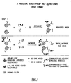
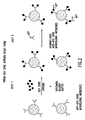
- the uniqueness of the current invention lies in its ability to detect HCV antibodies and HCV antigens simultaneously (see also International Application No. WO 00/07023 ).
- This combination test or "combo" assay utilizes antigen detection to identify exposure to HCV during the pre-seroconversion "window period" and antibody detection to identify exposure to HCV after seroconversion.
- the subject invention encompasses a method for simultaneously detecting the presence of at least one HCV core antigen and at least one HCV anti-core antibody in a test sample comprising the steps of:
- said at least one monoclonal anti-core antibody coated on the solid phase is not reactive with said at least one core antigen coated on the solid phase.
- said solid phase is a microparticle.
- the present invention further encompasses a kit comprising: a) a container containing at least one HCV core antigen coated on a solid phase, wherein said HCV viral core antigen is a recombinant protein comprising an amino acid sequence consisting of SEQ ID NO : 6 and conservative amino acid substitutions thereof, and b) a container containing at least one HCV anti-core antibody coated on a solid phase, wherein said at least one HCV anti-core antibody, coated on said solid phase is C11-14, and wherein said kit further comprises at least one conjugate comprising a signal generating compound attached to a second antibody, wherein said second antibody is C 11-10 .
- said signal generating compound is acridinium.
- the at least one HCV antigen coated on the solid phase may be selected from the group consisting of core antigen, NS3, NS4, NS5, and portions thereof.
- the at least one antibody coated on the solid phase may be a monoclonal antibody selected from the group consisting of, for example, 13-959-270, 14-1269-281, 14-1287-252, 14-153-234, 14-153-462, 14-1705-225, 14-1708-269, 14-1708-403, 14-178-125, 14-188-104, 14-283-112, 14-635-225, 14-726-217, 14-886-216, 14-947-104, 14-945-218, 13-975-157, 14-1350-210, 107-35-54, 110-81-17, C11-3, C11-7, C11-10, C11-14 and C11-15.
- the at least one monoclonal antibody coated on the solid phase is preferably not reactive with the at least one antigen coated on the solid phase.
- kits comprising: a container containing: 1) at least one HCV core antigen coated on a solid phase, wherein said HCV viral core antigen is a recombinant protein comprising an amino acid sequence consisting of SEQ ID NO : 6 and conservative amino acid substitutions thereof, and 2) at least one HCV anti-core antibody coated on said solid phase, wherein said at least one HCV anti-core antibody, coated on said solid phase is C11-14, and wherein said kit further comprises at least one conjugate comprising a signal generating compound attached to a second antibody, wherein said second antibody is C11-10.
- the signal-generating compound may be, for example, acridinium or an acridinium-containing compound.
- a method of detecting HCV antigen in a test sample comprising the steps of: a) contacting the test sample with at least one HCV antibody (e.g., monoclonal) coated on a solid phase, wherein the solid phase is a microparticle, for a time and under conditions sufficient for the formation of antibody/antigen complexes; and b) detecting the presence of antibody/antigen complexes, presence of the complexes indicating presence of antigen in the test sample.
- HCV antibody e.g., monoclonal
- a method of detecting HCV antigen in a test sample comprising the steps of: a) contacting the test sample with at least one HCV antibody (e.g., monoclonal) coated on a solid phase, wherein the solid phase is, preferably, a microparticle, for a time and under conditions sufficient for the formation of antibody/antigen complexes; b) adding a conjugate to the resulting antibody/antigen complexes for a time and under conditions sufficient to allow the conjugate to bind to the bound at least one antibody, wherein the conjugate comprises a second antibody attached to a label, for example, a chemiluminescent compound capable of generating a detectable signal; and c) detecting the signal generated by the label, for example, chemiluminescent compound, a signal generated by the label indicating the presence of antigen in the test sample.
- HCV antibody e.g., monoclonal
- the present invention includes a recombinant protein comprising an amino acid sequence consisting of SEQ ID NO:6, as well as an amino acid sequence comprising conservative amino acid substitutions of this sequence.
- a conservative substitution is defined as one or more amino acid substitutions in a sequence which do not change the function of the sequence.
- the present invention also includes a recombinant protein comprising an amino acid sequence encoded by a nucleotide sequence consisting of SEQ ID NO:5 . (Substitutions, deletions and additions within the sequences which do not affect functionally affect the protein encoded by the sequence are also considered to be within the scope of the present invention.)
- the present invention includes a vector or construct comprising a nucleotide sequence consisting of SEQ ID NO:5.
- the invention also includes a host cell comprising the vector or construct.
- the subject invention relates to methods to simultaneously detect antigens of HCV core protein and antibodies to HCV core protein in a biological sample.
- the methods of the present invention will yield a positive result.
- results may be used, for example, to diagnose the patient in terms of presence and status of infection (i.e., acute or chronic) as well as to determine the suitability of a donor blood or blood product sample for transfusion.
- the present invention overcomes the problems associated with the "window period" (i.e., 50-60 days post infection) wherein an individual may be infected with HCV but may not have developed antibodies yet. Such individuals may transmit HCV to others during this period.
- the present invention allows for a quick diagnosis of HCV, as opposed to waiting for the development of antibodies, and prevents contamination of the blood supply.
- HCV viral antigens e.g., core, N3, N4 and N5
- a solid phase or are in a liquid phase.
- the test or biological sample e.g., serum, plasma, urine, etc.
- the direct method comprises simply detecting presence of the complex itself and thus presence of the antibodies.
- a conjugate is added to the bound antibody.
- the conjugate comprises a second antibody, which binds to the first bound antibody, attached to a signal-generating compound or label. Should the second antibody bind to a bound first antibody, the signal-generating compound generates a measurable signal. Such signal then indicates presence of the first antibody in the test sample.
- solid phases used in diagnostic immunoassays are porous and non-porous materials, latex particles, magnetic particles, microparticles (see U.S. Patent No. 5,705,330 ), beads, membranes, microtiter wells and plastic tubes.
- the choice of solid phase material and method of labeling the antigen or antibody present in the conjugate, if desired, are determined based upon desired assay format performance characteristics.
- the conjugate (or indicator reagent) will comprise an antibody (or perhaps anti-antibody, depending upon the assay), attached to a signal-generating compound or label.
- This signal-generating compound or label is itself detectable or may be reacted with one or more additional compounds to generate a detectable product.
- signal-generating compounds include chromogens, radioisotopes (e.g., 125I, 131I, 32P, 3H, 35S and 14C), chemiluminescent compounds (e.g., acridinium), particles (visible or fluorescent), nucleic acids, complexing agents, or catalysts such as enzymes (e.g., alkaline phosphatase, acid phosphatase, horseradish peroxidase, beta-galactosidase and ribonuclease).
- radioisotopes e.g., 125I, 131I, 32P, 3H, 35S and 14C
- chemiluminescent compounds e.g., acridinium
- particles visible or fluorescent
- nucleic acids e.g., alkaline phosphatase, acid phosphatase, horseradish peroxidase, beta-galactosidase and ribonuclease.
- enzymes
- chromo-, fluro-, or lumo-genic substrate results in generation of a detectable signal.
- detection systems such as time-resolved fluorescence, internal-reflection fluorescence, amplification (e.g., polymerase chain reaction) and Raman spectroscopy are also useful.
- HCV antigens are also being detected; thus, the present invention methods obviate the need for the running of two different tests.
- This is accomplished by exposing the test sample to a solid phase (or liquid phase) coated with specific antibodies to HCV (e.g., human or animal monoclonal antibodies to core, polyclonal antibodies, chimeric antibodies, etc.).
- Antigens if present in the sample, bind to the solid phase and may then be detected by a direct or indirect method as described above. More specifically, the indirect method involves the addition of a conjugate comprising a second antibody (which binds to the bound antigen) attached to a label or signal-generating compound. When the second antibody binds to the bound antigen, a detectable signal is then generated indicating presence of HCV antigen in the test sample.
- the antibodies which are coated on the solid phase as well as the "second antibody” may be, as noted above, monoclonal antibodies or polyclonal antibodies.
- monoclonal antibodies may be selected from Abbott monoclonal antibodies 13-959-270, 14-1269-281, 14-1287-252, 14-153-234, 14-153-462, 14-1705-225, 14-1708-269, 14-1708-403, 14-178-125, 14-188-104, 14-283-112, 14-635-225, 14-726-217, 14-886-216, 14-947-104 and 14-945-218.
- anti-core monoclonal antibodies may also be utilized for purposes of the present invention: 107-35-54, 110-81-17, 13-975-157, 14-1350-210 (see U.S. Patent No. 5,753,430) and Tonen HCV core monoclonals C11-3, 7, 10, 14 and 15 (see PCT Application WO 99/06836 ), all of which are available from the American Type Culture Collection, 10801 University Boulevard, Manassas, VA 20110-2209.
- monoclonal antibodies may be created, see Kohler and Milstein, Nature (1975) 256:494 , and reviewed in Monoclonal Hybridoma Antibodies: Techniques and Applications, ed. Hurrell (CRC Press, Inc., 1982 ); see also J.W. Goding in Monoclonal Antibodies: Principles and Practice (Academic Press, N.Y., 1983 ; see also U.S. Patent No. 5,753,430 ).
- HCV core protein may be one possible target of the HCV antigen portion of the assay. More specifically, the detection of the core protein is accomplished by using monoclonal antibodies directed towards epitopes within the core protein. These anti-core monoclonals are placed on the solid phase and facilitate the capture of core antigen proteins from the test sample. For detection of HCV antibodies in the test sample, recombinant HCV core protein is also placed on the solid phase. It should be noted however that there are significant problems associated with the use of a single protein as the target for an antigen test and as the capture reagent for antibody detection, namely there is significant "cross-reactivity" between the core antigen and the anti-core monoclonal antibodies coated onto the solid phase(s). This results in a false positive signal, even in the absence of the test sample, since the monoclonal antibodies will bind to epitopes present on the recombinant protein.
- the core protein used in the antibody detection portion of the assay may be modified such that the ability of the anti-core monoclonals to bind HCV core is eliminated.
- modification may be achieved by use of recombinant DNA technology in which the epitope region (i.e., the short sequence of amino acids needed for monoclonal antibody binding) is eliminated or modified.
- the modified recombinant core protein would consequently maintain several human epitopes to which antibodies present in the serum of infected individuals would bind; however, the anti-core monoclonal antibodies used for antigen capture would not bind the modified protein.
- HCV core recombinant protein could replace the HCV core recombinant protein with polypeptides that include sequences known to bind to antibodies present in the serum of most infected individuals, but do not include sequences containing the epitopes recognized by the anti-core monoclonals used to detect HCV core antigens.
- the solid phase may be coated with nonstructural proteins (NS) 3, 4 and/or 5 (i.e., NS3, NS4 and/or NS5) and/or the core protein.
- NS nonstructural proteins
- the solid phase may be coated with any of the above-mentioned HCV proteins, or segments or portions thereof, either individually or in combination (for antibody detection).
- the antigens used for coating the solid phase may be generated as a contiguous recombinant protein, expressed as recombinant proteins, either as a single entity or as discrete entities, or as synthetic peptides designed either as a single entity or discrete entities.
- one may supplement such an anti-core antibody assay with the antigen assay portion of the combo assay described herein. See, e.g., U.S. Patent No. 6,156,495 relating to detection of HGBV E2 antibody or antigen.
- the monoclonal or polyclonal antibodies coated on the solid phase must not recognize the core antigens used on the solid phase (for antibody detection).
- the full antibody or a fragment thereof For purposes of the present invention, a "fragment” or “portion” of an antibody is defined as a subunit of the antibody which reacts in the same manner, functionally, as the full antibody with respect to binding properties.
- the initial capture antibody (for detecting HCV antigens) used in the immunoassay may be covalently or non-covalently (e.g., ionic, hydrophobic, etc.) attached to the solid phase.
- Linking agents for covalent attachment are known in the art and may be part of the solid phase or derivatized to it prior to coating.
- the second manner in which to use the solid phase for detecting HCV antibodies involves elimination of the core antigens entirely.
- the solid phase is coated with NS3, NS4 and/or NS5 and a substitute for the core protein or regions thereof (e.g., E2).
- the antibodies coated on the solid phase for detection of antigen are directed against the core protein of HCV.
- assay formats which may be used for purposes of the present disclosure, in order to simultaneously detect antigens and antibodies include, for example, Dual assay strip blots, a rapid test, a Western blot, as well as the use of paramagnetic particles in, for example, an Architect ® assay ( Frank Quinn, The Immunoassay Handbook, Second edition, edited by David Wild, pages 363-367, 2001 ). Such formats are known to those of ordinary skill in the art.
- the assays of the present disclosure may also be used to solely detect HCV antigens or HCV antibodies, rather than both, if desired.
- HCV antigens or HCV antibodies, rather than both.
- the stage of infection e.g., acute versus chronic
- kits may also comprise one container such as vial, bottles or strip, with each container with a pre-set solid phase, and other containers containing the respective conjugates.
- kits may also contain vials or containers of other reagents needed for performing the assay, such as washing, processing and indicator reagents.
- polystyrene beads One quarter-inch polystyrene beads were used as the solid phase for the peptide EIAs. Prior to coating, beads were washed with 15% isopropanol (in water) at room temperature for 30 minutes without agitation. Isopropanol was removed and the beads were rinsed once with deionized water. The washed beads were then added to a vial containing the peptide diluted to 5 ⁇ g/ml in 0.1 M sodium phosphate, pH 7.5 buffer (0.233 ml per bead). Beads were incubated at 56 °C for 2 hours with end-over-end mixing.
- Beads were then washed three times with PBS and then incubated in PBS containing 0.1% Triton X-100 at 40 °C for 1 hour with end-over-end mixing. They were again washed three times in PBS and then incubated at 40 °C in 5% BSA/PBS for 1 hour end-over-end mixing. Beads were washed four times with PBS and then incubated at room temperature in PBS containing 5% sucrose without mixing for 20 minutes. Sucrose buffer was removed and beads air-dried. Coated beads were stored desiccated at 4 °C.
- Bead coating validation To determine whether the biotinylated peptides were actually coated onto the beads, an assay was performed in which beads were incubated in buffer containing horseradish peroxidase-labeled streptavidin (200-400 ng/ml). The beads were then washed with deionized water and substrate added. Product was detected by absorbance at 492 nm. All peptides in Table I were shown to be coated onto the polystyrene beads by this assay (data not shown).
- HCV peptide EIAs Monoclonal antibodies generated against a recombinant HCV core protein (see Example I) were tested for their ability to bind to each of peptide-coated beads as follows: monoclonal antibodies were diluted to 50 ng/ml in sample diluent buffer (Tris buffer containing 20% goat serum, 10% calf serum, 0.2% Triton X-100 and sodium azide) of which 0.2 ml was added into a reaction well containing the peptide-coated bead and incubated at room temperature for 2 hours with mixing.
- sample diluent buffer Tris buffer containing 20% goat serum, 10% calf serum, 0.2% Triton X-100 and sodium azide
- Beads were then washed with deionized water followed by the addition of 0.2 ml of peroxidase-labeled goat anti-mouse IgG (0.3 ⁇ g/ml). Beads were incubated at room temperature for 60 minutes with mixing. Beads were washed with deionized water, transferred into plastic tubes to which 0.3 ml of OPD (0.3% O-phenylenediamine-2-HCl in citrate buffer containing 0.02% H 2 O 2 ) substrate was added, and incubated in the dark at room temperature for 30 min without mixing. Reaction was quenched by the addition of 1 ml of 1N H2SO4 and the OD at 492 nm determined. The absorbance is directly proportional to the amount of antibody bound to the bead.
- OPD O-phenylenediamine-2-HCl in citrate buffer containing 0.02% H 2 O 2
- a plasmid containing nucleotides 14-5294 of the H strain of HCV ( Ogata et al., Proc. Natl. Acad. Sci. USA 88:3392-3396 (1991 )) in pGEM-9Zf(-) (Promega Corp., Madison, WI) was partially digested using DNase I by the following method in order to obtain random epitope-encoding fragments:
- T7Select10-3b (Novagen, Inc., Madison, WI) was digested with EcoRI and dephosphorylated with calf intestinal alkaline phosphatase (New England Biolabs, Beverly, MA) as directed by the manufacturer. Size-selected digested DNA fragments (30 ng) (supra) were ligated with 0.5 ⁇ g digested T7Select10-3b in a 5 ⁇ l reaction volume at 16 °C overnight. The entire ligate was packaged into phage heads using T7Select packaging extract (Novagen, Inc., Madison, WI) and titered as directed by the manufacturer. The resulting unamplified library contained a total of 3.9 x 10 6 members (PFU).
- the packaged phage were amplified by liquid lysate amplification in E. coli BLT5403 (20 ml culture) as directed by the T7 Select System Manual (Novagen, Inc., Madison, WI).
- the amplified library had a titer of 1.3 x 10 11 PFU per ml.
- Each monoclonal antibody (20 ⁇ g) that was to be used for biopanning was incubated at 4 °C for 4 hours on an end-over-end rocker in 300 ⁇ l blocking buffer (2% BSA, 3% nonfat dry milk, 0.2% Tween 20, 0.02% sodium azide in phosphate-buffered saline).
- 300 ⁇ l blocking buffer 2% BSA, 3% nonfat dry milk, 0.2% Tween 20, 0.02% sodium azide in phosphate-buffered saline.
- an aliquot of the amplified HCV gene fragment library (supra) containing approximately 10 11 phage was precipitated as follows: 1/10 volume of 5 M NaCl was added to the phage, mixed thoroughly, followed by addition of 1/6 volume polyethylene glycol (MW 8000), mixed thoroughly again, and incubated on ice for 1-2 hours.
- the phage were centrifuged at 6000 x g for 10 min at room temp, all supernatant was removed and the phage pellet was vigorously resuspended in 120 ⁇ l buffer containing 1 M NaCl, 10 mM Tris-HCl pH 8.0, 1mM EDTA. The phage were added to the pre-incubated monoclonal antibody and incubated at 4 °C overnight on an end-over-end rocker.
- the antibody-phage complexes were captured on paramagnetic particles coupled to goat anti-mouse IgG (Fc specific) as follows.
- Fc specific goat anti-mouse IgG
- a 0.2 ml aliquot of Goat Anti-Mouse IgG Fc BioMag particles (Polysciences, Inc., Warrington, PA) was washed three times with 0.4 ml 0.1% Tween 20, 0.1% BSA, 0.02% sodium azide in phosphate-buffered saline (PBS) by gentle vortexing followed by capture on a magnetic stand for 0.5-1 minute. The supernatant was removed carefully without disturbing the particles.
- PBS phosphate-buffered saline
- Particles were then resuspended in the IgG-phage from overnight incubation above and incubated at room temperature on an end-over-end rocker for 3 hours. Particles were washed six times as above using 6.0 ml 0.5% Tween 20, 0.1% BSA in PBS per wash. Bound phage were eluted using 0.2 ml 0.1% Tween 20, 0.1% BSA, 1.0% SDS in PBS at room temperature on an end-over-end rocker for 90 minutes. The tube was placed on a magnetic stand for 1-2 minutes, after which the supernatant containing the eluted phage was removed to a clean tube. The sample containing eluted phage was titered as directed in the T7 Select System Manual.
- the eluted phage was amplified as follows. Ten ml LB Broth (Gibco BRL, Gaithersburg, MD) plus 100 ⁇ g/ml ampicillin was inoculated with E. coli BLT5403 and incubated at 37 °C overnight with shaking. The following morning, 35 ml LB Broth plus 100 ⁇ g/ml ampicillin, 1X M9 salts, 0.4% glucose, 1mM MgSO 4 was inoculated with 0.2 ml of the overnight culture and incubated at 37 °C with shaking until the A600 absorbance was 0.5-0.6.
- Eluted phage (185 ⁇ l) from first round biopanning (supra) was added and incubation at 37 °C was continued for 1.5-2 hours, until the A600 absorbance of the culture had dropped to approximately 0.5, indicating lysis.
- the culture was centrifuged at 8000 x g for 10 minutes and the supernatant was removed to a clean tube and stored at 4 °C. The culture supernatant was titered as directed in the T7 Select System Manual.
- a DNA fragment containing a region of the HCV genome that encodes amino acids 1-173 of the HCV nucleocapsid protein was utilized as a hybridization probe. This region was chosen because all of the monoclonal antibodies analyzed in the biopanning experiments recognize epitopes in the HCV core protein. Phage resulting from 2-3 rounds of biopanning and amplification were plated on E. coli BLT5403 and incubated at 37 °C until plaques formed. DNA was transferred onto Hybond-N+ membranes (Amersham Life Sciences, Inc., Arlington Heights, IL), denatured, neutralized, and UV cross-linked, as described by the manufacturer.
- the membranes were pre-hybridized, hybridized with the HCV nucleocapsid gene 32 P-labeled probe, washed and exposed as described and known in the art.
- Individual hybridizing plaques were isolated and the inserts were amplified by PCR using T7SelectUP and T7SelectDOWN primers (Novagen, Inc., Madison, WI) as directed in the T7Select System Manual.
- T7SelectUP and T7SelectDOWN primers Novagen, Inc., Madison, WI
- 30-50 independent hybridizing plaques were amplified and then purified using the QIAquick PCR purification kit (Qiagen, Inc., Chatsworth, CA).
- HCV Hepatitis C Virus
- Combination assays for the simultaneous detection of both core antigen and human anti-core antibody in a single assay combine the two assay formats.
- a recombinant core antigen is necessary that will be recognized by human anti-core antibodies present in the serum, while escaping recognition by the murine monoclonal antibodies used to capture and detect native core antigen also present in serum.
- Such recombinants can be constructed by eliminating small regions (1-30 or more amino acids) within the core antigen, thus disrupting or eliminating the epitope(s) recognized by the murine monoclonal antibodies while at the same time leaving undisturbed numerous other epitopes that will allow human anti-core antibody detection.
- the first of these antigens constructed contained HCV amino acids 8-100 in which amino acids 32-50 were deleted from the recombinant. In this manner, antibodies C11-10 and C11-14, which bind to epitopes at amino acids 32-36 and 45-50, respectively (Example II), will not bind to the resulting recombinant antigen.
- PCR was performed in a PE-9600 thermocycler in the presence of 0.5 ⁇ M of each oligonucleotide primer and 3-5 pg of plasmid template using the TaKaRa LA Taq PCR Kit (Pan Vera, Corp., Madison, WI) as per manufacturer's instructions (50 ⁇ l volume).
- a pre-incubation at 94 °C for 1 minute was followed by 35 cycles of PCR (denaturation at 94 °C for 20 seconds; annealing at 50 °C for 30 seconds; extension at 72 °C for 30 seconds), which was then followed by a final extension at 72 °C for 10 minutes.
- QIAquick PCR Purification Kit QIAGEN, Inc., Valencia, CA
- a second round of PCR was then performed as supra for 10 cycles to tether the two purified fragments (1 ⁇ l each in a 20 ⁇ l volume reaction) to one another at the regions of overlap within the oligonucleotide primers (SEQ ID NO:2 and SEQ ID NO:3).
- the product of the second PCR was amplified in a third PCR for 35 cycles utilizing 0.5 ⁇ M of the flanking oligonucleotide primers (SEQ ID NO:1 and SEQ ID NO:4) and the conditions described supra (100 ⁇ l volume).
- the product of the third PCR was purified using the QIAquick PCR Purification Kit, then digested with the restriction enzymes EcoRI and BamHI, digestion sites that were incorporated into the flanking oligonucleotide primers (SEQ ID NO:1 and SEQ ID NO:4).
- the fragment encoding the recombinant was ligated into the bacterial expression vector pJO200 that had been similarly digested with EcoRI and BamHI using the pGEM-T Easy Ligation Kit (Promega, Madison, WI), then transformed into XL1-Blue competent cells (Stratagene, La Jolla, CA).
- HCV core recombinants have been constructed, cloned and expressed in a manner identical to that described supra.
- the first of these contains HCV amino acids 8-100 in which amino acids 33-35 and 46-49 have been deleted.
- the oligonucleotide primers utilized to amplify the two fragments in the first PCR were SEQ ID NO:1 and SEQ ID NO:9, and SEQ ID NO:10 and SEQ ID NO:4, respectively, followed by final amplification in the third PCR with SEQ ID NO:1 and SEQ ID NO:4.
- the second of these recombinants encodes HCV amino acids 8-100 in which the leucine residue at amino acid 36 has been substituted with valine, and the arginine residue at amino acid 47 has been substituted with leucine.
- the oligonucleotide primers utilized to amplify the two fragments in the first PCR were SEQ ID NO:1 and SEQ ID NO:13, and SEQ ID NO:14 and SEQ ID NO:4, respectively, followed by final amplification in the third PCR with SEQ ID NO:1 and SEQ ID NO:4.
- a recombinant (see nucleotide sequence SEQ ID NO:15 and corresponding amino acid SEQ ID NO:16) encoding HCV amino acids 8-100 was constructed using oligonucleotide primers SEQ ID NO:1 and SEQ ID NO:4 in a single PCR reaction.
- Additional recombinant antigens constructed for use in an HCV antigen/antibody combination assay included antigens that contained the 33c region of HCV (amino acids 1192-1457) tethered to a core region of the virus.
- the template used for such constructions was a plasmid containing a bacterial codon-optimized sequence of amino acids 1192-1457, followed by amino acids 1-150 from the H strain of HCV (Ogata, 1991), with two non-HCV coding amino acids separating the two sequences.
- This recombinant (HC-43) is routinely used in multiple commercial assays for the detection of HCV.
- the HC43 recombinant is expressed as a non-fusion protein from the pL promoter of bacterial phage lambda.
- the additional recombinants were constructed by one of two methods. First, existing clones encoding distinct, related recombinants were joined by DNA ligation to form a third unique recombinant, or, new unique clones were constructed by tethering PCR described in Example III, Part B.
- the first of the newer antigens constructed contained HCV amino acids 1192-1457 (representing a segment of NS3) tethered to amino acids 1-100 (representing a segment of core protein) in which amino acids 32-50 had been deleted.
- This recombinant was constructed by restricting plasmid pHC43 with the endonucleases, Xma I and Bam HI.
- Xma I cuts pHC43 near amino acid 24 of the core-encoding region while Bam HI cuts downstream of the translation termination codon.
- This region of pHC43 was replaced by DNA ligation using the pGEM-T Easy Ligation Kit (Promega Corp., Madison, WI), with the Xma I-Bam HI fragment obtained from the pJO200 vector encoding HCV core amino acids 8-100 with 32-50 deleted described in Example III (SEQ ID NO:5 and SEQ ID NO:6).
- the new plasmid was then used to transform XL1-Blue competent cells (Stratagene, La Jolla, CA). After selection of clones containing the appropriately sized insert, the nucleotide sequence of the recombinant was confirmed (SEQ ID NO:17), exhibiting the deduced amino acid sequence in SEQ ID NO:18.
- Recombinant antigens made by tethering PCR were constructed as detailed in Example III, Part B.
- the first of these recombinants, p9MB-19 (SEQ ID NO:19 (nucleotide sequence) and SEQ ID NO:20 (amino acid sequence)) contains HCV amino acids 1192-1457 followed by amino acids 8-100 in which amino acids 32-50 had been deleted.
- the oligonucleotide primers used to amplify the initial two fragments in the first PCR were SEQ ID NO:21 and SEQ ID NO:22, and SEQ ID NO:23 and SEQ ID NO:4, respectively.
- Final amplification in the third PCR utilized oligonucleotide primers SEQ ID NO:21 and SEQ ID NO:4.
- Recombinant p9MB-20 contains HCV amino acids 1192-1457, four glycine residues and a serine residue, followed by HCV amino acids 8-100 in which amino acids 32-50 had been deleted.
- the oligonucleotides primers used to amplify the initial two fragments in the first PCR were SEQ ID NO:21 and SEQ ID NO:22, and SEQ ID NO:26 and SEQ ID NO:4, respectively.
- Final amplification in the third PCR utilized oligonucleotides primers SEQ ID NO:21 and SEQ ID NO:4.
- Recombinant p9MB-22 contains HCV amino acids 1192-1457, four glycine residues and a serine residue, followed by HCV amino acids 1-150.
- the oligonucleotides primers used to amplify the initial two fragments in the first PCR were SEQ ID NO:21 and SEQ ID NO:22, and SEQ ID NO:29 and SEQ ID NO:30, respectively.
- Final amplification in the third PCR utilized oligonucleotides primers SEQ ID NO:21 and SEQ ID NO:30.
- Recombinant p9MB-31 contains HCV amino acids 1192-1457 followed by amino acids 1-100 in which amino acids 31, 32, 33, 47 and 48 had been deleted.
- the oligonucleotides primers used to amplify the initial two fragments in the first PCR were SEQ ID NO:21 and SEQ ID NO:33, and SEQ ID NO:34 and SEQ ID NO:4, respectively.
- Final amplification in the third PCR utilized oligonucleotides primers SEQ ID NO:21 and SEQ ID NO:4.
- Recombinant 9MB-24 contains HCV amino acids 1192-1457 followed by amino acids 1-100 in which amino acids 33-35 and 46-49 were deleted from the recombinant.
- This recombinant was constructed by restricting plasmid pHC43 with the endonucleases, Xma I and Bam HI, and replacing the fragment by DNA ligation, with the Xma I-Bam HI fragment obtained from SEQ ID NO:7.
- Recombinant 9MB-25 contains HCV amino acids 1192-1457, four glycine residues and a serine residue, followed by HCV amino acids 1-100 in which amino acids 33-35 and 46-49 were deleted from the recombinant.
- This recombinant was constructed by restricting plasmid p9MB-22 with the endonucleases, Xma I and Bam HI, and replacing the fragment by DNA ligation, with the Xma I-Bam HI fragment obtained from SEQ ID NO: 7.
- Recombinant 9MB-26 contains HCV amino acids 1192-1457, four glycine residues and a serine residue, followed by HCV amino acids 1-100 in which amino acids 32-50 were deleted from the recombinant.
- This recombinant was constructed by restricting plasmid p9MB-22 with the endonucleases, Xma I and Bam HI, and replacing the fragment by DNA ligation, with the Xma I-Bam HI fragment obtained from p9MB-18 (SEQ ID NO:17).
- Example (IV) To 1 ml of coated microparticles from Example (IV) (A1)(vi) was added 375 ⁇ l of HCV Core peptide from Example (V)(A1)(v) and 11-28 aa at 1 mg/ml in PBS buffer. The mixture was then incubated for 2 hours at room temperature. The prepared microparticles were washed with wash buffer (DTT, EDTA, SDS in PBS, pH 6.5), and the microparticles were resuspended in 1 ml of microparticle diluent (Calf Bovine Serum, Horse IgG, TWEEN 20, BSA, Casein, EDTA, Sucrose and Proclin, pH 6.5) yielding 1% solids final concentration.
- wash buffer DTT, EDTA, SDS in PBS, pH 6.5
- mice were injected intraperitoneally with 15 ⁇ g of purified pHCV34 with 15 ⁇ g each of Trehalose dimycolate (TDM) and M. phlei in a buffer emulsion prepared according to the manufacturer's instructions. Subsequent immunizations were performed on day 14, 28 and 42. Mice were bled on days 21 and 49, and the immune response was studied by enzyme-linked immunosorbent assay utilizing pHCV34 coated on polystyrene beads, as detailed in U.S. Patent No. 5,753,430 .
- TDM Trehalose dimycolate
- M. phlei M. phlei
- mice Upon demonstration of specific anti-HCV antibody present at reasonable titers in the sera of immunized mice, mice were boosted with 40 ⁇ g of pHCV34 antigen. The mice were sacrificed and their spleens were removed; the white cells were mixed and fused with SP2/0 cells. The cell mixture was cultured in Biscoe's Modified Dubach's Medium (IMDM) supplemented with 20 % fetal calf serum, and the hybridized cells were selected by using a hypoxanthine and thymidine medium.
- IMDM Biscoe's Modified Dubach's Medium
- Hybridoma cell lines were established, and all monoclonal antibodies specific for antibodies to core were prepared from ascite fluids of the mice and were purified by chromatography on a protein-A column (Pharmacia, Uppsala, Sweden). The epitopes of the monoclonal antibodies were analyzed by an ELISA test described in Example I.
- HCV C11-3 microparticles (1% solid) prepared as described in Example (V)(B1)(iii) and 84 ⁇ l HCV C11-14 microparticles (1% solids) prepared as described in Example (V)(B1)(i) were mixed with 880 ⁇ l microparticle diluent (Calf Bovine Serum Horse IgG, Tween 20, BSA, Casein, EDTA, Sucrose, and Proclin, pH 6.5).
- both the antibody and antigen assays were performed on a single channel where the reagents for both antigen and antibody assay were combined in one kit.
- mouse anti-human IgG directly labeled with acridinium or a pre-complex of biotinylated anti-human F (ab')2 and acridinium anti biotin conjugate was used.
- monoclonal antibodies were coated onto microparticles or conjugated with acridinium as described in Example IV and V.
- Screening assays used monoclonal antibody-coated microparticles (0.40 ⁇ m diameter) at a working concentration of 0.09-0.15% solids and conjugated monoclonals at a working concentration of 100-125 ng/ml.
- the same positive and negative control plasma were used (0.1 ml for each assay).
- the positive control serum was from an HCV-infected individual who tested negative for HCV antibodies but whose plasma had an HCV RNA titer of 19,000,000 copies per ml.
- the negative control plasma was from a normal blood donor who was negative for HCV antibodies and RNA. Assays were performed using the instrumentation and operation methods as described in Example IX.
- Table VII shows the mean signal-to-negative (S/N) values obtained upon testing the various pairs of monoclonal antibodies for their ability to detect HCV core antigen in the positive control human plasma (nd: not determined). From this data, it is apparent that some pairs of monoclonal antibodies exhibit greater sensitivity than others and that the sensitivity was dependent upon the proper configuration of the assay. For example, when monoclonal antibody A05 was used as the capture reagent and C11-10 was used as the detection reagent, the resulting S/N value was 150.0; however, when the opposite configuration was used, the resulting S/N value was only 6.8
- the HCV core antigen assay for PRISM ® utilizes a sample diluent buffer (SDB) for dilution of the human serum or plasma sample to be tested.
- SDB sample diluent buffer
- the monoclonal antibody-coated microparticles are then added to form a reaction mixture. It is possible that the sensitivity and specificity of the antigen detection assay is affected by the composition of the SDB, in terms of the ingredients and their concentration.
- HCV is believed to be an enveloped virus
- detergent surfactant
- chaotropic salts to the SDB might aid in dissolution of the nucleocapsid complex which could enhance detectability of core antigen.
- HCV core antigen assay comprised of monoclonal antibody C11-7 or C11-14 coated microparticles (as stated in the table legends) and acridinium labeled monoclonal antibody C11-10 conjugate.
- the simplest SDB used (also referred to as basal buffer), in terms of number of components, consisted of 0.1 M potassium phosphate, pH 7.2, 10 mM EDTA. This is the buffer to which detergents and salts were added.
- the performance of the SDBs was determined by examining their effect on the signal-to-negative (S/N) ratio obtained upon testing of a positive control human plasma from an individual who tested negative for HCV antibodies but whose plasma had an HCV RNA titer of 19,000,000 copies per ml.
- the negative control plasma was from a normal blood donor who was negative for HCV antibodies and RNA.
- Screening assays used coated microparticles at a working concentration of 0.09-0.15% solids and C11-10 conjugate at a working concentration of 100-125 ng/ml. For all experiments, the same positive and negative control plasma was used (0.1 ml for each assay). Assays were performed using the instrumentation and operation methods as described in Example VIII.
- Assay format Anti-HCV core monoclonal C11-14 coated microparticles (0.227 um) and C11-10 conjugate.
- TABLE XII SDB Components and Final Concentration KCl SB-12 SB-16 CTAB Triton X-100 Urea S/N 0.5 M 2% 48.6 0.5 M 2% 2.5 M 86.8 0.5 M 2% 0.10% 52.8 0.5 M 2% 0.10% 2.5 M 77.6 0.5 M 2% 0.10% 1.80% 83.6 0.5 M 2% 0.10% 1.80% 2.5 M 106.8 0.5 M 2% 2.5 M 122.6 0.5 M 2% 0.10% 2.5 M 136 0.5 M 2% 0.10% 1.80% 127.9 0.5 M 2% 0.10% 1.80% 2.5 M 113.7 All SDBs were prepared in basal buffer containing 0.1 M potassium phosphate, pH 7.2, 10 mM EDTA.
- Assay format Anti-HCV core monoclonal C11-14 coated microparticlec (0.227 um) and C11-10 conjugate.
- TABLE XIII Effect of Urea on Antigen Assay Sensitivity S/N values at various urea concentrations HCV positive control plasma dilution factor 0.0 M 0.5 M 1.0 M 1.5 M 2.0 M 2.5 M 3.0 M 5.0 M 1:2 35.2 31.3 32.9 33.4 43.0 34.6 33.2 1.1 1:4 19.9 15.7 17.7 13.6 21.5 18.9 15.6 1.1 1:8 9.1 8.2 9.3 6.1 10.5 8.1 8.0 0.9 1:16 5.9 4.3 5.7 5.3 6.6 5.6 5.5 1.0 1:32 3.8 3.2 3.6 3.3 3.6 3.5 3.0 1.0 Urea was added to increasing final concentrations in the following buffer: 0.1 M potassium phosphate, pH 7.2, 10 mM EDTA, 0.5 M Nacl, 2.0% SB-16, 0.10 CTAB, 1.8% Triton X-100.
- HCV positive control plasma was diluted in negative control plasma.
- Assay format Anti-HCV core monoclonal C11-14 coated microparticles (0.227 um) and C11-10 conjugate.
- Table XIV Effect of Protein or Serum on Antigen Assay Sensitivity S/N S/N Component(s) Added (final conc., w/v)y PC 1:2 PC 1:16 No additions 69.3 9.6 1% BSA, 2% mouse serum 73.7 10.3 1% BSA, 0.1% casein 70.5 10.7 1% BSA 70.2 10.3 3% horse serum 68.2 9.6 2% mouse serum 65.6 10.9 0.1% casein 51.7 8.1 2% mouse serum, 0.1% casein 50.9 7.9 1% BSA, 3% horse serum 50.6 8.8 0.1% casein, 3% horse serum 40.7 6.9 2% mouse serum, 3% horse serum 34.3 4.9 Protein or sera were added to buffer containing 100 mM potassium phosphate, pH 7.2, 10 mM EDTA, 0.5 M NaC
- the PRISM ® antibody assay is described in U.S. Patent No. 5,705,330 , incorporated herein by reference and the PRISM ® antigen and antibody assays are described in Shah and Stewart, The Immunoassay Handbook, second edition, edited by David Wild, p 297-303 (2001 ), also incorporated herein by reference.
- test or sample 50 ⁇ l of control or sample, 50 ⁇ l of specimen diluent buffer (SDB, Phosphate buffer, pH 7.0 containing Tween 20, newborn calf serum, NaCl, superoxide dismutase (SOD), E. coli lysate and azide), and 50 ⁇ l of HCV antigen coated microparticles (prepared as described in Example (V) (A1) (iv) above) were dispensed into each incubation well and assay timing was started. These were mixed by mutual diffusion of each into the other without external agitation or shaking to form a reaction mixture.
- SDB specimen diluent buffer
- SOD superoxide dismutase
- E. coli lysate and azide 50 ⁇ l of HCV antigen coated microparticles
- the reaction mixture was transferred to a detection well which contained a fibrous matrix and washed twice with 300 ⁇ l of transfer wash (TW, containing borate buffer, pH 7.0, with NaCl, Tween -20, Glycerol and Proclin 300). After 18 minutes of incubation at room temperature, 50 ⁇ l of a pre-complexed biotinylated F(ab') 2 /acridinium labeled anti-biotin, (biotinylated F(ab') 2 fragment of goat anti-human IgG and acridinium labeled anti-biotin antibody), was dispensed into the matrix of the detection well at station 5.
- TW transfer wash
- a chemiluminescence (CL) signal was generated by addition of an alkaline hydrogen peroxide solution, and the photons were measured by a photo multiplier tube. The amount of light emitted is proportional to the amount of antibody in the sample.
- the presence or absence of antibody in the sample is determined by comparing the number of photons collected from the sample to a cutoff (S/CO) value determined from a calibration performed in the batch. The results are expressed as S/CO (signal to cutoff) in Table XV below.
- S/CO signal to cutoff
- test or sample 100 ⁇ l of control or sample, 50 ⁇ l of specimen diluent buffer (SDB, Sodium phosphate, EDTA, Triton X-100, Urea and sodium azide), and 50 ⁇ l of HCV Mab coated microparticles (prepared as described in Example (V) (B1) (i)) were dispensed into each incubation well and the assay timing was started. These were mixed by mutual diffusion of each into the other without external agitation or shaking to form a reaction mixture.
- SDB specimen diluent buffer
- EDTA Sodium phosphate
- Triton X-100 50 ⁇ l of HCV Mab coated microparticles
- reaction mixture was transferred to a detection well which contained a fibrous matrix and washed twice with 300 ⁇ l of transfer wash (TW) (MES, NaCl, Triton X-100, PEG, Antifoam, Proclin 300, pH 5.6) after 18 minutes of incubation at room temperature.
- TW transfer wash
- 50 ⁇ l of acridinylated C11-10 Mab conjugate was dispensed into the matrix of the detection well.
- HCV Ag Assay Mab C11-14 and C11-3/Mab C11-10 pair:
- Peptide aa 10-53 (ALAM-17) (KTKRNTNRRPQDVKFPGGGQIVGGVYLLPRRGPRLGVRATRKTS) (SEQ ID NO:42) contains HCV core amino acids 10-53 with no intervening deletions.
- Peptide aa 10-53 ⁇ 32-50 (ALAM-18) (KTKRNTNRRPQDVKFPGGGQIVKTS) (SEQ ID NO:43) contains HCV core amino acids 10-53 with a 19 amino acid deletion encompassing aa 32-50.
- Peptide aa 10-53 ⁇ 31-33 ⁇ 47-48 contains HCV core amino acids 10 through 53 where amino acids 31-33 and 47-48 were deleted (ALAM16) (KTKRNTNRRPQDVKFPGGGQIVYLLPRRGPRLGVTRKTS) (SEQ ID NO:41).
- Peptides ALAM-16 and ALAM-18 are both compatible with an HCV Ab/Ag combo format.
- ALAM-16 the deletion of amino acids 31-33 prevents monoclonal c11-10 (epitope 32-36) from binding to the antigen, and the deletion of amino acids 47 and 48 prevents binding of the c11-14 monoclonal (epitope 45-50).
- ALAM-18 contains a deletion that encompasses both the c11-10 and c11-14 binding regions.
- HCV genotyped seropositive specimens representing HCV genotypes 1, 2, 3, 4 and 6, were tested by peptide aa 10-53 ⁇ 31-33 ⁇ 47-48 (ALAM-16) to determine feasibility of this peptide as an antigenic target in an HCV Ab/Ag assay format. All 254 (100%) specimens were reactive toward this peptide.
- the deletions present in this peptide which serve to eliminate binding by the monoclonals necessary for Ag detection, do not negatively impact reactivity of antibodies toward the remaining core epitopes.
Landscapes
- Health & Medical Sciences (AREA)
- Life Sciences & Earth Sciences (AREA)
- Chemical & Material Sciences (AREA)
- Immunology (AREA)
- Engineering & Computer Science (AREA)
- Molecular Biology (AREA)
- General Health & Medical Sciences (AREA)
- Medicinal Chemistry (AREA)
- Hematology (AREA)
- Biochemistry (AREA)
- Biomedical Technology (AREA)
- Urology & Nephrology (AREA)
- Organic Chemistry (AREA)
- Cell Biology (AREA)
- Gastroenterology & Hepatology (AREA)
- Physics & Mathematics (AREA)
- Analytical Chemistry (AREA)
- Microbiology (AREA)
- Biotechnology (AREA)
- General Physics & Mathematics (AREA)
- Pathology (AREA)
- Virology (AREA)
- Food Science & Technology (AREA)
- Biophysics (AREA)
- Genetics & Genomics (AREA)
- Proteomics, Peptides & Aminoacids (AREA)
- Communicable Diseases (AREA)
- Peptides Or Proteins (AREA)
- Micro-Organisms Or Cultivation Processes Thereof (AREA)
- Investigating Or Analysing Materials By The Use Of Chemical Reactions (AREA)
Description
- The subject invention relates to methods for the simultaneous detection of Hepatitis C Virus (HCV) antigens as well as antibodies produced in response to HCV antigens. Furthermore, the subject invention allows one to detect antigens in the early, acute stage of infection, even prior to the development of antibodies, thereby allowing for early detection of infected blood and blood products, and thus improving the safety of the blood supply.
- Recent epidemiological studies indicate that HCV infects more than 170 million people worldwide and that, in more than 50% of the cases, the infection is chronic. In the United States, there are approximately 4 million people infected, and 30,000 new infections are estimated to occur annually (NIH Conference, Hepatology Suppl 1:2S (1997)). In addition, HCV is responsible for 8,000-10,000 deaths annually in the United States and is the leading indicator for liver transplantation.
- The HCV genome is a single-stranded RNA molecule of positive polarity that is approximately 9400-9500 nucleotides in length. The organization of the coding regions resembles that of other flaviviruses [Major et al., Hepatology 25:1527 (1997)] as well as the more recently discovered GB viruses [Muerhoff AS, et al., J Virol 69:5621 (1995)]. The HCV genome possesses a large open reading frame (ORF) encoding a polyprotein precursor of 3010 to 3033 amino acids depending on the particular isolate [Choo et al., Proc Natl Acad Sci USA 88:2451 (1991); Grakoui et al., J Virol 67:1385 (1993)]. HCV structural genes (core and envelope) are encoded near the 5'-end of the genome, followed by the proteases and helicase, the helicase cofactor and the replicase. Noncoding regions (NCR), thought to be important in replication, are found at each end of the genome.
- HCV infection occurs primarily through parenteral exposure, i.e., through shared needles, by tattooing, or through transfusion of contaminated blood or blood products. Following exposure, the virus enters a susceptible hepatocyte and viral replication occurs. There is an eclipse phase period of approximately 10 days during which time there is no evidence of viral presence (i.e., viral RNA cannot be detected), serum transaminase levels are within normal limits, and there is no evidence of an immune response to HCV [Busch et al., Transfusion 40:143 (2000)]. Typically, about 10 days following exposure, HCV RNA can be detected, often with viral loads between 100,000-120,000,000 HCV RNA copies per ml of serum. Several weeks later, there is typically an increase in ALT levels indicating inflammation of the liver; antibodies are detected an average of about 70 days after exposure.
- One of the preventive measures employed to limit the spread of HCV infections is to screen blood for exposure to HCV, either by the detection of antibodies to HCV or by the detection of viral-specific molecules (e.g., HCV RNA or HCV core proteins) in serum/plasma. Blood or blood products derived from individuals identified as having been exposed to HCV, by these tests, are removed from the blood supply and are not utilized for distribution to recipients of blood products (see, e.g.,
U.S. Patent No. 6,172,189 ). These tests may also be utilized in the clinical setting to diagnose liver disease attributable to HCV infection. - Due to the unavailability of native, intact HCV virions, serologic antibody tests have relied on recombinant antigens or synthetic peptides, representing selected fragments of the viral polyprotein. The first generation anti-HCV screening tests were based on detection of antibodies directed against a recombinant protein (HCV genotype 1a) originating from sequences located in the nonstructural NS-4 protein (C100-3) [Choo et al., Science 244:359 (1989) ; Kuo et al., Science 244:362 (1989)]. The first generation assays failed to detect antibodies in approximately 10% of individuals having chronic HCV infection and up to 10-30% of individuals presenting with acute HCV infection. The second generation anti-HCV assays have incorporated recombinant proteins from three different regions of the HCV genome (HCV genotype 1a), including amino acid sequences from the core, NS3, and NS4 protein [Mimms et al., Lancet 336:1590 (1990) ; Bresters et al., Vox Sang 62:213 (1992)], allowing a marked improvement over the first generation tests in identifying HCV infected blood donors [Aach et al., N Engl J Med 325:1325 (1991); Kleinman et al., Transfusion 32:805 (1992)]. The second generation assays detect antibodies in close to 100% of chronic HCV cases [Hino K., Intervirology 37:77 (1994)] and in nearly 100% of the acute cases by 12 weeks post infection [Alter et al., N Engl J Med 327:1899 (1992); Bresters et al., Vox Sang 62:213 (1992)]. The third generation test includes a recombinant protein expressing amino acid sequences from the NS5 region, as well as antigens from the core, NS3 and NS4. Some studies have indicated a slight improvement in sensitivity in comparing the third generation tests to second generation tests [Lee et al., Transfusion 35:845 (1995); Courouce et al. Transfusion 34:790-795 (1994)], but this improvement is largely attributed to changes in the NS3 protein rather than the inclusion of NS5 [Courouce et al., Lancet 343:853 (1994)].
- In general, the second and third generation HCV antibody tests detect exposure to HCV about 70 days after exposure. Since HCV establishes persistent, and in many cases lifelong infection, the detection of antibodies to HCV represents a very efficient method for determining exposure to HCV. However, antibody testing alone will frequently fail to detect HCV infected individuals during the first 70 days after exposure.
- The existing HCV antigen tests rely on detecting the presence of the HCV core antigen in serum or plasma. The core (or nucleocapsid) protein comprises the first 191 amino acids of the polyprotein. Two different types of serologic assays have been developed which permit detection of HCV core antigens in serum. One assay format detects HCV core antigens in subjects prior to seroconversion and is utilized in screening blood donors, while the other assay format detects core antigens only in hepatitis C patients, regardless of their HCV antibody status and is utilized in clinical laboratories to diagnose exposure to HCV or to monitor antiviral therapy.
- Recent data on samples obtained during the pre-seroconversion period indicate that the HCV antigen test detects exposure to HCV significantly earlier than antibody testing [Aoyagi et al., J Clin Microbiol 37:1802 (1999); Peterson et al., Vox Sang 78:80(2000); Dawson et al., Transfusion, SD161, 40 (2000) ; Muerhoff et al., 7th International Meeting on Hepatitis C virus and related viruses, December 3-7, 2000], and represents an alternative to nucleic acid testing for detecting exposure to HCV during the pre-seroconversion period. The advantages of HCV antigen detection are that the test is rapid, simple, may not require sample extraction or other pretreatment, and is not as prone to handling errors (e.g., contamination) as may occur in the HCV RNA tests.
- In clinical laboratories, the HCV antigen test has comparable sensitivity to the HCV DNA tests in detecting exposure to HCV in patients infected with different HCV genotypes [Dickson et al., Transplantation 68:1512 (1999)] and in monitoring antiviral therapy [Tanaka et al., Hepatology 32:388 (2000); Tanaka et al., J Hepatol 23:742 (1995)]. Thus, HCV core antigen tests present a practical alternative to HCV RNA for screening blood donors or for monitoring antiviral therapy.
- The uniqueness of the current invention lies in its ability to detect HCV antibodies and HCV antigens simultaneously (see also International Application No.
WO 00/07023 - The subject invention encompasses a method for simultaneously detecting the presence of at least one HCV core antigen and at least one HCV anti-core antibody in a test sample comprising the steps of:
- (a) contacting said test sample with:
- (1) at least one HCV viral core antigen coated on a solid phase, for a time and under conditions sufficient for the formation of antibody/antigen complexes, wherein said HCV viral core antigen is a recombinant protein comprising an amino acid sequence consisting of SEQ ID NO : 6 and conservative amino acid substitutions thereof, and
- (2) at least one HCV anti-core antibody coated on said solid phase, for a time and under conditions sufficient for the formation of antigen/antibody complexes, wherein said at least one HCV anti-core antibody coated on said solid phase is C11-14;
- (b) adding a conjugate to the resulting antibody/antigen complexes of (a) (1) for a time and under conditions sufficient to allow said conjugate to bind to the bound antibody in (a) (1), wherein said conjugate comprises a second antibody attached to a chemiluminescent compound capable of generating a detectable signal and simultaneously adding a second conjugate to the resulting antigen/antibody complexes of (a) (2) for a time and under conditions sufficient to allow said conjugate to bind to the bound core antigen in (a) (2), wherein said second conjugate comprises a third antibody attached to said chemiluminescent compound capable of generating a detectable signal, wherein said third antibody is C11-10; and
- (c) detecting said generated signal, presence of said signal indicating presence of said at least one HCV core antigen, said at least one HCV anti-core antibody, or both, in said test sample.
- In one embodiment, said at least one monoclonal anti-core antibody coated on the solid phase is not reactive with said at least one core antigen coated on the solid phase.
- In a further embodiment, said solid phase is a microparticle. The present invention further encompasses a kit comprising: a) a container containing at least one HCV core antigen coated on a solid phase, wherein said HCV viral core antigen is a recombinant protein comprising an amino acid sequence consisting of SEQ ID NO : 6 and conservative amino acid substitutions thereof, and b) a container containing at least one HCV anti-core antibody coated on a solid phase, wherein said at least one HCV anti-core antibody, coated on said solid phase is C11-14, and
wherein said kit further comprises at least one conjugate comprising a signal generating compound attached to a second antibody, wherein said second antibody is C11-10. In an embodiment, said signal generating compound is acridinium. - Further disclosed herein are methods, wherein the at least one HCV antigen coated on the solid phase may be selected from the group consisting of core antigen, NS3, NS4, NS5, and portions thereof. Further, the at least one antibody coated on the solid phase may be a monoclonal antibody selected from the group consisting of, for example, 13-959-270, 14-1269-281, 14-1287-252, 14-153-234, 14-153-462, 14-1705-225, 14-1708-269, 14-1708-403, 14-178-125, 14-188-104, 14-283-112, 14-635-225, 14-726-217, 14-886-216, 14-947-104, 14-945-218, 13-975-157, 14-1350-210, 107-35-54, 110-81-17, C11-3, C11-7, C11-10, C11-14 and C11-15. The at least one monoclonal antibody coated on the solid phase is preferably not reactive with the at least one antigen coated on the solid phase.
- Also, the present invention encompasses a kit comprising: a container containing: 1) at least one HCV core antigen coated on a solid phase, wherein said HCV viral core antigen is a recombinant protein comprising an amino acid sequence consisting of SEQ ID NO : 6 and conservative amino acid substitutions thereof, and 2) at least one HCV anti-core antibody coated on said solid phase, wherein said at least one HCV anti-core antibody, coated on said solid phase is C11-14, and
wherein said kit further comprises at least one conjugate comprising a signal generating compound attached to a second antibody, wherein said second antibody is C11-10. - The signal-generating compound may be, for example, acridinium or an acridinium-containing compound.
- Further disclosed herein is a method of detecting HCV antigen in a test sample comprising the steps of: a) contacting the test sample with at least one HCV antibody (e.g., monoclonal) coated on a solid phase, wherein the solid phase is a microparticle, for a time and under conditions sufficient for the formation of antibody/antigen complexes; and b) detecting the presence of antibody/antigen complexes, presence of the complexes indicating presence of antigen in the test sample.
- Further disclosed herein is a method of detecting HCV antigen in a test sample comprising the steps of: a) contacting the test sample with at least
one HCV antibody (e.g., monoclonal) coated on a solid phase, wherein the solid phase is, preferably, a microparticle, for a time and under conditions sufficient for the formation of antibody/antigen complexes; b) adding a conjugate to the resulting
antibody/antigen complexes for a time and under conditions sufficient to allow the conjugate to bind to the bound at least one antibody, wherein the conjugate comprises a second antibody attached to a label, for example, a chemiluminescent compound capable of generating a detectable signal; and c) detecting the signal generated by the label, for example, chemiluminescent compound, a signal generated by the label indicating the presence of antigen in the test sample. - Also, the present invention includes a recombinant protein comprising an amino acid sequence consisting of SEQ ID NO:6, as well as an amino acid sequence comprising conservative amino acid substitutions of this sequence. (A conservative substitution is defined as one or more amino acid substitutions in a sequence which do not change the function of the sequence.) The present invention also includes a recombinant protein comprising an amino acid sequence encoded by a nucleotide sequence consisting of SEQ ID NO:5 . (Substitutions, deletions and additions within the sequences which do not affect functionally affect the protein encoded by the sequence are also considered to be within the scope of the present invention.)
- Additionally, the present invention includes a vector or construct comprising a nucleotide sequence consisting of SEQ ID NO:5. The invention also includes a host cell comprising the vector or construct.
-
-
Figure 1 illustrates the Abbott PRISM® HCV Ab assay format. The assay uses a 2-step format that consists of microparticles coated with recombinant HCV antigens from the core, NS3, NS4 and NS5 regions of the HCV genome. These microparticles, when combined with the donor specimen, a diluent, and a complex of goat polyclonal anti-human F(ab')2 fragment/murine monoclonal anti-biotin:Acridinium conjugate yield an amount of photons representing a qualitative measurement of anti-HCV antibodies in the specimen when triggered with the PRISM® Activator solution. -
Figure 2 illustrates the HCV Ag assay format. This assay also uses a 2-step format. When microparticles coated with HCV Mab (e.g., c11-14) are combined with the donor specimen, a diluent and acridinium-labeled Mab (e.g., acridinium labeled c11-10), an amount of photons representing a qualitative measurement of anti-HCV antigens in the specimen will result. The measured amount of photons indicates the amount of HCV antigens in the specimen when triggered with the PRISM® Activator solution. -
Figure 3 illustrates the HCV Ag/Ab combo assay format. The assay uses a 2-step format. When HCV recombinant antigen and monoclonal antibody blended microparticles (e.g., HCV peptide from the core, and recombinant antigens from the NS3, NS4 and NS5 regions of the HCV genome blended with microparticles coated with c11-14) are combined with the donor specimen, a diluent and blended Acridinium-labeled Mabs (e.g., acridinium labeled c11-10 and acridinylated mouse-anti-human IgG), an amount of photons representing a qualitative measurement of anti-HCV antigens or anti-HCV antibodies or both in the specimen will result when triggered with PRISM® Activator solution. -
Figure 4 lists all of the nucleotide and amino acid sequences referred to herein as well as the corresponding sequence identifier numbers. - The subject invention relates to methods to simultaneously detect antigens of HCV core protein and antibodies to HCV core protein in a biological sample. Thus, if an individual has either developed specific antibodies to HCV core protein and/or has HCV core specific antigens in the biological sample tested, the methods of the present invention will yield a positive result. Such results may be used, for example, to diagnose the patient in terms of presence and status of infection (i.e., acute or chronic) as well as to determine the suitability of a donor blood or blood product sample for transfusion.
- Also, the present invention overcomes the problems associated with the "window period" (i.e., 50-60 days post infection) wherein an individual may be infected with HCV but may not have developed antibodies yet. Such individuals may transmit HCV to others during this period. Thus, by detecting HCV during this "window period", the present invention allows for a quick diagnosis of HCV, as opposed to waiting for the development of antibodies, and prevents contamination of the blood supply.
- In one aspect of the present disclosure, HCV viral antigens (e.g., core, N3, N4 and N5), or portions thereof, are coated on a solid phase (or are in a liquid phase). The test or biological sample (e.g., serum, plasma, urine, etc.) is then contacted with the solid phase. If antibodies are present in the sample, such antibodies bind to the antigens on the solid phase and are then detected by either a direct or indirect method. The direct method comprises simply detecting presence of the complex itself and thus presence of the antibodies. In the indirect method, a conjugate is added to the bound antibody. The conjugate comprises a second antibody, which binds to the first bound antibody, attached to a signal-generating compound or label. Should the second antibody bind to a bound first antibody, the signal-generating compound generates a measurable signal. Such signal then indicates presence of the first antibody in the test sample.
- Examples of solid phases used in diagnostic immunoassays are porous and non-porous materials, latex particles, magnetic particles, microparticles (see
U.S. Patent No. 5,705,330 ), beads, membranes, microtiter wells and plastic tubes. The choice of solid phase material and method of labeling the antigen or antibody present in the conjugate, if desired, are determined based upon desired assay format performance characteristics. - As noted above, the conjugate (or indicator reagent) will comprise an antibody (or perhaps anti-antibody, depending upon the assay), attached to a signal-generating compound or label. This signal-generating compound or "label" is itself detectable or may be reacted with one or more additional compounds to generate a detectable product. Examples of signal-generating compounds include chromogens, radioisotopes (e.g., 125I, 131I, 32P, 3H, 35S and 14C), chemiluminescent compounds (e.g., acridinium), particles (visible or fluorescent), nucleic acids, complexing agents, or catalysts such as enzymes (e.g., alkaline phosphatase, acid phosphatase, horseradish peroxidase, beta-galactosidase and ribonuclease). In the case of enzyme use (e.g., alkaline phosphatase or horseradish peroxidase), addition of a chromo-, fluro-, or lumo-genic substrate results in generation of a detectable signal. Other detection systems such as time-resolved fluorescence, internal-reflection fluorescence, amplification (e.g., polymerase chain reaction) and Raman spectroscopy are also useful.
- Examples of biological fluids which may be tested by the above immunoassays include plasma, urine, whole blood, dried whole blood, serum, cerebrospinal fluid, saliva, tears, nasal washes or aqueous extracts of tissues and cells.
- At the same time as the antibodies are being detected, HCV antigens are also being detected; thus, the present invention methods obviate the need for the running of two different tests. This is accomplished by exposing the test sample to a solid phase (or liquid phase) coated with specific antibodies to HCV (e.g., human or animal monoclonal antibodies to core, polyclonal antibodies, chimeric antibodies, etc.). Antigens, if present in the sample, bind to the solid phase and may then be detected by a direct or indirect method as described above. More specifically, the indirect method involves the addition of a conjugate comprising a second antibody (which binds to the bound antigen) attached to a label or signal-generating compound. When the second antibody binds to the bound antigen, a detectable signal is then generated indicating presence of HCV antigen in the test sample.
- The antibodies which are coated on the solid phase as well as the "second antibody" may be, as noted above, monoclonal antibodies or polyclonal antibodies. For example, if one chooses to utilize monoclonal antibodies, they may be selected from Abbott monoclonal antibodies 13-959-270, 14-1269-281, 14-1287-252, 14-153-234, 14-153-462, 14-1705-225, 14-1708-269, 14-1708-403, 14-178-125, 14-188-104, 14-283-112, 14-635-225, 14-726-217, 14-886-216, 14-947-104 and 14-945-218. The following anti-core monoclonal antibodies may also be utilized for purposes of the present invention: 107-35-54, 110-81-17, 13-975-157, 14-1350-210 (see U.S. Patent No. 5,753,430) and Tonen HCV core monoclonals C11-3, 7, 10, 14 and 15 (see
PCT Application WO 99/06836 U.S. Patent No. 5,753,430 ). - It should be noted that HCV core protein may be one possible target of the HCV antigen portion of the assay. More specifically, the detection of the core protein is accomplished by using monoclonal antibodies directed towards epitopes within the core protein. These anti-core monoclonals are placed on the solid phase and facilitate the capture of core antigen proteins from the test sample. For detection of HCV antibodies in the test sample, recombinant HCV core protein is also placed on the solid phase. It should be noted however that there are significant problems associated with the use of a single protein as the target for an antigen test and as the capture reagent for antibody detection, namely there is significant "cross-reactivity" between the core antigen and the anti-core monoclonal antibodies coated onto the solid phase(s). This results in a false positive signal, even in the absence of the test sample, since the monoclonal antibodies will bind to epitopes present on the recombinant protein.
- In order to avoid such cross-reactivity, the core protein used in the antibody detection portion of the assay may be modified such that the ability of the anti-core monoclonals to bind HCV core is eliminated. Such modification may be achieved by use of recombinant DNA technology in which the epitope region (i.e., the short sequence of amino acids needed for monoclonal antibody binding) is eliminated or modified. Thus, use of the modified recombinant core protein would consequently maintain several human epitopes to which antibodies present in the serum of infected individuals would bind; however, the anti-core monoclonal antibodies used for antigen capture would not bind the modified protein. Alternatively, one could replace the HCV core recombinant protein with polypeptides that include sequences known to bind to antibodies present in the serum of most infected individuals, but do not include sequences containing the epitopes recognized by the anti-core monoclonals used to detect HCV core antigens.
- More specifically, as noted above, in order to avoid cross-reactivity, one may use core antigens for antibody detection in the assay. In particular, in the present invention, the solid phase may be coated with nonstructural proteins (NS) 3, 4 and/or 5 (i.e., NS3, NS4 and/or NS5) and/or the core protein. Alternatively, the solid phase may be coated with any of the above-mentioned HCV proteins, or segments or portions thereof, either individually or in combination (for antibody detection). The antigens used for coating the solid phase may be generated as a contiguous recombinant protein, expressed as recombinant proteins, either as a single entity or as discrete entities, or as synthetic peptides designed either as a single entity or discrete entities.
- It should also be noted that one may also detect antibodies to HCV E2 in the combo assay. Thus, using the present assay described herein, one may replace an assay which detects anti-core antibody. Alternatively, one may supplement such an anti-core antibody assay with the antigen assay portion of the combo assay described herein. (See, e.g.,
U.S. Patent No. 6,156,495 relating to detection of HGBV E2 antibody or antigen.) - With respect to detection of antigens in the present disclosure, as noted above, the monoclonal or polyclonal antibodies coated on the solid phase must not recognize the core antigens used on the solid phase (for antibody detection). Thus, for example, one may use the full antibody or a fragment thereof. (For purposes of the present invention, a "fragment" or "portion" of an antibody is defined as a subunit of the antibody which reacts in the same manner, functionally, as the full antibody with respect to binding properties.)
- Additionally, it should also be noted that the initial capture antibody (for detecting HCV antigens) used in the immunoassay may be covalently or non-covalently (e.g., ionic, hydrophobic, etc.) attached to the solid phase. Linking agents for covalent attachment are known in the art and may be part of the solid phase or derivatized to it prior to coating.
- The second manner in which to use the solid phase for detecting HCV antibodies involves elimination of the core antigens entirely. For example, the solid phase is coated with NS3, NS4 and/or NS5 and a substitute for the core protein or regions thereof (e.g., E2). In contrast, the antibodies coated on the solid phase for detection of antigen are directed against the core protein of HCV.
- Other assay formats which may be used for purposes of the present disclosure, in order to simultaneously detect antigens and antibodies include, for example, Dual assay strip blots, a rapid test, a Western blot, as well as the use of paramagnetic particles in, for example, an Architect® assay (Frank Quinn, The Immunoassay Handbook, Second edition, edited by David Wild, pages 363-367, 2001). Such formats are known to those of ordinary skill in the art.
- It should also be noted that the assays of the present disclosure may also be used to solely detect HCV antigens or HCV antibodies, rather than both, if desired. Certainly, if one desires to establish that an infection initially exists, one may simply want to determine the presence of antigen in a test sample such as during the "window period". On the other hand, if one wants to establish the stage of infection (e.g., acute versus chronic), one may wish to look for the presence of antibodies and titer thereof.
- It should also be noted that the elements of the assay described above are particularly suitable for use in the form of a kit. The kit may also comprise one container such as vial, bottles or strip, with each container with a pre-set solid phase, and other containers containing the respective conjugates. These kits may also contain vials or containers of other reagents needed for performing the assay, such as washing, processing and indicator reagents.
- The present methods may be illustrated by the use of the following non-limiting examples:
- To determine the region within the HCV core protein to which each of the monoclonal antibodies binds, a series of overlapping, biotinylated peptides were synthesized (Table I). These peptides were used to develop EIAs, as described below. It should be noted that all monoclonals were able to detect a recombinant HCV core fusion protein using EIA methodologies similar to that described below (data not shown).
TABLE I HCV-Core Derived Peptides Peptide Sequence Core Region Represented A MSTNPKPQKKNKRNTNRR (SEQ ID NO:46) 1-18 B NKRNTNRRPQDVKFPGGG (SEQ ID NO:47) 11-28 C DVKFPGGGQIVGGVYLLP (SEQ ID NO:48) 21-38 D VGGVYLLPRRGPRLGVRA (SEQ ID NO:49) 31-48 E GPRLGVRATRKTSERSQP (SEQ ID NO:50) 41-58 F KTSERSQPRGRRQPIPKA (SEQ ID NO:51) 51-68 G RRQPIPKARRPEGRTWAQ (SEQ ID NO:52) 61-78 H PEGRTWAQPGYPWPLYGN (SEQ ID NO:53) 71-88 I QYPWPLYGNEGCGWAGWLL (SEQ ID NO:54) 81-98 J CGWAGWLLSPRGSRPSW (SEQ ID NO:55) 91-107 1 WLLSPRGSRPSWGPTDPRRRSRNLG (SEQ ID NO:56) 96-120 2 SWGPTDPRRRSRNLGKVIDTLTCGF (SEQ ID NO:57) 106-130 3 SRNLGKVIDTLTCGFADLMGYIPLV (SEQ ID NO:58) 116-140 4 LTCGFADLMGYIPLVGAPLGGAARA (SEQ ID NO:59) 126-150 5 YIPLVGAPLGGAARALAHGVRVLED (SEQ ID NO:60) 136-160 6 GAARALAHGVRVLEDGVNYATGNLP (SEQ ID NO:61) 146-170 7 LEDGVNYATGNLPGCSFSIFLLA (SEQ ID NO:62) 158-180 8 LPGCSFSIFLLALLSCLTVPASA (SEQ ID NO:63) 169-191 - Coating of polystyrene beads: One quarter-inch polystyrene beads were used as the solid phase for the peptide EIAs. Prior to coating, beads were washed with 15% isopropanol (in water) at room temperature for 30 minutes without agitation. Isopropanol was removed and the beads were rinsed once with deionized water. The washed beads were then added to a vial containing the peptide diluted to 5 µg/ml in 0.1 M sodium phosphate, pH 7.5 buffer (0.233 ml per bead). Beads were incubated at 56 °C for 2 hours with end-over-end mixing. Beads were then washed three times with PBS and then incubated in PBS containing 0.1% Triton X-100 at 40 °C for 1 hour with end-over-end mixing. They were again washed three times in PBS and then incubated at 40 °C in 5% BSA/PBS for 1 hour end-over-end mixing. Beads were washed four times with PBS and then incubated at room temperature in PBS containing 5% sucrose without mixing for 20 minutes. Sucrose buffer was removed and beads air-dried. Coated beads were stored desiccated at 4 °C.
- Bead coating validation: To determine whether the biotinylated peptides were actually coated onto the beads, an assay was performed in which beads were incubated in buffer containing horseradish peroxidase-labeled streptavidin (200-400 ng/ml). The beads were then washed with deionized water and substrate added. Product was detected by absorbance at 492 nm. All peptides in Table I were shown to be coated onto the polystyrene beads by this assay (data not shown).
- HCV peptide EIAs: Monoclonal antibodies generated against a recombinant HCV core protein (see Example I) were tested for their ability to bind to each of peptide-coated beads as follows: monoclonal antibodies were diluted to 50 ng/ml in sample diluent buffer (Tris buffer containing 20% goat serum, 10% calf serum, 0.2% Triton X-100 and sodium azide) of which 0.2 ml was added into a reaction well containing the peptide-coated bead and incubated at room temperature for 2 hours with mixing. Beads were then washed with deionized water followed by the addition of 0.2 ml of peroxidase-labeled goat anti-mouse IgG (0.3 µg/ml). Beads were incubated at room temperature for 60 minutes with mixing. Beads were washed with deionized water, transferred into plastic tubes to which 0.3 ml of OPD (0.3% O-phenylenediamine-2-HCl in citrate buffer containing 0.02% H2O2) substrate was added, and incubated in the dark at room temperature for 30 min without mixing. Reaction was quenched by the addition of 1 ml of 1N H2SO4 and the OD at 492 nm determined. The absorbance is directly proportional to the amount of antibody bound to the bead.
- Peptide mapping of monoclonals: Using the assay as described above, each of the monoclonals were assayed for their ability to bind each of the HCV-core-derived peptide coated beads. When a monoclonal antibody was found to bind to a specific peptide-coated bead, 10-fold serial dilutions of the monoclonal antibody were made which were then assayed for binding to the same peptide. This allowed the determination of binding specificity for each monoclonal antibody. Results shown in Table II indicate the lowest dilution of monoclonal antibody that exhibited binding (absorbance at least 3-times background).
TABLE II Anti-core Monoclonal Peptide Mapping Peptide A B C D E F G H I Monoclonal aa1-18 aa11-28aa 21-37 aa31-48 aa41-57 aa51-68 aa61-78 aa71-88a aa81-98 14-1350-210A07 --- --- --- --- --- --- --- --- --- 13-975-157A08 --- --- --- --- --- --- --- --- --- 13-959-270A09 --- --- --- --- --- --- --- --- --- 110-81-17A15 --- --- --- --- --- --- --- --- --- 107-35-54A04 --- --- --- --- --- --- --- --- --- 14-1708-269A269 --- --- --- --- --- 5 ng/ml --- --- --- 14-1705-255A10 --- --- --- --- --- 500 pq/ml --- --- --- 14-1287-252A12 --- --- --- --- --- 5 ng/ml --- --- --- 14-1269-281A03 --- --- --- --- --- 50 ng/ml --- --- --- 14-947-104A16 --- --- --- --- --- 500 pq/ml --- --- --- 14-945-218A218 --- --- --- --- --- --- --- --- --- 14-886-216A14 --- --- --- --- --- --- --- --- --- 14-726-217A06 --- --- --- --- --- 50 ng/ml --- --- --- 14-635-225A05 --- --- --- --- --- --- --- --- 14-283-112A112 --- --- --- --- --- 5 ng/ml --- --- --- 14-188-104A11 --- --- --- --- --- 5 ng/ml --- --- --- 14-178-125A13 --- --- --- --- --- 500 pg/ml --- --- --- 14-153-234A234 --- --- --- --- 500 pg/ml --- --- --- --- C11-3C11-3 --- --- --- --- --- --- --- --- --- C11-7C11-7 --- --- --- --- --- --- --- --- --- C11-10C11-10 --- --- --- --- --- --- --- --- --- C11-14C11-14 --- --- --- --- --- --- --- --- --- C11-15C11-15 --- 50mg/ml --- --- --- --- --- --- --- Peptide 1 2 3 4 5 6 7 8 Monoclonal aa96-120 aa106-130 aa116-140 aa126-150 aa136-160 aa146-170 aa158- 180 aa169-191 14-1350-210A07 --- --- --- --- --- --- --- --- 13-975-157A08 --- 50ng/ml --- --- --- --- --- --- 13-959-270A09 --- --- --- --- --- --- --- --- 110-81-17A15 50 ng/ml 5ng/ml --- --- --- --- --- --- 107-35-54A04 50 ng/ml --- --- --- --- --- --- --- 14-1708-269A269 --- --- --- --- --- --- --- --- 14-1705-255A10 --- --- --- --- --- --- --- --- 14-1287-252A12 --- --- --- --- --- --- --- --- 14-1269-281A03 --- --- --- --- --- --- --- --- 14-947-104A16 --- --- --- --- --- --- --- --- 14-945-218A218 --- --- --- --- --- --- --- --- 14-886-216A14 --- --- --- --- --- --- --- --- 14-726-217A06 --- --- --- --- --- --- --- --- 14-635-225A5 --- --- --- --- --- --- --- --- 14-283-112A112 --- --- --- --- --- --- --- --- 14-188-104A11 --- --- --- --- --- --- --- --- 14-178-125A13 --- --- --- --- --- --- --- --- 14-153-234A234 --- --- --- --- --- --- --- --- C11-3C11-3 5 ng/ml --- --- --- --- --- --- --- C11-7C11-7 --- 50pg/ml 5 ng/ml --- --- --- --- --- C11-10C11-10 --- --- --- --- --- --- --- --- C11-14C11-14 --- --- --- --- --- --- --- --- C11-15C11-15 --- --- --- --- --- --- --- --- - A plasmid containing nucleotides 14-5294 of the H strain of HCV (Ogata et al., Proc. Natl. Acad. Sci. USA 88:3392-3396 (1991)) in pGEM-9Zf(-) (Promega Corp., Madison, WI) was partially digested using DNase I by the following method in order to obtain random epitope-encoding fragments:
- Five µg aliquots of plasmid DNA were incubated at 15°C for 10 minutes in 0.5 M Tris-HCl, pH 7.6, and 10 mM MnCl2 in the presence of anywhere from 0.1 to 0.7 units of DNase I. Aliquots from each digestion were analyzed by agarose gel electrophoresis. The two digestion mixtures containing 0.6 and 0.7 units of DNase I were found to give the largest amount of fragments in the 50-200 bp range. These two mixtures were pooled and extracted one time with an equal volume of phenolchloroform (1:1, v/v) then precipitated by the addition of one tenth volume 3 M sodium acetate and 2.5 volumes 100% ethanol followed by centrifugation at 14,000 x g for 10 minutes. The ends of the DNA molecules were then made blunt using the PCR Polishing kit (Stratagene, Inc., La Jolla, CA) as per manufacturer's directions. The DNA was again extracted and precipitated as described above, followed by ligation to a double-stranded adaptor in a 10 µl reaction volume using a T4 DNA ligase kit (Stratagene, Inc., La Jolla, CA) as directed by the manufacturer. The sequence of this double stranded adaptor was:
- 5'-GATCGCTCGAATTCCTCG-3' (SEQUENCE ID NO: 44)
- 3'-TTCTAGCGAGCTTAAGGAGC-5' (SEQUENCE ID NO:45)
- T7Select10-3b (Novagen, Inc., Madison, WI) was digested with EcoRI and dephosphorylated with calf intestinal alkaline phosphatase (New England Biolabs, Beverly, MA) as directed by the manufacturer. Size-selected digested DNA fragments (30 ng) (supra) were ligated with 0.5 µg digested T7Select10-3b in a 5 µl reaction volume at 16 °C overnight. The entire ligate was packaged into phage heads using T7Select packaging extract (Novagen, Inc., Madison, WI) and titered as directed by the manufacturer. The resulting unamplified library contained a total of 3.9 x 10⁶ members (PFU). The packaged phage were amplified by liquid lysate amplification in E. coli BLT5403 (20 ml culture) as directed by the T7 Select System Manual (Novagen, Inc., Madison, WI). The amplified library had a titer of 1.3 x 10¹¹ PFU per ml.
- Each monoclonal antibody (20 µg) that was to be used for biopanning was incubated at 4 °C for 4 hours on an end-over-end rocker in 300 µl blocking buffer (2% BSA, 3% nonfat dry milk, 0.2
% Tween 20, 0.02% sodium azide in phosphate-buffered saline). During the incubation of the monoclonal antibody, an aliquot of the amplified HCV gene fragment library (supra) containing approximately 1011 phage was precipitated as follows: 1/10 volume of 5 M NaCl was added to the phage, mixed thoroughly, followed by addition of 1/6 volume polyethylene glycol (MW 8000), mixed thoroughly again, and incubated on ice for 1-2 hours. The phage were centrifuged at 6000 x g for 10 min at room temp, all supernatant was removed and the phage pellet was vigorously resuspended in 120 µl buffer containing 1 M NaCl, 10 mM Tris-HCl pH 8.0, 1mM EDTA. The phage were added to the pre-incubated monoclonal antibody and incubated at 4 °C overnight on an end-over-end rocker. - The next morning, the antibody-phage complexes were captured on paramagnetic particles coupled to goat anti-mouse IgG (Fc specific) as follows. A 0.2 ml aliquot of Goat Anti-Mouse IgG Fc BioMag particles (Polysciences, Inc., Warrington, PA) was washed three times with 0.4 ml 0.1
% Tween 20, 0.1% BSA, 0.02% sodium azide in phosphate-buffered saline (PBS) by gentle vortexing followed by capture on a magnetic stand for 0.5-1 minute. The supernatant was removed carefully without disturbing the particles. Particles were then resuspended in the IgG-phage from overnight incubation above and incubated at room temperature on an end-over-end rocker for 3 hours. Particles were washed six times as above using 6.0 ml 0.5% Tween 20, 0.1% BSA in PBS per wash. Bound phage were eluted using 0.2 ml 0.1% Tween 20, 0.1% BSA, 1.0% SDS in PBS at room temperature on an end-over-end rocker for 90 minutes. The tube was placed on a magnetic stand for 1-2 minutes, after which the supernatant containing the eluted phage was removed to a clean tube. The sample containing eluted phage was titered as directed in the T7 Select System Manual. - The eluted phage was amplified as follows. Ten ml LB Broth (Gibco BRL, Gaithersburg, MD) plus 100 µg/ml ampicillin was inoculated with E. coli BLT5403 and incubated at 37 °C overnight with shaking. The following morning, 35 ml LB Broth plus 100 µg/ml ampicillin, 1X M9 salts, 0.4% glucose, 1mM MgSO4 was inoculated with 0.2 ml of the overnight culture and incubated at 37 °C with shaking until the A600 absorbance was 0.5-0.6. Eluted phage (185 µl) from first round biopanning (supra) was added and incubation at 37 °C was continued for 1.5-2 hours, until the A600 absorbance of the culture had dropped to approximately 0.5, indicating lysis. The culture was centrifuged at 8000 x g for 10 minutes and the supernatant was removed to a clean tube and stored at 4 °C. The culture supernatant was titered as directed in the T7 Select System Manual.
- One to two subsequent rounds of biopanning and amplification were performed as above with the following modifications. After pre-blocking the monoclonal antibody for 4 hours at 4 °C, 150 µl amplified phage from the previous round of biopanning was added instead of 1011 PEG-precipitated phage from the starting library. In addition, after biopanning, a 20 ml culture rather than a 35 ml culture was used to amplify the eluted phage, and 100 µl rather than 185 µl of eluted phage was added to the culture.
- A DNA fragment containing a region of the HCV genome that encodes amino acids 1-173 of the HCV nucleocapsid protein was utilized as a hybridization probe. This region was chosen because all of the monoclonal antibodies analyzed in the biopanning experiments recognize epitopes in the HCV core protein. Phage resulting from 2-3 rounds of biopanning and amplification were plated on E. coli BLT5403 and incubated at 37 °C until plaques formed. DNA was transferred onto Hybond-N+ membranes (Amersham Life Sciences, Inc., Arlington Heights, IL), denatured, neutralized, and UV cross-linked, as described by the manufacturer. The membranes were pre-hybridized, hybridized with the HCV nucleocapsid gene 32P-labeled probe, washed and exposed as described and known in the art. Individual hybridizing plaques were isolated and the inserts were amplified by PCR using T7SelectUP and T7SelectDOWN primers (Novagen, Inc., Madison, WI) as directed in the T7Select System Manual. For each monoclonal antibody, 30-50 independent hybridizing plaques were amplified and then purified using the QIAquick PCR purification kit (Qiagen, Inc., Chatsworth, CA). Purified PCR products were sequenced directly on an ABI Model 377 DNA Sequencer using the ABI Big Dye Terminator Cycle Sequencing Ready Reaction kit (Perkin-Elmer) and the T7SelectUP primer. All of the sequences resulting from biopanning with a particular monoclonal antibody were aligned with the HCV nucleocapsid gene sequence and the minimum region of overlap among all clones was identified. This overlap region defined the epitope recognized by the monoclonal antibody. The epitopes recognized by several monoclonal antibodies that were identified using this method are shown in TABLE III.
TABLE III Monoclonal Antibody Region of HCV Core Recognized C11-15 Amino acids 19-27 C11-10 Amino acids 32-36 C11-14 Amino acids 45-50 C11-3 Amino acids 104-110 C11-7 Amino acids 112-124 14-635-225 Amino acids 49-53 14-153-462 Amino acids 50-63 14-726-217 Amino acids 57-63 14-178-125 Amino acids 59-64 14-1269-281 Amino acids 59-64 14-947-104 Amino acids 59-64 14-188-104 Amino acids 59-64 14-1708-269 Amino acids 59-64 107-35-54 Amino acids 102-109 - The human immune response to Hepatitis C Virus (HCV) core is, for the most part, exclusive to the N-terminal half of the native protein. Multiple epitopes (regions comprising a defined number of amino acids, usually <10) have been identified within the first 115 amino acids of the native protein (Sallberg et al). Therefore, recombinant antigens utilized in assays for the detection of human anti-core antibodies present in the serum of infected individuals need only contain this portion of the native protein. Conversely, in vitro assays for the detection of HCV core protein utilize murine monoclonal antibodies to capture and detect native core protein also present in the serum of infected individuals. Combination assays for the simultaneous detection of both core antigen and human anti-core antibody in a single assay combine the two assay formats. In this case, a recombinant core antigen is necessary that will be recognized by human anti-core antibodies present in the serum, while escaping recognition by the murine monoclonal antibodies used to capture and detect native core antigen also present in serum. Such recombinants can be constructed by eliminating small regions (1-30 or more amino acids) within the core antigen, thus disrupting or eliminating the epitope(s) recognized by the murine monoclonal antibodies while at the same time leaving undisturbed numerous other epitopes that will allow human anti-core antibody detection.
- The first of these antigens constructed contained HCV amino acids 8-100 in which amino acids 32-50 were deleted from the recombinant. In this manner, antibodies C11-10 and C11-14, which bind to epitopes at amino acids 32-36 and 45-50, respectively (Example II), will not bind to the resulting recombinant antigen. A plasmid containing a bacterial codon-optimized version of nucleotides 342-791 from the H strain of HCV (Ogata, supra (1991)) was used as template for the PCR. Oligonucleotide primers designed to this sequence were utilized to amplify two distinct fragments (1 = SEQ ID NO:1 and SEQ ID NO:2; 2 = SEQ ID NO:3 and SEQ ID NO:4) of the HCV core antigen, one upstream of the deletion and the other downstream of the deletion. Additionally, the oligonucleotide primers flanking the deletion on each fragment (SEQ ID NO:2 and SEQ ID NO:3) contained regions of overlap with one another. PCR was performed in a PE-9600 thermocycler in the presence of 0.5 µM of each oligonucleotide primer and 3-5 pg of plasmid template using the TaKaRa LA Taq PCR Kit (Pan Vera, Corp., Madison, WI) as per manufacturer's instructions (50 µl volume). A pre-incubation at 94 °C for 1 minute was followed by 35 cycles of PCR (denaturation at 94 °C for 20 seconds; annealing at 50 °C for 30 seconds; extension at 72 °C for 30 seconds), which was then followed by a final extension at 72 °C for 10 minutes. Following the independent amplification of the two fragments, each was purified using the QIAquick PCR Purification Kit (QIAGEN, Inc., Valencia, CA) and eluted into 25 µl of water. A second round of PCR was then performed as supra for 10 cycles to tether the two purified fragments (1 µl each in a 20 µl volume reaction) to one another at the regions of overlap within the oligonucleotide primers (SEQ ID NO:2 and SEQ ID NO:3). Finally, the product of the second PCR was amplified in a third PCR for 35 cycles utilizing 0.5 µM of the flanking oligonucleotide primers (SEQ ID NO:1 and SEQ ID NO:4) and the conditions described supra (100 µl volume).
- The product of the third PCR was purified using the QIAquick PCR Purification Kit, then digested with the restriction enzymes EcoRI and BamHI, digestion sites that were incorporated into the flanking oligonucleotide primers (SEQ ID NO:1 and SEQ ID NO:4). The fragment encoding the recombinant was ligated into the bacterial expression vector pJO200 that had been similarly digested with EcoRI and BamHI using the pGEM-T Easy Ligation Kit (Promega, Madison, WI), then transformed into XL1-Blue competent cells (Stratagene, La Jolla, CA). After selection of clones containing the appropriately sized inserts, the nucleotide sequence of the recombinant was confirmed (SEQ ID NO:5), exhibiting the deduced amino acid sequence (SEQ ID NO:6). (See
U.S. Patent No. 5,322,769 andU.S. Patent No. 6,172,189 for a description of the expression of recombinant proteins.) - Several other HCV core recombinants have been constructed, cloned and expressed in a manner identical to that described supra. The first of these (see nucleotide sequence SEQ ID NO:7 and corresponding amino acid sequence SEQ ID NO:8) contains HCV amino acids 8-100 in which amino acids 33-35 and 46-49 have been deleted. The oligonucleotide primers utilized to amplify the two fragments in the first PCR were SEQ ID NO:1 and SEQ ID NO:9, and SEQ ID NO:10 and SEQ ID NO:4, respectively, followed by final amplification in the third PCR with SEQ ID NO:1 and SEQ ID NO:4. The second of these recombinants (see nucleotide sequence SEQ ID NO:11 and corresponding amino acid sequence SEQ ID NO:12) encodes HCV amino acids 8-100 in which the leucine residue at
amino acid 36 has been substituted with valine, and the arginine residue atamino acid 47 has been substituted with leucine. The oligonucleotide primers utilized to amplify the two fragments in the first PCR were SEQ ID NO:1 and SEQ ID NO:13, and SEQ ID NO:14 and SEQ ID NO:4, respectively, followed by final amplification in the third PCR with SEQ ID NO:1 and SEQ ID NO:4. Finally, a recombinant (see nucleotide sequence SEQ ID NO:15 and corresponding amino acid SEQ ID NO:16) encoding HCV amino acids 8-100 was constructed using oligonucleotide primers SEQ ID NO:1 and SEQ ID NO:4 in a single PCR reaction. - Additional recombinant antigens constructed for use in an HCV antigen/antibody combination assay included antigens that contained the 33c region of HCV (amino acids 1192-1457) tethered to a core region of the virus. The template used for such constructions was a plasmid containing a bacterial codon-optimized sequence of amino acids 1192-1457, followed by amino acids 1-150 from the H strain of HCV (Ogata, 1991), with two non-HCV coding amino acids separating the two sequences. This recombinant (HC-43) is routinely used in multiple commercial assays for the detection of HCV. The HC43 recombinant is expressed as a non-fusion protein from the pL promoter of bacterial phage lambda. (See
U.S. Patent No. 5,705,330 ,U.S. Patent No. 5,616,460 andU.S. Patent No. 5,773,212 for a discussion of HC43 andU.S. Patent No. 6,153,377 andU.S. Patent No. 5,859,193 for a discussion of the lambda pL vector system.) The additional recombinants were constructed by one of two methods. First, existing clones encoding distinct, related recombinants were joined by DNA ligation to form a third unique recombinant, or, new unique clones were constructed by tethering PCR described in Example III, Part B. - The first of the newer antigens constructed (p9MB-18) contained HCV amino acids 1192-1457 (representing a segment of NS3) tethered to amino acids 1-100 (representing a segment of core protein) in which amino acids 32-50 had been deleted. This recombinant was constructed by restricting plasmid pHC43 with the endonucleases, Xma I and Bam HI. Xma I cuts pHC43 near
amino acid 24 of the core-encoding region while Bam HI cuts downstream of the translation termination codon. This region of pHC43 was replaced by DNA ligation using the pGEM-T Easy Ligation Kit (Promega Corp., Madison, WI), with the Xma I-Bam HI fragment obtained from the pJO200 vector encoding HCV core amino acids 8-100 with 32-50 deleted described in Example III (SEQ ID NO:5 and SEQ ID NO:6). The new plasmid was then used to transform XL1-Blue competent cells (Stratagene, La Jolla, CA). After selection of clones containing the appropriately sized insert, the nucleotide sequence of the recombinant was confirmed (SEQ ID NO:17), exhibiting the deduced amino acid sequence in SEQ ID NO:18. - Recombinant antigens made by tethering PCR were constructed as detailed in Example III, Part B. The first of these recombinants, p9MB-19 (SEQ ID NO:19 (nucleotide sequence) and SEQ ID NO:20 (amino acid sequence)), contains HCV amino acids 1192-1457 followed by amino acids 8-100 in which amino acids 32-50 had been deleted. The oligonucleotide primers used to amplify the initial two fragments in the first PCR were SEQ ID NO:21 and SEQ ID NO:22, and SEQ ID NO:23 and SEQ ID NO:4, respectively. Final amplification in the third PCR utilized oligonucleotide primers SEQ ID NO:21 and SEQ ID NO:4.
- Recombinant p9MB-20 (SEQ ID NO:24 and SEQ ID NO:25) contains HCV amino acids 1192-1457, four glycine residues and a serine residue, followed by HCV amino acids 8-100 in which amino acids 32-50 had been deleted. The oligonucleotides primers used to amplify the initial two fragments in the first PCR were SEQ ID NO:21 and SEQ ID NO:22, and SEQ ID NO:26 and SEQ ID NO:4, respectively. Final amplification in the third PCR utilized oligonucleotides primers SEQ ID NO:21 and SEQ ID NO:4.
- Recombinant p9MB-22 (SEQ ID NO:27 and SEQ ID NO:28) contains HCV amino acids 1192-1457, four glycine residues and a serine residue, followed by HCV amino acids 1-150. The oligonucleotides primers used to amplify the initial two fragments in the first PCR were SEQ ID NO:21 and SEQ ID NO:22, and SEQ ID NO:29 and SEQ ID NO:30, respectively. Final amplification in the third PCR utilized oligonucleotides primers SEQ ID NO:21 and SEQ ID NO:30.
- Recombinant p9MB-31 (SEQ ID NO:31 and SEQ ID NO:32) contains HCV amino acids 1192-1457 followed by amino acids 1-100 in which
amino acids - Recombinant 9MB-24 (SEQ ID NO:35 and SEQ ID NO:36) contains HCV amino acids 1192-1457 followed by amino acids 1-100 in which amino acids 33-35 and 46-49 were deleted from the recombinant. This recombinant was constructed by restricting plasmid pHC43 with the endonucleases, Xma I and Bam HI, and replacing the fragment by DNA ligation, with the Xma I-Bam HI fragment obtained from SEQ ID NO:7.
- Recombinant 9MB-25 (SEQ ID NO:37 and SEQ ID NO:38) contains HCV amino acids 1192-1457, four glycine residues and a serine residue, followed by HCV amino acids 1-100 in which amino acids 33-35 and 46-49 were deleted from the recombinant. This recombinant was constructed by restricting plasmid p9MB-22 with the endonucleases, Xma I and Bam HI, and replacing the fragment by DNA ligation, with the Xma I-Bam HI fragment obtained from SEQ ID NO: 7.
- Recombinant 9MB-26 (SEQ ID NO:39 and SEQ ID NO:40) contains HCV amino acids 1192-1457, four glycine residues and a serine residue, followed by HCV amino acids 1-100 in which amino acids 32-50 were deleted from the recombinant. This recombinant was constructed by restricting plasmid p9MB-22 with the endonucleases, Xma I and Bam HI, and replacing the fragment by DNA ligation, with the Xma I-Bam HI fragment obtained from p9MB-18 (SEQ ID NO:17).
- Microparticles, coated with several monoclonal antibodies, were prepared by coating several separate populations of microparticles with HCV monoclonal antibodies which recognize different regions within the HCV core protein. Similarly, microparticles were coated with recombinant antigens cloned from the NS3 and NS4 regions of HCV. The peptide used for microparticle coating was from the core region of HCV.
- The following recombinant proteins and peptides were used to coat the microparticles for antibody assays.
-
- i. HCV HC43 antigen HCV. HC43 recombinant antigen was obtained from Chiron Corporation, Emeryville, CA. It contained amino acid sequence 1-150 (corresponding to the core protein) and 1192-1457 (corresponding to amino acid residues within NS3) of HCV-1 (amino acid sequence available from GenBank®, as described hereinabove).
- ii. HCV C-100 antigen. HCV C-100 recombinant antigen was obtained from Chiron Corporation, Emeryville, Calif. It contained amino acid sequence 1569-1961 (corresponding to amino acid residues within NS4) of HCV-1 (available from GenBank®, as described hereinabove).
- iii. HCV NS5 antigen. HCV NS5 recombinant antigen was obtained from Chiron Corporation, Emeryville, Calif. It contained amino acid sequence 2054-2995 of HCV (available from GenBank®, described hereinabove).
- iv. HCV NS3 NS4 E. Coli construct CKS-33c-BCD antigen. HCV HC31 recombinant antigen was obtained from Chiron corporation, Emerville, Calif. It contained amino acid
Sequence 1192-1457 of HCV, and amino acid sequence 1676-1931 of the NS4 region. In addition, it consists of 239 amino acids of CKS (available from GenBank®, described hereinabove). -
- i. Preparation of HCV HC43/C100 Microparticles.
Microparticles coated with both HC43 and c-100 were prepared in the following manner. Briefly, a 500 µl aliquot of microparticles (10% weight/volume, 0.7-0.9 micron, available from Seradyn, Indianapolis, Ind.) was mixed with 962 µl of a coating buffer (Phosphate buffer, pH 5.0 with Tween-20) for approximately 1 minute at room temperature. Then, 154 µl of an HCV C100-3 antigen solution (0.65 mg/ml) and 308 ul of an HC43 antigen solution (650 µg/ml) were added to the microparticle solution, mixed and tumbled for 16 hours at room temperature. The microparticles were pelleted at 12,000 X g for 10 minutes in an Eppendorf microfuge. The suspension was removed, and the microparticles were washed with wash buffer (Phosphate, NaCl, dithiothreitol-DTT, EDTA, sodium dodecyl sulfate-SDS, pH 6.5) and heat stressed at 56 °C for 20 hours. The microparticles were then resuspended in 2.5 ml of microparticle diluent (Phosphate Buffer, pH 6.5, EDTA, DTT, NaCl and SDS, Sucrose, azide) at a final concentration of 2.0%. - ii. Preparation of HCV NS5 Microparticles. Five hundred and thirty microliters of an HCV NS5 coating buffer (Carbonate,
pH 10, SDS) and 200 µl of a 10% weight/volume 0.7-0.9 micron microparticles (available from Seradyn, Indianapolis, Ind.) were added to 270 µl of the HCV NS5 antigen solution (concentration of 650 µg/ml). The microparticles were mixed and tumbled for 16 hours at room temperature. The microparticles were pelleted at 12,000 X g for 10 minutes in an Eppendorf microfuge. The suspension was removed and the microparticles were washed with wash buffer (Phosphate, NaCl, DTT, EDTA, SDS, pH 6.5) and heat stressed at 56 °C for 20 hours. The washed microparticles were then resuspended in 2.5 ml of microparticle diluent (Phosphate Buffer, pH 6.5, EDTA, DTT, NaCl and SDS, Sucrose, azide) at a final concentration of 0.4%. - iii. Preparation of HCV NS3 NS4 E. Coli Construct CKS-33c-BCD Microparticles. A 100 µl aliquot of microparticles (10% weight/volume, 0.7-0.9 micron, available from Seradyn, Indianapolis, Ind.) was mixed with 452 µl of coating buffer (Phosphate buffer, pH 5.0 with Tween-20) for approximately 10 minutes at room temperature. Then, 200 µg of CKS-33C-BCD Ag was added and mixed for 16 hours at room temperature.
The microparticles were pelleted at 12,000 X g for 10 minutes in an Eppendorf microfuge. The prepared microparticles were washed with wash buffer (DTT, EDTA, SDS in PBS, pH 6.5). The supernatant was removed, and the microparticles were resuspended in 1 ml of microparticle diluent (Phosphate Buffer, pH 6.5, EDTA, DTT, NaCl, Sucrose and SDS, Sucrose). - iv. Blending of HCV HC43/C100 and HCV NS5 Microparticles. Two hundred twenty microliters of HCV HC43/C100 microparticles prepared as described in Example (IV)(A1)(i) and 330 µl of HCV NS5 microparticles prepared as described in Example (IV)(A1) (ii) were mixed together. This mixture was incubated at room temperature for 15 minutes and diluted to 50 ml in microparticle diluent. (Phosphate Buffer, pH 6.5, EDTA, DTT, NaCl, Sucrose and SDS, Sucrose).
- v. Preparation of Biotinylated Core Peptide. HCV core peptide aa 11-28 was biotinylated at N-terminus during synthesis using an automated peptide synthesizer withy 90% purity.
- vi. Preparation of Streptavidin Coated Microparticles. A four ml aliquot of carboxylated microparticles (10% weight/volume, 0.227 micron, Seradyn, Indianapolis, Ind) was mixed with 2486 ul of coupling buffer (MES (2-(N-morpholino) ethanesulfonic acid) pH 6.7) for 10 minutes at room temperature.
- To 1 ml of coated microparticles from Example (IV) (A1)(vi) was added 375 µl of HCV Core peptide from Example (V)(A1)(v) and 11-28 aa at 1 mg/ml in PBS buffer. The mixture was then incubated for 2 hours at room temperature. The prepared microparticles were washed with wash buffer (DTT, EDTA, SDS in PBS, pH 6.5), and the microparticles were resuspended in 1 ml of microparticle diluent (Calf Bovine Serum, Horse IgG,
TWEEN 20, BSA, Casein, EDTA, Sucrose and Proclin, pH 6.5) yielding 1% solids final concentration. - The methods for generating monoclonal antibodies are presented in
U.S. Patent Number 5,753,430 . Briefly, E. coli derived recombinant antigens encoded by HCV sequences, designated as pHCV34 (HCV-core, a.a. 1-150), were employed as immunogens for antibodies to core. Detailed information on the cloning of pHCV34 is disclosed inU.S. patent application Ser. No. 07/572,822 , incorporated herein by reference). The protein was prepared for immunization with appropriate adjuvants after purification, as would be performed by those skilled in the art. - BALB/c mice were injected intraperitoneally with 15 µg of purified pHCV34 with 15 µg each of Trehalose dimycolate (TDM) and M. phlei in a buffer emulsion prepared according to the manufacturer's instructions. Subsequent immunizations were performed on
day days U.S. Patent No. 5,753,430 . - Upon demonstration of specific anti-HCV antibody present at reasonable titers in the sera of immunized mice, mice were boosted with 40 µg of pHCV34 antigen. The mice were sacrificed and their spleens were removed; the white cells were mixed and fused with SP2/0 cells. The cell mixture was cultured in Biscoe's Modified Dubach's Medium (IMDM) supplemented with 20 % fetal calf serum, and the hybridized cells were selected by using a hypoxanthine and thymidine medium. Hybridoma cell lines were established, and all monoclonal antibodies specific for antibodies to core were prepared from ascite fluids of the mice and were purified by chromatography on a protein-A column (Pharmacia, Uppsala, Sweden). The epitopes of the monoclonal antibodies were analyzed by an ELISA test described in Example I.
-
- i. Preparation of HCV C11-14 Microparticles. Briefly, a 1 ml aliquot of carboxylated microparticles (10% weight/volume, 0.227 micron, available from Seradyn, Indianapolis, Ind) was mixed with 9 ml of coupling buffer (MES (2-(N-morpholino) ethanesulfonic acid), pH 6.7) for approximately 10 minutes at room temperature. Then, 150 µl of 1-ethyl-3-(3-dimethylaminopropyl) carbodiimide (EDAC, 10 mg/ml in coupling buffer, Sigma Chemical Company) was added to the microparticle solution and mixed for 15 minutes at room temperature. Eighteen hundred and twenty-two microliters of C11-14 monoclonal antibody solution (2.13mg/ml) was added to the activated microparticles, mixed and tumbled for 16 hours at room temperature. The microparticles were then pelleted at 12,000 X g for 3 minutes in an Eppendorf microfuge. The supernatant was removed, and the microparticles were washed with microparticle wash buffer (Phosphate Buffer Saline-PBS,
Tween 20, pH 7.2), followed by washing with microparticle coating buffer (Tris Buffer Saline-TBS, Bovine Serum Albumin-BSA, pH 7.2) and final washing with microparticle final exchange buffer (PBS,Tween 20, pH 7.2). The microparticles were resuspended in 5 ml of final exchange buffer and heat stressed at 45 °C for 72 hours. After heat stress, 5 ml of microparticle diluent (Calf Bovine Serum, Horse IgG,Tween 20, BSA, Casein, Ethylene diamine tetraacetic acid (EDTA), Sucrose and Proclin, pH 6.5) was added to give a final concentration of approximately 1.0%. - ii. Preparation of HCV A5 (14-635-225) Mab Microparticles. A similar procedure as mentioned in Example (V)(B1)(i) was used, except that instead of C11-14 Mab, A5 14-635-225) Mab was used for coating on microparticles.
- iii. Preparation of HCV C11-3 Microparticles. Six point six microliters of 1 N HCl was added to 300 µl (1.45 mg/ml) of C11-3 monoclonal antibody to bring the pH to 2.5. The monoclonal was then incubated at this pH for 5 minutes. The pH was then brought to 6.5 by adding 50 mM MES buffer. A 100 µl aliquot of carboxylated microparticles (10% weight/volume, 0.227 micron, Seradyn, Indianapolis, IN) was then mixed with 333 µl of coupling buffer (MES, pH 6.7) for 10 minutes at room temperature. Then, 15 µl of EDAC solution (10 mg/ml in coupling buffer) was added to the microparticle solution and mixed for 5 minutes at room temperature. Five hundred and fifty-two microliters of pH shocked C11-3 monoclonal antibody solution (0.725 mg/ml) were added to the activated microparticles, mixed and tumbled for 16 hours at room temperature. The microparticles were then pelleted at 12,000 x g for 3 minutes in an Eppendorf microfuge. The supernatant was removed, and the microparticles were washed with microparticle wash buffer (Phosphated Buffered Saline (PBS),
Tween 20, pH 7.2), followed by a wash with microparticle coating buffer (Tris Buffered Saline (TBS), Bovine Serum Albumin (BSA), pH 7.2) and a final wash with microparticle final exchange buffer (PBS,Tween 20, pH 7.2). The microparticles were resuspended in 0.5 ml of final exchange buffer and heat stressed at 45 °C for 72 hours. After heat stress, 0.5 ml of microparticle diluent (Calf Bovine Serum, Horse IgG,Tween 20, BSA, Casein, ethylene diamine tetraacetic acid (EDTA), Sucrose, and ProClin, pH 6.5) was added to give a final concentration of approximately 1.0%. - iv. Blending of HCV C11-14 and C11-3 Microparticles.
- Thirty-six microliters of HCV C11-3 microparticles (1% solid) prepared as described in Example (V)(B1)(iii) and 84 µl HCV C11-14 microparticles (1% solids) prepared as described in Example (V)(B1)(i) were mixed with 880 µl microparticle diluent (Calf Bovine Serum Horse IgG,
Tween 20, BSA, Casein, EDTA, Sucrose, and Proclin, pH 6.5). - For the dual assay, two separate PRISM® channels were used, one for the HCV antibody assay and one for the HCV antigen assay. For the combo assay, both the antibody and antigen assays were performed on a single channel where the reagents for both antigen and antibody assay were combined in one kit.
- i. Blending of C11-14 mAb Coated Microparticles With Core Antigen (Peptide)-Coated Microparticles and HCV HC33 Antigen Coated Microparticles. Three hundred and fifty microliters of core peptide coated microparticles (1% solids stock) prepared as in Example (V)(A1)(vii) and 700 µl of HCV NS3 NS4 E. coli Construct CKS-33C-BCD Ag coated microparticles (1% solids stock) prepared as described in Example (V)(A1)(iii) and 319 µl HCV C11-14 microparticles (1.0099 % solids stock) prepared as described in Example (V)(B1)(i) were mixed with 5631 µl microparticle diluent (calf Bovine Serum, Horse IgG,
Tween 20, BSA, Casein, EDTA, Sucrose and Proclin, pH 6.5). -
- i. HCV C-200 antigen. HCV C-200 recombinant antigen was obtained from Chiron Corporation, Emeryville, Calif. In particular, the antigen comprises amino acid sequence 1192-1932 of HCV (available from GenBank, as described hereinabove) and is from the NS3 and NS4 regions. The c200 antigen is a chimeric fusion protein, with 154 amino acids of human superoxide dismutase (hSOD).
- ii. Preparation of c200 Microparticles. A 100 µl aliquot of microparticles (10% % weight/volume, 0.7-0.9 micron, available from Seradyn, Indianapolis, Ind.) was mixed with 830.6 µl of coating buffer (MES buffer, pH 6.5 with SDS) for approximately 10 minutes at room temperature. Then, 69.4 µl of c200 antigen solution (0.72 mg/ml) was added to the microparticle solution, mixed and tumbled for 16 hours at room temperature. The microparticles were pelleted at 12,000 X g for 10 minutes in an Eppendorf microfuge. The suspension was removed, and the microparticles were washed with wash buffer (Phosphate, NaCl, EDTA, SDS, pH 6.5) and heat stressed at 56 °C for 16-20 hours. The microparticles were then resuspended in 500 µl of microparticle diluent (Phosphate Buffer, pH 6.5, EDTA, NaCl, Sucrose, SDS, azide) at a final concentration of 2.0%.
- iii. Preparation of p9MB-18 Microparticles. A 100 µl aliquot of microparticles (10% weight/volume, 0.7-0.9 micron (available from Seradyn, Indianapolis, Ind.)) was mixed with 788 µl of coating buffer (MES buffer, pH 6.5 with SDS) for approximately 10 minutes at room temperature. Then, 112 µl of an HCV p9MB-18 antigen solution (0.89 mg/ml) was added to the microparticle solution, mixed and tumbled for 16 hours at room temperature. The microparticles were pelleted at 12,000 X g for 10 minutes in an Eppendorf microfuge. The suspension was removed, and the microparticles were washed with wash buffer (phosphate, NaCl, EDTA, SDS, pH 6.5) and heat stressed at 56 °C for 16-20 hours. The microparticles were then resuspended in 500 ul of microparticle diluent (Phosphate Buffer, pH 6.5, EDTA, NaCl, Sucrose, SDS, azide) at a final concentration of 2.0%.
- iv. Blending of C11-14 mAb Coated Microparticles with HCV p9MB-18-Coated Microparticles and HCV c200 Antigen-Coated Microparticles. Fifty microliters of p9MB-18 coated microparticles (2% solids stock) prepared as in Example (V)(D)(iii), 62.5 µl of HCV c200 Ag coated microparticles (2% solids stock) prepared as described in Example (V)(D)(ii) and 125 µl HCV C11-14 microparticles (2 % solids stock) prepared as described in Example (V)(B1)(i) were mixed with 4762.5 µl microparticle diluent (EDTA, SDS, Sucrose and Proclin, PBS pH 6.5).
-
- i. A 100 µl aliquot of microparticles (10% weight/volume, 0.7-0.9 micron, available from Seradyn, Indianapolis, Ind.) was mixed with 788 µl of coating buffer (MES buffer, pH 6.5 with SDS) for approximately 10 minutes at room temperature. Then, 112 µl of an HCV p9MB-31 antigen solution (0.89 mg/ml) was added to the microparticle solution, mixed, and tumbled for 16 hours at room temperature. The microparticles were pelleted at 12,000 X g for 10 minutes in an Eppendorf microfuge. The suspension was removed, and the microparticles were washed with wash buffer (Phosphate, NaCl, EDTA, SDS, pH 6.5) and heat stressed at 56 °C for 16-20 hours. The microparticles were then resuspended in 500 µl of microparticle diluent (Phosphate. Buffer, pH 6.5, EDTA, NaCl, Sucrose, SDS, azide) at a final concentration of 2.0%.
- For the antibody assay, either mouse anti-human IgG directly labeled with acridinium or a pre-complex of biotinylated anti-human F (ab')2 and acridinium anti biotin conjugate was used..
- i. Pre-complex of biotinylated anti-human F(ab')2 and acridinium anti-biotin conjugate. The labeled anti-biotin antibody was prepared as disclosed in
U.S. Patent No. 5,705,330 . The pre-complex of biotinylated Anti-human F(ab')2 and acridinium anti-biotin conjugate were also prepared as disclosed inU.S. Patent No. 5,705,330 . - ii. Acridinium labeled Mouse anti-Human IgG. Fifty-three point six microliters of conjugation buffer (CB) containing sodium phosphate, NaCl, 3-(3-chlolamidopropyl)-dimethylammonio-1-propane-sulfonate (CHAPS, Sigma Chemical Company, Saint Louis, Mo), pH 8.0 and 7.2 µl of N-hydroxysuccinimide ester of 10-(3-sulfopropyl)-N-tosyl-N-(2-carboxyethyl)-9-acridinium carboxamide (4 mg/ml in dimethyl formamide) was added to 131 µl of Mouse anti-Human IgG (4.59 mg/ml) and 601 µl of PBS at room temperature. The reaction mixture was mixed with a rotator for 20 minutes at room temperature. The reaction was quenched by loading the reaction mixture onto the HPLC. This was applied to a 300 x 7.8 mm Bio-Sil SEC-250 gel filtration column (Bio-Rad, Richmond, California) which had been equilibrated with buffer containing CHAPS, NaCl and sodium phosphate, pH 6.3. The column was eluted at 1.0 ml/min with the same buffer using a Beckman 421A controller equipped with a model 114M pump. Fractions of 1 ml were collected, and the absorbance determined at 280 nm and 370 nm with a Beckman DU-7 spectrophotometer. The extent of acridinium incorporation was calculated as described in
U.S. Patent No. 5,705,330 . The acridinium to IgG ratio (mole/mole) obtained was approximately 2.5. The conjugate was stored at 4 °C. -
- i. Acridinylation of C11-10 conjugate. A similar procedure as mentioned in Example (V)(A) (ii) was used except for the following changes. Seven hundred microliters of conjugate buffer, 300 ul (lmg/ml) of C11-10 Mab and 2.9 ul (4mg/ml) of acridinium derivative were mixed for 10 minutes at room temperature. The acridinium to IgG ratio (mole/mole) obtained was approximately 2.0. The conjugate was stored at 4 °C.
- Blending of Acridinylated Mouse anti-Human IgG and Acridinylated C11-10 Mab Conjugate:
- Fourteen microliters of Acridinylated Mouse Anti-Human IgG (1 ug/ml) was mixed with 390 µl of Acridinylated C11-10 Mab (1.79 µg/ml) conjugate yielding 2 ng/ml Mouse anti-Human IgG with 100 ng/ml C11-10, incubated for 2 hours and filtered before use. Preparation of Mouse anti-Human IgG and Acridinylated C11-10 Mab conjugate are described in Example (V)(A)(ii) and (VI)(B), respectively.
- Since a large number of anti-HCV core monoclonal antibodies were available for use in developing an antigen detection assay, it was necessary to determine which combination of monoclonal antibodies would provide the greatest sensitivity. Because the number of combinations possible when using more than one monoclonal on the solid phase (i.e. for capture) and in the liquid phase (i.e. detection) is extremely large, a simplified "screening" method was used to identify the best performing pair of monoclonals. It was assumed that once the most sensitive pair was identified, other monoclonals could be added to improve assay sensitivity, if necessary.
- In order to identify the best pairs, therefore, monoclonal antibodies were coated onto microparticles or conjugated with acridinium as described in Example IV and V. Screening assays used monoclonal antibody-coated microparticles (0.40 µm diameter) at a working concentration of 0.09-0.15% solids and conjugated monoclonals at a working concentration of 100-125 ng/ml. For all experiments, the same positive and negative control plasma were used (0.1 ml for each assay). The positive control serum was from an HCV-infected individual who tested negative for HCV antibodies but whose plasma had an HCV RNA titer of 19,000,000 copies per ml. The negative control plasma was from a normal blood donor who was negative for HCV antibodies and RNA. Assays were performed using the instrumentation and operation methods as described in Example IX.
- Table VII shows the mean signal-to-negative (S/N) values obtained upon testing the various pairs of monoclonal antibodies for their ability to detect HCV core antigen in the positive control human plasma (nd: not determined). From this data, it is apparent that some pairs of monoclonal antibodies exhibit greater sensitivity than others and that the sensitivity was dependent upon the proper configuration of the assay. For example, when monoclonal antibody A05 was used as the capture reagent and C11-10 was used as the detection reagent, the resulting S/N value was 150.0; however, when the opposite configuration was used, the resulting S/N value was only 6.8

- The HCV core antigen assay for PRISM®, as described in Example XI, utilizes a sample diluent buffer (SDB) for dilution of the human serum or plasma sample to be tested. The monoclonal antibody-coated microparticles are then added to form a reaction mixture. It is possible that the sensitivity and specificity of the antigen detection assay is affected by the composition of the SDB, in terms of the ingredients and their concentration.
- It was hypothesized that, since HCV is believed to be an enveloped virus, it would be necessary to include detergent (surfactant) in the SDB to remove the lipid envelope, thereby exposing the core protein to solution. In addition, it was surmised that addition of chaotropic salts to the SDB might aid in dissolution of the nucleocapsid complex which could enhance detectability of core antigen.
- To investigate the possible effects of SDB composition on the HCV core antigen assay sensitivity, a series of buffers was prepared and tested in an HCV core antigen assay comprised of monoclonal antibody C11-7 or C11-14 coated microparticles (as stated in the table legends) and acridinium labeled monoclonal antibody C11-10 conjugate. The simplest SDB used (also referred to as basal buffer), in terms of number of components, consisted of 0.1 M potassium phosphate, pH 7.2, 10 mM EDTA. This is the buffer to which detergents and salts were added. The performance of the SDBs was determined by examining their effect on the signal-to-negative (S/N) ratio obtained upon testing of a positive control human plasma from an individual who tested negative for HCV antibodies but whose plasma had an HCV RNA titer of 19,000,000 copies per ml. The negative control plasma was from a normal blood donor who was negative for HCV antibodies and RNA. Screening assays used coated microparticles at a working concentration of 0.09-0.15% solids and C11-10 conjugate at a working concentration of 100-125 ng/ml. For all experiments, the same positive and negative control plasma was used (0.1 ml for each assay). Assays were performed using the instrumentation and operation methods as described in Example VIII.
- As shown in Table VIII, the S/N value obtained varies greatly depending upon the detergent added to the sample diluent buffer and its concentration. Addition of the zwitterionic surfactant SB-12 (lauryl sulfobetaine) gave the highest S/N values. In addition, as shown in Table IX, the highest S/N values were again seen with SB-12 when compared to other zwitterionic detergents of the same class but with different alkyl chain lengths. Titration of the amount of SB-12 added to the basal buffer in the presence of 0.5% or 2% Triton X-100 is shown in Table X. Increasing the SB-12 concentration over 6% diminished S/N values obtained in the core antigen assay significantly.
- Further experiments examined the effect of the addition of salts or different combinations of zwitterionic or nonionic detergents to the sensitivity of the core antigen assay. Results presented in Tables XI and XII suggest a marginal effect on S/N is observed when KCl is substituted for NaCl, the same is true for the addition of urea. The sample diluent buffers containing SB-16 (palmityl sulfobetaine) appear to exhibit enhanced S/N values. The effect of urea was examined by including increasing concentrations in one of the SDBs that gave reasonably high S/N values compared to the others in a previous experiment (Table XIII). In this particular buffer, addition of urea to a final concentration of 2.0-2.5 M appears to have increased S/N values most significantly.
- The effect on S/N values by the addition of various proteins or serum from nonhuman sources to a sample diluent buffer was also examined (Table XIV). The inclusion of bovine serum albumin, with or without other proteinacious components, only marginally increased the S/N values obtained upon testing of the HCV positive control serum. In contrast, some combinations of protein or sera actually decreased the S/N value relative to that observed for the protein-free buffer.
TABLE VIII Effect of Detergent on HCV Core Antigen Detection Detergent/Additive (in basal buffer) Acronym S/N @ 0.5% S/N @ 2% Dodecyldimethyl-3-amonio-propane sulfonate SB-12 2.4 7.9 1-dodecylpyridinium chloride DPC 0.1 6.9 Sodium dodecylsulfate SDS 2.7 5.0 Cholamidopropyldimethylamonio propanesulfonate CHAPS 1.0 4.7 3a,7a,12a-Trihydroxy-5b-cholanic acid Cholate 1.2 2.2 t-Octylphenoxypolyethoxyethanol Triton X-100 2.0 1.8 Carboxymethyltrimethylammonium Betaine 1.9 1.5 Taurocholic acid TCA 1.1 1.3 Dodecyltrimethylammonium bromide DTAB 0.2 1.1 Mixture of steroids, polysacc., detergents Saponin 3.1 0.9 N-Dodecanoyl-N-methylglycine (N-lauroyl sarcosine) NLS 1.6 0.2 Cetyltrimethylammonium bromide CTAB 1.6 nd Tetradecyltrimethylammonium bromide TDTAB 1.6 nd Assay format: Anti-HCV core monoclonal C11-7 coated microparticles (0.40 um) and C11-10 conjugate.
nd: not determined.TABLE IX Detergent/Additive (in basal buffer) Acronym S/N @ 0.5% S/N @ 2% (3-[(3-cholamidopropyl)dimethylammoniol-2-hydroxy-1-propanesulfonate CHAPSO 2.9 11.5 N-dodecyl-N,N-(dimethylammonio)butyrate DDMAB 1.9 nd N-dodecyl-N,N-(dimethylammonio)undecanoate DDMAU 4.1 9.4 N,N-dimethyldodecylamine-N-oxide LDAO 3.8 1.8 N-octyl-N,N-dimethyl-3-ammonio-1-propanesulfonate SB-8 2.5 3.8 N-decyl-N,N-dimethyl-3-ammonio-1-propanesulfonate SB-10 3.0 5.2 N-dodecyl-N,N-dimethyl-3-ammonio-1-propanesulfonate SB-12 16.0 38.3 N-tetradecyl-N,N-dimethyl-3-ammonio-1-propanesulfonate SB-14 4.5 0.8 N-hexadecyl-N,N-dimethyl-3-ammonio-1-propanesulfonate SB-16, 0.125% 5.0 nd Assay format: Anti-HCV core monoclonal C11-14 coated microparticles (0.40 um) and C11-10 conjugate.
nd: not determined.TABLE X Detergent/Additive (in basal buffer) S/N with 0.5% Triton X100 S/N with 2 % Triton X100 2% SB-12 13.3 14.8 4% SB-12 14.3 16.6 6% SB-12 10.7 15.3 8% SB-12 0.8 0.91 Assay format: Anti-HCV core monoclonal C11-14 coated microparticles (0.40 um) and C11-10 conjugate. TABLE XI SDB Components and Final Concentration Exp 1 Exp 2NaCl SB-12 SB-16 CTAB Triton X-100 Urea S/N S/N (no buffer added) 3.85 nd Water 3.41 4.7 0.5 M 2.84 6.7 0.5 M 1.80% 32.17 34.2 0.5 M 2% 93.4 78.2 0.5 M 2% 2.5 M 106.3 82.9 0.5 M 2% 0.10% 97.1 67.4 0.5 M 2% 0.10% 2.5 M 93.9 86.3 0.5 M 2% 0.10% 1.80% 98.8 79.1 0.5 M 2% 0.10% 1.80% 2.5 M 84.8 90.5 0.5 M 2% 2.5 M 105.2 92.9 0.5 M 2% 0.10% 106.8 108.1 0.5 M 2% 0.10% 2.5 M 142.2 102.3 0.5 M 2% 0.10% 1.80% 115.1 101.6 0.5 M 2% 0.10% 1.80% 2.5 M nd 103.0 All SDBs were prepared in basal buffer containing 0.1 M potassium phosphate, pH 7.2, 10 mM EDTA. Assay format: Anti-HCV core monoclonal C11-14 coated microparticles (0.227 um) and C11-10 conjugate. TABLE XII SDB Components and Final Concentration KCl SB-12 SB-16 CTAB Triton X-100 Urea S/N 0.5 M 2% 48.6 0.5 M 2% 2.5 M 86.8 0.5 M 2% 0.10% 52.8 0.5 M 2% 0.10% 2.5 M 77.6 0.5 M 2% 0.10% 1.80% 83.6 0.5 M 2% 0.10% 1.80% 2.5 M 106.8 0.5 M 2% 2.5 M 122.6 0.5 M 2% 0.10% 2.5 M 136 0.5 M 2% 0.10% 1.80% 127.9 0.5 M 2% 0.10% 1.80% 2.5 M 113.7 All SDBs were prepared in basal buffer containing 0.1 M potassium phosphate, pH 7.2, 10 mM EDTA. Assay format: Anti-HCV core monoclonal C11-14 coated microparticlec (0.227 um) and C11-10 conjugate. TABLE XIII Effect of Urea on Antigen Assay Sensitivity S/N values at various urea concentrations HCV positive control plasma dilution factor 0.0 M 0.5 M 1.0 M 1.5 M 2.0 M 2.5 M 3.0 M 5.0 M 1:2 35.2 31.3 32.9 33.4 43.0 34.6 33.2 1.1 1:4 19.9 15.7 17.7 13.6 21.5 18.9 15.6 1.1 1:8 9.1 8.2 9.3 6.1 10.5 8.1 8.0 0.9 1:16 5.9 4.3 5.7 5.3 6.6 5.6 5.5 1.0 1:32 3.8 3.2 3.6 3.3 3.6 3.5 3.0 1.0 Urea was added to increasing final concentrations in the following buffer: 0.1 M potassium phosphate, pH 7.2, 10 mM EDTA, 0.5 M Nacl, 2.0% SB-16, 0.10 CTAB, 1.8% Triton X-100. HCV positive control plasma was diluted in negative control plasma. Assay format: Anti-HCV core monoclonal C11-14 coated microparticles (0.227 um) and C11-10 conjugate. Table XIV Effect of Protein or Serum on Antigen Assay Sensitivity S/N S/N Component(s) Added (final conc., w/v)y PC 1:2 PC 1:16 No additions 69.3 9.6 1% BSA, 2% mouse serum 73.7 10.3 1% BSA, 0.1% casein 70.5 10.7 1% BSA 70.2 10.3 3% horse serum 68.2 9.6 2% mouse serum 65.6 10.9 0.1% casein 51.7 8.1 2% mouse serum, 0.1% casein 50.9 7.9 1% BSA, 3% horse serum 50.6 8.8 0.1% casein, 3% horse serum 40.7 6.9 2% mouse serum, 3% horse serum 34.3 4.9 Protein or sera were added to buffer containing 100 mM potassium phosphate, pH 7.2, 10 mM EDTA, 0.5 M NaCl, 2% SB-16, 1.10% CTAB, 1.8% Triton X-100, 2.5 M urea. Positive control plasma (PC) was diluted 1:2 or 1:16 in negative control plasma. - The PRISM® antibody assay is described in
U.S. Patent No. 5,705,330 , incorporated herein by reference and the PRISM® antigen and antibody assays are described in Shah and Stewart, The Immunoassay Handbook, second edition, edited by David Wild, p 297-303 (2001), also incorporated herein by reference. - With respect to the present invention, the following procedures were utilized:
- Generally, at
station Tween 20, newborn calf serum, NaCl, superoxide dismutase (SOD), E. coli lysate and azide), and 50 µl of HCV antigen coated microparticles (prepared as described in Example (V) (A1) (iv) above) were dispensed into each incubation well and assay timing was started. These were mixed by mutual diffusion of each into the other without external agitation or shaking to form a reaction mixture. Atstation 4, the reaction mixture was transferred to a detection well which contained a fibrous matrix and washed twice with 300 µl of transfer wash (TW, containing borate buffer, pH 7.0, with NaCl, Tween -20, Glycerol and Proclin 300). After 18 minutes of incubation at room temperature, 50 µl of a pre-complexed biotinylated F(ab')2/acridinium labeled anti-biotin, (biotinylated F(ab')2 fragment of goat anti-human IgG and acridinium labeled anti-biotin antibody), was dispensed into the matrix of the detection well atstation 5. The well was incubated for 23 minutes at 37 °C, and the fibrous matrix containing the reaction mixture was washed three times with 100 µl of FW, containing MES (2-[N-morpholino] ethanesulfonic acid), pH 5.7, with NaCl and Proclin 300 atstation 8. Atstation 9, as in all of the assays described below, a chemiluminescence (CL) signal was generated by addition of an alkaline hydrogen peroxide solution, and the photons were measured by a photo multiplier tube. The amount of light emitted is proportional to the amount of antibody in the sample. The presence or absence of antibody in the sample is determined by comparing the number of photons collected from the sample to a cutoff (S/CO) value determined from a calibration performed in the batch. The results are expressed as S/CO (signal to cutoff) in Table XV below. The cutoff value is calculated by the sum of product of the average chemiluminescence counts of the positive control (n=4) times 0.55 plus the average chemiluminescence counts of the negative control (n=4). - Sensitivity: The seroconversion sensitivity was 100 % as compared to the HCV RNA data provided in vendor certificate of analysis data for the selected seroconversion panels tested. Data is summarized in Table XV.
- Specificity: Based on repeat reactive rates, the specificity of the HCV Ab assay was > 99 % with the population tested (Table XVII).
- Assay format is provided in
Figure 2 . - Generally, at
station 1, 100 µl of control or sample, 50 µl of specimen diluent buffer (SDB, Sodium phosphate, EDTA, Triton X-100, Urea and sodium azide), and 50 µl of HCV Mab coated microparticles (prepared as described in Example (V) (B1) (i)) were dispensed into each incubation well and the assay timing was started. These were mixed by mutual diffusion of each into the other without external agitation or shaking to form a reaction mixture. Atstation 4, the reaction mixture was transferred to a detection well which contained a fibrous matrix and washed twice with 300 µl of transfer wash (TW) (MES, NaCl, Triton X-100, PEG, Antifoam, Proclin 300, pH 5.6) after 18 minutes of incubation at room temperature. Atstation station 9. The results are expressed as S/CO (signal to cutoff) in Table XV. The cutoff value is 2.2 times the average chemiluminescence count of the negative control (n=5). - Sensitivity: Two groups of commercially available seroconversion panels containing serially collected samples from individuals who developed antibodies to HCV were tested in the prototype PRISM® HCV antigen test and in the PRISM® HCV antibody test. For the first group of seroconversion samples, the first available bleed date was negative for HCV RNA. In subsequent bleed dates, HCV RNA was detected for one or more bleed dates, followed in all cases by detection of antibodies to HCV. For the second group of seroconversion panels, the first bleed date was already positive for HCV RNA; antibodies to HCV were detected in subsequent bleed dates. For the two groups seroconversion sensitivity was 98.5 % as compared to data obtained by HCV RNA testing. The PRISM® HCV Ag detected 67/68 HCV RNA positive bleeds as reactive. Data is summarized in Table XVI. These data indicate that HCV Ag testing detects HCV infection in individuals who have not yet mounted an antibody response.
- Specificity: Based on repeat reactive rates, the specificity of the HCV Ag assay was > 99 % with the population tested (Table XVIII).
- An assay procedure, similar to that mentioned for C11-14/C11-10, was used. The only difference was that the test used Abbott A5 (14-635-225) Mab coated microparticles instead of Mab C11-14 coated microparticles.
- Sensitivity: A total of 4 seroconversion panels were evaluated and sensitivity was compared with the data generated using C11-14/C11-10 pair. Both these pairs detected the same number of positive bleeds. Sensitivity data for A5 (14-635-225)/C11-10 pair is summarized in Table XVI.
- Specificity: Based on repeat reactive rates, the specificity of the HCV Ag assay was 100% with the mini population (n=100) tested (Table XIX).
- An assay procedure, similar to that mentioned for C11-14/C11-10, was used. The only difference was the use of C11-14 and C11-3 blended microparticles (Example (IV) (B1) (iv)) instead of Mab C11-14 coated microparticles.
- Sensitivity: The performance of this pair was assessed by comparing the S/N ratio against panels consisting of recalcified human plasma positive for HCV Core antigen (termed "PC") and a human plasma negative for HCV antigens and antibodies (termed "NC") (Table XX). The S/N was determined by the formula:

- Two different formats (i.e., Dual Combo assay and Real combo assay) were evaluated on the PRISM® HCV Ag/Ab Assay as follows:
- Dual Combo Assay: The HCV Ag/Ab dual combo assay is run simultaneously on PRISM® using two different channels. A total of six channels in PRISM® are used simultaneously to run several assays (HIV, HBcore, HBsAg, HTLV, and HCVAb) five of which are currently in use, while one channel remains open for new markers (e.g. HCV Ag assay) or can be reserved in case one of the channels become problematic. Thus, by using one channel for an HCV Ag assay and five other channels for five other assays, a reserve channel is not available for use.
- Real Combo Assay: The PRISM® HCV Ag and HCV Ab assays were combined and performed as a single assay in one of the PRISM® channels.
-
- Sensitivity: The seroconversion sensitivity of the HCV Ab/Ag Dual Combo assay was 98.5 %. Data is summarized in Table XV.
- Specificity: Based on repeat reactive rates, the specificity of the HCV Ab/Ag Dual Combo assay was > 99 % with the population tested (Table XXI).
- Assay format is provided in
Figure 3 . The 2 step PRISM® HCV Combo assay was performed as mentioned above for the HCV Ab or Ag assay with the following changes: - At
station 1, 100 µl of control or sample, 50 µl of specimen diluent buffer (Phosphate buffer, pH 7.0 containingTween 20, newborn calf serum, NaCl, Tween-20, superoxide dismutase (SOD), E. coli lysate and azide), and 50 µl of HCV antigen and Mab blended microparticles (prepared as described in Example (V)(C)(i) above) were dispensed into each incubation well and the assay timing was started. Atstation 4, the reaction mixture was transferred to a detection well which contained a fibrous matrix and washed twice with 300 ul of transfer wash (MES, NaCl, Triton X-100, PEG, Antifoam, Proclin 300, pH 5.6). After 18 minutes of incubation at 37 degree C, 50 ul of blended conjugate acridinylated C11-10 and Acridinylated Mouse anti-Human IgG (prepared as described in Example (VI)(C)) was dispensed into the matrix of the detection well atstation # 5. The well was incubated for 23 minutes, and the fibrous matrix containing the reaction mixture was washed three times with 100 µl of final wash (Tris buffer with LiCl and LDS). The CL signal was triggered and measured atstation 9. The results are expressed as S/CO (signal to cutoff) in Table XV below. The cutoff value is 2.2 times the average chemiluminescence count of the negative control (n=3). - Sensitivity : The seroconversion sensitivity was 95.8 % as compared to PCR data. The PRISM® HCV Ag/Ab Real Combo assay detected 23/24 positive bleeds as reactive. Data is summarized in Table XVI.
- Specificity : Based on repeat reactive rates the specificity of HCV Ag/Ab Real Combo assay was 100 % with the population tested (Table XX) :
- Three new peptides were synthesized, two of which are compatible with an HCV Ab/Ag combo format and one suitable for use as a control. Each peptide was synthesized with a N-terminal biotin for ease of tracking during preparation of the solid phase. Peptide aa 10-53 (ALAM-17) (KTKRNTNRRPQDVKFPGGGQIVGGVYLLPRRGPRLGVRATRKTS) (SEQ ID NO:42) contains HCV core amino acids 10-53 with no intervening deletions. Peptide aa 10-53^32-50 (ALAM-18) (KTKRNTNRRPQDVKFPGGGQIVKTS) (SEQ ID NO:43) contains HCV core amino acids 10-53 with a 19 amino acid deletion encompassing aa 32-50. Peptide aa 10-53^31-33^47-48 contains HCV
core amino acids 10 through 53 where amino acids 31-33 and 47-48 were deleted (ALAM16) (KTKRNTNRRPQDVKFPGGGQIVYLLPRRGPRLGVTRKTS) (SEQ ID NO:41). Peptides ALAM-16 and ALAM-18 are both compatible with an HCV Ab/Ag combo format. In the case of ALAM-16, the deletion of amino acids 31-33 prevents monoclonal c11-10 (epitope 32-36) from binding to the antigen, and the deletion ofamino acids TABLE XXIII Monoclonal Epitope (aa) ALAM-17 Peptide aa 10-53 S/N ALAM-18 Peptide aa 10-53^32-50 S/N ALAM-16 Peptide aa 10-53 ^31-33^47-48 S/N C11-15 19-27 >125.0 >200.0 >153.8 C11-10 32-36 >125.0 0.9 0.4 C11-14 45-50 >125.0 0.4 0.4 C11-3 104-110 1.0 0.4 0.4 C11-7 112-124 0.9 0.5 0.4 *All monoclonals were run at a concentration of 1 ug/ml. - In addition, 254 HCV genotyped seropositive specimens, representing HCV genotypes 1, 2, 3, 4 and 6, were tested by peptide aa 10-53^31-33^47-48 (ALAM-16) to determine feasibility of this peptide as an antigenic target in an HCV Ab/Ag assay format. All 254 (100%) specimens were reactive toward this peptide. Thus, the deletions present in this peptide, which serve to eliminate binding by the monoclonals necessary for Ag detection, do not negatively impact reactivity of antibodies toward the remaining core epitopes.
| Panel or Sample | p9MB18 coated uPS(V)(D)(iv) S/CO | p9MB31 coated uP(V)(E)(i) S/CO |
| Panel A | 13.03 | 9.43 |
| | 21.9 | 27.76 |
| | 28.21 | 33.04 |
| Panel A is mainly anti-Core panel |
| Genotype | Ag earlier than Ab by | Day Number | HCV RNA. Test Results | HCV Ab S/CO | HCV Ag S/CO | HCV Ab/Ag Real Combo S/ |
| 1a | ||||||
| 38 Days | 0 | - | 0.09 | 0.49 | 0.39 | |
| 24 | + | 0.11 | 10.63 | 2.47 | ||
| 27 | + | 0.10 | 43.77 | 4.66 | ||
| 31 | + | 0.11 | 72.92 | 8.09 | ||
| 62 | + | 5.19 | 44.41 | 5.61 | ||
| 64 | + | 5.22 | 69.55 | 5.86 | ||
| 69 | + | 5.91 | 12.92 | 4.22 | ||
| 71 | + | 6.29 | 7.09 | 3.77 | ||
| | 18 Days | 0 | + | 0.09 | 76.61 | 6.21 |
| 4 | + | 0.09 | 56.09 | 4.29 | ||
| 7 | + | 0.08 | 39.63 | 2.97 | ||
| 13 | + | 0.34 | 32.14 | 2.44 | ||
| 18 | + | 1.53 | 14.93 | 1.68 | ||
| 21 | + | 3.20 | 19.97 | 2.64 | ||
| 164 | Not Tested | 5.86 | 0.61 | 3.61 | ||
| | 23 Days | 0 | - | 0.09 | 0.45 | 0.39 |
| 2 | - | 0.08 | 0.45 | 0.42 | ||
| 17 | + | 0.07 | 20.06 | 1.58 | ||
| 19 | + | 0.09 | 45.84 | 4.11 | ||
| 24 | + | 0.09 | 81.03 | 6.26 | ||
| 26 | + | 0.07 | 63.30 | 5.95 | ||
| 36 | + | 0.31 | 74.78 | 8.44 | ||
| 40 | + | 4.03 | 49.53 | 5.74 | ||
| | 32 Days | 0 | - | 0.46 | 0.54 | 0.29 |
| 22 | - | 0.42 | 0.53 | 0.43 | ||
| 24 | - | 0.43 | 0.47 | 0.40 | ||
| 42 | + | 0.46 | 8.79 | 0.99 | ||
| 46 | + | 0.44 | 22.26 | 2.02 | ||
| 74 | + | 4.22 | 19.82 | 2.40 | ||
| 76 | + | 4.50 | 23.78 | 2.99 |
| Panel or sample | Peptide blended Combo ups (V) (C) (i) S/CO | P9MB-18 blended Combo ups (V) (D) (iv) S/CO |
| Panel A | 0.95 | 1.25 |
| | 0.44 | 2.27 |
| NABI 0141044662 | 1.15 | 2.04 |
| *S/CO values ≥ 1.00 are considered reactive |
| Genotype | Ag earlier than Ab by | Day Number | HCV RNA Test Results | HCV Ab S/CO | HCV Ag S/CO | HCV Ab/Ag Dual Combo S/CO |
| 1a | 38 Days | 0 | - | 0.09 | 0.49 | NEG |
| 24 | + | 0.11 | 10.63 | POS | ||
| 27 | + | 0.10 | 43.77 | POS | ||
| 31 | + | 0.11 | 72.92 | POS | ||
| 62 | + | 5.19 | 44.41 | POS | ||
| 64 | + | 5.22 | 69.55 | POS | ||
| 69 | + | 5.91 | 12.92 | POS | ||
| 71 | + | 6.29 | 7.09 | POS | ||
| 1a | 20 Days | 0 | + | 0.10 | 107.78 | POS |
| 3 | + | 0.11 | 97.81 | POS | ||
| 10 | + | 0.15 | 63.52 | POS | ||
| 20 | + | 1.57 | Not Tested | POS | ||
| 1b | 18 Days | 0 | + | 0.09 | 76.61 | POS |
| 4 | + | 0.09 | 56.09 | POS | ||
| 7 | + | 0.08 | 39.63 | POS | ||
| 13 | + | 0.34 | 32.14 | POS | ||
| 18 | + | 1.53 | 14.93 | POS | ||
| 21 | + | 3.20 | 19.97 | POS | ||
| 164 | Not Tested | 5.86 | 0.61 | POS | ||
| 2b | 14 Days | 0 | + | 0.10 | 8.41 | POS |
| 2 | + | 0.40 | 23.62 | POS | ||
| 7 | + | 2.62 | 14.09 | POS | ||
| 9 | + | 3.09 | 16.67 | POS | ||
| 14 | + | 3.99 | 4.74 | POS | ||
| 1a | 42 Days | 0 | - | 0.09 | 0.76 | NEG |
| 2 | - | 0.08 | 0.47 | NEG | ||
| 7 | - | 0.09 | 0.41 | NEG | ||
| 9 | - | 0.09 | 0.40 | NEG | ||
| 15 | - | 0.08 | 0.42 | NEG | ||
| 17 | - | 0.08 | 0.49 | NEG | ||
| 22 | - | 0.07 | 0.49 | NEG | ||
| 24 | - | 0.08 | 0.45 | NEG | ||
| 29 | - | 0.09 | 0.52 | NEG | ||
| 31 | - | 0.08 | 0.51 | NEG | ||
| 36 | - | 0.08 | 0.57 | NEG | ||
| 38 | - | 0.09 | 0.51 | NEG | ||
| 43 | - | 0.08 | 0.52 | NEG | ||
| 45 | - | 0.09 | 0.43 | NEG | ||
| 50 | - | 0.09 | 0.69 | NEG | ||
| 52 | - | 0.08 | 0.52 | NEG | ||
| 57 | - | 0.09 | 0.49 | NEG | ||
| 64 | - | 0.09 | 0.90 | NEG | ||
| 67 | - | 0.08 | 0.52 | NEG | ||
| 74 | - | 0.09 | 0.51 | NEG | ||
| 79 | - | 0.08 | 0.52 | NEG | ||
| 84 | - | 0.09 | 0.40 | NEG | ||
| 105 | - | 0.09 | 0.45 | NEG | ||
| 108 | - | 0.08 | 0.66 | NEG | ||
| 112 | - | 0.09 | 0.57 | NEG | ||
| 119 | - | 0.09 | 0.45 | NEG | ||
| 121 | - | 0.09 | 0.42 | NEG | ||
| 140 | + | 0.12 | 10.65 | POS | ||
| 143 | + | 0.09 | 3.81 | POS | ||
| 147 | + | 0.10 | 9.30 | POS | ||
| 150 | + | 0.09 | 34.08 | POS | ||
| 154 | + | 0.09 | 58.01 | POS | ||
| 157 | + | 0.09 | 80.90 | POS | ||
| 161 | + | 0.08 | 107.11 | POS | ||
| 164 | + | 0.09 | 114.41 | POS | ||
| 168 | + | 0.09 | 93.29 | POS | ||
| 171 | + | 0.10 | 89.06 | POS | ||
| 182 | + | 1.83 | 63.62 | POS | ||
| 186 | + | 4.39 | 68.72 | POS | ||
| 189 | + | 5.20 | 119.62 | POS | ||
| 1a | 21 Days | 0 | + | 0.08 | 63.86 | POS |
| 4 | + | 0.07 | 50.76 | POS | ||
| 17 | + | 0.09 | 73.66 | POS | ||
| 21 | + | 1.02 | 45.58 | POS | ||
| 25 | + | 4.03 | 60.94 | POS | ||
| 29 | + | 5.08 | 47.38 | POS | ||
| 1a | 37 Days | 0 | + | 0.08 | 33.66 | POS |
| 2 | + | 0.06 | 30.83 | POS | ||
| 7 | + | 0.08 | 30.10 | POS | ||
| 9 | + | 0.07 | 39.66 | POS | ||
| 26 | + | 0.07 | 25.51 | POS | ||
| 32 | + | 0.12 | 15.29 | POS | ||
| 37 | + | 2.43 | 15.51 | POS | ||
| 41 | + | 3.36 | 3.10 | POS | ||
| 1a | 28 Days | 0 | + | 0.09 | 67.75 | POS |
| 2 | + | 0.09 | 87.93 | POS | ||
| 10 | + | 0.10 | 36.53 | POS | ||
| 12 | + | 0.10 | 60.67 | POS | ||
| 19 | + | 0.10 | 39.62 | POS | ||
| 21 | + | 0.11 | 26.25 | POS | ||
| 28 | + | 2.78 | 9.94 | POS | ||
| 30 | + | 4.00 | 17.02 | POS | ||
| 35 | + | 4.71 | 15.26 | POS | ||
| 37 | Not tested | 4.84 | 13.02 | POS | ||
| 1a | 25 Days | 0 | + | 0.15 | 4.73 | POS |
| 2 | + | 0.40 | 6.63 | POS | ||
| 8 | + | 0.16 | 7.48 | POS | ||
| 10 | + | 0.11 | 5.20 | POS | ||
| 16 | + | 0.17 | 7.60 | POS | ||
| 18 | + | 0.11 | 7.58 | POS | ||
| 23 | + | 0.64 | 8.66 | POS | ||
| 25 | + | 2.11 | 9.58 | POS | ||
| 30 | + | 2.76 | 6.21 | POS | ||
| 32 | + | 3.39 | 7.84 | POS | ||
| 49 | + | 6.12 | 1.83 | POS | ||
| 53 | + | 6.13 | 1.93 | POS | ||
| 56 | + | 6.34 | 1.63 | POS | ||
| 1a | 28 Days | 0 | - | 0.13 | 1.27 | POS |
| 2 | - | 0.23 | 0.52 | NEG | ||
| 8 | - | 0.09 | 0.50 | NEG | ||
| 11 | + | 0.10 | 0.54 | NEG | ||
| 15 | + | 0.10 | 1.92 | POS | ||
| 18 | + | 0.11 | 1.90 | POS | ||
| 28 | + | 0.12 | 2.42 | POS | ||
| 30 | + | 0.10 | 7.04 | POS | ||
| 35 | + | 0.14 | 6.01 | POS | ||
| 37 | + | 0.98 | 13.68 | POS | ||
| 43 | + | 4.74 | 10.07 | POS | ||
| 46 | + | 5.27 | 4.91 | POS | ||
| 1a | 13 Days | 0 | + | 0.10 | 1.82 | POS |
| 3 | + | 0.10 | 1.72 | POS | ||
| 5 | + | 0.15 | 1.35 | POS | ||
| 11 | + | 0.97 | 1.70 | POS | ||
| 13 | + | 1.26 | 3.63 | POS | ||
| 19 | - | 3.70 | 2.94 | POS | ||
| 25 | - | 4.89 | 2.43 | POS | ||
| 27 | - | 5.20 | 1.61 | POS | ||
| 32 | - | 5.61 | 1.35 | POS | ||
| 35 | - | 5.86 | 1.30 | POS | ||
| 41 | - | 6.11 | 0.88 | POS | ||
| 45 | - | 5.69 | 0.67 | POS | ||
| 48 | - | 5.95 | 1.94 | POS | ||
| 1a | 23 Days | 0 | - | 0.09 | 0.45 | NEG |
| 2 | - | 0.08 | 0.45 | NEG | ||
| 17 | + | 0.07 | 20.06 | POS | ||
| 19 | + | 0.09 | 45.84 | POS | ||
| 24 | + | 0.09 | 81.03 | POS | ||
| 26 | + | 0.07 | 63.30 | POS | ||
| 36 | + | 0.31 | 74.78 | POS | ||
| 40 | + | 4.03 | 49.53 | POS | ||
| 1a | 33 Days | 0 | - | 0.09 | 0.54 | NEG |
| 3 | - | 0.08 | 0.50 | NEG | ||
| 7 | - | 0.08 | 0.58 | NEG | ||
| 12 | - | 0.09 | 0.54 | NEG | ||
| 14 | - | 0.09 | 0.56 | NEG | ||
| 19 | - | 0.10 | 0.53 | NEG | ||
| 25 | - | 0.10 | 0.51 | NEG | ||
| 28 | - | 0.09 | 0.50 | NEG | ||
| 32 | - | 0.09 | 0.50 | NEG | ||
| 35 | - | 0.10 | 0.35 | NEG | ||
| 39 | - | 0.09 | 0.53 | NEG | ||
| 45 | + | 0.10 | 9.43 | POS | ||
| 47 | + | 0.11 | 42.00 | POS | ||
| 52 | + | 0.11 | 28.05 | POS | ||
| 56 | + | 0.11 | 25.63 | POS | ||
| 60 | + | 0.09 | 78.15 | POS | ||
| 73 | + | 0.18 | 9.54 | POS | ||
| 78 | + | 1.83 | 5.34 | POS | ||
| 80 | + | 2.13 | 3.40 | POS | ||
| 1a | 32 Days | 0 | - | 0.46 | 0.54 | NEG |
| 22 | - | 0.42 | 0.53 | NEG | ||
| 24 | - | 0.43 | 0.47 | NEG | ||
| 42 | + | 0.46 | 8.79 | POS | ||
| 46 | + | 0.44 | 22.26 | POS | ||
| 74 | + | 4.22 | 19.82 | POS | ||
| 76 | + | 4.50 | 23.78 | POS | ||
| 3a | 141Days | 0 | + | 0.12 | 2.81 | POS |
| 4 | + | 0.49 | 1.95 | POS | ||
| 11 | + | 2.48 | 1.41 | POS | ||
| 13 | + | 2.54 | 1.41 | POS | ||
| 44 | + | 4.29 | 0.43 | POS | ||
| 46 | + | 4.68 | 0.43 | POS |
| Genotype | Ag earlier than Ab by | Day Number | HCV RNA Test Results | HCV Ab S/CO | HCV Ag S/CO | HCV Ab/Ag Real Combo S/CO |
| 1a | 38 Days | 0 | - | 0.09 | 0.49 | 0.38 |
| 24 | + | 0.11 | 10.63 | 1.60 | ||
| 27 | + | 0.10 | 43.77 | 5.32 | ||
| 31 | + | 0.11 | 72.92 | 11.04 | ||
| 62 | + | 5.19 | 44.41 | 10.95 | ||
| 64 | + | 5.22 | 69.55 | 10.16 | ||
| 69 | + | 5.91 | 12.92 | 4.51 | ||
| 71 | + | 6.29 | 7.09 | 3.68 | ||
| 1b | 18 Days | 0 | + | 0.09 | 76.61 | 14.09 |
| 4 | + | 0.09 | 56.09 | 5.59 | ||
| 7 | + | 0.08 | 39.63 | 4.75 | ||
| 13 | + | 0.34 | 32.14 | 3.81 | ||
| 18 | + | 1.53 | 14.93 | 4.16 | ||
| 21 | + | 3.20 | 19.97 | 3.81 | ||
| 164 | Not Tested | 5.86 | 0.61 | 2.72 | ||
| 1a | 23 Days | 0 | - | 0.09 | 0.45 | 0.31 |
| 2 | - | 0.08 | 0.45 | 0.48 | ||
| 17 | + | 0.07 | 20.06 | 1.36 | ||
| 19 | + | 0.09 | 45.84 | 2.72 | ||
| 24 | + | 0.09 | 81.03 | 4.57 | ||
| 26 | + | 0.07 | 63.30 | 4.15 | ||
| 36 | + | 0.31 | 74.78 | 6.90 | ||
| 40 | + | 4.03 | 49.53 | 5.11 | ||
| 1a | 32 Days | 0 | - | 0.46 | 0.54 | 0.48 |
| 22 | - | 0.42 | 0.53 | 0.31 | ||
| 24 | - | 0.43 | 0.47 | 0.32 | ||
| 42 | + | 0.46 | 8.79 | 0.99 | ||
| 46 | + | 0.44 | 22.26 | 2.11 | ||
| 74 | + | 4.22 | 19.82 | 3.99 | ||
| 76 | + | 4.50 | 23.78 | 3.27 |
| *S/CO values ≥ 1.00 are considered reactive. |
| Genotype | Ag earlier than Ab by | Day Number | HCV RNA Test Results | HCV Ab S/CO | HCV Ag c11-14/c11-10 S/CO | HCV Ag A5/c11-10 | HCV Ab/Ag Dual Combo S/CO |
| 1a | 38 Days | 0 | - | 0.09 | 0.49 | 0.53 | NEG |
| 24 | + | 0.11 | 10.63 | 6.47 | POS | ||
| 27 | + | 0.10 | 43.77 | 29.30 | POS | ||
| 31 | + | 0.11 | 72.92 | 64.97 | POS | ||
| 62 | + | 5.19 | 44.41 | 37.22 | POS | ||
| 64 | + | 5.22 | 69.55 | 39.55 | POS | ||
| 69 | + | 5.91 | 12.92 | 7.54 | POS | ||
| 71 | + | 6.29 | 7.09 | 4.42 | POS | ||
| 1b | 18 Days | 0 | + | 0.09 | 76.61 | 68.95 | POS |
| 4 | + | 0.09 | 56.09 | 48.69 | POS | ||
| 7 | + | 0.08 | 39.63 | 57.02 | POS | ||
| 13 | + | 0.34 | 32.14 | 34.85 | POS | ||
| 18 | + | 1.53 | 14.93 | 16.61 | POS | ||
| 21 | + | 3.20 | 19.97 | 13.78 | POS | ||
| 164 | Not Tested | 5.86 | 0.61 | 0.62 | NEG | ||
| 1a | 23 Days | 0 | - | 0.09 | 0.45 | 0.80 | NEG |
| 2 | - | 0.08 | 0.45 | 0.43 | NEG | ||
| 17 | + | 0.07 | 20.06 | 7.77 | POS | ||
| 19 | + | 0.09 | 45.84 | 17.28 | POS | ||
| 24 | + | 0.09 | 81.03 | 30.76 | POS | ||
| 26 | + | 0.07 | 63.30 | 27.95 | POS | ||
| 36 | + | 0.31 | 74.78 | 36.66 | POS | ||
| 40 | + | 4.03 | 49.53 | 21.78 | POS | ||
| 1a | 32 Days | 0 | - | 0.46 | 0.54 | 0.45 | NEG |
| 22 | - | 0.42 | 0.53 | 0.47 | NEG | ||
| 24 | - | 0.43 | 0.47 | 0.53 | NEG | ||
| 42 | + | 0.46 | 8.79 | 5.04 | POS | ||
| 46 | + | 0.44 | 22.26 | 14.18 | POS | ||
| 74 | + | 4.22 | 19.82 | 14.15 | POS | ||
| 76 | + | 4.50 | 23.78 | 8.54 | POS |
| *S/CO values ≥ 1.00 are considered reactive. |
| N Tested | 989* |
| | 9 |
| RRR | 0.91 |
| N negative | 980 |
| Mean S/CO Neg Pop | 0.12 |
| SD | 0.09 |
| SD to CO | 9.76 |
| N Tested | 989* |
| | 1 |
| RRR | 0.10 |
| N negative | 988 |
| Mean S/CO Neg Pop | 0.44 |
| SD | 0.08 |
| SD to CO | 7.15 |
| *Volunteer blood donors |
| N Tested | 100* |
| RR | 0 |
| RRR | 0.00 |
| N negative | 100 |
| Mean S/CO Neg Pop | 0.56 |
| 0.15 | |
| SD to CO | 2.98 |
| *Volunteer blood donor |
| C11-10 conjugate: | |||
| Panel | C11-14 microparticle only | C11-3 microparticle only | CII-14 + C11-3 Blended microparticle |
| PC S/N | 213.7 | 87.8 | 179.4 |
| NC Counts | 78.8 | 90 | 93.25 |
| N Tested | 989* |
| | 9 |
| RRR | 0.91 |
| N negative | 980 |
| Mean S/CO Neg Pop | 0.12 |
| SD | 0.09 |
| SD to CO | 9.76 |
| N Tested | 92* |
| RR | 0 |
| RRR | 0 |
| N negative | 92 |
| Mean S/CO Neg Pop | 0.49 |
| SD | 0.15 |
| SD to CO | 3.4 |
| *Volunteer blood donors |
Claims (10)
- A method for simultaneously detecting the presence of at least one HCV core antigen and at least one HCV anti-core antibody in a test sample comprising the steps of:(a) contacting said test sample with:(1) at least one HCV viral core antigen coated on a solid phase, for a time and under conditions sufficient for the formation of antibody/antigen complexes, wherein said HCV viral core antigen is a recombinant protein comprising an amino acid sequence consisting of SEQ ID NO : 6 and conservative amino acid substitutions thereof, and(2) at least one HCV anti-core antibody coated on said solid phase, for a time and under conditions sufficient for the formation of antigen/antibody complexes, wherein said at least one HCV anti-core antibody coated on said solid phase is C11-14;(b) adding a conjugate to the resulting antibody/antigen complexes of (a) (1) for a time and under conditions sufficient to allow said conjugate to bind to the bound antibody in (a) (1), wherein said conjugate comprises a second antibody attached to a chemiluminescent compound capable of generating a detectable signal and simultaneously adding a second conjugate to the resulting antigen/antibody complexes of (a) (2) for a time and under conditions sufficient to allow said conjugate to bind to the bound core antigen in (a) (2), wherein said second conjugate comprises a third antibody attached to said chemiluminescent compound capable of generating a detectable signal, wherein said third antibody is C11-10; and(c) detecting said generated signal, presence of said signal indicating presence of said at least one HCV core antigen, said at least one HCV anti-core antibody, or both, in said test sample.
- The method of claim 1 wherein said at least one monoclonal anti-core antibody coated on the solid phase is not reactive with said at least one core antigen coated on the solid phase.
- The method of claim 1 wherein said solid phase is a microparticle.
- A kit comprising: a) a container containing at least one HCV core antigen coated on a solid phase, wherein said HCV viral core antigen is a recombinant protein comprising an amino acid sequence consisting of SEQ ID NO : 6 and conservative amino acid substitutions thereof, and b) a container containing at least one HCV anti-core antibody coated on a solid phase, wherein said at least one HCV anti-core antibody, coated on said solid phase is C11-14, and
wherein said kit further comprises at least one conjugate comprising a signal generating compound attached to a second antibody, wherein said second antibody is C11-10. - A kit comprising: a container containing: 1) at least one HCV core antigen coated on a solid phase, wherein said HCV viral core antigen is a recombinant protein comprising an amino acid sequence consisting of SEQ ID NO : 6 and conservative amino acid substitutions thereof, and 2) at least one HCV anti-core antibody coated on said solid phase, wherein said at least one HCV anti-core antibody, coated on said solid phase is C11-14, and
wherein said kit further comprises at least one conjugate comprising a signal generating compound attached to a second antibody, wherein said second antibody is C11-10. - The kit of claim 4 or 5 wherein said signal generating compound is acridinium.
- A recombinant protein comprising an amino acid sequence consisting of SEQ ID NO : 6 and conservative amino acid substitutions thereof.
- A recombinant protein comprising an amino acid sequence encoded by a nucleotide sequence of SEQ ID NO : 5.
- A vector or construct comprising a nucleotide sequence of SEQ ID NO : 5.
- A host cell comprising said vector or construct of claim 9.
Priority Applications (1)
| Application Number | Priority Date | Filing Date | Title |
|---|---|---|---|
| EP11159491A EP2397566A1 (en) | 2001-06-26 | 2002-06-24 | Methods for the simultaneous detection of HCV antigens and HCV antibodies |
Applications Claiming Priority (5)
| Application Number | Priority Date | Filing Date | Title |
|---|---|---|---|
| US09/891,983 US7101683B2 (en) | 2001-06-26 | 2001-06-26 | Methods for the simultaneous detection of HCV antigens and HCV antibodies |
| US10/173,480 US6727092B2 (en) | 2001-06-26 | 2002-06-17 | Methods for the simultaneous detection of HCV antigens and HCV antibodies |
| US173480 | 2002-06-17 | ||
| PCT/US2002/019958 WO2003002749A2 (en) | 2001-06-26 | 2002-06-24 | Methods for the simultaneous detection of hcv antigens and hcv antibodies |
| US891983 | 2004-07-15 |
Related Child Applications (1)
| Application Number | Title | Priority Date | Filing Date |
|---|---|---|---|
| EP11159491.7 Division-Into | 2011-03-24 |
Publications (3)
| Publication Number | Publication Date |
|---|---|
| EP1412538A2 EP1412538A2 (en) | 2004-04-28 |
| EP1412538A4 EP1412538A4 (en) | 2005-04-06 |
| EP1412538B1 true EP1412538B1 (en) | 2013-03-27 |
Family
ID=26869191
Family Applications (1)
| Application Number | Title | Priority Date | Filing Date |
|---|---|---|---|
| EP02746647A Expired - Lifetime EP1412538B1 (en) | 2001-06-26 | 2002-06-24 | Methods for the simultaneous detection of hcv antigens and hcv antibodies |
Country Status (5)
| Country | Link |
|---|---|
| US (1) | US6855809B2 (en) |
| EP (1) | EP1412538B1 (en) |
| JP (1) | JP4353793B2 (en) |
| CA (1) | CA2450710C (en) |
| WO (1) | WO2003002749A2 (en) |
Families Citing this family (20)
| Publication number | Priority date | Publication date | Assignee | Title |
|---|---|---|---|---|
| US7049060B2 (en) * | 2001-11-05 | 2006-05-23 | Ortho-Clinical Diagnostics, Inc. | HCV anti-core monoclonal antibodies |
| FR2839555B1 (en) * | 2002-05-10 | 2007-07-27 | Bio Rad Pasteur | METHOD FOR THE SIMULTANEOUS DETECTION OF ANTIGEN AND ANTIBODY OF AN INFECTIOUS MICROORGANISM |
| US20040152070A1 (en) * | 2003-02-04 | 2004-08-05 | Shah Dinesh O. | Method of detection of HCV antibodies in combination assay or sole antibody assay |
| EP1769248A1 (en) * | 2004-07-16 | 2007-04-04 | Universiteit Gent | Methods for predicting the efficacy or outcome of hcv therapy |
| US8865398B2 (en) * | 2006-09-01 | 2014-10-21 | Abbott Laboratories | Combination hepatitis C virus antigen and antibody detection method |
| US7858752B2 (en) * | 2006-12-05 | 2010-12-28 | Abbott Laboratories | Recombinant antibodies against hepatitis C virus and methods of obtaining and using same |
| US20090068635A1 (en) * | 2007-09-06 | 2009-03-12 | Muerhoff Anthony S | Indirectly labelled assay conjugates and methods of preparing and using same |
| US20090104634A1 (en) * | 2007-10-21 | 2009-04-23 | Abbott Laboratories | One-step immunoassays exhibiting increased sensitivity and specificity |
| WO2010112733A1 (en) * | 2009-03-30 | 2010-10-07 | bioMérieux | Solid support for the detection of hcv |
| US20100297607A1 (en) * | 2009-05-20 | 2010-11-25 | Jian Zheng | Reagents For HCV Antigen-Antibody Combination Assays |
| CN102043048A (en) * | 2010-09-29 | 2011-05-04 | 中国医学科学院医学生物学研究所 | HCV antigen detection board and method for detecting HCV antigen by using same |
| FR2984328B1 (en) | 2011-12-20 | 2016-12-30 | Bio-Rad Innovations | METHOD FOR DETECTING HEPATITIS C VIRUS INFECTION |
| CN105378099B (en) | 2013-03-14 | 2021-05-11 | 雅培制药有限公司 | HCV core lipid binding domain monoclonal antibodies |
| EP2970947A4 (en) | 2013-03-14 | 2016-10-12 | Abbott Lab | Hcv ns3 recombinant antigens and mutants thereof for improved antibody detection |
| JP6505076B2 (en) | 2013-03-14 | 2019-04-24 | アボット・ラボラトリーズAbbott Laboratories | HCV antigen-antibody combination assay and methods and compositions for use therein |
| CN103630690B (en) * | 2013-12-17 | 2014-08-20 | 朱之炜 | Hepatitis C virus antigen-antibody combined detection kit and detection method |
| CN104360063A (en) * | 2014-12-08 | 2015-02-18 | 山东博科生物产业有限公司 | HCV core antigen detection kit based on magnetic micro-particle chemiluminescence method |
| US20170342470A1 (en) * | 2014-12-12 | 2017-11-30 | Konica Minolta, Inc. | Diluent for fluorescent nano particles, kit for immunofluorescent staining which utilizes same, solution for immunofluorescent staining, immunofluorescent staining method, and gene staining method |
| CN113413938B (en) * | 2015-04-06 | 2023-07-28 | 中尺度技术有限责任公司 | Method of operating an assay system and ECL immunoassay system |
| CN113189348A (en) * | 2021-06-08 | 2021-07-30 | 中山生物工程有限公司 | Hepatitis C virus antibody detection kit and application thereof |
Citations (1)
| Publication number | Priority date | Publication date | Assignee | Title |
|---|---|---|---|---|
| US5843639A (en) * | 1990-04-06 | 1998-12-01 | Genelabs Technologies, Inc. | Hepatitis C virus epitopes |
Family Cites Families (14)
| Publication number | Priority date | Publication date | Assignee | Title |
|---|---|---|---|---|
| CA1148859A (en) * | 1979-06-14 | 1983-06-28 | Lacy R. Overby | Simultaneous assay of two hepatitis viruses using a solid phase |
| US5322769A (en) * | 1988-03-11 | 1994-06-21 | Abbott Laboratories | Methods for using CKS fusion proteins |
| CA1335880C (en) * | 1988-07-14 | 1995-06-13 | Thomas P. O'connor | Detection of an antibody and antigen in an immunoassay |
| US5712087A (en) * | 1990-04-04 | 1998-01-27 | Chiron Corporation | Immunoassays for anti-HCV antibodies employing combinations of hepatitis C virus (HCV) antigens |
| US6172189B1 (en) * | 1990-08-24 | 2001-01-09 | Abbott Laboratories | Hepatitis C assay utilizing recombinant antigens |
| US5753430A (en) * | 1990-11-07 | 1998-05-19 | Abbott Laboratories | Monoclonal antibodies to hepatitis C virus and method for using same |
| JPH06501559A (en) * | 1990-11-09 | 1994-02-17 | アボット・ラボラトリーズ | Amplified heterogeneous chemiluminescence immunoassay |
| US5705330A (en) * | 1995-04-14 | 1998-01-06 | Abbott Laboratories | Chemiluminescent immunoassay for antibody detection |
| WO1997001758A1 (en) | 1995-06-29 | 1997-01-16 | Dako A/S | Method for the simultaneous detection of different antibodies and/or antigens |
| GB2313666A (en) * | 1996-05-28 | 1997-12-03 | Hoffmann La Roche | Assay for detecting HIV antigens and HIV antibodies |
| KR100523685B1 (en) | 1997-08-04 | 2005-10-26 | 가부시끼가이샤 센탈 세메 가가꾸 겐꾸쇼 | A monoclonal antibody which recognizes an amino acid sequence of hepatitis C virus core region |
| JP3514729B2 (en) * | 1998-07-30 | 2004-03-31 | 株式会社先端生命科学研究所 | Method for measuring hepatitis C virus |
| US6383740B2 (en) * | 1999-07-30 | 2002-05-07 | Bioergonomics, Inc. | Methods for simultaneously detecting both members of a binding pair |
| SK287694B6 (en) * | 2000-06-15 | 2011-06-06 | Novartis Vaccines & Diagnostics, Inc. | HCV antigen/antibody combination assay |
-
2002
- 2002-06-24 JP JP2003509110A patent/JP4353793B2/en not_active Expired - Fee Related
- 2002-06-24 WO PCT/US2002/019958 patent/WO2003002749A2/en not_active Ceased
- 2002-06-24 EP EP02746647A patent/EP1412538B1/en not_active Expired - Lifetime
- 2002-06-24 CA CA2450710A patent/CA2450710C/en not_active Expired - Fee Related
-
2004
- 2004-01-07 US US10/753,910 patent/US6855809B2/en not_active Expired - Lifetime
Patent Citations (1)
| Publication number | Priority date | Publication date | Assignee | Title |
|---|---|---|---|---|
| US5843639A (en) * | 1990-04-06 | 1998-12-01 | Genelabs Technologies, Inc. | Hepatitis C virus epitopes |
Non-Patent Citations (1)
| Title |
|---|
| DATABASE GENESEQ [online] 3 February 1999 (1999-02-03), "Hepatitis C virus antigenic capsid-derived protein.", retrieved from EBI accession no. GSP:AAW70831 Database accession no. AAW70831 * |
Also Published As
| Publication number | Publication date |
|---|---|
| CA2450710A1 (en) | 2003-01-09 |
| WO2003002749A2 (en) | 2003-01-09 |
| JP2005518186A (en) | 2005-06-23 |
| EP1412538A2 (en) | 2004-04-28 |
| EP1412538A4 (en) | 2005-04-06 |
| JP4353793B2 (en) | 2009-10-28 |
| US20040185436A1 (en) | 2004-09-23 |
| WO2003002749A3 (en) | 2003-07-10 |
| CA2450710C (en) | 2013-11-26 |
| US6855809B2 (en) | 2005-02-15 |
Similar Documents
| Publication | Publication Date | Title |
|---|---|---|
| US6727092B2 (en) | Methods for the simultaneous detection of HCV antigens and HCV antibodies | |
| US6855809B2 (en) | Methods for the simultaneous detection of HCV antigens and HCV antibodies | |
| JP2733138B2 (en) | Hepatitis C virus (HCV) antigen combinations for use in immunoassays for anti-HCV antibodies | |
| US5683864A (en) | Combinations of hepatitis C virus (HCV) antigens for use in immunoassays for anti-HCV antibodies | |
| US5712087A (en) | Immunoassays for anti-HCV antibodies employing combinations of hepatitis C virus (HCV) antigens | |
| EP0935662B1 (en) | Multiple epitope fusion protein | |
| US6428792B1 (en) | Hepatitis C virus multiple copy epitope fusion antigens | |
| JP2778886B2 (en) | Hepatitis C virus core antigen protein and diagnostic method and kit using the same | |
| JP5836569B2 (en) | Reagent for HCV antigen-antibody combination assay | |
| CN101858911A (en) | Method for simultaneous detection of antigens and antibodies of infectious microorganisms | |
| EP2414839B1 (en) | Solid support for the detection of hcv | |
| EP0642666B1 (en) | Hepatitis c assay | |
| KR100312535B1 (en) | Antibody Diagnosis of Hepatitis C Virus Using Mixed Antigens | |
| US20040152070A1 (en) | Method of detection of HCV antibodies in combination assay or sole antibody assay | |
| WO1992009634A1 (en) | Non-a non-b hepatitis virus antigen protein | |
| EP0624597B1 (en) | HCV peptides and uses thereof | |
| JPH06153959A (en) | Nonstructural protein of hepatitis c virus and diagnostic method and kit using said protein |
Legal Events
| Date | Code | Title | Description |
|---|---|---|---|
| PUAI | Public reference made under article 153(3) epc to a published international application that has entered the european phase |
Free format text: ORIGINAL CODE: 0009012 |
|
| 17P | Request for examination filed |
Effective date: 20040126 |
|
| AK | Designated contracting states |
Kind code of ref document: A2 Designated state(s): AT BE CH CY DE DK ES FI FR GB GR IE IT LI LU MC NL PT SE TR |
|
| A4 | Supplementary search report drawn up and despatched |
Effective date: 20050217 |
|
| 17Q | First examination report despatched |
Effective date: 20090324 |
|
| REG | Reference to a national code |
Ref country code: DE Ref legal event code: R079 Ref document number: 60244701 Country of ref document: DE Free format text: PREVIOUS MAIN CLASS: C12Q0001700000 Ipc: G01N0033576000 |
|
| RIC1 | Information provided on ipc code assigned before grant |
Ipc: C07K 14/005 20060101ALI20120808BHEP Ipc: G01N 33/576 20060101AFI20120808BHEP |
|
| GRAP | Despatch of communication of intention to grant a patent |
Free format text: ORIGINAL CODE: EPIDOSNIGR1 |
|
| RAP1 | Party data changed (applicant data changed or rights of an application transferred) |
Owner name: ABBOTT LABORATORIES |
|
| GRAS | Grant fee paid |
Free format text: ORIGINAL CODE: EPIDOSNIGR3 |
|
| GRAA | (expected) grant |
Free format text: ORIGINAL CODE: 0009210 |
|
| AK | Designated contracting states |
Kind code of ref document: B1 Designated state(s): AT BE CH CY DE DK ES FI FR GB GR IE IT LI LU MC NL PT SE TR |
|
| REG | Reference to a national code |
Ref country code: GB Ref legal event code: FG4D |
|
| REG | Reference to a national code |
Ref country code: CH Ref legal event code: EP |
|
| REG | Reference to a national code |
Ref country code: AT Ref legal event code: REF Ref document number: 603718 Country of ref document: AT Kind code of ref document: T Effective date: 20130415 |
|
| REG | Reference to a national code |
Ref country code: IE Ref legal event code: FG4D |
|
| REG | Reference to a national code |
Ref country code: DE Ref legal event code: R096 Ref document number: 60244701 Country of ref document: DE Effective date: 20130523 |
|
| REG | Reference to a national code |
Ref country code: ES Ref legal event code: FG2A Ref document number: 2409631 Country of ref document: ES Kind code of ref document: T3 Effective date: 20130627 |
|
| PG25 | Lapsed in a contracting state [announced via postgrant information from national office to epo] |
Ref country code: SE Free format text: LAPSE BECAUSE OF FAILURE TO SUBMIT A TRANSLATION OF THE DESCRIPTION OR TO PAY THE FEE WITHIN THE PRESCRIBED TIME-LIMIT Effective date: 20130327 |
|
| REG | Reference to a national code |
Ref country code: AT Ref legal event code: MK05 Ref document number: 603718 Country of ref document: AT Kind code of ref document: T Effective date: 20130327 |
|
| PG25 | Lapsed in a contracting state [announced via postgrant information from national office to epo] |
Ref country code: GR Free format text: LAPSE BECAUSE OF FAILURE TO SUBMIT A TRANSLATION OF THE DESCRIPTION OR TO PAY THE FEE WITHIN THE PRESCRIBED TIME-LIMIT Effective date: 20130628 Ref country code: FI Free format text: LAPSE BECAUSE OF FAILURE TO SUBMIT A TRANSLATION OF THE DESCRIPTION OR TO PAY THE FEE WITHIN THE PRESCRIBED TIME-LIMIT Effective date: 20130327 |
|
| REG | Reference to a national code |
Ref country code: NL Ref legal event code: VDEP Effective date: 20130327 |
|
| PG25 | Lapsed in a contracting state [announced via postgrant information from national office to epo] |
Ref country code: BE Free format text: LAPSE BECAUSE OF FAILURE TO SUBMIT A TRANSLATION OF THE DESCRIPTION OR TO PAY THE FEE WITHIN THE PRESCRIBED TIME-LIMIT Effective date: 20130327 |
|
| PG25 | Lapsed in a contracting state [announced via postgrant information from national office to epo] |
Ref country code: AT Free format text: LAPSE BECAUSE OF FAILURE TO SUBMIT A TRANSLATION OF THE DESCRIPTION OR TO PAY THE FEE WITHIN THE PRESCRIBED TIME-LIMIT Effective date: 20130327 Ref country code: NL Free format text: LAPSE BECAUSE OF FAILURE TO SUBMIT A TRANSLATION OF THE DESCRIPTION OR TO PAY THE FEE WITHIN THE PRESCRIBED TIME-LIMIT Effective date: 20130327 Ref country code: PT Free format text: LAPSE BECAUSE OF FAILURE TO SUBMIT A TRANSLATION OF THE DESCRIPTION OR TO PAY THE FEE WITHIN THE PRESCRIBED TIME-LIMIT Effective date: 20130729 |
|
| PG25 | Lapsed in a contracting state [announced via postgrant information from national office to epo] |
Ref country code: CY Free format text: LAPSE BECAUSE OF FAILURE TO SUBMIT A TRANSLATION OF THE DESCRIPTION OR TO PAY THE FEE WITHIN THE PRESCRIBED TIME-LIMIT Effective date: 20130327 |
|
| PG25 | Lapsed in a contracting state [announced via postgrant information from national office to epo] |
Ref country code: MC Free format text: LAPSE BECAUSE OF FAILURE TO SUBMIT A TRANSLATION OF THE DESCRIPTION OR TO PAY THE FEE WITHIN THE PRESCRIBED TIME-LIMIT Effective date: 20130327 Ref country code: DK Free format text: LAPSE BECAUSE OF FAILURE TO SUBMIT A TRANSLATION OF THE DESCRIPTION OR TO PAY THE FEE WITHIN THE PRESCRIBED TIME-LIMIT Effective date: 20130327 |
|
| PLBE | No opposition filed within time limit |
Free format text: ORIGINAL CODE: 0009261 |
|
| REG | Reference to a national code |
Ref country code: CH Ref legal event code: PL |
|
| STAA | Information on the status of an ep patent application or granted ep patent |
Free format text: STATUS: NO OPPOSITION FILED WITHIN TIME LIMIT |
|
| 26N | No opposition filed |
Effective date: 20140103 |
|
| REG | Reference to a national code |
Ref country code: IE Ref legal event code: MM4A |
|
| REG | Reference to a national code |
Ref country code: DE Ref legal event code: R097 Ref document number: 60244701 Country of ref document: DE Effective date: 20140103 |
|
| PG25 | Lapsed in a contracting state [announced via postgrant information from national office to epo] |
Ref country code: LI Free format text: LAPSE BECAUSE OF NON-PAYMENT OF DUE FEES Effective date: 20130630 Ref country code: CH Free format text: LAPSE BECAUSE OF NON-PAYMENT OF DUE FEES Effective date: 20130630 Ref country code: IE Free format text: LAPSE BECAUSE OF NON-PAYMENT OF DUE FEES Effective date: 20130624 |
|
| PG25 | Lapsed in a contracting state [announced via postgrant information from national office to epo] |
Ref country code: TR Free format text: LAPSE BECAUSE OF FAILURE TO SUBMIT A TRANSLATION OF THE DESCRIPTION OR TO PAY THE FEE WITHIN THE PRESCRIBED TIME-LIMIT Effective date: 20130327 |
|
| PG25 | Lapsed in a contracting state [announced via postgrant information from national office to epo] |
Ref country code: LU Free format text: LAPSE BECAUSE OF NON-PAYMENT OF DUE FEES Effective date: 20130624 |
|
| REG | Reference to a national code |
Ref country code: FR Ref legal event code: PLFP Year of fee payment: 15 |
|
| REG | Reference to a national code |
Ref country code: FR Ref legal event code: PLFP Year of fee payment: 16 |
|
| REG | Reference to a national code |
Ref country code: FR Ref legal event code: PLFP Year of fee payment: 17 |
|
| PGFP | Annual fee paid to national office [announced via postgrant information from national office to epo] |
Ref country code: DE Payment date: 20190515 Year of fee payment: 18 Ref country code: IT Payment date: 20190626 Year of fee payment: 18 |
|
| PGFP | Annual fee paid to national office [announced via postgrant information from national office to epo] |
Ref country code: FR Payment date: 20190522 Year of fee payment: 18 |
|
| PGFP | Annual fee paid to national office [announced via postgrant information from national office to epo] |
Ref country code: GB Payment date: 20190524 Year of fee payment: 18 Ref country code: ES Payment date: 20190701 Year of fee payment: 18 |
|
| REG | Reference to a national code |
Ref country code: DE Ref legal event code: R119 Ref document number: 60244701 Country of ref document: DE |
|
| GBPC | Gb: european patent ceased through non-payment of renewal fee |
Effective date: 20200624 |
|
| PG25 | Lapsed in a contracting state [announced via postgrant information from national office to epo] |
Ref country code: GB Free format text: LAPSE BECAUSE OF NON-PAYMENT OF DUE FEES Effective date: 20200624 Ref country code: FR Free format text: LAPSE BECAUSE OF NON-PAYMENT OF DUE FEES Effective date: 20200630 |
|
| PG25 | Lapsed in a contracting state [announced via postgrant information from national office to epo] |
Ref country code: DE Free format text: LAPSE BECAUSE OF NON-PAYMENT OF DUE FEES Effective date: 20210101 |
|
| PG25 | Lapsed in a contracting state [announced via postgrant information from national office to epo] |
Ref country code: IT Free format text: LAPSE BECAUSE OF NON-PAYMENT OF DUE FEES Effective date: 20200624 |
|
| PG25 | Lapsed in a contracting state [announced via postgrant information from national office to epo] |
Ref country code: ES Free format text: LAPSE BECAUSE OF NON-PAYMENT OF DUE FEES Effective date: 20200625 |