EP1411354A1 - Method of detecting skin stimulation effect - Google Patents
Method of detecting skin stimulation effect Download PDFInfo
- Publication number
- EP1411354A1 EP1411354A1 EP02733440A EP02733440A EP1411354A1 EP 1411354 A1 EP1411354 A1 EP 1411354A1 EP 02733440 A EP02733440 A EP 02733440A EP 02733440 A EP02733440 A EP 02733440A EP 1411354 A1 EP1411354 A1 EP 1411354A1
- Authority
- EP
- European Patent Office
- Prior art keywords
- skin
- concentration
- intracellular calcium
- receptor
- calcium ions
- Prior art date
- Legal status (The legal status is an assumption and is not a legal conclusion. Google has not performed a legal analysis and makes no representation as to the accuracy of the status listed.)
- Withdrawn
Links
- 230000000638 stimulation Effects 0.000 title claims abstract description 57
- 238000000034 method Methods 0.000 title claims abstract description 31
- 230000000694 effects Effects 0.000 title claims abstract description 28
- BHPQYMZQTOCNFJ-UHFFFAOYSA-N Calcium cation Chemical compound [Ca+2] BHPQYMZQTOCNFJ-UHFFFAOYSA-N 0.000 claims abstract description 74
- 229910001424 calcium ion Inorganic materials 0.000 claims abstract description 74
- 230000003834 intracellular effect Effects 0.000 claims abstract description 65
- 210000002919 epithelial cell Anatomy 0.000 claims abstract description 47
- 239000000126 substance Substances 0.000 claims abstract description 47
- 238000012360 testing method Methods 0.000 claims abstract description 32
- 210000001339 epidermal cell Anatomy 0.000 claims abstract description 18
- 210000003491 skin Anatomy 0.000 claims description 68
- 210000005175 epidermal keratinocyte Anatomy 0.000 claims description 42
- 238000001514 detection method Methods 0.000 claims description 38
- 108010025083 TRPV1 receptor Proteins 0.000 claims description 20
- 238000012216 screening Methods 0.000 claims description 19
- 210000002615 epidermis Anatomy 0.000 claims description 18
- 102000002294 Purinergic P2X Receptors Human genes 0.000 claims description 15
- 108010000836 Purinergic P2X Receptors Proteins 0.000 claims description 15
- 239000003814 drug Substances 0.000 claims description 13
- 229940079593 drug Drugs 0.000 claims description 10
- 230000001629 suppression Effects 0.000 claims description 3
- 208000017520 skin disease Diseases 0.000 claims description 2
- 230000001473 noxious effect Effects 0.000 abstract description 12
- 239000000243 solution Substances 0.000 description 29
- 210000004027 cell Anatomy 0.000 description 21
- YKPUWZUDDOIDPM-SOFGYWHQSA-N capsaicin Chemical compound COC1=CC(CNC(=O)CCCC\C=C\C(C)C)=CC=C1O YKPUWZUDDOIDPM-SOFGYWHQSA-N 0.000 description 20
- ZKHQWZAMYRWXGA-KQYNXXCUSA-N Adenosine triphosphate Chemical compound C1=NC=2C(N)=NC=NC=2N1[C@@H]1O[C@H](COP(O)(=O)OP(O)(=O)OP(O)(O)=O)[C@@H](O)[C@H]1O ZKHQWZAMYRWXGA-KQYNXXCUSA-N 0.000 description 18
- 102000005962 receptors Human genes 0.000 description 17
- 108020003175 receptors Proteins 0.000 description 17
- ZKHQWZAMYRWXGA-UHFFFAOYSA-N Adenosine triphosphate Natural products C1=NC=2C(N)=NC=NC=2N1C1OC(COP(O)(=O)OP(O)(=O)OP(O)(O)=O)C(O)C1O ZKHQWZAMYRWXGA-UHFFFAOYSA-N 0.000 description 16
- JVTAAEKCZFNVCJ-UHFFFAOYSA-N lactic acid Chemical compound CC(O)C(O)=O JVTAAEKCZFNVCJ-UHFFFAOYSA-N 0.000 description 14
- 208000037265 diseases, disorders, signs and symptoms Diseases 0.000 description 13
- 210000002510 keratinocyte Anatomy 0.000 description 13
- 208000035475 disorder Diseases 0.000 description 12
- 229960002504 capsaicin Drugs 0.000 description 10
- 235000017663 capsaicin Nutrition 0.000 description 10
- 239000000047 product Substances 0.000 description 10
- OYPRJOBELJOOCE-UHFFFAOYSA-N Calcium Chemical compound [Ca] OYPRJOBELJOOCE-UHFFFAOYSA-N 0.000 description 8
- 239000011575 calcium Substances 0.000 description 8
- 229910052791 calcium Inorganic materials 0.000 description 8
- 238000011156 evaluation Methods 0.000 description 8
- 101800004538 Bradykinin Proteins 0.000 description 7
- QXZGBUJJYSLZLT-UHFFFAOYSA-N H-Arg-Pro-Pro-Gly-Phe-Ser-Pro-Phe-Arg-OH Natural products NC(N)=NCCCC(N)C(=O)N1CCCC1C(=O)N1C(C(=O)NCC(=O)NC(CC=2C=CC=CC=2)C(=O)NC(CO)C(=O)N2C(CCC2)C(=O)NC(CC=2C=CC=CC=2)C(=O)NC(CCCN=C(N)N)C(O)=O)CCC1 QXZGBUJJYSLZLT-UHFFFAOYSA-N 0.000 description 7
- 102100035792 Kininogen-1 Human genes 0.000 description 7
- QXZGBUJJYSLZLT-FDISYFBBSA-N bradykinin Chemical compound NC(=N)NCCC[C@H](N)C(=O)N1CCC[C@H]1C(=O)N1[C@H](C(=O)NCC(=O)N[C@@H](CC=2C=CC=CC=2)C(=O)N[C@@H](CO)C(=O)N2[C@@H](CCC2)C(=O)N[C@@H](CC=2C=CC=CC=2)C(=O)N[C@@H](CCCNC(N)=N)C(O)=O)CCC1 QXZGBUJJYSLZLT-FDISYFBBSA-N 0.000 description 7
- 239000004310 lactic acid Substances 0.000 description 7
- 235000014655 lactic acid Nutrition 0.000 description 7
- 230000008058 pain sensation Effects 0.000 description 7
- 102100040460 P2X purinoceptor 3 Human genes 0.000 description 6
- HEMHJVSKTPXQMS-UHFFFAOYSA-M Sodium hydroxide Chemical compound [OH-].[Na+] HEMHJVSKTPXQMS-UHFFFAOYSA-M 0.000 description 6
- DSFVPALDDWRTFF-DIBLVGDCSA-N TNP-ATP Chemical compound O([C@@H]1[C@@H](COP(O)(=O)OP(O)(=O)OP(O)(O)=O)O[C@H]([C@@H]1O1)N2C=3N=CN=C(C=3N=C2)N)C21C([N+]([O-])=O)C=C([N+]([O-])=O)C=C2[N+]([O-])=O DSFVPALDDWRTFF-DIBLVGDCSA-N 0.000 description 6
- 239000000556 agonist Substances 0.000 description 6
- 239000000523 sample Substances 0.000 description 6
- 239000000730 antalgic agent Substances 0.000 description 5
- 230000008859 change Effects 0.000 description 5
- 101710189970 P2X purinoceptor 3 Proteins 0.000 description 4
- 239000002260 anti-inflammatory agent Substances 0.000 description 4
- 229940121363 anti-inflammatory agent Drugs 0.000 description 4
- 239000003153 chemical reaction reagent Substances 0.000 description 4
- 239000007850 fluorescent dye Substances 0.000 description 4
- 238000005259 measurement Methods 0.000 description 4
- 210000002569 neuron Anatomy 0.000 description 4
- 108090000765 processed proteins & peptides Proteins 0.000 description 4
- 206010061218 Inflammation Diseases 0.000 description 3
- OKKJLVBELUTLKV-UHFFFAOYSA-N Methanol Chemical compound OC OKKJLVBELUTLKV-UHFFFAOYSA-N 0.000 description 3
- 102100029613 Transient receptor potential cation channel subfamily V member 1 Human genes 0.000 description 3
- 125000003275 alpha amino acid group Chemical group 0.000 description 3
- 238000004458 analytical method Methods 0.000 description 3
- 239000005557 antagonist Substances 0.000 description 3
- DRCMAZOSEIMCHM-UHFFFAOYSA-N capsazepine Chemical compound C1C=2C=C(O)C(O)=CC=2CCCN1C(=S)NCCC1=CC=C(Cl)C=C1 DRCMAZOSEIMCHM-UHFFFAOYSA-N 0.000 description 3
- 230000006003 cornification Effects 0.000 description 3
- 230000004054 inflammatory process Effects 0.000 description 3
- 230000007246 mechanism Effects 0.000 description 3
- 239000000203 mixture Substances 0.000 description 3
- 210000005036 nerve Anatomy 0.000 description 3
- 108090000623 proteins and genes Proteins 0.000 description 3
- 230000001953 sensory effect Effects 0.000 description 3
- XLYOFNOQVPJJNP-UHFFFAOYSA-N water Chemical compound O XLYOFNOQVPJJNP-UHFFFAOYSA-N 0.000 description 3
- JKMHFZQWWAIEOD-UHFFFAOYSA-N 2-[4-(2-hydroxyethyl)piperazin-1-yl]ethanesulfonic acid Chemical compound OCC[NH+]1CCN(CCS([O-])(=O)=O)CC1 JKMHFZQWWAIEOD-UHFFFAOYSA-N 0.000 description 2
- 108010000239 Aequorin Proteins 0.000 description 2
- IJGRMHOSHXDMSA-UHFFFAOYSA-N Atomic nitrogen Chemical compound N#N IJGRMHOSHXDMSA-UHFFFAOYSA-N 0.000 description 2
- 241000700199 Cavia porcellus Species 0.000 description 2
- KCXVZYZYPLLWCC-UHFFFAOYSA-N EDTA Chemical compound OC(=O)CN(CC(O)=O)CCN(CC(O)=O)CC(O)=O KCXVZYZYPLLWCC-UHFFFAOYSA-N 0.000 description 2
- LFQSCWFLJHTTHZ-UHFFFAOYSA-N Ethanol Chemical compound CCO LFQSCWFLJHTTHZ-UHFFFAOYSA-N 0.000 description 2
- OZLGRUXZXMRXGP-UHFFFAOYSA-N Fluo-3 Chemical compound CC1=CC=C(N(CC(O)=O)CC(O)=O)C(OCCOC=2C(=CC=C(C=2)C2=C3C=C(Cl)C(=O)C=C3OC3=CC(O)=C(Cl)C=C32)N(CC(O)=O)CC(O)=O)=C1 OZLGRUXZXMRXGP-UHFFFAOYSA-N 0.000 description 2
- 239000007995 HEPES buffer Substances 0.000 description 2
- TWRXJAOTZQYOKJ-UHFFFAOYSA-L Magnesium chloride Chemical compound [Mg+2].[Cl-].[Cl-] TWRXJAOTZQYOKJ-UHFFFAOYSA-L 0.000 description 2
- SUHOOTKUPISOBE-UHFFFAOYSA-N O-phosphoethanolamine Chemical compound NCCOP(O)(O)=O SUHOOTKUPISOBE-UHFFFAOYSA-N 0.000 description 2
- 241000283973 Oryctolagus cuniculus Species 0.000 description 2
- 101710189973 P2X purinoceptor 1 Proteins 0.000 description 2
- 102100040479 P2X purinoceptor 2 Human genes 0.000 description 2
- 101710189968 P2X purinoceptor 2 Proteins 0.000 description 2
- 108091005804 Peptidases Proteins 0.000 description 2
- 229920001213 Polysorbate 20 Polymers 0.000 description 2
- 239000004365 Protease Substances 0.000 description 2
- -1 Quin 2-AM Chemical compound 0.000 description 2
- 102100037486 Reverse transcriptase/ribonuclease H Human genes 0.000 description 2
- UIIMBOGNXHQVGW-UHFFFAOYSA-M Sodium bicarbonate Chemical compound [Na+].OC([O-])=O UIIMBOGNXHQVGW-UHFFFAOYSA-M 0.000 description 2
- FAPWRFPIFSIZLT-UHFFFAOYSA-M Sodium chloride Chemical compound [Na+].[Cl-] FAPWRFPIFSIZLT-UHFFFAOYSA-M 0.000 description 2
- 230000000202 analgesic effect Effects 0.000 description 2
- 229940124599 anti-inflammatory drug Drugs 0.000 description 2
- 238000003556 assay Methods 0.000 description 2
- 230000000903 blocking effect Effects 0.000 description 2
- 230000001747 exhibiting effect Effects 0.000 description 2
- 108091006047 fluorescent proteins Proteins 0.000 description 2
- 102000034287 fluorescent proteins Human genes 0.000 description 2
- JYGXADMDTFJGBT-VWUMJDOOSA-N hydrocortisone Chemical compound O=C1CC[C@]2(C)[C@H]3[C@@H](O)C[C@](C)([C@@](CC4)(O)C(=O)CO)[C@@H]4[C@@H]3CCC2=C1 JYGXADMDTFJGBT-VWUMJDOOSA-N 0.000 description 2
- 230000003053 immunization Effects 0.000 description 2
- 238000011534 incubation Methods 0.000 description 2
- 230000004941 influx Effects 0.000 description 2
- 239000003112 inhibitor Substances 0.000 description 2
- 230000002401 inhibitory effect Effects 0.000 description 2
- 230000000297 inotrophic effect Effects 0.000 description 2
- NOESYZHRGYRDHS-UHFFFAOYSA-N insulin Chemical compound N1C(=O)C(NC(=O)C(CCC(N)=O)NC(=O)C(CCC(O)=O)NC(=O)C(C(C)C)NC(=O)C(NC(=O)CN)C(C)CC)CSSCC(C(NC(CO)C(=O)NC(CC(C)C)C(=O)NC(CC=2C=CC(O)=CC=2)C(=O)NC(CCC(N)=O)C(=O)NC(CC(C)C)C(=O)NC(CCC(O)=O)C(=O)NC(CC(N)=O)C(=O)NC(CC=2C=CC(O)=CC=2)C(=O)NC(CSSCC(NC(=O)C(C(C)C)NC(=O)C(CC(C)C)NC(=O)C(CC=2C=CC(O)=CC=2)NC(=O)C(CC(C)C)NC(=O)C(C)NC(=O)C(CCC(O)=O)NC(=O)C(C(C)C)NC(=O)C(CC(C)C)NC(=O)C(CC=2NC=NC=2)NC(=O)C(CO)NC(=O)CNC2=O)C(=O)NCC(=O)NC(CCC(O)=O)C(=O)NC(CCCNC(N)=N)C(=O)NCC(=O)NC(CC=3C=CC=CC=3)C(=O)NC(CC=3C=CC=CC=3)C(=O)NC(CC=3C=CC(O)=CC=3)C(=O)NC(C(C)O)C(=O)N3C(CCC3)C(=O)NC(CCCCN)C(=O)NC(C)C(O)=O)C(=O)NC(CC(N)=O)C(O)=O)=O)NC(=O)C(C(C)CC)NC(=O)C(CO)NC(=O)C(C(C)O)NC(=O)C1CSSCC2NC(=O)C(CC(C)C)NC(=O)C(NC(=O)C(CCC(N)=O)NC(=O)C(CC(N)=O)NC(=O)C(NC(=O)C(N)CC=1C=CC=CC=1)C(C)C)CC1=CN=CN1 NOESYZHRGYRDHS-UHFFFAOYSA-N 0.000 description 2
- QWTDNUCVQCZILF-UHFFFAOYSA-N isopentane Chemical compound CCC(C)C QWTDNUCVQCZILF-UHFFFAOYSA-N 0.000 description 2
- 239000002773 nucleotide Substances 0.000 description 2
- 125000003729 nucleotide group Chemical group 0.000 description 2
- 239000000256 polyoxyethylene sorbitan monolaurate Substances 0.000 description 2
- 235000010486 polyoxyethylene sorbitan monolaurate Nutrition 0.000 description 2
- 239000013641 positive control Substances 0.000 description 2
- 102000008344 purinergic nucleotide receptor activity proteins Human genes 0.000 description 2
- 108040002778 purinergic nucleotide receptor activity proteins Proteins 0.000 description 2
- 238000003757 reverse transcription PCR Methods 0.000 description 2
- 238000004293 19F NMR spectroscopy Methods 0.000 description 1
- FWMNVWWHGCHHJJ-SKKKGAJSSA-N 4-amino-1-[(2r)-6-amino-2-[[(2r)-2-[[(2r)-2-[[(2r)-2-amino-3-phenylpropanoyl]amino]-3-phenylpropanoyl]amino]-4-methylpentanoyl]amino]hexanoyl]piperidine-4-carboxylic acid Chemical compound C([C@H](C(=O)N[C@H](CC(C)C)C(=O)N[C@H](CCCCN)C(=O)N1CCC(N)(CC1)C(O)=O)NC(=O)[C@H](N)CC=1C=CC=CC=1)C1=CC=CC=C1 FWMNVWWHGCHHJJ-SKKKGAJSSA-N 0.000 description 1
- USFZMSVCRYTOJT-UHFFFAOYSA-N Ammonium acetate Chemical compound N.CC(O)=O USFZMSVCRYTOJT-UHFFFAOYSA-N 0.000 description 1
- 239000005695 Ammonium acetate Substances 0.000 description 1
- 241000283690 Bos taurus Species 0.000 description 1
- 125000001433 C-terminal amino-acid group Chemical group 0.000 description 1
- UXVMQQNJUSDDNG-UHFFFAOYSA-L Calcium chloride Chemical compound [Cl-].[Cl-].[Ca+2] UXVMQQNJUSDDNG-UHFFFAOYSA-L 0.000 description 1
- 235000008534 Capsicum annuum var annuum Nutrition 0.000 description 1
- 235000002568 Capsicum frutescens Nutrition 0.000 description 1
- WQZGKKKJIJFFOK-GASJEMHNSA-N Glucose Natural products OC[C@H]1OC(O)[C@H](O)[C@@H](O)[C@@H]1O WQZGKKKJIJFFOK-GASJEMHNSA-N 0.000 description 1
- 206010062767 Hypophysitis Diseases 0.000 description 1
- 102000004877 Insulin Human genes 0.000 description 1
- 108090001061 Insulin Proteins 0.000 description 1
- 102000004310 Ion Channels Human genes 0.000 description 1
- 108090000862 Ion Channels Proteins 0.000 description 1
- 101000614339 Mus musculus P2X purinoceptor 3 Proteins 0.000 description 1
- 125000001429 N-terminal alpha-amino-acid group Chemical group 0.000 description 1
- 229930182555 Penicillin Natural products 0.000 description 1
- JGSARLDLIJGVTE-MBNYWOFBSA-N Penicillin G Chemical compound N([C@H]1[C@H]2SC([C@@H](N2C1=O)C(O)=O)(C)C)C(=O)CC1=CC=CC=C1 JGSARLDLIJGVTE-MBNYWOFBSA-N 0.000 description 1
- 208000003251 Pruritus Diseases 0.000 description 1
- 101000614338 Rattus norvegicus P2X purinoceptor 3 Proteins 0.000 description 1
- 241001247145 Sebastes goodei Species 0.000 description 1
- 108010062740 TRPV Cation Channels Proteins 0.000 description 1
- 102000004338 Transferrin Human genes 0.000 description 1
- 108090000901 Transferrin Proteins 0.000 description 1
- APERIXFHHNDFQV-UHFFFAOYSA-N [2-[2-[2-[bis(carboxymethyl)amino]-5-methylphenoxy]ethoxy]-4-[3,6-bis(dimethylamino)xanthen-9-ylidene]cyclohexa-2,5-dien-1-ylidene]-bis(carboxymethyl)azanium;chloride Chemical compound [Cl-].C12=CC=C(N(C)C)C=C2OC2=CC(N(C)C)=CC=C2C1=C(C=1)C=CC(=[N+](CC(O)=O)CC(O)=O)C=1OCCOC1=CC(C)=CC=C1N(CC(O)=O)CC(O)=O APERIXFHHNDFQV-UHFFFAOYSA-N 0.000 description 1
- 230000009471 action Effects 0.000 description 1
- 230000004913 activation Effects 0.000 description 1
- 229960001456 adenosine triphosphate Drugs 0.000 description 1
- 210000000577 adipose tissue Anatomy 0.000 description 1
- 235000019257 ammonium acetate Nutrition 0.000 description 1
- 229940043376 ammonium acetate Drugs 0.000 description 1
- 238000013459 approach Methods 0.000 description 1
- 239000007864 aqueous solution Substances 0.000 description 1
- 239000003855 balanced salt solution Substances 0.000 description 1
- VYLDEYYOISNGST-UHFFFAOYSA-N bissulfosuccinimidyl suberate Chemical compound O=C1C(S(=O)(=O)O)CC(=O)N1OC(=O)CCCCCCC(=O)ON1C(=O)C(S(O)(=O)=O)CC1=O VYLDEYYOISNGST-UHFFFAOYSA-N 0.000 description 1
- 210000004369 blood Anatomy 0.000 description 1
- 239000008280 blood Substances 0.000 description 1
- 210000004556 brain Anatomy 0.000 description 1
- 239000001110 calcium chloride Substances 0.000 description 1
- 229910001628 calcium chloride Inorganic materials 0.000 description 1
- 210000000170 cell membrane Anatomy 0.000 description 1
- 239000013065 commercial product Substances 0.000 description 1
- 238000012790 confirmation Methods 0.000 description 1
- 230000002596 correlated effect Effects 0.000 description 1
- 230000000875 corresponding effect Effects 0.000 description 1
- 238000002316 cosmetic surgery Methods 0.000 description 1
- 230000003247 decreasing effect Effects 0.000 description 1
- 210000004207 dermis Anatomy 0.000 description 1
- 238000011161 development Methods 0.000 description 1
- AFABGHUZZDYHJO-UHFFFAOYSA-N dimethyl butane Natural products CCCC(C)C AFABGHUZZDYHJO-UHFFFAOYSA-N 0.000 description 1
- 201000010099 disease Diseases 0.000 description 1
- 238000010494 dissociation reaction Methods 0.000 description 1
- 230000005593 dissociations Effects 0.000 description 1
- 239000012153 distilled water Substances 0.000 description 1
- 230000007613 environmental effect Effects 0.000 description 1
- YFHXZQPUBCBNIP-UHFFFAOYSA-N fura-2 Chemical compound CC1=CC=C(N(CC(O)=O)CC(O)=O)C(OCCOC=2C(=CC=3OC(=CC=3C=2)C=2OC(=CN=2)C(O)=O)N(CC(O)=O)CC(O)=O)=C1 YFHXZQPUBCBNIP-UHFFFAOYSA-N 0.000 description 1
- VPSRLGDRGCKUTK-UHFFFAOYSA-N fura-2-acetoxymethyl ester Chemical compound CC(=O)OCOC(=O)CN(CC(=O)OCOC(C)=O)C1=CC=C(C)C=C1OCCOC(C(=C1)N(CC(=O)OCOC(C)=O)CC(=O)OCOC(C)=O)=CC2=C1OC(C=1OC(=CN=1)C(=O)OCOC(C)=O)=C2 VPSRLGDRGCKUTK-UHFFFAOYSA-N 0.000 description 1
- 229960000890 hydrocortisone Drugs 0.000 description 1
- 239000001257 hydrogen Substances 0.000 description 1
- 229910052739 hydrogen Inorganic materials 0.000 description 1
- 238000012744 immunostaining Methods 0.000 description 1
- PNDZEEPOYCVIIY-UHFFFAOYSA-N indo-1 Chemical compound CC1=CC=C(N(CC(O)=O)CC(O)=O)C(OCCOC=2C(=CC=C(C=2)C=2N=C3[CH]C(=CC=C3C=2)C(O)=O)N(CC(O)=O)CC(O)=O)=C1 PNDZEEPOYCVIIY-UHFFFAOYSA-N 0.000 description 1
- 230000002757 inflammatory effect Effects 0.000 description 1
- 229940125396 insulin Drugs 0.000 description 1
- 230000007803 itching Effects 0.000 description 1
- 229930027917 kanamycin Natural products 0.000 description 1
- SBUJHOSQTJFQJX-NOAMYHISSA-N kanamycin Chemical compound O[C@@H]1[C@@H](O)[C@H](O)[C@@H](CN)O[C@@H]1O[C@H]1[C@H](O)[C@@H](O[C@@H]2[C@@H]([C@@H](N)[C@H](O)[C@@H](CO)O2)O)[C@H](N)C[C@@H]1N SBUJHOSQTJFQJX-NOAMYHISSA-N 0.000 description 1
- 229960000318 kanamycin Drugs 0.000 description 1
- 229930182823 kanamycin A Natural products 0.000 description 1
- 239000007788 liquid Substances 0.000 description 1
- 229910001629 magnesium chloride Inorganic materials 0.000 description 1
- 210000005075 mammary gland Anatomy 0.000 description 1
- 208000030159 metabolic disease Diseases 0.000 description 1
- 239000003068 molecular probe Substances 0.000 description 1
- 210000000653 nervous system Anatomy 0.000 description 1
- 229910052757 nitrogen Inorganic materials 0.000 description 1
- 210000000929 nociceptor Anatomy 0.000 description 1
- 108091008700 nociceptors Proteins 0.000 description 1
- 230000037311 normal skin Effects 0.000 description 1
- 239000008188 pellet Substances 0.000 description 1
- 229940049954 penicillin Drugs 0.000 description 1
- 210000000578 peripheral nerve Anatomy 0.000 description 1
- 210000003635 pituitary gland Anatomy 0.000 description 1
- 238000002360 preparation method Methods 0.000 description 1
- 230000002062 proliferating effect Effects 0.000 description 1
- 230000035755 proliferation Effects 0.000 description 1
- 239000008213 purified water Substances 0.000 description 1
- 238000007788 roughening Methods 0.000 description 1
- 230000028327 secretion Effects 0.000 description 1
- 210000004927 skin cell Anatomy 0.000 description 1
- 210000000813 small intestine Anatomy 0.000 description 1
- 229910000030 sodium bicarbonate Inorganic materials 0.000 description 1
- 239000011780 sodium chloride Substances 0.000 description 1
- AJPJDKMHJJGVTQ-UHFFFAOYSA-M sodium dihydrogen phosphate Chemical compound [Na+].OP(O)([O-])=O AJPJDKMHJJGVTQ-UHFFFAOYSA-M 0.000 description 1
- 229910000162 sodium phosphate Inorganic materials 0.000 description 1
- 210000000278 spinal cord Anatomy 0.000 description 1
- 230000001954 sterilising effect Effects 0.000 description 1
- 238000004659 sterilization and disinfection Methods 0.000 description 1
- 230000004936 stimulating effect Effects 0.000 description 1
- 239000004094 surface-active agent Substances 0.000 description 1
- 230000004083 survival effect Effects 0.000 description 1
- 239000000725 suspension Substances 0.000 description 1
- 238000010998 test method Methods 0.000 description 1
- XAGUNWDMROKIFJ-UHFFFAOYSA-J tetrapotassium;2-[2-[[8-[bis(carboxylatomethyl)amino]-6-methoxyquinolin-2-yl]methoxy]-n-(carboxylatomethyl)-4-methylanilino]acetate Chemical compound [K+].[K+].[K+].[K+].C1=CC2=CC(OC)=CC(N(CC([O-])=O)CC([O-])=O)=C2N=C1COC1=CC(C)=CC=C1N(CC([O-])=O)CC([O-])=O XAGUNWDMROKIFJ-UHFFFAOYSA-J 0.000 description 1
- 210000001519 tissue Anatomy 0.000 description 1
- 239000012581 transferrin Substances 0.000 description 1
- 210000001170 unmyelinated nerve fiber Anatomy 0.000 description 1
Images

Classifications
-
- G—PHYSICS
- G01—MEASURING; TESTING
- G01N—INVESTIGATING OR ANALYSING MATERIALS BY DETERMINING THEIR CHEMICAL OR PHYSICAL PROPERTIES
- G01N33/00—Investigating or analysing materials by specific methods not covered by groups G01N1/00 - G01N31/00
- G01N33/15—Medicinal preparations ; Physical properties thereof, e.g. dissolubility
-
- G—PHYSICS
- G01—MEASURING; TESTING
- G01N—INVESTIGATING OR ANALYSING MATERIALS BY DETERMINING THEIR CHEMICAL OR PHYSICAL PROPERTIES
- G01N33/00—Investigating or analysing materials by specific methods not covered by groups G01N1/00 - G01N31/00
- G01N33/48—Biological material, e.g. blood, urine; Haemocytometers
- G01N33/50—Chemical analysis of biological material, e.g. blood, urine; Testing involving biospecific ligand binding methods; Immunological testing
- G01N33/5005—Chemical analysis of biological material, e.g. blood, urine; Testing involving biospecific ligand binding methods; Immunological testing involving human or animal cells
- G01N33/5008—Chemical analysis of biological material, e.g. blood, urine; Testing involving biospecific ligand binding methods; Immunological testing involving human or animal cells for testing or evaluating the effect of chemical or biological compounds, e.g. drugs, cosmetics
- G01N33/5044—Chemical analysis of biological material, e.g. blood, urine; Testing involving biospecific ligand binding methods; Immunological testing involving human or animal cells for testing or evaluating the effect of chemical or biological compounds, e.g. drugs, cosmetics involving specific cell types
-
- A—HUMAN NECESSITIES
- A61—MEDICAL OR VETERINARY SCIENCE; HYGIENE
- A61K—PREPARATIONS FOR MEDICAL, DENTAL OR TOILETRY PURPOSES
- A61K49/00—Preparations for testing in vivo
- A61K49/0004—Screening or testing of compounds for diagnosis of disorders, assessment of conditions, e.g. renal clearance, gastric emptying, testing for diabetes, allergy, rheuma, pancreas functions
- A61K49/0006—Skin tests, e.g. intradermal testing, test strips, delayed hypersensitivity
-
- A—HUMAN NECESSITIES
- A61—MEDICAL OR VETERINARY SCIENCE; HYGIENE
- A61P—SPECIFIC THERAPEUTIC ACTIVITY OF CHEMICAL COMPOUNDS OR MEDICINAL PREPARATIONS
- A61P17/00—Drugs for dermatological disorders
-
- G—PHYSICS
- G01—MEASURING; TESTING
- G01N—INVESTIGATING OR ANALYSING MATERIALS BY DETERMINING THEIR CHEMICAL OR PHYSICAL PROPERTIES
- G01N33/00—Investigating or analysing materials by specific methods not covered by groups G01N1/00 - G01N31/00
- G01N33/48—Biological material, e.g. blood, urine; Haemocytometers
- G01N33/50—Chemical analysis of biological material, e.g. blood, urine; Testing involving biospecific ligand binding methods; Immunological testing
- G01N33/5005—Chemical analysis of biological material, e.g. blood, urine; Testing involving biospecific ligand binding methods; Immunological testing involving human or animal cells
- G01N33/5008—Chemical analysis of biological material, e.g. blood, urine; Testing involving biospecific ligand binding methods; Immunological testing involving human or animal cells for testing or evaluating the effect of chemical or biological compounds, e.g. drugs, cosmetics
- G01N33/502—Chemical analysis of biological material, e.g. blood, urine; Testing involving biospecific ligand binding methods; Immunological testing involving human or animal cells for testing or evaluating the effect of chemical or biological compounds, e.g. drugs, cosmetics for testing non-proliferative effects
-
- G—PHYSICS
- G01—MEASURING; TESTING
- G01N—INVESTIGATING OR ANALYSING MATERIALS BY DETERMINING THEIR CHEMICAL OR PHYSICAL PROPERTIES
- G01N33/00—Investigating or analysing materials by specific methods not covered by groups G01N1/00 - G01N31/00
- G01N33/48—Biological material, e.g. blood, urine; Haemocytometers
- G01N33/50—Chemical analysis of biological material, e.g. blood, urine; Testing involving biospecific ligand binding methods; Immunological testing
- G01N33/68—Chemical analysis of biological material, e.g. blood, urine; Testing involving biospecific ligand binding methods; Immunological testing involving proteins, peptides or amino acids
- G01N33/6872—Intracellular protein regulatory factors and their receptors, e.g. including ion channels
-
- G—PHYSICS
- G01—MEASURING; TESTING
- G01N—INVESTIGATING OR ANALYSING MATERIALS BY DETERMINING THEIR CHEMICAL OR PHYSICAL PROPERTIES
- G01N33/00—Investigating or analysing materials by specific methods not covered by groups G01N1/00 - G01N31/00
- G01N33/48—Biological material, e.g. blood, urine; Haemocytometers
- G01N33/50—Chemical analysis of biological material, e.g. blood, urine; Testing involving biospecific ligand binding methods; Immunological testing
- G01N33/84—Chemical analysis of biological material, e.g. blood, urine; Testing involving biospecific ligand binding methods; Immunological testing involving inorganic compounds or pH
Definitions
- the present invention relates to a method of detecting specific characteristics of a test substance, and more particularly to a method of detecting the effect of a test substance in stimulating or irritating the skin.
- Pain sensation is induced through activation of a certain sensory nerve terminal by means of various types of stimulation. Pain sensation is essential for human survival, and control of pain is very important in the field of medicine.
- analgesic means is very important.
- a mechanism by which human pain sensation is induced must be elucidated, and various analgesic means such as analgesic agents must be developed on the basis of the results of studies performed on the thus-elucidated mechanism.
- the human nervous system senses pain when a sensory nerve having a noxious stimulation receptor (a polymodal nociceptor) is activated by noxious stimulation (e.g., stimulation by means of a chemical substance, mechanical stimulation, or thermal stimulation), and the sensory nerve transmits stimulation signals to the spinal cord or the brain.
- a sensory nerve having a noxious stimulation receptor a polymodal nociceptor
- noxious stimulation e.g., stimulation by means of a chemical substance, mechanical stimulation, or thermal stimulation
- capsaicin which is a primary component of chili pepper and is a fat-soluble, pain stimulation substance
- the capsaicin receptor has been elucidated to be an ion channel having six transmembrane regions and to be activated by means of, in addition to capsaicin, heat or proton, which induces pain.
- a noxious stimulation receptor e.g., a VR1 receptor (vanilloid receptor subtype 1) or a P2X receptor (inotropic purinoreceptor)
- a noxious stimulation receptor e.g., a VR1 receptor (vanilloid receptor subtype 1) or a P2X receptor (inotropic purinoreceptor)
- a noxious stimulation receptor e.g., a VR1 receptor (vanilloid receptor subtype 1) or a P2X receptor (inotropic purinoreceptor)
- peripheral nerve cells cells of unmyelinated C fibers.
- Objects of the present invention are to obtain new findings on the basis of the results of studies on the mechanism of human pain sensation, and to provide means which contributes to development of industry through use of the findings.
- stimulation receptors noxious stimulation receptors
- the present inventors have also found that, as in the case of nerve cells, the concentration of intracellular calcium ions in epithelial cells changes through such a stimulation receptor.
- the present inventors have found that skin stimulation can be objectively determined by use, as an indication, of an increase/decrease in the concentration of intracellular calcium ions in epithelial cells such as epidermal cells, and that the skin stimulation effect of a test substance can be detected by means of this determination.
- the present invention has been accomplished on the basis of this finding.
- the present invention provides a skin stimulation effect detection method comprising correlating the skin stimulation effect of a test substance which has been brought into contact with epithelial cells such as epidermal keratinocytes with an increase/decrease in the concentration of intracellular calcium ions in the epithelial cells, to thereby detect the skin stimulation effect of the test substance (hereinafter the method may be referred to as "the present detection method").
- the present detection method is also described as a method of detecting the skin stimulation effect of a test substance by bringing the test substance into contact with epithelial cells such as epidermal keratinocytes, and by correlating an increase/decrease in the concentration of intracellular calcium ions in the epithelial cells with the skin stimulation effect of the test substance; i.e., the effect of the test substance on changing the concentration of intracellular calcium ions in the epithelial cells by the mediation of the VR1 receptor and/or P2X receptor on the epidermis.
- screening of test drugs in relation to skin stimulation e.g., candidate substances for an analgesic agent or an anti-inflammatory agent
- candidate substances for an analgesic agent or an anti-inflammatory agent e.g., candidate substances for an analgesic agent or an anti-inflammatory agent
- the present invention also provides a method of screening drugs that are potentially useful for preventing skin stimulation and/or drugs that are potentially useful for preventing epidermal disorder, which comprises using the present detection method as screening means and, as an indication, suppression of an increase in the concentration of intracellular calcium ions in epithelial cells, which increase is caused by a skin stimulation substance (hereinafter the method will may be referred to as "the present screening method").
- skin stimulation refers to, for example, itching of the skin, pain of the skin, or irritated skin, which is induced by a chemical substance contained in an external use composition, etc., or by other environmental factors; and the term “epidermal disorder” refers to, for example, inflammation, epidermal proliferative disorder, or cornification disorder.
- the present invention also provides a method of evaluating conditions of the skin, which comprises correlating an increase/decrease in the concentration of intracellular calcium ions in epidermal cells with conditions of the skin (hereafter the method may be referred to as "the present evaluation method").
- epithelial cells employed in the present detection method which constitute an epidermal portion of the skin, include epidermal cells (e.g., epidermal keratinocytes), sebaceous cells, mammary gland cells, small intestine epithelial cells, and mucosal epithelial cells.
- epidermal keratinocytes which are a type of epidermal cells
- epithelial cells employed in the present detection method since cultured epidermal keratinocytes are readily available, and the present detection method relates to the state of the epidermis. In most cases, the ultimate target of the present detection method is considered to be a human subject. Therefore, preferably, epithelial cells employed in the method are obtained from a human.
- cultured epithelial cells are employed. Such cultured epithelial cells can be prepared by means of a customary technique.
- the keratinocytes are prepared through the following procedure: adipose tissues and blood are removed from excess skin pieces obtained through, for example, plastic surgery, dermatologic procedure, or surgical operation; a portion of the dermis is separated from the resultant skin pieces by means of protease treatment or a similar technique, to thereby prepare a human epidermis sample; epidermal keratinocytes are separated from the epidermis sample, and then subjected to primary culture by means of a customary technique employing, for example, a KGM medium; and the resultant primary culture cells of the human epidermal keratinocytes are subjected to subculture by use of, for example, protease, to thereby prepare cultured human epidermal keratinocytes of interest.
- Such epidermal keratinocytes are commercially available, and such a commercial product may be employed in the present invention.
- the present detection method employs the above-prepared cultured epidermal keratinocytes.
- the concentration of intracellular calcium ions in epithelial cells is determined before and after a test substance is brought into contact with the epithelial cells. Therefore, in general, after the epithelial cells are subjected to means for determining an increase/decrease in the intracellular calcium ion concentration in the cells, the test substance is brought into contact with the epithelial cells.
- the means for determining an increase/decrease in the intracellular calcium ion concentration No particular limitations are imposed on the means for determining an increase/decrease in the intracellular calcium ion concentration, and a typical example of the means is the following treatment: epithelial cells are treated with a reagent which has characteristic of binding to calcium ion (hereinafter, the reagent may be referred to as a "calcium indicator"), to thereby introduce the reagent into the cells.
- the calcium indicator so long as the concentration of intracellular calcium ions in the cells can be measured by use of the indicator.
- Examples of the calcium indicator which may be employed include calcium-sensitive fluorescent dyes such as Quin 2, Quin 2-AM, Fura-2, Fura-2-AM, Indo-1, Fluo-3, Fluo-3-AM, and Rhod-2; calcium-sensitive fluorescent proteins such as aequorin; and reagents employed for 19 F nuclear magnetic resonance spectroscopy, such as 5-FBAPTA.
- the present detection method may employ epithelial cells into which a calcium-sensitive fluorescent protein such as aequorin has been introduced in advance.
- epithelial cells are subjected to a treatment in accordance with the type of a chosen calcium indicator, the concentration of intracellular calcium ions in the epithelial cells is measured before and after a test substance is brought into contact with the epithelial cells, and an increase/decrease in the intracellular calcium ion concentration, which occurs through contact of the test substance with the epithelial cells, is determined on the basis of the measurement results.
- Stimulation receptors which have been found to be present in the surfaces of epithelial cells; i.e., a VR1 receptor (vanilloid receptor subtype 1) and a P2X receptor (inotropic purinoreceptor), can promote influx of calcium ions or hydrogen ions into the cells when a chemical substance which induces noxious stimulation, such as capsaicin or adenosine 5'-triphosphate, acts as an agonist. Through influx of such a chemical substance into the epithelial cells, the concentration of intracellular calcium ions in the cells increases.
- a chemical substance which induces noxious stimulation such as capsaicin or adenosine 5'-triphosphate
- an increase in the concentration of intracellular calcium ions in the epithelial cells induces pain sensation in the skin, etc., and triggers cornification or proliferation of the skin, etc., as well as secretion of an inflammatory substance. Therefore, an increase/decrease in the intracellular calcium ion concentration can be used as an indication for determining whether or not a phenomenon which directly leads to skin stimulation or epidermal disorder occurs in the epithelial cells.
- the skin stimulation effect of a test substance which has been brought into contact with epithelial cells is correlated with an increase/decrease in the concentration of intracellular calcium ions in the epithelial cells, to thereby detect the skin stimulation effect of the test substance.
- screening of test drugs in relation to skin stimulation e.g., candidate substances for an analgesic agent or an anti-inflammatory agent
- the present screening method can be carried out in an appropriate and convenient manner (the present screening method).
- test substance which has been elucidated, by means of the present detection method, to have ability to suppress an increase in the concentration of intracellular calcium ions in epithelial cells can be determined, through screening, as a substance exhibiting activity to suppress pain sensation or skin disorder such as inflammation.
- a chemical substance which is known to induce noxious stimulation is used as a positive control, and a test sample containing the chemical substance and a test substance to be screened are brought into contact with epithelial cells, and the concentration of intracellular calcium ions in the epithelial cells is measured by means of the present detection method.
- the test sample suppresses an increase in the concentration of intracellular calcium ions in the epithelial cells ⁇ which increase occurs in the case of the positive control ⁇ the test substance to be screened is determined to exhibit the effect of suppressing skin stimulation or epidermal disorder.
- a test substance exhibiting the effect of suppressing noxious stimulation can be determined through screening by use, as an indication, of a decrease in the intracellular calcium ion concentration.
- the present screening method i.e., a method of screening drugs that are potentially useful for preventing skin stimulation and/or drugs that are potentially useful for preventing epidermal disorder, in which the present detection method is employed as screening means, and suppression of an increase in the concentration of intracellular calcium ions in epithelial cells, which increase is caused by a skin stimulation substance, is employed as an indication.
- an increase/decrease in the concentration of intracellular calcium ions in epidermal cells reflects the stimulated state of the skin or epidermal disorder in the skin, and thus can be employed as an indication for evaluating conditions of the skin.
- the present evaluation method is provided.
- an increase/decrease in the concentration of intracellular calcium ions in epidermal cells can be evaluated through, for example, measurement of the electrical potential of the skin surface.
- the surface of normal skin contains calcium ions in a high concentration, and thus the skin surface exhibits a negative electrical potential.
- the concentration of calcium ions in the skin surface becomes nearly equal to that of intracellular calcium ions in epidermal cells, and the electrical potential of the skin surface tends to approach zero.
- an increase/decrease in the concentration of intracellular calcium ions in epidermal cells can be evaluated in the actual skin of a subject, whereby conditions of the skin of the subject can be evaluated.
- any inflammation, roughening, or cornification disorder occurs in the skin of the subject, and thus the condition of the skin is evaluated to be undesirable.
- concentration of intracellular calcium ions in epidermal cells of a subject is measured before and after treatment of the skin of the subject, and the intracellular calcium ion concentration is determined to return to a normal level after the skin treatment, efficacy of the skin treatment can be confirmed by means of the present evaluation method.
- the present evaluation method is periodically applied to a single subject, the skin conditions of the subject can be investigated with passage of time through evaluation of an increase/decrease in the concentration of intracellular calcium ions in epidermal cells.
- the back skin was collected from a hairless mouse (age: 7 to 10 weeks, HR-1) by means of a customary method, and the back skin was quickly frozen in a metallic jar filled with isopentane which was placed in liquid nitrogen, and stored at - 80°C.
- the freeze-stored epidermis of the mouse was sliced into pieces having a thickness of 5 ⁇ m, and the resultant epidermis slice was fixed with methanol of -20°C for 10 minutes, and then immersed in PBS. Subsequently, the epidermis piece was subjected to blocking by use of a blocking solution [product of Neuromics (USA)] at room temperature for one hour.
- the resultant epidermis piece was immersed in a diluted solution of an antiserum against a VR1 receptor or a P2X receptor (both of the receptors are found in nerve cells) at 4°C overnight, the antiserum being (a) polyclonal antiserum (product of Neuromics) which had been obtained by immunizing a guinea pig or a rabbit with a peptide which is generally found in a rat P2X3 receptor (one of P2X receptor subtypes), the peptide having the following amino acid sequence: VEKQSTDSGAYSIGH (SEQ ID NO: 1, the amino acid sequence is represented by one-letter code, the same shall apply hereinafter (exclusive of the amino acid sequences shown in the appended sequence listing)), or (b) polyclonal antiserum (product of Neuromics) which had been obtained by immunizing a guinea pig or a rabbit with a peptide of a rat VR1 receptor protein, the peptid
- the resultant epidermis piece was washed with PBS containing 0.05% Tween 20 three times for 15 minutes, and then the epidermis piece was subjected to treatment in a solution of a fluorescent secondary antibody (IgG-bound anti-guinea pig or anti-rabbit antibody, product of Molecular Probes, Inc.) at room temperature for one hour.
- a fluorescent secondary antibody IgG-bound anti-guinea pig or anti-rabbit antibody, product of Molecular Probes, Inc.
- the resultant epidermis piece was washed with PBS containing 0.05% Tween 20 three times for 15 minutes, and then a sample of the epidermis piece was prepared by use of Vectashied (Vector Laboratories, Inc., USA). The epidermis piece sample was subjected to observation within six hours after preparation thereof.
- the epidermis was separated from the aforementioned skin tissue through incubation carried out in a 10 mM EDTA PBS solution at 37°C for 30 minutes, and the total RNA of the epidermis was isolated as directed in the instructions of ISOGEN (product of ISOGEN).
- the resultant product was precipitated in ammonium acetate (10 mM) and ethanol.
- the thus-obtained pellets were suspended in distilled water (10 ⁇ l), and a portion (2 ⁇ l) of the resultant suspension was subjected to analysis by means of an RT-PCR assay.
- 5'-GACATGCCACCCAGCAGG (primer 1, SEQ ID NO: 4) and 5'-TCAATTCCCACACACCTCCC (primer 2, SEQ ID NO: 5) were employed in accordance with the nucleotide sequence of the gene encoding a known rat VR1 receptor.
- 5'-TGGAGTTCTGGGCATTAAGATCGG (primer 3, SEQ ID NO: 6) and 5'-TTAGTGACCAATAGAATAGGCCC (primer 4, SEQ ID NO: 7) were employed in accordance with the nucleotide sequence of the gene encoding a known mouse P2X3 receptor.
- the thus-cultured epidermal keratinocytes were inoculated into a 96-well plate (2 ⁇ 10 5 cells/well).
- the aforementioned culture solution was removed from the well plate by use of a pipette such that the cells were not exfoliated from the wells.
- a balanced salt solution (BSS: an aqueous solution prepared by dissolving NaCl (150 mM), KCl (5 mM), CaCl 2 (1.8 mM), NaH 2 PO 4 (1.2 mM), MgCl 2 (1.2 mM), D-glucose (10 mM), and HEPES (25 mM) in water, and adjusting the pH of the resultant solution to 7.4 by use of NaOH) and Fluo3-AM (product of Dainippon Pharmaceutical Co., Ltd.), which is a calcium-sensitive fluorescent dye, were added to the cultured epidermal keratinocytes (total amount of BSS and Fluo3-AM per well: 5 ⁇ M), and incubation was carried out at 37°C for 60 minutes, to thereby introduce the fluorescent dye into the cultured epidermal keratinocytes.
- BSS a balanced salt solution prepared by dissolving NaCl (150 mM), KCl (5 mM), CaCl 2 (1.8 mM
- the resultant cells were washed with a PBS(-) solution, and then a fresh BSS was added to the cells, followed by allowing the resultant cells to stand for 15 minutes. Thereafter, the BSS was removed from the cultured epidermal keratinocytes. When the cells were not stimulated, a fresh BSS was added to the cells, whereas when the cells were stimulated by a test substance, the test substance was dissolved in a BSS, and the resultant solution was added to the cells.
- the fluorescence intensity was measured over time by means of a customary method by use of a fluorescence microplate reader (485 nm, 538 nm).
- the concentration of intracellular calcium ions in the cells was calculated by use of the following formula.
- Ca 2+ (nM) ( ⁇ F - F min /F max - ⁇ F)Kd
- F min Fluorescence intensity of Fluo3-AM when EDTA is added and Ca 2+ is blocked (minimum fluorescence intensity)
- F max Fluorescence intensity of Fluo3-AM in the presence of excess Ca 2+ (breakage of cell membranes by a surfactant) (maximum fluorescence intensity)
- Kd Constant of dissociation of Ca 2+ from Fluo3-AM (390 nM)
- capsaicin solution a BSS containing capsaicin (final concentration: 100 nM to 10 ⁇ M)
- a BSS containing ATP final concentration: 10 ⁇ M
- a BSS containing ⁇ -me ATP final concentration: 10 ⁇ M
- ⁇ -me ATP solution a BSS containing ⁇ -me ATP
- epidermal keratinocytes were preliminarily treated with TNP-ATP (100 nM) for 10 minutes, and the ⁇ -me ATP solution was added to the keratinocytes.
- TNP-ATP 100 nM
- ⁇ -me ATP solution was added to the keratinocytes.
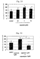
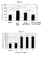
- change in the concentration of intracellular calcium ions in the keratinocytes was measured over time by use of fluorescence intensity as an indication. The results are shown in Figs. 1A and 1B (in Figs. 1A and 1B, the vertical axis corresponds to the intracellular calcium ion concentration, and the horizontal axis corresponds to the concentration and type of drugs).
- Fig. 1A when the solution of capsaicin, which is an agonist of a VR1 receptor, is added to epidermal keratinocytes, the concentration of intracellular calcium ions in the keratinocytes is significantly increased.
- Fig. 1B when epidermal keratinocytes are preliminarily treated with capsazepine, which is an antagonist of a VR1 receptor, for 10 minutes, and then a solution of a mixture of capsaicin and capsazepine is added to the keratinocytes, an increase in the concentration of intracellular calcium ions in the keratinocytes is suppressed.
- capsazepine which is an antagonist of a VR1 receptor
- ATP or ⁇ -me ATP which is an agonist of a P2X receptor
- the concentration of intracellular calcium ions in the keratinocytes is significantly increased.
- epidermal keratinocytes are preliminarily treated with TNP-ATP, which is an inhibitor of a P2X1 receptor, a P2X3 receptor, and a P2X2/3 receptor, for 10 minutes, and then a solution of a mixture of TNP-ATP and ⁇ -me ATP is added to the keratinocytes, an increase in the concentration of intracellular calcium ions in the keratinocytes is suppressed.
- Bradykinin which is a typical pain-producing substance, was added to a BSS to thereby prepare bradykinin solutions of different bradykinin concentrations, each of the resultant solutions was added to epidermal keratinocytes, and, two seconds after addition of the solution, the concentration of intracellular calcium ions in the keratinocytes was calculated by use of the aforementioned formula.
- Fig. 3 wherein the vertical axis corresponds to the intracellular calcium ion concentration, and the horizontal axis corresponds to the bradykinin concentration (log M)].
- the concentration of intracellular calcium ions in the epidermal keratinocytes is increased.
- lactic acid was added to a BSS to thereby prepare lactic acid solutions of different lactic acid concentrations, each of the resultant solutions was added to epidermal keratinocytes, and, two seconds after addition of the solution, the concentration of intracellular calcium ions in the keratinocytes was calculated by use of the aforementioned formula.
- the skin stimulation effect of a test substance can be detected by means of the present detection method employing, as an indication, an increase/decrease in the concentration of intracellular calcium ions in skin cells, and the present screening method, which utilizes the principle of the present detection method, is useful as means for screening, for example, various analgesic agents, anti-inflammatory agents, and drugs for preventing epidermal disorder.
- the present invention provides an excellent method of detecting the skin stimulation effect of a test substance; means for screening, for example, various analgesic agents, anti-inflammatory agents, and drugs for preventing epidermal disorder, the means employing the detection method; and means for evaluating conditions of the skin.
Landscapes
- Health & Medical Sciences (AREA)
- Life Sciences & Earth Sciences (AREA)
- Engineering & Computer Science (AREA)
- Immunology (AREA)
- Biomedical Technology (AREA)
- Chemical & Material Sciences (AREA)
- Molecular Biology (AREA)
- Urology & Nephrology (AREA)
- Hematology (AREA)
- General Health & Medical Sciences (AREA)
- Cell Biology (AREA)
- Pathology (AREA)
- Medicinal Chemistry (AREA)
- Analytical Chemistry (AREA)
- Food Science & Technology (AREA)
- Physics & Mathematics (AREA)
- Biochemistry (AREA)
- General Physics & Mathematics (AREA)
- Microbiology (AREA)
- Biotechnology (AREA)
- Toxicology (AREA)
- Bioinformatics & Cheminformatics (AREA)
- Tropical Medicine & Parasitology (AREA)
- Veterinary Medicine (AREA)
- Dermatology (AREA)
- Public Health (AREA)
- Animal Behavior & Ethology (AREA)
- Endocrinology (AREA)
- Pharmacology & Pharmacy (AREA)
- Epidemiology (AREA)
- Gastroenterology & Hepatology (AREA)
- Diabetes (AREA)
- Inorganic Chemistry (AREA)
- Proteomics, Peptides & Aminoacids (AREA)
- Rheumatology (AREA)
- Chemical Kinetics & Catalysis (AREA)
- Nuclear Medicine, Radiotherapy & Molecular Imaging (AREA)
- Organic Chemistry (AREA)
- General Chemical & Material Sciences (AREA)
- Biophysics (AREA)
Abstract
Description
Fmax: Fluorescence intensity of Fluo3-AM in the presence of excess Ca2+ (breakage of cell membranes by a surfactant) (maximum fluorescence intensity)
Kd: Constant of dissociation of Ca2+ from Fluo3-AM (390 nM)

Claims (6)
- A skin stimulation effect detection method comprising correlating the skin stimulation effect of a test substance which has been brought into contact with epithelial cells with an increase/decrease in the concentration of intracellular calcium ions in the epithelial cells, to thereby detect the skin stimulation effect of the test substance.
- A skin stimulation effect detection method according to claim 1, wherein the epithelial cells are epidermal keratinocytes.
- A skin stimulation effect detection method according to claim 1, wherein the test substance is a substance capable of changing the concentration of intracellular calcium ions in the epithelial cells by the mediation of a VR1 receptor and/or a P2X receptor on the epidermis.
- A skin stimulation effect detection method according to claim 2, wherein the test substance is a substance capable of changing the concentration of intracellular calcium ions in the epithelial cells by the mediation of a VR1 receptor and/or a P2X receptor on the epidermis.
- A method of screening drugs that are potentially useful for preventing skin stimulation and/or drugs that are potentially useful for preventing skin disorder, which comprises using a skin stimulation effect detection method as recited in any of claims 1 through 4 as screening means and, as an indication, suppression of an increase in the concentration of intracellular calcium ions in epithelial cells, which increase is caused by a skin stimulation substance.
- A method of evaluating conditions of the skin, which comprises correlating an increase/decrease in the concentration of intracellular calcium ions in epidermal cells with conditions of the skin.
Applications Claiming Priority (3)
| Application Number | Priority Date | Filing Date | Title |
|---|---|---|---|
| JP2001180366 | 2001-06-14 | ||
| JP2001180366A JP2002372530A (en) | 2001-06-14 | 2001-06-14 | How to detect skin irritation |
| PCT/JP2002/005736 WO2002103351A1 (en) | 2001-06-14 | 2002-06-10 | Method of detecting skin stimulation effect |
Publications (2)
| Publication Number | Publication Date |
|---|---|
| EP1411354A1 true EP1411354A1 (en) | 2004-04-21 |
| EP1411354A4 EP1411354A4 (en) | 2005-02-09 |
Family
ID=19020799
Family Applications (1)
| Application Number | Title | Priority Date | Filing Date |
|---|---|---|---|
| EP02733440A Withdrawn EP1411354A4 (en) | 2001-06-14 | 2002-06-10 | Method of detecting skin stimulation effect |
Country Status (5)
| Country | Link |
|---|---|
| US (1) | US20040191171A1 (en) |
| EP (1) | EP1411354A4 (en) |
| JP (1) | JP2002372530A (en) |
| KR (1) | KR20040010704A (en) |
| WO (1) | WO2002103351A1 (en) |
Families Citing this family (4)
| Publication number | Priority date | Publication date | Assignee | Title |
|---|---|---|---|---|
| JP2007174931A (en) * | 2005-12-27 | 2007-07-12 | Pola Chem Ind Inc | Epidermal keratinocyte layer membrane and utilization of the epidermal keratinocyte layer membrane |
| JP5548498B2 (en) * | 2010-03-30 | 2014-07-16 | 株式会社マンダム | Method for evaluating external preparation for skin |
| JP2013195289A (en) | 2012-03-21 | 2013-09-30 | Takasago Internatl Corp | Method for evaluating sense stimulating component |
| CN115191942A (en) * | 2022-07-05 | 2022-10-18 | 昆明医科大学第一附属医院 | A detection system for sensitive skin typing |
Family Cites Families (5)
| Publication number | Priority date | Publication date | Assignee | Title |
|---|---|---|---|---|
| JPH10229978A (en) * | 1997-02-18 | 1998-09-02 | Shiseido Co Ltd | Hair growth test method |
| JPH1149647A (en) * | 1997-07-31 | 1999-02-23 | Shiseido Co Ltd | Screening of hair growth period prolonging agent |
| GB9822124D0 (en) * | 1998-10-09 | 1998-12-02 | Univ London | Ion channels |
| JP4465779B2 (en) * | 1999-02-10 | 2010-05-19 | 大正製薬株式会社 | Hair restorer |
| JP3860956B2 (en) * | 2000-08-23 | 2006-12-20 | 株式会社資生堂 | Method and apparatus for measuring skin surface condition |
-
2001
- 2001-06-14 JP JP2001180366A patent/JP2002372530A/en active Pending
-
2002
- 2002-06-10 US US10/480,784 patent/US20040191171A1/en not_active Abandoned
- 2002-06-10 KR KR10-2003-7016310A patent/KR20040010704A/en not_active Abandoned
- 2002-06-10 EP EP02733440A patent/EP1411354A4/en not_active Withdrawn
- 2002-06-10 WO PCT/JP2002/005736 patent/WO2002103351A1/en not_active Ceased
Non-Patent Citations (6)
| Title |
|---|
| DATABASE MEDLINE [Online] US NATIONAL LIBRARY OF MEDICINE (NLM), BETHESDA, MD, US; March 1991 (1991-03) YASUI C: "[Changes in the intracellular free calcium of cultured human epidermal keratinocytes]" Database accession no. NLM1711997 XP002306217 & [HOKKAIDO IGAKU ZASSHI] THE HOKKAIDO JOURNAL OF MEDICAL SCIENCE. JAPAN MAR 1991, vol. 66, no. 2, March 1991 (1991-03), pages 111-121, ISSN: 0367-6102 * |
| DENDA M ET AL: "P2X purinergic receptor antagonist accelerates skin barrier repair and prevents epidermal hyperplasia induced gamma skin barrier discuption" JOURNAL OF INVESTIGATIVE DERMATOLOGY, NEW YORK, NY, US, vol. 119, no. 5, November 2002 (2002-11), pages 1034-1040, XP002966357 ISSN: 0022-202X * |
| DENDA MITSUHIRO ET AL: "Immunoreactivity of VR1 on epidermal keratinocyte of human skin" BIOCHEMICAL AND BIOPHYSICAL RESEARCH COMMUNICATIONS, vol. 285, no. 5, 3 August 2001 (2001-08-03), pages 1250-1252, XP002302117 ISSN: 0006-291X * |
| HAMILTON M G ET AL: "Modification of cytosolic free calcium concentrations in human keratinocytes after sulfur mustard exposure" TOXICOLOGY IN VITRO, vol. 12, no. 4, August 1998 (1998-08), pages 365-372, XP002302115 ISSN: 0887-2333 * |
| See also references of WO02103351A1 * |
| TOYODA M ET AL: "COMPARISON OF SOLANACEOUS GLYCOALKALOIDS-EVOKED CALCIUM ION INFLUX IN DIFFERENT TYPES OF CULTURED CELLS" TOXICOLOGY IN VITRO, vol. 5, no. 4, 1991, pages 347-351, XP002302116 ISSN: 0887-2333 * |
Also Published As
| Publication number | Publication date |
|---|---|
| WO2002103351A1 (en) | 2002-12-27 |
| US20040191171A1 (en) | 2004-09-30 |
| KR20040010704A (en) | 2004-01-31 |
| EP1411354A4 (en) | 2005-02-09 |
| JP2002372530A (en) | 2002-12-26 |
Similar Documents
| Publication | Publication Date | Title |
|---|---|---|
| CA2483961C (en) | Methods of screening for trpm4b modulators | |
| Grinstein et al. | Focal localization of the NHE‐1 isoform of the Na+/H+ antiport: assessment of effects on intracellular pH. | |
| Angers et al. | Biochemical and biophysical demonstration of GPCR oligomerization in mammalian cells | |
| JP4594924B2 (en) | TRPM5 modulator screening method | |
| Sotkis et al. | Calmodulin colocalizes with connexins and plays a direct role in gap junction channel gating | |
| EP2511707B1 (en) | Method for detection of basic peptide and reagent for detection of basic peptide | |
| Ducharme et al. | Stimulated myotube contractions regulate membrane-bound and soluble TLR4 to prevent LPS-induced signaling and myotube atrophy in skeletal muscle cells | |
| EP2150819B1 (en) | S100a9 interaction screening method | |
| US20040191171A1 (en) | Method of detecting skin stimulation effect | |
| US8580525B2 (en) | Methods of screening for LTRPC7 modulators | |
| JP5328190B2 (en) | Method for evaluating TRPA1 activator | |
| De Smet et al. | Regulatory volume decrease in cultured kidney cells (A6): role of amino acids. | |
| EP2589664A1 (en) | Material for screening for compound capable of acting on ion channel, and use thereof | |
| WO2001096372A2 (en) | Zinc transporters proteins and their use in medicinal preparations | |
| EP1360504B1 (en) | Method for screening compounds for activity in treating an osteoclast related bone disease | |
| JP2003514549A (en) | G protein-coupled receptor | |
| RU2345785C2 (en) | Application of "¦l¦" | |
| AU2002253807A1 (en) | Human LTRPC7 proteins, nucleic acid molecules encoding them, and uses thereof | |
| Modzelewska et al. | The effect of a selective inhibition of potassium channels on the relaxation induced by nitric oxide in the human pregnant myometrium | |
| EP1986009A1 (en) | Screening method | |
| Grahn | pH Regulation in Human Sperm | |
| US20050202472A1 (en) | Methods of screening for LTRPC2 modulators | |
| Farr et al. | Methods to visualise zinc transporter proteins of the SLC39A family in cells | |
| Alfonso et al. | Characterization of the Na+/Ca2+ Exchanger on Rat Mast Cells: Evidence for a Functional Role on the Regulation of the Cellular Response | |
| HUP0002557A2 (en) | Simple quantitative fluorescent assay method for determining the activity of transport proteins of interest |
Legal Events
| Date | Code | Title | Description |
|---|---|---|---|
| PUAI | Public reference made under article 153(3) epc to a published international application that has entered the european phase |
Free format text: ORIGINAL CODE: 0009012 |
|
| 17P | Request for examination filed |
Effective date: 20040113 |
|
| AK | Designated contracting states |
Kind code of ref document: A1 Designated state(s): AT BE CH CY DE DK ES FI FR GB GR IE IT LI LU MC NL PT SE TR |
|
| RIC1 | Information provided on ipc code assigned before grant |
Ipc: 7G 01N 33/68 B Ipc: 7G 01N 33/50 B Ipc: 7A 61K 49/00 B Ipc: 7A 61P 17/00 B Ipc: 7A 61K 45/00 B Ipc: 7G 01N 33/15 A |
|
| A4 | Supplementary search report drawn up and despatched |
Effective date: 20041228 |
|
| 17Q | First examination report despatched |
Effective date: 20080111 |
|
| STAA | Information on the status of an ep patent application or granted ep patent |
Free format text: STATUS: THE APPLICATION IS DEEMED TO BE WITHDRAWN |
|
| 18D | Application deemed to be withdrawn |
Effective date: 20100104 |