-
The present invention relates to the identification of polymorphisms in
the P2Y12 receptor associated with platelet aggregation and thrombotic risk.
-
ADP (adenosine 5'-diphosphate) is one of the most important
mediators of hemostasis and thrombosis (Storey et al., 2000). The effect of
ADP on platelets is mediated by two P2Y receptors designated P2Y1 and
P2Y12. Considerable evidence suggests that the P2Y12 receptor plays a central
role in the formation of hemostatic plugs and in the occurrence of arterial
thrombosis (Mills et al., 1996 ; Cattaneo et al., 1999). The key role of P2Y12 in
arterial thrombosis is confirmed by the beneficial effect of the thienopyridine
antithrombotic drugs, such as clopidogrel and ticlopidine that target the P2Y12
receptor (Hollopeter et al., 2001 ; Quinn et al., 1999), in a variety of thrombotic
diseases such as stroke, myocardial infarction and peripheral vascular disease
(Quinn et al., 1999 ; and see CAPRIE Steering Committee. Lancet.
1996;348:1329-1339). Transduction of the ADP signal involves both a transient
rise in Ca++ mediated by the Gq-coupled P2Y1 receptor (Jin et al., 1998a), and
inhibition of adenylate cyclase mediated by the Gi-coupled P2Y12 receptor
(Hollopeter et al., 2001 ; Foster et al., 2001). Simultaneous activation of the Gq
and Gi pathways by ADP is necessary for normal aggregation (Jin et al., 1998b
; Hechler et al., 1998a). Activation of the Gq pathway through P2Y1 leads to
platelet shape changes and a rapidly reversible wave of platelet aggregation
(Jin et al., 1998a ; Hechler et al., 1998a), while activation of the Gi pathway
through P2Y12 elicits slowly progressive and sustained platelet aggregation
(Hechler et al., 1998b). The effect of P2Y12 is not limited to inhibition of
adenylate cyclase and a subsequent reduction in intracellular cAMP content.
Indeed, it has been shown that P2Y12 receptor activates glycoprotein (GP)
IIbIIIa integrin via a phosphoinositide 3- (PI 3-) kinase pathway (Kauffenstein et
al., 2001) and/or another, unidentified G protein (Daniel et al., 1999). P2Y12
thus appears to have a pivotal role in the irreversible wave of platelet
aggregation that occurs on exposure to ADP.
-
The P2Y12 gene, which encodes the 342-amino-acid receptor, was
recently identified by two teams (Hollopeter et al., 2001 ; Foster et al., 2001 ;
Zhang et al., 2001). Four patients have been described whose platelets, when
exposed to ADP, undergo little or no reversible aggregation and show a
decrease in the normal adenylate cyclase inhibition of prostaglandin E1 (PGE1)-stimulated
platelets (Nurden et al., 1995 ; Cattaneo et al., 1992). In one patient,
this phenotype was associated with a P2Y12 gene abnormality (Hollopeter et
al., 2001).
-
Considerable inter-individual variations in the concentration of ADP
required to produce irreversible aggregation have been reported (Feng et al.,
1999).
-
As inherited factors have a major role in platelet aggregation
(O'Donnell et al., 2001), the inventors looked for sequence variations in the
P2Y12 gene that might explain the inter-individual variability of platelet
aggregation responses to ADP.
-
The inventors identified three single-nucleotide polymorphisms (SNP)
and a nucleotide insertion in the P2Y12 receptor gene, which determined two
haplotypes designated H1 and H2. The H2 haplotype was associated with
higher maximal platelet aggregation in response to ADP.
-
The identification of a P2Y12 receptor haplotype that is strongly
associated with increased ADP-induced platelet aggregation has important
clinical implications, particularly in screening for subjects at risk of thrombosis,
especially atherothrombosis, and suboptimal responses to thienopyridine
therapy.
-
Given the specificity of the P2Y12 receptor in high risk peripheral
arterial disease (PAD) patients and the gain of function of the H2 allele in
platelet aggregation, the inventors further investigated the association of the
P2Y12 gene H2 haplotype with PAD in a case-control study.
-
They observed a strong association of P2Y12 H2 haplotype with PAD.
-
Based on these results, the present invention provides a method for
distinguishing H1 and H2 haplotypes in the P2Y12 receptor, the latter being
associated with a higher risk to develop thrombosis and/or with lower sensitivity
toward a thienopyridine therapy.
-
This method is thus useful to determine the risk of developing
thrombosis, especially atherothrombosis, in a subject.
-
This method is also useful to determine the sensitivity of a subject
toward a thienopyridine therapy.
P2Y12 receptor
-
As above mentioned, the human P2Y
12 receptor has been cloned.
Variants in other species has also been characterized.
| | Code | Access. number (SWISSPROT) |
| Homo sapiens | P2Y12_HUMAN | Q9H244 |
| Macaca fascicularis | P2Y12_MACFA | Q35KC3 |
| Mus musculus | P2Y12_MOUSE | Q9CPV9 |
| Rattus norvegicus | P2Y12_RAT | Q9EPX4 |
-
In the context of the present invention, the term "P2Y12 receptor" refers
to the P2Y12 receptor of any species, especially in human, but also in other
mammals or vertebrates to which the methods of the invention can apply, if
desired. The term "subject" thus refers to any human patient or any mammal or
vertebrate.
-
The human P2Y12 cDNA sequence is available on Genbank (access
numbers: AF313449 and AY136754).
-
The inventors analyzed the P2Y
12 gene in a healthy population and
found that it contained at least two exons separated by one intron
approximately 1700 bp in length, located upstream of the ATG codon.
Exon 2
encodes the entire 342-amino-acid protein. Because of a 6-nucleotide (nt)
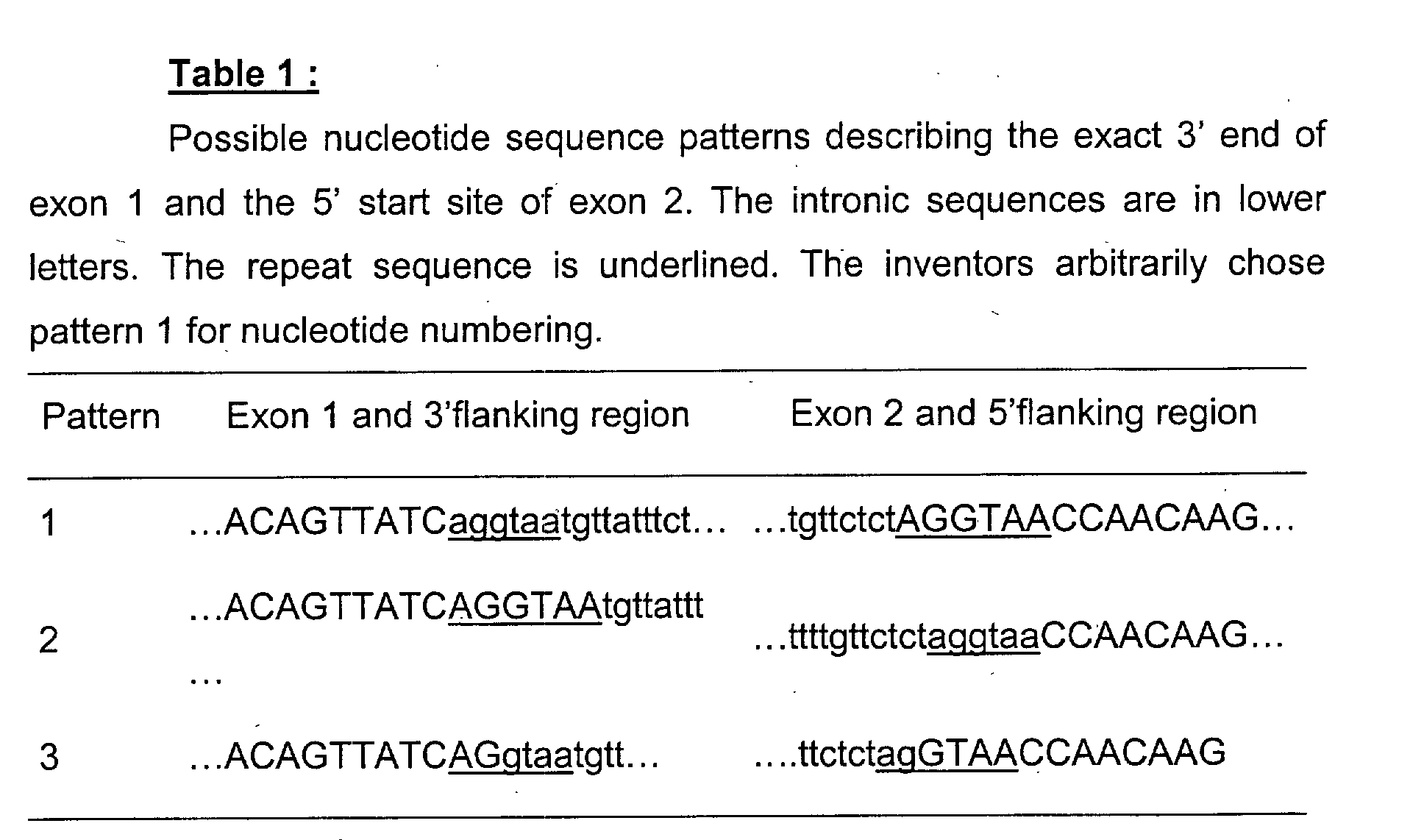
repeat sequence (table 1), the exact 3' end of
exon 1 and 5' start site of
exon 2
could not be determined.
Table 1 describes three possible sequence patterns,
pattern 3 being the most
probable. However the inventors begun their work while arbitrarily choosing
pattern 1 to number the polymorphisms that were studied. Consequently
pattern 1 has been kept when numbering the polymorphisms described below.
-
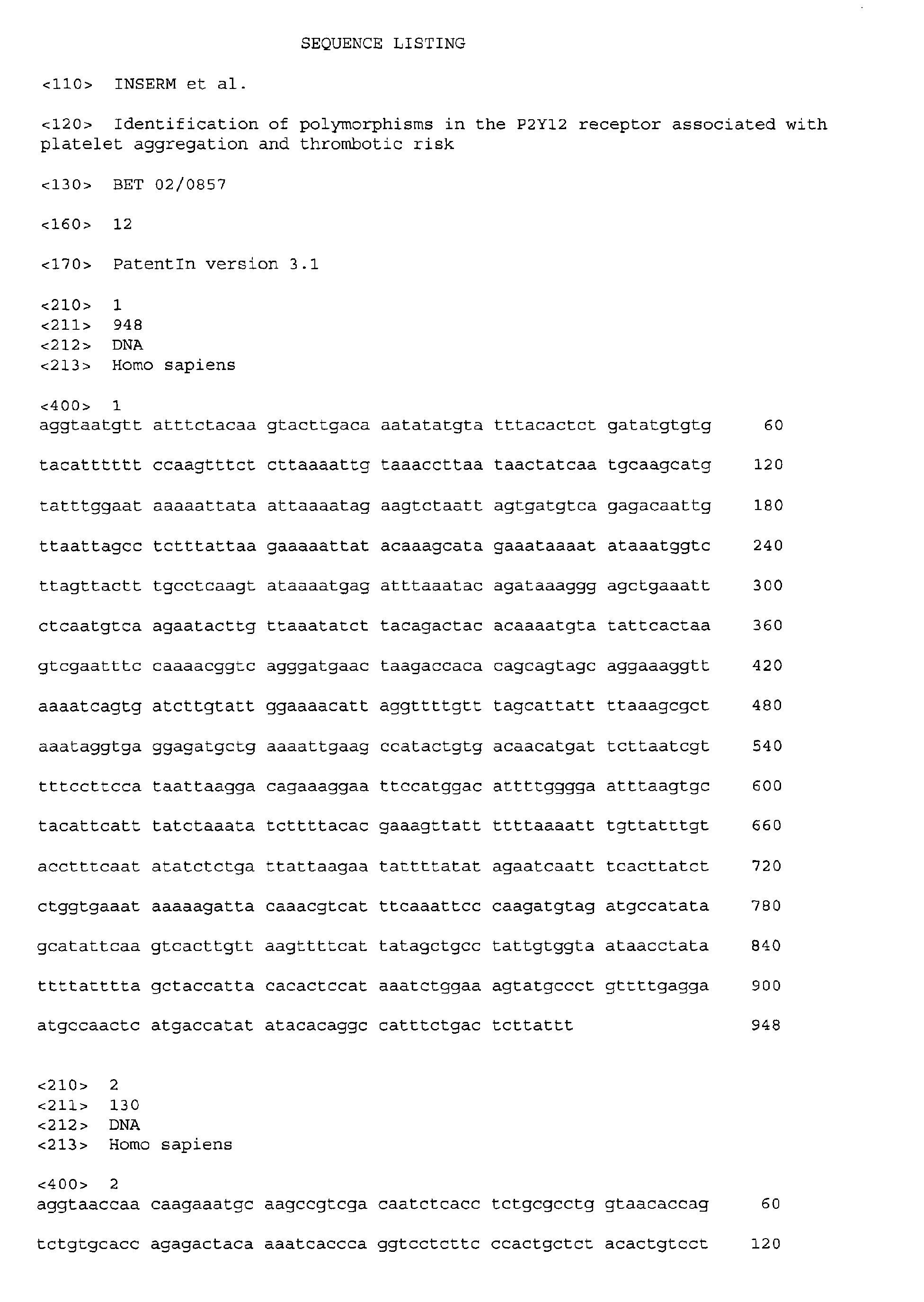
According to pattern 1, sequence SEQ ID No 1 represents a 5'
sequence of the intron, whereas sequence SEQ ID No 2 represents a 5'
sequence of exon 2, which are the parts of interest in the P2Y12 gene,
according to the present invention. Both sequences show the H2 haplotype, as
defined below. The A nucleotide of the starting codon ATG is nucleotide 17 in
SEQ ID No 2.
H1/H2 haplotypes
-
The inventors sequenced the P2Y
12 gene and found three single
nucleotide polymorphisms (SNPs) and a single nucleotide insertion
polymorphism of interest (Table 2). In the present invention, these
polymorphisms are sometimes referred to as "the polymorphic positions of
interest".
| Description of the polymorphisms detected in the study population. The H1/H2 haplotype is defined by the i-C139T, i-T745C, i-ins802A and G52T polymorphisms. |
| Name of SNP | amino acid | Region | Allele frequency |
| i-C139T | - | Intron | C: 86.2% | T: 13.8% |
| i-T745C | - | Intron | T: 86.2% | C: 13.8% |
| i-ins802A | - | Intron | Insertion of an | A: 13.8% |
| G52T | G / G | Exon 2 | G: 86.2% | T: 13.8% |
| C34T | N / N | Exon 2 | C: 72.5% | T: 27.5% |
-
Two variations were located 139 nt and 745 nt after the 5' intron
(abbreviated as "i" herein) start site, consisting of a C to T (i-C139T) and a T to
C (i-T745C) transition, respectively. Another polymorphism consisted of a
single nucleotide insertion (A) at position 802 of the intron (i-ins802A). Two
additional polymorphisms were found in exon 2, and consisted of a C to T
transition (C34T) and a G to T transversion (G52T), respectively 17 nt and 35
nt after the first nucleotide of the ATG codon; neither modified the encoded
amino acid (Asn6 and Gly12, respectively). The C34T polymorphism was not
associated with maximal aggregation in response to ADP.
-
As the i-C139T, i-T745C, i-ins802A and G52T polymorphisms were in
complete linkage disequilibrium in a population of 100 healthy volunteers of
caucasian origin, the inventors designated "H1" the haplotype of the major
allele (a C in position 139, a T in position 745, and absence of the i-ins802A in
the intron, and a G in position 52 of exon 2) and "H2" the haplotype of the
minor allele (a T in position 139, a C in position 745, presence of the i-ins802A
in the intron, and a T in position 52 of exon 2). The respective allele
frequencies of haplotypes H1 and H2 were 86% and 14%. The allele
frequencies of all the polymorphisms were in Hardy-Weinberg equilibrium.
-
Compared to the non carriers of the H2 allele (H1/H1), carriers of the
H2 allele (H1/H2 or H2/H2) had a stronger ADP-stimulated platelet aggregation
response in vitro, with different regulation of intracellular cAMP levels.
-
The isolated nucleic acid of the P2Y12 receptor gene with the H2
haplotype is also part of the present invention.
-
A subject of the invention is thus an isolated nucleic acid encoding the
P2Y12 receptor, which nucleic acid comprises the P2Y12 gene sequence with
the simultaneous presence of T at position 139 of the intron, presence of C at
position 745 of the intron, insertion of A at position 802 of the intron, and
presence of T at position 52 of exon 2.
-
Such nucleic acid preferably comprises SEQ ID No 1 and/or SEQ ID
No 2.
Thrombosis risk
-
The present invention provides an in vitro method for determining the
risk of developing thrombosis in a subject, which method comprises identifying
polymorphisms of P2Y12 receptor at positions 139, 745, and 802 of the intron
(SEQ ID No 1), and at position 52 of exon 2 (SEQ ID No 2), wherein the
simultaneous presence of T at position 139 of the intron, presence of C at
position 745 of the intron, insertion of A at position 802 of the intron, and
presence of T at position 52 of exon 2 are designated H2 haplotype and are
indicative of a higher risk to develop thrombosis in comparison with a control
subject, i.e. a subject showing no H2 haplotype on any of the alleles (i.e. a
H1/H1 subject).
-
A higher risk to develop thrombosis means a significantly greater risk
in carriers of the H2 allele than in non carriers.
-
Such a relative risk is frequently estimated through case-control
studies and the use of the odds ratio (OR). An OR of 1.1 corresponds to an
increase of 10% of the risk of developing disease and an OR of 2 corresponds
to a 100 % increased risk.
-
The risk of developing thrombosis is connected with the induction of
platelet aggregation.
-
Platelets are especially essential to arterial thrombosis and are also
involved in the initiation and development of atherosclerotic lesions through
adhesion to dysfunctional endothelium and release of growth factors and
cytokines. The most important vascular targets of atherosclerotic disease
include cerebral, coronary and lower limb arterial beds.
-
In the context of the present invention, the term "thrombosis" means
formation of a clot in a blood vessel or in a heart cavity.
-
In a preferred embodiment, atherothrombosis is encompassed.
-
The terms "atherothrombosis", "atherosclerosis" and "arterial
thrombosis" are synonymous.
-
Atherothrombosis is the central phenomenon of many thrombotic
diseases such as stroke, myocardial infarction and peripheral arterial disease
(PAD).
-
Concomitance of arterial thrombosis and atherosclerotic disease is of
particular importance in peripheral arterial disease (PAD). Indeed, PAD is
associated with a high risk of coronary and cerebrovascular atherosclerosis
events and epidemiological studies have shown that 75% of PAD patients die
of vascular causes (Ouriel et al., 2001).
-
Consequently, the identification of the H2 haplotype of the P2Y12
receptor in a subject is further indicative of a higher risk to develop such
thrombotic diseases, especially PAD.
-
Within the meaning of the invention, "thrombosis" as above defined
encompasses other thrombotic states such as venous thrombosis and venous
thromboembolic diseases, as well as thrombotic microangiopathy or
intravascular disseminated coagulation.
Sensitivity to thienopyridine therapy
-
The present invention further provides an in vitro method for
determining sensitivity of a subject toward a thienopyridine therapy, which
method comprises identifying polymorphisms of P2Y12 receptor at positions
139, 745, and 802 of the intron (SEQ ID No 1), and at position 52 of exon 2
(SEQ ID No 2), wherein the simultaneous presence of T at position 139 of the
intron, presence of C at position 745 of the intron, insertion of A at position 802
of the intron, and presence of T at position 52 of exon are designated H2
haplotype and, when present on at least one allele, are indicative of a lower
sensitivity of the subject toward a thienopyridine therapy in comparison with a
control subject, i.e. a subject with no H2 allele (i.e. a H1/H1 subject).
-
In the context of the invention, a "thienopyridine therapy" refers to the
antithrombotic therapy currently used, which involves administration of
thienopyridine drugs such as clopidogrel or ticlopidine, or any other agent that
blocks the interaction between ADP and P2Y12.
-
The "sensitivity of a subject toward a thienopyridine therapy" refers to
the variety of responses of a subject undergoing such therapy. A lower
sensitivity toward the therapy corresponds to a poorer response, and therefore
is not desired.
-
Determining the sensitivity of a subject toward a thienopyridine therapy
is particularly interesting to help the physician design the therapeutic strategy
that will be the most adapted to the subject, at an early stage.
Identification of the presence of the H2 allele
-
The methods described above, i.e. the methods for determining the
thrombosis risk and the methods to determine the sensitivity toward a
thienopyridine therapy, involve the identification of the H1/H2 haplotype of the
P2Y12 receptor gene.
-
Such identification may be performed by any technique well known
from the person skilled in the art.
-
In practicing the methods of the invention, an individual's polymorphic
pattern with regard to the H1/H2 haplotype can be established by obtaining
DNA from the individual and determining the sequence at the polymorphic
positions of the P2Y12 receptor gene interest.
-
The DNA may be obtained from any cell source. Non-limiting examples
of cell sources available in clinical practice include blood cells, buccal cells,
cervicovaginal cells, epithelial cells from urine, fetal cells, hair, or any cells
present in tissue obtained by biopsy. Cells may also be obtained from body
fluids, including without limitation blood, saliva, sweat, urine, cerebrospinal
fluid, feces, and tissue exudates at the site of infection or inflammation. DNA
may be extracted from the cell source or body fluid using any of the numerous
methods that are standard in the art. It will be understood that the particular
method used to extract DNA will depend on the nature of the source. In another
embodiment, the analysis is achieved without extraction of the DNA, e.g. on
whole blood (Mercier et al, 1990).
-
Determination of the sequence of the DNA at the polymorphic
positions of the H1/H2 haplotype is achieved by any means known in the art.
Numerous strategies for genotype analysis are available (Antonarakis et al.,
1989 ; Cooper et al., 1991 ; Grompe, 1993). The strategies include, but are not
limited to, direct sequencing, restriction fragment length polymorphism (RFLP)
analysis, hybridization with allele-specific oligonucleotides, allele-specific PCR,
PCR using mutagenic primers, ligase-PCR, HOT cleavage, denaturing gradient
gel electrophoresis (DGGE), temperature denaturing gradient gel
electrophoresis (TGGE), single-stranded conformational polymorphism (SSCP)
and denaturing high performance liquid chromatography (Kuklin et al., 1997).
Direct sequencing may be accomplished by any method, including without
limitation chemical sequencing, using the Maxam-Gilbert method ; by
enzymatic sequencing, using the Sanger method ; mass spectrometry
sequencing ; sequencing using a chip-based technology (see e.g. Little et al.,
1996); and real-time quantitative PCR. Preferably, DNA from a subject is first
subjected to amplification by polymerase chain reaction (PCR) using specific
amplification primers. However several other methods are available, allowing
DNA to be studied independently of PCR, such as the oligonucleotide ligation
assay (OLI), rolling circle amplification (RCA) or the Invader™assay.
-
In a particular embodiment of the invention, it is provided an in vitro
method for identifying at least one polymorphism of an haplotype of the P2Y12
receptor associated with thrombosis in a subject or associated with lower
sensitivity toward a thienopyridine therapy, which method comprises analyzing
genomic DNA of a biological sample, in at least one of the regions of the P2Y12
receptor gene, located around positions 139, 745 and 802 of the intron (SEQ ID
No 1) and position 52 of exon 2 (SEQ ID No 2);
wherein the simultaneous presence of T at position 139 of the intron,
presence of C at position 745 of the intron, insertion of A at position 802 of the
intron, and presence of T at position 52 of exon 2 are designated H2 haplotype
and, when present on at least one allele, are indicative of a higher risk to
develop thrombosis or of a lower sensitivity toward a thienopyridine therapy, in
comparison with a control subject.
-
The analysis may preferably comprise a step of amplification (e.g. by
PCR) of said regions of the genomic DNA.
-
In a particular embodiment the analysis is undertaken on isolated (i.e.
extracted) genomic DNA.
-
A preferred technique for identifying the polymorphisms of the P2Y12
receptor includes sequencing.
Kits
-
According to an aspect of the invention, the H1/H2 haplotype is
detected by contacting the DNA of the subject with a nucleic acid probe, that is
optionally labeled.
-
Primers may also be useful to amplify or sequence the portion of the
P2Y12 gene containing the polymorphic positions of interest.
-
Such probes or primers are nucleic acids that are capable of
specifically hybridizing with a portion of the P2Y12 gene sequence containing
the polymorphic positions of interest. That means that they are sequences that
hybridize with the nucleic acid sequence to which it refers under conditions of
high stringency (Sambrook et al, 1989). These conditions are determined from
the melting temperature Tm and the high ionic strength. Preferably, the most
advantageous sequences are those which hybridize in the temperature range
(Tm - 5°C) to (Tm - 30°C), and more preferably (Tm - 5°C) to (Tm - 10°C). A
ionic strength of 6xSSC is more preferred. For instance, high stringency
hybridization conditions correspond to the highest Tm, e.g., 50 % formamide,
5x or 6x SCC. SCC is a 0.15 M NaCl, 0.015 M Na-citrate. Hybridization
requires that the two nucleic acids contain complementary sequences, although
depending on the stringency of the hybridization, mismatches between bases
are possible. The appropriate stringency for hybridizing nucleic acids depends
on the length of the nucleic acids and the degree of complementation, variables
well known in the art. The greater the degree of similarity or homology between
two nucleotide sequences, the greater the value of Tm for hybrids of nucleic
acids having those sequences. The relative stability (corresponding to higher
Tm) of nucleic acid hybridizations decreases in the following order: RNA:RNA,
DNA:RNA, DNA:DNA. For hybrids of greater than 100 nucleotides in length,
equations for calculating Tm have been derived (see Sambrook et al., supra,
9.50-9.51). For hybridization with shorter nucleic acids, i.e., oligonucleotides,
the position of mismatches becomes more important, and the length of the
oligonucleotide determines its specificity (see Sambrook et al., supra, 11.7-11.8).
A minimum length for a hybridizable nucleic acid is at least about 10
nucleotides ; preferably at least about 15 nucleotides ; and more preferably the
length is at least about 20 nucleotides.
-
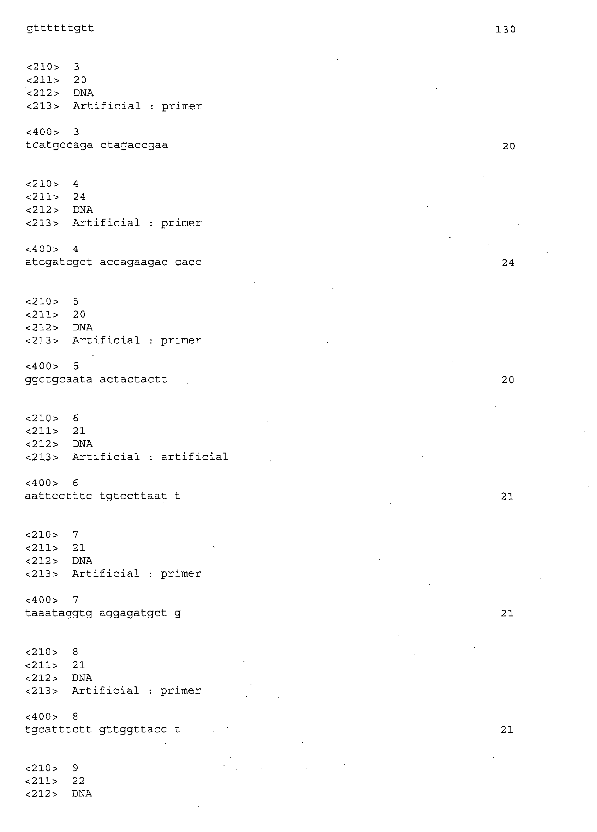
Preferred probes or primers are described in the legend to the figures or
in the examples below.
-
When the primers are mutagenic primers, the above rules for
determining the hybridisation conditions are to be adapted. The sequences of
mutagenic primers may indeed partially vary from the wild-type sequence, so
as to introduce restriction site when a given allele is amplified. Furthermore
such mutagenic primers are frequently 30 to 40 nucleotide-long.
-
The present invention further provides kits suitable for determining at
least one of the polymorphism of the H1/H2 haplotype of the P2Y12 receptor.
-
The kits may include the following components :
- (i) a probe as above defined, usually made of DNA, and that may be
pre-labelled. Alternatively, the probe may be unlabelled and the ingredients for
labelling may be included in the kit in separate containers ; and
- (ii) hybridization reagents : the kit may also contain other suitably
packaged reagents and materials needed for the particular hybridization
protocol, including solid-phase matrices, if applicable, and standards.
-
-
In another embodiment, the kits may include :
- (i) sequence determination or amplification primers : sequencing
primers may be pre-labelled or may contain an affinity purification or
attachment moiety ; and
- (ii) sequence determination or amplification reagents : the kit may
also contain other suitably packaged reagents and materials needed for the
particular sequencing amplification protocol. In one preferred embodiment, the
kit comprises a panel of sequencing or amplification primers, whose sequences
correspond to sequences adjacent to at least one of the polymorphic positions,
as well as a means for detecting the presence of each polymorphic sequence.
-
-
In a particular embodiment, it is provided a kit which comprises a pair
of nucleotide primers specific for amplifying all or part of the P2Y12 gene
comprising at least one of positions 139, 745 and 802 of the intron (SEQ ID No
1) and/or position 52 of exon 2 (SEQ ID No 2).
-
The below figures and examples illustrate the invention without limiting
its scope.
LEGENDS OF THE FIGURES :
-
Figure 1 is a schematic drawing showing the location of the primers
and polymorphisms in the P2Y12 gene.
-
A PCR product of approximately 3100 bp was obtained with primers E
and J. Subsequent sequencing analysis was performed using primers
are start and stop codons, respectively. i-C139T, i-T745C, i-ins802A, C34T and
G52T are the polymorphisms found in the study population.
-
Figure 2 is a graph showing maximal platelet aggregation responses to
ADP in 98 healthy volunteers.
-
After a 3-min incubation period, platelets (250 x 109/L in citrated PRP)
were stimulated with 2 µM ADP. The concordance coefficient between
aggregation values recorded at visit 1 and visit 2 (one week later) was 77%
(P<0.001).
-
Figure 3 is a graph showing maximal aggregation in response to 2 µM
ADP according to the P2Y12 haplotype.
-
The average of the maximal aggregation values recorded at visits 1
and 2 in each volunteer was used for analysis. The median value was 34.7% in
subjects carrying no H2 alleles (H1/H1, n = 74), 67.9% in subjects carrying one
H2 allele (H1/H2, n = 21) and 82.4% in the three subjects carrying two H2
alleles (H2/H2) (P=0.0071).
-
Figure 4 is a graph showing inhibition of iloprost-induced cAMP
formation by ADP in carriers and non carriers of the H2 haplotype.
-
Iloprost (20 µg/µL final concentration) was added to citrated PRP 1 min
after incubation at 37°C. Saline (control) or ADP at 1, 2 or 5 µM was added 1
min later. The reaction was stopped 3 min later and the cAMP concentration
was determined using a commercial assay kit. Triangles and circles denote
carriers and non carriers of the H2 haplotype, respectively. Means ± SEM.
EXAMPLES :
Example 1 : Identification of P2Y12 gene sequence variations.
Methods
Subjects
-
Ninety-eight unrelated healthy Caucasian male volunteers aged from
18 to 35 years were recruited and investigated in the Clinical Investigation
Center of Hôpital Européen Georges Pompidou. The volunteers were non
smokers, had an unremarkable personal and familial medical history, and
denied taking any medication for at least 10 days before blood collection. A
physical examination and routine laboratory tests, including white blood cell,
red blood cell and platelet counts, mean platelet volume, prothrombin time
(PT), activated partial thromboplastin time (aPTT), plasma fibrinogen and C-reactive
protein assays were performed before inclusion. Blood obtained from
all the volunteers on day 0 (visit 1) and day 7 (visit 2) was subjected to
aggregation tests. A third blood sample was obtained from a subset of 20
volunteers for platelet cAMP assay and comparison of maximal aggregation in
citrated and in hirudin-anticoagulated platelet-rich plasma (PRP). All the
subjects gave their written informed consent, and the study protocol was
approved by the local ethics committee.
Sample preparation
-
Venous blood was collected between 8:00 and 10:00 am, after an
overnight fast, in tubes containing either 0.105 M sodium citrate (1 vol/9 vol)
(BD Vacutainer®, Becton Dickinson, Le Pont de Claix, France) or 50 µg/mL
lepirudine (Refludan®, Hoechst, Paris, France) using a 19-gauge needle and no
tourniquet. The first 2 ml of blood was discarded. Platelet-rich plasma (PRP)
was obtained by centrifugation at 1000 rpm for 10 min at room temperature.
Autologous platelet-poor plasma, obtained by further centrifugation at 4000 rpm
for 15 min, was used to adjust the platelet count of PRP to 250 x 109/L.
Platelet aggregation studies
-
Aggregation studies were performed within 2 hours after blood
collection. Aggregation was measured at 37°C by using a photometric method
on a four-channel aggregometer (Regulest® Amneville, France). A 280-µL
aliquot of PRP was incubated for 3 min at 37°C and then was stirred at 1100
rpm for 2 min before adding 20 µL of saline or ADP (Sigma-Aldrich, St Quentin
Fallavier, France) at 1, 2 or 5 µM (final concentration). The platelet aggregation
response was recorded for 5 min.
CAMP measurement
-
Platelet cAMP content was measured in 20 selected subjects by
incubating 270 µL of PRP with 10 µL of a solution of the stable prostacyclin
analog iloprost (20 µg/L, final concentration) (IIomédine®, Schering-Plough) for
1 min, with stirring, then adding 20 µL of saline or ADP (1, 2 or 5 µM). The
reaction was stopped 3 min later by adding 20 µL of 12% trichloroacetic acid.
CAMP was then extracted and measured with an enzyme immunoassay (EIA)
(Amersham, Pharmacia Biotech, Orsay, France) according to the
manufacturer's instructions, without knowledge of the subject's P2Y12
genotype.
Genotyping studies
-
Genomic DNA was isolated from peripheral blood mononuclear cells
by using the Qiamp Maxi Kit® (Qiagen, Courtaboeuf, France) according to the
manufacturer's instructions.
-
P2Y12 receptor. The inventors took advantage of the cDNA sequence
reported by Hollopeter et al., (2001), which is available under GenBank
accession number AF313449. Assuming that P2Y12 gene organization was the
same as that of seven other transmembrane-domain receptor genes, most of
which comprise two exons separated by a single intron, the inventors
performed a preliminary amplification experiment using two oligonucleotides,
designated E and J, as sense and antisense primers; they were located at the
5' and 3' ends of the reported cDNA (figure 1). The amplification product was
then purified with the Pre-Sequencing Kit (Amersham Pharmacia Biotech).
Purification was followed by a cycle sequencing reaction using primers E, B
and J. Subsequent sequencing using several other primers (G, H, K and L)
(figure 1) allowed us to determine the nucleotide sequence of the amplification
product. Sequence analysis was performed with an ABI Prism 3700 (Applera).
-
Screening of 40 subjects in a population is sufficient to identify
polymorphisms with a frequency of 5% or more, with a confidence interval (CI)
of 95%. Thus, a first series of 48 consecutive healthy volunteers were screened
for P2Y12 gene polymorphisms by sequencing the 58 nt of exon 1 with primer
K, the 947 nt of its 3' flanking region with primers E and G, the 1274 nt of exon
2 with primers L, C and M, and the 570 nt of its 5' flanking region with primer H.
The primers are described in figure 1. The P2Y12 gene was then sequenced in
the other 50 subjects by using primers targeting the identified polymorphisms.
-
PCR and sequencing results were analyzed without knowledge of the
platelet aggregation results. Genotyping was repeated in ambiguous cases. All
98 subjects were successfully genotyped.
Platelet RNA extraction and reverse transcription
-
Four milliliters of PRP (109 platelets) was pelleted by centrifugation for
15 min at 4000 rpm. RNA was extracted with the RNeasy® kit (Qiagen)
according to the manufacturer's instructions. Reverse transcription was
performed using 10 units of RNasin™ ribonuclease inhibitor (Promega,
Charbonnières, France), 100 units of Superscript II Rnase H- reverse
transcriptase (Invitrogen) and 1.5 mM random hexanucleotides (Amersham
Pharmacia biotech). The cDNA was stored at -20°C.
Statistical analysis
-
Data are shown as means ± SEM, except for skewed variables, which
are expressed as medians. Individual subjects' values obtained at visits 1 and 2
were compared using a concordance test (Lin, 1989) The x 2 test was used to
compare the observed allele and genotype frequencies with the Hardy-Weinberg
equilibrium prediction. Trends across genotype groups were tested
using the non parametric Kruskall-Wallis test. The association between the
genotype and the maximal platelet aggregation response to ADP was tested,
after adjustment for other variables, by using multivariate logistic regression
models in which maximal aggregation < 50% was coded 0 and maximal
aggregation ≥ 50% was coded 1. Considering the small number of subjects
homozygous for the mutated allele (<7), whatever the polymorphism, these
subjects were pooled together with heterozygotes for regression analysis.
-
Statistical tests were performed using the STATA 7.0 software
package (Stata Corp, College Station, TX), and differences with P values <0.05
were considered statistically significant.
Results
-
To assess the variability of ADP-induced platelet aggregation, the inventors
tested two samples from each of 98 healthy volunteers, obtained one week
apart. Figure 2 compares the maximal aggregation responses to 2 µM ADP in
citrated PRP at the two visits. At least two phenotypic groups were identified.
Maximal aggregation was below 50% at both visits in 47 subjects (48.0%).
Moreover, 43 (91.5%) of the subjects in this subgroup had aggregation profiles
consistent with a reversible primary phase. Conversely, maximal aggregation
was above 50% at both visits in 29 subjects (29.6%), and 28 (96.5%) of these
subjects had an irreversible second phase of ADP-induced aggregation. As
maximal aggregation was stable between the two visits (r 2=77%, P<0.001), we
used the mean value for each subject (referred to below as maximal
aggregation) for subsequent analyses.
-
The existence of these two distinct phenotypic groups of platelet
aggregation, together with the stability of ADP responses over time, pointed to
possible genetic control of the ADP effect on platelet aggregation. Thus, the
inventors analyzed the P2Y12 gene in the study population and found that it
contained at least two exons separated by one intron approximately 1700 bp in
length, located upstream of the ATG codon (figure 1). Exon 2 encodes the
entire 342-amino-acid protein. Because of a 6-nucleotide (nt) repeat sequence
(table 1), the exact 3' end of exon 1 and 5' start site of exon 2 could not be
determined.
-
The inventors found three SNPs and a single nucleotide insertion
polymorphism (Table 2) that defined the H1 and H2 haplotypes.
-
The H2 haplotype was associated with higher maximal aggregation in
response to ADP, with median values of 34.7% in subjects carrying none of the
H2 alleles (H1/H1, n=74), 67.9% in subjects carrying one H2 allele (H1/H2,
n=21), and 82.4% in the three subjects carrying two H2 alleles (H2/H2) (Figure
3, P=0.0071). Multivariate logistic regression analysis indicated that the odds
ratio (OR) of this association (OR 3.3, 95% Cl: 1.1-10.4) did not change
substantially after adjustment for age, body mass index, platelet count, mean
platelet volume, white blood cell count, fibrinogen, PT, aPTT and the PIA1/PIA2
genotype. The C34T polymorphism was not associated with maximal
aggregation in response to ADP.
-
To investigate the association of the H2 haplotype and a possible
splice variant of the intron located between exon 1 and 2, the inventors
amplified and sequenced P2Y12 cDNA obtained by reverse transcription of
platelet mRNA from volunteers with the H1/H1 (n=3), H1/H2 (n=3) and H2/H2
(n=3) genotypes. Nucleotide sequencing revealed no variations, whatever the
haplotype. Moreover, the sequence profile was consistent with amplification of
both alleles in H1/H2 subjects, suggesting that both mRNA variants are
transcribed (data not shown).
-
To further study the association between the H2 haplotype and ADP-stimulated
platelet aggregation, the inventors randomly selected 10 carriers
and 10 non carriers of the H2 haplotype. As ADP-induced platelet aggregation
is considered to be dependent on the extracellular Ca++ concentration (Packam
et al., 1987), they measured maximal aggregation to 2 µM ADP in medium
containing a low Ca++ concentration (citrate-anticoagulated PRP) and in
medium containing a physiological Ca++ concentration (hirudin-anticoagulated
PRP). With citrated blood, carriers and non carriers of the H2 allele had
maximal aggregation of 71.5% ± 5.2% and 50.7% ± 7.2%, respectively
(P=0.023). Similarly, in hirudin-anticoagulated blood, carriers and non carriers
of the H2 allele had maximal aggregation of 53.8% ± 3.9% and 42.8% ± 3.4%,
respectively (P=0.034).
-
To identify a possible link between the H2 haplotype and P2Y12
regulation of adenylate cyclase, the inventors determined the capacity of ADP
to inhibit iloprost-stimulated cAMP accumulation in platelets from the same 20
subjects. ADP inhibited cAMP formation in platelets from non carriers in a
concentration-dependent manner, with 13%, 31% and 37% inhibition at 1, 2
and 5 µM ADP, respectively (figure 4), compared to 31%, 39% and 50%,
respectively, in carriers of the H2 haplotype (P=0.028).
Discussion
-
The inventors identified three single-nucleotide polymorphisms (SNP)
and a nucleotide insertion in the P2Y12 receptor gene, which determined two
haplotypes designated H1 and H2. The H2 haplotype was associated with
increased maximal platelet aggregation in response to ADP. This was related,
at least in part, to differences in the mechanism regulating cAMP inhibition by
ADP.
-
It has been shown in a large population study that ADP-induced platelet
responses exhibit considerable variability as regards the ADP concentration
required to produce a biphasic response with > 50% aggregation (Feng et al.,
1999). In the present study, ADP-induced aggregation in the 98 healthy
volunteers was stable between two measurements one-week apart, i.e. after
renewal of most platelets. These results pointed to genetic control of ADP-induced
platelet aggregation.
-
To further study the difference in the aggregation response to ADP
between carriers and non carriers of the H2 haplotype, the inventors examined
adenylate cyclase inhibition of iloprost-stimulated platelets. As shown in figure
4, a correlation was found between the H2 haplotype and the reduction in the
intracellular cAMP concentration by ADP.
-
Data obtained with specific P2Y12 receptor inhibitors such as the
thienopyridines ticlopidine and clopidogrel confirm that P2Y12 is the only known
platelet ADP receptor responsible for adenylate cyclase inhibition and the
subsequent reduction in intracellular camp (Geiger et al., 1999). Moreover,
selective P2Y1 receptor antagonists have no effect on ADP-induced adenylate
cyclase inhibition (Jin et al., 1998a). Thus, the difference in the platelet cAMP
concentration between carriers and non carriers of the H2 haplotype after ADP
stimulation is a plausible explanation for the difference in maximal ADP-induced
aggregation observed in these two groups of subjects, although we
cannot rule out the involvement of adenylate cyclase-independent mechanisms
(Kauffenstein et al., 2001).
-
The effect of the Ca++ concentration on ADP-induced aggregation has
been characterized with washed platelets (Packam et al., 1987). When hirudin
is used as anticoagulant, the blood Ca++ concentration is about 1.1 mM, and
ADP induces much less granule content release than in citrated PRP (Mustard
et al., 1975). However, although maximal aggregation was lower in hirudin-anticoagulated
PRP in our study, the H2 allele remained associated with the
aggregation response.
-
The molecular mechanism by which the H2 haplotype increases
platelet aggregation in response to ADP remains to be determined. As the
complete coding sequence of the P2Y12 gene was screened in the present
study population, an amino acid substitution affecting the protein structure can
be ruled out. Moreover, cDNA analysis of both haplotypes showed normal
sequences at the exon 1-exon 2 junction, ruling out a splice variant. Thus, an
increase in the number of receptors on the platelet surface is most likely to
explain the association between the H2 haplotype and platelet responsiveness
to ADP. Polymorphisms in intronic and exonic enhancer regions, that augment
transcription efficiency and the quantity of protein synthesized, have been
described in other genes (Scohy et al., 2000; Watakabe et al., 1993).
Example 2 : Association of the H2 haplotype with lower extremitiy
peripheral arterial disease (PAD)
Methods
-
Consecutive Caucasian male PAD patients younger than 70 years
from the Paris area were recruited. PAD patients were defined as having
symptomatic atherosclerotic disease of the lower limbs with an ankle/arm
systolic blood pressure ratio < 0.90 or having a history of symptomatic lower
limb atherosclerotic disease with previous surgical or endovascular
revascularization. Control subjects had no history of arterial disease and were
randomly selected, with age matching, among 703 caucasian males of a
previously described control group designed for the study of genetic risk factors
in vascular diseases (Arnaud et al., 2000). The calculation of the number of
subjects needed was based on the allelic frequency of P2Y12 H2 haplotype as
described in example 1, and was determined in order to detect an odds ratio of
2 or greater. With α=0.05 and β=0.20, 150 cases and 300 controls were
needed for the study. Over a 2 year-period, 185 cases were included and
matched to 331 controls. Each case was matched with 2 controls except for 38
cases who were each matched with one control. Case and control subjects
gave written informed consent and the study protocol was approved by Paris-Cochin
ethics committee.
-
Determination of the H2 allele of P2Y12 gene was performed by
polymerase chain reaction and sequence analysis as described in example 1.
Results and discussion
-
Table 3 summarizes the characteristics of cases and controls (table 3).
| Subjects characteristics. Values are given as mean±SD or percentages for each group. LDL and HDL, low and high density lipoproteins ; BMI, body mass index. |
| | Cases n=184 | Controls N=330 | P |
| Age (years) | 57.1±7.2 | 56.7±7.6 | 0.6 |
| Current and past smokers (%) | 97.8 | 47.0 | <0.0001 |
| Hypertension (%) | 43.5 | 16.1 | <0.0001 |
| Lipid lowering treatment (%) | 43.0 | 8.4 | <0.0001 |
| Total cholesterol (mmol/L) | 4.97±1.23 | 6.10±0.93 | <0.0001 |
| LDL cholesterol (mmol/L) | 3.04±1.03 | 3.94±0.87 | <0.0001 |
| HDL cholesterol (mmol/L) | 1.09±0.33 | 1.54±0.41 | <0.0001 |
| Triglycerides (mmol/L) | 1.96±1.35 | 1.41±0.87 | <0.0001 |
| Diabetes (%) | 24.5 | 8.5 | <0.0001 |
| Fasting blood glucose (mmol/L) | 6.5±2,4 | 5.9±0.7 | <0.0001 |
| Asymptomatic (%) | 10.3 |
| Intermittent claudication (%) | 75.6 |
| Critical ischemia (%) | 14.1 |
| Prior revascularization (%) | 55.4 |
| Ankle/arm systolic blood pressure ratio | 0.64±0.14 |
| Coronary heart disease (%) | 23.9 |
| Cereborvascular disease (%) | 10.4 |
-
The two groups differed significantly for prevalence of smoking status,
hypertension and diabetes. Hypercholesterolemia was similar in both groups.
Cases were more frequently treated with lipid lowering drugs and had lower
total and LDL cholesterol values. Most PAD patients suffered from intermittent
claudication and concurrent coronary artery or ischaemic cerebrovascular
disease was present in 31%. Among PAD patients, 30% of the subjects were
carriers of at least one H2 allele compared to 21% in the control group
(p=0.03). Logistic regression results are described in table 4.
| Genotype frequencies of P2Y12 H1/H2 haplotype in cases and controls with univariate and multivariate OR for PAD. H1 / H2 and H2 / H2 groups are pooled together for OR calculation. OR of multivariate analysis is adjusted for hypertension, hypercholesterolemia, diabetes and tobacco use as defined in Methods. |
| | Cases n=184 | Controls n = 330 | Univariate OR (95% CI) | P value | Multivariate OR (95% CI) | P value |
| H1 / H1 (n) | 129 | 262 |
| H1 / H2 (n) | 51 | 59 | 1.6 (1.1 - 2.5) | 0.019 | 2.2 (1.3 - 3.7) | 0.004 |
| H2 / H2 (n) | 4 | 9 |
-
The H2 allele was associated with PAD with an OR=1.6 in univariate
analysis. Upon adjustment on common cardiovascular risk factors including
hypertension, hypercholesterolemia, diabetes and smoking status, the
association was strengthened with an OR=2.2 (p=0.004).
-
This is the first study showing an association between a functional
P2Y12 gene polymorphism and atherosclerotic disease. P2Y12 is an important
step in the cascade of events leading to thrombotic complications in PAD
patients.
-
The association of the H2 haplotype and PAD highlights the role of
platelet in atherogenesis.
REFERENCES
-
- Antonarakis et al. (1989), N. Engl. J. Med. 320:153-163
- Arnaud et al. (2000) Arterioscler Thromb Vasc Biol 20(3):892-8
- Cattaneo et al. (1992) Blood 80:2787-2796
- Cattaneo et al. (1999) Arterioscler Thromb Vasc Biol 19:2281-2285
- Cooper et al. (1991) Diagnosis of genetic disease using recombinant DNA,
3rd edition, Hum. Genet, 87:519-560
- Daniel et al., (1999) Thromb Haemost 82:1322-1326
- Feng et al. (1999) Arterioscler Thromb Vasc Biol 19:1142-1147
- Foster et al. (2001) J Clin Invest 107:1591-1598
- Geiger et al. (1999) Arterioscler Thromb Vasc Biol19:2007-2011
- Grompe, (1993) Nat. Genet. 5(2):111-7
- Hechler et al. (1998a) Blood 92:152-159
- Hechler et al. (1998b) Br J Haematol 103:858-866
- Hollopeter (2001) Nature 409:202-207
- Kuklin et al. (1997-98), Genet. Test, 1(3):201-6
- Jin et al. (1998) J Biol Chem 273:2030-2034
- Jin et al. (1998) Proc Natl Acad Sci U S A 95:8070-8074
- Kauffenstein (2001) FEBS Lett 505:281-290
- Lin et al. (1989) Biometrics 45:255-268
- Little et al. (1996) Genet. Anal. 6:151
- Mercier et al. (1990) Acids Research, 18(19) :5908
- Mills et al. (1996) Thromb Haemost 76:835-856.
- Mustard et al. (1975) Am J Physiol 228:1757-1765
- Nurden et al. (1995) J Clin Invest 95:1612-1622
- O'Donnell et al. (2001) Circulation 103:3051-3056
- Ouriel et al. (2001) Lancet 358(9289):1257-64
- Packham et al. (1987) Blood 70:647-651
- Quinn et al. (1999) Circulation 100:1667-1672
- Sambrook et al. (1989) Molecular Cloning: A Laboratory Manual
- Scohy et al. (2000) Nucleic Acids Res 28:3743-3751
- Storey et al. (2000) Br J Haematol 110:925-934
- Watakabe et al. (1993) Genes Dev 7:407-418
- Zhang et al. (2001) J Biol Chem 276:8608-8615










 are start and stop codons, respectively. i-C139T, i-T745C, i-ins802A, C34T and G52T are the polymorphisms found in the study population.
