TECHNICAL FIELD
-
The present invention relates to a monoclonal antibody which specifically
reacts with an MT4-MMP catalytic domain. Furthermore, the present invention relates
to a diagnostic method, a diagnostic agent, a therapeutic agent, a reagent and a kit for
diseases relating to MP4-MMP such as inflammation and cancer, using the monoclonal
antibody.
BACKGROUND OF THE INVENTION
-
Matrix metalloproteinase (MMP) is an enzyme which hydrolyzes
extracellular matrix-constituting proteins and is produced from markedly varied cell
such as connective tissue cells, epithelial cells, leukocytes and cancer cells. It is
considered that MMP is related in phenomena which are important for maintaining life
such as development, differentiation, tissue formation and tissue restoration under
physiological conditions, and is also related in various diseases which accompany tissue
destruction such as arthritis and glomerulonephritis and in metastasis and infiltration of
cancer cells. MMP is a group of proteases which have Zn2+ in the active center and
show certain characteristics, e.g., they are produced as non-active precursor propeptides
and later activated in extracellular regions, and MMPs exceeding 20 species have so far
been reported [J. Clin. Oncol., 18, 1135-1149 (2000)].
-
Membrane type-matrix metalloproteinases (MT-MMP) are members of the
MMP gene family which have recently been discovered in succession, and the presence
of 6 species (MT1-MMP, MT2-MMP, MT3-MMP, MT4-MMP, MT5-MMP and MT6-MMP)
have so far been reported. Each of them has the propeptide domain, active
domain and hemopexin domain structures common in MMP, and also has a
hydrophobic region on the C-terminal end considered to be a transmembrane domain
which is not present in MMPs other than MT-MMP. Though there are many unclear
point regarding their functions, MT1-MMP (MMP-14), MT2-MMP (MMP-15), MT3-MMP
(MMP-16) and MT5-MMP (MMP-24) have relatively high homology of the
amino acid sequence in active domain, and it has been reported that each of them has an
activity to convert pro-MMP-2 into mature MMP-2 [Eur. J. Biochem., 231, 602-608
(1995); J. Biol. Chem., 274, 8925-8932 (1999)], so that it can be considered also that
they are positioned in the upstream of the MMP cascade. On the other hand, MT4-MMP
(MMP-17) and MT6-MMP (MMP-25) have high homology regarding their
amino acid sequences but have low homology with MT1-MMP, MT2-MMP, MT3-MMP
and MT5-MMP. Thus, it is considered that MT4-MMP (MMP-17) and MT6-MMP
(MMP-25) do not induce activation of the pro-MMP-2 [J. Biol. Chem., 275,
14046-14055 (2000); Cancer Res., 60, 877-882 (2000)]. Moreover, it has been
reported that though MT4-MMP and MT6-MMP have the hydrophobic region
considered to be a transmembrane domain, they lack in the cytoplasmic domain
structure which is composed of 20 to 23 amino acids and found in MT1-MMP, MT2-MMP,
MT3-MMP and MT5-MMP, and they exist as a glycosyl-phosphatidyl inositol
(GPI) anchor type existing form. Accordingly, it is considered that MT4-MMP and
MT6-MMP are different from known species of MMP or MT-MMP in terms of not
only their amino acid sequences but also their existing forms [J. Biol. Chem., 274,
34260-34266 (1999); FEBSLett., 480, 142-146 (2000)].
-
Although expression of MT4-MMP has so far been found frequently in
human leukocytes by the expression analysis of mRNA by Northern blotting, it has
been found that it is expressed in other tissues but limiting to the brain, the colon,
ovaries and testes [Cancer Res., 56, 944-949 (1996)]. Moreover, since it has been
reported that expression of the mRNA was found also in various tumor cell lines and
normal fibroblast cell lines [Matrix Biol., 18, 145-148 (1999); WO00/18805], it is
considered that this enzyme is concerned in various diseases with tissue destruction and
also in metastasis and infiltration of cancer cells.
-
An antibody having high antigen specificity and high affinity is
considerably important for examining function and expression of a specific protein in a
cell or tissue. A monoclonal antibody prepared by using, as the immunogen, a
hemopexin domain-containing amino acid sequence of positions 321 to 550, which
corresponds to the amino acid sequence of positions 335 to 526 described in SEQ ID
NO:6, is known as an antibody for MT4-MMP (WO00/18805). The antibody is useful
for detection under denaturing conditions such as Western blotting and detection of cells
of a protein expressing a large amount of protein such as MT4-MMP transfectants:
However, reactivity of the antibody is considerably weak against natural MT4-MMP
expressing on general cell lines or leukocyte or against solubilized MT4-MMP.
DISCLOSURE OF THE INVENTION
-
An object of the present invention is to provide a monoclonal antibody
having reactivity to natural MT4-MMP which is expressed on a cell line or leukocyte
and solubilized MT4-MMP which is present in a tissue or serum. The monoclonal
antibody of the present invention is useful for diagnosis or treatment of diseases relating
to MT4-MMP.
-
The present invention relates to the following (1) to (32).
- (1) A monoclonal antibody which specifically binds to an MT4-MMP catalytic
domain.
- (2) The monoclonal antibody according to the above (1), wherein the MT4-MMP
catalytic domain is an amino acid sequence comprising 128th to 296th positions
in the amino acid sequence represented by SEQ ID NO:6.
- (3) The monoclonal antibody according to the above (1) or (2), wherein the
monoclonal antibody is a monoclonal antibody produced by a hybridoma selected from
the group consisting of hybridoma KM2895, hybridoma KM2896, hybridoma KM2897
and hybridoma KM2904.
- (4) A hybridoma which produces the monoclonal antibody according to any one
of the above (1) to (3).
- (5) The hybridoma according to the above (4), wherein the hybridoma is a
hybridoma selected from the group consisting of hybridoma KM2895, hybridoma
KM2896, hybridoma KM2897 and hybridoma KM2904.
- (6) The monoclonal antibody according to any one of the above (1) to (3),
wherein the monoclonal antibody is a recombinant antibody.
- (7) The monoclonal antibody according to the above (6), wherein the
recombinant antibody is a monoclonal antibody selected from a humanized antibody
and an antibody fragment.
- (8) The monoclonal antibody according to. the above (7), wherein the
humanized antibody is a human chimeric antibody.
- (9) The human chimeric antibody according to the above (8), which comprises
an antibody heavy chain (H chain) variable region (V region) and an antibody light
chain (L chain) V region of the monoclonal antibody according to any one of the above
(1) to (3) and an H chain constant region (C region) and an L chain C region of a human
antibody.
- (10) The human chimeric antibody according to the above (9), wherein the amino
acid sequences of the H chain V region and L chain V region have the same amino acid
sequences of the H chain V region and L chain V region, respectively, of a monoclonal
antibody selected from the group consisting of monoclonal antibodies KM2895,
KM2896, KM2897 and KM2904.
- (11) The monoclonal antibody according to the above (7), wherein the
humanized antibody is a complementarity determining region-grafted antibody (CDR-grafted
antibody).
- (12) The CDR-grafted antibody according to the above (11), which comprises
complementarity determining regions of H chain and L chain V regions of the
monoclonal antibody according to any one of the above (1) to (3) and H chain and L
chain C regions and a framework region of a V region of a human antibody.
- (13) The CDR-grafted antibody according to the above (12), wherein the amino
acid sequences of the H chain V region and the L chain V region have the same amino
acid sequences of an H chain V region and an L chain V region, respectively, of a
monoclonal antibody selected from the group consisting of monoclonal antibodies
KM2895, KM2896, KM2897 and KM2904.
- (14) The monoclonal antibody according to the above (7), wherein the antibody
fragment is an antibody selected from the group consisting of Fab, Fab', F(ab')2, a
single chain antibody and a disulfide-stabilized antibody.
- (15) The single chain antibody according to the above (14), which comprises an
H chain V region and an L chain V region of the monoclonal antibody according to any
one of the above (1) to (3).
- (16) The single chain antibody according to the above (15), wherein the amino
acid sequences of the H chain V region and the L chain V region of the single chain
antibody have the same amino acid sequences of an H chain V region and an L chain V
region, respectively, of a monoclonal antibody selected from the group consisting of
monoclonal antibodies KM2895, KM2896, KM2897 and KM2904.
- (17) The disulfide-stabilized antibody according to the above (14), which
comprises an H chain V region and an L chain and V region of the monoclonal antibody
according to any one of the above (1) to (3).
- (18) The disulfide-stabilized antibody according to the above (17), wherein the
amino acid sequences of the H chain V region and the L chain V region of the disulfide-stabilized
antibody have the same amino acid sequences of an H chain V region and an
L chain V region, respectively, of a monoclonal antibody selected from the group
consisting of monoclonal antibodies KM2895, KM2896, KM2897 and KM2904.
- (19) The monoclonal antibody according to any one of the above (1) to (3) and
(6) to (18), wherein the monoclonal antibody is a fusion antibody linked with an agent
chemically or genetically.
- (20) A method for immunologically detecting an MT4-MMP catalytic subunit,
which comprises using the monoclonal antibody according to any one of the above (1)
to (3) and (6) to (19).
- (21) The method according to the above (20), wherein the immunologically
detecting method is selected from the group consisting of immunoassay, Western
blotting, immunohistochemical staining, cell immunostaining and dot blotting.
- (22) A method for immunologically determining an MT4-MMP catalytic subunit,
which comprises using the monoclonal antibody according to any one of the above (1)
to (3) and (6) to (19).
- (23) The method according to the above (22), wherein the immunologically
detecting method is a method selected from the group consisting of immunoassay,
Western blotting, immunohistochemical staining, cell immunostaining and dot blotting.
- (24) A method for diagnosing diseases relating to MT4-MMP, which comprises
using the monoclonal antibody according to any one of the above (1) to (3) and (6) to
(19).
- (25) The diagnostic method according to the above (24), wherein the disease
relating to MT4-MMP is rheumatoid arthritis.
- (26) An agent for diagnosing disease relating to MT4-MMP, which comprises
the monoclonal antibody according to any one of the above (1) to (3) and (6) to (19) as
an active ingredient.
- (27) The diagnostic agent according to the above (26), wherein the disease
relating to MT4-MMP is rheumatoid arthritis.
- (28) A therapeutic agent for treating diseases relating to MT4-MMP, which
comprises the monoclonal antibody according to any one of the above (1) to (3) and (6)
to (19) as an active ingredient.
- (29) The therapeutic agent according to the above (28), wherein the disease
relating to MT4-MMP is rheumatoid arthritis.
- (30) A reagent which comprises the monoclonal antibody according to any one
of the above (1) to (3) and (6) to (19).
- (31) A kit for detecting diseases relating to MT4-MMP, which comprises the
reagent according to the above (30).
- (32) The kit according to the above (31), wherein the disease relating to MT4-MMP
is rheumatoid arthritis.
-
-
The monoclonal antibody which specifically binds to an MT4-MMP
catalytic domain of the present invention may be any monoclonal antibody, so long as it
specifically binds to an MT4-MMP catalytic domain. It is preferably a monoclonal
antibody which specifically binds to an MT4-MMP catalytic domain comprising 128th
to 296th positions of the MT4-MMP amino acid sequence represented by SEQ ID NO:6.
-
The monoclonal antibody includes an antibody produced by a hybridoma
and a recombinant antibody produced by a transformant transformed with an antibody
gene-containing expression vector.
-
That is, an anti-MT4-MMP catalytic domain monoclonal antibody can be
obtained by preparing an MT4-MMP catalytic domain protein, a peptide chemically
synthesized based on the amino acid sequence of an MT4-MMP catalytic domain
protein (WO00/18805) or the like, as an antigen, inducing an antibody-producing cell
having the antigen specificity from an animal immunized with the antigen, preparing a
hybridoma by fusing it with a myeloma cell, and then culturing the hybridoma; or by
administering the hybridoma cells to an animal to cause ascitic tumor, and separating
and purifying the culture medium or ascitic fluid.
-
The recombinant antibody of the present invention is a product obtained by
modifying the above monoclonal antibody of the present invention using gene
recombination techniques. The recombinant antibody includes antibodies produced by
gene recombination techniques such as a humanized antibody and an antibody fragment.
Among these recombinant antibodies, those which have characteristics of a monoclonal
antibody, low antigenicity and prolonged blood half-life are preferable as therapeutic
agents. The humanized antibody includes a human chimeric antibody, a human CDR
(complementary determining region; hereinafter referred to as "CDR")-grafted antibody
and the like.
-
The antibody fragment of the present invention includes Fab (abbreviation
of fragment of antigen binding), Fab', F(ab')2, a single chain antibody (single chain Fv,
hereinafter referred to as "scFv") and a disulfide stabilized antibody (disulfide stabilized
Fv, hereinafter referred to as "dsFv").
-
A human chimeric antibody is an antibody which comprises a non-human
antibody heavy chain variable region (hereinafter referred to as "VH") and a non-human
antibody light chain variable region (hereinafter referred to as "VL"), and a human
antibody heavy chain constant region (hereinafter referred to as "CH") and a human
antibody light chain constant region (hereinafter referred to as "CL").
-
The human chimeric antibody of the present invention can be produced by
obtaining cDNAs encoding VH and VL from a hybridoma which produces a
monoclonal antibody which specifically binds to an MT4-MMP catalytic domain,
inserting them into an expression vector for host cell having genes encoding human
antibody CH and human antibody CL to thereby construct a human chimeric antibody
expression vector, and then introducing the vector into a host cell to express the human
chimeric antibody of the present invention on the host cell.
-
The structure of the C region of the human chimeric antibody of the present
invention may be one belonging to any immunoglobulin (hIg) class, is preferably the C
region of immunoglobulin of IgG class, and more preferably of IgG1, IgG2, IgG3, IgG4
and the like belonging to IgG class.
-
A human CDR-grafted antibody is an antibody in which CDRs of VH and
VL of a human antibody are respectively substituted with CDR sequences of an
antibody derived from a non-human animal.
-
The human CDR-grafted antibody of the present invention can be produced
by constructing cDNAs encoding V regions in which CDR sequences of VH and VL of
a human antibody are replaced with CDR sequences of VH and VL of an antibody
which specifically binds to an MT4-MMP catalytic domain derived from a non-human
animal, inserting the cDNAs into an expression vector for host cell having genes
encoding human antibody CH and human antibody CL to thereby construct a human
CDR-grafted antibody expression vector, and then introducing the expression vector
into a host cell to express the human CDR-grafted antibody of the present invention on
the host cell.
-
The structure of the C region of the human CDR-grafted antibody may be
one belonging to any immunoglobulin (hIg) class, is preferably the C region of
immunoglobulin of IgG class, and more preferably of IgG1, IgG2, IgG3, IgG4 and the
like belonging to IgG class.
-
An Fab is a fragment having a molecular weight of about 50,000 and
antigen binding activity, which comprises about a half of the N-terminal side of H chain
and the entire L chain obtained by digesting the upper peptide part of two disulfide
bonds crosslinking two H chains in the hinge region of IgG with an enzyme, papain.
-
The Fab of the present invention can be obtained by treating an antibody
which specifically binds to an MT4-MMP catalytic domain with a papain.
Furthermore, the Fab can be produced by inserting DNA encoding an Fab fragment of
the antibody into an expression vector for host cell, and introducing the vector into a
host cell to express the Fab of the present invention on the host cell.
-
An Fab' is a fragment having a molecular weight of about 50,000 and
antigen binding activity, which is obtained by cutting a disulfide bond of the hinge
region of the F(ab')2.
-
The Fab' of the present invention can be obtained by treating an antibody
which specifically binds to an MT4-MMP catalytic domain with a reducing agent,
dithiothreitol. Furthermore, the Fab' can be produced by inserting DNA encoding an
Fab' fragment of the antibody into an expression vector for host cell, and introducing the
vector into a host cell to express the Fab' of the present invention on the host cell.
-
An F(ab')2 is a fragment having a molecular weight of about 100,000 and
antigen binding activity, which comprises two Fabs bound via the hinge region obtained
by digesting the lower part of two disulfide bonds crosslinking two H chains in the
hinge region of IgG with an enzyme, trypsin.
-
The F(ab')2 of the present invention can be obtained by treating an antibody
which specifically binds to an MT4-MMP catalytic domain with a trypsin.
Furthermore, it can be produced by inserting DNA encoding an F(ab')2 fragment of the
antibody into an expression vector for host cell, and introducing the vector into a host
cell to express the F(ab')2 of the present invention on the host cell.
-
A single chain antibody (scFv) is a VH-P-VL or VL-P-VH polypeptide in
which one chain VH and one chain VL are linked using an appropriate peptide linker
(hereinafter referred to as "P"). The VH and VL in the scFv of the present invention
may be any of the monoclonal antibody and human CDR-grafted antibody of the
present invention.
-
The scFv of the present invention can be produced by obtaining cDNAs
encoding VH and VL from a hybridoma or transformant capable of producing an
antibody which specifically binds to an MT4-MMP catalytic domain, constructing an
expression vector for the single chain antibody, followed by inserting the cDNA into the
expression vector and introducing the expression vector into a host cell to express the
scFv.
-
A disulfide stabilized antibody (dsFV) is obtained by binding polypeptides
in which one amino acid residue of each of VH and VL is substituted with a cysteine
residue via a disulfide bond between the cysteine residues. The amino acid residue to
be substituted with a cysteine residue can be selected based on a three-dimensional
structure estimation of the antibody in accordance with the method described by Reiter
et al. [Protein Engineering, 7: 697 (1994)]. The VH and VL contained in the disulfide
stabilized antibody of the present invention may be any of a monoclonal antibody or a
human CDR-grafted antibody.
-
The disulfide stabilized antibody of the present invention can be produced
by obtaining cDNAs encoding VH and VL from a hybridoma or transformant capable
of producing an antibody which specifically binds to an MT4-MMP catalytic domain,
inserting the cDNA into an appropriate expression vector and introducing the
expression vector into a host cell to express the disulfide stabilized antibody.
-
A fusion antibody is obtained by linking the above antibody with an agent
chemically or genetically.
-
The agent may be any of a radioisotope, a protein, a low molecular weight
molecule and the like.
-
When the fusion antibody is used as a diagnostic agent, the agent includes a
label used in immunoassay. The label includes an enzyme such as alkaline
phosphatase, peroxidase and luciferase, a luminescent substance such as acridinium
ester and rofin, a fluorescent substance such as FITC and RITC, and the like.
-
The fusion antibody of the present invention can be produced by linking an
antibody which specifically binds to an MT4-MMP catalytic domain with an agent
chemically. Also, when the agent is a protein, the fusion antibody can be produced by
binding a cDNA encoding the protein to a cDNA encoding the antibody, inserting the
cDNA into an appropriate expression vector, and introducing the expression vector into
a host cell to express the fusion antibody.
-
The present invention is explained below in detail.
1. Preparation of anti-MT4-MMP (also referred to as "MMP-17") catalytic domain
monoclonal antibody
(1) Preparation of antigen
-
The antigen includes a cell expressing an MT4-MMP catalytic domain
intracellularly or a fraction thereof, an MT4-MMP catalytic domain protein or a partial
protein of an MT4-MMP catalytic domain, and a fusion protein of the protein with the
Fc region of an antibody.
-
The cell expressing an MT4-MMP catalytic domain includes U937 (human
histiocytic lymphoma), THP-1 (human monocyte), Jurkat (human acute T cell
leukemia) and the like (WO00/18805). Although the cell can be used directly as the
antigen, an MT4-MMP catalytic domain fractionated from the cell using an ordinary
enzyme separation and purification method which will be described later can also be
used as the antigen.
-
Furthermore, an MT4-MMP catalytic domain protein, a partial protein of
MT4-MMP catalytic domain, a fusion protein of said protein with the Fc region of an
antibody or the like can be expressed and used as the antigen by preparing a DNA
encoding an MT4-MMP catalytic domain from the above cell using genetic engineering
techniques. The method is described as follows.
-
In order to obtain a DNA encoding an MT4-MMP catalytic domain, a
cDNA library is prepared from the cDNA described in WO00/18805 or the above cell
expressing an MT4-MMP catalytic domain by the conventional method [Molecular
Cloning, 2nd edition, Cold Spring Harbor Lab. Press New York (1989), hereinafter
referred to as "Molecular Cloning, 2nd edition" and Current Protocols in Molecular
Biology, Supplement 1-38, hereinafter referred to as "Current Protocols"].
-
That is, mRNA is extracted, and cDNA is synthesized from the mRNA. A
cDNA library is prepared by inserting the thus obtained cDNA into a cloning vector and
introducing the vector into a host cell. A DNA coding for the MT4-MMP catalytic
domain can be obtained by selecting a transformant comprising the cDNA of interest
from the library.
-
The method for preparing a total RNA from a cell expressing the MT4-MMP
catalytic domain includes the guanidine/cesium chloride method and guanidine
thiocyanate method [Methods in Enzymol., 154, 3 (1987)] and the like. Also, the
method for preparing mRNA from a total RNA includes a column method or batch
method using oligo dT cellulose or the like. In addition, the mRNA can also be
prepared by using a kit such as Fast Track mRNA Isolation Kit (manufactured by
Invitrogen) or Quick Prep mRNA Purification Kit (manufactured by Pharmacia).
-
The method for synthesizing cDNA from the mRNA obtained in the above
includes Okayama-Berg method [Mol. Cell. Biol., 2, 161 (1982), Gubler-Hoffman
method [Gene, 25, 263 (1983)] and the like. Furthermore, the cDNA can also be
synthesized by using a kit such as Superscript Plasmid System for cDNA Synthesis and
Plasmid Cloning (manufactured by Gibco BRL) or Zap-cDNA Synthesis Kit
(manufactured by Stratagene).
-
As the cloning vector for insertion of cDNA, any vector such as a phage
vector or a plasmid vector can be used, so long as it is autonomously replicable in a host
cell. Examples include ZAP Express [manufactured by STRATAGENE, Strategies, 5,
58 (1992)], pBluescript II SK(+) [Nucleic Acids Research, 17, 9494 (1989)], λZAP II
(manufactured by STRATAGENE), λgt10 and λgt11 [DNA Cloning, A Practical
Approach, 1, 49 (1985)], λTriplEx (manufactured by Clontech), λExCell (manufactured
by Pharmacia), pT7T3 18U (manufactured by Pharmacia), pcD2 [Mol. Cell. Biol., 3,
280 (1983)], pUC18 [Gene, 33, 103 (1985)], pAMo [J. BIol. Chem., 268, 22782 (1993),
also known as pAMoPRCsSc (Japanese Published Unexamined Patent Application No.
336963/93)] and the like.
-
Any microorganism can be used as the host microorganism, so long as the
microorganism belongs to Escherichia coli. Examples include Escherichia coli XL1-Blue
MRF' [manufactured by Stratagene, Strategies, 5, 81 (1992)], Escherichia coli
C600 [Genetics, 39, 440 (1954)], Escherichia coli YI088 [Science, 222, 778 (1983)],
Escherichia coli YIO90 [Science, 222, 778 (1983)], Escherichia coli NM522 [J. Mol.
Biol., 148, 427-448 (1981)], JM105 [Gene, 38, 275 (1985)], Escherichia coli SOLRTM
Strain (commercially available from Stratagene), Escherichia coli LE392 (Molecular
Cloning, 2nd edition) and the like
-
A cDNA library is prepared by inserting cDNA into the above cloning
vector and introducing the cloning vector into a host cell.
-
When the cloning vector is a plasmid, introduction into a host cell is carried
out by electroporation, the calcium chloride method or the like. When the cloning
vector is a phage, introduction into a host cell is carried out by the in vitro packaging
method or the like.
-
In order to obtain a transformant comprising DNA encoding an MT4-MMP
catalytic domain from the thus obtained cDNA library, for example, a probe is prepared
based on the nucleotide sequence of DNA encoding an MT4-MMP catalytic domain
described in WO00/18805, and the probe is labeled with a fluorescent substance,
radiation, an enzyme or the like, and plaque hybridization, colony hybridization,
Southern hybridization and the like are carried out to select a hybridizable transformant.
-
A recombinant vector is constructed by inserting a full length or partial
fragment cDNA encoding the thus obtained MT4-MMP catalytic domain into
downstream of a promoter of an appropriate vector. An MT4-MMP catalytic domain-expressing
cell is obtained by introducing the recombinant vector into a host cell is
cultured in an appropriate medium to thereby produce a full length or partial fragment
of the MT4-MMP catalytic domain as it is or as a fusion protein intracellularly or in
culture supernatant.
-
As the host, any of a bacterium, yeast, an animal cell, an insect cell and the
like can be used, so long as the gene of interest can be expressed. The bacterium
includes bacteria belonging to the genus Escherichia, the genus Bacillus and the like
such as Escherichia coli and Bacillus subtilis. The yeast includes Saccharomyces
cerevisiae, Schizosaccharomyces pombe and the like. The animal cell includes human
cell Namalwa cell, monkey cell COS cell, Chinese hamster cell CHO cell, and the like.
The insect cell includes Sf9 and Sf21 (manufactured by PharMingen), High Five
(manufactured by Invitrogen), and the like.
-
As the vector into which the DNA of the present invention is inserted, any
vector can be used, so long as the DNA can be inserted and can be expressed in a host
cell. When a bacterium such as Escherichia coli is used as a host, the expression
vector is preferably constructed with a promoter, a ribosome binding sequence, the
DNA of the present invention, a transcription termination sequence, and optionally a
promoter-controlling gene. Examples include commercially available pGEX
(manufactured by Pharmacia), pET system (manufactured by Novagen) and the like.
-
As the method for introducing the recombinant vector into a bacterium, any
method can be used, so long as it is a method for introducing a DNA into a bacterium.
Examples include the method using a calcium ion [Proc. Natl. Acad. Sci., USA, 69, 211
(1972)], the protoplast method [Japanese Published Unexamined Patent Application No.
248394/88] and the like. When a yeast is used as a host, the expression vector includes
YEp13 (ATCC37115), YEp24 (ATCC37051), YCp50 (ATCC37419) and the like.
-
As the method for introducing the recombinant vector into a yeast, any
method can be used, so long as it is a method for introducing a DNA into a yeast.
Examples include electroporation [Methods. Enzymol., 194, 182 (1990)], the spheroplast
method [Proc. Natl. Acad. Sci. USA, 84, 1929 (1978)], the lithium acetate method [J.
Bacteriol., 153, 163 (1983)] and the like.
-
When an animal cell is used as the host, the expression vector includes
pAGE107 [Japanese Published Unexamined Patent Application No. 22979/91;
Cytotechnology, 3, 133 (1990)], pAGE103 [J. Biochemistry, 101, 1307 (1987)] and the
like.
-
Any promoter can be used, so long as it can be expressed in an animal cell.
Examples include a promoter of IE (immediate early) gene of cytomegalovirus, a
promoter of SV40 or metallothionein, and the like. Also, the enhancer of IE gene of
human CMV can be used together with the promoter.
-
As the method for introducing the recombinant vector into an animal cell,
any method can be used, so long as it is a method for introducing a DNA into an animal
cell. Examples include electroporation [Cytotechnology, 3, 133 (1990)], the calcium
phosphate method (Japanese Published Unexamined Patent Application No. 227075/90),
the lipofection method [Proc. Natl. Acad. Sci. USA, 84, 7413 (1987)] and the like.
-
When an insect cell is used as the host, a protein can be expressed by the
method described in, for example, Current Protocols (Supplements 1-34), Bacurovirus
Expression Vectors, A Laboratory Manual, or the like. Specifically, a recombinant
gene transfer vector and baculovirus described below are co-transfected into an insect
cell to obtain a recombinant virus in an insect cell culture supernatant, and then the
insect cell is infected with the resulting recombinant virus to obtain a protein-expressing
insect cell.
-
The gene transfer vector includes pVL1392, pVL1393, pBlueBacIII (all
manufactured by Invitrogen) and the like.
-
The bacurovirus includes Autographa californica nuclear polyhedrosis virus
which infects insects of the family Noctuidae, and the like.
-
The method for co-transfecting the above recombinant gene transfer vector
and the above bacurovirus for the preparation of the recombinant virus includes the
calcium phosphate method (Japanese Published Unexamined Patent Application No.
227075/90), the lipofection method [Proc. Natl. Acad Sci. USA, 84, 7413 (1987)] and
the like.
-
Furthermore, a recombinant bacurovirus is prepared by using Bacuro Gold
Starter Kit manufactured by PharMingen or the like and then a protein can be produced
by infecting an insect cell such as the above Sf9, Sf21 or High Five with the
recombinant virus [Bio/Technology, 6, 47 (1988)].
-
As a method for expressing a gene, secretory production, fusion protein
production and the like are being developed in addition to direct expression, and any of
the methods can be used. For example, it can be carried out according to the method
described in Molecular Cloning, 2nd Edition.
-
The protein to be fused includes β-galactosidase, protein A, IgG binding
region of protein A, chloramphenicol acetylransferase, poly(Arg), poly(Glu), protein G,
maltose binding protein, glutathione S-transferase, polyhistidine chain (His-tag), S
peptide, DNA binding protein domain, Tac antigen, thioredoxin, green fluorescent
protein, epitope of any antibody, and the like [Akio Yamakawa, Experimental Medicine
(Jikken Igaku), 13, 469-474 (1995)].
-
A full length or partial fragment of an MT4-MMP catalytic domain can be
produced as it is or as a fusion protein by culturing the thus obtained transformant in a
medium to form and accumulate a full length or partial fragment of an MT4-MMP
catalytic domain as it is or as a fusion protein in the culture, and recovering it from the
culture.
-
Culturing of the transformant of the present invention in a medium is carried
out by the ordinary used method in culturing of the host.
-
As the medium for culturing a transformant obtained using a microorganism
such as Escherichia coli or yeast as the host, the medium may be either a natural
medium or a synthetic medium, so long as it comprises materials such as a carbon
source, a nitrogen source, an inorganic salt and the like which can be assimilated by the
microorganism and culturing of the transformant can be efficiently carried out
(Molecular Cloning, 2nd edition). The culturing is carried out generally under aerobic
conditions such as shaking culture or submerged-aeration stirring culture at 15 to 40°C
for 16 hours to 96 hours. During the culturing, the pH is maintained at 3.0 to 9.0.
The pH is adjusted using an inorganic or organic acid, an alkali solution, urea, calcium
carbonate, ammonia or the like. If necessary, an antibiotic such as ampicillin or
tetracycline can be added to the medium during the culturing.
-
When a transformant obtained using an animal cell as the host is cultured,
the medium includes generally used RPMI 1640 medium, Eagle's MEM medium, the
media to which fetal calf serum, etc. is added, and the like. The culturing is carried out
generally at 35 to 37°C for 3 to 7 days in the presence of 5% CO2. If necessary, an
antibiotic such as kanamycin or penicillin can be added to the medium during the
culturing.
-
When a transformant obtained using an insect cell as the host is cultured, the
medium includes generally used TNM-FH medium (manufactured by PharMingen),
Sf900 II SFM medium (manufactured by Life Technologies), ExCell 400 and ExCell
405 (both manufactured by JRH Biosciences) and the like. The culturing is carried out
generally at 25 to 30°C for 1 to 4 days. If necessary, an antibiotic such as gentamicin
can be added to the medium during the culturing.
-
In the above, when culturing is carried out with a serum-free medium for
animal cell or insect cell, a full length or partial fragment of an MT4-MMP catalytic
domain obtained as it is or as a fusion protein can be easily purified. Thus, a serum-free
medium is preferred.
-
When a full length or partial fragment of an MT4-MMP catalytic domain is
accumulated as it is or as a fusion protein in the host cell, cells are centrifuged after the
culturing and suspended in an aqueous buffer, and then disrupted using ultrasonic
oscillator, French press or the like, and a protein is recovered from the supernatant
obtained by centrifugation.
-
Also, when insoluble body is formed intracellularly, a protein can be made
into three-dimensional structure by diluting or dialyzing the solubilized solution in at
such a concentration of the protein denaturing agent that does not denature protein or
without the protein denaturing agent, after solubilized with a protein denaturing agent.
-
When a full length or partial fragment of an MT4-MMP catalytic domain is
extracellularly secreted as it is or as a fusion protein, the expressed protein can be
recovered from the culture supernatant. Isolation and purification can be carried out
by separation operation such as solvent extraction, fractional precipitation by an organic
solvent, salting-out, dialysis, centrifugation, ultra filtration, ion exchange
chromatography, gel filtration chromatography, hydrophobic chromatography, affinity
chromatography, reverse-phase chromatography, crystallization and electrophoresis
alone or as a combination thereof.
-
Alternatively, a protein partial sequence of 5 to 30 residues is selected as the
polypeptide having partial sequence. In order to obtain an antibody which recognizes
said protein having natural structure, a partial sequence existing on the surface of the
protein in its three-dimensional structure is selected as an antigen peptide. As the
method for predicting the partial sequence existing on the surface of the protein in its
three-dimensional structure, commercially available protein sequence analyzing
software such as Genetyx Mac can be exemplified. Generally, a moiety having low
hydrophilic region is present inside of protein in view of its three-dimensional structure
in many cases, and a moiety having high hydrophilic region is present on the protein
surface in many cases. In addition, N-terminus and C-terminus of protein are present
on the protein surface in many cases. However, a partial peptide selected in this
manner does not always become an antigen which establishes the antibody of interest.
-
In order to crosslink a partial peptide with a carrier protein which will be
described later, cysteine is added to a terminus of the partial peptide. When an inner
sequence of the protein is selected, the N-terminus of the peptide is subjected to
acetylation, and the C-terminus to amidation, if necessary.
-
The partial peptide can be synthesized by a general liquid phase peptide
synthesis method or solid phase peptide synthesis method, optionally combined
methods thereof or modified methods thereof [cf. The Peptides, Analysis, Synthesis,
Biology, vol. 1, edited by Erhard Gross and Johannes Meinhofer, Academic Press, 1979,
vol. 2, 1980, vol. 3, 1981; Foundation and Experimentation of Peptide Synthesis,
Nobuo Izumiya et al., Maruzen, 1985; Development of Medicament, Second Series, vol.
14, Peptide Synthesis, edited by Haruaki Yajima, Hirokawa Shoten, 1991; International
Journal of Peptide Protein Research, 35, 161 (1990)].
-
In addition, the partial peptide can also be synthesized using an automatic
peptide synthesizer. Synthesis of a peptide by a peptide synthesizer can be carried out
by a commercially available peptide synthesizer such as a peptide synthesizer
manufactured by Shimadzu, a peptide synthesizer manufactured by Applied Biosystems,
Inc., USA (hereinafter referred to as "ABI") or a peptide synthesizer manufactured by
Advanced Chem Tech Inc., USA (hereinafter referred to as "ACT"), using appropriately
side chain-protected Nα-Fmoc-amino acids or Nα-Boc-amino acids and the like and in
accordance with respective synthesis program.
-
Also, the protected amino acids to be used as the material and the carrier
resin can be purchased from ABI, Shimadzu, Kokusan Kagaku, Nova Biochem,
Watanabe Kagaku, ACT, Peptide Laboratory and the like. Furthermore, protected
amino acids, protected organic acids and protected organic amines to be used as the
material of partial peptide can be synthesized by reported synthesis methods [cf. The
Peptides, Analysis, Synthesis, Biology, vol. 1, edited by Erhard Gross and Johannes
Meinhofer, Academic Press, 1979, vol. 2, 1980, vol. 3, 1981; Foundation and
Experimentation of Peptide Synthesis, Nobuo Izumiya et al., Maruzen, 1985;
Development of Medicament, Second Series, vol. 14, Peptide Synthesis, edited by
Haruaki Yajima, Hirokawa Shoten, 1991; International Journal of Peptide Protein
Research, 35, 161 (1990)] or in accordance therewith.
(2) Immunization of animal and preparation of antibody-producing cell
-
Immunization is carried out by using the thus obtained protein as the antigen-The
immunization is carried out by administering the antigen to the animal
intravenously or intraperitonealy as it is. It is preferable that the antigen is
administered by binding with a carrier protein having high antigenicity or together with
an appropriate adjuvant.
-
The carrier protein includes Macroschisma hemocyanin, keyhole limpet
hemocyanin, bovine serum albumin, bovine thyroglobulin and the like. The adjuvant
includes complete Freund's adjuvant, aluminum hydroxide gel, pertussis vaccine and the
like.
-
The animal to be immunized includes a non-human animal such as rabbit,
goat, mouse, rat, hamster and the like.
-
The administration of the antigen is carried out three to ten times per week
or 2 weeks after the first administration. A dosage of the antigen is preferably 50 to
100 µg per animal. After the administration, blood is collected from the fundus of the
eye or tail vein of the immunized animal and the reactivity of the serum with the antigen
is confirmed by enzyme immunoassay [Enzyme-linked Immunosorbent Assay (ELISA),
published by Igaku Shoin (1976)]. Then, a non-human animal showing a sufficient
antibody titer in the sera is used as the supply source of serum or antibody-producing
cells.
-
A monoclonal antibody can be prepared by fusing the antibody-producing
cell and a myeloma cell derived from a non-human mammal to prepare a hybridoma,
followed by culturing of the hybridoma, or administering the hybridoma into an animal
to transform the cell into ascites tumor, and isolating and purifying the culture liquid or
ascites. The antibody-producing cell is collected from spleen cells, lymph nodes or
peripheral blood of the non-human mammal into which the antigen is administered.
(3) Preparation of myeloma cell
-
Any myeloma cell can be used, so long as it proliferates in vitro.
Examples include established cell lines obtained from mouse such as 8-azaguanine-resistant
mouse (BALB/c) myeloma cell line P3-X63Ag8-U1 (P3-U1) [Europ. J.
Immunol, 6, 511 (1976)], SP2/0-Ag14 (SP-2) [Nature, 276, 269 (1978)), P3-X63-Ag8653
(653) [J. Immunol., 123, 1548 (1979)], P3-X63-Ag8 (X63) [Nature, 256, 495
(1975)] and the like. These cell lines are cultured and subcultured according to the
known method [Antibodies - A Laboratory Manual, Cold Spring Harbor Laboratory,
Chapter 8 (1988), hereinafter referred to as "Antibodies"] and 2×10⁷ cells or more are
secured until cell fusion.
(4) Cell fusion and selection of monoclonal antibody
-
The above-obtained antibody-producing cells and myeloma cells are washed,
a cell aggregating medium such as polyethylene glycol-1000 (PEG-1000) or the like,
was added thereto to fuse the cells, and the cells are suspended in the medium. For
washing the cells, MEM medium, PBS (1.83 g of disodium hydrogen phosphate, 0.21 g
of potassium dihydrogen phosphate, 7.65 g of sodium chloride, 1 liter of distilled water,
pH 7.2) or the like can be used. Also, in order to obtain the target fused cells
selectively, HAT medium [normal medium (a medium prepared by adding glutamine
(1.5 mM), 2-mercaptoethanol (5×10⁻⁵ M), gentamicin (10 µg/ml) and fetal calf serum
(FCS) (10%, manufactured by CSL) to RPMI-1640 medium) further supplemented with
hypoxanthine (10-4 M), thymidine (1.5×10⁻⁵ M) and aminopterin (4×10⁻⁷ M)], can be
used as the medium for suspending the fused cells.
-
After the culturing, a portion of the culture supernatant is sampled and a
sample which reacts with an antigen protein but does not react to a non-antigen protein
is selected by enzyme immunoassay. Thereafter, cloning is carried out by a limiting
dilution method, and a hybridoma which shows a stably high antibody titer is selected
as the monoclonal antibody-producing hybridoma.
Enzyme Immunoassay:
-
An antigen protein or an antigen-expressing cell is coated on a 96-well
ELISA plate. A reaction is carried out using a hybridoma culture supernatant or a
purified antibody obtained in the above method as a first antibody. After the reaction of
the first antibody, the plate is washed and a second antibody is added thereto. The
second antibody is obtained by labeling an antibody which can recognize
immunoglobulin of the first antibody with biotin, an enzyme, a chemiluminescent
substance, a radioactive compound or the like. Specifically, when a mouse is used for
the production of the hybridoma, an antibody which can recognize mouse
immunoglobulin is used as a second antibody. After the reaction, a reaction suitable
for the substance used for labeling the second antibody is carried out to select a
hybridoma producing a monoclonal antibody which specifically reacts with the antigen.
-
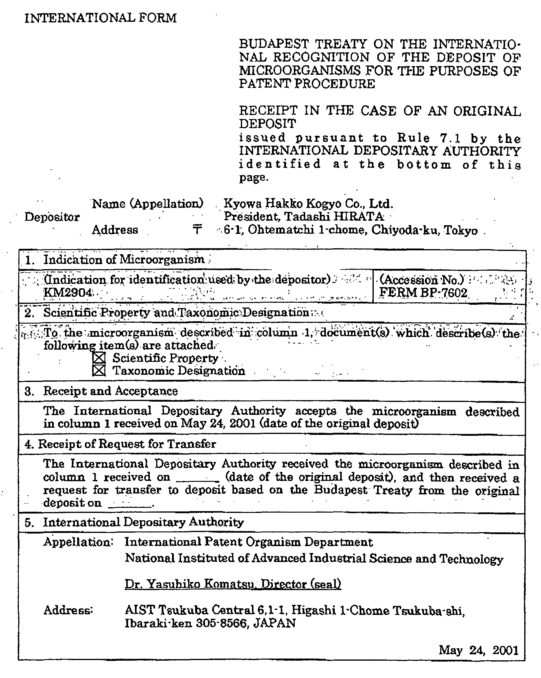
The hybridoma of the present invention includes hybridomas KM2895,
KM2896, KM2897 and KM2904. Hybridomas KM2895 and KM2904 have been
deposited on May 24, 2001, as FERM BP-7601 and FERM BP-7602, respectively, in
International Patent Organism Depositary, National Institute of Advanced Industrial
Science and Technology (AIST Tsukuba Central 6, 1-1, Higashi 1-Chome Tsukuba-shi,
Ibaraki-ken, Japan).
(5) Preparation of monoclonal antibody
-
The monoclonal antibody can be prepared by separation and purification
from a culture obtained by culturing hybridoma cells or an ascitic fluid obtained by
intraperitoneally administering monoclonal antibody-producing hybridoma cells into a
8- to 10-weeks-old mouse or nude mouse treated with pristane (0.5 ml of 2,6,10,14-tetramethylpentadecane
(pristane) is intraperitoneally administered, followed by feeding
for 2 weeks) and causing ascites tumor.
-
The method for separating and purifying the monoclonal antibody includes
centrifugation, centrifugation, salting out with 40 to 50% saturated ammonium sulfate,
caprylic acid precipitation, chromatography using a DEAE-Sepharose column, an anion
exchange column, a protein A (or G) column or a gel filtration column, which may be
used alone or in combination. According to the method, an IgG or IgM fraction can be
recovered to obtain a purified monoclonal antibody.
-
The subclass of the purified monoclonal antibody can be determined using a
monoclonal antibody typing kit or the like. The amount of the protein can be
determined by the Lowry method or by absorbance at 280 nm.
-
The subclass of an antibody means isotypes within the class such as IgG1,
IgG2a, IgG2b and IgG3 in the case of mouse, and IgG1, IgG2, IgG3 and IgG4 in the
case of human. The mouse IgG1 and IgG2a and human IgG1 types have complement-dependent
cytotoxic activity (hereinafter referred to as "CDC activity") and antibody-dependent
cell-mediated cytotoxic activity (hereinafter referred to as "ADCC activity")
so that they are useful in applying to medical treatments.
2. Preparation method of humanized antibody (I)
Preparation method of anti-MT4-MMP catalytic domain humanized antibody:
(1) Construction of vector for humanized antibody expression
-
A vector for humanized antibody expression required for production of a
humanized antibody from an antibody derived from a non-human animal is constructed.
The vector for humanized antibody expression is an expression vector for animal cell
into which genes encoding, CH and CL, C regions of a human antibody have been
inserted, and is constructed by cloning each of CH and CL of a human antibody into an
expression vector for animal cell.
-
As the C region of a human antibody, C region of an optional human
antibody, Cγ1 and Cγ4 in the case of human antibody H chains, and Cκ in the case of
human antibody L chain, and the like can be used. As the gene encoding the C region
of a human antibody, a chromosomal DNA comprising an exon and an intron or cDNA
can be used. As the expression vector for animal cell, any expression vector can be
used, so long as the C region of a human antibody can be inserted thereinto and
expressed therein.
-
Examples include pAGE107 [Cytotechnology, 3, 133 (1990)], pAGE103 [J.
Biochem., 101, 1307 (1987)], pHSG274 [Gene, 27, 223 (1984)], pKCR [Proc. Natl.
Acad. Sci. USA, 78, 1527 (1981)], pSGIβd2-4 [Cytotechnology, 4, 173 (1990)], and the
like. A promoter and enhancer used for an expression vector for animal cell includes
SV40 early promoter and enhancer [J. Biochem., 101, 1307 (1987)], LTR promoter and
enhancer of Moloney mouse leukemia virus [Biochem. Biophys. Res. Comun., 149, 960
(1987)], promoter [Cell, 41, 479 (1985)] and enhancer [Cell, 33, 717 (1983)] of
immunoglobulin H chain, and the like.
-
The vector for humanized antibody expression may be either of a type in
which a gene encoding an antibody H chain and a gene encoding an antibody L chain
exist on separate vectors or of a type in which both genes exist on the same vector
(tandem type). In respect of easiness of construction of a humanized antibody
expression vector, easiness of introduction into animal cells, and balance between the
expression amounts of antibody H and L chains in animal cells, a tandem type of the
humanized antibody expression vector is more preferred [J. Immunol. Methods, 167,
271 (1994)].
(2) Preparation of cDNA encoding VH and VL of antibody derived from non-human
animal
-
cDNAs encoding VH and VL of an antibody derived from an non-human
animal such as a mouse anti-MT4-MMP catalytic domain monoclonal antibody are
obtained as follows.
-
mRNA is extracted from a cell which produces an anti-MT4-MMP catalytic
domain monoclonal antibody, for example, a hybridoma which produces an anti-MT4-MMP
catalytic domain monoclonal antibody, to synthesize cDNA. The synthesized
cDNA is inserted into a vector such as a phage, a plasmid or the like to prepare a cDNA
library. Each of a recombinant phage or recombinant plasmid containing cDNA
encoding VH and a recombinant phage or recombinant plasmid containing cDNA
encoding VL is isolated from the library using a part of the C region or V region of a
non-human antibody such as a mouse antibody as the probe. The full nucleotide
sequences of the H chain V region and L chain V region of the mouse antibody of
interest on the recombinant phage or recombinant plasmid are determined, and the full
amino acid sequences of VH and VL are deduced from the nucleotide sequences.
(3) Construction of human chimeric antibody expression vector
-
A human chimeric antibody expression vector can be constructed by
inserting cDNAs encoding VH and VL of an antibody derived from a non-human
animal into upstream of genes encoding CH and CL of a human antibody on the vector
for humanized antibody expression as described in the above item 2(1). For example,
the vector for human chimeric antibody expression can be constructed by preparing a
recognition site of a restriction enzyme for cloning cDNAs encoding VH and VL of a
non-human animal antibody in upstream of genes encoding CH and CL of a human
antibody on the vector for chimeric antibody expression in advance, and inserting
cDNA encoding an antibody derived from a non-human animal into the cloning site via
a synthetic DNA described below. The synthetic DNA comprises a nucleotide
sequence at the 3'-terminal of a V region of an antibody derived from a non-human
animal and a nucleotide sequence at the 5'-terminal of an C region of a human antibody
and can be produced by using a DNA synthesizer so as to have a recognition site at both
ends.
(4) Identification of CDR sequence of antibody derived from non-human animal
-
VH and VL constructing an antigen-binding site of an antibody comprise
four framework regions (hereinafter referred to as "FR region") in which sequences are
relatively preserved and three complementarity determining regions (CDR) for linking
the FR region in which change of sequences are abundant [Sequence of Proteins of
Immunological Interest, US Dept. Health and Human Services (1991); hereinafter
referred to as "Sequence of Proteins of Immunological Interest". Each of the CDR
amino acid sequences (CDR sequences) can be identifying by comparison with the
amino acid sequence of the V region of a known antibody (Sequences of Proteins of
Immunological Interest).
(5) Construction of cDNA encoding V region of human CDR-grafted antibody
-
cDNAs encoding VH and VL of a human CDR-grafted antibody can be
obtained as follows.
-
First, amino acid sequences of FRs in each of VH and VL of a human
antibody to which amino acid sequences of desired CDRs in V region of an antibody
derived from a non-human animal antibody are grafted are selected. Any amino acid
sequences of FRs in V region of a human antibody can be used, so long as they are
amino acid sequences of FRs in V region derived from human.
-
Examples include amino acid sequences of FRs in V region of human
antibodies registered in database such as Protein Data Bank, and amino acid sequences
common to subgroups of FRs in V regions of human antibodies (Sequences of Proteins
of Immunological Interest). In order to provide a human CDR-grafted antibody having
sufficient activity, the amino acid sequence preferably has high homology, preferably
65% or more, with the amino acid sequence in V region of an antibody of interest
derived from a non-human animal. Then, DNA sequences encoding amino acid
sequences of CDRs in V region of the antibody derived from a non-human animal are
grafted to DNA sequences encoding the selected amino acid sequences of FRs of V
region in a human antibody to design a DNA sequence encoding an amino acid
sequence of each of VH and VL. In order to obtain a DNA sequence designed for
constructing a CDR-grafted antibody V region gene, several synthetic DNAs are
designed for each chain so as to cover all DNA sequences, and polymerase chain
reaction (hereinafter referred to as "PCR") is carried out by using them. Preferably, six
synthetic DNAs are designed for each chain based on reaction efficiency of the PCR
and the length of the DNA which can be synthesized. After the reaction, amplified
fragments are subcloned to an appropriate vector, and its nucleotide sequence is
determined to obtain a plasmid comprising cDNA encoding an amino acid sequence of
V region of each chain of the human CDR-grafted antibody of interest. Furthermore,
all sense and anti-sense sequences are synthesized by using synthetic DNAs having a
length of about 100 nucleotides, followed by annealing and ligating in order to construct
cDNAs encoding an amino acid sequence of V region of each chain of the desired
human CDR-grafted antibody.
(6) Modification of amino acid sequence of V region of human CDR-grafted antibody
-
It is known that when a human CDR-grafted antibody is prepared by simply
grafting CDRs in V region of an antibody derived from a non-human animal into FRs of
V region of a human antibody, its antigen-binding activity is lower than that of the
original antibody derived from a non-human animal [BIO/TECHNOLOGY, 9, 266
(1991)]. Accordingly, among the amino acid sequences of FRs in V region of a human
antibody, an amino acid residue which directly relates to binding to an antigen, an
amino acid residue which interacts with an amino acid residue in CDR or an amino acid
residue which has a possibility of, for example, maintaining the three-dimensional
structure of an antibody is modified with an amino acid residue which is found in the
original non-human animal antibody to thereby increase the antigen binding activity.
In order to efficiently identify these amino acid residues, construction and analysis of
the three-dimensional structure of an antibody are carried out by X-ray crystallography,
computer-modeling or the like. However, no method for preparing a human CDR-grafted
antibody which can be applied to any antibodies has been established yet, so that
various attempts must be currently necessary depending on each antibody.
-
The amino acid sequence of FRs in V region of a selected human antibody
can be modified by using various primers for introducing mutation according to PCR as
described in the above item 2(5). The amplified fragments after the PCR are
subcloned into an appropriate vector, and then the nucleotide sequence is determined to
obtain a vector comprising cDNA into which the mutation of interest is introduced
(hereinafter referred to as "amino acid-modified vector").
-
Also, in modification of an amino acid sequence in a narrow range, a PCR
mutation introducing method can be carried out by using primers for introducing
mutation consisting of 20 to 35 nucleotides. Specifically, a sense mutation primer
consisting of 20 to 35 nucleotides containing a DNA sequence encoding amino acid
residues after the modification and its antisense mutation primer are synthesized, and
two-stage PCR is carried out by using a plasmid containing cDNA encoding the amino
acid sequence of the V region to be modified as the template. Final amplified
fragments are subcloned into an appropriate vector, and then the amino acid sequence is
determined to obtain an amino acid sequence-modified vector containing cDNA into
which the mutation of interest is introduced.
(7) Construction of human CDR-grafted antibody expression vector
-
A human CDR-grafted antibody expression vector can be constructed by
introducing cDNA encoding VH and VL of the human CDR-grafted antibody obtained
in the above items 2(5) and 2(6) into upstream of the gene encoding CH and CL of the
human antibody in the vector for humanized antibody expression as described in the
above item 2(1). For example, when recognition sites for an appropriate restriction
enzymes are introduced to terminals of synthetic DNA of the 5'-terminal and 3'-terminal
in the PCR for constructing cDNA encoding the amino acid sequences of VH and VL of
the human CDR-grafted antibody, they can be introduced into upstream of the gene
encoding the C region of a desired human antibody for expression in an appropriate
form.
(8) Transient expression and activity evaluation of humanized antibody
-
In order to efficiently evaluate the activities of various humanized
antibodies, the human chimeric antibody expression vector described in the above item
2(3), the human CDR-grafted antibody expression vector in the above item 2(7) or a
modified vector thereof is introduced into COS-7 cell (ATCCC RL1651) for carrying
out transient expression of a humanized antibody [Methods in Nucleic Acids Res., CRC
Press, p. 283 (1991)], and its activity can be measured.
-
The method for introducing the expression vector into COS-7 cell includes a
DEAE-dextran method [Methods in Nucleic Acids Res., CRC Press, p. 283 (1991)], a
lipofection method [Proc. Natl. Acad. Sci., USA, 84, 7413 (1987)], and the like.
-
After introduction of the vector, the activity of the humanized antibody in
the culture supernatant can be determined by enzyme-linked immunosorbent assay
(ELISA), and the like.
(9) Stable expression and activity evaluation of humanized antibody
-
A transformant which produces a humanized antibody stably can be
obtained by introducing the human chimeric antibody expression vector described in the
above item 2(3) and the human CDR-grafted antibody expression vector described in
the above item 2(7) into an appropriate host cell.
-
The method for introducing the expression vector into a host cell includes
electroporation [Japanese Published Unexamined Patent Application No. 257891/90,
Cytotechnology, 3, 133 (1990)] and the like.
-
Any cell can be used as the host cell into which the humanized antibody
expression vector is to be introduced, so long as it can express a humanized antibody.
Examples include mouse SP2/0-Ag14 cell (ATCC CRL1581), mouse P3X63-Ag8.653
cell (ATCC CRL1580), CHO cell in which a dihydrofolate reductase gene (hereinafter
referred to as "DHFR gene") is detective [Proc. Natl. Acad. Sci. U.S.A., 77, 4216
(1980)], rat YB2/3HL.P2.G11.16Ag.20 cell (ATCC CRL1662, hereinafter referred to as
"YB2/0 cell"), and the like.
-
After introduction of the expression vector, a transformant which expresses
a humanized antibody stably is selected in accordance with the method disclosed in
Japanese Published Unexamined Patent Application No. 257891/90 by using
RPIM1640 medium containing G418 and FCS. The resulting transformant is cultured
in a medium to produce and accumulate a humanized antibody in the culture. The
activity of the humanized antibody in the culture is measured by the method described
in the above item 1 (4) and the like. Also, in the transformant, the expression amount
of the humanized antibody can be increased by using DHFR gene amplification system
or the like according to the method disclosed in Japanese Published Unexamined Patent
Application No. 257891/90.
-
The humanized antibody can be purified from the culture supernatant of the
transformant by using a protein A column (Antibodies, Chapter 8). Any other
conventional methods for protein purification can be used. For example, the
humanized antibody can be purified by a combination of gel filtration, ion-exchange
chromatography, ultrafiltration and the like. The molecular weight of the H chain or
the L chain of the purified humanized antibody or the antibody molecule as a whole is
determined by polyacrylamide gel electrophoresis (hereinafter referred to as "SDS-PAGE")
[Nature, 227, 680 (1970)], Western blotting (Antibodies, Chapter 12), or the
like.
-
The reactivity of the purified humanized antibody and the binding activity
of the humanized antibody to an MT4-MMP catalytic domain can be measured by the
method described in the above item 1(4) and the like.
3. Preparation method of recombinant antibody (II)
(1) Preparation method of antibody fragments Fab, Fab' and F(ab')2
-
An antibody fragment can be prepared by treating the above-described
antibody with an enzyme. The enzyme includes papain, trypsin and the like.
-
Also, DNA encoding Fab, Fab' or F(ab')2 fragment of the anti-MT4-MMP
catalytic domain antibody is inserted into an expression vector for animal cell, and the
vector is introduced into an animal cell for expression to prepare Fab, Fab' or F(ab')2.
-
The antibody fragment can be purified by a combination of gel filtration,
ion-exchange chromatography, ultrafiltration and the like. The molecular weight of
the purified Fab, Fab' or F(ab')2 is determined by polyacrylamide gel electrophoresis
(SDS-PAGE) [Nature, 227, 680 (1970)], Western blotting (Antibodies, Chapter 12), or
the like.
-
The reactivity of the purified Fab, Fab' or F(ab')2 and the binding activity of
the Fab, Fab' or F(ab')2 to an MT4-MMP catalytic domain can be measured by the
method described in the above item 1 (4) and the like.
(2) Preparation method of anti-MT4-MMP catalytic domain single chain antibody
-
cDNAs encoding VH and VL of the antibody derived from a non-human
animal or the human CDR-grafted antibody described in the above items 2(2), 2(5) and
2(6) are introduced into a vector for single chain antibody expression to construct a
single chain antibody expression vector of an antibody derived from a non-human
animal or a single chain antibody expression vector of a human CDR-grafted antibody.
Any of the vector for single chain antibody expression can be used, so long as cDNAs
encoding VH and VL of the antibody derived from a non-human animal or the human
CDR-grafted antibody can be inserted for expression. Examples includes pAGE107
[Cytotechnology, 3, 133 (1990)], pAGE103 [J. Biochemistry, 101, 1307 (1987)],
pHSG274 [Gene, 27, 223 (1984)], pKCR [Proc. Natl. Acad. Sci. U.S.A., 78, 1527
(1981)], pSG1βd2-4 [Cytotechnology, 4, 173 (1990)] and the like. As a host for
expression of the single chain antibody, an appropriate host can be selected from
Escherichia coli, yeast, animal cell and the like, and in this case, a vector for expression
suitable for the host can be selected.
-
Furthermore, when a cDNA encoding an appropriate signal peptide is
inserted into the vector for expression, the single chain antibody can be secreted
extracellularly, be transported to the periplasmic space or be remained intracellularly.
-
cDNA encoding a single chain antibody comprising VH-P-VL or VL-P-VH
(P is a peptide linker) is inserted into downstream of an appropriate vector and a signal
peptide of the selected vector for expression to construct a single chain antibody
expression vector into which cDNA encoding the single chain antibody of interest is
inserted.
-
cDNA encoding the single chain antibody can be obtained by linking cDNA
encoding VH and cDNA encoding VL using a synthetic DNA encoding a peptide linker
having recognition sites of appropriate restriction enzymes at both terminals. It is
important that the linker peptide is optimized so as not to inhibit binding of VH and VL
to an antigen as a result of addition of the linker peptide. For example, linker peptide
described by Pantoliano et al. [Biochemistry, 30, 10117 (1991)] or the modified linker
peptide thereof can be used.
(3) Preparation method of anti-MT4-MMP catalytic domain disulfide stabilized
antibody
-
A disulfide stabilized antibody can be prepared by modifying a DNA
sequence corresponding to one amino acid residue at an appropriate position of each of
cDNAs encoding VH and VL of an antibody derived from a non-human animal or of
cDNAs encoding VH and VL of a human CDR-grafted antibody with a DNA sequence
corresponding to a cysteine residue, followed by expression and purification to form a
disulfide bond. The modification of the amino acid residue with the cysteine residue
can be carried out according to the mutation introducing method using PCR as
described in the above item 2(5). The resulting cDNAs encoding the modified VH and
the modified VL are expressed in an appropriate vector for expression to construct a
disulfide stabilized antibody H chain expression vector and a disulfide stabilized
antibody L chain expression vector. Any of the vector for disulfide stabilized antibody
expression can be used, so long as cDNAs encoding the modified VH and the modified
VL can be inserted for expression. Examples includes pAGE107 [Cytotechnology, 3,
133 (1990)], pAGE103 [J. Biochemistry, 101, 1307 (1987)], pHSG274 [Gene, 27, 223
(1984)], pKCR [Proc. Natl. Acad. Sci. U.S.A., 78, 1527 (1981)], pSG1βd2-4
[Cytotechnology, 4, 173 (1990)] and the like. As a host for expression of the disulfide
stabilized antibody L chain expression vector and disulfide stabilized antibody H chain
expression vector for forming the disulfide stabilized antibody, an appropriate host can
be selected from Escherichia coli, yeast, animal cell and the like, and in this case, a
vector for expression suitable for the host can be selected.
-
Furthermore, when a cDNA encoding an appropriate signal peptide is
inserted into the vector for expression, the disulfide stabilized antibody can be secreted
extracellularly, be transported to the periplasmic space or be remained intracellularly.
(4) Expression and activity evaluation of various antibodies
-
A transformant which produces the antibody fragment, single chain
antibody, disulfide stabilized antibody H chain or disulfide stabilized antibody L chain
of interest can be obtained by introducing into a host cell the antibody fragment
expression vector, single chain antibody expression vector, disulfide stabilized antibody
H chain expression vector or disulfide stabilized antibody L chain expression vector
constructed in the above items 3(1) to 3(3) by electroporation [Japanese Published
Unexamined Patent Application No. 257891/90, Cytotechnology, 3, 133 (1990)] and the
like. After the introduction of the expression vector, expression of the antibody
fragment, single chain antibody, disulfide stabilized antibody H chain or disulfide
stabilized antibody L chain contained in the culture supernatant or the like can be
confirmed by the method described in the above item 1(4) or the like.
-
Recovery and purification of the single chain antibody, the disulfide
stabilized antibody H chain or the disulfide stabilized antibody L chain can be
accomplished by combining known techniques. For example, when the antibody
fragment, the single chain antibody, the disulfide stabilized antibody H chain or the
disulfide stabilized antibody L chain is secreted into a medium, the recovery and
purification can be accomplished by concentration using ultrafiltration, followed by
various chromatography or gel filtration. Furthermore, when it is transported to the
periplasmic space of the host cell, they can be accomplished by applying osmotic shock
to the host cell, followed by concentration using ultrafiltration and subsequent various
chromatography or gel filtration. When the antibody fragment, the single chain
antibody, the disulfide stabilized antibody H chain or the disulfide stabilized antibody L
chain is insoluble and present in the form of an inclusion body, they can be
accomplished by repeating centrifugation and washing for solubilizing the cells and
isolating the inclusion bodies, followed by solubilization with, for example, guanidine -
hydrochloric acid, and subsequent various chromatography or gel filtration.
-
The activity of the purified single chain antibody can be measured by the
method described in the above item 1 (4). The purified disulfide stabilized antibody H
chain and disulfide stabilized antibody L chain are mixed and a disulfide bond is formed
by operation leading to a structure having an activity [Refolding operation, Molecular
Immunology, 32, 249 (1995)], and the disulfide stabilized antibody having an activity
can be purified by antigen affinity chromatography, ion exchange chromatography or
gel filtration. The activity of the disulfide stabilized antibody can be measured by the
method described in the above item 1(4) or the like.
4. Preparation method of fusion antibody
-
A fusion antibody prepared by linking the antibody or antibody fragment
used in the present invention with a radioisotope, a protein, a low molecular weight
agent or the like chemically or genetically can also be used as an antibody derivative.
-
A fusion antibody in which an antibody and a toxin protein are linked
chemically can be prepared in accordance with the method described in literatures
[Anticancer Research, 11, 2003 (1991); Nature Medicine, 3, 350 (1996)].
-
A fusion antibody in which an antibody and a protein such as toxin,
cytokine or the like are linked genetically can be prepared in accordance with the
method described in literatures [Proc. Natl. Acad Science, USA, 93, 974 (1996); Proc.
Natl. Acad Science, USA, 93, 7826 (1996)].
-
A fusion antibody in which an antibody and a low molecular weight agent
are linked chemically can be prepared in accordance with methods described in
literatures [Science, 261, 212 (1993)]. A fusion antibody in which an antibody and a
radioisotope are linked chemically can be prepared in accordance with the method
described in literatures [Antibody Immunoconjugates and Radiopharmaceuticals, 3, 60
(1990); Anticancer Research, 11, 2003 (1991)].
-
Since these derivatives can accumulate a radioisotope, a protein (a cytokine,
a toxin, an enzyme or the like), a low molecular weight agent or the like into the
periphery of a target tissue according to the specificity of the antibody molecule, a
diagnosis or treatment which is more effective and has less side effects can be expected
from them.
5. Application method of antibody (I)
-
The above anti-MT4-MMP catalytic domain antibody, the antibody
fragment or the fusion antibody thereof with other molecule binds to an MT4-MMP
catalytic domain and destroys cells expressing MT4-MMP on the cell surface via
effector activity of the antibody such as antibody-dependent cell-mediated cytotoxic
activity (ADCC activity) and complement-dependent cytotoxic activity (CDC activity).
Thus, they are useful in treating diseases relating to MT4-MMP.
-
The diseases relating to MT4-MMP include osteoarthritis, rheumatoid
arthritis, asthma, autoimmune diseases, atopic dermatitis, psoriasis, contact dermatitis,
alopecia, ischemic heart disease, immune reaction accompanied by organ
transplantation, hepatitis, glomerulonephritis, pancreatitis, arteriosclerosis, leukemia,
malignant tumor, injury, corneal tumor, tissue injury, inflammation accompanied by
infiltration of leukocyte and the like.
-
The medicament comprising the antibody of the present invention can be
administered as a therapeutic agent alone, but generally, it is preferred to provide it as a
pharmaceutical formulation produced by an appropriate method well known in the
technical field of pharmaceutical, by mixing it with at least one pharmaceutically
acceptable carrier.
-
It is preferable to select a route of administration which is most effective in
treatment. Examples include oral administration and parenteral administration, such as
buccal, tracheal, rectal, subcutaneous, intramuscular and intravenous. In an antibody
preparation, intravenous administration is preferred.
-
The dosage form includes sprays, capsules, tablets, granules, syrups,
emulsions, suppositories, injections, ointments, tapes and the like.
-
The pharmaceutical preparation suitable for oral administration include
emulsions, syrups, capsules, tablets, powders, granules and the like.
-
Liquid preparations such as emulsions and syrups can be produced by using,
as additives, water; saccharides such as sucrose, sorbitol and fructose; glycols such as
polyethylene glycol and propylene glycol; oils such as sesame oil, olive oil and soybean
oil; antiseptics such as p-hydroxybenzoic acid esters; flavors such as strawberry flavor
and peppermint; and the like.
-
Capsules, tablets, powders, granules and the like can be produced by using,
as additive, excipients such as lactose, glucose, sucrose and mannitol; disintegrating
agents such as starch and sodium alginate; lubricants such as magnesium stearate and
talc; binders such as polyvinyl alcohol, hydroxypropylcellulose and gelatin; surfactants
such as fatty acid ester; plasticizers such as glycerine; and the like.
-
The pharmaceutical preparation suitable for parenteral administration
includes injections, suppositories, sprays and the like.
-
Injections may be prepared by using a carrier such as a salt solution, a
glucose solution or a mixture of both thereof.
-
Suppositories may be prepared by using a carrier such as cacao butter,
hydrogenated fat or carboxylic acid.
-
Also, sprays may be prepared by using the antibody composition as such or
using a carrier and the like which does not stimulate the buccal or airway mucous
membrane of the patient and can facilitate absorption of the antibody composition by
dispersing it as fine particles.
-
The carrier includes lactose, glycerol and the like. Depending on the
properties of the antibody composition and the carrier, it is possible to produce
pharmaceutical preparations such as aerosols and dry powders. In addition, the
components exemplified as additives for oral preparations can also be added to the
parenteral preparations.
-
Although the clinical dose or the frequency of administration varies
depending on the objective therapeutic effect, administration method, treating period,
age, body weight and the like, it is usually 10 µg/kg to 8 mg/kg for adult patients per
day.
-
Also, the method for examining antitumor effect of the antibody used in the
present invention against various tumor cells include in vitro tests such as CDC activity
measuring method, ADCC activity measuring method, and in vivo tests such as
antitumor experiments using a tumor system in an experimental animal such as a mouse.
-
CDC activity and ADCC activity measurements and antitumor experiments
can be carried out in accordance with the methods described in literatures [Cancer
Immunology Immunotherapy, 36, 373 (1993); Cancer Research, 54, 1511 (1994)] and
the like.
6. Application method of antibody (II)
-
Also, the present invention relates to a method for immunologically
detecting and determining an MT4-MMP catalytic domain or a microorganism, animal
cell or insect cell expressing the domain extracellularly by using the monoclonal
antibody of the present invention.
-
The method for immunologically detecting or determining a microorganism,
animal cell or insect cell expressing the MT4-MMP catalytic domain extracellularly by
using the monoclonal antibody of the present invention includes immunological assays
such as fluorescent antibody technique, enzyme-linked immunosorbent assay (ELISA)
and radioimmunoassay (RIA), immunohistochemical staining methods (ABC method,
CSA method, etc.) such as tissue immunostaining and cell immunostaining, Western
blotting, dot blotting, immunoprecipitation (Monoclonal Antibody Experimentation
Manual, Kodansha Scientific, 1987; A Sequel to Series Biochemistry Experimentation
Course, 5. Method for Studying Immunological Biochemistry, Tokyo Kagaku Dojin,
1986) and the like.
-
As the immunological assay, any known immunological assays can be used.
As described above, examples include radioimmunoassay (RIA), enzyme immunoassay
(EIA or ELISA), fluoroimmunoassay (FIA), luminescent immunoassay,
physicochemical detection method (TIA, LAPIA or PCIA) and the like based on the
difference in the labeling method, and the enzyme immunoassay is preferred.
-
As the label used in the enzyme immunoassay, any known (Enzyme
Immunoassay, edited by Eiji Ishikawa et al., Igaku Shoin) enzyme label can be used.
For example, an alkaline phosphatase label, a peroxidase label, a luciferase label and the
like can be used.
-
As the label used in the luminescent immunoassay, any known
[Bioluminescence and Chemiluminescence, edited by Kazuhiro Imai, Hirokawa Shoten;
Rinsho Kensa, 42 (1998)] luminescent label can be used. For example, an acridinium
ester label, a lophine label and the like can be used.
-
As the label used in the fluoroimmunoassay, any known (Fluorescent
Antibody Technique, edited by Akira Kawaoi, Soft Science) fluorescent label can be
used. For example, an FITC label, an RITC label and the like can be used.
-
The immunological assay is a method in which the amount of an antibody or
the amount of an antigen is measured by using an antigen or antibody treated with the
above various labels. The immunological assay of the present invention may be any
method for carrying out detection or measurement of antigens. For example, a
competitive method and a sandwich method [Immunology Illustrated, 5th edition,
Nankodo] can be exemplified, and a sandwich method is preferred.
-
The sandwich method is a method in which a primary antibody is linked to a
solid phase to trap an antigen to be measured and then allowed to react with a labeled
secondary antibody. The antibodies used in the sandwich method may be either
polyclonal antibodies or monoclonal antibodies, or antibody fragments such as the
above Fab, Fab' and F(ab)2 can be used. The combination of the two antibodies used
in the sandwich method may be a combination of monoclonal antibodies or antibody
fragments which recognize different epitopes or may be a combination of a polyclonal
antibody with a monoclonal antibody or an antibody fragment. A combination of
KM2895 with KM2904 which are monoclonal antibodies of the present invention is
preferred.
-
The fluorescent antibody technique is a method in which the monoclonal
antibody of the present invention is allowed to react with a microorganism, animal cell
or insect cell expressing the MT4-MMP catalytic domain extracellularly and further
allowed to react with an anti-mouse IgG antibody or binding fragment labeled with a
fluorescent material such as fluorescent isothiocyanate (FITC), and then the
fluorescence dye is measured by using a flow cytometer.
-
The enzyme-linked immunosorbent assay (ELISA) is a method in which the
monoclonal antibody of the present invention is allowed to react with a microorganism,
animal cell or insect cell expressing the MT4-MMP catalytic domain extracellularly and
further allowed to react with an anti-mouse IgG antibody or binding fragment treated
with an enzyme label such as peroxidase or biotin, and then the colored dye is measured
by using a flow cytometer.
-
The radioimmunoassay (RIA) is a method in which the monoclonal
antibody of the present invention is allowed to react with a microorganism, animal cell
or insect cell expressing the MT4-MMP catalytic domain extracellularly and further
allowed to react with an anti-mouse IgG antibody or binding fragment treated with a
radioisotope label and then the isotope is measured by using a scintillation counter or
the like.
-
The cell immunostaining or tissue immunostaining is a method in which the
monoclonal antibody of the present invention is allowed to react with a microorganism,
animal cell or insect cell expressing the MT4-MMP catalytic domain extracellularly and
further allowed to react with an anti-mouse IgG antibody or binding fragment treated
with a fluorescent material such as FITC or an enzyme label such as peroxidase or
biotin and then the cell or tissue is observed under a microscope.
-
The Western blotting is a method in which a cell extract of a microorganism,
animal cell or insect cell expressing the MT4-MMP catalytic domain extracellularly is
fractionated by SDS-polyacrylamide gel electrophoresis (Antibodies - A Laboratory
Manual, Cold Spring Harbor Laboratory, 1988), the gel is blotted on a PVDF
membrane or nitrocellulose membrane, the membrane is allowed to react with the
monoclonal antibody of the present invention and further allowed to react with an anti-mouse
IgG antibody or binding fragment treated with a fluorescent material such as
FITC or an enzyme label such as peroxidase or biotin, and then the result is confirmed.
-
The dot blotting is a method in which a cell extract of a microorganism,
animal cell or insect cell expressing the MT4-MMP catalytic domain extracellularly is
blotted on a nitrocellulose membrane, the membrane is allowed to react with the
monoclonal antibody of the present invention and further allowed to react with an anti-mouse
IgG antibody or binding fragment treated with a fluorescent material such as
FITC or an enzyme label such as peroxidase or biotin, and then the result is confirmed.
-
The immunoprecipitation is a method in which a cell extract of a
microorganism, animal cell or insect cell expressing the MT4-MMP catalytic domain
extracellularly is allowed to react with the monoclonal antibody of the present invention
and then an antigen-antibody complex is precipitated by adding a carrier having
immunoglobulin-specific binding ability such as protein G-Sepharose.
-
The method for diagnosing diseases relating to MT4-MMP includes a
method in which the MT4-MMP catalytic domain is immunologically detected or
determined as described above by using various human tumor cultured cells or cells
collected from patients by biopsy etc. and cell extracts prepared from the cells.
Accordingly, the monoclonal antibody of the present invention can be used as a
diagnostic agent for the above diseases relating to MT4-MMP.
7. Application method of antibody (III)
-
Furthermore, the anti-MT4-MMP catalytic domain monoclonal antibody of
the present invention can be used for purifying MT4-MMP. Specifically, affinity
chromatography is carried out by using the antibody of the present invention.
-
The anti-MT4-MMP catalytic domain monoclonal antibody is immobilized
on the carrier to prepare an antibody column by using a carrier having immunoglobulin-specific
binding ability such as protein G-Sepharose or using various coupling gels
which directly binds to immunoglobulin via an amino group,.
-
As the sample, cell extracts of animal cells or insect cells expressing the
MT4-MMP or cell extracts of various human tumor cultured cells or cells collected
from patients by biopsy etc. can be used.
-
The above MT4-MMP sample is applied to the antibody column and then
washed with 10 folds of the column volume of a phosphate buffer (pH 7.2) containing
0.5 M NaCl. Thereafter, the purified MT4-MMP is obtained by eluting it with a buffer
under conditions for dissociate the antigen-antibody reaction (high pH, low pH, high
salt concentration, surfactant, denaturing agent and the like). Also, it is necessary to
carry out the elution under such conditions that enzyme activity of MT4-MMP is not
inactivated.
-
The present invention is hereinafter explained in detail according to
Examples, but the present invention is not limited to these Examples.
BRIEF DESCRIPTION OF THE DRAWINGS
-
- Fig. 1 is a graph showing results of the enzyme immunoassay examination
of reactivity of the monoclonal antibody of the present invention obtained by using
MT4-MMP catalytic domain as the antigen, against MT4-MMP catalytic domain or
Escherichia coli protein.
- Fig. 2 is a chart showing results of measurement of MT4-MMP protein
distributing on the membrane of MT4-MMP gene-introduced COS-1 cell, detected by
cell immunostaining using the monoclonal antibody of the present invention. COS-1
and MT5-MMP gene-introduced COS-1 were used as control cells. The abscissa and
the ordinate show fluorescence intensity and the number of cells, respectively.
- Fig. 3 is a graph showing results of the detection of MT4-MMP protein and
MT5-MMP protein by a sandwich ELISA system using the monoclonal antibody of the
present invention. "○" and "" show reactivity for MT4-MMP and reactivity for
MT5-MMP, respectively.
- Fig. 4 shows results of the detection of MT4-MMP protein existing in sera
of mouse collagen-induced arthritis (CIA) model by a sandwich ELISA system using
the monoclonal antibody of the present invention. Amounts of MT4-MMP protein in
sera. of normal rats and arthritis-induced rats are compared.
-
BEST MODE FOR CARRYING OUT THE INVENTION
Example 1
Expression of human MT4-MMP catalytic domain protein:
(1) Sequence information of human MT4-MMP gene
-
As the information of human MT4-MMP cDNA, a sequence registered with
one of the gene sequence data bases, Genbank, (Accession No. X89576). Its
nucleotide sequence is shown in SEQ ID NO:5. The nucleotide sequence encoding a
human MT4-MMP catalytic domain corresponds to a nucleotide sequence of positions
481 to 987 in SEQ ID NO:5.
(2) Design of primers used in amplification of human MT4-MMP catalytic domain
-
Unless otherwise indicated, gene engineering techniques were carried out by
the methods described in Molecular Cloning, 2nd edition.
-
Nucleotide sequences of the primers used in the amplification of human
MT4-MMP catalytic domain are shown in SEQ ID NOs:1 and 2.
-
Also, primers for nucleotide sequence confirmation were designed from
sequences positioned in both sides of the cloning site of a cloning vector pCR-Blunt
(manufactured by Invitrogen, Carlsbad, CA, USA). Nucleotide sequences of the
primers for nucleotide sequence confirmation are shown in SEQ ID NOs:3 and 4.
(3) Amplification of human MT4-MMP catalytic domain by PCR method
-
As the template of PCR, 1 µl of a plasmid hMT4Δt/pRSET B
(WO00/18805) was used. Using PLATINUM pfx DNA Polymerase (manufactured by
Life Technologies) as a heat resistant enzyme, PCR was carried out by using 50 ml in
total of a reaction solution [1×pfx Amplification buffer, 1 mM MgSO4, 300 mM of
each of dNTP (dATP, dGTP, dCTP and dTTP), 0.3 mM of each of the primers MT4-MMPCDF1
and MT4-MMPCDR1 obtained in the above item (2), and 1.25 units of
PLATINUM pfx DNA Polymerase]. Using Thermal Cycler PTC-200 (manufactured
by MJ RESEARCH), the solution was heated at 94°C for 2 minutes, subjected to 25
cycles of a reaction at 94°C for 15 seconds, at 60 °C for 30 seconds and 68°C for 1
minute as one cycle, and then further heated at 68°C for 10 minutes. By agarose
electrophoresis, 5 ml of the PCR reaction solution was analyzed to confirm that the
expected DNA fragment of about 0.5 kb was amplified.
(4) Purification of the amplified DNA fragment and its insertion into a vector
-
All of the remaining PCR reaction solution was separated by agarose
electrophoresis to recover the DNA fragment of about 0.5 kb. The DNA fragment was
purified by using QIAEX II gel extraction kit (manufactured by QUIAGEN) in
accordance with the manufacture's instruction attached to the kit. As the vector DNA,
pCR-Blunt attached to Zero Blunt PCR Cloning Kit (manufactured by Invitrogen) was
used. About 50 ng of the purified PCR product was mixed with about 25 ng of pCR-Blunt,
and the DNA solution was mixed with the same volume of TaKaRa ligation
system ver. 2 (manufactured by Takara Shuzo) to carry out ligation at 16°C for 12 hours.
Using the recombinant plasmid DNA obtained by the reaction, E. coli TOP10
[genotype: F-, mcrA Δ(mrr-hsdRMS-mcrBC) 80 lacZΔM15 ΔlacX74 deoR recA1
araD139 Δ(ara-leu)7697 galU galK rps(StrR) endA1 nupG] was transformed to obtain a
plasmid pCR-BluntMT4MMPCD.
(5) Confirmation of pCR-BluntMT4MMPCD nucleotide sequence
-
Confirmation of the nucleotide sequence of cDNA introduced into pCR-BluntMT4MMPCD
was carried out by using ABI PRISM 377 DNA Sequencer
(manufactured by PE Applied Biosystems). The sequencing reaction was carried out
by using Big Dye Terminator Cycle Sequencing Ready Reaction (manufactured by PE
Applied Biosystems) in accordance with the manufacture's instruction attached to the kit.
Using the primers Fl and R1 obtained in the above item (2) as the primers for
sequencing, nucleotide sequences of both chains were confirmed. GENETYX WIN
ver. 3.2 (manufactured by Software) was used in the sequence data analysis.
-
When the cloned nucleotide sequence was confirmed, it coincided with a
nucleotide sequence of positions 484 to 1005 of a known sequence (Genbank Accession
No. X89576) shown in SEQ ID NO:5. This sequence corresponds to the human MT4-MMP
catalytic domain and hinge region.
(6) Recombination from pCR-Blunt to expression vector pET23a(+)
-
A DNA containing the MT4-MMP catalytic domain was cut out by
digesting about 6 mg of the pCR-BluntMT4MMPCD at 37°C for 1 hour in 40 µl of a
reaction solution comprising 10 mM Tris-HCl (pH 7.5), 10 mM MgCl2, 100 mM NaCl,
0.01% bovine serum albumin (BSA), 1 mM dithiothreitol (DTT), 30 units of NdeI
(manufactured by New England Biolabs) and 15 units of NotI (manufactured by Takara
Shuzo). By separating the reaction solution by agarose electrophoresis, an NdeI-NotI
DNA fragment of about 0.5 kb was recovered. The DNA fragment was purified by
using QIAEX II gel extraction kit (manufactured by QIAGEN).
-
Digestion of 5 µg of pET23a(+) (manufactured by Novagen) was carried out
at 37°C for 2 hours in 40 µl of a reaction solution comprising 10 mM Tris-HCl (pH 7.5),
10 mM MgCl2, 100 mM NaCl, 0.01% BSA, 1 mM DTT, 30 units of NdeI
(manufactured by New England Biolabs) and 15 units of NotI (manufactured by Takara
Shuzo). By separating the reaction solution by agarose electrophoresis, a DNA
fragment of about 3.7 kb was recovered. The DNA fragment was purified by using
QIAEX II gel extraction kit (manufactured by QIAGEN).
-
About 50 ng of the above an NdeI-NotI fragment containing the above
MT4-MMP catalytic domain was mixed with about 50 ng of the pET23a(+) fragment
which had been treated with NdeI and NotI, and the DNA solution was mixed with the
same volume of TaKaRa ligation system ver. 2 (manufactured by Takara Shuzo) to
carry out the ligation at 16°C for 2 hours. Using the recombinant plasmid DNA
obtained by the reaction, E. coli XL-1 Blue MRF' [genotype: Δ(mcrA)183 Δ(mcrCB-hsdSMR-mrr)173
endA1 supE44 thi-1 recA1 gyrA96 reLA1 lac [F proAB lacIq ZΔM15
Tn10(Tetr]] was transformed to obtain a plasmid pET23aMT4MMPCD. In the same
manner, the NdeI-NotI fragment containing the MT4-MMP catalytic domain was
inserted into pET24a(+) (manufactured by Novagen) to prepare pET24aMT4-MMPCD.
(7) Introduction of pET23aMT4-MMPCD into E. coli BL21/DE3pLysS
-
E. coli BL21/DE3pLysS [genotype: F- ompT hsdSB(rB -mB -)gal dcm(DE3)
pLysS] is a 1 phage, 1 DE3 lysogenic bacterium having T7 RNA polymerase gene in
the downstream of lacUV5 promoter. When the BL21/DE3pLysS introduced with an
expression plasmid is cultured and induction is carried out with isopropylthio-b-D-galactoside
(IPTG), the T7 RNA polymerase is firstly expressed and then strong
transcription of the gene of interest is started. Thus, pET23aMT4-MMPCD was
introduced into BL21/DE3pLysS. To 20 µl of BL21/DE3pLysS competent cell
(manufactured by Novagen), 1 µg of pET23aMT4MMPCD was added thereto, and the
mixture was allowed to stand on ice for 10 minutes. After heat shock is applied at
42°C for 30 seconds, the mixture was placed on ice for 2 minutes, 80 µl of SOC
(attached to the kit of competent cell) was added thereto, and then the mixture was
allowed to stand at 37°C for 30 minutes. A whole amount thereof was inoculated onto
Luria Bertani (LB) plate medium comprising 50 µg/ml ampicillin sodium salt
(manufactured by Nacalai Tesque) and cultured at 37°C until colonies were grown to a
diameter of about 1 mm.
(8) Expression induction of human recombinant MT4-MMP catalytic domain
-
Culturing of the recombinant E. coli was carried out using LB liquid
medium containing 100 µg/ml of ampicillin sodium salt (manufactured by Nacalai
Tesque).
-
The recombinant E. coli was inoculated into 2 ml of the medium and then
cultured on a constant temperature shaker BIO-SHAKER (model BR-40LF,
manufactured by Taiteck) at 37°C and 200 revolution/minute (rpm) overnight. Onto
75 ml of the medium, 0.5 ml of the pre-cultured medium was inoculated to start the
main culturing. After shaking culture at 37°C and 200 rpm for 2 hours, IPTG
(manufactured by Nacalai Tesque) was added to give a final concentration of 1 mM,
and the shaking culture was continued at 37°C and 200 rpm for 4 hours to induce
expression of human MT4-MMP catalytic domain.
(9) Purification of human recombinant MT4-MMP catalytic domain
-
The expression-induced cells using 75 ml of the medium were washed once
with 50 ml of 50 mM Tris-HCl (pH 7.4). To the cells, 300 ml of 50 mM Tris-HCl (pH
7.4) containing 0.1% Triton X-100, 10 mM EDTA (pH 8.0), 10 µg/ml DNase I
(manufactured by Sigma-Aldrich), 0.1 mM phenylmethylsulfonyl fluoride (PMSF)
(manufactured by Sigma-Aldrich), 0.1 µg/ml leupeptin (manufactured by Peptide
Laboratory) and 0.1 µg/ml pepstatin (manufactured by Peptide Laboratory) was added,
followed by incubation at 37°C for 2 hours. After centrifugation, the thus recovered
precipitate was washed 4 times with 50 mM Tris-HCl (pH 7.4) comprising 10 mM
EDTA (pH 8.0) and the above protease inhibitor. The precipitate was dissolved by
adding 2 ml of 50 mM Tris-HCl (pH 7.4) comprising 8 M urea and dispensed into 1.5
ml capacity tubes and then centrifuged at 15,000 rpm and at 4°C for 20 minutes to
recover the supernatant. The supernatant was desalted by using a handy filtration
column PD-10 (manufactured by Amersham Pharmacia Biotech) and stored at -20°C.
(10) Measurement of enzyme activity
-
To 190 µl of an assay buffer comprising 10 µM of a fluorescent substrate
MOCAc-Pro-Leu-Gly-Leu-A2pr(Dnp)-Ala-Arg-NH2 (manufactured by Peptide
Laboratory), 50 mM CaCl2, 0.005% Brij 35 and 62.5 nM ZnCl2, (final concentration:
0.8 µg/ml), 10 µl of a 16 µg/ml enzyme solution (diluted with 50 mM Tris-HCl (pH 7.4)
containing 8 M urea) was added, followed by reaction for 30 to 240 minutes, and then
the fluorescence intensity was measured. Determination of the amount of the enzyme
was carried out by using Protein Assay reagent (manufactured by Bio-Rad) and using
BSA as a standard. Also, using MOCAc-Pro-Leu-Gly (manufactured by Peptide
Laboratory) as a standard fluorescent substrate, a relationship between a fluorescence
value and a substrate degradation was calculated (fluorescence value = substrate
degradation × 6943.4 + 116.48). The measurement was carried out by using ARVO
(manufactured by Beltold Japan) under conditions of Ex. 320, Em. 393, Energy 2000
and 1 second.
-
By carrying out expression induction using the pET system, 2.5 ml of a 160
µg/ml enzyme solution was obtained from 75 ml of the cultured cells. When the
enzyme activity was measured, the MT4-MMP activity was detected and the substrate
degradation rate was 1.2 µmol/mg protein/hour.
-
The resulting product was generally dissolved in 8 M urea (manufactured by
Nacalai Tesque) and stored at 4°C, but when used, the product was diluted 20-fold or
more with the above buffer, and the mixture was allowed to stand at 25°C for 90
minutes for refolding, followed by optional dilution.
Example 2
Preparation of monoclonal antibody which specifically binds to anti-MT4-MMP
catalytic domain:
(1) Preparation of immunogen
-
The MT4-MMP catalytic domain obtained in Example 1 was added to a
solution comprising 50 mM Tris-HCl (pH 7.4), 50 mM CaCl2, 500 nM ZnCl2 and
0.005% Brij 35, and the mixture was allowed to stand at room temperature for 1 hour
for refolding and then used as the immunogen.
(2) Immunization of animal and preparation of antibody-producing cell
-
To 5-week-old mice (Balb/c) or week-old rats (SD), 50 µg of the MT4-MMP
catalytic domain prepared in Example 2(1) was administered, together with 2 mg
of aluminum gel and 1×10⁹ cells of pertussis vaccine (manufactured by Chiba Serum
Institute), and 2 weeks thereafter, administration of 100 µg of the MT4-MMP catalytic
domain was started once a week for a total of 4 times. Blood samples were collected
from the venous plexus of the fundus of the eye and the serum antibody titers were
examined by the enzyme immunoassay shown below, and the spleen was excised 3 days
after the final administration from a mouse showing a sufficient antibody titer. The
spleen was cut to pieces in MEM (manufactured by Nissui Pharmaceutical), was
loosened by using a pair of tweezers and centrifuged (1,200 rpm, 5 minutes), the
resulting supernatant was discarded and the sediment was treated with Tris-ammonium
chloride buffer (pH 7.65) for 1 to 2 minutes for eliminating erythrocytes, and then the
remaining cells were washed three times with MEM and used for cell fusion.
(3) Enzyme immunoassay
-
The MT4-MMP catalytic domain obtained in Example 2(1) was subjected to
refolding and used as the antigen in the assay. Into a 96 well EIA plate (manufactured
by Greiner), 10 µg/ml of the MT4-MMP catalytic domain prepared in the above was
dispensed at 50 µl/well and allowed to stand at 4°C overnight for adsorption. After
washing, 1% BSA/PBS (Dulbecco's phosphate buffered saline without magnesium and
calcium) was added at 100 µl/well, and the reaction was carry out at room temperature
for 1 hour to block the remaining active groups. After discarding 1% BSA/PBS,
antiserum of immunized mouse, a culture supernatant of anti-MT4-MMP catalytic
domain monoclonal antibody or purified monoclonal antibody was dispensed at 50
µl/well to carry out the reaction for 2 hours. After washing with 0.05% Tween 20/PBS,
a peroxidase-labeled rabbit anti-mouse immunoglobulin (manufactured by DAKO) or
peroxidase-labeled rabbit anti-rat immunoglobulin (manufactured by DAKO) was added
at 50 µl/well to carry out the reaction at room temperature for 1 hour, the plate was
washed with 0.05% Tween 20/PBS, and then color development was caused by using an
ABTS substrate solution [2,2-azinobis(3-ethylbenzothiazole-6-sulfonic acid)
ammonium], and the absorbance at OD 415 nm was measured by using a plate reader
(NJ 2001; manufactured by Japan Intermed).
(4) Preparation of mouse myeloma cells
-
The 8-azaguanine-resistant mouse myeloma cell strain P3-U1 was cultured
in the normal medium and 2×10⁷ or more of the cells were secured at the time of cell
fusion and used for cell fusion as a parent strain.
(5) Preparation of hybridoma
-
The mouse spleen cells obtained in Example 2(2) and the myeloma cells
obtained in Example 2(4) were mixed at a proportion of 10:1, the mixture was
centrifuged (1,200 rpm, 5 minutes), the resulting supernatant was discarded, the thus
precipitated cells were thoroughly disintegrated, a mixed solution comprising 2 g of
polyethylene glycol-1,000 (PEG-1,000), 2 ml of MEM and 0.7 ml of dimethyl sulfoxide
was added to the cells with stirring, in an amount of from 0.2 to 1 ml/108 mouse spleen
cells, at 37°C, 1 to 2 ml of MEM was added several times at 1- to 2-minute intervals,
and then the total volume was adjusted to 50 ml by adding MEM. After centrifugation
(900 rpm, 5 minutes), the supernatant was discarded, the resulting cells were loosened
gently and then the cells were gently suspended in 100 ml of HAT medium by repeated
sucking into and discharging from a measuring pipette.
-
The suspension was dispensed at 100 µl/well into a 96 well culture plate and
incubated in a 5% CO2 incubator at 37°C for 10 to 14 days. By examining the
resulting culture supernatants by the enzyme immunoassay described in Example 2(3),
wells which reacted with the MT4-MMP catalytic domain partial peptide but did not
react with the control peptide were selected, and then cloning was repeated twice by
changing the medium to HT medium and normal medium to establish anti-MT4-MMP
monoclonal antibody producing hybridomas.
-
By the above method, 4 mouse monoclonal antibodies KM2892, KM2893,
KM2894 and KM2895 were selected. Also, by applying the same method to rats, 11
rat monoclonal antibodies KM2896, KM2897, KM2898, KM2899, KM2900, KM2901,
KM2903, KM2904, KM2906, KM2909 and KM2910 were selected.
(6) Purification of monoclonal antibody
-
Each of the hybridomas obtained in Example 2(5) was intraperitoneally
injected into 8-week-old female nude mice (Balb/c) treated with pristane at a dose of 5
to 20×106 cells/head. The hybridoma caused ascites tumor in 10 to 21 days after the
injection. The ascitic fluid was collected (1 to 8 ml/head) from the ascitic fluid-filled
mice and centrifuged (3,000 rpm, 5 minutes) to remove solid matter. An IgG was
purified by the caprylic acid precipitation method (Antibodies) and used as a purified
monoclonal antibody.
-
The subclass of the antibody was determined by the enzyme immunoassay
using a subclass typing kit (Table 1).
| Animal species | Antibody name | Class |
| Mouse | KM2892 | IgG1 |
| KM2893 | IgG2a |
| KM2894 | IgA or IgE |
| KM2895 | IgG2a |
| Rat | KM2896 | IgG2b |
| KM2897 | IgG2b |
| KM2898 | IgG2b |
| KM2899 | IgG2b |
| KM2900 | IgG2b |
| KM2901 | IgG2b |
| KM2903 | IgG2b |
| KM2904 | IgG2b |
| KM2909 | IgG2b |
| KM2910 | IgG2b |
| KM2906 | IgG2b |
(7) Reactivity with MT4-MMP catalytic domain (enzyme immunoassay)
-
The reactivity of anti-MT4-MMP monoclonal antibodies selected in
Example 2(5) with the MT4-MMP catalytic domain was examined by the enzyme
immunoassay shown in Example 2(3). A protein obtained by carrying out ultrasonic
disintegration of E. coli (E. coli derived protein) was used as the control.
-
As shown in Fig. 1, the mouse monoclonal antibodies KM2892, KM2893,
KM2894 and KM2895 and the rat monoclonal antibodies KM2896, KM2897, KM2898,
KM2899, KM2900, KM2901, KM2903, KM2904, KM2906, KM2909 and KM2910, as
anti-MT4-MMP monoclonal antibodies obtained by immunizing the MT4-MMP
catalytic domain, specifically bound to the MT4-MMP catalytic domain.
(8) Western blotting
-
Using the anti-MT4-MMP catalytic domain monoclonal antibodies obtained
in Example 2(5), detection of the MT4-MMP protein by Western blotting was examined.
-
As the protein, a cell membrane fraction of an MT4-MMP gene-introduced
COS-1 cell was used, and a cell membrane fraction of an MT5-MMP gene-introduced
COS-1 cell as a control. Each of the gene-introduced cells was prepared by the
method described in WO00/18805.
-
Each of the cell membrane fractions was fractionated by SDS-polyacrylamide
electrophoresis (Antibodies) at 1.25×10² cells/lane and then blotted on a
PVDF membrane. After blocking with 1% BSA/PBS, culture supernatant of each of
the anti-MT4-MMP monoclonal antibodies was allowed to react at room temperature
for 2 hours. After washing thoroughly with 0.05% Tween 20/PBS, a peroxidase-labeled
anti-mouse immunoglobulin antibody (manufactured by DAKO) or a
peroxidase-labeled anti-rat immunoglobulin antibody (manufactured by DAKO) was
allowed to react as the secondary antibody at room temperature for 1 hour. After
washing thoroughly with 0.05% Tween 20/PBS, each sample was detected using an
ECL-detection kit (manufactured by Amersham) and exposed on an X-ray film. As a
result, a band was detected at around 70 KDa corresponding to the molecular weight of
MT4-MMP (shown by an arrow in the drawing) by the mouse monoclonal antibodies
KM2892, KM2893, KM2894 and KM2895 and the rat monoclonal antibodies KM2896,
KM2897, KM2898, KM2899, KM2900, KM2901, KM2903, KM2904, KM2906,
KM2909 and KM2910, as anti-MT4-MMP monoclonal antibodies. On the other hand,
all of these monoclonal antibodies did not show reactivity to MT5-MMP. Regarding
an anti-PEG-treated granulocyte colony-stimulating factor monoclonal antibody KM511
(Japanese Published Unexamined Patent Application No. 165300/96; FERM BP-4880)
which does not react with MT4-MMP and MT5-MMP, it did not specifically react with
the band of around 70 KDa.
-
Based on the above results, it was shown that the anti-MT4-MMP catalytic
domain monoclonal antibodies can detect the MT4-MMP protein on cells by Western
blotting and can be applied to the diagnosis of various diseases relating to MT4-MMP
such as inflammation and cancer.
(9) Cell immunostaining
-
Using the anti-MT4-MMP catalytic domain monoclonal antibodies selected
in Example 2(5), detection of the MT4-MMP protein expressing on cells by cell
immunostaining was examined.
-
As the cells, COS-1 cell (ATCC No. CRL-1650), MT4-MMP gene-introduced
COS-1 and MT5-MMP gene-introduced COS-1 were used. The MT4-MMP
gene-introduced COS-1 and MT5-MMP gene-introduced COS-1 were produced
by the method described in WO00/18805.
-
The cells were suspended in a mixed solution of trypsin and EDTA, washed
with PBS and then, in order to improve antibody permeability of the cell membrane,
treated with 100% methanol (ice-cooled) at 4°C for 10 minutes. After washing with
PBS, they were blocked with 10 µg/ml human immunoglobulin (manufactured by
Cappel) at room temperature for 30 minutes. The cells were dispensed at 1×10⁵
cells/tube and then centrifuged to discard the supernatant in each tube, and culture
supernatant of each anti-MT4-MMP monoclonal antibody was added thereto and
allowed to react at room temperature for 30 minutes.
-
After washing with PBS, an FITC-labeled anti-mouse immunoglobulin
antibody (manufactured by Wako Pure Chemical Industries) was dispensed at 100
µl/tube and allowed to reaction at 4°C for 30 minutes in the dark. After thoroughly
washing with PBS, they were analyzed by a cell analyzer (manufactured by Coulter;
EPICS XLsystemII). The results are shown in Table 2.
| mAb | Reactivity |
| | COS-1 | MT4-MMP/COS-1 | MT5-MMP/COS-1 |
| KM2892 | - | + | - |
| KM2893 | - | ++ | - |
| KM2894 | - | - | - |
| KM2895 | - | ++ | - |
| KM2896 | - | ++ | - |
| KM2897 | - | ++ | - |
| KM2898 | - | ++ | - |
| KM2899 | - | ++ | +w |
| KM2900 | - | + | - |
| KM2901 | - | ++ | - |
| KM2903 | - | ++ | - |
| KM2904 | - | ++ | - |
| KM2906 | - | ++ | +w |
| KM2909 | - | ++ | - |
| KM2910 | - | ++ | - |
| -: no reaction, +w: weak reaction, +: reaction, ++: strong reaction |
-
The results show the respective reactions of cells of COS-1, MT4-MMP
gene-introduced COS-1 and MT5-MMP gene-introduced COS-1 with monoclonal
antibodies KM2892, KM2893, KM2894, KM2895, KM2896, KM2897, KM2898,
KM2899, KM2900, KM2901, KM2903, KM2904, KM2906, KM2909 and KM2910,
and the mouse IgG1 class anti-PEG-treated granulocyte colony-stimulating factor
KM511 (Japanese Published Unexamined Patent Application No. 165300/96; FERM
BP-4880) used as a negative control.
-
Reactivity with the MT4-MMP gene-introduced COS-1 cell was found in all
of the anti-MT4-MMP monoclonal antibodies excluding the monoclonal antibody
KM2894. Also, reactivity with the control COS-1 cell and MT5-MMP gene-introduced
COS-1 cell was not found.
-
Thus, it was shown that the anti-MT4-MMP monoclonal antibodies can
detect the MT4-MMP protein on cells by cell immunostaining and can be applied to the
diagnosis of various diseases relating to MT4-MMP such as inflammation and cancer.
Example 3
Analysis of MT4-MMP on cell surface of hemocytic cell lines using flow cytometer:
-
The following analysis was carried out by using U937 (ATCC CRL-1593)
as the human cell line.
-
A total of 2×10⁵ cells were put into a polyethylene tube (manufactured by
Becton Dickinson Japan) and centrifuged at 1,500 rpm (400xg) for 3 minutes. After
discarding the supernatant, 50 ml of a purified antibody anti-MT4-MMP antibody
diluted to give a concentration of 5 mg/ml with 1% BSA/PBS was added. As the
control antibody, a rat IgG2b antibody or mouse IgG2a antibody diluted to give a
concentration of 5 mg/ml with 1% BSA/PBS was respectively used.
-
After stirring, the mixture was allowed to stand on ice for 30 minutes for
reaction, adding 2 ml of cooled PBS was added thereto, followed by stirring, and then
the mixture was centrifuged at 1,500 rpm (400×g) for 3 minutes to discard the
supernatant (hereinafter, this operation is called "centrifugal washing"). As the
secondary antibody, 50 ml of a biotinylated anti-mouse Ig antibody (manufactured by
DAKO) or biotinylated anti-rat Ig antibody (manufactured by DAKO) diluted 100 folds
with 1% BSA/PBS was added. After stirring, the mixture was allowed to stand on ice
for 30 minutes. After washing by centrifugation, avidin (Streptavidin-FITC,
manufactured by BD PharMingen) as a fluorescence label was diluted 200-fold with 1%
BSA/PBS and 50 ml of the avidin dilution was further added, followed by stirring, and
then mixture was allowed to stand on ice for 30 minutes. After washing by
centrifugation, 500 ml of PBS was added thereto, followed by stirring, and then
measurement was carried out by using FACScan (manufactured by Becton Dickinson
Japan).
-
Results of the reactivity of antibodies to U937 cell are shown in Fig. 2. As
shown in Fig. 2, the monoclonal antibodies KM2895, KM2896, KM2897 and KM2904
of the present invention reacted with the U 937 cell strongly. Based on the above
results, it was considered that natural MT4-MMP on the cell membrane surface can be
detected by using a monoclonal antibody which recognizes the MT4-MMP catalytic
domain. On the other hand, the anti-MT4-MMP monoclonal antibodies KM2561 and
KM2562 disclosed in WO00/18805 react with MT4-MMP transfectants which are
considered to express large amount of MT4-MMP expression quantity (WO00/18805),
but did not react with the cell surface MT4-MMP expressing in U937 cell.
-
Thus, it was shown that the anti-MT4-MMP catalytic domain monoclonal
antibodies of the present invention can detect the MT4-MMP protein on cells by cell
immunostaining and can be applied to diagnosis of various diseases relating to MT4-MMP
such as inflammation and cancer.
Example 4
Examination of MT4-MMP determination system by sandwich ELISA:
-
Combination of monoclonal antibodies, which does not react with MT5-MMP
but shows MT4-MMP-specific and high reactivity was examined by carrying out
sandwich ELISA for all combinations of the anti-MT4-MMP monoclonal antibodies
produced in Example 2.
-
To a 96 well flat bottom plate (Maxisorp, manufactured by Japan Intermed),
50 ml of each anti-MT4-MMP monoclonal antibody diluted to 5 mg/ml with PBS was
added and allowed to stand at 4°C overnight for coating the plate (hereinafter, the anti-MT4-MMP
monoclonal antibody is called "coating antibody"). After washing with
PBS three times, 1% BSA/PBS was added at 200 ml/well and allowed to stand at room
temperature for 2 to 4 hours to block the antibody. After removing 1% BSA/PBS,
MT4-MMP refolded in advance, MT5-MMP activated with trypsin (WO00/18805) or
the animal serum obtained in Example 5 was diluted to various concentrations with 1%
BSA/PBS and dispensed at 50 ml into the wells to carry out the reaction at room
temperature for 2 hours. After washing three times with 0.05% Tween 20/PBS, a rat
anti-MT4-MMP monoclonal antibody diluted to 2 mg/ml with 1% BSA/PBS was added
at 50 ml/well to carry out the reaction at room temperature for 1 hour (hereinafter, the
rat anti-MT4-MMP monoclonal antibody is called "detecting antibody"). After
washing with 0.005% Tween 20/PBS three times, a biotinylated anti-rat Ig
(manufactured by DAKO, 1/4000 dilution) diluted with 1% BSA/PBS was added at 50
ml/well for reaction at room temperature for 1 hour. After washing with 0.05% Tween
20/PBS three times, horse radish peroxidase avidin D (manufactured by Vecter, 1/4000
dilution) diluted with 1% BSA/PBS was added at 50 ml/well and allowed to stand at
room temperature for 20 minutes. After washing with 0.05% Tween 20/PBS three
times, 3,3',5,5'-tetramethylbenzidine (TMB, manufactured by SIGMA-Aldrich) as the
substrate of horse radish peroxidase avidin D was added at 100 ml/well for development
of color at room temperature, and the reaction was stopped by adding 100 ml of 10% by
volume H2SO4. Absorbance at a wavelength of 450 nm was measured by using an
automatic plate reader EL340 (manufactured by Molecular Devices), and the data were
calculated by Delta Soft (manufactured by Bio Metallics, Inc., Ver. 3-1.3B). The 4-parameter
was used in the standard curve.
-
As shown in Fig. 3, it was able to specifically detect MT4-MMP within the
concentration range of 10 to 5,000 ng/ml by the combination of KM2895 as the coating
antibody with KM2904 as the detecting antibody.
-
Furthermore, MT4-MMP in a sample can be determined by using an MT4-MMP
protein at known concentration as a standard sample.
Example 5
Determination of MT4-MMP in serum of rat collagen-induced arthritis:
(1) Rat collagen-induced arthritis (CIA) model
-
A CIA model which is one of the rheumatoid arthritis models was prepared
as follows.
-
Twelve-week-old DA rats (female, manufactured by Charles River Japan)
were subjected to the test. A bovine joint-derived type II collagen (manufactured by
Collagen Gijyutsu Kenshu Kai, or Collagen Techniques Training Club) was dissolved
in 0.3% by volume acetic acid solution to give a concentration of 1 mg/ml and mixed
with the same volume of incomplete Freund's adjuvant (hereinafter IFA, manufactured
by DIFCO) under ice-cooling using Polytron (manufactured by KINEMATICA) to
prepare an emulsion. The collagen was used by dissolving a freeze-dried preparation
in the acetic acid solution when used.
-
The tail base areas of rats (n = 6) were shaved under ether anesthesia and
100 µl/site of the emulsified collagen solution was administered under the tail base skin
using a 27 G injection needle for sensitization. At the same time, an untreated group
was arranged and used as a normal group (n = 6).
(2) Preparation of serum
-
On the 32nd day after the sensitization, blood samples were collected in
SepaRapid Tube Mini S (manufactured by Sekisui Chemical) from the normal group
and CIA-developed group, and serum samples were collected by centrifugation (Hitachi
small type cooling centrifuge himac CF7D, rotor RT3S3, manufactured by Hitachi
Koki) of the tubes at 3,000 revolutions per minute (1,500×g) at 4°C for 20 minutes.
(3) Determination of MT4-MMP in sera of CIA model
-
Using the sandwich ELISA constructed in Example 4, expression of MT4-MMP
in sera of the CIA model was detected. As a result of the detection of serum
MT4-MMP, its significant increase was observed in the rat CIA in comparison with the
untreated group (Fig. 4, p = 0.0195). A statistical analysis software SAS (Release 6.12,
SAS Inc.) was used in the statistical analysis, and the significance test was carried out
by Student's t-test.
-
MT4-MMP in serum and the like can also be carried out by sandwich
ELISA using an MT4-MMP protein at known concentration as a standard sample.
INDUSTRIAL APPLICABILITY
-
The present invention provides an anti-MT4-MMP monoclonal antibody
which specifically reacts with MT4-MMP and can specifically detect or determine
MT4-MMP protein by immunological assay. The anti-MT4-MMP monoclonal
antibody of the present invention and a diagnosing kit which uses the same can provide
highly sensitive and highly reliable detection in diagnosing various diseases relating to
MT4-MMP. In addition, since it also reacts with MT4-MMP on cells, a medicament
comprising the monoclonal antibody of the present invention is useful in treating
various diseases relating to MT4-MMP.
Free Text of Sequence
-
- SEQ ID NO:1 - Explanation of synthetic sequence: synthetic DNA
- SEQ ID NO:2 - Explanation of synthetic sequence: synthetic DNA
- SEQ ID NO:3 - Explanation of synthetic sequence: synthetic DNA
- SEQ ID NO:4 - Explanation of synthetic sequence: synthetic DNA
-