EP1405909A1 - Method of uniformizing dna fragment contents and subtraction method - Google Patents
Method of uniformizing dna fragment contents and subtraction method Download PDFInfo
- Publication number
- EP1405909A1 EP1405909A1 EP02738769A EP02738769A EP1405909A1 EP 1405909 A1 EP1405909 A1 EP 1405909A1 EP 02738769 A EP02738769 A EP 02738769A EP 02738769 A EP02738769 A EP 02738769A EP 1405909 A1 EP1405909 A1 EP 1405909A1
- Authority
- EP
- European Patent Office
- Prior art keywords
- dna fragments
- restriction enzyme
- stranded
- adaptors
- double
- Prior art date
- Legal status (The legal status is an assumption and is not a legal conclusion. Google has not performed a legal analysis and makes no representation as to the accuracy of the status listed.)
- Withdrawn
Links
Images


Classifications
-
- C—CHEMISTRY; METALLURGY
- C12—BIOCHEMISTRY; BEER; SPIRITS; WINE; VINEGAR; MICROBIOLOGY; ENZYMOLOGY; MUTATION OR GENETIC ENGINEERING
- C12Q—MEASURING OR TESTING PROCESSES INVOLVING ENZYMES, NUCLEIC ACIDS OR MICROORGANISMS; COMPOSITIONS OR TEST PAPERS THEREFOR; PROCESSES OF PREPARING SUCH COMPOSITIONS; CONDITION-RESPONSIVE CONTROL IN MICROBIOLOGICAL OR ENZYMOLOGICAL PROCESSES
- C12Q1/00—Measuring or testing processes involving enzymes, nucleic acids or microorganisms; Compositions therefor; Processes of preparing such compositions
- C12Q1/68—Measuring or testing processes involving enzymes, nucleic acids or microorganisms; Compositions therefor; Processes of preparing such compositions involving nucleic acids
- C12Q1/6809—Methods for determination or identification of nucleic acids involving differential detection
Definitions
- the present invention relates to a method of normalizing contents of DNAs in a sample among respective DNAs, a method of detecting a difference in gene expression among cells, a method of producing a library of genes that are different in expression among cells, and a method of producing a probe specific to a gene that is different in expression among cells.
- a subtraction method using a PCR representational difference analysis (RDA) method
- RDA representational difference analysis
- subtractive hybridization is repeated a plurality of times, whereby a very low background is realized, and is considered to be useful for subtraction among cell groups having a relatively small difference in gene expression.
- RDA representational difference analysis
- genes expressed in a large amount are concentrated more, so that genes expressed in a small amount cannot be isolated.
- SSH suppression subtractive hybridization
- this method subtraction is performed while the difference in an expression amount of genes is being normalized to some degree, and it is considered that this method also enables isolation of genes expressed in a small amount.
- a normalization method a method of performing partial hybridization between tester with an adaptor attached and drivers without an adaptor attached, and amplifying DNAs remaining single-stranded by a suppression PCR is used.
- the inventors of the present invention have considered that a gene can be isolated based on the difference in an expression amount even if the gene is expressed in a small amount, provided that a normalization step can be added to an RDA method.
- a normalization method that can be used for normalizing contents of DNAs in a reaction liquid among respective DNAs in an RDA method.
- the inventors have achieved the present invention based on this finding.
- the present invention provides the following.
- the present invention relates to a method of normalizing contents of DNA fragments in a sample among respective DNA fragments (hereinafter, which may also be referred to as a "normalization method”), characterized by including:
- DNA fragments include a cDNA synthesized from mRNA prepared from cells or fragments of the cDNA, DNA fragments of genes that are different in an expression among cells, obtained by subtractive hybridization.
- DNA fragments are prepared in which adaptors each comprising an oligodeoxyribonucleotide are attached to both ends of each DNA fragment in a sample to form restriction enzyme recognition sites that do not exist in the DNA fragments in the sample, and at least a part between the restriction enzyme recognition sites, including the restriction enzyme recognition sites, is made double-stranded.
- the nucleotide sequence of the adaptor is made to be cleaved only in the adaptor by a restriction enzyme used in the step (4), considering the nucleotide sequences of the DNA fragments. Because of this, the restriction enzyme used in the step (d) has a cleavage site only in the adaptor.
- the length of the adaptor is generally 20 to 30 bases.
- Formation of restriction enzyme recognition sites means that the DNA fragment with adaptors attached thereto has restriction enzyme recognition sites, and includes both the presence of the restriction enzyme recognition sites in the adaptors and the formation of the restriction enzyme recognition sites due to the binding between the adaptors and the DNA fragment.
- the DNA fragments in a sample have a common nucleotide sequence at both ends, it is easy to design the nucleotide sequences of the adaptors in such a manner that restriction enzyme recognition sites are formed due to the binding between the adaptors and the DNA fragments.
- a part to be double-stranded may be at least a part between the restriction enzyme recognition sites at both ends inclusive of the restriction enzyme recognition sites, or may equal the full length.
- the DNA fragments are single-stranded, they are made to be double-stranded by conventional methods in the field of genetic engineering.
- the adaptors may be attached to the DNA fragments before or after the DNA fragments are made to be double-stranded. More specifically, after the adaptors are attached to the single-stranded DNA fragments, required parts may be made to be double-stranded. Alternatively, the DNA fragments and the adaptors are made to be double-stranded, separately, and then, they may be attached to each other. Alternatively, it is also possible that the DNA fragments are made to be double-stranded, the adaptors are attached to one strand, and a single-stranded part is filled in to make a required part double-stranded. In the case where the DNA fragments are double-stranded, the above-mentioned latter two methods may be mentioned as a method of attaching adaptors. The attachment can be performed by a conventional method in the field of genetic engineering.
- DNA fragments are double-stranded having nucleotide sequences corresponding to the adaptors at both ends, they may be used as they are.
- the double-stranded DNA fragments prepared in the step (a) are denatured.
- the denaturation can be performed by a conventional method in the field of genetic engineering. For example, denaturation by heating is mentioned. In this case, the condition is varied depending upon the nucleotide sequences of the double-stranded DNA fragments. For example, denaturation is performed at 94°C to 100°C for 3 to 5 minutes.
- the DNA fragments denatured in the step (b) are hybridized under a condition that a part of the DNA fragments remains single-stranded.
- the hybridization of the denatured DNA fragments is also called annealing, which can be performed by a conventional method in the field of genetic engineering.
- annealing can be performed by a conventional method in the field of genetic engineering.
- DNA fragments existing in a large amount are hybridized quickly, and the DNA fragments existing in a small amount are hybridized slowly. Consequently, the difference in an existing amount of the DNA fragment remaining single-stranded is decreased.
- "normalization" in the present invention includes decreasing the difference in the DNA fragment contents.
- the condition for allowing a part of the DNA fragments to remain single-stranded there is no particular limit to the condition for allowing a part of the DNA fragments to remain single-stranded, as long as the treatment of the subsequent process can be started before double-strands are formed completely.
- the following may be performed: hybridization is started by decreasing a temperature, and hybridization is completed before double-strands are formed completely.
- the specific condition in this case is varied depending upon the nucleotide sequences of the DNA fragments.
- the hybridization is performed at 65°C to 70°C for 12 to 20 hours. Whether or not a part of the DNA fragments remains single-stranded can be confirmed by gel electrophoresis, and it would be easy for those skilled in the art to select an appropriate condition. It is considered that, by adjusting the concentration of the DNA fragments and the time of hybridization, only the DNA fragments existing in a relatively large amount can be decreased, or the DNA fragments existing in a very small amount can be increased.
- the double-stranded DNA fragments hybridized in the step (c) are cleaved by a restriction enzyme having a cleavage site only in the adaptors.
- the restriction enzyme is appropriately selected based on the sequences of the adaptors and the DNA fragments in the sample.
- the adaptors also include the attachment sites of the adaptors to the DNA fragments.
- the cleavage condition is varied depending upon the restriction enzyme. For example, the cleavage is performed at 30 to 37°C and pH 7.5 to 8.5 for 30 to 120 minutes.
- step (e) PCR is performed using the DNA fragments obtained in the step (d) as templates, and using a primer having a nucleotide sequence complementary to the nucleotide sequence of the adaptors before cleavage.
- the PCR can be performed by a conventional method. It would be easy for those skilled in the art to set an appropriate condition, considering the nucleotide sequences of the templates and the primer.
- the length of the primer is generally 20 to 30 bases.
- An oligonucleotide used as the adaptor may be used as the primer.
- the present invention also provides a kit for use in the normalization method of the present invention.
- This kit comprises an adaptor comprising an oligodeoxyribonucleotide to be attached to both ends of the DNA fragments in a sample, a restriction enzyme having a cleavage site in the adaptor, and a primer for PCR having a nucleotide sequence complementary to the nucleotide sequence of the adaptor before cleavage with the restriction enzyme.
- the adaptor, the restriction enzyme, and the primer for PCR are as described with reference to the above-mentioned normalization method of the present invention.
- the kit of the present invention may include a part of or an entirety of reagents (buffer, etc.) required for cleavage with a restriction enzyme and reagents (buffer, dNTP, DNA polymerase, etc.) required for PCR, in addition to the adaptor, the restriction enzyme, and the primer for PCR.
- reagents buffer, etc.
- buffer, dNTP DNA polymerase, etc.
- mRNAs or cDNAs prepared from two different kinds of cells are hybridized, and cDNAs that are not hybridized are isolated, whereby specific cDNAs (i.e., that are different in an expression amount among cells) are detected.
- the important point of influencing the efficiency of this method is the efficiency of hybridization. Genes expressed in a small amount have a low hybridization efficiency, compared with those expressed in a large amount. Therefore, it is difficult to isolate the genes expressed in a small amount.
- the difference in a gene expression among cells can be detected even in genes expressed in a small amount.
- the present invention also provides a method of detecting the difference in a gene expression among cells, comprising performing subtractive hybridization at least once (hereinafter, which may also be referred to as a "detection method"), in which normalization is performed by the normalization method of the present invention before or after performing at least one of the subtractive hybridization(s).
- the detection method of the present invention may be the same as that of detecting the difference in a gene expression among cells, comprising performing conventional subtractive hybridization at least once, except that normalization is performed by the normalization method of the present invention before or after performing at least one of the subtractive hybridization(s).
- the above-mentioned step (a) of the normalization method of the present invention can be simplified. Therefore, it is preferable to perform normalization after the subtractive hybridization.
- the following subtractive hybridization is performed a plurality of times. That is, cDNA fragments (tester) with an oligonucleotide called adaptor attached and cDNA fragments (driver) without adaptor attached are denatured and then hybridized. Then, among the hybridized double-strands, strands complementary to those in which the adaptor portions are single-stranded are synthesized to be double-stranded over the full length, and thereafter, PCR is performed using a primer complementary to the adaptors.
- cDNA fragments without the adaptors attached in an excess amount as driver By adding the cDNA fragments without the adaptors attached in an excess amount as driver, only those which are specific to the tester form double-strands with the adaptors at both ends, and only such cDNA fragments are amplified by the PCR.
- driver in a 100-fold amount of the tester, non-specific genes are decreased to 1/100, and thus, specific genes are concentrated 100-fold.
- the difference in an existing amount between the tester and the driver is very small, it is difficult to isolate specific genes with about 100-fold concentration.
- a subtraction operation is repeated twice or more (generally, 3 to 4 times) to completely remove non-specific genes, whereby genes that are specifically expressed among cells with a smaller difference can be isolated.
- genes expressed in a small amount have a lower efficiency of hybridization, compared with those expressed in a large amount. Therefore, the difference in an amount is increased as subtractive hybridization is repeated.
- the ratio of the driver is considerably increased to 100-fold (first time), 800-fold (second time), and 400000-fold (third time). This is considered to be effective for decreasing a background.
- the amount of cDNAs that can be used for the driver.
- the ratio is increased by decreasing the amount of the tester. Therefore, the frequency of forming double-strands of the genes expressed in a small amount is decreased more, and only genes expressed in a large amount are amplified preferentially.
- the normalization is performed after the first subtraction, and then, the second and subsequent subtractions are performed.
- the normalization is performed after the first subtraction, and then, the second and subsequent subtractions are performed.
- the ratio of the tester to the driver is decreased to increase the concentration of the tester so as to suppress the increase in the difference in an existing amount as the subtraction is performed.
- 30 to 50-fold, 100 to 300-fold, and 1000 to 5000-fold drivers are used in the first, second, and third subtractive hybridizations, respectively.
- the step of normalization for adjusting the difference in an expression amount is added between the first and second subtractive hybridizations.
- SSH suppression subtractive hybridization
- the cells for detecting the difference in a gene expression by the detection method of the present invention are not particularly limited.
- the detection method of the present invention can be applied to the detection of the difference in a gene expression among cells derived from different tissues and among the same cells cultured under different conditions.
- the present invention provides a method of producing a library of genes which are different in an expression among cells, comprising cloning genes whose difference in an expression is detected by the detection method of the present invention, and a method of producing a probe specific to a gene which is different in an expression among cells, comprising preparing a fragment of the gene whose difference in an expression is detected by the detection method of the present invention.
- the production of the library by cloning of the genes and the production of the probe by the preparation of the gene fragment can be performed by a conventional method, except that the gene or genes are detected for the difference in an expression by the detection method of the present invention.
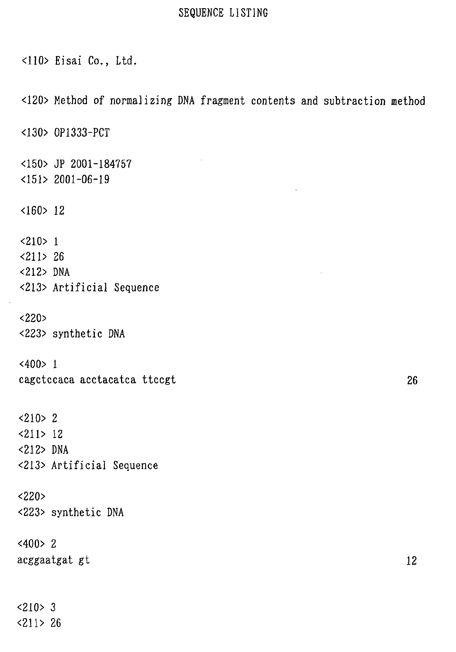
- Oligonucleotides (ad2S and ad2A) having nucleotide sequences of SEQ ID No: 1 and SEQ ID No: 2 are annealed to prepare an adaptor.
- the annealing is performed under the following condition.
- TE represents 10 mM Tris/HCl (pH 8.0) buffer containing 1 mM EDTA.
- adaptor solutions ad3, ad4, ad5, ad13, and ad14, respectively
- Double-stranded cDNAs are synthesized from a sample, using a TAKARA cDNA synthesis kit.
- the thus obtained cDNAs are purified, using a QIAquick PCR purification kit (hereinafter, referred to as "QIAquick") (eluted with 20 ⁇ l of TE).
- QIAquick QIAquick PCR purification kit
- the purified cDNAs are Rsa I-digested (TAKARA Afa I) in a 100- ⁇ l system, and purified with QIAquick.
- the cDNAs are eluted with 20 ⁇ l of TE.
- the cDNAs are eluted with 50 ⁇ l of TE, precipitated with ethanol, and dissolved in 2 ⁇ l of TE.
- the cDNAs are bound to the adaptor ad2, using the TAKARA DNA ligation kit Ver 2. 1 ⁇ l of cDNAs, 1 ⁇ l of 100 ⁇ M ad2, 2 ⁇ l of solution II, and 4 ⁇ l of solution I are mixed, and allowed to react at 16°C for 2 hours, whereby a cDNA solution (ad2 lig cDNA) with the adaptors ad2 added thereto is obtained.
- a PCR is performed under the following condition:
- the remaining portion of 40 ⁇ l at 1 ⁇ l of template is purified with QIAquick (eluted with 40 ⁇ l of TE), whereby an amplified cDNA solution (cDNA ad2 amp) is obtained.
- the products for driver are reacted at 37°C for 2 hours, and a restriction enzyme is inactivated by a treatment at 70°C for 5 minutes, whereby a complete driver solution (cDNA ad2 amp RsaI) is obtained.
- the products for tester are reacted at 37°C for one hour, and purified with QIAquick (eluted with 20 ⁇ l of TE) to obtain a digested product (cDNA ad2 amp RsaI).
- the digested product is bound to the adaptor ad3, using the TAKARA DNA ligation kit Ver2. Then, 3 ⁇ l of cDNA ad2 amp RsaI, 1 ⁇ l of 100 ⁇ M ad3, 4 ⁇ l of solution II, and 8 ⁇ l of solution I are mixed and allowed to react at 16°C for one hour, and a restriction enzyme is inactivated by treatment at 70°C for 5 minutes, whereby a complete tester solution (cDNA ad3 lig) is obtained.
- the solution in an amount corresponding to 5 runs is purified in one column with QIAquick and precipitated with ethanol.
- the precipitate is rinsed with 70% ethanol, and dried.
- 5 ⁇ l of TE, 5 ⁇ l of 10 ⁇ mung bean buffer, 38.5 ⁇ l of DW, and 1.5 ⁇ l of mung bean nuclease are added to the solution, and the mixture is allowed left at room temperature for 20 minutes.
- a PCR is performed under the following condition:
- reaction liquid 10 ⁇ l of the reaction liquid is electrophoresed to confirm that amplification is completed.
- the rest of the reaction mixture is purified with QIAquick (eluted with 40 ⁇ l of TE) to obtain an amplified product (S1 amp).
- the second PCR is also performed under the conditions of 5 and 7 cycles, and electrophoresis is performed.
- 1 ⁇ l of 2 ⁇ PCR buffer is added to 1 ⁇ l of S1 amp diluted 5-fold with TE, and mineral oil is overlaid. After the resultant solution is allowed to stand at 98°C for 5 minutes, 2 ⁇ l of 1 ⁇ PCR buffer + 1 M NaCl is added and mixed together, and treatment (98°C, 5 minutes ⁇ 68°C, 16 hours) is performed.
- a PCR is performed under the following condition:
- the digested product is bound to the adaptor ad4, using the TAKARA DNA ligation kit Ver2. Then, 2 ⁇ l of N1 amp RsaI, 1 ⁇ l of 100 ⁇ M ad4, 3 ⁇ l of solution II, and 6 ⁇ l of solution I are mixed and allowed to react at 16°C for one hour, and a restriction enzyme is inactivated by treatment at 70°C for 5 minutes, whereby a tester solution for the second time (N1 amp ad4 lig) is obtained.
- the digested product is bound to the adaptor ad5, using the TAKARA DNA ligation kit Ver2. Then, 1 ⁇ l of NS2 amp RsaI diluted 1/5-fold, 1 ⁇ l of 100 ⁇ M ad5, 2 ⁇ l of solution II, and 4 ⁇ l of solution I are mixed and allowed to react at 16°C for one hour, and a restriction enzyme is inactivated by treatment at 70°C for 5 minutes, whereby a tester solution for the third time (NS2 amp ad5 lig) is obtained.
- PCR is performed in 6, 8, and 12 cycles after treatment with mung bean nuclease.
- the products are cloned to a TA vector or the like as they are. If the number of kinds is large, the genes are diluted 100-fold, and 5 cycle-PCR is performed to make the products into double-strands. Thereafter, cloning is performed.
- Example 1 In order to check which degree of an expression amount and of a difference in an expression amount genes can be isolated to by the method described in Example 1, it was checked whether or not ⁇ X174 fragments can be isolated, using, as tester and driver, cDNAs prepared from a blood vessel endothelial cell line MS1 to which DNA Rsa I digestion fragments derived from bacteriophage ⁇ X174 are added.
- FIG. 2 A shows electrophoresis of final PCR products obtained in the case where the method described in Example 1 (N-RDA method) and the method omitting the process of normalization (RDA method) were performed, using MS1 cDNAs to which 6 kinds of ⁇ X174 Rsa I fragments were added in a ratio of 1/10 5 as tester and using MS1 cDNAs as driver.
- Lane 1 shows the result of the electrophoresis of 6 kinds of added ⁇ X174 Rsa I fragments (F1 to F6)
- Lane 2 shows the result of the electrophoresis of the amplified product after the third subtraction of the RDA method
- Lane 3 shows the result of the electrophoresis of the amplified product after the third subtraction of the N-RDA method.
- 5 out of 6 kinds were amplified. This clarified that almost all the specific fragments existing in a ratio of 1/10 5 can be isolated.
- FIG. 2 B shows the result obtained in the case of adding ⁇ X174 Rsa I fragments in a ratio of 1/10 6 in the same manner as in the A.
- ⁇ X174 Rsa I fragments were not amplified in any of the methods.
- the limit of these methods is between 1/10 5 and 1/10 6 .
- FIG. 3 A shows the result (result of electrophoresis of an amplified product after the third subtraction) obtained in the case of adding 6 kinds of ⁇ X174 Rsa I fragments in each ratio shown in the figure.
- F1 and F4 added in a ratio of 1/10 4 were amplified efficiently, and F2 added in a ratio of 1/10 5 was amplified as well.
- the N-RDA method the amount of F2 was increased compared with the RDA method, and F5 was also amplified. Therefore, it was confirmed that the effect of normalization was exhibited.
- Subtraction was performed among some blood vessel endothelial cells derived from normal human tissues.
- umbilical vein endothelial cell VE
- umbilical artery endothelial cell AE
- dermal microvascular endothelial cell ADM
- tonsil endothelial cell TE
- brain microcapillary endothelial cell BME
- MS1 mouse blood vessel endothelial cell line
- Eph4 mouse epithelial cell line
- FIG. 4 shows combinations of subtraction. In the subtraction between blood vessel endothelial cells, several to several tens of bands were amplified. In contrast, in the subtraction of MS1/Eph4, a great number of bands were amplified. This result is considered to reflect the number of genes of specific expression.
- the DNA fragment contents in a sample can be normalized.
- this normalization method with subtractive hybridization, even genes existing in a small expression amount can be isolated based on the difference in an expression amount.
Landscapes
- Chemical & Material Sciences (AREA)
- Life Sciences & Earth Sciences (AREA)
- Organic Chemistry (AREA)
- Analytical Chemistry (AREA)
- Zoology (AREA)
- Wood Science & Technology (AREA)
- Proteomics, Peptides & Aminoacids (AREA)
- Health & Medical Sciences (AREA)
- Engineering & Computer Science (AREA)
- Microbiology (AREA)
- Immunology (AREA)
- Molecular Biology (AREA)
- Biotechnology (AREA)
- Biophysics (AREA)
- Physics & Mathematics (AREA)
- Biochemistry (AREA)
- Bioinformatics & Cheminformatics (AREA)
- General Engineering & Computer Science (AREA)
- General Health & Medical Sciences (AREA)
- Genetics & Genomics (AREA)
- Measuring Or Testing Involving Enzymes Or Micro-Organisms (AREA)
Abstract
A method of normalizing contents of DNA fragments in
a sample among respective DNA fragments, comprising:
preparing DNA fragments in which adaptors each comprising
an oligodeoxyribonucleotide are attached to both ends of
each DNA fragment in the sample to form restriction enzyme
recognition sites that do not exist in the DNA fragments in
the sample, and at least a part between the restriction
enzyme recognition sites at both ends, including the
restriction enzyme recognition sites, is double-stranded;
denaturing the prepared double-stranded DNA fragments;
hybridizing the denatured DNA fragments under a condition
that a part of the DNA fragments remains single-stranded;
cleaving the hybridized double-stranded DNA fragments with
a restriction enzyme having a cleavage site exclusively in
the adaptors; and performing PCR using the obtained DNA
fragments as templates and using a primer having a
nucleotide sequence complementary to a nucleotide sequence
of the adaptors before the cleavage; and a subtraction
method comprising performing normalization by the method.
Description
The present invention relates to a method of
normalizing contents of DNAs in a sample among respective
DNAs, a method of detecting a difference in gene expression
among cells, a method of producing a library of genes that
are different in expression among cells, and a method of
producing a probe specific to a gene that is different in
expression among cells.
In order to understand life processes such as
differentiation and development of cells, a pathologic
condition such as tumorgenesis of cells, a response to a
drug, and the like, investigation of a gene expression
variation has been used often as one approach. As a method
of detecting the difference in gene expression among the
cells, a subtraction method, a differential display method,
and the like are known.
Regarding the subtraction method, a subtraction
method using a PCR (representational difference analysis
(RDA) method), which has a higher sensitivity and a lower
background compared with those of a conventional
subtraction method, has been developed, and is being spread.
According to this method, subtractive hybridization is
repeated a plurality of times, whereby a very low
background is realized, and is considered to be useful for
subtraction among cell groups having a relatively small
difference in gene expression. However, as a problem, the
following is being understood: genes expressed in a large
amount are concentrated more, so that genes expressed in a
small amount cannot be isolated. As a method of overcoming
the problem, a suppression subtractive hybridization (SSH)
method has been developed. According to this method,
subtraction is performed while the difference in an
expression amount of genes is being normalized to some
degree, and it is considered that this method also enables
isolation of genes expressed in a small amount. As a
normalization method, a method of performing partial
hybridization between tester with an adaptor attached and
drivers without an adaptor attached, and amplifying DNAs
remaining single-stranded by a suppression PCR is used.
An object of the present invention is to provide a
novel normalization method enabling a gene to be isolated
based on a difference in an expression amount, even if the
gene is expressed in a small amount. Another object of the
present invention is to provide a method of detecting the
difference in a gene expression among cells, including
performing normalization using the above method, and a
method of producing a library of genes that are different
in an expression among cells by this detection method.
The inventors of the present invention have
considered that a gene can be isolated based on the
difference in an expression amount even if the gene is
expressed in a small amount, provided that a normalization
step can be added to an RDA method. As a result of the
study, the inventors have found a normalization method that
can be used for normalizing contents of DNAs in a reaction
liquid among respective DNAs in an RDA method. Thus, the
inventors have achieved the present invention based on this
finding.
The present invention provides the following.
Hereinafter, the present invention will be described
in detail.
The present invention relates to a method of
normalizing contents of DNA fragments in a sample among
respective DNA fragments (hereinafter, which may also be
referred to as a "normalization method"), characterized by
including:
There is no limit to a sample as long as it contains
DNA fragments. Examples of the DNA fragments include a
cDNA synthesized from mRNA prepared from cells or fragments
of the cDNA, DNA fragments of genes that are different in
an expression among cells, obtained by subtractive
hybridization.
The principle of normalization according to the
present invention will be described with reference to FIG.
In the step (a), DNA fragments are prepared in which
adaptors each comprising an oligodeoxyribonucleotide are
attached to both ends of each DNA fragment in a sample to
form restriction enzyme recognition sites that do not exist
in the DNA fragments in the sample, and at least a part
between the restriction enzyme recognition sites, including
the restriction enzyme recognition sites, is made double-stranded.
The nucleotide sequence of the adaptor is made to be
cleaved only in the adaptor by a restriction enzyme used in
the step (4), considering the nucleotide sequences of the
DNA fragments. Because of this, the restriction enzyme
used in the step (d) has a cleavage site only in the
adaptor. The length of the adaptor is generally 20 to 30
bases.
Formation of restriction enzyme recognition sites
means that the DNA fragment with adaptors attached thereto
has restriction enzyme recognition sites, and includes both
the presence of the restriction enzyme recognition sites in
the adaptors and the formation of the restriction enzyme
recognition sites due to the binding between the adaptors
and the DNA fragment. In the case where the DNA fragments
in a sample have a common nucleotide sequence at both ends,
it is easy to design the nucleotide sequences of the
adaptors in such a manner that restriction enzyme
recognition sites are formed due to the binding between the
adaptors and the DNA fragments.
A part to be double-stranded may be at least a part
between the restriction enzyme recognition sites at both
ends inclusive of the restriction enzyme recognition sites,
or may equal the full length.
In the case where the DNA fragments are single-stranded,
they are made to be double-stranded by
conventional methods in the field of genetic engineering.
The adaptors may be attached to the DNA fragments before or
after the DNA fragments are made to be double-stranded.
More specifically, after the adaptors are attached to the
single-stranded DNA fragments, required parts may be made
to be double-stranded. Alternatively, the DNA fragments
and the adaptors are made to be double-stranded, separately,
and then, they may be attached to each other.
Alternatively, it is also possible that the DNA fragments
are made to be double-stranded, the adaptors are attached
to one strand, and a single-stranded part is filled in to
make a required part double-stranded. In the case where
the DNA fragments are double-stranded, the above-mentioned
latter two methods may be mentioned as a method of
attaching adaptors. The attachment can be performed by a
conventional method in the field of genetic engineering.
Furthermore, in the case where the DNA fragments are
double-stranded having nucleotide sequences corresponding
to the adaptors at both ends, they may be used as they are.
In the step (b), the double-stranded DNA fragments
prepared in the step (a) are denatured. The denaturation
can be performed by a conventional method in the field of
genetic engineering. For example, denaturation by heating
is mentioned. In this case, the condition is varied
depending upon the nucleotide sequences of the double-stranded
DNA fragments. For example, denaturation is
performed at 94°C to 100°C for 3 to 5 minutes.
In the step (c), the DNA fragments denatured in the
step (b) are hybridized under a condition that a part of
the DNA fragments remains single-stranded.
The hybridization of the denatured DNA fragments is
also called annealing, which can be performed by a
conventional method in the field of genetic engineering.
In this process, DNA fragments existing in a large amount
are hybridized quickly, and the DNA fragments existing in a
small amount are hybridized slowly. Consequently, the
difference in an existing amount of the DNA fragment
remaining single-stranded is decreased. Thus, by
hybridizing the DNA fragments under a condition that a part
of the DNA fragments remains single-stranded, the contents
of single-strands of the respective DNA fragments are
normalized. As is understood from this, "normalization" in
the present invention includes decreasing the difference in
the DNA fragment contents.
There is no particular limit to the condition for
allowing a part of the DNA fragments to remain single-stranded,
as long as the treatment of the subsequent
process can be started before double-strands are formed
completely. For example, the following may be performed:
hybridization is started by decreasing a temperature, and
hybridization is completed before double-strands are formed
completely. The specific condition in this case is varied
depending upon the nucleotide sequences of the DNA
fragments. For example, the hybridization is performed at
65°C to 70°C for 12 to 20 hours. Whether or not a part of
the DNA fragments remains single-stranded can be confirmed
by gel electrophoresis, and it would be easy for those
skilled in the art to select an appropriate condition. It
is considered that, by adjusting the concentration of the
DNA fragments and the time of hybridization, only the DNA
fragments existing in a relatively large amount can be
decreased, or the DNA fragments existing in a very small
amount can be increased.
In the step (d), the double-stranded DNA fragments
hybridized in the step (c) are cleaved by a restriction
enzyme having a cleavage site only in the adaptors.
The restriction enzyme is appropriately selected
based on the sequences of the adaptors and the DNA
fragments in the sample. The adaptors also include the
attachment sites of the adaptors to the DNA fragments.
The cleavage condition is varied depending upon the
restriction enzyme. For example, the cleavage is performed
at 30 to 37°C and pH 7.5 to 8.5 for 30 to 120 minutes.
In the step (e), PCR is performed using the DNA
fragments obtained in the step (d) as templates, and using
a primer having a nucleotide sequence complementary to the
nucleotide sequence of the adaptors before cleavage.
The PCR can be performed by a conventional method.
It would be easy for those skilled in the art to set an
appropriate condition, considering the nucleotide sequences
of the templates and the primer. The length of the primer
is generally 20 to 30 bases. An oligonucleotide used as
the adaptor may be used as the primer.
The present invention also provides a kit for use in
the normalization method of the present invention. This
kit comprises an adaptor comprising an
oligodeoxyribonucleotide to be attached to both ends of the
DNA fragments in a sample, a restriction enzyme having a
cleavage site in the adaptor, and a primer for PCR having a
nucleotide sequence complementary to the nucleotide
sequence of the adaptor before cleavage with the
restriction enzyme.
The adaptor, the restriction enzyme, and the primer
for PCR are as described with reference to the above-mentioned
normalization method of the present invention.
The kit of the present invention may include a part
of or an entirety of reagents (buffer, etc.) required for
cleavage with a restriction enzyme and reagents (buffer,
dNTP, DNA polymerase, etc.) required for PCR, in addition
to the adaptor, the restriction enzyme, and the primer for
PCR.
According to the subtractive hybridization, mRNAs or
cDNAs prepared from two different kinds of cells are
hybridized, and cDNAs that are not hybridized are isolated,
whereby specific cDNAs (i.e., that are different in an
expression amount among cells) are detected. The important
point of influencing the efficiency of this method is the
efficiency of hybridization. Genes expressed in a small
amount have a low hybridization efficiency, compared with
those expressed in a large amount. Therefore, it is
difficult to isolate the genes expressed in a small amount.
Thus, by normalizing the contents of the DNA fragments in a
sample by combination of the normalization method of the
present invention with the subtractive hybridization, the
difference in a gene expression among cells can be detected
even in genes expressed in a small amount.
Thus, the present invention also provides a method of
detecting the difference in a gene expression among cells,
comprising performing subtractive hybridization at least
once (hereinafter, which may also be referred to as a
"detection method"), in which normalization is performed by
the normalization method of the present invention before or
after performing at least one of the subtractive
hybridization(s).
The detection method of the present invention may be
the same as that of detecting the difference in a gene
expression among cells, comprising performing conventional
subtractive hybridization at least once, except that
normalization is performed by the normalization method of
the present invention before or after performing at least
one of the subtractive hybridization(s).
In the case where at least one of the subtractive
hybridization(s) generates double-stranded DNA fragments
with adaptors attached to both ends, the above-mentioned
step (a) of the normalization method of the present
invention can be simplified. Therefore, it is preferable
to perform normalization after the subtractive
hybridization.
It is known that even a small difference in an
expression amount can be detected by performing subtractive
hybridization twice or more. As a method of performing
subtractive hybridization twice or more, a representational
difference analysis (RDA) method is known.
According to the RDA method, the following
subtractive hybridization is performed a plurality of times.
That is, cDNA fragments (tester) with an oligonucleotide
called adaptor attached and cDNA fragments (driver) without
adaptor attached are denatured and then hybridized. Then,
among the hybridized double-strands, strands complementary
to those in which the adaptor portions are single-stranded
are synthesized to be double-stranded over the full length,
and thereafter, PCR is performed using a primer
complementary to the adaptors. By adding the cDNA
fragments without the adaptors attached in an excess amount
as driver, only those which are specific to the tester form
double-strands with the adaptors at both ends, and only
such cDNA fragments are amplified by the PCR. For example,
in the case of using the driver in a 100-fold amount of the
tester, non-specific genes are decreased to 1/100, and thus,
specific genes are concentrated 100-fold. However, in the
case where the difference in an existing amount between the
tester and the driver is very small, it is difficult to
isolate specific genes with about 100-fold concentration.
Thus, a subtraction operation is repeated twice or more
(generally, 3 to 4 times) to completely remove non-specific
genes, whereby genes that are specifically expressed among
cells with a smaller difference can be isolated.
However, genes expressed in a small amount have a
lower efficiency of hybridization, compared with those
expressed in a large amount. Therefore, the difference in
an amount is increased as subtractive hybridization is
repeated. According to the general RDA method, for example,
the ratio of the driver is considerably increased to 100-fold
(first time), 800-fold (second time), and 400000-fold
(third time). This is considered to be effective for
decreasing a background. However, there is a limit to the
amount of cDNAs that can be used for the driver. Actually,
the ratio is increased by decreasing the amount of the
tester. Therefore, the frequency of forming double-strands
of the genes expressed in a small amount is decreased more,
and only genes expressed in a large amount are amplified
preferentially.
Thus, in a method in which subtractive hybridization
is performed twice or more, such as the RDA method, it is
preferable to perform normalization by the normalization
method of the present invention before or after performing
at least one of the subtractive hybridization(s).
Furthermore, according to the subtractive hybridization in
the RDA method, double-stranded DNA fragments with adaptors
attached to both ends are generated. Therefore, if the
adaptors in the RDA method can be used as the adaptors in
the normalization method of the present invention, it is
preferable that normalization is performed after the
hybridization.
Regarding the subtractive hybridization and the
normalization, it is preferable that the normalization is
performed after the first subtraction, and then, the second
and subsequent subtractions are performed. In particular,
it is considered that, by performing normalization under a
condition that only genes in a relatively large amount are
decreased, genes expressed in a relatively small amount can
be isolated easily.
In the RDA method combined with the normalization
method of the present invention, as described above, it is
preferable that the ratio of the tester to the driver is
decreased to increase the concentration of the tester so as
to suppress the increase in the difference in an existing
amount as the subtraction is performed. For example, 30 to
50-fold, 100 to 300-fold, and 1000 to 5000-fold drivers are
used in the first, second, and third subtractive
hybridizations, respectively. The step of normalization
for adjusting the difference in an expression amount is
added between the first and second subtractive
hybridizations.
According to the RDA method, specific cDNA fragments
first cloned are added to the driver, whereby other kinds
of cDNA fragments can be amplified, and much more kinds of
genes can be isolated efficiently.
As a method of performing subtraction while
normalizing the difference in an expression amount of genes
to some degree, a suppression subtractive hybridization
(SSH) method is known. However, according to the SSH
method, subtractive hybridization is performed only once.
Therefore, it is impossible to sufficiently remove a
background, and it may not be expected that the genes are
concentrated to such a degree that specific cDNAs can be
seen as a band, depending upon the degree of an expression
amount. According to the RDA method combined with the
normalization method of the present invention, subtractive
hybridization is performed a plurality of times. Therefore,
even genes expressed in a much smaller amount are
considered to be isolated.
The cells for detecting the difference in a gene
expression by the detection method of the present invention
are not particularly limited. The detection method of the
present invention can be applied to the detection of the
difference in a gene expression among cells derived from
different tissues and among the same cells cultured under
different conditions.
Furthermore, the present invention provides a method
of producing a library of genes which are different in an
expression among cells, comprising cloning genes whose
difference in an expression is detected by the detection
method of the present invention, and a method of producing
a probe specific to a gene which is different in an
expression among cells, comprising preparing a fragment of
the gene whose difference in an expression is detected by
the detection method of the present invention.
The production of the library by cloning of the genes
and the production of the probe by the preparation of the
gene fragment can be performed by a conventional method,
except that the gene or genes are detected for the
difference in an expression by the detection method of the
present invention.
Hereinafter, the present invention will be described
more specifically by way of examples.
Oligonucleotides (ad2S and ad2A) having nucleotide
sequences of SEQ ID No: 1 and SEQ ID No: 2 are annealed to
prepare an adaptor. The annealing is performed under the
following condition. TE represents 10 mM Tris/HCl (pH 8.0)
buffer containing 1 mM EDTA.
| 0.2 M |
2 µl |
| 500 µM ad2S (in TE) | 9 µl |
| 500 µM ad2A (in TE) | 9 µl |
94°C, 5 minutes → 50°C, 5 minutes → 47°C, 5 minutes
→ 44°C, 5 minutes → 41°C, 5 minutes → 38°C, 5 minutes →
35°C, 5 minutes → 32°C, 5 minutes → 29°C, 5 minutes → 4°C
After annealing, 30 µl TE is added to obtain 100 µM
of an adaptor solution (ad2).
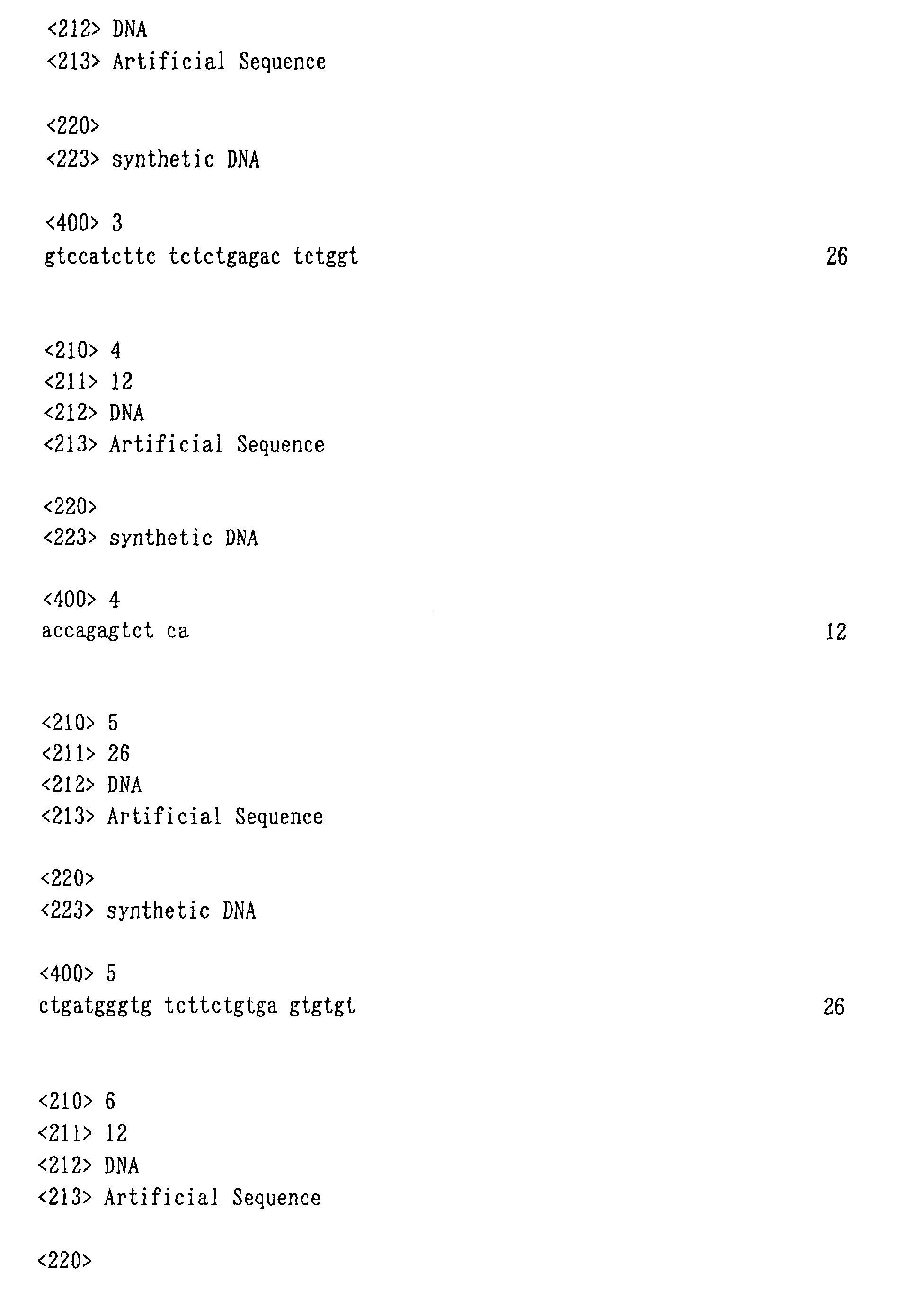
Similarly, using oligonucleotides (ad3S and ad3A)
having nucleotide sequences of SEQ ID No: 3 and SEQ ID No:
4, oligonucleotides (ad4S and ad4A) having nucleotide
sequences of SEQ ID No: 5 and SEQ ID No: 6,
oligonucleotides (ad5S and ad5A) having nucleotide
sequences of SEQ ID No: 7 and SEQ ID No: 8,
oligonucleotides (ad13S and ad13A) having nucleotide
sequences of SEQ ID No: 9 and SEQ ID No: 10, and
oligonucleotides (ad14S and ad14A) having nucleotide
sequences of SEQ ID No: 11 and SEQ ID No: 12, adaptor
solutions (ad3, ad4, ad5, ad13, and ad14, respectively) are
prepared.
Double-stranded cDNAs are synthesized from a sample,
using a TAKARA cDNA synthesis kit. The thus obtained cDNAs
are purified, using a QIAquick PCR purification kit
(hereinafter, referred to as "QIAquick") (eluted with 20 µl
of TE). The purified cDNAs are RsaI-digested (TAKARA AfaI)
in a 100-µl system, and purified with QIAquick. In the
case of using 1 µg of poly(A)+ RNA, the cDNAs are eluted
with 20 µl of TE. In the case of using 1 µg of total RNAs,
the cDNAs are eluted with 50 µl of TE, precipitated with
ethanol, and dissolved in 2 µl of TE.
The cDNAs are bound to the adaptor ad2, using the
TAKARA DNA ligation kit Ver 2. 1 µl of cDNAs, 1 µl of 100
µM ad2, 2 µl of solution II, and 4 µl of solution I are
mixed, and allowed to react at 16°C for 2 hours, whereby a
cDNA solution (ad2 lig cDNA) with the adaptors ad2 added
thereto is obtained.
A PCR is performed under the following condition:
| 10 × |
5 µl |
| 2.5 |
4 µl |
| ExTaq | 0.25 µl |
| 100 µM ad2S | 0.5 µl |
| ad2 lig |
1 µl or 0.1 µl |
| DW | 39.25 µl |
72°C, 5 minutes → (94°C, 30 seconds → 65°C, 30
seconds → 72°C, 2 minutes) × 15 cycles → 72°C, 2 minutes
When 10 µl of the reaction liquid is electrophoresed,
if products are maximally observed (about 500 ng/µl) at 1
µl of the templates, and observed in some amounts even at
0.1 µl, it is considered that 107 or more kinds are
contained.
The remaining portion of 40 µl at 1 µl of template is
purified with QIAquick (eluted with 40 µl of TE), whereby
an amplified cDNA solution (cDNA ad2 amp) is obtained.
Most of cDNAs contained in cDNA ad2 amp are single-stranded;
therefore, a PCR is performed again under the
following condition:
| 10 × |
5 µl |
| 2.5 |
4 µl |
| ExTaq | 0.25 µl |
| 100 µM ad2S | 0.5 µl |
| cDNA ad2 amp | 0.5 µl |
| DW | 39.75 µl |
94°C, 2 minutes → (94°C, 30 seconds → 65°C, 30
seconds → 72°C, 2 minutes) × 5 cycles → 72°C, 2 minutes
One PCR run for tester and 5 PCR runs per subtraction
cycle for driver (20 PCR runs in total) are performed.
Then, 10 µl of the reaction mixture is electrophoresed and
checked (about 100 ng/10 µl), and the rest is purified with
QIAquick (columns: one column for tester and four columns
for driver) (eluted with 20 µl of TE), whereby an amplified
cDNA solution (cDNA ad2 amp (5 cycles)) is obtained. This
solution is RsaI-digested under the following condition:
| cDNA ad2 amp (5 cycles) | 20 µl |
| 10 × T | 10 µl |
| BSA | 10 µl |
| RsaI (10 u/µl) | 2 µl |
| DW | 58 µl |
| (One for tester, four for driver) |
The products for driver are reacted at 37°C for 2
hours, and a restriction enzyme is inactivated by a
treatment at 70°C for 5 minutes, whereby a complete driver
solution (cDNA ad2 amp RsaI) is obtained.
The products for tester are reacted at 37°C for one
hour, and purified with QIAquick (eluted with 20 µl of TE)
to obtain a digested product (cDNA ad2 amp RsaI).
The digested product is bound to the adaptor ad3,
using the TAKARA DNA ligation kit Ver2. Then, 3 µl of cDNA
ad2 amp RsaI, 1 µl of 100 µM ad3, 4 µl of solution II, and
8 µl of solution I are mixed and allowed to react at 16°C
for one hour, and a restriction enzyme is inactivated by
treatment at 70°C for 5 minutes, whereby a complete tester
solution (cDNA ad3 lig) is obtained.
16 µl of the tester solution (cDNA ad3 lig) and 100
µl of the driver solution (cDNA ad2 amp RsaI) are mixed
(T:D = 1:40), and the mixture is purified with QIAquick
(eluted with 50 µl of TE) and precipitated with ethanol.
Then, 50 µl of cDNAs (tester & driver mixture), 5 µl
of 3 M sodium acetate, 1 µl of 10 mg/ml of glycogen, and
125 µl of ethanol are mixed, and centrifuged immediately
for 5 minutes. The resultant precipitate is rinsed with
70 % ethanol and dried. Thereafter, the precipitate is
dissolved in 1 µl of 1 × PCR buffer. The solution is
transferred to a PCR tube, and overlaid with mineral oil.
After the solution is retained at 98°C for 5 minutes, 1 µl
of 1 x PCR buffer + 1 M NaCl is added thereto and mixed
together, and treatment (98°C, 5 minutes → 68°C, 16 hours)
is performed.
Then, 10 µl of TE is added to the solution and
collected in a new tube. Thereafter, a PCR is effected
under the following condition:
| 10 × |
5 µl |
| 2.5 |
4 µl |
| Taq | 0.5 µl |
| 100 µM ad3S | 0.5 µl |
| Treated |
2 µl |
| DW | 38 µl |
72°C, 5 minutes → (94°C, 30 seconds → 65°C, 30
seconds → 72°C, 2 minutes) × 10 cycles → 4°C
The solution in an amount corresponding to 5 runs is
purified in one column with QIAquick and precipitated with
ethanol. The precipitate is rinsed with 70% ethanol, and
dried. Then, 5 µl of TE, 5 µl of 10 × mung bean buffer,
38.5 µl of DW, and 1.5 µl of mung bean nuclease are added
to the solution, and the mixture is allowed left at room
temperature for 20 minutes.
After 5 µl of 0.05 M EDTA and 45 µl of 50 mM Tris
7.6/100 mM NaCl are added, the mixture is purified with
QIAquick (eluted with 30 µl of TE) to obtain a purified
product (S1 pure).
A PCR is performed under the following condition:
| 10 × |
5 µl |
| 2.5 |
4 µl |
| Taq | 0.25 µl |
| 100 µM ad3S | 0.5 µl |
| S1 pure | 2 µl |
| DW | 38.25 µl |
94°C, 2 minutes → (94°C, 30 seconds → 65°C, 30
seconds → 72°C, 2 minutes) × 12 cycles → 72°C, 2 minutes
10 µl of the reaction liquid is electrophoresed to
confirm that amplification is completed. The rest of the
reaction mixture is purified with QIAquick (eluted with 40
µl of TE) to obtain an amplified product (S1 amp). In the
case where it is desired to confirm the pattern of a band,
the second PCR is also performed under the conditions of 5
and 7 cycles, and electrophoresis is performed.
1 µl of 2 × PCR buffer is added to 1 µl of S1 amp
diluted 5-fold with TE, and mineral oil is overlaid. After
the resultant solution is allowed to stand at 98°C for 5
minutes, 2 µl of 1 × PCR buffer + 1 M NaCl is added and
mixed together, and treatment (98°C, 5 minutes → 68°C, 16
hours) is performed.
After 10 µl of 10 × T buffer, 10 µl of BSA, 3 µl of
RsaI, and 73 µl of DW are added to the resultant solution,
and the solution is treated at 37°C for one hour, the
solution is purified with QIAquick (eluted with 20 µl of
TE) to obtain a purified product (N1 pure).
A PCR is performed under the following condition:
| 10 × |
5 µl |
| 2.5 |
4 µl |
| ExTaq | 0.25 µl |
| 100 µM ad3S | 0.5 µl |
| N1 pure | 2 µl |
| DW | 38.25 µl |
94°C, 2 minutes → (94°C, 30 seconds → 65°C, 30
seconds → 72°C, 2 minutes) × 11 to 15 cycles → 72°C, 2
minutes
Then, the rest of the sample after a certain number
of cycles where about 50 ng is contained (to such a degree
as to be slightly observed) when 10 µl of the reaction
liquid is electrophoresed, is purified with QIAquick
(eluted with 20 µl of TE), and digested with RsaI (37°C for
one hour). This is purified with QIAquick (eluted with 20
µl of TE) to obtain a digested product (N1 amp RsaI).
The digested product is bound to the adaptor ad4,
using the TAKARA DNA ligation kit Ver2. Then, 2 µl of N1
amp RsaI, 1 µl of 100 µM ad4, 3 µl of solution II, and 6 µl
of solution I are mixed and allowed to react at 16°C for
one hour, and a restriction enzyme is inactivated by
treatment at 70°C for 5 minutes, whereby a tester solution
for the second time (N1 amp ad4 lig) is obtained.
12 µl of the tester solution (N1 amp ad4 lig) and 100
µl of the driver solution (cDNA ad2 amp RsaI) are mixed
(T:D = 1:100), and the mixture is purified with QIAquick
(eluted with 50 µl of TE) and precipitated with ethanol.
Thereinafter, the process is performed in the same
manner as in the first subtraction (3 to 7 cycles for PCR
after treatment with mung bean nuclease).
Then, the rest of the sample after a certain number
of cycles where 50 ng is contained (to such a degree as to
be slightly observed) when 10 µl of the reaction liquid is
electrophoresed, is purified with QIAquick (eluted with 20
µl of TE), and digested with RsaI (37°C for one hour).
This is purified with QIAquick (eluted with 20 µl of TE) to
obtain a digested product (NS2 amp RsaI).
The digested product is bound to the adaptor ad5,
using the TAKARA DNA ligation kit Ver2. Then, 1 µl of NS2
amp RsaI diluted 1/5-fold, 1 µl of 100 µM ad5, 2 µl of
solution II, and 4 µl of solution I are mixed and allowed
to react at 16°C for one hour, and a restriction enzyme is
inactivated by treatment at 70°C for 5 minutes, whereby a
tester solution for the third time (NS2 amp ad5 lig) is
obtained.
8 µl of the tester solution (NS2 amp ad5 lig) and 100
µl of the driver solution (cDNA ad2 amp RsaI) are mixed
(T:D = 1:1000), and the mixture is purified with QIAquick
(eluted with 50 µl of TE) and precipitated with ethanol.
Thereinafter, the process is performed in the same
way as in the first subtraction (PCR is performed in 6, 8,
and 12 cycles after treatment with mung bean nuclease).
At this time, if the number of kinds of amplified
products is small, it is considered that products are
double-stranded. Therefore, the products are cloned to a
TA vector or the like as they are. If the number of kinds
is large, the genes are diluted 100-fold, and 5 cycle-PCR
is performed to make the products into double-strands.
Thereafter, cloning is performed.
In order to check which degree of an expression
amount and of a difference in an expression amount genes
can be isolated to by the method described in Example 1, it
was checked whether or not X174 fragments can be isolated,
using, as tester and driver, cDNAs prepared from a blood
vessel endothelial cell line MS1 to which DNA RsaI
digestion fragments derived from bacteriophage X174 are
added.
FIG. 2, A shows electrophoresis of final PCR products
obtained in the case where the method described in Example
1 (N-RDA method) and the method omitting the process of
normalization (RDA method) were performed, using MS1 cDNAs
to which 6 kinds of X174 RsaI fragments were added in a
ratio of 1/105 as tester and using MS1 cDNAs as driver.
Lane 1 shows the result of the electrophoresis of 6 kinds
of added X174 RsaI fragments (F1 to F6), Lane 2 shows the
result of the electrophoresis of the amplified product
after the third subtraction of the RDA method, and Lane 3
shows the result of the electrophoresis of the amplified
product after the third subtraction of the N-RDA method.
In any of the methods, 5 out of 6 kinds were amplified.
This clarified that almost all the specific fragments
existing in a ratio of 1/105 can be isolated.
FIG. 2, B shows the result obtained in the case of
adding X174 RsaI fragments in a ratio of 1/106 in the same
manner as in the A. In this ratio, X174 RsaI fragments
were not amplified in any of the methods. Thus, it was
clarified that the limit of these methods is between 1/105
and 1/106.
FIG. 3, A shows the result (result of electrophoresis
of an amplified product after the third subtraction)
obtained in the case of adding 6 kinds of X174 RsaI
fragments in each ratio shown in the figure. According to
the RDA method, F1 and F4 added in a ratio of 1/104 were
amplified efficiently, and F2 added in a ratio of 1/105 was
amplified as well. On the other hand, according to the N-RDA
method, the amount of F2 was increased compared with
the RDA method, and F5 was also amplified. Therefore, it
was confirmed that the effect of normalization was
exhibited.
Next, in order to study whether isolation can be
performed even with a 10-fold expression difference, an
experiment was performed by adding the same X174 RsaI
fragments to the driver in a ratio of 1/10 of the tester
(FIG. 3, B). According to the RDA method, it was confirmed
that those contained in a ratio of 1/104 in the tester (F1,
F3 and F5) were amplified even with a 10-fold difference,
but those existing in a ratio of 1/105 were not amplified.
In contrast, according to the N-RDA method, F2 and F4
contained in a ratio of 1/105 were also amplified.
Furthermore, as a model closest to the practical case,
the case has been studied in which X174 RsaI fragments
that are different in an existing amount and in an existing
amount ratio between tester and driver are added (FIG. 3,
C). Consequently, according to the RDA method, it was
confirmed that specific F3 in a large amount was amplified
preferentially, and F1, F4, and F5 were barely amplified.
In contrast, according to the N-RDA method, the amounts of
F1, F4, and F5 were increased, and F2 was further amplified.
From the above-mentioned results, the effect of
normalization was confirmed under any of the conditions.
Thus, it is apparent that the N-RDA method is very
effective for isolating as many genes as possible at once.
Subtraction was performed among some blood vessel
endothelial cells derived from normal human tissues. As
materials, umbilical vein endothelial cell (VE), umbilical
artery endothelial cell (AE), dermal microvascular
endothelial cell (ADM), tonsil endothelial cell (TE), and
brain microcapillary endothelial cell (BME) were used.
Furthermore, as a comparison between cells with a
relatively large difference, subtraction was performed
between a mouse blood vessel endothelial cell line (MS1)
and a mouse epithelial cell line (Eph4).
FIG. 4 shows combinations of subtraction. In the
subtraction between blood vessel endothelial cells, several
to several tens of bands were amplified. In contrast, in
the subtraction of MS1/Eph4, a great number of bands were
amplified. This result is considered to reflect the number
of genes of specific expression.
Next, in order to confirm whether or not the
amplified genes actually exhibit specific expression, the
bands amplified by the subtraction among blood vessel
endothelial cells were cloned, and cDNAs prepared from the
respective blood vessel endothelial cells were subjected to
southern blotting (FIG. 5). Consequently, it was confirmed
that 11 kinds out of 12 kinds were completely specific or
had a 10-fold or more difference in an expression, and it
was clarified that the remaining one kind also had a
several-fold difference. From the above result, it was
confirmed that, according to the N-RDA method, genes
specific in an expression can be isolated with a low
background even among cells actually having a relatively
small difference.
According to the present invention, the DNA fragment
contents in a sample can be normalized. By combining this
normalization method with subtractive hybridization, even
genes existing in a small expression amount can be isolated
based on the difference in an expression amount.


Claims (7)
- A method of normalizing contents of DNA fragments in a sample among respective DNA fragments, comprising:preparing DNA fragments in which adaptors each comprising an oligodeoxyribonucleotide are attached to both ends of each DNA fragment in the sample to form restriction enzyme recognition sites that do not exist in the DNA fragments in the sample, and at least a part between the restriction enzyme recognition sites at both ends, including the restriction enzyme recognition sites, is double-stranded;denaturing the prepared double-stranded DNA fragments;hybridizing the denatured DNA fragments under a condition that a part of the DNA fragments remains single-stranded;cleaving the hybridized double-stranded DNA fragments with a restriction enzyme having a cleavage site exclusively in the adaptors; andperforming PCR using the obtained DNA fragments as templates and using a primer having a nucleotide sequence complementary to a nucleotide sequence of the adaptors before the cleavage.
- A method of detecting a difference in a gene expression among cells, which comprises performing subtractive hybridization at least once, and further comprising performing normalization by the method as defined in claim 1 before or after performing at least one of the subtractive hybridization(s).
- A method according to claim 2, wherein at least one of the subtractive hybridization(s) generates double-stranded DNA fragments with adaptors attached to both ends, and the normalization is performed after the subtractive hybridization.
- A method according to claim 3, wherein the subtractive hybridization is performed at least twice, and the first subtractive hybridization generates the double-stranded DNA fragments with the adaptors attached to both the ends.
- A method of producing a library of genes which are different in an expression among cells, comprising cloning genes which are each detected for a difference in the expression by the method as defined in any one of claims 2 to 4.
- A method of producing a probe specific to a gene which is different in an expression among cells, comprising preparing a fragment of a gene which is detected for a difference in the expression by the method as defined in any one of claims 2 to 4.
- A kit for use in the method as defined in claim 1, comprising an adaptor comprising an oligodeoxyribonucleotide to be attached to both ends of DNA fragments in a sample, a restriction enzyme having a cleavage site in the adaptor, and a primer for PCR having a nucleotide sequence complementary to a nucleotide sequence of the adaptor before cleavage with the restriction enzyme.
Applications Claiming Priority (3)
| Application Number | Priority Date | Filing Date | Title |
|---|---|---|---|
| JP2001184757 | 2001-06-19 | ||
| JP2001184757 | 2001-06-19 | ||
| PCT/JP2002/006109 WO2002103007A1 (en) | 2001-06-19 | 2002-06-19 | Method of uniformizing dna fragment contents and subtraction method |
Publications (2)
| Publication Number | Publication Date |
|---|---|
| EP1405909A1 true EP1405909A1 (en) | 2004-04-07 |
| EP1405909A4 EP1405909A4 (en) | 2006-06-14 |
Family
ID=19024465
Family Applications (1)
| Application Number | Title | Priority Date | Filing Date |
|---|---|---|---|
| EP02738769A Withdrawn EP1405909A4 (en) | 2001-06-19 | 2002-06-19 | Method of uniformizing dna fragment contents and subtraction method |
Country Status (4)
| Country | Link |
|---|---|
| US (1) | US20050175997A1 (en) |
| EP (1) | EP1405909A4 (en) |
| JP (1) | JPWO2002103007A1 (en) |
| WO (1) | WO2002103007A1 (en) |
Cited By (2)
| Publication number | Priority date | Publication date | Assignee | Title |
|---|---|---|---|---|
| US9453840B2 (en) | 2011-07-27 | 2016-09-27 | Kyoto University | Markers for dopaminergic neuron progenitor cells |
| US9994816B2 (en) | 2004-07-22 | 2018-06-12 | Eisai R&D Management Co., Ltd. | Lrp4/corin dopamine-producing neuron precursor cell marker |
Families Citing this family (10)
| Publication number | Priority date | Publication date | Assignee | Title |
|---|---|---|---|---|
| WO2004065599A1 (en) | 2003-01-24 | 2004-08-05 | Eisai Co., Ltd. | Lrp4/CORIN DOPAMINE-PRODUCING NEURON PROLIFERATION PRECURSOR CELL MARKER |
| GB2412170A (en) * | 2004-03-16 | 2005-09-21 | Plant Bioscience Ltd | A method for enriching for target nucleic acids in a sample |
| JP4717797B2 (en) * | 2004-07-22 | 2011-07-06 | エーザイ・アール・アンド・ディー・マネジメント株式会社 | Lrp4 / Corin dopaminergic neuron progenitor cell marker |
| JP4618736B2 (en) * | 2004-07-22 | 2011-01-26 | エーザイ・アール・アンド・ディー・マネジメント株式会社 | Lrp4 / Corin dopaminergic neuron progenitor cell marker |
| CA2619736C (en) * | 2005-08-18 | 2012-05-22 | Eisai R & D Management Co., Ltd. | Dopaminergic neuron proliferative progenitor cell marker msx1/2 |
| CN101292031A (en) | 2005-08-18 | 2008-10-22 | 卫材R&D管理有限公司 | Marker Nato3 for dopaminergic neuron proliferative progenitor cells |
| PL225633B1 (en) | 2012-11-22 | 2017-05-31 | Univ Jagiellonski | Method for identification of RNA viruses and the application of the method |
| KR102478639B1 (en) | 2016-02-23 | 2022-12-19 | 노을 주식회사 | Labeling substance storing patch, tissue diagnostic method using the patch and device using the same |
| US10371610B2 (en) | 2016-02-23 | 2019-08-06 | Noul Co., Ltd. | Contact-type patch, staining method using the same, and manufacturing method thereof |
| KR20170099738A (en) | 2016-02-23 | 2017-09-01 | 노을 주식회사 | Contact-type staining patch and manufacturing method thereof |
Family Cites Families (9)
| Publication number | Priority date | Publication date | Assignee | Title |
|---|---|---|---|---|
| JPH06153952A (en) * | 1992-11-26 | 1994-06-03 | Nobuaki Tamamaki | Method for pretreatment for carrying out amplifying and labeling of unknown double-stranded dna molecule in trace amount |
| JPH0923885A (en) * | 1995-07-12 | 1997-01-28 | Dai Ichi Seiyaku Co Ltd | Gene expression library and its production |
| JPH09234074A (en) * | 1996-03-04 | 1997-09-09 | Sumitomo Electric Ind Ltd | Adapter double-stranded DNA and DNA amplification method using the same |
| US5726022A (en) * | 1996-11-18 | 1998-03-10 | Lifespan Biosciences, Inc. | Subtractive hybridization and capture methods and kits for differential isolation of nucleic acids including disease-associated sequences |
| US6060245A (en) * | 1996-12-13 | 2000-05-09 | Stratagene | Methods and adaptors for generating specific nucleic acid populations |
| US6017701A (en) * | 1996-12-13 | 2000-01-25 | Stratgene | Methods and adaptors for generating specific nucleic acid populations |
| WO1998029569A1 (en) * | 1996-12-13 | 1998-07-09 | Stratagene | Methods and adaptors for generating specific nucleic acid populations |
| US6090556A (en) * | 1997-04-07 | 2000-07-18 | Japan Science & Technology Corporation | Method for quantitatively determining the expression of a gene |
| JP4304357B2 (en) * | 1999-05-24 | 2009-07-29 | 独立行政法人理化学研究所 | How to create a full-length cDNA library |
-
2002
- 2002-06-19 WO PCT/JP2002/006109 patent/WO2002103007A1/en not_active Ceased
- 2002-06-19 US US10/481,228 patent/US20050175997A1/en not_active Abandoned
- 2002-06-19 EP EP02738769A patent/EP1405909A4/en not_active Withdrawn
- 2002-06-19 JP JP2003505330A patent/JPWO2002103007A1/en active Pending
Cited By (2)
| Publication number | Priority date | Publication date | Assignee | Title |
|---|---|---|---|---|
| US9994816B2 (en) | 2004-07-22 | 2018-06-12 | Eisai R&D Management Co., Ltd. | Lrp4/corin dopamine-producing neuron precursor cell marker |
| US9453840B2 (en) | 2011-07-27 | 2016-09-27 | Kyoto University | Markers for dopaminergic neuron progenitor cells |
Also Published As
| Publication number | Publication date |
|---|---|
| US20050175997A1 (en) | 2005-08-11 |
| JPWO2002103007A1 (en) | 2004-09-30 |
| EP1405909A4 (en) | 2006-06-14 |
| WO2002103007A1 (en) | 2002-12-27 |
Similar Documents
| Publication | Publication Date | Title |
|---|---|---|
| US20210032679A1 (en) | Methods and compositions for multiplex pcr | |
| EP2048248B1 (en) | Method of amplifying a target nucleic acid by rolling circle amplification | |
| US10266881B2 (en) | Methods and compositions for multiplex PCR | |
| EP3332024B1 (en) | Target enrichment by single probe primer extension | |
| EP2785860B1 (en) | Methods and compositions for multiplex pcr | |
| US20180320172A1 (en) | Ligation-based rna amplification | |
| JPH10509329A (en) | Analysis of gene expression by displaying the 3 'terminal restriction fragment of cDNA | |
| WO2013081864A1 (en) | Methods and compositions for multiplex pcr | |
| WO2020219838A1 (en) | Methods and enzymatic compositions for forming libraries of adapter ligated nucleic acid molecules | |
| US20050175997A1 (en) | Method of uniformizing dna fragment contents and subtraction method | |
| JP2022518917A (en) | Nucleic acid detection method and primer design method | |
| US20140336058A1 (en) | Method and kit for characterizing rna in a composition | |
| CN114341353A (en) | Method for amplifying mRNA and preparing full-length mRNA library | |
| CA2204637A1 (en) | Method of preparing oligonucleotide probes or primers, vector therefor and use thereof | |
| EP1333089A1 (en) | Method of synthesizing single-stranded nucleic acid | |
| EP3252168A1 (en) | Pcr primer linked to complementary nucleotide sequence or complementary nucleotide sequence including mis-matched nucleotides and method for amplifying nucleic acid using same | |
| JP2003199568A (en) | Method for improving efficiency of DNA amplification reaction | |
| EP2907872A1 (en) | Method for the quantitive analysis of terminal nucleotides of a g chain of human telomeric dna | |
| WO2025185331A1 (en) | Methods for constructing a library of polynucleotides having nucleic acids of interest | |
| JP5618227B2 (en) | Nucleic acid amplification method and gene mutation detection method | |
| JP4455168B2 (en) | DNA amplification method | |
| KR100844010B1 (en) | Multiple gene simultaneous amplification method |
Legal Events
| Date | Code | Title | Description |
|---|---|---|---|
| PUAI | Public reference made under article 153(3) epc to a published international application that has entered the european phase |
Free format text: ORIGINAL CODE: 0009012 |
|
| 17P | Request for examination filed |
Effective date: 20040102 |
|
| AK | Designated contracting states |
Kind code of ref document: A1 Designated state(s): AT BE CH CY DE DK ES FI FR GB GR IE IT LI LU MC NL PT SE TR |
|
| A4 | Supplementary search report drawn up and despatched |
Effective date: 20060512 |
|
| 17Q | First examination report despatched |
Effective date: 20060802 |
|
| STAA | Information on the status of an ep patent application or granted ep patent |
Free format text: STATUS: THE APPLICATION IS DEEMED TO BE WITHDRAWN |
|
| 18D | Application deemed to be withdrawn |
Effective date: 20061213 |