EP1398041A1 - Recombinant lentiviral vector pseudotyped with the hemagglutinin protein for gene transfer into the retina - Google Patents
Recombinant lentiviral vector pseudotyped with the hemagglutinin protein for gene transfer into the retina Download PDFInfo
- Publication number
- EP1398041A1 EP1398041A1 EP02292255A EP02292255A EP1398041A1 EP 1398041 A1 EP1398041 A1 EP 1398041A1 EP 02292255 A EP02292255 A EP 02292255A EP 02292255 A EP02292255 A EP 02292255A EP 1398041 A1 EP1398041 A1 EP 1398041A1
- Authority
- EP
- European Patent Office
- Prior art keywords
- protein
- lentiviral vector
- lentiviral
- virus
- eye
- Prior art date
- Legal status (The legal status is an assumption and is not a legal conclusion. Google has not performed a legal analysis and makes no representation as to the accuracy of the status listed.)
- Withdrawn
Links
Images




Classifications
-
- C—CHEMISTRY; METALLURGY
- C12—BIOCHEMISTRY; BEER; SPIRITS; WINE; VINEGAR; MICROBIOLOGY; ENZYMOLOGY; MUTATION OR GENETIC ENGINEERING
- C12N—MICROORGANISMS OR ENZYMES; COMPOSITIONS THEREOF; PROPAGATING, PRESERVING, OR MAINTAINING MICROORGANISMS; MUTATION OR GENETIC ENGINEERING; CULTURE MEDIA
- C12N15/00—Mutation or genetic engineering; DNA or RNA concerning genetic engineering, vectors, e.g. plasmids, or their isolation, preparation or purification; Use of hosts therefor
- C12N15/09—Recombinant DNA-technology
- C12N15/63—Introduction of foreign genetic material using vectors; Vectors; Use of hosts therefor; Regulation of expression
- C12N15/79—Vectors or expression systems specially adapted for eukaryotic hosts
- C12N15/85—Vectors or expression systems specially adapted for eukaryotic hosts for animal cells
- C12N15/86—Viral vectors
-
- A—HUMAN NECESSITIES
- A61—MEDICAL OR VETERINARY SCIENCE; HYGIENE
- A61K—PREPARATIONS FOR MEDICAL, DENTAL OR TOILETRY PURPOSES
- A61K48/00—Medicinal preparations containing genetic material which is inserted into cells of the living body to treat genetic diseases; Gene therapy
- A61K48/005—Medicinal preparations containing genetic material which is inserted into cells of the living body to treat genetic diseases; Gene therapy characterised by an aspect of the 'active' part of the composition delivered, i.e. the nucleic acid delivered
- A61K48/0058—Nucleic acids adapted for tissue specific expression, e.g. having tissue specific promoters as part of a contruct
-
- A—HUMAN NECESSITIES
- A61—MEDICAL OR VETERINARY SCIENCE; HYGIENE
- A61P—SPECIFIC THERAPEUTIC ACTIVITY OF CHEMICAL COMPOUNDS OR MEDICINAL PREPARATIONS
- A61P27/00—Drugs for disorders of the senses
- A61P27/02—Ophthalmic agents
-
- C—CHEMISTRY; METALLURGY
- C12—BIOCHEMISTRY; BEER; SPIRITS; WINE; VINEGAR; MICROBIOLOGY; ENZYMOLOGY; MUTATION OR GENETIC ENGINEERING
- C12N—MICROORGANISMS OR ENZYMES; COMPOSITIONS THEREOF; PROPAGATING, PRESERVING, OR MAINTAINING MICROORGANISMS; MUTATION OR GENETIC ENGINEERING; CULTURE MEDIA
- C12N2740/00—Reverse transcribing RNA viruses
- C12N2740/00011—Details
- C12N2740/10011—Retroviridae
- C12N2740/15011—Lentivirus, not HIV, e.g. FIV, SIV
- C12N2740/15041—Use of virus, viral particle or viral elements as a vector
- C12N2740/15043—Use of virus, viral particle or viral elements as a vector viral genome or elements thereof as genetic vector
-
- C—CHEMISTRY; METALLURGY
- C12—BIOCHEMISTRY; BEER; SPIRITS; WINE; VINEGAR; MICROBIOLOGY; ENZYMOLOGY; MUTATION OR GENETIC ENGINEERING
- C12N—MICROORGANISMS OR ENZYMES; COMPOSITIONS THEREOF; PROPAGATING, PRESERVING, OR MAINTAINING MICROORGANISMS; MUTATION OR GENETIC ENGINEERING; CULTURE MEDIA
- C12N2740/00—Reverse transcribing RNA viruses
- C12N2740/00011—Details
- C12N2740/10011—Retroviridae
- C12N2740/15011—Lentivirus, not HIV, e.g. FIV, SIV
- C12N2740/15041—Use of virus, viral particle or viral elements as a vector
- C12N2740/15045—Special targeting system for viral vectors
-
- C—CHEMISTRY; METALLURGY
- C12—BIOCHEMISTRY; BEER; SPIRITS; WINE; VINEGAR; MICROBIOLOGY; ENZYMOLOGY; MUTATION OR GENETIC ENGINEERING
- C12N—MICROORGANISMS OR ENZYMES; COMPOSITIONS THEREOF; PROPAGATING, PRESERVING, OR MAINTAINING MICROORGANISMS; MUTATION OR GENETIC ENGINEERING; CULTURE MEDIA
- C12N2810/00—Vectors comprising a targeting moiety
- C12N2810/50—Vectors comprising as targeting moiety peptide derived from defined protein
- C12N2810/60—Vectors comprising as targeting moiety peptide derived from defined protein from viruses
-
- C—CHEMISTRY; METALLURGY
- C12—BIOCHEMISTRY; BEER; SPIRITS; WINE; VINEGAR; MICROBIOLOGY; ENZYMOLOGY; MUTATION OR GENETIC ENGINEERING
- C12N—MICROORGANISMS OR ENZYMES; COMPOSITIONS THEREOF; PROPAGATING, PRESERVING, OR MAINTAINING MICROORGANISMS; MUTATION OR GENETIC ENGINEERING; CULTURE MEDIA
- C12N2830/00—Vector systems having a special element relevant for transcription
- C12N2830/008—Vector systems having a special element relevant for transcription cell type or tissue specific enhancer/promoter combination
Definitions
- the present invention relates to a novel lentiviral vector particularly well shaped for performing gene transfer into the retina.
- the invention also relates to compositions and methods for preventing or treating diseases of the eye, using the vector of the invention to transfer selected genes suitable for preventing or treating diseases of the eye.
- Retinal degenerative diseases such as retinal macular degeneration or retinitis pigmentosa constitute a broad group of diseases that all share one critical feature, the progressive loss of cells in the retina.
- Gene therapy represents a possible approach to treating retinal degenerations because the eye is easily accessible and allows local application of therapeutic vectors with reduced risk of systemic effects.
- transgene expression within the retina and effects of treatments may be monitored by a variety of non-invasive examinations.
- adenovirus including adenovirus, adenovirus-associated virus (AAV), or human immunodeficiency virus type 1 (HIV-1) were tested for in vivo retina transduction (see for example (Bainbridge, Stephens et al. 2001 )).
- AAV2-based vectors only.
- RPE retinal pigment epithelium
- the transgene expression is delayed. Maximal levels of expression are detected 4-6 weeks post-infection (Rolling, Shen et al. 1999; Auricchio, Kobinger et al. 2001). Such a feature may be detrimental when rapid transgene expression is required.
- VSV-G vesicular stomatitis virus G glycoprotein
- the inventors have now developed a novel lentiviral vector which exhibits a very low toxicity and high efficiency, for performing gene transfer into the eye.
- the present invention hence provides compositions and methods for preventing and treating a number of diseases of the eye, including for example, retinal macular degeneration or retinitis pigmentosa, as well as other diseases such as neovascular diseases.
- the present invention also provides other related advantages.
- the inventors have developed a lentiviral vector derived from SIVmac251, a nonpathogenic strain of simian immunodeficiency virus (SIV). It was previously demonstrated that HIV-1 and SIV vectors could transduce both dividing and resting cells in vitro with equivalent efficiencies (Mangeot, Negre et al. 2000; Negre, Mangeot et al. 2000). To assess both, the virus tropism in retina and the kinetic of the transgene expression, rats were injected with VSV-G-pseudotyped SIV vectors encoding for the green fluorescent protein (GFP). Sustained gene transfer into the retina was monitored by iterative non-invasive examinations (examples 1 and 2).
- GFP green fluorescent protein
- the inventors evaluated the cell populations transduced by SIV vectors pseudotyped with surface glycoproteins from other membraneenveloped viruses and, particularly, with the hemagglutinin (HA) protein from an orthomyxovirus, more precisely from an influenza A virus. They demonstrated that the HA-pseudotyped vectors were very efficient (example 5).
- the present invention hence mainly pertains to lentiviral vectors pseudotyped with a HA protein from an orthomyxovirus and comprising one or several gene(s) useful for preventing or treating diseases of the eye, pharmaceutical compositions comprising such vectors, and uses of these vectors.
- HA will hence designate the hemagglutinin protein of any kind of orthomyxovirus, in particular of any influenza virus including the surface glycoprotein of influenza C and, more particularly, the hemagglutinin of an influenza A virus.
- Influenza A viruses naturally infect a broad range of species, including humans, horses, swine, and a wide variety of avian species. Their surface glycoproteins, including HA, exhibit a great amino acid sequence variability. Therefore, the word "HA” in the present application designates a wide range of proteins that are characterized by their origin (orthomyxoviridae), and their function (hemagglutination).
- viral vector designates either a viral particle comprising genetic material to be transferred into a host cell, or the recombinant viral genome itself.
- the term "(lenti)viral vector particle” will be used to designate the physical viral particle, including a genome, a capsid and an enveloppe, whereas "(lenti)viral vector genome” will designate the nucleic acid construct to be transferred.
- the present invention hence provides a lentiviral vector particle comprising a lentiviral vector genome encoding a transgene which, when expressed in the retina, can prevent, inhibit or alleviate the effects of an eye disease, wherein said vector particle is pseudotyped with an hemagglutinin (HA) protein of an orthomyxovirus.
- a particular lentiviral vector particle of the invention is pseudotyped with an HA protein from an influenza A virus, and, for example, from a Fowl Plague Virus (FPV).
- FPV Fowl Plague Virus
- the results shown in the following experimental part have been obtained with lentiviral particles pseudotyped with an HA protein from strain influenza A, Fowl plague virus, Rostock, 34 (H7 N1).
- the sequence of this HA protein is accessible in GenBank under numbers M24457 (coding sequence) and AAA43150 (amino acids sequence).
- HA-pseudotyped vectors can be produced by co-transfecting plasmids, wherein one plasmid carries the vector genome and the others encode the necessary proteins for lentiviral encapsidation and the HA protein.
- the inventors have demonstrated that cotransfection of a further plasmid, encoding the neuraminidase (NA) from FPV, could result in improved vector production, with increased titers (titers of 10 8 infectious particles/ml were obtained after a 100-fold concentration of the collected supernatant, as described below).
- the present invention hence also pertains to a lentiviral vector particle comprising a lentiviral vector genome encoding a transgene which, when expressed in the retina, can prevent, inhibit or alleviate the effects of an eye disease, wherein said vector particle is pseudotyped with both an hemagglutinin (HA) and a neuraminidase (NA) proteins of an orthomyxovirus, preferably from an influenza virus, and more preferably from influenza A.
- HA hemagglutinin
- NA neuraminidase
- One particular NA protein which can be comprised in the vector particles according to the invention is the NA protein from FPV, for example from the strain Rostock, 34 (H7 N1). The sequence of this NA protein is accessible in GenBank under numbers X52226 (coding sequence) and CAA36475 (amino acids sequence).
- the invention also pertains to lentiviral vector particles such as described above, wherein said vector genome comprises a lentiviral 5' LTR, a tRNA binding site, a packaging signal, a promoter operably linked to said transgene, an origin of second strand DNA synthesis and a 3' lentiviral LTR.
- the vector genome within the lentiviral vector particle comprises 5' and/or 3' LTRs from a virus selected from the group consisting of SIV, HIV, HIV-1, HIV-2, EIAV and FIV.
- the lentiviral vector is self-inactivating.
- Self-inactivating vectors can be made by deleting promoter and enhancer elements in the U3 region of the 3' LTR, including the TATA box and binding sites for one or more transcription factors. The deletion is transferred to the 5' LTR after reverse transcription and integration in transduced cells. This results in the transcriptional inactivation of the LTR provirus.
- Such self-inactivating (SIN) vectors can present the advantage of a broader tropism. Indeed, Miyoshi et al.
- HIV-1 LTR can negatively influence the transgene expression through an internal promoter in some cell types, and hence that SIN vectors could present a double advantage in terms of safety and broader applicability, particularly concerning the photoreceptor cells (Miyoshi, Blomer et al. 1998).
- a wide variety of diseases of the eye may be readily treated or prevented, including for example, glaucoma, macular degeneration, diabetic retinopathies, inherited retinal degeneration such as retinitis pigmentosa, retinal detachment or injury and retinopathies (whether inherited, induced by surgery, trauma, a toxic compound or agent, or, photically).
- a wide variety of neurotrophic factors may be utilized (either alone or in combination) within the context of the present invention, including for example, NGF, BDNF, CNTF, NT-3, NT-4, FGF-2, FGF-5, FGF-18, FGF-20 and FGF-21.
- the gene delivery vector be utilized to deliver and express an anti-angiogenic factor for the treatment, prevention, or, inhibition of diabetic retinopathy, wet ARMD, and other neovascular diseases of the eye (e.g., ROP).
- an anti-angiogenic factor for the treatment, prevention, or, inhibition of diabetic retinopathy, wet ARMD, and other neovascular diseases of the eye (e.g., ROP).
- ROP neovascular diseases of the eye
- the gene delivery vector be utilized to deliver and express a neurotrophic growth factor to treat, prevent, or inhibit diseases of the eye, such as, for example, glaucoma, retinitis pigmentosa, and dry ARMD.
- gene delivery vectors which direct the expression of a neurotrophic factor, or, an anti-angiogenic factor.
- neurotrophic factors include NGF, BDNF, CNTF, NT-3, NT-4, FGF-2, FGF-5, FGF-18, FGF-20 and FGF-21.
- anti-angiogenic factors include soluble Flt-1, soluble Tie-2 receptor, and PEDF.
- the transgene can hence be selected (without being limitative) in the group comprising neurotrophic factors including NGF, BDNF, CNTF, NT-3, NT-4, FGF-2, FGF-5, FGF-18, FGF-20 and FGF-21, anti-angiogenic factors including soluble Flt-1, soluble Tie-2 receptor, and PEDF, and enzymes including ⁇ -glucuronidase, neuraminidase, sphingomyelinase, sulfatases, arylsulfatase ⁇ , ⁇ -neuraminidase, gangliosidase, tripeptidyl protease, CLN3 and palmitoyl protein thioesterase (PPT).
- neurotrophic factors including NGF, BDNF, CNTF, NT-3, NT-4, FGF-2, FGF-5, FGF-18, FGF-20 and FGF-21
- anti-angiogenic factors including soluble Flt-1, soluble Tie-2 receptor, and PEDF
- the transgene is preferably operably linked to a regulated promoter (e.g. , "tet” promoters, the ecdysome system, or other systems of regulation), a viral promoter (e.g. , the CMV or RSV promoters), a tissue specific promoter (e.g. , a rod, cone, or ganglia-derived promoter), or a rhodopsin promoter.
- a regulated promoter e.g. , "tet" promoters, the ecdysome system, or other systems of regulation
- a viral promoter e.g. , the CMV or RSV promoters
- tissue specific promoter e.g. , a rod, cone, or ganglia-derived promoter
- rhodopsin promoter e.g., a rhodopsin promoter.
- the present invention also pertains to a pharmaceutical composition for administration in the eye, comprising lentiviral vector particles according to the invention (as described above), and a pharmaceutically acceptable carrier.
- lentiviral vector particles according to the invention as described above
- a pharmaceutically acceptable carrier for administration in the eye, comprising lentiviral vector particles according to the invention (as described above), and a pharmaceutically acceptable carrier.
- a particular advantage of the vectors according to the invention is that time-course transductions showed that high levels of transgene expression were obtained as soon as 4 days post-injection. For the first time, the kinetic of the transgene expression could be monitored in vivo in lentivirally-infected cells by iterative non-invasive examinations (see examples 1 and 2). Moreover, the HA-pseudotyped vector showed an excellent transduction efficiency (see, Fig. 6A), and lower toxicity (Example 6).
- Another aspect of the invention is the use of a lentiviral vector particle as described above, for the manufacture of a composition for treating a disease in the eye or preventing cell damage in the eye.
- the disease is due to a deficiency in retinal pigment epithelium (RPE) cells.
- RPE retinal pigment epithelium
- the disease can be due to a deficiency in the photoreceptors.
- lentiviral particles which are able to transduce retinal pigment epithelium (RPE) cells and express a neurotrophic factor therein can be used according to the invention.
- RPE retinal pigment epithelium
- the diffusion of the neurotrophic factor around the transduced cells will attain the photoreceptors and have a beneficial effect on them.
- the lentiviral vector particles described above can be used for the manufacture of a composition for treating a neovascular disease of the eye, a glaucoma, a macular degeneration, a diabetic retinopathy, an inherited retinal degeneration, a retinitis pigmentosa, a cell damage in retinal epithelial cells associated with Sly syndrome, a retinal detachment or injury or a retinopathy.
- a further aspect of the present invention is the use of an hemagglutinin (HA) protein of an orthomyxovirus, to pseudotype an enveloped vector so that said vector is able to transduce efficiently retinal pigment epithelium (RPE) cells.
- the HA protein is from an influenza A virus and, for example, from a Fowl Plague Virus. More particularly, the HA protein can be from the strain Rostock 34 (H7 N1) mentioned above.
- the present application also relates to a method of treating, inhibiting or preventing a disease of the eye, comprising, administering intraocularly, in a mammal in need thereof, a gene delivery lentiviral vector particle which is pseudotyped with an orthomyxovirus hemagglutinin (HA) protein and which directs the expression of a transgene wherein said transgene, when expressed in the retina, can prevent, inhibit or alleviate the effects of an eye disease.
- the HA protein can be from an influenza A virus, e.g. , a Fowl Plague Virus, e.g. , strain influenza A , FPV, Rostock, 34 (H7 N1).
- the sequence of this HA protein is accessible in GenBank under numbers M24457 (coding sequence) and AAA43150 (amino acids sequence).
- the above method can be performed according to the invention, when the disease is due to a deficiency in retinal pigment epithelium (RPE) cells, or when the disease is due to a deficiency in the photoreceptors.
- the method can be performed with lentiviral particles that transduce retinal pigment epithelium (RPE) cells and express therein a neurotrophic factor, which will diffuse to the photoreceptors.
- These methods can be performed to treat, inhibit or alleviate a variety of diseases of the eye, including, without being restrictive, a neovascular disease of the eye, a glaucoma, a macular degeneration, a diabetic retinopathy, an inherited retinal degeneration, a retinitis pigmentosa, a cell damage in retinal epithelial cells associated with Sly syndrome, a retinal detachment or injury or a retinopathy.
- diseases of the eye including, without being restrictive, a neovascular disease of the eye, a glaucoma, a macular degeneration, a diabetic retinopathy, an inherited retinal degeneration, a retinitis pigmentosa, a cell damage in retinal epithelial cells associated with Sly syndrome, a retinal detachment or injury or a retinopathy.
- a gene delivery vector which expresses both an anti-angiogenic molecule and a neurotrophic growth factor, or two separate vectors which independently express such factors, in the treatment, prevention, or inhibition of an eye disease (e.g., for diabetic retinopathy).
- the above-mentioned methods utilizing gene delivery vectors may be administered along with other methods or therapeutic regimens, including for example, photodynamic therapy (e.g., for wet ARMD), laser photocoagulation (e.g., for diabetic retinopathy and wet ARMD), and intraocular pressure reducing drugs (e.g., for glaucoma).
- photodynamic therapy e.g., for wet ARMD
- laser photocoagulation e.g., for diabetic retinopathy and wet ARMD
- intraocular pressure reducing drugs e.g., for glaucoma
- 293 embryo kidney cell line (ATCC CRL-1573) was grown in Dulbecco's modified Eagle's medium (DMEM; Sigma) supplemented with 10% fetal calf serum (FCS).
- DMEM Dulbecco's modified Eagle's medium
- FCS fetal calf serum
- SIV packaging constructs were derived from the BK28 molecular clone of SIVmac251 (GenBank accession number M19499).
- pSIV3 encoding for Gag-Pol, Tat, Rev, Vif, Vpx, Vpr
- pSIV4 encoding for Gag-Pol, Tat and Rev
- pRMES4SA harboring the recombinant SIV.
- gfp vector genome derived from the pRMES4 described in (Negre, Mangeot et al. 2000).
- the vector genome contains the LTRs, the packaging signal overlapping the 57 first nucleotides of the GAG gene, the cPPT/CTS region (nt 5049-5189), the extended RRE sequence (nt 8057-8808), an internal CMV promoter and the gene encoding the enhanced GFP protein (eGFP, Clontech).
- the hr gfp gene was extracted from the pIRES-hr gfp plasmid (Stratagene) by BSTXI-Kpnl, blunt-ended by Klenow fragment digestion and ligated to Bam HI linkers.
- the 700-bp fragment was finally inserted into the Bam Hl sites within the pRMES4SA plasmid, in place of the e gfp gene.
- LacZ gene was extracted from the pMFG-LacZ plasmid (Duisit, Salvetti et al. 1999) and inserted into the pRMES4SA BamHl sites.
- plasmids were used to express the envelope glycoproteins.
- phCMV-G Negre, Mangeot et al. 2000
- phCMV-4070A Sandrin, Boson et al. 2002
- phCMV-HA (Hatziioannou, Valsesia-Wittmann et al. 1998) was obtained by deleting the VSV-G coding sequence from the phCMV-G plasmid [Yee, 1994 #12] and inserting in place thereof the Bg /II/ Bg /II fragment comprising the coding sequence of the FPV hemaggluntinin (GenBank accession number M24457), upstream from the rabbit betaglobin intron II sequence.
- the phCMV-NA plasmid was obtained by inserting the cDNA comprising the coding sequence of FPV neuraminidase (GenBank accession number X52226) into the EcoRI site of pCMV-G after excision of the gene encoding VSV-G.
- the glycoprotein gene (HA or NA) is under the control of the CMV immediate early promoter.
- phCMV-RD114 was further modified to express the RD114/TR chimeric glycoprotein carrying the 4070-ENV cytoplasmic tail (Sandrin, Boson et al. 2002).
- pMK-G was a gift from JM Heard, Pasteur (Desmaris, Bosch et al. 2001).
- the phCMV-NA plasmid encodes the FVP neuraminidase, the sequence of which is available at the GenBank under number X52226. This sequence is under the control of the CMV i.e. promoter.
- a fourth plasmid encoding the FPV neuraminidase protein, was also co-transfected for HA-pseudotyped vectors.
- the phCMV-NA plasmid was used therefor.
- the culture medium (DMEM + 101% FCS; 12 ml/plate) was changed 16 hrs later.
- Cell culture supernatants were recovered 2 and 3 days post-transfection, filtered through 0.45 ⁇ m pore-sized membranes, and ultracentrifuged. Pellets were carefully resuspended in Dulbecco's Phosphate Buffered Saline supplemented with calcium and magnesium (BioWhittaker Europe, Belgium).
- pseudoSIV gfp particules, pGFP-NI (Clontech) plasmid was transfected in place of the pRMES4SA plasmid.
- GFP-encoding vectors For the titration of the GFP-encoding vectors, 293 cells were plated at 2.5x10 5 cells/well in 6-well plates 1 day before transduction. Infectious supernatants were serially diluted in DMEM plus 10% FCS, 8 ⁇ g/ml polybrene. Target cells were maintained in culture for 3 days. Percentages of GFP-positive cells were determined by FACS analysis as described in (Negre, Mangeot et al. 2000). For the titration of the SIV.lacZ vectors, 293 cells were plated at 1x10 5 cells/well in 24-well plates 1 day before transduction. Infectious supernatants were serially diluted in DMEM plus 10% FCS, 8 ⁇ g/ml polybrene.
- Target cells were maintained in culture for 2 days before X-gal staining.
- serially-diluted aliquots of stocks were used for the determination of the reverse-transcriptase activity using a non-radioactive RT assay (Roche).
- Rats used for this study were cared for in accordance with the ARVO statement for the use of animals in ophthalmic and vision research.
- Wistar rats were anaesthetized with intramuscular injection of Ketamine/Xylazine (50 mg/kg and 6mg/kg respectively).
- Subretinal injections were performed via a transscleral transchoroidal approach on wild-type Wistar rats as previously described (Rolling, Shen et al. 1999). Briefly, the sclera and the choro ⁇ d were punctured; a 33-gauge needle was then inserted in a tangential direction under an operating microscope. Fluorescein was extemporarily added to the viral solution at a 1/1000 final dilution and the injection mixture was delivered into the subretinal space.
- the needle tip was advanced through the sclera posterior to the corneoscleral limbus into the vitrous cavity. Accuracy of subretinal and intravitreal injections were monitored by fluorescence fundus photography immediately following the injection procedure.
- GFP protein expression in live rats was monitored by fluorescence retinal imaging by using a Canon UVI retinal camera connected to a digital imaging system (Lhedioph win software). Retinas were examined at dayly and weekly intervals following injection. Identical experimental conditions and parameters were used for fluorescence fundus photography at each time point.
- the enucleated eyes were fixed with 4% paraformaldehyde for 1 hr at room temperature. After washing in PBS, the eyes were cut through the pars plana, the anterior segment and the lens were removed. The eye cup was cut peripherally in four sections under an operating microscope and flattened. The neuroretina was removed from the RPE-choroid-sclera with fine forceps, both cell layers were mounted on a glass slide and were examined by fluorescence microscopy.
- Example 1 Accurate assessment of vector administration
- subchoro ⁇ dal choroidal vascularization
- mixed injections could occur (subretinal/subchoro ⁇ dal, subretinal/intravitreal), and, without the fluorescein marker, could not be distinguished solely by direct vision using the operating microscope.
- Dye diffusion was followed by fluorescence fundus photography at various time points post subretinal and intravitreal administration, and no fluorescein could be detected 24 hrs post-vector injection.
- Example 2 Efficient gene transfer in the retina with VSV-G pseudotyped SIV following subretinal injection
- VSV-G pseudotyped SIV. gfp 10 8 transducing units (TU)/ml corresponding to 2 10 5 tu/injection
- TU transducing units
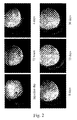
- Fig. 2 Subretinal injection of VSV-G pseudotyped SIV.
- gfp 10 8 transducing units (TU)/ml corresponding to 2 10 5 tu/injection
- retinal images showed efficient transgene expression within the detached portion of the retina as early as 72 hrs post injection.
- GFP expression was maximal and remained stable for more than one month (Fig. 2).
- VSV-G-pseudotyped MLV vectors encoding for the ß-Gal leads to the copurification of the enzyme. Consequently, injection of concentrated virus stocks may result in high rates of protein transfer rather than gene transfer, a process referred to as pseudotransduction.
- a stock of VSV-G-pseudotyped SIV empty particles was produced on 293 cells, except that the pRMES4SA plasmid containing the vector genome was replaced by an unrelated plasmid harboring the gfp expression unit alone but no encapsidation signal.
- GFP cDNA cannot be encapsidated within the particles, and only the GFP protein can eventually associate with the virions or be packaged in copurifying lipid vesicles.
- This preparation called pseudoSIV. gfp , was concentrated by ultracentrifugation and further processed as a standard SIV vector production (see Material & Methods).
- PseudoSIV. gfp (right eye) and SIV. gfp (left eye) vectors were subretinally injected in parallel in 3 rats.
- GFP detection occured in the pseudoSIV. gfp injected eye (Fig. 3A) even at 24 to 48 hrs post administration, while intense fluorescence was consistently detected in the SIV. gfp injected eye (Fig. 3B). This result confirmed that the early onset of GFP detection is due to active gene transfer.
- Example 3 VSV-G-pseudotyped SIV tropism is restricted to the RPE.
- Example 4 Stable SIV-mediated RPE transduction persists at least 6-month (duration of the experiment).
- Example 5 Comparative evaluation of five SIV. gfp pseudotypes.
- VSV-G pseudotyped SIV. gfp vector To exploit the powerful potential of envelope substitution on lentiviral vectors, other pseudotypes were compared, including MK-G, 4070A-Env, HA, LCMV-G and RD114-Env glycoproteins. Titers determined by end-point dilutions on 293 cells, showed differences among the various pseudotypes. Vectors pseudotyped with RD114-Env and HA were found at 10 8 TU/ml, while MK-G, 4070A-Env and LCMV-G pseudotypes were 100-fold less infectious.
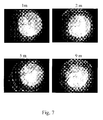
- HA-pseudotyped SIV showed the most efficient gene transfer in term of GFP expression level, which proved stable over at least 9 months when GFP was used as a transgene (Fig. 7).
- Vectors harboring MK-G, 4070A-Env and LCMV-G envelopes yielded lower transduction rates.
- HA, MK-G and 4070A-Env showed transgene expression kinetics similar to those obtained with the VSV-G with maximal GFP expression 6 days post-injection.
- GFP signal increased slowly with time in LCMV-G SIV transduced retina.
- RD114-Env pseudotype failed to transduce the retina.
Landscapes
- Health & Medical Sciences (AREA)
- Life Sciences & Earth Sciences (AREA)
- Engineering & Computer Science (AREA)
- Chemical & Material Sciences (AREA)
- Genetics & Genomics (AREA)
- Biotechnology (AREA)
- General Health & Medical Sciences (AREA)
- Organic Chemistry (AREA)
- Biomedical Technology (AREA)
- Bioinformatics & Cheminformatics (AREA)
- Pharmacology & Pharmacy (AREA)
- Medicinal Chemistry (AREA)
- General Engineering & Computer Science (AREA)
- Veterinary Medicine (AREA)
- Wood Science & Technology (AREA)
- Zoology (AREA)
- Public Health (AREA)
- Biochemistry (AREA)
- Animal Behavior & Ethology (AREA)
- Molecular Biology (AREA)
- Virology (AREA)
- Chemical Kinetics & Catalysis (AREA)
- Biophysics (AREA)
- Plant Pathology (AREA)
- Nuclear Medicine, Radiotherapy & Molecular Imaging (AREA)
- General Chemical & Material Sciences (AREA)
- Physics & Mathematics (AREA)
- Ophthalmology & Optometry (AREA)
- Microbiology (AREA)
- Epidemiology (AREA)
- Medicines Containing Material From Animals Or Micro-Organisms (AREA)
- Medicines That Contain Protein Lipid Enzymes And Other Medicines (AREA)
Abstract
The present invention relates to a novel lentiviral vector particularly well
shaped for performing gene transfer into the retina. These lentiviral vectors
are pseudotyped with a HA protein from an orthomyxovirus and comprise
one or several gene(s) useful for preventing or treating diseases of the eye.
The invention also relates to compositions and methods for preventing or
treating diseases of the eye, using the vector of the invention to transfer
selected genes suitable for preventing or treating diseases of the eye.
Description
The present invention relates to a novel lentiviral vector particularly well
shaped for performing gene transfer into the retina. The invention also
relates to compositions and methods for preventing or treating diseases of
the eye, using the vector of the invention to transfer selected genes suitable
for preventing or treating diseases of the eye.
Retinal degenerative diseases such as retinal macular degeneration or
retinitis pigmentosa constitute a broad group of diseases that all share one
critical feature, the progressive loss of cells in the retina. There is currently
no effective treatment available by which the course of these disorders can
be modified and visual dysfunction eventually progresses to total blindness.
Gene therapy represents a possible approach to treating retinal
degenerations because the eye is easily accessible and allows local
application of therapeutic vectors with reduced risk of systemic effects.
Furthermore, transgene expression within the retina and effects of
treatments may be monitored by a variety of non-invasive examinations.
Several viral vectors, including adenovirus, adenovirus-associated virus
(AAV), or human immunodeficiency virus type 1 (HIV-1) were tested for in
vivo retina transduction (see for example (Bainbridge, Stephens et al.
2001 )). Until now, an efficient long-term gene transfer was documented for
the AAV2-based vectors only. These vectors are able to transduce both the
photoreceptors and the retinal pigment epithelium (RPE) after subretinal
administration. However, the transgene expression is delayed. Maximal
levels of expression are detected 4-6 weeks post-infection (Rolling, Shen et
al. 1999; Auricchio, Kobinger et al. 2001). Such a feature may be
detrimental when rapid transgene expression is required.
Gene transfer into the retina was recently reported using HIV vectors
pseudotyped with vesicular stomatitis virus G glycoprotein (VSV-G), but the
tropism remains unclear. Subretinal injection in rats and mice leads to the
transduction of both photoreceptor cells and RPE (Miyoshi, Takahashi et al.
1997; Auricchio, Kobinger et al. 2001). In contrast, Brainbridge et al.
demonstrated that the transduction is restricted to the RPE in mice
(Bainbridge, Stephens et al. 2001).
The inventors have now developed a novel lentiviral vector which exhibits a
very low toxicity and high efficiency, for performing gene transfer into the
eye. The present invention hence provides compositions and methods for
preventing and treating a number of diseases of the eye, including for
example, retinal macular degeneration or retinitis pigmentosa, as well as
other diseases such as neovascular diseases. The present invention also
provides other related advantages.
The inventors have developed a lentiviral vector derived from SIVmac251,
a nonpathogenic strain of simian immunodeficiency virus (SIV). It was
previously demonstrated that HIV-1 and SIV vectors could transduce both
dividing and resting cells in vitro with equivalent efficiencies (Mangeot,
Negre et al. 2000; Negre, Mangeot et al. 2000). To assess both, the virus
tropism in retina and the kinetic of the transgene expression, rats were
injected with VSV-G-pseudotyped SIV vectors encoding for the green
fluorescent protein (GFP). Sustained gene transfer into the retina was
monitored by iterative non-invasive examinations (examples 1 and 2). Since
the tropism is mainly determined by the glycoproteins expressed onto the
virions, the inventors evaluated the cell populations transduced by SIV
vectors pseudotyped with surface glycoproteins from other membraneenveloped
viruses and, particularly, with the hemagglutinin (HA) protein
from an orthomyxovirus, more precisely from an influenza A virus. They
demonstrated that the HA-pseudotyped vectors were very efficient
(example 5).
The present invention hence mainly pertains to lentiviral vectors
pseudotyped with a HA protein from an orthomyxovirus and comprising one
or several gene(s) useful for preventing or treating diseases of the eye,
pharmaceutical compositions comprising such vectors, and uses of these
vectors.
Throughout this application, several words are employed, the meaning of
which should be understood according to the following definitions :
In the present application, the term "HA" will hence designate the
hemagglutinin protein of any kind of orthomyxovirus, in particular of any
influenza virus including the surface glycoprotein of influenza C and, more
particularly, the hemagglutinin of an influenza A virus. Influenza A viruses
naturally infect a broad range of species, including humans, horses, swine,
and a wide variety of avian species. Their surface glycoproteins, including
HA, exhibit a great amino acid sequence variability. Therefore, the word
"HA" in the present application designates a wide range of proteins that are
characterized by their origin (orthomyxoviridae), and their function (hemagglutination).
characterized by their origin (orthomyxoviridae), and their function (hemagglutination).
Usually, the phrase "viral vector" designates either a viral particle
comprising genetic material to be transferred into a host cell, or the
recombinant viral genome itself. Throughout this specification, and for
clarification purpose, the term "(lenti)viral vector particle" will be used to
designate the physical viral particle, including a genome, a capsid and an
enveloppe, whereas "(lenti)viral vector genome" will designate the nucleic
acid construct to be transferred. When neither one nor the other term is
specified, both can be understood, depending on the context.
Throughout this application, when two elements are said "operably linked",
this means that they are configured so as to interact in their usual way, to
perform their usual function. For example, when a coding sequence is
operably linked to a promoter or any other transcription control element, this
means that this coding sequence will be transcribed under the control of
this promoter or these elements.
The present invention hence provides a lentiviral vector particle comprising
a lentiviral vector genome encoding a transgene which, when expressed in
the retina, can prevent, inhibit or alleviate the effects of an eye disease,
wherein said vector particle is pseudotyped with an hemagglutinin (HA)
protein of an orthomyxovirus. A particular lentiviral vector particle of the
invention is pseudotyped with an HA protein from an influenza A virus, and,
for example, from a Fowl Plague Virus (FPV). The results shown in the
following experimental part have been obtained with lentiviral particles
pseudotyped with an HA protein from strain influenza A, Fowl plague virus,
Rostock, 34 (H7 N1). The sequence of this HA protein is accessible in
GenBank under numbers M24457 (coding sequence) and AAA43150
(amino acids sequence).
As described in the experimental part, HA-pseudotyped vectors can be
produced by co-transfecting plasmids, wherein one plasmid carries the
vector genome and the others encode the necessary proteins for lentiviral
encapsidation and the HA protein. The inventors have demonstrated that
cotransfection of a further plasmid, encoding the neuraminidase (NA) from
FPV, could result in improved vector production, with increased titers (titers
of 108 infectious particles/ml were obtained after a 100-fold concentration of
the collected supernatant, as described below). The present invention
hence also pertains to a lentiviral vector particle comprising a lentiviral
vector genome encoding a transgene which, when expressed in the retina,
can prevent, inhibit or alleviate the effects of an eye disease, wherein said
vector particle is pseudotyped with both an hemagglutinin (HA) and a
neuraminidase (NA) proteins of an orthomyxovirus, preferably from an
influenza virus, and more preferably from influenza A. One particular NA
protein which can be comprised in the vector particles according to the
invention is the NA protein from FPV, for example from the strain Rostock,
34 (H7 N1). The sequence of this NA protein is accessible in GenBank
under numbers X52226 (coding sequence) and CAA36475 (amino acids
sequence).
The invention also pertains to lentiviral vector particles such as described
above, wherein said vector genome comprises a lentiviral 5' LTR, a tRNA
binding site, a packaging signal, a promoter operably linked to said
transgene, an origin of second strand DNA synthesis and a 3' lentiviral
LTR.
According to a particular embodiment of the invention, the vector genome
within the lentiviral vector particle comprises 5' and/or 3' LTRs from a virus
selected from the group consisting of SIV, HIV, HIV-1, HIV-2, EIAV and
FIV.
According to another particular embodiment of the present invention, the
lentiviral vector is self-inactivating. Self-inactivating vectors can be made by
deleting promoter and enhancer elements in the U3 region of the 3' LTR,
including the TATA box and binding sites for one or more transcription
factors. The deletion is transferred to the 5' LTR after reverse transcription
and integration in transduced cells. This results in the transcriptional
inactivation of the LTR provirus. Such self-inactivating (SIN) vectors can
present the advantage of a broader tropism. Indeed, Miyoshi et al. have
suggested that the HIV-1 LTR can negatively influence the transgene
expression through an internal promoter in some cell types, and hence that
SIN vectors could present a double advantage in terms of safety and
broader applicability, particularly concerning the photoreceptor cells
(Miyoshi, Blomer et al. 1998).
Utilizing the methods and gene delivery vectors provided herein a wide
variety of diseases of the eye may be readily treated or prevented, including
for example, glaucoma, macular degeneration, diabetic retinopathies,
inherited retinal degeneration such as retinitis pigmentosa, retinal
detachment or injury and retinopathies (whether inherited, induced by
surgery, trauma, a toxic compound or agent, or, photically). Similarly, a
wide variety of neurotrophic factors may be utilized (either alone or in
combination) within the context of the present invention, including for
example, NGF, BDNF, CNTF, NT-3, NT-4, FGF-2, FGF-5, FGF-18, FGF-20
and FGF-21.
Within certain embodiments of the invention, it is preferred that the gene
delivery vector be utilized to deliver and express an anti-angiogenic factor
for the treatment, prevention, or, inhibition of diabetic retinopathy, wet
ARMD, and other neovascular diseases of the eye (e.g., ROP). Within other
embodiments it is desirable that the gene delivery vector be utilized to
deliver and express a neurotrophic growth factor to treat, prevent, or inhibit
diseases of the eye, such as, for example, glaucoma, retinitis pigmentosa,
and dry ARMD.
Within further aspects of the present invention gene delivery vectors are
provided which direct the expression of a neurotrophic factor, or, an anti-angiogenic
factor. As noted above, representative examples of neurotrophic
factors include NGF, BDNF, CNTF, NT-3, NT-4, FGF-2, FGF-5, FGF-18,
FGF-20 and FGF-21. Representative examples of anti-angiogenic factors
include soluble Flt-1, soluble Tie-2 receptor, and PEDF.
In the lentiviral vector particle according to the invention, the transgene can
hence be selected (without being limitative) in the group comprising
neurotrophic factors including NGF, BDNF, CNTF, NT-3, NT-4, FGF-2,
FGF-5, FGF-18, FGF-20 and FGF-21, anti-angiogenic factors including
soluble Flt-1, soluble Tie-2 receptor, and PEDF, and enzymes including β-glucuronidase,
neuraminidase, sphingomyelinase, sulfatases, arylsulfatase
β, α-neuraminidase, gangliosidase, tripeptidyl protease, CLN3 and
palmitoyl protein thioesterase (PPT).
In these lentiviral vectors, the transgene is preferably operably linked to a
regulated promoter (e.g., "tet" promoters, the ecdysome system, or other
systems of regulation), a viral promoter (e.g., the CMV or RSV promoters),
a tissue specific promoter (e.g., a rod, cone, or ganglia-derived promoter),
or a rhodopsin promoter.
The present invention also pertains to a pharmaceutical composition for
administration in the eye, comprising lentiviral vector particles according to
the invention (as described above), and a pharmaceutically acceptable
carrier. A particular advantage of the vectors according to the invention is
that time-course transductions showed that high levels of transgene
expression were obtained as soon as 4 days post-injection. For the first
time, the kinetic of the transgene expression could be monitored in vivo in
lentivirally-infected cells by iterative non-invasive examinations (see
examples 1 and 2). Moreover, the HA-pseudotyped vector showed an
excellent transduction efficiency (see, Fig. 6A), and lower toxicity (Example
6). This early onset of a robust expression of the transgene, and other
advantages of the vector of the invention, are attractive features for
degenerative retinal diseases showing rapid photoreceptors degeneration.
A recent study demonstrated correction of the retinal dystrophy phenotype
of the RCS rat by adenoviral gene transfer of Mertk. The rdy defect resides
in the RPE, and rescue needs immediate and strong RPE therapeutic gene
expression that matches the rapid degeneration in RCS rat. In this model,
HA pseudotyped lentivectors would represent the optimal choice.
Another aspect of the invention is the use of a lentiviral vector particle as
described above, for the manufacture of a composition for treating a
disease in the eye or preventing cell damage in the eye. In a particular
embodiment of this aspect of the invention, the disease is due to a
deficiency in retinal pigment epithelium (RPE) cells.
Alternatively, the disease can be due to a deficiency in the photoreceptors.
In this latter case, lentiviral particles which are able to transduce retinal
pigment epithelium (RPE) cells and express a neurotrophic factor therein
can be used according to the invention. The diffusion of the neurotrophic
factor around the transduced cells will attain the photoreceptors and have a
beneficial effect on them.
According to the invention, and depending on the transgene introduced into
the vector, the lentiviral vector particles described above can be used for
the manufacture of a composition for treating a neovascular disease of the
eye, a glaucoma, a macular degeneration, a diabetic retinopathy, an
inherited retinal degeneration, a retinitis pigmentosa, a cell damage in
retinal epithelial cells associated with Sly syndrome, a retinal detachment or
injury or a retinopathy.
A further aspect of the present invention is the use of an hemagglutinin
(HA) protein of an orthomyxovirus, to pseudotype an enveloped vector so
that said vector is able to transduce efficiently retinal pigment epithelium
(RPE) cells. In a particular embodiment of this aspect of the invention, the
HA protein is from an influenza A virus and, for example, from a Fowl
Plague Virus. More particularly, the HA protein can be from the strain
Rostock 34 (H7 N1) mentioned above.
The present application also relates to a method of treating, inhibiting or
preventing a disease of the eye, comprising, administering intraocularly, in
a mammal in need thereof, a gene delivery lentiviral vector particle which is
pseudotyped with an orthomyxovirus hemagglutinin (HA) protein and which
directs the expression of a transgene wherein said transgene, when
expressed in the retina, can prevent, inhibit or alleviate the effects of an eye
disease. As already mentioned above, the HA protein can be from an
influenza A virus, e.g., a Fowl Plague Virus, e.g., strain influenza A, FPV,
Rostock, 34 (H7 N1). The sequence of this HA protein is accessible in
GenBank under numbers M24457 (coding sequence) and AAA43150
(amino acids sequence).
The above method can be performed according to the invention, when the
disease is due to a deficiency in retinal pigment epithelium (RPE) cells, or
when the disease is due to a deficiency in the photoreceptors. In this latter
case, the method can be performed with lentiviral particles that transduce
retinal pigment epithelium (RPE) cells and express therein a neurotrophic
factor, which will diffuse to the photoreceptors.
These methods can be performed to treat, inhibit or alleviate a variety of
diseases of the eye, including, without being restrictive, a neovascular
disease of the eye, a glaucoma, a macular degeneration, a diabetic
retinopathy, an inherited retinal degeneration, a retinitis pigmentosa, a cell
damage in retinal epithelial cells associated with Sly syndrome, a retinal
detachment or injury or a retinopathy.
Within yet other embodiments, it may be desirable to utilize either a gene
delivery vector which expresses both an anti-angiogenic molecule and a
neurotrophic growth factor, or two separate vectors which independently
express such factors, in the treatment, prevention, or inhibition of an eye
disease (e.g., for diabetic retinopathy).
Within further embodiments of the invention, the above-mentioned methods
utilizing gene delivery vectors may be administered along with other
methods or therapeutic regimens, including for example, photodynamic
therapy (e.g., for wet ARMD), laser photocoagulation (e.g., for diabetic
retinopathy and wet ARMD), and intraocular pressure reducing drugs (e.g.,
for glaucoma).
While the invention has been described in terms of various preferred
embodiments, the skilled artisan will appreciate that various modifications,
substitutions, omissions and changes may be made without departing from
the scope thereof. Accordingly, it is intended that the scope of the present
invention be limited by the scope of the following claims, including
equivalents thereof.
Other characteristics of the invention will also become apparent in the
course of the description which follows of the biological assays which have
been performed in the framework of the invention and which provide it with
the required experimental support, without limiting its scope.
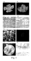
SIV.lacZ injected eyes were enucleated, eye cups were fixed and stained by X-gal (G). After histological sections, X-Gal staining was observed exclusively in the RPE cell layer (H). Original magnification: x20. These results demonstrated that accurate subretinal injection of pseudotyped SIV.gfp allowed specific transduction of the RPE cells.
The following examples can be performed using the materials and methods
described below:
293 embryo kidney cell line (ATCC CRL-1573) was grown in Dulbecco's
modified Eagle's medium (DMEM; Sigma) supplemented with 10% fetal calf
serum (FCS).
SIV packaging constructs were derived from the BK28 molecular clone of
SIVmac251 (GenBank accession number M19499). pSIV3 (encoding for
Gag-Pol, Tat, Rev, Vif, Vpx, Vpr) and pSIV4 (encoding for Gag-Pol, Tat and
Rev) were previously described in (Negre, Mangeot et al. 2000).
pRMES4SA harboring the recombinant SIV.gfp vector genome derived
from the pRMES4 described in (Negre, Mangeot et al. 2000). Briefly, the
vector genome contains the LTRs, the packaging signal overlapping the 57
first nucleotides of the GAG gene, the cPPT/CTS region (nt 5049-5189), the
extended RRE sequence (nt 8057-8808), an internal CMV promoter and
the gene encoding the enhanced GFP protein (eGFP, Clontech). For the
construction of the SIV.ltgfp vector, the hrgfp gene was extracted from the
pIRES-hrgfp plasmid (Stratagene) by BSTXI-Kpnl, blunt-ended by Klenow
fragment digestion and ligated to BamHI linkers. The 700-bp fragment was
finally inserted into the BamHl sites within the pRMES4SA plasmid, in place
of the egfp gene. For the construction of the SIV.lacZ vector, LacZ gene
was extracted from the pMFG-LacZ plasmid (Duisit, Salvetti et al. 1999)
and inserted into the pRMES4SA BamHl sites. Finally, plasmids were used
to express the envelope glycoproteins. phCMV-G (Negre, Mangeot et al.
2000), and phCMV-4070A (Sandrin, Boson et al. 2002) were previously
described. phCMV-HA (Hatziioannou, Valsesia-Wittmann et al. 1998) was
obtained by deleting the VSV-G coding sequence from the phCMV-G
plasmid [Yee, 1994 #12] and inserting in place thereof the Bg/II/Bg/II
fragment comprising the coding sequence of the FPV hemaggluntinin
(GenBank accession number M24457), upstream from the rabbit betaglobin
intron II sequence. The phCMV-NA plasmid was obtained by
inserting the cDNA comprising the coding sequence of FPV neuraminidase
(GenBank accession number X52226) into the EcoRI site of pCMV-G after
excision of the gene encoding VSV-G. In both phCMV-HA and phCMV-NA,
the glycoprotein gene (HA or NA) is under the control of the CMV
immediate early promoter. phCMV-RD114 was further modified to express
the RD114/TR chimeric glycoprotein carrying the 4070-ENV cytoplasmic tail
(Sandrin, Boson et al. 2002). pMK-G was a gift from JM Heard, Pasteur
(Desmaris, Bosch et al. 2001).
The phCMV-NA plasmid encodes the FVP neuraminidase, the sequence of
which is available at the GenBank under number X52226. This sequence is
under the control of the CMV i.e. promoter.
Production of recombinant vectors was achieved by three-plasmid
transfection into 293 cells, using pSIV3 or pSIV4, the plasmids coding for
the respective envelopes and for the vector genomes.
Production experiments showed that lentiviral vectors generated with the
FPV hemagglutinin could not egress from producer cells after GP
assembly. Indeed, when vector-producer cells expressing the FPV-HA were
treated with purified neuraminidase, infectivity of HA-pseudotyped vectors
was strongly increased by up to 100-fold. This enhancement correlated with
a 50-fold increased production of viral particles in the supernatant of
producer cells. This was induced by neuraminidase-mediated release of
virions from the cell surface on which they were retained because of
binding to sialic acid-containing cell surface molecules. Therefore, a fourth
plasmid, encoding the FPV neuraminidase protein, was also co-transfected
for HA-pseudotyped vectors. The phCMV-NA plasmid was used therefor.
Large-scale productions were generated according to the protocol
described by Patrick Salmon at the Trono's laboratory web site
(http://www.medecine.unige.ch/~salmon/) and by Negre et al. (2000).
Briefly, 293T cells were seeded in 10 cm culture plates (2.4 x 10⁶
cells/plate) and co-transfected the next day by calcium phosphate
precipitation. For producing the HA-pseudotyped vectors, 8.1 µg of plasmid
encoding the lentiviral capsid, 8.1 µg of plasmid comprising the vector, 2.7
µg of HA-coding plasmid, and 2.7 µg of phCMV-NA were co-transfected.
The culture medium (DMEM + 101% FCS; 12 ml/plate) was changed 16 hrs
later. Cell culture supernatants were recovered 2 and 3 days post-transfection,
filtered through 0.45 µm pore-sized membranes, and
ultracentrifuged. Pellets were carefully resuspended in Dulbecco's
Phosphate Buffered Saline supplemented with calcium and magnesium
(BioWhittaker Europe, Belgium). For the production of pseudoSIV.gfp
particules, pGFP-NI (Clontech) plasmid was transfected in place of the
pRMES4SA plasmid.
For the titration of the GFP-encoding vectors, 293 cells were plated at
2.5x10⁵ cells/well in 6-well plates 1 day before transduction. Infectious
supernatants were serially diluted in DMEM plus 10% FCS, 8 µg/ml
polybrene. Target cells were maintained in culture for 3 days. Percentages
of GFP-positive cells were determined by FACS analysis as described in
(Negre, Mangeot et al. 2000). For the titration of the SIV.lacZ vectors, 293
cells were plated at 1x10⁵ cells/well in 24-well plates 1 day before
transduction. Infectious supernatants were serially diluted in DMEM plus
10% FCS, 8 µg/ml polybrene. Target cells were maintained in culture for 2
days before X-gal staining. For the quantitative determination of reverse-transcriptase
contents, serially-diluted aliquots of stocks were used for the
determination of the reverse-transcriptase activity using a non-radioactive
RT assay (Roche).
Rats used for this study were cared for in accordance with the ARVO
statement for the use of animals in ophthalmic and vision research. Wistar
rats were anaesthetized with intramuscular injection of Ketamine/Xylazine
(50 mg/kg and 6mg/kg respectively).
Subretinal injections were performed via a transscleral transchoroidal
approach on wild-type Wistar rats as previously described (Rolling, Shen et
al. 1999). Briefly, the sclera and the choroïd were punctured; a 33-gauge
needle was then inserted in a tangential direction under an operating
microscope. Fluorescein was extemporarily added to the viral solution at a
1/1000 final dilution and the injection mixture was delivered into the
subretinal space.
For intravitreal injections, the needle tip was advanced through the sclera
posterior to the corneoscleral limbus into the vitrous cavity. Accuracy of
subretinal and intravitreal injections were monitored by fluorescence fundus
photography immediately following the injection procedure.
GFP protein expression in live rats was monitored by fluorescence retinal
imaging by using a Canon UVI retinal camera connected to a digital
imaging system (Lhedioph win software). Retinas were examined at dayly
and weekly intervals following injection. Identical experimental conditions
and parameters were used for fluorescence fundus photography at each
time point.
The enucleated eyes were fixed with 4% paraformaldehyde for 1 hr at room
temperature. After washing in PBS, the eyes were cut through the pars
plana, the anterior segment and the lens were removed. The eye cup was
cut peripherally in four sections under an operating microscope and
flattened. The neuroretina was removed from the RPE-choroid-sclera with
fine forceps, both cell layers were mounted on a glass slide and were
examined by fluorescence microscopy.
Fixed neuroretina flatmount was embedded in O.C.T. compound (Sakara
Finetek, Torrance, CA) and snap frozen in liquid nitrogen. Cryosections (10-12
µm) were cut and observed by fluorescence microscopy. For paraffin
sections, eyes were fixed in 4% paraformaldehyde for 24 hrs, removed and
the eye cups stained in 5-bromo-chloro-3-indolyl-β-D-galactopyranoside (X-Gal)
overmight. The eyes were then embedded in paraffin and sectioned.
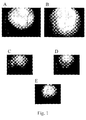
The small size of rodent eyes makes precise intravitreal, and more
evidently, subretinal injection difficult to control. In order to assess vector
administration, diluted fluorescein was added to the viral solution, and
accuracy of the injection site was monitored by fluorescence fundus
photography immediately after vector delivery. Therefore, detection of
retinal vessels over the surface of the green bleb characterized a restricted
subretinal administration (Fig. 1A), whereas retinal vascularization is
completely occulted by the dye which instantly diffuses with fusy border
(Fig1B) as a result of intravitreal injection. As observed in Fig. 1C, 1D and
1E, subchoroïdal (choroidal vascularization) and mixed injections could
occur (subretinal/subchoroïdal, subretinal/intravitreal), and, without the
fluorescein marker, could not be distinguished solely by direct vision using
the operating microscope. Dye diffusion was followed by fluorescence
fundus photography at various time points post subretinal and intravitreal
administration, and no fluorescein could be detected 24 hrs post-vector
injection.
To compare gene transfer efficiencies and SIV pseudotypes tropism, only
animals that have received a true and restricted subretinal or intravitreal
injection were included in the study.
Subretinal injection of VSV-G pseudotyped SIV.gfp (108 transducing units
(TU)/ml corresponding to 2 105 tu/injection) were performed on six Wistar
rats. For each animal, retinal images showed efficient transgene expression
within the detached portion of the retina as early as 72 hrs post injection. At
four days post injection, GFP expression was maximal and remained stable
for more than one month (Fig. 2).
It was previously demonstrated that ultracentrifugation of VSV-G-pseudotyped
MLV vectors encoding for the ß-Gal leads to the copurification
of the enzyme. Consequently, injection of concentrated virus stocks may
result in high rates of protein transfer rather than gene transfer, a process
referred to as pseudotransduction. To ensure the fluorescent signal
detected four days post-injection was actually due to newly synthesized
GFP, a stock of VSV-G-pseudotyped SIV empty particles was produced on
293 cells, except that the pRMES4SA plasmid containing the vector
genome was replaced by an unrelated plasmid harboring the gfp
expression unit alone but no encapsidation signal. In this setting, GFP
cDNA cannot be encapsidated within the particles, and only the GFP
protein can eventually associate with the virions or be packaged in
copurifying lipid vesicles. This preparation, called pseudoSIV.gfp, was
concentrated by ultracentrifugation and further processed as a standard
SIV vector production (see Material & Methods). PseudoSIV.gfp (right eye)
and SIV.gfp (left eye) vectors were subretinally injected in parallel in 3 rats.
In none of these animals, GFP detection occured in the pseudoSIV.gfp
injected eye (Fig. 3A) even at 24 to 48 hrs post administration, while
intense fluorescence was consistently detected in the SIV.gfp injected eye
(Fig. 3B). This result confirmed that the early onset of GFP detection is due
to active gene transfer.
Fluorescence fundus photography of subretinally injected eyes strongly
suggested that RPE cells were the actual transduced cells (Fig. 2 and Fig.
3). To further characterize the transduced cells within the retina, animals
were sacrificed and the SIV.gfp-injected eyes were enucleated. Retina
flatmounts were performed and the sclera/choroid/RPE layer was
separated from the neuroretina (Fig. 4A and B). By examination under a
fluorescence inverted microscope, GFP signal was only observed on the
sclera/choroid/RPE layer and showed that RPE cells were efficiently
transduced (Fig. 4E). No signal was detected in the neuroretina layer (Fig.
4F). To confirm the flatmount results, four animals were injected
subretinally with VSV-G-pseudotyped SIV.lacZ vectors. Seven days post-injection,
animals were sacrificed and histological sections were performed.
X-Gal staining was observed only in the RPE cell layer (Fig. 4H).
While VSV-G-pseudotyped SIV.gfp-mediated gene transfer remained stable
for more than one month, loss of GFP signal was seen, thereafter (Fig. 5A).
Concomitantly, RPE damage restricted to the transduced area was
documented by inverted fluorescence microscopy on the
sclera/choroïd/RPE flatmount (Fig. 5B). As monitored on the six injected
eyes, the rapidity and severity of RPE destruction correlated with the
amount of GFP expression.
No toxicity related to the use of lentivirus vectors was reported yet.
However, the cytotoxicity of the GFP protein itself was previously
mentionned (Doi, Hargitai et al. 2002). Therefore, a new recombinant vector
was generated, encoding the low-toxicity GFP (from mutant Renilla
reniformis) instead of the enhanced GFP (from Aequorea victoria) used
initially. Recombinant SIV.gfp and SIV.Itgfp vectors were produced at
equivalent titer (108 TU/ml) and were subretinally injected in 6 Wistar rats.
Fig. 5C shows that no rejection phenomenon occurred in any of these
animals receiving the SIV.Itgfp vector and long-term GFP expression
remained stable over a six-month period. Additionally, sclera/choroïd/RPE
flamount showed normal RPE morphology of transduced RPE cells six
months post-injection (Fig. 5D).
The inventors demonstrated a sustained GFP expression in RPE following
subretinal injection of a VSV-G pseudotyped SIV.gfp vector. To exploit the
powerful potential of envelope substitution on lentiviral vectors, other
pseudotypes were compared, including MK-G, 4070A-Env, HA, LCMV-G
and RD114-Env glycoproteins. Titers determined by end-point dilutions on
293 cells, showed differences among the various pseudotypes. Vectors
pseudotyped with RD114-Env and HA were found at 108 TU/ml, while MK-G,
4070A-Env and LCMV-G pseudotypes were 100-fold less infectious.
This discrepancy could reflect variable expression of the related receptors
onto the 293 cells rather than a misassembly of the particles. Therefore,
productions were normalized according to the reverse-transcriptase (RT)
activity measured in the concentrated stocks. Equivalent amount of total
particles, corresponding to an average of 5 ng of RT, were injected in the
following experiment.
Subretinal injections of each pseudotype were performed in parallel on
wistar rats (n=4 per pseudotype). GFP expression was monitored by
fluorescence fundus photography over a four-week period. Interestingly,
different patterns of expression were obtained (Fig. 6A). HA-pseudotyped
SIV showed the most efficient gene transfer in term of GFP expression
level, which proved stable over at least 9 months when GFP was used as a
transgene (Fig. 7). Vectors harboring MK-G, 4070A-Env and LCMV-G
envelopes yielded lower transduction rates. HA, MK-G and 4070A-Env
showed transgene expression kinetics similar to those obtained with the
VSV-G with maximal GFP expression 6 days post-injection. In contrast,
GFP signal increased slowly with time in LCMV-G SIV transduced retina.
Finally, among the five vectors, RD114-Env pseudotype failed to transduce
the retina.
To specify the cell populations actually transduced, two animals of each
group were sacrificed at 14 days post injection and eyes were enucleated.
Sclera/choroïd/RPE and neuroretina layers were analyzed under inverted
fluorescence microscope. No signal could be detected with RD114, as
expected. For all other SIV pseudotypes, the GFP fluorescence was
restricted to the RPE cells (Fig. 6B), while no signal could be detected in
the neuroretina.
Intravitreal injections (n=3 for each pseudotype) were also performed and
monitored over a one month period, resulting in no detectable transduction
(Fig. 8). Intravitreal injection of rAAV2.gfp was used as a positive control.
SIV-injected animals were sacrificed 20 days post-injection. No GFP signal
could be detected by fluorescence microscopy on retinal flatmounts and
neuroretina cryosections. This result indicated that neuroretina cells such
as ganglion or Müller cells exhibit a block during the infectious process for
each of the pseudotypes tested.
Auricchio, A., G. Kobinger, et al. (2001). "Exchange of surface proteins
impacts on viral vector cellular specificity and transduction
characteristics: the retina as a model." Hum Mol Genet 10(26): 3075-81.
Bainbridge, J. W., C. Stephens, et al. (2001). " In vivo gene transfer to the
mouse eye using an HIV-based lentiviral vector; efficient long-term
transduction of corneal endothelium and retinal pigment epithelium."
Gene Ther 8(21): 1665-8.
Desmaris, N., A. Bosch, et al. (2001). "Production and neurotropism of
lentivirus vectors pseudotyped with lyssavirus envelope
glycoproteins." Mol Ther 4(2): 149-56.
Doi, K., J. Hargitai, et al. (2002). "Lentiviral transduction of green
fluorescent protein in retinal epithelium: evidence of rejection." Vision
Res 42(4): 551-8.
Duisit, G., A. Salvetti, et al. (1999). "Functional characterization of
adenoviral/retroviral chimeric vectors and their use for efficient
screening of retroviral producer cell lines." Hum Gene Ther 10(2):
189-200.
Hatziioannou, T., S. Valsesia-Wittmann, et al. (1998). "Incorporation of fowl
plague virus hemagglutinin into murine leukemia virus particles and
analysis of the infectivity of the pseudotyped retroviruses." J Virol
72(6): 5313-7.
Mangeot, P. E., D. Negre, et al. (2000). "Development of minimal lentivirus
vectors derived from simian immunodeficiency virus (SIVmac251 )
and their use for gene transfer into human dendritic cells." J Virol
74(18): 8307-15.
Miyoshi, H., U. Blomer, et al. (1998). "Development of a self-inactivating
lentivirus vector." J Virol 72(10): 8150-7.
Miyoshi, H., M. Takahashi, et al. (1997). "Stable and efficient gene transfer
into the retina using an HIV-based lentiviral vector." Proc Natl Acad
Sci USA 94(19): 10319-23.
Negre, D., P. E. Mangeot, et al. (2000). "Characterization of novel safe
lentiviral vectors derived from simian immunodeficiency virus
(SlVmac251) that efficiently transduce mature human dendritic cells."
Gene Ther 7(19): 1613-23.
Rolling, F., W. Y. Shen, et al. (1999). "Evaluation of adeno-associated
virus-mediated gene transfer into the rat retina by clinical
fluorescence photography." Hum Gene Ther 10(4): 641-8.
Sandrin, V., B. Boson, et al. (2002). "Lentiviral vectors pseudotyped with a
modified RD114 envelope glycoprotein show increased stability in
sera and augmented transduction of primary lymphocytes and
CD34+ cells derived from human and nonhuman primates." Blood
100(3): 823-32.
Claims (23)
- A lentiviral vector particle comprising a lentiviral vector genome encoding a transgene which, when expressed in the retina, can prevent or alleviate the effects of an eye disease, wherein said vector particle is pseudotyped with an hemagglutinin (HA) protein of an orthomyxovirus.
- The lentiviral vector particle of claim 1, wherein said HA protein is from an influenza A virus.
- The lentiviral vector particle of claim 2, wherein said HA protein is from a Fowl Plague Virus (FPV).
- The lentiviral vector particle of claim 3, wherein said HA protein is from strain influenza A, FPV, Rostock, 34 (H7 N1), and has the sequence accessible in GenBank under number AAA43150.
- The lentiviral particle of any of claims 1 to 4, further comprising a neuraminidase (NA) protein from an influenza virus inserted in its membrane.
- The lentiviral particle of claim 5, wherein said NA protein is from an influenza A virus.
- The lentiviral vector particle of claim 6, wherein said NA protein is from a Fowl Plague Virus (FPV).
- The lentiviral vector particle of claim 7, wherein said HA protein is from strain influenza A, FPV, Rostock, 34 (H7 N1), and has the sequence accessible in GenBank under number CAA36475.
- The lentiviral vector particle of any of claims 1 to 8, wherein said vector genome comprises a lentiviral 5' LTR, a tRNA binding site, a packaging signal, a promoter operably linked to said transgene, an origin of second strand DNA synthesis and a 3' lentiviral LTR.
- The lentiviral vector particle of any of claims 1 to 9, wherein said vector genome comprises 5' and/or 3' LTRs from a virus selected from the group consisting of SIV, HIV, HIV-1, HIV-2, EIAV and FIV.
- The lentiviral vector particle of any of claims 1 to 10, which is self-inactivating.
- The lentiviral vector particle of any of claims 1 to 11, wherein said transgene is selected in the group consisting of neurotrophic factors including NGF, BDNF, CNTF, NT-3, NT-4, FGF-2, FGF-5, FGF-18, FGF-20 and FGF-21, anti-angiogenic factors including soluble Flt-1, soluble Tie-2 receptor, and PEDF, and enzymes including β-glucuronidase, neuraminidase, sphingomyelinase, sulfatases, arylsulfatase β, α-neuraminidase, gangliosidase, tripeptidyl protease, CLN3 and palmitoyl protein thioesterase (PPT).
- The lentiviral vector particle of any of claims 1 to 12, wherein said transgene is operably linked to a regulated promoter, a photo-activated promoter, a viral promoter, a tissue specific promoter or a rhodopsin promoter.
- A pharmaceutical composition for administration in the eye, comprising lentiviral vector particles according to any of claims 1 to 13, and a pharmaceutically acceptable carrier.
- Use of a lentiviral vector particle according to any of claims 1 to 13, for the manufacture of a composition for treating a disease in the eye or preventing cell damage in the eye.
- Use according to claim 15, wherein said disease is due to a deficiency in retinal pigment epithelium (RPE) cells.
- Use according to claim 15, wherein said disease is due to a deficiency in the photoreceptors.
- Use according to claim 17, wherein the lentiviral particles are able to transduce retinal pigment epithelium (RPE) cells and express a neurotrophic factor therein.
- Use according to any of claims 15 to 18, for the manufacture of a composition for treating a neovascular disease of the eye, a glaucoma, a macular degeneration, a diabetic retinopathy, an inherited retinal degeneration, a retinitis pigmentosa, a cell damage in retinal epithelial cells associated with Sly syndrome, a retinal detachment or injury or a retinopathy.
- Use of an hemagglutinin (HA) protein of an orthomyxovirus, for pseudotyping an enveloped vector so that said vector is able to transduce efficiently retinal pigment epithelium (RPE) cells.
- Use according to claim 20, wherein said HA protein is from an influenza A virus.
- Use according to claim 21, wherein said HA protein is from a Fowl Plague Virus.
- Use according to claim 22, wherein the HA protein is from strain influenza A, FPV, Rostock, 34 (H7 N1), and has the sequence accessible in GenBank under number AAA43150.
Priority Applications (3)
| Application Number | Priority Date | Filing Date | Title |
|---|---|---|---|
| EP02292255A EP1398041A1 (en) | 2002-09-13 | 2002-09-13 | Recombinant lentiviral vector pseudotyped with the hemagglutinin protein for gene transfer into the retina |
| AU2003279318A AU2003279318A1 (en) | 2002-09-13 | 2003-09-15 | Recombinant lentivirale vector pseudotyped with the hemagglutinin protein for gene transfer into the retina |
| PCT/EP2003/011815 WO2004024190A2 (en) | 2002-09-13 | 2003-09-15 | Recombinant lentivirale vector pseudotyped with the hemagglutinin protein for gene transfer into the retina |
Applications Claiming Priority (1)
| Application Number | Priority Date | Filing Date | Title |
|---|---|---|---|
| EP02292255A EP1398041A1 (en) | 2002-09-13 | 2002-09-13 | Recombinant lentiviral vector pseudotyped with the hemagglutinin protein for gene transfer into the retina |
Publications (1)
| Publication Number | Publication Date |
|---|---|
| EP1398041A1 true EP1398041A1 (en) | 2004-03-17 |
Family
ID=31725511
Family Applications (1)
| Application Number | Title | Priority Date | Filing Date |
|---|---|---|---|
| EP02292255A Withdrawn EP1398041A1 (en) | 2002-09-13 | 2002-09-13 | Recombinant lentiviral vector pseudotyped with the hemagglutinin protein for gene transfer into the retina |
Country Status (3)
| Country | Link |
|---|---|
| EP (1) | EP1398041A1 (en) |
| AU (1) | AU2003279318A1 (en) |
| WO (1) | WO2004024190A2 (en) |
Cited By (2)
| Publication number | Priority date | Publication date | Assignee | Title |
|---|---|---|---|---|
| WO2008087563A3 (en) * | 2006-12-29 | 2008-11-27 | Inst Pasteur Of Shanghai | Lentivirus pseudotyped with influenza hemagglutinin and methods of use |
| CN101180082B (en) * | 2005-02-23 | 2012-02-08 | 生物载体株式会社 | A drug for treating diseases accompanied by apoptosis and degeneration of ocular tissue cells using SIV-PEDF carrier |
Citations (1)
| Publication number | Priority date | Publication date | Assignee | Title |
|---|---|---|---|---|
| WO2001092508A1 (en) * | 2000-06-01 | 2001-12-06 | Dnavec Research Inc. | Pseudo-type retrovirus vector containing membrane protein having hemagglutinin activity |
-
2002
- 2002-09-13 EP EP02292255A patent/EP1398041A1/en not_active Withdrawn
-
2003
- 2003-09-15 AU AU2003279318A patent/AU2003279318A1/en not_active Abandoned
- 2003-09-15 WO PCT/EP2003/011815 patent/WO2004024190A2/en not_active Ceased
Patent Citations (2)
| Publication number | Priority date | Publication date | Assignee | Title |
|---|---|---|---|---|
| WO2001092508A1 (en) * | 2000-06-01 | 2001-12-06 | Dnavec Research Inc. | Pseudo-type retrovirus vector containing membrane protein having hemagglutinin activity |
| EP1291419A1 (en) * | 2000-06-01 | 2003-03-12 | Dnavec Research Inc. | Pseudo-type retrovirus vector containing membrane protein having hemagglutinin activity |
Non-Patent Citations (7)
| Title |
|---|
| ARVO ANNUAL MEETING ABSTRACT SEARCH AND PROGRAM PLANNER, vol. 2002, 2002, Annual Meeting of the Association For Research in Vision and Ophthalmology;Fort Lauderdale, Florida, USA; May 05-10, 2002, pages Abstract No. 4608 * |
| AURICCHIO ALBERTO ET AL: "Exchange of surface proteins impacts on viral vector cellular specificity and transduction characteristics: The retina as a model.", HUMAN MOLECULAR GENETICS, vol. 10, no. 26, 2001, 15 December, 2001, pages 3075 - 3081, XP002242670, ISSN: 0964-6906 * |
| BAINBRIDGE J W B ET AL: "In vivo gene transfer to the mouse eye using an HIV-based lentiviral vector;efficient long-term transduction of corneal endothelium and retinal pigment epithelium.", GENE THERAPY, vol. 8, no. 21, November 2001 (2001-11-01), pages 1665 - 1668, XP008017785, ISSN: 0969-7128 * |
| DATABASE BIOSIS [online] BIOSCIENCES INFORMATION SERVICE, PHILADELPHIA, PA, US; 2002, CONRATH H ET AL: "SIV-mediated Gene Transfer Into Rat Retina.", XP002242674, Database accession no. PREV200300165691 * |
| DUISIT GHISLAINE ET AL: "Five recombinant simian immunodeficiency virus pseudotypes lead to exclusive transduction of retinal pigmented epithelium in rat.", MOLECULAR THERAPY: THE JOURNAL OF THE AMERICAN SOCIETY OF GENE THERAPY. UNITED STATES OCT 2002, vol. 6, no. 4, October 2002 (2002-10-01), pages 446 - 454, XP002242673, ISSN: 1525-0016 * |
| MIYOSHI HIROYUKI ET AL: "Stable and efficient gene transfer into the retina using an HIV-based lentiviral vector.", PROCEEDINGS OF THE NATIONAL ACADEMY OF SCIENCES OF THE UNITED STATES, vol. 94, no. 19, 1997, 1997, pages 10319 - 10323, XP002242671, ISSN: 0027-8424 * |
| SANDRIN VIRGINIE ET AL: "Lentiviral vectors pseudotyped with a modified RD114 envelope glycoprotein show increased stability in sera and augmented transduction of primary lymphocytes and CD34+ cells derived from human and nonhuman primates.", BLOOD, vol. 100, no. 3, 1 August 2002 (2002-08-01), August 1, 2002, pages 823 - 832, XP002242672, ISSN: 0006-4971 * |
Cited By (3)
| Publication number | Priority date | Publication date | Assignee | Title |
|---|---|---|---|---|
| CN101180082B (en) * | 2005-02-23 | 2012-02-08 | 生物载体株式会社 | A drug for treating diseases accompanied by apoptosis and degeneration of ocular tissue cells using SIV-PEDF carrier |
| US8278284B2 (en) | 2005-02-23 | 2012-10-02 | Masanori Miyazaki | Therapeutic agents for diseases associated with apoptotic degeneration in ocular tissue cells that use SIV-PEDF vectors |
| WO2008087563A3 (en) * | 2006-12-29 | 2008-11-27 | Inst Pasteur Of Shanghai | Lentivirus pseudotyped with influenza hemagglutinin and methods of use |
Also Published As
| Publication number | Publication date |
|---|---|
| AU2003279318A8 (en) | 2004-04-30 |
| WO2004024190A3 (en) | 2004-10-14 |
| AU2003279318A1 (en) | 2004-04-30 |
| WO2004024190A2 (en) | 2004-03-25 |
Similar Documents
| Publication | Publication Date | Title |
|---|---|---|
| JP4861314B2 (en) | Non-integrating and non-replicating recombinant lentivirus, its preparation and use | |
| EP1895010B1 (en) | Equine infectious anaemia virus (eiav) based vectors | |
| Duisit et al. | Five recombinant simian immunodeficiency virus pseudotypes lead to exclusive transduction of retinal pigmented epithelium in rat | |
| AU2004245175C1 (en) | Improved secretion of neublastin | |
| JP4700888B2 (en) | Pseudotype retroviral vector containing a membrane protein with hemagglutinin activity | |
| US20100172871A1 (en) | Muller Cell Specific Gene Therapy | |
| WO1997012622A9 (en) | Vector and method of use for nucleic acid delivery to non-dividing cells | |
| JP2007509109A (en) | In vivo gene therapy for Parkinson's disease | |
| KR20010074493A (en) | Retroviral delivery system | |
| US6818209B1 (en) | Retroviral delivery system | |
| JP4959547B2 (en) | Therapeutic agent for diseases with apoptotic degeneration in ocular tissue cells using SIV-PEDF vector | |
| JP4971974B2 (en) | A therapeutic agent for diseases associated with apoptotic degeneration in ocular tissue cells, including PEDF and FGF2 | |
| WO2004024190A2 (en) | Recombinant lentivirale vector pseudotyped with the hemagglutinin protein for gene transfer into the retina | |
| Molina et al. | A synthetic Rev-independent bovine immunodeficiency virus-based packaging construct | |
| HK1115324B (en) | Therapeutic agent for disease with apoptotic degeneration in eye tissue cell containing pedf and fgf2 | |
| GB2345062A (en) | Non-primate lentivirus retroviral vectors | |
| HK1056576B (en) | Pseudotype retroviral vectors containing membrane proteins having hemagglutinin activity |
Legal Events
| Date | Code | Title | Description |
|---|---|---|---|
| PUAI | Public reference made under article 153(3) epc to a published international application that has entered the european phase |
Free format text: ORIGINAL CODE: 0009012 |
|
| AK | Designated contracting states |
Kind code of ref document: A1 Designated state(s): AT BE BG CH CY CZ DE DK EE ES FI FR GB GR IE IT LI LU MC NL PT SE SK TR |
|
| AX | Request for extension of the european patent |
Extension state: AL LT LV MK RO SI |
|
| AKX | Designation fees paid | ||
| REG | Reference to a national code |
Ref country code: DE Ref legal event code: 8566 |
|
| STAA | Information on the status of an ep patent application or granted ep patent |
Free format text: STATUS: THE APPLICATION IS DEEMED TO BE WITHDRAWN |
|
| 18D | Application deemed to be withdrawn |
Effective date: 20040918 |