-
The present invention relates to the use of at least one 6-amino-quinoline-5,8-quinone
for preparing a pharmaceutical composition for the treatment
of tumors. A further subject matter of the present invention are nucleic
acids associated with senescence and their use for the treatment of
tumors.
-
Several cellular mechanisms have evolved to prevent proliferation of cells,
which have acquired mutations with carcinogenic potential. One
mechanism is represented by apoptosis: cells with deregulated expression
of oncogenes are removed through this route (Green and Evan, 2002).
Another cellular response, that gained more attention recently, is
senescence: a state of terminal arrest in which cells are metabolically
active for extended periods, but are unable to re-enter the cell cycle after
mitogenic stimulation (Hayflick and Moorehead, 1961; Shay and Wright,
2000). Several tumor-suppressor genes (e.g. p53 and p16INK4A) are
involved in the induction and maintenance of senescence, strongly
suggesting that senescence is a tumor suppressive mechanism (Serrano
and Blasco, 2001; Campisi, 2001). Supporting this notion on the
organismal level, mice, which have been engineered to display elevated
p53 activity, show a decreased rate of tumor-formation and premature
aging (Tyner et al., 2002).
-
Replicative senescence results from progressive shortening of
chromosomes, which occurs during every cell division. After 50-70 cell
divisions, the loss of telomeric DNA repeats results in unprotected
chromosome ends, which generate a DNA-damage signal resulting in
activation of the tumor suppressor gene product p53. Therefore, several
downstream targets of the p53 transcription factor are induced during
replicative senescence, e.g. p21WAF1 / SDI / CIP1 and 14-3-3sigma (Noda et al.,
1994; Dellambra et al., 2000). Consistent with an essential function of
these p53 target-genes during senescence, deletion of p21 through
homologous recombination is sufficient to bypass replicative senescence in
human diploid fibroblasts (Brown et al., 1997). Similarly, inactivation of 14-3-3sigma
leads to immortalization of primary human keratinocytes
(Dellambra et al., 2000).
-
In contrast to telomer-dependent replicative senescence, cellular
senescence, also referred to as premature senescence, is a response to
diverse stimuli (e.g. oncogenic signalling, sub-optimal tissue culture
conditions), which generally involves induction of the tumor suppressor
gene p16/INK4. Cellular senescence accompanied by induction of p16 is
also observed after serial passaging of epithelial cells under standard tissue
culture conditions. Interestingly, the p16 gene is not induced in epithelial
cells, when these are cultivated on feeder-layers: under these conditions
epithelial cells have a greatly elevated replicative potential and terminally
arrest due to shortened telomeres, i.e. replicative senescence (Ramirez et
al., 2001 ) . In contrast to epithelial cells, primary human fibroblasts undergo
replicative senescence under standard tissue culture conditions (reviewed
in Christofalo et al., 1998). These examples point to the existence of cell-type
dependent signalling pathways, which activate the senescence
program.
-
DNA-damaging agents commonly used for cancer therapy induce a
senescence-like state in normal and cancer cells (Di Leonardo et al., 1994;
Chang et al., 1999). However, since these substances also induce DNA-damage
in normal proliferating cells, numerous unwanted side effects are
observed during chemotherapy (Bast et al., 2000). Furthermore, these
agents generate mutations in pre-cancerous cells, which may result in
secondary cancer. In the studies underlying the present invention, we
aimed to identify substances, which activate the cellular senescence
without inducing DNA-damage. We hypothesized that pharmacological
inhibition of signalling pathways, which are specifically down-regulated
during replicative senescence, may result in the reactivation of the
senescence program in tumor cells. The ideal drug-target for this strategy
would be an enzyme encoded by a gene that is repressed during
senescence. Inhibition of such an enzyme by a small, membrane permeable
drug molecule in early passage or tumor cells should theoretically be
sufficient to induce cellular senescence. In order to detect genes and
pathways repressed during replicative senescence, the gene expression
pattern of senescent human fibroblasts was compared to the expression
signature of confluent, early passage cells. Using microarray analysis, we
have discovered within the framework of the present invention down-regulation
of several components of the cGMP- signalling pathway during
replicative senescence of primary human diploid fibroblasts (HDF).
Therefore, the effect of pharmacological inhibition of cGMP-synthesis was
analyzed in HDF. 6-Anilino-quinoline-5,8-quinone (LY83583 or LY), an
inhibitor of guanylate cyclase, unexpectedly induced cellular senescence.
114 genes differentially expressed after treatment with LY were regulated
similarly in HDF undergoing replicative senescence, indicating that these
genes may constitute components of a transcriptional program, which
mediates the senescent phenotype. Among the LY-induced genes was the
cdk-inhibitor p21WAF1 / SDI / CIP1 . In colorectal cancer cells, transcription of p21
was induced by LY in a p53-independent manner. Furthermore, p21 but
not p53, was required for cell-cycle inhibition by LY. Inactivation of the
retinoblastoma tumor suppressor protein, an effector of p21-mediated cell
cycle inhibition, converted LY-induced growth arrest to apoptosis. These
results suggest that LY, or derivatives thereof, may be useful tumor
therapeutics.
-
Therefore, a first subject matter of the invention relates to the use of at
least one 6-amino-quinoline-5,8-quinone for preparing a pharmaceutical
composition for the treatment of tumors.
-
The pharmaceutical composition of the invention constitutes a new class of
non-genotoxic anti-tumor drugs which are capable of inducing cellular
senescence and/or apoptosis in tumor cells. An obvious advantage of said
pharmaceutical composition is its ability to interfere with cellular
proliferation without inducing DNA damage. Commonly used
chemotherapeutics like, e.g. adriamycin, cause DNA damage and thereby
selectively induce apoptosis in tumor cells which lack cell cycle
checkpoints. However, DNA-damaging substances cause major side effects
on proliferating tissues and may induce further mutations in pre-cancerous
and healthy cells which lead to secondary cancer.
-
The pharmaceutical composition of the invention can contain a single 6-amino-quinoline-5,8-quinone
or a combination of several 6-amino-quinoline-5,8-quinones.
Furthermore, the pharmaceutical composition can contain
conventionally used pharmaceutical additives well-known to the skilled
artisan, if desired, such as, for example, physiologically acceptable carrier
substances, diluents and adjuvants.
-
In a preferred embodiment of the invention the 6-amino-quinoline-5,8-quinone
is 6-anilino-quinoline-5,8-quinone (LY83583 or LY).
-
The pharmaceutical composition of the invention can be topically,
parenterally, intravenously, intramuscularly, subcutaneously or
transdermally applicable and can be produced according to conventional
methods well-known in the art. Preferably, the pharmaceutical composition
of the invention is produced in the form of tablets or as an intravenous
injection or infusion.
-
The pharmaceutical composition according to the invention is used for the
treatment of tumors. The term "tumor", as used herein, comprises any
local increase in tissue volume, i.e., in the broadest sense, any localized
swelling due to oedema, acute and chronic inflammation, aneurysmal
dilatation and organ swelling caused by inflammation as well as, in the
narrowest sense, formation of new tissue (e.g. tumor, blastoma, neoplasia)
in the form of spontaneous, differently disinhibited, autonomous and
irreversible excess growth of endogenous tissue which normally is
associated with loss of specific cell and tissue functions in various
degrees. Examples of tumor diseases which can be treated by means of
the pharmaceutical composition according to the invention comprise
intestinal cancer, brain tumor, eye tumor, pancreatic tumor, urinary bladder
carcinoma, lung cancer, breast cancer, ovarian tumor, uterine carcinoma,
bone tumor, gall bladder and bile duct carcinoma, head-neck tumor,
urethral cancer, skin cancer, testicular cancer, renal tumor, germinal tumor,
liver carcinoma, leucemia, gastric carcinoma, malignant lymphoma, adrenal
carcinoma, neuroma, neuroblastoma, malignant melanoma, oesophageal
cancer, prostate carcinoma, soft-tissue tumor, thymoma and carcinoma in
the case of unknown primary cancer.
-
The expression "treatment of tumors", as used herein, comprises at least
one of the following features: alleviation of the symptoms associated with
the tumor disease, reduction of the extent of the tumor disease (e.g.
reduction of tumor growth), stabilization of the state of the tumor disease
(e.g. inhibition of tumor growth), inhibition of proliferation of the tumor
disease (e.g. metastasis), deceleration or retardation of the progress of the
tumor disease, improvement of the state of the tumor disease (e.g.
reduction of the tumor) or prevention of the occurrence or reoccurrence of
a tumor disease.
-
Preferably, the pharmaceutical composition of the invention is administered
to a patient with a tumor disease, with a presumed tumor disease or with
a high tumor risk in an amount sufficient to achieve treatment of the tumor
or prevention of a tumor. Particularly preferred, the pharmaceutical
composition is administered to a patient suffering from a tumor disease in
a therapeutically effective amount. The amount of the pharmaceutical
composition to be administered thereby depends on several factors, e.g. on
the 6-amino-quinoline-5,8-quinone chosen, the type of administration
(tablet, injection, infusion, etc.), the kind and extent of the tumor disease
and the age, weight and general condition of the patient and can be easily
determined by one skilled in the field of tumor diseases, considering the
above-mentioned factors. Preferably, however, the complexes of the
invention are administered in the range of from 1 µg/kg body weight of the
patient to 5 mg/kg body weight of the patient, preferably from 1 µg/kg
body weight of the patient to 0.5 mg/kg body weight of the patient and,
especially preferred, from 10 µg/kg body weight of the patient to 0.1
mg/kg body weight of the patient.
-
The pharmaceutical composition of the invention is administered topically,
parenterally, intravenously, intramuscularly, subcutaneously or
transdermally. Preferably, the pharmaceutical composition is administered
in the form of tablets or as intravenous injection or infusion. In isolated
cases the pharmaceutical composition can be injected specifically in body
cavities or via a catheter in the blood vessels of the tumor region or the
organ where the tumor is located.
-
Preferably, the pharmaceutical composition of the invention leads to
inhibition of tumor cell proliferation by 6-amino-quinoline-5,8-quinone-induced
activation of cellular senescence, cell cycle arrest and/or
apoptosis. By means of the studies underlying the present invention it
could be shown that 6-anilino-quinoline-5,8-quinone inhibits tumor cell
proliferation by induction of p21 in a p53-independent manner. Additional
inactivation of the retinoplasma tumor suppressor protein (RB protein),
occuring e.g. in many tumor cells which have lost RB protein function
either through mutation in RB protein or through expression of viral
proteins, converted the 6-anilino-quinoline-5,8-quinone-induced growth
arrest to apoptosis.
-
Particularly preferred, the composition of the invention leads to inhibition or
reduction of tumor growth, reduction of tumor mass or inhibition or
reduction of metastatic spread due to 6-amino-quinoline-5,8-quinone-induced
activation of cellular senescence.
-
Furthermore, a second subject matter of the present invention relates to a
nucleic acid associated with senescence characterized in that it encodes
one of the proteins shown in Table 2 or functionally active variants or
fragments thereof.
-
The studies underlying the present application could observe 114
differently expressed genes during replicative senescence and during 6-anilino-quinoline-5,8-quinone-induced
cellular senescence (Table 2). Among
these were genes which had previously been identified as differently
regulated during senescence: PAI-1, metallix metalloproteinase 10, fibrillin,
cdc25b, cyclin D1, fibromodulin and osf-2, and which therefore do not
represent nucleic acids according to the invention. However, most of the
genes identified represent new additions to the growing number of genes
identified as components of the senescence program.
-
The nucleic acid according to the invention is a senescence-associated
nucleic acid, i.e. a nucleic acid which is associated with the occurrence of
replicative or cellular senescent processes in a cell, especially in a
mammalian cell, and which encodes one of the proteins or functionally
active variants or fragments thereof, as shown in Table 2. The nucleic acid
can be from any organism, with eukaryotic organisms such as nematodes,
e.g. C.elegans, arthropodes such as Drosophila, and vertebrates, e.g.
mammals, being preferred. Particularly preferred, it is a mammalian
sequence, e.g. human, whereby said sequence can be modified, if desired,
by known molecular biological techniques such as site-specific
mutagenesis, PCR, restriction cleavage and ligation. The nucleic acid can
be double-stranded or single-stranded, e.g. in the form of DNA or RNA.
Preferably, the nucleic acid is a senescence-inducing nucleic acid, i.e. a
nucleic acid causing and/or enhancing senescence. Particularly preferred,
the nucleic acid of the invention is a dominantly inducing nucleic acid
which is capable of inducing cellular senescence upon expression in a cell
and of producing the characteristic features of senescence, i.e. cell cycle
arrest, in which cells are metabolically active for extended periods but are
unable to re-enter the cell cycle after mitogenic stimulation.
-
Preferably, the nucleic acid of the invention is operatively linked with an
expression control sequence so that it can be transcribed and translated, if
desired, in a suitable host cell. Particularly preferred, the expression control
sequence is a heterologous expression control sequence. Expression
control sequences usually comprise a promoter and regulatory sequences,
if desired, such as operators or enhancers. Furthermore, translation
initation sequences may be present. Suitable expression control sequences
are well-known to the skilled artisan.
-
Another subject matter of the present invention is a recombinant vector
containing a nucleic acid according to the invention which is operatively
linked with an expression control sequence. The recombinant vector can
also contain usual elements such as an origin of replication and a selection
marker. Examples of suitable recombinant vectors, e.g. plasmids, cosmids,
phages, viruses, etc. are known to the skilled artisan.
-
Still another subject matter of the invention is a recombinant cell which is
transformed or transfected with a nucleic acid or a vector according to the
invention. Transformation and transfection, respectively, can be effected
according to known methods, e.g. by calcium phosphate coprecipitation,
lipofection, electroporation, particle bombardment or viral infection. The
cell of the invention can contain the recombinant nucleic acid in
extrachromosomal or chromosomally integrated form.
-
A still further subject matter of the present invention is a senescence-associated
polypeptide encoded by a nucleic acid of the invention. A
senescence-associated polypeptide can be obtained by expressing the
senescence-associated nucleic acid of the invention, by chemical synthesis
or by combining these two methods.
-
Further, one subject matter of the invention is a pharmaceutical
composition containing a nucleic acid according to the invention, a vector
according to the invention or a polypeptide according to the invention,
optionally together with usual pharmaceutical carrier substances and
auxiliary substances. The pharmaceutical composition, in particular, can be
used for the treatment and prevention of diseases characterized by
hyperproliferation such as, for example, tumor diseases, autoimmune
diseases and viral infections. Preferably, the pharmaceutical composition is
used for the treatment of tumor diseases. In this context reference is made
to pages 4 to 7 of the present application which also apply here, too.
-
Another subject matter of the present invention, finally, is the use of a
nucleic acid according to the invention, a vector according to the invention
or a protein according to the invention for preparing a pharmaceutical
composition for the treatment of diseases associated with
hyperproliferation. Preferably, the pharmaceutical composition serves for
the treatment of tumors. In this context, reference is also made to pages 4
to 7 of the present application which apply here, too.
-
The present invention is further illustrated by the following Figures, Tables
and Examples.
Tables
-
Table 1 shows a microarray analysis of senescent human diploid
fibroblasts. Gene expression patterns of early passage confluent HDF were
compared to senescent HDF using microarrays as described in the
Examples. A. Detection of differential regulation of genes previously known
to be associated with senescence. B. Repression of genes encoding
components of the cGMP signalling pathway. Confirmation of expression
data by quantitative real-time PCR is indicated in the column labeled
"qPCR" including standard deviations in brackets.
-
Table 2 shows 114 mRNAs co-regulated in replicative senescence and LY-treated
cells detected by microarray analysis. Genes, which showed a
similar differential regulation (> 1.7 fold induction or repression) in the two
microarray analyses, are shown after classification according to their
function. For each gene, fold differential expression during replicative
senescence (repl. sen.) or after LY treatment is shown.
Figures
-
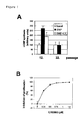
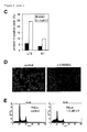
Fig. 1 shows the modulation of the cGMP-pathway in HDF effected by LY.
A: Response of early passage and senescent HDF to activators of cGMP-synthesis.
Cells were treated with 100 µM SNP (sodium nitroprusside) for
2 hours in the absence or presence of LY. cGMP levels were determined as
described in the Examples. Treatments were performed in triplicates and
measured twice. B: Inhibition of proliferation by LY. Subconfluent HDF
were treated with the indicated concentrations of LY for 3 days. Cell
numbers were determined in triplicates. C: Comparison of HDF after LY
treatment and HDF after reaching replicative senescence. Morphology (left
panel, 100x magnification) and detection of senescence associated β-galactosidase
at pH 6 (right panel, magnification 200x) using phase
contrast microscopy. D: Irreversible effect of LY on proliferation of HDF.
Cells were treated with 1 µM LY for the indicated periods. After
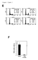
trypsinization cell numbers were determined in triplicates. E: FACS analysis
of HDF treated with LY. HDF were synchronized by confluence and
released by trypsinization. After 24 and 36 hours incubation in media
containing 1 µM LY or vehicle cells HDF were collected for FACS analysis.
F: Rate of DNA-synthesis after LY treatment. BrdU-incorporation was
determined after incubation of HDF in 10 µM BrdU for 6 hours.
-
Fig.2 shows the effects of LY on the expression and phosphorylation
status of cell cycle regulatory proteins. A: Northern blot analysis of LY-treated
HDF. Total RNA was isolated from HDF treated with 2 µM LY or
0.2 µg/ml adriamycin for the indicated periods. 7.5 µg of total RNA was
loaded per lane. p21WAF1 / CIP1 / SDI and, as a control, ELF1α mRNA was
detected with 32P-labeled probes. B: Western blot analysis of HDF treated
with LY. Lysates were obtained from HDF treated with LY for the indicated
periods. 20 µg protein was loaded per lane. After separation on 4-20 %
gradient gels and transfer to PVDF membranes, proteins were detected
with antibodies against human p21, p53, p16, and as a loading control α-tubulin.
C: pRb phosphorylation status after treatment with LY. After
addition of LY for the indicated periods cell lysates were prepared and
proteins were separated on a 7-12 % gradient denaturing PAGE-gel. Abbreviations:
ppRb, hyper-phosphorylated pRb; pRb, hypo-phosphorylated pRb.
-
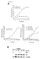
Fig.3 shows the effects of LY on epithelial cancer cells. A: Growth-curves
of MCF-7, A-375, HCT116, and DLD1 cells. Cells were trypsinized at the
indicated time points after addition of LY to the media and cell numbers
were determined. B: Induction of p21 protein after treatment with LY.
HCT116 colorectal cancer cells were treated with 1.5 µM LY for the
indicated periods. Cell lysates were prepared and subjected to Western blot
analysis with the indicated antibodies.
-
Fig.4 shows the requirement of p21, but not p53, for LY-induced cell cycle
arrest. A: HCT116 colorectal cancer cell line deficient for p21. Cells were
treated with LY or, as a control, with vehicle (DMSO) and cell numbers
were assessed. Measurements were performed in triplicates. B: p53+/+
and p53-/- HCT116. Cells were treated with LY or, as a control, with
vehicle (DMSO) and cell numbers were assessed. Measurements were
performed in triplicates. C: Confirmatory analysis of HCT116-derived
knock-out cell lines. Western blot analysis of p53 and p21 protein levels
after addition of adriamycin at 0.2 µg/ml for 8 hours. D: p53-independent
induction of p21 mRNA by LY. Northern blot analysis with RNA isolated at
the indicated time-points after addition of 1.5 µM LY to p53-deficient
HCT116 cells. E: p53-independent induction of p21 protein levels by LY.
Western blot analysis with lysates from p53-/- HCT116 cells treated with
1.5 µM LY. Membranes were probed with antibodies specific for p21 and
as a loading control α-tubulin.
-
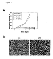
Fig.5 shows the effect of LY on cells with inactivated pRb pathways. A:
Response of cell lines with lesions in the pRb-pathway to LY. NIH3T3-L1
cells expressing SV40 large T antigen (LT3) and NIH3T3-L1 cells
expressing a point mutant of SV40 large T antigen (K1 ) unable to bind to
pRb-like proteins were treated with 1.5 µM LY. Cell numbers were
determined at the indicated time points in triplicates. B: Morphology of
NIH3T3-L1 cells after LY treatment. Phase contrast microscopy was
performed after incubation of NIH3T3-L1-K1 (K1) and NIH3T3-L1-LT3
(LT3) cells in 1.5 µM LY for 3 days (magnification: 100x). C: Quantification
of early stages of apoptosis. Control and LY-treated for 48 hours K1 and
LT3 cells were subjected to double staining with anti-annexin V and
propidium iodide (PI) to discriminate living, early apoptotic, and late
apoptotic/necrotic populations and analyzed with flow cytometry. The
percentage of PI-negative annexin V-positive cells is shown. D: Induction
of apoptosis by LY. HeLa cells expressing a histone H2B-eGFP fusion
protein (kindly provided by G. Wahl) were treated with 2 µM LY for 48
hours and the chromatin morphology in living cells was analyzed at a
wave-length of 495 nm. E: Quantification of apoptosis by detection of cells
with sub-G1 DNA content. HeLa cells were treated for 60 hours with LY.
DNA-content was determined after propidium iodide staining as described
in the Examples.
Examples
-
This study characterizes the effects of LY on non-transformed primary cells
and tumor cells with different lesions in key tumor suppressor pathways.
LY represents a new type of non-genotoxic drug which inhibits tumor cell
proliferation by induction of p21 and subsequent context dependent
activation of cell cycle arrest or apoptosis. The p53 independence of the
antiproliferative effect of LY and its ability to induce apoptosis in cells with
non-functional RB protein make this substance an interesting prototype for
the development of new non-genotoxic cancer therapeutics. Comparative
analysis of gene expression profiles of replicative and LY-induced
premature senescence suggests the existence of a transcriptional program
specific for senescence which is activated irrespective of the inducing
signal and presumably represents a safeguard mechanism to prevent
proliferation of potentially cancerous cells.
I. Experimental procedures
I.1. Cell culture and drug treatments
-
HDF were obtained from Clonetics and passaged in Dulbecco's modified
Eagles medium (DMEM, Gibco) supplemented with 10 % fetal bovine
serum (FBS, Sigma). HCT116 were cultured in McCoy's (Gibco)
supplemented with 10 % FBS (Sigma). HeLa, HEK293, P16-/-MEFs and
NIH3T3-L1 derivatives were kept in DMEM containing 10 % FBS. LY
(Calbiochem) was dissolved in DMSO (Sigma) at a concentration of 300
µM (~ 300x solution). As a control, cells were treated with equal volumes
of DMSO (< 1 %). The LY concentration was restored in intervals of 24
hours by media exchange.
I.2. Microarray analysis of gene expression
-
RNA was isolated using RNAgents reagents (Promega). After mRNA
isolation, integrity and enrichment was ensured using Northern blot
analysis. 600 ng poly-A mRNA was converted to cDNA with incorporation
of Cy3- or Cy5-labeled dNTPs. Hybridization to arrays coated on glass,
quality control and normalization was performed by IncyteGenomics (Palo
Alto, CA). The 'Human Unigene 1 '-array, used for all microarray analyses of
HDF, contained probes for 8,392 annotated genes/EST clusters and 74
non-annotated genes/ESTs (probes are derived from the 5'-cDNA region),
whereas the 'Human Drug Target'-array, used for analysis of MCF7 treated
with LY, represented 8,031 unique annotated genes/EST clusters and 207
non-annotated genes/EST clusters (probes are derived from the 3'-cDNA
region).
I.3. Northern blot analysis
-
RNA was isolated using the RNAgents kit (Promega). A Northern probe
directed against the 3'-untranslated region of ELF1 α mRNA was generated
by PCR using an EST as template and subsequent gel purification. The p21
probe corresponds to the open reading frame of p21. A probe
corresponding to the 5'-region of sGC-β3 was amplified from HDF-cDNA
and subcloned. Hybridizations were performed in QuickHyb, following the
manufacturer's instructions (Stratagene).
I.4. Quantitative real-time PCR
-
qPCR was performed using the LightCycler and the FastStart DNA Master
SYBR Green 1 kit (Roche Applied Science) as previously described
(Menssen and Hermeking, 2002). For qPCR of cDNA, primer pairs were
designed to generate intron-spanning products of 100-200 bp. Primer
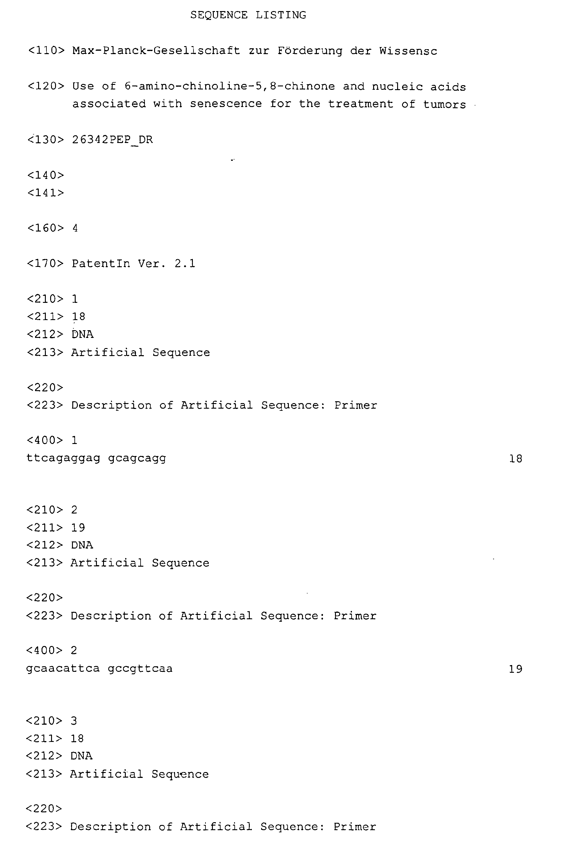
sequences were: GUCY1A3-sense 5'-TTCAGAGGAGGCAGCAGG-3' (SEQ
ID NO. 1), GUCY1 A3-antisense 5'-GCAACATTCAGCCGTTCAA-3' (SEQ ID
NO.2; annealing temperature 62°C, efficiency 1.97); GUCY1 B3-sense 5'-AGGAATCACGCATCAGCC-3'
(SEQ ID NO.3), GUCY1 B3-antisense 5'-TATGAGGACGAACCAGCGA-3'
(SEQ ID NO.4; annealing temperature
62°C, efficiency 1.94 ).
-
cDNA was generated using the RevertAid First Strand cDNA synthesis kit
(MBI Fermentas). The generation of specific PCR products was confirmed
by melting curve analysis and gel-electrophoresis. Each primer pair was
tested with a logarithmic dilution of a cDNA mix to generate a linear
standard curve (crossing point (CP) plotted vs. log of template
concentration), which was used to calculate the primer pair efficiency (E =
10(-1/slope)). Elongation factor 1α (ELF1α) mRNA was used as an external
standard since its expression was not altered significantly in senescent
HDF (data not shown). For data analysis, the second derivative maximum
method was applied and induction of a cDNA species (geneX) was
calculated according to Pfaffl (2001):
(EgeneX ^ΔCP(confl. cDNA - sen.cDNA) geneX) / (EELF1α ^ ΔCP (confl.cDNA - sen.cDNA) ELF1α) = fold
induction.
I.5. Measurement of DNA content and apoptosis by flow cytometry
-
Cell samples (both adherent and floating cells) were harvested by
trypsinization, washed once with PBS and fixed in 70 % ethanol for > 2
hours on ice, washed once with PBS and incubated for 30 minutes at room
temperature in staining solution containing 50 µg/ml of propidium iodide,
0.2 mg/ml of RNase A and 0.1 % (v/v) Triton X-100 in PBS. Quantification
of apoptotic cells was performed using the Annexin V-FITC apoptosis
detection kit (Becton Dickinson). Samples were analyzed with a FACScan
unit (Becton Dickinson). 1×10⁴ cells were analyzed for each assay.
Quantitation was performed using single histogram statistics.
I.6. Proliferation assay
-
Cells were seeded in equal numbers in 6-well plates the day before addition
of LY in order to ensure uniform cell layers at the start of the experiments.
Cells were treated in triplicates with daily exchange of medium containing
drug or drug-free vehicle. For each time point cells were trypsinized and
cell proliferation was assessed using a Z1 Counter (Coulter).
I. 7. cGMP assay
-
Cells of early and late passages were seeded in equal numbers in 6-well
plates. 24 hours later SNP (Sigma) was dissolved in complete medium and
added to cells to a final concentration of 100 µM. Control cells received
the same volume of drug-free medium. After 2 hours of incubation,
medium was removed and cells were lysed by addition of 400 µl 0.1 M HCl
per well for 10 minutes. cGMP concentration in lysates was determined
using a specific competitive immunoassay according to the manufacturer's
instructions (Sigma). A plate reader (LAMBDA E, MWG-Biotech) was used
to detect the signal at 405 nm. Experiments were performed in triplicates
with all samples measured twice.
I.8. SA-β-gal staining
-
Cells were fixed by incubation in 0.5 % glutaraldehyde in PBS for 5
minutes at room temperature and stained for SA-ß-galactosidase at pH 6.0
as described by Dimri et al., 1995.
I.9. Detection of DNA-synthesis
-
Cells were plated on cell locate glass grids (Eppendorf) and labeled for 6
hours using the 5'-bromo-2'-deoxy-uridine labelling and detection kit I
(Roche Applied Sciences). After staining with a FITC-labeled anti-BrdU-antibody,
positive cells were detected by fluorescence microscopy.
I.10. Western blot analysis
-
Cells were lysed in a lysis buffer composed of 50 mM HEPES pH 7.5, 150
mM NaCl, 1 mM EGTA, 10 % glycerol, 1 % Triton X-100, 100 mM NaF,
10 mM Na4P2O7, 1 µM PMSF, and 1 µM Na3VO4. Protein concentration in
whole lysates was determined using a Bradford assay (BioRad). 20 µg of
lysates diluted with Laemmli-buffer were separated on 12 % SDS-polyacrylamide
gels and transferred to Immobilon-P membranes (Millipore).
For detection of pRb, separation was performed on a 7-12 % gradient gel.
Antibodies against p53 (Pab 1801), p21 (c-19), p16 (c-20) and α-Tubulin
(TU-02) were purchased from Santa Cruz Biotechnology. Anti-pRB
antibodies (G3-245) were from BD PharMingen. Enhanced
chemiluminescence generated by horse radish peroxidase-coupled
secondary antibodies was detected with a CF440 imager (Kodak/Perkin
Elmer).
I.11.Microscopy
-
For phase contrast and β-gal staining an Axiovert 25 microscope (Zeiss)
equipped with a HyperHAD CCD camera (Sony) and ImageBase software
(Kappa) was used. In vivo expression of histone-H2B-eGFP was
documented with a Photometrics CooISNAP-HQ CCD camera (Roper
Scientific) on an inverted Axiovert 200M microscope (Zeiss) using the
Metamorph software (Universal Imaging).
II. Results
II. 1. Microarray analysis of senescence-specific gene expression
-
We employed microarray analysis in order to identify genes and pathways
involved in the induction and maintenance of replicative senescence.
Primary human diploid fibroblasts (HDF) were cultivated for ~78
population doublings until proliferation of all cells ceased due to replicative
senescence. At this point, HDF showed characteristic signs of senescence:
cellular enlargement, SA-β-galactosidase staining at pH 6 (Figure 1 C) and
terminal arrest in the presence of high serum concentration (mainly in the
G1-phase as determined by FACS analysis, data not shown). mRNA was
isolated from senescent HDF and subjected to analysis with a microarray,
which allows detection of 8,392 annotated cDNAs. As a reference, a
microarray analysis was performed with RNA derived from presenescent
HDF, which had been arrested in the G1-phase by confluence at population
doubling 12. This state was selected to avoid a comparison of non-proliferating
senescent HDF with proliferating early passage HDF, which
would presumably result in the detection of a large number of differences
in gene expression secondary to growth-arrest. Several genes previously
identified as consistently up- or down-regulated during senescence of HDF
were detected as regulated in a similar fashion in the system employed
here (Table 1 A): e.g. PAI-1 was previously detected as up-regulated in
senescent HDF (West et al., 1996; Ly et al., 2000; Shelton et al., 1999)
and was induced 5.9 fold in senescent HDF in this study (Table 1 A).
Among the genes repressed during senescence were four genes encoding
enzymatic components of the cGMP-pathway (Table 1 B): soluble guanylate
cyclase β3 (sGC-β3), sGC-α3 and cGMP-dependent protein kinase I and II.
Differential regulation of sGC-α3 and sGC-β3 during senescence was
confirmed by quantitative real-time PCR (Table 1). Down-regulation of sGC-α3
during senescence was also detected using Northern blot analysis (data
not shown). sGC-α3 and sGC-β3 represent the large and small subunit of
soluble guanylate cyclase, which converts GTP to 3 ',5 '-cyclic GMP and
pyrophosphate (Giuili et al., 1992). Interestingly, both genes are localized
in near proximity on chromosome 4q31.3-q33 (Giuili et al., 1993), which
could provide the basis for the co-regulation observed here.
II.2. Induction of senescence by 6-anilino-5,8-quinolinequinone
-
The co-ordinated repression of two genes encoding the subunits of soluble
guanylate cyclase, which generates cGMP in response to signals as e.g.
extra-cellular NO (reviewed in Hofmann et al., 2000), suggested, that
senescent cells may have an attenuated response to NO-donors as sodium
nitroprusside (SNP). Indeed, exposure to SNP did not result in elevated
cGMP levels in senescent HDF, whereas early passage HDF showed a
three-fold increase in cGMP levels after addition of SNP (Fig. 1 A). In order
to specifically inhibit the cGMP-signalling-pathway, we employed a
competitive inhibitor of soluble guanylate cyclase: 6-anilino-quinoline-5,8-quinone
(LY 83583, referred to as LY hereafter), with an IC50 of 2 µM
(Fleisch et al., 1984). Addition of 1 µM LY HDF significantly inhibited the
SNP-induced synthesis of cGMP by 60 % (Fig.1A), showing that LY had
the expected inhibitory effect on guanylate cyclase.
-
We next asked, whether addition of LY would affect the proliferation of
early passage HDF, as we would expect according to our working
hypothesis described above. The effect of LY was tested at a
concentration ranging from 0.25 to 1.5 µM (Figure 1B). Indeed, treatment
of early passage fibroblasts with a concentration of 1 µM of LY was
sufficient to completely inhibit proliferation, and the presence of 250 nM
LY led to a 50 % reduction in proliferation of HDF after 3 days (Figure 1 B).
Strikingly, the inhibition of proliferation of early passage HDF by LY was
accompanied by morphological changes characteristic of senescence and
by SA-β-galactosidase staining at pH 6 (Figure 1 C). Consistent with the
induction of cellular senescence, inhibition of proliferation by LY was
irreversible after a treatment period of more than 2 days (Figure 1 D).
Extended treatment of HDF with LY for 8 days did not cause any obvious
toxicity but resulted in stable inhibition of proliferation (Figure 1 D). In order
to characterize the cell cycle arrest induced by LY, HDF were synchronized
in the G1-phase by confluence (91.2 % G1- and 2 % S-phase) and then
released by trypsinization (Figure 1 E): untreated cells entered S-phase at a
high percentage. Addition of LY almost completely blocked the entry into
S-phase of HDF, suggesting that LY acts during the G1-phase of the cell
cycle. Furthermore, addition of 1 µM LY to synchronized HDF caused
complete inhibition of DNA synthesis as determined by BrdU incorporation
(Figure 1 F).
II.3. Microarray analysis of LY-regulated genes
-
In order to identify the down-stream mediators of the LY-induced cell cycle
arrest and senescence, a microarray analysis of LY-treated HDF was
performed with the same arrays used for the analysis of replicative
senescence in HDF described above. RNA was isolated 36 hours after
exposure of early passage HDF to LY. The control RNA was isolated from
confluent early passage HDF. In HDF, which had reached replicative
senescence, 216 transcripts were induced significantly, whereas 272
mRNAs were induced after LY treatment. Repression was observed for 266
mRNAs in HDF undergoing replicative senescence, whereas 294 mRNAs
were repressed after LY treatment. There was a substantial overlap in the
differentially expressed genes observed during replicative senescence and
during LY-induced senescence, with 114 transcripts differentially regulated
in a similar manner (Table 2). Among these were genes, which had been
previously identified as differentially regulated during replicative
senescence: PAI-1, matrix metalloproteinase 10, fibrillin, cdc25b, cyclin
D1, fibromodulin and osf-2 (Shelton et al., 1999; Ly et al., 2000).
However, most of the genes identified here represent new additions to the
growing number of genes identified as components of the senescence
program. Supporting this notion, many of the co-regulated genes have
functions, which may contribute to the phenotype of terminally arrested
cells. E.g. down-regulation of the PDGF-receptor α and β chains may
contribute to the refractory state of senescent cells, which do not respond
to mitogenic stimulation with growth factors (Table 2). The changes in
expression observed in genes encoding components of the cytoskeleton
(e.g. α1-tubulin, β2-tropomyosin, y-filamin C, α-actinin, α-tubulin) may
contribute to the flattened and enlarged shape characteristic of senescent
HDF (Figure 1C).
II.4. Rapid induction of p21WAF1 / CIP1 / SDI by LY
-
Among the genes induced by both, replicative senescence as well as LY,
was p21WAF1 / CIP1 / SDI (Table 2), which encodes an inhibitor of cyclin
dependent kinases (cdk). Cdk/cyclin complexes drive cell cycle progression
and proliferation by phosphorylation of key substrates (reviewed in Nurse,
2000). Since induction of p21 could potentially explain the anti-proliferative
effect of LY, we analyzed the connection between LY and p21
in more detail. 6 hours after treatment with LY, the levels of p21 mRNA in
HDF were similar to the levels observed 6 hours after addition of
adriamycin as shown by Northern blot analysis (Figure 2A). However, after
treatment with LY, the level of p53 protein, which consistently increases
after generation of DNA damage and mediates transactivation of p21, did
not increase (Figure 2B), indicating that LY does not induce DNA-damage.
The induction of p21 mRNA by adriamycin demonstrates, that the
signalling pathways necessary for p53 activation were still intact in the
HDF employed (Figure 2A). Induction of p21 protein was detectable
between 3 and 6 hours after addition of LY, whereas the cdk-inhibitor
p16INK4A was not induced even after 48 hours of treatment with LY (Figure
2B). This result was unexpected, since p 16INK4A is commonly induced in
scenarios, which lead to premature senescence, as e.g. ectopic expression
of an activated ras gene (Serrano et al., 1997) or sub-optimal cell culture
conditions (Ramirez et al., 2001).
-
The major down-stream targets of p21 are the pocket proteins: the
retinoblastoma tumor suppressor protein pRb and its homologs p 107 and
p 130. p21-mediated inhibition of G1/S-phase associated cdk/cyclin-complexes,
as cdk2/cyclin A, leads to hypo-phosphorylation of pocket
proteins, which allows their subsequent binding and inhibition of E2F
transcription factors. The association between pRb and E2Fs results in
inhibition of G1/S progression and proliferation (reviewed in Weinberg,
1995). Consistent with the induction of p21 by LY, the phosphorylation
status of pRb changed rapidly after addition of LY: after only 6 hours the
ratio of phosphorylated to hypo-phosphorylated, active pRb was clearly
shifted towards the hypo-phosphorylated form and by 19 hours pRb was
almost completely hypo-phosphorylated (Figure 2C). Even after extension
of the LY treatment to 12 days, the pRb hypo-phosphorylation was not
reversed (Figure 2C). These results show, that LY inhibits proliferation by
inducing p21 to levels, which are sufficient to inhibit cdk activity and
activate the cell cycle inhibitory function of pRb.
II.5. Effect of LY on cancer cell proliferation
-
In order to determine, whether LY may be useful to terminate cancer cell
proliferation, the proliferation of colorectal cancer cell lines (HCT116,
DLD1) and a breast cancer derived cell line (MCF7) were analyzed after
addition of LY: all cell lines showed complete cessation of cell proliferation
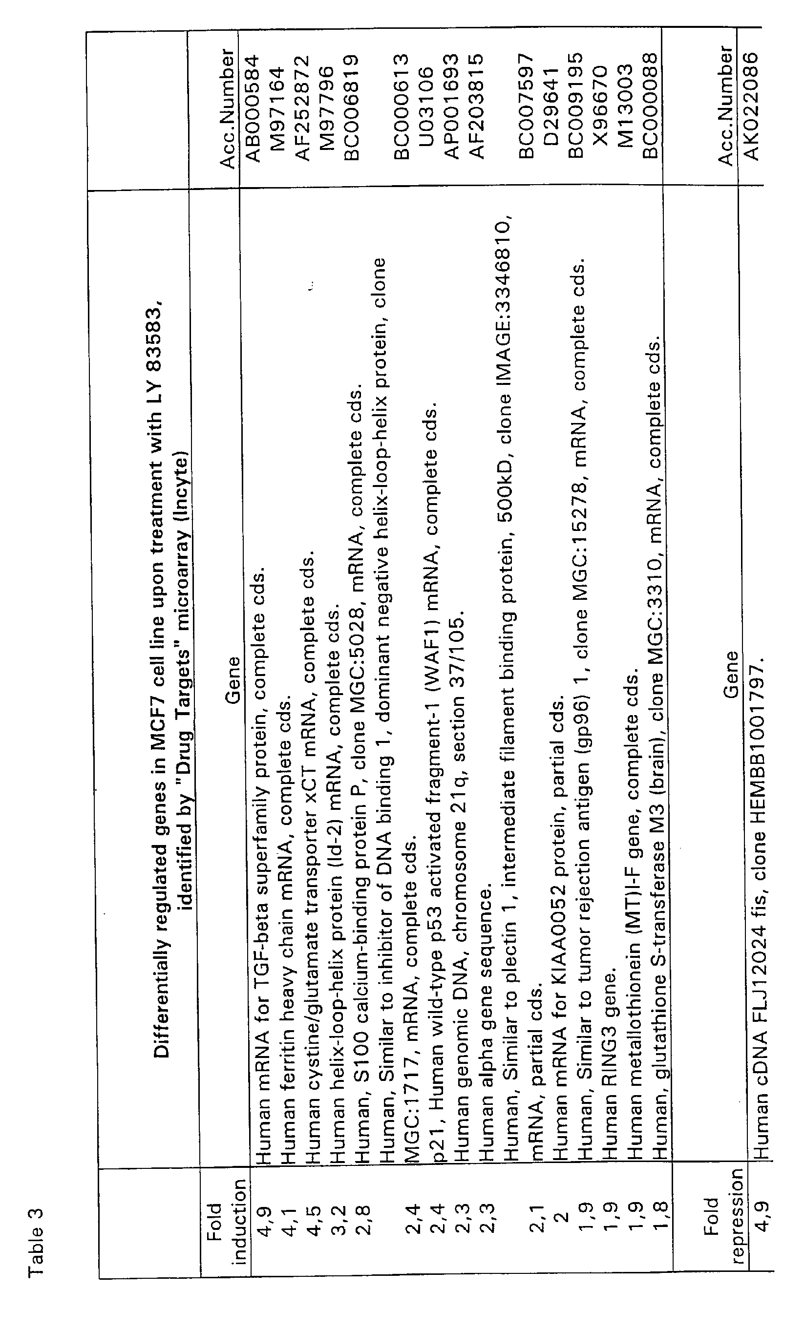
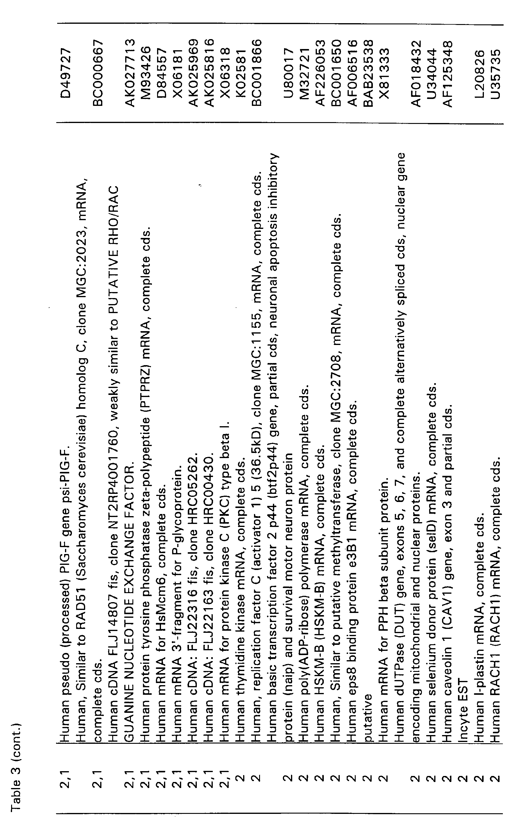
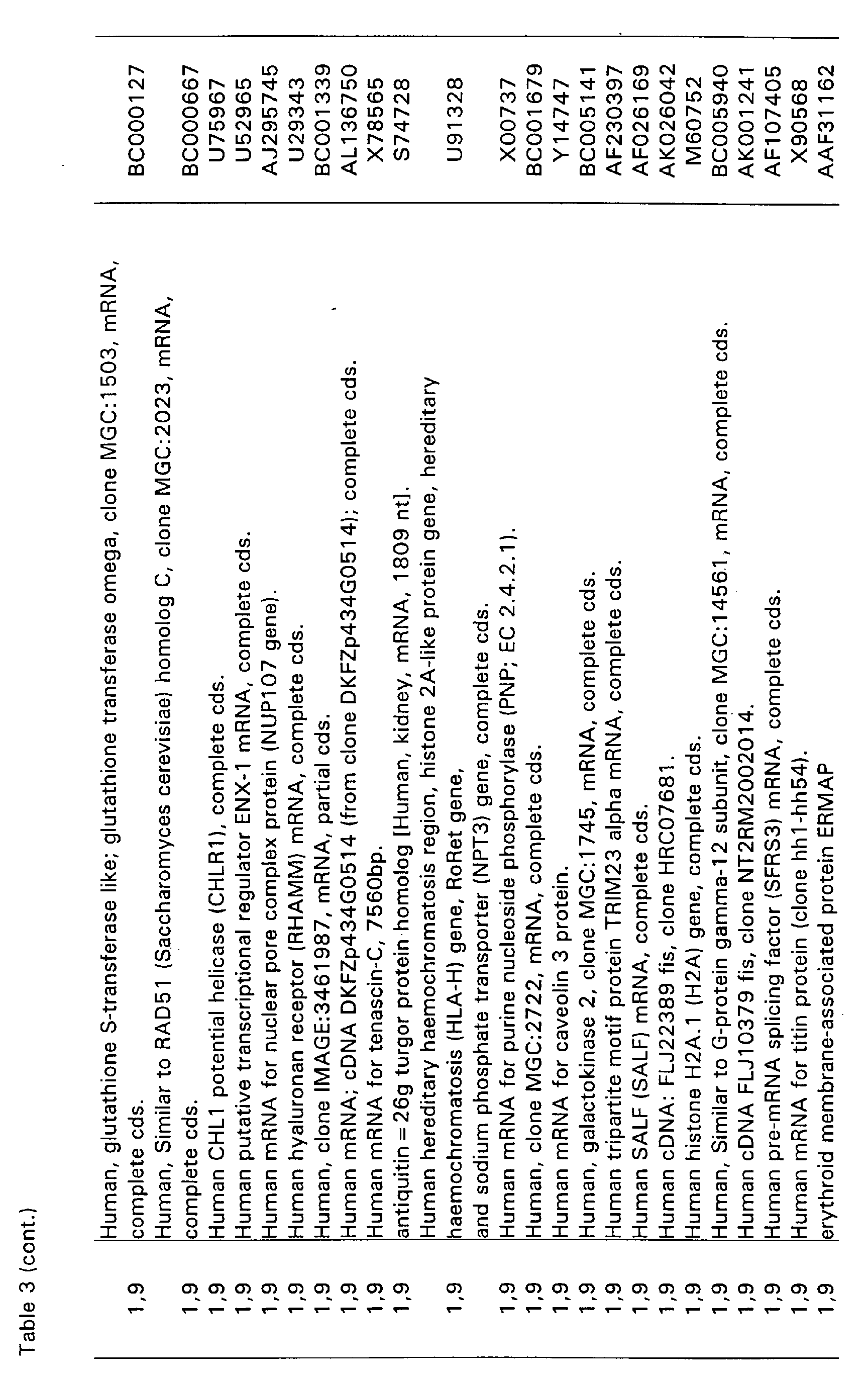
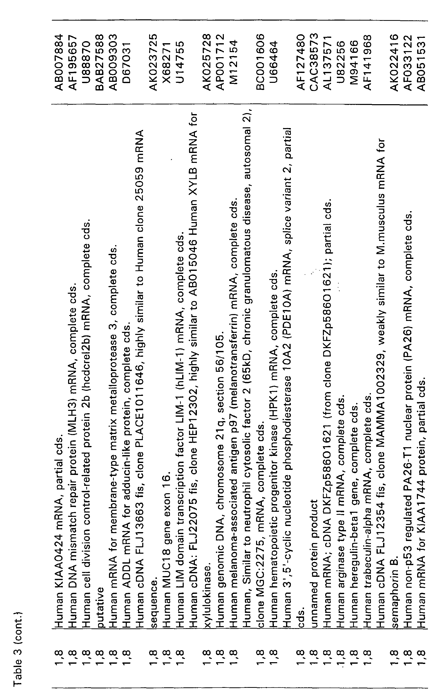
(Figure 3A). Using microarray analysis 360 genes were identified as
differentially regulated by LY in MCF7 cells (Table 3). However, the
amount of overlap with genes differentially expressed in LY-treated HDF
could not be determined, since the array employed for the analysis of gene
expression in MCF7 differed substantially from the arrays used for HDF
before. However, p21 was among the genes induced by LY in MCF7 cells,
suggesting that p21 may be involved in the LY-induced arrest in MCF7 and
other epithelial cancer cells. Consistent with this assumption, western blot
analysis of HCT116 cells showed induction of p21 after addition of LY
(Figure 3B).
II.6. Requirement of p21, but not p53, for LY-induced cell cycle arrest
-
In order to determine, whether induction of p21 is essential for LY-induced
cell-cycle arrest, p21-deficient HCT116 colorectal cancer cells, generated
via homologous recombination by Waldman et al. (1996), were analyzed
(Figure 4A): p21-deficient HCT1 16 were resistant to cell-cycle inhibition by
LY. Compared to cells treated with vehicle alone, LY-treatment resulted in
an initial delay of proliferation in p21-/- HCT116, suggesting other
immediate, p21-independent effects of LY on HCT116 cells. However,
after 5 days of LY treatment, the p21-deficient HCT116 cells had regained
exponential proliferation, whereas wild-type HCT116 cells showed
complete cessation of proliferation. These results show that p21 is required
for the effects of LY on the cell cycle.
-
Consistent with the absence of p53 accumulation in LY-treated HDF, p53-deficient
HCT116 cells engineered by Bunz et al. (1999) showed a
complete block of proliferation after LY addition (Figure 4B). The identity of
the 3 different HCT116 cell lines employed in this study was confirmed by
treatment with adriamycin, which induces DNA double-strand breaks
(Figure 4C). As expected, only wildtype HCT116 cells showed induction of
p21 protein after exposure to adriamycin (Figure 4C). The levels of p21
mRNA and protein of p53-/- HCT116 after treatment cells with LY were
induced with kinetics similar to those observed in HDF (Figure 4D and E).
These results exclude p53 and thereby induction of DNA damage as
mediators of cell cycle arrest by LY and induction of p21 expression.
Furthermore, LY-treatment did not lead to induction of the cdk-inhibitors
p27 and p15 (data not shown). In addition, proliferation of p16INK4A -deficient
mouse embryo fibroblasts (MEF) was inhibited by LY, showing
that p 16INK4A is not required for the effects of LY on the cell cycle (data not
shown). Taken together, these results show, that the inhibitory effects of
LY on proliferation are mediated through induction of p21.
II.7. Conversion of LY-induced arrest to apoptosis in pRb-negative cells
-
Since the anti-proliferative effect of p21 is mediated through the pRb-pathway
(reviewed in Weinberg, 1995), we analyzed the response of cells
with non-functional pRb to treatment with LY. Isogenic NIH3T3-L1 cell
lines stably expressing either SV40 large T antigen or a SV40-large T
antigen point mutant (K1) deficient in binding and inactivating pRb-like
pocket-proteins (described in Hermeking et al., 1994) were treated with
LY: most NIH3T3-L1 cells expressing the K1-mutant showed inhibition of
proliferation, whereas NIH3T3-L1 cells expressing wildtype SV40 large T
antigen underwent apoptosis (Figure 5A and B). Induction of apoptosis by
LY was further quantified using annexin V staining (Figure 5C).
-
In order to extend these observations to human cancer cells, the cell lines
HeLa and HEK293 were analyzed. The cervical cancer cell line HeLa
expresses the human papilloma virus (HPV) 18 encoded E7 protein, which
binds to and inactivates pocket proteins (pRb, p107, p130). In HEK293
cells, the adenoviral E1A protein performs analogous functions. HeLa and
HEK293 cells did not arrest after treatment with LY, but instead,
underwent cell death, which was accompanied by cell shrinkage
suggesting apoptosis (data not shown). HeLa cells expressing a histone
H2B-eGFP-fusion protein allowed to confirm the induction of apoptosis by
LY (Figure 5D): treatment with LY led to chromatin condensation, which is
characteristic of apoptosis. Furthermore, analysis of a larger cell population
by flow-cytometry revealed a substantial increase of cells with a sub-G1
DNA content after addition of LY (Figure 5E).
III. Discussion
-
The results described here demonstrate, that LY induces cellular
senescence in HDF and cell cycle arrest in several human and murine cell
types. One requirement for these effects of LY is the transcriptional
induction of the cdk-inhibitor p21, since p21-deficient cells are resistant to
LY-induced arrest. Furthermore, in the absence of functional pocket
proteins (pRb, p107, p130), which are downstream effectors of p21
function, cell cycle arrest is converted to induction of apoptosis in a p53-independent
manner. We assume, that in this case apoptosis is a cellular
response to conflicting signals: negative signals due to LY-induced p21 and
resulting cdk-inhibition versus cell cycle driving signals due to the lack of
active pocket proteins, which allow high levels of active E2F transcription
factors.
-
The induction of p21 is not mediated through the DNA-damage/p53-pathway,
since p53-deficient cells also show up-regulation of p21 by LY
and subsequent cell cycle arrest. These features of LY indicate that it may
be a useful agent for treatment of cancer. Many tumor cells have lost pRb
function, either through mutation in pRb (Horowitz et al., 1990) or through
expression of viral proteins. E.g. in cervical cancer expression of E7 by
HPV16/18 leads to inactivation of pRb (reviewed in zur Hausen, 2001). In
HPV16/18-infected cells treatment with LY would presumably induce
apoptosis. Since LY induces p21, cell cycle arrest, and apoptosis in a p53-independent
manner, it may proof useful for the treatment of cancers
harboring p53 mutations, which represent more than half of all human
cancers (reviewed in Hollstein et al., 1999).
-
In order to induce senescence in early passage cells, we decided to inhibit
the cGMP-pathway, since components of this pathway were
transcriptionally suppressed during senescence. For this purpose LY was
selected, since it specifically inhibits soluble guanylate cyclase (Mulsch et
al., 1988). LY was first described as an inhibitor of leukotriene release
(Fleisch et al., 1984). Later it was found to lower cGMP levels (Schmidt et
al., 1985) by inhibiting soluble guanylate cyclase (Mulsch et al., 1988). In
the experiments presented here LY was able to mimic cellular senescence
in primary human fibroblasts and induced a cell cycle arrest in several
cancer cell lines. However, it is unclear, whether inhibition of cGMP
generation is the only pathway by which LY achieves its effects. It has
been proposed that the quinone structure of LY may lead to the generation
of superoxide anions (Cherry et al., 1990). However, a recent report,
employing the colorectal cancer cell line HCT116 and isogenic p21- or p53-deficient
cells also analyzed in this study, showed, that reactive oxygen
species (ROS) generated by hyperoxic conditions induce expression of p21
in a p53-dependent manner (Helt et al., 2001). Furthermore, proliferation
of HCT116 cells is inhibited by hyperoxic conditions in a p21- and p53-independent
manner (Helt et al., 2001). In contrast to induction of p21 by
ROS, induction of p21 by LY was not mediated by p53. Furthermore,
inhibition of proliferation by LY was dependent on p21. These results
suggest, that LY does not act by generating ROS, which in turn mediate
cell cycle arrest or senescence.
-
Furthermore, it is important to analyze whether LY or derived substances
can be used to prevent tumor growth in vivo using animal models. The
results provided thereby may serve as a basis for further characterization
and optimization of LY-derived drugs as tumor therapeutics. An obvious
advantage of LY is its ability to interfere with cellular proliferation without
inducing DNA-damage. Commonly used chemotherapeutics, like
adriamycin, generate DNA damage and thereby selectively induce
apoptosis in cancer cells, which lack cell cycle check points (reviewed in
Hartwell et al., 1997). However, DNA damaging substances cause major
side effects on proliferating tissues (Batchelor, 2001) and may induce
further mutations in pre-cancerous and healthy cells, which lead to
secondary cancer (Felix et al., 1998).
-
The regulation of more than one hundred senescence-specific genes by LY
shows, that treatment with LY activates a transcriptional program, which
is analogous to the program induced after HDF have reached replicative
senescence. Therefore, the genes shown in Table 2 represent a
transcriptional program characteristic for senescence of HDF.
-
The strategy underlying the present invention, i.e. identification of
pathways, which are repressed during senescence and their inhibition in
tumor cells by synthetic drugs may also proof useful for the identification
of other potential targets for effective inhibitors of tumor cell proliferation.
Literature
-
- Bast, R.C., Kufe, D.W., Pollock, R.E., Weichselbaum, R.R.; Holland, J.F.
and Frei, E., eds.(2000). Cancer Medicine (BC Decker Inc.).
- Batchelor, D. (2001). Hair and cancer chemotherapy: consequences and
nursing care-a literature study. Eur. J. Cancer Care 10,147-163.
- Brown, J.P., Wei, W. and Sedivy, J.M. (1997). Bypass of senescence after
disruption of p21CIP1/WAF1 gene in normal diploid human fibroblasts.
Science 277, 831-834.
- Bunz, F., Dutriaux, A., Lengauer, C., Waldman, T., Zhou, S., Brown, J.P.,
Sedivy, J.M., Kinzler, K.W. and Vogelstein, B. (1998). Requirement for
p53 and p21 to sustain G2 arrest after DNA damage. Science 282, 1497-1501.
- Campisi, J. (2001). Cellular senescence as a tumor-suppressor mechanism.
Trends Cell Biol. 11, 27-31.
- Chang, B.D., Xuan, Y., Broude, E.V., Zhu, H., Schott, B., Fang, J. and
Roninson, I.B. (1999). Role of p53 and p21 waf1/cip1 in senescence-like
terminal proliferation arrest induced in human tumor cells by
chemotherapeutic drugs. Oncogene 26, 4808-4818.
- Cherry, P.D., Omar, H.A., Farrel, K.A., Stuart, J.S. and Wolin, M.S.
(1990). Superoxide anion inhibits cGMP-associated bovine pulmonary
arterial relaxation. Am. J. Physiology 259, 1056-1062
- Cristofalo, V.J., Volker, C., Francis, M.K. and Tresini, M. (1998). Age-dependent
modifications of gene expression in human fibroblasts. Crit.
Rev. Eukaryot. Gene Expr. 8, 43-80.
- Dellambra, E., Golisano, O., Bondanza, S., Siviero, E., Lacal, P., Molinari,
M., D'Atri, S. and De Luca, M. (2000). Downregulation of 14-3-3sigma
prevents clonal evolution and leads to immortalization of primary human
keratinocytes. J Cell Biol. 149, 1117-1130.
- Di Leonardo, A., Linke, S.P., Clarkin, K. and Wahl, G.M. (1994). DNA
damage triggers a prolonged p53-dependent G1 arrest and long-term
induction of Cip1 in normal human fibroblasts. Genes Dev. 8, 2540-2551.
- Dimri, G.P., Lee, X., Basile, G., Acosta, M., Scott, G., Roskelley, C.,
Medrano, E.E., Linskens, M., Rubelj, I., Pereira-Smith, O., et al. (1995). A
biomarker that identifies senescent human cells in culture and in aging skin
in vivo. Proc. Natl. Acad. Sci. USA. 92, 9363-9637.
- Green, D.R. and Evan, G.I. (2002). A matter of life and death. Cancer Cell
1, 19-30.
- Giuili, G., Roechel, N., Scholl, U., Mattei, M.-G. and Guellaen, G. (1993).
Colocalization of the genes coding for the alpha-3 and beta-3 subunits of
soluble guanylyl cyclase to human chromosome 4 at q31.3-q33. Hum.
Genet. 91, 257-260.
- Giuili, G., Scholl, U., Bulle, F. and Guellaen, G. (1992). Molecular cloning
of the cDNAs coding for the two subunits of soluble guanylyl cyclase from
human brain. FEBS Lett. 304, 83-88.
- Felix, C.A. (1998). Secondary leukemias induced by topoisomerase-targeted
drugs. Biochim. Biophys. Acta. 1400, 233-255.
- Fleisch, J.H., Haisch, K.D., Spaethe, S.M., Rinkema, L.E., Cullinan, G.J.,
Schmidt, M.J. and Marshall, W.S. (1984). Pharmacologic analysis of two
novel inhibitors of leukotriene (slow reacting substance) release. J
Pharmacol Exp Ther. 229, 681-689.
- Hartwell, L.H., Szankasi, P., Roberts, C.J., Murray, A.W. and Friend, S.H.
(1997). Integrating genetic approaches into the discovery of anticancer
drugs. Science 278, 1064-1068.
- Hayflick, L. and Moorehead, P.S. (1961). Exp. Cell Res. 25, 585-621.
- Helt, C.E., Rancourt, R.C., Staversky, R.J. and O'Reilly, M.A. (2001). p53-dependent
induction of p21(Cip1/WAF1/Sdi1) protects against oxygen-induced
toxicity. Toxicol Sci. 63, 214-222.
- Hermeking, H., Wolf, D.A., Kohlhuber, F., Dickmanns, A., Billaud, M.,
Fanning, E. and Eick, D. (1994). Role of c-myc in simian virus 40 large
tumor antigen-induced DNA synthesis in quiescent 3T3-L1 mouse
fibroblasts. Proc. Natl. Acad. Sci. USA. 91, 10412-10416.
- Hofmann, F., Ammendola, A. and Schlossmann, J. (2000). Rising behind
NO: cGMP-dependent protein kinases. J. Cell Sci. 113, 1671-1676.
- Hollstein, M., Hergenhahn, M., Yang, Q., Bartsch, H., Wang, Z.Q. and
Hainaut, P. (1999). New approaches to understanding p53 gene tumor
mutation spectra. Mutat. Res. 431, 7-12
- Horowitz, J.M., Park, S.H., Bogenmann, E., Cheng, J.C., Yandell, D.W.,
Kaye, F.J., Minna, J.D., Dryja, T.P. and Weinberg, R.A. (1990). Frequent
inactivation of the retinoblastoma anti-oncogene is restricted to a subset of
human tumor cells. Proc. Natl. Acad. Sci. USA. 87, 2775-2779.
- Ly, D.H., Lockhart, D.J., Lerner, R.A. and Schultz, P.G. (2000). Mitotic
misregulation and human aging. Science 287, 2486-2492.
- Menssen, A. and Hermeking, H. (2002). Characterization of the c-MYC-regulated
transcriptome using SAGE: Identification and characterization of
c-MYC-target genes. Proc. Natl. Acad. Sci. USA., in press
- Mulsch, A., Busse, R., Liebau, S. and Forstermann, U. (1988). LY 83583
interferes with the release of endothelium-derived relaxing factor and
inhibits soluble guanylate cyclase. J. Pharmacol. Exp. Ther. 247, 283-288.
- Noda, A., Ning, Y., Venable, S.F., Pereira-Smith, O.M. and Smith, J.R.
(1994). Cloning of senescent cell-derived inhibitors of DNA synthesis using
an expression screen. Exp Cell Res. 211, 90-98.
- Nurse, P. (2000). A long twentieth century of the cell cycle and beyond.
Cell 100, 71-78.
- Pfaffl, M.W. (2001). A new mathematical model for relative quantification
in real-time RT-PCR. Nucleic Acids Res. 29, 2003-2007.
- Ramirez, R.D., Morales, C.P., Herbert, B.S., Rohde, J.M., Passons, C.,
Shay, J.W. and Wright, W.E. (2001). Putative telomere-independent
mechanisms of replicative aging reflect inadequate growth conditions.
Genes Dev. 15, 398-403.
- Shay, J.W. and Wright, W.E. (2000). Hayflick, his limit, and cellular
ageing. Nat. Rev. Mol. Cell Biol. 1, 72-76.
- Serrano, M. and Blasco, M.A. (2001). Putting the stress on senescence.
Curr. Opin. Cell. Biol. 13, 748-753.
- Serrano M., Lin A.W., McCurrach M.E., Beach D. and Lowe S.W. (1997).
Oncogenic ras provokes premature cell senescence associated with
accumulation of p53 and p16INK4a. Cell 88, 593-602.
- Schmidt, M.J., Sawyer, B.D., Truex, L.L., Marshall, W.S. and Fleisch, J.H.
(1985). LY: an agent that lowers intracellular levels of cyclic guanosine
3',5'-monophosphate. J Pharmacol Exp Ther. 232, 764-769.
- Shelton, D.N., Chang, E., Whittier, P.S., Choi, D. and Funk, W.D. (1999).
Microarray analysis of replicative senescence. Curr. Biol., 9, 939-945.
- Tyner, S.D., Venkatachalam, S., Choi, J., Jones, S., Ghebranious, N.,
Igelmann, H., Lu, X., Soron, G., Cooper, B., Brayton, C., Hee Park, S.,
Thompson, T., Karsenty, G., Bradley, A. and Donehower, L.A. (2002). p53
mutant mice that display early ageing-associated phenotypes. Nature 415,
45-53.
- Waldman, T., Lengauer, C., Kinzler, K.W. and Vogelstein, B. (1996).
Uncoupling of S phase and mitosis induced by anticancer agents in cells
lacking p21. Nature 381, 713-716.
- Weinberg, R.A. (1995). The retinoblastoma protein and cell cycle control. Cell
81, 323-830.
- West, M.D., Shay, J.W., Wright, W.E. and Linskens, M.H. (1996). Altered
expression of plasminogen activator and plasminogen activator inhibitor
during cellular senescence. Exp. Gerontology, 31, 175-193.
- zur Hausen H. (2001). Oncogenic DNA viruses. Oncogene 20, 7820-7830.
|
A
|
|
symbol
|
description
|
differential expression
|
|
PAI1
|
plasminogen activator inhibitor I |
+5.9 |
|
PAI2
|
plasminogen activator inhibitor II |
+4.7 |
|
MMP10
| matrix metalloproteinase | 10 |
+3.1 |
|
p21
|
cyclin-dependent kinase inhibitor 1A |
+2.0 |
|
CTGF
|
connective tissue growth factor |
+3.7 |
|
CCND1
|
cyclin D1 |
+5.0 |
|
PK3
|
pyruvate kinase |
+5.0 |
|
ID2
|
inhibitor of DNA binding 2, ID2 |
-2.0 |
|
B
|
|
symbol
|
description
|
differential expression
|
qPCR (st. dev.)
|
|
GUCY1B3
|
soluble guanylate cyclase 1, beta 3 |
-7.8 |
-9.4(0.7) |
|
GUCY1A3
|
soluble guanylate cyclase 1, alpha 3 |
-2.5 |
-2.2 (0.4) |
|
PRKG2
|
cGMP-dependent protein kinase II |
-9.7 |
|
PRKG1
|
cGMP-dependent protein kinase I |
-2.5 |
|
Gene name (functional class)
|
rapl.sen. fold
|
LY fold
|
| Cell cycle |
| wee1+ homolog |
-4.2 |
-3.2 |
| ® cell division cycle 25B |
-2.3 |
-3.8 |
| cyclin G2 |
-1.7 |
-1.9 |
| checkpoint suppressor 1 |
-1.8 |
-2.2 |
| cdc-like kinase 1 |
-2.2 |
-2.8 |
| cydinD1 |
+5.0 |
+2.0 |
| cyclin-dependent kinase inhibitor 1A (p21, Cip1) |
+2.0 |
+3.0 |
| cyclin-dependent kinase 7 |
+1.7 |
+1.6 |
|
Transcription
|
| transcription factor 8 |
-1.8 |
-2.2 |
| Cbp/p300-interacting transactivator (CITED2) |
+3.0 |
+2.3 |
| TBP-associated factor, RNA pol II, G, 32kD |
+1.4 |
+1.8 |
| zinc finger protein 238 |
-5.5 |
-5.6 |
| SWI/SNF rel., matrix assoc., reg.of chromatin 2 |
-3.5 |
-2.8 |
| ribonuclease, RNase A family, 4 |
-4.8 |
-7.8 |
| RNA hellcase-related protein |
+3.2 |
+5.5 |
| c-maf |
-4.8 |
-2.8 |
| microphthalmia-associated transcription factor |
-2.1 |
-2.5 |
|
Signalling
|
| PDGF receptor-like (PDGFRL) |
-2.9 |
-3.0 |
| PDGF receptor, alpha |
-2.0 |
-2.5 |
| PDGF receptor, beta |
-2.4 |
-2.7 |
| guanylate cyclase 1, soluble, beta 3 |
-7.8 |
-3.9 |
| mitogen-activated protein kinase 7 |
-1.9 |
-2.2 |
| B-factor, properdin |
-2.5 |
-5.0 |
| glutamate receptor, ionotropic, AMPA 1 |
-2.8 |
-4.3 |
| TGFb receptor III (betaglycan, 300kD) |
-3.8 |
-5.1 |
| purinergic receptor P2X, lig.-gated ion channel, 4 |
-1.3 |
-1.8 |
| prostaglandin F receptor (FP) |
-2.1 |
-2.2 |
| IGFBP 5 |
-16.7 |
-10.1 |
| interleukin 1 receptor, type I |
-4.9 |
-2.2 |
| mitogen-activated protein kinase kinase 5 |
-1.8 |
-2.0 |
| Interleukin 11 receptor, alpha |
-2.9 |
-2.1 |
| connective tissue growth factor |
+3.7 |
+5.0 |
| fibroblast growth factor 2 (basic) |
+3.0 |
+3.0 |
| annexin A2 |
+4.1 |
+2.3 |
| adenytyl cyclase-associated protein |
+2.0 |
+2.0 |
| regulator of G-protein signalling 4 |
+1.6 |
+5.7 |
| neurotrophic tyrosine kinase, receptor, type 1 |
+2.5 |
+2.8 |
| SHC 1 |
+1.9 |
+1.8 |
| regulator of G-protein signalling 4 |
+1.6 |
+5.7 |
| osteoblast specific factor 2 (fasciclin I-like) |
-59.9 |
-3.3 |
| stromal cell-derived factor 1 |
-19 |
-2.2 |
|
Metabolism
|
| mannosidase, alpha, class 1A, member 1 |
-4.2 |
-4.0 |
| alcohol dehydrogenase 1A (class I), a |
-3.6 |
-3.6 |
| glucosidase, alpha; acid |
-2.6 |
-2.8 |
| superoxide dismutase 2, mitochondrial |
-2.0 |
-2.8 |
| phosphatidic add phosphatase type 2B |
-7.1 |
-6.5 |
| short-chain dehydrogenase/reductase 1 |
-2.1 |
-3.5 |
| prostaglandin 12 (prostacydin) synthase |
-2.1 |
-2.3 |
| acid ceramidase |
-1.9 |
-25 |
| selenophosphate synthetase |
-2.4 |
-2.2 |
| fucosyltransferase 8 |
-1.9 |
-2.6 |
| phospholipid scramblase 1 |
-2.3 |
-2.1 |
| pyruvate kinase |
+5.0 |
+2.1 |
| UDP-N-acteylglucosamine pyrophosphorylase 1 |
+2.2 |
+4.1 |
| prostaglandin-endoperoxide synthase 2 (COX2) |
+2.1 |
+1.8 |
| sialic acid synthase |
+2.0 |
+1.9 |
|
Extracellular Matrix
|
| fibulin 2 |
-3.4 |
-3.4 |
| fibrillin 2 |
-2.2 |
-4.8 |
| fibromodulin |
-2.2 |
-3.8 |
| lumican |
-2.2 |
-4.8 |
| matrix metalloproteinase 1 |
-3.3 |
-2.6 |
| matrix metalloprotelnase 2 |
-3.9 |
-2.4 |
| matrix metalloproteinase 10 (stromelysin 2) |
+2.3 |
+2.9 |
| matrix metalloproteinase 15 |
-8.9 |
-3.6 |
| matrix Gla protein |
-4.8 |
-3.1 |
| adducin 3, gamma |
-4.1 |
-2.6 |
| dermatopontin |
-2.0 |
-2.9 |
| procollagen C-endopeptidase enhancer |
-2.5 |
-3.3 |
| cathepsin O |
-2.0 |
-3.2 |
| cathepsin F |
-2.0 |
-2.7 |
| protein geranylgeranyltransferase type I, b |
-2.0 |
+3.2 |
| complement component 1,r subcomponent |
-15.7 |
-5.2 |
| complement component 1, subcomponent |
-9.8 |
-5.4 |
| limbic system-associated membrane protein |
-1.8 |
-3.9 |
| protein S, alpha |
-5.7 |
-2.7 |
| thrombospondin 2 |
-2.0 |
-2.6 |
| carboxypeptidase Z |
-2.4 |
-2.7 |
| glypican 3 |
-32.1 |
-3.1 |
| PAI-1 |
+5.9 |
+12.6 |
| coagulation factor III |
+4.3 |
+2.3 |
| metallothlonein 1L |
+2.9 |
+5.0 |
|
Cell shape and motillty
|
| laminin, alpha 2 |
-1.9 |
-2.2 |
| microfibrillar-associated protein 4 |
-4.0 |
-5.6 |
| microfibrillar-associated glycoprotein-2 |
-3.6 |
-2.0 |
| tropomyosin 2, beta |
+3.7 |
+2.1 |
| filamin C, gamma |
+1.9 |
+3.3 |
| tubulin, alpha 1 |
+1.8 |
+4.3 |
| actin, gamma 1 |
+2.2 |
+3.7 |
| actinin, alpha 1 |
+3.8 |
+1.9 |
| actin, beta |
+2.5 |
+2.8 |
| moesin |
+2.6 |
+2.4 |
| ARP3 (actin-related protein 3, yeast) |
+3.1 |
+2.5 |
|
Other functions
|
| cysteine-rich, angiogenic inducer, 61 |
+4.1 |
+5.5 |
| BRCA1 associated protein |
+3.8 |
+4.7 |
| heat shock 70kD protein 4 |
+1.9 |
+1.9 |
| heat shock 70kD protein 8 |
+1.9 |
+3.7 |
| glioma pathogenesis-related protein |
+2.7 |
+2.3 |
| butyrophilin, subfamily 3, member A2 |
-2.2 |
-2.8 |
| glycoprotein A repetitions predominant |
-2.1 |
-3.0 |
| thioredoxin reductase 1 |
+2.2 |
+7.8 |
| clusterin |
-2.3 |
-3.1 |
| upregulated by 1,25-dihydroxyvitamin D-3 |
-3.6 |
-3.3 |
| selenium binding protein 1 |
-2.3 |
-2.9 |
| arginyl-tRNA synthetase |
+2.0 |
+1.8 |
| ubiquitin carboxyl-terminal esterase L1 |
+2.1 |
+2.0 |
| Fc fragment of IgG, receptor transporter, alpha |
-2.6 |
-2.8 |
| retinoic acid receptor responder 2 |
+2.0 |
-2.3 |
| PEG1 |
-2.1 |
-2.5 |
| latent TGFß binding protein 4 |
-11.2 |
-2.1 |
| matrilin 2 |
-3,9 |
-1,9 |
| carboxypeptidase E |
-2,4 |
-2.0 |
| TGFB inducible early growth response 2 |
-1,8 |
-1,9 |
| gamma-interferon inducible protein 16 |
-1,8 |
-1,8 |
| poliovirus receptor |
+2.0 |
+2.5 |
| glypican 1 |
+2,1 |
+2.6 |







-