EP1394749A2 - Automated optimization of medical 3D visualizations - Google Patents
Automated optimization of medical 3D visualizations Download PDFInfo
- Publication number
- EP1394749A2 EP1394749A2 EP03255038A EP03255038A EP1394749A2 EP 1394749 A2 EP1394749 A2 EP 1394749A2 EP 03255038 A EP03255038 A EP 03255038A EP 03255038 A EP03255038 A EP 03255038A EP 1394749 A2 EP1394749 A2 EP 1394749A2
- Authority
- EP
- European Patent Office
- Prior art keywords
- casting
- medical
- anatomical structures
- dimensional visualization
- angle
- Prior art date
- Legal status (The legal status is an assumption and is not a legal conclusion. Google has not performed a legal analysis and makes no representation as to the accuracy of the status listed.)
- Withdrawn
Links
Images


Classifications
-
- G—PHYSICS
- G06—COMPUTING OR CALCULATING; COUNTING
- G06T—IMAGE DATA PROCESSING OR GENERATION, IN GENERAL
- G06T15/00—Three-dimensional [3D] image rendering
- G06T15/06—Ray-tracing
-
- G—PHYSICS
- G06—COMPUTING OR CALCULATING; COUNTING
- G06T—IMAGE DATA PROCESSING OR GENERATION, IN GENERAL
- G06T15/00—Three-dimensional [3D] image rendering
- G06T15/10—Geometric effects
- G06T15/40—Hidden part removal
Definitions
- the present invention relates generally to a method and apparatus for medical imaging visualization, and more particularly, to a method and apparatus for automated optimization of medical three dimensional visualizations.
- Medical imaging commonly utilizes medical scanning devices such as computed tomography (CT) and Magnetic Resonance Imaging (MRI) to produce two-dimensional images of anatomical structures.
- CT computed tomography
- MRI Magnetic Resonance Imaging
- Groups of such two dimensional images, representing slices of the anatomical structure can be analyzed in order for physicians to identify pathologies within the anatomical structure.
- a physician was often required to analyze the plurality of slices in order to conceptually visualize the resulting three-dimensional structure.
- Multi-planar reformation (MPR), maximum intensity projection (MIP), volume rendering (VR), and surface rendering (SR) tools are just a few of the available visualization tools. These tools are capable of processing and displaying large quantities of information. Often, however, operators do not have the time to go through the entire volume of reconstructed data to determine the optimal parameters of the three-dimensional image generation. Commonly default sets of parameters are utilized to produce the MPR, MIP, VR, or SR images.
- the predefined 3D image orientations and number of 3D images often leave significant room for improvement. Diagnostic quality can be significantly degraded by overlapped structures due to inappropriate selection of the 3D casting angles. Anatomical structures often cannot be clearly visualized in the resulting 3D image due to this overlapping. This often not acceptable for proper diagnosis. Although parameters may be adjusted and the number of 3D images may be modified, often such reconstruction may result in an undesirable expense of time and cost. Furthermore, reprocessing of the original two-dimensional data sets may become impractical or impossible if the original imaging data is archived or deleted. Clinical studies may find such reprocessing is not an available option.
- an object of the present invention to provide an apparatus and method for automatically optimizing a medical three-dimensional visualization such that adequate 3D casting angles are achieved. It is a further object of the present invention to provide an apparatus and method optimizing a medial three-dimensional visualization such that an adequate number of 3D images are produced.
- a method for automatically optimizing medical three-dimensional visualizations includes isolating a plurality of anatomical structures within the 3D visualization. Calculating the number of ray intersects intersecting more than one of said plurality of anatomical structures for a plurality of casting angles is then performed. Selecting a casting angle that minimizes said ray intersects to allow for the selection of an optimized three-dimensional visualization. Finally the optimized medical three-dimensional visualization is displayed from the selected casting angle.
- FIG. 1 is an illustration of a medical three-dimensional visualization 10 in accordance with the present invention.
- the visualization 10 is intended to encompass any of a wide variety of three-dimensional visualizations known in the medical community including, but not limited to, MPR, MIP, VR, and SR visualizations. These visualizations are commonly reconstructed groupings of two-dimensional images 12 of anatomical image 14 created using CT, MR or other modalities.
- the anatomical image 14 commonly is comprised of a plurality of anatomical structures 16 such as the blood vessels filled with contrast medium 18 illustrated in Figures 1-3. Although blood vessels 18 have been utilized for illustrative purposes, it should be understood that the present invention is applicable to a wide rage of anatomical images 14 representing a wide variety of anatomical structures 16.
- the casting angle 20 illustrated in Figure 1 is a non-optimized casting angle 22. It can be understood to be non-optimized because the casting rays 24 intersect the plurality of anatomical structures 16, creating ray intersects 25, such that they would undesirably overlap when viewed by the examining physician. Since the three vessels 18 are nearly co-planar from this non-optimized casting angle 20, the vessels 18 cannot be clearly visualized in the resulting three-dimensional visualization 10 due to the overlapping.
- the present invention selects an optimized casting angle 26 as illustrated in Figure 2 such that the casting rays 24 intersect the plurality of anatomical structures 16 such that the resulting overlap of the three-dimensional visualization 10 is minimized.
- the three-dimensional visualization 10 can then be projected on a display 28 at this optimized casting angle 26 as illustrated in Figure 3. It should be understood that although the figures utilize casting angles 20 orientated parallel to the reconstructed two-dimensional dataset, the orientation need not be limited to parallel planes.
- the casting angles may constitute oblique angles that pass through several reconstructed 2D images.
- the present invention contemplates processing the three-dimensional visualization 10 automatically in order to optimize the casting angle 20.
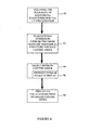
- One embodiment of the methodology contemplated is illustrated in Figure 4.
- the present invention begins by isolating the plurality of anatomical structures 16 within the three-dimensional visualization 10 (40). This can be performed through a variety of understood techniques. Simple thresholding or segmentation techniques are well understood methods for isolating anatomical structures 16 within images. Known segmentation techniques are also capable of automatically separating bony structures and separating them from the images. Once individual anatomical structures 30 are identified, a plurality of casting angles 26 can be evaluated.
- the plurality of casting angles 20 are evaluated by calculating the number of ray intersects 25 intersecting more than one of said plurality of anatomical structures 16 for each casting angle 20 (42). In addition to the number of ray intersects 25, the amount of ray intersect 32 may also be calculated. The optimum casting angle 26 is then selected (44) based upon the least amount of overlapping by minimizing the number of ray intersects 25 intersecting more than one of the plurality of anatomical structures 16. Although this may be accomplished through a variety of methods and algorithms, one embodiment contemplates selection 44 based upon minimizing the number of overlapped pixels in the display 46.
- the casting angle ⁇ should be selected such that it minimizes the set M ⁇ .
- the optimized medical three-dimensional visualization can be displayed from the selected casting angle 48.
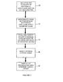
- FIG. 5 An alternate approach is illustrated in Figure 5. This approach makes use of the simple geometrical relationship of anatomical structure cross-sections 50 (see Figure 2). Once the anatomical structures are identified and isolated 40, this embodiment approximates the cross-section of each anatomical structure with a simple geometrical shape 52.
- the geometrical shapes 52 of the illustrated blood vessels 18 are illustrated as ovals, although a variety of geometrical shapes 52 are contemplated for a variety of cross-sections.
- the amount of overlap can be determined analytically for each of the casting angles 54. The optimum casting angle can thereby be selected 44 as before based upon the minimization of overlap. Again, a wide variety of simple geometrical calculations can be utilized to make this selection 44. And again, after selection 44, the optimized medical three-dimensional visualization can be displayed from the selected casting angle 48.
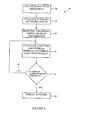
- FIG. 6 illustrates the present inventions preferable approach to such scenarios.
- This embodiment isolates the plurality of anatomical structures 60 and evaluates the plurality of casting angles 62 in either of the above identified methods, or in alternate variations.
- a first 3D visualization is selected based upon overall minimization of overlap 64 again as previously discussed. This embodiment, however, calculates and stores the locations of anatomical structure overlap 66.
- a second 3D visualization is then selected based upon a new casting angle that minimizes the number of overlaps in the region of previous overlap 68. In other words, if a minimized pixel approach is utilized, a new projection angle ⁇ should be selected such that it minimizes the overlaps in M ⁇ .
- the process can be repeated until the amount of overlap is less than a predetermined threshold 70.
- anatomical structures 16 that are not part of the kidney structure will not carry the same weight as ones that do. Neighboring structures that might obstruct the view of the kidney itself may be deleterious as well. Therefore, the present invention contemplates an embodiment illustrated in Figure 7. This embodiment includes the step of assigning weighting functions (or penalty function) to different anatomical structures 72. This can be utilized in a variety of fashions. Slight overlapping of large vessels may be less weighted than the same size overlapping of small vessels.
- Percentage of overlap may be utilized as a weighting factor for minimization. Other utilizations, as discussed may weight overlap of relevant structures higher than those of insignificant structures. In addition, it is contemplated that structures obstructing the weighted anatomical structures may be automatically removed from the display altogether. This embodiment can be utilized in conjunction with any of the disclosed embodiments. It adds a further improvement to automated visualization.
Landscapes
- Engineering & Computer Science (AREA)
- Physics & Mathematics (AREA)
- Computer Graphics (AREA)
- General Physics & Mathematics (AREA)
- Theoretical Computer Science (AREA)
- Geometry (AREA)
- Apparatus For Radiation Diagnosis (AREA)
- Magnetic Resonance Imaging Apparatus (AREA)
- Image Generation (AREA)
- Measuring And Recording Apparatus For Diagnosis (AREA)
- Image Processing (AREA)
- Image Analysis (AREA)
Abstract
Description
Claims (10)
- A method of automatically optimizing medical three-dimensional visualizations comprising:isolating a plurality of anatomical structures within the medical three-dimensional visualization;calculating the number of ray intersects, that intersect more than one of said plurality of anatomical structures, for a plurality of casting angles;selecting an optimum casting angle that minimizes said ray intersects from one of said plurality casting angles; anddisplaying the optimized medical three-dimensional visualization from said optimized casting angle.
- A method of automatically optimizing medical three-dimensional visualizations comprising:isolating a plurality of anatomical structures within the medical three-dimensional visualization;calculating the number of overlaps of said plurality of anatomical structures, for a plurality of casting angles;selecting an optimum casting angle that minimizes said number of overlaps from one of said plurality casting angles; anddisplaying the optimized medical three-dimensional visualization from said optimized casting angle.
- A method as claimed in claim 1 or claim 2, wherein said optimum casting angle is selected based upon minimizing the number of overlapped pixels in the three-dimensional visualization.
- A method as claimed in claim 1 or claim 2, further comprising:approximating a cross-section of each of said plurality of anatomical structures with a geometric shape; andselecting the optimum casting angle based upon the location and shape of said geometric shapes.
- A method as claimed in claim 1 or claim 2, further comprising:calculating and storing the locations of anatomical structure overlap in the optimized medical three-dimensional visualization;selecting a second casting angle that minimizes said locations of anatomical structure overlap; anddisplaying a second three-dimensional visualization from said second casting angle.
- A method as claimed in claim 1, further comprising:weighting said ray intersects based upon an importance factor of each of said plurality of anatomical structures; andselecting an optimum casting angle that minimizes said weighted ray intersects from one of said plurality casting angles.
- A method as claimed in claim 2, further comprising:weighting said overlaps based upon an importance factor of each of said plurality of anatomical structures; andselecting an optimum casting angle that minimizes said weighted overlaps from one of said plurality casting angles.
- An apparatus of automatically optimizing medical three-dimensional visualizations comprising:an isolating component, said isolating component isolating a plurality of anatomical structures within the medical three-dimensional visualization;a calculator component for calculating the number of overlaps of said plurality of anatomical structures, for a plurality of casting angles;a selector component, said selector component selecting an optimum casting angle that minimizes said number of overlaps from one of said plurality casting angles; anda display element for displaying the optimized medical three-dimensional visualization from said optimized casting angle.
- An apparatus as claimed in claim 8, further comprising:a geometric approximator for approximating a cross-section of each of said plurality of anatomical structures with a geometric shape.
- An apparatus as claimed in claim 8, further comprising;a weighting component, said weighting component weighting said overlaps based upon the size or an importance factor of each of said plurality of anatomical structures.
Applications Claiming Priority (2)
| Application Number | Priority Date | Filing Date | Title |
|---|---|---|---|
| US64762 | 1993-05-19 | ||
| US10/064,762 US7092558B2 (en) | 2002-08-14 | 2002-08-14 | Automated optimization of medical 3D visualizations |
Publications (2)
| Publication Number | Publication Date |
|---|---|
| EP1394749A2 true EP1394749A2 (en) | 2004-03-03 |
| EP1394749A3 EP1394749A3 (en) | 2006-02-15 |
Family
ID=31493954
Family Applications (1)
| Application Number | Title | Priority Date | Filing Date |
|---|---|---|---|
| EP03255038A Withdrawn EP1394749A3 (en) | 2002-08-14 | 2003-08-13 | Automated optimization of medical 3D visualizations |
Country Status (3)
| Country | Link |
|---|---|
| US (1) | US7092558B2 (en) |
| EP (1) | EP1394749A3 (en) |
| JP (1) | JP4572401B2 (en) |
Families Citing this family (3)
| Publication number | Priority date | Publication date | Assignee | Title |
|---|---|---|---|---|
| US7092558B2 (en) * | 2002-08-14 | 2006-08-15 | General Electric Company | Automated optimization of medical 3D visualizations |
| US9024949B2 (en) * | 2004-10-13 | 2015-05-05 | Sony Corporation | Object representation using distance functions |
| FI20195977A1 (en) * | 2019-11-15 | 2021-05-16 | Disior Oy | Arrangement and method for provision of enhanced two-dimensional imaging data |
Family Cites Families (19)
| Publication number | Priority date | Publication date | Assignee | Title |
|---|---|---|---|---|
| US5226113A (en) * | 1989-10-30 | 1993-07-06 | General Electric Company | Method and apparatus for volumetric projection rendering using reverse ray casting |
| GB9315852D0 (en) * | 1993-07-30 | 1993-09-15 | Video Logic Ltd | Shading three-dimensional images |
| JP3311830B2 (en) * | 1993-09-20 | 2002-08-05 | 株式会社東芝 | 3D video creation device |
| GB9424273D0 (en) * | 1994-12-01 | 1995-01-18 | Wrigley Adrian M T | Improvements in and relating to image constrcution |
| JPH09305790A (en) * | 1996-05-10 | 1997-11-28 | Tokyo Electric Power Co Inc:The | Three-dimensional information display device |
| FR2752975B1 (en) | 1996-09-04 | 1998-12-04 | Ge Medical Syst Sa | METHOD FOR RECONSTRUCTING A THREE-DIMENSIONAL IMAGE OF AN OBJECT, IN PARTICULAR A THREE-DIMENSIONAL ANGIOGRAPHIC IMAGE |
| US5954653A (en) * | 1997-05-07 | 1999-09-21 | General Electric Company | Method and apparatus for automatically enhancing contrast in projected ultrasound image |
| US6126603A (en) * | 1997-05-07 | 2000-10-03 | General Electric Company | Method and apparatus for segmenting color flow mode data using velocity information in three-dimensional ultrasound imaging |
| US6074348A (en) | 1998-03-31 | 2000-06-13 | General Electric Company | Method and apparatus for enhanced flow imaging in B-mode ultrasound |
| US6514074B1 (en) * | 1999-05-14 | 2003-02-04 | Align Technology, Inc. | Digitally modeling the deformation of gingival |
| US6193657B1 (en) | 1998-12-31 | 2001-02-27 | Ge Medical Systems Global Technology Company, Llc | Image based probe position and orientation detection |
| US6301497B1 (en) | 1999-07-12 | 2001-10-09 | Ge Medical Systems Global Technology Company, Llc | Method and apparatus for magnetic resonance imaging intersecting slices |
| FR2799028B1 (en) | 1999-09-27 | 2002-05-03 | Ge Medical Syst Sa | METHOD FOR RECONSTRUCTING A THREE-DIMENSIONAL IMAGE OF ELEMENTS OF STRONG CONTRAST |
| US6277075B1 (en) | 1999-11-26 | 2001-08-21 | Ge Medical Systems Global Technology Company, Llc | Method and apparatus for visualization of motion in ultrasound flow imaging using continuous data acquisition |
| US6947038B1 (en) * | 2000-04-27 | 2005-09-20 | Align Technology, Inc. | Systems and methods for generating an appliance with tie points |
| US6348793B1 (en) | 2000-11-06 | 2002-02-19 | Ge Medical Systems Global Technology, Company, Llc | System architecture for medical imaging systems |
| US6404853B1 (en) | 2001-11-02 | 2002-06-11 | Ge Medical Systems Global Technology Company, Llc | Method for identifying and correcting pixels with excess pixel lag in a solid state x-ray detector |
| US6979196B2 (en) * | 2002-06-21 | 2005-12-27 | Align Technology, Inc. | Systems and methods for automated bite-setting of tooth models |
| US7092558B2 (en) * | 2002-08-14 | 2006-08-15 | General Electric Company | Automated optimization of medical 3D visualizations |
-
2002
- 2002-08-14 US US10/064,762 patent/US7092558B2/en not_active Expired - Lifetime
-
2003
- 2003-08-13 JP JP2003292751A patent/JP4572401B2/en not_active Expired - Fee Related
- 2003-08-13 EP EP03255038A patent/EP1394749A3/en not_active Withdrawn
Also Published As
| Publication number | Publication date |
|---|---|
| EP1394749A3 (en) | 2006-02-15 |
| JP2004078960A (en) | 2004-03-11 |
| JP4572401B2 (en) | 2010-11-04 |
| US20040032978A1 (en) | 2004-02-19 |
| US7092558B2 (en) | 2006-08-15 |
Similar Documents
| Publication | Publication Date | Title |
|---|---|---|
| US20250166250A1 (en) | Apparatus and method for visualizing digital breast tomosynthesis and other volumetric images | |
| Nelson et al. | Visualization of 3D ultrasound data | |
| JP4359647B2 (en) | Automatic analysis in endoscopy that cannot be detected directly | |
| EP0216156B1 (en) | Dividing cubes and method for the display of surface structures contained within the interior region of a solid body | |
| US7397475B2 (en) | Interactive atlas extracted from volume data | |
| EP2212859B1 (en) | Method and apparatus for volume rendering of data sets | |
| US7890155B2 (en) | Feature emphasis and contextual cutaways for image visualization | |
| US20070276214A1 (en) | Systems and Methods for Automated Segmentation, Visualization and Analysis of Medical Images | |
| US8659602B2 (en) | Generating a pseudo three-dimensional image of a three-dimensional voxel array illuminated by an arbitrary light source by a direct volume rendering method | |
| US20060239540A1 (en) | Methods and systems for creating 4D images using multiple 2D images acquired in real-time ("4D ultrasound") | |
| US20120301003A1 (en) | Reconstructing an object of interest | |
| EP2116973A1 (en) | Method for interactively determining a bounding surface for segmenting a lesion in a medical image | |
| JPH0632042B2 (en) | Device and method for displaying three-dimensional surface structure | |
| US8170640B2 (en) | System and method for tree projection for detection of pulmonary embolism | |
| US20090087046A1 (en) | Digital blink comparator apparatus and software and methods for operation | |
| US9237849B1 (en) | Relative maximum intensity projection | |
| CN1692377B (en) | Method and device for forming an isolated visualization of body structures | |
| WO2001046912A1 (en) | Iterative determination of the shortest path between two points on a polygonal surface | |
| US7092558B2 (en) | Automated optimization of medical 3D visualizations | |
| ROMENY et al. | Advances in three-dimensional diagnostic radiology | |
| Wang | Three-dimensional medical CT image reconstruction | |
| US20060103678A1 (en) | Method and system for interactive visualization of locally oriented structures | |
| EP4428827A1 (en) | Apparatus for generating a visualization of a medical image volume | |
| Delegacz et al. | Three-dimensional visualization system as an aid for lung cancer detection | |
| Turlington et al. | Improved techniques for fast sliding thin-slab volume visualization |
Legal Events
| Date | Code | Title | Description |
|---|---|---|---|
| PUAI | Public reference made under article 153(3) epc to a published international application that has entered the european phase |
Free format text: ORIGINAL CODE: 0009012 |
|
| AK | Designated contracting states |
Kind code of ref document: A2 Designated state(s): AT BE BG CH CY CZ DE DK EE ES FI FR GB GR HU IE IT LI LU MC NL PT RO SE SI SK TR |
|
| AX | Request for extension of the european patent |
Extension state: AL LT LV MK |
|
| PUAL | Search report despatched |
Free format text: ORIGINAL CODE: 0009013 |
|
| AK | Designated contracting states |
Kind code of ref document: A3 Designated state(s): AT BE BG CH CY CZ DE DK EE ES FI FR GB GR HU IE IT LI LU MC NL PT RO SE SI SK TR |
|
| AX | Request for extension of the european patent |
Extension state: AL LT LV MK |
|
| 17P | Request for examination filed |
Effective date: 20060816 |
|
| AKX | Designation fees paid |
Designated state(s): DE NL |
|
| 17Q | First examination report despatched |
Effective date: 20061013 |
|
| STAA | Information on the status of an ep patent application or granted ep patent |
Free format text: STATUS: THE APPLICATION IS DEEMED TO BE WITHDRAWN |
|
| 18D | Application deemed to be withdrawn |
Effective date: 20150303 |