EP1394549A1 - Biomarkers for diagnosing Alzheimer's disease - Google Patents
Biomarkers for diagnosing Alzheimer's disease Download PDFInfo
- Publication number
- EP1394549A1 EP1394549A1 EP02018283A EP02018283A EP1394549A1 EP 1394549 A1 EP1394549 A1 EP 1394549A1 EP 02018283 A EP02018283 A EP 02018283A EP 02018283 A EP02018283 A EP 02018283A EP 1394549 A1 EP1394549 A1 EP 1394549A1
- Authority
- EP
- European Patent Office
- Prior art keywords
- polypeptide
- disease
- seq
- alzheimer
- molecular mass
- Prior art date
- Legal status (The legal status is an assumption and is not a legal conclusion. Google has not performed a legal analysis and makes no representation as to the accuracy of the status listed.)
- Withdrawn
Links
- 208000024827 Alzheimer disease Diseases 0.000 title claims abstract description 49
- 239000000090 biomarker Substances 0.000 title description 2
- 238000000034 method Methods 0.000 claims abstract description 35
- 238000001514 detection method Methods 0.000 claims abstract description 30
- 239000012634 fragment Substances 0.000 claims abstract description 19
- 101000912205 Homo sapiens Cystatin-C Proteins 0.000 claims abstract description 8
- 238000000756 surface-enhanced laser desorption--ionisation time-of-flight mass spectrometry Methods 0.000 claims abstract description 8
- 208000031124 Dementia Alzheimer type Diseases 0.000 claims abstract description 7
- 101000937544 Homo sapiens Beta-2-microglobulin Proteins 0.000 claims abstract description 7
- 101000635854 Homo sapiens Myoglobin Proteins 0.000 claims abstract description 7
- 102000047279 human B2M Human genes 0.000 claims abstract description 7
- 102000049632 human CST3 Human genes 0.000 claims abstract description 7
- 108090000765 processed proteins & peptides Proteins 0.000 claims description 96
- 102000004196 processed proteins & peptides Human genes 0.000 claims description 86
- 229920001184 polypeptide Polymers 0.000 claims description 78
- 210000001175 cerebrospinal fluid Anatomy 0.000 claims description 28
- 150000001413 amino acids Chemical class 0.000 claims description 15
- 210000002700 urine Anatomy 0.000 claims description 7
- 210000004369 blood Anatomy 0.000 claims description 5
- 239000008280 blood Substances 0.000 claims description 5
- 239000000284 extract Substances 0.000 claims description 4
- 210000002966 serum Anatomy 0.000 claims description 4
- 239000012530 fluid Substances 0.000 claims description 3
- 210000002445 nipple Anatomy 0.000 claims description 3
- 210000000582 semen Anatomy 0.000 claims description 3
- 210000002381 plasma Anatomy 0.000 claims description 2
- 108090000623 proteins and genes Proteins 0.000 abstract description 28
- 102000004169 proteins and genes Human genes 0.000 abstract description 27
- 210000001124 body fluid Anatomy 0.000 abstract description 8
- 239000010839 body fluid Substances 0.000 abstract description 8
- 238000004949 mass spectrometry Methods 0.000 abstract description 8
- 238000012544 monitoring process Methods 0.000 abstract description 5
- WEVYAHXRMPXWCK-UHFFFAOYSA-N Acetonitrile Chemical compound CC#N WEVYAHXRMPXWCK-UHFFFAOYSA-N 0.000 description 24
- 235000018102 proteins Nutrition 0.000 description 24
- 239000000499 gel Substances 0.000 description 22
- 239000003550 marker Substances 0.000 description 18
- 239000000523 sample Substances 0.000 description 16
- 102000012192 Cystatin C Human genes 0.000 description 13
- 108010061642 Cystatin C Proteins 0.000 description 13
- OKKJLVBELUTLKV-UHFFFAOYSA-N Methanol Chemical compound OC OKKJLVBELUTLKV-UHFFFAOYSA-N 0.000 description 12
- 238000004458 analytical method Methods 0.000 description 12
- 208000037265 diseases, disorders, signs and symptoms Diseases 0.000 description 12
- DTQVDTLACAAQTR-UHFFFAOYSA-N Trifluoroacetic acid Chemical compound OC(=O)C(F)(F)F DTQVDTLACAAQTR-UHFFFAOYSA-N 0.000 description 10
- 201000010099 disease Diseases 0.000 description 10
- 239000001488 sodium phosphate Substances 0.000 description 10
- 229910000162 sodium phosphate Inorganic materials 0.000 description 10
- 239000000243 solution Substances 0.000 description 10
- RYFMWSXOAZQYPI-UHFFFAOYSA-K trisodium phosphate Chemical compound [Na+].[Na+].[Na+].[O-]P([O-])([O-])=O RYFMWSXOAZQYPI-UHFFFAOYSA-K 0.000 description 10
- 239000012149 elution buffer Substances 0.000 description 9
- FAPWRFPIFSIZLT-UHFFFAOYSA-M Sodium chloride Chemical compound [Na+].[Cl-] FAPWRFPIFSIZLT-UHFFFAOYSA-M 0.000 description 8
- QKNYBSVHEMOAJP-UHFFFAOYSA-N 2-amino-2-(hydroxymethyl)propane-1,3-diol;hydron;chloride Chemical compound Cl.OCC(N)(CO)CO QKNYBSVHEMOAJP-UHFFFAOYSA-N 0.000 description 7
- 235000001014 amino acid Nutrition 0.000 description 7
- 210000004556 brain Anatomy 0.000 description 7
- 239000000872 buffer Substances 0.000 description 7
- QTBSBXVTEAMEQO-UHFFFAOYSA-N Acetic acid Chemical compound CC(O)=O QTBSBXVTEAMEQO-UHFFFAOYSA-N 0.000 description 6
- ATRRKUHOCOJYRX-UHFFFAOYSA-N Ammonium bicarbonate Chemical compound [NH4+].OC([O-])=O ATRRKUHOCOJYRX-UHFFFAOYSA-N 0.000 description 6
- 229910000013 Ammonium bicarbonate Inorganic materials 0.000 description 6
- KRKNYBCHXYNGOX-UHFFFAOYSA-K Citrate Chemical compound [O-]C(=O)CC(O)(CC([O-])=O)C([O-])=O KRKNYBCHXYNGOX-UHFFFAOYSA-K 0.000 description 6
- 235000012538 ammonium bicarbonate Nutrition 0.000 description 6
- 239000001099 ammonium carbonate Substances 0.000 description 6
- 102000015736 beta 2-Microglobulin Human genes 0.000 description 6
- 108010081355 beta 2-Microglobulin Proteins 0.000 description 6
- 239000012148 binding buffer Substances 0.000 description 6
- 239000001509 sodium citrate Substances 0.000 description 6
- 238000000672 surface-enhanced laser desorption--ionisation Methods 0.000 description 6
- CFBILACNYSPRPM-UHFFFAOYSA-N 2-amino-2-(hydroxymethyl)propane-1,3-diol;2-[[1,3-dihydroxy-2-(hydroxymethyl)propan-2-yl]amino]acetic acid Chemical compound OCC(N)(CO)CO.OCC(CO)(CO)NCC(O)=O CFBILACNYSPRPM-UHFFFAOYSA-N 0.000 description 5
- 238000001962 electrophoresis Methods 0.000 description 5
- 239000006167 equilibration buffer Substances 0.000 description 5
- 230000035945 sensitivity Effects 0.000 description 5
- 238000001228 spectrum Methods 0.000 description 5
- 208000018282 ACys amyloidosis Diseases 0.000 description 4
- 206010012289 Dementia Diseases 0.000 description 4
- 102000004142 Trypsin Human genes 0.000 description 4
- 108090000631 Trypsin Proteins 0.000 description 4
- DZHSAHHDTRWUTF-SIQRNXPUSA-N amyloid-beta polypeptide 42 Chemical compound C([C@@H](C(=O)N[C@@H](C)C(=O)N[C@@H](CCC(O)=O)C(=O)N[C@@H](CC(O)=O)C(=O)N[C@H](C(=O)NCC(=O)N[C@@H](CO)C(=O)N[C@@H](CC(N)=O)C(=O)N[C@@H](CCCCN)C(=O)NCC(=O)N[C@@H](C)C(=O)N[C@H](C(=O)N[C@@H]([C@@H](C)CC)C(=O)NCC(=O)N[C@@H](CC(C)C)C(=O)N[C@@H](CCSC)C(=O)N[C@@H](C(C)C)C(=O)NCC(=O)NCC(=O)N[C@@H](C(C)C)C(=O)N[C@@H](C(C)C)C(=O)N[C@@H]([C@@H](C)CC)C(=O)N[C@@H](C)C(O)=O)[C@@H](C)CC)C(C)C)NC(=O)[C@H](CC=1C=CC=CC=1)NC(=O)[C@@H](NC(=O)[C@H](CC(C)C)NC(=O)[C@H](CCCCN)NC(=O)[C@H](CCC(N)=O)NC(=O)[C@H](CC=1N=CNC=1)NC(=O)[C@H](CC=1N=CNC=1)NC(=O)[C@@H](NC(=O)[C@H](CCC(O)=O)NC(=O)[C@H](CC=1C=CC(O)=CC=1)NC(=O)CNC(=O)[C@H](CO)NC(=O)[C@H](CC(O)=O)NC(=O)[C@H](CC=1N=CNC=1)NC(=O)[C@H](CCCNC(N)=N)NC(=O)[C@H](CC=1C=CC=CC=1)NC(=O)[C@H](CCC(O)=O)NC(=O)[C@H](C)NC(=O)[C@@H](N)CC(O)=O)C(C)C)C(C)C)C1=CC=CC=C1 DZHSAHHDTRWUTF-SIQRNXPUSA-N 0.000 description 4
- 238000003556 assay Methods 0.000 description 4
- 230000006378 damage Effects 0.000 description 4
- 238000003745 diagnosis Methods 0.000 description 4
- 238000000605 extraction Methods 0.000 description 4
- 239000011159 matrix material Substances 0.000 description 4
- BDAGIHXWWSANSR-UHFFFAOYSA-N methanoic acid Natural products OC=O BDAGIHXWWSANSR-UHFFFAOYSA-N 0.000 description 4
- 230000004770 neurodegeneration Effects 0.000 description 4
- 239000011780 sodium chloride Substances 0.000 description 4
- 238000004885 tandem mass spectrometry Methods 0.000 description 4
- 239000012588 trypsin Substances 0.000 description 4
- 101710137189 Amyloid-beta A4 protein Proteins 0.000 description 3
- 101710151993 Amyloid-beta precursor protein Proteins 0.000 description 3
- 102100022704 Amyloid-beta precursor protein Human genes 0.000 description 3
- 208000005145 Cerebral amyloid angiopathy Diseases 0.000 description 3
- 108010062374 Myoglobin Proteins 0.000 description 3
- 102000036675 Myoglobin Human genes 0.000 description 3
- DBMJMQXJHONAFJ-UHFFFAOYSA-M Sodium laurylsulphate Chemical compound [Na+].CCCCCCCCCCCCOS([O-])(=O)=O DBMJMQXJHONAFJ-UHFFFAOYSA-M 0.000 description 3
- 238000007792 addition Methods 0.000 description 3
- 108010064539 amyloid beta-protein (1-42) Proteins 0.000 description 3
- 238000003491 array Methods 0.000 description 3
- 210000004027 cell Anatomy 0.000 description 3
- 230000003247 decreasing effect Effects 0.000 description 3
- 238000010828 elution Methods 0.000 description 3
- 238000002474 experimental method Methods 0.000 description 3
- 230000006870 function Effects 0.000 description 3
- 238000003018 immunoassay Methods 0.000 description 3
- 150000002500 ions Chemical class 0.000 description 3
- 208000015122 neurodegenerative disease Diseases 0.000 description 3
- 210000002682 neurofibrillary tangle Anatomy 0.000 description 3
- 238000011002 quantification Methods 0.000 description 3
- 239000011347 resin Substances 0.000 description 3
- 229920005989 resin Polymers 0.000 description 3
- PCMORTLOPMLEFB-ONEGZZNKSA-N sinapic acid Chemical compound COC1=CC(\C=C\C(O)=O)=CC(OC)=C1O PCMORTLOPMLEFB-ONEGZZNKSA-N 0.000 description 3
- PCMORTLOPMLEFB-UHFFFAOYSA-N sinapinic acid Natural products COC1=CC(C=CC(O)=O)=CC(OC)=C1O PCMORTLOPMLEFB-UHFFFAOYSA-N 0.000 description 3
- 238000002415 sodium dodecyl sulfate polyacrylamide gel electrophoresis Methods 0.000 description 3
- XLYOFNOQVPJJNP-UHFFFAOYSA-N water Chemical compound O XLYOFNOQVPJJNP-UHFFFAOYSA-N 0.000 description 3
- AXAVXPMQTGXXJZ-UHFFFAOYSA-N 2-aminoacetic acid;2-amino-2-(hydroxymethyl)propane-1,3-diol Chemical compound NCC(O)=O.OCC(N)(CO)CO AXAVXPMQTGXXJZ-UHFFFAOYSA-N 0.000 description 2
- OSWFIVFLDKOXQC-UHFFFAOYSA-N 4-(3-methoxyphenyl)aniline Chemical compound COC1=CC=CC(C=2C=CC(N)=CC=2)=C1 OSWFIVFLDKOXQC-UHFFFAOYSA-N 0.000 description 2
- 208000037259 Amyloid Plaque Diseases 0.000 description 2
- XKRFYHLGVUSROY-UHFFFAOYSA-N Argon Chemical compound [Ar] XKRFYHLGVUSROY-UHFFFAOYSA-N 0.000 description 2
- IJGRMHOSHXDMSA-UHFFFAOYSA-N Atomic nitrogen Chemical compound N#N IJGRMHOSHXDMSA-UHFFFAOYSA-N 0.000 description 2
- -1 CSF Substances 0.000 description 2
- 206010061818 Disease progression Diseases 0.000 description 2
- 238000002965 ELISA Methods 0.000 description 2
- SEQKRHFRPICQDD-UHFFFAOYSA-N N-tris(hydroxymethyl)methylglycine Chemical compound OCC(CO)(CO)[NH2+]CC([O-])=O SEQKRHFRPICQDD-UHFFFAOYSA-N 0.000 description 2
- 102000012412 Presenilin-1 Human genes 0.000 description 2
- 108010036933 Presenilin-1 Proteins 0.000 description 2
- 102000012419 Presenilin-2 Human genes 0.000 description 2
- 108010036908 Presenilin-2 Proteins 0.000 description 2
- 108010029485 Protein Isoforms Proteins 0.000 description 2
- 102000001708 Protein Isoforms Human genes 0.000 description 2
- 230000004075 alteration Effects 0.000 description 2
- 206010002026 amyotrophic lateral sclerosis Diseases 0.000 description 2
- 230000015572 biosynthetic process Effects 0.000 description 2
- 230000003931 cognitive performance Effects 0.000 description 2
- XUJNEKJLAYXESH-UHFFFAOYSA-N cysteine Natural products SCC(N)C(O)=O XUJNEKJLAYXESH-UHFFFAOYSA-N 0.000 description 2
- 230000005750 disease progression Effects 0.000 description 2
- 208000035475 disorder Diseases 0.000 description 2
- 239000012153 distilled water Substances 0.000 description 2
- 238000005516 engineering process Methods 0.000 description 2
- 238000011156 evaluation Methods 0.000 description 2
- 235000019253 formic acid Nutrition 0.000 description 2
- 230000002068 genetic effect Effects 0.000 description 2
- 230000005484 gravity Effects 0.000 description 2
- 230000001900 immune effect Effects 0.000 description 2
- PGLTVOMIXTUURA-UHFFFAOYSA-N iodoacetamide Chemical compound NC(=O)CI PGLTVOMIXTUURA-UHFFFAOYSA-N 0.000 description 2
- 238000000816 matrix-assisted laser desorption--ionisation Methods 0.000 description 2
- 239000000203 mixture Substances 0.000 description 2
- 239000000101 novel biomarker Substances 0.000 description 2
- 239000000137 peptide hydrolase inhibitor Substances 0.000 description 2
- 229920002401 polyacrylamide Polymers 0.000 description 2
- 238000002360 preparation method Methods 0.000 description 2
- 238000012552 review Methods 0.000 description 2
- 238000007619 statistical method Methods 0.000 description 2
- 238000006467 substitution reaction Methods 0.000 description 2
- 230000001225 therapeutic effect Effects 0.000 description 2
- QFVHZQCOUORWEI-UHFFFAOYSA-N 4-[(4-anilino-5-sulfonaphthalen-1-yl)diazenyl]-5-hydroxynaphthalene-2,7-disulfonic acid Chemical compound C=12C(O)=CC(S(O)(=O)=O)=CC2=CC(S(O)(=O)=O)=CC=1N=NC(C1=CC=CC(=C11)S(O)(=O)=O)=CC=C1NC1=CC=CC=C1 QFVHZQCOUORWEI-UHFFFAOYSA-N 0.000 description 1
- QTBSBXVTEAMEQO-UHFFFAOYSA-M Acetate Chemical compound CC([O-])=O QTBSBXVTEAMEQO-UHFFFAOYSA-M 0.000 description 1
- HRPVXLWXLXDGHG-UHFFFAOYSA-N Acrylamide Chemical compound NC(=O)C=C HRPVXLWXLXDGHG-UHFFFAOYSA-N 0.000 description 1
- 108700028369 Alleles Proteins 0.000 description 1
- 208000022099 Alzheimer disease 2 Diseases 0.000 description 1
- 208000000044 Amnesia Diseases 0.000 description 1
- 108010090849 Amyloid beta-Peptides Proteins 0.000 description 1
- 102000013455 Amyloid beta-Peptides Human genes 0.000 description 1
- 108010060159 Apolipoprotein E4 Proteins 0.000 description 1
- 108010039627 Aprotinin Proteins 0.000 description 1
- 206010003694 Atrophy Diseases 0.000 description 1
- 208000035404 Autolysis Diseases 0.000 description 1
- 108010001478 Bacitracin Proteins 0.000 description 1
- 206010006187 Breast cancer Diseases 0.000 description 1
- 208000026310 Breast neoplasm Diseases 0.000 description 1
- 101150076592 CST3 gene Proteins 0.000 description 1
- CURLTUGMZLYLDI-UHFFFAOYSA-N Carbon dioxide Chemical compound O=C=O CURLTUGMZLYLDI-UHFFFAOYSA-N 0.000 description 1
- 206010057248 Cell death Diseases 0.000 description 1
- 102100026897 Cystatin-C Human genes 0.000 description 1
- 102000004190 Enzymes Human genes 0.000 description 1
- 108090000790 Enzymes Proteins 0.000 description 1
- UNXHWFMMPAWVPI-UHFFFAOYSA-N Erythritol Natural products OCC(O)C(O)CO UNXHWFMMPAWVPI-UHFFFAOYSA-N 0.000 description 1
- 101710097670 Extracellular cysteine protease Proteins 0.000 description 1
- 206010064571 Gene mutation Diseases 0.000 description 1
- 208000034826 Genetic Predisposition to Disease Diseases 0.000 description 1
- 108700039691 Genetic Promoter Regions Proteins 0.000 description 1
- 101000971703 Homo sapiens Kinesin-like protein KIF1C Proteins 0.000 description 1
- 101000979579 Homo sapiens NK1 transcription factor-related protein 1 Proteins 0.000 description 1
- 108090000144 Human Proteins Proteins 0.000 description 1
- 102000003839 Human Proteins Human genes 0.000 description 1
- 206010061218 Inflammation Diseases 0.000 description 1
- 102000004877 Insulin Human genes 0.000 description 1
- 108090001061 Insulin Proteins 0.000 description 1
- 102000004195 Isomerases Human genes 0.000 description 1
- 108090000769 Isomerases Proteins 0.000 description 1
- 102100021525 Kinesin-like protein KIF1C Human genes 0.000 description 1
- QNAYBMKLOCPYGJ-REOHCLBHSA-N L-alanine Chemical compound C[C@H](N)C(O)=O QNAYBMKLOCPYGJ-REOHCLBHSA-N 0.000 description 1
- 102000004407 Lactalbumin Human genes 0.000 description 1
- 108090000942 Lactalbumin Proteins 0.000 description 1
- 108700018351 Major Histocompatibility Complex Proteins 0.000 description 1
- 241000124008 Mammalia Species 0.000 description 1
- 102000018697 Membrane Proteins Human genes 0.000 description 1
- 108010052285 Membrane Proteins Proteins 0.000 description 1
- 241000699670 Mus sp. Species 0.000 description 1
- 208000012902 Nervous system disease Diseases 0.000 description 1
- 208000025966 Neurological disease Diseases 0.000 description 1
- 241000283973 Oryctolagus cuniculus Species 0.000 description 1
- 208000018737 Parkinson disease Diseases 0.000 description 1
- 102000035195 Peptidases Human genes 0.000 description 1
- 108091005804 Peptidases Proteins 0.000 description 1
- 206010057249 Phagocytosis Diseases 0.000 description 1
- 239000004743 Polypropylene Substances 0.000 description 1
- 108010076504 Protein Sorting Signals Proteins 0.000 description 1
- 208000006011 Stroke Diseases 0.000 description 1
- 229930006000 Sucrose Natural products 0.000 description 1
- CZMRCDWAGMRECN-UGDNZRGBSA-N Sucrose Chemical compound O[C@H]1[C@H](O)[C@@H](CO)O[C@@]1(CO)O[C@@H]1[C@H](O)[C@@H](O)[C@H](O)[C@@H](CO)O1 CZMRCDWAGMRECN-UGDNZRGBSA-N 0.000 description 1
- UZMAPBJVXOGOFT-UHFFFAOYSA-N Syringetin Natural products COC1=C(O)C(OC)=CC(C2=C(C(=O)C3=C(O)C=C(O)C=C3O2)O)=C1 UZMAPBJVXOGOFT-UHFFFAOYSA-N 0.000 description 1
- AYFVYJQAPQTCCC-UHFFFAOYSA-N Threonine Natural products CC(O)C(N)C(O)=O AYFVYJQAPQTCCC-UHFFFAOYSA-N 0.000 description 1
- 239000004473 Threonine Substances 0.000 description 1
- 239000007997 Tricine buffer Substances 0.000 description 1
- 230000005856 abnormality Effects 0.000 description 1
- 230000032683 aging Effects 0.000 description 1
- 235000004279 alanine Nutrition 0.000 description 1
- 230000003941 amyloidogenesis Effects 0.000 description 1
- 125000000129 anionic group Chemical group 0.000 description 1
- 229960004405 aprotinin Drugs 0.000 description 1
- 229910052786 argon Inorganic materials 0.000 description 1
- 230000037444 atrophy Effects 0.000 description 1
- 229960003071 bacitracin Drugs 0.000 description 1
- 229930184125 bacitracin Natural products 0.000 description 1
- CLKOFPXJLQSYAH-ABRJDSQDSA-N bacitracin A Chemical compound C1SC([C@@H](N)[C@@H](C)CC)=N[C@@H]1C(=O)N[C@@H](CC(C)C)C(=O)N[C@H](CCC(O)=O)C(=O)N[C@@H]([C@@H](C)CC)C(=O)N[C@@H]1C(=O)N[C@H](CCCN)C(=O)N[C@@H]([C@@H](C)CC)C(=O)N[C@H](CC=2C=CC=CC=2)C(=O)N[C@@H](CC=2N=CNC=2)C(=O)N[C@H](CC(O)=O)C(=O)N[C@@H](CC(N)=O)C(=O)NCCCC1 CLKOFPXJLQSYAH-ABRJDSQDSA-N 0.000 description 1
- 239000011324 bead Substances 0.000 description 1
- 239000013060 biological fluid Substances 0.000 description 1
- UDSAIICHUKSCKT-UHFFFAOYSA-N bromophenol blue Chemical compound C1=C(Br)C(O)=C(Br)C=C1C1(C=2C=C(Br)C(O)=C(Br)C=2)C2=CC=CC=C2S(=O)(=O)O1 UDSAIICHUKSCKT-UHFFFAOYSA-N 0.000 description 1
- 235000011089 carbon dioxide Nutrition 0.000 description 1
- 230000001364 causal effect Effects 0.000 description 1
- 230000003915 cell function Effects 0.000 description 1
- 238000005119 centrifugation Methods 0.000 description 1
- 150000001793 charged compounds Chemical class 0.000 description 1
- 239000003153 chemical reaction reagent Substances 0.000 description 1
- 238000004587 chromatography analysis Methods 0.000 description 1
- 238000003776 cleavage reaction Methods 0.000 description 1
- 238000003759 clinical diagnosis Methods 0.000 description 1
- 238000012398 clinical drug development Methods 0.000 description 1
- 230000006999 cognitive decline Effects 0.000 description 1
- 208000010877 cognitive disease Diseases 0.000 description 1
- 208000030251 communication disease Diseases 0.000 description 1
- NKLPQNGYXWVELD-UHFFFAOYSA-M coomassie brilliant blue Chemical compound [Na+].C1=CC(OCC)=CC=C1NC1=CC=C(C(=C2C=CC(C=C2)=[N+](CC)CC=2C=C(C=CC=2)S([O-])(=O)=O)C=2C=CC(=CC=2)N(CC)CC=2C=C(C=CC=2)S([O-])(=O)=O)C=C1 NKLPQNGYXWVELD-UHFFFAOYSA-M 0.000 description 1
- 235000018417 cysteine Nutrition 0.000 description 1
- 150000001945 cysteines Chemical class 0.000 description 1
- 230000034994 death Effects 0.000 description 1
- 230000007423 decrease Effects 0.000 description 1
- 230000003111 delayed effect Effects 0.000 description 1
- 238000004925 denaturation Methods 0.000 description 1
- 230000036425 denaturation Effects 0.000 description 1
- 230000008021 deposition Effects 0.000 description 1
- 238000003795 desorption Methods 0.000 description 1
- 238000011161 development Methods 0.000 description 1
- 230000018109 developmental process Effects 0.000 description 1
- 238000007435 diagnostic evaluation Methods 0.000 description 1
- 230000004069 differentiation Effects 0.000 description 1
- 230000029087 digestion Effects 0.000 description 1
- KCFYHBSOLOXZIF-UHFFFAOYSA-N dihydrochrysin Natural products COC1=C(O)C(OC)=CC(C2OC3=CC(O)=CC(O)=C3C(=O)C2)=C1 KCFYHBSOLOXZIF-UHFFFAOYSA-N 0.000 description 1
- VHJLVAABSRFDPM-QWWZWVQMSA-N dithiothreitol Chemical compound SC[C@@H](O)[C@H](O)CS VHJLVAABSRFDPM-QWWZWVQMSA-N 0.000 description 1
- 238000009509 drug development Methods 0.000 description 1
- 230000000694 effects Effects 0.000 description 1
- 230000008030 elimination Effects 0.000 description 1
- 238000003379 elimination reaction Methods 0.000 description 1
- 230000007613 environmental effect Effects 0.000 description 1
- 238000001704 evaporation Methods 0.000 description 1
- 230000008020 evaporation Effects 0.000 description 1
- 238000005194 fractionation Methods 0.000 description 1
- 238000013467 fragmentation Methods 0.000 description 1
- 238000006062 fragmentation reaction Methods 0.000 description 1
- 239000007789 gas Substances 0.000 description 1
- 102000054766 genetic haplotypes Human genes 0.000 description 1
- 239000011521 glass Substances 0.000 description 1
- 238000003384 imaging method Methods 0.000 description 1
- 230000028993 immune response Effects 0.000 description 1
- 239000002955 immunomodulating agent Substances 0.000 description 1
- 230000002584 immunomodulator Effects 0.000 description 1
- 229940121354 immunomodulator Drugs 0.000 description 1
- 238000001727 in vivo Methods 0.000 description 1
- 238000011534 incubation Methods 0.000 description 1
- 230000004054 inflammatory process Effects 0.000 description 1
- 230000002401 inhibitory effect Effects 0.000 description 1
- ZPNFWUPYTFPOJU-LPYSRVMUSA-N iniprol Chemical compound C([C@H]1C(=O)NCC(=O)NCC(=O)N[C@H]2CSSC[C@H]3C(=O)N[C@@H](CCCCN)C(=O)N[C@@H](C)C(=O)N[C@@H](CCCNC(N)=N)C(=O)N[C@H](C(N[C@H](C(=O)N[C@@H](CCCNC(N)=N)C(=O)N[C@@H](CC=4C=CC(O)=CC=4)C(=O)N[C@@H](CC=4C=CC=CC=4)C(=O)N[C@@H](CC=4C=CC(O)=CC=4)C(=O)N[C@@H](CC(N)=O)C(=O)N[C@@H](C)C(=O)N[C@@H](CCCCN)C(=O)N[C@@H](C)C(=O)NCC(=O)N[C@@H](CC(C)C)C(=O)N[C@@H](CSSC[C@H](NC(=O)[C@H](CC(O)=O)NC(=O)[C@H](CCC(O)=O)NC(=O)[C@H](C)NC(=O)[C@H](CO)NC(=O)[C@H](CCCCN)NC(=O)[C@H](CC=4C=CC=CC=4)NC(=O)[C@H](CC(N)=O)NC(=O)[C@H](CC(N)=O)NC(=O)[C@H](CCCNC(N)=N)NC(=O)[C@H](CCCCN)NC(=O)[C@H](C)NC(=O)[C@H](CCCNC(N)=N)NC2=O)C(=O)N[C@@H](CCSC)C(=O)N[C@@H](CCCNC(N)=N)C(=O)N[C@@H]([C@@H](C)O)C(=O)N[C@@H](CSSC[C@H](NC(=O)[C@H](CC=2C=CC=CC=2)NC(=O)[C@H](CC(O)=O)NC(=O)[C@H]2N(CCC2)C(=O)[C@@H](N)CCCNC(N)=N)C(=O)N[C@@H](CC(C)C)C(=O)N[C@@H](CCC(O)=O)C(=O)N2[C@@H](CCC2)C(=O)N2[C@@H](CCC2)C(=O)N[C@@H](CC=2C=CC(O)=CC=2)C(=O)N[C@@H]([C@@H](C)O)C(=O)NCC(=O)N2[C@@H](CCC2)C(=O)N3)C(=O)NCC(=O)NCC(=O)N[C@@H](C)C(O)=O)C(=O)N[C@@H](CCC(N)=O)C(=O)N[C@H](C(=O)N[C@@H](CC=2C=CC=CC=2)C(=O)N[C@H](C(=O)N1)C(C)C)[C@@H](C)O)[C@@H](C)CC)=O)[C@@H](C)CC)C1=CC=C(O)C=C1 ZPNFWUPYTFPOJU-LPYSRVMUSA-N 0.000 description 1
- 230000000977 initiatory effect Effects 0.000 description 1
- 230000009545 invasion Effects 0.000 description 1
- 238000011835 investigation Methods 0.000 description 1
- 238000004811 liquid chromatography Methods 0.000 description 1
- 239000006193 liquid solution Substances 0.000 description 1
- 231100000863 loss of memory Toxicity 0.000 description 1
- 238000009593 lumbar puncture Methods 0.000 description 1
- 230000002132 lysosomal effect Effects 0.000 description 1
- 238000002595 magnetic resonance imaging Methods 0.000 description 1
- 238000004519 manufacturing process Methods 0.000 description 1
- 239000000463 material Substances 0.000 description 1
- 238000001906 matrix-assisted laser desorption--ionisation mass spectrometry Methods 0.000 description 1
- 238000001840 matrix-assisted laser desorption--ionisation time-of-flight mass spectrometry Methods 0.000 description 1
- 238000005259 measurement Methods 0.000 description 1
- 230000001404 mediated effect Effects 0.000 description 1
- 230000003340 mental effect Effects 0.000 description 1
- 230000002503 metabolic effect Effects 0.000 description 1
- 235000006109 methionine Nutrition 0.000 description 1
- 150000002742 methionines Chemical class 0.000 description 1
- 239000011325 microbead Substances 0.000 description 1
- 230000000813 microbial effect Effects 0.000 description 1
- 238000012986 modification Methods 0.000 description 1
- 230000004048 modification Effects 0.000 description 1
- 210000005087 mononuclear cell Anatomy 0.000 description 1
- 230000035772 mutation Effects 0.000 description 1
- 238000010844 nanoflow liquid chromatography Methods 0.000 description 1
- 230000001537 neural effect Effects 0.000 description 1
- 210000004498 neuroglial cell Anatomy 0.000 description 1
- 238000010984 neurological examination Methods 0.000 description 1
- 210000002569 neuron Anatomy 0.000 description 1
- 230000016273 neuron death Effects 0.000 description 1
- 229910052757 nitrogen Inorganic materials 0.000 description 1
- 238000010606 normalization Methods 0.000 description 1
- 230000003647 oxidation Effects 0.000 description 1
- 238000007254 oxidation reaction Methods 0.000 description 1
- 230000036542 oxidative stress Effects 0.000 description 1
- 230000001717 pathogenic effect Effects 0.000 description 1
- 230000001575 pathological effect Effects 0.000 description 1
- 230000007170 pathology Effects 0.000 description 1
- 238000000955 peptide mass fingerprinting Methods 0.000 description 1
- 230000002093 peripheral effect Effects 0.000 description 1
- 230000008782 phagocytosis Effects 0.000 description 1
- 238000001050 pharmacotherapy Methods 0.000 description 1
- 229920003023 plastic Polymers 0.000 description 1
- 239000004033 plastic Substances 0.000 description 1
- 229920001155 polypropylene Polymers 0.000 description 1
- 239000002243 precursor Substances 0.000 description 1
- 230000008569 process Effects 0.000 description 1
- 230000000750 progressive effect Effects 0.000 description 1
- 210000002307 prostate Anatomy 0.000 description 1
- 235000019833 protease Nutrition 0.000 description 1
- 238000000159 protein binding assay Methods 0.000 description 1
- 230000013777 protein digestion Effects 0.000 description 1
- 230000004844 protein turnover Effects 0.000 description 1
- 208000020016 psychiatric disease Diseases 0.000 description 1
- 238000000746 purification Methods 0.000 description 1
- 150000003254 radicals Chemical class 0.000 description 1
- 230000002829 reductive effect Effects 0.000 description 1
- 238000011160 research Methods 0.000 description 1
- 230000002441 reversible effect Effects 0.000 description 1
- 238000005070 sampling Methods 0.000 description 1
- 230000007017 scission Effects 0.000 description 1
- 230000028043 self proteolysis Effects 0.000 description 1
- 238000000926 separation method Methods 0.000 description 1
- 239000011734 sodium Substances 0.000 description 1
- 239000007787 solid Substances 0.000 description 1
- 239000000758 substrate Substances 0.000 description 1
- 239000005720 sucrose Substances 0.000 description 1
- 230000020382 suppression by virus of host antigen processing and presentation of peptide antigen via MHC class I Effects 0.000 description 1
- 208000024891 symptom Diseases 0.000 description 1
- 210000000225 synapse Anatomy 0.000 description 1
- 102000013498 tau Proteins Human genes 0.000 description 1
- 108010026424 tau Proteins Proteins 0.000 description 1
- 238000012360 testing method Methods 0.000 description 1
- WROMPOXWARCANT-UHFFFAOYSA-N tfa trifluoroacetic acid Chemical compound OC(=O)C(F)(F)F.OC(=O)C(F)(F)F WROMPOXWARCANT-UHFFFAOYSA-N 0.000 description 1
- 238000001269 time-of-flight mass spectrometry Methods 0.000 description 1
- 238000001196 time-of-flight mass spectrum Methods 0.000 description 1
- 239000013598 vector Substances 0.000 description 1
- 238000001262 western blot Methods 0.000 description 1
- 238000010626 work up procedure Methods 0.000 description 1
- AFVLVVWMAFSXCK-UHFFFAOYSA-N α-cyano-4-hydroxycinnamic acid Chemical compound OC(=O)C(C#N)=CC1=CC=C(O)C=C1 AFVLVVWMAFSXCK-UHFFFAOYSA-N 0.000 description 1
- 235000021241 α-lactalbumin Nutrition 0.000 description 1
- DGVVWUTYPXICAM-UHFFFAOYSA-N β‐Mercaptoethanol Chemical compound OCCS DGVVWUTYPXICAM-UHFFFAOYSA-N 0.000 description 1
Images
Classifications
-
- G—PHYSICS
- G01—MEASURING; TESTING
- G01N—INVESTIGATING OR ANALYSING MATERIALS BY DETERMINING THEIR CHEMICAL OR PHYSICAL PROPERTIES
- G01N33/00—Investigating or analysing materials by specific methods not covered by groups G01N1/00 - G01N31/00
- G01N33/48—Biological material, e.g. blood, urine; Haemocytometers
- G01N33/50—Chemical analysis of biological material, e.g. blood, urine; Testing involving biospecific ligand binding methods; Immunological testing
- G01N33/68—Chemical analysis of biological material, e.g. blood, urine; Testing involving biospecific ligand binding methods; Immunological testing involving proteins, peptides or amino acids
- G01N33/6893—Chemical analysis of biological material, e.g. blood, urine; Testing involving biospecific ligand binding methods; Immunological testing involving proteins, peptides or amino acids related to diseases not provided for elsewhere
- G01N33/6896—Neurological disorders, e.g. Alzheimer's disease
-
- C—CHEMISTRY; METALLURGY
- C07—ORGANIC CHEMISTRY
- C07K—PEPTIDES
- C07K14/00—Peptides having more than 20 amino acids; Gastrins; Somatostatins; Melanotropins; Derivatives thereof
- C07K14/435—Peptides having more than 20 amino acids; Gastrins; Somatostatins; Melanotropins; Derivatives thereof from animals; from humans
- C07K14/705—Receptors; Cell surface antigens; Cell surface determinants
- C07K14/70503—Immunoglobulin superfamily
- C07K14/70539—MHC-molecules, e.g. HLA-molecules
-
- C—CHEMISTRY; METALLURGY
- C07—ORGANIC CHEMISTRY
- C07K—PEPTIDES
- C07K14/00—Peptides having more than 20 amino acids; Gastrins; Somatostatins; Melanotropins; Derivatives thereof
- C07K14/795—Porphyrin- or corrin-ring-containing peptides
- C07K14/805—Haemoglobins; Myoglobins
-
- C—CHEMISTRY; METALLURGY
- C07—ORGANIC CHEMISTRY
- C07K—PEPTIDES
- C07K14/00—Peptides having more than 20 amino acids; Gastrins; Somatostatins; Melanotropins; Derivatives thereof
- C07K14/81—Protease inhibitors
- C07K14/8107—Endopeptidase (E.C. 3.4.21-99) inhibitors
- C07K14/8139—Cysteine protease (E.C. 3.4.22) inhibitors, e.g. cystatin
-
- C—CHEMISTRY; METALLURGY
- C07—ORGANIC CHEMISTRY
- C07K—PEPTIDES
- C07K7/00—Peptides having 5 to 20 amino acids in a fully defined sequence; Derivatives thereof
- C07K7/04—Linear peptides containing only normal peptide links
- C07K7/08—Linear peptides containing only normal peptide links having 12 to 20 amino acids
-
- G—PHYSICS
- G01—MEASURING; TESTING
- G01N—INVESTIGATING OR ANALYSING MATERIALS BY DETERMINING THEIR CHEMICAL OR PHYSICAL PROPERTIES
- G01N2800/00—Detection or diagnosis of diseases
- G01N2800/28—Neurological disorders
- G01N2800/2814—Dementia; Cognitive disorders
- G01N2800/2821—Alzheimer
Definitions
- the invention is in the field of diagnostics. More specifically, the invention is in the field of assessing the state of Alzheimer's disease in subjects by detection of Alzheimer's disease-specific marker polypeptides.
- Alzheimer's disease is an increasingly prevalent form of neurodegeneration that accounts for approximately 50-60% of the overall cases of dementia among people over 65 years of age.
- Alzheimer's disease neurodegeneration is characterised by prominent atrophy of corticolimbic structures with neuronal death and loss of neuronal synapses, neurofibrillary tangle (NFT) formation, and the formation of senile plaques containing deposits of amyloid ⁇ 1-42 (A ⁇ 42) aggregates in the brain.
- NFT neurofibrillary tangle
- a ⁇ 42 amyloid ⁇ 1-42
- Alzheimer's disease can only be established at later stages of the disease, when cognitive performance is significantly decreased and paralleled by structural alterations of the brain.
- the clinical diagnostic work up requires a careful medical history; physical and neurological examination; blood, urine and cerebrospinal fluid (CSF) examinations to exclude metabolic and medical disease states that might masquerade Alzheimer's disease; detailed psychometric examinations to assess mental status and cognitive performance, and imaging techniques such as computed tomographic scan or magnetic resonance imaging of the brain. Diagnostic evaluations at expert centres reach an accuracy of about 80-85%.
- biomarkers with sufficient sensitivity and specificity for (i) detecting Alzheimer's disease as early as possible, and (ii) to allow disease differentiation from other types of dementia or neurodegenerative diseases, and (iii) monitoring therapeutic efficacy as surrogate parameter, e.g. in clinical drug development, and to initiate pharmacotherapy as early as possible and postpone loss of memory and disease progression.
- Protein chip mass spectrometry has already been used by several groups to detect potentially novel biomarkers of prostate and bladder [Adam BL 2001] or breast cancer [Wulfkuhle JD 2001] in serum, seminal plasma, nipple fluid, urine or cell extracts.
- biomarker search using SELDI-TOF MS see [Issaq HJ 2002].
- cystatin C ( ⁇ trace or post- ⁇ globulin) is a small cystein proteinase inhibitor present in all human body fluids at physiologically relevant concentrations.
- the physiological role of cystatin C is likely to regulate extracellular cysteine protease activity, which results from microbial invasion or release of lysosomal proteinases from dying or diseased cells.
- Cystatin C colocalises with ⁇ -amyloid (A ⁇ ) within the arteriolar walls in Alzheimer's disease brains and cerebral amyloid angiopathy [Levy E 2001].
- Beta-2-microglobulin constitutes the small constant component of the class I major histocompatibility complex (CMH) and its presence in biological fluids represents the balance between membrane protein turnover and elimination. Since this peptide seems to be increased in some diseases characterised by an elevation of the immune response, its quantification in body fluids has become a useful index of immunological state in vivo [Hoekman et al 1985]. The function of this protein is unclear, but it seems to be implicated in diseases, which involve glial cells destruction [Ernerudh et al 1987].
- CMH major histocompatibility complex
- Preferred subjects in which Alzheimer's disease is to be diagnosed or monitored are human subjects. However, diagnosis of Alzheimer's disease according to the invention is also possible with other mammals. If necessary, orthologues of the peptide markers of the invention can be used.
- the invention also relates to the use of mass spectrometry (MS) for detecting Alzheimer's disease in human subjects and for assessing the progression of Alzheimer's disease in human subjects by detecting and/or quantifying the amount of specific polypeptides in samples drawn from the subject's body fluids.
- the sample is drawn from the subject's cerebrospinal fluid (CSF).
- Detection and/or quantification of the polypeptides of the invention is preferably achieved by quantifying the signal which is detected by MS at specific molecular mass to charge ratios (M/z) which correspond to the M/z ratios of the polypeptides of the invention.
- M/z ratios close to the one of the polypeptides of the invention are also measured.
- Detection and/or quantification of the polypeptides of the invention can also be achieved by using an immunoassay using specific antibodies raised against the specified marker(s) or polypeptide fragments thereof.
- Antibodies can be prepared by using the purified marker(s) or fragments thereof, or using synthetic or recombinantly expressed polypeptide(s) consisting of the specific amino acid sequence of the marker(s) using any suitable method known in the art [Coligan 1991].
- Such techniques include, but are not limited to, antibody preparation by selection of antibodies from libraries of recombinant antibodies in phage or appropriate vectors, as well as preparation of polyclonal and monoclonal antibodies by immunising rabbits or mice [Huse 1989, Ward 1989].
- a marker can be detected and/or quantified using any of a number of standard immunological binding assays [US Patents 4,366,241; 4,376,110; 4,517,288; and 4,837,168].
- Useful assays include, but are not limited to, for example, an enzyme immune assay (EIA) such as enzyme-linked immunosorbent assay (ELISA), a radioimmune assay (RIA), a Western blot assay, or a slot or dot blot assay.
- EIA enzyme immune assay
- ELISA enzyme-linked immunosorbent assay
- RIA radioimmune assay
- Western blot assay a Western blot assay
- slot or dot blot assay See [Coligan 1991].
- a sample obtained from a subject can be contacted with the antibody that specifically binds the marker.
- a powerful technique to capture the specified marker(s) from a complex body fluid sample is to use the antibody fixed to solid supports, such as glass or plastic, e.g. microtiter plate, a stick, a bead, or microbead.
- marker(s) can also be captured from the body fluid sample by the specific antibody immobilised to a probe substrate or a ProteinChipTM array, as described for the SELDI-based immunoassay [Xiao 2001]. After incubating the sample with antibodies, the non-bound material is washed under specified conditions and the antibody-marker complex formed can be detected, using appropriate detection reagents.
- the marker(s) selectively enriched by the immobilised antibody can be detected and quantified by matrix-assisted laser desorption ionisation mass spectrometry.
- the invention specifically relates to
- Example 1 Patients evaluation and CSF sampling
- NINCDS-ADRDA National Institute of Neurologic and Communicative Disorders and Stroke-Alzheimer's disease and Related Disorders Association
- SAX spin column lot number SAX2-001116-01, (Ciphergen Biosystems, Fremont, CA, USA) was rehydrated overnight at 4°C in the equilibration buffer (20mM tris(hydroxymethyl)aminomethane hydrochloride (Tris-HCl), 5mM NaCl, pH 9.0). The column was warmed up at room temperature and air bubbles were removed. The equilibration buffer was let flow through column matrix by gravity. Equilibration buffer (0.5ml) was added to the column and passed through the resin twice. Two ml of control CSF was diluted in the equilibration buffer (1:1, vol/vol).
- Protein sample was loaded to the column by fraction of 0.8ml and allowed to run through the column by gravity until no drops came out of the column.
- the column was then centrifuged at 150 x g for 1 min.
- the resin was then washed with an equivalent volume of equilibration buffer. This step was repeated several times in order to load the whole sample onto the resin. Elution of the bound proteins was performed by decreasing the pH.
- Tris Tricine gel Since the cluster of 13.4kDa could not be seen on the Tris Tricine gel, a Tris glycine SDS-PAGE electrophoresis was performed on crude CSF samples. The band corresponding to the beta-2-microglobulin could be easily found on this stained gel. We concluded that the protein migrating just above the beta-2-microglobulin could correspond to the next abundant protein seen on the SELDI profile, namely the 13.4kDa peak. The band was excised from the gel and digested by trypsin before MALDI analysis. The peptide mass fingerprint analysis allowed to identify the Cystatin C. The sequence coverage provided by the analysis was 60%.
- Tris tricine SDS-PAGE electrophoresis was performed according to Schagger and von Jagow [1987] using precast 16.5 % T gels (Biorad, Hercules, CA).
- the anode buffer consisted of 0.2M Tris-HCl, pH 8.9 and the cathode buffer consisted of 0.1M Tris-HCl, 0.1M Tricine, 0.1% SDS, pH 8.25.
- Samples were diluted in 10 ⁇ l of 50mM Tris-HCl, 4% (w/v) SDS, 12% (w/v) sucrose, 5% (v/v) ⁇ -mercaptoethanol, and trace of bromophenol blue, pH 6.8. After denaturation at 95°C for 5min, samples were loaded onto the gel.
- DTT solution was then replaced by 25 ⁇ l of 55 mM iodoacetamide in 50 mM ammonium bicarbonate and incubated during 45 min at room temperature in the dark. Gel pieces were washed for 10 min with 100 ⁇ l of 50 mM ammonium bicarbonate and for 10 min with 100 ⁇ l of 50 mM ammonium bicarbonate and 30% (v/v) acetonitrile.
- the TFA solution containing the peptides was transferred to a polypropylene tube.
- a second elution was performed with 20 ⁇ l of 0.1% (v/v) TFA in 50% (v/v) acetonitrile for 20 min at room temperature with occasional shaking.
- the second TFA solution was pooled with the first one.
- the volume of the pooled extracts was reduced to 1-2 ⁇ l by evaporation under vacuum. Control extractions (blanks) were performed using pieces of gels devoid of proteins.
- Example 7 Protein identification by peptide mass fingerprinting analysis
- the analyser was used in the reflectron mode at an accelerating voltage of 20 kV, a delayed extraction parameter of 100-140 ns and a low mass gate of 850 Da.
- Laser power was set slightly above threshold (10-15 % higher than the threshold) for molecular ion production.
- Spectra were obtained by summation of 10 to 256 consecutive laser shots. Masses of the 60 highest peaks were extracted from the spectra and used for protein identification using the SmartIdent peptide mass fingerprint tool [Gras 1999].
- the research was conducted against SWISS-PROT and TrEMBL databases.
- a flow splitter was used in order to decrease the flow rate after the pumps from 200 to 0.4 ⁇ l/min.
- Peptides were analysed with a quadrupole time-of-flight (Q-TOF) mass spectrometer (Micromass, Wythenshawe, England).
- Q-TOF quadrupole time-of-flight
- a 2700 V tension was applied on the nanoelectrospray capillary (New Objective, Woburn, MA, USA).
- Argon was used as collision gas. The collision energy was settled as a function of the precursor ion mass.
- MS/MS spectra were acquired by automatic switching between MS and MS/MS mode.
- P-values were calculated using standard statistical methods known to the person skilled in the art. P-values smaller than 0.05 were considered to be statistically significant.
Landscapes
- Health & Medical Sciences (AREA)
- Chemical & Material Sciences (AREA)
- Life Sciences & Earth Sciences (AREA)
- Organic Chemistry (AREA)
- Molecular Biology (AREA)
- General Health & Medical Sciences (AREA)
- Proteomics, Peptides & Aminoacids (AREA)
- Biochemistry (AREA)
- Medicinal Chemistry (AREA)
- Engineering & Computer Science (AREA)
- Immunology (AREA)
- Genetics & Genomics (AREA)
- Biophysics (AREA)
- Biomedical Technology (AREA)
- Gastroenterology & Hepatology (AREA)
- Urology & Nephrology (AREA)
- Hematology (AREA)
- Cell Biology (AREA)
- Pathology (AREA)
- Physics & Mathematics (AREA)
- Neurosurgery (AREA)
- General Physics & Mathematics (AREA)
- Microbiology (AREA)
- Analytical Chemistry (AREA)
- Neurology (AREA)
- Biotechnology (AREA)
- Food Science & Technology (AREA)
- Toxicology (AREA)
- Zoology (AREA)
- Peptides Or Proteins (AREA)
- Investigating Or Analysing Biological Materials (AREA)
- Other Investigation Or Analysis Of Materials By Electrical Means (AREA)
Abstract
Description
- The invention is in the field of diagnostics. More specifically, the invention is in the field of assessing the state of Alzheimer's disease in subjects by detection of Alzheimer's disease-specific marker polypeptides.
- Alzheimer's disease is an increasingly prevalent form of neurodegeneration that accounts for approximately 50-60% of the overall cases of dementia among people over 65 years of age. Pathologically, Alzheimer's disease neurodegeneration is characterised by prominent atrophy of corticolimbic structures with neuronal death and loss of neuronal synapses, neurofibrillary tangle (NFT) formation, and the formation of senile plaques containing deposits of amyloid β1-42 (Aβ42) aggregates in the brain. [Francis PT 1999] The duration of the progressive cognitive decline is approximately 7 years from the occurrence of first signs until death. It is assumed that the clinical phase is preceded by a 15-30 years preclinical period of continuous deposition of amyloid plaques and neurofibrillary tangles. Age of onset and progression of the disease are largely determined by causative gene mutations and by genetic susceptibility factors. Several environmental risk factors may add to the individual genetic risk factors. Genetic factors known to be involved in the familial form of Alzheimer's disease with early onset of the disease are: mutations in presenilin 1 (PS1), presenilin 2 (PS2), and amyloid precursor protein (APP) genes, and the presence of the apolipoprotein E4 allele. However, the majority (95%) of Alzheimer's disease cases is sporadic and heterogeneous.
- Currently, clinical diagnosis of Alzheimer's disease can only be established at later stages of the disease, when cognitive performance is significantly decreased and paralleled by structural alterations of the brain. The clinical diagnostic work up requires a careful medical history; physical and neurological examination; blood, urine and cerebrospinal fluid (CSF) examinations to exclude metabolic and medical disease states that might masquerade Alzheimer's disease; detailed psychometric examinations to assess mental status and cognitive performance, and imaging techniques such as computed tomographic scan or magnetic resonance imaging of the brain. Diagnostic evaluations at expert centres reach an accuracy of about 80-85%. Due to the fact that these tests are expensive and time consuming, and are particularly inconvenient to patients, there is an increasing need for easy-accessible specific diagnostic biomolecule markers, which can be measured in body fluids, such as CSF, blood or urine, and which have a high positive predictive value for diagnosis of Alzheimer's disease, or would help to distinguish Alzheimer's disease from other forms of dementia. Furthermore, reliable markers sensitive to disease progression may constitute surrogate parameters, a major prerequisite for the evaluation and development of new causal oriented and disease modifying therapeutic strategies in Alzheimer's disease.
- Since CSF directly surrounds the brain, changes in its protein composition may most accurately reflect pathologic conditions that are associated with specific alterations of the protein expression patterns. Over the last decade, a number of biological abnormalities have been reported in the cerebrospinal fluid (CSF) of Alzheimer's disease patients, in particular altered levels of the Aβ1-42 fragment of the amyloid precursor protein, and altered levels of the hyperphosphorylated tau protein. The sensitivity and specificity of these markers, however, is low or only modest [Robles A 1998b, Teunissen 2002].
- Hence, there is a need for novel biomarkers with sufficient sensitivity and specificity for (i) detecting Alzheimer's disease as early as possible, and (ii) to allow disease differentiation from other types of dementia or neurodegenerative diseases, and (iii) monitoring therapeutic efficacy as surrogate parameter, e.g. in clinical drug development, and to initiate pharmacotherapy as early as possible and postpone loss of memory and disease progression.
- A Protein chip technology called Surface Enhanced Laser Desorption/Ionisation time of flight mass spectrometry (SELDI-TOF MS) has recently been developed to facilitate protein profiling of complex biological mixtures [Davies HA 2000, Fung ET 2001, Merchant M 2000].
- Protein chip mass spectrometry has already been used by several groups to detect potentially novel biomarkers of prostate and bladder [Adam BL 2001] or breast cancer [Wulfkuhle JD 2001] in serum, seminal plasma, nipple fluid, urine or cell extracts. For a review on biomarker search using SELDI-TOF MS, see [Issaq HJ 2002].
- Initially described in 1961 in cerebrospinal fluid (CSF), cystatin C (γ trace or post-γ globulin) is a small cystein proteinase inhibitor present in all human body fluids at physiologically relevant concentrations. The physiological role of cystatin C is likely to regulate extracellular cysteine protease activity, which results from microbial invasion or release of lysosomal proteinases from dying or diseased cells. Cystatin C colocalises with β-amyloid (Aβ) within the arteriolar walls in Alzheimer's disease brains and cerebral amyloid angiopathy [Levy E 2001]. There are two common haplotypes of the CST3 gene coding for cystatin C (A and B) that differ from each other at three sites: two single base pair changes in the promoter region and one in the signal peptide domain that causes an amino acid substitution (alanine to threonine). Recently, case control studies found associations of CST3 with increased risk for late onset Alzheimer's disease [Crawford FC 2000, Finckh U 2000, Beyer K 2001].
- Hereditary cerebral hemorrhage with amyloidosis, Icelandic type (HCHWA-I), also called hereditary cystatin C amyloid angiopathy (HCCAA), is an autosomal dominant form of cerebral amyloid angiopathy (CAA). The amyloid deposited in the brain vessel's walls is composed mainly of a variant of cystatin C characterised by the presence of the Leu68-Gln substitution [Cohen 1983, Ghiso 1986]. This pathology is also coupled to a decreased concentration of this major cystein proteinase inhibitor in cerebrospinal fluid and leads to its amyloid deposition in the brain [Grubb AO 1984].
- Leung-Tack et al have also purified two N-terminal truncated isoforms of cystatin C in urine from one patient who had received renal transplant. According to their data, (des1-4 cystatin C has an inhibiting effect on two functions of human peripheral mononuclear cells (PMN): O2 - release and phagocytosis, which may be due to the N-terminal sequence 'KPPR'. Their data support a potentially important role for cystatin C as a possible immunomodulator during inflammation. Accumulating evidence indicates that increased free radical mediated damage to cellular function contributes to the ageing process and age-related neurodegenerative disorders. Oxidative stress may play a role in Alzheimer's disease, Parkinson's disease, amyotrophic lateral sclerosis (ALS). Although free-radical damage to neurons may not be the primary event initiating these diseases, it appears that free-radical damage is involved in the pathogenetic cascade of these disorders.
- Beta-2-microglobulin constitutes the small constant component of the class I major histocompatibility complex (CMH) and its presence in biological fluids represents the balance between membrane protein turnover and elimination. Since this peptide seems to be increased in some diseases characterised by an elevation of the immune response, its quantification in body fluids has become a useful index of immunological state in vivo [Hoekman et al 1985]. The function of this protein is unclear, but it seems to be implicated in diseases, which involve glial cells destruction [Ernerudh et al 1987].
- The technical problem which is solved by the present invention is the provision of improved methods for diagnosing Alzheimer's disease and/or monitoring the progression of Alzheimer's disease in a subject.
- The invention is based on the surprising finding that five specific polypeptides are differentially expressed in subjects having Alzheimer's disease when compared to a healthy control group. These five differentially expressed polypeptides can be, e.g., detected in samples of cerebrospinal fluid of the subject in which Alzheimer's disease is to be diagnosed. The individual polypeptides of the invention can be detected and/or quantified alone or in combination with other polypeptides of the invention.
- The polypeptide markers of the invention are defined by their respective molecular weight:
- Marker 1 (M1): 4824 ± 20 Da;
- Marker 2 (M2): 7691 ± 20 Da;
- Marker 3 (M3): 11787 ± 20 Da;
- Marker 4 (M4): 11988 ± 20 Da;
- Marker 5 (M5): 13416 ± 20 Da.
-
- Table one shows the observed molecular weight of polypeptide markers M1 to M5 as determined by SELDI-TOF MS, the amino acid sequences of observed fragments of polypeptide markers M1 to M5, and the protein from which the polypeptide markers M1 to M5 originate.

- These five differentially expressed polypeptides can be, e.g., detected in samples of cerebrospinal fluid of the subject in which Alzheimer's disease is to be diagnosed. In addition, depending on the specific embodiment, the source of samples to measure the abundance of the polypeptide markers of the invention, can also be blood, serum, or urine, but is not limited to these body compartments.
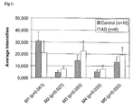
- As shown in Figure 1, compared to a negative diagnosis (healthy controls), M1 is under-expressed in the CSF of Alzheimer's disease patients (p < 0.05), while the markers M2 to M5 are over-expressed in CSF of Alzheimer's disease patients (p < 0.05).
- An altered level of one or several polypeptides of the invention, compared to the level of polypeptides of the invention in healthy control subjects, will allow assessing the state of and/or monitoring the progression of Alzheimer's disease in a subject, will allow monitoring the effectiveness of Alzheimer's disease treatment, and will be useful information for drug development. Furthermore, these biomolecule markers are useful for differentiating Alzheimer's disease from other forms of dementia and neurodegenerative disorders.
- Preferred subjects in which Alzheimer's disease is to be diagnosed or monitored are human subjects. However, diagnosis of Alzheimer's disease according to the invention is also possible with other mammals. If necessary, orthologues of the peptide markers of the invention can be used.
- The invention also relates to the use of mass spectrometry (MS) for detecting Alzheimer's disease in human subjects and for assessing the progression of Alzheimer's disease in human subjects by detecting and/or quantifying the amount of specific polypeptides in samples drawn from the subject's body fluids. In a preferred embodiment of the invention, the sample is drawn from the subject's cerebrospinal fluid (CSF).
- Detection and/or quantification of the polypeptides of the invention is preferably achieved by quantifying the signal which is detected by MS at specific molecular mass to charge ratios (M/z) which correspond to the M/z ratios of the polypeptides of the invention. Preferably, M/z ratios close to the one of the polypeptides of the invention are also measured.
- Detection and/or quantification of the polypeptides of the invention can also be achieved by using an immunoassay using specific antibodies raised against the specified marker(s) or polypeptide fragments thereof. Antibodies can be prepared by using the purified marker(s) or fragments thereof, or using synthetic or recombinantly expressed polypeptide(s) consisting of the specific amino acid sequence of the marker(s) using any suitable method known in the art [Coligan 1991]. Such techniques include, but are not limited to, antibody preparation by selection of antibodies from libraries of recombinant antibodies in phage or appropriate vectors, as well as preparation of polyclonal and monoclonal antibodies by immunising rabbits or mice [Huse 1989, Ward 1989]. After the antibody is provided, a marker can be detected and/or quantified using any of a number of standard immunological binding assays [US Patents 4,366,241; 4,376,110; 4,517,288; and 4,837,168]. Useful assays include, but are not limited to, for example, an enzyme immune assay (EIA) such as enzyme-linked immunosorbent assay (ELISA), a radioimmune assay (RIA), a Western blot assay, or a slot or dot blot assay. For a review of the general immunoassays see [Coligan 1991]. Generally, a sample obtained from a subject can be contacted with the antibody that specifically binds the marker. A powerful technique to capture the specified marker(s) from a complex body fluid sample is to use the antibody fixed to solid supports, such as glass or plastic, e.g. microtiter plate, a stick, a bead, or microbead. Alternatively, marker(s) can also be captured from the body fluid sample by the specific antibody immobilised to a probe substrate or a ProteinChip™ array, as described for the SELDI-based immunoassay [Xiao 2001]. After incubating the sample with antibodies, the non-bound material is washed under specified conditions and the antibody-marker complex formed can be detected, using appropriate detection reagents. In an embodiment using the SELDI ProteinChip™ array technique the marker(s) selectively enriched by the immobilised antibody can be detected and quantified by matrix-assisted laser desorption ionisation mass spectrometry.
- The invention specifically relates to
- 1. a method of assessing the state of Alzheimer's disease in a subject comprising
detection of at least one polypeptide comprised in a group of polypeptides
consisting of
- i) a polypeptide having a molecular mass of 4824 ± 20 Da,
- ii) a polypeptide having a molecular mass of 7691 ± 20 Da,
- iii) a polypeptide having a molecular mass of 11787 ± 20 Da,
- iv) a polypeptide having a molecular mass of 11988 ± 20 Da, and
- v) a polypeptide having a molecular mass of 13416 ± 20 Da. Whereas detection of one such polypeptide is in most cases sufficient to reliably diagnose Alzheimer's disease, detection of two or more polypeptides of the invention can increase the sensitivity and robustness of the method. Preferably, 1, 2, 3, 4, and, most preferred, 5 of said polypeptides will be detected from the same sample. The detection can also be carried out simultaneously with the detection of other polypeptides which are preferably also differentially expressed in subjects having Alzheimer's disease as compared to healthy subjects.
- 2. The invention further relates to the method of point 1 in which 2, or 3, or 4, or 5 polypeptides of said group of peptides are detected.
- 3. The invention further relates to a method of assessing the state of Alzheimer's disease in a subject comprising detection of at least one polypeptide comprising the sequence of SEQ ID NO:1, SEQ ID NO:2, SEQ ID NO:3, SEQ ID NO:4, SEQ ID NO:5, SEQ ID NO:6, SEQ ID NO:7, SEQ ID NO:8, SEQ ID NO:9, SEQ ID NO:10, SEQ ID NO:11, SEQ ID NO:12, SEQ ID NO:13, SEQ ID NO:14, SEQ ID NO:15, and/or SEQ ID NO:16. Whereas detection of one such polypeptide is in most cases sufficient to reliably diagnose Alzheimer's disease, detection of two or more polypeptides of the invention can increase the sensitivity and robustness of the method. Preferably, 1, 2, 3, 4, 5, 10, or all of said polypeptides will be detected from the same sample. The detection can also be carried out simultaneously with the detection of other polypeptides which are preferably also differentially expressed in subjects having Alzheimer's disease as compared to healthy subjects.
- 4. The invention further relates to a method of assessing the state of Alzheimer's
disease in a subject comprising detection of at least one polypeptide comprised
in a group of polypeptides consisting of
- i) human cystatin C,
- ii) human beta-2-microglobulin,
- iii) human myoglobin,
- iv) a fragment of at least 5, 8, 10 , or 20 amino acids of human cystatin C,
- v) a fragment of at least 5, 8, 10 , or 20 amino acids of human beta-2-microglobulin, and
- vi) a fragment of at least 5, 8, 10 , or 20 amino acids of human myoglobin. Whereas detection of one such polypeptide is in most cases sufficient to reliably diagnose Alzheimer's disease, detection of two or more polypeptides of the invention can increase the sensitivity and robustness of the method. Preferably, 1, 2, 3, 4, 5, or 6 of said polypeptides will be detected from the same sample. The detection can also be carried out simultaneously with the detection of other polypeptides which are preferably also differentially expressed in subjects having Alzheimer's disease as compared to healthy subjects.
- 5. The invention further relates to a method of investigating the progression of Alzheimer's disease in a subject characterised in that a method of any of claims 1 to 4 is performed with at least two distinct samples drawn from the same subject. For this purpose, samples drawn from a subject at different points in time will be analysed. Changes in the amount of the respective polypeptide(s) will allow to draw conclusions on the progression of Alzheimer's disease in the subject.
- 6. The invention further relates to a method of any of points 1 to 5, wherein detection of said polypeptide(s) is by SELDI-TOF MS. Other suitable mass spectrometric methods and other methods of detection can alternatively be used.
- 7. The invention further relates to a method of any of points 1 to 5, wherein specific antibodies or antibodies recognising said polypeptide(s) are used for detection of said polypeptide(s).
- 8. The invention further relates to a method of any of claims 1 to 7, wherein detection is in a sample comprising CSF of said patient. A sample drawn from a subject can be processed immediately after it has been taken, or it can first be frozen and be analysed later. Samples may also consist of or contain other body fluids such as blood, serum, plasma, urine, seminal plasma, nipple fluid, or cell extracts.
- 9. The invention further relates to a kit comprising a polypeptide having a molecular mass of 4824 ± 20 Da, a polypeptide having a molecular mass of 7691 ± 20 Da, a polypeptide having a molecular mass of 11787 ± 20 Da, a polypeptide having a molecular mass of 11988 ± 20 Da, and/or a polypeptide having a molecular mass of 13416 ± 20 Da. The invention further relates to a kit comprising a polypeptide having a molecular mass of 4824 ± 20 Da, a polypeptide having a molecular mass of 7691 ± 20 Da, a polypeptide having a molecular mass of 11787 ± 20 Da, a polypeptide having a molecular mass of 11988 ± 20 Da, and a polypeptide having a molecular mass of 13416 ± 20 Da. This kit can be applied for various purposes, e.g., for use as a standard in one of the above mentioned methods.
- 10. The invention further relates to a kit comprising a fragment of at least 5 amino acids of human cystatin C, a fragment of at least 5 amino acids of human beta-2-microglobulin, and a fragment of at least 5 amino acids of human myoglobin. This kit can be applied for various purposes, e.g., for use as a standard in one of the above mentioned methods.
-
- Average intensities of the five marker peptides differentially expressed in the diseased group when compared to the control groups.
- The invention is further described by one or several of the following examples. These examples are not to be understood as restricting the scope of the invention to the examples by any means.
- Diagnosis of Alzheimer's disease in human subjects was made according to criteria of the National Institute of Neurologic and Communicative Disorders and Stroke-Alzheimer's disease and Related Disorders Association (NINCDS-ADRDA). The Alzheimer's disease group consisted of 9 patients aged 75±7 years, six men and three women. The group of healthy control subjects consisted of 10 individuals aged 78 ± 14 years, two men and eight women with no history, symptoms or signs of psychiatric or neurological disease.
- Informed consent was given by each patient and the patients' caregivers before the investigation. The study was approved by the local ethics committee. After lumbar puncture, CSF samples were frozen on dry ice immediately upon withdrawal at the bedside in 0.5 ml aliquots and stored at -80°C until analyses.
- SAX 2 Proteinchip array (Ciphergen Biosystems, Fremont, CA, USA) were equilibrated for 5 min with 5µl of binding buffer (100mM Na Acetate pH=4.0). The buffer was carefully removed with an handkerchief and 2.5µl of binding buffer was added to the wells. Crude CSF samples (2.5µl) were added to the wells and incubated for 20 min at room temperature in a humidity chamber on a rocking platform. CSF was removed and the wells were individually washed with 10µl of binding buffer for 5 min. The arrays were then placed in a 15ml conical Eppendorf and washed twice with the binding buffer for 5 min. Finally, the chip was rinsed twice with distilled water. Excess of H2O was removed and while the surface was still moist, two additions per well of 0.5µl of sinapinic acid (SPA) (2mg/ml) in 50% (vol/vol) acetonitrile and 0.5% (vol/vol) trifluoroacetic acid was performed and dried. The arrays were then read in a ProteinChip reader system, PBS II series (Ciphergen Biosystems). The laser beam was focused on the sample in vacuo. This caused the proteins absorbed to the matrix to become ionised and, simultaneously to be desorbed from the Proteinchip array surface. The ionised proteins were detected and their molecular masses were determined according to their time-of-flight (TOF). TOF mass spectra, collected in the positive ion mode were generated using an average of 65 laser shots throughout the spot at a laser power set slightly above threshold (10-15 % higher than the threshold) High mass to acquire was set at 40kDa, optimised from 1 to 15kDa. Spectra were collected and analysed using the Ciphergen Proteinchip (version 3.0) software. External calibration of the reader was performed using the "all-in-1" peptide molecular weight standards (Ciphergen biosystems, Inc.) diluted in the SPA matrix (1:1, vol/vol) and directly applied onto a well. Protein profile comparison was performed after normalisation on total ion current of all the spectra included in the same experiment. The reproducibility was tested by analysing different aliquots of the same CSF sample on 4 different wells of the same proteinchip array (intraassay intrachip reproducibility), on two different chips (intraassay interchip reproducibility) processed in parallel, and reproduced in an other experiment (interassay reproducibility).
- Analysis of CSF samples from 9 patients diagnosed with Alzheimer's disease relative to 10 controls revealed that 5 peaks were significantly differentially expressed between the two groups (p<0.05). The approximate average SELDI mass associated with the five differentially expressed proteins was 4.82kDa, 7.7kDa, 11.8kDa and 12.0kDa and 13.4kDa (p<0.05) (see Fig. 1).
- In order to identify the proteins corresponding to these peaks, a fractionation of crude CSF on a SAX spin column was performed. The eluted fractions were analysed by SELDI-TOF MS.
- SAX spin column, lot number SAX2-001116-01, (Ciphergen Biosystems, Fremont, CA, USA) was rehydrated overnight at 4°C in the equilibration buffer (20mM tris(hydroxymethyl)aminomethane hydrochloride (Tris-HCl), 5mM NaCl, pH 9.0). The column was warmed up at room temperature and air bubbles were removed. The equilibration buffer was let flow through column matrix by gravity. Equilibration buffer (0.5ml) was added to the column and passed through the resin twice. Two ml of control CSF was diluted in the equilibration buffer (1:1, vol/vol). Protein sample was loaded to the column by fraction of 0.8ml and allowed to run through the column by gravity until no drops came out of the column. The column was then centrifuged at 150 x g for 1 min. The resin was then washed with an equivalent volume of equilibration buffer. This step was repeated several times in order to load the whole sample onto the resin. Elution of the bound proteins was performed by decreasing the pH. Elution buffer A consisted of 20mM Tris-HCl 5mM NaCl pH 8.0; elution buffer B = 20mM sodium phosphate pH 7.0; elution buffer C = 20mM sodium phosphate pH 6.0; elution buffer D = 20mM sodium phosphate and citrate pH 5.0; elution buffer E = 20mM sodium phosphate and citrate pH 4.0; elution buffer F = 20mM sodium phosphate and citrate pH 3.4; elution buffer G = 30% acetonitrile in elution buffer F. Elution was performed by applying 2 x 75 µl of the elution buffer and centrifugation at 150 x g for 1min. Each collected fraction (150 µl) was concentrated on a speed-vac to a volume of 10µl. Protein profiles were analysed on SELDI-TOF MS using SAX 2 Proteinchip arrays. The chip was equilibrated with a binding buffer consisting of 20mM Tris-HCl, 5mM NaCl, pH=9.0. An aliquot of 0.5 µl of each concentrated fraction was applied directly onto 2.5µl of binding buffer per spot and processed as previously described. The rest of the fractions were loaded onto a Tris tricine gel as described below.
- The differentially expressed peak of 13.4kDa was eluted with buffer A (20mM Tris-HCl 5mM NaCl pH 8.0) and B (20mM sodium phosphate pH 7.0). The differentially expressed peaks of 11.8kDa and 12.0kDa were found in the fraction eluted with buffer C (20mM sodium phosphate pH 6.0) and D (20mM sodium phosphate and citrate pH 5.0). The cluster of 7.7kDa was eluted with buffer D (20mM sodium phosphate and citrate pH 5.0) and E (20mM sodium phosphate and citrate pH 4.0).
- Each eluted fraction was loaded on a 16.5% Tris Tricine sodium dodecyl sulfate polyacrylamide gel and electrophoresed (SDS PAGE). After coloration with coomassie blue, the bands seen on the gel confirmed the results obtained by SELDI analysis. The band corresponding to the cluster of 7.7kDa, 11.8kDa and 12.0kDa were cut out. Proteins were extracted as described in Example 6 and identified by Q-TOF. The 7.7kDa peak MS analysis did not match with any known human protein, however, may indicate to a new variant or homologue of myoglobin. Peptide sequences were the followingand


- The MS analysis of the 11.8kDa and 12.0kDa peaks identified beta-2-microglobulin for both of them.
- Since the cluster of 13.4kDa could not be seen on the Tris Tricine gel, a Tris glycine SDS-PAGE electrophoresis was performed on crude CSF samples. The band corresponding to the beta-2-microglobulin could be easily found on this stained gel. We concluded that the protein migrating just above the beta-2-microglobulin could correspond to the next abundant protein seen on the SELDI profile, namely the 13.4kDa peak. The band was excised from the gel and digested by trypsin before MALDI analysis. The peptide mass fingerprint analysis allowed to identify the Cystatin C. The sequence coverage provided by the analysis was 60%.
- Twenty µl of CSF were mixed with 10 µl of denaturing Laemmli buffer [Laemmli 1970]. The samples were heated to 95°C for 5 min, and loaded on a 15% T (T = total acrylamide concentration) SDS-polyacrylamide gel according to the method of Laemmli. Gels were stained in a solution containing Coomassie Brilliant Blue R-250 (0.1% w/v) and methanol (50% v/v) for 30 min. Destaining was done in a solution containing methanol (40% v/v) and acetic acid (10% v/v).
- Tris tricine SDS-PAGE electrophoresis was performed according to Schagger and von Jagow [1987] using precast 16.5 % T gels (Biorad, Hercules, CA). The anode buffer consisted of 0.2M Tris-HCl, pH 8.9 and the cathode buffer consisted of 0.1M Tris-HCl, 0.1M Tricine, 0.1% SDS, pH 8.25. Samples were diluted in 10µl of 50mM Tris-HCl, 4% (w/v) SDS, 12% (w/v) sucrose, 5% (v/v) β-mercaptoethanol, and trace of bromophenol blue, pH 6.8. After denaturation at 95°C for 5min, samples were loaded onto the gel. Gels were run at 80V for 3 hours. After electrophoresis, gels were fixed in 40% methanol, 10% acetic acid for 30min. Gels were then stained with Colloidal blue coomassie G250 overnight and destained in 30% methanol. Bands to be identified were immediately cut, placed in an eppendorf and kept at 4°C until further analysis. The apparent molecular masses were determined by running polypeptide molecular weight (MW) standards: Triosephophate isomerase MW 26,625; Myoglobin MW 16,950; α-lactalbumin MW 14,437; Aprotinin MW 6,512; Insulin b chain, oxidised MW 3,496 and Bacitracin MW 1,423 (Biorad).
- Fragments of gels containing proteins of interest were cut out for digestion of the proteins with trypsin using previous published procedures [Shevchenko 1996, Hellman 1994, Rosenfeld 1992] and modified as described below. The piece of gel was first destained with 100 µl of 50 mM ammonium bicarbonate, 30% (v/v) acetonitrile during 15 min at room temperature. Destaining solution was removed and replaced by 25 µl of 10 mM DL-dithiothreitol (DTT) in 50 mM ammonium bicarbonate and incubated 35 min at 56°C. DTT solution was then replaced by 25 µl of 55 mM iodoacetamide in 50 mM ammonium bicarbonate and incubated during 45 min at room temperature in the dark. Gel pieces were washed for 10 min with 100 µl of 50 mM ammonium bicarbonate and for 10 min with 100 µl of 50 mM ammonium bicarbonate and 30% (v/v) acetonitrile.
- Gel pieces were then dried for 30 min in a Hetovac vacuum centrifuge (HETO, Allerod, Denmark). Dried pieces of gel were rehydrated for 45 min at 4°C in 5-20 µl of a solution of 50 mM ammonium bicarbonate containing trypsin at 6.25 ng/µl. After an over-night incubation at 37°C, gel pieces were dried under high vacuum centrifuge before being rehydrated by the addition of 20 µl of distilled water and finally dried again in a speed-vac for 30 min. Extraction of the peptides was performed with 20 µl of 0.1% (v/v) trifluoroacetic acid (TFA) for 20 min at room temperature with occasional shaking. The TFA solution containing the peptides was transferred to a polypropylene tube. A second elution was performed with 20 µl of 0.1% (v/v) TFA in 50% (v/v) acetonitrile for 20 min at room temperature with occasional shaking. The second TFA solution was pooled with the first one. The volume of the pooled extracts was reduced to 1-2 µl by evaporation under vacuum. Control extractions (blanks) were performed using pieces of gels devoid of proteins.
- 1.5 µl of sample was placed on a MALDI 100-well target plate. Same volumes of matrix (10 mg/ml α-Cyano-4-hydroxycinnamic acid in 50% (v/v) acetonitrile, 0.1% (v/v) TFA) were added to the previously loaded digest. Samples were dried as quickly as possible using a vacuum container. Mass measurement from liquid solution were conducted with a MALDI-TOF mass spectrometer Voyager™ Elite and Super STR (PerSeptive Biosystems, Framingham MA, USA) equipped with a 337 nm nitrogen laser. The analyser was used in the reflectron mode at an accelerating voltage of 20 kV, a delayed extraction parameter of 100-140 ns and a low mass gate of 850 Da. Laser power was set slightly above threshold (10-15 % higher than the threshold) for molecular ion production. Spectra were obtained by summation of 10 to 256 consecutive laser shots. Masses of the 60 highest peaks were extracted from the spectra and used for protein identification using the SmartIdent peptide mass fingerprint tool [Gras 1999]. The research was conducted against SWISS-PROT and TrEMBL databases. The query was made for the human, the minimum number of matched masses was 4, the maximal tolerance for masses was 50 ppm after an internal calibration using autolysis product of trypsin, at most one missed cleavage for tryptic peptides was allowed, and the modifications accepted were carboxymethylation with iodoacetamide of cysteines and artefactual oxidation of methionines.
- Prior to nanoLC (LC = liquid chromatography) separation, the volumes of peptide containing solutions were adjusted to 7µl by addition of a 0.1 % (v/v) formic acid solution. Samples were settled in a Triathlon autosampler (Spack, Emmen, Holland). For each experiment, 5 µl of peptide containing solution were injected on a C18 reverse phase column of 75 µm inner diameter (YMS-ODS-AQ200, Michrom Bioresource, Auburn, CA). Peptides were eluted with an acetonitrile (ACN) gradient in the presence of 0.1 % (v/v) formic acid, using SunFlow pumps (SunChrom, Friderichsdorf, Germany). A flow splitter was used in order to decrease the flow rate after the pumps from 200 to 0.4 µl/min. Peptides were analysed with a quadrupole time-of-flight (Q-TOF) mass spectrometer (Micromass, Wythenshawe, England). A 2700 V tension was applied on the nanoelectrospray capillary (New Objective, Woburn, MA, USA). Argon was used as collision gas. The collision energy was settled as a function of the precursor ion mass. MS/MS spectra were acquired by automatic switching between MS and MS/MS mode. Acquired MS/MS data were converted in a compatible format (DTA files) by ProteinLynx software (Micromass, Wythenshawe, England) and analysed using conventional search engines against SWISS-PROT, TrEMBL, NCBInr and EST databases. In cases of manual interpretation of MS/MS data, identification was performed by sequence only search.
- It was found that marker M5 was a fragment of Cystatin C, markers M3 and M4 were isoforms of beta-2-microglobulin and M2 was a new variant or homologue of myoglobin. Marker M1 could not be assigned to any known protein and appears to be an unknown polypeptide.
- P-values were calculated using standard statistical methods known to the person skilled in the art. P-values smaller than 0.05 were considered to be statistically significant.
-
- Adam, B.L. et al. 2001, Proteomics 1(10): 1264-1270
- Asgeirsson B, Haebel S, Thorsteinsson L, Helgason E, Gudmundsson KO, Gudmundsson G, Roepstorff P. Hereditary cystatin C amyloid angiopathy: monitoring the presence of the Leu68Gln cystatin C variant in cerebrospinal fluid and monocyte cultures by MS. Biochem J. 1998, 329, 497-503.
- Beyer K, Lao JI, Gomez M, Riutort N, Latorre P, Mate JL, Ariza A. Alzheimer's disease and the cystatin C gene polymorphism: an association study. Neuroscience letters. 2001, 315, 17-20.
- Cohen DH, Feiner H, Jensson O, Frangione B. Amyloid fibril in hereditary cerebral hemorrhage with amyloidosis (HCHWA-I) is related to the gastroentero-pancreatic neuroendocrine protein, □-trace. J. Exp. Med. 1983, 158, 623-628.
- Current Protocols in Immunology, Eds. Coligan JE, Kruisbeek AM, Margulies DH, Shevach EM, and Strober W., John Wiley & Sons Inc. 1991
- Crawford U, Freeman MJ, Schinka JA, Abdullah LI, Gold M, Hartman R, Krivian K, Morris MD, Richards D, Duara R, Anand R, Mullan MJ. A polymorphism in the cystatin C gene is a novel rsik factor for late-onset Alzheimer's disease. Neurology. 2000, 55, 763-768.
- Davies, H.A. 2000. The ProteinChip(R) System from Ciphergen: A new technique for rapid, micro-scale protein biology. J. Molecular Medicine, 78 (7):B29 Deng A, Irizarry MC, Nitsch RM, Growdon JH, Rebeck GW. Elevation of Cystatin C in susceptible neurons in Alzheimer's disease. Am. J. Pathol. 2001, 159, 1061-1068.
- Ernerudh J, Olsson T, Berlin G, von Schenck H. Cerebrospinal fluid immunoglobulins and beta 2-microglobulin in lymphoproliferative and other neoplastic diseases of the central nervous system. Arch Neurol. 1987, 44(9), 915-20.
- Finckh U, Von der Kammer H, Velden J, Michel T, Andresen B, Deng A, Zhang J, Muller-Thomsen T, Zuchowski K, Menzer G, Mann U, Papassotiropoulos A, Heun R, Zurdel J, Holst F, benussi L, Stoppe G, Reiss J, Miserez AR, Staehelin HB, Rebeck W, Hyman BT, Binetti G, Hock C, Growdon JH, Nitsch RM. Genetic association of a cystatin C gene polymorphism with late-onset Alzheimer's disease. Arch Neurol. 2000, 57, 1579-1583.
- Francis, P.T. et al. J. Neurol. Neurosurg. Psychiatry 1999, 66(2): 137-147 Fung, ET et al. Protein biochips for differential profiling. Current Opinion in Biotechnology, 2001, 12(1): 65-69
- Ghiso J, Jensson O, Frangione B. Amyloid fibrils in hereditary cerebral hemorrhage with amyloidosis of Icelandic type is a variant of gamma-trace basic protein (cystatin C). Proc Natl Acad Sci U S A. 1986;83(9):2974-8.
- Ghiso J, Pons-Estel B, Frangione B. Hereditary cerebral amyloid angiopathy: the amyloid fibrils contain a protein which is a variant of cystatin C, an inhibitor of lysosomal cysteine proteases. Biochem Biophys Res Commun. 1986;136(2):548-54. Gras R, Müller M, Gasteiger E, Gay S, Binz PA, Bienvenut WV, Hoogland C, Sanchez JC, Bairoch A, Hochstrasser DF, Appel R. Improving protein identification from peptide mass fingerprinting through a parameterised multi-level scoring algorithm and an optimised peak detection. Electrophoresis 1999, 20: 3535-3550 Grubb AO, Jensson O, Gudmundsson G, Arnason A, Lofberg H, Malm J. Abnormal metabolism of g-trace alkaline microprotein. The basic defect in hereditary cerebral hemorrhage with amyloidosis. N. Engl. J. Med 1984, 311, 1547-1549.
- Hellman U, Wernstedt C, Góñez, J, Heldin CH. Improvement of an "In-Gel" digestion procedure for the micropreparation of internal protein fragments for amino acid sequencing. Anal. Biochem. 1995, 224, 451-5.
- Hoekman K, Van Nieuwkoop JA, Willemze R. The significance of beta-2 microglobulin in clinical medicine. Neth J Med. 1985;28(12):551-7. Huse et al. 1989, Science 246: 1275-1281
- Issaq, H.J. et al. 2002, Biochem. Biophys. Res. Commun. 292: 587-592 Kalman J, Marki-Zay J, Juhasz A, Santha A, Dux L, Janka Z. Serum and Cerebrospinal fluid cystatin C levels in vascular and Alzheimer's dementia. Acta Neurol. Scand. 2000, 101, 279-282.
- Laemmli UK. Cleavage of structural proteins during the assembly of the head of bacteriophage T4. Nature. 1970 Aug 15;227(259):680-5.
- Leung-Tack J, Tavera C, Gensac MC, Martinez J, Colle A. Modulation of phagocytosis-associated respiratory burst by human cystatin C: role of the N-terminal tetrapeptide Lys-Pro-Pro-Arg. Exp. Cell Research. 1990, 188, 16-22.
- Levy E, Sastre M, Kumar A, Gallo G, Piccardo P, Ghetti B, Tagliavini F, Codeposition of cystatin C with amyloid-beta protein in the brain of Alzheimer's disease patients. J. Neuropathol Exp Neurol 2001, 60, 94-104
- Merchant, M. et al. 2000. Electrophoresis, 21:1164-1167
- Popovic T, Brzin J, Ritonja A, Turk V. Different forms of human Cystatin C. Biol. Chem. Hoppe-Seyler. 1990, 371, 575-580.
- Raymackers J, Daniels A, DeBrabandere V, Missiaen C, Dauwe M, Verhaert P, Vanmechelen E, Meheus L. Identification of two dimensionally separated human cerebrospinal fluid proteins by N-terminal sequencing, matrix-assisted laser-desorption/ionization-mass spectrometry, nanoliquid chromatography-electrospray ionization-time of flight-mass spectrometry, and tandem mass spectrometry. Electrophoresis, 2000, 21, 2266-2283.
- Robles A. 1998. Neurobiology of Aging, 19, 109-116.
- Robles A. 1998b. Neurobiology of Aging, 19 (2): 153-157
- Rosenfeld J, Capdevielle J, Guillemot JC, Ferrara P. In-gel digestion of proteins for internal sequence analysis after one- or two-dimensional gel electrophoresis. Anal. Biochem. 1992, 203, 173-9.
- Shevchenko A, Wilm M, Vorm O, Mann M. Mass spectrometric sequencing of proteins silver-stained polyacrylamide gels. Anal. Chem. 1996, 68, 850-8.
- Teunissen CE et al 2002. Neurobiology of aging 23: 485-508
- US Patent 4,366,241
- US Patent 4,376,110
- US Patent 4,517,288
- US Patent 4,837,168
- Ward ES et al., Binding activities of a repertoire of single immunoglobulin variable domains secreted from Escherichia coli. Nature 1989, 341, 544-546
- Wei L, Berman Y, Castano EM, Cadene M, Beavis RC, Devi L, Levy E. Instability of the amyloidogenic cystatin C variant of hereditary cerebral hemorrhage with amyloidosis, Icelandic type. J. Biol. Chem. 1998, 273, 11806-11814.
- Wulfkuhle J.D. et al. 2001, Proteomics 1(10): 1205-1215
- Xiao Z. et al. 2001, Cancer Research 61 (16): 6029-6033
-
"Assessing the state of Alzheimer's disease" shall be understood as diagnosing the presence of Alzheimer's disease in a subject or a patient, as assessing the progression of the disease in a subject or a patient, and/or as assessing the proneness of a subject to develop Alzheimer's disease.
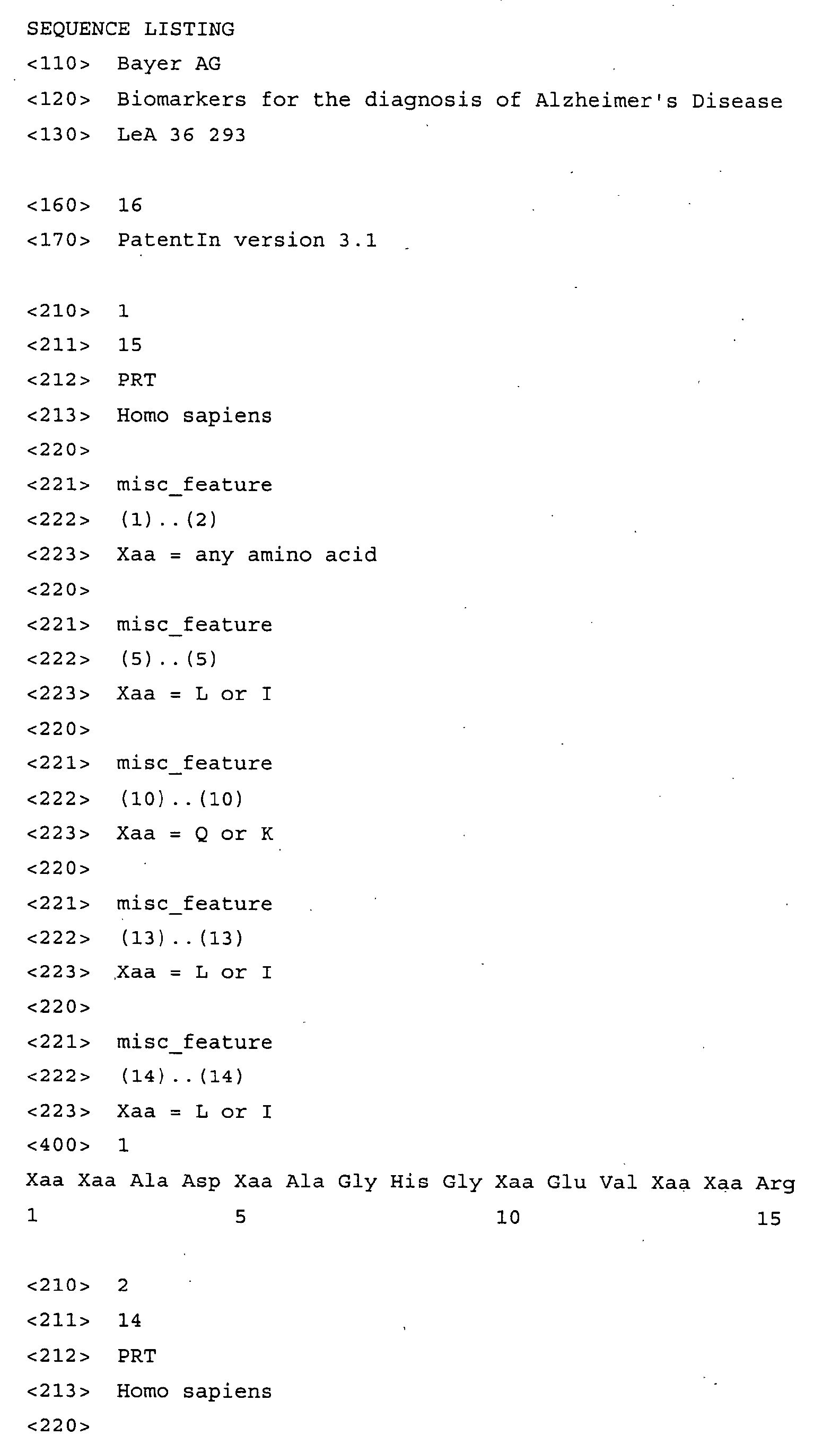
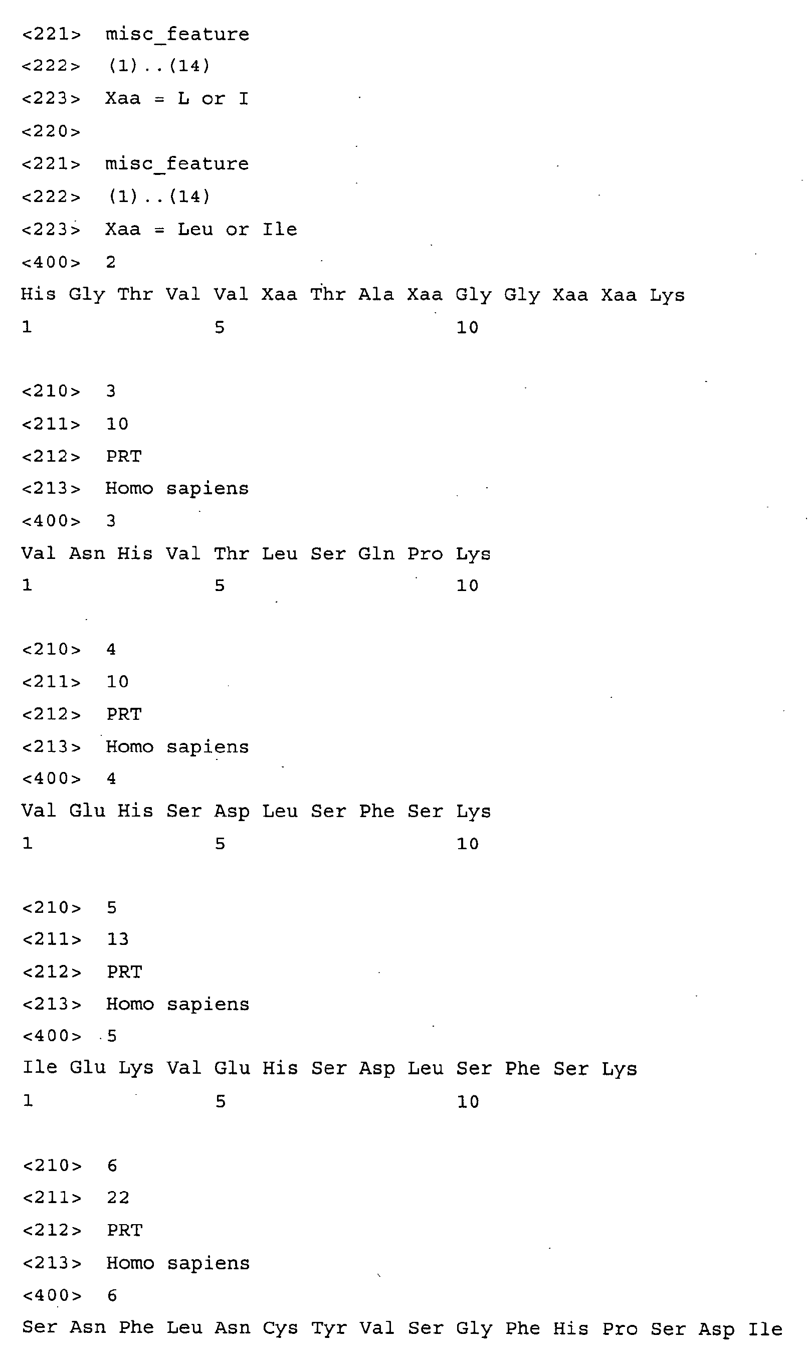


Claims (10)
- Method of assessing the state of Alzheimer's disease in a subject comprising detection of at least one polypeptide comprised in a group of polypeptides consisting ofi) a polypeptide having a molecular mass of 4824 ± 20 Da,ii) a polypeptide having a molecular mass of 7691 ± 20 Da,iii) a polypeptide having a molecular mass of 11787 ± 20 Da,iv) a polypeptide having a molecular mass of 11988 ± 20 Da, andv) a polypeptide having a molecular mass of 13416 ± 20 Da.
- Method of claim 1 in which 2, or 3, or 4, or 5 polypeptides of said group of peptides are detected.
- Method of assessing the state of Alzheimer's disease in a subject comprising detection of at least one polypeptide comprising the sequence of SEQ ID NO:1, SEQ ID NO:2, SEQ ID NO:3, SEQ ID NO:4, SEQ ID NO:5, SEQ ID NO:6, SEQ ID NO:7, SEQ ID NO:8, SEQ ID NO:9, SEQ ID NO:10, SEQ ID NO:11, SEQ ID NO:12, SEQ ID NO:13, SEQ ID NO:14, SEQ ID NO:15, and/or SEQ ID NO:16.
- Method of assessing the state of Alzheimer's disease in a subject comprising detection of at least one polypeptide comprised in a group of polypeptides consisting ofi) human cystatin C,ii) human beta-2-microglobulin,iii) human myoglobin,iv) a fragment of at least 5 amino acids of human cystatin C,v) a fragment of at least 5 amino acids of human beta-2-microglobulin, andvi) a fragment of at least 5 amino acids of human myoglobin.
- Method of investigating the progression of Alzheimer's disease in a subject characterised in that a method of any of claims 1 to 4 is performed with at least two distinct samples drawn from the same subject.
- Method of any of claims 1 to 5, wherein detection of said polypeptide(s) is by SELDI-TOF MS.
- Method of any of claims 1 to 5, wherein specific antibodies or antibodies recognising said polypeptides are used for detection of said polypeptide(s).
- Method of any of claims 1 to 7, wherein detection is in a sample comprising CSF, blood, serum, plasma, urine, seminal plasma, nipple fluid, and/or cell extracts of said patient.
- A kit comprising a polypeptide having a molecular mass of 4824 ± 20 Da, a polypeptide having a molecular mass of 7691 ± 20 Da, a polypeptide having a molecular mass of 11787 ± 20 Da, a polypeptide having a molecular mass of 11988 ± 20 Da, and/or a polypeptide having a molecular mass of 13416 ± 20 Da.
- A kit comprising a fragment of at least 5 amino acids of human cystatin C, a fragment of at least 5 amino acids of human beta-2-microglobulin, and a fragment of at least 5 amino acids of human myoglobin.
Priority Applications (29)
| Application Number | Priority Date | Filing Date | Title |
|---|---|---|---|
| EP02018283A EP1394549A1 (en) | 2002-08-23 | 2002-08-23 | Biomarkers for diagnosing Alzheimer's disease |
| ES10164475T ES2436318T3 (en) | 2002-08-23 | 2003-08-11 | Polypeptide biomarkers to diagnose Alzheimer's disease |
| CA2496321A CA2496321C (en) | 2002-08-23 | 2003-08-11 | Biomarkers for diagnosing alzheimer's disease |
| EP08002793A EP1953556B1 (en) | 2002-08-23 | 2003-08-11 | Polypeptide biomarkers for diagnosing alzheimer's disease |
| DK03792291T DK1535076T3 (en) | 2002-08-23 | 2003-08-11 | Polypeptide biomarkers for the diagnosis of Alzheimer's disease |
| AT03792291T ATE403875T1 (en) | 2002-08-23 | 2003-08-11 | POLYPEPTIDE BIOMARKERS FOR THE DIAGNOSIS OF ALZHEIMER'S DISEASE |
| ES08002793T ES2347924T3 (en) | 2002-08-23 | 2003-08-11 | POLIPEPTIDIC BIOMARKERS TO DIAGNOSTICATE ALZHEIMER'S DISEASE. |
| CA2745998A CA2745998C (en) | 2002-08-23 | 2003-08-11 | Biomarkers for diagnosing alzheimer's disease |
| EP03792291A EP1535076B1 (en) | 2002-08-23 | 2003-08-11 | Polypeptide biomarkers for diagnosing alzheimer's disease |
| AT08002793T ATE475888T1 (en) | 2002-08-23 | 2003-08-11 | BIOMARRKERS FOR THE DIAGNOSIS OF ALZHEIMER |
| EP10164475.5A EP2221620B1 (en) | 2002-08-23 | 2003-08-11 | Polypeptide biomarkers for diagnosing Alzheimer's disease |
| US10/525,633 US20060240561A1 (en) | 2002-08-23 | 2003-08-11 | Biomarkers for diagnosing alzheimer's disease |
| DE60333608T DE60333608D1 (en) | 2002-08-23 | 2003-08-11 | Biomarker for the diagnosis of Alzheimer's |
| DK10164475.5T DK2221620T3 (en) | 2002-08-23 | 2003-08-11 | Polypeptide biomarkers for the diagnosis of Alzheimer's disease |
| CA2745731A CA2745731C (en) | 2002-08-23 | 2003-08-11 | Biomarkers for diagnosing alzheimer's disease |
| PCT/EP2003/008879 WO2004019043A2 (en) | 2002-08-23 | 2003-08-11 | Polypeptide biomarkers for diagnosing alzheimer's disease |
| ES03792291T ES2311751T3 (en) | 2002-08-23 | 2003-08-11 | POLIPEPTIDE BIOMARKERS FOR THE DIAGNOSIS OF ALZHEIMER'S DISEASE. |
| JP2004530120A JP4405919B2 (en) | 2002-08-23 | 2003-08-11 | Biomarkers for the diagnosis of Alzheimer's disease |
| CA2745569A CA2745569C (en) | 2002-08-23 | 2003-08-11 | Biomarkers for diagnosing alzheimer`s disease |
| DK08002793.1T DK1953556T3 (en) | 2002-08-23 | 2003-08-11 | Polypeptide biomarkers for the diagnosis of Alzheimer's disease |
| CA2745724A CA2745724C (en) | 2002-08-23 | 2003-08-11 | Biomarkers comprising peptide sequence vnhvtlsqpk for diagnosing alzheimer's disease |
| PT03792291T PT1535076E (en) | 2002-08-23 | 2003-08-11 | Polypeptide biomarkers for diagnosing alzheimer`s disease |
| DE60322719T DE60322719D1 (en) | 2002-08-23 | 2003-08-11 | POLYPEPTIDE BIOMARKERS FOR THE DIAGNOSIS OF MORBUS ALZHEIMER |
| CA2746000A CA2746000C (en) | 2002-08-23 | 2003-08-11 | Biomarkers for diagnosing alzheimer's disease |
| AU2003251702A AU2003251702B2 (en) | 2002-08-23 | 2003-08-11 | Polypeptide biomarkers for diagnosing alzheimer's disease |
| AU2010212317A AU2010212317B2 (en) | 2002-08-23 | 2010-08-12 | Polypeptide Biomarkers for diagnosing Alzheimer's disease |
| AU2010212316A AU2010212316B2 (en) | 2002-08-23 | 2010-08-12 | Polypeptide Biomarkers for diagnosing Alzheimer's disease |
| US13/099,110 US8772042B2 (en) | 2002-08-23 | 2011-05-02 | Biomarkers for diagnosing alzheimer's disease |
| US14/217,716 US20140194318A1 (en) | 2002-08-23 | 2014-03-18 | Biomarkers for diagnosing alzheimer's disease |
Applications Claiming Priority (1)
| Application Number | Priority Date | Filing Date | Title |
|---|---|---|---|
| EP02018283A EP1394549A1 (en) | 2002-08-23 | 2002-08-23 | Biomarkers for diagnosing Alzheimer's disease |
Publications (1)
| Publication Number | Publication Date |
|---|---|
| EP1394549A1 true EP1394549A1 (en) | 2004-03-03 |
Family
ID=31197803
Family Applications (1)
| Application Number | Title | Priority Date | Filing Date |
|---|---|---|---|
| EP02018283A Withdrawn EP1394549A1 (en) | 2002-08-23 | 2002-08-23 | Biomarkers for diagnosing Alzheimer's disease |
Country Status (2)
| Country | Link |
|---|---|
| US (3) | US20060240561A1 (en) |
| EP (1) | EP1394549A1 (en) |
Cited By (2)
| Publication number | Priority date | Publication date | Assignee | Title |
|---|---|---|---|---|
| WO2008010660A1 (en) * | 2006-07-18 | 2008-01-24 | Hyun-Kee Kim | Marker for diagnosing neurodegenerative diseases |
| WO2022262624A1 (en) * | 2021-06-16 | 2022-12-22 | 厦门大学 | PHARMACEUTICAL USE OF β2-MICROGLOBULIN OR INHIBITOR THEREOF |
Families Citing this family (10)
| Publication number | Priority date | Publication date | Assignee | Title |
|---|---|---|---|---|
| US7749716B2 (en) * | 2004-05-18 | 2010-07-06 | Vermilllion, Inc. | Methods of detecting a fragment of neurosecretory protein VGF for diagnosing alzheimer's disease |
| US20110236917A1 (en) * | 2009-11-17 | 2011-09-29 | Power3 Medical Products, Inc. | Diagnosis of Alzheimer's Disease |
| US20160208011A1 (en) | 2010-01-28 | 2016-07-21 | The Board Of Trustees Of The Leland Stanford Junior University | Ccr3 modulation in the treatment of aging-associated impairments, and compositions for practicing the same |
| US10626399B2 (en) | 2010-01-28 | 2020-04-21 | The Board Of Trustees Of The Leland Stanford Junior University | Methods of treating cognitive symptoms of an aging-associated impairment by modulating C-C chemokine receptor type 3 (CCR3) |
| US10487148B2 (en) | 2010-01-28 | 2019-11-26 | The Board Of Trustees Of The Leland Stanford Junior University | Methods and compositions for treating aging-associated impairments |
| US9161968B2 (en) | 2011-04-08 | 2015-10-20 | The Board Of Trustees Of The Leland Stanford Junior University | Methods of neuroprotection involving macrophage colony stimulating factor receptor agonists |
| NZ720949A (en) | 2013-12-09 | 2019-03-29 | Univ Leland Stanford Junior | Methods and compositions for treating aging-associated conditions |
| US10905779B2 (en) | 2013-12-09 | 2021-02-02 | The Board Of Trustees Of The Leland Stanford Junior University | Methods for screening human blood products comprising plasma using immunocompromised rodent models |
| US20150267936A1 (en) * | 2014-03-24 | 2015-09-24 | Quirky, Inc. | Environmental control apparatus and related methods |
| AU2016279804B2 (en) | 2015-06-15 | 2019-03-07 | The Board Of Trustees Of The Leland Stanford Junior University | Methods and compositions for treating aging-associated conditions |
Citations (2)
| Publication number | Priority date | Publication date | Assignee | Title |
|---|---|---|---|---|
| WO2000025138A2 (en) * | 1998-10-23 | 2000-05-04 | Roger Nitsch | Methods of diagnosing or prognosing alzheimer's disease |
| WO2001063294A2 (en) * | 2000-02-24 | 2001-08-30 | Oxford Glycosciences (Uk) Limited | Diagnosis of bipolar affective disorder (bad) and unipolar depression |
Family Cites Families (4)
| Publication number | Priority date | Publication date | Assignee | Title |
|---|---|---|---|---|
| US6925389B2 (en) * | 2000-07-18 | 2005-08-02 | Correlogic Systems, Inc., | Process for discriminating between biological states based on hidden patterns from biological data |
| WO2002082075A2 (en) * | 2001-04-06 | 2002-10-17 | Biovision Ag | Method for detecting chronic dementia diseases, and corresponding peptides and detection reagents |
| AU2002313737A1 (en) * | 2001-08-13 | 2003-04-01 | University Of Kentucky Research Foundation | Gene expression profile biomarkers and therapeutic targets for brain aging and age-related cognitive impairment |
| WO2004013172A2 (en) * | 2002-07-24 | 2004-02-12 | Innogenetics N.V. | Fragments of beta-amyloid as targets for vaccination against alzheimer disease |
-
2002
- 2002-08-23 EP EP02018283A patent/EP1394549A1/en not_active Withdrawn
-
2003
- 2003-08-11 US US10/525,633 patent/US20060240561A1/en not_active Abandoned
-
2011
- 2011-05-02 US US13/099,110 patent/US8772042B2/en not_active Expired - Fee Related
-
2014
- 2014-03-18 US US14/217,716 patent/US20140194318A1/en not_active Abandoned
Patent Citations (2)
| Publication number | Priority date | Publication date | Assignee | Title |
|---|---|---|---|---|
| WO2000025138A2 (en) * | 1998-10-23 | 2000-05-04 | Roger Nitsch | Methods of diagnosing or prognosing alzheimer's disease |
| WO2001063294A2 (en) * | 2000-02-24 | 2001-08-30 | Oxford Glycosciences (Uk) Limited | Diagnosis of bipolar affective disorder (bad) and unipolar depression |
Non-Patent Citations (2)
| Title |
|---|
| UTAL A ET AL: "Preliminary expression analysis in Alzheimer's disease using SELDI protein chips.", SOCIETY FOR NEUROSCIENCE ABSTRACTS, vol. 26, no. 1-2, 2000, 30th Annual Meeting of the Society of Neuroscience;New Orleans, LA, USA; November 04-09, 2000, pages Abstract No. - 83.7, XP000989844, ISSN: 0190-5295 * |
| WEI, LIHONG ET AL: "Cystatin C: Icelandic-like mutation in an animal model of cerebrovascular.beta.-amyloidosis", STROKE (DALLAS) (1996), 27(11), 2080-2085, XP000998699 * |
Cited By (2)
| Publication number | Priority date | Publication date | Assignee | Title |
|---|---|---|---|---|
| WO2008010660A1 (en) * | 2006-07-18 | 2008-01-24 | Hyun-Kee Kim | Marker for diagnosing neurodegenerative diseases |
| WO2022262624A1 (en) * | 2021-06-16 | 2022-12-22 | 厦门大学 | PHARMACEUTICAL USE OF β2-MICROGLOBULIN OR INHIBITOR THEREOF |
Also Published As
| Publication number | Publication date |
|---|---|
| US20120016205A1 (en) | 2012-01-19 |
| US8772042B2 (en) | 2014-07-08 |
| US20060240561A1 (en) | 2006-10-26 |
| US20140194318A1 (en) | 2014-07-10 |
Similar Documents
| Publication | Publication Date | Title |
|---|---|---|
| AU2010212317B2 (en) | Polypeptide Biomarkers for diagnosing Alzheimer's disease | |
| US8772042B2 (en) | Biomarkers for diagnosing alzheimer's disease | |
| Carrette et al. | A panel of cerebrospinal fluid potential biomarkers for the diagnosis of Alzheimer's disease | |
| JP5479379B2 (en) | Biomarkers for Alzheimer's disease | |
| Carrette et al. | Truncated cystatin C in cerebrospiral fluid: technical artefact or biological process? | |
| CN115066610A (en) | Reagent and method for determining TAU pathological changes and dementia-related diseases | |
| Conti et al. | Pigment epithelium‐derived factor is differentially expressed in peripheral neuropathies | |
| Ásgeirsson et al. | Hereditary cystatin C amyloid angiopathy: monitoring the presence of the Leu-68→ Gln cystatin C variant in cerebrospinal fluids and monocyte cultures by MS | |
| Stem et al. | Antibodies to the β-amyloid peptide cross-react with conformational epitopes in human fibrinogen subunits from peripheral blood | |
| Guiroy et al. | Neurofibrillary tangles of Guamanian amyotrophic lateral sclerosis, parkinsonism-dementia and neurologically normal Guamanians contain a 4-to 4.5-kilodalton protein which is immunoreactive to anti-amyloid β/A4-protein antibodies | |
| WO2020179953A1 (en) | Composite marker for diagnosis of diabetic retinopathy and use thereof | |
| US20220251171A1 (en) | Apolipoprotein e fragments | |
| WO2017086703A1 (en) | Biomarker for diagnosing age-related macular degeneration or diabetic retinopathy and method for diagnosis using same |
Legal Events
| Date | Code | Title | Description |
|---|---|---|---|
| PUAI | Public reference made under article 153(3) epc to a published international application that has entered the european phase |
Free format text: ORIGINAL CODE: 0009012 |
|
| AK | Designated contracting states |
Kind code of ref document: A1 Designated state(s): AT BE BG CH CY CZ DE DK EE ES FI FR GB GR IE IT LI LU MC NL PT SE SK TR |
|
| AX | Request for extension of the european patent |
Extension state: AL LT LV MK RO SI |
|
| 17P | Request for examination filed |
Effective date: 20040903 |
|
| AKX | Designation fees paid |
Designated state(s): AT BE BG CH CY CZ DE DK EE ES FI FR GB GR IE IT LI LU MC NL PT SE SK TR |
|
| 17Q | First examination report despatched |
Effective date: 20050207 |
|
| STAA | Information on the status of an ep patent application or granted ep patent |
Free format text: STATUS: THE APPLICATION HAS BEEN WITHDRAWN |
|
| 18W | Application withdrawn |
Effective date: 20051129 |