-
The present invention relates to diagnostic methods and therapeutics in the field of
tumors involving Hepatocyte Nuclear Factor 1α (HNF1α).
-
The Hepatic Nuclear Factor 1α (HNF1α) gene, also known as the transcription
factor 1 or TCF1 gene, maps to chromosome 12. It is expressed in different tissues including
liver, kidney, pancreas and digestive tract. More particularly this factor represents a crucial
transcriptional activator of many hepatic genes, including albumin, β fibrinogen and α1
antitrypsin (Frain M. et al. (1989), Cereghini S. et al (1990), Baumhueter S. et al (1990),
Chouard T. et al, (1990)). HNF1α is encoded by 10 exons, which span 23 kb. The protein is
comprised of three functional domains. Exon 1 of the HNF1α gene encodes the domain
necessary for dimerization. Exons 2, 3 and 4 encode the domain required for binding to DNA.
Exon 5-10 encode the domain required for transactivation of transcription (C.R. Acad. Sci.
Paris, Life Science (1993) 316:385-94). HNF1α regulates transcription of a particular gene by
dimerizing and binding upstream of the gene's promoter. The transcription factor can form
homodimers with itself or can heterodimerize with the protein product of a different gene
known as vHNF 1.
-
In 1996, Yamagata et al. demonstrated that heterozygous germline mutations of
HNF1α were linked to the occurrence of MODY3 (Maturity Onset Diabetes of the Young
type 3, OMIM #600496) in humans. MODY3 is a dominantly inherited, rare subtype of non-insulin-dependent
diabetes mellitus (NIDDM) characterized by early onset, usually before the
age of 25, and a primary defect in insulin secretion. U.S. patents No. 6,187,533 and U.S. No.
6,143,491 propose that mutations in the HNF1α gene are associated with diabetes and as a
result contemplate methods for diagnosing diabetes and methods for treating diabetes.
-
In a study of hepatic adenomas and hepatocellular carcinomas (HCC), the inventors
have discovered that HNF1α was a tumor suppressor gene.
-
Liver adenomas are benign tumors at risk of malignant transformation. A genome
wide search for loss of heterozygosity (LOH) revealed a chromosome 12q deletion in 5 of 10
adenomas. In most cases, 12q LOH was the only recurrent genetic alteration observed,
suggesting the presence of a tumor suppressor gene therein. A minimal common region of
deletion was defined in 12q24 including the HNF1α gene. Bi-allelic inactivation of HNF1α
was found in 10 out of 16 screened adenomas and heterozygous germline mutation was
present in 3 patients. Furthermore, somatic bi-allelic mutations were also found in two well-differentiated
hepatocellular carcinomas (HCC), occurring in normal liver, out of 30 screened
HCCs. These results reveal that HNF1α inactivation, whether sporadic or associated with
MODY3, is an important genetic event in the occurrence of human liver adenomas and may
be an early step in HCCs.
-
The inventors further studied the impact of HNF1α in colorectal carcinogenesis.
Sequencing of all HNF1α exons with their intron-boundaries was performed in 10 colon
adenomas and 29 colorectal carcinomas. Additional mutations were identified.
-
These results open new diagnostic and therapeutic pathways in the field of tumors
and cancers.
-
Moreover, as liver adenomas are usually observed in young women using oral
contraceptives, the inventors make a link between HNF1α alteration, cellular proliferation
and the use of estroprogestatives.
General definitions
-
The present invention is aimed at providing diagnostic and therapeutic methods in
the field of tumors. In the context of the present invention, the term "tumor" means any
abnormal proliferation of cells. According to the invention, the tumors are generally solid
tumors, that can be benign or malign. Cancers are particularly encompassed. All types of
tissues may be affected, including liver, kidney, pancreas, digestive tract (e.g. stomach,
intestines, and especially colon), or gall-blader. Preferably the tumor is a liver tumor. More
preferably the tumor is a hepatic adenoma or a hepatocellular carcinoma.
-
Hepatic or hepatocellular adenomas are solitary or less frequently multiple primary
tumors usually observed in young women using oral contraceptives (Edmondson H.A. et al
(1976)). Occasionally, their development is associated with the use of anabolic corticosteroids
or with type I glycogen storage disease (Sale G.E. et al (1977), Howell R.R. et al. (1976)).
Histologically, adenomas are well-circumscribed tumors with loss of the normal lobular
architecture, composed of cells resembling normal hepatocytes arranged in cords or plates of
one to two cells thickness, often with areas of dilated sinusoids. Multiple adenomas, or
adenomatosis, is a separate entity where the adenomas have equal frequency in both sexes and
are not influenced by oral contraceptive use. No significant histological differences are
observed between solitary adenomas and those developed in hepatic adenomatosis (Flejou
J.F. et al. (1985), Chiche L. et al. (2000)). A relationship between adenoma and HCC has
been suggested by several clinical reports of malignant transformation of hepatic adenomas
(Goldfard S. (1976), Ferrell L.D. (1993)).
-
Colon or colorectal tumors are also encompassed in the present invention. Liver and
colon carcinogenesis are multi-step processes implicating common pathways such as
activation of the Wnt signalisation, or the P53 and CDKN2A inactivation. In both tumor
types, chromosome instability has been described (Lengauer, et al., 1997 ; Laurent-Puig, et
al., 2001). Genetic instability defining the microsatellite instability phenotype (MSI-H) has
been shown in colorectal cancer and accounts for 15% of colorectal cancer cases. The MSI-H
phenotype is associated with mismatch repair deficiency leading to the accumulation of
genetic alterations in targeted genes with simple nucleotide repeats in their coding sequence.
-
The "subject" or "patient" to be diagnosed or treated may be any human or non-human
animal, preferably a mammal, including rodents, monkeys, cattle, sheep, feline,
canine, horses, rabbits, goats,... Naturally the subject is of a species that expresses HNF1α
and preferably is likely to be at risk to develop a tumor.
-
According to the present invention, "HNF1α protein" "HNF1αgene" or "TCF1
gene " refers to the human form or homologous (namely sequence-conservative and function-conservative)
variants in other species. The cDNA sequence that encodes human HNF1α is
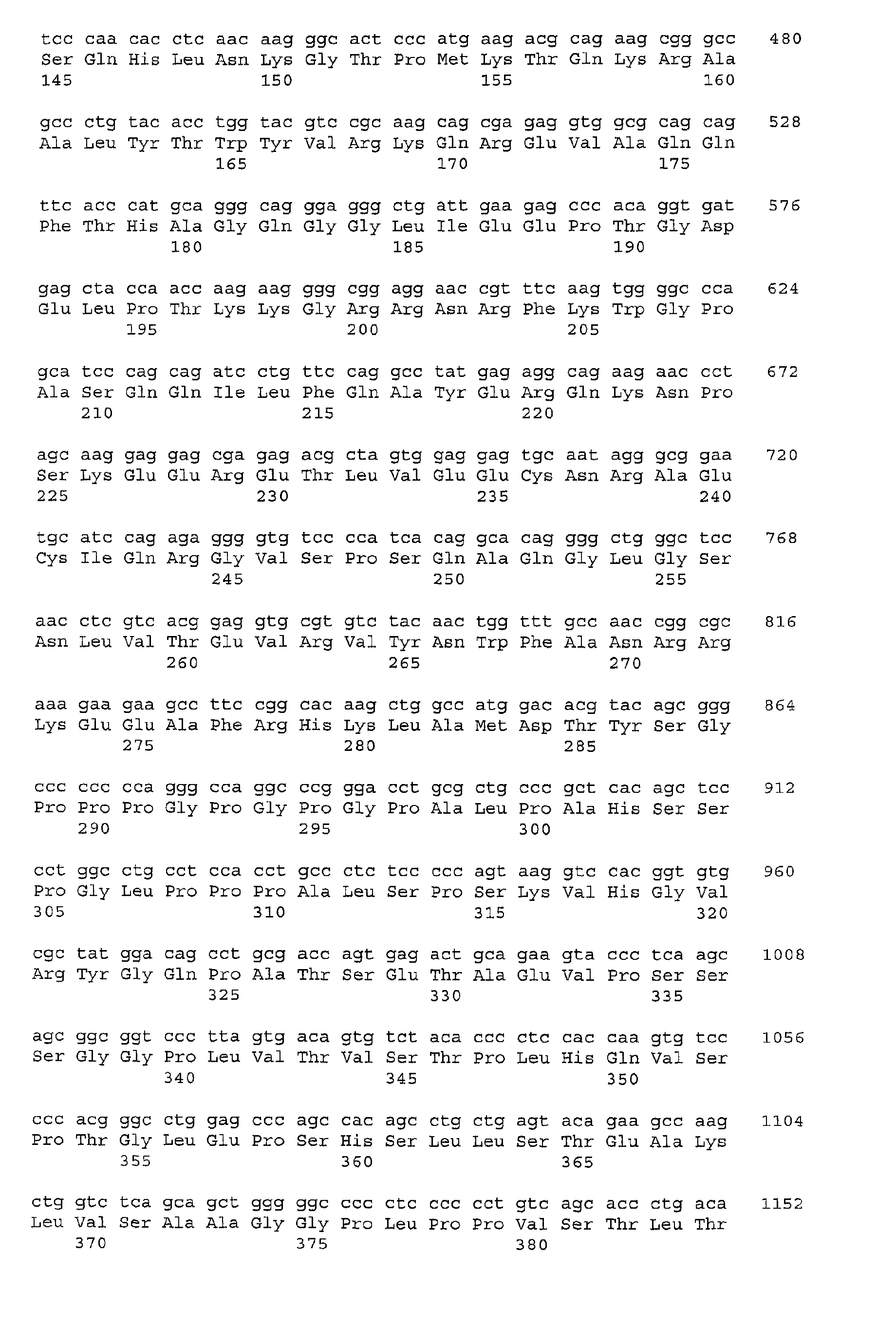
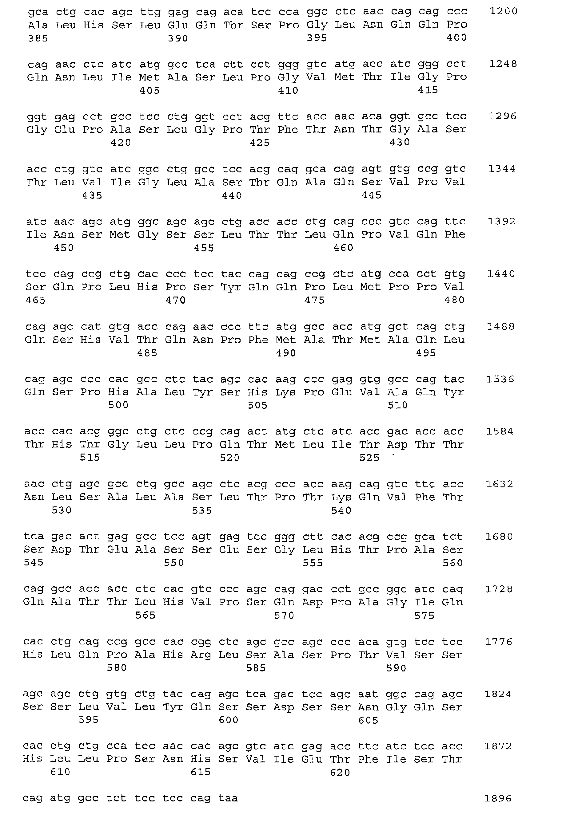
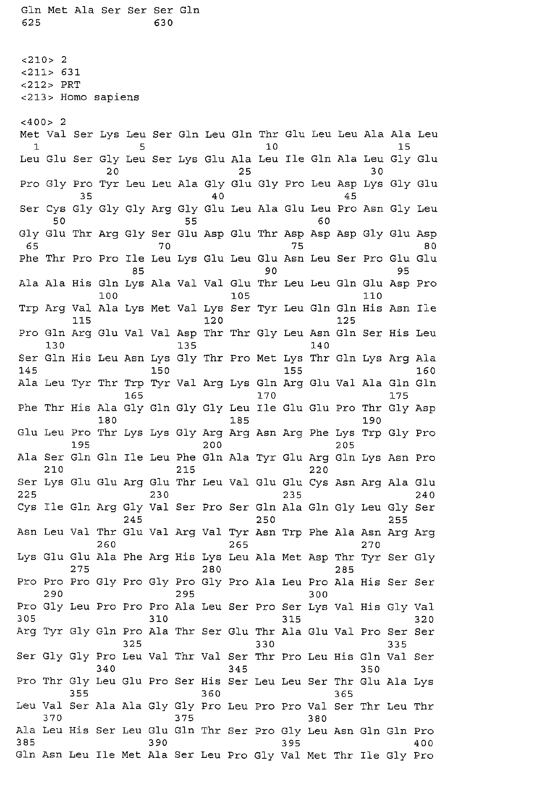
shown in the sequence listing as SEQ ID No. 1. The corresponding amino acid sequence is
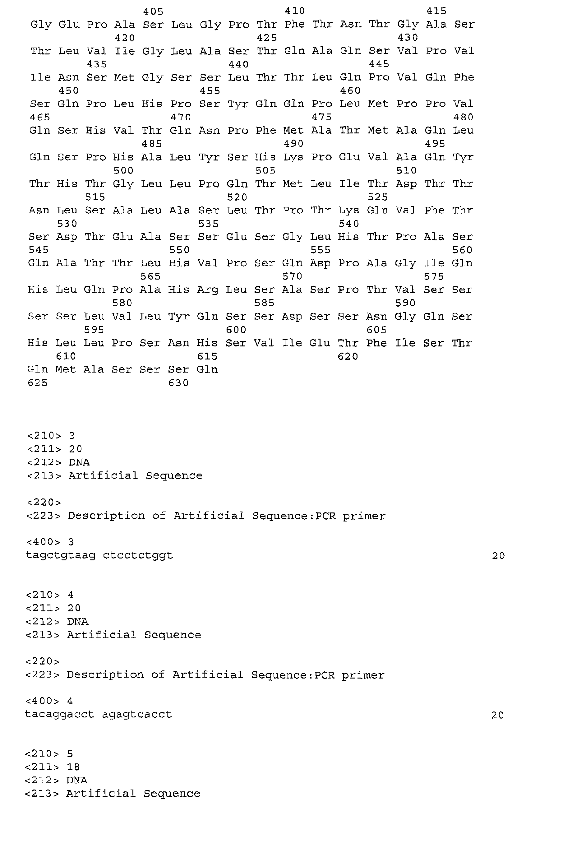
shown as SEQ ID No. 2. HNF1α protein is a 631 amino acids protein composed of three
functional domains: a dimerization domain (AA 1-33), a DNA-binding domain that includes a
region (AA 100-184) necessary for specific DNA recognition and a homeodomain (AA 198-281),
and a transactivation domain (AA 282-631) (Chouard et al, 1990 ; Nicosia et al, 1990;
Schott et al, 1997 ; Vaxillaire et al, 1999). The full gene has been sequenced EMBL Access =
HSHNF1; M57732; J04771 ; Swiss prot: P20823 ; RefSeq (NCBI): NM_000545).
-
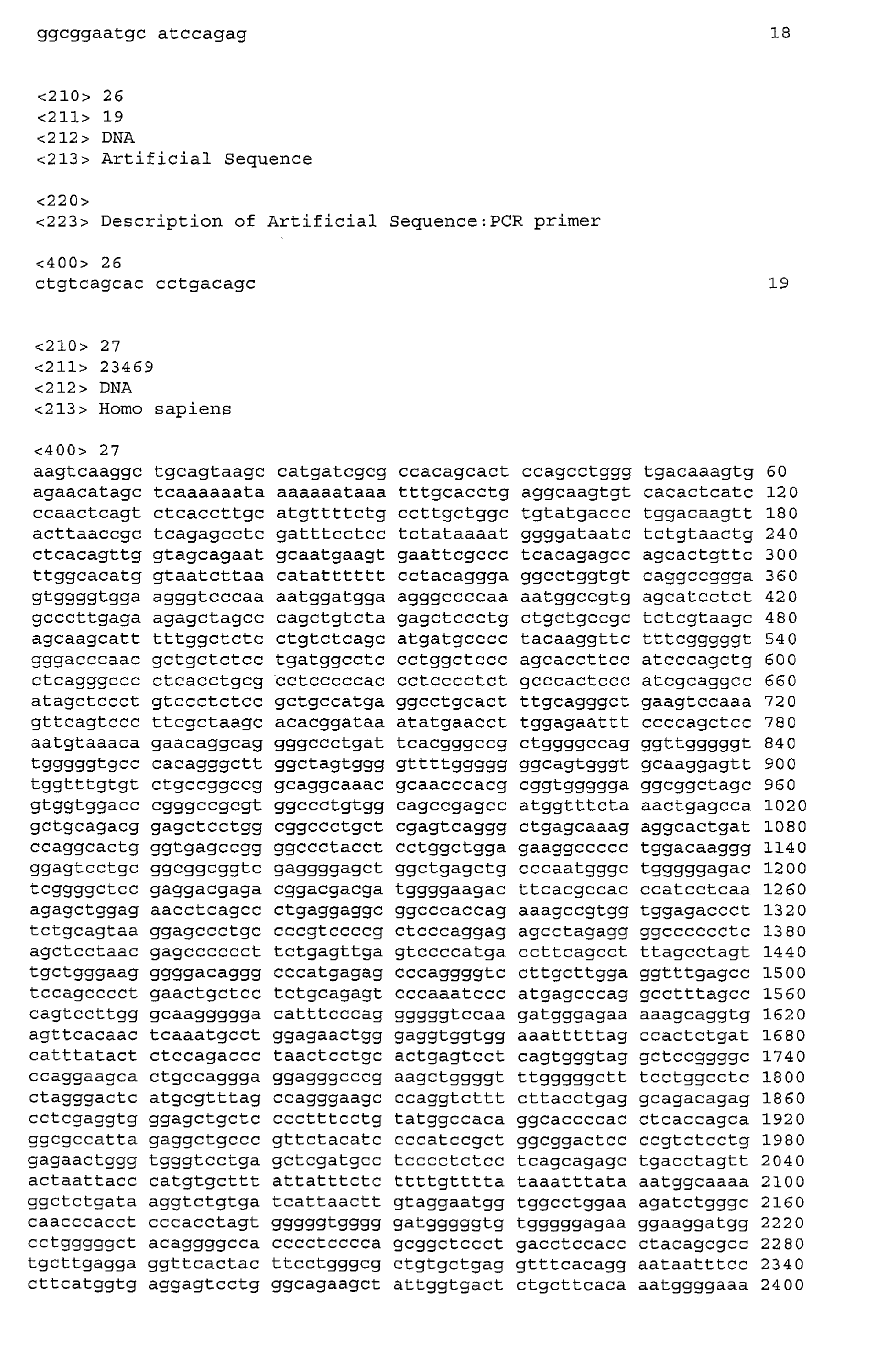
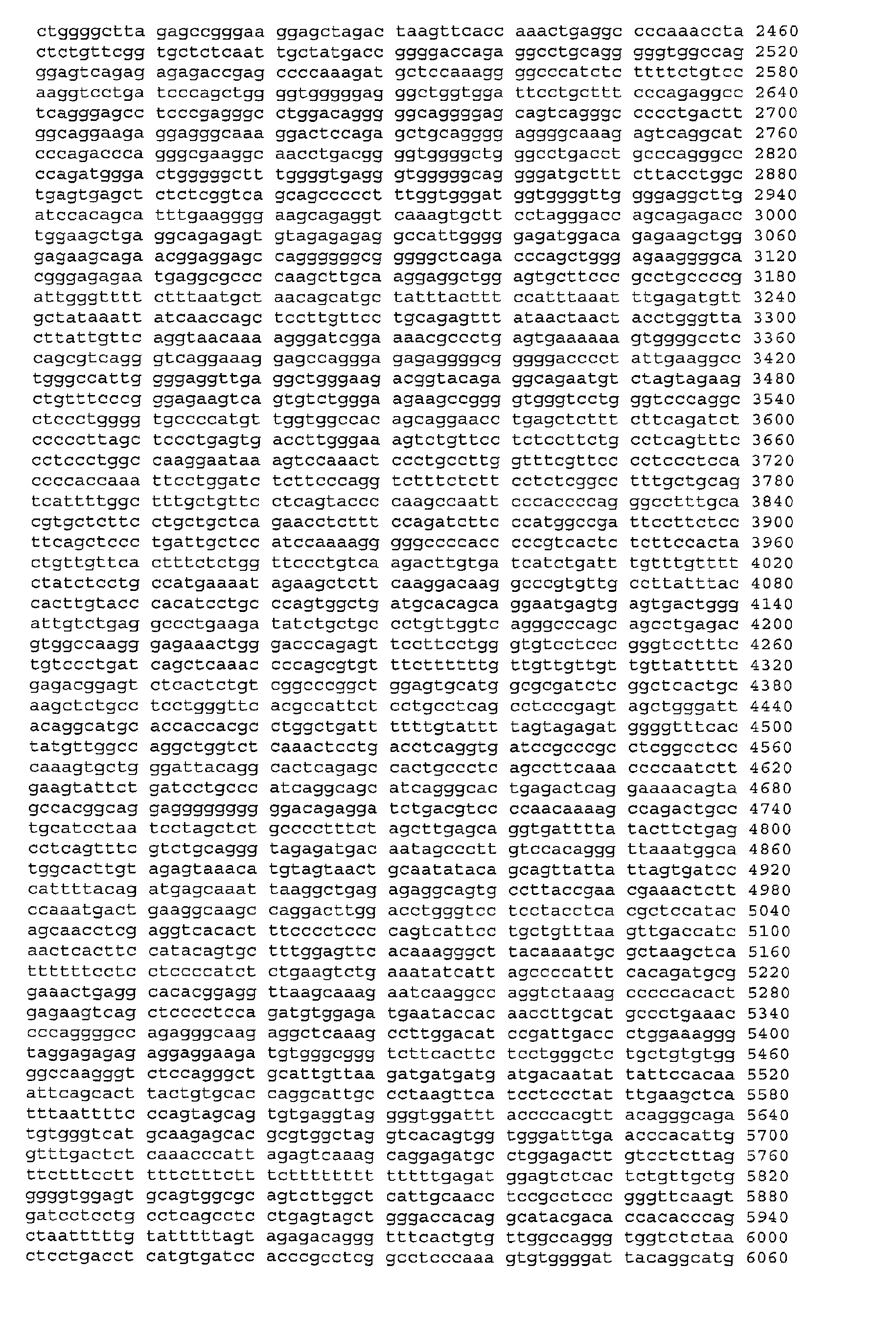
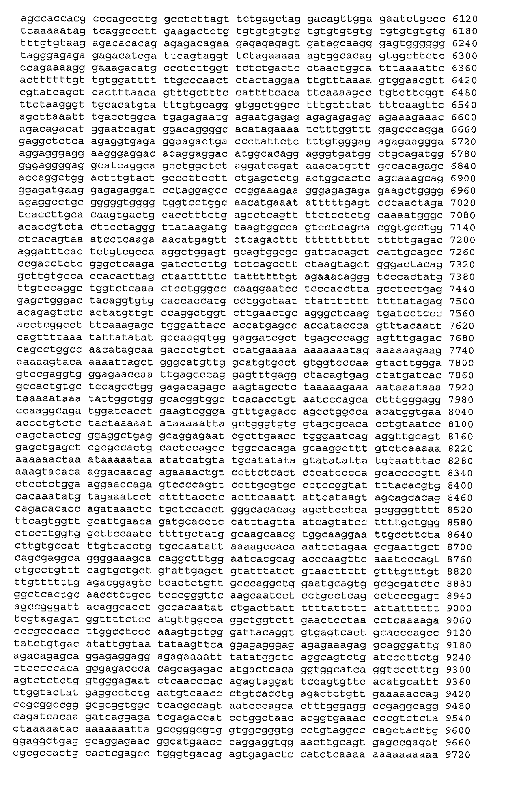
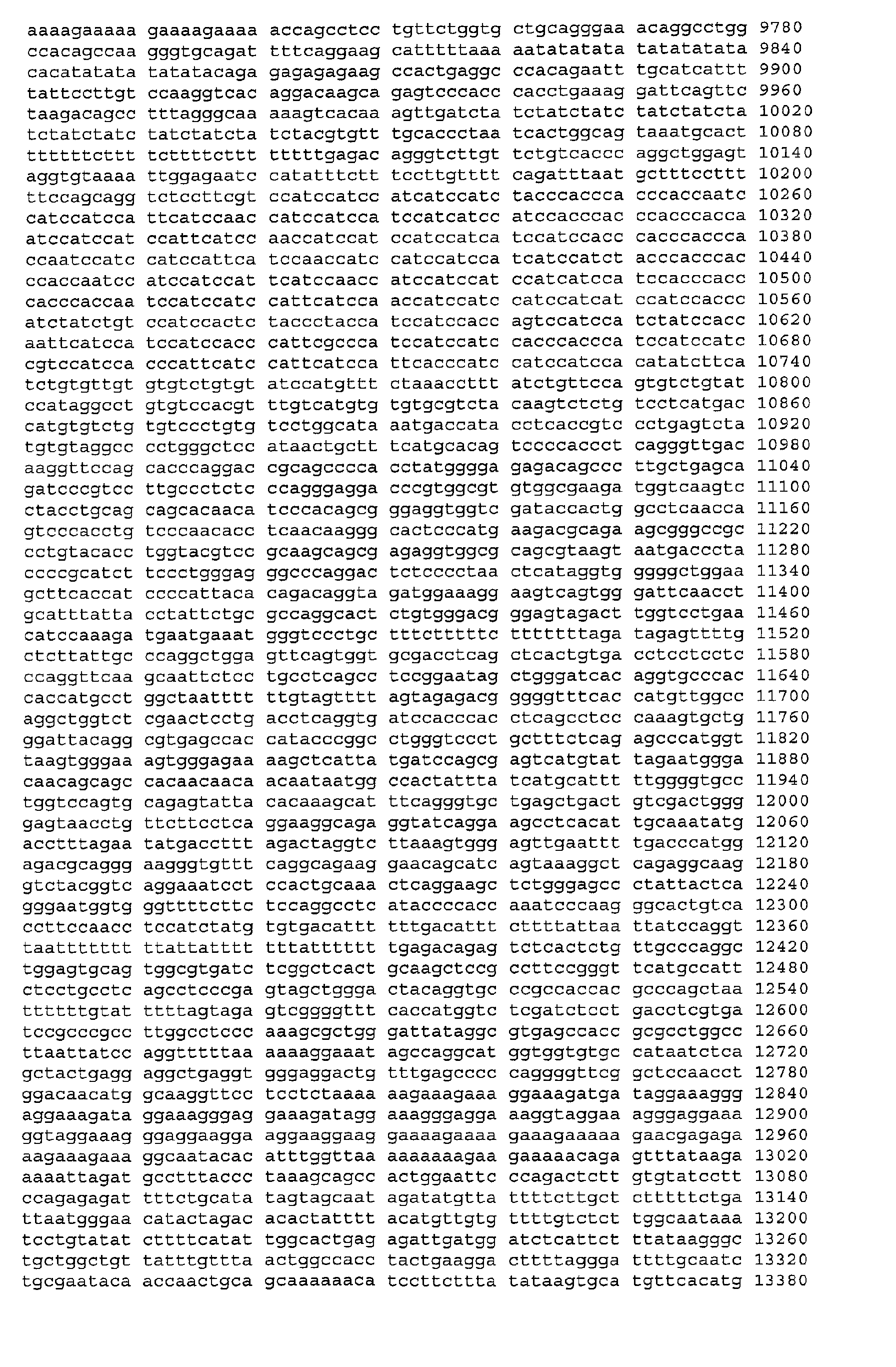
The genomic sequence of the human HFN1α gene is shown in SEQ ID No 27.
-
HNF1α has been characterized in various other species too, including mouse, rat and
xenopus.
-
A "functional HNF1α protein" refers to a protein that shows essentially the same
activity or level of activity as the wild-type HNF1α protein. This activity includes the
transcriptional activation of genes such as albumin, β fibrinogen and α1-antitripsin, by
dimerizing and binding upstream of the gene's promoter.
-
A «biological sample » refers to any source of biological material (DNA, mRNA,
or protein) available in practice. It includes without limitation body fluids, such as blood,
urine, plasma, serum, cerebrospinal fluid, saliva, and biopsy tissues or surgical specimen.
Liver biopsies are particularly preferred.
-
A "promoter" or "promoter sequence" is a DNA regulatory region capable of
binding RNA polymerase in a cell and initiating transcription of a downstream (3' direction)
coding sequence. The promoter sequence is generally bounded at its 3' terminus by the
transcription initiation site and extends upstream (5' direction) to include the minimum
number of bases or elements necessary to initiate transcription at levels detectable above
background. Within the promoter sequence are found a transcription initiation site
(conveniently defined for example, by mapping with nuclease SI), as well as protein binding
domains (consensus sequences) responsible for the binding of RNA polymerase. The
promoter may be operatively associated with other expression control sequences, including
enhancer and repressor sequences.
-
Promoters which may be used to control gene expression include, but are not limited
to, cytomegalovirus (CMV) promoter (U.S. Patents No. 5,385,839 and No. 5,168,062), the
SV40 early promoter region the promoter contained in the 3' long terminal repeat of Rous
sarcoma virus the herpes thymidine kinase promoter, the regulatory sequences of the
metallothionein gene; prokaryotic expression vectors such as the beta-lactamase promoter or
the tac promoter; promoter elements from yeast or other fungi such as the GaI 4 promoter, the
ADC (alcohol dehydrogenase) promoter, PGK (phosphoglycerol kinase) promoter, alkaline
phosphatase promoter; and transcriptional control regions that exhibit hematopoietic tissue
specificity, in particular: beta-globin gene control region which is active in myeloid cells,
hematopoietic stem cell differentiation factor promoters, erythropoietin receptor promoter,
etc.
-
The terms "mutant" and "mutation" mean any detectable change in genetic material,
e.g. DNA, or any process, mechanism, or result of such a change. This includes gene
mutations, in which the structure (e.g. DNA sequence) of a gene is altered, any gene or DNA
arising from any mutation process, and any expression product (e.g. protein) expressed by a
modified gene or DNA sequence.
-
"Sequence-conservative variants" of a polynucleotide sequence are those in which a
change of one or more nucleotides in a given codon position results in no alteration in the
amino acid encoded at that position.
-
"Function-conservative variants" are those in which a given amino acid residue in a
protein or enzyme has been changed without altering the overall conformation and function of
the polypeptide, including, but not limited to, replacement of an amino acid with one having
similar properties (such as, for example, polarity, hydrogen bonding potential, acidic, basic,
hydrophobic, aromatic, and the like). Amino acids with similar properties are well known in
the art. For example, arginine, histidine and lysine are hydrophilic-basic amino acids and may
be interchangeable. Similarly, isoleucine, a hydrophobic amino acid, may be replaced with
leucine, methionine or valine. Such changes are expected to have little or no effect on the
apparent molecular weight or isoelectric point of the protein or polypeptide. Amino acids
other than those indicated as conserved may differ in a protein so that the percent protein or
amino acid sequence similarity between any two proteins of similar function may vary and
may be, for example, from 70 % to 99 % as determined according to an alignment scheme
such as by the Cluster Method, wherein similarity is based on the MEGALIGN algorithm. A
"function-conservative variant" also includes a polypeptide which has at least 60 % amino
acid identity as determined by BLAST or FASTA algorithms, preferably at least 75 %, most
preferably at least 85%, and even more preferably at least 90 %, and which has the same or
substantially similar properties or functions as the native or parent protein to which it is
compared.
-
As used herein, the term "homologous" in all its grammatical forms and spelling
variations refers to the relationship between proteins that possess a "common evolutionary
origin," including proteins from superfamilies (e.g., the immunoglobulin superfamily) and
homologous proteins from different species (e.g., myosin light chain, etc.) (Reeck et al.,
(1987)). Such proteins (and their encoding genes) have sequence homology, as reflected by
their sequence similarity, whether in terms of percent similarity or the presence of specific
residues or motifs at conserved positions.
-
Accordingly, the term "sequence similarity" in all its grammatical forms refers to the
degree of identity or correspondence between nucleic acid or amino acid sequences of
proteins that may or may not share a common evolutionary origin (see Reeck et al., supra).
However, in common usage and in the instant application, the term "homologous," when
modified with an adverb such as "highly," may refer to sequence similarity and may or may
not relate to a common evolutionary origin.
-
In a specific embodiment, two DNA sequences are "substantially homologous" or
"substantially similar" when at least about 80 %, and most preferably at least about 90 or 95
%, of the nucleotides match over the defined length of the DNA sequences, as determined by
sequence comparison algorithms, such as BLAST, FASTA, DNA Strider, etc. An example of
such a sequence is an allelic or species variant of the specific genes of the invention.
Sequences that are substantially homologous can be identified by comparing the sequences
using standard software available in sequence data banks, or in a Southern hybridization
experiment under, for example, stringent conditions as defined for that particular system.
-
Similarly, in a particular embodiment, two amino acid sequences are "substantially
homologous" or "substantially similar" when greater than 80 % of the amino acids are
identical, or greater than about 90 % are similar (functionally identical). Preferably, the
similar or homologous sequences are identified by alignment using, for example, the GCG
(Genetics Computer Group, Program Manual for the GCG Package, Version 7, Madison,
Wisconsin) pileup program, or any of the programs described above (BLAST, FASTA, etc.).
-
A nucleic acid molecule is "hybridizable" to another nucleic acid molecule, such as a
cDNA, genomic DNA, or RNA, when a single stranded form of the nucleic acid molecule can
anneal to the other nucleic acid molecule under the appropriate conditions of temperature and
solution ionic strength (see Sambrook et al., 1989). The conditions of temperature and ionic
strength determine the "stringency" of the hybridization. For preliminary screening for
homologous nucleic acids, low stringency hybridization conditions, corresponding to a Tm
(melting temperature) of 55°C, can be used, e.g., 5x SSC, 0.1 % SDS, 0.25 % milk, and no
formamide ; or 30 % formamide, 5x SSC, 0.5 % SDS). Moderate stringency hybridization
conditions correspond to a higher Tm, e.g., 40 % formamide, with 5x or 6x SCC. High
stringency hybridization conditions correspond to the highest Tm, e.g., 50 % formamide, 5x
or 6x SCC. SCC is a 0.15 M NaCl, 0.015 M Na-citrate. Hybridization requires that the two
nucleic acids contain complementary sequences, although depending on the stringency of the
hybridization, mismatches between bases are possible. The appropriate stringency for
hybridizing nucleic acids depends on the length of the nucleic acids and the degree of
complementation, variables well known in the art. The greater the degree of similarity or
homology between two nucleotide sequences, the greater the value of Tm for hybrids of
nucleic acids having those sequences. The relative stability (corresponding to higher Tm) of
nucleic acid hybridizations decreases in the following order: RNA:RNA, DNA:RNA,
DNA:DNA. For hybrids of greater than 100 nucleotides in length, equations for calculating
Tm have been derived (see Sambrook et al., 1989, 9.50-9.51). For hybridization with shorter
nucleic acids, i.e., oligonucleotides, the position of mismatches becomes more important, and
the length of the oligonucleotide determines its specificity (see Sambrook et al., 1989, 11.7-11.8).
A minimum length for a hybridizable nucleic acid is at least about 10 nucleotides ;
preferably at least about 15 nucleotides ; and more preferably the length is at least about 20
nucleotides.
-
In a specific embodiment, the term "standard hybridization conditions" refers to a
Tm of 55°C, and utilizes conditions as set forth above. In a preferred embodiment, the Tm is
60°C. In a more preferred embodiment, the Tm is 65°C. In a specific embodiment, "high
stringency" refers to hybridization and/or washing conditions at 68°C in 0.2 X SSC, at 42°C
in 50 % formamide, 4 X SSC, or under conditions that afford levels of hybridization
equivalent to those observed under either of these two conditions.
-
The terms "vector", "cloning vector" and "expression vector" mean the vehicle by
which a DNA or RNA sequence (e.g. a foreign gene) can be introduced into a host cell, so as
to transform the host and promote expression (e.g. transcription and translation) of the
introduced sequence. Vectors include plasmids, phages, viroses, etc.; they are discussed in
greater detail below.
-
Vectors typically comprise the DNA of a transmissible agent, into which foreign
DNA is inserted. A common way to insert one segment of DNA into another segment of
DNA involves the use of enzymes called restriction enzymes that cleave DNA at specific sites
(specific groups of nucleotides) called restriction sites. A "cassette" refers to a DNA coding
sequence or segment of DNA that codes for an expression product that can be inserted into a
vector at defined restriction sites. The cassette restriction sites are designed to ensure insertion
of the cassette in the proper reading frame. Generally, foreign DNA is inserted at one or more
restriction sites of the vector DNA, and then is carried by the vector into a host cell along with
the transmissible vector DNA. A segment or sequence of DNA having inserted or added
DNA, such as an expression vector, can also be called a "DNA construct." A common type of
vector is a "plasmid", which generally is a self-contained molecule of double-stranded DNA,
usually of bacterial origin, that can readily accept additional (foreign) DNA and which can
readily introduced into a suitable host cell. A plasmid vector often contains coding DNA and
promoter DNA and has one or more restriction sites suitable for inserting foreign DNA.
Coding DNA is a DNA sequence that encodes a particular amino acid sequence for a
particular protein or enzyme. Promoter DNA is a DNA sequence which initiates, regulates, or
otherwise mediates or controls the expression of the coding DNA. Promoter DNA and coding
DNA may be from the same gene or from different genes, and may be from the same or
different organisms. A large number of vectors, including plasmid and fungal vectors, have
been described for replication and/or expression in a variety of eukaryotic and prokaryotic
hosts. Non-limiting examples include pKK plasmids (Clonetech), pUC plasmids, pET
plasmids (Novagen, Inc., Madison, WI), pRSET or pREP plasmids (Invitrogen, San Diego,
CA), or pMAL plasmids (New England Biolabs, Beverly, MA), and many appropriate host
cells, using methods disclosed or cited herein or otherwise known to those skilled in the
relevant art. Recombinant cloning vectors will often include one or more replication systems
for cloning or expression, one or more markers for selection in the host, e.g. antibiotic
resistance, and one or more expression cassettes.
-
The terms "express" and "expression" mean allowing or causing the information in a
gene or DNA sequence to become manifest, for example producing a protein by activating the
cellular functions involved in transcription and translation of a corresponding gene or DNA
sequence. A DNA sequence is expressed in or by a cell to form an "expression product" such
as a protein. The expression product itself, e.g. the resulting protein, may also be said to be
"expressed" by the cell. An expression product can be characterized as intracellular,
extracellular or secreted. The term "intracellular" means something that is inside a cell. The
term "extracellular" means something that is outside a cell. A substance is "secreted" by a cell
if it appears in significant measure outside the cell, from somewhere on or inside the cell.
-
The term "transfection" means the introduction of a foreign nucleic acid into a cell.
The term "transformation" means the introduction of a "foreign" (i.e. extrinsic or
extracellular) gene, DNA or RNA sequence to a host cell, so that the host cell will express the
introduced gene or sequence to produce a desired substance, typically a protein or enzyme
coded by the introduced gene or sequence. The introduced gene or sequence may also be
called a "cloned" or "foreign" gene or sequence, may include regulatory or control sequences,
such as start, stop, promoter, signal, secretion, or other sequences used by a cell's genetic
machinery. The gene or sequence may include nonfunctional sequences or sequences with no
known function. A host cell that receives and expresses introduced DNA or RNA has been
"transformed" and is a "transformant" or a "clone." The DNA or RNA introduced to a host
cell can come from any source, including cells of the same genus or species as the host cell, or
cells of a different genus or species.
-
The term "host cell" means any cell of any organism that is selected, modified,
transformed, grown, or used or manipulated in any way, for the production of a substance by
the cell, for example the expression by the cell of a gene, a DNA or RNA sequence, a protein.
-
The term "expression system" means a host cell and compatible vector under suitable
conditions, e.g. for the expression of a protein coded for by foreign DNA carried by the vector
and introduced to the host cell. Common expression systems include E. coli host cells and
plasmid vectors, insect host cells and Baculovirus vectors, and mammalian host cells and
vectors. In a specific embodiment, the protein of interest is expressed in COS-1 or C2C12 cells.
Other suitable cells include CHO cells, HeLa cells, 293T (human kidney cells), mouse
primary myoblasts, and NIH 3T3 cells.
-
"Pharmaceutically" or "pharmaceutically acceptable" refer to molecular entities and
compositions that do not produce an adverse, allergic or other untoward reaction when
administered to an animal, or a human, as appropriate.
-
As used herein, "pharmaceutically acceptable carrier" includes any and all solvents,
dispersion media, coatings, antibacterial and antifungal agents, isotonic and absorption
delaying agents and the like. The use of such media and agents for pharmaceutical active
substances is well known in the art. Except insofar as any conventional media or agent is
incompatible with the active ingredient, its use in the therapeutic compositions is
contemplated. Supplementary active ingredients can also be incorporated into the
compositions.
Diagnostics
-
An aspect of the present invention provides methods for determining whether a
subject has or is at risk for developing a tumor.
-
A subject of the invention is an in vitro method for the diagnosis of a tumor in a
patient, which method comprises determining the expression or activity of Hepatocyte
Nuclear Factor 1α (HNF1α) in a biological sample, wherein an abnormal expression or
activity in comparison with a control sample is indicative of a tumor or of a susceptibility to
develop a tumor.
-
The step of determining the expression or activity of HNF1α may be performed
according to various embodiments.
Genetic lesion:
-
In a first embodiment, the step of determining the expression or activity of HNF1α
comprises or consists in detecting a mutation in the HNF1α gene or in the promoter thereof,
wherein said mutation results in impaired expression or activity of the HNF1α protein. At
least one mutation may be detected.
-
The mutation maybe bi or mono-allelic. The inventors have more particularly found
out that bi-allelic mutations could lead to hepatic adenomas or hepatocellular carcinomas,
whereas mono-allelic mutations could result in colon tumors. Dominant negative mutations in
HNF1α have already been described, in connection with MODY3 (e.g. mutation P291fsinsC,
Yamagata et al, 1998, and Vaxillaire et al, 1999). These include mutants that encode proteins
presenting an intact dimerization domain but forming non-productive dimers with wild-type
protein, thereby inhibiting its activity.
-
The mutations are preferably located in the coding sequence of the HNF1α gene, or
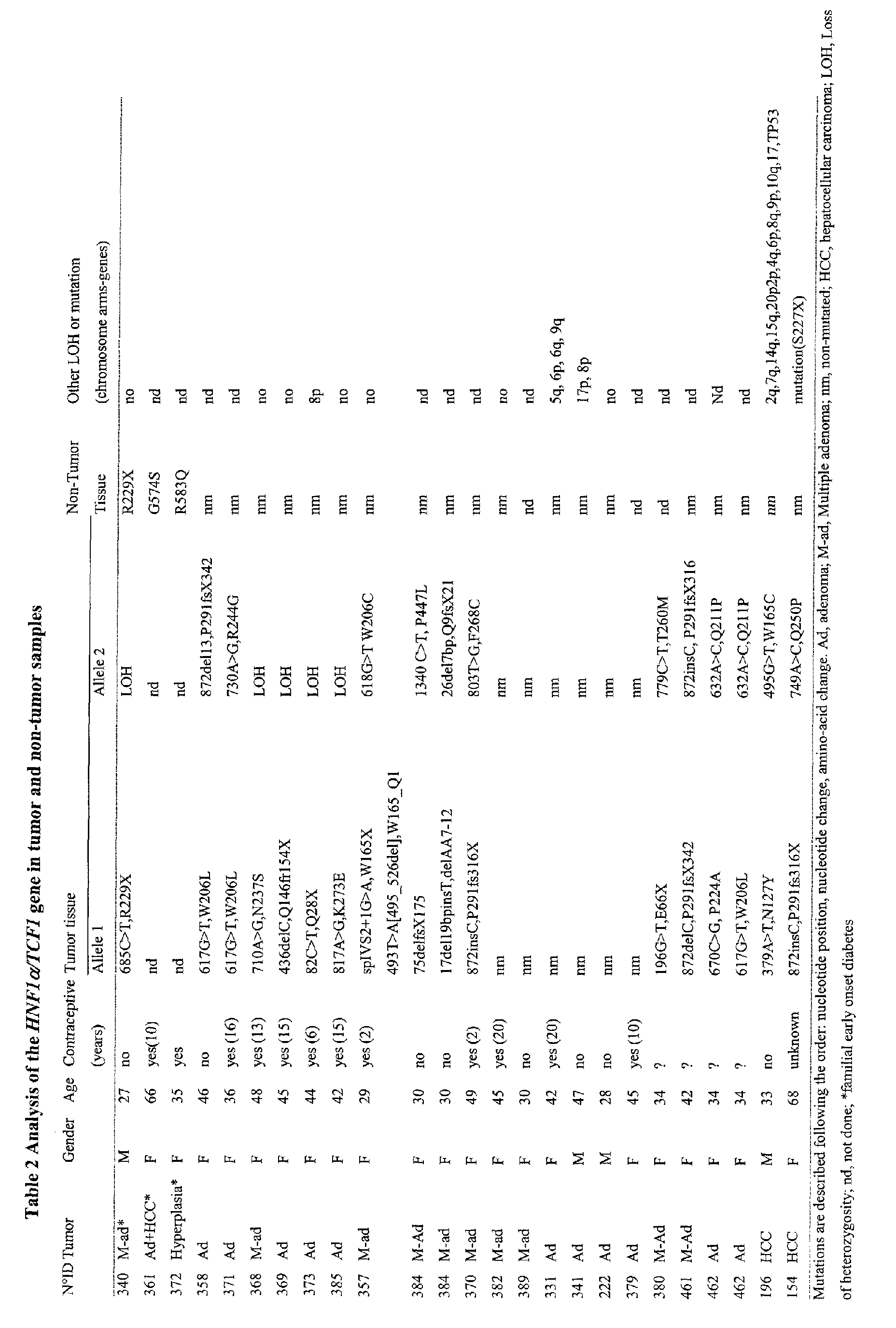
in the intron-exon splicing regions. Mutations of particular interest are shown in Table 2.
-
Preferred mutations include :
685C>T;617G>T;710A>G;436delC;82C>T;817A>G;spIVS2+1G>A;493T>A[495_526del];;
17del19bpinsT;872insC;196G>T;872delC;670C>G;379A>T;872insC;872del13;730A>G;618
G>T;1340C>T;26del17bp;803T>G;779C>T;872insC;632A>C;495G>T; and 749A>C.
-
Mutations 872delC(291frX342) and 872insC(291frX3156) are of particular interest
especially in connection with colon tumors.
-
Mutations are herein described by giving the nucleotide position, and the change (>:
substitution ; del : deletion ; ins : insertion ; sp : splicing site; IVS: Intron variant sequence
followed by intron number ; in [ ] : mutation at the RNA level in the human sequence (SEQ
ID n° 1)).
-
Mutations may also occur in the promoter.
-
Generally speaking, the mutation or genetic lesion may be (a) a deletion of one or
more nucleotides from a wildtype gene; (b) an addition of one or more nucleotides to a
wildtype gene; (c) a substitution of one or more nucleotides of a wildtype gene; (d) a gross
chromosomal rearrangement of a wildtype gene.
-
The mutation is preferably a transversion G>T at nucleotide 617 resulting in an
amino acid substitution W206L.
-
The mutation results in impaired expression or activity of the HNF1α protein. This
means that the mutation results either in full inactivation of HNF1α, or in a diminished
expression or activity of the protein. For instance, some mutants may have a reduced protein
stability, whereas others may be defective in the DNA binding or impaired in their intrinsic
trans-activation potential.
-
The detection of the mutation or genetic lesion may be performed by a variety of
techniques well-known by the skilled artisan.
-
Genomic DNA used for the diagnosis may be obtained from body cells, such as
those present in blood, tissue biopsy, or surgical specimen. The DNA may be isolated and
used directly for detection of a specific sequence or may be PCR amplified prior to analysis.
To detect a specific nucleic acid sequence, hybridization using specific oligonucleotides,
direct nucleotide sequencing, RFLP, restriction enzyme digest, RNase protection, chemical
cleavage, or ligase-mediated detection may be used. Oligonucleotides specific to mutant
sequences can be chemically synthesized and labelled radioactively with isotopes, or non-radioactively
using biotin tags, and hybridize to individual samples immobilized on
membranes or other solid-supports by dot-blot or transfer from gels after electrophoresis. The
presence or absence of these mutant sequences is then visualized using methods such as
autoradiography, fluorometry, or colorimetric reaction. Cloned genomic or cDNA segments
may be used as probes to detect specific DNA segments. Amplification of the nucleic acid,
e.g. by PCR (polymerase chain reaction) or LCR (ligase chain reaction), can be used to
enhance the sensitivity of this method.
-
More particularly, detecting the genetic lesion can include: (i) providing probes or
primers comprised of an oligonucleotide which hybridizes to a sense or antisense sequence of
an HNF1α, gene or gene fragment (wildtype or mutant); (ii) contacting the probes or primers
to an appropriate nucleic acid containing biological sample obtained from the subject; and
(iii) detecting, by hybridization of the probes or primers to the nucleic acid, the presence or
absence of the genetic lesion.
-
In a preferred embodiment, the diagnostic methods utilize a set of primers for
amplifying (e.g. via PCR or LCR) at least one region of an HNF1α gene, and means for
analyzing the amplification product for differences (e.g. mutations) from the normal, wildtype
coding sequence.
-
In another preferred embodiment, the diagnostic methods utilize a probe to
determine its ability to hybridize under appropriately stringent conditions to a complementary
nucleic acid sequence in the biological sample, wherein an inability of a probe, which is
comprised of a wildtype HNF1α sequence, to hybridize to the sample nucleic acid is
indicative of the presence of a mutation in the sample nucleic acid; or the ability of a probe,
which is comprised of a mutant HNF1α, sequence to hybridize to the sample nucleic acid is
indicative of the presence of a mutation in the sample nucleic acid.
-
Other nucleotide sequence amplification techniques may be used, such as ligation-mediated
PCR, anchored PCR and enzymatic amplification as will be understood by those
skilled in the art.
-
Sequence alterations may also generate fortuitous restriction enzyme recognition
sites which are revealed by the use of appropriate enzyme digestion followed by gel-blot
hybridization. DNA fragments carrying the site (normal or mutant) are detected by their
increase or reduction in size, or by the increase or decrease of corresponding restriction
fragment numbers. Genomic DNA samples may also be amplified by PCR prior to treatment
with the appropriate restriction enzyme and the fragments of different sizes are visualize, for
example under UV light in the presence of ethidium bromide, after gel electrophoresis.
-
Genetic testing based on DNA sequence differences may be achieved by detection
of alteration in electrophoretic mobility of DNA fragments in gels. Small sequence deletions
and insertions can be visualized by high resolution gel electrophoresis of single stranded
DNA, or as changes in the migration pattern of DNA heteroduplexes in non-denaturing gel
electrophoresis. Alternatively, a single base substitution mutation may be detected based on
differential PCR product length in PCR. The PCR products of the normal and mutant gene
may be differentially detected in acrylamide gels.
-
Nuclease protection assays (S1 or ligase-mediated) also reveal sequence changes at
specific locations.
-
Alternatively, to confirm or detect a mutation or polymorphism resulting in
restriction mapping changes, ligated PCR, ASO, REF-SSCP chemical cleavage, endonuclease
cleavage at mismatch sites, SSCP or dHPLC (Denaturating High Pressure Liquid
Chromatography) may be used. Both REF-SSCP and SSCP are mobility shift assays which
are based upon the change in conformation due to mutations. SSCP and dHPLC are preferred
methods of pre-screening particularly useful to detect mutations in the HNF1α gene.
-
DNA fragments may also be visualized by methods in which the individual DNA
samples are not immobilized on membranes. The probe and target sequences may be in
solution or the probe sequence may be immobilized. Autoradiography, radioactive decay,
spectrophotometry and fluorometry may also be used to identify specific individual
genotypes. Mutations can also be detected by direct nucleotide sequencing.
-
Preferably, mutations in the HNF1α gene are detected by PCR amplification of the
screened region followed by direct sequencing of the purified PCR fragment using one of the
primer used during PCR amplification.
-
According to an embodiment of the invention, the portion of the DNA segment that
is informative for a mutation can be amplified using PCR. For example, the DNA segment
immediately surrounding the 617G>T,W206L mutation acquired from tumor (e.g. adenoma)
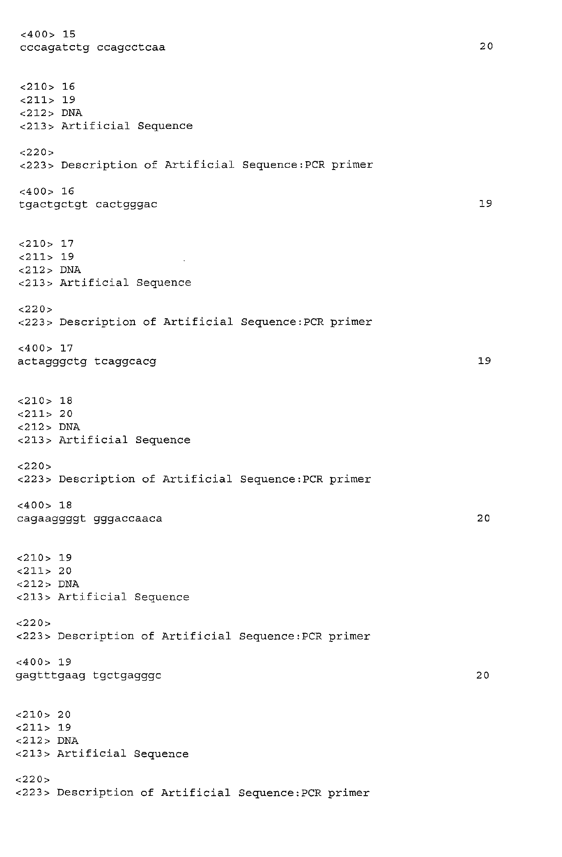
samples from an individual can be screened using the primers 5'-tagctgtaagctcctctggtand
(SEQ ID No 3) and 5'-tacaggacctagagtcacct (SEQ ID No 4). This region is then amplified by
PCR, the products are separated by electrophoresis, and transferred to membrane. Normal and
mutant PCR products may then be detected using, for example, hybridization of labeled
oligonucleotide probes and autoradiography, RFLP analysis, or direct sequencing.
RNA assays:
-
In another embodiment of the invention, the step of determining the expression or
activity of HNF1α comprises or consists in detecting a mutation in the HNF1α messenger
RNA (mRNA) transcript, wherein said mutation results in impaired expression or activity of
the HNF1α protein. At least one mutation may be detected. In that respect determining the
presence of a non-wild type splicing pattern of a mRNA transcript of the gene may also be of
particular interest.
-
The methods described above to analyze DNA may be adapted to analyze RNA.
RNA may be isolated from a biological sample, e.g. a biopsy tissue, using standard methods
well known to those of ordinary skill in the art such as guanidium thiocyanate-phenol-chloroform
extraction. The isolated RNA may then be subjected to coupled reverse
transcription and amplification (RT-PCR), using specific oligonucleotide primers that are
specific for a selected site. Conditions for primer annealing are chosen to ensure specific
reverse transcription and amplification; thus the appearance for an amplification product is
diagnostic of the presence of a particular genetic variation. Alternatively, RNA is reverse-transcribed
and amplified, after which mutations in the cDNA are identified by any technique
as described above, e.g. direct sequencing. In still another embodiment, cDNA can be cloned
and/or directly sequenced to identify a mutation.
-
In yet another embodiment, the step of determining the expression or activity of
HNF1α comprises or consists in assessing the level of mRNA that encodes the HNF1α
protein. Quantification of the level of HNF1α transcription can be determined using standard
methods well known to those of ordinary skill in the art such as quantitative RT/PCR or
northern blot analysis. An alteration in the level of a messenger RNA transcript of a gene is
indicative of a tumor or of a risk to develop a tumor.
Protein assays:
-
In still another embodiment, the step of determining the expression or activity of
HNF1α comprises or consists in assessing the level of HNF1α protein or the level of activity
of this protein. A reduced level of wild-type protein or a reduced level of activity of the
protein is indicative of a tumor or of a risk to develop a tumor.
-
Determining the level of HNF1α protein may be performed by a variety of
techniques. Immunodiagnostic techniques are particularly suitable therefore, including
immunoassays such as competition, direct reaction or sandwich type assays. Such assays
include, but are not limited to, Western blots, immunohistochemistry, agglutination tests,
enzyme-labelled and mediated immunoassays, such as ELISAs, biotin/avidin type assays,
radioimmunassays, immunoelectrophoresis, immunoprecipitation, etc. The reactions generally
include revealing labels such as fluorescent, chemiluminescent, radioactive, enzymatic labels
or dye molecules, or other methods for detecting the formation of a complex between the
antigen (the HNF1α protein likely to be present in the sample) and the antibody or antibodies
reacted therewith.
-
The term "antibody" in its various grammatical forms is used herein to refer to
immunoglobulin molecules and immunologically active portions of immunoglobulin
molecules, i.e., molecules that contain an antibody combining site or paratope. Exemplary
antibody molecules are intact immunoglobulin molecules, substantially intact
immunoglobulin molecules and portions of an immunoglobulin molecule, including those
portions known in the art as Fab, Fab', F(ab')2 and F(v).
-
Whereas polyclonal antibodies, that are easy to obtain, may be used, monoclonal
antibodies are preferred because they are more reproductible in the long run.
-
Procedures for raising polyclonal antibodies are well known. Typically, such
antibodies can be raised by administering the BARD1 protein subcutaneously to New Zealand
white rabbits which have first been bled to obtain pre-immune serum. The antigens can be
injected at a total volume of 100 µl per site at six different sites. Each injected material will
contain adjuvants with or without pulverized acrylamide gel containing the protein or
polypeptide after SDS-polyacrylamide gel electrophoresis. The rabbits are then bled two
weeks after the first injection and periodically boosted with the same antigen three times
every six weeks. A sample of serum is then collected 10 days after each boost. Polyclonal
antibodies are then recovered from the serum by affinity chromatography using the
corresponding antigen to capture the antibody. This and other procedures for raising
polyclonal antibodies are disclosed in Harlow et. al. (1988).
-
A "monoclonal antibody" in its various grammatical forms refers to a population of
antibody molecules that contain only one species of antibody combining site capable of
immunoreacting with a particular epitope. A monoclonal antibody thus typically displays a
single binding affinity for any epitope with which it immunoreacts. A monoclonal antibody
may therefore contain an antibody molecule having a plurality of antibody combining sites,
each immunospecific for a different epitope, e.g. a bispecific monoclonal antibody. Although
historically a monoclonal antibody was produced by immortalization of a clonally pure
immunoglobulin secreting cell line, a monoclonally pure population of antibody molecules
can also be prepared by the methods of the present invention.
-
Laboratory methods for preparing monoclonal antibodies are well known in the art
(see, for example, Harlow et al. (1988)). Monoclonal antibodies (Mabs) may be prepared by
immunizing purified HNF1α protein isolated from any of a variety of mammalian species into
a mammal, e.g. a mouse, rabbit, goat, human and the like mammal. The antibody-producing
cells in the immunized mammal are isolated and fused with myeloma or heteromyeloma cells
to produce hybrid cells (hybridoma). The hybridoma cells producing the monoclonal
antibodies are utilized as a source of the desired monoclonal antibody.
-
While Mabs can be produced by hybridoma culture, the invention is not to be so
limited. Also contemplated is the use of Mabs produced by an expressing nucleic acid cloned
from a hybridoma of this invention. That is, the nucleic acid expressing the molecules
secreted by a hybridoma of this invention can be transferred into another cell line to produce a
transformant. The transformant is genotypically distinct from the original hybridoma but is
also capable of producing antibody molecules of this invention, including immunologically
active fragments of whole antibody molecules, corresponding to those secreted by the
hybridoma. See, for example, U.S. Pat. No. 4,642,334 to Reading; PCT Publication No. WO
890099 to Robinson et al.; European Patent Publications No. 0239400 to Winter et al. and No.
0125023 to Cabilly et al. In addition, the literature provides methods for forming chimeric
antibodies, humanized antibodies, single chain antibodies and the like variations on a basic
immunoreactive antibody fragment. All of these are considered within the scope of the
invention insofar as a class and specificity of antibody is disclosed and claimed, regardless of
the precise variant structure that one skilled in the art may construct.
-
Antibodies specific for the HNF1α protein can be purchased from Pharmingen or
Santa Cruz Biotechnology. A set of polyclonal and monoclonal antibodies raised against the
rat protein has also been described in Chouard et al, 1997.
-
Alternatively, one may assess the activity of the HNF1α protein present in the
biological sample. This may be effected by any method known by the skilled person. For
example, one may assess the ability of HNF1α to trans-activate down-stream genes, such as
albumin, α and β fibrinogen and α1- antitrypsin. Reporter systems may be particularly useful
for that purpose. According to one of these reporter systems, it is provided a chimeric
construct, wherein the promoter of such a down-stream gene is fused or operatively associated
with a reporter gene which expresses a polypeptide capable of producing a detectable signal.
When the HNF1α protein is functional, it activates the promoter of said down-stream gene
and causes a measurable detectable signal. The reporter gene may encode a luciferase,
chloramphenicol acetyltransferase, β glucuronidase, β galactosidase, neomycin
phosphotransferase, or guanine xanthine phosphoribosyltransferase, etc.
-
In still another embodiment the step of determining the expression or activity of
HNF1α comprises or consists in detecting a mutation in the amino acid sequence of the
HNF1α protein. This may be performed by different techniques used in protein analyses such
as: western blotting, mass spectrometry or protein sequencing to detect HNF1α protein with
aberrant migration or sequence pattern.
Specific embodiments:
-
In a specific embodiment, the diagnostic methods of the invention make it possible
to diagnose liver tumors, especially hepatic adenoma or hepatocellular carcinoma.
-
More particularly, the invention provides an in vitro method for the diagnosis of a
liver tumor in a patient, which method comprises detecting a mutation in Hepatocyte Nuclear
Factor 1a (HNF1α) gene in a liver sample, which mutation results in bi-allelic inactivation of
HNF1α that is indicative of liver tumor selected from a hepatic adenoma and a hepatocellular
carcinoma, or is indicative of a susceptibility to develop said liver tumor.
-
In another particular embodiment, colon tumors e.g., colorectal adenomas or
carcinomas, may be advantageously detected by the methods of the invention.
-
More particularly, the invention provides in vitro method for the diagnosis of a
colon tumor in a patient, which method comprises detecting a mutation in HNF1α gene in a
colon sample, which mutation results in mono-allelic inactivation of HNF1α that is indicative
of a colon tumor selected from a colorectal adenoma or carcinoma of the MSI-H phenotype,
or is indicative of a susceptibility to develop said colon tumor.
-
In another aspect of the invention, it is provided a method for determining the
susceptibility of a subject to develop a colon tumor, which method comprises detecting a
polymorphism consisting of a mutation in the poly C tract repeat in exon 4 of the HNF1α
gene (nucleotides 865 to 872 in the coding sequence), wherein a C(9) repeat (i.e. a
polymorphic 864 G>C transversion) is correlated with a higher risk of HNF1α mutation, and
as a result with a higher risk to develop a colon tumor.
-
In a further aspect of the invention, it may be advantageous to test any diabetic
patient, especially a Maturity-Onset Diabetes of the Young type 3 (MODY3) patient, for its
susceptibility to develop a tumor, e.g. a liver or a colon tumor.
-
The diagnostic methods of the invention may also be useful for female patients who
use estroprogestative, especially as oral contraception.
Therapeutics
-
Another aspect of the invention provides therapeutic methods in the prevention or
treatment of tumors.
-
These methods are based on the discovery that HNF1α is a tumor suppressor gene
whose expression is impaired in tumors. The purpose of the therapeutic methods of the
invention is to restore or enhance the expression or activity of HNF1α protein.
-
The invention contemplates the use of (i) a nucleic acid that encodes a functional
HNF1α protein or (ii) a functional HNF1α protein, for the preparation of a medicament for
the prevention or treatment of a tumor.
-
The invention also contemplates a method for preventing or treating a tumor in a
patient, which method comprises administering a efficient amount of (i) a nucleic acid that
encodes a functional HNF1α protein or (ii) a functional HNF1α protein, in association with
pharmaceutically acceptable carrier, in a patient in need a such treatment.
-
The first embodiment (i) involves gene therapy techniques, as discussed below. The
second embodiment (ii), is a classic protein therapy, as described in greater detail below.
Gene therapy:
-
In accordance with the present invention, the HNF1α nucleic acid, that is preferably
a DNA sequence, forms part of a vector. Such vector is a nucleic acid comprising a HNF1α
coding sequence operatively associated with sequences that control expression of HNF1α in a
cell transfected with the vector.
-
The use of such a vector indeed makes it possible to improve the administration of
the nucleic acid into the cells to be treated, and also to increase its stability in the said cells,
which makes it possible to obtain a durable therapeutic effect. Furthermore, it is possible to
introduce several nucleic acid sequences into the same vector, which also increases the
efficacy of the treatment.
-
The vector used may be of diverse origin, as long as it is capable of transforming
animal cells, preferably human tumor cells. In a preferred embodiment of the invention, a
viral vector is used which can be chosen from adenoviruses, retroviruses, adeno-associated
viruses (AAV), herpes virus, cytomegalovirus (CMV), vaccinia virus and the like. Vectors
derived from adenoviruses, retroviruses or AAVs, HIV-derived retroviral vectors,
incorporating heterologous nucleic acid sequences have been described in the literature (EP
185 573, WO 91/18088).
-
The present invention therefore also relates to any recombinant virus comprising,
inserted into its genome, a HNF1α nucleic acid sequence as defined before.
-
Advantageously, the recombinant virus according to the invention is a defective
virus. The term "defective virus" designates a virus incapable of replicating in the target cell.
Generally, the genome of the defective viruses used within the framework of the present
invention is therefore devoid of at least the sequences necessary for the replication of the said
virus in the infected cell. These regions can either be removed (completely or partially), or
rendered non-functional, or substituted by other sequences and especially by the HNF1α
nucleic acid of the invention. Preferably, the defective virus nevertheless conserves the
sequences of its genome which are necessary for the encapsulation of the viral particles.
-
It is particularly advantageous to use the nucleic acid sequences of the invention in a
form incorporated in an adenovirus, an AAV or a defective recombinant retrovirus.
-
As regards adenoviruses, various serotypes exist whose structure and properties vary
somewhat, but which are not pathogenic for man, and especially non-immunosuppressed
individuals. Moreover, these viruses do not integrate into the genome of the cells which they
infect, and can incorporate large fragments of exogenous DNA. Among the various serotypes,
the use of the type 2 or 5 adenoviruses (Ad2 or Ad5) is preferred within the framework of the
present invention. In the case of the Ad5 adenoviruses, the sequences necessary for the
replication are the E1A and E1B regions.
-
The defective recombinant viruses of the invention can be prepared by homologous
recombination between a defective virus and a plasmid carrying, inter alia, the HNF1α
encoding sequence. The homologous recombination is produced after co-transfection of the
said viruses and plasmid into an appropriate cell line. The cell line used should preferably (i)
be transformable by the said elements, and (ii), contain sequences capable of complementing
the part of the genome of the defective virus, preferably in integrated form so as to avoid the
risks of recombination. As example of a line which can be used for the preparation of
defective recombinant adenoviruses, there may be mentioned the human embryonic kidney
line 293 which contains especially, integrated into its genome, the left part of the genome of
an Ad5 adenovirus (12%). As example of a line which can be used for the preparation of
defective recombinant retroviruses, there may be mentioned the CRIP line. Alternative
vectors, such as shuttle vectors, can also be used that permit the cloning of the desired gene in
the vector backbone.
-
Then the viruses which have multiplied are recovered and purified according to
conventional molecular biology techniques.
-
Targeted gene delivery is described in International Patent Publication WO
95/28494.
-
Alternatively, the vector can be introduced in vivo by lipofection. There has been
increasing use of liposomes for encapsulation and transfection of nucleic acids in vitro.
Information regarding liposome is provided in the "pharmaceutical composition" section of
the present application as well. Synthetic cationic lipids designed to limit the difficulties and
dangers encountered with liposome mediated transfection can be used to prepare liposomes
for in vivo transfection of a gene encoding a marker. The use of cationic lipids may promote
encapsulation of negatively charged nucleic acids, and also promote fusion with negatively
charged cell membranes. The use of lipofection to introduce exogenous genes into the specific
organs in vivo has certain practical advantages. Molecular targeting of liposomes to specific
cells represents one area of benefit. It is clear that directing transfection to particular cell types
would be particularly advantageous in a tissue with cellular heterogeneity, such as pancreas,
liver, or kidney. Lipids may be chemically coupled to other molecules for the purpose of
targeting. Targeted peptides, e.g. hormones or neurotransmitters, and proteins such as
antibodies, or non-peptide molecules could be coupled to liposomes chemically.
-
It is also possible to introduce the vector in vivo as a naked DNA plasmid. Naked
DNA vectors for gene therapy can be introduced into the desired host cells by methods known
in the art, e.g., transfection, electroporation, microinjection, transduction, cell fusion, DEAE
dextran, calcium phosphate precipitation, use of a gene gun, or use of a DNA vector
transporter.
Protein therapy:
-
In another embodiment, the present invention provides a method for preventing or
treating a tumor, which method comprises augmenting the amount of HNF1α protein in the
tumor cell by administering the cell with a HNF1α protein.
-
For that purpose, the HNF1α protein may be chemically or enzymatically modified
to improve stability or bioavailability.
Pharmaceutical compositions:
-
The present invention further encompasses preparing pharmaceutical compositions
useful for the above therapeutic methods. Examples of pharmaceutical formulations are
provided hereafter. They can be readily adapted to the preparation of any type of
pharmaceutical composition, comprising an active ingredient selected from HNF1α encoding
nucleic acid, and HNF1α protein.
-
A preferred subject of the present invention is a pharmaceutical composition
containing at least a HNF1α nucleotide sequence as defined above, preferably inserted in a
recombinant virus.
-
The form of the pharmaceutical compositions, the route of administration, the
dosage and the regimen naturally depend upon the condition to be treated, the severity of the
illness, the age, weight, and sex of the patient, etc.
-
The pharmaceutical compositions of the invention can be formulated for a topical,
oral, parenteral, intranasal, intravenous, intramuscular, subcutaneous or intraocular
administration and the like.
-
Preferably, the pharmaceutical compositions contain vehicles which are
pharmaceutically acceptable for a formulation capable of being injected, optionally directly
into the tumour to be treated. These may be in particular isotonic, sterile, saline solutions
(monosodium or disodium phosphate, sodium, potassium, calcium or magnesium chloride and
the like or mixtures of such salts), or dry, especially freeze-dried compositions which upon
addition, depending on the case, of sterilized water or physiological saline, permit the
constitution of injectable solutions.
-
The doses of nucleic acids (sequence or vector) used for the administration can be
adapted as a function of various parameters, and in particular as a function of the mode of
administration used, of the relevant pathology, of the nucleic acid to be expressed, or
alternatively of the desired duration of treatment. Generally, with regard to recombinant
viruses, the latter are formulated and administered in the form of doses of between 104 and
1014 pfu/ml, and preferably 106 to 1010 pfu/ml. The term pfu ("plaque forming unit")
corresponds to the infectivity of a virus solution, and is determined by infecting an
appropriate cell culture and measuring, generally after 48 hours, the number of plaques of
infected cells. The techniques for determining the pfu titer of a viral solution are well
documented in the literature.
-
To prepare pharmaceutical compositions for protein therapy, an effective amount of
the HNF1α protein may be dissolved or dispersed in a pharmaceutically acceptable carrier or
aqueous medium.
-
The pharmaceutical forms suitable for injectable use include sterile aqueous
solutions or dispersions; formulations including sesame oil, peanut oil or aqueous propylene
glycol ; and sterile powders for the extemporaneous preparation of sterile injectable solutions
or dispersions. In all cases, the form must be sterile and must be fluid to the extent that easy
syringability exists. It must be stable under the conditions of manufacture and storage and
must be preserved against the contaminating action of microorganisms, such as bacteria and
fungi.
-
Solutions of the active compounds as free base or pharmacologically acceptable salts
can be prepared in water suitably mixed with a surfactant, such as hydroxypropylcellulose.
Dispersions can also be prepared in glycerol, liquid polyethylene glycols, and mixtures
thereof and in oils. Under ordinary conditions of storage and use, these preparations contain a
preservative to prevent the growth of microorganisms.
-
A HNF1α protein can be formulated into a composition in a neutral or salt form.
Pharmaceutically acceptable salts include the acid addition salts (formed with the free amino
groups of the protein) and which are formed with inorganic acids such as, for example,
hydrochloric or phosphoric acids, or such organic acids as acetic, oxalic, tartaric, mandelic,
and the like. Salts formed with the free carboxyl groups can also be derived from inorganic
bases such as, for example, sodium, potassium, ammonium, calcium, or ferric hydroxides, and
such organic bases as isopropylamine, trimethylamine, histidine, procaine and the like.
-
The carrier can also be a solvent or dispersion medium containing, for example,
water, ethanol, polyol (for example, glycerol, propylene glycol, and liquid polyethylene
glycol, and the like), suitable mixtures thereof, and vegetables oils. The proper fluidity can be
maintained, for example, by the use of a coating, such as lecithin, by the maintenance of the
required particle size in the case of dispersion and by the use of surfactants. The prevention of
the action of microorganisms can be brought about by various antibacterial and antifungal
agents, for example, parabens, chlorobutanol, phenol, sorbic acid, thimerosal, and the like. In
many cases, it will be preferable to include isotonic agents, for example, sugars or sodium
chloride. Prolonged absorption of the injectable compositions can be brought about by the use
in the compositions of agents delaying absorption, for example, aluminium monostearate and
gelatin.
-
Sterile injectable solutions are prepared by incorporating the active compounds in
the required amount in the appropriate solvent with various of the other ingredients
enumerated above, as required, followed by filtered sterilization. Generally, dispersions are
prepared by incorporating the various sterilized active ingredients into a sterile vehicle which
contains the basic dispersion medium and the required other ingredients from those
enumerated above. In the case of sterile powders for the preparation of sterile injectable
solutions, the preferred methods of preparation are vacuum-drying and freeze-drying
techniques which yield a powder of the active ingredient plus any additional desired
ingredient from a previously sterile-filtered solution thereof.
-
In terms of using peptide or protein therapeutics as active ingredients, U.S. patents
4,608,251 ; 4,601,903 ; 4,599,231 ; 4,599,230 ; 4,596,792 and 4,572,770 provide useful
information.
-
The preparation of more, or highly concentrated solutions for direct injection is also
contemplated, where the use of DMSO as solvent is envisioned to result in extremely rapid
penetration, delivering high concentrations of the active agents to a small tumor area.
-
Upon formulation, solutions will be administered in a manner compatible with the
dosage formulation and in such amount as is therapeutically effective. The formulations are
easily administered in a variety of dosage forms, such as the type of injectable solutions
described above, but drug release capsules and the like can also be employed.
-
For parenteral administration in an aqueous solution, for example, the solution
should be suitably buffered if necessary and the liquid diluent first rendered isotonic with
sufficient saline or glucose. These particular aqueous solutions are especially suitable for
intravenous, intramuscular, subcutaneous and intraperitoneal administration. In this
connection, sterile aqueous media which can be employed will be known to those of skill in
the art in light of the present disclosure. For example, one dosage could be dissolved in 1 ml
of isotonic NaCl solution and either added to 1000 ml of hypodermoclysis fluid or injected at
the proposed site of infusion, (see for example, "Remington's Pharmaceutical Sciences" 15th
Edition, pages 1035-1038 and 1570-1580). Some variation in dosage will necessarily occur
depending on the condition of the subject being treated. The person responsible for
administration will, in any event, determine the appropriate dose for the individual subject.
-
The active HNF1α protein may be formulated within a therapeutic mixture to
comprise about 0.0001 to 1.0 milligrams, or about 0.001 to 0.1 milligrams, or about 0.1 to 1.0
or even about 10 milligrams per dose or so. Multiple doses can also be administered.
-
In addition to the compounds formulated for parenteral administration, such as
intravenous or intramuscular injection, other pharmaceutically acceptable forms include, e.g.
tablets or other solids for oral administration ; liposomal formulations ; time release capsules ;
and any other form currently used, including cremes.
-
Other routes of administration are contemplated, including nasal solutions or sprays,
aerosols or inhalants, or vaginal or rectal suppositories and pessaries.
-
In certain embodiments, the use of liposomes and/or nanoparticles is contemplated
for the introduction of HNF1α protein into host cells. The formation and use of liposomes is
generally known to those of skill in the art, and is also described below, as well as in the
"gene therapy" section.
-
Nanocapsules can generally entrap compounds in a stable and reproducible way. To
avoid side effects due to intracellular polmeric overloading, such ultrafine particles (sized
around 0.1 µm) should be designed using polymers able to be degraded in vivo.
Biodegradable polyalkyl-cyanoacrylate nanoparticles that meet these requirements are
contemplated for use in the present invention, and such particles may be are easily made.
-
Liposomes are formed from phospholipids that are dispersed in an aqueous medium
and spontaneously form multilamellar concentric bilayer vesicles (also termed multilamellar
vesicles (MLVs)). MLVs generally have diameters of from 25 nm to 4 µm. Sonication of
MLVs results in the formation of small unilamellar vesicles (SUVs) with diameters in the
range of 200 to 500 Å, containing an aqueous solution in the core.
-
The following information may also be utilized in generating liposomal
formulations. Phospholipids can form a variety of structures other than liposomes when
dispersed in water, depending on the molar ratio of lipid to water. At low ratios the liposome
is the preferred structure. The physical characteristics of liposomes depend on pH, ionic
strength and the presence of divalent cations. Liposomes can show low permeability to ionic
and polar substances, but at elevated temperatures undergo a phase transition which markedly
alters their permeability. The phase transition involves a change from a closely packed,
ordered structure, known as the gel state, to a loosely packed, less-ordered structure, known as
the fluid state. This occurs as a characteristic phase-transition temperature and results in an
increase in permeability to ions, sugars and drugs.
-
Liposomes interact with cells via four different mechanisms : endocytosis by
phagocytic cells of the reticuloendothelial system such as macrophages and neutrophils ;
adsorption to the cell surface, either by non-specific weak hydrophobic or electrostatic forces,
or by specific interactions with cell-surface components ; fusion with the plasma cell
membrane by insertion of the lipid bilayer of the liposome into the plasma membrane, with
simultaneous release of liposomal contents into the cytoplasm ; and by transfer of liposomal
lipids to cellular or subcellular membrane, or vice versa, without any association of the
liposome contents. Varying the liposome formulation can alter which mechanism is operative,
although more than one may operate at the same time.
DNA
toxicity by
estroprogestative
-
A further aspect of the invention provides a method for determining the toxicity of
an estroprogestative agent on genomic DNA.
-
This method is preferably performed in vitro, in a acellular or cellular system. In
these two systems the principle is to detect lesions in the HNF1α gene, either directly (e.g. by
detecting an abnormal expression of the protein or an abnormal level of mRNA resulting
from such mutations).
-
In the acellular system, control may be performed with a DNA sequence that
encodes HNF1α but is not treated with the estroprogestative agent, and by a DNA sequence
different from the HNF1α sequence and that does not exhibit a sequence targeted by
estrogens or estroprogestatives.
-
In the cellular system, control may be performed using different cell lines expressing
or not estrogen receptors and the HNF1α protein. To increase sensibility of the method,
combination of cell lines deficient in mismatch repair can be used.
-
In a particular embodiment this method comprises
- a) contacting an estroprogestative agent to be tested with a DNA sequence that encodes
HNF1α (acellular system) or with a cell expressing HNF1α (cellular system);
- b) determining the effect of the estroprogestative agent on the DNA sequence that
encodes HNF1α or on the expression or activity of HNF1α;
wherein the presence of mutations in the HNF1α DNA sequence or an abnormal expression
or activity of HNF1α in comparison respectively with a control DNA or cell is indicative of a
toxic effect of the oestroprogestative agent on genomic DNA.-
-
In another embodiment DNA adducts induced by estroprogestative can be searched at
the HNF1α sequence, more specifically in the sequence around nucleotide 617. This system
can employ a classical technique to search for DNA adducts using as a target, synthetic
oligonucleotides identical to HNF1α region extending from nucleotides 600 to 640
transfected in cells expressing or not HNF1α.
-
Estroprogestative agents include, without limitation, 17beta-estradiol (E(2)), estrone
(E(1)), diethylstilbestrol (DES), trans-dienestrol (alpha-DIES), cis-dienestrol (beta-DIES)
catechol estrogens, 2-hydroxyestradiol (2-OH E2), or 4-hydroxyestradiol (4-OH E2), as well
as any estroprogestative agent that could be identified in the future. This method is thus
particularly useful as a tool in preclinical tests and to develop new estroprogestative agents
substantially devoid of side effects.
-
In particular it is well-known that accumulation of premutagenic DNA lesions is
implicated in the etiology of many degenerative diseases, aging and cancer. The present
invention, by providing a new and reliable way of determining the toxicity of an
estroprogestative exposure on DNA, is especially useful to screen estroprogestatives for their
potential effect on tumorigenesis (Coumoul and Barouki, 2002).
-
The below examples and associated figures illustrate the invention without limiting
its scope.
LEGENDS TO THE FIGURES:
-
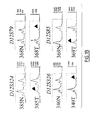
- Figures 1A and 1B show the LOH at chromosome 12q in adenoma tumors. Figure 1A:
Summary of LOH patterns in 5 adenoma tumors. Markers are shown along chromosome 12
according to the UCSC goldenpath sequencing map. Each vertical line represents a tumor
sample. Black, white and gray circles represent LOH, retention of heterozygosity and non-polymorphic
genotyping results, respectively. SNP 1 to 3 are polymorphic markers located in
the TCF1 gene i.e. L17L (rs1169289), I27L (rs1169288) and IVS9-24, respectively. The
critical region defined by genotyping is indicated on the left side between D12S86 and
D12S324. Figure 1B show representative fluorescent electropherograms for LOH. Tumor and
marker ID numbers are indicated. Fluorescent intensity scales are indicated on the right.
Alleles lost are shown by an arrow.
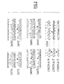
- Figure 2 shows representative TCF1 sequence variants. TCF1 sequence variants in adenoma
cases (T) and their matching non-tumor DNA (N). Mutated bases are indicated by an arrow.
In case 154, the RNA extraction was performed on the same tissue sample than the DNA
extraction. In the non-tumor tissue 154 (154DNA N) no mutation was observed at nucleotide
positions 749 and 872. At the tumoral DNA level (154DNA T) both mutated alleles were
found on the same exon 4 PCR product whereas at the tumoral RNA level (154RNA T) only
the 749A>C mutated allele is observed, the frameshifted mRNA is destabilized.
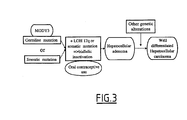
- Figure 3 is a schema of the occurrence of genetic alteration in liver tumors
-
EXAMPLES:
Example 1: Bi-allelic inactivation of HNF1α in hepatic tumors
Methods
-
Patient samples. A series of 16 adenomas and of 30 HCC were collected in the Liver Tumor
Database: Hôpital Pellegrin Bordeaux (28 cases), and Hôpital Antoine Béclère Clamart (2
cases). Tumor and non-tumor matching liver tissues were frozen immediately in liquid
nitrogen and stored at -80°C until DNA extraction. In two additional cases (CHC361 and
372) only non-tumor DNAs were available. All the pathological slides and medical records
were reviewed to confirm the diagnosis and the risk factors.
-
Genotyping. DNAs was extracted using a salting out procedure (Miller S.A. et al, (1988)).
Allelic losses were determined by genotyping 400 markers from the LMS2 microsatellites
panel (PE Biosystems) on tumor and corresponding non-tumor DNA as described previously
(Laurent-Puig P. et al, (2001)). All physical map information used in this work is available in
the Human Genome Project Working Draft at UCSC (http://genome.ucsc.edu/, (Lander E.S.
et al (2001)).
-
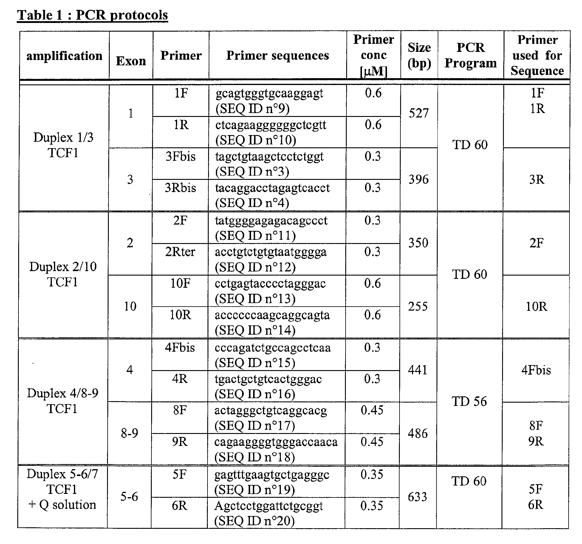
Mutation analysis. HNF1a exons were amplified by PCR and purified with a Millipore PCR
purification kit. Direct sequencing was performed on purified PCR products using big dye
terminator chemistry (Applied Biosystems) on an Applied Biosystems A377 or 3700
sequencer. Detailed PCR protocols and sequence of the primers are described below.
Analyses were performed using Factura and Autoassembler softwares (PE biosystems). The
somatic origin of mutations was confirmed by sequencing DNA derived from the non-tumor
corresponding tissue or from peripheral blood.
-
RT-PCR procedure. Tumor RNAs were extracted using Rneasy kit (Qiagen). Reverse-transcriptase
PCRs were performed using Omniscript kit (Qiagen) with oligo-dT primers and
AmpliTaq Perkin-Elmer polymerase to search for genetic alterations in adenomas and HCC
tumors. In each case, the coding sequence from codon 12 to 625 was amplified in two
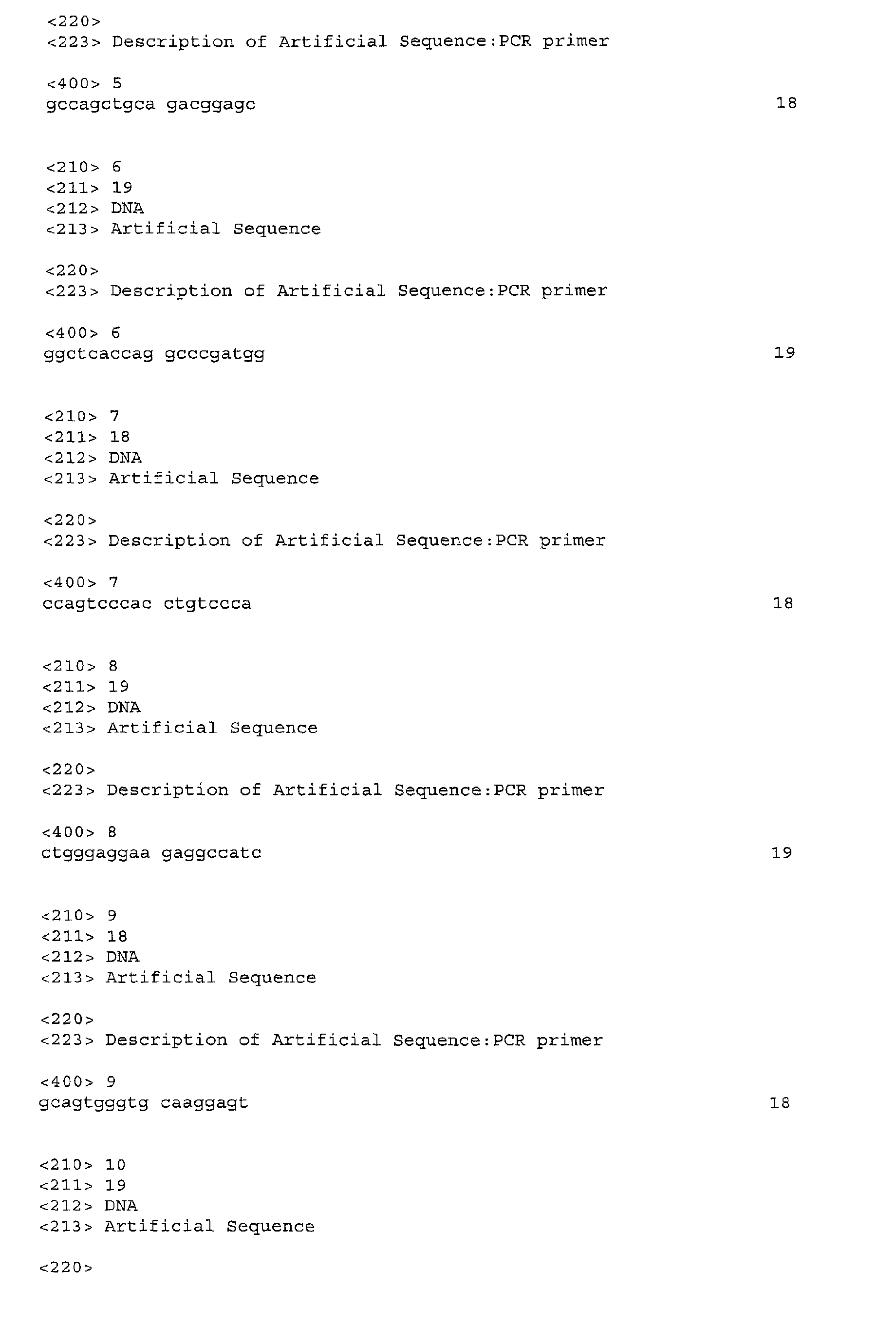
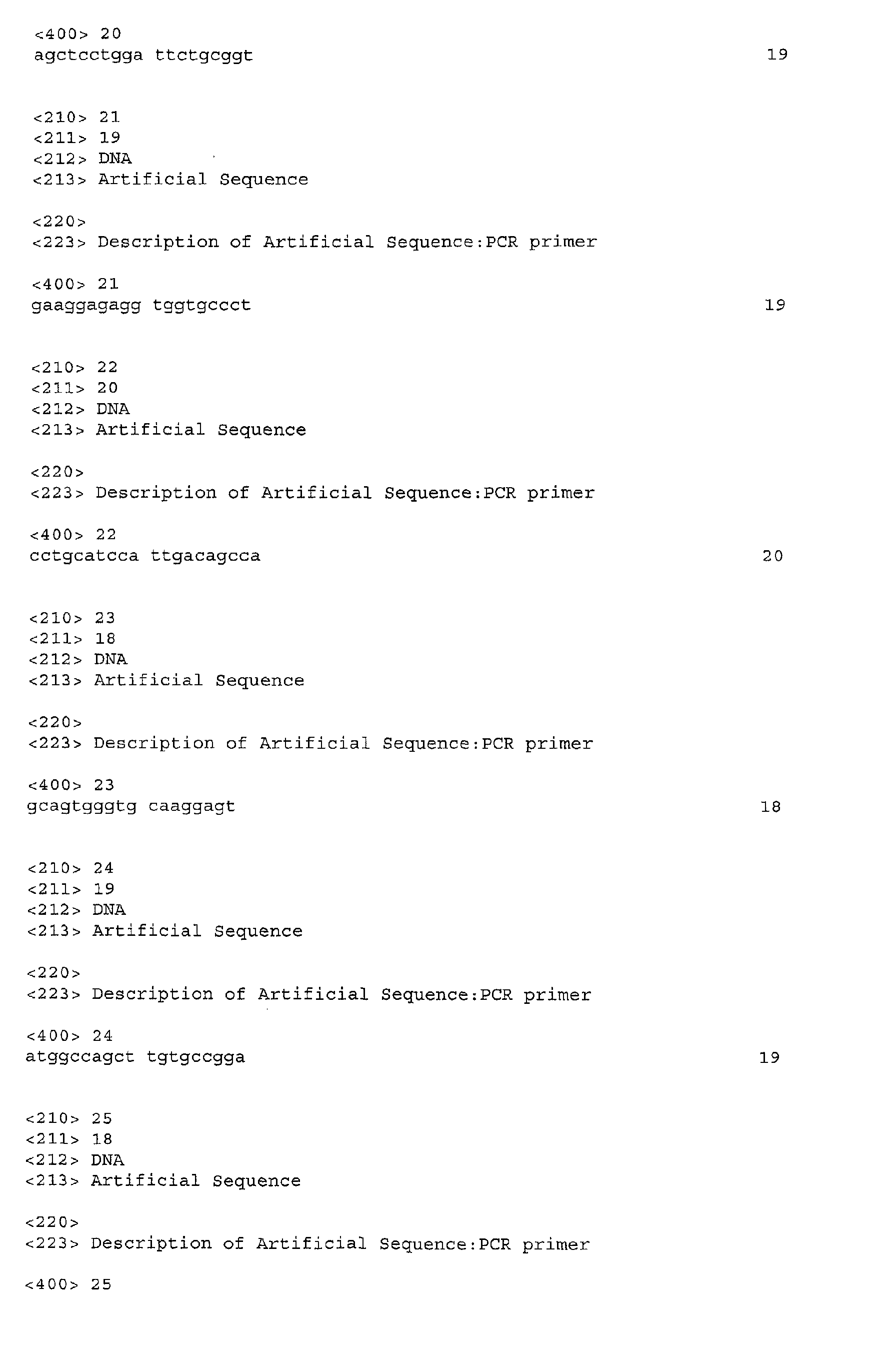
overlapping fragments using primers located in exon 1 (GCCAGCTGCAGACGGAGC ; SEQ
ID No. 5)) and 6 (GGCTCACCAGGCCCGATGG ; SEQ ID No 6)) and using primer from
exon 2 (CCAGTCCCACCTGTCCCA ; SEQ ID No 7)) and 10
(CTGGGAGGAAGAGGCCATC ; SEQ ID No 8)). Subsequently, amplified fragments were
directly sequenced with primers used for the amplification and internal primers.
PCR protocols
-
Results
-
In a search for putative tumor suppressor genes, DNA from a series of 10 adenomas (solitary
or multiple) and matched non-tumor tissue were genotyped for 400 microsatellite markers
distributed over the genome. Chromosome 12q loss was observed in 5 of the tumor samples
(Fig. 1A and 1B) with an 8 Mb minimal common region of deletion at 12q24, bounded by
D12S86 and D12S324. Subsequent in silico analysis of this critical region revealed the
presence of forty genes. The TCF1 gene, commonly known as HNF1α, was selected as a first
candidate gene because it encodes a transcription factor, HNF1α, that is implicated in
hepatocyte differentiation and that is required for liver-specific expression of several genes.
Sequencing of the HNF1α transcripts after RT-PCR, and of all exons with their intron-boundaries,
in DNA from 16 adenomas and the corresponding non-tumor tissue, was
performed. The analysis of ten known single nucleotide polymorphisms (SNP)3 in tumor and
non-tumor DNA for 14 of the cases confirmed the 12q24 deletion previously detected by
microsatellite genotyping (Fig. 1A and 1B) and did not reveal additional microdeletion. In ten
tumors, bi-allelic alteration of HNF1a was observed either by mutation and gene deletion in
half of the tumors, or by the existence of two mutations in the remaining tumors (Table 2). In
those tumors, a bi-allelic origin was confirmed by allele specific amplification and sequencing
of RT-PCR products. In patient 340, one of the HNF1a alterations (R229X) was germline,
similar to those found in patients with a MODY3 diagnosis (Kaisaki P.J. et al (1997)). Indeed,
this patient presented a typical familial MODY phenotype and his father, who carried the
same mutation, was also affected by hepatic adenomatosis. In the tumor sample of patient 340
the wild type allele was lost by a 12q deletion, while the mutant allele was retained, in
accordance with the 2 hit model of tumor predisposition. For the remaining 9 adenomas, both
HNF1α alterations were of somatic origin (including 4 with 12q LOH). Two of the patients
(358 and 371) had the same somatic mutation (W206L) on one allele.
-
The inventors also screened 30 HCC samples and found bi-allelic mutations of HNF1 in two
of them (Table 2). These mutations were not present in corresponding non-neoplastic liver
tissues and all were point mutations rather than LOH. The two HNF1a mutated HCC had an
infrequent presentation, with a very well differentiated tumor (Edmondson grade I) occurring
in normal liver, and with no risk factor usually associated with HCC, such as infection by
hepatitis B or C virus, or alcohol intake (Tsukuma H. et al. (1993)). These clinical
observations are compatible with the hypothesis that these HCC arose in the cells of an
adenomatous tumor, itself carrying inactivating mutations in HNF1α.
-
Among the 19 mutations observed in adenomas and HCC tumors, 9 were non-sense or
frameshift mutations leading to a premature stop codon (Table 2). The 10 remaining
mutations all occurred in the DNA binding domain, including two that were in the region
required for specific HNF binding and 8 in the homeodomain. All these missense mutations
occurred at amino acids that are highly conserved in both amphibians and rodents, suggesting
that they are important for protein function. These mutations were not found in more than 500
alleles screened in the literature (Ellard S. et al (2000)).
-
Two insertions of a cytosine and one 13 base pair deletion were observed in the cytosine tract
(C8-position 872), but these three tumors did not exhibit a microsatellite instability phenotype
as defined by the National Cancer Institute Workshop on Microsatellite Instability for cancer
detection (Boland C.R. et al (1998)). Interestingly, the mutations occurring at this hotspot
resulted in complete (CHC154) or partial (CHC370 and 358) destabilization of the
frameshifted mRNA, as was shown by the dramatic decrease in the intensity of the peaks
corresponding to the frameshifted mRNA (Fig. 2). This frameshift mutation, which leads to
the truncated protein P291fsinsC, has been observed previously in 20% of patients with
MODY3 and overexpression of this mRNA in transfection experiments had a dominant
negative effect on promoters normally induced by HNF1α (Vaxillaire M. et al. (1999),
Yamagata K. et al. (1998)). Nevertheless, HNF1α activity will be absent in the adenomas due
to the destabilization of this mRNA and the mutation of the second allele. As the 19 mutations
described here led to either protein truncation, modification of the DNA-binding domain
and/or RNA destabilization, these mutations are likely to generate a decrease in HNF1α
transactivation activity (Vaxillaire M. et al. (1999)). Furthermore, in all the mutated adenoma
and carcinoma samples, the alterations were bi-allelic and expected to result in a dramatic loss
of activity.
-
The inventors also found germline HNF1a mutations in two additional patients who had
previously a liver tumor resection in a context of familial diabetes. Patient 361 presented a
HCC developed in an adenoma, while patient 372 had a hyperplasic liver tumor (Table 2).
The G574S mutation was recently described with a high prevalence in African diabetics
(Collet C. et al. (2002)), whereas, the second germline mutation, R583Q, altered a highly
conserved amino-acid that has not been observed previously. These results suggest a role of
germline HNF1α mutation in predisposition to liver benign tumor development and may
explain a previous observation describing cosegregation of liver adenoma with diabetes
mellitus in a large family (Foster J.H. et al. (1978)). Because most of the HNF1α mutations in
MODY3 patients and in tumors are unique, further studies are required to elucidate the
relationship between genotype and phenotype.
-
Finally, 6 adenomas showed neither 12q LOH nor HNF1α mutations. These cases could be
due to undetected alteration in the non-coding region or in the promoter of HNF1α that could
have been missed by our screening process. Alternatively, mutations in another gene could
play a part in the occurrence of adenomas in those patients such as the other transcription
factors mutated in rare subtypes of MODY (HNF1β, HNF4α and IPF-1). However, the
present results did not indicate another common putative tumor suppressor gene, as there were
no other recurrently deleted chromosome regions.
-
In these series, HNF1α alteration is the only genetic event detected in most of the adenoma
cases but it is associated with multiple genetic alterations in HCC. These results suggest that
acquisition of a malignant phenotype by the tumor requires accumulation of additional genetic
alterations such as multiple LOH or TP53 mutations (Table 2). The observation of HNF1α bi-allelic
alteration in human liver tumors meets the criteria of the classical two-hit recessive
model of oncogenesis (Knudson A.G. et al. (1971), Comings D.E. (1973)) and supports the
hypothesis that HNF1α is a tumor suppressor gene altered early in carcinogenesis leading to
adenoma formation. The hypothesis of a tumor suppressor function for HNF1α is also
reinforced by the phenotypes observed in the two models of homozygous HNF1α deficient
mice. These mice have either a dramatic liver enlargement caused by an increased
proliferation of hepatocytes (Pontoglio M. et al. (1996)) or a liver enlargement with central
lobular hypertrophy and degeneration of individual hepatocytes (Lee Y.H. et al. (1998)).
Interestingly in the present series, mutated adenomas appeared to have typical clinical
presentations, i.e. most of the cases occurred in women with mild or long term use of oral
contraceptives suggesting a link between HNFα alteration, cellular proliferation and the use
of estrogen/progestin.
-
This study demonstrates that HNF1α inactivation is an early oncogenic event in liver
adenomas and in some hepatocellular carcinomas, and that inactivation occurs sporadically or
in association with familial germline mutation (Fig. 3). These results have immediate clinical
implications. In particular, MODY3 patients should benefit from liver monitoring to prevent
and detect early tumor occurrence, and patients presenting liver adenoma, especially in a
familial context, should be investigated for diabetes.
Example 2: Mutations in HNF1α in colon cancer
Material and methods :
Patients and methods
-
In a primary screening, a series of 10 adenomas and 29 colorectal cancers were collected in
two French pathological departments: Hôpital Européen Georges Pompidou (29 cases), and
CHU Dijon (10 cases). Tumor and non-tumor matching tissues were frozen immediately in
liquid nitrogen and stored at -80°C until DNA extraction. All tumors were characterized by
five microsatellites (BAT26, BAT25, D2S123, D5S346 and D17S250) and the MSI-H
phenotype was assigned following the criteria defined by international consensus conference
on microsatellite instability (Tsukuma, H. et al. (1993)). Twelve tumors were identified MSI-H,
17 MSS and all adenomas were MSS. In a second screening, 52 MSI-H colorectal cancers
were selected for HNF1α analysis in the Department of Genetics, Cliniques Universitaires St
Luc , Brussels , Belgium.
Mutation analysis
-
HNF1α mutations were screened by direct sequencing of all exons with their intronic
boundaries, as described in example 1. Sequence analyses were performed using Factura and
Autoassembler softwares (Applied Biosystems Courtaboeuf, France). The somatic origin of
mutations was confirmed by sequencing matched non-tumoral DNA.
RT-PCR
-
In the first series, nineteen colorectal tumors were available for RNA extraction using Rneasy
kit (Qiagen). Reverse-transcriptase PCRs were performed using Omniscript kit (Qiagen) with
oligo-dT primers and AmpliTaq Perkin-Elmer polymerase to search for genetic alterations in
colorectal cancer tumors. In each case, the coding sequence from codon 12 to 625 was
amplified in two overlapping fragments using primers located in exon 1
(GCCAGCTGCAGACGGAGC ; SEQ ID n° 5) and 6 (GGCTCACCAGGCCCGATGG ;
SEQ ID n° 6) and using primer from exon 2 (CCAGTCCCACCTGTCCCA ; SEQ ID n° 7)
and 10 (CTGGGAGGAAGAGGCCATC ; SEQ ID n° 8). Subsequently, amplified fragments
were directly sequenced with primers used for the amplification and with internal primers.
Results
-
Colorectal cancer is characterized by the existence of different entities known as
microsatellite stable (MSS) or unstable tumors (MSI-H). At first, a series of 10 adenomas and
29 colon cancers regardless of the MSI status were screened for HNF1α mutations on the
entire coding sequence. Three mutations in MSI-H tumors were found in the exon 4 poly-cytosin
(C)8 or (C)9 tract and consisted of a cytosin deletion at position 291. To further
characterize the importance of HNF1 mutations in the subgroup of MSI-H tumors, 52 MSI-H
additional samples were screened for exon 4 alterations and 23% CI95%[14%-36%] of MSI-H
tumors were found to harbor frameshifts at the cytosin track. The (C)9 allele was significantly
more frequently mutated than the (C)8 allele (22% versus 8% p=0.03) demonstrating a higher
instability of the longer repetition. This shows a role for HNF1 in MSI-H colorectal
carcinogenesis.
-
Results of these experiments are given in greater detail herebelow.
Screening for HNF1α mutation in colorectal adenoma and carcinoma :
-
Sequencing all HNF1α exons with their intron-boundaries was performed in 10 colon
adenomas and 29 colorectal carcinomas DNAs. A one base pair deletion in exon 4 was found
in 3 colorectal carcinomas. In all three cases it occurred at nucleotide 872 within a
polymorphic poly-cytosin (C)8 or (C)9 tract. These mutations led to a frameshift and a protein
truncation at position 342. All three HNF1α mutated tumors exhibited a MSI-H phenotype.
19 colorectal cancer samples, including the three mutated cases, were subjected to RNA
mutation screening on 95% of the coding sequence Screening at RNA level did not reveal
additional mutation in Adenoma or MSS tumors. Although very weakly expressed in one
case, the RNA corresponding to the 872del C allele could be detected suggesting the absence
of a destabilizing effect of this mutation.
Prevalence of the 872delC mutation in MSI-H colorectal carcinoma
-
Fifty-two MSI-H colorectal DNA samples were further sequenced in exon 4 in order to
evaluate the actual frequency of the polyC tract alteration in this subgroup of tumors. Twelve
additional MSI-H colorectal tumors were found mutated leading to a mutation rate of 23%
IC95% [14%-36%] in MSI-H colorectal cancers. All but one mutation consisted of a deletion
of one cytosin leading to a stop codon at position 342. The remaining case consisted of an
insertion of one cytosin leading to premature stop codon at position 316.
Mutations occurred more frequently on the (C)9 allele :
-
The nucleotide 864 (G>C) of HNF1α is polymorphic in the general population generating a
(C)8 or (C)9 polyC tract. The (C)8 and (C)9 alleles were observed at a frequency of 72% and
28% in the colorectal cancer patients. No significant difference in the frequency of the
different genotypes distribution was observed between MSI-H and MSS tumors. However, in
the MSI-H tumors, the (C)9 allele was significantly more frequently mutated than the allele
(C)8 22% versus 8% p=0.03. Finally, in the group of MSI-H patients, the clinical and
anatomopathological characteristics of the HNF1α mutated tumors did not differ from those
non-mutated.
-
The frameshift mutations found in HNF1α exon 4 are predicted to lead to the production of
truncated proteins containing the dimerization and the DNA binding domain. In contrast these
truncated proteins lack the COOH-terminal part containing the transactivation domain. In
MODY3 patients, the most common HNF1α mutation (20%) is an insertion of a cytosine in
the exon 4 polyC tract leading to the synthesis of a protein of 315 amioacids. Over expression
of this truncated protein in MIN6 and C33 cells revealed the dominant negative function of
the P291fsinsC mutation (Schott, et al. (1997)); (Vaxillaire, et al. (1999)). It could also be the
case for the 872delC observed in colorectal cancers, which would contrast with the biallelic
HNF1α inactivation observed in hepatocellular adenoma. However in liver adenoma a variety
of mutations exist and exon 4 frameshift mutation encounts for only 15% of them. Moreover,
in hepatocellular adenoma exon 4 frameshift mutation leads to RNA destabilization. In MSI-H
colorectal cancer no destabilization of the frameshift RNA was observed and mutations
seemed to be mono-allelic. These two arguments are in favor of a dominant negative effect of
the P291fsdelC mutation in the intestine origin cells.
-
The HNF1α polyC tract in exon 4 is polymorphic in the Caucasian population. A higher rate
of mutations occurring on the C(9) allele in comparison with the average frequency suggested
a preferential targeting of the longer tract by mismatch repair deficiency. According to this
observation, this polymorphism could be implied in the risk of colorectal cancer in general
population.
References
-
- Baumhueter, S. et al. HNF-1 shares three sequence motifs with the POU domain proteins
and is identical to LF-B1 and APF. Genes Dev 4, 372-9. (1990).
- Boland, C.R. et al. A National Cancer Institute Workshop on Microsatellite Instability for
cancer detection and familial predisposition: development of international criteria for the
determination of microsatellite instability in colorectal cancer. Cancer Res 58, 5248-57.
(1998).
- Boland, C. R., Thibodeau, S. N., Hamilton, S. R., Sidransky, D., Eshleman, J. R., Burt, R.
W., Meltzer, S. J., Rodriguez-Bigas, M. A., Fodde, R., Ranzani, G. N., and Srivastava, S. A
National Cancer Institute Workshop on Microsatellite Instability for cancer detection and
familial predisposition: development of international criteria for the determination of
microsatellite instability in colorectal cancer, Cancer Res. 58: 5248-57., 1998.
- Cereghini, S., Yaniv, M. & Cortese, R. Hepatocyte dedifferentiation and extinction is
accompanied by a block in the synthesis of mRNA coding for the transcription factor
HNF1/LFB1. Embo J 9, 2257-63. (1990).
- Chiche, L. et al. Liver adenomatosis: reappraisal, diagnosis, and surgical management: eight
new cases and review of the literature. Ann Surg 231, 74-81. (2000).
- Chouard et al, 1997, Biochimie, 79 : 707-715.
- Chouard, T. et al. A distal dimerization domain is essential for DNA-binding by the atypical
HNF1 homeodomain. Nucleic Acids Res 18, 5853-63. (1990).
- Collet, C. et al. Prevalence of the missense mutation Gly574Ser in the hepatocyte nuclear
factor-1alpha in Africans with diabetes. Diabetes Metab 28, 39-44. (2002).
- Comings, D.E. A general theory of carcinogenesis. Proc Natl Acad Sci U S A 70, 3324-8.
(1973).
- Coumoul and Barouki, Medecine/Sciences 2002, 18:86.
- Edmondson, H.A., Henderson, B. & Benton, B. Liver-cell adenomas associated with use of
oral contraceptives. N Engl J Med 294, 470-2. (1976).
- Ellard, S. Hepatocyte nuclear factor 1 alpha (HNF-1 alpha) mutations in maturity- onset
diabetes of the young. Hum Mutat 16, 377-85. (2000).
- Ferrell, L.D. Hepatocellular carcinoma arising in a focus of multilobular adenoma. A case
report. Am J Surg Pathol 17, 525-9. (1993).
- Flejou, J.F. et al. Liver adenomatosis. An entity distinct from liver adenoma?
Gastroenterology 89, 1132-8. (1985).
- Foster, J.H., Donohue, T.A. & Berman, M.M. Familial liver-cell adenomas and diabetes
mellitus. N Engl J Med 299, 239-41. (1978).
- Frain, M. et al. The liver-specific transcription factor LF-B1 contains a highly diverged
homeobox DNA binding domain. Cell 59, 145-57. (1989).
- Goldfarb, S. Sex hormones and hepatic neoplasia. Cancer Res 36, 2584-8. (1976).
- Harlow et al, 1988 editors, Antibodies : A laboratory manual
- Howell, R.R., Stevenson, R.E., Ben-Menachem, Y., Phyliky, R.L. & Berry, D.H. Hepatic
adenomata with type 1 glycogen storage disease. Jama 236, 1481-4. (1976).
- Kaisaki, P.J. et al. Mutations in the hepatocyte nuclear factor-1alpha gene in MODY and
early-onset NIDDM: evidence for a mutational hotspot in exon 4. Diabetes 46, 528-35.
(1997).
- Knudson, A.G., Jr. Mutation and cancer: statistical study of retinoblastoma. Proc Natl Acad
Sci U S A 68, 820-3. (1971).
- Lander, E.S. et al. Initial sequencing and analysis of the human genome. Nature 409, 860-921.
(2001).
- Laurent-Puig, P. et al. Genetic alterations associated with hepatocellular carcinomas define
distinct pathways of hepatocarcinogenesis. Gastroenterology 120, 1763-73. (2001).
- Laurent-Puig, P., Legoix, P., Bluteau, O., Belghiti, J., Franco, D., Binot, F., Monges, G.,
Thomas, G., Bioulac-Sage, P., and Zucman-Rossi, J. Genetic alterations associated with
hepatocellular carcinomas define distinct pathways of hepatocarcinogenesis,
Gastroenterology. 120: 1763-73., 2001.
- Lee, Y.H., Sauer, B. & Gonzalez, F.J. Laron dwarfism and non-insulin-dependent diabetes
mellitus in the Hnf- 1alpha knockout mouse. Mol Cell Biol 18, 3059-68. (1998).
- Lengauer, C., Kinzler, K. W., and Vogelstein, B. Genetic instability in colorectal cancers,
Nature. 386: 623-7., 1997.
- Miller, S.A., Dykes, D.D. & Polesky, H.F. A simple salting out procedure for extracting
DNA from human nucleated cells. Nucleic Acids Res 16, 1215. (1988).
- Nicosia, A. et al. A myosin-like dimerization helix and an extra-large homeodomain are
essential elements of the tripartite DNA binding structure of LFB1. Cell 61, 1225-36. (1990).
- Pontoglio, M. et al. Hepatocyte nuclear factor 1 inactivation results in hepatic dysfunction,
phenylketonuria, and renal Fanconi syndrome. Cell 84, 575-85. (1996).
- Reeck et al, 1987, Cell, 50 : 667.
- Sale, G.E. & Lerner, K.G. Multiple tumors after androgen therapy. Arch Pathol Lab Med
101, 600-3. (1977).
- Sambrook et al, 1989, Molecular Cloning : A laboratory manual, second edition Cold Spring
Harbor Laboratory press, Cold Spring Harbor, New-York.
- Schott, O., Billeter, M., Leiting, B., Wider, G. & Wuthrich, K. The NMR solution structure
of the non-classical homeodomain from the rat liver LFB1/HNF1 transcription factor. J Mol
Biol 267, 673-83. (1997).
- Tsukuma, H. et a1. Risk factors for hepatocellular carcinoma among patients with chronic
liver disease. N Engl J Med 328, 1797-801. (1993).
- Vaxillaire, M. et al. Anatomy of a homeoprotein revealed by the analysis of human MODY3
mutations. J Biol Chem 274, 35639-46. (1999).
- Yamagata, K. et al. Mutation P291fsinsC in the transcription factor hepatocyte nuclear
factor-1alpha is dominant negative. Diabetes 47, 1231-5. (1998).
- Yamagata, K. et al. Mutations in the hepatocyte nuclear factor-1alpha gene in maturity-onset
diabetes of the young (MODY3). Nature 384, 455-8. (1996).