EP1391209A1 - Protein formulations - Google Patents
Protein formulations Download PDFInfo
- Publication number
- EP1391209A1 EP1391209A1 EP02776487A EP02776487A EP1391209A1 EP 1391209 A1 EP1391209 A1 EP 1391209A1 EP 02776487 A EP02776487 A EP 02776487A EP 02776487 A EP02776487 A EP 02776487A EP 1391209 A1 EP1391209 A1 EP 1391209A1
- Authority
- EP
- European Patent Office
- Prior art keywords
- buffer
- antibody
- sample
- protein
- derivative
- Prior art date
- Legal status (The legal status is an assumption and is not a legal conclusion. Google has not performed a legal analysis and makes no representation as to the accuracy of the status listed.)
- Withdrawn
Links
Images








Classifications
-
- A—HUMAN NECESSITIES
- A61—MEDICAL OR VETERINARY SCIENCE; HYGIENE
- A61K—PREPARATIONS FOR MEDICAL, DENTAL OR TOILETRY PURPOSES
- A61K39/00—Medicinal preparations containing antigens or antibodies
- A61K39/395—Antibodies; Immunoglobulins; Immune serum, e.g. antilymphocytic serum
- A61K39/39591—Stabilisation, fragmentation
-
- A—HUMAN NECESSITIES
- A61—MEDICAL OR VETERINARY SCIENCE; HYGIENE
- A61K—PREPARATIONS FOR MEDICAL, DENTAL OR TOILETRY PURPOSES
- A61K47/00—Medicinal preparations characterised by the non-active ingredients used, e.g. carriers or inert additives; Targeting or modifying agents chemically bound to the active ingredient
- A61K47/06—Organic compounds, e.g. natural or synthetic hydrocarbons, polyolefins, mineral oil, petrolatum or ozokerite
- A61K47/20—Organic compounds, e.g. natural or synthetic hydrocarbons, polyolefins, mineral oil, petrolatum or ozokerite containing sulfur, e.g. dimethyl sulfoxide [DMSO], docusate, sodium lauryl sulfate or aminosulfonic acids
-
- A—HUMAN NECESSITIES
- A61—MEDICAL OR VETERINARY SCIENCE; HYGIENE
- A61K—PREPARATIONS FOR MEDICAL, DENTAL OR TOILETRY PURPOSES
- A61K47/00—Medicinal preparations characterised by the non-active ingredients used, e.g. carriers or inert additives; Targeting or modifying agents chemically bound to the active ingredient
- A61K47/06—Organic compounds, e.g. natural or synthetic hydrocarbons, polyolefins, mineral oil, petrolatum or ozokerite
- A61K47/22—Heterocyclic compounds, e.g. ascorbic acid, tocopherol or pyrrolidones
Definitions
- the present invention relates to protein formulations with improved stability. More specifically, the present invention relates to improving the stability of physiologically active proteins (e.g., antibodies) in a highly concentrated solution form by using a buffer such as acetate buffer, imidazole buffer, 2-morpholinoethanesulfonate buffer or 3-morpholinopropanesulfonate buffer.
- a buffer such as acetate buffer, imidazole buffer, 2-morpholinoethanesulfonate buffer or 3-morpholinopropanesulfonate buffer.
- proteins When proteins are stored in a highly concentrated solution form, they are usually associated with a problem of deterioration including the formation of insoluble aggregates; and therefore, it is necessary to prevent the phenomena of deterioration.
- antibodies such as immunoglobulins, monoclonal antibodies and humanized antibodies are unstable proteins and more likely to undergo physical and chemical changes (e.g., association, aggregation) due to stresses applied during virus removal in the purification process, including filtration stress, concentration stress and heat stress.
- stabilization effects are obtained by adding a stabilizer for preventing chemical and physical changes, including high-molecular weight materials such as proteins (e.g., human serum albumin, purified gelatin) or low-molecular weight materials such as polyols, amino acids and surfactants.
- proteins e.g., human serum albumin, purified gelatin
- low-molecular weight materials such as polyols, amino acids and surfactants.
- organism-derived high-molecular weight materials like proteins are disadvantageous in requiring very complicated processes to remove contaminants, such as viruses, originating from the stabilizers per se.
- heat treatment is carried out for the purpose of virus inactivation, such stabilizers may cause problems such as association and aggregation due to heat stress.
- insoluble aggregates in a protein solution it is caused by the formation of denatured proteins, i.e., denatured intermediates; there has been a need to develop a method for preventing the formation of insoluble aggregates.
- the object of the present invention is to provide stable formulations containing physiologically active proteins (e..g., antibodies, enzymes, hormones and cytokines) by improving the stability of the physiologically active proteins in a highly concentrated solution form to prevent the formation of denatured intermediates and insoluble aggregates.
- physiologically active proteins e..g., antibodies, enzymes, hormones and cytokines
- the inventors of the present invention have found that the stability of proteins can be increased in one or more buffers, particularly of about pH 5 to 7, selected from the group consisting of acetate buffer, imidazole buffer, a buffer of aminoethanesulfonate or a derivative thereof, and a buffer of aminopropanesulfonate or a derivative thereof, thereby preventing the formation of denatured intermediates as well as insoluble aggregates.
- buffers particularly of about pH 5 to 7
- the present invention provides the following:
- physiologically active protein examples include, but are not limited to, antibodies, enzymes, cytokines and hormones. Specific examples include, but are not limited to, hematopoietic factors such as granulocyte colony-stimulating factor (G-CSF), granulocyte-macrophage colony-stimulating factor (GM-CSF), erythropoietin (EPO) and thrombopoietin, cytokines such as interferons, IL-1 and IL-6, immunoglobulins, monoclonal antibodies, humanized antibodies, tissue plasminogen activator (tPA), urokinase, serum albumin, blood coagulation factor VIII, leptin, insulin, and stem cell growth factor (SCF). Among these proteins, preferred are immunoglobulins, and more preferred are monoclonal antibodies and humanized antibodies.
- tPA tissue plasminogen activator
- SCF stem cell growth factor
- a “physiologically active protein” refers to a protein having substantially the same biological activities as a corresponding physiologically active protein of mammalian (especially human) origin. Such a protein may be natural or recombinant, preferably recombinant. A recombinant protein encompasses those having the same amino acid sequence as the corresponding natural protein, as well as those comprising deletion, substitution or addition of one or more amino acid residues in the amino acid sequence, but retaining the biological activities as mentioned above. Further, the physiologically active protein as used herein includes those chemically modified with PEG, etc.
- a physiologically active protein when a physiologically active protein is a monoclonal antibody, it may be prepared in any manner.
- a monoclonal antibody can be produced using known techniques by immunizing a sensitized antigen in a standard manner for immunization, fusing the resulting immunocytes with known parent cells in a standard manner for cell fusion, and then screening monoclonal antibody-producing cells in a standard manner for screening.
- the monoclonal antibody as used herein is not limited to those produced by hybridomas, but includes chimeric antibodies which are artificially modified, e.g., for the purpose of reducing xenoantigenicity to human.
- reshaped humanized antibodies may also be used in the present invention.
- Such antibodies are prepared by replacing complementarity-determining regions of human antibodies with those of non-human mammalian (e.g., mouse) antibodies; standard recombinant procedures for this purpose are also known. Such known procedures may be used to give reshaped humanized antibodies.
- FR framework
- hPM-1 humanized anti-IL-6 receptor antibody
- a humanized anti-HM1.24 antigen monoclonal antibody see WO98/14580
- a humanized anti-parathyroid hormone-related peptide antibody anti-PTHrP antibody
- a humanized anti-tissue factor antibody see WO99/51743 and the like are also preferred for use in the present invention.
- human antibodies produced in transgenic animals and the like are also preferred.
- the antibody as used herein encompasses antibody fragments including Fab, (Fab') 2 , Fc, Fc' and Fd, as well as reshaped antibodies including monovalent or polyvalent single chain antibodies (scFV).
- sample containing a physiologically active protein or an “antibody-containing sample” refers to a sample containing any protein or antibody, regardless of whether derived from organisms or produced recombinantly. It preferably refers to a culture medium of mammalian cells (e.g., CHO cells) containing physiologically active protein or antibody molecules produced by culture, which may further be subjected to partial purification or other specific treatment.
- mammalian cells e.g., CHO cells
- the inventors of the present invention have studied various factors involved in heat treatment-induced denaturation of antibody-containing samples. As a result, they have found that when heat treatment at 80°C for 2 hours or at 60°C for 2 or 4 weeks is performed on antibody molecules dissolved in acetate buffer, imidazole buffer, a buffer of aminoethanesulfonate or a derivative thereof or a buffer of aminopropanesulfonate or a derivative thereof, these buffers achieve a larger stabilization effect than phosphate or citrate buffer because they allow reduction of denatured intermediates (i.e., soluble associated molecules), prevention of the denaturation phenomenon, as well as avoidance of insoluble aggregate formation. In particular, it has also been observed that these buffers inhibit further aggregation of denatured intermediates into insoluble aggregates.
- the protein formulation of the present invention may be prepared by dissolving a physiologically active protein and various ingredients as shown below into one or more buffers selected from the group consisting of acetate buffer, imidazole buffer, a buffer of aminoethanesulfonate or a derivative thereof, and a buffer of aminopropanesulfonate or a derivative thereof. These buffers may be used alone or in combination.
- aminoethanesulfonate or a derivative thereof include, but are not limited to:
- aminopropanesulfonate or a derivative thereof include, but are not limited to:
- Such a buffer preferably has a pH of 5.0 to 8.0, more preferably 5.0 to 7.0, and even more preferably 5.5 to 6.5.
- the concentration of the buffer usually ranges from 1 to 500 mM, preferably from 5 to 100 mM, more preferably from 5 to 30 mM, and most preferably from 10 to 20 mM.
- the buffer used in the present invention may be prepared in any manner.
- a given amount of a reagent preferably of special grade
- Non-limiting examples of buffer preparation will be presented below.
- Acetic acid (0.90 g) is dissolved in purified water (about 900 mL), adjusted to the desired pH with 1 M sodium hydroxide and then adjusted to a total volume of 1 L.
- Imidazole (1.02 g) is dissolved in purified water (about 900 mL), adjusted to the desired pH with 1 M hydrochloric acid and then adjusted to a total volume of 1 L.
- 2-Morpholinoethanesulfonate monohydrate (3.20 g) is dissolved in purified water (about 900 mL), adjusted to the desired pH with 1 M sodium hydroxide and then adjusted to a total volume of 1 L.
- 3-Morpholinopropanesulfonate (3.14 g) is dissolved in purified water (about 900 mL), adjusted to the desired pH with 1M sodium hydroxide and then adjusted to a total volume of 1 L.
- the formulation of the present invention may further comprise a surfactant.
- a surfactant include:
- Preferred surfactants are polyoxyethylene sorbitan fatty acid esters. Particularly preferred are Polysorbate 20, 21, 40, 60, 65, 80, 81 and 85, and most preferred are Polysorbate 20 and 80.
- the formulation of the present invention may also comprise various amino acids as stabilizers.
- polyethylene glycol or saccharides e.g., dextran, mannitol, sorbitol, inositol, glucose, fructose, lactose, xylose, mannose, maltose, raffinose
- isotonizing agent e.g., dextran, mannitol, sorbitol, inositol, glucose, fructose, lactose, xylose, mannose, maltose, raffinose
- the protein formulation of the present invention may further comprise a diluent, a solubilizer, an excipient, a pH adjuster, a soothing agent, a buffer, a sulfur-containing reducing agent, an antioxidant and the like.
- a sulfur-containing reducing agent include those having a sulfhydryl group such as N-acetylcysteine, N-acetylhomocysteine, thioctic acid, thiodiglycol, thioethanolamine, thioglycerol, thiosorbitol, thioglycolic acid and a salt thereof, sodium thiosulfate, glutathione, and a C 1 -C 7 thioalkanoic acid.
- antioxidants examples include erythorbic acid, dibutylhydroxytoluene, butylhydroxyanisole, ⁇ -tocopherol, tocopherol acetate, L-ascorbic acid and a salt thereof, L-ascorbic acid palmitate, L-ascorbic acid stearate, sodium bisulfite, sodium sulfite, triamyl gallate and propyl gallate, as well as chelating agents such as disodium ethylenediaminetetraacetate (EDTA), sodium pyrophosphate and sodium metaphosphate.
- EDTA disodium ethylenediaminetetraacetate
- the protein formulation of the present invention may comprise other commonly used ingredients, for example, inorganic salts such as sodium chloride, potassium chloride, calcium chloride, sodium phosphate, potassium phosphate and sodium bicarbonate, as well as organic salts such as sodium citrate, potassium citrate and sodium acetate.
- inorganic salts such as sodium chloride, potassium chloride, calcium chloride, sodium phosphate, potassium phosphate and sodium bicarbonate
- organic salts such as sodium citrate, potassium citrate and sodium acetate.
- the protein formulation of the present invention is usually administered by parenteral route in the dosage form of injections (e.g., subcutaneous, intravenous, intramuscular or intraperitoneal injections) or transdermal, transmucosal, transnasal or transpulmonary preparations, but it may also be administered orally.
- parenteral route in the dosage form of injections (e.g., subcutaneous, intravenous, intramuscular or intraperitoneal injections) or transdermal, transmucosal, transnasal or transpulmonary preparations, but it may also be administered orally.
- the protein formulation of the present invention may be a liquid formulation or a lyophilized formulation which is reconstituted before use.
- excipients for lyophillization for example, sugar alcohols or saccharides (e.g., mannitol, glucose) may be used.
- the protein formulation of the present invention can usually be provided in the form of containers with defined volume, including sealed and sterilized plastic or glass vials, ampoules and syringes, as well as in the form of large volume containers like bottles.
- the amount of a physiologically active protein in the formulation of the present invention can be determined as appropriate for the type of physiologically active protein to be used, the type and severity of disease to be treated, the age of a patient, etc.
- a physiologically active protein is incorporated in an amount of 0.01 ⁇ g/ml to 200 mg/ml, preferably 0.5 ⁇ g/ml to 100 mg/ml, based on the total volume of the formulation or an injectable composition thereof, which is supplemented with saccharides.
- a physiologically active protein is an antibody such as an immunoglobulin, a monoclonal antibody or a humanized antibody
- it is usually incorporated at a final administration concentration of 0.1 to 200 mg/ml, preferably 1 to 200 mg/ml, more preferably 10 to 200 mg/ml, and most preferably 10 to 150 mg/ml.
- the protein formulation of the present invention shows a remarkable stabilization effect, particularly when formulated into a highly concentrated protein solution containing a physiologically active protein in an amount of 10 mg/ml or more.
- the amount of a physiologically active protein in the protein-containing sample of the present invention usually ranges from 0.01 ⁇ g/ml to 200 mg/ml, preferably from 0.5 ⁇ g/ml to 100 mg/ml, based on the total volume of the sample.
- a physiologically active protein is an antibody such as an immunoglobulin, a monoclonal antibody or a humanized antibody
- it is usually incorporated at a final administration concentration of 0.1 to 200 mg/ml, preferably 1 to 200 mg/ml, more preferably 10 to 200 mg/ml, and most preferably 10 to 150 mg/ml.
- the physiologically active protein formulation of the present invention was tested for the stability by fluorescence spectroscopy (Trp residue, ANS binding), cation-exchange HPLC, gel filtration HPLC and SDS-PAGE.
- the thermal denaturation phenomenon was measured by cation-exchange HPLC, gel filtration HPLC and ANS-binding fluorescence spectroscopy, indicating that thermal denaturation after treatment at 60°C for 4 weeks was most effectively inhibited around pH 6. This could be explained by the results of differential scanning calorimetry. Namely, it was considered that under acidic conditions, the reduction of heat stability would be caused by low denaturation midpoint temperatures in all 3 steps of denaturation, while under basic conditions, it would be caused by the high tendency of intermediates to aggregate after denaturation.
- antibody fragments (Fab and Fc fragments) were treated at 60°C and analyzed by gel filtration HPLC and ANS-binding fluorescence spectroscopy, indicating that the Fab fragment showed undetectable denaturation after heat treatment, regardless of buffer types, whereas the Fc fragment showed a buffer-dependent thermal denaturation phenomenon, as in the case of the whole antibody.
- Acetate and MES buffers were found to inhibit thermal denaturation of the Fc fragment, as compared to phosphate and citrate buffers.
- differential scanning calorimetry was performed to assess thermal denaturation temperatures and the reversibility of thermal denaturation phenomenon, indicating that in the case of the Fab domain alone, no aggregation was observed in all buffers after denaturation. This suggested that the Fc domain would greatly contribute to aggregation of the whole antibody, and that the denaturation phenomenon of the whole antibody would be influenced by the interaction between Fab and Fc domains, rather than being equal to the mere sum of both domains.
- the present invention provides a method for improving the stability of a protein formulation, which comprises dissolving a physiologically active protein in one or more buffers selected from the group consisting of acetate buffer, imidazole buffer, a buffer of aminoethanesulfonate or a derivative thereof, and a buffer of aminopropanesulfonate or a derivative thereof.
- the present invention further provides a method for stabilizing a protein-containing sample under heat treatment, which comprises performing heat treatment on the protein-containing sample in such a form that a physiologically active protein is contained in one or more buffers selected from the group consisting of acetate buffer, imidazole buffer, a buffer of aminoethanesulfonate or a derivative thereof, and a buffer of aminopropanesulfonate or a derivative thereof.
- the stabilizing method of the present invention is particularly advantageous for use in heat treatment for virus removal in the purification process of protein formulations, etc.
- a humanized anti-IL-6 receptor antibody was used for measurement. Procedures for production of the humanized anti-IL-6 receptor antibody can be found in WO92/19759. Among antibodies found in WO92/19759, an antibody comprising the L chain version "a” and the H chain version "f” was used in the present invention. This antibody was recombinantly produced in CHO cells.
- a stock solution of the humanized anti-IL-6 receptor antibody (about 40 mg/mL, pH 6.5) was diluted with 15 mM. phosphate buffer (pH 6.5) to prepare a sample solution with an antibody concentration of 10 mg/mL, which was then filled into glass ampoules (2 mL each) and flame-sealed (Sample 1: phosphate buffer, pH 6.5).
- pH as used herein refers to the pH of the prepared sample.
- the solution with an antibody concentration of 10 mg/mL was further dialyzed against the buffers shown below to prepare sample solutions in different buffers, which were then filled into glass ampoules (2 mL each) and flame-sealed.
- the buffer concentration was set to 15 mM in all cases.
- the heat-treated samples were assessed for their physical properties in the following manner to compare their stability.
- Each sample was diluted with 15 mM phosphate buffer (pH 6.5) to give an antibody concentration of 133 ⁇ g/mL (0.89 ⁇ M) and used as a test sample.
- Each test sample (about 1.5 mL) was introduced into a fluorescence cell and measured for the fluorescence spectrum between 280 and 400 nm with an excitation wavelength of 280 nm. This measurement is intended to analyze the state of tryptophan residues. Upon protein denaturation, tryptophan residues in hydrophobic region will undergo structural changes to increase the fluorescence intensity and to give the maximum fluorescence wavelength shifted to a longer wavelength.
- Each sample was diluted with 15 mM phosphate buffer (pH 6.5) to give an antibody concentration of 667 ⁇ g/mL (0.89 ⁇ M), 900 ⁇ L of which was supplemented with a 0.4 M solution of 1-anilino-8-naphthalene sulfonate (ANS) (300 ⁇ L) for use as a test sample.
- ANS 1-anilino-8-naphthalene sulfonate
- the whole volume of each test sample was introduced into a fluorescence cell and measured for the fluorescence spectrum and intensity between 450 and 550 nm with an excitation wavelength of 365 nm.
- ANS has the property of binding to hydrophobic regions on the protein surface and emits fluorescence upon binding. Thus, this measurement is intended to analyze the hydrophobicity of the protein surface as a change in fluorescence intensity.
- Each sample (1 ⁇ g as an antibody) in 10% SDS-containing dilution buffer was applied to a polyacrylamide gel (12% for reducing, 8% for non-reducing) and electrophoresed for about 40 minutes with a maximum voltage of 200 V and a maximum current of 200 mA.
- the bands were stained with coomassie brilliant blue.
- imidazole, acetate, MES and MOPS buffers When compared to phosphate and citrate buffers, imidazole, acetate, MES and MOPS buffers showed lower fluorescence intensity for both spectra after heat treatment at 80°C for 2 hours; these buffers were found to inhibit denaturation.
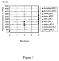
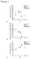
- Figure 1 shows the test results of ANS binding after heat treatment at 60°C for 2 and 4 weeks.
- imidazole, acetate, MES and MOPS buffers were found to inhibit denaturation. In fact, no insoluble aggregate was observed in the samples using imidazole, acetate, MES and MOPS buffers.
- Heat treatment at 80°C for 2 hours reduced the peak area of the native molecules, while producing a peak of denatured intermediates.
- imidazole, acetate, MES and MOPS buffers When compared to phosphate and citrate buffers, imidazole, acetate, MES and MOPS buffers showed a higher content of the native molecules remaining after treatment at 80°C for 2 hours; these buffers were found to inhibit the formation of denatured intermediates.
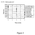
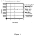
- Figures 2 and 3 show the test results after treatment at 60°C for 2 and 4 weeks.
- imidazole, acetate, MES and MOPS buffers When compared to phosphate and citrate buffers, imidazole, acetate, MES and MOPS buffers showed a higher content of the remaining native molecules; these buffers were found to inhibit the formation of denatured intermediates. In fact, no insoluble aggregate was observed in the samples using imidazole, acetate, MES and MOPS buffers.
- Table 3 shows the test results after heat treatment at 80°C for 2 hours. (at 80°C for 2 hours) Native molecule (monomer) Denatured intermediate (associated) Sample 1 Phosphate buffer pH 6.5 219350 528928 Sample 2 Phosphate buffer pH 5.5 381133 379504 Sample 3 Citrate buffer pH 6.5 178756 504121 Sample 4 Citrate buffer pH 5.5 166392 584894 Sample 5 Imidazole buffer pH 6.5 455483 286100 Sample 6 Acetate buffer pH 5.5 481455 252296 Sample 7 MES buffer pH 6.5 422489 353231 Sample 8 MES buffer pH 5.5 585661 76946 Sample 9 MOPS buffer pH 6.5 412389 310318
- Heat treatment at 80°C for 2 hours reduced the peak area of monomer molecules corresponding to the native molecules, while producing a peak of soluble aggregates corresponding to denatured intermediates in the higher molecular weight region.
- imidazole, acetate, MES and MOPS buffers When compared to phosphate and citrate buffers, imidazole, acetate, MES and MOPS buffers showed a higher content of the monomer molecules remaining after treatment at 80°C for 2 hours; these buffers were found to inhibit the formation of soluble aggregates.
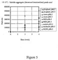
- Figures 4 and 5 show the test results after treatment at 60°C for 2 and 4 weeks.
- imidazole, acetate, MES and MOPS buffers When compared to phosphate and citrate buffers, imidazole, acetate, MES and MOPS buffers showed a higher content of the remaining monomer molecules; these buffers were found to inhibit the formation of soluble aggregates. In fact, no insoluble aggregate was observed in the samples using imidazole, acetate, MES and MOPS buffers.
- imidazole, acetate, MES and MOPS buffers When compared to phosphate and citrate buffers, imidazole, acetate, MES and MOPS buffers showed weaker bands in the higher molecular weight region after treatment at 80°C for 2 hours; these buffers were found to inhibit the formation of covalently associated molecules.
- the humanized anti-IL-6 receptor antibody described in Example 1 was used for measurement.
- a stock solution of the humanized anti-IL-6 receptor antibody (about 40 mg/mL, pH 6.5) was dialyzed against purified water and then supplemented with buffer ingredients, followed by dilution with purified water to prepare the following sample solutions with an antibody concentration of 10 mg/mL.
- Each sample was filled into glass ampoules (2 mL each) and flame-sealed.
- the heat-treated samples were assessed for their physical properties in the following manner to compare their stability.
- Each sample was diluted with 15 mM phosphate buffer (pH 6.5) to give an antibody concentration of 667 ⁇ g/mL (0.89 ⁇ M), 900 ⁇ L of which was supplemented with a 0.4 M solution of 1-anilino-8-naphthalene sulfonate (ANS) (300 ⁇ L) for use as a test sample.
- ANS 1-anilino-8-naphthalene sulfonate
- the stained gel was further scanned with a photoscanner and converted into an image file for use in calculation of the band density (turbidity) corresponding to the H chain.
- the calculation was carried out on a computer using the image analysis software Scion-Image to determine the % residual H chain (vs. initial).
- ammonia content was determined using an enzymatic assay kit based on the glutamate dehydrogenase reaction (F kit, ammonia, Roche Diagnostics) in accordance with the instructions attached to the kit.
- the antibody (about 1 mg) was dissolved in 6 M hydrochloric acid and hydrolyzed by heat treatment at 110°C for 4 hours under reduced pressure. During hydrolysis, asparagine and aspartic acid residues are both released in the form of aspartic acid.
- the hydrolyzed sample was lyophilized and then reconstituted with purified water (500 ⁇ L). This aspartic acid solution (20 ⁇ L) was mixed with an O-phthalaldehyde (OPA)/N-acetyl-L- cysteine (NAC) solution (10 ⁇ L) and, after 3 minutes, further mixed with 0.05 M acetate buffer (470 ⁇ L, pH 5.2). The resulting mixture was used as a test sample.
- OPA O-phthalaldehyde
- NAC N-acetyl-L- cysteine
- the OPA/NAC solution was prepared by mixing OPA (4 mg) with methanol (300 ⁇ L), 0.4 M borate buffer (250 ⁇ L, pH 9.4) and distilled water (390 ⁇ L), followed by addition of a 1 M NAC solution (60 ⁇ L, pH 5.5).
- the thermal denaturation phenomenon of the antibody was assessed at each pH by differential scanning calorimetry (DSC). The measurement was carried out under the following conditions:
- the samples used in the measurement were prepared by diluting Samples 1 to 14 from Section 1-1 with the same buffers as used in the respective samples to give a concentration of 800 ⁇ g/mL.
- N-terminal amino acid sequencing was performed on individual fragments to identify cleavage sites.
- the electrophoresed gel from SDS-PAGE was electrically transferred to a PVDF membrane and stained with coomassie brilliant blue, followed by excision of bands corresponding to H-chain cleavage products.
- the PVDF membrane pieces containing the bands of interest were desalted, dried and then provided for sequencing.
- N-terminal amino acid sequencing was performed using the following instrument based on the Edman degradation technique under analysis conditions standard for such an instrument.
- the antibody and peptide were cleaved under the following conditions:
- the % residual H chain (vs. initial) was determined in the same manner as shown in Section 1-2-4 and plotted against time to calculate the . H-chain cleavage rate from the negative slope of the resulting plot.
- the % residual peptide (vs. initial) was determined from the peak area of uncleaved peptide by reversed-phase HPLC and plotted against time to calculate the cleavage rate from the negative slope of the resulting plot.
- Figure 6 shows the fluorescence intensity of ANS-binding at 470 nm measured for the samples treated at 60°C for 4 weeks. The intensity was minimized at pH 6 in all buffers, indicating that the thermal denaturation phenomenon was inhibited around neutral pH. Also, the fluorescence intensity of ANS tended to be stronger under acidic conditions. In contrast, the degree of denaturation could not be assessed by ANS binding under basic conditions because denatured intermediates aggregated and precipitated.
- Figure 7 shows the percentages of residual native molecules and formed denatured intermediates as a result of cation-exchange HPLC analysis performed on the samples treated at 60°C for 4 weeks.
- the formation of denatured intermediates was inhibited most effectively around neutral pH.
- the peak area corresponding to denatured intermediates increased under acidic conditions, whereas it decreased under basic conditions. This is because under basic conditions, the apparent peak area is smaller than the real peak area since denatured intermediates appear to be unstable and further aggregated into insoluble aggregates in light of the finding that the samples produced a large amount of precipitates under basic conditions.
- the unknown peak eluted faster than the native peak. reached the maximum level around neutral pH, but this unknown peak is affected by the formation of precipitates under basic conditions.
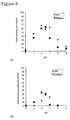
- Figure 8 shows the percentages of residual monomer molecules and formed aggregates as a result of gel filtration HPLC analysis performed on the samples treated at 60°C for 4 weeks.
- the % residual monomer molecules was the highest around neutral pH and hence the thermal denaturation phenomenon was inhibited in this pH range.
- no formation of aggregates was observed and peaks appeared in the lower molecular weight region.
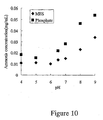
- Figure 10 shows the ammonia content of the samples treated at 60°C for 4 weeks. All of the buffers were found to have a positive relationship between pH and ammonia production, in which more ammonia was produced at higher pH. Ammonia production is mostly caused by deamidation of asparagine or glutamine residues. It could therefore be confirmed that deamidation was facilitated at higher pH. Ammonia production was also higher in phosphate buffer, regardless of pH, which suggests that the probability of deamidation would vary from buffer to buffer.
- deamidated antibody molecules Since the charge of deamidated proteins will shift to negative, deamidated antibody molecules would be contained in the above unknown peak eluted faster than the native peak on cation-exchange HPLC.
- Figure 11 shows the isomerization degree of asparagine and aspartic acid residues (the ratio of D-isomers to total isomers) measured for the samples treated at 60°C for 4 weeks.
- the isomerization phenomenon was inhibited most effectively under neutral conditions, regardless of buffer types. In contrast, isomerization was more likely to occur under basic conditions and the isomerization degree was higher in phosphate buffer.
- Asx denotes the sum of asparagine residues and aspartic acid residues.
- Table 4 below shows the denaturation midpoint temperatures determined by differential scanning calorimetry. All of the buffers showed a low value for each denaturation midpoint temperature at acidic pH. In contrast, an accurate determination could not be performed on the denaturation midpoint temperatures in the third step of denaturation at basic pH because aggregation was observed in this step. These results indicate that under acidic pH, destabilization would be caused by decreases in denaturation temperatures, while under basic pH, it would be caused by the high tendency of denatured molecules to aggregate. This would be responsible for high stability around pH 6.
- Unknown 1 is a band of higher molecular weight than the L chain
- Unknown 2 is a band of lower molecular weight than the L chain.
- Unknown 1 could not be sequenced, but the sequence Pro-Glu-Val-Lys-Phe was read from the N-terminal of Unknown 2.
- this N-terminal sequence was relevant to the sequence on the H chain: - P 273 -E 274 -V 275 -K 276 -F 277 - It is generally known that the aspartic acid-proline sequence is more likely to be cleaved by hydrolysis under acidic conditions, and this antibody has only one Asp-Pro sequence (D 272 -P 273 ) over the entire sequence. Thus, fragmentation of the H chain under acidic conditions would be caused by cleavage at the "/" site in the sequence -E 271 -D 272 /P 273 -E 274 -V 275 -K 276 -F 277 -. The reason why Unknown 1 could not be sequenced would be because the glutamine residue at the N-terminal of the H chain was pyroglutamylated and hence could not be cleaved.
- Figure 12 shows the time course of % residual uncleaved antibody and peptide during heat treatment at 60°C. As shown in this figure, a decrease in cleavage rate was observed after 1 week for the antibody H chain and after 36 hours for the peptide. Then, the initial rate of cleavage phenomena was compared between antibody and peptide, indicating that the initial rate of the antibody was about 10 times slower than that of the corresponding peptide. This is because upon exposing the Asp-Pro sequence on the antibody surface by losing the higher-order structure of antibody. Such an event constitutes a rate-limiting step of cleavage. It is therefore suggested that maintaining the antibody's native structure contributes to the inhibition of sequence cleavage between Asp and Pro.
- Example 2 The same stock solution of humanized anti-IL-6 receptor antibody (antibody concentration: about 40 mg/mL) as used in Example 1 was lyophilized and then dissolved at a concentration of 5 mg/mL in 50 mM phosphate buffer (pH 7.5, PBS) containing 0.15 M sodium chloride. This antibody solution (1 mL) was then mixed with a papain solution (1 mL) which had been dissolved at a concentration of 500 ⁇ g/mL in PBS containing 2 mM EDTA and 2 mM cysteine, followed by incubation at 37°C for 2 hours.
- Fab and Fc fragments were isolated and purified from the papain digest as follows.
- the papain-digested antibody fragment solution was loaded onto a Protein A column (HiTrap rProtein A FF 1 mL, Amersham Pharmacia Biotech) to collect an eluate passed through the column as a crude Fab fragment fraction.
- the column was washed with 0.1 M phosphate buffer (pH 7.4) containing 0.15 M sodium chloride and then eluted with 0.1 M glycine buffer (pH 3.0) to give a crude Fc fragment fraction.
- the crude Fc fragment fraction was neutralized with an equal volume of 0.2 M tris-hydrochloric acid buffer (pH 8.0) and then dialyzed against 0.1 M phosphate buffer (pH 7.4) containing 0.15 M sodium chloride.
- the crude Fab fragment fraction was purified by cation-exchange HPLC under the following conditions:
- the crude Fc fragment fraction was purified by loading again the fraction onto a Protein A column. The resulting individual fragments were confirmed for their . purity by SDS-PAGE (gel concentration: 12%).
- Fab and Fc fragments of the humanized antibody fragmented in 1-1-1 and purified in 1-1-2 were adjusted to a concentration of 1 mg/mL and then dialyzed against the individual buffers shown below to prepare sample solutions in different buffers. Each sample solution was filled into glass ampoules (1 mL each) and flame-sealed for use in heat treatment.
- Heat treatment conditions were set at 60°C for 1, 2 or 4 weeks for Fab fragment solutions and at 60°C for 2, 4 or 7 days for Fc fragment solutions. After heat treatment, these sample solutions were assessed for their stability in the following manner.
- the antibody fragment solutions were assessed for their heat stability by measuring a change in the peak area of monomer molecules on gel filtration HPLC and a change in the fluorescence spectrum of 1-anilino-8-naphthalene sulfonate (ANS) upon binding to a hydrophobic surface.
- ANS 1-anilino-8-naphthalene sulfonate
- the heat stability of each antibody fragment solution was measured as a change in the fluorescence spectrum of 1-anilino-8-naphthalene sulfonate (ANS) upon binding to a hydrophobic surface.
- ANS 1-anilino-8-naphthalene sulfonate
- Each heat-treated or untreated antibody fragment solution (900 ⁇ L, about 10 nmol) was supplemented with a 0.4 M ANS solution (300 ⁇ L) for use as a test sample.
- the whole volume of each test sample was introduced into a fluorescence cell and measured for the fluorescence spectrum between 450 and 550 nm with an excitation wavelength of 365 nm.
- the Fab and Fc fragment samples were prepared as described in 1-1.
- the antibody (whole) samples were prepared from the antibody stock solution by dialysis against and dilution in each buffer to give a concentration of 1 mg/mL.
- the Fab and Fc fragments and the whole antibody in MES buffer (15 mM, pH 5.5) were assessed for reversibility of their thermal denaturation phenomenon in each denaturation step by differential scanning calorimetry (DSC). More specifically, the Fab and Fc fragments and the whole antibody were heated to their respective denaturation midpoint temperatures in each denaturation step where the reversibility was assessed (first temperature scan). After cooling to the initial temperature, each sample was heated again (second temperature scan) to compare the height of the endothermic peak between first and second scans in each denaturation step. The measurement was carried out under the following conditions:
- the Fab fragment and the whole antibody in MES buffer were also assessed for the effect of DSC heating rate on their denaturation midpoint temperature in each denaturation step. The measurement was carried out under the following conditions:
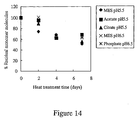
- Figures 13 and 14 show the results of gel filtration HPLC analysis performed on the heat-treated samples of Fab and Fc fragments (Figure 13: Fab fragment; Figure 14: Fc fragment), which were used to determine the % residual monomer molecules.
- the Fab fragment was very stable in all buffers excluding citrate buffer.
- the % residual monomer molecules was 90% or more in these buffers even after heat treatment at 60°C for 4 weeks, whereas it was as low as about 70% in citrate buffer.
- the % residual monomer molecules was reduced to 50%-70% after heat treatment at 60°C for 1 week, but no significant tendency was found in the difference of stability between buffer species.
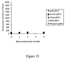
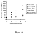
- Figures 15 and 16 show the results of ANS-binding fluorescence spectroscopy performed on the heat-treated samples of Fab and Fc fragments (Figure 15: Fab fragment; Figure 16: Fc fragment), which were used to analyze the surface exposure of hydrophobic residues.
- the Fab fragment showed no change in fluorescence intensity after heat treatment at 60°C for 4 weeks in all buffers; there was no binding of ANS onto the hydrophobic surface.
- the Fc fragment showed an increase in ANS-binding fluorescence intensity after heat treatment at 60°C for 1 week (7 days).
- the fluorescence intensity after heat treatment was higher in phosphate and citrate buffers in which the whole antibody was less heat-stable.
- the Fab fragment showed undetectable denaturation after heat treatment, regardless of buffer types, whereas the Fc fragment showed a buffer-dependent thermal denaturation phenomenon, as in the case of the whole antibody.
- Acetate and MES buffers were found to inhibit thermal denaturation of the Fc fragment, as compared to phosphate and citrate buffers.
- the denaturation of the whole antibody consists of three steps in total, i.e., the first two steps resulting from the Fc domain and the last step resulting from the Fab domain.
- the reversibility of denaturation could be observed in the first and second steps, whereas denaturation in the last third step was less reversible (Figure 18).
- the denaturation of the Fab fragment was also less reversible, as in the case of the third denaturation step of the whole antibody where the Fab domain was denatured.
- the second temperature scan resulted in an endothermic peak higher than that of the whole antibody ( Figure 19). This means that the reversibility of denaturation is higher in the Fab fragment alone than in the whole antibody, which in turn suggests that reversibility in the third denaturation step of the whole antibody would be influenced by the presence of the Fc domain.
- the whole antibody and the Fab fragment were also assessed for the effect of DSC heating rate on their denaturation midpoint temperatures in MES buffer.
- denaturation in the second and third steps resulted in a higher denaturation midpoint temperature at a higher heating rate, whereas the denaturation temperature of the first step remained constant independently of heating rate ( Figure 21).
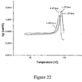
- the denaturation of the Fab fragment also resulted in a higher denaturation midpoint temperature at a higher heating rate ( Figure 22).
Landscapes
- Health & Medical Sciences (AREA)
- Chemical & Material Sciences (AREA)
- Medicinal Chemistry (AREA)
- Life Sciences & Earth Sciences (AREA)
- Epidemiology (AREA)
- Public Health (AREA)
- Veterinary Medicine (AREA)
- General Health & Medical Sciences (AREA)
- Animal Behavior & Ethology (AREA)
- Pharmacology & Pharmacy (AREA)
- Engineering & Computer Science (AREA)
- Chemical Kinetics & Catalysis (AREA)
- General Chemical & Material Sciences (AREA)
- Oil, Petroleum & Natural Gas (AREA)
- Mycology (AREA)
- Microbiology (AREA)
- Bioinformatics & Cheminformatics (AREA)
- Immunology (AREA)
- Medicines Containing Antibodies Or Antigens For Use As Internal Diagnostic Agents (AREA)
- Peptides Or Proteins (AREA)
- Medicines That Contain Protein Lipid Enzymes And Other Medicines (AREA)
Abstract
A protein formulation with improved stability, which comprises a physiologically active protein in one or more buffers selected from the group consisting of acetate buffer, imidazole buffer, a buffer of aminoethanesulfonate or a derivative thereof, and a buffer of aminopropanesulfonate or a derivative thereof. <IMAGE>
Description
The present invention relates to protein formulations
with improved stability. More specifically, the present
invention relates to improving the stability of
physiologically active proteins (e.g., antibodies) in a
highly concentrated solution form by using a buffer such as
acetate buffer, imidazole buffer, 2-morpholinoethanesulfonate
buffer or 3-morpholinopropanesulfonate
buffer.
Advances in gene recombinant technology have enabled
a pharmaceutical application of physiologically active
proteins such as antibodies, enzymes, hormones and
cytokines. To ensure a stable supply of high quality
proteins, it is necessary to establish production and
storage conditions capable of maintaining the structure and
activity of the proteins.
When proteins are stored in a highly concentrated
solution form, they are usually associated with a problem
of deterioration including the formation of insoluble
aggregates; and therefore, it is necessary to prevent the
phenomena of deterioration.
For example, antibodies such as immunoglobulins,
monoclonal antibodies and humanized antibodies are unstable
proteins and more likely to undergo physical and chemical
changes (e.g., association, aggregation) due to stresses
applied during virus removal in the purification process,
including filtration stress, concentration stress and heat
stress.
To prevent protein deterioration for stable storage,
stabilization by freeze-drying has been conventionally and
widely used. However, proteins are likely to undergo
denaturation or chemical changes due to mechanical stresses
during freezing and subsequent drying; and hence a
cryoprotective agent should be added to avoid these
phenomena in such a strategy.
It has also been found that stabilization effects are
obtained by adding a stabilizer for preventing chemical and
physical changes, including high-molecular weight materials
such as proteins (e.g., human serum albumin, purified
gelatin) or low-molecular weight materials such as polyols,
amino acids and surfactants. However, when added as
stabilizers, organism-derived high-molecular weight
materials like proteins are disadvantageous in requiring
very complicated processes to remove contaminants, such as
viruses, originating from the stabilizers per se. Also,
when heat treatment is carried out for the purpose of virus
inactivation, such stabilizers may cause problems such as
association and aggregation due to heat stress.
Further, as to the formation of insoluble aggregates
in a protein solution, it is caused by the formation of
denatured proteins, i.e., denatured intermediates; there
has been a need to develop a method for preventing the
formation of insoluble aggregates.
The object of the present invention is to provide
stable formulations containing physiologically active
proteins (e..g., antibodies, enzymes, hormones and
cytokines) by improving the stability of the
physiologically active proteins in a highly concentrated
solution form to prevent the formation of denatured
intermediates and insoluble aggregates.
As a result of extensive and intensive efforts made
to achieve the above object, the inventors of the present
invention have found that the stability of proteins can be
increased in one or more buffers, particularly of about pH
5 to 7, selected from the group consisting of acetate
buffer, imidazole buffer, a buffer of aminoethanesulfonate
or a derivative thereof, and a buffer of
aminopropanesulfonate or a derivative thereof, thereby
preventing the formation of denatured intermediates as well
as insoluble aggregates. This finding led to the
completion of the present invention.
Namely, the present invention provides the following:
Examples of a physiologically active protein as used
herein include, but are not limited to, antibodies, enzymes,
cytokines and hormones. Specific examples include, but are
not limited to, hematopoietic factors such as granulocyte
colony-stimulating factor (G-CSF), granulocyte-macrophage
colony-stimulating factor (GM-CSF), erythropoietin (EPO)
and thrombopoietin, cytokines such as interferons, IL-1 and
IL-6, immunoglobulins, monoclonal antibodies, humanized
antibodies, tissue plasminogen activator (tPA), urokinase,
serum albumin, blood coagulation factor VIII, leptin,
insulin, and stem cell growth factor (SCF). Among these
proteins, preferred are immunoglobulins, and more preferred
are monoclonal antibodies and humanized antibodies.
A "physiologically active protein" refers to a
protein having substantially the same biological activities
as a corresponding physiologically active protein of
mammalian (especially human) origin. Such a protein may be
natural or recombinant, preferably recombinant. A
recombinant protein encompasses those having the same amino
acid sequence as the corresponding natural protein, as well
as those comprising deletion, substitution or addition of
one or more amino acid residues in the amino acid sequence,
but retaining the biological activities as mentioned above.
Further, the physiologically active protein as used herein
includes those chemically modified with PEG, etc.
When a physiologically active protein is a monoclonal
antibody, it may be prepared in any manner. In principle,
a monoclonal antibody can be produced using known
techniques by immunizing a sensitized antigen in a standard
manner for immunization, fusing the resulting immunocytes
with known parent cells in a standard manner for cell
fusion, and then screening monoclonal antibody-producing
cells in a standard manner for screening. Further, the
monoclonal antibody as used herein is not limited to those
produced by hybridomas, but includes chimeric antibodies
which are artificially modified, e.g., for the purpose of
reducing xenoantigenicity to human. Alternatively,
reshaped humanized antibodies may also be used in the
present invention. Such antibodies are prepared by
replacing complementarity-determining regions of human
antibodies with those of non-human mammalian (e.g., mouse)
antibodies; standard recombinant procedures for this
purpose are also known. Such known procedures may be used
to give reshaped humanized antibodies.
If necessary, amino acid substitutions may be made in
the framework (FR) regions of antibody variable domains
such that the complementarity-determining regions of
reshaped humanized antibodies form appropriate antigen-binding
sites (Sato et al., Cancer Res. 53:1-6, 1993).
A humanized anti-IL-6 receptor antibody (hPM-1) can be
presented as a preferred example for such reshaped
humanized antibodies (see WO92/19759). In addition to this,
a humanized anti-HM1.24 antigen monoclonal antibody (see
WO98/14580), a humanized anti-parathyroid hormone-related
peptide antibody (anti-PTHrP antibody)(see WO98/13388), a
humanized anti-tissue factor antibody (see WO99/51743) and
the like are also preferred for use in the present
invention.
Further, human antibodies produced in transgenic
animals and the like are also preferred.
Furthermore, the antibody as used herein encompasses
antibody fragments including Fab, (Fab')2, Fc, Fc' and Fd,
as well as reshaped antibodies including monovalent or
polyvalent single chain antibodies (scFV).
As used herein, a "sample containing a
physiologically active protein" or an "antibody-containing
sample" refers to a sample containing any protein or
antibody, regardless of whether derived from organisms or
produced recombinantly. It preferably refers to a culture
medium of mammalian cells (e.g., CHO cells) containing
physiologically active protein or antibody molecules
produced by culture, which may further be subjected to
partial purification or other specific treatment.
The inventors of the present invention have studied
various factors involved in heat treatment-induced
denaturation of antibody-containing samples. As a result,
they have found that when heat treatment at 80°C for 2
hours or at 60°C for 2 or 4 weeks is performed on antibody
molecules dissolved in acetate buffer, imidazole buffer, a
buffer of aminoethanesulfonate or a derivative thereof or a
buffer of aminopropanesulfonate or a derivative thereof,
these buffers achieve a larger stabilization effect than
phosphate or citrate buffer because they allow reduction of
denatured intermediates (i.e., soluble associated
molecules), prevention of the denaturation phenomenon, as
well as avoidance of insoluble aggregate formation. In
particular, it has also been observed that these buffers
inhibit further aggregation of denatured intermediates into
insoluble aggregates.
The protein formulation of the present invention may
be prepared by dissolving a physiologically active protein
and various ingredients as shown below into one or more
buffers selected from the group consisting of acetate
buffer, imidazole buffer, a buffer of aminoethanesulfonate
or a derivative thereof, and a buffer of
aminopropanesulfonate or a derivative thereof. These
buffers may be used alone or in combination.
Preferred examples of aminoethanesulfonate or a
derivative thereof include, but are not limited to:
Preferred examples of aminopropanesulfonate or a
derivative thereof include, but are not limited to:
Such a buffer preferably has a pH of 5.0 to 8.0, more
preferably 5.0 to 7.0, and even more preferably 5.5 to 6.5.
The concentration of the buffer usually ranges from 1 to
500 mM, preferably from 5 to 100 mM, more preferably from
5 to 30 mM, and most preferably from 10 to 20 mM.
The buffer used in the present invention may be
prepared in any manner. In general, a given amount of a
reagent (preferably of special grade) is dissolved in
purified water and adjusted to the desired pH with
hydrochloric acid, sodium hydroxide, etc. Non-limiting
examples of buffer preparation will be presented below.
Acetic acid (0.90 g) is dissolved in purified water
(about 900 mL), adjusted to the desired pH with 1 M sodium
hydroxide and then adjusted to a total volume of 1 L.
Imidazole (1.02 g) is dissolved in purified water
(about 900 mL), adjusted to the desired pH with 1 M
hydrochloric acid and then adjusted to a total volume of
1 L.
2-Morpholinoethanesulfonate monohydrate (3.20 g) is
dissolved in purified water (about 900 mL), adjusted to the
desired pH with 1 M sodium hydroxide and then adjusted to a
total volume of 1 L.
3-Morpholinopropanesulfonate (3.14 g) is dissolved in
purified water (about 900 mL), adjusted to the desired pH
with 1M sodium hydroxide and then adjusted to a total
volume of 1 L.
The formulation of the present invention may further
comprise a surfactant. Typical examples of a surfactant
include:
Preferred surfactants are polyoxyethylene sorbitan
fatty acid esters. Particularly preferred are Polysorbate
20, 21, 40, 60, 65, 80, 81 and 85, and most preferred are
Polysorbate 20 and 80.
The formulation of the present invention may also
comprise various amino acids as stabilizers. Likewise, it
is further possible to use polyethylene glycol or
saccharides (e.g., dextran, mannitol, sorbitol, inositol,
glucose, fructose, lactose, xylose, mannose, maltose,
raffinose) as an isotonizing agent.
If desired, the protein formulation of the present
invention may further comprise a diluent, a solubilizer, an
excipient, a pH adjuster, a soothing agent, a buffer, a
sulfur-containing reducing agent, an antioxidant and the
like. For instance, examples of a sulfur-containing
reducing agent include those having a sulfhydryl group such
as N-acetylcysteine, N-acetylhomocysteine, thioctic acid,
thiodiglycol, thioethanolamine, thioglycerol, thiosorbitol,
thioglycolic acid and a salt thereof, sodium thiosulfate,
glutathione, and a C1-C7 thioalkanoic acid. Examples of an
antioxidant include erythorbic acid, dibutylhydroxytoluene,
butylhydroxyanisole, α-tocopherol, tocopherol acetate, L-ascorbic
acid and a salt thereof, L-ascorbic acid palmitate,
L-ascorbic acid stearate, sodium bisulfite, sodium sulfite,
triamyl gallate and propyl gallate, as well as chelating
agents such as disodium ethylenediaminetetraacetate (EDTA),
sodium pyrophosphate and sodium metaphosphate. Furthermore,
the protein formulation of the present invention may
comprise other commonly used ingredients, for example,
inorganic salts such as sodium chloride, potassium chloride,
calcium chloride, sodium phosphate, potassium phosphate and
sodium bicarbonate, as well as organic salts such as sodium
citrate, potassium citrate and sodium acetate.
The protein formulation of the present invention is
usually administered by parenteral route in the dosage form
of injections (e.g., subcutaneous, intravenous,
intramuscular or intraperitoneal injections) or transdermal,
transmucosal, transnasal or transpulmonary preparations,
but it may also be administered orally.
The protein formulation of the present invention may
be a liquid formulation or a lyophilized formulation which
is reconstituted before use. As excipients for
lyophillization, for example, sugar alcohols or saccharides
(e.g., mannitol, glucose) may be used. In the case of a
liquid formulation, the protein formulation of the present
invention can usually be provided in the form of containers
with defined volume, including sealed and sterilized
plastic or glass vials, ampoules and syringes, as well as
in the form of large volume containers like bottles.
The amount of a physiologically active protein in the
formulation of the present invention can be determined as
appropriate for the type of physiologically active protein
to be used, the type and severity of disease to be treated,
the age of a patient, etc.
In general, a physiologically active protein is
incorporated in an amount of 0.01 µg/ml to 200 mg/ml,
preferably 0.5 µg/ml to 100 mg/ml, based on the total
volume of the formulation or an injectable composition
thereof, which is supplemented with saccharides. For
example, in a case where a physiologically active protein
is an antibody such as an immunoglobulin, a monoclonal
antibody or a humanized antibody, it is usually
incorporated at a final administration concentration of
0.1 to 200 mg/ml, preferably 1 to 200 mg/ml, more
preferably 10 to 200 mg/ml, and most preferably 10 to 150
mg/ml. The protein formulation of the present invention
shows a remarkable stabilization effect, particularly when
formulated into a highly concentrated protein solution
containing a physiologically active protein in an amount of
10 mg/ml or more.
The amount of a physiologically active protein in the
protein-containing sample of the present invention usually
ranges from 0.01 µg/ml to 200 mg/ml, preferably from 0.5
µg/ml to 100 mg/ml, based on the total volume of the sample.
For example, in a case where a physiologically active
protein is an antibody such as an immunoglobulin, a
monoclonal antibody or a humanized antibody, it is usually
incorporated at a final administration concentration of 0.1
to 200 mg/ml, preferably 1 to 200 mg/ml, more preferably
10 to 200 mg/ml, and most preferably 10 to 150 mg/ml.
Using antibody samples, the physiologically active
protein formulation of the present invention was tested for
the stability by fluorescence spectroscopy (Trp residue,
ANS binding), cation-exchange HPLC, gel filtration HPLC and
SDS-PAGE.
The results indicated that after treatment at 80°C
for 2 hours, denatured intermediates (i.e., soluble
aggregates) were produced in a reduced amount and hence an
inhibitory effect on the denaturation phenomenon was
observed in imidazole buffer, acetate buffer,
2-morpholinoethanesulfonate buffer (MES) and 3-morpholinopropanesulfonate
buffer (MOPS). After treatment
at 60°C for 4 weeks, on the other hand, production of
thermal denatured intermediates was higher in imidazole,
MES and MOPS buffers than in phosphate buffer, but no
formation of insoluble aggregates was observed in these
buffers. Thus, these buffers were believed to inhibit
further aggregation of denatured intermediates into
insoluble aggregates.
In addition, the aggregated samples were analyzed by
SDS-PAGE with 2-mercaptoethanol (reducing SDS-PAGE) to give
the same electrophoresis pattern as the monomer sample,
thus indicating that most of the covalent linkages found in
aggregates were disulfide linkages. After treatment at 60°C,
however, higher-molecular-weight bands were observed even
in reducing SDS-PAGE, suggesting that a part of the
covalent linkages contributed to non-disulfide linking.
Meanwhile, in order to assess the effect of pH on the
stability of physiologically active proteins, the thermal
denaturation phenomenon and chemical changes after
treatment at 60°C were assessed for both phosphate and MES
buffers using antibody samples.
The thermal denaturation phenomenon was measured by
cation-exchange HPLC, gel filtration HPLC and ANS-binding
fluorescence spectroscopy, indicating that thermal
denaturation after treatment at 60°C for 4 weeks was most
effectively inhibited around pH 6. This could be explained
by the results of differential scanning calorimetry.
Namely, it was considered that under acidic conditions, the
reduction of heat stability would be caused by low
denaturation midpoint temperatures in all 3 steps of
denaturation, while under basic conditions, it would be
caused by the high tendency of intermediates to aggregate
after denaturation.
Chemical changes were assessed by SDS-PAGE, ammonia
content and the isomerization degree of aspartic acid
(including asparagine), indicating that under acidic
conditions, hydrolysis was very likely to occur between
aspartic acid residue 272 and proline residue 273 (Asp-Pro)
on the H chain, and isomerization of aspartic acid residues
was also likely to occur. Under basic conditions, it was
indicated that not only deamidation of asparagine and
glutamine residues, but also isomerization of aspartic acid
residues was likely to occur. These results confirmed that
the most effective inhibition of chemical changes occurred
under near-neutral conditions. In addition, hydrolysis of
the Asp-Pro sequence proceeded about 10 times slower than
in the corresponding peptide, suggesting that it was
important to maintain the protein's higher-order structure
in inhibiting chemical changes.
Further, antibody fragments (Fab and Fc fragments)
were treated at 60°C and analyzed by gel filtration HPLC
and ANS-binding fluorescence spectroscopy, indicating that
the Fab fragment showed undetectable denaturation after
heat treatment, regardless of buffer types, whereas the Fc
fragment showed a buffer-dependent thermal denaturation
phenomenon, as in the case of the whole antibody. Acetate
and MES buffers were found to inhibit thermal denaturation
of the Fc fragment, as compared to phosphate and citrate
buffers. Likewise, differential scanning calorimetry was
performed to assess thermal denaturation temperatures and
the reversibility of thermal denaturation phenomenon,
indicating that in the case of the Fab domain alone, no
aggregation was observed in all buffers after denaturation.
This suggested that the Fc domain would greatly contribute
to aggregation of the whole antibody, and that the
denaturation phenomenon of the whole antibody would be
influenced by the interaction between Fab and Fc domains,
rather than being equal to the mere sum of both domains.
These results indicated that the physiologically
active protein formulation of the present invention would
have a very excellent stabilization effect.
Thus, the present invention provides a method for
improving the stability of a protein formulation, which
comprises dissolving a physiologically active protein in
one or more buffers selected from the group consisting of
acetate buffer, imidazole buffer, a buffer of
aminoethanesulfonate or a derivative thereof, and a buffer
of aminopropanesulfonate or a derivative thereof.
The present invention further provides a method for
stabilizing a protein-containing sample under heat
treatment, which comprises performing heat treatment on the
protein-containing sample in such a form that a
physiologically active protein is contained in one or more
buffers selected from the group consisting of acetate
buffer, imidazole buffer, a buffer of aminoethanesulfonate
or a derivative thereof, and a buffer of
aminopropanesulfonate or a derivative thereof. The
stabilizing method of the present invention is particularly
advantageous for use in heat treatment for virus removal in
the purification process of protein formulations, etc.
The present invention will be further described in
the following Examples, which are not intended to limit the
scope of the invention. Based on the detailed description,
various changes and modifications will be apparent to those
skilled in the art, and such changes and modifications fall
within the scope of the invention.
A humanized anti-IL-6 receptor antibody was used for
measurement. Procedures for production of the humanized
anti-IL-6 receptor antibody can be found in WO92/19759.
Among antibodies found in WO92/19759, an antibody
comprising the L chain version "a" and the H chain version
"f" was used in the present invention. This antibody was
recombinantly produced in CHO cells.
A stock solution of the humanized anti-IL-6 receptor
antibody (about 40 mg/mL, pH 6.5) was diluted with 15 mM.
phosphate buffer (pH 6.5) to prepare a sample solution with
an antibody concentration of 10 mg/mL, which was then
filled into glass ampoules (2 mL each) and flame-sealed
(Sample 1: phosphate buffer, pH 6.5). pH as used herein
refers to the pH of the prepared sample.
The solution with an antibody concentration of 10
mg/mL was further dialyzed against the buffers shown below
to prepare sample solutions in different buffers, which
were then filled into glass ampoules (2 mL each) and flame-sealed.
The buffer concentration was set to 15 mM in all
cases.
These sample solutions were heat-treated under
conditions of 80°C for 2 hours and at 60°C for 2 or 4 weeks
and then provided for stability assessment.
The heat-treated samples were assessed for their
physical properties in the following manner to compare
their stability.
Each sample was diluted with 15 mM phosphate buffer
(pH 6.5) to give an antibody concentration of 133 µg/mL
(0.89 µM) and used as a test sample. Each test sample
(about 1.5 mL) was introduced into a fluorescence cell and
measured for the fluorescence spectrum between 280 and
400 nm with an excitation wavelength of 280 nm. This
measurement is intended to analyze the state of tryptophan
residues. Upon protein denaturation, tryptophan residues
in hydrophobic region will undergo structural changes to
increase the fluorescence intensity and to give the maximum
fluorescence wavelength shifted to a longer wavelength.
Each sample was diluted with 15 mM phosphate buffer
(pH 6.5) to give an antibody concentration of 667 µg/mL
(0.89 µM), 900 µL of which was supplemented with a 0.4 M
solution of 1-anilino-8-naphthalene sulfonate (ANS) (300
µL) for use as a test sample. The whole volume of each
test sample was introduced into a fluorescence cell and
measured for the fluorescence spectrum and intensity
between 450 and 550 nm with an excitation wavelength of
365 nm. ANS has the property of binding to hydrophobic
regions on the protein surface and emits fluorescence upon
binding. Thus, this measurement is intended to analyze the
hydrophobicity of the protein surface as a change in
fluorescence intensity.
Each sample was measured under the following analysis
conditions:
| Time (min) | A (%) | B (%) |
| 0 | 100 | 0 |
| 60 | 0 | 100 |
| 65 | 0 | 100 |
| 66 | 100 | 0 |
Each sample was measured under the following
analysis conditions:
Each sample (1 µg as an antibody) in 10% SDS-containing
dilution buffer was applied to a polyacrylamide
gel (12% for reducing, 8% for non-reducing) and
electrophoresed for about 40 minutes with a maximum voltage
of 200 V and a maximum current of 200 mA.
The bands were stained with coomassie brilliant blue.
Table 1 below shows the respective test results after
heat treatment at 80°C for 2 hours.
| (at 80°C for 2 hours) | ||||
| Fluorescence intensity εm=330nm (εx = 280 nm) | ANS binding εm=470nm (εx = 365 nm) | |||
| | Phosphate buffer | pH 6.5 | 5774 | 6089 |
| | Phosphate buffer | pH 5.5 | 5221 | 4487 |
| | Citrate buffer | pH 6.5 | 5779 | 6677 |
| | Citrate buffer | pH 5.5 | 5556 | 6511 |
| | Imidazole buffer | pH 6.5 | 5020 | 3003 |
| | Acetate buffer | pH 5.5 | 4770 | 2776 |
| | MES buffer | pH 6.5 | 5185 | 3665 |
| | MES buffer | pH 5.5 | 4670 | 1503 |
| | MOPS buffer | pH 6.5 | 5143 | 3306 |
All of the heat-treated samples showed increased
fluorescence intensity for both spectra due to the thermal
denaturation phenomenon. In a control sample (heat-untreated),
the fluorescence intensity was about 4500 and
the ANS binding was about 250.
When compared to phosphate and citrate buffers,
imidazole, acetate, MES and MOPS buffers showed lower
fluorescence intensity for both spectra after heat
treatment at 80°C for 2 hours; these buffers were found to
inhibit denaturation.
When compared between pH 6.5 and 5.5 for the same
buffer, denaturation was inhibited more effectively at
pH 5.5.
Figure 1 shows the test results of ANS binding after
heat treatment at 60°C for 2 and 4 weeks.
When compared to phosphate and citrate buffers,
imidazole, acetate, MES and MOPS buffers were found to
inhibit denaturation. In fact, no insoluble aggregate was
observed in the samples using imidazole, acetate, MES and
MOPS buffers.
When compared between pH 6.5 and 5.5 for the same
buffer, ANS binding was higher at pH 5.5 in contrast to the
results at 80°C .
Table 2 below shows the test results after heat
treatment at 80°C for 2 hours.
| (at 80°C for 2 hours) | ||||
| Native molecule | Denatured | |||
| Sample | ||||
| 1 | Phosphate buffer | pH 6.5 | 170890 | 410576 |
| | Phosphate buffer | pH 5.5 | 350017 | 225267 |
| | Citrate buffer | pH 6.5 | 122496 | 448531 |
| | Citrate buffer | pH 5.5 | 144343 | 409860 |
| | Imidazole buffer | pH 6.5 | 383687 | 167740 |
| | Acetate buffer | pH 5.5 | 466018 | 160059 |
| | MES buffer | pH 6.5 | 351641 | 238077 |
| | MES buffer | pH 5.5 | 590792 | Not detected |
| | MOPS buffer | pH 6.5 | 380758 | 168208 |
Heat treatment at 80°C for 2 hours reduced the peak
area of the native molecules, while producing a peak of
denatured intermediates.
When compared to phosphate and citrate buffers,
imidazole, acetate, MES and MOPS buffers showed a higher
content of the native molecules remaining after treatment
at 80°C for 2 hours; these buffers were found to inhibit
the formation of denatured intermediates.
When compared between pH 6.5 and 5.5 for the same
buffer, the content of the remaining native molecules was
higher at pH 5.5, indicating that the formation of
denatured intermediates was inhibited more effectively at
pH 5.5.
Figures 2 and 3 show the test results after treatment
at 60°C for 2 and 4 weeks.
When compared to phosphate and citrate buffers,
imidazole, acetate, MES and MOPS buffers showed a higher
content of the remaining native molecules; these buffers
were found to inhibit the formation of denatured
intermediates. In fact, no insoluble aggregate was
observed in the samples using imidazole, acetate, MES and
MOPS buffers.
When compared between pH 6.5 and 5.5 for the same
buffer, the content of the remaining native molecules was
lower at the lower pH of 5.5 in contrast to the results at
80°C, thus resulting in higher production of denatured
intermediates at pH 5.5.
Table 3 below shows the test results after heat
treatment at 80°C for 2 hours.
| (at 80°C for 2 hours) | ||||
| Native molecule (monomer) | Denatured intermediate (associated) | |||
| | Phosphate buffer | pH 6.5 | 219350 | 528928 |
| | Phosphate buffer | pH 5.5 | 381133 | 379504 |
| | Citrate buffer | pH 6.5 | 178756 | 504121 |
| | Citrate buffer | pH 5.5 | 166392 | 584894 |
| | Imidazole buffer | pH 6.5 | 455483 | 286100 |
| | Acetate buffer | pH 5.5 | 481455 | 252296 |
| | MES buffer | pH 6.5 | 422489 | 353231 |
| | MES buffer | pH 5.5 | 585661 | 76946 |
| | MOPS buffer | pH 6.5 | 412389 | 310318 |
Heat treatment at 80°C for 2 hours reduced the peak
area of monomer molecules corresponding to the native
molecules, while producing a peak of soluble aggregates
corresponding to denatured intermediates in the higher
molecular weight region.
When compared to phosphate and citrate buffers,
imidazole, acetate, MES and MOPS buffers showed a higher
content of the monomer molecules remaining after treatment
at 80°C for 2 hours; these buffers were found to inhibit
the formation of soluble aggregates.
When compared between pH 6.5 and 5.5 for the same
buffer, the formation of soluble aggregates was inhibited
more effectively at pH 5.5.
Figures 4 and 5 show the test results after treatment
at 60°C for 2 and 4 weeks.
When compared to phosphate and citrate buffers,
imidazole, acetate, MES and MOPS buffers showed a higher
content of the remaining monomer molecules; these buffers
were found to inhibit the formation of soluble aggregates.
In fact, no insoluble aggregate was observed in the samples
using imidazole, acetate, MES and MOPS buffers.
When compared between pH 6.5 and 5.5 for the same
buffer, the content of the remaining monomer molecules was
lower at the lower pH of 5.5 in contrast to the results at
80°C, thus resulting in higher production of soluble
aggregates at pH 5.5.
As is evident from the results of SDS-PAGE after heat
treatment at 80°C for 2 hours, higher-molecular-weight
bands were observed in non-reducing SDS-PAGE, which
suggests the presence of covalently associated molecules.
However, covalent linkages found in such associated
molecules appeared to be disulfide linkages because there
was no difference between treated and untreated samples in
reducing SDS-PAGE.
When compared to phosphate and citrate buffers,
imidazole, acetate, MES and MOPS buffers showed weaker
bands in the higher molecular weight region after treatment
at 80°C for 2 hours; these buffers were found to inhibit
the formation of covalently associated molecules.
When compared between pH 6.5 and 5.5 for the same
buffer, the formation of covalently associated molecules
was inhibited more effectively at pH 5.5.
As is evident from the results of SDS-PAGE after heat
treatment at 60°C for 4 weeks, when compared to phosphate
and citrate buffers, imidazole, acetate, MES and MOPS
buffers showed weaker bands in the higher molecular weight
region; these buffers were found to inhibit the formation
of covalently associated molecules. Also, detectable (but
very weak) bands were observed in the higher molecular
weight region even in the samples reduced with 2-mercaptoethanol
(reducing SDS-PAGE), as compared to the
untreated samples. This suggested that most of covalent
linkages were disulfide linkages, but other chemical
changes also occurred during association.
When compared between pH 6.5 and 5.5 for the same
buffer, higher-molecular-weight bands in non-reducing SDS-PAGE
were stronger at the lower pH of 5.5 in contrast to
the results at 80°C, thus resulting in higher production of
covalently associated molecules at pH 5.5.
The humanized anti-IL-6 receptor antibody described
in Example 1 was used for measurement. A stock solution of
the humanized anti-IL-6 receptor antibody (about 40 mg/mL,
pH 6.5) was dialyzed against purified water and then
supplemented with buffer ingredients, followed by dilution
with purified water to prepare the following sample
solutions with an antibody concentration of 10 mg/mL. Each
sample was filled into glass ampoules (2 mL each) and
flame-sealed.
These sample solutions were heat-treated under
conditions of 60°C for 4 weeks and then provided for
stability assessment.
The heat-treated samples were assessed for their
physical properties in the following manner to compare
their stability.
Each sample was diluted with 15 mM phosphate buffer
(pH 6.5) to give an antibody concentration of 667 µg/mL
(0.89 µM), 900 µL of which was supplemented with a 0.4 M
solution of 1-anilino-8-naphthalene sulfonate (ANS) (300
µL) for use as a test sample. The whole volume of each
test sample was introduced into a fluorescence cell and
measured for the fluorescence spectrum and intensity
between 450 and 550 nm with an excitation wavelength of
365 nm.
Each sample was measured under the following analysis
conditions:
| Time (min) | A (%) | B (%) |
| 0 | 100 | 0 |
| 60 | 0 | 100 |
| 65 | 0 | 100 |
| 66 | 100 | 0 |
Each sample was measured under the following analysis
conditions:
Each sample (1 µg as an antibody) in 10% SDS-containing
dilution buffer, which had been reduced with
2-mercaptoethanol, was applied to a polyacrylamide gel
(12%) and electrophoresed for about 40 minutes with a
maximum voltage of 200 V and a maximum current of 200 mA.
The bands were stained with coomassie brilliant blue.
The stained gel was further scanned with a
photoscanner and converted into an image file for use in
calculation of the band density (turbidity) corresponding
to the H chain. The calculation was carried out on a
computer using the image analysis software Scion-Image to
determine the % residual H chain (vs. initial).
The ammonia content was determined using an enzymatic
assay kit based on the glutamate dehydrogenase reaction (F
kit, ammonia, Roche Diagnostics) in accordance with the
instructions attached to the kit.
The antibody (about 1 mg) was dissolved in 6 M
hydrochloric acid and hydrolyzed by heat treatment at 110°C
for 4 hours under reduced pressure. During hydrolysis,
asparagine and aspartic acid residues are both released in
the form of aspartic acid. The hydrolyzed sample was
lyophilized and then reconstituted with purified water (500
µL). This aspartic acid solution (20 µL) was mixed with an
O-phthalaldehyde (OPA)/N-acetyl-L- cysteine (NAC) solution
(10 µL) and, after 3 minutes, further mixed with 0.05 M
acetate buffer (470 µL, pH 5.2). The resulting mixture was
used as a test sample.
The OPA/NAC solution was prepared by mixing OPA (4
mg) with methanol (300 µL), 0.4 M borate buffer (250 µL, pH
9.4) and distilled water (390 µL), followed by addition of
a 1 M NAC solution (60 µL, pH 5.5).
The thermal denaturation phenomenon of the antibody
was assessed at each pH by differential scanning
calorimetry (DSC). The measurement was carried out under
the following conditions:
The samples used in the measurement were prepared by
diluting Samples 1 to 14 from Section 1-1 with the same
buffers as used in the respective samples to give a
concentration of 800 µg/mL.
To analyze the H-chain fragmentation observed on
SDS-PAGE, N-terminal amino acid sequencing was performed on
individual fragments to identify cleavage sites.
The electrophoresed gel from SDS-PAGE was
electrically transferred to a PVDF membrane and stained
with coomassie brilliant blue, followed by excision of
bands corresponding to H-chain cleavage products. The PVDF
membrane pieces containing the bands of interest were
desalted, dried and then provided for sequencing.
N-terminal amino acid sequencing was performed using
the following instrument based on the Edman degradation
technique under analysis conditions standard for such an
instrument.
To analyze the H-chain fragmentation caused by
hydrolysis between aspartic acid and proline residues
(Asp-Pro sequence) adjacent to each other on the primary
sequence, the cleavage rate under acidic conditions was
measured. A 15-residue peptide containing the Asp-Pro
sequence and its surrounding sequence found in this
antibody was synthesized in a routine manner and also
measured for the hydrolysis rate.
The antibody and peptide were cleaved under the
following conditions:
In the case of the antibody, the % residual H chain
(vs. initial) was determined in the same manner as shown in
Section 1-2-4 and plotted against time to calculate the .
H-chain cleavage rate from the negative slope of the
resulting plot.
In the case of the peptide, the % residual peptide
(vs. initial) was determined from the peak area of
uncleaved peptide by reversed-phase HPLC and plotted
against time to calculate the cleavage rate from the
negative slope of the resulting plot.
| Time (min) | A (%) | B (%) |
| 0 | 100 | 0 |
| 100 | 0 | 100 |
Figure 6 shows the fluorescence intensity of ANS-binding
at 470 nm measured for the samples treated at 60°C
for 4 weeks. The intensity was minimized at pH 6 in all
buffers, indicating that the thermal denaturation
phenomenon was inhibited around neutral pH. Also, the
fluorescence intensity of ANS tended to be stronger under
acidic conditions. In contrast, the degree of denaturation
could not be assessed by ANS binding under basic conditions
because denatured intermediates aggregated and precipitated.
Figure 7 shows the percentages of residual native
molecules and formed denatured intermediates as a result of
cation-exchange HPLC analysis performed on the samples
treated at 60°C for 4 weeks. In all buffers, the formation
of denatured intermediates was inhibited most effectively
around neutral pH. In addition, the peak area
corresponding to denatured intermediates increased under
acidic conditions, whereas it decreased under basic
conditions. This is because under basic conditions, the
apparent peak area is smaller than the real peak area since
denatured intermediates appear to be unstable and further
aggregated into insoluble aggregates in light of the
finding that the samples produced a large amount of
precipitates under basic conditions.
The unknown peak eluted faster than the native peak.
reached the maximum level around neutral pH, but this
unknown peak is affected by the formation of precipitates
under basic conditions.
Figure 8 shows the percentages of residual monomer
molecules and formed aggregates as a result of gel
filtration HPLC analysis performed on the samples treated
at 60°C for 4 weeks. As in the case of cation-exchange HPLC
analysis, the % residual monomer molecules was the highest
around neutral pH and hence the thermal denaturation
phenomenon was inhibited in this pH range. Under weak
acidic conditions, no formation of aggregates was observed
and peaks appeared in the lower molecular weight region.
Thus, these findings indicate that the peak eluted later
than the native peak on cation-exchange HPLC does not
result from the formation of denatured intermediates (i.e.,
soluble aggregates) and lower-molecular-weight cleavage
products are produced instead of such aggregates.
The results of reducing SDS-PAGE analysis of the
samples treated at 60°C for 4 weeks indicates that the band
density corresponding to the H chain was decreased under
acidic conditions. In contrast, newly produced bands were
observed before and after the L chain. This suggests the
possibility of H chain cleavage under acidic conditions.
In addition, Figure 9 shows the % residual H chain (vs.
untreated H chain) assessed by quantifying the band density
of the H chain. The % residual H chain was decreased at
lower pH or in phosphate buffer, suggesting that H chain
cleavage was facilitated.
Figure 10 shows the ammonia content of the samples
treated at 60°C for 4 weeks. All of the buffers were found
to have a positive relationship between pH and ammonia
production, in which more ammonia was produced at higher pH.
Ammonia production is mostly caused by deamidation of
asparagine or glutamine residues. It could therefore be
confirmed that deamidation was facilitated at higher pH.
Ammonia production was also higher in phosphate buffer,
regardless of pH, which suggests that the probability of
deamidation would vary from buffer to buffer.
Since the charge of deamidated proteins will shift to
negative, deamidated antibody molecules would be contained
in the above unknown peak eluted faster than the native
peak on cation-exchange HPLC.
Figure 11 shows the isomerization degree of
asparagine and aspartic acid residues (the ratio of D-isomers
to total isomers) measured for the samples treated
at 60°C for 4 weeks. The isomerization phenomenon was
inhibited most effectively under neutral conditions,
regardless of buffer types. In contrast, isomerization was
more likely to occur under basic conditions and the
isomerization degree was higher in phosphate buffer. In
Figure 11, Asx denotes the sum of asparagine residues and
aspartic acid residues.
Table 4 below shows the denaturation midpoint
temperatures determined by differential scanning
calorimetry. All of the buffers showed a low value for
each denaturation midpoint temperature at acidic pH. In
contrast, an accurate determination could not be performed
on the denaturation midpoint temperatures in the third step
of denaturation at basic pH because aggregation was
observed in this step. These results indicate that under
acidic pH, destabilization would be caused by decreases in
denaturation temperatures, while under basic pH, it would
be caused by the high tendency of denatured molecules to
aggregate. This would be responsible for high stability
around pH 6.
| (A) MES buffer | |||
| pH | Tm 1st | Tm 2nd | Tm 3rd |
| 4 | 64.04 | 77.89 | 92.58 |
| 5 | 66.22 | 81.53 | 93.47 |
| 6 | 70.74 | 81.98 | 94.13 |
| 6.5 | 71.02 | 80.49 | 94.17 |
| 7 | 71.45 | 82.15 | 93.67 |
| 8 | 71.69 | 81.85 | 92.34 |
| 9 | 71.66 | 81.37 | 90.58 |
| (B) phosphate buffer | |||
| pH | Tm 1st | Tm 2nd | Tm 3rd |
| 4 | 60.87 | 78.33 | 91.54 |
| 5 | 64.35 | 81.02 | 93.29 |
| 6 | 68.81 | 82.65 | 93.09 |
| 6.5 | 71.6 | 82.17 | 92.57 |
| 7 | 71.48 | 81.54 | 91.91 |
| 8 | 71.12 | 80.82 | 91.05 |
| 9 | 70.45 | 80.27 | 91.30 |
N-terminal amino acid sequencing was performed on two
peaks (Unknown 1 and 2) newly found on reducing SDS-PAGE
after treatment at 60°C for 4 weeks. Unknown 1 is a band of
higher molecular weight than the L chain, while Unknown 2
is a band of lower molecular weight than the L chain. As a
result, Unknown 1 could not be sequenced, but the sequence
Pro-Glu-Val-Lys-Phe was read from the N-terminal of Unknown
2. When contrasted with the primary sequence of this
antibody, this N-terminal sequence was relevant to the
sequence on the H chain: - P273-E274-V275-K276-F277- It is
generally known that the aspartic acid-proline sequence is
more likely to be cleaved by hydrolysis under acidic
conditions, and this antibody has only one Asp-Pro sequence
(D272-P273) over the entire sequence. Thus, fragmentation of
the H chain under acidic conditions would be caused by
cleavage at the "/" site in the sequence -E271-D272/P273-E274-V275-K276-F277-.
The reason why Unknown 1 could not be
sequenced would be because the glutamine residue at the
N-terminal of the H chain was pyroglutamylated and hence
could not be cleaved.
Figure 12 shows the time course of % residual
uncleaved antibody and peptide during heat treatment at
60°C. As shown in this figure, a decrease in cleavage rate
was observed after 1 week for the antibody H chain and
after 36 hours for the peptide. Then, the initial rate of
cleavage phenomena was compared between antibody and
peptide, indicating that the initial rate of the antibody
was about 10 times slower than that of the corresponding
peptide. This is because upon exposing the Asp-Pro
sequence on the antibody surface by losing the higher-order
structure of antibody. Such an event constitutes a rate-limiting
step of cleavage. It is therefore suggested that
maintaining the antibody's native structure contributes to
the inhibition of sequence cleavage between Asp and Pro.
In view of the assessment of thermal denaturation
phenomenon, the maximum stability was observed around pH 6
after treatment at 60°C for 4 weeks. This can be explained
by the results of differential scanning calorimetry.
Namely, it is considered that under acidic conditions,
destabilization would be caused by low denaturation
midpoint temperatures in all 3 steps of denaturation, while
under basic conditions, it would be caused by the high
tendency of intermediates to aggregate after denaturation.
As to chemical changes, it was indicated that under
acidic conditions, hydrolysis was very likely to occur
between aspartic acid residue 272 and proline residue 273
on the H chain, and isomerization of aspartic acid residues
was also likely to occur. Under basic conditions, it was
indicated that not only deamidation of asparagine and
glutamine residues, but also isomerization of aspartic acid
residues was likely to occur. These results showed that
near-neutral conditions were also most favorable in view of
the assessment of chemical changes. In addition, the
hydrolysis phenomenon of the Asp-Pro sequence proceeded
about 10 times slower than in the corresponding peptide,
suggesting that it was also important to maintain the
native higher-order structure in inhibiting chemical
changes.
The same stock solution of humanized anti-IL-6
receptor antibody (antibody concentration: about 40 mg/mL)
as used in Example 1 was lyophilized and then dissolved at
a concentration of 5 mg/mL in 50 mM phosphate buffer (pH
7.5, PBS) containing 0.15 M sodium chloride. This antibody
solution (1 mL) was then mixed with a papain solution
(1 mL) which had been dissolved at a concentration of
500 µg/mL in PBS containing 2 mM EDTA and 2 mM cysteine,
followed by incubation at 37°C for 2 hours.
Fab and Fc fragments were isolated and purified from
the papain digest as follows.
The papain-digested antibody fragment solution was
loaded onto a Protein A column (HiTrap rProtein A FF 1 mL,
Amersham Pharmacia Biotech) to collect an eluate passed
through the column as a crude Fab fragment fraction. The
column was washed with 0.1 M phosphate buffer (pH 7.4)
containing 0.15 M sodium chloride and then eluted with
0.1 M glycine buffer (pH 3.0) to give a crude Fc fragment
fraction. The crude Fc fragment fraction was neutralized
with an equal volume of 0.2 M tris-hydrochloric acid buffer
(pH 8.0) and then dialyzed against 0.1 M phosphate buffer
(pH 7.4) containing 0.15 M sodium chloride.
The crude Fab fragment fraction was purified by
cation-exchange HPLC under the following conditions:
| Time (min) | A (%) | B (%) |
| 0 | 100 | 0 |
| 60 | 0 | 100 |
The crude Fc fragment fraction was purified by
loading again the fraction onto a Protein A column. The
resulting individual fragments were confirmed for their .
purity by SDS-PAGE (gel concentration: 12%).
Fab and Fc fragments of the humanized antibody
fragmented in 1-1-1 and purified in 1-1-2 were adjusted to
a concentration of 1 mg/mL and then dialyzed against the
individual buffers shown below to prepare sample solutions
in different buffers. Each sample solution was filled into
glass ampoules (1 mL each) and flame-sealed for use in heat
treatment.
Heat treatment conditions were set at 60°C for 1, 2
or 4 weeks for Fab fragment solutions and at 60°C for 2, 4
or 7 days for Fc fragment solutions. After heat treatment,
these sample solutions were assessed for their stability in
the following manner.
The antibody fragment solutions were assessed for
their heat stability by measuring a change in the peak area
of monomer molecules on gel filtration HPLC and a change in
the fluorescence spectrum of 1-anilino-8-naphthalene
sulfonate (ANS) upon binding to a hydrophobic surface.
Each sample was measured under the following analysis
conditions:
The heat stability of each antibody fragment solution
was measured as a change in the fluorescence spectrum of
1-anilino-8-naphthalene sulfonate (ANS) upon binding to a
hydrophobic surface.
Each heat-treated or untreated antibody fragment
solution (900 µL, about 10 nmol) was supplemented with a
0.4 M ANS solution (300 µL) for use as a test sample. The
whole volume of each test sample was introduced into a
fluorescence cell and measured for the fluorescence
spectrum between 450 and 550 nm with an excitation
wavelength of 365 nm.
The thermal denaturation phenomenon of Fab and Fc
fragments and the whole antibody was assessed in each
buffer by differential scanning calorimetry (DSC). The
measurement was carried out under the following conditions:
Among the test samples, the Fab and Fc fragment
samples were prepared as described in 1-1. The antibody
(whole) samples were prepared from the antibody stock
solution by dialysis against and dilution in each buffer to
give a concentration of 1 mg/mL.
The Fab and Fc fragments and the whole antibody in
MES buffer (15 mM, pH 5.5) were assessed for reversibility
of their thermal denaturation phenomenon in each
denaturation step by differential scanning calorimetry
(DSC). More specifically, the Fab and Fc fragments and the
whole antibody were heated to their respective denaturation
midpoint temperatures in each denaturation step where the
reversibility was assessed (first temperature scan). After
cooling to the initial temperature, each sample was heated
again (second temperature scan) to compare the height of
the endothermic peak between first and second scans in each
denaturation step. The measurement was carried out under
the following conditions:
The Fab fragment and the whole antibody in MES buffer
(15 mM, pH 5.5) were also assessed for the effect of DSC
heating rate on their denaturation midpoint temperature in
each denaturation step. The measurement was carried out
under the following conditions:
Since the results of SDS-PAGE confirmed that the Fab
and Fc fragments were each highly purified, these fragments
were dialyzed against each buffer and then provided for
heat stability assessment.
Figures 13 and 14 show the results of gel filtration
HPLC analysis performed on the heat-treated samples of Fab
and Fc fragments (Figure 13: Fab fragment; Figure 14: Fc
fragment), which were used to determine the % residual
monomer molecules.
The Fab fragment was very stable in all buffers
excluding citrate buffer. The % residual monomer molecules
was 90% or more in these buffers even after heat treatment
at 60°C for 4 weeks, whereas it was as low as about 70% in
citrate buffer.
In the Fc fragment, on the other hand, the % residual
monomer molecules was reduced to 50%-70% after heat
treatment at 60°C for 1 week, but no significant tendency
was found in the difference of stability between buffer
species.
Figures 15 and 16 show the results of ANS-binding
fluorescence spectroscopy performed on the heat-treated
samples of Fab and Fc fragments (Figure 15: Fab fragment;
Figure 16: Fc fragment), which were used to analyze the
surface exposure of hydrophobic residues.
The Fab fragment showed no change in fluorescence
intensity after heat treatment at 60°C for 4 weeks in all
buffers; there was no binding of ANS onto the hydrophobic
surface.
In contrast, the Fc fragment showed an increase in
ANS-binding fluorescence intensity after heat treatment at
60°C for 1 week (7 days). When compared at the same pH, the
fluorescence intensity after heat treatment was higher in
phosphate and citrate buffers in which the whole antibody
was less heat-stable.
Thus, the Fab fragment showed undetectable
denaturation after heat treatment, regardless of buffer
types, whereas the Fc fragment showed a buffer-dependent
thermal denaturation phenomenon, as in the case of the
whole antibody. Acetate and MES buffers were found to
inhibit thermal denaturation of the Fc fragment, as
compared to phosphate and citrate buffers.
As shown in Figure 17, the whole antibody resulted in
3 endothermic peaks, the first two of which could be
assigned to Fc domain denaturation, and the last of which
could be assigned to Fab domain denaturation.
Except for MES buffer (pH 5.5), the whole antibody
produced an exothermic peak due to aggregation after the
third step of denaturation, whereas the Fab fragment showed
no such a phenomenon after the corresponding denaturation.
The Fc fragment also produced an exothermic peak in these
buffers after the second step of denaturation. In citrate
buffer (pH 5.5) and phosphate buffer (pH 6.5), such an
aggregation tendency was observed not only in the Fc
fragment but also in the Fab fragment. In MES buffer (pH
5.5), on the other hand, both of the whole antibody and the
Fc fragment produced no exothermic peak, thus resulting in
inhibition of aggregation of denatured molecule. However,
there was no great difference in denaturation temperatures
(Tm) between buffers, as shown in Table 5 below.

In 15 mM MES buffer (pH 5.5), the Fab and Fc
fragments and the whole antibody experienced no aggregation
phenomenon after thermal denaturation. Then, the thermal
denaturation phenomenon in MES buffer was assessed for
reversibility in each denaturation step.
The denaturation of the whole antibody consists of
three steps in total, i.e., the first two steps resulting
from the Fc domain and the last step resulting from the Fab
domain. The reversibility of denaturation could be
observed in the first and second steps, whereas
denaturation in the last third step was less reversible
(Figure 18).
The denaturation of the Fab fragment was also less
reversible, as in the case of the third denaturation step
of the whole antibody where the Fab domain was denatured.
However, the second temperature scan resulted in an
endothermic peak higher than that of the whole antibody
(Figure 19). This means that the reversibility of
denaturation is higher in the Fab fragment alone than in
the whole antibody, which in turn suggests that
reversibility in the third denaturation step of the whole
antibody would be influenced by the presence of the Fc
domain.
In the Fc fragment, denaturation was completely
reversible in the first step, but less reversible in the
second step (Figure 20). This is in contrast to the
corresponding results of the Fc domain in the whole
antibody and suggests that reversibility in the second
denaturation step of the whole antibody would be influenced
by the presence of the Fab domain.
It was therefore demonstrated that the denaturation
of each antibody domain in MES buffer was irreversible,
except for the first and second steps of the whole antibody
and the first step of the Fc domain.
The whole antibody and the Fab fragment were also
assessed for the effect of DSC heating rate on their
denaturation midpoint temperatures in MES buffer. In the
whole antibody, denaturation in the second and third steps
resulted in a higher denaturation midpoint temperature at a
higher heating rate, whereas the denaturation temperature
of the first step remained constant independently of
heating rate (Figure 21). Likewise, the denaturation of
the Fab fragment also resulted in a higher denaturation
midpoint temperature at a higher heating rate (Figure 22).
These results also supported the reversibility of
denaturation in the first step of the whole antibody. When
the individual denaturation midpoint temperatures were
plotted against heating rate and extrapolated to calculate
the denaturation midpoint temperature at a heating rate of
0°C/min, the resulting value was 69.02°C, 79.83°C and
90.96°C in the whole antibody and 91.01°C in the Fab
fragment.
These results indicated that the heat stabilization
effect in MES buffer at pH 5.5 was ascribed to the
inhibition of denatured molecule aggregation rather than
increases in thermal denaturation temperatures. Namely,
this suggested that the difference in stability between
buffer species was ascribed to the difference in
aggregation properties after denaturation rather than the
difference in denaturation temperatures.
Since in the case of the Fab domain alone, no
aggregation was observed in all buffers after denaturation,
it was indicated that the Fc domain would greatly
contribute to aggregation of the whole antibody. It was
also believed that the denaturation phenomenon of the whole
antibody would be influenced by the interaction between
both Fab and Fc domains, rather than being equal to the
mere sum of both domains.
When the reversibility of denaturation was assessed,
it could be observed only in the first and second steps of
the whole antibody and in the first step of the Fc domain.
This suggested that the reversibility of denaturation
phenomenon would make little contribution to the stability
of antibody solutions.
Claims (11)
- A protein formulation with improved stability, which comprises a physiologically active protein in one or more buffers selected from the group consisting of acetate buffer, imidazole buffer, a buffer of aminoethanesulfonate or a derivative thereof, and a buffer of aminopropanesulfonate or a derivative thereof.
- The protein formulation according to claim 1, wherein the buffer(s) has a pH of 5 to 7.
- The protein formulation according to claim 1 or 2, wherein said aminoethanesulfonate or a derivative thereof is selected from the group consisting of:2-morpholinoethanesulfonate (MES),N-(2-acetamide)-2-aminoethanesulfonate (ACES),N,N-bis(2-hydroxyethyl)-2-aminoethanesulfonate (BES),2-[4-(2-hydroxyethyl)-1-piperazinyl]ethanesulfonate (HEPES),piperazine-1,4-bis(2-ethanesulfonate) (PIPES), andN-tris(hydroxymethyl)methyl-2-aminoethanesulfonate (TES).
- The protein formulation according to claim 1 or 2, wherein said aminopropanesulfonate or a derivative thereof is selected from the group consisting of:3-morpholinopropanesulfonate (MOPS),2-hydroxy-3-[4-(2-hydroxyethyl)-1-piperazinyl]propanesulfonate (HEPPSO),2-hydroxy-3-morpholinopropanesulfonate (MOPSO), andpiperazine-1,4-bis(2-hydroxy-3-propanesulfonate) (POPSO).
- The protein formulation according to claim 1 or 2, wherein the protein is selected from an antibody, an enzyme, a cytokine and a hormone.
- The protein formulation according to claim 5, wherein the protein is an antibody.
- The protein formulation according to claim 6, wherein the antibody is a monoclonal antibody.
- The protein formulation according to claim 7, wherein the monoclonal antibody is a humanized antibody.
- A method for improving the stability of a protein formulation, which comprises dissolving a physiologically active protein in one or more buffers selected from the group consisting of acetate buffer, imidazole buffer, a buffer of aminoethanesulfonate or a derivative thereof, and a buffer of aminopropanesulfonate or a derivative thereof.
- A method for stabilizing a protein-containing sample under heat treatment, which comprises performing heat treatment on the protein-containing sample in such a form that a physiologically active protein is contained in one or more buffers selected from the group consisting of acetate buffer, imidazole buffer, a buffer of aminoethanesulfonate or a derivative thereof, and a buffer of aminopropanesulfonate or a derivative thereof.
- A method for stabilizing the Fc region of an antibody, which comprises dissolving the antibody in one or more buffers selected from the group consisting of acetate buffer, imidazole buffer, a buffer of aminoethanesulfonate or a derivative thereof, and a buffer of aminopropanesulfonate or a derivative thereof.
Applications Claiming Priority (3)
| Application Number | Priority Date | Filing Date | Title |
|---|---|---|---|
| JP2001162512 | 2001-05-30 | ||
| JP2001162512 | 2001-05-30 | ||
| PCT/JP2002/004793 WO2002098445A1 (en) | 2001-05-30 | 2002-05-17 | Protein preparation |
Publications (2)
| Publication Number | Publication Date |
|---|---|
| EP1391209A1 true EP1391209A1 (en) | 2004-02-25 |
| EP1391209A4 EP1391209A4 (en) | 2009-12-16 |
Family
ID=19005619
Family Applications (1)
| Application Number | Title | Priority Date | Filing Date |
|---|---|---|---|
| EP02776487A Withdrawn EP1391209A4 (en) | 2001-05-30 | 2002-05-17 | PROTEIN FORMULATIONS |
Country Status (4)
| Country | Link |
|---|---|
| US (1) | US20040156835A1 (en) |
| EP (1) | EP1391209A4 (en) |
| JP (1) | JP5052736B2 (en) |
| WO (1) | WO2002098445A1 (en) |
Cited By (3)
| Publication number | Priority date | Publication date | Assignee | Title |
|---|---|---|---|---|
| US7705132B2 (en) | 2006-10-20 | 2010-04-27 | Amgen Inc. | Stable polypeptide formulations |
| US8858935B2 (en) | 2005-05-19 | 2014-10-14 | Amgen Inc. | Compositions and methods for increasing the stability of antibodies |
| US9574005B2 (en) | 2011-07-19 | 2017-02-21 | Chugai Seiyaku Kabushiki Kaisha | Stable Protein-containing preparation containing argininamide or analogous compound thereof |
Families Citing this family (25)
| Publication number | Priority date | Publication date | Assignee | Title |
|---|---|---|---|---|
| US7883704B2 (en) * | 1999-03-25 | 2011-02-08 | Abbott Gmbh & Co. Kg | Methods for inhibiting the activity of the P40 subunit of human IL-12 |
| US6914128B1 (en) * | 1999-03-25 | 2005-07-05 | Abbott Gmbh & Co. Kg | Human antibodies that bind human IL-12 and methods for producing |
| ES2298378T3 (en) | 2001-06-28 | 2008-05-16 | Novo Nordisk A/S | STABLE FORMULATION OF MODIFIED GLP-1. |
| US20040033228A1 (en) | 2002-08-16 | 2004-02-19 | Hans-Juergen Krause | Formulation of human antibodies for treating TNF-alpha associated disorders |
| MY150740A (en) * | 2002-10-24 | 2014-02-28 | Abbvie Biotechnology Ltd | Low dose methods for treating disorders in which tnf? activity is detrimental |
| EP1633391B1 (en) | 2003-06-03 | 2011-10-19 | Novo Nordisk A/S | Stabilized pharmaceutical peptide compositions |
| US20060247167A1 (en) * | 2003-09-01 | 2006-11-02 | Novo Nordisk A/S | Stable formulations of peptides |
| KR101243648B1 (en) * | 2003-11-20 | 2013-03-14 | 노보 노르디스크 에이/에스 | Propylene glycol-containing peptide formulations which are optimal for production and for use in injection devices |
| RU2413530C9 (en) * | 2004-11-12 | 2021-05-18 | Ново Нордиск А/С | Stable preparations of insulin peptides |
| JP2006315964A (en) * | 2005-05-10 | 2006-11-24 | Chugai Pharmaceut Co Ltd | Antibody stabilization method |
| JP2008542235A (en) * | 2005-05-25 | 2008-11-27 | ノボ・ノルデイスク・エー/エス | Stabilized polypeptide preparation |
| CN101180081B (en) | 2005-05-25 | 2015-08-26 | 诺沃-诺迪斯克有限公司 | Stable polypeptide formulations |
| EP2129401B8 (en) * | 2006-12-21 | 2020-01-15 | Amgen Inc. | Stable buffered formulations containing polypeptides |
| WO2008088823A2 (en) * | 2007-01-16 | 2008-07-24 | Abbott Laboratories | Methods for treating psoriasis |
| CA2681752A1 (en) | 2007-03-29 | 2008-10-09 | Abbott Laboratories | Crystalline anti-human 1l-12 antibodies |
| US8883146B2 (en) | 2007-11-30 | 2014-11-11 | Abbvie Inc. | Protein formulations and methods of making same |
| KR20100126515A (en) * | 2008-03-18 | 2010-12-01 | 아보트 러보러터리즈 | How to treat psoriasis |
| CA2737045C (en) * | 2008-11-20 | 2017-11-14 | Bruce Kabakoff | Therapeutic protein formulations |
| MX2011005672A (en) * | 2008-11-28 | 2011-06-20 | Abbott Lab | Stable antibody compositions and methods for stabilizing same. |
| US20100278822A1 (en) * | 2009-05-04 | 2010-11-04 | Abbott Biotechnology, Ltd. | Stable high protein concentration formulations of human anti-tnf-alpha-antibodies |
| AU2010291927A1 (en) * | 2009-09-14 | 2012-04-12 | AbbVie Deutschland GmbH & Co. KG | Methods for treating psoriasis |
| TWI603739B (en) | 2010-11-11 | 2017-11-01 | 艾伯維生物技術有限責任公司 | Liquid formulation with high concentration of anti-TNFα antibody |
| BR112020002804A2 (en) | 2017-08-24 | 2020-07-28 | Novo Nordisk A/S | liquid pharmaceutical composition and kit |
| US20200289621A1 (en) * | 2017-09-22 | 2020-09-17 | Asahi Kasei Pharma Corporation | Teriparatide-containing liquid pharmaceutical composition having excellent stability |
| IL294521A (en) | 2020-02-18 | 2022-09-01 | Novo Nordisk As | Glp-1 compositions and uses thereof |
Family Cites Families (27)
| Publication number | Priority date | Publication date | Assignee | Title |
|---|---|---|---|---|
| FR2750604B1 (en) * | 1996-07-02 | 2002-09-20 | Oreal | HETEROMER BINDING ARACHIDONIC ACID AND ITS USE IN COSMETICS OR PHARMACY |
| US4277562A (en) * | 1976-09-13 | 1981-07-07 | Modrovich Ivan Endre | Stabilized liquid enzyme and coenzyme compositions |
| JPS63243032A (en) * | 1987-03-27 | 1988-10-07 | Green Cross Corp:The | Method for heat-treating thrombin |
| DE3854127T2 (en) * | 1987-07-17 | 1995-11-16 | Modrovich Ivan Endre | STABLE ALPHA AMYLASE REAGENT. |
| US5866322A (en) * | 1988-01-29 | 1999-02-02 | Abbott Laboratories | Method for performing Rubella assay |
| WO1989007945A1 (en) * | 1988-02-26 | 1989-09-08 | Genentech, Inc. | Human relaxin formulation |
| JPH02174726A (en) * | 1988-12-23 | 1990-07-06 | Toyo Jozo Co Ltd | Elcatonin aqueous solution composition |
| CA2027930C (en) * | 1989-10-19 | 1998-06-30 | Tsuyoshi Miyazaki | Polymer complexes of a sugar response type |
| US6475486B1 (en) * | 1990-10-18 | 2002-11-05 | Aventis Pharma Deutschland Gmbh | Glycosyl-etoposide prodrugs, a process for preparation thereof and the use thereof in combination with functionalized tumor-specific enzyme conjugates |
| JPH04346934A (en) * | 1991-05-24 | 1992-12-02 | Green Cross Corp:The | Liquid preparation of gamma-globulin |
| IL109350A (en) * | 1993-05-12 | 2001-01-28 | Genentech Inc | Stable liquid compositions of gamma interferon |
| EP0825852B1 (en) * | 1995-04-18 | 2004-07-07 | Yissum Research Development Company Of The Hebrew University Of Jerusalem | Liposome drug-loading method and composition |
| JPH0977684A (en) * | 1995-09-18 | 1997-03-25 | Green Cross Corp:The | Activator of autoimmune suppressive T cells |
| US5914112A (en) * | 1996-01-23 | 1999-06-22 | Genentech, Inc. | Anti-CD18 antibodies in stroke |
| US6210707B1 (en) * | 1996-11-12 | 2001-04-03 | The Regents Of The University Of California | Methods of forming protein-linked lipidic microparticles, and compositions thereof |
| US6991790B1 (en) * | 1997-06-13 | 2006-01-31 | Genentech, Inc. | Antibody formulation |
| ATE230277T1 (en) * | 1997-06-13 | 2003-01-15 | Genentech Inc | STABILIZED ANTIBODY FORMULATION |
| CA2251342A1 (en) * | 1997-10-23 | 1999-04-23 | Yutaka Hirao | Room temperature storable immunoglobulin preparation for intravenous injection |
| MY120063A (en) * | 1997-12-09 | 2005-08-30 | Lilly Co Eli | Stabilized teriparatide solutions |
| US6284194B1 (en) * | 1998-03-11 | 2001-09-04 | Albert E. Chu | Analytical assay device and methods using surfactant treated membranes to increase assay sensitivity |
| US6979442B1 (en) * | 1998-08-17 | 2005-12-27 | Pfizer Inc. | Stabilized protein compositions |
| DK0988861T3 (en) * | 1998-08-17 | 2004-04-19 | Pfizer Prod Inc | Stabilized protein preparations |
| US6669951B2 (en) * | 1999-08-24 | 2003-12-30 | Cellgate, Inc. | Compositions and methods for enhancing drug delivery across and into epithelial tissues |
| US20030172398A1 (en) * | 1999-12-21 | 2003-09-11 | Browse John A. | Novel delta-12 desaturase and methods of using it for synthesis of polyunsaturated fatty acids |
| JP4516711B2 (en) * | 1999-12-28 | 2010-08-04 | 中外製薬株式会社 | Stable antibody composition and injection preparation |
| US20020119148A1 (en) * | 2000-09-01 | 2002-08-29 | Gerritsen Mary E. | ErbB4 antagonists |
| GB0029407D0 (en) * | 2000-12-01 | 2001-01-17 | Affitech As | Product |
-
2002
- 2002-05-17 EP EP02776487A patent/EP1391209A4/en not_active Withdrawn
- 2002-05-17 WO PCT/JP2002/004793 patent/WO2002098445A1/en not_active Ceased
- 2002-05-17 US US10/479,281 patent/US20040156835A1/en not_active Abandoned
- 2002-05-17 JP JP2003501484A patent/JP5052736B2/en not_active Expired - Fee Related
Cited By (5)
| Publication number | Priority date | Publication date | Assignee | Title |
|---|---|---|---|---|
| US8858935B2 (en) | 2005-05-19 | 2014-10-14 | Amgen Inc. | Compositions and methods for increasing the stability of antibodies |
| US7705132B2 (en) | 2006-10-20 | 2010-04-27 | Amgen Inc. | Stable polypeptide formulations |
| US8241632B2 (en) | 2006-10-20 | 2012-08-14 | Amgen Inc. | Stable polypeptide formulations |
| US9574005B2 (en) | 2011-07-19 | 2017-02-21 | Chugai Seiyaku Kabushiki Kaisha | Stable Protein-containing preparation containing argininamide or analogous compound thereof |
| US10898572B2 (en) | 2011-07-19 | 2021-01-26 | Chugai Seiyaku Kabushiki Kaisha | Stable protein-containing preparation containing argininamide or analogous compound thereof |
Also Published As
| Publication number | Publication date |
|---|---|
| US20040156835A1 (en) | 2004-08-12 |
| EP1391209A4 (en) | 2009-12-16 |
| JPWO2002098445A1 (en) | 2004-09-16 |
| JP5052736B2 (en) | 2012-10-17 |
| WO2002098445A1 (en) | 2002-12-12 |
Similar Documents
| Publication | Publication Date | Title |
|---|---|---|
| US20040156835A1 (en) | Protein preparation | |
| JP4607010B2 (en) | Protein-containing stabilized preparation | |
| JP4601989B2 (en) | Antibody-containing solution formulation | |
| EP2311492B1 (en) | Antibody-containing stabilized preparations | |
| CN104998269B (en) | Compositions and methods useful for stabilizing protein-containing formulations | |
| CN103068367B (en) | Alkyl glycoside-containing compositions and methods for stabilizing protein-containing formulations | |
| JP2005513110A (en) | Lyophilized preparation containing antibody against EGF receptor | |
| TWI856084B (en) | Compositions and methods for stabilizing protein-containing formulations | |
| KR20250093620A (en) | Process for lyophilized pharmaceutical formulation of a therapeutic protein | |
| US20250361303A1 (en) | Formulations for anti-pd-l1/anti-4-1bb bispecific antibodies | |
| HK1069547A (en) | Antibody-containing solution pharmaceuticals | |
| HK1084340B (en) | Stabilized preparation containing protein |
Legal Events
| Date | Code | Title | Description |
|---|---|---|---|
| PUAI | Public reference made under article 153(3) epc to a published international application that has entered the european phase |
Free format text: ORIGINAL CODE: 0009012 |
|
| 17P | Request for examination filed |
Effective date: 20031205 |
|
| AK | Designated contracting states |
Kind code of ref document: A1 Designated state(s): AT BE CH CY DE DK ES FI FR GB GR IE IT LI LU MC NL PT SE TR |
|
| AX | Request for extension of the european patent |
Extension state: AL LT LV MK RO SI |
|
| A4 | Supplementary search report drawn up and despatched |
Effective date: 20091118 |
|
| 17Q | First examination report despatched |
Effective date: 20100311 |
|
| STAA | Information on the status of an ep patent application or granted ep patent |
Free format text: STATUS: THE APPLICATION IS DEEMED TO BE WITHDRAWN |
|
| 18D | Application deemed to be withdrawn |
Effective date: 20141118 |