EP1384486A1 - Use of p43 for stimulating wound healing - Google Patents
Use of p43 for stimulating wound healing Download PDFInfo
- Publication number
- EP1384486A1 EP1384486A1 EP03016724A EP03016724A EP1384486A1 EP 1384486 A1 EP1384486 A1 EP 1384486A1 EP 03016724 A EP03016724 A EP 03016724A EP 03016724 A EP03016724 A EP 03016724A EP 1384486 A1 EP1384486 A1 EP 1384486A1
- Authority
- EP
- European Patent Office
- Prior art keywords
- wound
- polypeptide
- group
- wound healing
- mice
- Prior art date
- Legal status (The legal status is an assumption and is not a legal conclusion. Google has not performed a legal analysis and makes no representation as to the accuracy of the status listed.)
- Granted
Links
Images

Classifications
-
- A—HUMAN NECESSITIES
- A61—MEDICAL OR VETERINARY SCIENCE; HYGIENE
- A61K—PREPARATIONS FOR MEDICAL, DENTAL OR TOILETRY PURPOSES
- A61K38/00—Medicinal preparations containing peptides
- A61K38/16—Peptides having more than 20 amino acids; Gastrins; Somatostatins; Melanotropins; Derivatives thereof
-
- A—HUMAN NECESSITIES
- A61—MEDICAL OR VETERINARY SCIENCE; HYGIENE
- A61K—PREPARATIONS FOR MEDICAL, DENTAL OR TOILETRY PURPOSES
- A61K38/00—Medicinal preparations containing peptides
- A61K38/16—Peptides having more than 20 amino acids; Gastrins; Somatostatins; Melanotropins; Derivatives thereof
- A61K38/17—Peptides having more than 20 amino acids; Gastrins; Somatostatins; Melanotropins; Derivatives thereof from animals; from humans
- A61K38/19—Cytokines; Lymphokines; Interferons
-
- A—HUMAN NECESSITIES
- A61—MEDICAL OR VETERINARY SCIENCE; HYGIENE
- A61P—SPECIFIC THERAPEUTIC ACTIVITY OF CHEMICAL COMPOUNDS OR MEDICINAL PREPARATIONS
- A61P17/00—Drugs for dermatological disorders
- A61P17/02—Drugs for dermatological disorders for treating wounds, ulcers, burns, scars, keloids, or the like
Definitions
- the present invention relates to a novel use of p43 for stimulating wound healing, and more particularly to a use of at least one polypeptide which is selected from the group consisting of (a) a polypeptide having an amino acid sequence set forth in SEQ ID NO: 1; and (b) a polypeptide having at least 70% sequence homology with the polypeptide of (a) for the manufacture of a pharmaceutical composition which is intended to stimulate wound healing in a subject.
- wound healing is any reaction of tissue against damage inflicted upon the tissue, and it is known as a complex biological process comprising chemotaxis, cell differentiation and replication, synthesis of matrix protein, angiogenesis and reconstitution of wound, as a series of tissue repair processes ( Steed, D. L. et al., Clin. Plast. Surg. 25:397, 1998 ).
- the growth factor may be considered, which regulate cell growth, cell differentiation, cell metabolism and the like through the general wound healing process, as well as the environment around a wound, and have consistently been used for the development of treatment agents.
- various kinds of cytokine are known to participate in wound healing.
- TGF- ⁇ transforming growth factor- ⁇
- the TGF- ⁇ is involved in the growth and differentiation of various cells. It has been reported that the TGF- ⁇ is involved in several complicated functions including growth control, the regulation of immune response, the stimulation of osteogenesis, the induction of cartilage-specific macromolecule and the promotion of wound healing (Bennett, N. T. et al., Am. J. Surg. 165:728, 1993).
- a wound may be classified into two types and may be treated with different healing mechanisms.
- a wound does not involve skin deficiency, as in most mild injuries, for example a case of having injury only to the epidermis, keratinocytes move from the wound periphery, cover the wound, and reform new epidermis and keratin (Knighton, D. R. and Fiegel, V. D., Invest. Radiol. 26:604-611, 1991).
- new connective tissues called granulation tissues, fill wound space where regenerated epidermis from the periphery of the wound sites must be covered.
- the granulation tissues are formed by the deposition of extracellular matrix components, such as collagen, from fibroblasts moving into the wound space. Consequently, the healing mechanisms of wound significantly varied depending on the presence of such skin deficiency.
- Successful wound healing requires completing a series of wound healing multistage processes. Since the deletion of one or more of wound healing-related components prevents the wound healing and skin repair, the wound remains exposed. Such an exposed wound can be easily infected, resulting in delayed healing processes, and the formation of ulcers and erosion of skin.
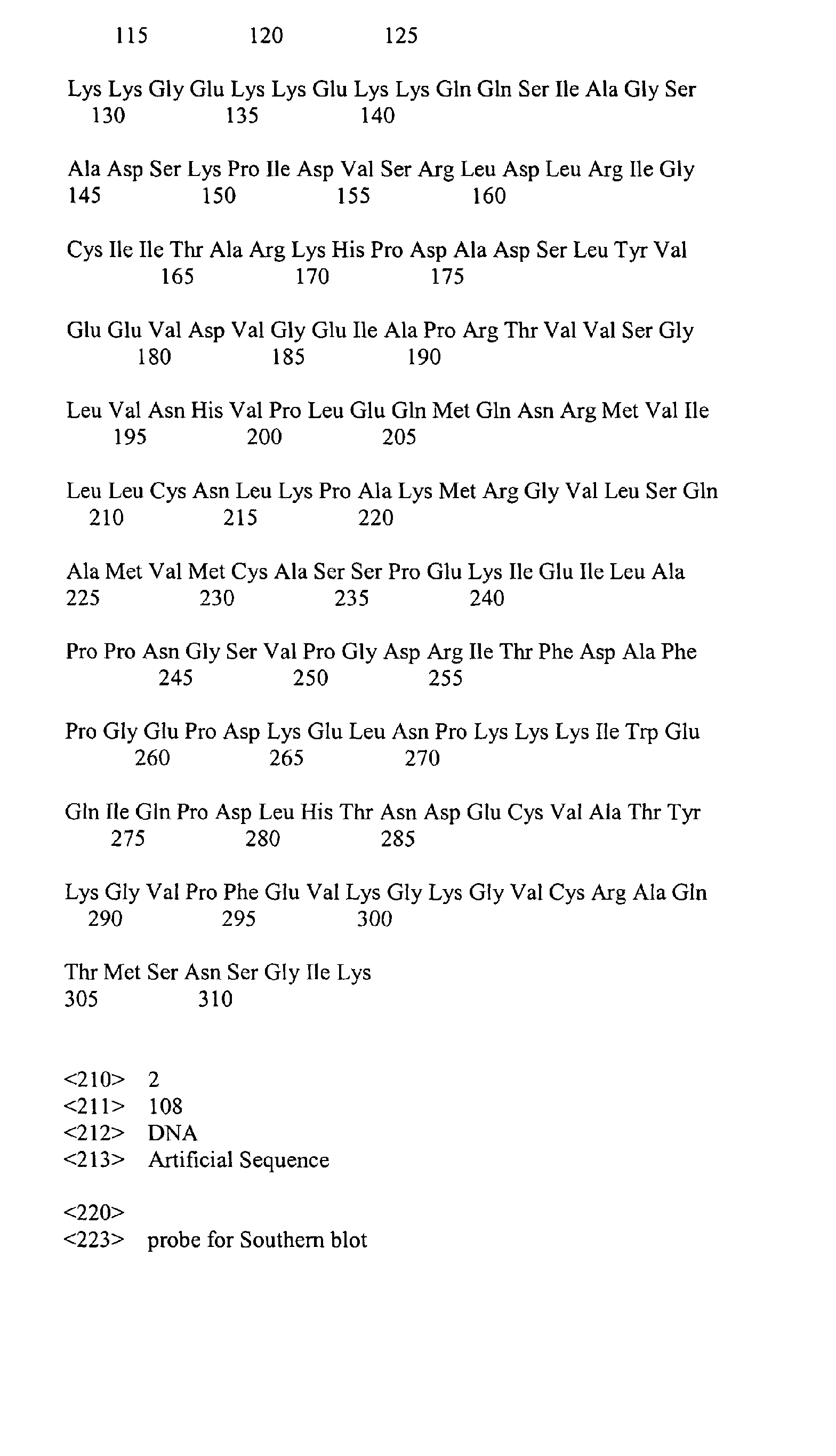
- p43 is a protein consisting of 312 amino acids which is associated with a multi-tRNA synthetase complex to facilitate the catalytic activity of the bound enzyme.
- the p43 is highly expressed by microneuron in the lesions of autoimmune diseases in vitro including encephalomyelitis, neuritis and uveitis.
- the phenomenon in which p43 is highly expressed at certain developmental stages and tissues suggests that p43 is related to inflammation response and cell apoptosis (Berger, A. C. et al., J. Immunother. 23:519-527, 2000).
- PCT International application No. PCT/KR00/00630 an antitumor agent
- p43 may be employed for treating wound.
- polypeptide which is selected from the group consisting of (a) a polypeptide having an amino acid sequence set forth in SEQ ID NO: 1; and (b) a polypeptide having at least 70% sequence homology with the polypeptide of (a) for the manufacture of a pharmaceutical composition which is intended to stimulate wound healing in a subject.
- polypeptide which is selected from the group consisting of (a) a polypeptide having an amino acid sequence set forth in SEQ ID NO: 1; and (b) a polypeptide having at least 70% sequence homology with the polypeptide of (a) for the manufacture of a pharmaceutical composition which is intended to stimulate wound healing in a subject.
- wound includes any injury to any portion of the body of a subject including, but not limited to, acute conditions such as thermal burns, chemical burns, radiation burns, burns caused by excess exposure to ultraviolet radiation such as sunburn, damage to bodily tissues such as the perineum as a result of labor and childbirth, including injuries sustained during medical procedures such as episiotomies, trauma-induced injuries including cuts, incisions, excoriations, those injuries sustained in automobile and other mechanical accidents, and those caused by bullets, knives and other weapons, ulcer such as pressure ulcer, plaster ulcer and decubitus ulcer, and post-surgical injuries, as well as chronic conditions such as pressure sores, bedsores, conditions related to diabetes and poor circulation, and all types of acne.
- said wound includes dermatitis such as impetigo, intertrigo, folliculitis and eczema.
- Areas of the body which can be treated with the present invention include, but are not limited to, skin, muscle and internal organs.
- subject refers to a human or lower animal on whom the present invention is practiced.
- composition used in the present invention comprises, as an effective ingredient, p43 having an amino acid sequence set forth in SEQ ID NO: 1.
- the composition used in the present invention may also comprise the functional equivalents of the p43 as well as the p43.
- “Functional equivalents”, as used herein, refer to polypeptides having the physiological activity to stimulate wound healing substantially equivalent with that of p43 having the amino acid sequences set forth in SEQ ID NO: 1.
- the functional equivalents may be a polypeptide having at least 70% sequence homology, preferably at least 80%, more preferably at least 90%, and most preferably at least 95%, compared to the amino acid sequence set forth in SEQ ID NO: 1.
- the functional equivalents include amino acid sequence variants, for example, in which a portion or all of natural p43 amino acid sequence is substituted, or a portion of the p43 amino acid sequence is deleted or added.
- the substitution of the amino acid is preferably a conservative substitution.
- the amino acids occurring the conservative substitution in nature include aliphatic amino acids (Gly, Ala, Pro); hydrophobic amino acids (Ile, Leu, Val), aromatic amino acids (Phe, Tyr, Trp), acidic amino acids (Asp, Glu), basic amino acids (His, Lys, Arg, Gln, Asn), and sulfur-containing amino acids (Cys, Met).
- the amino acid deletion is preferably located at a portion that does not affect the physiological activity of p43.
- p43 or functional equivalents thereof can be administered (or applied) in an appropriate amount depending on the size of wound.
- the p43 is preferably administered in an amount range of about 1ng to 5 ⁇ g.
- composition comprising, as an effective component, the p43 or functional equivalents thereof according to the present invention may further comprise at least one selected from the group consisting of antibiotics, such as tetracycline, oxytetracycline, gentamicin, neomycin sulfate, bacitracin and polymyxin B sulfate; antihistamines, such as diphenhydramine, promethazine, tripelennamine, phenothiazine, chlorophenylamine, antazoline and pantholin; anti-inflammatory drugs; anti-viral drugs; anti-fungal agents; and growth factors, such as PDGF (platelet-derived growth factor), PDAF, PDEGF, TGF- ⁇ (transforming growth factor- ⁇ ), PF-4, ⁇ FGF, bFGF (basic fibroblast growth factor), VEGF (vascular endothelial growth factor), GH (growth hormone), EGF (epidermal growth factor) and IGF (insulin-like growth factor).
- composition used in the present invention may be compounded with a pharmaceutically acceptable carrier and employed in the form of powder, liniment, gel, lotion, cream, ointment, paste, puff, aerosol or suppository, and in particular, ointment and paste are more preferable.
- the carrier may comprise hydrocarbons including petrolatum, liquid paraffin and gelling hydrocarbon; animal or vegetable oils including medium chain triglyceride, pig fat, hard fat and cacao oil; higher fatty alcohols including cetanol and stearyl alcohol; fatty acids and esters thereof including stearic acid isopropyl palmitate; aqueous bases including polyethylene glycol, 1,3-butyleneglycol, glycerol, gelatin, white sugar and sugar alcohol; emulsifiers including glycerine fatty acid esters, polyoxyl stearate and polyoxyethylene hydrogenated castor oil; conglutinants including acrylate ester and sodium alginate; propellants including liquefied petroleum gas and carbon dioxide; and preservatives including paraoxybenzoates, depending on each formulation type.
- hydrocarbons including petrolatum, liquid paraffin and gelling hydrocarbon
- animal or vegetable oils including medium chain triglyceride, pig fat, hard fat and cacao oil
- higher fatty alcohols including
- the p43 having an amino acid sequence set forth in SEQ ID NO: 1 shows excellent effects on rapid healing of a wound by increasing granulation tissue and stimulating angiogenesis (blood vessel generation) in any wound including scald and ulcer regions.
- the stimulation of wound healing by p43 identified in the present invention is considered to be a result from its multiple effect on inflammation as well as angiogenesis.
- the attraction of macrophage/monocyte by p43 is consistent with our expectation because various proinflammatory cytokines and chemokines such as monocyte chemotactic protein 1 (MCP-1) or macrophage inflammatory protein 1 ⁇ (MIP-1 ⁇ ) have been reported to be induced by p43 [Young-Gyu Ko, et al., J. Biol. Chem. 276:23028-23033, 2001].
- the pro-angiogenic activity of p43 shown in the present invention is surprising because p43 was previously shown to suppress the cell proliferation and induce apoptosis of endothelial cells at high concentration [Sun Young Chang, et al., J. Biol. Chem., 277:8388-8394, 2002]. Based on these results, the present invention provides the first evidences on a novel activity of p43 in angiogenesis associated with wound repair.
- an effective ingredient of the composition of the present invention has a novel physiological activity including the induction of macrophage/monocyte and endothelial cell, re-epithelialization, proliferation of fibroblasts or angiogenesis. Therefore, it is expected that the method for stimulating wound healing using the composition containing the p43 or functional equivalents thereof according to the present invention can be extensively used in the general medical areas involving wound healing.
- the present inventors introduced a mutation to the structural gene of p43 in mouse.
- the mutation of the mouse genomic DNA may be effected according to conventional methods used in the prior art, including a gene trap method, a gene targeting method or an N-ethyl-N-nitrosourea (ENU) treatment method.
- the gene trap vector was used herein to mutate the mouse genomic DNA.
- the gene trap vector may be one of those conventionally used in the prior art. More preferably, the gene trap vector may be VICTR20 vector.
- the VICTR20 comprises two functional units, a neomycin resistant gene ( ⁇ -geo / neo r ) and a puromycin resistant gene ( puro ) (see Fig.
- the neomycin resistant gene is expressed by an upstream promoter of genomic DNA in which the vector is inserted and the puromycin resistant gene is expressed by the phosphoglycerate kinase-1 (PGK) promoter of the vector.
- the gene trap vector, VICTR20 includes long terminal repeats (LTRs) in both ends, a splice accepter (SA), a neomycin resistant gene ( neo r ), a poly-A sequence (pA), a PGK promoter, a puromycin resistant gene ( puro ) and a splice donor (SD).
- mutant library the genomic DNA in which a mutation is derived from the insertion of the gene trap is introduced into embryonic stem cells of mice.
- mutant library the clone containing the disrupted p43 gene is identified and used to prepare heterozygous mutant mice.
- the nucleotide sequence of p43 allele in the p43 mutant mice is determined, and thereby it can be confirmed that the gene trap vector is inserted at the first intron of the p43 gene (See Fig. 1).
- the mutation of p43 is determined by Southern blot and PCR analysis (See Fig. 2) and the mutational effect on the expression of p43 is determined by Northern and Western blot (See Fig. 3).
- the mating of the heterozygous mutant mice generated 16 of wild type, 53 of heterozygous mutant mice and 28 of homozygous mutant mice. This ratio is close to the Mendelian segregation, indicating no significant embryonic lethality of the mutant mice.
- the homozygous mutants significantly reduced body size compared to the wild type mice (data not shown).
- the wounds in the mutant mice recovered at much slower rate compared to those in the wild type mice and direct administration of the purified p43 to the wound region of the wild type mice stimulated the healing process (See Fig. 4).
- the cross-sections of the wound showed the increase of granulation tissue and rapid re-epithelialization in the wounds of the wild type mice treated with p43 (See Fig. 5).
- immunostaining of the wound regions with an antibody that is specific to macrophage/monocyte showed the enrichment of these cells in the wound region tissue of the p43-treated wild type mice (WT+p43) while fewer numbers of these cells were observed in the wound region tissue of the p43 mutant mice (Ho) (See Fig. 6).
- the macrophage/monocyte-specific antibody may include, but not limited to, anti-MOMA-2 antibody.
- the immunostaining of the wound regions with an antibody specific to endothelial cells showed that vascularization was also significantly enhanced in the p43-treated wild type mice (WT+p43) while the reverse was the case in the p43 mutants (Ho) (See Fig. 6).
- the antibody specific to endothelial cell may include, but not limited to, anti-CD31 antibody.
- p43 stimulates the generation of endothelial cell and induces angiogenesis
- the present inventors carried out several different experiments that can determine the activity of p43 in angiogenesis.
- the present inventors investigated whether p43 induces the migration of endothelial cells by cellular wound migration assay.
- the endothelial cell migration was enhanced in the presence of p43 (See Fig. 9A).
- the endothelial cells were cultivated on Matrigel and then the stimulated tube formation was also observed in the presence of p43 (See Fig. 9B).
- CAM chorioallantoic membrane assay
- bFGF basic fibroblast growth factor
- p43 structural gene was allowed to be mutated using the gene trap method [Cecconi, F. & Meyer, B. I., FEBS Lett., 480:63-71, 2000].
- the gene trap vector, VICTR20 (Lexicon Genetics, USA) was used to mutate the genomic DNA of SvEvBrd mice (Lexicon Genetics, USA) according to Zambrowicz's method [Zambrowicz, B. P. et al., Nature 392:608-611, 1998].
- the mutated genomic DNA was introduced into the embryonic stem cells derived from 129/SvEvBrd mice (Omnibank) to generate the mutant library.
- the clone containing p43 gene disrupted by the insertion of the gene trap vector was screened and called 'OST58507'.
- the clone was used to generate the heterozygous C57/BL6 mice (Samtako) by the standard method of Lexicon Genetics.
- the mating of the heterozygous mice generated 16 of wild type, 53 of heterozygous mutant mice and 28 of homozygous mutant mice. The ratio is close to the Mendelian segregation, indicating no significant embryonic lethality of the mutant.
- the p43 mutant allele was analyzed by sequencing.
- the sequencing was determined by Pangenomcis, a company for sequence analysis. As shown in Fig. 1, the first exon (Exon I) and second exon (Exon II) of p43 mutant gene were separated by the intron of about 7kb. It was confirmed that the gene trap vector was inserted at about 1.5kb downstream of the first exon.
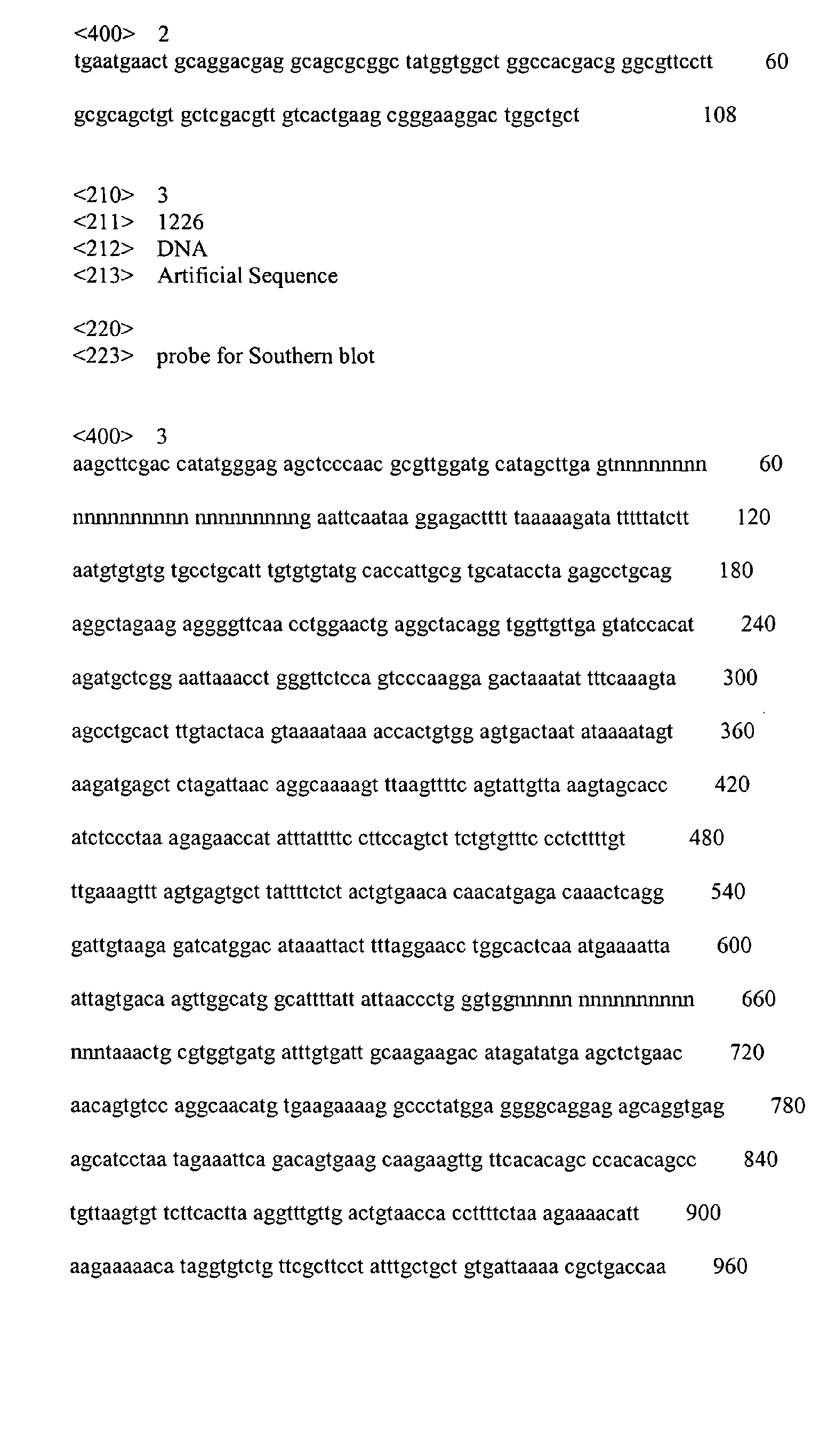
- the genomic DNA was isolated from the tail of each mouse and digested with NcoI. The cleaved DNA fragments were separated by denaturing gel electrophoresis. Then, southern blot analysis was performed using the neomycin resistant gene ( Neo r )(SEQ ID NO: 2) of the gene trap vector or the Hind III fragment (SEQ ID NO: 3) of the p43 mutant gene as a prove, according to a conventional method [Southern, E. M., J. Mol. Biol., 98:503, 1975]. As shown in Fig. 2A, the Neor gene of the gene trap vector and the p43 mutant gene (7kb) were detected in the homozygous mutant mice. However, the Neo r gene was not detected in the wild type mice.
- the Neo r gene was mildly detected and both intact p43 gene (10kb) and the p43 mutant gene (7kb) were detected.
- the size of the p43 mutant gene (7kb) was smaller than that of the intact p43 gene (10kb) because of the presence of Nco I site within Neo r gene of the gene trap vector. From the results, it can be demonstrated that the p43 mutant mouse of the present invention contains a mutation within the p43 gene.
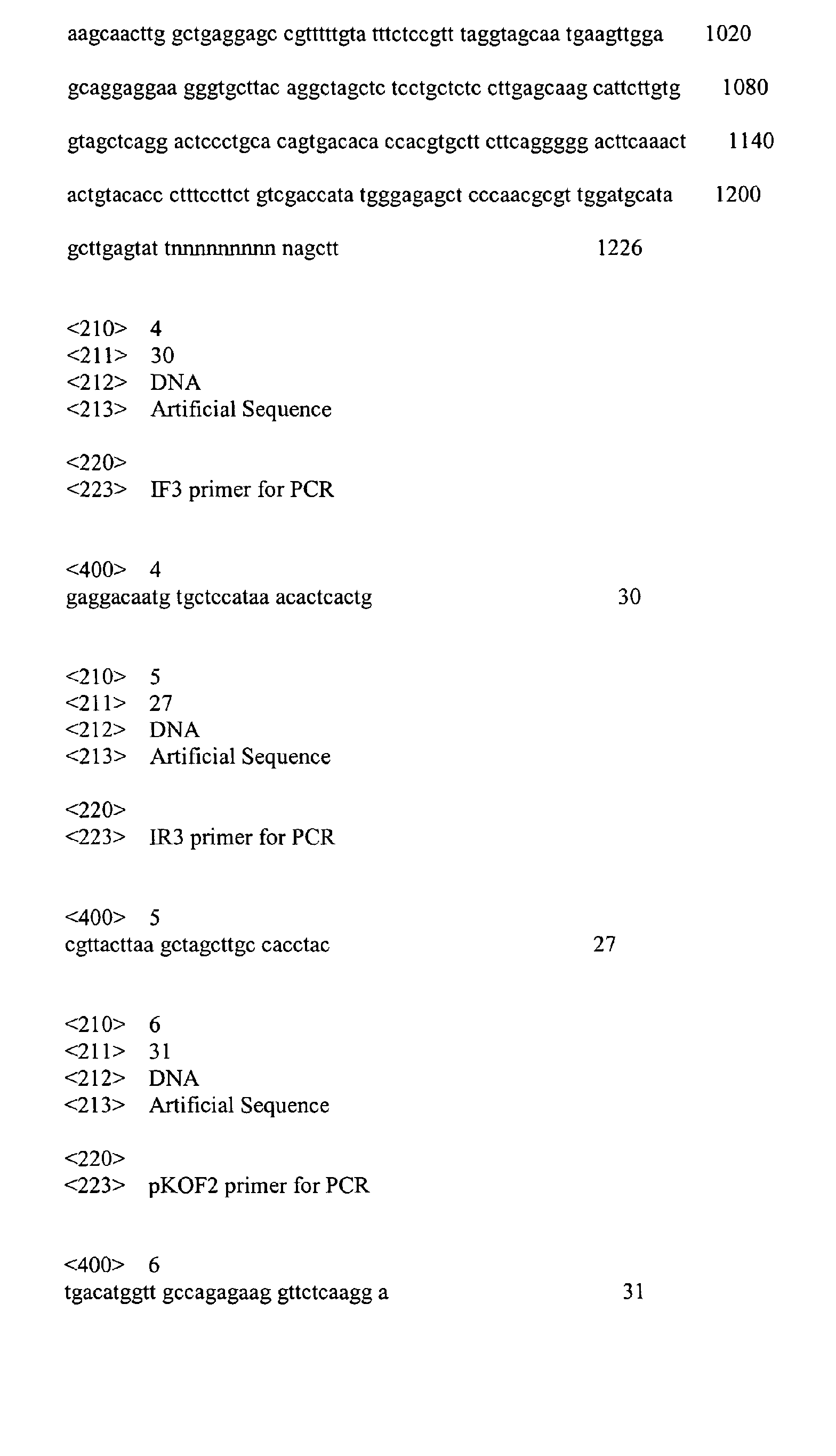
- the genomic DNA was used as a template for PCR analysis with IF3 primer (SEQ ID NO: 4) and IR3 primer (SEQ ID NO: 5) (See Fig. 1).
- IF3 primer SEQ ID NO: 4
- IR3 primer SEQ ID NO: 5
- pKOF2 primer SEQ ID NO: 6
- the IR3 primer were used in PCR analysis (See Fig. 1).
- the template DNA was denatured at a temperature of 94°C for 5 min and cycled 25 times at 94°C for 1 min, at 57°C for 1min and at 72°C for 1min.
- the protein was isolated from the mouse organ according to Ziak et al. (Ziak M, et al., Biochem. Biophys. Res. Commun. 280:363-367, 2001). Western blotting was carried out with anti-p43 antibody as described previously [Park S. G., et al., J. Biol. Chem. 274:16673-16676, 1999]. As shown in Fig. 3B, p43 protein was detected in the wild type (+/+) and the heterozygous mutant mice, but was not detected in the homozygous mutant mice (-/-). It was consistent with the result of the Northern blot.
- Example 4 Stimulatory activity of p43 in wound healing
- mice wild type and homozygous mutant mice
- Werner et al. the back skin and panniculus carnosus muscle of 7 to 8 weeks old mice (wild type and homozygous mutant mice) were excised according to Werner et al. [Werner, S., et al., Science 266:819-822, 1994].
- the present inventor disinfected the back skin of mice with 70% ethanol, and shaved fur from the back under avertin anesthetization.
- the circular excision wound of bilateral symmetry having a diameter of 0.5 cm was prepared using a circular punch of 1 cm on the back skin. The wounds were left uncovered without the drug treatment or dressing.
- p43 was prepared from E.coli as described previously [Park S. G., et al., J. Biol.
- the wounds in the wild type mice treated with p43 recovered at much faster rate compared to those in the homozygous mutant mice (Ho) or the wild type mice which were not treated with p43 with the lapse of time.
- the wounds in the homozygous mutant mice (Ho) recovered at a much slower rate compared to those in the wild type mice which were not treated with p43 even after much time had passed.
- the wound closure was calculated as the percentage of the initial wound area.
- the wound area of the wild type mice treated with p43 (WT+p43) was significantly decreased with the lapse of time.
- the wild mice treated with p43 were confirmed to indicate the increase of granulation tissue (purple sites below the wound areas) and rapid re-epithelialization by the increase of advancing epithelial layer (arrows).
- the homozygous mutant mice (Ho) showed little increase of advancing epithelial layer in the wounds.
- the wound tissues of the wild type mice treated with p43 showed the enrichment of the macrophage/monocyte while the wound tissues of the homozygous mutant mice (Ho) showed fewer numbers of the macrophage/monocyte.
- the immunostaining of the wounds with the antibody specific to endothelial cells showed that vascularization was also significantly enhanced in the p43-treated wounds while the reverse was the case in the p43 mutants (See Fig. 6).
- the foreskin fibroblasts (American Type Culture Collection) were cultivated in the presence of the different amounts of p43 (1, 10, and 100 ng/ml) for 48 h, and then in fresh medium conaining 1 uCi tritium-labeled thymidine for 4 h. After washing and lysing the cells with RIPA buffer, the amounts of the incorporated thymidine were quantified by liquid scintillation counting.
- p43 was prepared from E.coli as described previously [Park S. G., et al., J. Biol. Chem. 274:16673-16676, 1999]. 4 ⁇ g of the purified p43 was dissolved in 5 ⁇ l of PBS buffer containing 20% glycerol.
- the wounds in 25 wild type mice were treated with either p43 (4ug/wound) or the PBS buffer with 20% glycerol alone (control), twice in every other days.
- the mice were sacrificed in 3 days after wounding and the wound area was isolated for histological analysis.
- the crosssection of the wound area was stained using hematoxylin and eosin.
- the boxed region in the wound was analyzed by mmunofluorescence with the antibody against Ki-67 that is the marker of the cell proliferation (Ben-Izhak, O. et al. Ki67 antigen and PCNA proliferation markers preducts survival in anorectal malignant melanoma. Histopathology 41, 519-525, 2002).
- 3 day-aged wounds were isolated with 3mm of adjacent normal tissue.
- the isolated wounds were fixed in 4% paraformaldehyde in PBS and frozen in OCT compound.
- the frozen sections (6 ⁇ m) were equilibrated 3 times with PBS for 5 min. Non-specific interactions were blocked with PBS containing 0.1% Tween 20, 1% non fat milk for 2 h at room temperature and sections were incubated with 1/50 diluted anti-KI67 antibody (Santacruz) in PBST(PBS containing 0.1% Tween 20) for 2 hrs at 37°C.
- the sections were washed 2 times with PBST and subsequently incubated for 1 h at 37°C with the FITC conjugated antibody.
- the sections were counterstained with propium iodide (10ug/ml for 15 min and washed 3 times with PBST for 5 min. Fluorescence signals were examined under the confocal immunofluorescence microscopy (Radiance, BioRad).
- p43 can stimulated the proliferation of fibroblasts.
- aorta was excised from the butchered cattle and washed by dipping in cold PBS buffer containing 1% penicillin and 0.25 ⁇ g/ml fungizone. Then, bovine aortic endothelial cells (BAECs) were isolated from the surface of the tracheal aorta using a razor blade and incubated in DMEM containing 20% fetal bovine serum (FBS). The incubated cell cultures were wounded with a razor blade in 2 mm in width and the injury lines were marked.
- BAECs bovine aortic endothelial cells
- Example 5.1 From the result of Example 5.1 described above, it was confirmed that p43 induced the endothelial cell migration. And then, to investigate whether p43 induces blood tube formation, BAECs (5x10 5 cells) were cultivated on Matrigel (Becton Dickinson) in the presence of 1nM of p43 at 37°C for 6 hours. A control was not treated with p43. Then, the changes of cell morphology were captured by phase contrast microscopy.
- Fertilized chick eggs were incubated in the humidified egg breeder at 37 ⁇ . On the third day of incubation, about 2ml of egg albumin was removed by an 18-gauge hypodermic needle to detach the developing chorioallantoic membrane (CAM) from the shell. After the incubation for additional 6 days, thermanox coverslips (Nunc) loaded with 0.1 ⁇ g of p43 (dissolved in PBS buffer containing 20% glycerol) were placed on the CAM surface and vascularization was observed. A control was not treated with p43. As shown in Fig. 9C, blood vessels were attracted to the area to which p43 was spotted.
- matrigel plugs containing p43 were subcutaneously injected into the skin of mice and stained by Masson Trichome method, and then red blood cells within the gels were counted.
- the Matrigel (0.25ml, Becton Dickinson) was supplemented with p43 (1ng/ml) plus heparin (9U/ml, Sigma).
- PBS buffer containing 20% glycerol was used as a negative control.
- bFGF bFGF plus heparin
- bFGF was known as a growth factor to stimulate the blood tube formation by associating with heparin in a various form.
- the mixtures were subcutaneously injected into 7 weeks old C57BL6/J mice (Samtako) in a dose of 0.25 ml/site. The mice were sacrificed 7 days after the inoculation and the gels were removed, fixed in 4% paraformaldehyde for 24 hours. The gel plugs were embedded in the OCT compound (Sakura) and then sliced into sections of 10 ⁇ m thick using Cyo-microtome (Zeiss).
- the blood vessel formation within the gels was also monitored by counting red blood cells.
- the gels containing bFGF or p43 contained higher concentration of red blood cells than the control.
- the concentration of hemoglobin within the gels was determined as described previously [Passaniti, A., et al., Lab. Invest., 67:519-528,1992].
- the gels containing bFGF or p43 contained higher concentration of hemoglobin than the control.
- the present invention relates to a novel use of p43 for stimulating wound healing, and more particularly to a use of at least one polypeptide which is selected from the group consisting of (a) a polypeptide having an amino acid sequence set forth in SEQ ID NO: 1; and (b) a polypeptide having at least 70% sequence homology with the polypeptide of (a) for the manufacture of a pharmaceutical composition which is intended to stimulate wound healing in a subject.
- the p43 has an excellent effect on the wound healing by its action including the induction of macrophage/monocyte and endothelial cell, re-epithelialization, proliferation of fibroblasts or angiogenesis.
Landscapes
- Health & Medical Sciences (AREA)
- Life Sciences & Earth Sciences (AREA)
- Engineering & Computer Science (AREA)
- Bioinformatics & Cheminformatics (AREA)
- Chemical & Material Sciences (AREA)
- Veterinary Medicine (AREA)
- Public Health (AREA)
- Medicinal Chemistry (AREA)
- General Health & Medical Sciences (AREA)
- Animal Behavior & Ethology (AREA)
- Pharmacology & Pharmacy (AREA)
- Immunology (AREA)
- Gastroenterology & Hepatology (AREA)
- Proteomics, Peptides & Aminoacids (AREA)
- Epidemiology (AREA)
- Zoology (AREA)
- Organic Chemistry (AREA)
- Nuclear Medicine, Radiotherapy & Molecular Imaging (AREA)
- General Chemical & Material Sciences (AREA)
- Chemical Kinetics & Catalysis (AREA)
- Dermatology (AREA)
- Medicines That Contain Protein Lipid Enzymes And Other Medicines (AREA)
- Surgical Instruments (AREA)
- Materials For Medical Uses (AREA)
Abstract
Description
- The present invention relates to a novel use of p43 for stimulating wound healing, and more particularly to a use of at least one polypeptide which is selected from the group consisting of (a) a polypeptide having an amino acid sequence set forth in SEQ ID NO: 1; and (b) a polypeptide having at least 70% sequence homology with the polypeptide of (a) for the manufacture of a pharmaceutical composition which is intended to stimulate wound healing in a subject.
- In general, wound healing is any reaction of tissue against damage inflicted upon the tissue, and it is known as a complex biological process comprising chemotaxis, cell differentiation and replication, synthesis of matrix protein, angiogenesis and reconstitution of wound, as a series of tissue repair processes (Steed, D. L. et al., Clin. Plast. Surg. 25:397, 1998). As one of the typical factors for controlling wound healing process, the growth factor may be considered, which regulate cell growth, cell differentiation, cell metabolism and the like through the general wound healing process, as well as the environment around a wound, and have consistently been used for the development of treatment agents. In addition, various kinds of cytokine are known to participate in wound healing. As an example of the cytokine, there is transforming growth factor-β (TGF-β). The TGF-β is involved in the growth and differentiation of various cells. It has been reported that the TGF-β is involved in several complicated functions including growth control, the regulation of immune response, the stimulation of osteogenesis, the induction of cartilage-specific macromolecule and the promotion of wound healing (Bennett, N. T. et al., Am. J. Surg. 165:728, 1993).
- According to whether skin deficiency exists or not, a wound may be classified into two types and may be treated with different healing mechanisms. When a wound does not involve skin deficiency, as in most mild injuries, for example a case of having injury only to the epidermis, keratinocytes move from the wound periphery, cover the wound, and reform new epidermis and keratin (Knighton, D. R. and Fiegel, V. D., Invest. Radiol. 26:604-611, 1991). However, when entire skin layers are totally injured or destroyed, new connective tissues, called granulation tissues, fill wound space where regenerated epidermis from the periphery of the wound sites must be covered. The granulation tissues are formed by the deposition of extracellular matrix components, such as collagen, from fibroblasts moving into the wound space. Consequently, the healing mechanisms of wound significantly varied depending on the presence of such skin deficiency. Successful wound healing requires completing a series of wound healing multistage processes. Since the deletion of one or more of wound healing-related components prevents the wound healing and skin repair, the wound remains exposed. Such an exposed wound can be easily infected, resulting in delayed healing processes, and the formation of ulcers and erosion of skin. There is a need for the development of a drug promoting the growth of granulation tissue and the regeneration of skin on wound involving skin deficiency.
- Meanwhile, p43 is a protein consisting of 312 amino acids which is associated with a multi-tRNA synthetase complex to facilitate the catalytic activity of the bound enzyme. The p43 is highly expressed by microneuron in the lesions of autoimmune diseases in vitro including encephalomyelitis, neuritis and uveitis. The phenomenon in which p43 is highly expressed at certain developmental stages and tissues suggests that p43 is related to inflammation response and cell apoptosis (Berger, A. C. et al., J. Immunother. 23:519-527, 2000). We have previously shown that p43 can be employed as an effective cytokine and an antitumor agent (PCT International application No. PCT/KR00/00630). However, it is still not shown that p43 may be employed for treating wound. We identified that p43 has a novel activity of promoting wound healing process by acting on the wound healing process comprising the inflammation stage, epithelialization stage and angiogenesis stage, and completed the present invention.
- Therefore, it is an object of the present invention to provide a use of at least one polypeptide which is selected from the group consisting of (a) a polypeptide having an amino acid sequence set forth in SEQ ID NO: 1; and (b) a polypeptide having at least 70% sequence homology with the polypeptide of (a) for the manufacture of a pharmaceutical composition which is intended to stimulate wound healing in a subject.
- The foregoing and other objects, features and advantages of the present invention will become more apparent from the following detailed description when taken in conjunction with the accompanying drawings in which:
- Fig. 1 is a schematic drawing showing where the gene trap is inserted within p43 mutant allele, wherein A, K, S and H indicate the restriction sites for ApaI, KpnI, SacI and HindIII, respectively. IF3, IR3 and pKOF2 are primers for PCR.
- Fig. 2 shows the results of Southern blot (A) and PCR (B) for identifying the
mutation of p43.
+/+: wild type mouse (WT);
+/-: heterozygous mutant mouse;
- /-: homozygous mutant mouse (Ho). - Fig. 3 shows the results of Northern blot (A) and Western blot for determining
the expression of p43.
+/+: wild type mouse (WT);
+/-: heterozygous mutant mouse;
- /-: homozygous mutant mouse (Ho). - Fig. 4 is a photograph showing wound healing levels with the lapse of time
(A) and a bar graph representing a percentage of wound healing levels to an initial wound
area.
Ho: homozygous mutant mouse;
WT: wild type mouse which is not treated with p43 (control);
WT+p43: wild type mouse treated with p43. - Fig. 5 shows the result of histochemical analysis representing the effect of
p43 to stimulate wound healing.
Arrows: advancing epithelial layers;
Purple region: granulation tissues. - Fig. 6 shows the results of immunostaining for determining the generation levels of macrophage/monocyte and endothelial cells.
- Fig 7 is a bar graph representing the effect of p43 on the proliferation of fibroblasts.
- Fig 8 shows the result of histochemical analysis representing the effect of p43 on
the proliferation of fibroblasts.
Left: hematoxylin and eosin staining result of the crosssection of the wound area (arrows : the boundary of the wound area);
Right: immuno-fluorescence staining results of the boxed region in the wound with the antibody against Ki-67. - Fig. 9 shows the results of wound migration assay (A), tube formation assay (B) and CAM assay (C) for estimating the effect of p43 in blood vessel generation.
- Fig. 10 shows the results of the matrigel plug assay for confirming the stimulation of neovascularization by p43.
- Fig. 11 is a bar graph representing the hemoglobin (Hb) contents within matrigel containing p43.
-
- According to an aspect of the present invention, there is provided a use of at least one polypeptide which is selected from the group consisting of (a) a polypeptide having an amino acid sequence set forth in SEQ ID NO: 1; and (b) a polypeptide having at least 70% sequence homology with the polypeptide of (a) for the manufacture of a pharmaceutical composition which is intended to stimulate wound healing in a subject.
- Hereinafter, the term "wound" includes any injury to any portion of the body of a subject including, but not limited to, acute conditions such as thermal burns, chemical burns, radiation burns, burns caused by excess exposure to ultraviolet radiation such as sunburn, damage to bodily tissues such as the perineum as a result of labor and childbirth, including injuries sustained during medical procedures such as episiotomies, trauma-induced injuries including cuts, incisions, excoriations, those injuries sustained in automobile and other mechanical accidents, and those caused by bullets, knives and other weapons, ulcer such as pressure ulcer, plaster ulcer and decubitus ulcer, and post-surgical injuries, as well as chronic conditions such as pressure sores, bedsores, conditions related to diabetes and poor circulation, and all types of acne. In addition, said wound includes dermatitis such as impetigo, intertrigo, folliculitis and eczema.
- Areas of the body which can be treated with the present invention include, but are not limited to, skin, muscle and internal organs. Hereinafter, the term "subject" refers to a human or lower animal on whom the present invention is practiced.
- The composition used in the present invention comprises, as an effective ingredient, p43 having an amino acid sequence set forth in SEQ ID NO: 1. The composition used in the present invention may also comprise the functional equivalents of the p43 as well as the p43. "Functional equivalents", as used herein, refer to polypeptides having the physiological activity to stimulate wound healing substantially equivalent with that of p43 having the amino acid sequences set forth in SEQ ID NO: 1. The functional equivalents may be a polypeptide having at least 70% sequence homology, preferably at least 80%, more preferably at least 90%, and most preferably at least 95%, compared to the amino acid sequence set forth in SEQ ID NO: 1. The functional equivalents include amino acid sequence variants, for example, in which a portion or all of natural p43 amino acid sequence is substituted, or a portion of the p43 amino acid sequence is deleted or added. The substitution of the amino acid is preferably a conservative substitution. For example, the amino acids occurring the conservative substitution in nature include aliphatic amino acids (Gly, Ala, Pro); hydrophobic amino acids (Ile, Leu, Val), aromatic amino acids (Phe, Tyr, Trp), acidic amino acids (Asp, Glu), basic amino acids (His, Lys, Arg, Gln, Asn), and sulfur-containing amino acids (Cys, Met). The amino acid deletion is preferably located at a portion that does not affect the physiological activity of p43.
- Herein, p43 or functional equivalents thereof can be administered (or applied) in an appropriate amount depending on the size of wound. In the case of a circular excision wound of bilateral symmetry having a diameter of 0.5cm, the p43 is preferably administered in an amount range of about 1ng to 5µg.
- The composition comprising, as an effective component, the p43 or functional equivalents thereof according to the present invention may further comprise at least one selected from the group consisting of antibiotics, such as tetracycline, oxytetracycline, gentamicin, neomycin sulfate, bacitracin and polymyxin B sulfate; antihistamines, such as diphenhydramine, promethazine, tripelennamine, phenothiazine, chlorophenylamine, antazoline and pantholin; anti-inflammatory drugs; anti-viral drugs; anti-fungal agents; and growth factors, such as PDGF (platelet-derived growth factor), PDAF, PDEGF, TGF-β (transforming growth factor-β), PF-4, αFGF, bFGF (basic fibroblast growth factor), VEGF (vascular endothelial growth factor), GH (growth hormone), EGF (epidermal growth factor) and IGF (insulin-like growth factor).
- The composition used in the present invention may be compounded with a pharmaceutically acceptable carrier and employed in the form of powder, liniment, gel, lotion, cream, ointment, paste, puff, aerosol or suppository, and in particular, ointment and paste are more preferable. The carrier may comprise hydrocarbons including petrolatum, liquid paraffin and gelling hydrocarbon; animal or vegetable oils including medium chain triglyceride, pig fat, hard fat and cacao oil; higher fatty alcohols including cetanol and stearyl alcohol; fatty acids and esters thereof including stearic acid isopropyl palmitate; aqueous bases including polyethylene glycol, 1,3-butyleneglycol, glycerol, gelatin, white sugar and sugar alcohol; emulsifiers including glycerine fatty acid esters, polyoxyl stearate and polyoxyethylene hydrogenated castor oil; conglutinants including acrylate ester and sodium alginate; propellants including liquefied petroleum gas and carbon dioxide; and preservatives including paraoxybenzoates, depending on each formulation type.
- When damaged skin was exposed to exterior environment, the damaged region may be infiltrated by toxins or bacteria, resulting in inflammation due to body's defense function. In treatment, the rapid generation of granulation tissue should be fostered in the damaged region and, above all, it is important for bacterial infection or excessive inflammation to be suppressed. Otherwise, not only much time is necessary in wound healing but also big scars will remain. The p43 having an amino acid sequence set forth in SEQ ID NO: 1 shows excellent effects on rapid healing of a wound by increasing granulation tissue and stimulating angiogenesis (blood vessel generation) in any wound including scald and ulcer regions.
- The stimulation of wound healing by p43 identified in the present invention is considered to be a result from its multiple effect on inflammation as well as angiogenesis. The attraction of macrophage/monocyte by p43 is consistent with our expectation because various proinflammatory cytokines and chemokines such as monocyte chemotactic protein 1 (MCP-1) or macrophage inflammatory protein 1α (MIP-1α) have been reported to be induced by p43 [Young-Gyu Ko, et al., J. Biol. Chem. 276:23028-23033, 2001]. However, the pro-angiogenic activity of p43 shown in the present invention is surprising because p43 was previously shown to suppress the cell proliferation and induce apoptosis of endothelial cells at high concentration [Sun Young Chang, et al., J. Biol. Chem., 277:8388-8394, 2002]. Based on these results, the present invention provides the first evidences on a novel activity of p43 in angiogenesis associated with wound repair.
- As can be seen from the foregoing, p43, an effective ingredient of the composition of the present invention has a novel physiological activity including the induction of macrophage/monocyte and endothelial cell, re-epithelialization, proliferation of fibroblasts or angiogenesis. Therefore, it is expected that the method for stimulating wound healing using the composition containing the p43 or functional equivalents thereof according to the present invention can be extensively used in the general medical areas involving wound healing.
- In a preferred embodiment of the present invention, the present inventors introduced a mutation to the structural gene of p43 in mouse. The mutation of the mouse genomic DNA may be effected according to conventional methods used in the prior art, including a gene trap method, a gene targeting method or an N-ethyl-N-nitrosourea (ENU) treatment method. Specifically, the gene trap vector was used herein to mutate the mouse genomic DNA. The gene trap vector may be one of those conventionally used in the prior art. More preferably, the gene trap vector may be VICTR20 vector. The VICTR20 comprises two functional units, a neomycin resistant gene (β-geo/neor ) and a puromycin resistant gene (puro) (see Fig. 1). The neomycin resistant gene is expressed by an upstream promoter of genomic DNA in which the vector is inserted and the puromycin resistant gene is expressed by the phosphoglycerate kinase-1 (PGK) promoter of the vector. As shown in Fig. 1, the gene trap vector, VICTR20 includes long terminal repeats (LTRs) in both ends, a splice accepter (SA), a neomycin resistant gene (neor ), a poly-A sequence (pA), a PGK promoter, a puromycin resistant gene (puro) and a splice donor (SD).
- Then, to construct a mutant library, the genomic DNA in which a mutation is derived from the insertion of the gene trap is introduced into embryonic stem cells of mice. In the mutant library, the clone containing the disrupted p43 gene is identified and used to prepare heterozygous mutant mice.
- In another preferred embodiment of the present invention, the nucleotide sequence of p43 allele in the p43 mutant mice is determined, and thereby it can be confirmed that the gene trap vector is inserted at the first intron of the p43 gene (See Fig. 1). The mutation of p43 is determined by Southern blot and PCR analysis (See Fig. 2) and the mutational effect on the expression of p43 is determined by Northern and Western blot (See Fig. 3). The mating of the heterozygous mutant mice generated 16 of wild type, 53 of heterozygous mutant mice and 28 of homozygous mutant mice. This ratio is close to the Mendelian segregation, indicating no significant embryonic lethality of the mutant mice. Interestingly, the homozygous mutants significantly reduced body size compared to the wild type mice (data not shown).
- It has been reported that p43 is associated with immune mechanism and works on endothelial cells [Chang S.Y., et al., J Biol. Chem., 277:8388-8394, 2002; Park H-Y., et al., J. Leukocyte Biol. 71:223-230, 2002; Ko Y-G., et al., J. Biol. Chem. 276:23028-23033, 2001]. We expected that p43 might be involved in the processes of inflammation and angiogenesis occurring in the wound healing process. Thus, to confirm our expectation, the biopsies were introduced to the wild type and homozygous mutant mice, and the healing process was compared by gross examination and histological inspection. The wounds in the mutant mice (Ho) recovered at much slower rate compared to those in the wild type mice and direct administration of the purified p43 to the wound region of the wild type mice stimulated the healing process (See Fig. 4). The cross-sections of the wound showed the increase of granulation tissue and rapid re-epithelialization in the wounds of the wild type mice treated with p43 (See Fig. 5).
- Further, immunostaining of the wound regions with an antibody that is specific to macrophage/monocyte showed the enrichment of these cells in the wound region tissue of the p43-treated wild type mice (WT+p43) while fewer numbers of these cells were observed in the wound region tissue of the p43 mutant mice (Ho) (See Fig. 6). The macrophage/monocyte-specific antibody may include, but not limited to, anti-MOMA-2 antibody. The immunostaining of the wound regions with an antibody specific to endothelial cells showed that vascularization was also significantly enhanced in the p43-treated wild type mice (WT+p43) while the reverse was the case in the p43 mutants (Ho) (See Fig. 6). The antibody specific to endothelial cell may include, but not limited to, anti-CD31 antibody.
- To determine the effect of p43 on the proliferation of different cells in the wound region, we have treated the cultivated foreskin fibroblast cells with p43. The proliferation of fibroblasts was significantly increased in dose-dependent manner by p43 (See Fig. 7). p43 did not stimulate the proliferation of keratinocyte and endothelial cells (data not shown). To see whether p43 can also show the proliferative effect on the fibroblasts in the dermal region of the wound area, we have generated the wound in the back skin of the mice and treated p43 after wounding twice a day in every other day. The proliferation of dermal fibroblasts was then compared by the immuno-fluorescence staining with anti-Ki67 antibody 3 days after wounding. The proliferation of dermal fibroblasts was stimulated in the p43 treated wound region (See Fig. 8). This result is consistent with the effect of p43 on the cultivated fibroblasts. All of these results indicate that p43 can stimulate the proliferation of fibroblasts.
- To further clarify that p43 stimulates the generation of endothelial cell and induces angiogenesis, the present inventors carried out several different experiments that can determine the activity of p43 in angiogenesis.
- First, the present inventors investigated whether p43 induces the migration of endothelial cells by cellular wound migration assay. In this assay, the endothelial cell migration was enhanced in the presence of p43 (See Fig. 9A). Then, the endothelial cells were cultivated on Matrigel and then the stimulated tube formation was also observed in the presence of p43 (See Fig. 9B). Secondly, chorioallantoic membrane assay (CAM) was conducted. It could be seen that blood vessels were attracted to the area to which p43 was spotted (See Fig. 9C).
- Lastly, the matrigel plugs containing p43 were used to see how p43 affects neovascularization. As a positive control, the present inventors used basic fibroblast growth factor (bFGF) which was known to be associated with heparin in various forms and then to promote angiogenesis, epithelialization and the deposition of collagen fiber (Tsuboi, R. et al., J. Exp. Med. 172:245, 1990; Kinsnorth, A. N. et al., Br. J. Surg. 77:409, 1990). The blood vessel formation within the gel was monitored by immunostaining with the antibody specific to endothelial cells. It could be confirmed that the gels containing bFGF or p43 showed strongly enhanced neovascularization (See Fig. 10). The result of immunostaining with the antibody specific to macrophage/monocyte was consistent with the result above (See Fig. 8). Further, it could be seen that the gels containing bFGF or p43 contained higher concentration of red blood cells than the control (See Figs. 10 and 11).
- This invention is further illustrated by the following examples, which are not be construed in any way as imposing limitations upon the scope thereof.
- To prepare p43 mutant mice, p43 structural gene was allowed to be mutated using the gene trap method [Cecconi, F. & Meyer, B. I., FEBS Lett., 480:63-71, 2000]. The gene trap vector, VICTR20 (Lexicon Genetics, USA) was used to mutate the genomic DNA of SvEvBrd mice (Lexicon Genetics, USA) according to Zambrowicz's method [Zambrowicz, B. P. et al., Nature 392:608-611, 1998]. The mutated genomic DNA was introduced into the embryonic stem cells derived from 129/SvEvBrd mice (Omnibank) to generate the mutant library. In the library, the clone containing p43 gene disrupted by the insertion of the gene trap vector was screened and called 'OST58507'. The clone was used to generate the heterozygous C57/BL6 mice (Samtako) by the standard method of Lexicon Genetics. The mating of the heterozygous mice generated 16 of wild type, 53 of heterozygous mutant mice and 28 of homozygous mutant mice. The ratio is close to the Mendelian segregation, indicating no significant embryonic lethality of the mutant.
- To determine the site where the gene trap vector was inserted into p43 allele, the p43 mutant allele was analyzed by sequencing. The sequencing was determined by Pangenomcis, a company for sequence analysis. As shown in Fig. 1, the first exon (Exon I) and second exon (Exon II) of p43 mutant gene were separated by the intron of about 7kb. It was confirmed that the gene trap vector was inserted at about 1.5kb downstream of the first exon.
- The genomic DNA was isolated from the tail of each mouse and digested with NcoI. The cleaved DNA fragments were separated by denaturing gel electrophoresis. Then, southern blot analysis was performed using the neomycin resistant gene (Neor )(SEQ ID NO: 2) of the gene trap vector or the HindIII fragment (SEQ ID NO: 3) of the p43 mutant gene as a prove, according to a conventional method [Southern, E. M., J. Mol. Biol., 98:503, 1975]. As shown in Fig. 2A, the Neor gene of the gene trap vector and the p43 mutant gene (7kb) were detected in the homozygous mutant mice. However, the Neor gene was not detected in the wild type mice. In the heterozygous mutant mice, the Neor gene was mildly detected and both intact p43 gene (10kb) and the p43 mutant gene (7kb) were detected. The size of the p43 mutant gene (7kb) was smaller than that of the intact p43 gene (10kb) because of the presence of NcoI site within Neor gene of the gene trap vector. From the results, it can be demonstrated that the p43 mutant mouse of the present invention contains a mutation within the p43 gene.
- To amplify the 1kb DNA fragment containing the first intron of the p43 gene, the genomic DNA was used as a template for PCR analysis with IF3 primer (SEQ ID NO: 4) and IR3 primer (SEQ ID NO: 5) (See Fig. 1). To amplify the 2.2kb DNA fragment containing a portion of the gene trap vector and a portion of the p43 gene, pKOF2 primer (SEQ ID NO: 6) and the IR3 primer were used in PCR analysis (See Fig. 1). For the PCR analysis, the template DNA was denatured at a temperature of 94°C for 5 min and cycled 25 times at 94°C for 1 min, at 57°C for 1min and at 72°C for 1min. As can be seen from Fig. 2B, only one band of 1kb was detected in the wild type mice (+/+) and two bands corresponding to 1kb and 2.2kb, respectively, were detected in the heterozygous mutant mice (+/-). In the homozygous mutant mice (-/-), a band of 2.2kb was only detected. From the results, it can be demonstrated that the p43 mutant mouse of the present invention contains a mutation within the p43 gene.
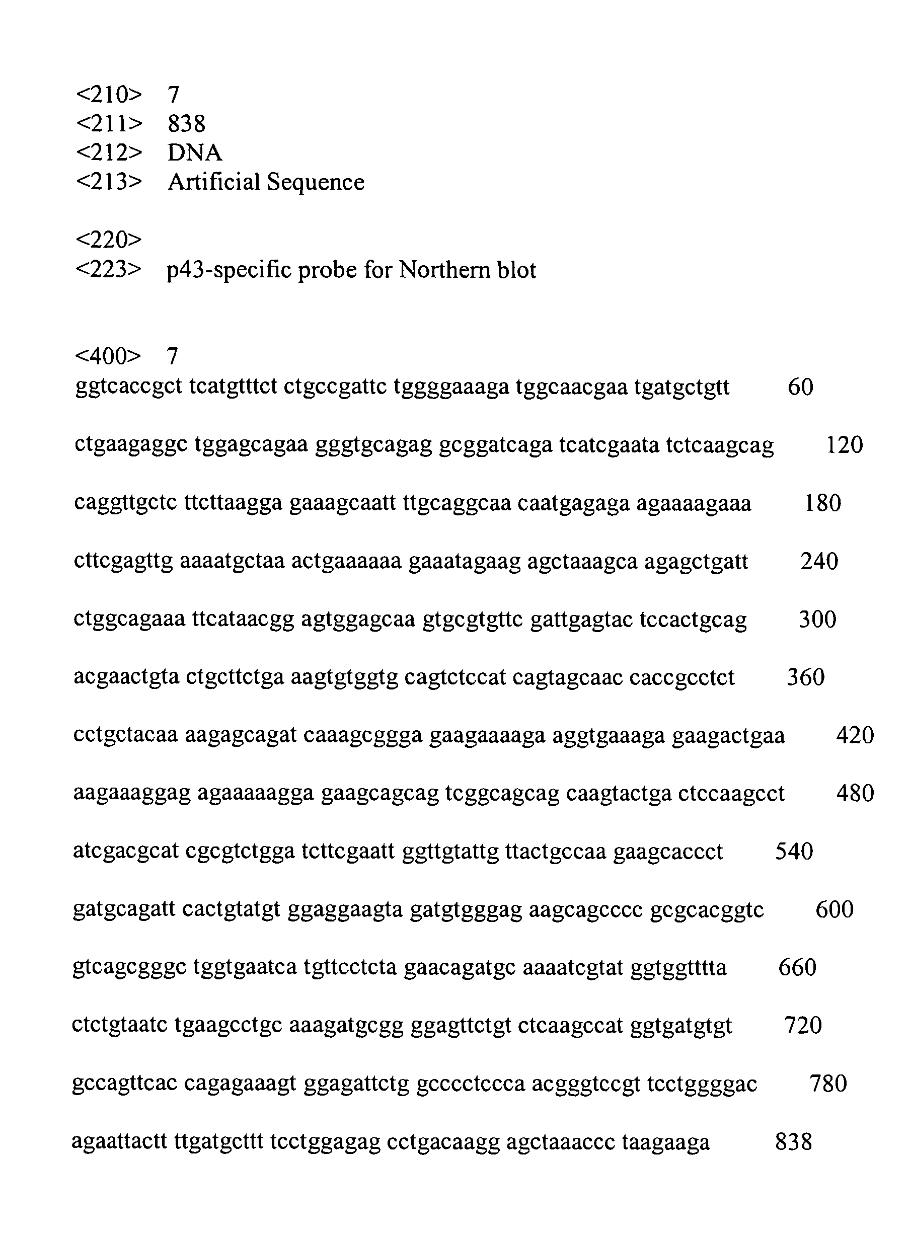
- For Northern analysis, total cellular RNA was prepared from mouse embryonic fibroblast (MEF) cells using RNeasy Midi Kit (QIAGEN) according to the manufacturer's instruction. The isolated RNA was separated on denaturing gel, and then transferred to the Hybond-N+ membrane (Amersham). The RNAs on the membranes were hybridized with the specific probes for p43 set forth in SEQ ID NO: 7. As shown in Fig. 3A, p43 mRNA was detected in the wild type (+/+) and the heterozygous mutant mice, but was not detected in the homozygous mutant mice (-/-). It indicates that p43 is not expressed in p43 mutant mice (-/-) of the present invention.
- For Western blot analysis, the protein was isolated from the mouse organ according to Ziak et al. (Ziak M, et al., Biochem. Biophys. Res. Commun. 280:363-367, 2001). Western blotting was carried out with anti-p43 antibody as described previously [Park S. G., et al., J. Biol. Chem. 274:16673-16676, 1999]. As shown in Fig. 3B, p43 protein was detected in the wild type (+/+) and the heterozygous mutant mice, but was not detected in the homozygous mutant mice (-/-). It was consistent with the result of the Northern blot.
- For gross appearance, the back skin and panniculus carnosus muscle of 7 to 8 weeks old mice (wild type and homozygous mutant mice) were excised according to Werner et al.[Werner, S., et al., Science 266:819-822, 1994]. Firstly, the present inventor disinfected the back skin of mice with 70% ethanol, and shaved fur from the back under avertin anesthetization. Then, the circular excision wound of bilateral symmetry having a diameter of 0.5 cm was prepared using a circular punch of 1 cm on the back skin. The wounds were left uncovered without the drug treatment or dressing. Meanwhile, p43 was prepared from E.coli as described previously [Park S. G., et al., J. Biol. Chem. 274:16673-16676, 1999]. 4µg of the purified p43 was dissolved in 5µl of PBS buffer containing 20% glycerol. Each group of the wounds in 25 wild type mice was treated with either p43 (4ug/wound) or the PBS buffer with 20% glycerol alone (control), 4 times at 2 days interval. Also, the p43 mutant mice were left without the treatment of p43. The wound closure was monitored daily using the Image-Pro Plus Software (Media Cybernetics).
- As shown in Fig. 4A, the wounds in the wild type mice treated with p43 (WT+p43) recovered at much faster rate compared to those in the homozygous mutant mice (Ho) or the wild type mice which were not treated with p43 with the lapse of time. However, the wounds in the homozygous mutant mice (Ho) recovered at a much slower rate compared to those in the wild type mice which were not treated with p43 even after much time had passed. The wound closure was calculated as the percentage of the initial wound area. As shown in Fig. 4B, the wound area of the wild type mice treated with p43 (WT+p43) was significantly decreased with the lapse of time.
- 5 day-aged wounds were isolated with 3mm of adjacent normal tissue, fixed overnight in Bouin's solution (Sigma, HT10-1-32), and then embedded in paraffin. Paraformaldehyde-fixed paraffin sections (6µm) of the wounds were stained by the Masson Trichome procedure (Lund, L. R., et al., EMBO J. 18:4645-4656, 1999). Only littermates of the same sex were used for direct histological comparison.
- As shown in Fig. 5, the wild mice treated with p43 (WT+p43) were confirmed to indicate the increase of granulation tissue (purple sites below the wound areas) and rapid re-epithelialization by the increase of advancing epithelial layer (arrows). However, the homozygous mutant mice (Ho) showed little increase of advancing epithelial layer in the wounds.
- 5 day-aged wounds were isolated with 3mm of adjacent normal tissue. The isolated wounds were fixed in 4% paraformaldehyde in PBS and then frozen in the OCT compound (Sakura). The cyro-sections of the wounds were analyzed by immune-fluorescence. To detect macrophage/monocyte and endothelial cells, the frozen sections were incubated in 1.7µg/ml of anti-mouse MOMA-2 antibody (Serotec) and 1 .0µg/ml of anti-CD31 antibody (Pharmingen) that are specific to the cells, respectively. After incubation for 1 hour with the primary antibodies, 1.0 µg/ml of FITC-conjugated secondary antibody was added and further incubated for 1 hour. The wounds were then counterstained with propidium iodide (1µg/ml), mounted on slides and investigated using confocal immunofluorescence microscopy (µRadience; BioRad).
- As shown in Fig. 6, the wound tissues of the wild type mice treated with p43 (WT+p43) showed the enrichment of the macrophage/monocyte while the wound tissues of the homozygous mutant mice (Ho) showed fewer numbers of the macrophage/monocyte. Also, the immunostaining of the wounds with the antibody specific to endothelial cells showed that vascularization was also significantly enhanced in the p43-treated wounds while the reverse was the case in the p43 mutants (See Fig. 6).
- To determine the effect of p43 on the proliferation of foreskin fibroblasts, the foreskin fibroblasts(American Type Culture Collection) were cultivated in the presence of the different amounts of p43 (1, 10, and 100 ng/ml) for 48 h, and then in fresh medium conaining 1 uCi tritium-labeled thymidine for 4 h. After washing and lysing the cells with RIPA buffer, the amounts of the incorporated thymidine were quantified by liquid scintillation counting.
- As shown in Fig.7, the proliferation of fibroblasts was significantly increased in dose-dependent manner by p43.
- To determine the effect of p43 on the proliferation of dermal fibroblasts in the cutaneous wound area, the cutaneous wounds were generated on the back skin of 7 to 8 weeks old mice mice according to Werner et al.[Werner, S., et al., Science 266:819-822, 1994]. Meanwhile, p43 was prepared from E.coli as described previously [Park S. G., et al., J. Biol. Chem. 274:16673-16676, 1999]. 4µg of the purified p43 was dissolved in 5µl of PBS buffer containing 20% glycerol. The wounds in 25 wild type mice were treated with either p43 (4ug/wound) or the PBS buffer with 20% glycerol alone (control), twice in every other days. The mice were sacrificed in 3 days after wounding and the wound area was isolated for histological analysis. The crosssection of the wound area was stained using hematoxylin and eosin. The boxed region in the wound (see Fig 8 Left) was analyzed by mmunofluorescence with the antibody against Ki-67 that is the marker of the cell proliferation (Ben-Izhak, O. et al. Ki67 antigen and PCNA proliferation markers preducts survival in anorectal malignant melanoma. Histopathology 41, 519-525, 2002). 3 day-aged wounds were isolated with 3mm of adjacent normal tissue. The isolated wounds were fixed in 4% paraformaldehyde in PBS and frozen in OCT compound. The frozen sections (6 µm) were equilibrated 3 times with PBS for 5 min. Non- specific interactions were blocked with PBS containing 0.1% Tween 20, 1% non fat milk for 2 h at room temperature and sections were incubated with 1/50 diluted anti-KI67 antibody (Santacruz) in PBST(PBS containing 0.1% Tween 20) for 2 hrs at 37°C. The sections were washed 2 times with PBST and subsequently incubated for 1 h at 37°C with the FITC conjugated antibody. The sections were counterstained with propium iodide (10ug/ml for 15 min and washed 3 times with PBST for 5 min. Fluorescence signals were examined under the confocal immunofluorescence microscopy (Radiance, BioRad).
- As shown in Fig.8, p43 can stimulated the proliferation of fibroblasts.
- To investigate whether p43 induces the migration of endothelial cells, the cellular wound migration assay was performed. First, aorta was excised from the butchered cattle and washed by dipping in cold PBS buffer containing 1% penicillin and 0.25µg/ml fungizone. Then, bovine aortic endothelial cells (BAECs) were isolated from the surface of the tracheal aorta using a razor blade and incubated in DMEM containing 20% fetal bovine serum (FBS). The incubated cell cultures were wounded with a razor blade in 2 mm in width and the injury lines were marked. Then, the cultures were washed with serum-free medium and further incubated in DMEM with 1% serum, 1mM thymidine and 1nM p43. The cells were allowed to migrate for 16 hours and then rinsed. A control was not treated with p43. The cultures were fixed with absolute methanol (100%) and stained with Giemsa (Chameleon Chemical).
As shown in Fig. 9A, the endothelial cell migration was enhanced in the presence of p43. From the result, it was demonstrated that p43 stimulates the endothelial cell migration. - From the result of Example 5.1 described above, it was confirmed that p43 induced the endothelial cell migration. And then, to investigate whether p43 induces blood tube formation, BAECs (5x10⁵ cells) were cultivated on Matrigel (Becton Dickinson) in the presence of 1nM of p43 at 37°C for 6 hours. A control was not treated with p43. Then, the changes of cell morphology were captured by phase contrast microscopy.
- As shown in Fig. 9B, the stimulated tube formation was observed in the presence of p43, but in the control. The result indicated that p43 stimulates tube formation and it was consistent with the endothelial cell migration.
- Fertilized chick eggs were incubated in the humidified egg breeder at 37□. On the third day of incubation, about 2ml of egg albumin was removed by an 18-gauge hypodermic needle to detach the developing chorioallantoic membrane (CAM) from the shell. After the incubation for additional 6 days, thermanox coverslips (Nunc) loaded with 0.1µg of p43 (dissolved in PBS buffer containing 20% glycerol) were placed on the CAM surface and vascularization was observed. A control was not treated with p43. As shown in Fig. 9C, blood vessels were attracted to the area to which p43 was spotted.
- To see how p43 affects neovascularization, matrigel plugs containing p43 were subcutaneously injected into the skin of mice and stained by Masson Trichome method, and then red blood cells within the gels were counted. First, the Matrigel (0.25ml, Becton Dickinson) was supplemented with p43 (1ng/ml) plus heparin (9U/ml, Sigma). PBS buffer containing 20% glycerol was used as a negative control. And as a positive control, 143 ng/ml of bFGF (R&D systems) plus heparin was used, in which bFGF was known as a growth factor to stimulate the blood tube formation by associating with heparin in a various form. The mixtures were subcutaneously injected into 7 weeks old C57BL6/J mice (Samtako) in a dose of 0.25 ml/site. The mice were sacrificed 7 days after the inoculation and the gels were removed, fixed in 4% paraformaldehyde for 24 hours. The gel plugs were embedded in the OCT compound (Sakura) and then sliced into sections of 10 µm thick using Cyo-microtome (Zeiss). The sections were reacted with anti-CD31 antibodies (Pharmingen) and anti-MOMA2 antibodies (Serotec) that are specific to the endothelial cells and macrophage/monocyte, respectively, and then stained by Masson Trichome method [Lund, L. R., et al., EMBO J. 18:4645-4656, 1999]. As a result, in the presence of p43, neovascularization was strongly enhanced (See Fig. 10, top row) and macrophage/monocyte were also recruited to the gel compared to the control (See Fig. 10, middle row, green fluorescence). Interestingly, other cells were also enriched in the gels treated with p43 (red fluorescence).
- The blood vessel formation within the gels was also monitored by counting red blood cells. As shown in Fig. 10 (bottom row, arrowheads), the gels containing bFGF or p43 contained higher concentration of red blood cells than the control. Further, the concentration of hemoglobin within the gels was determined as described previously [Passaniti, A., et al., Lab. Invest., 67:519-528,1992]. As shown in Fig. 11, the gels containing bFGF or p43 contained higher concentration of hemoglobin than the control.
- While this invention has been described in connection with what is presently considered to be the most practical and preferred embodiment, it is to be understood that the invention is not limited to the disclosed embodiment and the drawings, but, on the contrary, it is intended to cover various modifications and variations within the spirit and scope of the appended claims.
- The entire disclosure of Korea Patent Application No. 2002-42858, filed on July 22, 2002 including its specification, claims, drawings and summary are incorporated herein by reference in its entirety.
- The present invention relates to a novel use of p43 for stimulating wound healing, and more particularly to a use of at least one polypeptide which is selected from the group consisting of (a) a polypeptide having an amino acid sequence set forth in SEQ ID NO: 1; and (b) a polypeptide having at least 70% sequence homology with the polypeptide of (a) for the manufacture of a pharmaceutical composition which is intended to stimulate wound healing in a subject. The p43 has an excellent effect on the wound healing by its action including the induction of macrophage/monocyte and endothelial cell, re-epithelialization, proliferation of fibroblasts or angiogenesis.

Claims (9)
- A use of at least one polypeptide which is selected from the group consisting of (a) a polypeptide having an amino acid sequence set forth in SEQ ID NO: 1; and (b) a polypeptide having at least 70% sequence homology with the polypeptide of (a) for the manufacture of a pharmaceutical composition which is intended to stimulate wound healing in a subject.
- The use according to claim 1, wherein the pharmaceutical composition further comprises at least one selected from the group consisting of antibiotics, antihistamines, anti-inflammatory drugs, anti-viral drugs, anti-fungal drugs and growth factors.
- The use according to claim 1, wherein the pharmaceutical composition further comprises a pharmaceutically acceptable carrier.
- The use according to claim 1, wherein the pharmaceutical composition is formulated with any one form selected from the group consisting of a powder, liniment, gel, lotion, cream, ointment, paste, puff, aerosol and suppository.
- The use of claim 1, wherein said wound is selected from the group consisting of burn, ulcer, trauma, post-surgical, post-childbirth, chronic wound and dermatitis.
- The use of claim 5, wherein said burn is selected from the group consisting of sunburn, chemical burn, radiation burn and thermal burn.
- The use of claim 5, wherein said ulcer is selected from the group consisting of pressure ulcer, plaster ulcer and decubitus ulcer.
- The use for claim 5, wherein said chronic wound is selected from the group consisting of bedsores, pressure sores, diabetes-related and poor circulation-related.
- The use of claim 6, wherein said dermatitis is selected from the group of consisting of impetigo, intertrigo, folliculitis and eczema.
Applications Claiming Priority (2)
| Application Number | Priority Date | Filing Date | Title |
|---|---|---|---|
| KR2002042858 | 2002-07-22 | ||
| KR10-2002-0042858A KR100515016B1 (en) | 2002-07-22 | 2002-07-22 | Pharmaceutical composition for wound treatment containing p43 as an effective component |
Publications (2)
| Publication Number | Publication Date |
|---|---|
| EP1384486A1 true EP1384486A1 (en) | 2004-01-28 |
| EP1384486B1 EP1384486B1 (en) | 2010-10-20 |
Family
ID=29997531
Family Applications (1)
| Application Number | Title | Priority Date | Filing Date |
|---|---|---|---|
| EP03016724A Expired - Lifetime EP1384486B1 (en) | 2002-07-22 | 2003-07-22 | Use of p43 for stimulating wound healing |
Country Status (5)
| Country | Link |
|---|---|
| US (4) | US7282208B2 (en) |
| EP (1) | EP1384486B1 (en) |
| KR (1) | KR100515016B1 (en) |
| AT (1) | ATE485050T1 (en) |
| DE (1) | DE60334589D1 (en) |
Cited By (8)
| Publication number | Priority date | Publication date | Assignee | Title |
|---|---|---|---|---|
| WO2006083087A1 (en) * | 2005-02-01 | 2006-08-10 | Imagene Co., Ltd. | Method for stimulation collagen synthesis and/or kgf expression |
| WO2008007818A1 (en) * | 2006-07-13 | 2008-01-17 | Seoul National University Industry Foundation | Novel use of aimp1 for controlling glucose level |
| WO2008094012A1 (en) * | 2007-02-01 | 2008-08-07 | Imagene Co., Ltd. | Novel polypeptide having anti-tumor activity |
| JP2009523844A (en) * | 2006-01-23 | 2009-06-25 | イマジェネ カンパニー リミテッド | Novel peptides and uses thereof |
| WO2010041892A3 (en) * | 2008-10-10 | 2010-08-19 | 서울대학교산학협력단 | Novel application of aimp1 polypeptide |
| US9637730B2 (en) | 2010-07-12 | 2017-05-02 | Atyr Pharma, Inc. | Innovative discovery of therapeutic, diagnostic, and antibody compositions related to protein fragments of histidyl-tRNA synthetases |
| US10093915B2 (en) | 2013-03-15 | 2018-10-09 | Atyr Pharma Inc. | Histidyl-tRNA synthetase-Fc conjugates |
| US11767520B2 (en) | 2017-04-20 | 2023-09-26 | Atyr Pharma, Inc. | Compositions and methods for treating lung inflammation |
Families Citing this family (12)
| Publication number | Priority date | Publication date | Assignee | Title |
|---|---|---|---|---|
| KR100515016B1 (en) * | 2002-07-22 | 2005-09-15 | 재단법인서울대학교산학협력재단 | Pharmaceutical composition for wound treatment containing p43 as an effective component |
| EP2403864B1 (en) | 2009-02-27 | 2015-08-12 | Atyr Pharma, Inc. | Polypeptide structural motifs associated with cell signaling activity |
| AU2010226726B8 (en) | 2009-03-16 | 2014-08-14 | Pangu Biopharma Limited | Compositions and methods comprising histidyl-tRNA synthetase splice variants having non-canonical biological activities |
| US9127268B2 (en) | 2009-12-11 | 2015-09-08 | Atyr Pharma, Inc. | Aminoacyl tRNA synthetases for modulating inflammation |
| US10279007B2 (en) | 2010-11-15 | 2019-05-07 | Oxygenetix Institute, Inc. | Topical treatment method for healing wounds, disinfecting, covering and concealing the wound until healing occurs |
| US9714419B2 (en) | 2011-08-09 | 2017-07-25 | Atyr Pharma, Inc. | PEGylated tyrosyl-tRNA synthetase polypeptides |
| WO2013086228A1 (en) | 2011-12-06 | 2013-06-13 | Atyr Pharma, Inc. | Pegylated aspartyl-trna synthetase polypeptides |
| WO2013086216A1 (en) | 2011-12-06 | 2013-06-13 | Atyr Pharma, Inc. | Improved aspartyl-trna synthetases |
| CA2858613A1 (en) | 2011-12-29 | 2013-08-08 | Atyr Pharma, Inc. | Aspartyl-trna synthetase-fc conjugates |
| EP2814514B1 (en) | 2012-02-16 | 2017-09-13 | Atyr Pharma, Inc. | Histidyl-trna synthetases for treating autoimmune and inflammatory diseases |
| JP6521995B2 (en) | 2014-02-20 | 2019-05-29 | ユニヴァーシティー オブ ノートル ダム デュ ラック | Selective matrix metalloproteinase inhibitors |
| US10357546B2 (en) | 2014-09-19 | 2019-07-23 | University Of Notre Dame Du Lac | Acceleration of diabetic wound healing |
Citations (1)
| Publication number | Priority date | Publication date | Assignee | Title |
|---|---|---|---|---|
| WO2001095927A1 (en) * | 2000-06-14 | 2001-12-20 | Imagene Co., Ltd. | P43 anti-tumor therapeutic agent and three dimensional structure of its cytokine domain |
Family Cites Families (5)
| Publication number | Priority date | Publication date | Assignee | Title |
|---|---|---|---|---|
| US5641867A (en) * | 1993-09-29 | 1997-06-24 | The Trustees Of Columbia University In The City Of New York | Antibody which specifically binds to endothelial-monocyte activating polypeptide II |
| US6391311B1 (en) * | 1998-03-17 | 2002-05-21 | Genentech, Inc. | Polypeptides having homology to vascular endothelial cell growth factor and bone morphogenetic protein 1 |
| MXPA01002875A (en) * | 1998-11-13 | 2002-04-08 | Los Angeles Childrens Hospital | Methods of facilitating vascular growth. |
| KR100405919B1 (en) * | 2001-06-05 | 2003-11-14 | 주식회사 이매진 | Pharmaceutical composition for immunological enhancement comprising N-terminal peptide of p43 as an effective component |
| KR100515016B1 (en) * | 2002-07-22 | 2005-09-15 | 재단법인서울대학교산학협력재단 | Pharmaceutical composition for wound treatment containing p43 as an effective component |
-
2002
- 2002-07-22 KR KR10-2002-0042858A patent/KR100515016B1/en not_active Expired - Fee Related
-
2003
- 2003-07-22 AT AT03016724T patent/ATE485050T1/en not_active IP Right Cessation
- 2003-07-22 EP EP03016724A patent/EP1384486B1/en not_active Expired - Lifetime
- 2003-07-22 US US10/623,567 patent/US7282208B2/en not_active Expired - Fee Related
- 2003-07-22 DE DE60334589T patent/DE60334589D1/en not_active Expired - Lifetime
-
2007
- 2007-07-26 US US11/829,018 patent/US7572452B2/en not_active Expired - Fee Related
-
2009
- 2009-08-10 US US12/538,714 patent/US7981426B2/en not_active Expired - Fee Related
-
2011
- 2011-06-08 US US13/155,750 patent/US20120010141A1/en not_active Abandoned
Patent Citations (1)
| Publication number | Priority date | Publication date | Assignee | Title |
|---|---|---|---|---|
| WO2001095927A1 (en) * | 2000-06-14 | 2001-12-20 | Imagene Co., Ltd. | P43 anti-tumor therapeutic agent and three dimensional structure of its cytokine domain |
Non-Patent Citations (2)
| Title |
|---|
| SANG GYU PARK ET AL.: "Dose-dependent biphasic activity of tRNA synthetase-associating factor, p43, in angiogenesis.", JOURNAL OF BIOLOGICAL CHEMISTRY, vol. 277, no. 47, 22 November 2002 (2002-11-22), BALTIMORE, MD, US, pages 45243 - 45248, XP002261380 * |
| YOUNG-GYU KO ET AL.: "Biphasic regulation of angiogenesis by a cofactor of aminoacyl-tRNA synthetase, p43.", MOLECULAR BIOLOGY OF THE CELL, vol. 12, no. supplement, November 2001 (2001-11-01), pages 127a, XP008024541 * |
Cited By (16)
| Publication number | Priority date | Publication date | Assignee | Title |
|---|---|---|---|---|
| JP2008528577A (en) * | 2005-02-01 | 2008-07-31 | イマジェネ カンパニー リミテッド | Method for promoting collagen synthesis and / or KGF expression |
| KR100903984B1 (en) * | 2005-02-01 | 2009-06-25 | 주식회사 이매진 | Cosmetic composition for preventing skin aging, improving wrinkles, enhancing skin suppleness, and enhancing elasticity comprising AIMP1 polypeptide or a fragment thereof as an active ingredient |
| WO2006083087A1 (en) * | 2005-02-01 | 2006-08-10 | Imagene Co., Ltd. | Method for stimulation collagen synthesis and/or kgf expression |
| JP2009523844A (en) * | 2006-01-23 | 2009-06-25 | イマジェネ カンパニー リミテッド | Novel peptides and uses thereof |
| WO2008007818A1 (en) * | 2006-07-13 | 2008-01-17 | Seoul National University Industry Foundation | Novel use of aimp1 for controlling glucose level |
| WO2008094012A1 (en) * | 2007-02-01 | 2008-08-07 | Imagene Co., Ltd. | Novel polypeptide having anti-tumor activity |
| WO2010041892A3 (en) * | 2008-10-10 | 2010-08-19 | 서울대학교산학협력단 | Novel application of aimp1 polypeptide |
| US10669533B2 (en) | 2010-07-12 | 2020-06-02 | Atyr Pharma, Inc. | Innovative discovery of therapeutic, diagnostic, and antibody compositions related to protein fragments of Histidyl-tRNA synthetases |
| US9637730B2 (en) | 2010-07-12 | 2017-05-02 | Atyr Pharma, Inc. | Innovative discovery of therapeutic, diagnostic, and antibody compositions related to protein fragments of histidyl-tRNA synthetases |
| US10196628B2 (en) | 2010-07-12 | 2019-02-05 | Atyr Pharma, Inc. | Innovative discovery of therapeutic, diagnostic, and antibody compositions related to protein fragments of histidyl-tRNA synthetases |
| US10093915B2 (en) | 2013-03-15 | 2018-10-09 | Atyr Pharma Inc. | Histidyl-tRNA synthetase-Fc conjugates |
| US10472618B2 (en) | 2013-03-15 | 2019-11-12 | Atyr Pharma, Inc. | Histidyl-tRNA synthetase-Fc conjugates |
| US10711260B2 (en) | 2013-03-15 | 2020-07-14 | Atyr Pharma, Inc. | Histidyl-tRNA synthetase-Fc conjugates |
| US11072787B2 (en) | 2013-03-15 | 2021-07-27 | Atyr Pharma Inc. | Histidyl-tRNA synthetase-Fc conjugates |
| US12492392B2 (en) | 2013-03-15 | 2025-12-09 | Atyr Pharma, Inc. | Histidyl-tRNA synthetase-Fc conjugates and therapeutics using the same |
| US11767520B2 (en) | 2017-04-20 | 2023-09-26 | Atyr Pharma, Inc. | Compositions and methods for treating lung inflammation |
Also Published As
| Publication number | Publication date |
|---|---|
| US7282208B2 (en) | 2007-10-16 |
| EP1384486B1 (en) | 2010-10-20 |
| DE60334589D1 (en) | 2010-12-02 |
| US20080064627A1 (en) | 2008-03-13 |
| US7572452B2 (en) | 2009-08-11 |
| US20120010141A1 (en) | 2012-01-12 |
| KR100515016B1 (en) | 2005-09-15 |
| US7981426B2 (en) | 2011-07-19 |
| US20050119175A1 (en) | 2005-06-02 |
| ATE485050T1 (en) | 2010-11-15 |
| US20100041608A1 (en) | 2010-02-18 |
| KR20040009048A (en) | 2004-01-31 |
Similar Documents
| Publication | Publication Date | Title |
|---|---|---|
| US7572452B2 (en) | Method for stimulating wound healing | |
| Levine et al. | Spatial and temporal patterns of immunoreactive transforming growth factor beta 1, beta 2, and beta 3 during excisional wound repair | |
| Huang et al. | Impaired wound healing results from the dysfunction of the Akt/mTOR pathway in diabetic rats | |
| Tkalčević et al. | Enhancement by PL 14736 of granulation and collagen organization in healing wounds and the potential role of egr-1 expression | |
| Thompson et al. | Expression of transforming growth factor-beta 1 in specific cells and tissues of adult and neonatal mice. | |
| Thi Hai Yen et al. | An overview on keratinocyte growth factor: from the molecular properties to clinical applications | |
| JP5960944B2 (en) | Improved methods and compositions for wound healing | |
| Roan et al. | Exendin-4, a glucagon-like peptide-1 analogue, accelerates diabetic wound healing | |
| AU728851B2 (en) | Wound healing | |
| CZ285996B6 (en) | In vitro method of stimulating the production of non-keratinocyte epithelial cells and keratinocyte growth factor for the treatment of diseases | |
| JP2003502350A (en) | Accelerated wound dressing for promoting wound healing | |
| CN102325789A (en) | Visfatin Therapeutic Drugs Used to Treat Acne and Other Conditions | |
| EP0855916B1 (en) | Pharmaceutical composition containing an activin stimulator | |
| Pereira et al. | Liposomal gene transfer of keratinocyte growth factor improves wound healing by altering growth factor and collagen expression | |
| Li et al. | Topical GDF11 accelerates skin wound healing in both type 1 and 2 diabetic mouse models | |
| WO2007007098A2 (en) | Promotion of epithelial regeneration | |
| CN113398246B (en) | Application of eIF3I-PDL1-IRS4 axis in preparation of drug for treating refractory ulcer | |
| Schlemm et al. | Medicinal yeast extracts | |
| US20240181125A1 (en) | Compositions and methods for scarless wound healing in diabetes | |
| CN113876956B (en) | Application of PCSK9 inhibitor in preparation of product for promoting skin pigmentation | |
| CN119464294A (en) | Application of Hsa_circ_0005379 in promoting diabetic wound healing | |
| US20240165166A1 (en) | Primed placental tissue and uses in regenerative medicine | |
| US9314504B2 (en) | Use of members of IL-10 cytokine family | |
| Lai | The role of androgen receptor in cutaneous wound healing | |
| Jadin | Trois trios pour deux violons et basse |
Legal Events
| Date | Code | Title | Description |
|---|---|---|---|
| PUAI | Public reference made under article 153(3) epc to a published international application that has entered the european phase |
Free format text: ORIGINAL CODE: 0009012 |
|
| AK | Designated contracting states |
Kind code of ref document: A1 Designated state(s): AT BE BG CH CY CZ DE DK EE ES FI FR GB GR HU IE IT LI LU MC NL PT RO SE SI SK TR |
|
| AX | Request for extension of the european patent |
Extension state: AL LT LV MK |
|
| 17P | Request for examination filed |
Effective date: 20040402 |
|
| AKX | Designation fees paid |
Designated state(s): AT BE BG CH CY CZ DE DK EE ES FI FR GB GR HU IE IT LI LU MC NL PT RO SE SI SK TR |
|
| RAP1 | Party data changed (applicant data changed or rights of an application transferred) |
Owner name: IMAGENE CO., LTD. |
|
| 17Q | First examination report despatched |
Effective date: 20070112 |
|
| RAP1 | Party data changed (applicant data changed or rights of an application transferred) |
Owner name: ATYR PHARMA, INC. |
|
| GRAP | Despatch of communication of intention to grant a patent |
Free format text: ORIGINAL CODE: EPIDOSNIGR1 |
|
| GRAS | Grant fee paid |
Free format text: ORIGINAL CODE: EPIDOSNIGR3 |
|
| GRAA | (expected) grant |
Free format text: ORIGINAL CODE: 0009210 |
|
| AK | Designated contracting states |
Kind code of ref document: B1 Designated state(s): AT BE BG CH CY CZ DE DK EE ES FI FR GB GR HU IE IT LI LU MC NL PT RO SE SI SK TR |
|
| REG | Reference to a national code |
Ref country code: GB Ref legal event code: FG4D |
|
| REG | Reference to a national code |
Ref country code: CH Ref legal event code: EP |
|
| REG | Reference to a national code |
Ref country code: IE Ref legal event code: FG4D |
|
| REF | Corresponds to: |
Ref document number: 60334589 Country of ref document: DE Date of ref document: 20101202 Kind code of ref document: P |
|
| REG | Reference to a national code |
Ref country code: NL Ref legal event code: VDEP Effective date: 20101020 |
|
| PG25 | Lapsed in a contracting state [announced via postgrant information from national office to epo] |
Ref country code: BG Free format text: LAPSE BECAUSE OF FAILURE TO SUBMIT A TRANSLATION OF THE DESCRIPTION OR TO PAY THE FEE WITHIN THE PRESCRIBED TIME-LIMIT Effective date: 20110120 Ref country code: PT Free format text: LAPSE BECAUSE OF FAILURE TO SUBMIT A TRANSLATION OF THE DESCRIPTION OR TO PAY THE FEE WITHIN THE PRESCRIBED TIME-LIMIT Effective date: 20110221 Ref country code: FI Free format text: LAPSE BECAUSE OF FAILURE TO SUBMIT A TRANSLATION OF THE DESCRIPTION OR TO PAY THE FEE WITHIN THE PRESCRIBED TIME-LIMIT Effective date: 20101020 Ref country code: SI Free format text: LAPSE BECAUSE OF FAILURE TO SUBMIT A TRANSLATION OF THE DESCRIPTION OR TO PAY THE FEE WITHIN THE PRESCRIBED TIME-LIMIT Effective date: 20101020 Ref country code: NL Free format text: LAPSE BECAUSE OF FAILURE TO SUBMIT A TRANSLATION OF THE DESCRIPTION OR TO PAY THE FEE WITHIN THE PRESCRIBED TIME-LIMIT Effective date: 20101020 Ref country code: SE Free format text: LAPSE BECAUSE OF FAILURE TO SUBMIT A TRANSLATION OF THE DESCRIPTION OR TO PAY THE FEE WITHIN THE PRESCRIBED TIME-LIMIT Effective date: 20101020 Ref country code: AT Free format text: LAPSE BECAUSE OF FAILURE TO SUBMIT A TRANSLATION OF THE DESCRIPTION OR TO PAY THE FEE WITHIN THE PRESCRIBED TIME-LIMIT Effective date: 20101020 |
|
| PG25 | Lapsed in a contracting state [announced via postgrant information from national office to epo] |
Ref country code: GR Free format text: LAPSE BECAUSE OF FAILURE TO SUBMIT A TRANSLATION OF THE DESCRIPTION OR TO PAY THE FEE WITHIN THE PRESCRIBED TIME-LIMIT Effective date: 20110121 Ref country code: BE Free format text: LAPSE BECAUSE OF FAILURE TO SUBMIT A TRANSLATION OF THE DESCRIPTION OR TO PAY THE FEE WITHIN THE PRESCRIBED TIME-LIMIT Effective date: 20101020 |
|
| PG25 | Lapsed in a contracting state [announced via postgrant information from national office to epo] |
Ref country code: EE Free format text: LAPSE BECAUSE OF FAILURE TO SUBMIT A TRANSLATION OF THE DESCRIPTION OR TO PAY THE FEE WITHIN THE PRESCRIBED TIME-LIMIT Effective date: 20101020 Ref country code: ES Free format text: LAPSE BECAUSE OF FAILURE TO SUBMIT A TRANSLATION OF THE DESCRIPTION OR TO PAY THE FEE WITHIN THE PRESCRIBED TIME-LIMIT Effective date: 20110131 Ref country code: CZ Free format text: LAPSE BECAUSE OF FAILURE TO SUBMIT A TRANSLATION OF THE DESCRIPTION OR TO PAY THE FEE WITHIN THE PRESCRIBED TIME-LIMIT Effective date: 20101020 |
|
| PLBE | No opposition filed within time limit |
Free format text: ORIGINAL CODE: 0009261 |
|
| STAA | Information on the status of an ep patent application or granted ep patent |
Free format text: STATUS: NO OPPOSITION FILED WITHIN TIME LIMIT |
|
| PG25 | Lapsed in a contracting state [announced via postgrant information from national office to epo] |
Ref country code: DK Free format text: LAPSE BECAUSE OF FAILURE TO SUBMIT A TRANSLATION OF THE DESCRIPTION OR TO PAY THE FEE WITHIN THE PRESCRIBED TIME-LIMIT Effective date: 20101020 Ref country code: SK Free format text: LAPSE BECAUSE OF FAILURE TO SUBMIT A TRANSLATION OF THE DESCRIPTION OR TO PAY THE FEE WITHIN THE PRESCRIBED TIME-LIMIT Effective date: 20101020 Ref country code: RO Free format text: LAPSE BECAUSE OF FAILURE TO SUBMIT A TRANSLATION OF THE DESCRIPTION OR TO PAY THE FEE WITHIN THE PRESCRIBED TIME-LIMIT Effective date: 20101020 |
|
| 26N | No opposition filed |
Effective date: 20110721 |
|
| REG | Reference to a national code |
Ref country code: DE Ref legal event code: R097 Ref document number: 60334589 Country of ref document: DE Effective date: 20110721 |
|
| PG25 | Lapsed in a contracting state [announced via postgrant information from national office to epo] |
Ref country code: IT Free format text: LAPSE BECAUSE OF FAILURE TO SUBMIT A TRANSLATION OF THE DESCRIPTION OR TO PAY THE FEE WITHIN THE PRESCRIBED TIME-LIMIT Effective date: 20101020 |
|
| PGFP | Annual fee paid to national office [announced via postgrant information from national office to epo] |
Ref country code: CH Payment date: 20111227 Year of fee payment: 9 |
|
| REG | Reference to a national code |
Ref country code: CH Ref legal event code: NV Representative=s name: ING. MARCO ZARDI C/O M. ZARDI & CO. S.A. |
|
| PG25 | Lapsed in a contracting state [announced via postgrant information from national office to epo] |
Ref country code: MC Free format text: LAPSE BECAUSE OF NON-PAYMENT OF DUE FEES Effective date: 20110731 |
|
| REG | Reference to a national code |
Ref country code: IE Ref legal event code: MM4A |
|
| PG25 | Lapsed in a contracting state [announced via postgrant information from national office to epo] |
Ref country code: IE Free format text: LAPSE BECAUSE OF NON-PAYMENT OF DUE FEES Effective date: 20110722 |
|
| PGFP | Annual fee paid to national office [announced via postgrant information from national office to epo] |
Ref country code: GB Payment date: 20120808 Year of fee payment: 10 |
|
| PGFP | Annual fee paid to national office [announced via postgrant information from national office to epo] |
Ref country code: FR Payment date: 20120823 Year of fee payment: 10 Ref country code: DE Payment date: 20120808 Year of fee payment: 10 |
|
| PG25 | Lapsed in a contracting state [announced via postgrant information from national office to epo] |
Ref country code: CY Free format text: LAPSE BECAUSE OF EXPIRATION OF PROTECTION Effective date: 20101020 Ref country code: LU Free format text: LAPSE BECAUSE OF NON-PAYMENT OF DUE FEES Effective date: 20110722 |
|
| PG25 | Lapsed in a contracting state [announced via postgrant information from national office to epo] |
Ref country code: TR Free format text: LAPSE BECAUSE OF FAILURE TO SUBMIT A TRANSLATION OF THE DESCRIPTION OR TO PAY THE FEE WITHIN THE PRESCRIBED TIME-LIMIT Effective date: 20101020 |
|
| PG25 | Lapsed in a contracting state [announced via postgrant information from national office to epo] |
Ref country code: HU Free format text: LAPSE BECAUSE OF FAILURE TO SUBMIT A TRANSLATION OF THE DESCRIPTION OR TO PAY THE FEE WITHIN THE PRESCRIBED TIME-LIMIT Effective date: 20101020 |
|
| REG | Reference to a national code |
Ref country code: CH Ref legal event code: PL |
|
| GBPC | Gb: european patent ceased through non-payment of renewal fee |
Effective date: 20130722 |
|
| REG | Reference to a national code |
Ref country code: DE Ref legal event code: R119 Ref document number: 60334589 Country of ref document: DE Effective date: 20140201 |
|
| REG | Reference to a national code |
Ref country code: FR Ref legal event code: ST Effective date: 20140331 |
|
| PG25 | Lapsed in a contracting state [announced via postgrant information from national office to epo] |
Ref country code: GB Free format text: LAPSE BECAUSE OF NON-PAYMENT OF DUE FEES Effective date: 20130722 Ref country code: LI Free format text: LAPSE BECAUSE OF NON-PAYMENT OF DUE FEES Effective date: 20130731 Ref country code: DE Free format text: LAPSE BECAUSE OF NON-PAYMENT OF DUE FEES Effective date: 20140201 Ref country code: CH Free format text: LAPSE BECAUSE OF NON-PAYMENT OF DUE FEES Effective date: 20130731 |
|
| PG25 | Lapsed in a contracting state [announced via postgrant information from national office to epo] |
Ref country code: FR Free format text: LAPSE BECAUSE OF NON-PAYMENT OF DUE FEES Effective date: 20130731 |