EP1369809A2 - Method of and system for evaluating joint and computer program for carrying out the method - Google Patents
Method of and system for evaluating joint and computer program for carrying out the method Download PDFInfo
- Publication number
- EP1369809A2 EP1369809A2 EP03012616A EP03012616A EP1369809A2 EP 1369809 A2 EP1369809 A2 EP 1369809A2 EP 03012616 A EP03012616 A EP 03012616A EP 03012616 A EP03012616 A EP 03012616A EP 1369809 A2 EP1369809 A2 EP 1369809A2
- Authority
- EP
- European Patent Office
- Prior art keywords
- joint
- index
- image data
- region
- interest
- Prior art date
- Legal status (The legal status is an assumption and is not a legal conclusion. Google has not performed a legal analysis and makes no representation as to the accuracy of the status listed.)
- Withdrawn
Links
Images





Classifications
-
- G—PHYSICS
- G06—COMPUTING OR CALCULATING; COUNTING
- G06T—IMAGE DATA PROCESSING OR GENERATION, IN GENERAL
- G06T7/00—Image analysis
- G06T7/0002—Inspection of images, e.g. flaw detection
- G06T7/0012—Biomedical image inspection
-
- A—HUMAN NECESSITIES
- A61—MEDICAL OR VETERINARY SCIENCE; HYGIENE
- A61B—DIAGNOSIS; SURGERY; IDENTIFICATION
- A61B5/00—Measuring for diagnostic purposes; Identification of persons
- A61B5/45—For evaluating or diagnosing the musculoskeletal system or teeth
- A61B5/4504—Bones
- A61B5/4509—Bone density determination
-
- A—HUMAN NECESSITIES
- A61—MEDICAL OR VETERINARY SCIENCE; HYGIENE
- A61B—DIAGNOSIS; SURGERY; IDENTIFICATION
- A61B5/00—Measuring for diagnostic purposes; Identification of persons
- A61B5/45—For evaluating or diagnosing the musculoskeletal system or teeth
- A61B5/4528—Joints
-
- G—PHYSICS
- G06—COMPUTING OR CALCULATING; COUNTING
- G06T—IMAGE DATA PROCESSING OR GENERATION, IN GENERAL
- G06T2207/00—Indexing scheme for image analysis or image enhancement
- G06T2207/30—Subject of image; Context of image processing
- G06T2207/30004—Biomedical image processing
- G06T2207/30008—Bone
Definitions
- This invention relates to a method of and a system for evaluating a joint and a computer program for carrying out the method, and more particularly to a method of and system for calculating a joint index, a system for evaluating a joint, and a method of and system for outputting change with time of a joint which are suitable for diagnosing inflammation or rheumatism of a finger joint, and a computer program for the systems and methods.
- the primary object of the present invention is to provide a method of and system for calculating a joint index, a system for evaluating a joint, and a method of and system for outputting change with time of a joint which are higher in objectivity and reproducibility, and a computer program for the systems and methods.
- the joint index means a physical value which represents the state of a given joint calculated on the basis of the part of the bone image data corresponding to the region of interest according to a predetermined method of calculation and on the basis of which the degree of disease of the joint is calculated.
- a joint index may be, for instance, a value representing the thickness of bones forming the joint, a value representing the recess in the bones (the area over which the bone is dissolved or the number of dissolved parts) or a value representing the gap between opposed edges of the bones forming the joint.
- the interesting region setting means may comprise, for instance, a hand region extracting means which extracts a part of image data representing the hand region from the image data obtained by the image data obtaining means on the basis of the values of density signals of the image data, a morphology processing means which repeats morphology processing on the extracted part of image data representing the hand region until the finger region disappears, and a finger region extracting means which extracts a part of image data representing the finger region as the region of interest on the basis of the difference between the finger-region-free hand region image (the image of the hand region from which the finger region disappears) enlarged to the original size and the hand region image before morphology processing.
- a hand region extracting means which extracts a part of image data representing the hand region from the image data obtained by the image data obtaining means on the basis of the values of density signals of the image data
- a morphology processing means which repeats morphology processing on the extracted part of image data representing the hand region until the finger region disappears
- a finger region extracting means which extract
- the index calculating means may calculates a plurality of different indexes each representing the degree of disease of the joint, and may calculate a new index on the basis of the plurality of indexes.
- the system in accordance with the second aspect of the present invention may further comprise a storage means which stores the index relating to information which identifies the joint from which the index is calculated. For example, information for identifying the patient such as an ID number is attached to the bone image data, a different label is attached to each set region of interest, and the bone image data is stored together with the label and the calculated index attached to the bone image data.
- a storage means which stores the index relating to information which identifies the joint from which the index is calculated. For example, information for identifying the patient such as an ID number is attached to the bone image data, a different label is attached to each set region of interest, and the bone image data is stored together with the label and the calculated index attached to the bone image data.
- the system in accordance with the second aspect of the present invention may further comprise a display means which displays an image on its screen.
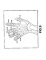
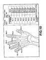
- the display means displays, for instance, the region of interest set by the interesting region setting means together with the bone image, the value of the index calculated by the index calculating means near the corresponding joint in the bone image or in a region separate from the region where the bone image is displayed, or a comment according to the value of the index.
- the system in accordance with the second aspect of the present invention may further comprise a hard copy output means which outputs an image as a hard copy recorded on a recording medium such as paper or film.
- the hard copy output means may output the image displayed by the display means, the images and the indexes one by one, or the image and the index together.
- evaluation means evaluation in a clinical sense. For example, progress of rheumatism may be evaluated by the level.
- a system for evaluating a joint comprising
- the "predetermined part” may be, for instance, the left or right hand of the patient or the left or right foot of the patient and "to evaluate the degree of disease of the predetermined part” means, for instance, “to evaluate the degree of progress of rheumatism of the left hand of the patient by the level on the basis of the indexes for the finger joints of the left hand of the patient”.
- the evaluation means either may evaluate the degree of disease of the predetermined part directly on the basis of the indexes calculated for the respective joints or may once evaluate the degree of disease of the respective joints on the basis of the indexes calculated for the respective joints and evaluate the degree of disease of the predetermined part on the basis of the result of evaluation on the respective joints.
- a system for evaluating a joint comprising
- the “different parts of a patient” may be one and the other of parts, e.g., the left and right hands, or the left and right feet, as well as elbow and knee or hands and feet.
- To "evaluate the degree of disease of the patient” means, for instance, “to evaluate the degree of entire progress of rheumatism of the patient by the level on the basis of the indexes for the finger joints of the left and right hands and left and right feet of the patient".
- a system for outputting change with time of a joint in accordance with a seventh aspect of the present invention comprises
- the output means may output the change with time of the index in various forms.
- the output means may output the joint indexes obtained from images taken at different times, as they are, the difference between two of the indexes or the degree of improvement or deterioration in disease of the bone joint calculated on the basis of the difference between two of the indexes.
- the output means may comprise a display means which displays an image on its screen and a hard copy output means which outputs an image as a hard copy recorded on a recording medium such as paper or film.
- the display means may display, for instance, the images of the bones taken at different times together with the indexes calculated on the basis of the images, the change with time of the index, or a comment according to the value of the index.
- the hard copy output means may output the image displayed by the display means, the images and the indexes one by one, or the image and the index together.
- a computer program for causing a computer to perform the method of the present invention may be installed in a computer. Otherwise such a computer program may be recorded in a computer readable medium so that the computer can perform the method when loaded with the recording medium.
- the computer readable medium is not limited to any specific type of storage devices and includes any kind of device, including but not limited to CDs, floppy disks, RAMs, ROMs, hard disks, magnetic tapes and internet downloads, in which computer instructions can be stored and/or transmitted. Transmission of the computer code through a network or through wireless transmission means is also within the scope of this invention.
- computer code/instructions include, but are not limited to, source, object and executable code and can be in any language including higher level languages, assembly language and machine language.
- the "bone joint” as used here need not be limited to those of a human body but may be those of an animal body.
- image as used here should be broadly interpreted to include image data as well as a visible image actually displayed.
- an expression “to obtain an image” includes to merely obtain image data on the basis of which an image is displayed.
- an index representing the degree of disease of the bone joint is calculated according to a predetermined calculation on the basis of image data representing an image of the bone joint and the state of the joint can be numerically expressed. Accordingly, by the use of the calculated joint index, evaluation higher in objectivity and reproducibility as compared with the conventional technology where the degree of disease is visually evaluated can be realized.
- the system for calculating a joint index of the second aspect of the present invention further comprises a storage means which stores the index relating to information which identifies the joint from which the index is calculated, the index or indexes calculated in the past can be obtained on the basis of the information which identifies the joint from which the index is calculated, and accordingly, it is not necessary to calculate again an index which has been once calculated.
- an index representing the degree of disease of the bone joint is calculated according to a predetermined calculation on the basis of image data representing an image of the bone joint and the degree of disease of the joint is evaluated on the basis of the calculated index. Accordingly, evaluation of the bone joint can be effected in a manner which substantially corresponds to clinical evaluation generally carried out by readers and is more understandable, as well as higher in objectivity and reproducibility.
- joint indexes representing the degrees of disease of the joints in a predetermined part are calculated, and the degree of disease of the predetermined part is evaluated on the basis of the indexes calculated for the respective joints. Accordingly, not only each of bone joints but also the part formed by the bone joints can be evaluated and the demand of readers, e.g., doctors, for information on generic evaluation of joints by part can be met.
- joint indexes representing the degrees of disease of the joints in parts of a patient are calculated, and the degree of disease of the patient is evaluated on the basis of the indexes calculated for the respective joints. Accordingly, not only each of bone joints of the patient but also the patient having the bone joints can be evaluated and the demand of readers, e.g., doctors, for information on generic evaluation of joints by patient can be met.
- the joint indexes of different times which have been calculated and stored are searched for on the basis of the information which identifies the joint from which the index is calculated and the change with time of the index is output on the basis of the plurality of indexes obtained. Accordingly, the degree of improvement or deterioration in disease of the bone joint can be grasped and the degree of wider disease of joint can be evaluated.
- a bone evaluation system in accordance with an embodiment of the present invention comprises an image data obtaining means 10 which obtains image data P representing a bone image including a joint, an interesting region setting means 20 which sets a region of interest R including the joint on the image represented by the obtained bone image data P, an index calculating means 30 which calculates at least one index S representing the degree of disease of the joint in the region of interest R on the basis of the part of the bone image data P corresponding to the region of interest R, an evaluation means 40 which evaluates the degree of disease of the joint on the basis of the calculated index, an output means 50 which outputs a medical image represented by the obtained image data P, the set region of interest R, the calculated index S and the evaluated degree H of disease of the joint, a storage means 60 which stores the obtained image data P together with information on the patient having the joint represented by the image data P (e.g., an ID number), information on the image (e.g., the object part, the date of taking the image P and the file number) and information on the joint (e.g.,
- the out means 50 further comprises an output control means 51 which controls output of the medical image represented by the bone image data P, the set region of interest R, the calculated index S and the evaluated degree of disease H, a display means 52 such as a CRT or a liquid crystal display which displays these pieces of output information, and a hard copy output means 53 such as a printer which outputs an image as a hard copy recorded on a recording medium such as paper or film.
- an output control means 51 which controls output of the medical image represented by the bone image data P, the set region of interest R, the calculated index S and the evaluated degree of disease H
- a display means 52 such as a CRT or a liquid crystal display which displays these pieces of output information
- a hard copy output means 53 such as a printer which outputs an image as a hard copy recorded on a recording medium such as paper or film.
- index means a physical value which represents the state of a given joint calculated on the basis of the part of the bone image data corresponding to the region of interest according to a predetermined method of calculation and on the basis of which the degree of disease of the joint is calculated.
- evaluation means evaluation in a clinical sense.
- rheumatism of a finger joint is the suspected disease and a radiation image of a hand including a finger joint is input as a bone image data .
- a radiation image of a hand including a finger joint is input as a bone image data .
- an image of a hand including the finger joint obtained by CT or MRI may be used in place of the radiation image.
- the image obtaining means 19 obtains bone image data P representing a radiation image of a left hand including a finger joint such as shown in Figure 2A from, for instance, an image read-out system (not shown) or the storage means 60.
- the image data P can be attached with information on the patient having the joint represented by the image data P (e.g., an ID number), information on the image (e.g., the object part, the date of taking the image P and the file number) and information on the joint(e.g., the size and/or the position of the set region of interest, the calculated index and the evaluated degree of disease of the joint) and the image obtaining means 10 may search for an image on the basis of these pieces of information.
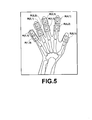
- the interesting region setting means sets a region of interest R(i,j) on the image represented by the obtained bone image data P, i standing for the kind of the finger, e.g., 1 representing a little finger, 2 representing a ring finger, 3 representing a middle finger, 4 representing an index finger and 5 representing a thumb, and j standing for the kind of the joint, e.g., 1 representing a first joint and 2 representing a second joint.
- the region of interest R may be manually set, for instance, by designating a rectangular frame by the input means 90 or automatically set by carrying out an image analysis on the obtained image data P.
- the region of interest R can be automatically set, for instance, in the following manner.
- FIGS 2A to 2E and Figure 3 are views for illustrating the procedure of extracting the finger image data from the radiation image data P.
- First the radiation image data P representing an image of a hand as shown in Figure 2A is binary-coded to divide the radiation image P into a hand region and a background region, thereby obtaining a hand region image data P1 representing the hand region as shown in Figure 2B.
- a processing for reducing the hand region (to obtain image data P2 representing a reduced hand region as shown in Figure 2C) is carried out on the hand region image data P1 until the finger region disappears, thereby obtaining finger-less image data P3 representing an image without finger as shown in Figure 2D.
- the finger-less image P3 is enlarged to such an extent that the back of the hand of the image represented by the image data P3 is equivalent to that represented by the image data P1 in size as shown in Figure 2E, thereby obtaining an enlarged finger-less image data P4.
- finger region image data F representing the fingers is obtained by subtracting image data P4 from the image data P1 (subtracting from a value of each pixel of the image data P1 a value of the corresponding pixel of the image data P4) as shown in Figure 3.
- the hand region is reduced by morphology processing.
- the morphology processing includes dilation for searching for a maximum pixel value in a range of a predetermined width about a pixel of interest in the image and erosion for searching for a minimum pixel value in a range of a predetermined width about a pixel of interest in the image.
- edges of regions having a high brightness is deleted and the image is reduced when the erosion is carried out on the image data.
- edges of regions having a high brightness is deleted and the image is reduced when the dilation is carried out on the image data.
- the hand region image data P1 is assumed to be expressed by a high-brightness, high-signal-level image signal.
- the edges of the hand region is eroded and the profile of the hand region is reduced.
- the morphology processing is discussed in detail, for instance, in Japanese Unexamined Patent Publication No. 2002-109510.
- the extracted finger region image data F representing the fingers is further divided into parts representing each of the fingers, the thumb, the index finger, the middle finger, the ring finger and the little finger on the basis of the image information, e.g., relative positions, the size and/or the length, and the images of the respective fingers are labeled as shown in Figure 4A, where labels F1 to F5 respectively indicate the thumb to the little finger.
- densities of pixels arranged in perpendicular to the length of the finger are averaged, and the averages are plotted in the direction of the length of the finger, thereby obtaining an average density curve.
- a radiation image since bones generally absorb radiation, pixels in the bone region is low in density, and as the finger becomes thicker, the average density becomes smaller.
- FIG. 4B shows the image F4 of the index finger and the average density curve J of the image F4 .
- J1 and J2 indicate peaks of the average density curve J.
- the region of interest R can be automatically set in the following manner.
- past image data representing an image of the same object joint which has been taken in the past (will be referred to as "a past image", hereinbelow) and information attached to the past image data.
- the past image data is obtained by searching, for instance, the storage means 60, and the images represented by the object image data and the past image data are superposed one on the other. Coordinates on the object image corresponding to the regions of interest on the past image are obtained, and regions of interest can be set on the object image on the basis of the coordinates.
- regions of interest When regions of interest are once set on an image, it is not necessary to set again regions of interest on images which are subsequently taken. This procedure is better in reproducibility of the positions of the regions of interest.
- regions of interest can be set by superposing the object image on an image having a standard shape common to the object so long as the shape of the object (hand in this particular embodiment) is substantially constant.
- Figure 5 shows the object image P on which the regions of interest R have been set and labeled.
- the index calculating means 30 calculates indexes S(i, j, k, m) representing the degree of progress of rheumatism in the regions of interest R(i, j) on the basis of the part of the bone image data corresponding to the regions of interest, k representing a number corresponding to the kind of the index, and m representing a number for distinguishing the part for which the index is to be calculated in the case where there are plurality of parts for which the index is to be calculated.
- the average density (the average radiation absorption) S(i, j, 1) of the bone region representing the thickness of the bone; the number of the recessed portions S(i, j, 2) and the size of each recessed portion S(i, j, 3, m) which represent the degree of generation of recessed portion in the bone; and the gap between opposed edges of the bones forming the joint S(i, j, 4) are calculated as the indexes.
- the index S(i, j, 1) representing the thickness of the bone is the average density of pixels arranged in perpendicular to the length of the finger across the joint described above in conjunction with detection of the joint. Since the bone region absorbs more radiation and is expressed as a low density portion in a radiation image, the average density of pixels arranged in perpendicular to the length of the finger across the joint may be regarded as an index quantifying the thickness of the joint relatively to the thickness of the finger.
- the number of the recessed portions S(i, j, 2) and the size of each recessed portion S(i, j, 3, m) are valued by measuring the number of the recessed portions and the size (depth) of each recessed portion on the basis of difference information representing the difference from an assumed normal profile of the joint extracted from the image of the joint on the head of the bone forming the joint.
- Figure 6A shows a joint formed by a bone A and a bone B having recessed portions Y1 and Y2.
- the profile of the bone B is first approximated by a circle or an ellipse and determines a center O.
- the profile of the bone B is expressed on a polar co-ordinate system having its axis on the center O, thereby obtaining a curve f shown in Figure 6B.
- ⁇ represents an angle and r represents a radius.
- multidimensional polynomial approximation is carried out on the curve f to obtain a curve g shown in Figure 6C and the curve g is considered to be a normal profile.
- a difference curve f-g representing the difference between the curves f and g.
- the gap between opposed edges of the bones forming the joint S(i, j, 4) represents the gap d between the opposed edges of the bone A and the bone B.
- the evaluation means 40 evaluates the degree of progress of rheumatism of the joint on the basis of the calculated indexes by the clinical grades .
- the following grades have been employed wide.
- Figures 8A to 8F respectively show the following 6 grades.
- the above evaluation standard includes abstract expressions, it is necessary to arrange the evaluation standard so that the degree of progress of rheumatism of the joint can be quantitatively determined on the basis of the calculated indexes.
- points are added up according to the values of the index on the basis of the standard shown in the following table 1, and the degree of progress of rheumatism of the joint is judged in the following manner on the basis of the total point.
- TH31 represents a value corresponding to 1mm
- TH 32 represents a predetermined value over which the dissolution is determined to be large
- TH41 represents a gap smaller than which the gap is determined to be narrow.
- S(i, j , 3, m) max represents a maximum value of the depth in the depths of a plurality of recessed portions in the joint.
- the index S(i, j, 1) representing the thickness of the joint is not used here.
- the output control means 51 causes the display means 52 to display the indexes and the evaluation for each joint together with the image thereof and causes the storage means 60 to store the same information and/or causes the hard copy output means 53 to output the same information as a hard copy under the control of instruction input through the input means 90.
- the output control means 51 can cause the display means (e.g., a CRT or a liquid crystal display) 52 to display information in various patterns.
- the output control means 51 may cause the display means 52 to display an index near to the image of each joint on the hand region image P as shown in Figure 9, or may cause the display means 52 to display an index and/or other information in an area separate from the area in which the hand region image P is displayed as shown in Figure 10.
- the output control means 51 causes the storage means 60 to store data on each joint such as the obtained image data P, information on the patient having the joint represented by the image data P (e.g., an ID number), information on the image (e.g., the object part, the date of taking the image P and/or the file number) and information on the joint (e.g., the size and/or the position of the set region of interest, the calculated index and/or the evaluated degree of disease of the joint) .
- data on each joint such as the obtained image data P, information on the patient having the joint represented by the image data P (e.g., an ID number), information on the image (e.g., the object part, the date of taking the image P and/or the file number) and information on the joint (e.g., the size and/or the position of the set region of interest, the calculated index and/or the evaluated degree of disease of the joint) .
- the image obtaining means 10 may search the storage means 60 for an image which is taken from the same object part at a different time and the information attached to the image on the basis of the information on the patient, and the output control means 51 may cause the display means 52 to display the images taken at different times or to display in a time series indexes calculated at different times for each joint or to display the difference between the images or the indexes so that the change with time of the state of the joint can be known from the image on the screen.
- the display means 52 may display indexes taken at different times near to the image of each joint and information on the date of taking each index relating to the index.
- the date of taking the index can be related to the index, for instance, by attaching the same numeral or sign or by displaying in the same color.
- the output control means 51 further causes the hard copy output means 53 to output the same information as a hard copy.
- the degree of progress of rheumatism of each joint is evaluated in a plurality of grades
- the degree of progress of rheumatism of a part may be evaluated in a plurality of grades on the basis of the indexes calculated for the joints in the part (e.g., the left hand) .
- the image obtaining means obtains bone image data representing a bone image including a plurality of joints in a predetermined part
- the interesting region setting means sets a plurality of regions of interest each including a desired joint on the image represented by the obtained bone image data
- the index calculating means calculates at least one joint index representing the degree of disease of the joint in each region of interest on the basis of the part of the bone image data corresponding to the region of interest
- the evaluation means evaluates the degree of disease of the predetermined part on the basis of the indexes calculated for the respective joints.
- the degree of progress of rheumatism of a patient may be evaluated in a plurality of grades on the basis of the indexes calculated for the joints in different parts of the patient (e.g., the left and right hands of the patient) .
- the image obtaining means obtains a plurality of pieces of bone image data representing a plurality of images of bones including a joint in different parts of a patient
- the interesting region setting means sets at least one region of interest including a desired joint on each of the images represented by the plurality of pieces of bone image data
- the index calculating means calculates at least one joint index representing the degree of disease of the joint in each region of interest on the basis of the part of the bone image data corresponding to the region of interest
- the evaluation means evaluates the degree of disease of the patient on the basis of the indexes calculated for the respective joints.
Landscapes
- Engineering & Computer Science (AREA)
- Quality & Reliability (AREA)
- General Health & Medical Sciences (AREA)
- Medical Informatics (AREA)
- Nuclear Medicine, Radiotherapy & Molecular Imaging (AREA)
- Radiology & Medical Imaging (AREA)
- Health & Medical Sciences (AREA)
- Computer Vision & Pattern Recognition (AREA)
- Physics & Mathematics (AREA)
- General Physics & Mathematics (AREA)
- Theoretical Computer Science (AREA)
- Apparatus For Radiation Diagnosis (AREA)
- Measuring And Recording Apparatus For Diagnosis (AREA)
Abstract
Description
| point | index | ||
| number of recesses S(i, j, 2) | depth of recess S(i, j, 3, m) | gap S(i, j, 4) | |
| 0 | S ( i , j , , 2 ) = 0 | S(i, j, 3, m)max<TH31 | TH42≦S(i, j, 4) |
| 1 | 1≦ S ( i , j , 2) ≦ 2 | TH31≦S(i, j, 3, m)max<TH32 | TH41≦S(i, j, 4)< |
| 2 | 3 ≦ S (i, j , 2 ) | TH32≦S(i, j, 3, m)max | S(i, j, 4)<TH41 |
Claims (16)
- A method of calculating a joint index comprising the steps ofobtaining bone image data representing a bone image including a desired joint,setting a region of interest including the desired joint on the image represented by the obtained bone image data, andcalculating a joint index representing the degree of disease of the joint in the region of interest on the basis of the part of the bone image data corresponding to the region of interest.
- A method of outputting change with time of a joint comprising the steps of ,calculating a plurality of joint indexes on a bone joint by a method of calculating a joint index as defined in Claim 1 on the basis of a plurality of pieces of image data representing the image of the bone joint taken at different times,storing the indexes relating to information which identifies the joint from which the indexes are calculated,searching for the joint indexes on the basis of the information, andoutputting the change with time of the index on the basis of the plurality of indexes obtained.
- A system for calculating a joint index comprisingan image obtaining means which obtains bone image data representing a bone image including a desired joint,an interesting region setting means which sets a region of interest including the desired joint on the image represented by the obtained bone image data, andan index calculating means which calculates a joint index representing the degree of disease of the joint in the region of interest on the basis of the part of the bone image data corresponding to the region of interest.
- A system as defined in Claim 3 in which joint index represents the thickness of bones forming the joint.
- A system as defined in Claim 3 in which joint index represents the recess in the bones forming the joint.
- A system as defined in any one of Claims 3 to 5 further comprising a storage means which stores the index relating to information which identifies the joint from which the index is calculated.
- A system for evaluating a joint comprisingan image obtaining means which obtains bone image data representing a bone image including a desired joint,an interesting region setting means which sets a region of interest including the desired joint on the image represented by the obtained bone image data,an index calculating means which calculates at least one joint index representing the degree of disease of the joint in the region of interest on the basis of the part of the bone image data corresponding to the region of interest, andan evaluation means which evaluates the degree of disease of the joint in the region of interest on the basis of the index calculated by the index calculating means.
- A system for evaluating a joint comprisingan image obtaining means which obtains bone image data representing a bone image including a plurality of joints in a predetermined part,an interesting region setting means which sets a plurality of regions of interest each including a desired joint on the image represented by the obtained bone image data,an index calculating means which calculates at least one joint index representing the degree of disease of the joint in each region of interest on the basis of the part of the bone image data corresponding to the region of interest, andan evaluation means which evaluates the degree of disease of the predetermined part on the basis of the indexes calculated for the respective joints by the index calculating means.
- A system for evaluating a joint comprisingan image obtaining means which obtains a plurality of pieces of bone image data representing a plurality of images of bones including a joint in different parts of a patient,an interesting region setting means which sets at least one region of interest including a desired joint on each of the images represented by the plurality of pieces of bone image data,an index calculating means which calculates at least one joint index representing the degree of disease of the joint in each region of interest on the basis of the part of the bone image data corresponding to the region of interest, andan evaluation means which evaluates the degree of disease of the patient on the basis of the indexes calculated for the respective joints by the index calculating means.
- A system for outputting change with time of a joint comprisinga storage means which stores a plurality of joint indexes calculated on a bone joint by a method of calculating a joint index of the first aspect of the present invention on the basis of a plurality of pieces of image data representing the image of the bone joint taken at different times relating to information which identifies the joint from which the indexes are calculated,an index obtaining means which searches for the joint indexes on the basis of the information, andan output means which outputs the change with time of the index on the basis of the plurality of indexes obtained.
- A computer program for causing a computer to perform a procedure comprising the steps ofobtaining bone image data representing a bone image including a desired joint,setting a region of interest including the desired joint on the image represented by the obtained bone image data, andcalculating a joint index representing the degree of disease of the joint in the region of interest on the basis of the part of the bone image data corresponding to the region of interest.
- A computer program for causing a computer to perform a procedure comprising the stepsobtaining means bone image data representing a bone image including a desired joint,setting a region of interest including the desired joint on the image represented by the obtained bone image data,calculating at least one joint index representing the degree of disease of the joint in the region of interest on the basis of the part of the bone image data corresponding to the region of interest, andevaluating the degree of disease of the joint in the region of interest on the basis of the index calculated by the index calculating means.
- A computer program for causing a computer to perform a procedure comprising the steps ofobtaining bone image data representing a bone image including a plurality of joints in a predetermined part,setting means a plurality of regions of interest each including a desired joint on the image represented by the obtained bone image data,calculating at least one joint index representing the degree of disease of the joint in each region of interest on the basis of the part of the bone image data corresponding to the region of interest, andevaluating the degree of disease of the predetermined part on the basis of the indexes calculated for the respective joints by the index calculating means.
- A computer program for causing a computer to perform a procedure comprising the steps ofobtaining means a plurality of pieces of bone image data representing a plurality of images of bones including a joint in different parts of a patient,setting at least one region of interest including a desired joint on each of the images represented by the plurality of pieces of bone image data,calculating at least one joint index representing the degree of disease of the joint in each region of interest on the basis of the part of the bone image data corresponding to the region of interest, andevaluating the degree of disease of the patient on the basis of the indexes calculated for the respective joints by the index calculating means.
- A computer program for causing a computer to perform a procedure comprising the steps ofcalculating a plurality of joint indexes on a bone joint by a method of calculating a joint index as defined in Claim 1 on the basis of a plurality of pieces of image data representing the image of the bone joint taken at different times,storing the indexes relating to information which identifies the joint from which the indexes are calculated,searching for the joint indexes on the basis of the information, andoutputting the change with time of the index on the basis of the plurality of indexes obtained.
- A computer-readable medium on which a computer program as defined in any one of Claims 11 to 15 is recorded.
Applications Claiming Priority (4)
| Application Number | Priority Date | Filing Date | Title |
|---|---|---|---|
| JP2002164691 | 2002-06-05 | ||
| JP2002164691 | 2002-06-05 | ||
| JP2003080638A JP2004057804A (en) | 2002-06-05 | 2003-03-24 | Method and apparatus for evaluating joint and program therefor |
| JP2003080638 | 2003-03-24 |
Publications (2)
| Publication Number | Publication Date |
|---|---|
| EP1369809A2 true EP1369809A2 (en) | 2003-12-10 |
| EP1369809A3 EP1369809A3 (en) | 2008-12-10 |
Family
ID=29552391
Family Applications (1)
| Application Number | Title | Priority Date | Filing Date |
|---|---|---|---|
| EP03012616A Withdrawn EP1369809A3 (en) | 2002-06-05 | 2003-06-03 | Method of and system for evaluating joint and computer program for carrying out the method |
Country Status (3)
| Country | Link |
|---|---|
| US (1) | US20030229299A1 (en) |
| EP (1) | EP1369809A3 (en) |
| JP (1) | JP2004057804A (en) |
Cited By (1)
| Publication number | Priority date | Publication date | Assignee | Title |
|---|---|---|---|---|
| WO2010064200A1 (en) * | 2008-12-05 | 2010-06-10 | Koninklijke Philips Electronics N.V. | Method and device for optically examining the condition of joints |
Families Citing this family (30)
| Publication number | Priority date | Publication date | Assignee | Title |
|---|---|---|---|---|
| WO2003009762A1 (en) * | 2001-07-24 | 2003-02-06 | Sunlight Medical, Ltd. | Joint analysis using ultrasound |
| JP2004535882A (en) * | 2001-07-24 | 2004-12-02 | サンライト メディカル リミテッド | Bone age evaluation method using ultrasound |
| US7849063B2 (en) * | 2003-10-17 | 2010-12-07 | Yahoo! Inc. | Systems and methods for indexing content for fast and scalable retrieval |
| US20050144241A1 (en) * | 2003-10-17 | 2005-06-30 | Stata Raymond P. | Systems and methods for a search-based email client |
| US7620624B2 (en) * | 2003-10-17 | 2009-11-17 | Yahoo! Inc. | Systems and methods for indexing content for fast and scalable retrieval |
| JP5194420B2 (en) * | 2006-06-23 | 2013-05-08 | コニカミノルタエムジー株式会社 | Radiation imaging equipment |
| DE102006042157B4 (en) * | 2006-09-06 | 2013-03-21 | Leica Microsystems Cms Gmbh | Method and microscope system for scanning a sample |
| JP4934786B2 (en) * | 2006-10-13 | 2012-05-16 | 国立大学法人 東京大学 | Knee joint diagnosis support method, apparatus and program |
| WO2008090711A1 (en) * | 2007-01-26 | 2008-07-31 | Konica Minolta Medical & Graphic, Inc. | Bone joint evaluation system |
| JP2008200114A (en) * | 2007-02-16 | 2008-09-04 | Konica Minolta Medical & Graphic Inc | Bone trabecula evaluation system |
| JP4992456B2 (en) * | 2007-02-16 | 2012-08-08 | コニカミノルタエムジー株式会社 | Bone disease evaluation system |
| JP2010158258A (en) * | 2007-04-11 | 2010-07-22 | Konica Minolta Medical & Graphic Inc | Soft tissue evaluation system |
| JPWO2008132988A1 (en) * | 2007-04-12 | 2010-07-22 | コニカミノルタエムジー株式会社 | X-ray image analysis system and program |
| US20100128957A1 (en) * | 2007-04-12 | 2010-05-27 | Konica Minolta Medical & Graphic, Inc. | Bone disease evaluating system |
| WO2008129940A1 (en) * | 2007-04-12 | 2008-10-30 | Konica Minolta Medical & Graphic, Inc. | Bone disease evaluation system |
| EP2291112B1 (en) * | 2008-05-26 | 2017-11-08 | Hemics B.V. | Optical detection method and device for optical detection of the condition of joints |
| EP2378953B1 (en) * | 2008-12-05 | 2017-08-16 | Hemics B.V. | Device for optical detection of the condition of joints |
| US9064320B2 (en) | 2009-09-11 | 2015-06-23 | Straxcorp Pty Ltd | Method and system for image analysis of selected tissue structures |
| JP5730228B2 (en) * | 2012-03-07 | 2015-06-03 | 日本メジフィジックス株式会社 | Processing and display of nuclear medicine images |
| JP2015061592A (en) * | 2013-08-21 | 2015-04-02 | コニカミノルタ株式会社 | Ultrasonic diagnostic equipment, ultrasonic image processing method, and computer-readable non-temporary recording medium |
| US9436872B2 (en) * | 2014-02-24 | 2016-09-06 | Hong Kong Applied Science and Technology Research Institute Company Limited | System and method for detecting and tracking multiple parts of an object |
| JP6264949B2 (en) * | 2014-03-05 | 2018-01-24 | 富士ゼロックス株式会社 | Image processing apparatus and program |
| JP6301164B2 (en) * | 2014-03-14 | 2018-03-28 | キヤノンメディカルシステムズ株式会社 | Medical image processing apparatus and medical image processing method |
| JP6507581B2 (en) * | 2014-11-11 | 2019-05-08 | コニカミノルタ株式会社 | Chest dynamic imaging support system |
| JP6858047B2 (en) * | 2017-03-27 | 2021-04-14 | 国立大学法人 東京大学 | Diagnostic image processing device, evaluation support method and program |
| JP2020151270A (en) * | 2019-03-20 | 2020-09-24 | 学校法人慶應義塾 | Joint state value acquisition device, joint state learning device, joint position identification device, joint position learning device, joint state value acquisition method, joint state learning method, joint position identification method, joint position learning method and program |
| KR102444274B1 (en) * | 2019-07-17 | 2022-09-16 | 주식회사 크레스콤 | Apparatus and method for precise analysis of arthritis severity |
| JP7527773B2 (en) * | 2019-11-18 | 2024-08-05 | キヤノン株式会社 | Radiography system, radiation photography method, image processing device, and program |
| KR102495365B1 (en) * | 2020-11-11 | 2023-02-06 | 순천향대학교 산학협력단 | System for analysising body joint score of bone scan image using artificial intelligence and method thereof |
| KR102842473B1 (en) | 2021-08-11 | 2025-08-05 | 주식회사 크레스콤 | Method for, device for, and system analazying an medical image for quantifying joint condition |
Family Cites Families (11)
| Publication number | Priority date | Publication date | Assignee | Title |
|---|---|---|---|---|
| US5099859A (en) * | 1988-12-06 | 1992-03-31 | Bell Gene D | Method and apparatus for comparative analysis of videofluoroscopic joint motion |
| EP0411155B1 (en) * | 1989-02-23 | 1997-05-02 | Teijin Limited | Method of measuring bone shape, apparatus therefor and system for evaluating bone |
| US5800363A (en) * | 1996-03-27 | 1998-09-01 | Cheng; Shu Lin | Method for diagnosing and monitoring osteoporosis |
| US5696805A (en) * | 1996-09-17 | 1997-12-09 | Eastman Kodak Company | Apparatus and method for identifying specific bone regions in digital X-ray images |
| US6495579B1 (en) * | 1996-12-02 | 2002-12-17 | Angiotech Pharmaceuticals, Inc. | Method for treating multiple sclerosis |
| US5885231A (en) * | 1997-01-07 | 1999-03-23 | The General Hospital Corporation | Digital motor event recording system |
| GB9702202D0 (en) * | 1997-02-04 | 1997-03-26 | Osteometer Meditech As | Diagnosis of arthritic conditions |
| US5891060A (en) * | 1997-10-13 | 1999-04-06 | Kinex Iha Corp. | Method for evaluating a human joint |
| US6161080A (en) * | 1997-11-17 | 2000-12-12 | The Trustees Of Columbia University In The City Of New York | Three dimensional multibody modeling of anatomical joints |
| US6560477B1 (en) * | 2000-03-17 | 2003-05-06 | The Regents Of The University Of California | Joint imaging system utilizing magnetic resonance imaging and associated methods |
| EP1293925A1 (en) * | 2001-09-18 | 2003-03-19 | Agfa-Gevaert | Radiographic scoring method |
-
2003
- 2003-03-24 JP JP2003080638A patent/JP2004057804A/en active Pending
- 2003-06-03 EP EP03012616A patent/EP1369809A3/en not_active Withdrawn
- 2003-06-05 US US10/454,572 patent/US20030229299A1/en not_active Abandoned
Cited By (1)
| Publication number | Priority date | Publication date | Assignee | Title |
|---|---|---|---|---|
| WO2010064200A1 (en) * | 2008-12-05 | 2010-06-10 | Koninklijke Philips Electronics N.V. | Method and device for optically examining the condition of joints |
Also Published As
| Publication number | Publication date |
|---|---|
| US20030229299A1 (en) | 2003-12-11 |
| EP1369809A3 (en) | 2008-12-10 |
| JP2004057804A (en) | 2004-02-26 |
Similar Documents
| Publication | Publication Date | Title |
|---|---|---|
| EP1369809A2 (en) | Method of and system for evaluating joint and computer program for carrying out the method | |
| US11937888B2 (en) | Artificial intelligence intra-operative surgical guidance system | |
| US20240096508A1 (en) | Systems and methods for using generic anatomy models in surgical planning | |
| US11540794B2 (en) | Artificial intelligence intra-operative surgical guidance system and method of use | |
| US12475993B2 (en) | Apparatus and method for medical image reading assistant providing representative image based on medical use artificial neural network | |
| US7065235B2 (en) | Radiographic scoring method | |
| US8696603B2 (en) | System for measuring space width of joint, method for measuring space width of joint and recording medium | |
| US8126231B2 (en) | Medical image segmentation apparatus and medical image segmentation program | |
| JP2008259622A (en) | Report creation support apparatus and program thereof | |
| JP2009082441A (en) | Medical diagnosis support system | |
| Kim et al. | Automatic spine segmentation and parameter measurement for radiological analysis of whole-spine lateral radiographs using deep learning and computer vision | |
| Jeong et al. | Development of AI-based diagnostic algorithm for nasal bone fracture using deep learning | |
| Suojärvi et al. | Radiographic measurements of the normal distal radius: reliability of computer-aided CT versus physicians’ radiograph interpretation | |
| CN114596251B (en) | A knee joint X-ray image quality control method and device | |
| US20090129654A1 (en) | Image analysis of tube tip positioning | |
| Lezak et al. | Diagnostic performance of deep learning for leg length measurements on radiographs in leg length discrepancy: a systematic review | |
| Przystalski et al. | Automated correction angle calculation in high tibial osteotomy planning | |
| Duryea et al. | Automated algorithm for the identification of joint space and phalanx margin locations on digitized hand radiographs | |
| Schultheis et al. | Using deep learning segmentation for endotracheal tube position assessment | |
| Casciaro et al. | Towards automatic measurement of anteversion and neck–shaft angles in human femurs using CT images | |
| JP2022184272A (en) | Information processor, information processing method and program | |
| CN116158774B (en) | Methods, apparatus, and computer storage media for measuring tibial plateau caster angle | |
| CN117079806A (en) | Intelligent auxiliary diagnosis and treatment method and system for fracture between trochanters of femur in self-adaptive mode | |
| Zhong et al. | Automatic landmark detection and angle measurement in radiographs based on deep learning in application to coronal plane alignment of the knee classification | |
| Kauppi et al. | A framework for constructing benchmark databases and protocols for retinopathy in medical image analysis |
Legal Events
| Date | Code | Title | Description |
|---|---|---|---|
| PUAI | Public reference made under article 153(3) epc to a published international application that has entered the european phase |
Free format text: ORIGINAL CODE: 0009012 |
|
| AK | Designated contracting states |
Kind code of ref document: A2 Designated state(s): AT BE BG CH CY CZ DE DK EE ES FI FR GB GR HU IE IT LI LU MC NL PT RO SE SI SK TR |
|
| AX | Request for extension of the european patent |
Extension state: AL LT LV MK |
|
| RAP1 | Party data changed (applicant data changed or rights of an application transferred) |
Owner name: FUJIFILM CORPORATION |
|
| PUAL | Search report despatched |
Free format text: ORIGINAL CODE: 0009013 |
|
| AK | Designated contracting states |
Kind code of ref document: A3 Designated state(s): AT BE BG CH CY CZ DE DK EE ES FI FR GB GR HU IE IT LI LU MC NL PT RO SE SI SK TR |
|
| AX | Request for extension of the european patent |
Extension state: AL LT LV MK |
|
| RIC1 | Information provided on ipc code assigned before grant |
Ipc: G06T 7/00 20060101ALI20081103BHEP Ipc: G06F 19/00 20060101AFI20030923BHEP |
|
| STAA | Information on the status of an ep patent application or granted ep patent |
Free format text: STATUS: THE APPLICATION HAS BEEN WITHDRAWN |
|
| 17P | Request for examination filed |
Effective date: 20090126 |
|
| 18W | Application withdrawn |
Effective date: 20090210 |