The present invention concerns the in vitro
localization of HER2 receptor in breast cancer cells.
The HER2 receptor, also known as c-erb-B2, is a
member of the epidermal growth factor receptor family.
HER2 is a trans-membrane receptor, with an intra-cytoplasmic
(internal domain) and an extra-cytoplasmic
domain (external domain).
The interest for Her2 is due to the fact that Her2
is present at low level in normal epithelial cells,
whereas it is over-expressed in cells of some
carcinomas (approximately from 15 to 35% of invasive
breast carcinomas over-express HER2 receptor protein).
The over-expression of HER2, which induces the
appearance of high levels of the antigen over the cell
membrane, is the consequence of the amplification of
the corresponding gene, being such an amplification
detectatble by Southern blot and in situ hybridization
(FISH and CISH) techniques. FISH (Fluorescence In Situ
Hybridization) is an in situ hybridization technique
(ISH), based on the use of probes linked to
flourochromes. CISH (Chromogenic In Situ Hybridization)
is instead based on the use of probes linked either to
digoxigenin or to biotin and the reaction is revealed
by enzymatic colorimetric methods that have
diaminobenzidine as a substrate. Thus, the
amplification is demonstrated by the presence of
intranuclear precipitates seen by the optical
microscope. HER2 gene amplification has been widely
studied using both techniques (Tanner et al Am, J
Pathol 2000;157:1467-72). In breast carcinomas, HER2
gene amplification has been studied in order to confirm
the specificity of immunocytohemical reactions that
demonstrate the over-expression of the receptor protein
(Ridolfi et al Mod Pathol 2000;13: 866-73).
In addition to breast carcinomas HER2 expression
has been studied in tumor of different origin, for
example originating from the respiratory tract (Bunn et
al Clin Cancer Res 2001; 7:3239-50, Hirashima N et al
Mod Pathol 2001; 14: 556-62, Kazkayasi M et al.Eur Arch
Otorhinolaryngol 2001; 258:329-35), from the gastrointestinal
tract(Ross JS et al Cancer Invest.
2001;19:554-68, Kono K et al Int J Cancer 2002; 98:216-20)
and of renal origin (Stumm G et al. Int J Cancer
1996; 69:17-22).
The interest on the localization of HER2 is linked
to the recent introduction in the therapy of tumors
expressing the receptor protein of a "humanized"
antibody, called Trastuzumab or Herceptin® (Genentech,
Inc., South San Francisco, CA, License #1048).
Herceptin® (distributed in Italy by Roche S.p.A.,
Milan, national identification number A034949014/E as
vials containing 150 mg of powder (Trastuzumab) to be
dissolved and administered as endovenous infusion) is a
specific antibody directed against the extra-cytoplamic
domain of the HER2 receptor (Carter P, et al. Proc Natl
Acad Sci U S A. 1992;89:4285-9).
Herceptin® antibody is produced by DNA recombinant
genetic technology, wherein the antibody sequence of
the antigen binding site of the murine monoclonal
antibody 4D5 developed by Genentech Inc. (as described
by the US-A-5,677,171 patent) direct against the
extra-cellular domain of HER2, is linked to the
constant fraction Fc of a human antibody. Thus,
Herceptin® antibody works in the same way as a human
antibody, being minimal the murine fraction.
The antibody was shown to be effective in the
treatment of advanced tumors, but only in cases with
over-expression of the HER2 antigen on the cellular
membrane, detectable by immunocytochemical techniques
(Vogel CL, et al. J Clin Oncol 2002;20:719-26).
Thus, the first priority for eligibility for a
therapeutic use of Herceptin® is the demonstration,
for example by immunocytochemistry, of the over-expression
of the membrane domain of HER2.
At present, different procedures are commercially
available for HER2 protein detection.
The USA Food and Drug Administration has validated
and approved as immunocytochemical procedure
polyclonal and monoclonal antibodies directed against
c-erbB2/HER2; among these a rabbit antiserum directed
against the aminoacidic sequence of the intra-cytoplasmic
domain of HER2 (the polyclonal antibody
commercialized as HERCEPTEST by Dako). Widely used is
a murine monoclonal antibody (CB11), directed also
against the intra-cytoplasmic sequence. Discrepancies
on the evaluation of the results on HER2 over-expression
are well demonstrated in the literature,
depending on test and on antibodies used (Tubbs RR, et
al. J Clin Oncol 2002; 19: 2714-21).
During the immunocytochemical reaction, the
primary reagent (rabbit antiserum or monoclonal
antibody), specific for the target antigen, is
followed by a biotinylated secondary antibody that
recognizes a constant portion of the primary reagent.
Following this reaction, an avidin biotinylated-peroxidase
complex links to the secondary antibody
allowing the localization of the specific antigen
through a peroxidase colorimetric reaction.
It is suggested to report the results of the
immunocytochemical reactions of the anti c-erbB2
antibodies using the FDA approved procedure (Birner P,
Clin Cancer Res 2001;7:1669-75).
This procedure takes into consideration the
membrane localization of the reaction, its intensity
(light, moderate, strong) and the percentage of
stained cells (higher or lower than 10%). The scoring
of these three parameters gives rise to the following
semi-quantitative score: 0/1+ (no over-expression), 2+
(light to moderate over-expression) and 3+ (moderate
to strong over-expression). Cut-off for the evaluation
of positive or negative reaction has been determined
on the base of clinical trials evaluating the response
to Herceptin® treatment. As a matter of fact among the
criteria for patient eligibility to Herceptin®
treatment are: (a)the immunocytochemical evidence of
the receptor protein over-expression by the evaluation
score 3+, or (b)the immunocytochemical evidence of the
receptor protein over-expression by the evaluation
score 2+ associated to the HER2 receptor gene
amplification evaluated by FISH technique (BCNA policy
statement, www.bcna.org.au/bcna_policies/herceptin).
The detection of the extra-cytoplasmic portion of
the HER2 receptor is thus essential, both in order to
obtain valid results with Herceptin® treatment and
avoid overtreatment in patients affected by breast
carcinoma.
This is stressed by the fact that extra-cellular
fragments of the receptor could be either lost or
mutated (Mendrola JM, et al. J Biol Chem.
2002;277:4704-12; Siegel PM et al.EMBO J.
1999;18:2149-64).Such receptor alterations would lead
to the non-effectiveness of the Herceptin® treatment
in patient previously selected on the basis of the
positive results with diagnostic tests that
recognized the intracytoplasmic portion of the HER2
receptor.
Herceptin® antibody as such cannot be used as
primary antibody in immunocytochemical diagnostic
tests performed on a human tumor tissue sample -
immunocytochemistry - since the secondary antibody
directed against human immunoglobulins would react
with the serum immunoglobulins present within the
tissue, which are non-specific for the
extracytoplasmic portion of HER2 receptor, and would
cause a false localization.
The present invention, in the preferred
embodiment, consists in a modification of Herceptin®
antibody, which maintains the antibody reactivity, but
allows a direct utilization of Herceptin® as a
diagnostic test, for example in immunocytochemistry.
For this purpose the humanized antibody
Herceptin® is conjugated with biotin molecules that
lind to lateral -NH2 groups of the Fc portion of the
human immunoglobulin present in Herceptin®.
The biotinylated Herceptin® (called BiotHER) can
be directly used on tissue sections, where it links to
the extra-cytoplamic portion of HER2 forming the
antigen-antibody complex. Biotin will be then revealed
owing to the specific finding of the protein with
avidin or streptavidin conjugated with a marker, such
as an enzyme (e.g. horse radish peroxidase), a
radioactive, fluorescent or chemiluminiscent
substance.
As an alternative, the humanized antibody
Herceptin®, instead of being directly marked with
biotin, can be linked in vitro with an anti-human IgG
antibody or a corresponding fragment, for example a FAB
fragment marked with biotin.
The specific receptor-ligand system constituted
by biotin-avidin or biotin-streptavidin can be
substituted by any other receptor-ligand system that
allows to detect the primary antibody by exploiting
the binding affinity that the ligand conjugated to the
antibody has for its receptor. Examples of receptor-ligand
complex are fluorescin isothiocyanate-anti-fluorescin
antibodies and digoxygenin-anti digoxygenin
antibodies.
The in vitro diagnostic test here described shows
definite advantages as compared to the methods used so
far. First of all, it recognizes the same antigenic
site (extra-cytoplasmic) that is responsible of the
therapeutic function of Herceptin® in vivo. This point
is very important because, as previously said, a
shedding of HER2 extra-cytoplasmic fragment in the
serum of patient with breast carcinoma may happen
(Molina R et al. Anticancer Res. 1999;19:2551-5), with
preservation of the intra-cytoplasmic fragment. In
these cases, immunocytochemical tests such as
HERCEPEST (Dako) that employ antibodies directed
against the HER2 intra-cytoplasmic domain will give
positive results, while the Herceptin® treatment will
be ineffective.
As a second point the procedure is much faster and
less prone to mistakes because the proposed procedure
is one step shorter, (biotinylated antibody + avidin-peroxidase)
instead of (primary antibody +
biotinylated secondary antibody + avidin-peroxidase).
The procedure is faster (about 1-2 hours less) because
the incubation of the sections with the secondary
antibody and the corresponding washing steps are
avoided; it is less expensive because one reagent is
avoided and in addition it avoids the use of
monoclonal antibodies and polyclonal sera that might
give rise to unspecific reactions. In fact, the
unmique antibody applied is the one with definite
reactivity, applied and validated for therapy.
In addition, this method gives a specific
staining of the target cells (with absolutely negative
staining of other cellular types) and exclusive
membrane reactivity. On the contrary, using other
commercially available antibodies reactivity of normal
epithelial cells and a diffuse cytoplasmic staining are
sometimes observed. This leads to some difficulties in
the interpretation of results concerning both
specificity of the reaction and the scoring of the
membrane staining, if any.
The cytoplasmic reaction gives rise to important
interpretation problems, but consensus in the
literature indicates that this localization is an
artifact (Taylor et al Oncol Res 1999; 11:311-7). Thus,
guidelines on the interpretation of HER2 staining in
immunocytochemistry suggest to consider as invalid the
reaction when the cytoplasm is stained and to disregard
it (American Pathologist Consensus Statement 1999. Arch
Pathol Lab Med 2000, 124:966)
The use of the humanized biotinylated antibody
BiotHER in immunohistochemistry gives definite
advantages as compared to the use of the 4D5 murine
monoclonal antibody. It is important to remind that the
antibody Herceptin® has been obtained - with DNA
recombinant technology - keeping the sequence of the
binding site of 4D5 murine antibody directed against
the HER2 extra-cytoplasmic domain. The comparison in a
series of mammary carcinomas stained on serial sections
with either the BiotHER procedure and with the 4D5
monoclonal antibody has demonstrated similar results.
However, the samples stained with 4D5 antibody show
often a diffuse cytoplasmic staining, that disturbs the
reading of the samples and might lead to mistakes in
the diagnosis. So far Herceptin® treatment was given
only to patients whit high immunocytochemical
expression (score 3+) of HER2. It is possible that
using for diagnostic purposes the same antibody
(biotinylated Herceptin® - BiotHER) used for treatment
(Herceptin®)even cases with score 1+ or 2+ of the
immunocytichemical reaction could be selected and
potentially benefit of Herceptin® treatment as well.
Recently, clinical trials have been performed
using humanized antibodies direct against another
member of epidermal growth factor family, specifically
against the "epidermal growth factor receptor" (EGFr)
using the humanized monoclonal antibody EMD72000, in
patients with laringeal and hypolaringeal carcinomas
(Bier H et al Cancer Chemother Pharmacol 2001; 47: 519-24).
Specific EGFr antibodies have been used for
treatment of tumor of non epithelial origin as cerebral
gliomas (Kuan CT Brain Tumor Pathol 2000; 17: 71-8).
The diagnostic application of this antibody or of
antibodies against other members of the epidermal
growth factors family using the biotinylation procedure
herein described might have an important clinical
impact, because in analogy to BiotHER application, it
might allow to specifically and directly recognize
tumors that would benefit of treatment with humanized
antibodies.
Detailed description of embodiments of the present
invention
The invention will be now described in detail,
purely by way of non-limitative examples, referring to
the following drawings, wherein:
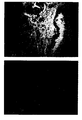
- Figure 1 shows a section of a breast carcinoma
immunocytochemically stained using BiotHER. On the
left of the figure cells invasive breast carcinoma
show to over-express HER2 as demonstrated by the
intense membrane staining (black staining), whereas on
the right a normal duct of the mammary gland does not
express HER2 and does not show any cytoplasmic or
membrane staining. Nuclei counter-stained with
Hematoxilin;
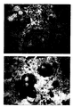
- Figure 2 shows a section of breast carcinoma
immunocytochemically stained using BiotHER. In this
case 100% of cells show an intense staining that is
localized only to the membrane (score 3+);
- Figure 3 shows a section of breast carcinoma
immunocytochemically stained using BiotHER. In this
case 80% of cells shows a moderate staining that is
localized only to the membrane (score 2+);
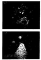
- Figure 4 shows a section of a cluster of breast
carcinoma cells obtained after centrifugation of a
metastic pleural effusion. The immunocytochemical
reaction using BiotHER shows the strong (score3+)
membrane over-expression of HER2.
- Figures 5 and 6 refer to the same area of breast
carcinoma stained, either with the murine monoclonal
antibody 4D5 ( figure 5) and with BiotHER antibody
(figure 6).
Example 1
Herceptin® (Trastuzumab) produced by Roche is
commercially available, distributed in vials and used
for breast carcinoma therapy. One mg of immunoglobulin
is obtained from a vial of Herceptin® and diluted in
physiologic saline solution at a concentration of 1
mg/ml and then dialyzed overnight against a 0.1M
solution of Na2CO3(sodium bicarbonate) at pH 8.5.
To 1 ml of solution containing 1 mg of Herceptin®,
0.12 ml of ε- caproylamido-biotin-n-hydroxy-succinimide
ester (Biospa) is added. The preparation
is well mixed for 4 hours at room temperature.
Finally, the preparation is dyalized against buffered
physiologic solution at pH 7.2 (PBS, physiologic
solution buffered with NaH2PO4 and Na2HPO4 0.1M).
The final solution (called BiotHER- mother
solution) is diluted 1:10 in PBS containing Sodium
Azyde 0.1% and then stocked in a refrigerator at 4°C.
the reactive is stable for months
The test is performed on breast carcinoma sections
(routinely fixed in formaldheyde 4% and paraffin
embedded) or on secondary lesions of breast carcinoma
(lymph node metastases, carcinoma cells in pleural
effusion). In vitro cultured cells of a stabilized
breast carcinoma cell line BT474, over-expressing HER2
are used as positive control. BT474 cells fixed in 4%
formaldheyde are processed following routine cytology
procedures in order to obtain a cell pellet to be
embedded in paraffin. Test and control sections are
then deparaffinized and brought to water.
The sections are covered with a drop of
biotinylated Herceptin® (BiotHER), final dilution
1:200 in PBS .
The sections are incubated for 60 minutes at room
temperature. The sections are then washed twice for 5
minutes in PBS-Tween and incubated in streptavidin-peroxidase
conjugate diluted 1:50 (Biogenex) for 13
minutes at room temperature. After this, the sections
are washed in H2O and then the reaction is developed in
a solution of 3'-3-diaminobenzidine (DAB) and H2O2 for
5 minutes. After PBS washing the nuclei are counter
stained with Mayer Haemalum for 30 seconds, then
dehydrated in alcohols, clarified in xilol. The slides
are then mounted for microscopic examination with a
coverslip using as a mounting medium 1 drop of
Entellan.
Positive reactions correspond to the membrane
staining.
Example 2
A series of 47 breast carcinomas has been
studied with BiotHER. In all cases the amplification
of HER2 gene has been evaluated using the CISH
procedure. All cases have been studied on parallel
sections using the immunocytochemical procedure and
the following antibodies: Herceptest (Dako), CB11
(Ylem, diluted 1:80 incubated for 30 minutes at 37°
after microwave treatment), TAB250 (Zymed, diluted
1:40 incubated for 30 minutes at 37°). For CB11, a
pre-treatment for the sections for antigen-retrieval
was performed by immersion in 0.01 mol/L citrate
buffer, pH 7.3, at 94°C for 20 minutes, using a
temperature-controlled microwave oven. After blocking
non-specific binding sites (using the blocking reagent
Histostain Plus kit, Zymed) the sections have been
incubated with the specific primary antibody
(Herceptest, or CB11, or TAB250). After incubation
with the primary antibody the slides have been
incubated for 20 minutes with the secondary
biotinylated antibody and then with peroxidase-cojugated
streptavidin (1/50, StrAviGen MultiLink®
Kit, BioGenex) and then for additional 20 minutes at
room temperature. Finally, a chromogenic solution of
di 3'-3-diaminobenzidine (DAB) (LiquidDAB Substrate
Pack, BioGenex) has been used to reveal the reaction.
The slides have been counterstained in Mayer
Haematoxylin (Biogenex) for 30 seconds, dehydrated and
mounted in balsam.
On this series the BiotHER procedure has been
applied as previously described (see example 1).
The evaluation of immunocytochemically stained
samples has been performed using the scoring system
approved by FDA for Herceptest. Briefly, only the
cytoplasmic membrane reaction has been considered as
valid and the score evaluation has been done
considering the percentage of positive cells, the
intensity of reaction and the completeness of the
membrane staining.
Table 1 shows the results. None of the cases
stained with BiotHER showed staining of normal
epithelial cells of ducts or lobules, nor staining of
stromal or inflammatory cells. Only the carcinoma
cells showed a membrane staining and did not show any
background staining of the background or of the
cytoplasm (figure 1).
| Comparative evaluation of immunocytochemical results using different anti c-erbB2 antibodies and BiotHER |
| 12 cases with AMPLIFIED HER2 (CISH) |
| CB11 | HERCEPTEST | BiotHER |
| score | case n° | score | case n° | score | case n° |
| 0/1+ | 0 | 0/1+ | 1 | 0/1+ | 3 |
| 2+ | 5 | 2+ | 3 | 2+ | 3 |
| 3+ | 7 | 3+ | 8 | 3+ | 6 |
| 35 cases with NOT AMPLIFIED HER2 (CISH) |
| CB11 | HERCEPTEST | BiotHER |
| score | case n° | score | case n° | score | case n° |
| 0/1+ | 25 | 0/1+ | 27 | 0/1+ | 35 |
| 2+ | 6 | 2+ | 7 | 2+ | 0 |
| 3+ | 4 | 3+ | 1 | 3+ | 0 |
Data of table 1 show that BiotHER test is positive
in the majority (about 85%) of breast carcinomas with
amplified HER2 gene. Specifically, BiotHER recognized
high expression (score 3+) (figure 2) of the extra-cytoplasmic
domain of the receptor target of Herceptin®
in 50% of cases showing HER2 gene amplification. A
lower reactivity (score 2+) (figure 3),but still
helpful to demonstrate that Herceptin® can react even
in a lower percentage of cells, was demonstrated in 25%
of carcinomas with gene amplification. In HER2 non-amplified
cases BiotHER was never reacting,
demonstrating the high specificity of the reaction.
Thus, it can be argued that BiotHER allows
recognizing in a highly specific way about 85% of cases
of breast carcinomas with HER2 gene amplification that
will completely or partially respond to Herceptin®
treatment.
Table 2 shows the clinical response to Herceptin®
on recurrences of different patients selected following
a
score 3+ of Herceptest (Dako).
| Evaluation of the answer to Herceptin® depending on the score obtained with different antibodies and BiotHER |
| Diagnosis | Score CB11 | Score Herceptest | Score BiotHER | CISH | Time of response (months) |
| Metastatic pleural effusion | 3+ | 3+ | 3+ | HER2 amplif | 18 |
| Metastatic pleural effusion | 3+ | 3+ | 0 | HER2 amplif | 6 |
| Metastatic pleural effusion | 3+ | 3+ | 0 | HER2 amplif | 9 |
| Metastatic pleural effusion | 3+ | 3+ | 0 | HER2 amplif | 3 |
| Sovra-clavear Metastic lymph node | 2+ | 3+ | 3+ | HER2 amplif | 15 |
As shown in Table 2, independently from HER2
gene amplification, the duration time of answer to
Herceptin® is reduced in patients whose tumor is not
reacting with BiotHER (score 0/1+), as compared to that
of patients (case n°1 and 5) with an expression score
3+ (figure 4). This result further confirms the high
specificity of BiotHER in recognizing cases that would
benefit of Herceptin® treatment as suggested in Table
1.
Example 3
Sections of breast carcinoma (see above) were
dewaxed, brought to water and treated with a reagent
formed of Herceptin® conjugated with monovalent
biotinylated FAB fragments of anti-human IgG
antibodies.
The procedure to prepare the reagent is as
follows:the Herceptin® antibody, at a concentration of
1mg/ml is mixed in vitro with biotinylated monovalent
FAB fragments of anti-human IgG rabbit antibodies
(acquired from commerce from LISTAR, Milan) at a
concentration of 1 mg/ml. Herceptin® and FAB fragments
were mixed in a 1:1 ratio, so that the ratio was 1
molecule of Herceptin® to 4 molecules of FAB fragments.
The mixture is incubated at 37°C for 30 min, then
applied on the sections instead of BiotHER.
The principle of this procedure is that the
humanized antibody Herceptin® is bound to an excessof
biotinylated specific antibody fragments. The FAB
fragments, being monovalent, do not produce a
precipitation of the antibody and they bind only to the
"human" part of Herceptin®, while the antibody site,
murine, is not bound.
Sections are incubated with the above complex for
one hour, then washed and treated as in the above
exemples, with avidin-biotinylated peroxidase
complexes, in order to reveal the biotin bound to the
FAB fragments, hence to the humanized Herceptin®
antibody.
The results obtained with this procedure in a case
series of 20 breast carcinomas were similar to those
obtained with the above examples using biotinilated
Herceptin®.
Example 4
In a series of 20 cases of breast carcinoma,
already examined with the immunocytochemical procedures
as above, we made serial parallel sections. One of
these sections was stained with the BiotHER procedure
as above described, while other section wAS treated
with the murine 4D5 monoclonal antibody, directed
against the same antigenic site recognized by
Herceptin®.
The murine 4D5 antibody was used at a
concentration of 1 microgram/ml, similar to that
currently used for other murine antibodies, such as TAB
250 and CB11 antibodies. Following the incubation with
the 4D5 antibody, the sections were incubated for 20
min with the secondary biotinylated antibody and
finally with the streptavidin-peroxidase complexes
(1/50, StrAviGen MultiLink® Kit, BioGenex) for
additional 20 minutes at room temperature. Finally, a
chromogenic solution of 3'-3-diaminobenzidine (DAB)
(LiquidDAB Substrate Pack, BioGenex) was employed to
reveal the reactions. Slides were counterstained with
Mayer's Haematoxylin (Biogenex) for 30 seconds,
dehydrated and mounted in balsam.
The comparison, on serial sections, of the same
cases and tumor areas stained either with the BiotHER
procedure or with the 4D5 antibody showed analogies,
being the same cases and areas positive or negative.
However, we observed that while staining with BiotHER
is only at the cell membrane, staining with 4D5 gives,
in addition to the membrane staining, also a diffuse
cytoplasmic staining, similar to that observed when
using other murine or rabbit anti-HER2 receptor
antibodies. It is well known that such cytoplasmic
staining can cause a severe disturbance of the result
interpretation and that it can lead to erroneous
diagnoses.
Example 5
In addition to the above described in vitro
procedures, Biotinylated Herceptin ( BiotHER) can be
used for the in vivo imaging and treatment of HER2
positive tumors, using a technique with avidin-radioactive
biotin, similar in principle to the above
described immunocytochemical procedure.
Herceptin is biotinylated following the procedure
described in detain in Example 1.
The reagent is then injected in patients with
breast cancer where over-expression of HER2 receptor
antigen was previously demonstrated by
immunocytochemical procedures ( Example 2).
To the patients, BiotHER is administered
endovenously at a concentration of 35 mg/m2 ( 1st
step). Thirtysix hours later, 30 mg of avidin and 50
mg of streptavidin are injected. Biotin labeled with
Y90 or In111 is injected endovenously 18-24 later.
This procedure is similar in principle to that
used for the diagnosis and therapy of human gliomas
expressing tenascin (Paganelli et al. Eur. J. Nucl.
Med. 26:348-57, 1999). However, in the latter
procedure murine anti-tenascin antibodies are used in
the first step. Use of murine antibodies gives rise to
problems involved in the liver and spleen binding,
which are avoided when using the humainized Herceptin
antibody.
The advantage for the diagnosis and treatment of
HER2 over-expressing carcinomas is linked to the use
of the humanized antibody avoiding the binding to
extra-tumoral sites, mainly to liver and spleen, of
murine antibodies. The FC fragment of the latter is in
fact recognized as heterogeneous by the lympho-reticular
tissue of the patients.