-
The present invention is directed to peptides comprising the sequence ASNKL
(SEQ ID N° 1), inducing attraction of the axonal growth in presence of the semaphorin
(Sema3A), L1 and NP-1 proteins and their use for the manufacture of a drug for
neuronal/axonal regeneration or for treating neurodegenerative diseases. The present
invention also includes a method for identifying a compound able to bind the NP-1, a
method for identifying a compound able to revert the repulsory effect of Sema3A on the
axonal growth from a suited cell, or methods to screen for molecules capable of
blocking Sema3A- induced endocytosis.
-
Axon pathfinding is regulated by a number of chemotropic and cell-contact
guidance cues that act as attractants or repellents for developing neuronal projections. At
choice points along the pathways, a complex series of events may take place to ensure
appropriate decisions made by the growth cone (Cook et al., Curr Opin Neurobiol.,
8:64-72, 1998). In particular, this has been documented for commissural projections
where a highly dynamic mechanism of up- and down-regulation of axonal responses to
guidance cues occurs to sequentially orientate the axons towards, across, or away from
the midline. These events were found to involve cell adhesion molecules of the
immunoglobulin (Ig) superfamily as well as secreted factors of the netrin, slit and
semaphorin families (Stoeckli and Landmesser, Opin Neurobiol, 8:73-79, 1998). In
rodents, loss of function of the Ig superfamily cell adhesion molecule (IgCAM) L1 has
implicated this protein in axonal guidance at the pyramidal decussation (Cohen et al.,
Curr Biol, 8:26-33, 1998). This particular stage in corticospinal pathfinding has been
characterized by two major events: axon growth across the midline followed by a switch
from a ventral to a dorsal trajectory in the spinal cord. L1 is likely to be important for
each of these steps as it may be required as a cell-contact cue for crossing the midline
(Cohen et al., 1998). In addition, L1 has also been shown in vitro to be required for
axons to respond to a ventrally-secreted semaphorin, Sema3A, that triggers the
dorsalization of the pathway (Castellani et al., Curr Biol., 8:26-33, 2000).
-
Recent studies suggest that semaphorins signal through multi-molecular receptor
complexes, containing neuropilin-1 (NP-1) and /or neuropilin-2 (NP-2) as specific
ligand binding subunits (Raper, Curr Opin Neurobiol., 10:88-94, 2000), and plexins as
signal transducers (Tamagnone and Comoglio, Trends Cell Biol., 10:377-383, 2000). L1
has also been shown to associate with NP-1 and is required as part of the Sema3A
receptor complex for axon guidance responses (Castellani et al., 2000). A functional
link between L1 and NP-1/Sema3A signaling is supported by the finding that a soluble
L1Fc chimera switches Sema3A-induced chemorepulsion to attraction (Castellani et al.,
2000). Previously it has been suggested that growth cones can switch between attraction
and repulsion via a modulation of internal cyclic nucleotide levels (Song et al., Science,
281:1515-1518, 1998). How binding of soluble L1 on the cell surface can induce such a
switch is largely unclear. L1 displays a highly complex pattern of homophilic and
heterophilic interactions. In addition to L1 and NP-1, as components of the Sema3A
receptor complex, axons express a number of other binding partners that could
potentially be the receptor for L1Fc in the Sema3A reversion. These include other Ig
CAMs such as F3/contactin, and TAG-1/axonin-1, α3-β5 integrins as well as several
ECM proteins, such as laminin and some chondroitin sulfate proteoglycans
(Brümmendorf and Rathjen, Curr Opin Neurobiol, 6:584-593, 1996). Furthermore, L1
expression on the growth cone is highly dynamically regulated. Following ligand
activation, L1 is internalized, transported into vesicles to be recycled on the cell surface
(Kamiguchi and Lemmon, J. Neurosci., 20:3676-3686, 2000).
-
A clear requirement for L1 in axon development is reflected by the variety of
neurological abnormalities associated with L1 mutations in the human disorder referred
to as X-linked hydrocephalus, MASA syndrome or SPG type 1 (hydrocephalus, enlarged
ventricles, corpus callosum agenesis, corticospinal tract hypoplasia, Rosenthal et al., Nat
Genet. 2:107-112, 1992 ; Vits et al., Nat Genet., 7:408-413, 1994 ; Jouet et al., Nat
Genet., 7:402-407, 1994). Mutations in L1 that give rise to these disorders include many
that would truncate the mature protein and eliminate cell surface expression. More
subtle missense mutations within L1 have been shown to have multiple effects on L1
function disrupting intracellular protein trafficking or ligand binding or both (De
Angelis et al., EMBO J., 18:4744-4753, 1999). The possibility that these mutations
affect axonal responses to Sema3A has hitherto not been examined.
-
Surprisingly, the inventors, after having dissected the molecular mechanisms
underlying the switch of axonal responses to Sema3A by soluble L1, using L1Fcs that
contain pathological missense mutations, have demonstrated that binding of L1Fc (also
noted L1-Fc in the present description) to NP-1 but not to L1 or to two other Ig CAMs
expressed on growth cones is crucial for L1Fc to activate the conversion process. They
have also demonstrated that binding and co-immunoprecipitation studies implicate the
region containing mutation L120V in Ig domain 1 of L1 in both trans and cis L1/NP-1
complex formation. Moreover, they have demonstrated that a peptide composed of the
(F)ASNKL120 amino-acid sequence was sufficient to reverse Sema3A-mediated
repulsion.
-
So, in a first aspect, the present invention is directed to a peptide comprising the
sequence ASNKL (SEQ ID N° 1), preferably, inducing attraction of the axonal growth,
in particular in presence of the semaphorin (Sema3A), L1 and NP-1 proteins.
-
In a preferred embodiment of this first aspect, the present invention includes the
peptides comprising the sequence SEQ ID N° 1 having no more than 10 amino acids
residues.
-
The peptide consisting of SEQ ID N° 1 and a peptide comprising or consisting of
the sequence of FASNKL (SEQ ID N° 2) or FASNKLGTAM (SEQ ID N° 3) are
particularly preferred.
-
An inducer of axonal growth comprising one of the peptide of the present
invention forms also part of the first aspect of the present invention.
-
In a second aspect, the present invention is directed to a method for identifying a
compound able to bind the NP-1 protein, comprising the steps of:
- a) contacting said compound with a sample comprising the NP-1 protein, before,
after or concomitantly with contacting said sample with the L1-Fc protein or with a
peptide according to the present invention, particularly with a peptide comprising or
consisting of the sequence SEQ ID N° 1 , SEQ ID N° 2 or SEQ ID N° 3,
- b) studying the binding between NP-1 and L1Fc or said peptide according to the
invention,
the binding of said compound to the NP-1 protein being deduced from the alteration of
the binding of NP-1 with L1-Fc or said peptide according to the invention observed in
absence of said compound.
-
-
In a preferred embodiment of this second aspect, the present invention is related
to the method for identifying a compound able to bind the NP-1 protein according to the
present invention, wherein said sample also comprises the L1 protein.
-
In a further preferred embodiment of this second aspect, the present invention is
related to the method for identifying a compound able to bind the NP-1 protein
according to the present invention, wherein said NP-1 protein is a recombinant protein.
-
In a further preferred embodiment of this second aspect, the present invention is
related to the method for identifying a compound able to bind the NP-1 protein
according to the present invention, wherein said sample comprises a cell expressing the
NP-1 protein at its surface.
-
In a further preferred embodiment of this second aspect, the present invention is
related to the method for identifying a compound able to bind the NP-1 protein
according to the present invention, wherein said sample comprises a cell expressing the
NP-1 and L1 proteins at its surface.
-
In a further preferred embodiment of this second aspect, the present invention is
related to the method for identifying a compound able to bind the NP-1 protein
according to the present invention, wherein said L1-Fc protein or said peptide according
to the invention is labeled and said binding of NP-1 protein with said L1-Fc protein or
said peptide according to the invention is assayed by level of signal associated with said
NP-1 protein.
-
The inventors have shown that L1/NP-1 cis interaction is necessary to the
endocytosis of the Sema3A receptor complex associated with a repulsive response
whereas L1/NP-1 trans interaction prevents it. This later interaction activates nitric
oxide (NO) synthase and cGMP guanylyl cyclase. As indicated in the following
examples, it has been identified molecular mechanisms involved in switching Sema3A
repulsion to attraction and points to endocytosis as critical in determining the nature of
axonal responses to guidance cues. Also it is provided a new explanation for the effects
of human pathological mutations in the X-linked L1 gene.
-
So, in a particularly preferred embodiment of this second aspect, the present
invention is related to the method for identifying a compound able to bind the NP-1
protein according to the present invention, wherein said alteration of binding of NP-1
with L1-Fc or said peptide according to the invention is assayed by measuring a
secondary signal appearing in the presence of said binding.
-
In this particularly preferred embodiment of this second aspect, it is preferred
that said secondary signal is induced in the presence of Sema3A.
-
In this particularly preferred embodiment of this second aspect, it is further
preferred that said secondary signal is the reversion of the repulsory effect of Sema3A
on axonal growth.
-
In this particularly preferred embodiment of this second aspect, it is also
preferred that said secondary signal is the activation of NO synthesis.
-
In this particularly preferred embodiment of this second aspect, it is also
preferred that said secondary signal is an increased production of cGMP.
-
In a third aspect, the present invention is directed to a method for
attracting/modulating the direction of axonal growth by a cell suited to promote axonal
growth, submitted to a Sema3A flow, comprising the step of contacting said cell with
the peptide or the inducer according to the present invention.
-
In a fourth aspect, the present invention is directed to a method for identifying a
compound able to revert the repulsory effect of Sema3A on the axonal growth from a
suited cell, comprising the steps of contacting said cell expressing L1 and NP-1 at its
surface with said compound in the presence of Sema3A, and studying the increase in
NO synthesis in said cell.
-
In a fifth aspect, the present invention is directed to a method to screen for
molecules that prevent the internalization of the dextran induced by the Sema3A
treatment or to screen for molecules capable of blocking Sema3A- induced endocytosis
by cells expressing L1 and NP-1 proteins at their surface characterized in that said
method comprises the following steps of:
- a) culturing cells expressing L1 and NP-1 in the presence of Sema3A, a labelled
dextran and the molecule to be assayed ; and
- b) visualizing or determining if said labelled dextran has been internalized into
the cells ;
- c) selecting the assayed molecule if no uptake of the labelled dextran by the cells
could be detected.
-
-
In a preferred embodiment, said cells in step a) are recombinant cell lines
transfected with expression vectors capable of expressing the L1 and NP-1 proteins at
their surface or neuronal cells which naturally co-express the L1 and NP-1 proteins.
-
Such vectors will be prepared according to the methods commonly used by
persons skilled in the art, and the clones resulting therefrom may be introduced into an
appropriate host by standard methods such as, for example, lipofection, electroporation
or heat shock.
-
In a further preferred embodiment, said labelled dextran in step a) is selected
from the group consisting of rhodamine-dextran, FITC-dextran or dextran coupled to
fluorescent molecules.
-
In a sixth aspect, the present invention is directed to a method to screen for
molecules capable of blocking Sema3A- induced co-internalization of L1 and NP-1
proteins, to screen molecules capable of maintaining cell surface expression of L1 and
NP-1 proteins or to screen molecules capable of blocking Sema3A- induced endocytosis
characterized in that said method comprises the following steps of:
- a) culturing cells expressing L1 and NP-1 in the presence of Sema3A and the
molecule to be assayed ; and
- b) visualizing or determining if said L1 and NP-1 proteins have been cointernalized
into the cells using an immunocytochemical method in presence of
antibodies, mono- or polyclonal, if appropriate, labelled, directed against L1 and NP-1
proteins ;
- c) selecting the assayed molecule if L1 and NP-1 proteins could be detected at
the surface of the cells.
-
-
In a preferred embodiment, said cells in step a) of the method of that sixth aspect
according to the present invention, are recombinant cell lines transfected with
expression vectors capable of expressing the L1 and NP-1 proteins at their surface or
neuronal cells which naturally co-express the L1 and NP-1 proteins.
-
By immunocytochemical methods, it is intended to mean any
immunocytochemical methods known by the skilled person which are capable of
specifically detecting the presence of L1 and NP-1 at the surface of a cell using
antibodies directed specifically against these L1 and NP-1 proteins.
-
Specific polyclonal antibodies may be obtained from a serum of an animal
immunized against these L1 and NP-1 proteins. The specific monoclonal antibodies may
be obtained according to the conventional hybridoma culture method described by Köhler
and Milstein, 1975.
-
Said anti L1 and/or NP-1 antibodies, or Fab or F(ab')2 fragments thereof, may also
be in the form of immunoconjugates or of labelled antibodies so as to obtain a detectable
and/or quantifiable signal and so constitute a means for the immunocytochemical analysis
of the expression of the L1 and NP-1 proteins at the surface of cells, for example by
immunofluorescence, gold labelling, enzymatic immunoconjugates.
-
In a seventh aspect, the present invention is directed to use of the peptides or the
inducer according to the present invention, or the molecules capable of blocking
Sema3A- induced endocytosis selected by the method according to the present
invention, for the manufacture of a drug for neuronal/axonal regeneration or for treating
neurodegenerative diseases.
-
Finally, the present invention is directed to use of the peptides or the inducer
according to the present invention, as a targeting agent, in the manufacture of a
composition to be used in cell therapy.
-
The peptides or inducers according to the present invention as active ingredients
of a medicament will be preferably in soluble form, combined with a pharmaceutically
acceptable vehicle.
-
Such peptides, inducers or selected molecules capable of blocking Sema3A-induced
endocytosis according to the present invention and which can be used as a
medicament offer a new approach of treatment requiring or linked to neuronal/axonal
regeneration, such as medicament for preventing and/or treating neurodegenerative
diseases.
-
Preferably, these peptides, inducers or selected molecules capable of blocking
Sema3A- induced endocytosis according to the present invention will be administered by
the systemic route, in particular by the intravenous route, by the intramuscular or
intradermal route or by the oral route.
-
Their modes of administration, optimum dosages and galenic forms can be
determined according to the criteria generally taken into account in establishing a
treatment suited to a patient, such as for example the age or body weight of the patient, the
seriousness of his general condition, the tolerance to treatment and the side effects
observed, and the like.
-
Other characteristics and advantages of the invention appear in the remainder of
the description with the examples and figures whose legends are represented below.
Figures legends
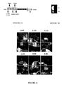
Figures 1A to 1F: Co-cultures of cortical slices and ventral spinal cord explants in
the presence of mutated L1Fc chimeras
-
Figure 1A: L1Fc chimeras with individual missense mutations affecting distinct
Ig domains selected for the study.
-
Figure 1B: Scheme of the co-culture system of cortical slice and ventral spinal
cord explant.
-
Figure 1C: The microphotographs illustrate the preferential growth of axons
towards the source of Sema3A, in the presence of several mutant L1 chimeras. In
contrast, the chimera carrying the L120V mutation was unable to switch the repulsive
activity of Sema3A to attraction. In this case growth is directed away from the Sema3A
source as in the control. Scale bar: 50 µm.
-
Figure 1D: H210Q, E309K, D598N, A426D and G121S mutant L1Fc chimeras
were able to reverse Sema3A repulsion to attraction in the co-culture assay as indicated
by the positive guidance indices.
-
Figures 1E and 1F: The histograms represent the number of axons growing
towards (open bars) and away (filled bars) from the spinal cord explants (figure 1F) and
their length (figure 1E). * Statistical difference in axon growth between the side facing
the spinal cord explant (towards) and the side opposed to it (away) with P<0.0001.
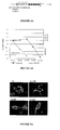
Figures 2A and 2B: Homophilic binding of mutant L1 chimeras to wild-type L1
-
Figure 2A: Binding of mutated L1Fc protein to wild-type L1 expressed in COS
cells and detected by immunofluorescence analysis. Cell surface L1 was detected using
Texas red-conjugated secondary antibodies and mutant L1Fc chimeras using FITC-conjugated
secondary antibodies. A426D, H210Q and L120V mutated chimeras bound
to wild-type L1 whereas G121S did not. Scale bar: 5 µm.
-
Figure 2B: Homophilic binding in wild-type/mutant and mutant/mutant
combinations is compared by FACS analysis in the two-color aggregation assay. The
histogram represents the percentage of mixed aggregates formed at the 30-minute time-point
with values expressed as percent wild-type binding and error bars represent the
SEM of 3 independent experiments. Wild-type L1-Fc is 100 %. For mutations A426D
and H210Q, increased mutant to wild-type compared with mutant to mutant binding was
observed, whereas binding of G121S remained extremely low for both combinations.
wt: wild-type, m: mutant.
Figure 3: Binding assay with L1Fcs on Plexin-A1 and NP-1 expressing cells
-
Cell surface expression of L1, Plexin-A1 and NP-1 was detected using Texas
red-conjugated secondary antibodies. Mutant L1Fc chimeras were visualized using
FITC-conjugated secondary antibodies. Wild-type L1Fc bound to L1 but not Plexin-A1-expressing
cells. A426D and G121S L1Fc chimeras bound to NP-1 expressing cells,
whereas the L120V chimera did not. Scale bar: 5µm.
Figures 4A and 4B: Switching Sema3A repulsion by a mimetic peptide of L1
-
Figure 4A: Part of the amino-acid sequence of L1 Ig1 domain. Six-amino-acid
peptides containing either the native leucine or the mutant valine 120 residue were
synthesized.
-
Figure 4B: Guidance induces in the co-culture assay for 5 different
concentrations of the peptides. Note that at 10-5 and 10-6M, the peptide with C-terminal
leucine switched Sema3A repulsion to attraction whereas the C-terminal valine did not.
Figures 5A and 5B: Effects of pathological L1 mutations on the formation of L1-NP-1
receptor complex
-
Figure 5A: Expression plasmids encoding full-length L1 containing missense
mutations were transfected into COS7 cells and cell surface expression was detected by
immunofluorescence analysis. As illustrated, mutant L1 constructs were expressed on
the cell surface. Scale bar: 5 µm.
-
Figure 5B: Co-immunoprecipitation experiments. Cells transfected with NP-1
lacking its cytoplasmic domain (ΔNP-1) and full-length L1 were lysed and proteins were
precipitated with anti-myc antibodies (see methods). Immunoblots were probed with
anti-L1 and anti-NP-1 antibodies. Both L1 and NP-1 were detected in the precipitates,
indicating that the cis-interaction involves the extracellular domains of each protein.
Western blot analysis of cell lysate immunoprecipitated with anti-HA antibodies is
shown for double transfectants expressing mutant L1 constructs (L120V, G121S or
A426D) and full-length NP-1. L1 proteins with either the G121S or the A426D
mutation co-immunoprecipitated with NP-1 whereas L120V -L1 did not.
Figures 6A and 6B: Peptide mimetic of soluble L1 blocks Sema3A-induced
endocytosis
-
Figure 6A: Neonatal cortical cells were incubated with Sema3A ("Cortex
P0:Sema3A 15"'). The treatment induced internalization of Rhodamine-Dextran that
accumulated along axons and around the soma. Internalization was blocked when
Sema3A was applied in combination with the L1 mimetic peptide ("Cortex P0:Sema3A
+ peptide 15"'). Uptake of Rhodamine-Dextran was not detected in control conditions
and in cultures treated with the peptide alone (not shown). Scale bar: 10 µm.
-
Figure 6B: COS7 cell co-expressing L1 and NP-1 also internalized Rhodamine-Dextran
when treated with Sema3A ("L1/NP-1: Sema3A 15"'). This process was
blocked by a Sema3A/peptide treatment ("L1/NP-1: Sema3A + peptide 15"'). As
observed in cortical cell cultures, no endocytosis could be detected in control conditions
or in cells treated with the peptide alone (not shown).
Figures 7A to 7H: L1 ensures endocytosis of the Sema3A receptor complex
-
Figure 7A: Cell surface immunolabeling of L1 and NP-1 was undetectable after
Sema3A treatment.
-
Figures 7B and 7C: In contrast, L1/NP-1 cell surface expression was observed in
control condition (figure 7B) and after treatment with the peptide alone (figure 7C).
-
Figures 7D and 7E: When cells were treated with Sema3A in combination with
the peptide, L1 and NP-1 were detected on the cell membrane, as illustrated in with
these two panels.
-
Figures 7F to 7H: After cell permeabilization, co-localized spots of L1 and NP-1
immunolabeling were observed, indicating that loss of cell surface expression in figure
7A was due to internalization of the two proteins.
Figure 8: Activation of the NO/cGMP pathway in the reversion of Sema3A
repulsion
-
Pharmacological blockade of soluble guanylyl cyclase (with ODQ) and nNOS
(with 7NI) in the co-culture of cortical slices and ventral spinal cord explants. In the
presence of ODQ and 7NI alone, axons were repelled by the Sema3A source however,
in the presence of L1Fc alone, axons switched from repulsion (negative guidance index)
to attraction for the Sema3A source (positive guidance index). After addition of ODQ
and 7NI in combination with L1Fc, axons no longer switched from repulsive to
attractive responses. The number and length of cortical axons growing away from and
towards the spinal cord explant were quantified for each condition. The number and
length of axons were significantly reduced on the side facing the spinal cord explants,
compared to the opposite side in ODQ, 7NI, ODQ+L1Fc, 7NI+L1Fc and control
conditions. In the presence of L1Fc alone, axons were attracted, as indicated by the
higher number and length of axons growing towards the spinal cord explant. Thus, ODQ
and 7NI prevented the L1Fc induced switching of Sema3A from repulsion to attraction.
Statistical analysis was conducted using the ANOVA test. * p<0.0001. Bars in the
histograms: growth directed away (black bars) and towards (white bars) the spinal cord
explant.
Figures 9A and 9B: Models for L1-NP-1 interaction and modulation of the
Sema3A signal
-
Figure 9A: The diagrams represent L1-L1 and L1/NP-1 trans interactions and
the effects of missense mutations on the modulation of Sema3A signaling. (a) cis
interaction of L1 with NP-1 triggers the repulsive Sema3A signal. (b) Soluble L1 (L1Fc)
binds both L1 and NP-1 on the growth cone and switches Sema3A repulsion to
attraction. (c) Soluble L1 containing the L120V mutation binds L1 but no longer NP-1
and fails to reverse Sema3A repulsion. (d) G121S, a mutation that destroys homophilic
binding, does not affect L1 binding to NP-1 or alter the ability of the chimera to induce
the switch. This shows that interaction of soluble L1 with NP-1 but not with L1 is
crucial for the reversion process.
-
Figure 9B: A model for the interaction of L1 with NP-1 in the Sema3A receptor
complex. (a) Association of L1 with the Sema3A receptor complex activates a
chemorepulsive signal that is initiated via L1/NP-1 co-internalization. (b) In the absence
of L1 (L1-deficient mice), the receptor complex is no longer functional and the
repulsive signaling is blocked. (c) Simultaneous cis and trans interactions of L1 with
NP-1 block endocytosis and modify the signaling pathway. Activation of the
nNOS/cGMP pathway is involved in the switch from repulsive to attractive responses.
(d) L1/NP-1 complex formation in the cell membrane is also necessary for triggering
Sema3A attractive responses since wild-type L1Fc cannot switch Sema3A repulsion to
attraction in the absence of neuronal L1 (L1-deficient mice) even though it binds NP-1.
EXAMPLES
Example 1: Material and Methods
A. Co-culture experiments
-
Blocks of cortex were dissected from P0 (postnatal day 0) to P2 mice and cut
into 250 µm thick slices with a McIlwain tissue chopper. The ventral part of the cervical
spinal cord was dissected and cut into 250 µm thick slices. Co-culture of cortical slices
and ventral spinal cord explants were performed in a three-dimensional plasma clot, as
described previously (Castellani et al., 2000). Cortical slices were oriented in the plasma
clot as indicated in Figure 1B to give to axons equivalent chances to be deflected away
or towards the spinal cord explant. Purified wild-type or mutant L1Fc chimeras were
added to the culture medium (3 µg/ml). Inhibitors of the guanylyl cyclase (ODQ, 10
-5 M,
Sigma) and NOS (7NI, 10
-4 M, Sigma) were added to the culture medium, either alone
or with wild-type L1Fc (3 µg/ml). After one or two days in vitro (DIV), the cultures
were fixed with 4 % paraformaldehyde (PFA) and analyzed by phase contrast
microscopy (Axiovert 35-M, Zeiss). A qualitative guidance index was attributed to the
co-cultures, to assess the degree of repulsive (negative values) or attractive (positive
values) effects (Castellani et al., 2000). The index ranged from -2 (when most fibers
grew away from the spinal cord explant) to +2 (when most fibers grew towards the
explant), with intermediate situations (index of -1 and +1 for moderate guidance
effects). An index of zero was attributed to the cultures showing no preferential growth.
Quantitative analysis of axon length and number of axons growing from cortical slices
was performed as described previously (Castellani et al., 2000). A proximal and a distal
region, each of them covering a 600 µm-piece of cortical tissue were determined for
each culture. All fibers extending from these two regions were counted and the length of
the three longest fibers was measured, using computer analysis software (Visiolab 2000,
Biocom). Immunofluorescence labeling of axons was performed using the mouse anti-phosphorylated
neurofilament antibody SMI31 (Sternberger and Meyer Inc.), and Texas
red-conjugated secondary anti-mouse antibody (see result obtained for number of axons
and axonal length in the following tables 1 and 2).
| Number of axons |
| Ctx/vSC | Away | Towards |
| Control | 25.8 +/- 2 | 4.7 +/- 0.8,* |
| +L1FC | 5.8 +/- 0.7 | 22.6 +/- 1.6, * |
| +ODQ | 25.2 +/- 1.8 | 5.1 +/- 0.5, * |
| +L1Fc +ODQ | 24.2 +/- 1 | 6 +/- 0.4, * |
| +7NI | 25.6 +/- 1.6 | 5 +/- 0.6, * |
| +L1Fc +7NI | 26.1 +/- 1.1 | 5 +/- 0.4, * |
| Axonal length |
| Ctx/vSC | Away | Towards |
| Control | 411.2 +/- 5 | 213.4 + /-4, * |
| +L1FC | 198.6 +/ -4 | 346.8 +/ -5, * |
| +ODQ | 384.1 +/- 7 | 206.9 +/- 8, * |
| +L1Fc +ODQ | 410.5 +/- 3 | 207.1 +/- 7, * |
| +7NI | 407.6 +/- 5 | 203.1 +/- 6, * |
| +L1Fc +7NI | 408.2 +/- 4 | 213.2 +/- 4, * |
B. Binding assays
-
The mutant L1Fc chimeras were produced and purified as described previously
(De Angelis et al., 1999). Briefly, COS-7 cells were transiently transfected with 10 µg
DNA per 150 mm culture dish, the soluble Fc chimeric protein was allowed to
accumulate in the media for 6 days and was recovered and purified by protein A-Sepharose
affinity chromatography. In the binding experiments, L1Fcs were crosslinked
to anti-human Fc antibodies (50 µg/ml, Jackson Immunoresearch) for 1 hr at
37°C. Binding detection to NP-1 requested concentrations of L1Fcs 10 folds higher than
L1. Antibody conjugated L1-Fc protein was then incubated with monolayers of COS7
cells transiently transfected with expression vectors encoding full length human L1 or
myc-tagged NP-1 where the cytoplasmic domain is deleted (gift of J. Raper, 2000 and
Renzi et al., 1999). Following incubation, the cells were fixed with 4 % PFA, and
processed for double-immunofluorescence detection. NP-1 was detected using a mouse
anti-c myc antibody (clone 9E10, Sigma) and L1 using a rat monoclonal anti-L1
antibody. Secondary antibody anti-c-myc (Texas-red conjugated anti-mouse antibodies)
was used to detect NP-1, anti-L1 (Texas red conjugated anti-rat antibodies) to detect L1,
and anti-Fc (FITC conjugated anti-rabbit antibodies) to detect bound L1Fc chimeras.
After secondary antibody incubation, the cells were washed with PBS and mounted in
Mowiol (Calbiochem).
C. Two color aggregation assay
-
A two-color aggregation assay was developed, modified from the homophilic
binding assay described by De Angelis et al. (1999). For the two-color assay, red and
green fluorescent microsphere beads (0.6um, Duke Scientific Corps.) were coated with
anti-human IgG antibody (Fc specific; Sigma, I-2136). 2.5 µg of wild type (red beads) or
mutant (green beads) L1Fc-proteins were conjugated to 10 µl of antibody-coated beads
by incubating for 2 hrs at 37°C. Excess unbound protein was removed by washing with
PBS/5 % FCS. For each mutant, a 1:1 mixture of wild-type L1 and mutant L1 coated
beads was prepared, dissaggregated to produce a single bead suspension and allowed to
form mixed aggregates at 37°C with samples removed in duplicate over a 30-minute
time course. Samples were diluted 1:5000 into ice cold PBS and the proportion of 2
colour aggregates was assessed using a fluorescence activated cell sorter (Becton
Dickenson FACSort). Ten thousand particles were counted per sample and categorised
as red only (WT-L1: WT-L1), green only (Mutant L1: Mutant L1) or mixed aggregates.
Every mutant protein was assayed at least 3 separate times and standardised to a wild-type
control conducted in parallel.
D. Coimmunoprecipitation experiments
-
COS cells were transfected with various expression vectors coding for full-length
HA-tagged NP-1, myc-tagged cyt-deleted NP-1, wild-type L1, and L1 mutant
constructs (G121S, A426D, L120V) using a lipofection transfection method
(lipofectamine). After 2 DIV, cells were lysed with immunoprecipitation buffer (IP
buffer: Hepes 25mM, EDTA 5mM, MgCl2 1mM, PMSF 2mM, glycerol 10 % and triton
X100 1 %, pH 7). The lysate was incubated at 4°C for 30 min, centrifuged for 5 min and
pre-cleared with protein A-Sepharose for 2 hrs at 4°C. Samples were
immunoprecipitated with mouse anti-HA (3 µl, clone 12CA5, Roche), rabbit anti-L1
antibodies (3 µl), mouse anti-myc antibodies (3 µl, 9E10, Sigma) coupled to rabbit anti-mouse
antibodies on protein A preformed complexes for 16 hrs at 4°C. The beads were
washed, and the precipitates analyzed by immunoblot using anti-HA (1:1000) and anti-L1
(1:1000) antibodies.
E. Rhodamine-Dextran uptake
-
Neonatal cortical cells or COS7 cells transfected with expression vectors for L1
and NP-1 were incubated for 15 min with Rhodamine-Dextran (2 mg/ml) mixed to a
solution of Sema3A-AP supernatant, Sema3A and peptide (10-5M), peptide alone
(10-5M) or control supernatant. Cells were fixed with PFA 4 % and processed for
immunocytochemistry as described in the above sections. To detect intracellular
proteins, cells were permeabilized with methanol for 2 min.
Example 2: Mutation L120V in Ig domain 1 of L1Fc abrogates the switch of
Sema3A repulsion to attraction
-
It has been previously developed an assay system using explants of ventral spinal
cord co-cultured with cortical axons in order to determine the factors involved in axonal
repulsion and attraction (Castellani et al., 2000). Using this system we have shown that
Sema3A secreted from the ventral spinal cord repels cortical axons. In the presence of
soluble wild-type L1Fc chimeric protein however, repulsion is switched to attraction
(Castellani et al., 2000). In the present study, we aimed to identify the L1 interactions
underlying the switching process. As Ig domains have been implicated in binding of Ig
CAMs to many of their ligands (Brümmendorf and Rathjen, 1996) we hypothesized that
they would also be required for L1Fc-mediated switching. Six L1Fc chimeras containing
pathological missense mutations affecting specific Ig domains (De Angelis et al., 1999
and Figures 1A, 1B) were assessed for their ability to reverse Sema3A repulsion to
attraction.
-
For each mutant, soluble Fc-chimeric protein was produced in mammalian cells
and purified by protein A-Sepharose affinity chromatography. The purified L1Fc
chimeras were subjected to SDS PAGE and immunoblotted with anti-Fc antibodies to
verify their integrity. In the co-culture assay, five out of six L1-Fc proteins containing a
single point mutation in Ig domains 1, 2, 3, 5 or 6 (G121S, H210Q, E309K, A426D and
D598N, respectively) were found to efficiently induce switching of Sema3A repulsion
to attraction (Figure 1C). As illustrated by both the qualitative guidance index and
quantitative analysis, the majority of axons exhibited preferential growth towards the
Sema3A source in the presence of the L1Fc chimera, rather than extending away from it
as seen in the control (Figures 1D-1F, total of 147 co-cultures in three separate
experiments). In contrast, one missense mutation, L120V in Ig domain 1, abrogated the
reversal of Sema3A repulsion to attraction. When the L120V chimeric Fc protein was
added to the culture medium, axons continued to be repelled from the spinal cord
explant. This resulted in a negative guidance index of -1.3 attributed to the co-cultures,
and a statistically significant preferential growth of fibers on the side opposing rather
than facing the spinal cord explant (Figures 1C-1F). Thus, at least part of Ig domain 1 is
required for L1Fc to switch the axonal response.
-
It has been aimed to identify the receptor on the cortical axons that binds L1Fc
and initiates the reversal of Sema3A repulsion to attraction. L1 and/or NP-1 could fulfill
this function, as they are both components of the Sema3A receptor complex and both
interact in trans with L1 (Castellani et al, 2000). Plexin-A1 may also mediate the
reversion of Sema3A as it associates with NP-1 in the Sema3A receptor. Other known
L1 ligands, including Ig CAMs F3/contactin and TAG-1/axonin are also candidate
receptors for L1Fc. Previous work has indicated that the mutations described above can
selectively affect either homophilic binding or heterophilic binding to F3 or axonin-1
(De Angelis et al, 1999). Two of these mutations G121S and A426D reduce homophilic
binding and heterophilic binding to F3, axonin-1 and the human homologue of axonin-1,
TAX-1. H210Q and E309K affect homophilic or heterophilic binding respectively.
D598N has a very modest effect on homophilic binding but significantly reduces
heterophilic binding to F3 and axonin-1, though not to TAX-1. L120V does not affect
homophilic or heterophilic binding. Therefore we were able to compare the ability of the
mutated chimeras to reverse the Sema3A repulsion with their binding capabilities.
Example 3: IgCAMs including L1 are not axonal receptors for L1Fc in the
reversion of Sema3A repulsion
-
Four mutations (E309K, D598N, A426D and G121S) that were found to alter
L1Fc binding to the Ig CAMs F3 and axonin-1switched Sema3A from repulsion to
attraction to the same extent as wild type L1Fc. Thus, Ig CAMs F3 and TAG-1 (the
rodent homologue of axonin-1) are unlikely to be axonal receptors for L1Fc in the
reversion process. Mutations A426D, H210Q and G121S were previously found to
reduce homophilic binding when individual mutant L1Fc proteins were bound to
themselves (De Angelis et al., 1999). In the present study however, mutated L1Fc
proteins were required to interact with wild-type L1 expressed on growth cones. To
formally exclude a role for homophilic binding of L1 in the initiation of the reversion
process, it was therefore necessary to examine whether homophilic binding of the
mutated L1Fc chimeras to wild-type L1 was also disrupted. Mutated L1Fcs were
therefore incubated with COS7 cells transfected with full-length wild-type L1 cDNA
and binding was assessed by immunofluorescence. In parallel, homophilic binding of
mutant/mutant and mutant/wild-type L1Fcs was compared in a two-color FACS
aggregation assay, allowing a quantitative assessment to be made (see Methods).
Interestingly, the immunofluorescence studies showed that the A426D and H210Q L1Fc
chimeras bound to wild-type L1 (Figure 2A). Similarly, in the two color FACS assay
homophilic binding was restored to 74.4% and 90.5 % of wild-type levels for A426D
and H210Q respectively (Figure 2B). In contrast, the mutation G121S, almost abolished
homophilic binding to wild-type L1 in both assays (Figures 2A, 2B). This mutation
however still promoted the L1Fc induced switching of Sema3A repulsion to attraction in
the co-culture assay. Thus, the ability of L1Fc to switch Sema3A repulsion is not
dependent on homophilic binding. Consequently, the trans interaction of L1Fc with L1
in the receptor complex is not required for the change in growth cone responsiveness to
Sema3A. In support of this conclusion we also observed that the L120V mutated L1Fc,
retained 100 % homophilic binding capability but failed to reverse Sema3A effect
(Figures 2A, 2B).
Example 4: NP-1, but not Plexin-A1, is the axonal receptor for L1Fc in the switch
of Sema3A repulsion
-
To determine whether soluble L1Fc binds plexin-A1, COS7 cells were
transiently transfected with a vector encoding Plexin-A1 fused to a VSV-tag (Rohm et
al., Mech Dev., 93:95-104, 2000) and incubated with wild-type L1Fc protein. As a
positive control, chimeric protein was also incubated with COS7 cells transiently
transfected with full-length wild-type L1. Immunodetection of L1Fc with anti-Fc
antibodies showed clear binding of the chimera on L1 expressing cells but no binding to
Plexin-A1 expressing cells (Figure 3). Therefore it is unlikely that Plexin-A1 is the
axonal binding partner of L1Fc in the switch of Sema3A repulsion to attraction.
-
Next, we examined the ability of the mutant L1Fc chimeras to bind NP-1. COS7
cells were transfected with a vector encoding the extracellular and transmembrane
domains of NP-1 fused to a myc tag (ΔNP-1, Renzi et al., J. Neurosci., 15:7870-7880,
1999), and incubated with the mutant L1Fc chimeras. Binding was detected by
immunofluorescence. The assays showed that mutant chimeras containing G121S,
H210Q, E309K, D598N and A426D bound NP-1 expressing cells. In contrast, L120V-L1Fc
did not bind NP-1. Binding of L120V, G121S and A426D L1Fcs to NP-1 is
illustrated in Figure 3. As L120V-L1Fc does not interact with NP-1 in binding assays
nor switches Sema3A repulsion to attraction in the co-culture experiments, this strongly
suggests that NP-1 is the axonal receptor for L1Fc-mediated switching (See Model in
Figure 9A).
Example 5: A six amino-acid peptide mimics the L1Fc reversing bioactivity
-
It has been postulated that only a restricted sequence within Ig1 domain of L1 is
involved in the interaction with NP-1. G121S, a mutation that is likely to disrupt Ig
domain 1 conformation (Bateman et al, EMBO J., 15:6050-6059, 1996), modified
neither NP-1 binding nor the switching process, whereas the neighboring mutation
L120V, affecting a surface residue, abrogated both. According to this hypothesis,
isolated peptides should mimic L1Fc in reversing Sema3A repulsion to attraction. Since
the G121S mutation did not abrogate L1Fc switching, this amino-acid should be
dispensable for peptidic bio-activity. Based on these criteria, a six amino-acid peptide,
FASNKL120 (SEQ ID N° 2), with free NH2 and COOH terminals (Schafer-N,
Copenhagen DK), was designed and tested in the co-culture assay (Figures 4A, 4B). It
successfully converted Sema3A-induced chemorepulsion into attraction at 10-6M
concentration (Figure 4B).
-
The inventors have also demonstrated that the five amino-acid peptide ASNKL120
and the ten amino-acid peptide FASNKLGTAM (SEQ ID N° 3) successfully converted
Sema3A-induced chemorepulsion into attraction in the same conditions as tested for the
six amino-acids peptide FASNKL120 and so also mimic the L1Fc reversing bioactivity
(data not shown).
-
Furthermore, mimicking the human mutation by replacing the C-terminal
leucine120 by valine in the peptide induced a 100-fold decrease in its converting activity.
A residual effect was only observed at 10-4M (Figure 4B). Thus, the six amino-acid
peptide FASNKL120 and the five amino-acid peptide ASNKL120contains the L1 sequence
that binds to NP-1 and leucine120 is a critical residue for the interaction.
Example 6: The pathological mutation L120V disrupts the L1/NP-1 receptor
complex for Sema3A
-
It has been showed previously that on the cell membrane, L1 and NP-1 associate
in a common molecular complex that serves as a receptor for Sema3A (Castellani et al.,
2000). We investigated the effects of L1 mutations on this complex formation. First, we
determined whether L1 and NP-1 extra-cellular domains are required for the cis-interaction.
COS7 cells were co-transfected with vectors encoding NP-1 lacking its
cytoplasmic domain (ΔNP-1, Renzi et al., 1999) and full-length wild-type L1. The cells
were lysed and proteins immunoprecipitated by protein A-Sepharose coupled to rabbit
anti-mouse and mouse anti-myc antibodies. Immunoprecipitated proteins were identified
by western blot analysis using anti-L1 and anti-NP-1 antibodies. Co-immunoprecipitation
of both ΔNP-1 and L1 indicated that cis association of the
extracellular domains is required for the formation of the L1/NP-1 complex (Figure 5B,
lane 3). Second, we expressed the mutated L1 proteins in COS7 cells and verified their
cell surface expression by immunofluorescence (Figure 5A). Third, COS7 cells were co-transfected
with vectors encoding full-length HA-tagged NP-1 and wild-type or mutant
L1. Cells were lysed and immunoprecipitated with protein A-Sepharose coated with
anti-HA antibodies. Recovered proteins were subjected to SDS-PAGE, transferred and
immunoblotted with anti-L1 and anti-HA antibodies. The results show that L1
containing missense mutations A426D and G121S successfully immunoprecipitated
NP-1 whereas L1 containing L120V did not (Figure 5 lanes 4-6). Therefore both trans
and cis interactions of L1 with NP-1 depend on the same binding site within the six
amino-acid sequence of Ig1 domain described above.
-
A possible molecular mechanism by which soluble L1 and its mimetic peptide
could exert their modulatory effect on Sema3A signaling is that they compete with
transmembrane L1 in the L1/NP-1 complex. To address this issue we examined in
immunoprecipitation experiments the L1/NP-1 complex in conditions mimicking either
a repulsive (Sema3A alone) or an attractive (Sema3A combined to the peptide)
environment. We observed that the presence of the peptide did not abrogate the cis
interaction between L1 and NP-1 (Figure 5C), as L1 was found to co-precipitate with
NP-1 in the presence of Sema3A alone or combined to the peptide, as well as in the
control conditions. Since we could not evidence competition between cis and trans
interactions of L1 with NP-1, we attempted to identify other mechanisms that might
account for switching axonal repulsion to attraction.
Example 7: cis and trans L1/NP-1 interactions control Sema3A-induced
endocytosis
-
Endocytosis and re-organization of the Sema3A receptor complex within axon
terminals were shown to accompany Sema3A-induced growth cone collapse (Fournier et
al., 2000). It has been found that soluble L1 prevents growth cone collapse in response
to Sema3A (Castellani et al., 2000). Therefore it has been tested whether L1 mimetic
peptide was able to block endocytosis. To visualize endocytosis we examined the uptake
of Rhodamine-Dextran applied on live cortical cultures in various conditions. Cells
were treated for 15 min with Rhodamine-Dextran together with Sema3A alone, Sema3A
combined with the L1 mimetic peptide, peptide alone or untreated as control conditions.
Strikingly, uptake of Rhodamine-Dextran was exclusively observed when cells were
treated with Sema3A alone. Under this condition, spots of internalized fluorescence
were observed along axon shafts and surrounding the cell bodies. In contrast, no uptake
of Rhodamine-Dextran could be detected following all other treatments (Figure 6A).
This therefore indicated that the peptide mimicking soluble L1 blocks Sema3A-induced
endocytosis. To correlate this process with L1/NP-1 cis and trans interactions, the
experiments of Rhodamine-Dextran uptake were performed on COS7 cells transfected
to co-express L1 and NP-1 (Figure 6B). As in cortical cells Sema3A alone but not
Sema3A combined with the mimetic peptide induced internalization of the Rhodamine-Dextran
in co-transfected cells. No fluorescent labeling was detected following
treatment with the peptide alone or in the control conditions (Figure 6B).
-
In parallel, cells submitted to the same treatments were processed for
immunocytochemical detection of L1 and NP-1. Interestingly, we observed that the cell-surface
expression of both L1 and NP-1 was abolished by the Sema3A treatment (Figure
7A). In contrast, the two proteins were detected in all other conditions, including
treatment by Sema3A combined to the peptide (Figures 7B-7E). Furthermore, in the
Sema3A-treated cultures L1 and NP-1 immunoreactivities were detected after
permeabilization, as co-localized cytoplasmic patches (Figures 7F-7H). These results
indicated first that Sema3A induced a co-internalization of L1 and NP-1, and second
that this process was blocked by the switching peptide.
-
This finding strongly suggested that L1 function could be to ensure the
appropriate endocytosis of NP1 associated to Sema3A-induced axonal repulsion In
support, we verified that Sema3A is unable to induce endocytosis and internalization of
NP-1 in the absence of L1. COS7 cells were transfected to express NP-1 alone and the
uptake of Rhodamine-Dextran was examined following Sema3A treatment. As
expected, NP-1 endocytosis did not occur in the absence of L1 since immunochemical
labeling confirmed that NP-1 remains on the surface of cells treated with Sema3A and
that Sema3A - induced endocytosis did not occur in the absence of L1 (data non shown).
Example 8: L1/NP-1 trans interaction activates NO synthesis and subsequently
increases cGMP levels
-
Previous studies have shown that Sema3A chemorepulsion is converted into
attraction by an increase in the internal level of cGMP (Song et al., 1998). In order to
explore the possibility that L1/NP-1 trans interaction activates the cGMP pathway to
specify a Sema3A attractive effect, we used a pharmacological approach. 1H-(1,2,4)oxadiazolo(4,3-a)quinoxalin-1-one
(ODQ) prevents cGMP synthesis by
specifically blocking soluble guanylyl cyclase. ODQ was added to co-cultures of spinal
cord and cortical explants either alone or in combination with L1Fc (Figure 8). Addition
of ODQ alone (10-5M) did not modify chemorepulsion compared with control (guidance
index of -1.6 and -1.7, respectively). The number and the length of axons differed
significantly between the growth towards and away from the spinal cord explant (Figure
8). L1Fc alone switched repulsion to attraction as expected (guidance index of +1.5).
However, when ODQ was combined to L1Fc (3µg/ml), reversion was prevented
(guidance index of -1.5). This suggests that cGMP synthesis is necessary for L1Fc to
reverse Sema3A repulsion. Nitric oxide (NO) is a well-known activator of guanylyl
cyclase (Hanafy et al., Med. Sci. Monit., 7:801-819, 2000). We examined whether
activation of the neuronal NO synthase (nNOS) is also required for the reversion
process. A specific inhibitor of NOS, 7 nitroindazole (7NI) was added to cortical slices
and ventral spinal cord co-cultures. As observed with ODQ, 7NI alone (10-4 M) did not
modify the chemorepulsion observed in the controls (guidance index of -1.6 and -1.7,
respectively, Figure 8), but it did block L1Fc-mediated chemoattraction. Most axons
grew preferentially away from the spinal cord explants, as in controls (Figure 8). This
suggests that L1-NP-1 trans interaction triggers NO synthesis and a subsequent
activation of cGMP guanylyl cyclase to induce the reversion of Sema3A repulsion to
attraction.
Example 9: Peptide mimicking soluble L1 blocks Sema3A-induced endocytosis
-
Endocytosis and re-organization of the Sema3A receptor complex within axon
terminals were shown to accompany Sema3A-induced growth cone collapse (Fournier et
al., 2000).
-
It has been found that soluble L1 prevents growth cone collapse in response to
Sema3A (Castellani et al., 2000).
Experiments:
-
Therefore, it has been tested whether L1 mimetic peptide was able to block
endocytosis. To visualize endocytosis the uptake of Rhodamine-Dextran applied on live
cortical cultures has been examined in various conditions. Cells were treated for 15 min
with Rhodamine-Dextran together with Sema3A alone, Sema3A combined with the L1
mimetic peptide, peptide alone or untreated as control conditions. Strikingly, uptake of
Rhodamine-Dextran was exclusively observed when cells were treated with Sema3A
alone. Under this condition, spots of internalized fluorescence were observed along
axon shafts and surrounding the cell bodies. In contrast, no uptake of Rhodamine-Dextran
could be detected following all other treatments.
Conclusion: the peptide mimicking soluble L1 blocks Sema3A-induced endocytosis.
-
To correlate this process with L1/NP-1 interactions, the experiments of
Rhodamine-Dextran uptake were performed on COS7 cells transfected to co-express L1
and NP-1. As in cortical cells, Sema3A alone but not Sema3A combined with the
mimetic peptide induced internalization of the Rhodamine-Dextran in co-transfected
cells. No fluorescent labeling was detected following treatment with the peptide alone or
in the control conditions.
-
In parallel, cells submitted to the same treatments were processed for
immunocytochemical detection of L1 and NP-1. We observed that the cell-surface
expression of both L1 and NP-1 was abolished by the Sema3A treatment (Figure 7A). In
contrast, the two proteins were detected in all other conditions, including treatment by
Sema3A combined to the peptide. Furthermore, in the Sema3A-treated cultures, L1 and
NP-1 immunoreactivities were detected after permeabilization, as co-localized
cytoplasmic patches.
-
Thus, Sema3A induces a co-internalization of L1 and NP-1. The endocytsis of
the two proteins is blocked by the peptide.
-
On the basis of these results, two cellular tests for the screen of active peptides
or molecules can be performed.
- The test N° 1 consists in cultering cells expressing L1 and NP-1 (cell lines
transfected with expression vectors or neuronal cells) in the presence of Sema3A and
rhodamine-dextran (or FITC-dextran or dextran coupled to fluorescent molecules). The
test is used to screen for molecules that prevent the internalization of the dextran
induced by the Sema3A treatment.
- In the test N° 2 the cells expressing L1 and NP-1 (cell lines or neuronal cells)
are treated with Sema3A and screened with candidate molecules. Immunocytochemistry
for L1 and/or NP-1 is performed to detect cell surface expression of the protein(s). No
labeling should be detected following the Sema3A treatment (control conditions). The
test is used to screen for molecules that maintain a cell surface expression.
-
These results provide evidence that Sema3A repulsion is associated with L1-mediated
NP-1 endocytosis. Switching of Sema3A repulsion into attraction is initiated
via a specific trans interaction of L1 with NP-1 that blocks endocytosis. This is
followed by downstream activation of the intracellular NO/cGMP pathway. One
individual missense mutation L120V located in Ig domain 1 of L1, completely
prevented the switch from repulsion to attraction and disrupted both the trans and cis
L1/NP-1 interactions. In contrast, other mutant L1 proteins that had previously been
shown to alter homophilic and heterophilic binding to other IgCAMs still effectively
reversed Sema3A repulsion and bound NP-1 (Figure 9A). A five and a 6 amino-acid
peptide derived from Ig1 domain was sufficient to convert Sema3A-induced repulsion,
suggesting that the L1/NP-1 binding site is restricted to FASNKL amino-acid sequence.
In the light of these findings we propose a model whereby cis and trans L1/NP-1
interaction may control the responsiveness of growth cones to Sema3A.
The L1/NP-1 binding site is located in L1 Ig1 domain
-
The Sema3A receptor complex has been shown to contain a binding subunit
(NP-1) and a signal transducer (Plexin) (Tamagnone et al., 2000). Recently, we
proposed that L1 is also a component of this receptor complex (Castellani et al., 2000).
Previous studies have shown that several Ig domains of L1 are required for homophilic
and heterophilic Ig CAM binding (Appel et al., Neurosci., 13:4764-4775, 1993 ; Su et
al., Nat Genet., 7:408-413, 1998 ; Haspel et al., J Neurobiol., 15:287-302, 2000). This is
highlighted by the number of pathological mutations in Ig domains that affect these
interactions (De Angelis et al., 1999). In contrast, our finding that several of these
mutations did not alter the L1/NP-1 interaction suggested that a restricted region of L1
might define the binding site for NP-1. This idea was confirmed by our results showing
that a 6 amino-acid peptide could mimic the integral L1Fc in reversing Sema3A
repulsion to attraction. As demonstrated by the experiments using L1Fc with the
mutation L120V and the peptide, modifying residue leucine120 to valine completely
disrupted both the L1-NP-1 interaction and the reversion process. Together, these
findings indicate that the binding site for NP-1 is contained in the FASNKL sequence,
with the leucine being a crucial residue.
-
Our previous finding suggested that L1Fc may act independently of the Sema3A
signaling pathway, activating through homophilic binding, an intracellular cascade that
results in increased cGMP levels and a subsequent switch from repulsion to attraction
(Castellani et al., 2000; He, Neuron, 27:191-193, 2000). Although we found that L1Fc
does indeed activate guanylyl cyclase, this is not dependent on homophilic interaction as
shown by the experiments using G121S-L1Fc. We therefore propose that the switch
occurs through a selective interaction with NP-1 in the receptor complex. Interestingly,
cis and trans L1-NP-1 interactions were both found to use the same binding site.
However, soluble and transmembrane L1 do not compete for the binding to NP-1. This
is consistent with our previous finding that indicated that L1Fc did not enable L1-deficient
axons to become susceptible to Sema3A-induced chemorepulsion. In addition,
L1Fc was also unable to induce attraction of L1-deficient axons (Castellani et al., 2000).
L1-NP-1 cis interaction is therefore a pre-requisite for the Sema3A receptor complex to
transduce both repulsive and attractive signals. Thus, dimeric forms of L1 and NP-1 in
the membrane might allow both trans and cis interaction to occur simultaneously.
Dual function of L1/NP-1 cis and trans interactions on endocytosis and control of
Sema3A-induced repulsive and attractive behaviors
-
The present work demonstrates that transmembrane L1 mediates NP-1
endocytosis associated to Sema3A-induced repulsive responses (Fournier et al., 2000).
Conversely, trans interaction of soluble L1 mimetic peptide blocks L1/NP-1 receptor
complex endocytosis, switching Sema3A from repulsion to attraction. One can correlate
endocytosis with the retraction of filopodial and lamellipodial structures that occurs
during repulsive contacts. How blockade of endocytosis leads to the opposite effect
remains presently unclear. One possibility is that persistence of the ligand/receptor
interaction subsequently stabilizes the growth cone structures. Whatever the
mechanisms at play they must ultimately result in intracellular signaling modifications.
Our results are consistent with recent findings indicating that local protein synthesis and
degradation are necessary for guidance cues to steer growth cone responses (Campbell
and Holt, Neuron, 32:1013-1026, 2001). As evidenced in several recent works, receptor
endocytosis controls levels of degradation by the proteasome, cell surface expression
and intracellular downstream signaling (Lloyd et al., Cell., 25:261-269, 2002 ;
Soubeyran et al., Nature, 14:183-187, 2002). Thus, control of endocytosis is likely to be
instructive in the equilibrium between synthesis and proteolysis in the growth cone and
determinant in the nature of the responses elicited by guidance cues.
The switch of Sema3A chemorepulsion to attraction requires an activation of the
NO/cGMP pathway
-
Blocking soluble guanylyl cyclase in co-culture assays using a pharmacological
inhibitor prevented L1Fc from reversing Sema3A repulsion to attraction. The
requirement of cGMP is consistent with previous work (Song et al., 1998) that
established increased levels of cGMP as pivotal to Sema3A signaling. In the developing
cortex, Sema3A has been shown to exert a dual effect, repelling axons whilst attracting
dendrites (Polleux et al., Nature, 6:567-573, 2000). Based on its asymmetric subcellular
localization in the cell body, it was proposed that guanylyl cyclase was responsible for
the specific attractive behaviour of the dendritic growth cones to Sema3A (Polleux et
al., 2000). Our findings however indicate that cortical axons also express the appropriate
molecular machinery to display attractive responses to Sema3A. This may be due to the
different stages in development that the cortical neurons were examined in each study
(postnatal neurons were used in the present work compared with embryonic cells in the
Polleux study). Alternatively, changes in either the localization of the guanylyl cyclase
during maturation or up or downstream events may account for the differences observed
in the response of cortical axons and dendrites.
-
Furthermore, our findings also suggest that NO acts as a key messenger in the
reversal of Sema3A chemorepulsion to attraction. Switching of Sema3A-induced
responses may therefore depend closely upon the maturation of nNOS expression. In the
mouse cerebral cortex, nNOS immunoreactivity is present from late embryonic stages,
peaking during the first postnatal week, and then declining to adult levels (Oermann et
al., Anat. Embryol. (Berl) 200:27-41, 1999). NO is thought to regulate the refinement of
immature synaptic connections (Wu et al., Science, 265:1593-1596, 1994) and some
forms of adult synaptic plasticity (reviewed by Kiss and Vizi, Trends Neurosci., 24:211-215,
2001). Our observations suggest a novel function of NO in patterning of neuronal
projections by modulating axonal responses to guidance cues, particularly in postnatally
developing cortical tracts.
Modulation of Sema3A signaling and axonal pathfinding
-
It is likely that modification of Sema3A-mediated responses by soluble L1 also
occurs in vivo. The dynamic changes within the Sema3A receptor complex may occur
as a result of contact between the growth cone and either membrane-bound L1 or L1
released from the cell surface. In support, several studies reported proteolytic cleavage
of L1 by plasmin(ogen) and by ADAM10, a member of the disintegrin
metalloproteinase family (Nayeem et al., J. Cell. Sci., 112:4739-4749, 1999 ; Gutwein et
al., J. Biol. Chem., 19:15490-15497, 2000 ; Mechtersheimer et al., J. Cell. Biol.,
12:661-674, 2001). Furthermore, membrane-proximal cleavage of L1 is detected in the
postnatal brain and might be required for L1-dependent migratory processes
(Mechtersheimer et al., 2001). With respect to axon guidance, our data allow us to
propose that soluble L1 may act in at least two non-exclusive ways. On the one hand
soluble L1 can induce the conversion of Sema3A-induced responses from repulsion to
attraction in neighboring growth cones. On the other hand, shedding of the L1
ectodomain may disrupt the Sema3A receptor complex, thereby desensitizing the
growth cone to Sema3A. This is consistent with recent findings that revealed
metalloprotease activity as the determining factor in axon responsiveness to other types
of guidance cues (Galko et al., Science, 25:1365-1367, 2000 ; Hattori et al., Science,
25:1360-1365, 2000). Switched responses to a guidance cue of the Slit family have
recently been shown to be operative in directing the migration of muscle pioneers
(Kramer et al., Science, 292:737-740, 2001). It is therefore tempting to speculate that
switching Sema3A responsiveness is important during the formation of neuronal
projections.
Sema3A signaling and the human disease associated with L1 mutations
-
Interestingly, our data may elucidate molecular events that could contribute to
the human disease pathology. Several theories have been proposed to explain the
pathological defects associated with the L120V mutation. The first proposal is that this
mutation generates a nucleotide sequence with a potential donor splicing site (De
Angelis et al., 1999). Secondly, it has been suggested that the L120V mutation affects
extracellular matrix interactions as it was able to disrupt L1 binding to the chondroitin
sulfate proteoglycan Neurocan in in vitro assays (Oleszewski et al., J. Biol. Chem.,
3:3478-3485, 2000). Our data would favor a third possibility whereby some aspects of
the human pathology may be the result of a defective L1/NP-1 interaction affecting
Sema3A signal transduction, thus leading to axon guidance defects during the
development of cortical fiber tracts. Since we found that L1 mediates endocytosis of the
Sema3A receptor complex, any type of mutations that affects L1 cell surface expression
and trafficking can potentially alter the Sema3A signal transduction. This therefore
increases the spectrum of mutations susceptible to disturb axonal responses to Sema3A
during the development of neuronal projections.