EP1337858B1 - A reagent and kit for determining the global coagulability and the hemostatic potential - Google Patents
A reagent and kit for determining the global coagulability and the hemostatic potential Download PDFInfo
- Publication number
- EP1337858B1 EP1337858B1 EP01983998A EP01983998A EP1337858B1 EP 1337858 B1 EP1337858 B1 EP 1337858B1 EP 01983998 A EP01983998 A EP 01983998A EP 01983998 A EP01983998 A EP 01983998A EP 1337858 B1 EP1337858 B1 EP 1337858B1
- Authority
- EP
- European Patent Office
- Prior art keywords
- thrombomodulin
- reagent
- activator
- tissue factor
- kit
- Prior art date
- Legal status (The legal status is an assumption and is not a legal conclusion. Google has not performed a legal analysis and makes no representation as to the accuracy of the status listed.)
- Expired - Lifetime
Links
- 239000003153 chemical reaction reagent Substances 0.000 title claims abstract description 54
- 230000002439 hemostatic effect Effects 0.000 title claims abstract description 19
- 239000012190 activator Substances 0.000 claims abstract description 51
- 230000015271 coagulation Effects 0.000 claims abstract description 48
- 238000005345 coagulation Methods 0.000 claims abstract description 48
- 201000005665 thrombophilia Diseases 0.000 claims abstract description 44
- 238000003556 assay Methods 0.000 claims abstract description 42
- 238000012360 testing method Methods 0.000 claims abstract description 27
- 230000037361 pathway Effects 0.000 claims abstract description 21
- 108010000499 Thromboplastin Proteins 0.000 claims description 67
- 102000002262 Thromboplastin Human genes 0.000 claims description 67
- 108010079274 Thrombomodulin Proteins 0.000 claims description 39
- 150000003904 phospholipids Chemical class 0.000 claims description 27
- 239000003146 anticoagulant agent Substances 0.000 claims description 20
- 229940127219 anticoagulant drug Drugs 0.000 claims description 19
- PGOHTUIFYSHAQG-LJSDBVFPSA-N (2S)-6-amino-2-[[(2S)-5-amino-2-[[(2S)-2-[[(2S)-2-[[(2S)-2-[[(2S)-4-amino-2-[[(2S)-2-[[(2S)-2-[[(2S)-2-[[(2S)-2-[[(2S)-5-amino-2-[[(2S)-5-amino-2-[[(2S)-2-[[(2S)-2-[[(2S)-2-[[(2S,3R)-2-[[(2S)-5-amino-2-[[(2S)-2-[[(2S)-2-[[(2S,3R)-2-[[(2S)-2-[[(2S)-2-[[(2S)-2-[[(2S)-2-[[(2S)-5-amino-2-[[(2S)-1-[(2S,3R)-2-[[(2S)-2-[[(2S)-2-[[(2R)-2-[[(2S)-2-[[(2S)-2-[[2-[[(2S)-2-[[(2S)-2-[[(2S)-2-[[(2S)-1-[(2S)-2-[[(2S)-2-[[(2S)-2-[[(2S)-2-amino-4-methylsulfanylbutanoyl]amino]-3-(1H-indol-3-yl)propanoyl]amino]-5-carbamimidamidopentanoyl]amino]propanoyl]pyrrolidine-2-carbonyl]amino]-3-methylbutanoyl]amino]-4-methylpentanoyl]amino]-4-methylpentanoyl]amino]acetyl]amino]-3-hydroxypropanoyl]amino]-4-methylpentanoyl]amino]-3-sulfanylpropanoyl]amino]-4-methylsulfanylbutanoyl]amino]-5-carbamimidamidopentanoyl]amino]-3-hydroxybutanoyl]pyrrolidine-2-carbonyl]amino]-5-oxopentanoyl]amino]-3-hydroxypropanoyl]amino]-3-hydroxypropanoyl]amino]-3-(1H-imidazol-5-yl)propanoyl]amino]-4-methylpentanoyl]amino]-3-hydroxybutanoyl]amino]-3-(1H-indol-3-yl)propanoyl]amino]-5-carbamimidamidopentanoyl]amino]-5-oxopentanoyl]amino]-3-hydroxybutanoyl]amino]-3-hydroxypropanoyl]amino]-3-carboxypropanoyl]amino]-3-hydroxypropanoyl]amino]-5-oxopentanoyl]amino]-5-oxopentanoyl]amino]-3-phenylpropanoyl]amino]-5-carbamimidamidopentanoyl]amino]-3-methylbutanoyl]amino]-4-methylpentanoyl]amino]-4-oxobutanoyl]amino]-5-carbamimidamidopentanoyl]amino]-3-(1H-indol-3-yl)propanoyl]amino]-4-carboxybutanoyl]amino]-5-oxopentanoyl]amino]hexanoic acid Chemical group CSCC[C@H](N)C(=O)N[C@@H](Cc1c[nH]c2ccccc12)C(=O)N[C@@H](CCCNC(N)=N)C(=O)N[C@@H](C)C(=O)N1CCC[C@H]1C(=O)N[C@@H](C(C)C)C(=O)N[C@@H](CC(C)C)C(=O)N[C@@H](CC(C)C)C(=O)NCC(=O)N[C@@H](CO)C(=O)N[C@@H](CC(C)C)C(=O)N[C@@H](CS)C(=O)N[C@@H](CCSC)C(=O)N[C@@H](CCCNC(N)=N)C(=O)N[C@@H]([C@@H](C)O)C(=O)N1CCC[C@H]1C(=O)N[C@@H](CCC(N)=O)C(=O)N[C@@H](CO)C(=O)N[C@@H](CO)C(=O)N[C@@H](Cc1cnc[nH]1)C(=O)N[C@@H](CC(C)C)C(=O)N[C@@H]([C@@H](C)O)C(=O)N[C@@H](Cc1c[nH]c2ccccc12)C(=O)N[C@@H](CCCNC(N)=N)C(=O)N[C@@H](CCC(N)=O)C(=O)N[C@@H]([C@@H](C)O)C(=O)N[C@@H](CO)C(=O)N[C@@H](CC(O)=O)C(=O)N[C@@H](CO)C(=O)N[C@@H](CCC(N)=O)C(=O)N[C@@H](CCC(N)=O)C(=O)N[C@@H](Cc1ccccc1)C(=O)N[C@@H](CCCNC(N)=N)C(=O)N[C@@H](C(C)C)C(=O)N[C@@H](CC(C)C)C(=O)N[C@@H](CC(N)=O)C(=O)N[C@@H](CCCNC(N)=N)C(=O)N[C@@H](Cc1c[nH]c2ccccc12)C(=O)N[C@@H](CCC(O)=O)C(=O)N[C@@H](CCC(N)=O)C(=O)N[C@@H](CCCCN)C(O)=O PGOHTUIFYSHAQG-LJSDBVFPSA-N 0.000 claims description 18
- 239000000203 mixture Substances 0.000 claims description 18
- 101800004937 Protein C Proteins 0.000 claims description 12
- 101800001700 Saposin-D Proteins 0.000 claims description 12
- 229910052751 metal Inorganic materials 0.000 claims description 12
- 239000002184 metal Substances 0.000 claims description 12
- 229960000856 protein c Drugs 0.000 claims description 12
- 210000004369 blood Anatomy 0.000 claims description 8
- 239000008280 blood Substances 0.000 claims description 8
- 150000003839 salts Chemical class 0.000 claims description 6
- TZCPCKNHXULUIY-RGULYWFUSA-N 1,2-distearoyl-sn-glycero-3-phosphoserine Chemical compound CCCCCCCCCCCCCCCCCC(=O)OC[C@H](COP(O)(=O)OC[C@H](N)C(O)=O)OC(=O)CCCCCCCCCCCCCCCCC TZCPCKNHXULUIY-RGULYWFUSA-N 0.000 claims description 5
- OYPRJOBELJOOCE-UHFFFAOYSA-N Calcium Chemical compound [Ca] OYPRJOBELJOOCE-UHFFFAOYSA-N 0.000 claims description 5
- ZWZWYGMENQVNFU-UHFFFAOYSA-N Glycerophosphorylserin Natural products OC(=O)C(N)COP(O)(=O)OCC(O)CO ZWZWYGMENQVNFU-UHFFFAOYSA-N 0.000 claims description 5
- 229910052791 calcium Inorganic materials 0.000 claims description 5
- 239000011575 calcium Substances 0.000 claims description 5
- 239000002502 liposome Substances 0.000 claims description 5
- WTJKGGKOPKCXLL-RRHRGVEJSA-N phosphatidylcholine Chemical compound CCCCCCCCCCCCCCCC(=O)OC[C@H](COP([O-])(=O)OCC[N+](C)(C)C)OC(=O)CCCCCCCC=CCCCCCCCC WTJKGGKOPKCXLL-RRHRGVEJSA-N 0.000 claims description 5
- 239000000872 buffer Substances 0.000 claims description 4
- 239000012528 membrane Substances 0.000 claims description 4
- 230000001732 thrombotic effect Effects 0.000 claims description 4
- 101000763314 Homo sapiens Thrombomodulin Proteins 0.000 claims description 3
- 230000001413 cellular effect Effects 0.000 claims description 3
- 102000051206 human THBD Human genes 0.000 claims description 3
- FYYHWMGAXLPEAU-UHFFFAOYSA-N Magnesium Chemical compound [Mg] FYYHWMGAXLPEAU-UHFFFAOYSA-N 0.000 claims description 2
- 230000002008 hemorrhagic effect Effects 0.000 claims description 2
- 229910052749 magnesium Inorganic materials 0.000 claims description 2
- 239000011777 magnesium Substances 0.000 claims description 2
- 239000011859 microparticle Substances 0.000 claims description 2
- 239000003381 stabilizer Substances 0.000 claims description 2
- 102000012607 Thrombomodulin Human genes 0.000 claims 15
- 150000001768 cations Chemical class 0.000 claims 6
- JZNWSCPGTDBMEW-UHFFFAOYSA-N Glycerophosphorylethanolamin Natural products NCCOP(O)(=O)OCC(O)CO JZNWSCPGTDBMEW-UHFFFAOYSA-N 0.000 claims 4
- 150000008104 phosphatidylethanolamines Chemical class 0.000 claims 4
- 101710111620 Protein C activator Proteins 0.000 claims 2
- 102100036546 Salivary acidic proline-rich phosphoprotein 1/2 Human genes 0.000 claims 2
- PWHULOQIROXLJO-UHFFFAOYSA-N Manganese Chemical compound [Mn] PWHULOQIROXLJO-UHFFFAOYSA-N 0.000 claims 1
- 229910052748 manganese Inorganic materials 0.000 claims 1
- 239000011572 manganese Substances 0.000 claims 1
- 108090000190 Thrombin Proteins 0.000 abstract description 65
- 229960004072 thrombin Drugs 0.000 abstract description 65
- 238000006116 polymerization reaction Methods 0.000 abstract description 43
- 108010073385 Fibrin Proteins 0.000 abstract description 42
- 102000009123 Fibrin Human genes 0.000 abstract description 42
- BWGVNKXGVNDBDI-UHFFFAOYSA-N Fibrin monomer Chemical compound CNC(=O)CNC(=O)CN BWGVNKXGVNDBDI-UHFFFAOYSA-N 0.000 abstract description 42
- 229950003499 fibrin Drugs 0.000 abstract description 42
- 230000000977 initiatory effect Effects 0.000 abstract description 18
- 108090001015 cancer procoagulant Proteins 0.000 abstract description 17
- 230000015572 biosynthetic process Effects 0.000 abstract description 16
- 230000003321 amplification Effects 0.000 abstract description 12
- 238000003199 nucleic acid amplification method Methods 0.000 abstract description 12
- 230000001419 dependent effect Effects 0.000 abstract description 9
- 238000012544 monitoring process Methods 0.000 abstract description 6
- 230000036962 time dependent Effects 0.000 abstract description 6
- 238000004880 explosion Methods 0.000 abstract description 3
- 210000002381 plasma Anatomy 0.000 description 60
- 238000010790 dilution Methods 0.000 description 44
- 239000012895 dilution Substances 0.000 description 44
- 239000000523 sample Substances 0.000 description 36
- 238000000034 method Methods 0.000 description 33
- 102100026966 Thrombomodulin Human genes 0.000 description 24
- 230000035602 clotting Effects 0.000 description 23
- 230000008569 process Effects 0.000 description 19
- 230000000694 effects Effects 0.000 description 16
- 108010049003 Fibrinogen Proteins 0.000 description 14
- 102000008946 Fibrinogen Human genes 0.000 description 14
- 230000002950 deficient Effects 0.000 description 14
- 229940012952 fibrinogen Drugs 0.000 description 14
- 238000009472 formulation Methods 0.000 description 14
- 206010053567 Coagulopathies Diseases 0.000 description 12
- 102000017975 Protein C Human genes 0.000 description 10
- 210000001772 blood platelet Anatomy 0.000 description 10
- 230000023597 hemostasis Effects 0.000 description 10
- 230000008859 change Effects 0.000 description 9
- 239000003112 inhibitor Substances 0.000 description 9
- 102000029301 Protein S Human genes 0.000 description 7
- 108010066124 Protein S Proteins 0.000 description 7
- 108010094028 Prothrombin Proteins 0.000 description 7
- 230000001133 acceleration Effects 0.000 description 7
- 230000035945 sensitivity Effects 0.000 description 7
- 102100026735 Coagulation factor VIII Human genes 0.000 description 6
- 108090000790 Enzymes Proteins 0.000 description 6
- 102000004190 Enzymes Human genes 0.000 description 6
- 108010054218 Factor VIII Proteins 0.000 description 6
- 208000031220 Hemophilia Diseases 0.000 description 6
- 208000009292 Hemophilia A Diseases 0.000 description 6
- 101000911390 Homo sapiens Coagulation factor VIII Proteins 0.000 description 6
- 206010020608 Hypercoagulation Diseases 0.000 description 6
- 229940096437 Protein S Drugs 0.000 description 6
- 102100027378 Prothrombin Human genes 0.000 description 6
- 230000007547 defect Effects 0.000 description 6
- 229940088598 enzyme Drugs 0.000 description 6
- 108010091897 factor V Leiden Proteins 0.000 description 6
- 230000003993 interaction Effects 0.000 description 6
- 238000004519 manufacturing process Methods 0.000 description 6
- 238000005259 measurement Methods 0.000 description 6
- 230000003287 optical effect Effects 0.000 description 6
- 229940039716 prothrombin Drugs 0.000 description 6
- 102000004411 Antithrombin III Human genes 0.000 description 5
- 108090000935 Antithrombin III Proteins 0.000 description 5
- 102000001690 Factor VIII Human genes 0.000 description 5
- 230000002159 abnormal effect Effects 0.000 description 5
- 229960005348 antithrombin iii Drugs 0.000 description 5
- 238000003776 cleavage reaction Methods 0.000 description 5
- 239000003805 procoagulant Substances 0.000 description 5
- 230000001105 regulatory effect Effects 0.000 description 5
- 230000007017 scission Effects 0.000 description 5
- 239000000758 substrate Substances 0.000 description 5
- 101100129500 Caenorhabditis elegans max-2 gene Proteins 0.000 description 4
- 108010074860 Factor Xa Proteins 0.000 description 4
- 208000032843 Hemorrhage Diseases 0.000 description 4
- 230000004913 activation Effects 0.000 description 4
- 108010072035 antithrombin III-protease complex Proteins 0.000 description 4
- 208000034158 bleeding Diseases 0.000 description 4
- 230000000740 bleeding effect Effects 0.000 description 4
- 210000004027 cell Anatomy 0.000 description 4
- 239000003593 chromogenic compound Substances 0.000 description 4
- 229960000301 factor viii Drugs 0.000 description 4
- 230000010112 hemostatic balance Effects 0.000 description 4
- 230000004048 modification Effects 0.000 description 4
- 238000012986 modification Methods 0.000 description 4
- 208000003343 Antiphospholipid Syndrome Diseases 0.000 description 3
- UXVMQQNJUSDDNG-UHFFFAOYSA-L Calcium chloride Chemical compound [Cl-].[Cl-].[Ca+2] UXVMQQNJUSDDNG-UHFFFAOYSA-L 0.000 description 3
- 206010010356 Congenital anomaly Diseases 0.000 description 3
- 239000003154 D dimer Substances 0.000 description 3
- 108010014172 Factor V Proteins 0.000 description 3
- 208000007536 Thrombosis Diseases 0.000 description 3
- 102100030951 Tissue factor pathway inhibitor Human genes 0.000 description 3
- 210000004204 blood vessel Anatomy 0.000 description 3
- 239000001110 calcium chloride Substances 0.000 description 3
- 229910001628 calcium chloride Inorganic materials 0.000 description 3
- 238000006243 chemical reaction Methods 0.000 description 3
- AGVAZMGAQJOSFJ-WZHZPDAFSA-M cobalt(2+);[(2r,3s,4r,5s)-5-(5,6-dimethylbenzimidazol-1-yl)-4-hydroxy-2-(hydroxymethyl)oxolan-3-yl] [(2r)-1-[3-[(1r,2r,3r,4z,7s,9z,12s,13s,14z,17s,18s,19r)-2,13,18-tris(2-amino-2-oxoethyl)-7,12,17-tris(3-amino-3-oxopropyl)-3,5,8,8,13,15,18,19-octamethyl-2 Chemical compound [Co+2].N#[C-].[N-]([C@@H]1[C@H](CC(N)=O)[C@@]2(C)CCC(=O)NC[C@@H](C)OP(O)(=O)O[C@H]3[C@H]([C@H](O[C@@H]3CO)N3C4=CC(C)=C(C)C=C4N=C3)O)\C2=C(C)/C([C@H](C\2(C)C)CCC(N)=O)=N/C/2=C\C([C@H]([C@@]/2(CC(N)=O)C)CCC(N)=O)=N\C\2=C(C)/C2=N[C@]1(C)[C@@](C)(CC(N)=O)[C@@H]2CCC(N)=O AGVAZMGAQJOSFJ-WZHZPDAFSA-M 0.000 description 3
- 239000002131 composite material Substances 0.000 description 3
- 239000000262 estrogen Substances 0.000 description 3
- 108010052295 fibrin fragment D Proteins 0.000 description 3
- 230000000670 limiting effect Effects 0.000 description 3
- 150000002632 lipids Chemical class 0.000 description 3
- 108010013555 lipoprotein-associated coagulation inhibitor Proteins 0.000 description 3
- 230000035772 mutation Effects 0.000 description 3
- 230000003389 potentiating effect Effects 0.000 description 3
- 108090000623 proteins and genes Proteins 0.000 description 3
- 102000004169 proteins and genes Human genes 0.000 description 3
- 230000002829 reductive effect Effects 0.000 description 3
- 230000004044 response Effects 0.000 description 3
- 238000007423 screening assay Methods 0.000 description 3
- PJVWKTKQMONHTI-UHFFFAOYSA-N warfarin Chemical compound OC=1C2=CC=CC=C2OC(=O)C=1C(CC(=O)C)C1=CC=CC=C1 PJVWKTKQMONHTI-UHFFFAOYSA-N 0.000 description 3
- 229960005080 warfarin Drugs 0.000 description 3
- 102000004506 Blood Proteins Human genes 0.000 description 2
- 108010017384 Blood Proteins Proteins 0.000 description 2
- KRKNYBCHXYNGOX-UHFFFAOYSA-K Citrate Chemical compound [O-]C(=O)CC(O)(CC([O-])=O)C([O-])=O KRKNYBCHXYNGOX-UHFFFAOYSA-K 0.000 description 2
- 108010048049 Factor IXa Proteins 0.000 description 2
- 108010074864 Factor XI Proteins 0.000 description 2
- 108010000196 Factor XIIIa Proteins 0.000 description 2
- 206010020973 Hypocoagulable state Diseases 0.000 description 2
- 108091005804 Peptidases Proteins 0.000 description 2
- 102000035195 Peptidases Human genes 0.000 description 2
- 239000004365 Protease Substances 0.000 description 2
- 230000004075 alteration Effects 0.000 description 2
- 238000004458 analytical method Methods 0.000 description 2
- 238000013459 approach Methods 0.000 description 2
- 230000033228 biological regulation Effects 0.000 description 2
- 230000003247 decreasing effect Effects 0.000 description 2
- 230000007812 deficiency Effects 0.000 description 2
- 238000001514 detection method Methods 0.000 description 2
- 238000011161 development Methods 0.000 description 2
- 230000004069 differentiation Effects 0.000 description 2
- 229940079593 drug Drugs 0.000 description 2
- 239000003814 drug Substances 0.000 description 2
- 230000007613 environmental effect Effects 0.000 description 2
- 230000002255 enzymatic effect Effects 0.000 description 2
- 229940011871 estrogen Drugs 0.000 description 2
- 230000002068 genetic effect Effects 0.000 description 2
- 230000006872 improvement Effects 0.000 description 2
- 206010025135 lupus erythematosus Diseases 0.000 description 2
- 210000001616 monocyte Anatomy 0.000 description 2
- 239000006225 natural substrate Substances 0.000 description 2
- 239000013610 patient sample Substances 0.000 description 2
- 230000002035 prolonged effect Effects 0.000 description 2
- 108010014806 prothrombinase complex Proteins 0.000 description 2
- 230000009467 reduction Effects 0.000 description 2
- 239000003998 snake venom Substances 0.000 description 2
- 230000006641 stabilisation Effects 0.000 description 2
- 238000011105 stabilization Methods 0.000 description 2
- 210000001519 tissue Anatomy 0.000 description 2
- 108010047303 von Willebrand Factor Proteins 0.000 description 2
- 102100036537 von Willebrand factor Human genes 0.000 description 2
- JWICNZAGYSIBAR-LEEGLKINSA-N (4s)-4-[[2-[[(2s)-2-[[(2s)-2-[[(2s)-2-aminopropanoyl]amino]-3-carboxypropanoyl]amino]-3-hydroxypropanoyl]amino]acetyl]amino]-5-[[2-[[(2s)-3-carboxy-1-[[(2s)-1-[[1-[[(2s)-1-[[(2s)-4-carboxy-1-[[2-[[2-[[2-[[(2s)-1-[[(1s)-1-carboxy-4-(diaminomethylideneamino Chemical compound NC(N)=NCCC[C@@H](C(O)=O)NC(=O)[C@H](C(C)C)NC(=O)CNC(=O)CNC(=O)CNC(=O)[C@H](CCC(O)=O)NC(=O)[C@H](C)NC(=O)C(CC(C)C)NC(=O)[C@@H](NC(=O)[C@H](CC(O)=O)NC(=O)CNC(=O)[C@H](CCC(O)=O)NC(=O)CNC(=O)[C@H](CO)NC(=O)[C@H](CC(O)=O)NC(=O)[C@H](C)N)CC1=CC=CC=C1 JWICNZAGYSIBAR-LEEGLKINSA-N 0.000 description 1
- 239000000592 Artificial Cell Substances 0.000 description 1
- 102000015081 Blood Coagulation Factors Human genes 0.000 description 1
- 108010039209 Blood Coagulation Factors Proteins 0.000 description 1
- BHPQYMZQTOCNFJ-UHFFFAOYSA-N Calcium cation Chemical compound [Ca+2] BHPQYMZQTOCNFJ-UHFFFAOYSA-N 0.000 description 1
- 102100022641 Coagulation factor IX Human genes 0.000 description 1
- 102100023804 Coagulation factor VII Human genes 0.000 description 1
- 206010070954 Congenital hypercoagulation Diseases 0.000 description 1
- 229940123900 Direct thrombin inhibitor Drugs 0.000 description 1
- 108010079356 FIIa Proteins 0.000 description 1
- 108010029144 Factor IIa Proteins 0.000 description 1
- 108010076282 Factor IX Proteins 0.000 description 1
- 108010023321 Factor VII Proteins 0.000 description 1
- 201000003542 Factor VIII deficiency Diseases 0.000 description 1
- 108010061932 Factor VIIIa Proteins 0.000 description 1
- 108010054265 Factor VIIa Proteins 0.000 description 1
- 108010074105 Factor Va Proteins 0.000 description 1
- 108010014173 Factor X Proteins 0.000 description 1
- 108010080805 Factor XIa Proteins 0.000 description 1
- 101800000974 Fibrinopeptide A Proteins 0.000 description 1
- 102400000525 Fibrinopeptide A Human genes 0.000 description 1
- 206010062713 Haemorrhagic diathesis Diseases 0.000 description 1
- 208000024659 Hemostatic disease Diseases 0.000 description 1
- 206010061218 Inflammation Diseases 0.000 description 1
- 101000882917 Penaeus paulensis Hemolymph clottable protein Proteins 0.000 description 1
- 108010022999 Serine Proteases Proteins 0.000 description 1
- 102000012479 Serine Proteases Human genes 0.000 description 1
- 208000027418 Wounds and injury Diseases 0.000 description 1
- 208000013633 acquired hemophilia Diseases 0.000 description 1
- 230000003213 activating effect Effects 0.000 description 1
- 230000002785 anti-thrombosis Effects 0.000 description 1
- 239000002506 anticoagulant protein Substances 0.000 description 1
- 239000003114 blood coagulation factor Substances 0.000 description 1
- 229910001424 calcium ion Inorganic materials 0.000 description 1
- 239000003054 catalyst Substances 0.000 description 1
- 230000003197 catalytic effect Effects 0.000 description 1
- 230000009918 complex formation Effects 0.000 description 1
- 238000004132 cross linking Methods 0.000 description 1
- 230000006378 damage Effects 0.000 description 1
- 108700041286 delta Proteins 0.000 description 1
- 238000013461 design Methods 0.000 description 1
- 238000003745 diagnosis Methods 0.000 description 1
- 229940042399 direct acting antivirals protease inhibitors Drugs 0.000 description 1
- 201000010099 disease Diseases 0.000 description 1
- 208000037265 diseases, disorders, signs and symptoms Diseases 0.000 description 1
- 238000012362 drug development process Methods 0.000 description 1
- 238000012912 drug discovery process Methods 0.000 description 1
- 239000012636 effector Substances 0.000 description 1
- 238000005516 engineering process Methods 0.000 description 1
- 238000001125 extrusion Methods 0.000 description 1
- 229960004222 factor ix Drugs 0.000 description 1
- 229940012413 factor vii Drugs 0.000 description 1
- 229940049370 fibrinolysis inhibitor Drugs 0.000 description 1
- 239000003527 fibrinolytic agent Substances 0.000 description 1
- 230000003480 fibrinolytic effect Effects 0.000 description 1
- 239000012530 fluid Substances 0.000 description 1
- 239000012634 fragment Substances 0.000 description 1
- 150000004820 halides Chemical class 0.000 description 1
- 230000035876 healing Effects 0.000 description 1
- 208000031169 hemorrhagic disease Diseases 0.000 description 1
- 239000002874 hemostatic agent Substances 0.000 description 1
- 230000003027 hypercoagulation Effects 0.000 description 1
- 230000000023 hypocoagulative effect Effects 0.000 description 1
- 238000000338 in vitro Methods 0.000 description 1
- 238000001727 in vivo Methods 0.000 description 1
- 230000002779 inactivation Effects 0.000 description 1
- 230000004054 inflammatory process Effects 0.000 description 1
- 208000014674 injury Diseases 0.000 description 1
- 238000011835 investigation Methods 0.000 description 1
- 150000002500 ions Chemical class 0.000 description 1
- 208000028867 ischemia Diseases 0.000 description 1
- WPBNNNQJVZRUHP-UHFFFAOYSA-L manganese(2+);methyl n-[[2-(methoxycarbonylcarbamothioylamino)phenyl]carbamothioyl]carbamate;n-[2-(sulfidocarbothioylamino)ethyl]carbamodithioate Chemical compound [Mn+2].[S-]C(=S)NCCNC([S-])=S.COC(=O)NC(=S)NC1=CC=CC=C1NC(=S)NC(=O)OC WPBNNNQJVZRUHP-UHFFFAOYSA-L 0.000 description 1
- 239000003550 marker Substances 0.000 description 1
- 239000011159 matrix material Substances 0.000 description 1
- 230000001404 mediated effect Effects 0.000 description 1
- 230000003278 mimic effect Effects 0.000 description 1
- 239000000178 monomer Substances 0.000 description 1
- 230000014508 negative regulation of coagulation Effects 0.000 description 1
- 230000036961 partial effect Effects 0.000 description 1
- 230000007310 pathophysiology Effects 0.000 description 1
- 239000000137 peptide hydrolase inhibitor Substances 0.000 description 1
- 230000002085 persistent effect Effects 0.000 description 1
- 239000008177 pharmaceutical agent Substances 0.000 description 1
- 230000004962 physiological condition Effects 0.000 description 1
- 230000035790 physiological processes and functions Effects 0.000 description 1
- 230000007180 physiological regulation Effects 0.000 description 1
- 238000002360 preparation method Methods 0.000 description 1
- 230000017854 proteolysis Effects 0.000 description 1
- 238000011002 quantification Methods 0.000 description 1
- 102000005962 receptors Human genes 0.000 description 1
- 108020003175 receptors Proteins 0.000 description 1
- 238000011160 research Methods 0.000 description 1
- 108010056765 thrombin alpha 2-macroglobulin complex Proteins 0.000 description 1
- 239000003868 thrombin inhibitor Substances 0.000 description 1
- 230000003558 thrombophilic effect Effects 0.000 description 1
- 230000002792 vascular Effects 0.000 description 1
- 230000003966 vascular damage Effects 0.000 description 1
- 229960001134 von willebrand factor Drugs 0.000 description 1
Images



Classifications
-
- G—PHYSICS
- G01—MEASURING; TESTING
- G01N—INVESTIGATING OR ANALYSING MATERIALS BY DETERMINING THEIR CHEMICAL OR PHYSICAL PROPERTIES
- G01N33/00—Investigating or analysing materials by specific methods not covered by groups G01N1/00 - G01N31/00
- G01N33/48—Biological material, e.g. blood, urine; Haemocytometers
- G01N33/50—Chemical analysis of biological material, e.g. blood, urine; Testing involving biospecific ligand binding methods; Immunological testing
- G01N33/86—Chemical analysis of biological material, e.g. blood, urine; Testing involving biospecific ligand binding methods; Immunological testing involving blood coagulating time or factors, or their receptors
-
- Y—GENERAL TAGGING OF NEW TECHNOLOGICAL DEVELOPMENTS; GENERAL TAGGING OF CROSS-SECTIONAL TECHNOLOGIES SPANNING OVER SEVERAL SECTIONS OF THE IPC; TECHNICAL SUBJECTS COVERED BY FORMER USPC CROSS-REFERENCE ART COLLECTIONS [XRACs] AND DIGESTS
- Y10—TECHNICAL SUBJECTS COVERED BY FORMER USPC
- Y10T—TECHNICAL SUBJECTS COVERED BY FORMER US CLASSIFICATION
- Y10T436/00—Chemistry: analytical and immunological testing
- Y10T436/10—Composition for standardization, calibration, simulation, stabilization, preparation or preservation; processes of use in preparation for chemical testing
Definitions
- the invention is directed to a Reagent and Kit for determining whether a patient is hypercoagulable, hypocoagulable or normal in a single test on a sample from the patient.
- the invention allows for globally assessing both the hypercoagulable potential and hypocoagulable potential of a patient in a single assay.
- Hemostasis is the entire physiological process of maintaining blood in a fluid state within intact blood vessels and preventing excess blood loss by arresting flow via the formation of a hemostatic plug.
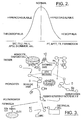
- Normal hemostasis is maintained by tightly regulated interactions of the blood vessel wall, blood platelets and blood plasma proteins. Under normal conditions there is a delicate balance between the individual components of the hemostatic system. Any disturbances in this hemostatic balance, the hemostatic potential, could result in bleeding or thrombosis, Figure 1 .
- hemostatic potential we mean the ability to maintain a balance between procoagulant and anticoagulante states, as measured by fibrin polymerization, when coagulation is initiated by a trigger or activator.
- a thrombotic tendency results from the generation of excess thrombin activity and increased fibrin polymerization and clot formation (hypercoagulability) while a bleeding tendency (hemophilia) results from insufficient thrombin generation and reduced fibrin polymerization and clot formation (hypocoagulability).
- thrombophilia results from the generation of excess thrombin activity and increased fibrin polymerization and clot formation (hypercoagulability)
- hemophilia results from insufficient thrombin generation and reduced fibrin polymerization and clot formation (hypocoagulability).
- Thrombophilia and haemophilia can be either congenital or acquired.
- the congenital forms have a genetic basis and are therefore not readily corrected.
- the acquired forms generally result from environmental changes, often the effect of drugs, and are therefore susceptible to manipulation. For example a normal individual given warfarin develops acquired haemophilia, stopping the warfarin abolishes the condition.
- a normal individual given high dose estrogen develops acquired thrombophilia, stopping the estrogen abolishes the condition.
- the fundamental basis of both the congenital (genetic) and acquired (environmental) thrombophilias and haemophilias is a change in either the amount or activity of one or more key components of the coagulation pathway.
- thrombophilia is a mutation in the factor V gene which results in the production of a structurally altered factor V protein (Factor V Leiden) that is resistant to enzymatic cleavage by protein C, a critical regulatory component.
- Classical Haemophilia A is due to a mutation in the factor VIII gene which results in either reduced production of factor VIII, or production of a structurally altered factor VIII protein that does not function correctly.
- the acquired forms do not result from altered structure but rather alteration of the amount of a key component, typically more than one at a time.
- FIG. 2 illustrates the various states of coagulability and lists examples of assays used to assess the degree or presence of an imbalance. There is currently not an assay that can be used to assess both hyper and hypocoagulability simultaneously. This is due in part to the complexity of the coagulation process, the interdependence of the various components and the identification of a means to monitor the hemostatic potential of the entire coagulation system.
- Figure 3 presents an overview of the coagulation process. The process can be divided into four dependent phases, (1) the initiation phase, (2) the propagation phase, (3) the amplification phase and (4) the polymerization phase. All of the phases are affected by regulatory and feedback processes referred to as anticoagulant pathways.
- Initiation or triggering of coagulation occurs by exposure of tissue factor due to vascular damage, plaque rupture or monocyte expression as a result of inflammation. Trace amounts of FVIIa and tissue factor form the extrinsic Xase complex. This complex enhances the catalytic activity of VIIa towards factors X and IX resulting in the formation of the active enzymes Xa and IXa.
- Factor Xa generated by the extrinsic Xase complex forms a small amount of thrombin (IIa). The thrombin generated is capable of activating small amounts of the cofactors VIII and V.
- the extrinsic Xase complex in vivo, is quickly inactivated by Tissue Pathway Factor Inhibitor, TFPI, via the formation of a quaternary complex consisting of TF, VIIa and Xa. Under physiological conditions the extrinsic Xase generates only picomolar amounts of thrombin.
- TFPI Tissue Pathway Factor Inhibitor
- Factor Xa is alternatively generated by the complex of the enzymes IXa and its cofactor VIIIa.
- This enzyme complex is referred to as intrinsic Xase. Formation of the Xa by the intrinsic Xase complex is approximately 50 fold more efficient than the extrinsic Xase.
- Factor Xa and its activated cofactor, FVa form a complex on the surface of activated platelets. This is an efficient catalyst for the conversion of prothrombin to thrombin, referred to as the prothrombinase complex.
- Thrombin formed via the intrinsic Xase complex is capable of amplifying its own production by positive feedback (activation). Thrombin activates Factors VIII and V and Factor XI activation leads to further production of the enzymatic component of intrinsic Xase (Factor IXa). Normal thrombin production is highly regulated and localized. TFPI neutralizes the trigger for thrombin generation. Active proteases (IIa, Xa, IXa) must be inactivated by protease inhibitors to avoid disseminated thrombosis. One of the most significant of these inhibitors is antithrombin III (ATIII).
- thrombin and Xa are rapidly inhibited by ATIII.
- Thrombin can also bind non-damaged sub-endothelium via a receptor molecule, Thrombomodulin (TM ).
- TM Thrombomodulin
- the formation of the IIa/TM complex changes the substrate specificity of thrombin from a procoagulant to an anticoagulant.
- Thrombin bound to TM is a potent activator of Protein C, converting it to the active enzyme Activated Protein C (APC).
- APC Activated Protein C
- APC together with its cofactor protein S cleaves activated cofactors FVIIIa and FVa yielding their inactive forms, FVIIIi and FVi.
- Thrombomodulin also accelerates the inactivation of thrombin by ATIII.
- thrombin leads ultimately to cleavage of fibrinogen to form fibrin.
- cross-linking of soluble fibrin strands is mediated by Factor XIIIa, an enzyme generated by thrombin activation.
- the thrombin-TM complex activates the procarboxypeptidase thrombin activated fibrinolysis inhibitor (TAFI).
- TAFI procarboxypeptidase thrombin activated fibrinolysis inhibitor
- TAFI procarboxypeptidase thrombin activated fibrinolysis inhibitor
- TAFI procarboxypeptidase thrombin activated fibrinolysis inhibitor
- Clot initiation occurs at the intersection between the initiation and propagation phases when only approximately 5% of thrombin has been formed.
- the majority of the thrombin formed is generated after the initiation of fibrin polymerization, thus the rate of fibrin polymerization is a more sensitive indicator of the dynamics of coagulation. Changes in the propagation phase, amplification phase and anticoagulant pathways alter the rate of thrombin generation and the impact of thrombin availability on rate of fibrin polymerization.
- Recent studies by Cawthern et al. (1998) suggested that measurement of this thrombin is more informative than clotting time in assessing the pathophysiology of hemophitias.
- thrombin by looking at the kinetics of formation of the thrombin-antithrombin complex (indictor of thrombin generation) and formation of fibrinopeptide A (indicator of fibrinogen cleavage) and not by measuring the kinetics of fibrin polymerization. Variations in concentration or quality of the fibrinogen or fibrin strands can only be measured as a function of the actual polymerization process. Assays currently used to assess variations in the coagulation process typically can only assess variations in - one or two phases. These assays measure events independently and therefore negate or eliminate the ability to detect variations in the other phases or interactions between the various phases.
- Assays associated with the assessment of bleeding risk include the Prothrombin Time (PT), Activated Partial Thromboplastin Time (aPTT), Thrombin Time (TT) and Fibrinogen (Fib) assays ( figure 2 ). These assays are based on the addition of potent activators of the coagulation process and thus are only abnormal when major defects are present. These assays are not designed to detect the composite effect of multiple minor alterations. For example in the PT test, which utilizes a very high concentration of a tissue extract, called thromboplastin, and calcium are added to citrated plasma. Whole blood is mixed with citrate when the blood sample is taken.
- PT Prothrombin Time
- aPTT Activated Partial Thromboplastin Time
- TT Thrombin Time
- Fibrinogen Fibrinogen
- the citrate binds the calcium and "anticoagulates" the blood as calcium ions are required for assembly of the tenase and prothrombinase complexes.
- the blood sample is then centrifuged and the plasma is separated.
- the tenase (or Xase) and prothrombinase complexes can form and thrombin can be generated.
- the source of tissue factor is then thromboplastin.
- the concentration of tissue factor is extremely high (supraphysiological) and so only the initiation phase of thrombin generation is required. The propagation and amplification phases are bypassed.
- the prothrombin time is therefore insensitive to many changes in the coagulation pathway and is incapable of detecting hypercoagulability.
- Assays based on diluted thromboplastin have been formulated to aid in the diagnosis of patients with antiphospholipid syndrome (APS).
- APS antiphospholipid syndrome
- the thromboplastin together with the phospholipids are diluted to enhance the sensitivity of the PT to the presence of antiphospholipid antibodies.
- the dilute PT clotting time is prolonged in APS due the unavailability of phospholipid surfaces and therefore the assay is phospholipid dependent instead of TF dependent.
- Assays associated with the assessment of a hypercoagulable state include the Thrombin Anti-Thrombin Complex (TAT), Prothrombin fragment F1.2, PAI 1, APCr and D-dimer. These assays are designed to measure a specific marker or product of the coagulation process. For example, the measurement of elevated levels of D-dimer indicates that the clotting process has been activated. However, there is no way of determining whether the D-dimer was being produced as a product of the normal healing process or if there is an underlying hypercoagulable risk.
- the hypercoagulable state cannot be globally assessed by a single assay but currently requires a battery of tests.
- a global assay for the assessment of hemostatic potential would be able to identify an imbalance utilizing a single assay principle that is sensitive to defects, singular or in combination. The assay would also be sensitive to effects of intervention to restore the hemostatic balance.
- the assay assumes that there is a direct correlation between endogenous thrombin potential measured via an exogenously added artificial substrate and the assessment of a hemostatic imbalance.
- the use of an artificial substrate instead of thrombin's natural substrate, fibrinogen ignores the effects of variations in fibrinongen concentration and fibrinogen configuration.
- Thrombin is a cleavage product from the proteolysis of Prothrombin, a serine protease. Thrombin then cleaves fibrinogen, its natural substrate, resulting in soluble fibrin monomers that are crossed linked via FXllla to formed crossed linked polymerized clots.
- Thrombin is a highly regulated molecule that possesses both procoagulant and antithrombotic behavior. Additionally, there are numerous substrates that inactivate thrombin before it can cleave fibrinogen. In addition to not directly measuring the ability to form a clot the ETP assay has several other major limitations. Limitations of the test include:
- Duchemin et al. described a further modification of the ETP where the protein C pathway is assessed by adding exogenous thrombomodulin. This method was also modified to take into account proteins that modulate anticoagulant activity, including antithrombin III. Like ETP, this modified assay is designed to only measure thrombin generation and not the effects of thrombin, i.e. dynamic clot formation.
- the assay is not sensitive to defects in the propagation and amplification phases, the kinetics of clot polymerization or to the interrelationships between the factors responsible for thrombin generation.
- the present invention assesses both the anticoagulant and procoagulant potential of a blood sample. Furthermore, the present invention's sensitivity can be enhanced by using more dilute coagulation activator, more dilute than has previously been used, since the endpoint method is not restricted to clot time but analysis can be conducted for the entire dynamic coagulation process as measured by evaluating kinetic parameters of the optical data profile. Analysis of more than simply clot time can be accomplished even when very weak and unstable clots are formed.
- Variations in the amplification and/or propagation phases will reduce or alter the rate of generation of thrombin and thus impact the rate of fibrinogen cleavage and ultimately the rate of fibrin polymerization. Because the present invention can measure the rate of fibrin polymerization throughout the dynamic coagulation process, it measures the clinically important thrombin that is generated after clotting time.
- Thrombin generation is measured as a function of TAT complex formation or the use of a chromogenic substrate and the formation of fibrin as measured by the release of FPA.
- All of the systems and models to date have been designed to understand a discrete process or interaction of the coagulation process and cannot provide an assessment of the overall hemostatic potential.
- the method of the present invention is designed to not only assess the interplay of the coagulation proteins together with synthetic cell surfaces, it is aimed at capturing this in a dynamic measurement that correlates to clinical outcome.
- the technology and methods described in the present invention can also be modified to introduce components of the fibrinolytic system as well as cells and flow conditions.
- a reagent and kit for a global test of coagulation has now been developed, which is accurate and easy to use.
- a single test can be used to quantify both hyper- and hypocoagulability.
- the concept is based on the addition of a minimal concentration of a thromboplastin coagulation activator sufficient to trigger but insufficient to result in complete fibrin polymerization so as to allow detection of perturbances in the propagation, amplification and polymerization pathways.
- the concentration of the coagulation activator is II picomolars or less.
- the coagulability (hyper/hypo) of a sample determines the magnitude of the thrombin explosion and the direct and Indirect influence that has on the rate of fibrin polymerization.
- This concept is contrary to an assay system such as the PT, which uses excess amounts of TF (or thromboplastin).
- TF or thromboplastin
- the rate of fibrin polymerization produced by a standardized coagulation activator dilution is then used to indicate if a plasma sample is normal, hyper- or hypocoagulable.
- the technique can be used to determine how much the plasma needs to be modified in order to restore coagulability to normal. For example, in the case of hypocoagulability, this might be achieved by clotting factor replacement or in the case of hypercoagulability, by the addition of a natural anticoagulant or the use of an anticoagulant drug.
- the rate of fibrin polymerization of haemophilia plasmas are less than the rate of polymerization for a normal plasma and the rate of fibrin polymerization of thrombophilia plasmas are greater than that of a normal sample.
- the rate of fibrin polymerization is sensitive to minor changes in the components of hemostasis even when differences in clotting time cannot be detected.
- Figure 6 illustrates waveforms from the normal, hypercoagulable and hypocoagulable specimens. The rate of polymerization is affected even though the time of clot initiation is essentially unchanged.
- a reagent and kit are provided that can be used to determine the degree of hyper- or hypocoagulability of a plasma sample. Furthermore, it can be used on samples containing platelets or other cells as a measure of the contribution of cellular components to coagulability.
- the test in some embodiments, relies on the use of a standardized dilution of tissue factor in the presence of an excess of phospholipids with the rate of fibrin formation as the detection endpoint.
- the test is simple and can be automated on standard laboratory coagulometers.
- the test in the present invention can be run on a test sample in the absence of the addition of an exogenous substrate, e.g. a chromogenic substrate. The test is sensitive to fibrin concentration and/or configuration.
- modifications to the components or concentrations of the reagent or endpoint selection are tailored to facilitate the development and/or monitoring of novel pharmaceutical agents.
- examples of such applications are inhibitors of initiation of the TF pathway (TFPI, FVIIa inhibitors), inhibitors of thrombin generation such as inhibitors of FXa, (synthetic pentasaccharides) and inhibitors of thrombin activity (direct thrombin inhibitors).
- Lipid composition, size or concentration can also be modified to tailor the assay towards the development of drugs targeted to the propagation and amplification pathways. For example, lipid composition can be altered to produce vesicles that maximize Xa generation or alternatively, designed to maximize prothrombinase activity.
- the invention can also be modified to focus on the anticoagulant potential of the plasma by including thrombomodulin, an activator of protein C. Lipid vesicles maximizing the.activity of APC could also be added to the reagent.
- the assay can also be modified to exaggerate a mildly abnormal subpopulation. The consequences of this approach are that severely thrombotic or hemorrhagic samples will exceed the signal to noise ratio and not be measured but subtle differences at the onset of a disease or an earlier indication of effective intervention would be gained.
- the present invention is directed to a. reagent and kit for determining if a patient or specimen from said patent is hypercoagulable, hypocoagulable or normal in a single test as defined in the claims.
- the test comprises the steps of initiating coagulation In a patient's sample in vitro in the presence of an activator. Said activator is added to the sample in an amount which will result in intrinsic tenase-dependent fibrin polymerization (involves propagation and amplification loops).
- the coagulation activator is a thromboplastin in a concentration of II picomolars or less.
- the plasma sample is undiluted thus allowing for sufficient concentrations of all of the endogenous proteases and inhibitors. Formation of the fibrin polymerization is recorded over time so as to derive a graphic time-dependent polymerization profile. This profile will show whether the patient is hypercoagulable, normal, or hypocoagulable by comparing the sample profile with a profile from a known sample.
- the activator is a thromboplastin, preferably Tissue Factor (TF).
- the TF is recombinant TF (rTF) that is relipidated with phospholipids, which form liposome vesicles.
- rTF recombinant TF
- phospholipids provide the surfaces to assemble intrinsic Xase and prothrombinase complexes.
- the phospholipids are present at a concentration, which is not rate limiting to the coagulation process and remains constant and independent of dilution. These phospholipid vesicles or liposomes mimic platelet and monocyte surfaces.
- Optical data profiles are generated on an automated coagulation analyzer such as the MDATM180 offered by Organon Teknika Corporation.
- endpoints such as the time of clot initiation and the rate of polymerization are calculated from the data profiles. More preferably the 1 st and 2 nd derivatives from the data profile are calculated and the min and max of the derivatives are calculated with respect to value and the associated time index. Most preferably the endpoints are calculated and one or more of the following ratios are calculated using the mentioned endpoints:
- Figure 1 illustrates the consequences of any disturbance in this so-called hemostatic balance or potential. Too little hemostasis (decreased platelet function, hypo-coagulation, hyper-fibrinolysis) at the site of injury leads to persistent bleeding, while too much hemostasis (increased platelets function, hyper-coagulation, hypo-fibrinolysis) leads to the formation of an excessive thrombus with vascular obstruction and ischemia.
- Figure 2 illustrates the conditions associated with being out of hemostasis and lists examples of assays used to assess the degree or presence of an imbalance.
- Figures 3 illustrates the four dependent phases of the coagulation process, (1) the initiation phase, (2) the amplification phase, (3) the propagation phase and (4) the polymerization phase of hemostasis. All of the phases are affected by regulation and feedback processes referred to as anticoagulant pathways.
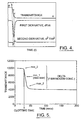
- Figure 4 illustrates the optical data from a clotting assay and the first and second derivative calculated from that data.
- Table 1 describes a set of parameters calculated from the data and derivatives illustrated in figure 4 .
- Table 1 Parameter Description Slope 1 Initial slope from point A to point B Delta 1 Amplitude of signal change from point A to point B Slope 3 Final slope from point D to point E Delta Amplitude of signal change Index Min 1 Time at point C Min 1 Minimum value of 1 st derivative (Rate of change at point C) Index Max 2 Time at point D Max 2 Max. value of 2nd derivative (Acceleration at point D) Index Min 2 Time at point B Min 2 Minimum value of 2nd derivative (Acceleration at point B)
- Figure 5 illustrates where min_2, the time index of min2 (clotting time), min_1, max_2 and delta (proportional to fibrinogen concentration) are located in the optical data profile.
- Figure 6 contains examples of waveforms for the global screening assay at dilute tissue factor.
- the APC resistant, hypercoagulable specimen generates a waveform that has essentially the same time of clot initiation compared to the normal.
- the rate of fibrin polymerization for the hypercoagulable specimen is significantly greater than that of the normal.
- the FVIII and FIX deficient hypocoagulable specimens have only a slightly prolonged time of clot initiation whereas the rates of polymerization are significantly reduced when compared to normal or hypercoagulable specimens.
- Figure 7 illustrates the change in ratio as a function of dilution for a FVIII deficient specimen and a Protein S deficient Specimen.
- the ratio values at 1:50,000 dilution of thromboplastin deviate from the response of the normal plasma.
- the hypocoagulabe specimen produces ratios that are greater than 1 and the hypercoagulable specimen has ratios that are less than 1 for this endpoint (clot time) / ratio combination. Additionally, the abnormal specimen deviates from normal at different dilutions and in opposite directions.
- Figure 8 contains ratios of the min-1 values (the maximum rate of fibrin polymerization) for hypocoagulable specimens at three dilutions of rTF compared to the min_1 values of the ratio of the same dilution of a normal plasma. All of the ratios of the hypocoagulable plasmas for all three dilutions are less than the normal response (values of ⁇ 1). As the dilution increase, i.e. less tissue factor is provided, the difference in the ratios increases.
- Figure 9 illustrates ratios of the min_1 values for hypercoagulable specimens at three dilutions of rTF and 10nM thrombomodulin compared to min_1 values of the ratio for the same conditions of a normal plasma. All of the ratios of the hypercoagulable plasmas for all three dilutions are greater than the normal response (values of >1). As the dilution increase, i.e. less tissue factor is provided, the difference in the ratios increases.
- Figure 10 illustrates the effects on min_1 values of varying tissue factor and thrombomodulin concentrations on results for hypercoagulable, hypocoagulable and normal plasmas.
- the data indicate that an optimal concentration can be defined to facilitate differentiation between normal, hypercoagulable and hypocoagulable plasmas. Additionally, other concentrations of tissue factor and thrombomodulin facilitate improvements in sensitivity and specificity for a particular condition at the expense of the sensitivity and specificity of another type of condition.
- Tables 2 and 3 summarize the results of measuring the kinetic parameters, min 1 and min 2 with a series of defined patient plasmas.
- concentration of TF was 10 pM and TM was adjusted to 10 nM.
- the phospholipid concentration was kept constant at 150 micromolar.
- the data shows that the reagent in the presence of TM is able to differentiate hyper and hypocoagulable plasmas with a single reagent formulation. Additionally, the data indicates that TM is not essential to obtain discrimination between the hypocoagulable and a normal standard plasma pool. Data are calculated as ratios to a normal pool with and without thrombomodulin. Ratios of the min2 parameter were higher than the corresponding min1 values for the hypercoagulable plasmas.
- Tables 2 and 3 illustrate the behavior of defined plasmas in the presence and absence of thrombomodulin as determined by the kinetic endpoints min_1 and min_2.
- Table 2 Plasma Type Min _1 values with no TM Min _1 values with 10 nM TM Min_1 Ratio Specimen without TM/Normal plasma without TM Min_1 Ratio Spedmen with 10 nM TM/Normal plasma with 10 nM TM Normal Plasma 101 68 PC Defident 110 105 1.09 1.54 Lupus 116 79 1.15 1.16 FV Leiden 95 77 0.94 1.13 FV Leiden & PT 20210 260 248 2.57 3.64 FIX Deficient 71 40 0.70 0.59 FVIII Defident 84 46 0.83 0.68
- Table 3 Plasma Type Min_2 values with no TM Min_2 values with 10 nM TM Min_2 Ratio Specimen without TM/Normal plasma without TM Min_2 Ratio Spedmen with 10 nM
- the assay was conducted by adding 50uL of plasma to 50uL of the activator and then adding 50uL of the start reagent.
- a normal sample, a hypocoagulable sample (Factor VIII deficient plasma) and a hypercoagulable plasma (protein S deficient plasma) were evaluated at various dilutions of the activator.
- the activator was a commercially available thromboplastin (Thromborel R, Behring Diagnostics) diluted with a buffer at two dilutions, a 1:100 and 1:50000 of its original concentration.
- the start reagent consisted of 0.25 M Calcium Chloride.
- the assay was conducted at 37 C and the reaction was monitored at 580nm for 300 seconds. Endpoints were calculated for time and rate indices of clot formation.
- the results for the two abnormal plasmas began to deviate from the calculated endpoints or ratios of the normal plasma.
- the results can be expressed as the magnitude of deviation at a given dilution or as the dilution required to deviate from ideal (normal value or normal range).
- Figure 7 illustrates that the hypercoagulable and hypocoagulable results deviate in opposite directions indicating the ability to differentiate between the two conditions.
- the assay was conducted by adding 50uL of plasma to 50uL of the activator and then adding 50uL of the start reagent.
- a set of normal samples, a series of samples from hypocoagulable individuals and a series of plasmas from hypercoagulable individuals were evaluated at various dilutions of the activator.
- the activator was a preparation of TF reconstituted with phospholipids to between 20 to 3.3 pM (1:20,000 to 1:120,000 dilution) and phospholipid prepared by extrusion with and without TM.
- the start reagent consisted of 0.025 M Calcium Chloride.
- the assay was conducted at 37 C and the reaction was monitored at 580nm for 300 seconds. The value of the minimum of the 1 st derivative and the value of the minimum of the 2 nd derivative were calculated for all samples. Ratios of the endpoints were compared to other dilutions and other samples as follows:
- Figures 8 and 9 illustrate the differentiation for hypercoagulable and hypercoagulable specimens when compared to normal.
- Tables 2 and 3 illustrate the behavior of defined plasmas in the presence and absence of thrombomodulin as determined by the kinetic endpoints min_1 and min_2.
- Figure 10 demonstrates the effect of varying tissue factor and thrombomodulin on the results from hypercoagulable, hypercoagulable and normal plasmas. The data indicate that variations in the concentrations facilitate improvements in sensitivity and specificity for a condition at the expense of the sensitivity and specificity of another type of condition.
- the TF is added to the sample at a concentration of about less than or equal to 10 picomolar and the phospholipid concentration of between 10 to 300 ⁇ M.
- the TF can be added to the sample at a concentration of 3 to 10 picomolar and the phospholipid vesicles can be added at 100 to 150 micromolar.
- thrombomodulin is added at 0 to 30 nanomolar and most preferably at a concentration of 5 to 15 nanomolar.
- Calcium Chloride is most preferably added at a concentration of about 25 mM. All of the reagent component concentrations described are further diluted 1:3 in the plasma/buffer matrix in the cuvette.
- One or more parts or endpoints of the time dependent measurement profile obtained by monitoring fibrin polymerization in the test sample can be compared to the same parts or endpoints of a time dependent measurement profile obtained by monitoring fibrin polymerization in the test sample at a different coagulation activator concentration and/or to the same parts or endpoints for a known (e.g. normal) test sample.
- the part of the profile can be one or more of initiation of clot formation, overall change in profile, slope of profile after initiation of clot formation, and acceleration at the time of clot initiation.
- an additional profile can be obtained for a known sample from computer memory or by adding the activator at at least one concentration to a known sample and monitoring the formation of fibrin polymerization over time.
- the parameter from each time-dependent fibrin polymerization profile having varying activator concentrations can be determined and a concentration at which the at least one parameter of said sample being tested deviates from normal can be determined. The point of deviation is indicative of the hypercoagulable or hypocoagulable state.
- the part of the profile is preferably a time index of the minimum of the first derivative, the value of the minimum of the first derivative, the time index for the minimum of the second derivative, the value for the minimum of the second derivative, the time index of the maximum of the second derivative, the value of the maximum of the second derivative, or the overall magnitude of change. More preferably, the part is rate or acceleration of fibrin polymerization, wherein the rate or acceleration is compared to rate or acceleration at the same activator concentration for the known sample.
- a difference or ratio of said parameters for said test sample and said normal sample can instead be determined.
- the parameter is clot time
- a ratio of clot times at different activator concentrations can be determined.
- a ratio of other parameters, rate of clot formation, maximum acceleration of clot formation, turbidity at a predetermined time period, and total change in turbidity can also be determined in order to measure defects in the thrombin propagation and/or amplification phases.
- a ratio can be taken of the at least one parameter for said test sample to the same parameter for a normal sample.
- the ratio can be determined for multiple concentrations of activator to better characterize the hypo- or hyper-coagulability. For example, the concentration at which said ratio (test sample/known sample) departs from 1 (or a range around 1) can show the abnormal coagulability.
- ratios aid determination of the hemostatic potential (e.g. the hypocoagulability, stasis, or hypercoagulability; or the bleeding or thrombotic tendency of the patient).
- a first ratio can be calculated for the at least one parameter at two different concentrations of the activator.
- a second ratio can be calculated of said first ratio at the two different activator concentrations relative to a first ratio calculated for a known sample at two different activator concentrations.
- a third ratio can be calculated of the second ratio at a first reagent formulation and the second ratio at a second reagent formulation.
- the second reagent can vary in a number of ways from the first, in one embodiment the first reagent formulation can comprise a coagulation activator and the second reagent formulation can comprise a coagulation activator and an activator of an anticoagulant pathway.
- a fourth ratio could be calculated of the second ratio calculated for one endpoint relative to the second ratio calculated for a different endpoint. Significant information can be obtained by changing the reagent formulation and comparing the same endpoints, or by maintaining the reagent formulation (though possibly at a different concentration) and comparing different endpoints (or both endpoint and reagent formulation and/or concentration can be altered).
- An activator of one or more anticoagulant pathways can be added along with the coagulation activator.
- Such an additional activator can be any activator of an anticoagulant pathway, such as the protein C pathway.
- Thrombomodulin is one example, which can be added in the form of purified human thrombomodulin, purified non-human mammalian thrombomodulin, soluble or membrane associated thrombomodulin, native thrombomodulin or reconstituted with phospholipids, partially or fully glycolsylated thrombomodulin, or fully deglycosylated thrombomodulin, with added heparin-like molecules.
- the coagulation activator can be any suitable activator including recombinant or purified tissue factor, truncated tissue factor, or cells expressing tissue factor on their surface. If vesicles or liposomes are added, they can be in the form of platelets, cellular debris, phospholipids or platelet microparticles. A metal salt if added can be a halide of magnesium, calcium or manganese, or other divalent metal salt. Buffers and stabilizers could also be added if desired.
- a reagent or kit for assessing hemostatic potential should have a coagulation activator. Additional components of the reagent or kit could include the above-mentioned vesicles, metal salt or ions, and anticoagulant pathway activator, if desired. In the kit, the components could all be provided in separate containers, or mixed together in any combination in one or more containers.
- phospholipid vesicles can be any suitable phospholipid or combination of phospholipids including one or more of phosphatidylcholine, phosphatidylethandamine and phosphatidylserine, which can be provided at a ratio of approximately 5 to 30 mole percent phosphatidylethandamine, 1 to 10 percent phosphatidylserine and the remainder phosphatidylcholine.
- phosphatidylcholine phosphatidylethandamine and phosphatidylserine
- These vesicles can be prepared in a variety of ways to yield liposomes of various sizes.
- Phospholipids can be provided at a concentration that is not rate limiting, e.g. at a concentration of from 10 to 300 micromolar, and preferably in the range of from 50 to 200 micromolar.
- Tissue factor can be provided at a concentration of 10 picomolar or less, 8 picomolar or less, or preferably 6 picomolar or less. The concentration could be 3 picomolar or less, though whatever concentration of tissue factor, it should allow for hemostatic potential assessment as set forth herein. If it is desired to add thrombomodulin, it can be provided at a concentration of 30 nanomolars or less, preferably in a range of from 5 to 20 nanomolar. If a metal salt is to be added, it can be provided in a reagent or kit at a concentration of from 5 to 50 mM, preferably from 15 to 35 mM.
Landscapes
- Health & Medical Sciences (AREA)
- Life Sciences & Earth Sciences (AREA)
- Hematology (AREA)
- Engineering & Computer Science (AREA)
- Molecular Biology (AREA)
- Biomedical Technology (AREA)
- Chemical & Material Sciences (AREA)
- Immunology (AREA)
- Urology & Nephrology (AREA)
- Biotechnology (AREA)
- Biochemistry (AREA)
- Cell Biology (AREA)
- Food Science & Technology (AREA)
- Medicinal Chemistry (AREA)
- Physics & Mathematics (AREA)
- Analytical Chemistry (AREA)
- Microbiology (AREA)
- General Health & Medical Sciences (AREA)
- General Physics & Mathematics (AREA)
- Pathology (AREA)
- Investigating Or Analysing Biological Materials (AREA)
- Measuring Or Testing Involving Enzymes Or Micro-Organisms (AREA)
- Medicines That Contain Protein Lipid Enzymes And Other Medicines (AREA)
Abstract
Description
- The invention is directed to a Reagent and Kit for determining whether a patient is hypercoagulable, hypocoagulable or normal in a single test on a sample from the patient. The invention allows for globally assessing both the hypercoagulable potential and hypocoagulable potential of a patient in a single assay.
- Hemostasis is the entire physiological process of maintaining blood in a fluid state within intact blood vessels and preventing excess blood loss by arresting flow via the formation of a hemostatic plug. Normal hemostasis is maintained by tightly regulated interactions of the blood vessel wall, blood platelets and blood plasma proteins. Under normal conditions there is a delicate balance between the individual components of the hemostatic system. Any disturbances in this hemostatic balance, the hemostatic potential, could result in bleeding or thrombosis,
Figure 1 . By "hemostatic potential we mean the ability to maintain a balance between procoagulant and anticoagulante states, as measured by fibrin polymerization, when coagulation is initiated by a trigger or activator. - A thrombotic tendency (thrombophilia) results from the generation of excess thrombin activity and increased fibrin polymerization and clot formation (hypercoagulability) while a bleeding tendency (hemophilia) results from insufficient thrombin generation and reduced fibrin polymerization and clot formation (hypocoagulability). There is as yet no single laboratory parameter that is increased in all forms of hypercoagulability and decreased in all forms of hypocoagulability. This is in part due to factors other than plasma that play a part in hemostasis. As described above, these other factors include the blood vessel wall and platelets. However, large proportions of the hemostatic disorders are related to defects or deficiencies in the blood proteins that constitute the coagulation system. These proteins are responsible for the stabilization of the platelet plug by the formation of fibrin. Therefore, a global measure of the plasma contribution to coagulation would facilitate the investigation and management of patients with altered hemostasis.
- Thrombophilia and haemophilia can be either congenital or acquired. The congenital forms have a genetic basis and are therefore not readily corrected. The acquired forms generally result from environmental changes, often the effect of drugs, and are therefore susceptible to manipulation. For example a normal individual given warfarin develops acquired haemophilia, stopping the warfarin abolishes the condition. A normal individual given high dose estrogen develops acquired thrombophilia, stopping the estrogen abolishes the condition. The fundamental basis of both the congenital (genetic) and acquired (environmental) thrombophilias and haemophilias is a change in either the amount or activity of one or more key components of the coagulation pathway. For example the most commonly recognized hereditary form of thrombophilia is a mutation in the factor V gene which results in the production of a structurally altered factor V protein (Factor V Leiden) that is resistant to enzymatic cleavage by protein C, a critical regulatory component. Classical Haemophilia A is due to a mutation in the factor VIII gene which results in either reduced production of factor VIII, or production of a structurally altered factor VIII protein that does not function correctly. In contrast to the congenital thrombophilias and haemophilias the acquired forms do not result from altered structure but rather alteration of the amount of a key component, typically more than one at a time. For example the thrombophilic effect of oestrogen is due to the composite effects of a rise in factors XI, IX, VIII, II and fibrinogen and a reduction in the anticoagulant protein S. The haemophilic effect of warfarin is due to a reduction in factors II, VII, IX and X.
Figure 2 illustrates the various states of coagulability and lists examples of assays used to assess the degree or presence of an imbalance. There is currently not an assay that can be used to assess both hyper and hypocoagulability simultaneously. This is due in part to the complexity of the coagulation process, the interdependence of the various components and the identification of a means to monitor the hemostatic potential of the entire coagulation system.Figure 3 presents an overview of the coagulation process. The process can be divided into four dependent phases, (1) the initiation phase, (2) the propagation phase, (3) the amplification phase and (4) the polymerization phase. All of the phases are affected by regulatory and feedback processes referred to as anticoagulant pathways. - Initiation or triggering of coagulation occurs by exposure of tissue factor due to vascular damage, plaque rupture or monocyte expression as a result of inflammation. Trace amounts of FVIIa and tissue factor form the extrinsic Xase complex. This complex enhances the catalytic activity of VIIa towards factors X and IX resulting in the formation of the active enzymes Xa and IXa. Factor Xa generated by the extrinsic Xase complex forms a small amount of thrombin (IIa). The thrombin generated is capable of activating small amounts of the cofactors VIII and V. in vivo, the extrinsic Xase complex is quickly inactivated by Tissue Pathway Factor Inhibitor, TFPI, via the formation of a quaternary complex consisting of TF, VIIa and Xa. Under physiological conditions the extrinsic Xase generates only picomolar amounts of thrombin.
- During the propagation phase of coagulation the role of the extrinsic Xase is minimized and Factor Xa is alternatively generated by the complex of the enzymes IXa and its cofactor VIIIa. This enzyme complex is referred to as intrinsic Xase. Formation of the Xa by the intrinsic Xase complex is approximately 50 fold more efficient than the extrinsic Xase. Factor Xa and its activated cofactor, FVa, form a complex on the surface of activated platelets. This is an efficient catalyst for the conversion of prothrombin to thrombin, referred to as the prothrombinase complex. Thrombin formed via the intrinsic Xase complex is capable of amplifying its own production by positive feedback (activation). Thrombin activates Factors VIII and V and Factor XI activation leads to further production of the enzymatic component of intrinsic Xase (Factor IXa). Normal thrombin production is highly regulated and localized. TFPI neutralizes the trigger for thrombin generation. Active proteases (IIa, Xa, IXa) must be inactivated by protease inhibitors to avoid disseminated thrombosis. One of the most significant of these inhibitors is antithrombin III (ATIII). Both thrombin and Xa, and to a lesser extent IXa released from membrane surfaces, are rapidly inhibited by ATIII. Thrombin can also bind non-damaged sub-endothelium via a receptor molecule, Thrombomodulin (TM ). The formation of the IIa/TM complex changes the substrate specificity of thrombin from a procoagulant to an anticoagulant. Thrombin bound to TM is a potent activator of Protein C, converting it to the active enzyme Activated Protein C (APC). APC together with its cofactor protein S cleaves activated cofactors FVIIIa and FVa yielding their inactive forms, FVIIIi and FVi. Thrombomodulin also accelerates the inactivation of thrombin by ATIII.
- The formation of thrombin leads ultimately to cleavage of fibrinogen to form fibrin. During the polymerization phase cross-linking of soluble fibrin strands is mediated by Factor XIIIa, an enzyme generated by thrombin activation. The thrombin-TM complex activates the procarboxypeptidase thrombin activated fibrinolysis inhibitor (TAFI). Thus thrombin plays a role during this phase by both influencing the architecture and stabilization of the fibrin clot. Thrombin is a key enzyme and effector of the coagulation process. Thrombin is both a potent procoagulant and anticoagulant. However, it is thrombin's ability to cleave fibrinogen and its contribution to fibrin polymerization events that are critical to maintaining stasis.
- Clot initiation, often referred to as clotting time, occurs at the intersection between the initiation and propagation phases when only approximately 5% of thrombin has been formed. The majority of the thrombin formed is generated after the initiation of fibrin polymerization, thus the rate of fibrin polymerization is a more sensitive indicator of the dynamics of coagulation. Changes in the propagation phase, amplification phase and anticoagulant pathways alter the rate of thrombin generation and the impact of thrombin availability on rate of fibrin polymerization. Recent studies by Cawthern et al. (1998) suggested that measurement of this thrombin is more informative than clotting time in assessing the pathophysiology of hemophitias. However these investigators measured thrombin by looking at the kinetics of formation of the thrombin-antithrombin complex (indictor of thrombin generation) and formation of fibrinopeptide A (indicator of fibrinogen cleavage) and not by measuring the kinetics of fibrin polymerization. Variations in concentration or quality of the fibrinogen or fibrin strands can only be measured as a function of the actual polymerization process. Assays currently used to assess variations in the coagulation process typically can only assess variations in - one or two phases. These assays measure events independently and therefore negate or eliminate the ability to detect variations in the other phases or interactions between the various phases.
- Assays associated with the assessment of bleeding risk include the Prothrombin Time (PT), Activated Partial Thromboplastin Time (aPTT), Thrombin Time (TT) and Fibrinogen (Fib) assays (
figure 2 ). These assays are based on the addition of potent activators of the coagulation process and thus are only abnormal when major defects are present. These assays are not designed to detect the composite effect of multiple minor alterations. For example in the PT test, which utilizes a very high concentration of a tissue extract, called thromboplastin, and calcium are added to citrated plasma. Whole blood is mixed with citrate when the blood sample is taken. The citrate binds the calcium and "anticoagulates" the blood as calcium ions are required for assembly of the tenase and prothrombinase complexes. The blood sample is then centrifuged and the plasma is separated. When calcium is added back, the tenase (or Xase) and prothrombinase complexes can form and thrombin can be generated. The source of tissue factor is then thromboplastin. However, the concentration of tissue factor is extremely high (supraphysiological) and so only the initiation phase of thrombin generation is required. The propagation and amplification phases are bypassed. The prothrombin time is therefore insensitive to many changes in the coagulation pathway and is incapable of detecting hypercoagulability. Assays based on diluted thromboplastin have been formulated to aid in the diagnosis of patients with antiphospholipid syndrome (APS). In these methods the thromboplastin together with the phospholipids are diluted to enhance the sensitivity of the PT to the presence of antiphospholipid antibodies. The dilute PT clotting time is prolonged in APS due the unavailability of phospholipid surfaces and therefore the assay is phospholipid dependent instead of TF dependent. - Assays associated with the assessment of a hypercoagulable state (
figure 2 ) include the Thrombin Anti-Thrombin Complex (TAT), Prothrombin fragment F1.2,PAI 1, APCr and D-dimer. These assays are designed to measure a specific marker or product of the coagulation process. For example, the measurement of elevated levels of D-dimer indicates that the clotting process has been activated. However, there is no way of determining whether the D-dimer was being produced as a product of the normal healing process or if there is an underlying hypercoagulable risk. The hypercoagulable state cannot be globally assessed by a single assay but currently requires a battery of tests. A global assay for the assessment of hemostatic potential would be able to identify an imbalance utilizing a single assay principle that is sensitive to defects, singular or in combination. The assay would also be sensitive to effects of intervention to restore the hemostatic balance. - Recognising the limitations of the screening assays available for hypcoagulable assessment and the battery of assays required for hypercoagulable assessment, others have tried to develop global tests. These tests were designed to be sensitive to the amount of the biological components and their interactions, as well as measure the dynamics of thrombin generation including regulation. The thrombin generation curve was described more than 30 years ago as a measure of the thrombin generating potential of plasma. A modification of the thrombin generation curve has been described with quantification of thrombin with a exogenously added chromogenic substrate. This has been called the endogenous thrombin potential (ETP). The assay assumes that there is a direct correlation between endogenous thrombin potential measured via an exogenously added artificial substrate and the assessment of a hemostatic imbalance. The use of an artificial substrate instead of thrombin's natural substrate, fibrinogen, ignores the effects of variations in fibrinongen concentration and fibrinogen configuration. Thrombin is a cleavage product from the proteolysis of Prothrombin, a serine protease. Thrombin then cleaves fibrinogen, its natural substrate, resulting in soluble fibrin monomers that are crossed linked via FXllla to formed crossed linked polymerized clots. Thrombin is a highly regulated molecule that possesses both procoagulant and antithrombotic behavior. Additionally, there are numerous substrates that inactivate thrombin before it can cleave fibrinogen. In addition to not directly measuring the ability to form a clot the ETP assay has several other major limitations. Limitations of the test include:
- 1. The plasma sample must be defibrinated, typically with a snake venom. Defibrinating snake venoms activate FX and they also cleave the chromogenic substrate used to quantitate thrombin. This can cause a variable over-estimate of the thrombin potential.
- 2 The plasma sample is considerably diluted in order to prolong the dynamics of thrombin generation. This results in a non-physiological regulation of the thrombin explosion.
- 3 The technique involves multiple subsampling at specified timepoints. For example, a computer linked pipeting device designed in order to terminate thrombin activity in the subsamples exactly at a specified time. It is possible to perform the assay manually but it is beyond the ability of many technologists and requires considerable skill. The test cannot be automated on standard clinical laboratory coagulometers.
- 4 The formation of thrombin-α2 macroglobulin complex leads to over-estimation of the thrombin potential. A complex mathematical manipulation of the results to approximate it to the true thrombin potential is therefore required.
- 5 Does not take into account the rate or ability of thrombin to cleave fibrinogen.
- Duchemin et al. described a further modification of the ETP where the protein C pathway is assessed by adding exogenous thrombomodulin. This method was also modified to take into account proteins that modulate anticoagulant activity, including antithrombin III. Like ETP, this modified assay is designed to only measure thrombin generation and not the effects of thrombin, i.e. dynamic clot formation.
- Other investigators have attempted to design assays sensitive to the composite of biological components of the coagulation process and their interactions. One such example is described by Kraus (Canadian application
2,252,983 ). The method is however limited to determining the anticoagulant potential of a sample by adding thrombomodulin and thromboplastin in a coagulation test. In the described method the emphasis is on dilutions of thromboplastin such that thrombin is produced at a rate slow enough to enable sufficient activation of protein C during the measuring time of the coagulation apparatus. A disadvantage of this method is that because it depends on clot time, the amount of thromboplastin is more restrictive and higher concentrations are required to compensate for increases in clotting time when thrombomodulin is added. Because the method described is aimed at assessing anticoagulant potential and not global hemostatic potential the assay is not sensitive to defects in the propagation and amplification phases, the kinetics of clot polymerization or to the interrelationships between the factors responsible for thrombin generation. - The present invention however assesses both the anticoagulant and procoagulant potential of a blood sample. Furthermore, the present invention's sensitivity can be enhanced by using more dilute coagulation activator, more dilute than has previously been used, since the endpoint method is not restricted to clot time but analysis can be conducted for the entire dynamic coagulation process as measured by evaluating kinetic parameters of the optical data profile. Analysis of more than simply clot time can be accomplished even when very weak and unstable clots are formed.
- Variations in the amplification and/or propagation phases will reduce or alter the rate of generation of thrombin and thus impact the rate of fibrinogen cleavage and ultimately the rate of fibrin polymerization. Because the present invention can measure the rate of fibrin polymerization throughout the dynamic coagulation process, it measures the clinically important thrombin that is generated after clotting time.
- Other prior art (Mann et. al.) assesses coagulation problems by taking a series of independent and indirect measurements. Thrombin generation is measured as a function of TAT complex formation or the use of a chromogenic substrate and the formation of fibrin as measured by the release of FPA. All of the systems and models to date have been designed to understand a discrete process or interaction of the coagulation process and cannot provide an assessment of the overall hemostatic potential. In contrast, the method of the present invention is designed to not only assess the interplay of the coagulation proteins together with synthetic cell surfaces, it is aimed at capturing this in a dynamic measurement that correlates to clinical outcome. The technology and methods described in the present invention can also be modified to introduce components of the fibrinolytic system as well as cells and flow conditions.
- Givens et. al. demonstrated that a model which characterizes the process of clot formation and utilizes parameters in addition to clotting time is sensitive to defects in the clotting proteins. Table 1 describes the parameters defined by Givens et al. and
Figures 4 and 5 illustrate how those parameters are determined and how they relate to fibrin polymerization for the PT and aPTT assays. However, this work was conducted utilizing data from the PT and APT assays, which as discusses earlier, are only sensitive to events associated with the hypocoagulable state. Additionally, the work described was conducted in the presence of strong clot formation because of the addition of supraphysiological concentrations of tissue factor. Fibrin polymerization is significantly altered in a dilute systems designed for global hemostatic assessment resulting in weak and unstable clot formation. Global hemostatic assessment and new methods for monitoring and quantifying fibrin polymerization are required. - In order to overcome the deficiencies in the prior art as noted above, a reagent and kit for a global test of coagulation has now been developed, which is accurate and easy to use. With the present invention, a single test can be used to quantify both hyper- and hypocoagulability. The concept is based on the addition of a minimal concentration of a thromboplastin coagulation activator sufficient to trigger but insufficient to result in complete fibrin polymerization so as to allow detection of perturbances in the propagation, amplification and polymerization pathways. The concentration of the coagulation activator is II picomolars or less. In a dilute system, the coagulability (hyper/hypo) of a sample determines the magnitude of the thrombin explosion and the direct and Indirect influence that has on the rate of fibrin polymerization. This concept is contrary to an assay system such as the PT, which uses excess amounts of TF (or thromboplastin). In the method' of the present invention, therefore, disturbances in the propagation and amplification loops are accessible, whereas in the traditional PT test, these parts of the coagulation pathway are overshadowed by the excessive amounts of Factor IIa produced by the initiation phase.
- In one embodiment of the invention, the rate of fibrin polymerization produced by a standardized coagulation activator dilution is then used to indicate if a plasma sample is normal, hyper- or hypocoagulable. In addition, the technique can be used to determine how much the plasma needs to be modified in order to restore coagulability to normal. For example, in the case of hypocoagulability, this might be achieved by clotting factor replacement or in the case of hypercoagulability, by the addition of a natural anticoagulant or the use of an anticoagulant drug.
- In the present invention, at a given coagulation activator dilution, the rate of fibrin polymerization of haemophilia plasmas are less than the rate of polymerization for a normal plasma and the rate of fibrin polymerization of thrombophilia plasmas are greater than that of a normal sample. The rate of fibrin polymerization is sensitive to minor changes in the components of hemostasis even when differences in clotting time cannot be detected.
Figure 6 illustrates waveforms from the normal, hypercoagulable and hypocoagulable specimens. The rate of polymerization is affected even though the time of clot initiation is essentially unchanged. - In another embodiment of the present invention, a reagent and kit are provided that can be used to determine the degree of hyper- or hypocoagulability of a plasma sample. Furthermore, it can be used on samples containing platelets or other cells as a measure of the contribution of cellular components to coagulability. The test, in some embodiments, relies on the use of a standardized dilution of tissue factor in the presence of an excess of phospholipids with the rate of fibrin formation as the detection endpoint. The test is simple and can be automated on standard laboratory coagulometers. The test in the present invention can be run on a test sample in the absence of the addition of an exogenous substrate, e.g. a chromogenic substrate. The test is sensitive to fibrin concentration and/or configuration.
- In a further embodiment of the invention modifications to the components or concentrations of the reagent or endpoint selection are tailored to facilitate the development and/or monitoring of novel pharmaceutical agents. Examples of such applications are inhibitors of initiation of the TF pathway (TFPI, FVIIa inhibitors), inhibitors of thrombin generation such as inhibitors of FXa, (synthetic pentasaccharides) and inhibitors of thrombin activity (direct thrombin inhibitors). Lipid composition, size or concentration can also be modified to tailor the assay towards the development of drugs targeted to the propagation and amplification pathways. For example, lipid composition can be altered to produce vesicles that maximize Xa generation or alternatively, designed to maximize prothrombinase activity. Thus the efficacy of inhibitors of Xa and those directed at the prothrombinase complex may be assessed. The invention can also be modified to focus on the anticoagulant potential of the plasma by including thrombomodulin, an activator of protein C. Lipid vesicles maximizing the.activity of APC could also be added to the reagent. The assay can also be modified to exaggerate a mildly abnormal subpopulation. The consequences of this approach are that severely thrombotic or hemorrhagic samples will exceed the signal to noise ratio and not be measured but subtle differences at the onset of a disease or an earlier indication of effective intervention would be gained. Endpoint selection and ratios derived from comparison to known samples would be exploited to further improve sensitivity and specificity of the reagent modifications. These approaches would therefore be utilized in the drug discovery and drug development processes where an assay designed for a global assessment of the hemostatic potential is required.
-
-
Figure 1 illustrates the consequences of any disturbance in this so-called hemostatic balance or potential. -
Figure 2 illustrates the conditions associated with being out of hemostasis and lists examples of assays used to assess the degree or presence of an imbalance. -
Figures 3 illustrates the four dependent phases of the coagulation process. -
Figure 4 illustrates the optical data from a clotting assay and the first and second derivative calculated from that data. -
Figure 5 illustrates where min_2, the time index of min2 (clotting time), min_1, max_2 and delta (proportional to fibrinogen concentration) are located in the optical data profile. -
Figure 6 illustrates examples of waveforms for the global screening assay at dilute tissue factor. -
Figure 7 illustrates the change in ratio as a function of dilution for a FVIII deficient specimen and a Protein S deficient Specimen. -
Figure 8 illustrates ratios of the min-1 values (the maximum rate of fibrin polymerization) for hypocoagulable specimens at three dilutions of rTF compared to the min_1 values of the ratio of the same dilution of a normal plasma. -
Figure 9 illustrates ratios of the min_1 values for hypercoagulable specimens at three dilutions of rTF and 10nM thrombomodulin compared to min_1 values of the ratio for the same conditions of a normal plasma. -
Figure 10 illustrates the effects on min_1 values of varying tissue factor and thrombomodulin concentrations on results for hypercoagulable, hypocoagulable and normal plasmas. - Abbreviations in the figures are as follows:
- Activated Factor IX (FIXa)
- Activated Factor V (FVva)
- Activated Factor VII (FVIIa)
- Activated Factor VIII (FVIIIa)
- Activated Factor X (FXa)
- Activated Factor XI (FXIa)
- Activated Factor XIII (FXllla)
- Activated Protein C (APC)
- Factor II (FII)
- Factor IX (FIX or F9)
- Factor V (FV)
- Factor V Leiden (FVL)
- Factor VII (FVII)
- Factor VIII (FVIII or F8)
- Factor VIII Deficient (FVII1-def)
- Factor X (FX)
- Factor XI (FXI)
- Fador XIII (FXIII)
- George King (GK)
- HRF (Hemophilia Research Foundation)
- Organon Teknika Normal Pool Plasma (OT NPP)
- Protein C (PC)
- Protein C Deficient (PC Def.)
- Protein S (PS)
- Protein S Deficient (PS-Def)
- Prothrombin Mutation 20210 (PT 20210)
- Recombinant Tissue Factor (rTF)
- Thrombin or activated Factor II (FIIa)
- Thrombomodulin (TM)
- Tissue Factor (TF)
- Von Willebrand Factor (vWF)
- The present invention is directed to a. reagent and kit for determining if a patient or specimen from said patent is hypercoagulable, hypocoagulable or normal in a single test as defined in the claims. The test comprises the steps of initiating coagulation In a patient's sample in vitro in the presence of an activator. Said activator is added to the sample in an amount which will result in intrinsic tenase-dependent fibrin polymerization (involves propagation and amplification loops). The coagulation activator is a thromboplastin in a concentration of II picomolars or less. Preferably the plasma sample is undiluted thus allowing for sufficient concentrations of all of the endogenous proteases and inhibitors. Formation of the fibrin polymerization is recorded over time so as to derive a graphic time-dependent polymerization profile. This profile will show whether the patient is hypercoagulable, normal, or hypocoagulable by comparing the sample profile with a profile from a known sample.
- The activator is a thromboplastin, preferably Tissue Factor (TF). In its most preferred form, the TF is recombinant TF (rTF) that is relipidated with phospholipids, which form liposome vesicles. Preferably phospholipids provide the surfaces to assemble intrinsic Xase and prothrombinase complexes. The phospholipids are present at a concentration, which is not rate limiting to the coagulation process and remains constant and independent of dilution. These phospholipid vesicles or liposomes mimic platelet and monocyte surfaces.
- Optical data profiles are generated on an automated coagulation analyzer such as the MDA™180 offered by Organon Teknika Corporation. Preferably endpoints such as the time of clot initiation and the rate of polymerization are calculated from the data profiles. More preferably the 1st and 2nd derivatives from the data profile are calculated and the min and max of the derivatives are calculated with respect to value and the associated time index. Most preferably the endpoints are calculated and one or more of the following ratios are calculated using the mentioned endpoints:
- option 1 - Endpoint(s)
- Option 2 - Ratio at different dilutions (ratio 1)
- Endpoint (z) for dilution (x)
- Endpoint (z) for dilution (y)
- Option 3 - Ratio of dilutions compared to normal (ratio 2)
-
Ratio 1 for patient sample -
Ratio 1 for normal plasma
-
- Option 4 - Ratio for different reagent formulations
-
Ratio 2 with formulation (a) -
Ratio 2 with formulation (b)
-
- Option 5 - Ratio of different endpoints
-
Ratio 2 with endpoint (z) -
Ratio 2 with endpoint (z')
-
- Option 6 - Ratio of specimen to normal at a given dilution
- Endpoint (z) at dilution x for a specimen
- Endpoint (z) at dilution x for a normal plasma
-
Figure 1 illustrates the consequences of any disturbance in this so-called hemostatic balance or potential. Too little hemostasis (decreased platelet function, hypo-coagulation, hyper-fibrinolysis) at the site of injury leads to persistent bleeding, while too much hemostasis (increased platelets function, hyper-coagulation, hypo-fibrinolysis) leads to the formation of an excessive thrombus with vascular obstruction and ischemia. -
Figure 2 illustrates the conditions associated with being out of hemostasis and lists examples of assays used to assess the degree or presence of an imbalance. -
Figures 3 illustrates the four dependent phases of the coagulation process, (1) the initiation phase, (2) the amplification phase, (3) the propagation phase and (4) the polymerization phase of hemostasis. All of the phases are affected by regulation and feedback processes referred to as anticoagulant pathways. -
Figure 4 illustrates the optical data from a clotting assay and the first and second derivative calculated from that data. Table 1 describes a set of parameters calculated from the data and derivatives illustrated infigure 4 .Table 1 Parameter Description Slope 1 Initial slope from point A to point B Delta 1 Amplitude of signal change from point A to point B Slope 3 Final slope from point D to point E Delta Amplitude of signal change Index Min 1 Time at point C Min 1 Minimum value of 1 st derivative (Rate of change at point C) Index Max 2Time at point D Max 2 Max. value of 2nd derivative (Acceleration at point D) Index Min 2Time at point B Min 2 Minimum value of 2nd derivative (Acceleration at point B) -
Figure 5 illustrates where min_2, the time index of min2 (clotting time), min_1, max_2 and delta (proportional to fibrinogen concentration) are located in the optical data profile. -
Figure 6 contains examples of waveforms for the global screening assay at dilute tissue factor. The APC resistant, hypercoagulable specimen, generates a waveform that has essentially the same time of clot initiation compared to the normal. However, the rate of fibrin polymerization for the hypercoagulable specimen is significantly greater than that of the normal. The FVIII and FIX deficient hypocoagulable specimens, have only a slightly prolonged time of clot initiation whereas the rates of polymerization are significantly reduced when compared to normal or hypercoagulable specimens. -
Figure 7 illustrates the change in ratio as a function of dilution for a FVIII deficient specimen and a Protein S deficient Specimen. The ratio values at 1:50,000 dilution of thromboplastin deviate from the response of the normal plasma. The hypocoagulabe specimen produces ratios that are greater than 1 and the hypercoagulable specimen has ratios that are less than 1 for this endpoint (clot time) / ratio combination. Additionally, the abnormal specimen deviates from normal at different dilutions and in opposite directions. -
Figure 8 contains ratios of the min-1 values (the maximum rate of fibrin polymerization) for hypocoagulable specimens at three dilutions of rTF compared to the min_1 values of the ratio of the same dilution of a normal plasma. All of the ratios of the hypocoagulable plasmas for all three dilutions are less than the normal response (values of <1). As the dilution increase, i.e. less tissue factor is provided, the difference in the ratios increases. -
Figure 9 illustrates ratios of the min_1 values for hypercoagulable specimens at three dilutions of rTF and 10nM thrombomodulin compared to min_1 values of the ratio for the same conditions of a normal plasma. All of the ratios of the hypercoagulable plasmas for all three dilutions are greater than the normal response (values of >1). As the dilution increase, i.e. less tissue factor is provided, the difference in the ratios increases. -
Figure 10 illustrates the effects on min_1 values of varying tissue factor and thrombomodulin concentrations on results for hypercoagulable, hypocoagulable and normal plasmas. The data indicate that an optimal concentration can be defined to facilitate differentiation between normal, hypercoagulable and hypocoagulable plasmas. Additionally, other concentrations of tissue factor and thrombomodulin facilitate improvements in sensitivity and specificity for a particular condition at the expense of the sensitivity and specificity of another type of condition. - Tables 2 and 3 summarize the results of measuring the kinetic parameters,
min 1 andmin 2 with a series of defined patient plasmas. The concentration of TF was 10 pM and TM was adjusted to 10 nM. The phospholipid concentration was kept constant at 150 micromolar. The data shows that the reagent in the presence of TM is able to differentiate hyper and hypocoagulable plasmas with a single reagent formulation. Additionally, the data indicates that TM is not essential to obtain discrimination between the hypocoagulable and a normal standard plasma pool. Data are calculated as ratios to a normal pool with and without thrombomodulin. Ratios of the min2 parameter were higher than the corresponding min1 values for the hypercoagulable plasmas. - Tables 2 and 3 illustrate the behavior of defined plasmas in the presence and absence of thrombomodulin as determined by the kinetic endpoints min_1 and min_2.
Table 2 Plasma Type Min _1 values with no TM Min _1 values with 10 nM TM Min_1 Ratio Specimen without TM/Normal plasma without TM Min_1 Ratio Spedmen with 10 nM TM/Normal plasma with 10 nM TM Normal Plasma 101 68 PC Defident 110 105 1.09 1.54 Lupus 116 79 1.15 1.16 FV Leiden 95 77 0.94 1.13 FV Leiden & PT 20210 260 248 2.57 3.64 FIX Deficient 71 40 0.70 0.59 FVIII Defident 84 46 0.83 0.68 Table 3 Plasma Type Min_2 values with no TM Min_2 values with 10 nM TM Min_2 Ratio Specimen without TM/Normal plasma without TM Min_2 Ratio Spedmen with 10 nM TM/Normal plasma with 10 nM TM Normal Plasma 34.8 11.9 PC Deficient 36.4 27.6 1.05 2.32 Lupus 47.4 23.6 1.36 1.98 FV Leiden 32.6 20.3 0.94 1.71 FV Leiden & PT 20210 181 165 5.2 13.9 FIX Deficient 21 9 0.60 0.76 FVIII Deficient 16.4 6.4 0.47 0.54 - The assay was conducted by adding 50uL of plasma to 50uL of the activator and then adding 50uL of the start reagent. A normal sample, a hypocoagulable sample (Factor VIII deficient plasma) and a hypercoagulable plasma (protein S deficient plasma) were evaluated at various dilutions of the activator. The activator was a commercially available thromboplastin (Thromborel R, Behring Diagnostics) diluted with a buffer at two dilutions, a 1:100 and 1:50000 of its original concentration. The start reagent consisted of 0.25 M Calcium Chloride. The assay was conducted at 37 C and the reaction was monitored at 580nm for 300 seconds. Endpoints were calculated for time and rate indices of clot formation. Ratios of the endpoints were compared to other dilutions and other samples as follows:

Where x is a 1:100 dilution and y is a series of dilutions - As the dilution of the reagent become greater (y becomes larger) the results for the two abnormal plasmas (the aforementioned hypercoagulabe and hypocoagulable plasmas) tested began to deviate from the calculated endpoints or ratios of the normal plasma. The results can be expressed as the magnitude of deviation at a given dilution or as the dilution required to deviate from ideal (normal value or normal range).
Figure 7 illustrates that the hypercoagulable and hypocoagulable results deviate in opposite directions indicating the ability to differentiate between the two conditions. - The assay was conducted by adding 50uL of plasma to 50uL of the activator and then adding 50uL of the start reagent. A set of normal samples, a series of samples from hypocoagulable individuals and a series of plasmas from hypercoagulable individuals were evaluated at various dilutions of the activator. The activator was a preparation of TF reconstituted with phospholipids to between 20 to 3.3 pM (1:20,000 to 1:120,000 dilution) and phospholipid prepared by extrusion with and without TM. The start reagent consisted of 0.025 M Calcium Chloride. The assay was conducted at 37 C and the reaction was monitored at 580nm for 300 seconds. The value of the minimum of the 1st derivative and the value of the minimum of the 2nd derivative were calculated for all samples. Ratios of the endpoints were compared to other dilutions and other samples as follows:
- Option 1 - Endpoint(s)
- Option 2 - Ratio at different dilutions (ratio 1)
- Endpoint (z) for dilution (x)
- Endpoint (z) for dilution (y)
- Option 3 - Ratio of dilutions compared to normal (ratio 2)
-
Ratio 1 for patient sample -
Ratio 1 for normal plasma
-
- Option 4 - Ratio for different reagent formulations
-
Ratio 2 with formulation (a) -
Ratio 2 with formulation (b)
-
- Option 5 - Ratio of specimen to normal at a given dilution
- Endpoint (z) at dilution x for a specimen
- Endpoint (z) at dilution x for a normal plasma
-
Figures 8 and 9 illustrate the differentiation for hypercoagulable and hypercoagulable specimens when compared to normal. Tables 2 and 3 illustrate the behavior of defined plasmas in the presence and absence of thrombomodulin as determined by the kinetic endpoints min_1 and min_2.Figure 10 demonstrates the effect of varying tissue factor and thrombomodulin on the results from hypercoagulable, hypercoagulable and normal plasmas. The data indicate that variations in the concentrations facilitate improvements in sensitivity and specificity for a condition at the expense of the sensitivity and specificity of another type of condition. - In one preferred embodiment, the TF is added to the sample at a concentration of about less than or equal to 10 picomolar and the phospholipid concentration of between 10 to 300 µM. The TF can be added to the sample at a concentration of 3 to 10 picomolar and the phospholipid vesicles can be added at 100 to 150 micromolar. Preferably thrombomodulin is added at 0 to 30 nanomolar and most preferably at a concentration of 5 to 15 nanomolar. Calcium Chloride is most preferably added at a concentration of about 25 mM. All of the reagent component concentrations described are further diluted 1:3 in the plasma/buffer matrix in the cuvette.
- One or more parts or endpoints of the time dependent measurement profile obtained by monitoring fibrin polymerization in the test sample can be compared to the same parts or endpoints of a time dependent measurement profile obtained by monitoring fibrin polymerization in the test sample at a different coagulation activator concentration and/or to the same parts or endpoints for a known (e.g. normal) test sample. The part of the profile can be one or more of initiation of clot formation, overall change in profile, slope of profile after initiation of clot formation, and acceleration at the time of clot initiation. Also, if at least two time-dependent fibrin polymerization profiles are obtained, an additional profile can be obtained for a known sample from computer memory or by adding the activator at at least one concentration to a known sample and monitoring the formation of fibrin polymerization over time. The parameter from each time-dependent fibrin polymerization profile having varying activator concentrations can be determined and a concentration at which the at least one parameter of said sample being tested deviates from normal can be determined. The point of deviation is indicative of the hypercoagulable or hypocoagulable state. The part of the profile is preferably a time index of the minimum of the first derivative, the value of the minimum of the first derivative, the time index for the minimum of the second derivative, the value for the minimum of the second derivative, the time index of the maximum of the second derivative, the value of the maximum of the second derivative, or the overall magnitude of change. More preferably, the part is rate or acceleration of fibrin polymerization, wherein the rate or acceleration is compared to rate or acceleration at the same activator concentration for the known sample.
- Though endpoints can be directly compared as noted above, a difference or ratio of said parameters for said test sample and said normal sample can instead be determined. If the parameter is clot time, a ratio of clot times at different activator concentrations can be determined. A ratio of other parameters, rate of clot formation, maximum acceleration of clot formation, turbidity at a predetermined time period, and total change in turbidity can also be determined in order to measure defects in the thrombin propagation and/or amplification phases. Also, a ratio can be taken of the at least one parameter for said test sample to the same parameter for a normal sample. And, the ratio can be determined for multiple concentrations of activator to better characterize the hypo- or hyper-coagulability. For example, the concentration at which said ratio (test sample/known sample) departs from 1 (or a range around 1) can show the abnormal coagulability.
- Other ratios aid determination of the hemostatic potential (e.g. the hypocoagulability, stasis, or hypercoagulability; or the bleeding or thrombotic tendency of the patient). For example, a first ratio can be calculated for the at least one parameter at two different concentrations of the activator. A second ratio can be calculated of said first ratio at the two different activator concentrations relative to a first ratio calculated for a known sample at two different activator concentrations. A third ratio can be calculated of the second ratio at a first reagent formulation and the second ratio at a second reagent formulation. Though the second reagent can vary in a number of ways from the first, in one embodiment the first reagent formulation can comprise a coagulation activator and the second reagent formulation can comprise a coagulation activator and an activator of an anticoagulant pathway. A fourth ratio could be calculated of the second ratio calculated for one endpoint relative to the second ratio calculated for a different endpoint. Significant information can be obtained by changing the reagent formulation and comparing the same endpoints, or by maintaining the reagent formulation (though possibly at a different concentration) and comparing different endpoints (or both endpoint and reagent formulation and/or concentration can be altered).
- An activator of one or more anticoagulant pathways can be added along with the coagulation activator. Such an additional activator can be any activator of an anticoagulant pathway, such as the protein C pathway. Thrombomodulin is one example, which can be added in the form of purified human thrombomodulin, purified non-human mammalian thrombomodulin, soluble or membrane associated thrombomodulin, native thrombomodulin or reconstituted with phospholipids, partially or fully glycolsylated thrombomodulin, or fully deglycosylated thrombomodulin, with added heparin-like molecules. The coagulation activator can be any suitable activator including recombinant or purified tissue factor, truncated tissue factor, or cells expressing tissue factor on their surface. If vesicles or liposomes are added, they can be in the form of platelets, cellular debris, phospholipids or platelet microparticles. A metal salt if added can be a halide of magnesium, calcium or manganese, or other divalent metal salt. Buffers and stabilizers could also be added if desired.
- A reagent or kit for assessing hemostatic potential should have a coagulation activator. Additional components of the reagent or kit could include the above-mentioned vesicles, metal salt or ions, and anticoagulant pathway activator, if desired. In the kit, the components could all be provided in separate containers, or mixed together in any combination in one or more containers. If phospholipid vesicles are added, they can be any suitable phospholipid or combination of phospholipids including one or more of phosphatidylcholine, phosphatidylethandamine and phosphatidylserine, which can be provided at a ratio of approximately 5 to 30 mole percent phosphatidylethandamine, 1 to 10 percent phosphatidylserine and the remainder phosphatidylcholine. These vesicles can be prepared in a variety of ways to yield liposomes of various sizes. Phospholipids can be provided at a concentration that is not rate limiting, e.g. at a concentration of from 10 to 300 micromolar, and preferably in the range of from 50 to 200 micromolar. Tissue factor can be provided at a concentration of 10 picomolar or less, 8 picomolar or less, or preferably 6 picomolar or less. The concentration could be 3 picomolar or less, though whatever concentration of tissue factor, it should allow for hemostatic potential assessment as set forth herein. If it is desired to add thrombomodulin, it can be provided at a concentration of 30 nanomolars or less, preferably in a range of from 5 to 20 nanomolar. If a metal salt is to be added, it can be provided in a reagent or kit at a concentration of from 5 to 50 mM, preferably from 15 to 35 mM.
- Variations to the above described method, kit and reagent are possible, and the embodiments disclosed herein should be considered illustrative and not limiting.
Claims (25)
- Reagent comprising a coagulation activator capable of allowing assessment of the hemostatic potential of a blood or plasma sample, wherein the coagulation activator is a thromboplastin and wherein the concentration of the coagulation activator is 11 picomolars or less.
- Reagent according to claim 1, wherein the thromboplastin is tissue factor.
- Reagent according to claim 2, wherein the tissue factor comprises recombinant or purified tissue factor, truncated tissue factor, or cells expressing tissue factor on their surface.
- Reagent according to claim 2 or 3, wherein the tissue factor is present at a concentration of 6 picomolars or less.
- Reagent according to any of claims 1-4, wherein the reagent further comprises vesicles or liposomes and a metal cation or metal salt, which disassociates into a metal cation.
- Reagent according to claim 5, wherein the metal cation is a divalent metal cation selected from magnesium, calcium or manganese.
- Reagent according to claim 5 or 6, wherein the vesicles comprise phospholipids.
- Reagent according to claim 7, wherein the phospholipids comprise phosphatidylcholine, phosphatidylethanolamine, phosphatidylserine or mixtures thereof.
- Reagent according to claim 8, wherein the phospholipid mixture comprises all of phosphatidylcholine, phosphatidylethanolamine and phosphatidylserine and at a ratio of approximately from 1 to 10 mole percent phosphatidylserine and from about 5 to 30 mole percent phosphatidylethanolamine and the remainder phosphatidylcholine.
- Reagent according to any of claims 1-9, wherein the reagent further comprises an activator of an anticoagulant pathway.
- Reagent according to claim 10, wherein the activator of the anticoagulant pathway is an activator of protein C.
- Reagent according to claim 11, wherein the protein C activator is purified human thrombomodulin, purified non-human mammalian thrombomodulin, soluble or membrane-associated thrombomodulin, native thrombomodulin or thrombomodulin reconstituted with phospholipids, partially glycosylated thrombomodulin, fully glycosylated thrombomodulin or fully deglycosylated thrombomodulin.
- Reagent according to any of claims 1-12, wherein the reagent further comprises buffers and/or stabilizers.
- Reagent according to any of claims 1-13, wherein the reagent is capable of indicating a patient, from which the sample was taken, to have any of thrombotic tendency, hemorrhagic tendency, or normal coagulability, depending on the patient.
- Kit for assessing the hemostatic potential of a test sample comprising a coagulation activator, which is a thromboplastin, at a concentration of 11 picomolars or less;
vesicles; and
a metal divalent cation or a metal salt capable of dissociating into a metal divalent cation. - Kit according to claim 15, wherein the thromboplastin is tissue factor.
- Kit according to claim 16, wherein the tissue factor comprises recombinant or purified tissue factor, truncated tissue factor, or cells expressing tissue factor on their surface.
- Kit according to any of claims 15-17, wherein the kit further comprises an activator of an anticoagulant pathway.
- Kit according to claim 18, wherein the activator of the anticoagulant pathway is an activator of protein C.
- Kit according to claim 19, wherein the protein C activator is purified human thrombomodulin, purified non-human mammalian thrombomodulin, soluble or membrane-associated thrombomodulin, native thrombomodulin or thrombomodulin reconstituted with phospholipids, partially glycosylated thrombomodulin, fully glycosylated thrombomodulin or fully deglycosylated thrombomodulin.
- Kit according to claim 20, wherein thrombomodulin is relipidated with phospholipids comprising at least 10% phosphatidylethanolamine.
- Kit according to any of claims 15-21, wherein the kit further comprises phospholipids at a concentration of from 10 to 300 micromolar.
- Kit according to any of claims 15-22, wherein the vesicles comprise platelets, cellular debris, phospholipid vesicles or platelet microparticles.
- Use of a reagent according to any of claims 1-14 in a single assay for determining a hypercoagulable, normal coagulability or hypocoagulable condition.
- Use of a kit according to any of claims 15-23 in a single assay for determining a hypercoagulable, normal coagulability or hypocoagulable condition.
Applications Claiming Priority (3)
| Application Number | Priority Date | Filing Date | Title |
|---|---|---|---|
| US09/698,589 US6645768B1 (en) | 2000-10-27 | 2000-10-27 | Reagent and kit for determining global coagulability and hemostatic potential |
| US698589 | 2000-10-27 | ||
| PCT/US2001/032563 WO2002034109A2 (en) | 2000-10-27 | 2001-10-18 | A reagent and kit for determining global coagulability and hemostatic potential |
Publications (3)
| Publication Number | Publication Date |
|---|---|
| EP1337858A2 EP1337858A2 (en) | 2003-08-27 |
| EP1337858A4 EP1337858A4 (en) | 2005-08-17 |
| EP1337858B1 true EP1337858B1 (en) | 2011-03-16 |
Family
ID=24805872
Family Applications (1)
| Application Number | Title | Priority Date | Filing Date |
|---|---|---|---|
| EP01983998A Expired - Lifetime EP1337858B1 (en) | 2000-10-27 | 2001-10-18 | A reagent and kit for determining the global coagulability and the hemostatic potential |
Country Status (11)
| Country | Link |
|---|---|
| US (2) | US6645768B1 (en) |
| EP (1) | EP1337858B1 (en) |
| JP (1) | JP4065195B2 (en) |
| AT (1) | ATE502303T1 (en) |
| AU (2) | AU1538202A (en) |
| BR (1) | BR0114942A (en) |
| CA (1) | CA2427297A1 (en) |
| DE (1) | DE60144242D1 (en) |
| ES (1) | ES2363210T3 (en) |
| MX (1) | MXPA03003683A (en) |
| WO (1) | WO2002034109A2 (en) |
Cited By (2)
| Publication number | Priority date | Publication date | Assignee | Title |
|---|---|---|---|---|
| RU2660706C1 (en) * | 2017-09-26 | 2018-07-09 | федеральное государственное бюджетное учреждение "Национальный медицинский исследовательский центр профилактической медицины" Министерства здравоохранения Российской Федерации (ФГБУ "НМИЦ ПМ" Минздрава России) | Screening-test of determining coagulation contact way (stokpk) |
| RU2835822C1 (en) * | 2024-04-12 | 2025-03-04 | Федеральное государственное бюджетное научное учреждение "Федеральный исследовательский центр оригинальных и перспективных биомедицинских и фармацевтических технологий" | Method for determining anticoagulant potential of blood plasma for diagnosing prethrombotic conditions |
Families Citing this family (15)
| Publication number | Priority date | Publication date | Assignee | Title |
|---|---|---|---|---|
| US6743596B1 (en) * | 2000-10-27 | 2004-06-01 | Biomerieux, Inc. | Method of determining global coagulability hemostatic potential |
| JP2005538383A (en) * | 2002-09-10 | 2005-12-15 | プラコア・インコーポレーテッド | Method and apparatus for monitoring platelet function |
| US7235377B2 (en) * | 2003-06-09 | 2007-06-26 | The University Of Vermont And State Agriculture College | Global test of the hemostatic system |
| US7439069B2 (en) | 2004-02-27 | 2008-10-21 | Nippoldt Douglas D | Blood coagulation test cartridge, system, and method |
| US7422905B2 (en) | 2004-02-27 | 2008-09-09 | Medtronic, Inc. | Blood coagulation test cartridge, system, and method |
| US7399637B2 (en) | 2004-04-19 | 2008-07-15 | Medtronic, Inc. | Blood coagulation test cartridge, system, and method |
| EP1745145A4 (en) * | 2004-05-11 | 2008-03-26 | Heptest Lab Inc | Compositions, kit and one-step method for monitoring compounds having anti-factor xa and/or anti factor iia activities |
| US20060269978A1 (en) * | 2005-04-25 | 2006-11-30 | Haworth William S | Method and device for monitoring platelet function |
| EP2380905A1 (en) * | 2010-04-19 | 2011-10-26 | Thrombotargets Europe, S.L. | Phospholipid-enriched vesicles bearing tissue factor having haemostatic activities and uses thereof |
| US20140242707A1 (en) * | 2011-07-21 | 2014-08-28 | Philadelphia Health & Education Corporation D/B/A Drexel University College Of Medicine | Compositions and Methods for Diagnosing Hypercoagulability and Hypocoagulability |
| WO2013101836A1 (en) * | 2011-12-31 | 2013-07-04 | The University Of Vermont And State Agriculture College | Methods for dynamic visualization of clinical parameters over time |
| US11136370B2 (en) * | 2016-05-13 | 2021-10-05 | The Scripps Research Institute | Compositions and methods for thrombin generation assay |
| JP6952668B2 (en) * | 2018-09-28 | 2021-10-20 | シスメックス株式会社 | Blood coagulation analysis method, blood coagulation analyzer, program |
| US20220252624A1 (en) | 2019-06-15 | 2022-08-11 | Academisch Medisch Centrum | Method for determining the risk of a thromboembolic event |
| EP4269605A4 (en) * | 2020-12-28 | 2024-11-20 | Fujimori Kogyo Co., Ltd. | Blood coagulation inspection method |
Family Cites Families (17)
| Publication number | Priority date | Publication date | Assignee | Title |
|---|---|---|---|---|
| US4208479A (en) * | 1977-07-14 | 1980-06-17 | Syva Company | Label modified immunoassays |
| NL8902406A (en) | 1989-09-27 | 1991-04-16 | Hendrik Coenraad Hemker Prof D | METHOD FOR DETERMINING THE ENDOGENIC THROMBINE POTENTIAL OF PLASMA, AND BLOOD AND A KIT TO BE USED IN THIS METHOD. |
| US5646046A (en) | 1989-12-01 | 1997-07-08 | Akzo Nobel N.V. | Method and instrument for automatically performing analysis relating to thrombosis and hemostasis |
| JPH04228648A (en) * | 1990-12-27 | 1992-08-18 | Bridgestone Corp | Air bag |
| US5716795A (en) | 1991-10-04 | 1998-02-10 | Matschiner; John T. | Thrombomodulin-based coagulometric assay of the protein C system |
| ATE163768T1 (en) | 1991-10-04 | 1998-03-15 | Dade Int Inc | PREPARATION OF PROTHROMBINE TIME REAGENTS FROM RECOMBINANT HUMAN TISSUE FACTOR AND SYTHETIC PHOSPHOLIPIDS |
| DE4133946A1 (en) | 1991-10-14 | 1993-04-15 | Behringwerke Ag | FUNCTIONAL TEST AND REAGENT FOR DETERMINING FIBRINOGEN |
| SE470274B (en) | 1991-11-13 | 1993-12-20 | Bjoern Dahlbaeck | Use of an in vitro method for diagnosis of blood coagulation diseases |
| JP3224607B2 (en) | 1992-09-11 | 2001-11-05 | 株式会社アズウェル | Method for measuring blood coagulation factor XIII activity and reagent kit for the measurement |
| US5472852A (en) | 1993-09-15 | 1995-12-05 | Oklahoma Medical Research Foundation | Assay for detection of selective Protein C inhibition by patients |
| DE4427785A1 (en) | 1994-08-08 | 1996-02-15 | Behringwerke Ag | Method for the detection of disorders of the Protein C / Protein S system |
| ES2154703T5 (en) | 1994-11-10 | 2008-02-01 | Dade Behring Marburg Gmbh | PROCEDURE FOR THE SPECIFIC DETECTION OF AN ACTIVATED COAGULATION FACTOR WITH A STABILITY ELEVATED AGAINST ACTIVATED PROTEIN C. |
| DE69832628T2 (en) * | 1997-04-23 | 2006-09-07 | Instrumentation Laboratory S.P.A. | RECOMBINANT RABBIT FACTOR BASED PROTHROMBIN TIME REAGENT |
| DE19749197A1 (en) | 1997-11-07 | 1999-05-12 | Dade Behring Marburg Gmbh | Method for determining the anticoagulant potential of a sample |
| US6245573B1 (en) * | 1998-02-12 | 2001-06-12 | University Of Medicine And Dentistry Of New Jersey | Rapid assessment of the coagulant activity of blood |
| EP1084270B1 (en) * | 1998-06-08 | 2004-08-25 | The University Of Vermont And State Agricultural College | Methods for assaying clotting in plasma |
| US6627193B1 (en) * | 1999-01-13 | 2003-09-30 | University Of Georgia Research Foundation, Inc. | Methods and compositions for control of blood coagulation |
-
2000
- 2000-10-27 US US09/698,589 patent/US6645768B1/en not_active Expired - Fee Related
-
2001
- 2001-10-18 AU AU1538202A patent/AU1538202A/en active Pending
- 2001-10-18 WO PCT/US2001/032563 patent/WO2002034109A2/en not_active Ceased
- 2001-10-18 EP EP01983998A patent/EP1337858B1/en not_active Expired - Lifetime
- 2001-10-18 DE DE60144242T patent/DE60144242D1/en not_active Expired - Lifetime
- 2001-10-18 AT AT01983998T patent/ATE502303T1/en not_active IP Right Cessation
- 2001-10-18 AU AU2002215382A patent/AU2002215382B2/en not_active Ceased
- 2001-10-18 JP JP2002537169A patent/JP4065195B2/en not_active Expired - Fee Related
- 2001-10-18 MX MXPA03003683A patent/MXPA03003683A/en active IP Right Grant
- 2001-10-18 BR BR0114942-3A patent/BR0114942A/en not_active IP Right Cessation
- 2001-10-18 CA CA002427297A patent/CA2427297A1/en not_active Abandoned
- 2001-10-18 ES ES01983998T patent/ES2363210T3/en not_active Expired - Lifetime
-
2003
- 2003-09-16 US US10/663,872 patent/US20050191751A1/en not_active Abandoned
Cited By (5)
| Publication number | Priority date | Publication date | Assignee | Title |
|---|---|---|---|---|
| RU2660706C1 (en) * | 2017-09-26 | 2018-07-09 | федеральное государственное бюджетное учреждение "Национальный медицинский исследовательский центр профилактической медицины" Министерства здравоохранения Российской Федерации (ФГБУ "НМИЦ ПМ" Минздрава России) | Screening-test of determining coagulation contact way (stokpk) |
| WO2019066685A1 (en) * | 2017-09-26 | 2019-04-04 | Федеральное Государственное Бюджетное Учреждение "Национальный Медицинский Исследовательский Центр Профилактической Медицины" Министерства Здравоохранения Российской Федерации | Screening test for determining contact coagulation pathway |
| EA037879B1 (en) * | 2017-09-26 | 2021-05-31 | Федеральное Государственное Бюджетное Учреждение "Национальный Медицинский Исследовательский Центр Профилактической Медицины" Министерства Здравоохранения Российской Федерации | Screening test for determining contact coagulation pathway |
| RU2835822C1 (en) * | 2024-04-12 | 2025-03-04 | Федеральное государственное бюджетное научное учреждение "Федеральный исследовательский центр оригинальных и перспективных биомедицинских и фармацевтических технологий" | Method for determining anticoagulant potential of blood plasma for diagnosing prethrombotic conditions |
| RU2835822C9 (en) * | 2024-04-12 | 2025-03-27 | Федеральное государственное бюджетное научное учреждение "Федеральный исследовательский центр оригинальных и перспективных биомедицинских и фармацевтических технологий" | Method for determining anticoagulant potential of blood plasma for diagnosing prethrombotic conditions |
Also Published As
| Publication number | Publication date |
|---|---|
| JP4065195B2 (en) | 2008-03-19 |
| US20050191751A1 (en) | 2005-09-01 |
| CA2427297A1 (en) | 2002-05-02 |
| DE60144242D1 (en) | 2011-04-28 |
| EP1337858A2 (en) | 2003-08-27 |
| JP2004512511A (en) | 2004-04-22 |
| ATE502303T1 (en) | 2011-04-15 |
| MXPA03003683A (en) | 2003-08-07 |
| WO2002034109A3 (en) | 2002-08-15 |
| AU1538202A (en) | 2002-05-06 |
| WO2002034109A2 (en) | 2002-05-02 |
| BR0114942A (en) | 2003-12-23 |
| ES2363210T3 (en) | 2011-07-27 |
| EP1337858A4 (en) | 2005-08-17 |
| US6645768B1 (en) | 2003-11-11 |
| AU2002215382B2 (en) | 2005-11-24 |
Similar Documents
| Publication | Publication Date | Title |
|---|---|---|
| US7074582B2 (en) | Method for assessing coagulation | |
| AU2002214619A1 (en) | Method of determining global coagulability and hemostatic potential | |
| US6645768B1 (en) | Reagent and kit for determining global coagulability and hemostatic potential | |
| Van Veen et al. | Thrombin generation testing in routine clinical practice: are we there yet? | |
| van Geffen et al. | Global haemostasis assays, from bench to bedside | |
| Berntorp et al. | Standardization and clinical utility of thrombin-generation assays | |
| US5051357A (en) | Method and assay using inactivation of factors Va and VIIIa by activated Protein C to diagnose thrombic disease or assay for Protein C and kit therefor | |
| AU2002215382A1 (en) | A reagent and kit for determining global coagulability and hemostatic potential | |
| EP0623213B1 (en) | Instrumentation and method for evaluating clot elastic modulus, force development and erythrocyte flexibility during clotting and dissolution | |
| Bergström et al. | Determination of vitamin K sensitive coagulation factors in plasma. Studies on three methods using synthetic chromogenic substrates | |
| US5266462A (en) | Measurement of platelet activities | |
| Barrowcliffe | Monitoring haemophilia severity and treatment: new or old laboratory tests? | |
| EP2235542B1 (en) | Diagnostic in vitro method for assessing von willebrand disease and increased bleeding risk associated with von willebrand disease and acquired or congenital disorders of platelet function | |
| Bruhn et al. | Multicentric evaluation of a new assay for prothrombin fragment F1+ 2 determination | |
| US5643739A (en) | Assay for determining sensitivity to activated protein C | |
| Manzato et al. | Evaluation of the activated partial thrombo-plastin time (APTT) sensitivity to heparin using five commercial reagents: Implications for therapeutic monitoring | |
| Antovic | Screening haemostasis-looking for global assays: the overall haemostasis potential (OHP) method-a possible tool for laboratory investigation of global haemostasis in both hypo-and hypercoagulable conditions | |
| US5753510A (en) | Calibrator for use in test methods for detecting a defective coagulation factor V | |
| Cellai et al. | Assessment of fibrinolytic activity by measuring the lysis time of a tissue factor-induced clot: a feasibility evaluation | |
| US12405281B1 (en) | Method and kit for monitoring anticoagulant therapy | |
| US20250355004A1 (en) | Method and kit for monitoring anticoagulant therapy | |
| Sørensen et al. | Evaluation of coagulation kinetics using thromboelastometry—methodologic influence of activator and test medium | |
| JPH0630793A (en) | Use of prothrombin fragment | |
| WO2025132678A1 (en) | Qualitative test for direct oral anticoagulants (doacs) | |
| Barrowcliffe | Factor VIII and thrombin generation assays: relevance to pharmacokinetic studies in haemophilia A |
Legal Events
| Date | Code | Title | Description |
|---|---|---|---|
| PUAI | Public reference made under article 153(3) epc to a published international application that has entered the european phase |
Free format text: ORIGINAL CODE: 0009012 |
|
| 17P | Request for examination filed |
Effective date: 20030522 |
|
| AK | Designated contracting states |
Designated state(s): AT BE CH CY DE DK ES FI FR GB GR IE IT LI LU MC NL PT SE TR |
|
| AX | Request for extension of the european patent |
Extension state: AL LT LV MK RO SI |
|
| A4 | Supplementary search report drawn up and despatched |
Effective date: 20050705 |
|
| 17Q | First examination report despatched |
Effective date: 20050930 |
|
| RAP1 | Party data changed (applicant data changed or rights of an application transferred) |
Owner name: CAMBRIDGE UNIVERSITY HOSPITALS NHS FOUNDATION TRUS |
|
| GRAP | Despatch of communication of intention to grant a patent |
Free format text: ORIGINAL CODE: EPIDOSNIGR1 |
|
| RTI1 | Title (correction) |
Free format text: A REAGENT AND KIT FOR DETERMINING THE GLOBAL COAGULABILITY AND THE HEMOSTATIC POTENTIAL |
|
| RTI1 | Title (correction) |
Free format text: A REAGENT AND KIT FOR DETERMINING THE GLOBAL COAGULABILITY AND THE HEMOSTATIC POTENTIAL |
|
| GRAS | Grant fee paid |
Free format text: ORIGINAL CODE: EPIDOSNIGR3 |
|
| GRAA | (expected) grant |
Free format text: ORIGINAL CODE: 0009210 |
|
| AK | Designated contracting states |
Kind code of ref document: B1 Designated state(s): AT BE CH CY DE DK ES FI FR GB GR IE IT LI LU MC NL PT SE TR |
|
| REG | Reference to a national code |
Ref country code: GB Ref legal event code: FG4D |
|
| REG | Reference to a national code |
Ref country code: CH Ref legal event code: EP |
|
| REG | Reference to a national code |
Ref country code: IE Ref legal event code: FG4D |
|
| REF | Corresponds to: |
Ref document number: 60144242 Country of ref document: DE Date of ref document: 20110428 Kind code of ref document: P |
|
| REG | Reference to a national code |
Ref country code: DE Ref legal event code: R096 Ref document number: 60144242 Country of ref document: DE Effective date: 20110428 |
|
| REG | Reference to a national code |
Ref country code: NL Ref legal event code: VDEP Effective date: 20110316 |
|
| REG | Reference to a national code |
Ref country code: ES Ref legal event code: FG2A Ref document number: 2363210 Country of ref document: ES Kind code of ref document: T3 Effective date: 20110727 |
|
| PG25 | Lapsed in a contracting state [announced via postgrant information from national office to epo] |
Ref country code: GR Free format text: LAPSE BECAUSE OF FAILURE TO SUBMIT A TRANSLATION OF THE DESCRIPTION OR TO PAY THE FEE WITHIN THE PRESCRIBED TIME-LIMIT Effective date: 20110617 Ref country code: SE Free format text: LAPSE BECAUSE OF FAILURE TO SUBMIT A TRANSLATION OF THE DESCRIPTION OR TO PAY THE FEE WITHIN THE PRESCRIBED TIME-LIMIT Effective date: 20110316 |
|
| PG25 | Lapsed in a contracting state [announced via postgrant information from national office to epo] |
Ref country code: FI Free format text: LAPSE BECAUSE OF FAILURE TO SUBMIT A TRANSLATION OF THE DESCRIPTION OR TO PAY THE FEE WITHIN THE PRESCRIBED TIME-LIMIT Effective date: 20110316 Ref country code: CY Free format text: LAPSE BECAUSE OF FAILURE TO SUBMIT A TRANSLATION OF THE DESCRIPTION OR TO PAY THE FEE WITHIN THE PRESCRIBED TIME-LIMIT Effective date: 20110316 Ref country code: AT Free format text: LAPSE BECAUSE OF FAILURE TO SUBMIT A TRANSLATION OF THE DESCRIPTION OR TO PAY THE FEE WITHIN THE PRESCRIBED TIME-LIMIT Effective date: 20110316 |
|
| PG25 | Lapsed in a contracting state [announced via postgrant information from national office to epo] |
Ref country code: BE Free format text: LAPSE BECAUSE OF FAILURE TO SUBMIT A TRANSLATION OF THE DESCRIPTION OR TO PAY THE FEE WITHIN THE PRESCRIBED TIME-LIMIT Effective date: 20110316 |
|
| PG25 | Lapsed in a contracting state [announced via postgrant information from national office to epo] |
Ref country code: PT Free format text: LAPSE BECAUSE OF FAILURE TO SUBMIT A TRANSLATION OF THE DESCRIPTION OR TO PAY THE FEE WITHIN THE PRESCRIBED TIME-LIMIT Effective date: 20110718 |
|
| PG25 | Lapsed in a contracting state [announced via postgrant information from national office to epo] |
Ref country code: NL Free format text: LAPSE BECAUSE OF FAILURE TO SUBMIT A TRANSLATION OF THE DESCRIPTION OR TO PAY THE FEE WITHIN THE PRESCRIBED TIME-LIMIT Effective date: 20110316 |
|
| PLBE | No opposition filed within time limit |
Free format text: ORIGINAL CODE: 0009261 |
|
| STAA | Information on the status of an ep patent application or granted ep patent |
Free format text: STATUS: NO OPPOSITION FILED WITHIN TIME LIMIT |
|
| 26N | No opposition filed |
Effective date: 20111219 |
|
| PG25 | Lapsed in a contracting state [announced via postgrant information from national office to epo] |
Ref country code: DK Free format text: LAPSE BECAUSE OF FAILURE TO SUBMIT A TRANSLATION OF THE DESCRIPTION OR TO PAY THE FEE WITHIN THE PRESCRIBED TIME-LIMIT Effective date: 20110316 |
|
| REG | Reference to a national code |
Ref country code: DE Ref legal event code: R097 Ref document number: 60144242 Country of ref document: DE Effective date: 20111219 |
|
| PG25 | Lapsed in a contracting state [announced via postgrant information from national office to epo] |
Ref country code: MC Free format text: LAPSE BECAUSE OF NON-PAYMENT OF DUE FEES Effective date: 20111031 |
|
| REG | Reference to a national code |
Ref country code: CH Ref legal event code: PL |
|
| PG25 | Lapsed in a contracting state [announced via postgrant information from national office to epo] |
Ref country code: LI Free format text: LAPSE BECAUSE OF NON-PAYMENT OF DUE FEES Effective date: 20111031 Ref country code: CH Free format text: LAPSE BECAUSE OF NON-PAYMENT OF DUE FEES Effective date: 20111031 |
|
| PGFP | Annual fee paid to national office [announced via postgrant information from national office to epo] |
Ref country code: IE Payment date: 20120427 Year of fee payment: 11 Ref country code: DE Payment date: 20120426 Year of fee payment: 11 |
|
| PGFP | Annual fee paid to national office [announced via postgrant information from national office to epo] |
Ref country code: FR Payment date: 20120419 Year of fee payment: 11 |
|
| PGFP | Annual fee paid to national office [announced via postgrant information from national office to epo] |
Ref country code: IT Payment date: 20120430 Year of fee payment: 11 |
|
| PGFP | Annual fee paid to national office [announced via postgrant information from national office to epo] |
Ref country code: ES Payment date: 20120430 Year of fee payment: 11 |
|
| PG25 | Lapsed in a contracting state [announced via postgrant information from national office to epo] |
Ref country code: LU Free format text: LAPSE BECAUSE OF NON-PAYMENT OF DUE FEES Effective date: 20111018 |
|
| GBPC | Gb: european patent ceased through non-payment of renewal fee |
Effective date: 20121018 |
|
| REG | Reference to a national code |
Ref country code: IE Ref legal event code: MM4A |
|
| REG | Reference to a national code |
Ref country code: FR Ref legal event code: ST Effective date: 20130628 |
|
| PG25 | Lapsed in a contracting state [announced via postgrant information from national office to epo] |
Ref country code: IE Free format text: LAPSE BECAUSE OF NON-PAYMENT OF DUE FEES Effective date: 20121018 Ref country code: DE Free format text: LAPSE BECAUSE OF NON-PAYMENT OF DUE FEES Effective date: 20130501 Ref country code: GB Free format text: LAPSE BECAUSE OF NON-PAYMENT OF DUE FEES Effective date: 20121018 |
|
| REG | Reference to a national code |
Ref country code: DE Ref legal event code: R119 Ref document number: 60144242 Country of ref document: DE Effective date: 20130501 |
|
| PG25 | Lapsed in a contracting state [announced via postgrant information from national office to epo] |
Ref country code: FR Free format text: LAPSE BECAUSE OF NON-PAYMENT OF DUE FEES Effective date: 20121031 Ref country code: IT Free format text: LAPSE BECAUSE OF NON-PAYMENT OF DUE FEES Effective date: 20121018 |
|
| PG25 | Lapsed in a contracting state [announced via postgrant information from national office to epo] |
Ref country code: TR Free format text: LAPSE BECAUSE OF FAILURE TO SUBMIT A TRANSLATION OF THE DESCRIPTION OR TO PAY THE FEE WITHIN THE PRESCRIBED TIME-LIMIT Effective date: 20110316 |
|
| REG | Reference to a national code |
Ref country code: ES Ref legal event code: FD2A Effective date: 20140116 |
|
| PG25 | Lapsed in a contracting state [announced via postgrant information from national office to epo] |
Ref country code: ES Free format text: LAPSE BECAUSE OF NON-PAYMENT OF DUE FEES Effective date: 20121019 |