EP1334982A2 - Solubilized complement receptor type 1 in crystal form - Google Patents
Solubilized complement receptor type 1 in crystal form Download PDFInfo
- Publication number
- EP1334982A2 EP1334982A2 EP03250762A EP03250762A EP1334982A2 EP 1334982 A2 EP1334982 A2 EP 1334982A2 EP 03250762 A EP03250762 A EP 03250762A EP 03250762 A EP03250762 A EP 03250762A EP 1334982 A2 EP1334982 A2 EP 1334982A2
- Authority
- EP
- European Patent Office
- Prior art keywords
- scr1
- soluble
- receptor type
- complement receptor
- crystal
- Prior art date
- Legal status (The legal status is an assumption and is not a legal conclusion. Google has not performed a legal analysis and makes no representation as to the accuracy of the status listed.)
- Withdrawn
Links
Images









Classifications
-
- C—CHEMISTRY; METALLURGY
- C07—ORGANIC CHEMISTRY
- C07K—PEPTIDES
- C07K14/00—Peptides having more than 20 amino acids; Gastrins; Somatostatins; Melanotropins; Derivatives thereof
- C07K14/435—Peptides having more than 20 amino acids; Gastrins; Somatostatins; Melanotropins; Derivatives thereof from animals; from humans
- C07K14/705—Receptors; Cell surface antigens; Cell surface determinants
- C07K14/70596—Molecules with a "CD"-designation not provided for elsewhere
-
- A—HUMAN NECESSITIES
- A61—MEDICAL OR VETERINARY SCIENCE; HYGIENE
- A61P—SPECIFIC THERAPEUTIC ACTIVITY OF CHEMICAL COMPOUNDS OR MEDICINAL PREPARATIONS
- A61P37/00—Drugs for immunological or allergic disorders
- A61P37/02—Immunomodulators
- A61P37/06—Immunosuppressants, e.g. drugs for graft rejection
-
- C—CHEMISTRY; METALLURGY
- C30—CRYSTAL GROWTH
- C30B—SINGLE-CRYSTAL GROWTH; UNIDIRECTIONAL SOLIDIFICATION OF EUTECTIC MATERIAL OR UNIDIRECTIONAL DEMIXING OF EUTECTOID MATERIAL; REFINING BY ZONE-MELTING OF MATERIAL; PRODUCTION OF A HOMOGENEOUS POLYCRYSTALLINE MATERIAL WITH DEFINED STRUCTURE; SINGLE CRYSTALS OR HOMOGENEOUS POLYCRYSTALLINE MATERIAL WITH DEFINED STRUCTURE; AFTER-TREATMENT OF SINGLE CRYSTALS OR A HOMOGENEOUS POLYCRYSTALLINE MATERIAL WITH DEFINED STRUCTURE; APPARATUS THEREFOR
- C30B7/00—Single-crystal growth from solutions using solvents which are liquid at normal temperature, e.g. aqueous solutions
Definitions
- the present invention relates to crystals of soluble-form complement receptor type 1 (sCR1), a complement activity inhibitor in which such crystals are used, a method of producing such crystals, a method of cultivating animal cells for obtaining such crystals, and a method of purifying them.
- sCR1 soluble-form complement receptor type 1
- Complement receptor 1 which is a membrane glycoprotein, exists on the membranes of erythrocytes, monocytes/macrophages, granulocytes, B cells, some T cells, spleen follicular dendritic cells, and glomerular podocytes.
- CR1 binds C3b and C4b, hence is called C3b/C4b receptor. Its most amino acid sequence has already been determined (Science, 249: 146.151 (1990)).
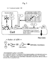
- sCR1 is also known as a substance inhibiting the activation of the complement system; it inhibits the classical pathway and alternative pathway of the complement system. As a background of this pathway inhibition, it is known that the complement components C1-C9 repeatedly undergo restricted cleavage in the complement pathways. The sCR1 binds activated C3b and C4b, which are intermediates in the complement system, and inactivates and destructs these complement components by its cofactor activity in cooperation with factor I, which is a complement factor, to thereby eventually prevent cell death (necrosis) (Fig. 1).
- sCR1 is known to inhibit oxidative nucleophil burst, complement-mediated hemolysis, and the in vitro production of C3a and C5a, which are complement components. Reportedly, it exhibits in vitro activity also in reverse passive Arthus reaction (reaction similar to allergic reaction), in suppressed postischemic myocarditis, and in necrosis. Therefore, sCR1 is an important regulatory protein in the complement system and functions to prevent autolytic host tissue destruction by complement activation.
- the present inventors have newly developed a two-step cell culture process comprising cell proliferation culture using a serum-containing medium and cell culture using a serum-free medium in which the production of sCR1 by cells is improved, and a rapid two-step column purification process comprising two type of column chromatography, namely affinity column chromatography and size exclusion chromatography. Further, it was found that when these means are used, sCR1 can be produced in an intact state. Thus, the inventors have succeeded for the first time in crystallizing complement receptor type 1 (sCR1).
- sCR1 crystallizing complement receptor type 1
- the present inventors have developed a two-step cell culture method comprising cell propagation culture in a serum-containing medium and culture in a serum-free medium for improving the production of the sCR1 protein as well and a two-step column method for protein purification comprising affinity column purification and a size exclusion column chromatography. They found that when these means are used, soluble-form complement receptor type 1 (sCR1) can be produced in an intact state (capable of retaining the complete structure thereof) and, accordingly, crystals of soluble-form complement receptor type 1 (cSR1) can be produced with ease.
- the process of preparing crystals of soluble-form complement receptor type 1 (sCR1) is outlined in Fig. 2.
- the means for preparing crystals of soluble-form complement receptor type 1 (sCR1) according to the invention makes it possible to prepare crystals of sCR1, sCR1 is a high molecular weight protein, and thus its steric has not yet been analyzed. Such crystallization can provide not only information on the steric structure and functions of sCR1 but also a very pure and highly safe complement activity inhibitor with extremely high purity and high safety.
- soluble-form complement receptor type 1 (sCR1)
- the success in crystallizing soluble-form complement receptor type 1 (sCR1) in the present invention depends on the fact that the protein can be obtained with good efficiency and in high purity and high concentration by means of the two-step cell culture method and two-step column purification method.
- the sCR1 to be used in the practice of the invention is one resulting from mutation, deletion or like modification of the transmembrane site of CR1 on the gene thereof. It comprises a water-soluble portion with a molecular weight of about 220 kDa. This soluble CR1 fragment shows physiological activity, i.e. can bind active forms of C3b and/or C4b, which are intermediates in the complement system.
- sCR1 is known as a substance inhibiting the activation of the complement system, as mentioned above; it inhibits the classical pathway and alternative pathway of the complement system.
- the complement components C1-C9 repeatedly undergo restricted cleavage in the complement pathways. Then sCR1 binds activated C3b and C4b, which are intermediates in the complement system, and destructs and inactivates these complement components by exerting its cofactor activity in cooperation with factor I, a complement factor, to thereby eventually prevent cell death (necrosis).
- sCR1 is known to inhibit in vitro oxidative nucleophil burst, complement-mediated hemolysis, and the production of C3a and C5a, which are complement components. Reportedly, it exhibits in vitro activity also in reverse passive Arthus reaction (reaction similar to allergic reaction), in suppressed postischemic myocarditis, and in necrosis. Therefore, sCR1 is an important regulatory protein in the complement system and functions to prevent autolytic destruction of the host tissue by complement activation.
- the sCR1 according to the invention is a product of expression by CHO cells introduced with a human tonsil-derived sCR1 gene. More specifically, it is a product of expression by Chinese hamster ovary (CHO) cells containing the plasmid pBSCR1c/pTCSgpt, which has been deposited with the ATCC with an accession number of CRL10052. Since, as mentioned above, CR1 molecules substantially free of any transmembrane domain can be secreted out of cells, it can be recovered from the cell culture supernatant.
- the above-mentioned cells secrete sCR1 with high efficiency.
- the rate of proliferation of animal cells can be accelerated by addition of serum, such as FBS (fetal bovine serum), and various amino acids. Therefore, the first step cell proliferation culture is carried out in a serum-containing medium.
- serum-containing medium such as FBS (fetal bovine serum)
- FBS fetal bovine serum
- a serum-free medium is used so that the number and amounts of contaminant species may be markedly reduced to facilitate the purification of the sCR1 protein. This purification method requires no expensive column, and is simple and easy to perform at low cost.
- sCR1 can be purified with a high purity and a high concentration by two steps of column chromatography only in two steps while all complicated purification steps have been omitted.
- an affinity column chromatography showing high affinity to sCR1 is used to accomplish purification of sCR1 with a higher level of purity.
- sCR1 can be separated from other proteins owing to the difference in molecular size.
- the purified sCR1 was subjected to SDS-PAGE and western blotting, among others, for confirmation of the eluate fraction and of the intact sCR1 structure and, then, investigations were made about the conditions of crystallization by the vapor diffusion method. Further, the purified sCR1 was checked for its cofactor activity and in vitro and in vivo activities.
- sCR1 is obtained by cell proliferation culture (step 1) in a serum-containing medium using ATCC CRL10052 Chinese hamster ovary (CHO) cells introduced with a human tonsil-derived sCR1 gene, for instance, followed by serum-free culture (step 2) in a serum-free medium for producing sCR1 alone.
- the medium to be used in the above proliferation culture is, for example, ⁇ -MEM (Gibco BRL), F-12 (sigma), DMEM/F-12 (Sigma), F-10 (Sigma), or RPMI (Sigma) containing FBS (fetal bovine serum, Sigma) and 500 nM methotrexate (MTX, Sigma), the culture is carried out for 0 to 336 hours.
- the serum-free medium to be used in the serum-free culture is, for example, a medium (e.g. ASF104 (Ajinomoto), ⁇ -MEM (Gibco BRL), F-12 (Sigma), DMEM/F-12 (Sigma), F-10 (Sigma)) containing transferrin and/or insulin and addition of 500 nM methotrexate (MTX; Sigma).
- ASF104 Alkaline
- ⁇ -MEM Gabco BRL
- F-12 Sigma
- DMEM/F-12 DMEM/F-12
- F-10 F-10
- the culture is carried out for 0 to 360 hours. Characteristically, the cell culture is always carried out at 37°C in the presence of 5% CO 2 .
- the sCR1 in the serum-free culture supernatant obtained in the above manner can be purified in the subsequent two-step column purification process. This process can give several tens of milligrams, per liter, of a protein with a 90% or higher purity. In the practice of the invention, the scale can be increased, hence the scale is not restricted.
- glyco-chain elimination may be made using tunicamycin (Wako) in the cell culture system.
- tunicamycin Wako
- a protein which is added by glyco-chains in a proportion of 20% or more is difficult to form a crystal with good quality, because it has a regular three-dimensional arrangement due to the flexibility thereof after purification. Therefore, the glyco-chain removal by tunicamycin influences on the crystal core formation.
- sCR1 is a glyco-chain-containing protein, it is desirable that the effects of the glyco-chain be taken into consideration.
- glyco-chain-containing sCR1 can also be crystallized in accordance with the present invention.
- This glyco-chain elimination can be realized by providing a tunicamycin invasion period by adding tunicamycin during cell culture, whereby a product (Aglyco-sCR1) resulting from N-linked glyco-chain elimination from sCR1 is obtained.
- the tunicamycin invasion period is provided by adding tunicamycin, to a concentration of 2-10 ⁇ g/ml, to the culture after cell proliferation in the cell proliferation culture step. It lasts 12 to 48 hours.
- the serum-containing medium used in the proliferation culture is used, and MTX is also added. After invasion, the medium is exchanged for the serum-free medium for use in the substance production culture step, and the Aglyco-sCR1 production is allowed to proceed for 12 to 60 hours.
- the Aglyco-sCR1 is allowed to proceed for 12 to 60 hours.
- sCR1 can be purified from the cell culture supernatant containing sCR1 secreted by the two-step column purification process.
- an affinity column chromatography is used in the first step, and a a size exclusion column chromatography in the second step.
- the two-step column purification method according to the invention has overcome the problems encountered in the prior art, such as mentioned just below.
- the problems are, among others, elution of a denatured protein due to changes in pH, complicated exchange of buffers used in an ion exchange column, for instance, protein denaturation due to ammonium sulfate precipitation, and procedural conditions incapable of retaining the intact sate of sCR1. Therefore, in the first step, a gradient of a salt such as NaCl (Wako) is employed for the elution from the affinity column, and in the second step, the intact protein is separated and eluted depending on the molecular size thereof through size exclusion chromatography.
- a salt such as NaCl (Wako)
- the affinity chromatography is carried out using heparin, for instance.
- Heparin-Sepharose CL-6B (Pharmacia Biotech, Code No. 17-0467-01), for instance, may be used as the heparin-binding carrier.
- Heparin is a glucosaminoglycan having, as a skeleton, a structure of repetitions of a disaccharide composed of a uronic acid residue derived either from D-glucuronic acid or from L-iduronic acid and D-glucosamine. It is not uniform in molecular weight, and the molecular weight ranges from 3,000 to 100,000.
- heparin binds tyrosine kinase type receptors, which are heparin-binding growth factors (HBGFs), von Willebrand factor (vWF), which has an effective action in hemostasis, and various cytokines. It is also reported that heparin and sCR1 have strong affinity for each other (Shigeharu Nagasawa: Men-eki to Seitai Bogyo (Immunity and Living Body Protection) (vol. 10)).
- elution is carried out using a buffer solution containing a surfactant low in micelle forming rate.
- the eluent buffer also contains 0.1 to 2 M NaCl.
- the surfactant concentration is adjusted within the range of 0.01% to 0.5% (w/v) relative to the whole volume of the buffer solution.
- the pH is adjusted so that it may fall within the range of from slight acidity to the alkaline side (pH 6.0-10.0).
- the buffer is preferably selected so that its pH may fall within the above range.
- the technique of gel filtration chromatography for instance, is employed.
- a surfactant with lower micelle forming rate than that used in the first step while the eluent buffer in this second step does not contain any salt.
- the eluent flow rate is 2-5 ml/min in the first step of purification, and that in the second step is 0.5-1 ml/min.
- the surfactant to be used in the first step is, for example, CHAPS or the like, and that to be used in the second step includes, among others, DDM (n-dodecyl ⁇ -D-maltoside) and DM (n-decyl- ⁇ -D-maltopyranoside).
- a conventional concentration procedure using a pressure concentrator (Amicon), an ultrafiltration membrane (Amicon) or the like is repeated.
- a pressure concentrator Amicon
- an ultrafiltration membrane Amicon
- the pressure concentrator is operated normally under a pressure of 2-3 kPa, and the centrifugation on the ultrafiltration membrane-containing concentrator is carried out at 2000-3000 x g.
- electrophoresis can be carried out using a 7.5% polyacrylamide gel containing 0.1% SDS to determine the yield of sCR1, the contaminant content, the eluate fractions containing sCR1 and the efficiency of purification.
- SDS-PAGE conditions the electrophoresis is always carried out under a reducing condition (in a state in which the cysteine residues in the protein are cleaved from each other) using DDT (dithiothreitol (Sigma)).
- the protein After electrophoresis, the protein is subjected to CBB (Bio-Rad) staining and/or silver staining (Daiichi Pure Chemicals), which is higher in detection sensitivity than CBB staining, for detecting the band of the protein.
- CBB Bio-Rad
- silver staining Daiichi Pure Chemicals
- a satisfactory protein concentration can be obtained by an ordinary concentration procedure, such as the above-mentioned ultrafiltration. Then, using the thus-concentrated sCR1 solution, sCR1 is crystallized.
- the crystallization technique are, for example, the batch method which comprises adding a precipitating agent to the protein solution to a state of supersaturation and allowing the mixture to stand, the liquid-liquid diffusion method (free interface diffusion method) which comprises pouring a precipitating agent into a thin tube or capillary to the middle, gently forming a protein interface thereon and allowing the solvent or precipitating agent to migrate through that interface, the dialysis method which comprises using a capillary or a dialysis device called button, covering the outside thereof with a dialyzing membrane and immersing the same in a precipitating agent solution to thereby gradually lower the solubility, and the vapor diffusion method which utilizes the phenomenon that a volatile solvent or precipitating agent migrates by diffusion through the gaseous phase. Preferred among them is the batch method which comprises adding a precipit
- the vapor diffusion method utilizes the mechanisms of an equilibrium being attained by vapor diffusion, through the gaseous phase, from a liquid drop (of a mixture of a protein solution and a small amount of a precipitating agent) to the precipitating agent solution or by a reverse process.
- the method is characterized by its utilizing the phenomenon that vapor diffusion between the protein solution and precipitating agent solution causes increases in both protein concentration and precipitating agent concentration to allow crystallization to occur in the supersaturated region.
- the vapor diffusion method includes the hanging drop technique and sitting drop technique according to the form of mounting. The sitting drop method is preferred, however. The reason is as follows.
- a surfactant is used in the purification system.
- the sample after protein purification thus contains the surfactant and is weak in surface tension.
- the hanging drop technique may possibly allow the drop to spread on the cover glass.
- the crystal preparation method using the sitting drop technique is schematically shown in Fig. 14.
- Crystals can be obtained by mixing the precipitating agent (buffer, surfactant, organic solvent) to be used for crystallization by the vapor diffusion method with the protein solution in a ratio of 1:1.
- the precipitating agent buffer, surfactant, organic solvent
- the sCR1 crystal preparation conditions e.g. buffer, surfactant, organic solvent, pH, temperature
- the method of crystallization e.g. vapor diffusion method, batch method, free interface diffusion method
- the thus-obtained crystals of sCR1 can be used in collecting diffraction data using X rays or synchrotron radiation, namely in crystal structure analysis, as well as in three-dimensional structure analysis by electron microscopy.
- Electron microscopy has been used in case of difficulty in preparing a large crystal or for obtaining rough information for crystallization.
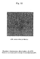
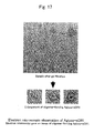
- the present inventors first attempted to apply electron microscopy by ice embedment, which enables high resolution analysis, to unimolecular observation of the sCR1 protein obtained.
- observations and analyses were performed using transmission electron microscopy following negative staining with a heavy metal.
- the acute lethal shock reaction (which is a reaction similar to allergic reaction and known as anaphylactic shock) was utilized.
- This is an acute inflammatory response caused by the interaction between an antigen in a tissue and a circulating antibody.
- This is used as a classical example of an in vivo inflammatory response and is characterized by immune complex formation, complement activation, inflammatory cell recruitment, edema, and tissue injury (P. Baily and A. Sturm, 1983, Biochem. Pham. 32:475). From the experimental viewpoint, it is a general practice to establish an animal model of the reverse passive Arthus reaction by intravenous injection of an antigen, followed by administration of an antibody.
- nude model rats rats in which no immune system functions; model animals in which the complement reaction can be induced by administration of lipopolysaccharide (LPS); LPS-sensitized 512 shock model) administered with a monoclonal antibody (mAb against rat Crry,512) capable of specifically binding to the complement regulating protein occurring on the cell membrane were used.
- LPS lipopolysaccharide
- mAb against rat Crry,512 monoclonal antibody
- CHO cells (ATCC CRL-10052) expressing sCR1 were cultured according to the protocol recommended by the ATCC. Thus, cells were cultured and proliferated using Alpha modified Eagle medium ( ⁇ -MEM, Gibco BRL) supplemented with 10% fetal bovine serum (PBS; Gibco BRL), essential amino acids, L-glutamine, proline, penicillin (100 U/ml) -streptomycin (100 ⁇ g/ml), and 500 nM methotrexate and using a 150 mm dish (capacity: 25 ml). Since, on that occasion, the medium was free of sodium hydrogen carbonate (NaHCO 3 ), 2.2 g, per liter, thereof was added for pH adjustment.
- NaHCO 3 sodium hydrogen carbonate
- the medium should be free of deoxyribonucleotides and ribonucleotides so that it may adapt itself to CHO cells.
- cell were subcultured in new 150 mm dishes.
- the 25-ml container was washed with two or three portions of phosphate-buffered saline (PBS; Nissui) and then cells were detatched from the plate with trypsin solution(0.25% trypsin-1 mM EDTA-4Na, Gibco BRL) and put into the 25-ml containers at the concentration of about 2.0 x 10 6 cells/ml.
- PBS phosphate-buffered saline
- the medium used was ⁇ -MEM (proliferation medium) supplemented with 10% fetal bovine serum (FBS) and penicillin-streptomycin. This operation was repeated until about 50 plates were obtained. Cells were always cultured in an incubator in the presence of 5% CO 2 at 37°C.
- FBS fetal bovine serum
- the medium used was a serum-free medium (Ajinomoto; ASF104; albumin-free). Reportedly, the ASF104 medium makes it possible to attain high levels of antibody productivity with various hybridomas.
- the medium was prepared by blending the principal components, buffer and additives of this medium. After medium preparation, methotrexate, which was used in the proliferation medium, was added, and the resulting medium was designated as ASF (+) (+: methotrexate-containing).
- the 150 mm dishes confluent with cells were washed two or three times with phosphate-buffered saline (PBS), and then the ASF(+) medium prepared was added. Thereafter, cultivation was carried out at 37°C for 36 hours using a 5% CO 2 incubator. After the 36 hours, the sCR1-containing medium was recovered and, in exchange thereof, the ASF(+) medium was added and cultivation was again carried out at 37°C for 36 hours (total cultivation time: 72 hours) in a 5% CO 2 incubator. The sCR1-containing medium recovered was centrifuged at 800-1,000 x g for 5 minutes using a sting rotor (Tomy) to remove contaminants. The supernatant alone was collected by decantation.
- PBS phosphate-buffered saline
- tunicamycin solution was prepared at a concentration of 200 ⁇ g/ml using phosphate-buffered saline (PBS; Nissui). Since tunicamycin is not soluble in the solution, an organic solvent such as an aprotic polar solvent, for example dimethyl sulfoxide (DMSO; Wako) or isopropanol (2-propanol; Wako), was added in an amount of 0.2-0.4% relative to the whole amount. Then, the CHO cells at about 80-90% confluency in proliferation medium in 150 mm dishes (Greiner) were washed two or three times of phosphate-buffered saline (PBS).
- PBS phosphate-buffered saline
- the medium ⁇ -MEM (Gibco BRL), containing 10% FBS
- the tunicamycin preparation prepared in advance was added to the whole amount in each dish to give a concentration of 6-8 ⁇ g/ml.
- the tunicamycin invasion culture was carried out at 37°C for 24-36 hours (optimally 24 hours to avoid cell disintegration) in a 5% CO 2 incubator.
- each dish was again washed two or three times, and the serum-free medium (ASF(+)) was added.
- ASF(+) serum-free medium
- Substance (Aglyco-sCR1) production culture in the presence of tunicamycin was again performed at 37°C for 36 hours in a 5% CO 2 incubator. After the 36 hours, the medium was recovered and treated in the same manner as mentioned above. The filtrate obtained was used as a solution for purifying Aglyco-sCR1.
- heparin affinity chromatography Laboratory scale purification of sCR1 was accomplished by heparin affinity chromatography.
- the heparin carrier used was Pharmacia Biotech's Heparin-Sepharose CL-6B (Code No. 17-0467-01).
- the heparin column was prepared according to the protocol recommended by the manufacturer. The carrier was allowed to swell in 200 ml, per gram of the carrier, of ultrapure water for 15 minutes and, then, the gel was poured slowly into a column (diameter 1.5 x 15 cm) .
- 0.1 M Tris-Cl buffer (pH 8.5) containing 5% NaCl and 0.1 M sodium acetate buffer (pH 5.0) containing 0.5% NaCl were passed through the column alternately three times each in a volume at least three times of the column volume and, then, at least 5-fold volumes of the column volume, of the equilibrating buffer was passed through the column to thereby remove contaminants in the column and regenerate the column (each time the flow rate was 5 ml/min). From the next run and thereon, the column operation was started from the passage of the equilibrating buffer, and the same procedure was repeated.
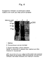
- each eluate fraction was subjected to 7.5% polyacrylamide SDS gel electrophoresis under a reducing condition. After electrophoresis, Coomassie Brilliant Blue (CBB; Bio-Rad) staining was carried out. As a result, a single protein band was detected at a position (about 220 kDa) exceeding about 200 kDa (in the case of sCR1). This is the size of the sCR1 protein as expected from the estimated amino acid sequence thereof.
- CBB Coomassie Brilliant Blue
- the eluate fractions obtained were pooled and 10 to 15-fold concentrated in a pressure concentrator (cutoff molecular weight: 100 kDa; Amicon).
- a pressure concentrator cutoff molecular weight: 100 kDa; Amicon.
- the sCR1 concentrate after concentration to about 10 ml was again concentrated using an ultrafiltration membrane (Centri-plus; cutoff molecular weight 100 kDa; Millipore) until the volume was reduced to about half.
- HPLC gel filtration column chromatography Tosoh's analytical column TSKgel G3000SWXL, which ensures separation even at high flow rates, was employed.
- 20 mM Tris-Cl buffer (pH 7.4) containing 0.05% CHAPS (ICN) was initially used as the elution buffer, CHAPS, which is a surfactant, is not very suitable for crystallization of the soluble form of membrane protein-derived sCR1. Therefore, an eluent was prepared by dissolving DDN (n-decyl-beta-D-maltopyranoside; ICN) having a sugar moiety added to a concentration of 0.05% relative to the whole amount of the eluent.
- DDN n-decyl-beta-D-maltopyranoside
- each eluate fraction was subjected to a 7.5% polyacrylamide SDS gel electrophoresis under a reducing condition, followed by CBB staining for band detection (Fig. 8).
- Fig. 8 CBB staining for band detection
- the sample was concentrated using an ultrafiltration membrane (Amicon).
- the sample was dialyzed using a dialysis membrane (Spectra/Por, cutoff molecular weight 12-14000),
- sCR1 obtained by the two-step cell culture process and two-step purification process according to the invention can be recovered in a yield of 10-20 mg per liter of the medium recovered (in the case of sCR1) or 2-3 mg per liter of the medium recovered (in the case of Aglyco-sCR1).
- the purified grade of sCR1 as obtained by HPLC gel filtration column chromatography in Example 2 (2) was observed under an electron microscope.
- the protein (sCR1) was placed on a hydrophilic support membrane, followed by negative staining.
- the experimental procedure was as follows. A grid coated with a vapor deposited carbon layer was provided, and the carbon was activated by utilizing glow discharge, for instance, under vacuum to thereby make the carbon hydrophilic. Then, 5 ⁇ l of the sCR1 sample (200-400 ⁇ g/ml) was placed thereon and, after waiting for 20-30 seconds, the sample was soaked up using a filter paper moistened in advance, a staining agent (1% uranyl acetate) was placed thereon.
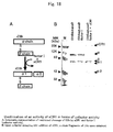
- sCR1 was checked for its activity in terms of cofactor activity utilizing the restricted cleavage of C3b in cooperation with factor I.
- C3ma methylamine-treated human C3
- C3ma methylamine-treated human C3
- sCR1 methylamine-treated human C3
- CH50 complement hemolysis 50
- E erythrocytes sensitized with an antibody (with the antibody adhering thereto)
- the complement amount necessary for causing hemolysis of 50% of EAs added is taken as one CH50 unit, and the complement amount per unit volume of the original sample is expressed in CH50 units. Thus, the unit is CH50/ml.
- the microplate method using 96-well plates was employed. The microplate method comprises diluting and changing the volume of the sample on a plate, adding EAs thereto and allowing the reaction to proceed, and subjecting the microplate as it is to centrifugation and absorbance measurement.
- EAs sinsitized sheep erythrocytes (with a rabbit antibody adhering thereto)
- GVB gelatin veronal buffer
- NHS normal human serum
- a 1.5 x 10 8 cells/ml EA preparation and 1-120 ⁇ g/ml-NHS (ml) preparations prepared by preparing a dilution series of sCR1, followed by 10-fold dilution with NHS) were prepared.
- GVB and distilled water were applied to a 96-well plate except for the uppermost row, and sCR1-NHS solutions prepared in the same manner by preparing a dilution series followed by further 10-fold dilution with GVB were applied in 75- ⁇ l portions to the vacant uppermost row.
- sCR1 inhibited the membrane attack complex (MAC) formation owing to the complement inhibiting effect of sCR1 in the classical pathway, namely via restricted cleavage of C3b and C4b.
- MAC membrane attack complex
- ACH50 is an abbreviation for alternative complement hemolysis 50 and indicates the collective complement activity via the alternative pathway of complement activation. (It is also called AP-CH50 or AH50).
- C3, C5, C6, C7, C8 and C9 are activated in that order through the intermediary of C3, B factor, D factor and P.
- Antibodies, C1, C4 and C2, which participate in the classical pathway, are not involved in the alternative pathway. Rabbit erythrocytes, for instance, are known as an activating substance.
- Er rabbit erythrocytes
- the group of factors in the alternative pathway in the sample are activated by Ers and the C5b-9 complex is formed on the Er membrane, and Ers are lyzed.
- Mg 2+ is required (in the step of B factor activation) but Ca 2+ is not required.
- human serum is reacted with Ers, hemolysis via the classical pathway also takes place since natural antibodies against Er are present in human serum.
- a reaction mixture containing Mg 2+ but containing no Ca 2+ is required. Therefore, EGTA, which is a chelating agent specific to Ca 2+ , is used.
- This ACH50 measuring method like the CH50 determination, positive detection is obtained only when all of C3, B, D, P and C5-C9 are present, and the measurement is carried out by the 50% hemolysis method and the results are expressed using ACH50/ml as a unit. The experiments were carried out according to the protocol for the microplate method (shigeharu Nagasawa: Seitai to Men-eki Bogyo I, published 1997).
- First 5VB was prepared and, then, GVB (gelatin 1.0 g, 5VB 100 ml, dextrose (anhydrous) 1 g, 0.15 M Ca and 1 M Mg solution 1 ml, total amount 1,000 ml) and an EGTA-Mg stock solution (1 M EGTA and 0.02 M Mg solution) were prepared. Then, EGTA ⁇ GGVB(Mg 2+ ) (gelatin 1.0 g, 5VB 80 ml, dextrose (anhydrous) 25 g, EGTA-Mg stock 100 ml. total amount 1,000 ml), 0.9% NaCl, sCR1, and NHS were prepared.
- sCR1 inhibited the membrane attack complex (MAC) formation owing to the complement inhibiting effect of sCR1 in the alternative pathway, namely via restricted cleavage of C3b.
- MAC membrane attack complex
- the detection kit (QUIDEL) is designed for the sandwich technique using anti-iC3b and SC5b-9 antibodies and makes it possible to quantitatively determine the level of MAC formation and the level of iC3b formation.
- Two kinds of supernatants (uppermost row and third row) after the reaction for CH50 determination were used, and how these complement components present in NHS changed upon actual addition of sCR1 was checked.
- the experiment procedure was as follows. First, sCR1 samples were prepared.
- the iC3b plate was incubated for 30 minutes, and the SC5b-9 plate for 60 minutes, at room temperature. After incubation, the solution in each well was sucked off, about 300 ⁇ l of w.s. was added and, after 1 minute of standing, the w.s. was sucked off. This washing procedure was repeated 5 times in all. (During this operation, the second antibody, namely HRP-conjugate, was prepared.) The iC3b or SC5b-9 conjugate Ab was distributed in 50- ⁇ l portions into the wells, and the iC3b plate was incubated for 30 minutes, and the SC5b-9 plate for 60 minutes, at room temperature.
- the solution in each well was sucked off, 300 ⁇ l of w.s. was added and, after 1 minute of standing, the w.s. was sucked off. This washing procedure was repeated 5 times in all.
- the substrate was 21-fold diluted; 550 ⁇ l Sub.: 11 ml S. Diluent).
- the diluted substrate was distributed in 100- ⁇ l portions into the wells and, after wrapping with an aluminum foil, the plates were incubated at room temperature (shielded against light) . At the proper time for color development, the reaction was stopped by adding the stop solution. Using a microplate reader set and adjusted at the wavelength 405 nm, and absorbance measurements were carried out.
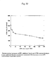
- a calibration curve was constructed from the values read for solutions for calibration curve construction. Then, the iC3b and SC5b-9 concentrations of the respective samples were determined using the calibration curve, and used for graphic representation (Fig. 22, Fig. 23). The following equations were used as the calibration curve equations.
- iC3b protein concentration (O.D. value obtained - 219.42)
- SC5b-9 protein concentration (O.D. value obtained - 198.93) x dilution factor/2260.2
- C3bBb which is the basis thereof, is cleaved into iC3b by sCR1, and C3bBb, which catalyzes the restricted cleavage of C3 to C3b, is no more present, so that the total amount of C3b decreased and the cleavage of iC3b decreases.
- SC5b-9 (soluble C5b-9)
- sCR1 concentration 20 ⁇ g/ml or above
- SC5b-9 formation almost no SC5b-9 formation can be observed.
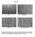
- the cells used herein were of a mouse cranial bone-derived stroma cell line (PA-6). Their behavior was observed using inactivated NHS, NHS, and sCR1 after concentration adjustment.
- the experimental procedure was as follows. First, cells of the mouse cranial bone-derived stroma cell line (PA-6) were sowed onto a 24-well plate in advance, and allowed to proliferate until confluency. After a confluent state was obtained, two washing operations were carried out with PBS, and ⁇ -MEM was added.
- the samples of sCR1 used were purified sCR1 and Aglyco-sCR1 obtained by HPLC gel filtration column chromatography in Example 2 (2).
- sCR1 in vivo activity of sCR1 was confirmed using model rats.
- the experimental procedure is schematically shown in Fig. 25.
- the procedure was as follows. First, anti-Crry antibody capable of inhibiting membrane receptors (e.g. CR1) on the cell membrane was administered to rats to thereby create a state in which the complement regulating protein could not function (nude rats showing no immune system function). Then, sCR1 was administered to the rats at a dose of 20 mg/kg. After sCR1 administration, lipoplysaccharide (LPS), a substance capable of activating the complement system, was administered. For each rat surviving thereafter, various organs were subjected to immune staining to see whether the deposition of C3 had occurred or not.
- LPS lipoplysaccharide
- solubilized complement receptor type 1 sCR1
- sCR1 solubilized complement receptor type 1
- the product can be used as a complement activity inhibitor which is pure and shows reduced side effects and the crystals obtained make it possible to analyze the three-dimensional structure thereof using X-ray crystal structure analyzing techniques.
- the invention further may form a valuable contribution to the investigations concerning the crystallization of similar solubilized membrane proteins and the three-dimensional structure analysis thereof.
- the high-purity sCR1 production process with high efficiency developed for enabling crystallization for the purpose of structure analysis as intended by the invention makes it possible to stably supply high-quality sCR1 which can maintain its physiological activities at high levels.
- the invention can widely contribute to the medical field and to the field of new drug development.
Landscapes
- Health & Medical Sciences (AREA)
- Chemical & Material Sciences (AREA)
- Life Sciences & Earth Sciences (AREA)
- Organic Chemistry (AREA)
- Immunology (AREA)
- Engineering & Computer Science (AREA)
- Medicinal Chemistry (AREA)
- General Health & Medical Sciences (AREA)
- Biochemistry (AREA)
- Biophysics (AREA)
- Gastroenterology & Hepatology (AREA)
- Genetics & Genomics (AREA)
- Zoology (AREA)
- Molecular Biology (AREA)
- Proteomics, Peptides & Aminoacids (AREA)
- Toxicology (AREA)
- Cell Biology (AREA)
- Crystallography & Structural Chemistry (AREA)
- Materials Engineering (AREA)
- Metallurgy (AREA)
- Bioinformatics & Cheminformatics (AREA)
- Transplantation (AREA)
- Chemical Kinetics & Catalysis (AREA)
- General Chemical & Material Sciences (AREA)
- Nuclear Medicine, Radiotherapy & Molecular Imaging (AREA)
- Pharmacology & Pharmacy (AREA)
- Animal Behavior & Ethology (AREA)
- Public Health (AREA)
- Veterinary Medicine (AREA)
- Preparation Of Compounds By Using Micro-Organisms (AREA)
- Peptides Or Proteins (AREA)
- Micro-Organisms Or Cultivation Processes Thereof (AREA)
- Medicines That Contain Protein Lipid Enzymes And Other Medicines (AREA)
- Measuring Or Testing Involving Enzymes Or Micro-Organisms (AREA)
Abstract
Description
Characteristically, the cell culture is always carried out at 37°C in the presence of 5% CO2.
Paradoxically, it is considered that the glyco-chain elimination by tunicamycin had been achieved completely.
Then, a 1.5 x 10⁸ cells/ml EA preparation and 1-120 µg/ml-NHS (ml) preparations (prepared by preparing a dilution series of sCR1, followed by 10-fold dilution with NHS) were prepared. GVB and distilled water were applied to a 96-well plate except for the uppermost row, and sCR1-NHS solutions prepared in the same manner by preparing a dilution series followed by further 10-fold dilution with GVB were applied in 75-µl portions to the vacant uppermost row. Using a diluter, a 50-µl portion of the contents in each uppermost row well was transferred to the next row well therebelow and this type of transfer was repeated toward each lowermost row well to thereby produce a continuous series of 25-
Therefore, for causing hemolysis via the alternative pathway alone, not via the classical pathway, a reaction mixture containing Mg2+ but containing no Ca2+ is required. Therefore, EGTA, which is a chelating agent specific to Ca2+, is used. This ACH50 measuring method, like the CH50 determination, positive detection is obtained only when all of C3, B, D, P and C5-C9 are present, and the measurement is carried out by the 50% hemolysis method and the results are expressed using ACH50/ml as a unit. The experiments were carried out according to the protocol for the microplate method (shigeharu Nagasawa: Seitai to Men-eki Bogyo I, published 1997). First 5VB was prepared and, then, GVB (gelatin 1.0 g,
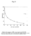
After the 60 minutes, 50 µl of EGTA-GGVB(Mg2+) was added to each well, and the mixture was centrifuged at 1,600-2,000 x g for 10 minutes. After centrifugation, the OD of each supernatant was read at the wavelengths 415/630 nm using a spectrophotometer for microplates. Then, the complement values were calculated and used for graphic representation. The results are shown in Fig. 21. From these results, it was confirmed that sCR1 inhibited the membrane attack complex (MAC) formation owing to the complement inhibiting effect of sCR1 in the alternative pathway, namely via restricted cleavage of C3b. In the case of about 20 µg/ml-NHS, it has a complement inhibiting effect of about 50% or more in terms of CH50.
Solution (1) :GVB = 100 (µl):400 (µl) = 1:4 (5-fold dilution)
Solution (2) :GVB = 100 (µl):400 (µl) = 1:4 (5-fold dilution)
Solution (3) :GVB = 100 (µl):400 (µl) = 1:4 (5-fold dilution)
In the case of ACH50
Solution (1):EGTA-Mg2+-GGVB = 200 (µl):200 (µl) = 1:1 (2-fold dilution)
Solution (2):EGTA-Mg2+-GGVB = 200 (µl):200 (µl) = 1:1 (2-fold dilution)
Solution (3):EGTA-Mg2+-GGVB = 200 (µl):200 (µl) = 1:1 (2-fold dilution)
Claims (21)
- A crystal of the soluble-form complement receptor type 1 (sCR1) in crystal form.
- A crystal of the soluble-form complement receptor type 1 (sCR1) in crystal form according to Claim 1, obtained by crystallization of the soluble-form complement receptor type 1 purified by a process comprising the following steps a and b:a: propagating animal cells introduced with a gene for soluble-form complement receptor type 1 (sCR1) in a serum-containing medium and then cultivating the cells in a serum-free medium for causing them to produce soluble-form complement receptor type 1 (sCR1); andb: purifying the produced soluble-form complement receptor type 1 (sCR1) by the successive use of an affinity column chromatography and a size exclusion column chromatography.
- A crystal of the soluble-form complement receptor type 1 (sCR1) according to Claim 1 or 2 which contains no transmembrane domain.
- A crystal of the soluble-form complement receptor type 1 (sCR1) according to any of Claims 1 to 3 which has no glyco-chain.
- A crystal of the soluble-form complement receptor type 1 (sCR1) according to any of Claims 1 to 4, wherein the purification step(s) a and/or b are(is) carried out in the presence of a surfactant.
- A crystal of soluble-form complement receptor type 1 (sCR1) according to any of Claims 1 to 5, wherein the crystallization means comprises a combination of a precipitation procedure using a protein precipitating agent and a vapor diffusion method
- A complement activity inhibitor which comprises a crystal of the soluble-form complement receptor type 1 (sCR1) according to any of Claims 1 to 6.
- A method of producing a crystal of soluble-form complement receptor type 1 (sCR1) which comprises crystallizing the soluble-form complement receptor type 1 (sCR1) obtained by a process comprising the following steps a and b:a: propagating animal cells introduced with a gene for the soluble-form complement receptor type 1 (sCR1) in a serum-containing medium and then cultivating the cells in a serum-free medium for causing them to produce the soluble-form complement receptor type 1 (sCR1); andb: purifying the produced soluble-form complement receptor type 1 (sCR1) by the successive use of an affinity column chromatography and a size exclusion column chromatography.
- A method of producing a crystal of the soluble-form complement receptor type 1 (sCR1) according to Claim 8, wherein the gene for the soluble-form complement receptor type 1 (sCR1) is one resulting from mutation or deletion in the transmembrane domain-encoding region.
- A method of producing a crystal of soluble-form complement receptor type 1 (sCR1) according to Claim 9, wherein the soluble-form complement receptor type 1 (sCR1) is excreted extracellularly.
- A method of producing a crystal of soluble-form complement receptor type 1 (sCR1) according to any of Claims 8 to 10, wherein the cultivation step(s) a and/or b are(is) carried out in the presence of tunicamycin.
- A method of producing a soluble-form complement receptor type 1 (sCR1) in crystal form according to any of Claims 8 to 11, wherein the purification step(s) a and/or b are(is) carried out in a state wherein the soluble-form complement receptor type 1 (sCR1) is dispersed by means of a surfactant.
- A method of producing a crystal of soluble-form complement receptor type 1 (sCR1 according to any of Claims 8 to 12, wherein the crystallization means comprises a combination of a precipitation procedure using a protein precipitating agent and a vapor diffusion method.
- A two-step cell culture method comprising propagating animal cells introduced with a gene for soluble-form complement receptor type 1 (sCR1) in a serum-containing medium, followed by cultivation in a serum-free medium.
- A two-step culture method according to Claim 14, wherein the soluble-form complement receptor type 1 (sCR1) gene is one resulting from mutation or deletion in the transmembrane domain-encoding region.
- A two-step culture method according to Claim 15, wherein the animal cells introduced with the soluble-form complement receptor type 1 (sCR1) gene extracellularly secrete the soluble-form complement receptor type 1 (sCR1).
- A two-step culture method according to any of Claims 14 to 16, wherein the cultivation step(s) is (are) carried out in the presence of tunicamycin.
- A two-step process for purifying a soluble-form complement receptor type 1 (sCR1), which comprises purifying the soluble-form complement receptor type 1-containing animal cell culture medium treated or untreated, obtained by the two-step culture method according to any of Claims 14 to 17, by using an affinity column chromatography and a size exclusion column chromatography successively.
- A two-step purification process according to Claim 18, wherein the animal cell culture supernatant containing the soluble-form complement receptor type 1 (sCR1) is directly applied to an affinity column chromatography.
- A two-step purification process according to Claim 18 or 19, wherein the soluble-form complement receptor type 1 (sCR1) is subjected, in a dispersed state by a surfactant, to purification by using an affinity column chromatography and/or size exclusion column chromatography.
- A complement activity inhibitor wherein the soluble-form complement receptor type 1 (sCR1) obtained by the two-step purification process according to any of Claims 18 to 20 is used.
Applications Claiming Priority (2)
| Application Number | Priority Date | Filing Date | Title |
|---|---|---|---|
| JP2002031571 | 2002-02-07 | ||
| JP2002031571A JP4300286B2 (en) | 2002-02-07 | 2002-02-07 | Solubilized complement receptor type 1 crystals |
Publications (2)
| Publication Number | Publication Date |
|---|---|
| EP1334982A2 true EP1334982A2 (en) | 2003-08-13 |
| EP1334982A3 EP1334982A3 (en) | 2003-09-17 |
Family
ID=27606524
Family Applications (1)
| Application Number | Title | Priority Date | Filing Date |
|---|---|---|---|
| EP03250762A Withdrawn EP1334982A3 (en) | 2002-02-07 | 2003-02-06 | Solubilized complement receptor type 1 in crystal form |
Country Status (3)
| Country | Link |
|---|---|
| US (1) | US20040014136A1 (en) |
| EP (1) | EP1334982A3 (en) |
| JP (1) | JP4300286B2 (en) |
Cited By (1)
| Publication number | Priority date | Publication date | Assignee | Title |
|---|---|---|---|---|
| EP1738763A1 (en) * | 2005-06-30 | 2007-01-03 | AVANT Immunotherapeutics, Inc. | Use of complement inhibitory proteins to treat spinal cord injury |
Families Citing this family (16)
| Publication number | Priority date | Publication date | Assignee | Title |
|---|---|---|---|---|
| KR100563685B1 (en) * | 2002-02-25 | 2006-03-28 | 엘지전자 주식회사 | How to manage playlists on rewritable recording media |
| KR20030087193A (en) * | 2002-05-07 | 2003-11-14 | 엘지전자 주식회사 | Method for managing a multi-channel broadcast stream record |
| KR100582957B1 (en) * | 2002-06-21 | 2006-05-23 | 엘지전자 주식회사 | Recording medium having data structure for managing reproduction of video data recorded thereon |
| AU2003241198B2 (en) * | 2002-06-21 | 2009-09-03 | Lg Electronics Inc. | Recording medium having data structure for managing reproduction of video data recorded thereon |
| KR20040000290A (en) | 2002-06-24 | 2004-01-03 | 엘지전자 주식회사 | Method for managing multi-path data stream of high density optical disc |
| CN100380496C (en) * | 2002-06-24 | 2008-04-09 | Lg电子株式会社 | Recording medium having data structure for managing reproduction of multiple title video data recorded thereon and recording and reproducing methods and apparatuses |
| RU2309467C2 (en) * | 2002-06-24 | 2007-10-27 | Эл Джи Электроникс Инк. | Data carrier with data structure including navigation-control information, for controlling reproduction of video data recorded on it and methods and devices for recording and reproduction |
| RU2347284C2 (en) * | 2002-10-14 | 2009-02-20 | Эл Джи Электроникс Инк. | Carrier of record with structure of data for guidance of reproduction of set of audio streams written down on it and methods and record and reproduction devices |
| CA2469178C (en) * | 2002-10-15 | 2009-09-01 | Lg Electronics Inc. | Recording medium having data structure for managing reproduction of multiple graphics streams recorded thereon and recording and reproducing methods and apparatuses |
| US7606463B2 (en) | 2003-02-24 | 2009-10-20 | Lg Electronics, Inc. | Recording medium having data structure for managing playback control and recording and reproducing methods and apparatuses |
| US7809775B2 (en) * | 2003-02-27 | 2010-10-05 | Lg Electronics, Inc. | Recording medium having data structure for managing playback control recorded thereon and recording and reproducing methods and apparatuses |
| CN100397882C (en) * | 2003-02-28 | 2008-06-25 | Lg电子株式会社 | Recording medium having data structure for managing random/shuffle reproduction of video data recorded thereon, and recording and reproducing method and apparatus |
| US7620301B2 (en) * | 2003-04-04 | 2009-11-17 | Lg Electronics Inc. | System and method for resuming playback |
| WO2008036135A2 (en) * | 2006-06-01 | 2008-03-27 | Genentech, Inc. | Crystal structure of crig and c3b: crig complex |
| JP2012139106A (en) * | 2009-04-22 | 2012-07-26 | Uha Mikakuto Co Ltd | Method for producing soluble sr-a protein, and evaluation method using the same |
| CN103237557A (en) * | 2010-09-15 | 2013-08-07 | 赛尔戴克斯治疗公司 | Treatment of chronic kidney disease with soluble complement receptor type I (sCR1) |
Family Cites Families (1)
| Publication number | Priority date | Publication date | Assignee | Title |
|---|---|---|---|---|
| EP0913470A3 (en) * | 1988-04-01 | 1999-09-22 | The Johns Hopkins University | The human C3b/C4b receptor (CR1) |
-
2002
- 2002-02-07 JP JP2002031571A patent/JP4300286B2/en not_active Expired - Lifetime
-
2003
- 2003-02-06 EP EP03250762A patent/EP1334982A3/en not_active Withdrawn
- 2003-02-07 US US10/360,357 patent/US20040014136A1/en not_active Abandoned
Cited By (1)
| Publication number | Priority date | Publication date | Assignee | Title |
|---|---|---|---|---|
| EP1738763A1 (en) * | 2005-06-30 | 2007-01-03 | AVANT Immunotherapeutics, Inc. | Use of complement inhibitory proteins to treat spinal cord injury |
Also Published As
| Publication number | Publication date |
|---|---|
| US20040014136A1 (en) | 2004-01-22 |
| EP1334982A3 (en) | 2003-09-17 |
| JP4300286B2 (en) | 2009-07-22 |
| JP2003238596A (en) | 2003-08-27 |
Similar Documents
| Publication | Publication Date | Title |
|---|---|---|
| US20040014136A1 (en) | Solubilized complement receptor type 1 in crystal form | |
| Vacquier et al. | The fucose sulfate polymer of egg jelly binds to sperm REJ and is the inducer of the sea urchin sperm acrosome reaction | |
| Chiu et al. | Sickled erythrocytes accelerate clotting in vitro: an effect of abnormal membrane lipid asymmetry | |
| Pasvol et al. | Erythrocytes deficient in glycophorin resist invasion by the malarial parasite Plasmodium falciparum | |
| Bithell et al. | Platelet‐function studies in the Bernard‐Soulier syndrome | |
| Cabral et al. | Taxol-requiring mutant of Chinese hamster ovary cells with impaired mitotic spindle assembly. | |
| Waugh et al. | Heinz bodies induce clustering of band 3, glycophorin, and ankyrin in sickle cell erythrocytes. | |
| Filbin et al. | The role of complex carbohydrates in adhesion of the myelin protein, P0 | |
| Crabb et al. | In vitro reconstitution of exocytosis from plasma membrane and isolated secretory vesicles. | |
| Tarui et al. | Purification and properties of rabbit erythrocyte phosphofructokinase | |
| Buc-Caron et al. | Presence of a mouse embryonic antigen on human spermatozoa | |
| Richmond et al. | Nature of Anaemia in Rheumatoid Arthritis: III. Changes in the Bone Marrow and Their Relation to Other Features of the Disease | |
| Kauskot et al. | MAGT1 deficiency in XMEN disease is associated with severe platelet dysfunction and impaired platelet glycoprotein N-glycosylation | |
| Focarelli et al. | The 220-kDa vitelline coat glycoprotein mediates sperm binding in the polarized egg of Unio elongatulus through O-linked oligosaccharides | |
| Stephens | Reconstitution of ciliary membranes containing tubulin. | |
| Davey et al. | The isolation of a component of the venom of Trimeresurus okinavensis that causes the aggregation of blood platelets | |
| Shinozuka | Changes in human red blood cells during aging in vivo | |
| Bloom et al. | A rapid technique for the preparation of factor V deficient plasma | |
| Ham et al. | Physical properties of red cells as related to effects in vivo. IV. Oxidant drugs producing abnormal intracellular concentration of hemoglobin (eccentrocytes) with a rigid-red-cell hemolytic syndrome | |
| Nagel et al. | Compound heterozygosity for hemoglobin C and Korle-Bu: moderate microcytic hemolytic anemia and acceleration of crystal formation [corrected][published erratum appears in Blood 1994 May 15; 83 (10): 3105] | |
| Krüger et al. | Effect of age on mast cell granules | |
| JP3922390B2 (en) | Method for producing high-purity solubilized complement receptor type 1 (sCR1) | |
| Glaubensklee et al. | Structural and biochemical analysis of the McLeod erythrocyte membrane | |
| Litosch et al. | Sickle red cell calcium metabolism: Studies on Ca2+‐Mg2+ ATPase and ca‐binding properties of sickle red cell membranes | |
| Garriga et al. | Hexose transport across the basolateral membrane of the chicken jejunum |
Legal Events
| Date | Code | Title | Description |
|---|---|---|---|
| PUAI | Public reference made under article 153(3) epc to a published international application that has entered the european phase |
Free format text: ORIGINAL CODE: 0009012 |
|
| PUAL | Search report despatched |
Free format text: ORIGINAL CODE: 0009013 |
|
| AK | Designated contracting states |
Designated state(s): AT BE BG CH CY CZ DE DK EE ES FI FR GB GR HU IE IT LI LU MC NL PT SE SI SK TR |
|
| AX | Request for extension of the european patent |
Extension state: AL LT LV MK RO |
|
| AK | Designated contracting states |
Kind code of ref document: A3 Designated state(s): AT BE BG CH CY CZ DE DK EE ES FI FR GB GR HU IE IT LI LU MC NL PT SE SI SK TR |
|
| AX | Request for extension of the european patent |
Extension state: AL LT LV MK RO |
|
| 17P | Request for examination filed |
Effective date: 20040317 |
|
| AKX | Designation fees paid |
Designated state(s): AT BE BG CH CY CZ DE DK EE ES FI FR GB GR HU IE IT LI LU MC NL PT SE SI SK TR |
|
| STAA | Information on the status of an ep patent application or granted ep patent |
Free format text: STATUS: THE APPLICATION IS DEEMED TO BE WITHDRAWN |
|
| 18D | Application deemed to be withdrawn |
Effective date: 20051125 |