TECHNICAL FIELD
The present invention relates to a method of screening a biologically
active substance, a gene controlling agent and a method of controlling a gene.
BACKGROUND ART
An organism is subjected to various stresses caused by external factors
such as change in living environment and internal factors such as viral and
bacterial infections. The stress itself is a factor which hinders a normal life of an
organism, and when the stress cannot be well avoided or when excessive reaction
rather takes place, a more severe disorder, for instance, a disease is caused.
In a state where stress is loaded or in a state where the stage is progressed
to a disease, various functions and elements in a living body are involved in a
compound manner, and there basically exists a change in the gene expression
state. Therefore, the stress can be supposedly avoided smoothly by regulation on
the gene level. However, the details of the regulation of gene expression in the
state under stress, and in the process where the stress is avoided are not clarified.
Therefore, a means of avoiding stress by the regulation of gene level as
mentioned above has not yet been established.
On the other hand, L-glycero-1,5-epoxy-1αβ,6-dihydroxy-cis-hex-3-en-2-one
(hereinafter referred to as DGE), which can be obtained from agarose, has
been known as a compound that possesses various kinds of biological activities,
and is useful for maintaining homeostasis in a living body (WO 99/64424).
However, the action mechanism on the gene level of the compound has not been
clarified.
An object of the present invention is to provide a method of screening a
biologically active substance or a candidate substance for a biologically active
substance, possessing an activity for controlling gene expression to the same
extent to that of DGE; a gene controlling agent which is effective for maintaining
homeostasis in a living body, for instance, purposed for mitigation or prevention
of stress; and a method of controlling a gene.
DISCLOSURE OF INVENTION
A first invention of the present invention relates to a method of screening
a biologically active substance, which comprises the following steps:
a derivative thereof and a salt thereof; and/or
a compound of the following (ii): (ii) at least one compound selected from the group consisting of
3,6-anhydrogalactose represented by the following formula (II):
an aldehyde thereof, a hydrate thereof, and a 2-O-methylated product and a
2-O-sulfated product of the foregoing compounds; and/or at least one compound
selected from the group consisting of soluble saccharide compounds having the
above compound at reducing end; (2) determining an expression level of a gene in the living body of (1), and
comparing resulting values between a case where 1) is loaded and a case where
2) is loaded; and (3) selecting a test substance which causes a change in an expression level of
at least one gene to substantially the same extent to a change where 2) is loaded.
A second invention of the present invention relates to a method of
screening a biologically active substance, which comprises the following steps:
A third invention of the present invention relates to a method of screening
a biologically active substance, which comprises the following steps:
In the third invention of the present invention, when the stress loaded to a
living body is a treatment with phorbol myristate acetate, the gene expression
pattern is exemplified by the above (a) to (d), and when the stress is a treatment
with lipopolysaccharide, the gene expression pattern is exemplified by the above
(e) to (g).
In the first to third inventions of the present invention, as the living body,
there is exemplified an organism individual or a cultured cell. As the gene
expression level, there is exemplified an expression level of a gene determined
by an amount of mRNA transcribed from the gene. It is especially preferable
that the expression level is determined by hybridization method using a DNA
array. Also, the present invention encompasses substances which can be
obtained by the first to third inventions of the present invention.
A fourth invention of the present invention relates to a DNA array usable
for the screening method of the first to third inventions of the present invention,
characterized in that at least two genes selected from the genes of Groups A to G
as defined above or fragments thereof are immobilized on a support at a given
position.
A fifth invention of the present invention relates to a gene controlling
agent characterized in that the gene controlling agent comprises a compound
possessing an action of decreasing an expression level of at least one gene
selected from Group A, Group C, Group E and Group F as defined above, and/or
increasing an expression level of at least one gene selected from Group B, Group
D and Group G as defined above.
A sixth invention of the present invention relates to a gene controlling
agent characterized in that the gene controlling agent comprises a compound
possessing an action of decreasing an expression level of at least one gene
selected from Group A, Group C, Group E and Group F as defined above, and/or
increasing an expression level of at least one gene selected from Group B, Group
D and Group G as defined above, in a living body to which stress is loaded.
A seventh invention of the present invention relates to a gene controlling
agent characterized in that the gene controlling agent comprises a compound
possessing an action of decreasing an expression level of at least one gene
selected from Group A and Group E as defined above in a living body to which
stress is loaded, wherein the expression level of at least one gene is increased by
the stress.
An eighth invention of the present invention relates to a gene controlling
agent characterized in that the gene controlling agent comprises a compound
possessing an action of increasing an expression level of at least one gene
selected from Group B and Group G as defined above in a living body to which
stress is loaded, wherein the expression level of at least one gene is increased by
the stress.
A ninth invention of the present invention relates to a gene controlling
agent characterized in that the gene controlling agent comprises a compound
possessing an action of decreasing an expression level of at least one gene
selected from Group C and Group F as defined above in a living body to which
stress is loaded, wherein the expression level of at least one gene is decreased by
the stress.
A tenth invention of the present invention relates to a gene controlling
agent characterized in that the gene controlling agent comprises a compound
possessing an action of increasing an expression level of at least one gene
selected from Group D as defined above in a living body to which stress is
loaded, wherein the expression level of at least one gene is decreased by the
stress.
In the fifth to tenth inventions of the present invention, there is
exemplified a gene controlling agent in which each compound contained is a
substance obtained by the screening method of any one of the first to third
inventions of the present invention. Also, as the gene controlling agent of the
fifth to tenth inventions of the present invention, there is exemplified a gene
control agent which is purposed for mitigating or preventing stress.
An eleventh invention of the present invention relates to a method of
controlling a gene characterized by administering to a living body either at least
one compound selected from the group consisting of compounds of (i) and (ii) as
defined above and a compound possessing an activity for controlling gene
expression to substantially the same extent to that of the compounds for at least
one gene, or a composition comprising the compound, thereby decreasing an
expression level of at least one gene selected from Group A, Group C, Group E
and Group F as defined above, and/or increasing an expression level of at least
one gene selected from Group B, Group D and Group G, wherein Groups A to G
are as defined above.
A twelfth invention of the present invention relates to a method of
controlling a gene characterized by administering to a living body to which stress
is loaded either at least one compound selected from the group consisting of
compounds of (i) and (ii) as defined above and a compound possessing an
activity for controlling gene expression to substantially the same extent to that of
the compounds for at least one gene, or a composition comprising the compound,
thereby decreasing an expression level of at least one gene selected from Group
A, Group C, Group E and Group F as defined above, and/or increasing an
expression level of at least one gene selected from Group B, Group D and Group
G, wherein Groups A to G are as defined above.
In the eleventh and twelfth inventions of the present invention, as the
compound possessing an activity for controlling gene expression to substantially
the same extent to that of compounds of (i) or (ii) mentioned above for at least
one gene, there is exemplified a substance obtained by the screening method of
any one of the first to third inventions of the present invention.
BEST MODE FOR CARRYING OUT THE INVENTION
In one embodiment of the method of screening a biologically active
substance of the present invention, a compound having the structure represented
by the above-mentioned formula (I) is used. One example of the compound
includes L-glycero-1,5-epoxy-1αβ,6-dihydroxy-cis-hex-3-en-2-one (DGE).
The compound can be obtained by treating (keeping) a compound having
3,6-anhydrogalactose at a reducing end thereof, such as at least one compound
selected from the group consisting of agarobiose, κ-carrabiose and a compound
having 3,6-anhydrogalactose at a reducing end thereof under neutral to alkaline
conditions. In addition, the compound can also be obtained by subjecting the
compound having 3,6-anhydrogalactose at a reducing end thereof to acid
hydrolysis at a pH of less than 7 and/or enzymolysis, and subsequently treating
(keeping) the resulting acid hydrolyzate and/or enzymolyzate under neutral to
alkaline conditions.
The above-mentioned enzymolysis includes, for instance, degradation of
agarose by using one compound having 3,6-anhydrogalactose at a reducing end
thereof by using α-agarase. The conditions for carrying out the enzymolytic
reaction are not particularly limited, and may be according to known conditions
for used enzymes.
In the alkali treatment of the compound having 3,6-anhydrogalactose at a
reducing end thereof, such as at least one compound selected from the group
consisting of agarobiose, κ-carrabiose and a compound having
3,6-anhydrogalactose at a reducing end thereof, at a pH exceeding 7, the kinds of
an alkali in which the compound is dissolved or suspended are not particularly
limited. There can be used an inorganic base such as sodium hydroxide,
potassium hydroxide, calcium hydroxide or ammonia, or an organic base such as
Tris, ethylamine or triethylamine. The concentration of the alkali is not
-particularly limited. The alkali can be used in a concentration of preferably from
0.0001 to 5 N, more preferably from 0.001 to 1 N. In addition, the treatment
temperature is preferably, but is not particularly limited to, from 0° to 200°C,
more preferably from 20° to 130°C. The treatment time is preferably, but is not
particularly limited to, several seconds to several days. The kinds and
concentration of the alkali, the temperature and time during the treatment may be
properly chosen depending upon the kinds of the above-mentioned compound
used as a raw material and the amount of the desired compound represented by
the formula (I) formed. In general, the pH may be exceeding 7. The formation
of the compound represented by the formula (I) proceeds quickly at a higher
alkali concentration than those at a lower alkali concentration, and at a higher
temperature than those at a lower temperature.
For instance, the compound represented by the formula (I) is formed by
preparing a solution of agarobiose or κ-carrabiose at a pH of 11.5, and keeping
the solution at 37°C for 5 minutes.
The resulting alkali solution containing the compound represented by the
formula (I) may be used after neutralization according to its purpose, or may be
used as an acidic solution having a pH of less than 7.
As the purification means for the compound represented by the formula
(I) of the present invention, there may be employed a known means of
purification such as a chemical method or a physical method. For instance, the
purification may be carried out by combining various purification methods such
as gel filtration, fractionation using a molecular weight fractionation membrane,
solvent extraction, and various chromatographies using ion exchange resin or the
like.
For instance, a compound represented by the formula (I) in which X is
CH2OH and Y is H is purified from the product obtained by treating agarobiose
under neutral to alkaline conditions, and a compound represented by the formula
(I) in which X is H and Y is CH2OH is purified from the product obtained by
treating κ-carrabiose under neutral to alkaline conditions.
The derivative of the compound represented by the formula (I) is not
particularly limited, as long as the derivative is capable of showing the gene
expression pattern to substantially the same extent as that of the above compound
in a living body. For instance, there can be used a compound obtainable by
reacting the compound represented by the formula (I) with an
SH-group containing compound (for instance, cysteine, glutathione, or known
derivatives thereof). Here, a process for preparing the compound obtainable by
reacting the compound represented by the formula (I) with an SH-group
containing compound includes a known process described in WO 99/64424.
The salt of the compound represented by the formula (I) or a derivative
thereof is preferably those acceptable as medicaments, and can be converted by a
known method.
In addition, DGE, the compound represented by the formula (I), is a
compound which is generated when an agarooligosaccharide is taken into a
living body. Therefore, when the agarooligosaccharide is administered to a
living body, it is anticipated that the agarooligosaccharide causes the same living
body reaction as that of DGE [Jpn. J. Phycol. (Sorui) 48:13-19, March 10, 2000].
Therefore, in the present invention, there can be also used at least one compound
selected from the group consisting of 3,6-anhydrogalactose represented by the
above formula (II), an aldehyde thereof, a hydrate thereof, and a 2-O-methylated
product and a 2-O-sulfated product of the foregoing compounds; and/or at least
one compound selected from the group consisting of soluble saccharide
compounds having the above compound at reducing end (Here, an aldehyde or a
hydrate of 3,6-anhydrogalactose, and a 2-O-methylated product and a 2-O-sulfated
product of the foregoing compounds is exemplified by the compound
described in WO 00/41579), the compound from which DGE is thought to be
generated when administered to a living body in place of the compound
represented by the formula (I). Examples of the above compound are not
particularly limited. For instance, the compound is exemplified by
agarooligosaccharides such as agarobiose, agarotetrose, agarohexose, and
agarooctose.
[1] Method of Screening Biologically Active Substance of the Present
Invention
DGE, which is one of the above-mentioned compound represented by the
formula (I), possesses actions of mitigating the damage to which the living body
is subjected and progressing the maintenance of homeostasis in a living body. It
has been known that DGE possesses an activity for regulating an expression
level for a gene in which a change in an expression level is observed in a living
body subjected to stress as mentioned above, namely an activity (or action) for
controlling gene expression, and this activity is shown via expression control of
various kinds of genes in a living body. Furthermore, DGE has known to
possess various biological activities, for instance, apoptosis-inducing activity,
anticancer activity, anti-oxidative activity such as activity for suppressing active
oxygen production, activity for suppressing lipid peroxide radical production,
and activity for suppressing nitrogen monoxide production, anti-pathogenic
microbial activity, anti-mutagenic activity, α-glycosidase inhibitory activity,
immunomodulatory action, action for suppressing prostaglandin synthesis, antiinflammatory
action, anti-allergic action, action for regulating cytokine
production, anti-rheumatic action, anti-diabetic action, activity for suppressing
proliferation of synovial cell, action for inducing heat shock protein production,
and the like. Therefore, the biological activity as described above is thought to
be exhibited by regulating the gene expression level by gene controlling action of
DGE.
The method of screening a biologically active substance of the present
invention provides a method of screening a substance other than DGE capable of
showing a change in gene expression level to the same extent as that of DGE by
using as an index a change in gene expression level based on the gene controlling
action of DGE which are known to have various biologically active substances as
described above. In other words, there is provided a method of screening a
substance capable of showing at least the same gene expression pattern as that of
DGE in a living body.
Since the substance obtained according to the present invention can
exhibit an activity for controlling gene expression to the same extent to that of
DGE, it is deduced that the substance possesses, for instance, biological activities
to the same extent as that of DGE. Therefore, by using the compound, there can
be provided a medicament, food, beverage or the like which is effective for
treatment or prevention of a disease and/or symptom (for instance, a disease
requiring the above-mentioned biological activities of DGE, such as cancer,
inflammation, allergy and diabetes) to which the expression of the biological
activity effectively contributes.
Also, a substance having unknown biological activities or the like, which
are different from the biological activities of DGE can be obtained. From this
viewpoint, the method of screening a biologically active substance of the present
invention can be also understood as a method of screening a candidate substance
for a biologically active substance.
In the method of screening a biologically active substance of the present
invention, concretely, a substance possessing an activity for controlling gene
expression to the same extent to that of DGE can be obtained by the following
steps:
Here, the term "a change in an expression level ... to substantially the
same extent" is not necessarily intended to mean that the extent of the expression
level is the same, but to mean that at least a change in the expression level
pattern is the same.
Concretely, a gene expression level in a sample derived from a living
body to which a test substance 1) is loaded, namely administered or orally taken
(hereinafter referred to the same) is determined. On the other hand, a gene
expression level in a sample derived from a living body to which a compound 2),
for instance, the above-mentioned compound represented by the formula (I),
especially DGE, is loaded is determined. Next, each of the gene expression
levels and/or its change is compared to judge whether or not the actions for
controlling gene expression in each of the test substance and DGE is similar.
The substance having a similar action for controlling gene expression has a high
possibility of possessing the biological activities to the same extent as that of
DGE, so that it is expected that the substance can be used for the same purpose
as that of DGE.
The above-mentioned action for controlling gene expression owned by
DGE is not particularly limited. For instance, the action can be confirmed by
comparing the gene expression state (expression pattern) between a case where
DGE is loaded to a living body and a case where a living body is not loaded
(control). In this case, the gene having a difference in expression levels
therebetween is thought to be a gene to be controlled by DGE. Here, the action
for controlling gene expression owned by DGE, as described in Example 1
described later, can be also confirmed via loading stress to a living body.
In the present specification, the living body is not particularly limited.
The preferred living body includes an organism individual (excluding human) or
cultured cell. For instance, when the organism individual is used, the gene
expression state can be examined by taking an appropriate sample from the
individual, for instance a part of a tissue, body fluid or the like.
The test substance usable in the screening method of the present invention
is not limited by any means, and there can be used those derived from nature,
those artificially synthesized, those obtained by artificially modifying a
substance derived from nature, and the like. As these substances, any of those
which are isolated and those containing plural substances can be used. For
instance, appropriate separation procedures and the screening method of the
present invention are combined to purify or isolate a useful substance.
In addition, by using the screening method of the present invention, an
active component thereof can be specified from a composition possessing
biological activities to substantially the same extent as that of the above-mentioned
compound (i) or the above-mentioned compound (ii), for instance,
from a composition derived from nature such as those derived from a plant, an
animal, Basidiomycetes, or an microorganism. For instance, an active
component can be specified by carrying out the method of screening a
biologically active substance of the present invention by using the compound
constituting the above-mentioned composition as a test substance.
In addition, the present invention encompasses the above-mentioned
substance obtained by the screening method of the present invention, namely the
substance having the gene controlling activity to the same extent as that of the
above-mentioned compound (i) or the above-mentioned compound (ii). The
above substance can be obtained by subjecting a test substance to the screening
method of the present invention, and judging whether or not the substance
possesses gene controlling activity as defined in the above method. In addition,
the substance is exemplified by, for instance, a substance derived from a plant, a
substance derived from Basidiomycetes, or a derivative thereof. The plant
usable herein is exemplified by medicinal plants, marine algae, and the like,
which have been known to have various biological activities.
Also, the substance derived from these origins is not particularly limited.
The constituent thereof includes a substance containing a saccharide, for instance,
polysaccharides, degradation products thereof or derivatives thereof. Among
them, those substances having a molecular weight of 100 to 2000 or so are
preferable. Further, among them, a substance possessing a gene controlling
activity other than the substance used as a control (for instance, DGE) according
to the screening method of the present invention can be selected. The above-mentioned
substance containing a saccharide as a constituent is especially
preferably a substance having 3,6-anhydrogalactose in its molecule. Here, the
degradation product, the derivative, and the originating substance are not
particularly limited, as long as they can show the gene expression pattern of the
substantially same extent in a living body.
The comparison of the gene expression states can be carried out by a
known method, for instance, the determination of the level of the above gene
product (polypeptide), or the determination of the amount of mRNA transcribed
from the above gene. The method of determining the amount of mRNA is more
effective for the above-mentioned purposes, and is preferable.
For instance, it is preferable that the level of the polypeptide is determined
by using an antibody capable of specifically binding to the polypeptide or a
fragment thereof. The antibody may be any of polyclonal antibodies and
monoclonal antibodies. Furthermore, the antibody may be those antibodies
which are modified by known techniques, and the derivatives of the antibodies,
for instance, Fab fragment, single-strand antibody and the like. These antibody
and derivative thereof or the like can be produced in accordance with a known
method (for instance, see Current Protocols in Immunology, published by John
Wiely & Sons, Inc., 1992, edited by John E. Coligan). The method for
determining the level of the polypeptide includes, for instance, known enzyme
immunoassay, fluoroimmunoassay, luminescent immunoassay, and the like.
Further, the above-mentioned antibody or the like may be subjected to various
kinds of labeling in order to facilitate the detection in the above-mentioned
method for determining the level of the polypeptide.
The method for determining mRNA is not particularly limited, and a
known method such as RT-PCR method, Northern hybridization method, dot
blot hybridization method, a hybridization method using a DNA array, a DNA
microbeads method, or the like can be employed. The method using a DNA
array is especially preferable, for the above-mentioned purpose, because the
expression states for numerous genes can be examined with a very little amount
of a sample.
The "DNA array" as referred to herein is one in which a gene or a DNA
fragment derived from the gene is immobilized on a support in a given region
(position), and encompasses, for instance, those referred to as DNA chips. In
addition, one in which gene or a DNA fragment derived from the gene is
immobilized on a support in a high density is referred to as DNA microarray.
The support of the DNA array is not particularly limited, as long as it can
be used for hybridization, and slide glass, silicon chip, nitrocellulose or nylon
membrane or the like can be usually used. The surface of the support may be
any of those in which a single-stranded DNA or double-stranded DNA can be
immobilized by covalent bonding or non-covalent bonding and those having a
hydrophilic or hydrophobic functional group on the surface of the support
without being particularly limited thereto. For instance, there can be preferably
used those having hydroxyl group, amino group, thiol group, aldehyde group,
carboxyl group, an acyl group or the like without being particularly limited
thereto. These functional groups may exist to give the surface properties of the
support itself, or may be introduced by surface treatment. The surface-treated
product as described above includes, for instance, those obtained by treating
glass with a commercially available silane coupling agent such as an
aminoalkylsilane; those obtained by treating glass with a polycation such as
polylysine or polyethyleneimine; and the like. Also, some of the slide glass
subjected to these treatments are commercially available.
The gene or a DNA fragment thereof, which is immobilized to the support,
is not particularly limited, and includes, for instance, genomic DNA library and
cDNA library, and a DNA amplified with these libraries as a template by a
known nucleic acid amplification method, for instance, PCR (polymerase chain
reaction) method, LCR (ligase chain reaction) method, SDA (strand
displacement amplification) method, ICAN (isothermal and chimeric primer-initiated
amplification of nucleic acids) method (described in WO00/56877), and
the like. The reaction conditions in the nucleic acid amplification are not
particularly limited. In addition, there can be used a DNA obtained by
subjecting amplified RNA to reverse transcription by transcription-based
amplification system (TAS) method using RNA as a template, self-sustained
sequence replication (3SR) method, NASBA (nucleic acid sequence-based
amplification) method, TMA (transcription-mediated amplification) method or
Qβ replicase method.
The procedure for immobilization to a support can be carried out by using
a commercially available apparatus for preparing a DNA array, for instance, an
apparatus for preparing DNA chip manufactured by Affymetrix. By using the
apparatus, a DNA array (DNA microarray) in which the genes are aligned and
immobilized in a high density can be prepared.
In the DNA array usable in the present invention, those obtained by
immobilizing an arbitrary gene or a DNA fragment derived from the gene can be
used. It is preferable to use an array to which a gene encoding a protein
possessing a function associated with maintenance of homeostasis in a living
body or a DNA fragment derived from the gene is immobilized. Also, there are
made commercially available a DNA microarray in which a fragment of various
genes is immobilized (IntelliGene Human Cytokine CHIP or the like
manufactured by Takara Shuzo Co., Ltd.), which may be also used. Also, the
selection of the gene or a DNA fragment derived from the gene which is to be
immobilized to the DNA array can be carried out by the DNA microbeads
method described in Example 3 given later, the subtraction method, the
differential display method or the like, whereby selecting a gene which is found
to show a change in expression levels between a living body subjected to loading
of stress to a living body, for instance, a drug which causes hindrances in the
maintenance of homeostasis in a living body, such as phorbol myristate acetate
and lipopolysaccharide, and a living body without being subjected to loading.
By using the DNA array as described above, the amount of numerous
kinds of the nucleic acid molecules contained in a nucleic acid sample can be
simultaneously determined. Also, there is an advantage such that the
determination can be made with a small amount of a nucleic acid sample. For
instance, mRNA in a sample is labeled, or labeled cDNA is prepared with
mRNA as a template, and the hybridization is carried out between the labeled
product and the DNA array, whereby a plural mRNAs transcribed in the sample
can be simultaneously detected and their expression levels can be also
determined.
The difference in the gene expression levels in a living body in a case
where DGE is loaded and a case where DGE is not loaded can be examined by,
for instance, comparing the gene expression patterns in both cases as follows.
Concretely, the results for each of hybridization of a nucleic acid in a
sample derived from an organism to which DGE is loaded (DGE-loaded sample)
with the DNA array and hybridization of a nucleic acid in a sample derived from
a living body to which DGE is not loaded (control sample) with the DNA array
are compared, whereby a gene having different expression levels in both cases
can be detected. For instance, an appropriate signal is detected depending upon
the label used in a DNA array hybridized with a nucleic acid in the sample,
wherein the nucleic acid is labeled by any methods, and thereafter each gene on
the array are compared for the expression levels in the DGE-loaded sample and
in the control sample. It is preferable that each of nucleic acids in the DGE-loaded
sample and the control sample is previously labeled by a different method,
whereby the difference in the gene expression level in the DGE-loaded sample
and the gene expression level in the control sample can be compared by
competitive hybridization on the same DNA array by using a multi-wavelength
detectable fluorescence analyzer which is capable of detecting plural labels, for
instance two kinds of fluorescence. Specifically, the difference in the gene
expression of both the samples can be detected as a difference in the color of the
signals and in intensities by carrying out hybridization of a mixture of equivalent
amount of both samples with the DNA array. As a result, the gene having a
significant difference in the intensities of the signals obtained between both the
samples is specified as the gene to be controlled in its expression by DGE.
More concretely, the difference in the gene expression levels between the
DGE-loaded sample and the control sample can be examined by the following
steps.
mRNA is prepared from a sample derived from an organism to be
compared with each other, for instance, from a cell subjected to different
treatments, and mRNA in the sample, cDNA derived from the mRNA, or a
nucleic acid transcribed or amplified by the cDNA is labeled. As the labeling
substance usable in labeling, there can be used substances such as radioisotopes,
fluorescent substances, chemiluminescence substances, substances having
chromophores, and the like. For instance, the fluorescent substance includes Cy2,
FluorX, Cy3, Cy3.5, Cy5, Cy5.5, Cy7, fluorescein isothiocyanate (FITC), texas
red, Rhodamine, and the like. In addition, it is desired that each of the DGE-loaded
sample and the control sample is labeled with a different fluorescent
substance from each other using two or more fluorescent substances, preferably
two fluorescent substances from the viewpoint of simultaneous detection. In
addition, the labeling can be carried out, for instance, by co-existence of a
labeled nucleotide during cDNA synthesis from mRNA or during transcription
and amplification reaction from cDNA, besides the method of chemically
labeling mRNA in the sample, cDNA derived from the mRNA or a nucleic acid
transcribed or amplified from the cDNA.
Next, hybridization of the above-mentioned labeled nucleic acid with the
DNA array, to which a nucleic acid corresponding to an appropriate gene or a
fragment thereof is immobilized, is carried out. The hybridization may be
carried out by a known method, and its conditions may be appropriately selected
for those suitable for the usable DNA array and labeled cDNA. For instance, the
hybridization can be carried out under the conditions described in Molecular
Cloning, A Laboratory Manual, Second Edition, 9.52-9.55 (1989).
The signals from the labels obtained by the above-mentioned
hybridization are compared, whereby a gene which has a significant difference in
the signal intensities between the samples can be detected. Concretely, signal
intensities in radioactivity, fluorescence, luminescence and the like for the array
which is subjected to hybridization with the sample of nucleic acid labeled by the
above-mentioned method are detected with a specialized detector, for instance, a
chromatoscanner, image analyzer or the like, whereby the gene showing a
significant changes in its expression levels can be detected from the difference in
the signal intensities. The method for detecting the label may be selected
appropriately depending upon the kinds of the usable labeling substances.
By labeling the nucleic acid derived from each sample (mRNA, cDNA
derived from the mRNA or a nucleic acid transcribed or amplified by the cDNA)
with a labeling substance which can be distinguished from each other, the
difference in the gene expression levels in both the samples can be compared in a
single hybridization. For instance, when the above-mentioned Cy3 and Cy5 are
used as labeling substances, the gene expression levels in both the samples can
be determined on a single DNA array by scanning at a wavelength of 532 nm for
Cy3 and at a wavelength of 635 nm for Cy5.
Thus, the expression levels of numerous kinds of genes between different
samples can be compared using a very little amount of the samples.
Here, in order to determine the difference in the gene expressions between
the DGE-loaded sample and the control sample, the amounts of mRNA of both
the samples and the difference in intensities between the used fluorescent
substances need to be corrected. The correction can be carried out by a known
method by using a standard gene with a relatively little expression variation
(preferably housekeeping gene).
Also, in the present specification, a case where the difference in gene
expression levels is judged to be significant is a case where the ratio of the gene
expression level in the DGE-loaded sample to the gene expression level in the
control sample, namely a relative expression ratio (gene expression level in
DGE-loaded sample/gene expression level in control sample) is 1.7 or more or
0.6 or less. Here, it is judged as a gene of which expression level is increased
when the relative expression ratio is 1.7 or more, and as a gene of which
expression level is decreased when the relative expression ratio is 0.6 or less.
Furthermore, the difference in the gene expression levels between the
DGE-loaded sample and the control sample can be carried out by a known DNA
microbeads array method. According to this method, all the expressible genes
can be analyzed regardless of the expression level or being known or unknown.
The gene expression states are compared according to the above-mentioned
method between a living body that is administered or orally taken
with DGE and a control to which DGE is not given, whereby the gene which is
controlled by DGE for its expression can be confirmed. The genes to which
DGE can affect through its action for controlling gene expression, which are
confirmed as described above, include, for instance, the genes listed as Groups A
to G shown in Tables 1 to 3 given later.
An organism is subjected to various stresses caused by external factors
such as change in living environment and internal factors such as viral and
bacterial infections. The stress includes, for instance, high-temperature and low-temperature
environments, starvation state, exposure to chemical substances, and
the like. The chemical substance as referred to herein includes, for instance,
phorbol myristate acetate (TPA), lipopolysaccharide (LPS), phytohemagglutinin
(PHA), okadaic acid and the like. Among them, TPA and LPS cause
inflammation by administration to a living body, and have been used for the
purpose of artificially producing inflammation model by utilizing its action.
As a consequence of being subjected to the stress as mentioned above,
there are changes in expression of various genes in a living body. Among them,
there are a group of genes of which expressions are induced or enhanced as a
consequence of the stress, thereby causing bad influence to a living body (for
instance, genes belonging to Group A and Group E); or a group of genes of
which expressions are induced or enhanced in order to remove the bad influence
by the stress, or expressions are the same extent as that without the stress (for
instance, genes belonging to Group B and Group G). If stress leads to a high
expression state in the genes belonging to Group A and Group E, and the state in
which the expression state does not decrease continues, it is thought to lead to
incidence of various diseases. Therefore, leading to the state where expression
of genes belonging to Group A and Group E is difficult to increase when
subjected to stress results in prevention of various diseases. Also, promptly
lowering the increased expression of genes belonging to Group A and Group E
when subjected to stress result in prevention, treatment or inhibition of
worsening of various diseases. On the other hand, the genes belonging to Group
B and Group G are a group of genes of which expressions are increased to avoid
the state of stress. The state of stress can be promptly avoided by expressing the
group of genes mentioned above at a higher level when subjected to stress,
whereby probably leading to prevention, treatment or inhibition of worsening of
various diseases caused by the stress.
In addition, as a consequence of the stress, there are some groups of genes
of which expressions are decreased or of the same extent as that without stress in
order to remove the bad influence of the stress (for instance, genes belonging to
Group C and Group F), and other groups of genes of which expressions are
decreased and which give bad influences to a living body (for instance, genes
belonging to Group D). The genes belonging to Group C is a group of genes of
which expressions are decreased in order to avoid the state of stress. The state of
stress can be promptly avoided by further suppressing the expression level of the
group of genes described above when subjected to stress, whereby probably
leading to prevention, treatment or inhibition of worsening of various diseases
caused by the stress. On the other hand, if stress leads to a low expression state
in the genes belonging to Group D, and the state in which expression does not
increase continues, it is thought to lead to incidence of various diseases.
Therefore, increasing the expression of the genes belonging to Group D when
subjected to stress results in prevention of various diseases. In addition,
promptly increasing the decreased expression of the genes belonging to Group D
when the living body is subjected to stress also results in prevention, treatment or
inhibition of worsening of various diseases.
The genes belonging to Group A, Group C, Group E and Group F of
which expressions are suppressed (decreased) by DGE, and the genes belonging
to Group B, Group D and Group G of which expressions are accelerated
(increased) by DGE, which are elucidated according to the present invention, are
shown in Tables 1 to 3.
| Gene Name | GenBank Accession No. |
| Group A |
| Bone morphogenetic protein-6 (BMP-6) | NM_001718 |
| Chemokine receptor-1 (CCR-1) | NM_001295 |
| Hepatocyte growth factor (HGF) | X16323 |
| Intercellular adhesion molecule-1 (ICAM-1) | NM_000201 |
| Interleukin-1β (IL-1β) | X02532 |
| Interleukin-16 (IL-16) | M90391 |
| Leukemia inhibitory factor (LIF) | NM_002309 |
| Osteopontin | NM_000582 |
| Tumor necrosis factor-α (TNF-α) | X02910 |
| Neuropilin-1 | NM_003873 |
| signal transducer and activator of transcription 6 (STAT6) | AF067575 |
| Gene containing Homo sapiens cDNA: FLJ22298 fis, | AK025951 |
| clone HRC04637 |
| Group B |
| Heme oxygenase-1 (HO-1) | NM_002133 |
| Thioredoxin reductase-1 | AF106697 |
| putative translation initiation factor (SUI1) | AF083441 |
| Homo sapiens hypothetical protein (HSPC014) | NM_015932 |
| Gene Name | GenBank Accession No. |
| Group C |
| N-myc downstream regulated gene 1 (NDRG1) | NM_006096 |
| adenosine deaminase, RNA specific (ADAR) | NM_015841 |
| splicing factor 3b, subunit2 (SF3B2) | NM_006842 |
| interleukin enhancer binding factor 3 (ILF3) | NM_004516 |
| valyl-tRNA synthetase 2 (VARS2) | NM_006295 |
| Gene containing Nucleotide Sequence Shown in | None |
| SEQ ID NO: 12 of Sequence Listing |
| Group D |
| eukaryotic translation elongation factor 1 alpha 1 (EEF1A1) | NM_001402 |
| ribosomal protein L17 (RPL17) | NM_000985 |
| ribosomal protein L24 (RPL24) | NM_000986 |
| splicing factor, arginine/serine-rich 2 (SFRS2) | NM_003016 |
| tumor protein, translationally-controlled 1 (TPT1) | NM_003295 |
| ubiquinol-cytochrome c reductase binding protein | NM_006294 |
| (UQCRB) |
| ribosomal protein S15a (RPS15A) | NM_001019 |
| ribosomal protein L27 (RPL27) | NM_000988 |
| Gene Name | GenBank Accession No. |
| Group E |
| Interleukin-2 receptor α (IL2RA) | X01057 |
| Interleukin-6 (IL6) | X04430 |
| CD166 | Y10183 |
| Interferon regulatory factor-1(IRF1) | X14454 |
| Interleukin-15 receptor α (IL15RA) | U31628 |
| Nuclear factor kappa-b p105 subunit (NFKB1) | M58603 |
| Caspase-1(CASP1) | M87507 |
| Interleukin -1β (IL1B) | M15330 |
| Tumor necrosis factor-α (TNF-α) | X02910 |
| Gro1 oncogene (GRO1) | X54489 |
| Interleukin -1α (IL1A) | M28983 |
| Inhibin βA (INHBA) | J03634 |
| Interleukin-7 receptor (IL7R) | M29696 |
| Precursor B cell colony enhancing factor (PBEF) | U02020 |
| Liver and activation regulated chemokine (LARC) | U64197 |
| Interleukin-8 (IL8) | M26383 |
| Group F |
| Colony-stimulating factor-1 receptor (CSF1R) | X03663 |
| Chemokine (C-X-C motif) receptor-4 (CXCR4) | AF147204 |
| Endoglin (ENG) | NM_000118 |
| Group G |
| Heme oxygenase-1 (HMOX1) | Z82244 |
The action for controlling gene expression is examined for a test
substance in the same manner as in DGE described above, and the action is
compared with that of DGE, whereby a substance having an activity for
controlling gene to the same extent as that of DGE can be selected. Here, in the
comparison of a change in gene expression level of a case where a test substance
is loaded with that of a case where DGE is loaded, concretely in the comparison
of the expression patterns, at least one of the genes may be compared, and it is
preferable that plural gene expression patterns are compared, from the viewpoint
of finding a more useful substance. Although the present invention is not
particularly limited, for instance, the expression patterns of preferably two or
more kinds of genes, more preferably four or more kinds of genes, still more
preferably seven or more kinds of genes are compared to select a substance
having the desired activity for controlling gene.
In a preferred embodiment, a comparison may be made for at least one
gene selected from those of Groups A to G shown in Tables 1 to 3 mentioned
above. More preferably, the expression patterns of at least one gene selected
from Group A, Group C, Group E and Group F and at least one gene selected
from Group B, Group D and Group G (total of two or more genes), still more
preferably the expression patterns of at least one gene selected from each group
of Groups A to G (total of seven or more genes), are compared.
In addition, on the bases of the findings, according to the present
invention, there is provided another embodiment of a method of screening a
biologically active substance of the present invention as described below.
Specifically, according to the present invention, there is provided a method of
screening a biologically active substance, which comprises the following steps:
According to this method, there can be conveniently obtained a substance
possessing an activity for controlling gene expression to the same extent to that
of DGE.
In addition, there is provided another embodiment of a method of
screening a biologically active substance of the present invention as described in
further detail below. Specifically, according to the present invention, there is
provided a method of screening a biologically active substance, which comprises
the following steps:
Here, in the above-mentioned two of another embodiments, the action for
controlling gene expression of the test substance can be examined in the same
manner as that of DGE mentioned above. Also, the load of stress to a living
body is carried out by loading various stresses as mentioned above to a living
body, which is preferably carried out by using a chemical substance. Among
them, the treatment with TPA (phorbol myristate acetate) or the treatment with
LPS (lipopolysaccharide) is more preferred. Here, the treatment refers to
administration or oral intake.
TPA has been known as a promoter of carcinogenesis, and carcinogenic
action caused by inflammation induction of a living body has been known as its
action besides a direct mutation action. Concretely, TPA causes freeing of an
inflammatory mediator such as NO or prostaglandin E2 from an inflammatory
cell such as macrophage.
LPS is a cytokine-inducing factor, and causes various influences such as
fervescence, lethal effects and lowering in blood pressure in a living body, and
its action is not a direct action of LPS itself, but an action of inflammatory
cytokine or the like, produced by a host cell centering about macrophage by LPS.
The above-mentioned Groups A to D are a group of genes which are
confirmed to fluctuate when TPA is used as stress, and the above-mentioned
Groups E to G are a group of genes which are confirmed to fluctuate when LPS
is used. DGE suppresses inflammation induced by TPA or LPS. Therefore,
concretely, if the compound showing any of the above-mentioned gene
expression patterns (a) to (d) is selected when treated with TPA as stress in the
above-mentioned step, there can be expected to obtain a compound having
effects such that the inflammation induced by the same mechanism as that of the
inflammation induced by TPA is treated or prevented. Also, if the compound
showing any of the above-mentioned gene expression patterns (e) to (g) is
selected when treated with LPS as stress, there can be expected to obtain a
compound having effects such that the inflammation induced by the same
mechanism as that of the inflammation induced by LPS is treated or prevented.
Even if a substance has slightly different action for controlling gene
expression, there is a possibility that the substance possess some of the biological
activities owned by DGE. Such a substance can be used according to its purpose
as a substitute substance for DGE.
Also, in another embodiment, there can be provided a kit suitably usable
for the method of screening a biologically active substance of the present
invention. The kit may at least contain primers and/or a probe for detecting
mRNA or a fragment thereof expressed from at least one gene, for instance,
selected from Groups A to G in Tables 1 to 3, or an antibody or a fragment
thereof against a polypeptide or a fragment thereof, wherein the polypeptide is
encoded by the gene.
[2] DNA Array of the Present Invention
Also, the present invention provides a DNA array usable for the above-mentioned
method of screening a biologically active substance, wherein at least
two genes selected from the genes of Groups A to G as defined above or
fragments thereof are immobilized on a support at a given position. The DNA
array of the present invention is an array in which genes of which expression is
controlled by the above-mentioned DGE, or DNA fragments derived from the
gene are immobilized on a support at a given position. In other words, the DNA
corresponding to each of the genes or fragments thereof are arranged in given
positions on the support. Therefore, when such an array is used, there can
immediately be known from which gene of DNA or fragments thereof a signal is
derived on the basis of the position of the detected signal. The array is useful for
screening the biologically active substance according to the above [1], and
genetic pathogenic analysis in a disease for which administration or intake of
DGE is effective.
The genes or DNA fragments thereof to be immobilized on the above-mentioned
DNA array are not particularly limited, as long as they are genes or
DNA fragments thereof of which expression is controlled by stress to a living
body or DGE, or those genes or fragments containing these. For instance, the
genes or DNA fragments thereof to be immobilized can be selected by using the
DNA microbeads method described in Example 3 mentioned later. Also, there
may be also contained a gene other than the gene of which expression is
controlled by stress to a living body or DGE, for instance, a gene usable as a
negative control or the like. Also, the production of the array may be carried out
by a known method. For instance, the apparatus for preparing a DNA array as
described in the above [1] can be used.
In the DNA array of the present invention, the genes or DNA fragments
thereof may be immobilized in either form of single-strand or double-strand.
Also, they may be any of polynucleotides and oligonucleotides. In addition,
their production method is not particularly limited, which may be those
chemically synthesized, those derived from nature, those enzymatically
synthesized, or a combination of these. Here, the strand length of the gene or a
DNA fragment thereof is not particularly limited. For instance, it is desired that
the strand length is about 20 bases long to about 1 kilo bases long. Also, those
having a strand length shorter or longer than that specified above may be used, as
long as they are capable of specifically hybridizing with a nucleic acid in any
sample to be tested upon the hybridization.
The support for the array and the density for immobilizing DNA are not
particularly limited, and there may be selected those appropriate for the purpose
and the number of genes to be immobilized. For instance, the support for the
DNA array includes those mentioned above, and is not particularly limited as
long as the support can be used for hybridization. Usually, there may be
employed slide glass, silicon chip, nitrocellulose or nylon membrane, or the like.
Also, when a large number of genes are immobilized or when an array suitable
for a very little amount of the sample is to be prepared, a microarray in which
genes are immobilized to a support in a high density can be suitably used. As the
microarray, those in which nucleic acids, especially DNAs, are immobilized in a
density of 100 dots/cm2 or more are preferable. In addition, the shape is not
particularly limited, and there can be appropriately selected those having flat
plate-shape, disc-shape, tape-shape, or the like.
[3] Gene Controlling Agent and Method of Controlling Gene of the Present
Invention
Also, regarding the method of controlling a gene expression in a living
body, the present invention provides, but not particularly limited to, a method of
controlling (regulating) a gene expression for a gene of which expression level is
changed in a living body subjected to a stress, thereby maintaining homeostasis
in a living body, and a gene controlling agent for the method.
For instance, it is thought that the influence in a body subjected to a stress
as mentioned above can be eliminated by properly controlling the gene
expression in the living body. For this purpose, it is effective to use a compound
capable of controlling the gene, for instance, those listed in Tables 1 to 3, or a
composition comprising the compound as an effective ingredient, concretely the
gene controlling agent provided by the present invention. Therefore, the gene
controlling agent is especially preferable for mitigating or preventing stress.
As the compound which can be contained as the effective ingredient in the
above-mentioned gene controlling agent, there can be used, for instance, DGE, a
derivative of DGE and/or a salt thereof, and furthermore a compound possessing
an activity for controlling gene expression to the same extent as that of DGE, a
derivative thereof and/or a salt thereof. The derivative or the like of the
compound can be prepared in the same manner as the derivative or the like of the
compound represented by the formula (I). As the compound possessing an
activity for controlling gene expression to the same extent as that of DGE, there
can be preferably used a substance obtained by the screening method described
in the above [1].
The gene controlling agent can be prepared in accordance with a known
method for producing a medicament. For instance, the above-mentioned
effective ingredient is mixed with a known pharmacologically acceptable liquid
or solid vehicle, and further a solvent, a dispersant, an emulsifier, a stabilizer, a
lubricant, or the like which has been known is optionally added thereto, to give a
liquid agent such as a general liquid agent, a suspension agent, or an emulsion
agent, or a solid agent such as a tablet, a granule, a powder or a fine powder.
The above-mentioned vehicle can be selected depending upon the
administration form and preparation form of the gene controlling agent of the
present invention. In the case of an orally administered preparation, there can be
preferably utilized, for instance, starch, lactose, mannitol, carboxymethyl
cellulose, or the like. On the other hand, in the case of a non-orally administered
preparation, there can be preferably utilized distilled water for injection,
physiological saline, soybean oil, propylene glycol or the like.
The content of the above-mentioned effective ingredient in the gene
controlling agent of the present invention cannot be absolutely determined
because the content changes depending upon the administration form and the
administration method of the gene controlling agent. The content is not
particularly limited as long as the amount is such that the above-mentioned
effective ingredient is administered in an amount that can give the desired effects
of the present invention.
The gene controlling agent of the present invention can be administered
orally or non-orally via an appropriate administration route appropriate for the
preparation form. In the case of a non-orally administered preparation, the
injection can be carried out, for instance, intravenously, intramuscularly,
subcutaneously, intracutaneously, or the like. The dose for the gene controlling
agent of the present invention may be properly determined within the range in
which the desired effects of the present invention can be obtained depending
upon its preparation form, administration method, purpose of use, or age, body
weight, or symptom of the patient to be administered or the like.
Concretely, the gene controlling agent of the present invention is
exemplified as follows:
Further, according to the present invention, there are provided:
Here, a living body refers to a mammal, especially human. Also, as the
compound possessing an activity for controlling gene expression to substantially
the same extent for at least one gene, there can be preferably used the substances
obtained according to the screening method described in the above [1].
According to the method of controlling gene, the expressions of genes
shown in Tables 1 to 3 can be preferably controlled, whereby stress to which a
living body is subjected can be mitigated and/or avoided.
The administration method, the dose and the like of the compound
administered as the effective ingredient or the composition comprising the
compound may be, for instance, the same as those of the above-mentioned gene
controlling agent.
According to the method of controlling the gene of the present invention,
the expression of a group of genes causing worsening effects to a living body
when subjected to stress can be suppressed, and further expression of a group of
genes involved in avoidance of stress state is enhanced, whereby various diseases
can be prevented, treated, or inhibited from becoming worsened.
The present invention will be more concretely described below by means
of Examples, without limiting the present invention thereto.
Example 1
(1) Preparation of RNA
HL-60 cells (ATCC CCL-240) were suspended in RPMI 1640 medium
(manufactured by Bio Whittaker, 12-702F) containing 10% fetal bovine serum
(manufactured by JRH Biosciences) and 1.3% dimethyl sulfoxide, and the cells
were cultured at 37°C for one week in the presence of 5% carbon dioxide gas to
induce differentiation to neutrophile-like cells.
The cells obtained as described above were collected by centrifugation,
and suspended in 10% fetal bovine serum-containing RPMI 1640 medium so as
to have a concentration of 106 cells/mL, and 10-mL portions of the cells were
added to a 10-cm petri dish. In each petri dish, there was set a group in which
100 µL of a 4 mM DGE aqueous solution was added thereto, and total RNA was
collected 4 hours thereafter; a group in which 100 µL of a 100 µg/mL dimethyl
sulfoxide solution of phorbol myristate acetate (TPA, manufactured by Gibco,
13139-019) was added thereto, and total RNA was collected 4 hours thereafter;
or a group in which 100 µL of a 4 mM DGE aqueous solution was added thereto,
and the cells were cultured for 6 hours, and thereafter, additional 10 µL of
100 µg/mL TPA dimethyl sulfoxide solution was added thereto, and total RNA
was collected 4 hours thereafter. Also, as the negative control, there was set a
group in which 100 µL of water was added to a petri dish, and total RNA was
collected 4 hours thereafter. Here, RNA was prepared by using RNeasy Mini Kit
(manufactured by QIAGEN, 74104) in accordance with the instruction for the kit.
(2) Synthesis of cDNA
Twenty-five micrograms of each of the total RNAs obtained as described
above and 100 pg of λ poly A
+ RNA-A (manufactured by Takara Shuzo Co.,
Ltd., TX802) were subjected to synthesis reaction of fluorescence-labeled cDNA
by using RNA Fluorescence Labeling Core Kit (manufactured by Takara Shuzo
Co., Ltd., TX807) and 1 mM FluoroLink Cy3-dUTP or Cy5-dUTP
(manufactured by Amersham-Pharmacia Biotech, PA53022(Cy3),
PA55022(Cy5) in accordance with the instruction for the kit and the resulting
cDNA was purified. The solutions of fluorescence-labeled DNA were mixed in
the combination of Table 4, and the mixture was subjected to a chloroformisoamyl
alcohol (24:1) extraction. DNA was collected by ethanol precipitation
with 2 µL of 5 × Competitor I (Human) (manufactured by Takara Shuzo Co.,
Ltd., TX808) added. The resulting DNA was dissolved in 10 µL of a
hybridization solution (a solution containing 6 × SSC, 0.2% SDS, 5 × Denhardt's
solution, 0.1 mg/mL denatured salmon sperm DNA solution passed through a
filter having a size of 0.22 µm), and subjected to hybridization.
| Combination 1 | Negative Control (Cy3-Labeled) and Group with Addition of DGE (Cy5-Labeled) |
| Combination 2 | Negative Control (Cy3-Labeled) and Group with Addition of TPA (Cy5-Labeled) |
| Combination 3 | Negative Control (Cy3-Labeled) and Group with Addition of DGE and TPA (Cy5-Labeled) |
| Combination 4 | Group with Addition of TPA (Cy3-Labeled) and Group with Addition of DGE and TPA (Cy5-Labeled) |
Ten microliters of a pre-hybridization solution (a solution containing
6 × SSC, 0.2% SDS, 5 × Denhardt's solution, 1 mg/mL denatured salmon sperm
DNA solution passed through a filter having a size of 0.22 µm) was added
dropwise on a cover glass (22 ×22 mm), and covered with Human Cytokine
CHIP (ver. 1.0) (manufactured by Takara Shuzo Co., Ltd., X104) with the side
of the array DNA facing downward so that no gas bubbles entered onto the cover
glass. Pre-hybridization was carried out by flipping the chip upside-down so that
the side of the array DNA of the chip faced up, adding dropwise Paper Bond
(manufactured by KOKUYO CO., LTD., Ta-100) to the periphery of the cover
glass to seal the solution, and allowing the chip to stand at room temperature for
2 hours. The chip was transferred to 2 × SSC, and Paper Bond and the cover
glass were removed, and the chip was washed with 2 × SSC and then with
0.2 × SSC, and thereafter dried by dissipating water by centrifugation.
The solution of fluorescence-labeled DNA dissolved in the above-mentioned
hybridization solution was thermally denatured at 95°C for 2 minutes,
and thereafter the solution was centrifuged to give supernatant. The supernatant
was added dropwise on the cover glass (22 ×22 mm), covered with the chip
treated as described above with the side of the array DNA of the chip facing
downward so that no gas bubbles entered onto the cover glass. The chip was
flipped upside-down so that the side of the array DNA of the chip faced up, and
Paper Bond was added dropwise to the periphery of the cover glass, and the
solution was sealed. This chip was placed in a wet box, and thereafter incubated
in a thermostat at 65°C for 16 hours. The chip was transferred to 2 × SSC, and
Paper Bond and the cover glass were removed, and the chip was washed in
2 × SSC, 0.2% SDS at 55°C for 30 minutes twice, thereafter washed in 2 × SSC,
0.2% SDS at 65°C for 5 minutes, and thereafter washed in 0.05 × SSC at room
temperature for 5 minutes, and dried by dissipating water by centrifugation.
(4) Analysis
The fluorescence signal was scanned for each spot of the above-mentioned
chip by using Affymetrix 418 Array Scanner (manufactured by
Takara Shuzo Co., Ltd., GM105). The fluorescence signal was quantified for
each spot by using ImaGene (manufactured by Takara Shuzo Co., Ltd., BD001)
on the bases of the image data. As a result, the fluctuation in some of gene
expressions could be confirmed in the group with addition of TPA, while the
fluctuation can be confirmed to be made even larger in the group with addition of
DGE + TPA. In other words, there could be confirmed that DGE has an action
of modifying the fluctuation of gene by TPA in some of the genes. The results
are summarized and shown in Table 5. Table 5 summarizes those genes of
which fluctuations are modified by DGE among those genes showing fluctuation
by TPA, wherein Group A-1 are those genes of which expressions are
suppressed by DGE, and wherein Group B-1 are those genes of which
expressions are accelerated by DGE.
| Gene Name | GenBank Accession No. |
| Group A-1 |
| Bone morphogenetic protein-6 (BMP-6) | NM_001718 |
| Chemokine receptor-1 (CCR-1) | NM_001295 |
| Hepatocyte growth factor (HGF) | X16323 |
| Intercellular adhesion molecule-1 (ICAM-1) | NM_000201 |
| Interleukin-1β (IL-1β) | X02532 |
| Interleukin-16 (IL-16) | M90391 |
| Leukemia inhibitory factor (LIF) | NM_002309 |
| Osteopontin | NM_000582 |
| Tumor necrosis factor-α (TNF-α) | X02910 |
| Neuropilin-1 | NM_003873 |
| Group B-1 |
| Heme oxygenase-1 (HO-1) | NM_002133 |
| Thioredoxin reductase-1 | AF106697 |
Four-hundred milliliters of blood was collected from a human normal
individual donor. The collected blood was diluted 2-folds with PBS(-), overlaid
on Ficoll-paque (manufactured by Pharmacia), and centrifuged at 500 × g for
20 minutes. The peripheral blood mononuclear cells (PBMCs) in the
intermediate layer was collected with a pipette, and washed with RPMI 1640
medium (manufactured by Bio Whittaker). The collected PBMCs were
suspended in a storage solution of 90% FCS (manufactured by JRH
Biosciences)/10% dimethyl sulfoxide, and stored in liquid nitrogen. During the
experiment, these stored PBMCs were rapidly melted in water bath at 37°C, and
washed with RPMI 1640 medium containing 10 µg/ml Dnase (manufactured by
Calbiochem). Thereafter, the number of living cells was calculated by trypan
blue staining method, and subjected to each experiment.
(2) Isolation of Plastic Adherent Monocytes
PBMCs were suspended in RPMI 1640 medium containing 5% human
serum of blood type-AB, 0.1 mM nonessential amino acid, 1 mM sodium
pyruvate, 2 mM L-glutamine (hereinabove manufactured by Bio Whittaker),
10 mM HEPES (manufactured by nakalai tesque), and 1% streptomycin-penicillin
(manufactured by Gibco BRL) (hereinafter simply referred to as
"5HRPMI medium") so as to have a concentration of 1 × 10⁷ cells/mL.
Thereafter, 15 ml of the suspension was spread on a 100-mm petri dish
(manufactured by Asahi Techno Glass), and the cells were incubated in a wet-type
incubator at 37°C in the presence of 5% CO2 for 1.5 hours. Thereafter, the
non-adherent cells were removed by suction, and each well was washed with
RPMI 1640 medium, and 10 mL of the 5HRPMI medium was added to the wells,
to give plastic adherent monocytes.
(3) Preparation of RNA
In each petri dish obtained in item (2) of Example 2, there was set a group
in which 10 µL of an 1 µg/mL lipopolysaccharide (LPS, manufactured by Sigma,
L-2012) aqueous solution thereto, and total RNA was collected 4 hours
thereafter; or a group in which 10 µL of a 20 mM DGE aqueous solution was
added thereto, and the cells were cultured for 4 hours, and thereafter, additional
10 µL of 1 µg/mL LPS aqueous solution was added thereto, and total RNA was
collected 4 hours thereafter. Also, as the negative control, there was set a group
in which 10 µL of water was added to a petri dish, and total RNA was collected
4 hours thereafter. Here, RNA was prepared by using RNeasy Mini Kit
(manufactured by QIAGEN, 74104) in accordance with the instruction for the kit.
(4) Synthesis of cDNA
The synthesis of cDNA was carried out in accordance with the method
described in item (2) of Example 1, provided that the combination of the
solutions of fluorescence-labeled DNA was in accordance with that shown in
Table 6.
| Combination 1 | Negative Control (Cy3-Labeled) and Group with Addition of LPS (Cy5-Labeled) |
| Combination 2 | Negative Control (Cy3-Labeled) and Group with Addition of DGE and LPS (Cy5-Labeled) |
Hybridization was carried out in accordance with the method described in
item (3) of Example 1, provided that Human Cytokine CHIP (ver. 2.0)
(manufactured by Takara Shuzo Co., Ltd., X104) was used as the chip.
(6) Analysis
The fluorescence signal was scanned for each spot of the above-mentioned
chip by using Affymetrix 428 Array Scanner (manufactured by
Takara Shuzo Co., Ltd.). The fluorescence signal was quantified for each spot
by using ImaGene (manufactured by Takara Shuzo Co., Ltd., BD001) on the
bases of the image data. As a result, in some of the genes, the increase in their
expressions could be confirmed in the group with addition of LPS, while the
increase could be confirmed to be suppressed by the addition of DGE. In
addition in some of the genes, the fluctuation in their expressions could not be
found in the group with addition of LPS, while the increase and the decrease in
their expressions could be confirmed in the group with addition of DGE + LPS.
The results are summarized and shown in Table 7. Table 7 summarizes those
genes in which the increase in their expressions was suppressed by DGE among
those genes showing the increase in their expressions by LPS (Group E); those
genes of which expressions did not fluctuate by LPS, but were decreased by
DGE + LPS (Group F); and those genes of which expressions were increased by
DGE + LPS (Group G).
| Gene Name | GenBank Accession No. |
| Group E |
| Interleukin-2 receptor α (IL2RA) | X01057 |
| Interleukin-6 (IL6) | X04430 |
| CD166 | Y10183 |
| Interferon regulatory factor-1(IRF1) | X14454 |
| Interleukin-15 receptor α (IL15RA) | U31628 |
| Nuclear factor kappa-b p105 subunit (NFKB1) | M58603 |
| Caspase-1(CASP1) | M87507 |
| Interleukin -1β (IL1B) | M15330 |
| Tumor necrosis factor-α (TNF-α) | X02910 |
| Gro1 oncogene (GRO1) | X54489 |
| Interleukin -1α (IL1A) | M28983 |
| Inhibin βA (INHBA) | J03634 |
| Interleukin-7 receptor (IL7R) | M29696 |
| Precursor B cell colony enhancing factor (PBEF) | U02020 |
| Liver and activation regulated chemokine (LARC) | U64197 |
| Interleukin-8 (IL8) | M26383 |
| Group F |
| Colony-stimulating factor-1 receptor (CSF1R) | X03663 |
| Chemokine (C-X-C motif) receptor-4 (CXCR4) | AF147204 |
| Endoglin (ENG) | NM_000118 |
| Group G |
| Heme oxygenase-1 (HMOX1) | Z82244 |
DNA microbeads were prepared as shown below in accordance with the
method described in Proc. Natl. Acad. Sci. USA, 97, 1665-1670 (2000)
(hereinafter referred to as "Literature 1").
(1) Synthesis of cDNA
Human promyelocytic leukemia cell line HL-60 cells (ATCC CCL-240)
were suspended in RPMI 1640 medium (manufactured by Bio Whittaker)
containing 10% fetal bovine serum (manufactured by JRH Biosciences) and
1.3% dimethyl sulfoxide, and the cells were cultured at 37°C for one week in the
presence of 5% carbon dioxide gas to induce differentiation to neutrophile-like
cells. The cells obtained as described above were collected by centrifugation,
and suspended in 10% fetal bovine serum-containing RPMI 1640 medium so as
to have a concentration of 1 × 10⁶ cells/ml. This suspension was added to two
10-cm petri dishes in an amount of 10 ml each. In one petri dish, 100 µl of water
was added to the suspension and named (A); and in the other petri dish, 10 µl of
a 100 µg/ml dimethyl sulfoxide solution of phorbol myristate acetate (TPA,
manufactured by Gibco) was added to the suspension and named (B), and each
of the cells was cultured for 4 hours. A whole RNA was prepared from each of
the cells of (A) and (B) collected from each petri dish by using Trizol Reagent
(manufactured by Gibco), and thereafter mRNA was purified from each whole
RNA by using Oligotex™-dT30 (manufactured by Takara Shuzo Co., Ltd.).
cDNA was synthesized by using cDNA synthesizing kit (manufactured by
STRATAGENE) with 2.5 µg of the mRNA as a template. During the synthesis,
as a primer used for reverse transcription, a 5'-biotin-labeled primer of the
nucleotide sequence shown in SEQ ID NO: 1 of Sequence Listing was used.
The cDNA thus obtained was subjected to phenol-chloroform treatment,
and purified by gel filtration using Sepharose CL4B (manufactured by
Amersham-Pharmacia Biotech), and thereafter subjected to ethanol precipitation.
cDNA collected as the precipitates was dissolved in 200 µl of DpnII buffer
(manufactured by New England Biolabs), and digestion was carried out with a
restriction enzyme DpnII (manufactured by New England Biolabs). A 3'-side
portion of cDNA was collected from the resulting digest by using Streptoavidin
Magnet Beads (manufactured by Dynal). Thereafter, 20 µl of 10 × NEB #2
buffer (manufactured by New England Biolabs) was added thereto to adjust its
total volume to 200 µl, and digestion was carried out with a restriction enzyme
BsmBI (manufactured by New England Biolabs). The reaction solution was
subjected to phenol-chloroform treatment and ethanol precipitation. The
resulting precipitates were dissolved in 10.5 µl of sterilized water, and the
solution was used in the following experiment as a 3'-side cDNA solution.
(2) Hybridization of 3'-Side cDNA with Anti-Tagged Microbeads
Each of 1 µl of the 3'-side cDNA solutions derived from (A) and (B)
synthesized in item (1) of Example 3 was inserted in 200 ng of a tag vector
pLCV2 described in Literature 1, to give 6 plasmid pools, each pool comprising
140000 to 180000 clones. PCR was carried out for 48 runs for each plasmid
pool using a 5'-biotin-labeled primer of the nucleotide sequence shown in
SEQ ID NO: 2 of Sequence Listing and a 5'-FAM-labeled primer of the
nucleotide sequence shown in SEQ ID NO: 3 of Sequence Listing, wherein each
primer was used in an amount of 100 pmol per 100 µl of one reaction solution,
and wherein 125 ng of each of the above-mentioned plasmid pool per one
reaction solution was used as a template. PCR was carried out under the
following conditions of:
After the termination of the reaction, the PCR reaction solutions were
collected together for each pool, and each reaction solution was subjected to
phenol-chloroform treatment and ethanol precipitation. Thereafter, the resulting
precipitates were dissolved in 1200 µl of sterilized water. One-hundred and
eighty microliters of 10 × NEB #4 buffer (manufactured by New England
Biolabs) was added thereto to adjust its total volume to 1800 µl, and digestion
was carried out with a restriction enzyme PacI (manufactured by New England
Biolabs). A cDNA solution obtained by removing a fraction adsorbed to an
Ultralink SA Resin (manufactured by Pierce) from the reaction solution was
subjected to phenol-chloroform treatment and ethanol precipitation. Thereafter,
the resulting precipitates were dissolved in 1200 µl of sterilized water.
One-hundred and eighty microliters of 10 × NEB #2 buffer (manufactured by
New England Biolabs) was added thereto to adjust its total volume to 1800 µl,
and the solution was treated with T4 DNA Polymerase (manufactured by Takara
Shuzo Co., Ltd.). During the treatment, only dGTP was added as a substrate to
form a single-strand in the tagged portion.
Three-hundred micrograms of cDNA having this single-stranded tag for
each pool was mixed with 144000000 anti-tagged microbeads described in
Literature 1, and hybridization was carried out for 3 days in 400 µl of
hybridization buffer (10 mM phosphate buffer (pH 7.2), 0.5 M NaCl, 0.01%
Tween 20, 1.2% dextran sulfate), with shaking at 72°C. The microbeads were
collected by centrifugation, and suspended in 500 µl of 10 mM Tris-HCl
(pH 7.9), 10 mM MgCl2, 50 mM NaCl, 1 mM dithiothreitol, 0.01% Tween 20,
and the beads were washed at 64° to 70°C for 30 minutes, with shaking. After
repeating this washing procedure several times, washed microbeads attached
with cDNA for 6 pools were collected, and the fluorescence intensity (excitation
wavelength: 488 nm, detection wavelength: 530 nm) of FAM for the microbeads
was determined by using MoFlo Cytometer (manufactured by Dako Cytomation).
One percent of the microbeads were collected from those having larger
fluorescence intensity. By the above procedures, about 6000000 microbeads to
which cDNA was bound via hybridization were obtained.
(3) Preparation of DNA Microbeads Suspension
About 2000000 microbeads among the microbeads to which cDNA was
bound via hybridization, obtained in item (2) of Example 3 were suspended in
100 µl of 10 mM Tris-HCl (pH 7.9), 10 mM MgCl2, 50 mM NaCl, 1.4 mM
dithiothreitol, 0.01% Tween 20, 0.1 mM each of dATP, dGTP, dTTP, 5-methyl
dCTP, 1 mM ATP, and 20 U T4 DNA Polymerase and 400 U T4 DNA Ligase
were added thereto, and the mixture was reacted at 12°C for 2 hours. The
reaction was stopped by pronase treatment, and thereafter the microbeads were
collected by centrifugation from the reaction solution. The microbeads were
suspended in 100 µl of DpnII buffer (manufactured by New England Biolabs),
and digestion was carried out with DpnII (manufactured by New England
Biolabs). The microbeads were collected by centrifugation, and suspended in
100 µl of 10 mM Tris-HCl (pH 7.5), 5 mM MgCl2, 0.033 mM dGTP, 7.5 mM
dithiothreitol, 0.01% Tween 20, and 10 U DNA Polymerase I Large Fragment
(manufactured by Takara Shuzo Co., Ltd.) was added thereto, and the mixture
was reacted at 25°C for 15 minutes. The microbeads were collected by
centrifugation, and suspended in 100 µl of ligation buffer (manufactured by
Takara Shuzo Co., Ltd.). Thereafter, 0.1 nmol of an adaptor previously prepared
by annealing DNA of the nucleotide sequence shown in SEQ ID NO: 4 and
SEQ ID NO: 5 of Sequence Listing was added thereto, and a ligation reaction
was carried out by using T4 DNA Ligase (manufactured by Takara Shuzo Co.,
Ltd.). The microbeads were collected by centrifugation, and suspended in beads
stripping solution (150 mM NaOH, 0.01% Tween 20), and the reaction was
carried out at room temperature for 10 minutes. The supernatant was removed
from the reaction solution by centrifugation, and thereafter the microbeads were
washed with 10 mM Tris-HCl (pH 8.0), 1 mM EDTA, 0.01% Tween 20, and the
washed microbeads were suspended in 1 ml of 10 mM Tris-HCl (pH 8.0), 1 mM
EDTA, 0.01% Tween 20, to give DNA microbeads suspensions derived from
Sample 1 and Sample 2.
(4) Preparation of Probe
Each of 1 µl of the 3'-side cDNAs derived from (A) and (B) prepared in
item (1) of Example 3 was inserted to 200 ng of probe vector pLCV described in
Literature 1, to give plasmids of 8000000 to 10000000 clones. Twenty
microliters of universal buffer H (manufactured by Takara Shuzo Co., Ltd.) was
added to each of 10 µg of this plasmid to make up a total volume of 200 µl, and
thereafter digestion was carried out with a restriction enzyme
Sau3AI
(manufactured by Takara Shuzo Co., Ltd.). Each of linear PCRs was carried out
by using 2 nmol of 5'-FAM-labeled primer of the nucleotide sequence shown in
SEQ ID NO: 6 of Sequence Listing with 1.2 µg of the
Sau3AI-digested plasmid
derived from (A) as a template; or using 4 nmol of 5'-Cy5-labeled primer of the
nucleotide sequence shown in SEQ ID NO: 7 of Sequence Listing with 2.4 µg of
the
Sau3AI-digested plasmid derived from (B) as a template. The linear PCR
was carried out under the following conditions of:
An amplified DNA was purified from each of the reaction mixture after
the linear PCR by using QIAquick PCR Purification Kit (manufactured by
QIAGEN), and the purified product was subjected to ethanol precipitation.
Thereafter, the precipitates were dissolved in 21 µl of sterilized water, to give an
FAM fluorescence-labeled probe derived from Sample 1 and a Cy5 fluorescence-labeled
probe derived from Sample 2.
(5) Hybridization with DNA Microbeads
About 360000 each of the DNA microbeads derived from Sample 1 and
the DNA microbeads derived from Sample 2 prepared in item (3) of Example 3
were mixed with each other. Five micrograms each of the FAM fluorescence-labeled
probe derived from Sample 1 and the Cy5 fluorescence-labeled probe
derived from Sample 2 prepared in item (4) were added to the beads.
Hybridization was carried out overnight at 65°C in 50 µl buffer (4 × SSC, 25%
formamide, 0.1% SDS) with shaking. The microbeads collected by
centrifugation were washed in 1 × SSC, 0.1% SDS solution at 65°C for
30 minutes, and thereafter washed in 0.1 × SSC, 0.1% SDS solution at 65°C for
30 minutes. The washed DNA microbeads were collected by centrifugation, and
suspended in 10 mM Tris-HCl (pH 8.0), 1 mM EDTA, 0.01% Tween 20 to give
hybridized DNA microbeads.
(6) Sorting
The hybridized DNA microbeads obtained in item (5) of Example 3 were
subjected to sorting by using MoFlo Cytometer (manufactured by Cytomation).
The fluorescence intensity of FAM (excitation wavelength: 488 nm, detection
wavelength: 530 nm) and the fluorescence intensity of Cy5 (excitation
wavelength: 647 nm, detection wavelength: 670 nm) for the DNA microbeads
were determined, and 3692 DNA microbeads having potent fluorescence
intensity of FAM (beads suspension A) and 3349 DNA microbeads having
potent fluorescence intensity of Cy5 (beads suspension B) were collected.
(7) PCR and DNA Nucleotide Sequencing
PCR was carried out by using each of primers of the nucleotide sequences
shown in SEQ ID NO: 8 and SEQ ID NO: 9 of Sequence Listing with the beads
suspension A or B collected in item (6) of Example 3 as a template. Two-hundred
template beads and 40 pmol each of the primers were added to 100 µl of
the reaction solution, and 5 runs of reactions were carried out for each of the
beads suspension A and B. PCR was carried out under the following conditions
of:
The reaction solutions of PCR were collected together for each of the
suspensions A and B, and ligation was carried out by mixing 1 µl of each
suspension with cloning vector pT7Blue (manufactured by Novagen), and
thereby cloning was carried out. A plasmid was purified from 960 colonies
derived from the beads suspension A and 960 colonies derived from the beads
suspension B, a total of 1920 colonies, and nucleotide sequencing was carried
out for an inserted DNA fragment. Among the nucleotide sequences obtained,
those satisfying either one of the following conditions were selected.
Condition 1: The length of the insertion sequence being 40 bases or more.
Condition 2: The length of the insertion sequence being 30 bases or more
and 40 bases or less, and not having polyA sequence.
When the same insertion sequence appears in plural plasmid, any one of
the plasmids is selected. Finally, 740 plasmids were obtained as a template
plasmid for DNA microarray.
Example 4 Analysis of Expression Using DNA Microarray
(1) Preparation of DNA Microarray
The reaction for PCR was carried out by using a primer pair having the
sequences shown in SEQ ID NO: 10 and SEQ ID NO: 11 of Sequence Listing,
with the 740 clones of plasmids obtained in Example 3 as a template. Next, the
amplified product was purified by ethanol precipitation, and thereafter the
product was dissolved in 1 × SolutionT (manufactured by Takara Shuzo Co.,
Ltd.) so as to have a concentration of 0.3 µg/µl, and this solution was used as a
spotting solution. The spotting solution was spotted on MAS-coated slide glass
(manufactured by Matsunami Glass), amino-group carrying slide glass, by using
Affymetrix 417 Arrayer (manufactured by Affymetrix). The spotted glass was
incubated at 37°C and humidity of 90% for 1 hour, and thereafter cross-linked by
UV irradiation at energy of 60 mJ/cm2. Further, the slide glass was washed once
with 0.2% SDS and then twice with distilled water, and thereafter immersed in a
blocking solution comprising 2.75 g of succinic anhydride, 162.5 ml of N-methyl-2-pyrrolidone
and 15 ml of 1 M borate buffer (pH 8.0) for 20 minutes, to
block free amino groups. Next, the slide glass was washed twice with distilled
water and subjected to thermal denaturation in boiling water for 2 minutes, and
rapidly cooled in 100% ethanol at ice temperature. Thereafter, the slide glass
was dried by a low-speed centrifugation at 1000 rpm or so, and used for the
following experiment.
(2) Preparation of Cells
HL-60 cells (ATCC CCL-240) were suspended in RPMI 1640 medium
(manufactured by Bio Whittaker, 12-702F) containing 10% fetal bovine serum
(manufactured by JRH Biosciences) and 1.3% dimethyl sulfoxide, and the cells
were cultured at 37°C for one week in the presence of 5% carbon dioxide gas to
induce differentiation to neutrophile-like cells. The cells obtained as described
above were collected by centrifugation, and suspended in 10% fetal bovine
serum-containing RPMI 1640 medium so as to have a concentration of
106 cells/mL, and 10-mL portions of the cells were added to a 10-cm petri dish.
In each petri dish, there was set a group in which 100 µL of a 4 mM DGE
aqueous solution was added thereto, and RNA was collected 4 hours thereafter; a
group in which 10 µL of a 100 µg/mL dimethyl sulfoxide solution of phorbol
myristate acetate (TPA, manufactured by Gibco, 13139-019) was added thereto,
and RNA was collected 4 hours thereafter; or a group in which 100 µL of a
4 mM DGE aqueous solution was added thereto, and the cells were cultured for
6 hours, and thereafter, additional 10 µL of 100 µg/mL TPA dimethyl sulfoxide
solution was added thereto, and RNA was collected 4 hours thereafter. Also, as
the negative control, there was set a group in which 100 µL of water was added
to a petri dish, and RNA was collected 4 hours thereafter.
(3) Preparation of Labeled cDNA Used for Detection and Hybridization
A PolyA(+) RNA was prepared from HL-60 cells prepared in item (2) of
Example 4 by using Trizol Reagent (manufactured by Gibco BRL) and Oligotex-dT30
<Super> (manufactured by Takara Shuzo Co., Ltd.) in accordance with the
protocol for each of the kits. Next, each of cDNAs labeled with Cy3 and Cy5
was prepared by using RNA Fluorescence Labeling Core Kit (M-MLV Version)
(manufactured by Takara Shuzo Co., Ltd.) in accordance with the protocol for
the kit with 1.0 µg of PolyA(+) RNA as a template, and their cDNAs were mixed
in the combinations of the sample RNA shown in Table 8.
| Sample No. | Cy3 | Cy5 |
| 1 | Untreated | Treated with DGE |
| 2 | Untreated | Treated with TPA |
| 3 | Treated with TPA | Treated with DGE + Treated with TPA |
| 4 | Untreated | Treated with DGE + Treated with TPA |
Next, hybridization of the DNA microarray prepared in item (1) of
Example 4 with the above-mentioned mixtures (4 kinds) of Cy3-labeled cDNA
and Cy5-labeled cDNA was carried out in accordance with the instruction
manual of IntelliGene (manufactured by Takara Shuzo Co., Ltd.), and the array
was subjected to procedures of washing and drying. Subsequently, the
fluorescence images were obtained for Cy3 (excitation wavelength: 532 nm,
detection wavelength: 570 nm) and Cy5 (excitation wavelength: 635 nm,
detection wavelength: 660 nm) by using Affymetrix 428 Array Scanner
(manufactured by Affymetrix). Thereafter, an average of the signal intensities in
the spot region of each spot, and an average and standard deviation of the signal
intensities in the surrounding region of the spot (hereinafter referred to as
"background region") were quantified and calculated by using an image
quantification analyzing software ImaGene 4.1 (manufactured by Biodiscovery.
(4) Grouping of Each Clone
In order to group each clone according to statistical analysis on the basis
of expression ratio, it is necessary to remove data showing low reliability, and
data of clones not showing significant fluctuations. Accordingly, the clones
subjected to grouping were selected by the following procedures. In other words,
the threshold of the signal intensity was calculated from the average and standard
deviation of the signal intensity in the background region obtained in item (3) of
Example 4 in accordance with the following mathematical equation:
Threshold of Signal Intensity
= Average of Signal Intensity in Background Region +
2 × (Standard Deviation of Signal Intensity in Background Region)
The significance of the spot signal in each channel was judged by whether
or not the average of the signal intensity of each spot region is greater than the
above-mentioned threshold. Next, those clones of which spots do not have an
effective signal in both Cy3 and Cy5 channels for all of the four samples were
excluded. Further, the ratio of the signal intensity of Cy5 to that of Cy3
(hereinafter referred to as expression ratio) was calculated, and those clones of
which expression ratios were greater than one-half and less than 2 for all of the
samples were excluded in the later analysis as those clones not showing
fluctuations. As a result of the above-mentioned selection, 227 clones were
selected from 740 clones.
Next, the 227 clones selected as described above, of which expressions
were fluctuated by the treatment with TPA and DGE, namely the clones having
an expression ratio of Sample No. 3 or 4 of 0.55 or less or 1.8 or more, could be
divided into the following four groups according to the relation of fluctuation by
DGE treatment.
These clones were subjected to homology search. According to the
results, the names of the nucleotide sequences acknowledged to be homologous
with the clones were divided into the four groups, and their GenBank Accession
Nos. are shown in Tables 9 and 10. In Tables 9 and 10, those clones of which
homologies were acknowledged are shown by the name of the nucleotide
sequence as registered in GenBank, and those clones of which homologies were
not acknowledged are shown by SEQ ID NO of Sequence Listing.
| Nucleotide sequence of which homologies were Acknowledged | GenBank Accession No. |
| Group A-3 |
| Homo sapiens signal transducer and activator of transcription 6 (STAT6) gene, exons 15 through 23 and complete cds | AF067575 |
| Homo sapiens cDNA: FLJ22298 fis, clone HRC04637 | AK025951 |
| Group B-3 |
| Homo sapiens SUI1 isolog mRNA, complete cds | AF083441 |
| Homo sapiens hypothetical protein (HSPC014), mRNA | NM_015932 |
| Group C-3 |
| Homo sapiens cDNA: FLJ22730 fis, clone HSI15793, highly similar to AF004162 Homo sapiens nickel-specific induction protein (Cap43) mRNA | AK026383 |
| Homo sapiens adenosine deaminase, RNA-specific (ADAR), transcript variant ADAR-c, mRNA | NM_015841 |
| Homo sapiens splicing factor 3b, subunit 2, 145 kD (SF3B2), mRNA | NM_006842 |
| Homo sapiens partial mRNA for double stranded RNA binding nuclear protein ILF3, alternative transcript | AJ271747 |
| Human transfer valyl-tRNA synthetase mRNA, 3' end of cds | M98326 |
| Nucleotide Sequence Shown in SEQ ID NO: 12 of Sequence Listing | None |
| Nucleotide sequence of which homologies were Acknowledged | GenBank Accession No. |
| Group D-3 |
| Homo sapiens cDNA: FLJ22997 fis, clone KAT11962, highly similar to HSEF1AC Human mRNA for elongation factor 1-alpha (clone CEF4) | AK026650 |
| Homo sapiens ribosomal protein L17 (RPL17) mRNA | NM_000985 |
| Homo sapiens ribosomal protein L24 (RPL24) mRNA | NM_000986 |
| Homo sapiens chromosome 17, clone hRPK.318_A_15, complete sequence | X75755 |
| Homo sapiens TPT1 gene for translationally controlled tumor protein (TCTP), exons 1-6 | AJ400717 |
| Homo sapiens ubiquinol-cytochrome c reductase binding protein (UQCRB) mRNA | NM_006294 |
| Homo sapiens ribosomal protein S15a (RPS15A) mRNA | NM_001019 |
| Homo sapiens ribosomal protein L27 (RPL27) mRNA | NM_000988 |
It was deduced from these results that each of the clones classified into
Group A-3, Group B-3, Group C-3 and Group D-3 was a partial sequence of the
genes shown in Table 11. As to those genes of which complete nucleotide
sequence has not been elucidated, GenBank Accession Nos. of the nucleotide
sequence shown in Tables 9 and 10 are given in GenBank Accession No. in
Table 11.
| Name of Genes | GenBank Accession No. |
| Group A-3 |
| signal transducer and activator of transcription 6 (STAT6) | AF067575 |
| Gene Containing Homo Sapiens cDNA: FLJ22298 fis, clone HRC04637 | AK025951 |
| Group B-3 |
| putative translation initiation factor (SUI1) | AF083441 |
| Homo sapiens hypothetical protein (HSPC014) | NM_015932 |
| Group C-3 |
| N-myc downstream regulated gene 1 (NDRG1) | NM_006096 |
| adenosine deaminase, RNA specific (ADAR) | NM_015841 |
| splicing factor 3b, subunit2 (SF3B2) | NM_006842 |
| interleukin enhancer binding factor 3 (ILF3) | NM_004516 |
| valyl-tRNA synthetase 2 (VARS2) | NM_006295 |
| Gene containing the nucleotide sequence shown in SEQ ID NO: 12 of Sequence Listing | None |
| Group D-3 |
| eukaryotic translation elongation factor 1 alpha 1 (EEF1A1) | NM_001402 |
| ribosomal protein L17 (RPL17) | NM_000985 |
| ribosomal protein L24 (RPL24) | NM_000986 |
| splicing factor, arginine/serine-rich 2 (SFRS2) | NM_003016 |
| tumor protein, translationally-controlled 1 (TPT1) | NM_003295 |
| ubiquinol-cytochrome c reductase binding protein (UQCRB) | NM_006294 |
| ribosomal protein S15a (RPS15A) | NM_001019 |
| ribosomal protein L27 (RPL27) | NM_000988 |
The sequence of SEQ ID NO: 1 is a sequence for reverse transcription
primer for total RNA.
The sequence of SEQ ID NO: 2 is a sequence for PCR primer for
amplifying a portion of TagVector pLCV2.
The sequence of SEQ ID NO: 3 is a sequence for PCR primer for
amplifying a portion of TagVector pLCV2.
The sequence of SEQ ID NO: 4 is a sequence for an oligonucleotide
adaptor.
The sequence of SEQ ID NO: 5 is a sequence for an oligonucleotide
adaptor.
The sequence of SEQ ID NO: 6 is a sequence for linear PCR primer for
amplifying a portion of ProbeVector pLCV.
The sequence of SEQ ID NO: 7 is a sequence for linear PCR primer for
amplifying a portion of ProbeVector pLCV.
The sequence of SEQ ID NO: 8 is a sequence for PCR primer for cDNA
on DNA microbeads.
The sequence of SEQ ID NO: 9 is a sequence for PCR primer for cDNA
on DNA microbeads.
The sequence of SEQ ID NO: 10 is a sequence for PCR primer for
amplifying a portion of pT7Blue Vector.
The sequence of SEQ ID NO: 11 is a sequence for PCR primer for
amplifying a portion of pT7Blue Vector.
INDUSTRIAL APPLICABILITY
The present invention provides a method of screening a physiologically
active substance or a candidate substance for a physiologically active substance,
possessing gene controlling activity like that of DGE possessing various
physiological activities; an agent for controlling gene effective for maintaining
homeostasis in a living body, for instance, for mitigation or prevention of stress;
and a method for controlling a gene. According to the present invention, there
can be expected, for instance, therapeutic or prophylactic effects for diseases or
symptoms on which the expression of various physiological activities owned by
DGE effectively acts.

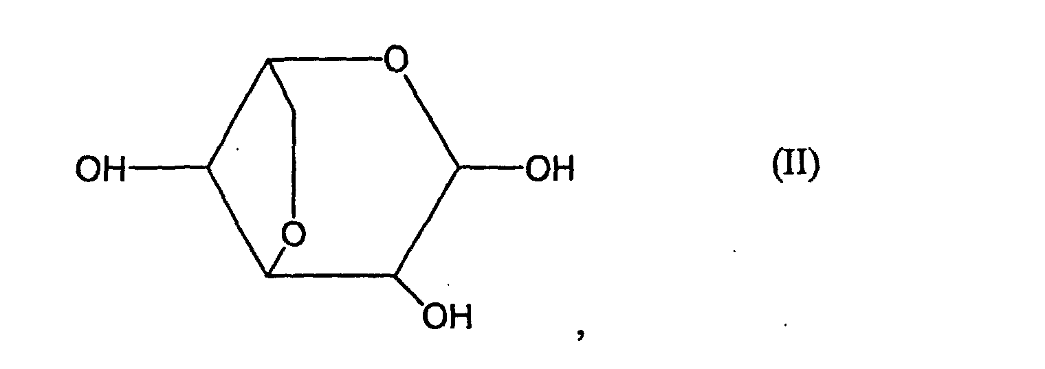
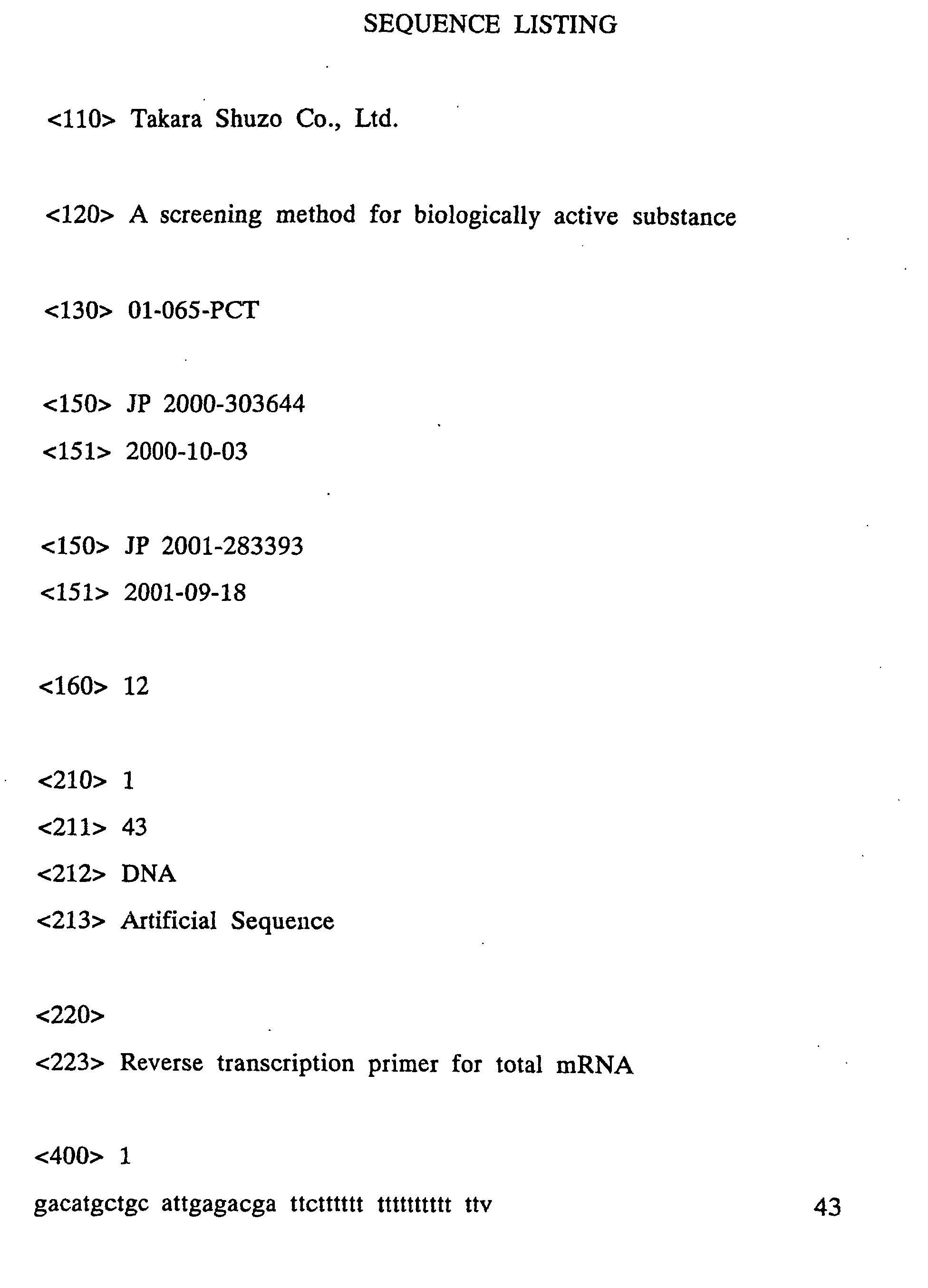


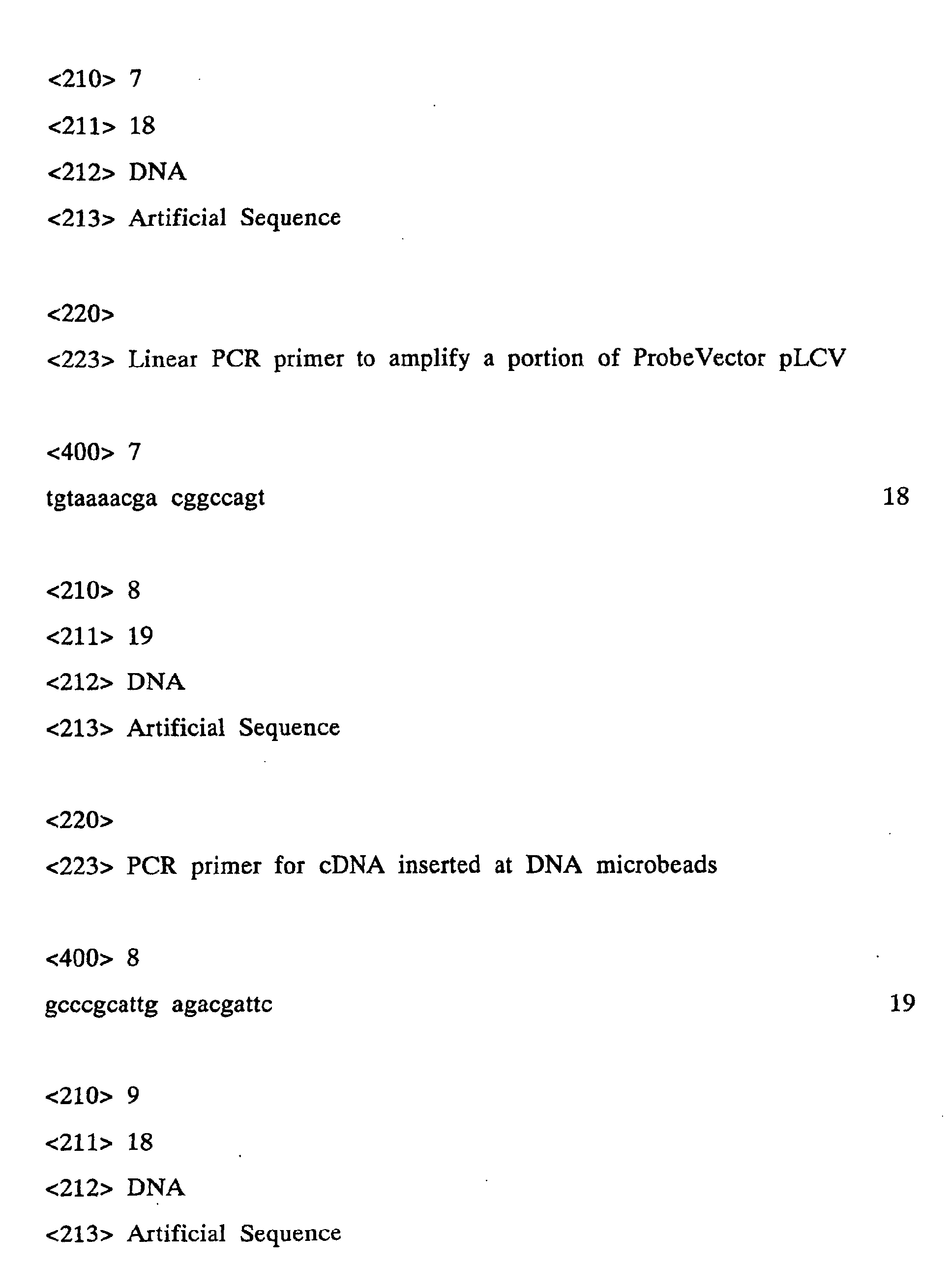


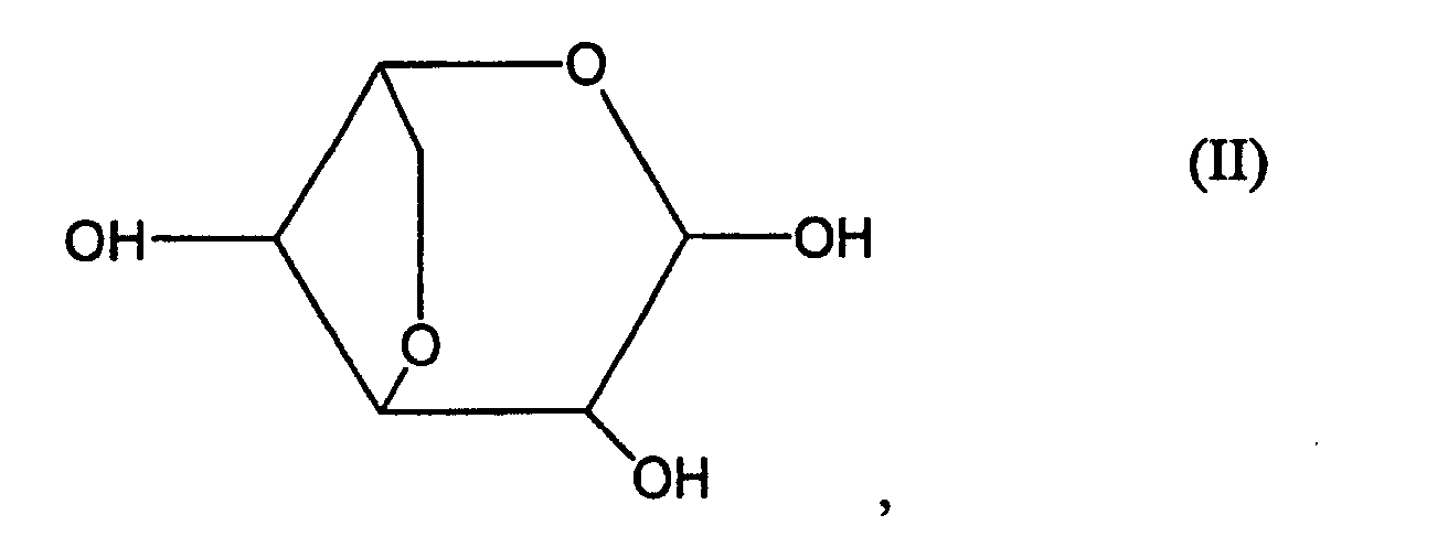 (2) determining an expression level of a gene in the living body of said (1), and comparing resulting values between a case where 1) is loaded and a case where 2) is loaded; and(3) selecting a test substance which causes a change in an expression level of at least one gene to substantially the same extent as a change where 2) is loaded.