EP1313396B1 - Photoacoustic assay and imaging system - Google Patents
Photoacoustic assay and imaging system Download PDFInfo
- Publication number
- EP1313396B1 EP1313396B1 EP01958348A EP01958348A EP1313396B1 EP 1313396 B1 EP1313396 B1 EP 1313396B1 EP 01958348 A EP01958348 A EP 01958348A EP 01958348 A EP01958348 A EP 01958348A EP 1313396 B1 EP1313396 B1 EP 1313396B1
- Authority
- EP
- European Patent Office
- Prior art keywords
- bolus
- light
- blood
- transducer
- ultrasound
- Prior art date
- Legal status (The legal status is an assumption and is not a legal conclusion. Google has not performed a legal analysis and makes no representation as to the accuracy of the status listed.)
- Expired - Lifetime
Links
- 238000003556 assay Methods 0.000 title description 24
- 238000003384 imaging method Methods 0.000 title description 5
- 238000002604 ultrasonography Methods 0.000 claims abstract description 83
- 238000010521 absorption reaction Methods 0.000 claims abstract description 33
- 238000000034 method Methods 0.000 claims abstract description 27
- 230000005855 radiation Effects 0.000 claims abstract description 5
- 210000004369 blood Anatomy 0.000 claims description 96
- 239000008280 blood Substances 0.000 claims description 96
- WQZGKKKJIJFFOK-GASJEMHNSA-N Glucose Natural products OC[C@H]1OC(O)[C@H](O)[C@@H](O)[C@@H]1O WQZGKKKJIJFFOK-GASJEMHNSA-N 0.000 claims description 76
- 239000008103 glucose Substances 0.000 claims description 76
- 238000008214 LDL Cholesterol Methods 0.000 claims description 6
- 230000008878 coupling Effects 0.000 claims description 6
- 238000010168 coupling process Methods 0.000 claims description 6
- 238000005859 coupling reaction Methods 0.000 claims description 6
- 230000008569 process Effects 0.000 claims description 5
- 108010071584 oxidized low density lipoprotein Proteins 0.000 claims description 3
- 125000002791 glucosyl group Chemical group C1([C@H](O)[C@@H](O)[C@H](O)[C@H](O1)CO)* 0.000 claims 1
- 239000000463 material Substances 0.000 claims 1
- 230000008859 change Effects 0.000 abstract description 7
- 230000000694 effects Effects 0.000 abstract description 6
- 210000001519 tissue Anatomy 0.000 description 39
- 210000004204 blood vessel Anatomy 0.000 description 37
- HVYWMOMLDIMFJA-DPAQBDIFSA-N cholesterol Chemical compound C1C=C2C[C@@H](O)CC[C@]2(C)[C@@H]2[C@@H]1[C@@H]1CC[C@H]([C@H](C)CCCC(C)C)[C@@]1(C)CC2 HVYWMOMLDIMFJA-DPAQBDIFSA-N 0.000 description 24
- 238000005259 measurement Methods 0.000 description 22
- 239000000126 substance Substances 0.000 description 22
- 235000012000 cholesterol Nutrition 0.000 description 12
- 239000000306 component Substances 0.000 description 11
- 238000005286 illumination Methods 0.000 description 11
- 238000001514 detection method Methods 0.000 description 6
- 238000001727 in vivo Methods 0.000 description 5
- 230000004044 response Effects 0.000 description 5
- 230000003287 optical effect Effects 0.000 description 4
- 210000000056 organ Anatomy 0.000 description 4
- 230000035559 beat frequency Effects 0.000 description 3
- 210000000746 body region Anatomy 0.000 description 3
- 230000012010 growth Effects 0.000 description 3
- 230000035515 penetration Effects 0.000 description 3
- 238000010895 photoacoustic effect Methods 0.000 description 3
- 102000009027 Albumins Human genes 0.000 description 2
- 108010088751 Albumins Proteins 0.000 description 2
- 206010028980 Neoplasm Diseases 0.000 description 2
- 238000004458 analytical method Methods 0.000 description 2
- 230000007423 decrease Effects 0.000 description 2
- 210000003722 extracellular fluid Anatomy 0.000 description 2
- 238000001914 filtration Methods 0.000 description 2
- 230000014509 gene expression Effects 0.000 description 2
- 238000010438 heat treatment Methods 0.000 description 2
- 230000031700 light absorption Effects 0.000 description 2
- 239000000523 sample Substances 0.000 description 2
- 239000007787 solid Substances 0.000 description 2
- 230000007480 spreading Effects 0.000 description 2
- 238000012285 ultrasound imaging Methods 0.000 description 2
- 230000000007 visual effect Effects 0.000 description 2
- 208000007536 Thrombosis Diseases 0.000 description 1
- 238000013459 approach Methods 0.000 description 1
- 230000002238 attenuated effect Effects 0.000 description 1
- 230000005540 biological transmission Effects 0.000 description 1
- 239000012503 blood component Substances 0.000 description 1
- 230000001010 compromised effect Effects 0.000 description 1
- 238000001816 cooling Methods 0.000 description 1
- 206010012601 diabetes mellitus Diseases 0.000 description 1
- 230000005670 electromagnetic radiation Effects 0.000 description 1
- 230000005284 excitation Effects 0.000 description 1
- 239000003925 fat Substances 0.000 description 1
- 210000000245 forearm Anatomy 0.000 description 1
- 230000007274 generation of a signal involved in cell-cell signaling Effects 0.000 description 1
- 210000004013 groin Anatomy 0.000 description 1
- 238000000338 in vitro Methods 0.000 description 1
- 238000005462 in vivo assay Methods 0.000 description 1
- 150000002632 lipids Chemical class 0.000 description 1
- 238000013507 mapping Methods 0.000 description 1
- 230000000737 periodic effect Effects 0.000 description 1
- 238000012545 processing Methods 0.000 description 1
- 108090000623 proteins and genes Proteins 0.000 description 1
- 102000004169 proteins and genes Human genes 0.000 description 1
- 210000004872 soft tissue Anatomy 0.000 description 1
- 238000012360 testing method Methods 0.000 description 1
- 210000000707 wrist Anatomy 0.000 description 1
Images

Classifications
-
- A—HUMAN NECESSITIES
- A61—MEDICAL OR VETERINARY SCIENCE; HYGIENE
- A61B—DIAGNOSIS; SURGERY; IDENTIFICATION
- A61B5/00—Measuring for diagnostic purposes; Identification of persons
- A61B5/145—Measuring characteristics of blood in vivo, e.g. gas concentration or pH-value ; Measuring characteristics of body fluids or tissues, e.g. interstitial fluid or cerebral tissue
- A61B5/1455—Measuring characteristics of blood in vivo, e.g. gas concentration or pH-value ; Measuring characteristics of body fluids or tissues, e.g. interstitial fluid or cerebral tissue using optical sensors, e.g. spectral photometrical oximeters
-
- A—HUMAN NECESSITIES
- A61—MEDICAL OR VETERINARY SCIENCE; HYGIENE
- A61B—DIAGNOSIS; SURGERY; IDENTIFICATION
- A61B5/00—Measuring for diagnostic purposes; Identification of persons
- A61B5/0093—Detecting, measuring or recording by applying one single type of energy and measuring its conversion into another type of energy
- A61B5/0095—Detecting, measuring or recording by applying one single type of energy and measuring its conversion into another type of energy by applying light and detecting acoustic waves, i.e. photoacoustic measurements
-
- A—HUMAN NECESSITIES
- A61—MEDICAL OR VETERINARY SCIENCE; HYGIENE
- A61B—DIAGNOSIS; SURGERY; IDENTIFICATION
- A61B5/00—Measuring for diagnostic purposes; Identification of persons
- A61B5/145—Measuring characteristics of blood in vivo, e.g. gas concentration or pH-value ; Measuring characteristics of body fluids or tissues, e.g. interstitial fluid or cerebral tissue
- A61B5/14532—Measuring characteristics of blood in vivo, e.g. gas concentration or pH-value ; Measuring characteristics of body fluids or tissues, e.g. interstitial fluid or cerebral tissue for measuring glucose, e.g. by tissue impedance measurement
-
- A—HUMAN NECESSITIES
- A61—MEDICAL OR VETERINARY SCIENCE; HYGIENE
- A61B—DIAGNOSIS; SURGERY; IDENTIFICATION
- A61B8/00—Diagnosis using ultrasonic, sonic or infrasonic waves
-
- A—HUMAN NECESSITIES
- A61—MEDICAL OR VETERINARY SCIENCE; HYGIENE
- A61B—DIAGNOSIS; SURGERY; IDENTIFICATION
- A61B8/00—Diagnosis using ultrasonic, sonic or infrasonic waves
- A61B8/08—Clinical applications
- A61B8/0833—Clinical applications involving detecting or locating foreign bodies or organic structures
- A61B8/085—Clinical applications involving detecting or locating foreign bodies or organic structures for locating body or organic structures, e.g. tumours, calculi, blood vessels, nodules
Definitions
- the invention relates to non-invasive in-vivo methods and apparatus for determining the concentration of a substance in a body and for determining the concentration of the substance as a function of position in the body.
- PCT Publication WO 98/38904 describes a "non-invasive, in - vivo glucometer" that uses a photoacoustic effect in which light energy is converted to acoustic energy to measure a person's blood glucose.
- Pulses of light at a wavelength for which light is absorbed by glucose is directed by the glucometer to illuminate a part of the person's body, such as a fingertip, comprising soft tissue.
- the light pulses are typically focused to a relatively small focal region inside the body part and light from the light pulses is absorbed by glucose and converted to kinetic energy in a region of tissue in the neighborhood of the focal region.
- the kinetic energy causes temperature and pressure of the absorbing tissue region to increase and generates acoustic waves, hereinafter referred to as "photoacoustic waves", that radiate out from the absorbing tissue region.
- An acoustic sensor comprised in the glucometer contacts the body part and senses the photoacoustic waves. Intensity of the waves is a function of the concentration of glucose in the absorbing tissue region and their intensity as measured by the sensor is used to assay the glucose.
- US Patent 5,941,821 and EPO 0 919 180 which claims priority from the U.S. Patent describe another non-invasive in-vivo glucometer that uses a photoacoustic effect to assay blood glucose.
- Light at a wavelength at which glucose absorbs light is modulated at a suitable frequency and directed by the glucometer to illuminate a region of a person's body.
- Glucose in blood and interstitial fluid in tissue near the surface of the region absorbs the light and converts the absorbed energy to kinetic energy that heats the tissue.
- Temperature of the tissue increases and decreases cyclically in cadence with the modulation of the light.
- the alternate heating and cooling of the tissue results in periodic heating of air in contact with the surface of the illuminated region, which generates sound waves in the air.
- a microphone comprised in the glucometer provides measurements of intensity of the sound waves that are used to determine a concentration of glucose.
- the invention is defined in claim 1.
- An aspect of some embodiments of the present invention relates to providing a non-invasive, in-vivo glucometer that determines a person's glucose level substantially only from glucose concentration in the person's blood.
- An aspect of some embodiments of the present invention relates to providing a glucometer that locates at least one blood vessel in a person's body using ultrasound.
- the glucometer determines the person's glucose level by assaying glucose present substantially only in blood in the located at least one blood vessel.
- the glucometer comprises at least one ultrasound transducer that radiates ultrasound into the person's body and at least one ultrasound sensor that receives energy from the radiated ultrasound that is reflected or transmitted by features in the body.
- the glucometer locates the at least one blood vessel from the reflected/transmitted energy using methods known in the art.
- the glucometer comprises at least one light source that illuminates a region of tissue in which the at least one blood vessel is located with at least one pulse of light at a wavelength for which light is absorbed by glucose.
- Glucose in a volume of blood hereinafter referred to as a "bolus" of blood
- the at least one blood vessel absorbs energy from the at least one light pulse.
- the absorbed energy generates changes in the bolus that are a function of the amount of energy absorbed, which in turn is a function, inter alia, of the concentration of glucose in the bolus.
- At least one change in an acoustic property of the bolus and/or acoustic phenomena generated in the bolus by the absorbed energy is measured by the glucometer and used to assay glucose in the bolus.
- a measurement of a change and/or phenomenon is performed during illumination of the bolus with the at least one light pulse.
- a measurement is performed after illumination of the bolus with the at least one light pulse.
- a photoacoustic wave generated by the absorbed energy is used to assay glucose in the bolus
- the bolus and generally tissue surrounding the bolus When the region of tissue in which the at least one blood vessel is located is illuminated with the at least one light pulse, the bolus and generally tissue surrounding the bolus generate pbotoacoustic waves responsive to energy that they absorb from the at least one light pulse.
- the glucometer senses the photoacoustic waves and identifies locations of their origins using methods known in the art. The glucometer compares the identified locations with the location of the bolus to identify which of the photoacoustic waves originates in the bolus. The amplitude of the photoacoustic wave so identified is a function of the concentration of glucose in the bolus and is used to assay the person's glucose.
- the glucometer reflects ultrasound waves from the bolus at times during which a photoacoustic wave is being generated by the bolus to measure a change in the bolus from which to determine glucose concentration.
- the surface of the bolus is moving and ultrasound waves reflected from the bolus are therefore Doppler shifted.
- surface regions of the bolus move at speeds that are substantial fractions of the speed of sound in the illuminated tissue.
- frequencies of ultrasound waves that are reflected by the bolus from ultrasound waves incident on the bolus are Doppler shifted substantially compared to the frequency of the incident ultrasound waves. Intensities of these reflected waves and magnitude of their Doppler shifts are sensed and used to determine concentration of glucose in the bolus.
- the glucometer has been described as illuminating the bolus with light at a wavelength for which light is absorbed by glucose. However, it is not possible to choose a wavelength for the light for which components of the bolus other than glucose do not absorb the light. As a result, an acoustic effect measured by the glucometer resulting from absorption of light by the bolus at any wavelength is due to absorption of the light by substances other than glucose in the bolus, such as cholesterol, albumin and various lipids, as well as by glucose.
- the bolus is preferably illuminated with pulses of light at a plurality of different wavelengths and the effect measured at the different wavelengths. Using known absorption cross-sections for light by substances in the blood, the contribution to the effect from glucose and thereby the concentration of glucose in the bolus is determined.
- the invention is not limited to assaying blood glucose.
- absorption coefficients of other substances in the blood are determined and the absorption coefficients may be used to determine concentrations of these substances.
- absorption coefficients of N different components of the blood bolus are determined and may be used to estimate concentrations of the components in the bolus.
- Different particular substances in the bolus whose concentrations are to be estimated can be assayed, in accordance with an embodiment of the present invention, by proper "tuning" of frequencies used to illuminate the blood bolus.
- the present invention is not limited to assaying blood components. Some embodiments of the present invention can be used to assay components of other tissues and features of the body, such as for example, components of interstitial fluid, blood clots or plaque in blood vessels that are located using ultrasound.
- Some embodiments of the present invention are used to provide a spatial map of concentration of a substance in a region of the body.
- a sub-region of the region of the body for which concentration of the substance is to be mapped is illuminated with collimated light that is absorbed by the substance.
- collimated light that is absorbed by the substance.
- changes in acoustic properties of tissue in the sub-region can be determined for highly localized "voxels" in the sub-region.
- the changes can be determined by sensing photoacoustic waves generated in the voxels and/or characteristics of ultrasonic waves that are, reflected from and/or transmitted through the voxels, in accordance with embodiments of the present invention.
- Any of the changes in acoustic properties that affect transmitted, reflected or generated acoustic waves, such as those discussed above, can be localized to voxels in the sub-region having dimensions in a range from a few to about ten wavelengths of ultrasound that is used to probe the sub-region.
- concentration of the substance in different voxels of the sub-region can be determined, in accordance with preferred embodiments of the present invention.
- a map of the concentration of the substance in the body region is acquired by moving the illuminated sub-region to different "scan" positions in the body region.
- a concentration map of the substance will, in general, image the organs and/or features.
- some embodiments of the present invention are used to imago organs and or features of organs in a region of a body.
- LDL cholesterol and oxidized LDL cholesterol are highly concentrated in plaque.
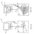
- Figs. 1A-1C schematically show a glucometer 20 measuring a person's blood glucose by sensing photoacoustic waves generated in blood in a part 24 of the person's body, in accordance with an embodiment of the present invention.
- Glucometer 20 comprises at least one ultrasound transducer 26, a light source 28 that provides light at a wavelength at which light is absorbed by glucose and a controller 30 that controls the at least one ultrasound transducer and the light source.
- At least one ultrasound transducer 26 is acoustically coupled to region 24 and is used to profile acoustic impedance and velocity of sound in region 24 and identify and locate features in the region using ultrasound imaging techniques and methods known in the art.
- at least one ultrasound transducer 24 is used to identify and locate at least one blood vessel 22 in body part 24 using methods known in the art.
- location of blood vessel 22 can be performed by detecting a Doppler shift in ultrasound reflected from the blood vessel caused by velocity of blood in the blood vessel, assuming that a component of the velocity of the blood is parallel to ultrasound incident on the blood vessel.
- location of a blood vessel can be performed by sensing reflections of ultrasound from walls of the blood vessel.
- At least one transducer 26 may comprise a single transducer or a plurality of transducers. At least one transducer 26 may compriso a relatively small transducer that performs as a point source when generating ultrasound waves and performs as a point sensor that senses ultrasound waves in a relatively large solid angle. At least one transducer 26 may comprise a shaped transducer that generates a collimated beam of ultrasound and senses ultrasound in a relatively small solid angle. At least one transducer may comprise a phased array of ultrasound transducers that can be controlled by controller 30 to generate and steer a beam of ultrasound.
- At least one transducer 26 may comprise a piezoelectric transducer or other transducer known in the art, such as an "optical” ultrasound transducer that converts pulses of optical energy into acoustic energy.
- Optical ultrasound transducers are described in an article entitled “Theory of Detection of Shear Stress Pulses with Laser Picosecond Acoustics", by O. Matsuda, 11th ICPPP, Japan 2000. Such transducers can provide and detect ultrasound pulses having sub-picosecond pulse lengths.
- same transducers are used to both generate and sense ultrasound waves.
- at least one transducer that is used to transmit ultrasound is not used to sense ultrasound.
- At least one ultrasound transducer 26 is assumed, by way of example, to both transmit and sense ultrasound and to be controllable to generate a collimated and/or focused pulse of ultrasound.
- a controller 30 controls at least one ultrasound transducer 26 to transmit at least one collimated pulse of ultrasound waves into body part 24, which ultrasound pulse is represented by arc line segments 32. Some of the energy in at least one ultrasound pulse 32 is reflected from a localized region 36 of at least one blood vessel 22 and returns to at least one transducer 26 as a reflected at least one ultrasound pulse 38.
- a reflected pulse 38 is sensed by at least one ultrasound transducer 26, which generates signals responsive thereto that the ultrasound transducer transmits to controller 30.
- Controller 30 processes the received signals, using methods known in the art, to identify and locate at least one blood vessel 22. For example, assume a bolus of blood 40 moving through region 36 of at least one blood vessel 22 has a component of velocity parallel to pulse 32 or reflected pulse 38. Reflected pulse 38 may then be identified as originating in at least one blood vessel 22 by sensing a Doppler shift in the reflected pulse resulting from the pulse being reflected from bolus 40.
- the location of blood vessel 22 may be determined from a time lapse between a time at which the pulses of ultrasound waves 32 are transmitted and a time at which Doppler shifted ultrasound pulse 38 arrives at ultrasound transducer 26.
- reflected pulses 38 may arise from reflections of energy from transmitted pulses 32 by walls of at least one blood vessel 22 as a result of the walls having acoustic impedance different from the acoustic impedance of tissue adjacent to the walls.
- energy from at least one ultrasound pulse 32 is not only reflected by at least one blood vessel 22 and features thereof.
- energy from at least one ultrasound pulse 32 is reflected by tissue interfaces and regions in body part 24 in which the acoustic impedance is changing rapidly.
- energy in the reflections that is sensed by at least one transducer 26 is used to determine acoustic properties, such as for example acoustic impedance and the speed of sound, of regions in body part 24.
- acoustic energy reflections are used to determine acoustic properties of coupling of at least one transducer 26 to body part 24. Measurements of the acoustic properties of body part 24 and of the coupling of at least one transducer 26 to the body part are hereinafter referred to as "acoustic calibration measurements”.
- ultrasound generated by at least one transducer 26 has a frequency greater than 5MHz. In some embodiments of the present invention the frequency is substantially equal to or greater than 10MHz. Sound propagates in body tissue at a velocity of about 1.5mm per microsecond and has a useful penetration depth for detection of features in tissue, which is limited by absorption and decreases as frequency increases.
- the wavelength of ultrasound generated by the transducer is about 0.15 mm and a useful penetration depth of about 35 mm.
- glucometer 20 can determine location of at least one blood vessel 22 in a direction along which the transducer transmits ultrasound pulses with an accuracy of about 0.15mm ( i . e . about a wavelength).
- transmitted ultrasound pulses 32 are collimated so that they have a cross-sectional diameter of about 1.5mm.
- Glucometer 20 therefore has spatial resolution and penetration depth sufficient to locate blood vessels having dimensions typical of dimensions of blood vessels located, by way of example, in a person's wrist, forearm or groin.
- controller 30 controls light source 28 to illuminate body part 24 with at least one pulse 42 of collimated light "aimed" at region 36 of at least one blood vessel 22.
- tissue in body part 24 scatters the light and the collimated light spreads out laterally. Therefore as light pulse 42 penetrates deeper into body part 24 it illuminates an increasing volume of tissue in the body part.
- a volume of tissue greater than the volume of bolus 40 is illuminated. It is therefore seen that, because of scattering, collimated light cannot, generally, be used to illuminate substantially only a relatively small, accurately defined region of tissue in body part 24.
- photoacoustic waves can be generated by light pulse 42 in locations in body part 24 that are not restricted to blood bolus 40 and the light pulse may generate photoacoustic waves at locations substantially removed from the location of the blood bolus.
- lines 44 schematically represent an envelope that defines the spatial extent of at least one light pulse 42 perpendicular to the direction of travel of the light pulse and spreading of lines 44 inside body part 24 indicate lateral spreading of at least one light pulse 42.
- ultrasound transducer 26 is formed with a hole and light source 28 is positioned to transmit light pulses through the hole. Transmission of light pulses through a hole in ultrasound transducer 26 provides relatively uniform and more symmetric illumination of a "field of view" of the transducer in body part 24 in which the transducer detects features of the body part.
- light source 28 is shown as a single light source, light source 28 may comprise a plurality of light sources that provide light at wavelengths suitable for determining glucose concentration in bolus 40.
- Fig. 1C schematically shows photoacoustic waves, which are represented by sets of concentric circles, that are generated in body part 24 as a result of illumination by light 42.
- An innermost concentric circle in a set of concentric circles schematically represents a location of an origin of a photoacoustic wave.
- Sets of concentric circles 48 drawn with dashed lines schematically represent photoacoustic waves that are generated by absorption of energy from light pulse 42 at locations outside of blood vessel 22 and bolus 40.
- Set 50 of concentric circles drawn in solid lines schematically represents a photoacoustic wave having an origin in bolus 40.
- Ultrasound transducer 26 senses photoacoustic waves 48 and 50 and transmits signals responsive thereto to controller 30.
- controller 30 determines a location for the origin of each of photoacoustic waves 48 and 50 using results of acoustic calibration measurements performed previously and noted in the discussion of Fig. 1A. Controller 30 than compares the locations to the location of bolus 40, which was determined previously, as indicated in Fig. 1A. Methods for determining the locations of origins of photoacoustic waves are described in PCT Publication WO 98/14118 and US Patent 5,713,356.
- Controller 30 in accordance with an embodiment of the present invention, is therefore able to discriminate between photoacoustic waves that are generated in bolus 40 and photoacoustic waves that are generated outside of bolus 40. Controller 30 determines that photoacoustic wave 50 is located in bolus 40 and that photoacoustic waves 48 are located outside of bolus 40. In accordance with an embodiment of the present invention, controller 30 therefore uses the locations and amplitudes of photoacoustic waves 50 and 48 and calibration measurements to determine a concentration of glucose in the person's blood.
- the amplitude of pressure from photoacoustic wave 50 and the shape of photoacoustic wave 50 that is sensed by ultrasound sensor 26 is a function of an amount of energy absorbed by bolus 40 from at least one light pulse 42.
- the amount of energy absorbed from at least one light pulse 42 is, of course, a function of an absorption coefficient in blood for light in light pulse 42. In general the amount of energy absorbed is relatively small, as a result of which, the amount of energy is approximately proportional to the absorption coefficient of light 42 in blood.
- ⁇ b is the absorption coefficient of blood in bolus 40, which is a function of glucose concentration in the blood, for light in at least one light pulse 42.
- the exponential factor e - ⁇ d is an attenuation factor by which intensity of at least one light pulse 42 is attenuated along the path length d.
- the attenuation factor may be estimated from d, which is known from the location of blood bolus 40 and experimentally known values for ⁇ .
- the absorption coefficient a is determined as a function of distance along the path length d using the equation for P given above or in one of the other referenced documents and a finits element analysis of the measured photoacoustic signal.
- the wavelength of light in at least one light pulse 42 is chosen so that the light is absorbed by glucose.
- the absorption coefficient ⁇ b is therefore a function of a known absorption cross section of glucose for light at the wavelength of light in at least one light pulse 42 and an unknown concentration of glucose in the blood of bolus 40.
- a measure of the amplitude P of photoacoustic waves generated in blood bolus 40 can therefore be used to determine concentration of glucose in blood bolus 40.
- controller 30 adjusts amplitude P using results of the acoustic calibration measurements to remove biases in P introduced by acoustic properties of tissue in body part 24 and coupling of at least one transducer 26 to the body part Controller 30 uses the adjusted P to determine glucose concentration.
- Glucose has absorption peaks at a number of different wavelengths that can be used in the practice of the present invention.
- glucose has absorption peaks in the mid infrared (IR) at 9.7 microns in the combination region at 2.10, 2.27, and 2.32 microns, in the first overtone region at 1.73, 1.69, and 1.61 microns, and in the near infrared, with relatively low absorption, in bands centered at 0.76, 0.92, and 1.00 micron that can be used in the practice of the present invention.
- IR mid infrared
- Many different substances in the blood such as cholesterol, albumin and various fats and proteins absorb light at or near wavelengths of light at which glucose absorbs light.
- the absorption coefficient ⁇ b is a function not only of glucose concentration in blood bolus 40 but also of the concentrations (and absorption coefficients) the other absorbing substances in the blood bolus. Therefore, in accordance with an embodiment of the present invention, to assay the person's blood glucose, absorption coefficients ⁇ b of light by a bolus 40 of the person's blood are determined from measured amplitudes of photoacoustic waves for a plurality of different wavelengths of light. The concentration of glucose in the person's blood is then determined from analysis of the determined absorption coefficients using algorithms known in the art.
- Suitable light sources such as lasers, laser diodes (LDs), and light emitting diodes (LEDs) for providing light at wavelengths used to assay glucose, in accordance with embodiments of the present invention, are readily available.
- LDs laser diodes
- LEDs light emitting diodes
- filtered lamps can also be used for providing light.
- photoacoustic waves generated in blood bolus 40 by at least one light pulse 42 are detected by heterodyning the photoacoustic waves with a reference acoustic wave, in accordance with an embodiment of the present invention.
- At least one light pulse 42 comprises a train of light pulses radiated at a first frequency. Photoacoustic waves will then be generated by blood bolus 40 at substantially the first frequency. Assume that the reference source is controlled to generate "reference" acoustic waves at a second frequency, offset from the first frequency, which are focused on blood bolus 40. Assume further that the reference acoustic waves are sufficiently intense so that in the presence of the reference acoustic waves the blood bolus has a substantial non-linear response to acoustic stimuli.
- photoacoustic waves generated in blood bolus 40 by light pulses 42 are a function not only of the intensity and frequency of the light pulses but are a function also of the intensity and frequency of the reference acoustic waves.
- the "acoustic non-linearity" of blood bolus 40 caused by the reference waves couples the reference waves and the photoacoustic response of the blood bolus to light pulses 42.
- the coupling of the reference waves and the photoacoustic response of the blood bolus will generate a "heterodyned photoacoustio signal".
- the heterodyned photoacoustic signal will have a frequency substantially equal to the beat frequency of the first and second frequencies will be transmitted from the blood bolus.
- the pressure amplitude of the heterodyned signal is proportional to the power in the reference beam and can be substantially larger than the pressure amplitude of photoacoustic waves generated by blood bolus 40 responsive to illumination by light pulse 42.
- the heterodyned signal can be detected by appropriate filtering of signals generated by at least one transducer 26 using techniques well known in the art.
- the inventors estimate that a reference acoustic beam that focuses about 1mW of power in a blood bolus having a volume of about 1 mm 2 , focuses sufficient energy in the blood bolus to couple the photoacoustic response to the reference acoustic beam.
- the coupling of two acoustic signals in the human body to generate a heterodyned signal is discussed in a book entitled "Physical Ultrasonics" by Robert T. Beyer and Stephen V. Letcher, Academic Press 1969. Calculations in the book indicate possible gain factors.
- a heterodyning detection technique in accordance with an embodiment of the present invention, enables simultaneous excitation and subsequent simultaneous detection of photoacoustic waves generated in bolus 40 by light at a plurality of different wavelengths.
- blood bolus 40 is simultaneously illuminated with light at three different wavelengths in order to determine glucose concentration in the blood bolus.
- the pulse rate of the light is different for each wavelength and that during illumination a reference acoustic wave is focused on the bolus.
- the photoacoustic response of the blood bolus will result in generation of a strong photoacoustic signal at a different beat frequency for each of the three wavelengths.
- the signals at each of the frequencies can be simultaneously detected and identified using appropriate filtering and signal processing techniques known in the art.
- a heterodyning detection technique in accordance with an embodiment of the present invention, can be used to determine a location of the source of photoacoustic waves generated in a body.
- Energy in a reference acoustic beam can be focused to a relatively small focal volume at a known location.
- Photoacoustic signals generated in the body that are characterized by a frequency equal to a beat frequency must have originated in the reference beam focal volume.
- At least one light pulse 42 comprises a train of light pulses and glucometer 20' measures a change in reflectance following each light pulse in the train of light pulses.
- the pulse width and pulse repetition rate are such that between light pulses bolus 40 cools down to a temperature substantially equal to the ambient temperature.
- energy reflectance of bolus 40 is measured by reflecting an ultrasound pulse 46 from the bolus before and after each light pulse in the train of light pulses to determine a change in reflectance resulting from illumination by the light pulse.
- the pulse width of the light pulses is about 10 nanoseconds and the pulse repetition rate is about 10 kHz.
- ultrasound pulses 46 are pulses of ultrasound waves at a frequency of at least 10 MHz.
- changes in acoustic impedance of blood bolus 40 are measured to determine blood glucose levels while bolus 40 is being heated by at least one light pulse 42 or shortly thereafter.
- glucometer 20' reflects ultrasound from bolus 40 while the bolus is being heated by light pulse 42, or shortly thereafter, during a time period in which photoacoustic waves are being generated in bolus 40 responsive to illumination by the light pulse.
- Ultrasound waves reflected from bolus 40 while it is expanding following absorption of energy from light pulse 42 often carry a very definite and relatively easily identified signature - a very large Doppler shift.
- a photoacoustic wave is being generated in blood bolus 40 a surface of a volume of blood in bolus 40 expands at a speed close to the speed of sound.
- sound waves reflected from bolus 40 while it is being heated by light pulse 42 are generally characterized by very large Doppler shifts.
- Figs. 2A and 2B show a glucometer 21, similar to glucometer 20, operating to assay glucose in a person's blood by reflecting ultrasound waves from blood bolus 40 while the blood bolus is being heated by light that is absorbed by blood.
- glucometer 21 locates at least one blood vessel 22 and blood bolus 40 and, optionally, performs acoustic calibration measurements.
- controller 30 controls light source 28 to illuminate bolus 40 with a light pulse 42 and while bolus is being illuminated by the light pulse, controls ultrasound transducer 26 to transmit a pulse 51 of ultrasound that is focused on the blood bolus.
- bolus 40 expands rapidly. The surface of the bolus moves outward from the bolus at speeds close to the speed of sound and generates a photoaconstic wave.
- the intensity of Doppler shifted pulse 54 is a function of the energy reflectance of the surface of bolus 40 which in turn is a function of a difference between the absorption coefficieat of light in blood bolus 40 and the absorption coefficient of tissue surrounding the bolus.
- glucometer 21 measures intensity of Doppler shifted waves reflected from a blood bolus while the blood bolus is being illuminated with light for different wavelengths of the illuminating light. The measurements are used to determine absorption coefficients for blood at the wavelengths and a concentration of glucose in the blood. In some embodiments of the present invention, the measurements are corrected responsive to acoustic calibration measurements and the corrected measurements are used to determine glucose concentration.
- concentrations of cholesterol in the regions can be determined.
- Light at wavelengths between 1.70-1.80 microns is absorbed by cholesterol and light at these wavelengths can be used in the practice of the present invention to determine cholesterol concentrations in regions of a person's body.
- Methods of determining concentration of a substance in a body provide the concentration of the substances as a function of position in the body.
- a spatial map of concentrations of a substance in a body is used to image features and/or components of the body.
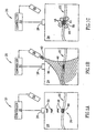
- Figs. 3A and 3B schematically show a "glucometer” 100, hereinafter referred to as "assay imager 100", used, by way of example, to assay and image cholesterol in a person's body in accordance with an embodiment of the present invention.
- Assay imager 100 comprises a visual display 102.
- controller 30 controls at least one transducer 26 to locate a region 36 of at least one blood vessel 22.
- assay imager 100 then illuminates region 36 with light at wavelengths suitable for assaying cholesterol and determines concentrations of cholesterol in region 36 using methods similar to those described above for determining concentrations of glucose.
- Controller 30 then controls transducer 26 to locate other regions of at least one blood vessel 22 and proceeds to determine concentrations of cholesterol in these other regions.
- a map 110 of cholesterol concentration as a function of position for regions scanned by assay imager 100 is displayed on visual display 102.
- At least one blood vessel 22 is compromised by plaque deposits 104 and 106 at two locations. The locations of plaque deposits 104 and 106 are visible on cholesterol concentration map 110 shown on display 102.
- assay imager 100 may also be used to image tumors.
- Tumorous growths generally have an unusually high concentration of blood vessels in the growths and in tissue surrounding the growths.
- tissue having an unusually high density of blood vessels can be clearly contrasted against tissue having normal blood vessel density.
- tumor tissue in the imaged region can often be clearly contrasted with healthy tissue.
- an "assay image" of a region of the body responsive to concentration of a substance in the region that is provided by an assay imager is overlaid on an image of the region provided by another imaging modality, such as for example an MRI or CT image.
- the image on which the assay image is overlaid is an ultrasound image.
- the ultrasound image is acquired using the same ultrasound detectors that are used to acquire the assay image. By using the same ultrasound detectors for acquiring both the assay image and the ultrasound image on which the assay image is overlaid the assay image is automatically registered to the ultrasound image.
- each of the verbs, "comprise” “include” and “have”, and conjugates thereof, are used to indicate that the object or objects of the verb are not necessarily a complete listing of members, components, elements or parts of the subject or subjects of the verb.
Landscapes
- Health & Medical Sciences (AREA)
- Life Sciences & Earth Sciences (AREA)
- Physics & Mathematics (AREA)
- Surgery (AREA)
- General Health & Medical Sciences (AREA)
- Biophysics (AREA)
- Pathology (AREA)
- Engineering & Computer Science (AREA)
- Biomedical Technology (AREA)
- Heart & Thoracic Surgery (AREA)
- Medical Informatics (AREA)
- Molecular Biology (AREA)
- Veterinary Medicine (AREA)
- Animal Behavior & Ethology (AREA)
- Public Health (AREA)
- Optics & Photonics (AREA)
- Emergency Medicine (AREA)
- Spectroscopy & Molecular Physics (AREA)
- Acoustics & Sound (AREA)
- Investigating Or Analyzing Materials By The Use Of Ultrasonic Waves (AREA)
- Ultra Sonic Daignosis Equipment (AREA)
- Measurement Of The Respiration, Hearing Ability, Form, And Blood Characteristics Of Living Organisms (AREA)
- Investigating Or Analysing Materials By Optical Means (AREA)
- Holo Graphy (AREA)
- Eye Examination Apparatus (AREA)
- Testing, Inspecting, Measuring Of Stereoscopic Televisions And Televisions (AREA)
Abstract
Description
- The invention relates to non-invasive in-vivo methods and apparatus for determining the concentration of a substance in a body and for determining the concentration of the substance as a function of position in the body.
- Methods and apparatus for in-vivo and in-vitro measurements of blood glucose levels are known in the art. Generally, the methods and apparatus are relatively complicated and measurements of a person's blood glucose levels are usually performed in a clinic or laboratory with the aid of a technician and costs of the measurements are relatively high.
- Methods and apparatus for determining blood glucose levels for use in the home, for example by a diabetic who must monitor blood glucose levels frequently, are available. These methods and associated devices arc generally invasive and usually involve taking blood samples by finger pricking. Finger pricking is perceived as inconvenient and unpleasant and to avoid finger pricking diabetics tend to monitor their glucose levels less frequently than is advisable. Moreover, many conventional glucometers require routine purchasing of sample sticks and pricking needles, which is bothersome and adds cost to the user. There is a need for glucometers that are easy to use and that perform non-invasive in-vivo assays of blood sugar.
- PCT Publication WO 98/38904, describes a "non-invasive, in-vivo glucometer" that uses a photoacoustic effect in which light energy is converted to acoustic energy to measure a person's blood glucose. Pulses of light at a wavelength for which light is absorbed by glucose is directed by the glucometer to illuminate a part of the person's body, such as a fingertip, comprising soft tissue. The light pulses are typically focused to a relatively small focal region inside the body part and light from the light pulses is absorbed by glucose and converted to kinetic energy in a region of tissue in the neighborhood of the focal region. The kinetic energy causes temperature and pressure of the absorbing tissue region to increase and generates acoustic waves, hereinafter referred to as "photoacoustic waves", that radiate out from the absorbing tissue region. An acoustic sensor comprised in the glucometer contacts the body part and senses the photoacoustic waves. Intensity of the waves is a function of the concentration of glucose in the absorbing tissue region and their intensity as measured by the sensor is used to assay the glucose.
- However, light is scattered by body tissue and even though the light is focused to a small focal region inside the body, the location and size of the absorbing tissue region are not accurately known. As a result, the generated photoacoustic effect and measurements of the person's glucose levels are not necessarily the result only of glucose concentration in the person's blood. Characteristics of the absorbing tissue region, such as density of blood vessels therein, that can affect concentration of glucose in the absorbing region are often not accurately known. Measurements of blood glucose levels can therefore be affected by unknown variables that substantially compromise the reliability of the measurements.
- US Patent 5,941,821 and EPO 0 919 180, which claims priority from the U.S. Patent describe another non-invasive in-vivo glucometer that uses a photoacoustic effect to assay blood glucose. Light at a wavelength at which glucose absorbs light is modulated at a suitable frequency and directed by the glucometer to illuminate a region of a person's body. Glucose in blood and interstitial fluid in tissue near the surface of the region absorbs the light and converts the absorbed energy to kinetic energy that heats the tissue. Temperature of the tissue increases and decreases cyclically in cadence with the modulation of the light. The alternate heating and cooling of the tissue results in periodic heating of air in contact with the surface of the illuminated region, which generates sound waves in the air. A microphone comprised in the glucometer provides measurements of intensity of the sound waves that are used to determine a concentration of glucose.
- The invention is defined in claim 1.
- An aspect of some embodiments of the present invention relates to providing a non-invasive, in-vivo glucometer that determines a person's glucose level substantially only from glucose concentration in the person's blood.
- An aspect of some embodiments of the present invention relates to providing a glucometer that locates at least one blood vessel in a person's body using ultrasound. The glucometer determines the person's glucose level by assaying glucose present substantially only in blood in the located at least one blood vessel.
- In embodiments of the present invention, the glucometer comprises at least one ultrasound transducer that radiates ultrasound into the person's body and at least one ultrasound sensor that receives energy from the radiated ultrasound that is reflected or transmitted by features in the body. The glucometer locates the at least one blood vessel from the reflected/transmitted energy using methods known in the art. In some embodiments of the present invention, the glucometer comprises at least one light source that illuminates a region of tissue in which the at least one blood vessel is located with at least one pulse of light at a wavelength for which light is absorbed by glucose. Glucose in a volume of blood, hereinafter referred to as a "bolus" of blood, in the at least one blood vessel absorbs energy from the at least one light pulse. The absorbed energy generates changes in the bolus that are a function of the amount of energy absorbed, which in turn is a function, inter alia, of the concentration of glucose in the bolus.
- At least one change in an acoustic property of the bolus and/or acoustic phenomena generated in the bolus by the absorbed energy is measured by the glucometer and used to assay glucose in the bolus. In some embodiments of the present invention a measurement of a change and/or phenomenon is performed during illumination of the bolus with the at least one light pulse. In some embodiments of the present invention a measurement is performed after illumination of the bolus with the at least one light pulse.
- According to an aspect of some embodiments of the present invention a photoacoustic wave generated by the absorbed energy is used to assay glucose in the bolus,
- When the region of tissue in which the at least one blood vessel is located is illuminated with the at least one light pulse, the bolus and generally tissue surrounding the bolus generate pbotoacoustic waves responsive to energy that they absorb from the at least one light pulse. The glucometer senses the photoacoustic waves and identifies locations of their origins using methods known in the art. The glucometer compares the identified locations with the location of the bolus to identify which of the photoacoustic waves originates in the bolus. The amplitude of the photoacoustic wave so identified is a function of the concentration of glucose in the bolus and is used to assay the person's glucose.
- According to an aspect of some embodiments of the present invention the glucometer reflects ultrasound waves from the bolus at times during which a photoacoustic wave is being generated by the bolus to measure a change in the bolus from which to determine glucose concentration. During generation of the photoacoustic wave, the surface of the bolus is moving and ultrasound waves reflected from the bolus are therefore Doppler shifted. Generally, during generation of the photoacoustic wave, surface regions of the bolus move at speeds that are substantial fractions of the speed of sound in the illuminated tissue. As a result, frequencies of ultrasound waves that are reflected by the bolus from ultrasound waves incident on the bolus are Doppler shifted substantially compared to the frequency of the incident ultrasound waves. Intensities of these reflected waves and magnitude of their Doppler shifts are sensed and used to determine concentration of glucose in the bolus.
- The glucometer has been described as illuminating the bolus with light at a wavelength for which light is absorbed by glucose. However, it is not possible to choose a wavelength for the light for which components of the bolus other than glucose do not absorb the light. As a result, an acoustic effect measured by the glucometer resulting from absorption of light by the bolus at any wavelength is due to absorption of the light by substances other than glucose in the bolus, such as cholesterol, albumin and various lipids, as well as by glucose. In order to isolate a contribution to the effect due to glucose, and thereby the concentration of glucose in the bolus, the bolus is preferably illuminated with pulses of light at a plurality of different wavelengths and the effect measured at the different wavelengths. Using known absorption cross-sections for light by substances in the blood, the contribution to the effect from glucose and thereby the concentration of glucose in the bolus is determined.
- It is readily concluded from remarks made in the previous paragraph that the invention is not limited to assaying blood glucose. In the process of determining glucose concentration, in accordance with an embodiment of the present invention, absorption coefficients of other substances in the blood are determined and the absorption coefficients may be used to determine concentrations of these substances. For example if "N" different wavelengths of light are used to determine glucose concentration, in accordance with an embodiment of the present invention, absorption coefficients of N different components of the blood bolus are determined and may be used to estimate concentrations of the components in the bolus. Different particular substances in the bolus whose concentrations are to be estimated can be assayed, in accordance with an embodiment of the present invention, by proper "tuning" of frequencies used to illuminate the blood bolus. Furthermore, the present invention is not limited to assaying blood components. Some embodiments of the present invention can be used to assay components of other tissues and features of the body, such as for example, components of interstitial fluid, blood clots or plaque in blood vessels that are located using ultrasound.
- Whereas only light has been described as being used to introduce energy to a blood bolus, other forms of electromagnetic radiation, such as microwave or RF radiation, may be used to impart energy to components being assayed in a tissue volume, in accordance with an embodiment of the present invention. Some embodiments of the present invention are used to provide a spatial map of concentration of a substance in a region of the body. In some embodiments, a sub-region of the region of the body for which concentration of the substance is to be mapped is illuminated with collimated light that is absorbed by the substance. As noted above, because of scattering of light in body tissue, the size and location of the sub-region is not accurately known. However, changes in acoustic properties of tissue in the sub-region can be determined for highly localized "voxels" in the sub-region. The changes can be determined by sensing photoacoustic waves generated in the voxels and/or characteristics of ultrasonic waves that are, reflected from and/or transmitted through the voxels, in accordance with embodiments of the present invention. Any of the changes in acoustic properties that affect transmitted, reflected or generated acoustic waves, such as those discussed above, can be localized to voxels in the sub-region having dimensions in a range from a few to about ten wavelengths of ultrasound that is used to probe the sub-region. Since these changes are a function of concentration of the substance, the concentration of the substance in different voxels of the sub-region can be determined, in accordance with preferred embodiments of the present invention. A map of the concentration of the substance in the body region is acquired by moving the illuminated sub-region to different "scan" positions in the body region.
- Since different organs and/or features in the body region will usually be distinguished by different levels of concentration of a substance, a concentration map of the substance, provided in accordance with an embodiment of the present invention, will, in general, image the organs and/or features. As a result some embodiments of the present invention are used to imago organs and or features of organs in a region of a body. For example, LDL cholesterol and oxidized LDL cholesterol are highly concentrated in plaque. By mapping concentration of LDL cholesterol or oxidized LDL cholesterol in a region of a person's body, images of plaque deposits on walls of blood vessels in the region can be acquired.
- Non-limiting examples of embodiments of the present invention are described below with reference to figures attached hereto. In the figures, identical structures, elements or parts that appear in more than one figure are generally labeled with the same numeral in all the figures in which they appear. Dimensions of components and features shown in the figures are chosen for convenience and clarity of presentation and are not necessarily shown to scale. The figures are listed below.
- Figs. 1A-1C schematically show a glucometer measuring a person's blood glucose by sensing photoacoustic waves generated in blood in a region of the person's body by illumination of the region with light absorbed by glucose, in accordance with an embodiment of the present invention;
- Figs. 2A and 2B schematically show a glucometer measuring blood glucose by sensing changes in velocity of sound in blood, in accordance with an embodiment of the present invention;
- Figs. 3A-3B schematically show a glucometer assaying and imaging cholesterol in a person's body, in accordance with an embodiment of the present invention.
- Figs. 1A-1C schematically show a
glucometer 20 measuring a person's blood glucose by sensing photoacoustic waves generated in blood in apart 24 of the person's body, in accordance with an embodiment of the present invention.Glucometer 20 comprises at least oneultrasound transducer 26, alight source 28 that provides light at a wavelength at which light is absorbed by glucose and acontroller 30 that controls the at least one ultrasound transducer and the light source. - At least one
ultrasound transducer 26 is acoustically coupled toregion 24 and is used to profile acoustic impedance and velocity of sound inregion 24 and identify and locate features in the region using ultrasound imaging techniques and methods known in the art. In particular, at least oneultrasound transducer 24 is used to identify and locate at least oneblood vessel 22 inbody part 24 using methods known in the art. For example, location ofblood vessel 22 can be performed by detecting a Doppler shift in ultrasound reflected from the blood vessel caused by velocity of blood in the blood vessel, assuming that a component of the velocity of the blood is parallel to ultrasound incident on the blood vessel. Or, location of a blood vessel can be performed by sensing reflections of ultrasound from walls of the blood vessel. - Numerous and varied ultrasound transducers, configurations of transducers and ultrasound imaging methods suitable for use in practicing the present invention and locating blood vessels are known in the art.
- At least one
transducer 26 may comprise a single transducer or a plurality of transducers. At least onetransducer 26 may compriso a relatively small transducer that performs as a point source when generating ultrasound waves and performs as a point sensor that senses ultrasound waves in a relatively large solid angle. At least onetransducer 26 may comprise a shaped transducer that generates a collimated beam of ultrasound and senses ultrasound in a relatively small solid angle. At least one transducer may comprise a phased array of ultrasound transducers that can be controlled bycontroller 30 to generate and steer a beam of ultrasound. - At least one
transducer 26 may comprise a piezoelectric transducer or other transducer known in the art, such as an "optical" ultrasound transducer that converts pulses of optical energy into acoustic energy. Optical ultrasound transducers are described in an article entitled "Theory of Detection of Shear Stress Pulses with Laser Picosecond Acoustics", by O. Matsuda, 11th ICPPP, Japan 2000. Such transducers can provide and detect ultrasound pulses having sub-picosecond pulse lengths. - In some embodiments of the present invention same transducers are used to both generate and sense ultrasound waves. In some embodiments of the present invention at least one transducer that is used to transmit ultrasound is not used to sense ultrasound.
- In Figs 1A - 1C and figures that follow, for convenience of presentation, at least one
ultrasound transducer 26 is assumed, by way of example, to both transmit and sense ultrasound and to be controllable to generate a collimated and/or focused pulse of ultrasound. In Fig. 1A controller 30 controls at least oneultrasound transducer 26 to transmit at least one collimated pulse of ultrasound waves intobody part 24, which ultrasound pulse is represented byarc line segments 32. Some of the energy in at least oneultrasound pulse 32 is reflected from alocalized region 36 of at least oneblood vessel 22 and returns to at least onetransducer 26 as a reflected at least oneultrasound pulse 38. - A reflected
pulse 38 is sensed by at least oneultrasound transducer 26, which generates signals responsive thereto that the ultrasound transducer transmits tocontroller 30.Controller 30 processes the received signals, using methods known in the art, to identify and locate at least oneblood vessel 22. For example, assume a bolus ofblood 40 moving throughregion 36 of at least oneblood vessel 22 has a component of velocity parallel topulse 32 or reflectedpulse 38.Reflected pulse 38 may then be identified as originating in at least oneblood vessel 22 by sensing a Doppler shift in the reflected pulse resulting from the pulse being reflected frombolus 40. The location ofblood vessel 22 may be determined from a time lapse between a time at which the pulses of ultrasound waves 32 are transmitted and a time at which Doppler shiftedultrasound pulse 38 arrives atultrasound transducer 26. In addition, reflectedpulses 38 may arise from reflections of energy from transmittedpulses 32 by walls of at least oneblood vessel 22 as a result of the walls having acoustic impedance different from the acoustic impedance of tissue adjacent to the walls. - It should be noted that energy from at least one
ultrasound pulse 32 is not only reflected by at least oneblood vessel 22 and features thereof. In general, energy from at least oneultrasound pulse 32 is reflected by tissue interfaces and regions inbody part 24 in which the acoustic impedance is changing rapidly. In some embodiments of the present invention, energy in the reflections that is sensed by at least onetransducer 26 is used to determine acoustic properties, such as for example acoustic impedance and the speed of sound, of regions inbody part 24. In some embodiments of the present invention acoustic energy reflections are used to determine acoustic properties of coupling of at least onetransducer 26 tobody part 24. Measurements of the acoustic properties ofbody part 24 and of the coupling of at least onetransducer 26 to the body part are hereinafter referred to as "acoustic calibration measurements". - In some embodiments of the present invention, ultrasound generated by at least one
transducer 26 has a frequency greater than 5MHz. In some embodiments of the present invention the frequency is substantially equal to or greater than 10MHz. Sound propagates in body tissue at a velocity of about 1.5mm per microsecond and has a useful penetration depth for detection of features in tissue, which is limited by absorption and decreases as frequency increases. For an ultrasound frequency of about 10MHz, the wavelength of ultrasound generated by the transducer is about 0.15 mm and a useful penetration depth of about 35 mm. For this frequency,glucometer 20 can determine location of at least oneblood vessel 22 in a direction along which the transducer transmits ultrasound pulses with an accuracy of about 0.15mm (i.e. about a wavelength). In some embodiments of the present invention, transmittedultrasound pulses 32 are collimated so that they have a cross-sectional diameter of about 1.5mm.Glucometer 20 therefore has spatial resolution and penetration depth sufficient to locate blood vessels having dimensions typical of dimensions of blood vessels located, by way of example, in a person's wrist, forearm or groin. - In Fig. 1B, after at least one
blood vessel 22 has been located,controller 30 controlslight source 28 to illuminatebody part 24 with at least onepulse 42 of collimated light "aimed" atregion 36 of at least oneblood vessel 22. Aslight pulse 42 entersbody part 24, tissue inbody part 24 scatters the light and the collimated light spreads out laterally. Therefore aslight pulse 42 penetrates deeper intobody part 24 it illuminates an increasing volume of tissue in the body part. In the region of bolus 40 a volume of tissue greater than the volume ofbolus 40 is illuminated. It is therefore seen that, because of scattering, collimated light cannot, generally, be used to illuminate substantially only a relatively small, accurately defined region of tissue inbody part 24. As a result, photoacoustic waves can be generated bylight pulse 42 in locations inbody part 24 that are not restricted toblood bolus 40 and the light pulse may generate photoacoustic waves at locations substantially removed from the location of the blood bolus. In Fig. 1B,lines 44 schematically represent an envelope that defines the spatial extent of at least onelight pulse 42 perpendicular to the direction of travel of the light pulse and spreading oflines 44 insidebody part 24 indicate lateral spreading of at least onelight pulse 42. - It should be noted that whereas
light source 28 is shown, by way of example, located at a side ofultrasound transducer 26 other positions oflight source 28 are possible and can be advantageous. In some embodiments of the present invention, for example,ultrasound transducer 26 is formed with a hole andlight source 28 is positioned to transmit light pulses through the hole. Transmission of light pulses through a hole inultrasound transducer 26 provides relatively uniform and more symmetric illumination of a "field of view" of the transducer inbody part 24 in which the transducer detects features of the body part. Furthermore whereaslight source 28 is shown as a single light source,light source 28 may comprise a plurality of light sources that provide light at wavelengths suitable for determining glucose concentration inbolus 40. - Fig. 1C schematically shows photoacoustic waves, which are represented by sets of concentric circles, that are generated in
body part 24 as a result of illumination bylight 42. An innermost concentric circle in a set of concentric circles schematically represents a location of an origin of a photoacoustic wave. Sets ofconcentric circles 48 drawn with dashed lines schematically represent photoacoustic waves that are generated by absorption of energy fromlight pulse 42 at locations outside ofblood vessel 22 andbolus 40. Set 50 of concentric circles drawn in solid lines schematically represents a photoacoustic wave having an origin inbolus 40. -
Ultrasound transducer 26 senses photoacousticwaves controller 30. In accordance with an embodiment of thepresent invention controller 30 determines a location for the origin of each ofphotoacoustic waves Controller 30 than compares the locations to the location ofbolus 40, which was determined previously, as indicated in Fig. 1A. Methods for determining the locations of origins of photoacoustic waves are described in PCT Publication WO 98/14118 and US Patent 5,713,356. -
Controller 30, in accordance with an embodiment of the present invention, is therefore able to discriminate between photoacoustic waves that are generated inbolus 40 and photoacoustic waves that are generated outside ofbolus 40.Controller 30 determines thatphotoacoustic wave 50 is located inbolus 40 and thatphotoacoustic waves 48 are located outside ofbolus 40. In accordance with an embodiment of the present invention,controller 30 therefore uses the locations and amplitudes ofphotoacoustic waves - The amplitude of pressure from
photoacoustic wave 50 and the shape ofphotoacoustic wave 50 that is sensed byultrasound sensor 26 is a function of an amount of energy absorbed bybolus 40 from at least onelight pulse 42. The amount of energy absorbed from at least onelight pulse 42 is, of course, a function of an absorption coefficient in blood for light inlight pulse 42. In general the amount of energy absorbed is relatively small, as a result of which, the amount of energy is approximately proportional to the absorption coefficient of light 42 in blood. The relationship between the amplitude of a photoacoustic wave and an amount of energy absorbed by a region of tissue that generates the photoacoustic wave is described in US Patent 4,385,634 to Bowen and in PCT publication WO 98/14118 referenced above. Expressions for the amplitude of a photoacoustic wave arc also given in an article by Lai, H. M. and Young, K.J. in Acoust. Soc. Am. Vol 76, pg 2000 (1982), in an article by MacKenzie et al., "Advances in Photoacoustic Noninvasive Glucose Testing", Clin. Chem. Vol 45, pp 1587-1595 (1999) and in an article by C.G.A. Hoelen et al., "A New Theoretical Approach To Photoacoustic Signal Generation", Acoust. Soc. Am. 106 2 (1999). - If P represents the amplitude of
photoacoustic wave 50 then, adopting the expression for amplitude given by Hoelen et al., P = A(βC2/H)(Ioτe-αd)αb, where A is a constant of proportionality, β is the temperature expansion coefficient for blood, C the speed of sound and H the specific heat capacity of blood. Io and τ are respectively the intensity and pulse length of at least onelight pulse 42. α is the absorption coefficient for light in at least onelight pulse 42 in tissue ofbody part 24 and d is a path length thatlight pulse 42 travels in the body part to reachblood bolus 40. αb is the absorption coefficient of blood inbolus 40, which is a function of glucose concentration in the blood, for light in at least onelight pulse 42. The exponential factor e-αd is an attenuation factor by which intensity of at least onelight pulse 42 is attenuated along the path length d. The attenuation factor may be estimated from d, which is known from the location ofblood bolus 40 and experimentally known values for α. In some embodiments of the present invention, the absorption coefficient a is determined as a function of distance along the path length d using the equation for P given above or in one of the other referenced documents and a finits element analysis of the measured photoacoustic signal. - The wavelength of light in at least one
light pulse 42 is chosen so that the light is absorbed by glucose. The absorption coefficient αb is therefore a function of a known absorption cross section of glucose for light at the wavelength of light in at least onelight pulse 42 and an unknown concentration of glucose in the blood ofbolus 40. A measure of the amplitude P of photoacoustic waves generated inblood bolus 40 can therefore be used to determine concentration of glucose inblood bolus 40. In some embodiments of the present invention,controller 30 adjusts amplitude P using results of the acoustic calibration measurements to remove biases in P introduced by acoustic properties of tissue inbody part 24 and coupling of at least onetransducer 26 to thebody part Controller 30 uses the adjusted P to determine glucose concentration. - Glucose has absorption peaks at a number of different wavelengths that can be used in the practice of the present invention. For example, glucose has absorption peaks in the mid infrared (IR) at 9.7 microns in the combination region at 2.10, 2.27, and 2.32 microns, in the first overtone region at 1.73, 1.69, and 1.61 microns, and in the near infrared, with relatively low absorption, in bands centered at 0.76, 0.92, and 1.00 micron that can be used in the practice of the present invention. However, there is no wavelength for the light at which only glucose in the blood absorbs the light. Many different substances in the blood such as cholesterol, albumin and various fats and proteins absorb light at or near wavelengths of light at which glucose absorbs light. As a result, the absorption coefficient αb is a function not only of glucose concentration in
blood bolus 40 but also of the concentrations (and absorption coefficients) the other absorbing substances in the blood bolus. Therefore, in accordance with an embodiment of the present invention, to assay the person's blood glucose, absorption coefficients αb of light by abolus 40 of the person's blood are determined from measured amplitudes of photoacoustic waves for a plurality of different wavelengths of light. The concentration of glucose in the person's blood is then determined from analysis of the determined absorption coefficients using algorithms known in the art. - Appropriate criteria for the choice different wavelengths of light used to determine glucose concentration and the methods and techniques for analyzing the absorption coefficients are well known. US patent 5,957,841 to Maruo et aL, US Patent 5,452,716 to V. Clift, US Pat 5,348,002 to Caro, and U.S. Patent 4,975,581 to Robinson et al., describe methods of determining glucose concentration from absorption measurements at a plurality of wavelengths. US Patent 5,957,841 describes determining glucose concentration in tissue from measurements of absorption of light in the tissue in three wavelength bands: 1.48 microns - 1.55 microns, 1.55 microns -1.65 microns and 1.65 microns - 1.88 microns. Suitable light sources, such as lasers, laser diodes (LDs), and light emitting diodes (LEDs) for providing light at wavelengths used to assay glucose, in accordance with embodiments of the present invention, are readily available. For methods of assaying glucose, in accordance with embodiments of the present invention, such as a method discussed below with reference to Fig. 4, in which continuous illumination of a blood bolus at desired frequencies is required, filtered lamps can also be used for providing light.
- In some embodiments of the present invention, photoacoustic waves generated in
blood bolus 40 by at least onelight pulse 42 are detected by heterodyning the photoacoustic waves with a reference acoustic wave, in accordance with an embodiment of the present invention. - To illustrate the heterodyning detection technique, in accordance with an embodiment of the present invention, assume that at least one
light pulse 42 comprises a train of light pulses radiated at a first frequency. Photoacoustic waves will then be generated byblood bolus 40 at substantially the first frequency. Assume that the reference source is controlled to generate "reference" acoustic waves at a second frequency, offset from the first frequency, which are focused onblood bolus 40. Assume further that the reference acoustic waves are sufficiently intense so that in the presence of the reference acoustic waves the blood bolus has a substantial non-linear response to acoustic stimuli. As a result, photoacoustic waves generated inblood bolus 40 bylight pulses 42 are a function not only of the intensity and frequency of the light pulses but are a function also of the intensity and frequency of the reference acoustic waves. The "acoustic non-linearity" ofblood bolus 40 caused by the reference waves couples the reference waves and the photoacoustic response of the blood bolus tolight pulses 42. - In particular, if the phase of the reference waves and the envelope of optical energy of
light pulses 42 atblood bolus 40 are stable, the coupling of the reference waves and the photoacoustic response of the blood bolus will generate a "heterodyned photoacoustio signal". The heterodyned photoacoustic signal will have a frequency substantially equal to the beat frequency of the first and second frequencies will be transmitted from the blood bolus. The pressure amplitude of the heterodyned signal is proportional to the power in the reference beam and can be substantially larger than the pressure amplitude of photoacoustic waves generated byblood bolus 40 responsive to illumination bylight pulse 42. The heterodyned signal can be detected by appropriate filtering of signals generated by at least onetransducer 26 using techniques well known in the art. - The inventors estimate that a reference acoustic beam that focuses about 1mW of power in a blood bolus having a volume of about 1 mm2, focuses sufficient energy in the blood bolus to couple the photoacoustic response to the reference acoustic beam. The coupling of two acoustic signals in the human body to generate a heterodyned signal is discussed in a book entitled "Physical Ultrasonics" by Robert T. Beyer and Stephen V. Letcher, Academic Press 1969. Calculations in the book indicate possible gain factors.
- It is to be noted that using a heterodyning detection technique, in accordance with an embodiment of the present invention, enables simultaneous excitation and subsequent simultaneous detection of photoacoustic waves generated in
bolus 40 by light at a plurality of different wavelengths. For example, assume thatblood bolus 40 is simultaneously illuminated with light at three different wavelengths in order to determine glucose concentration in the blood bolus. Assume further that the pulse rate of the light is different for each wavelength and that during illumination a reference acoustic wave is focused on the bolus. The photoacoustic response of the blood bolus will result in generation of a strong photoacoustic signal at a different beat frequency for each of the three wavelengths. The signals at each of the frequencies can be simultaneously detected and identified using appropriate filtering and signal processing techniques known in the art. - It is to be further recognized that a heterodyning detection technique, in accordance with an embodiment of the present invention, can be used to determine a location of the source of photoacoustic waves generated in a body. Energy in a reference acoustic beam can be focused to a relatively small focal volume at a known location. Photoacoustic signals generated in the body that are characterized by a frequency equal to a beat frequency must have originated in the reference beam focal volume.
- In some embodiments of the present invention, at least one
light pulse 42 comprises a train of light pulses and glucometer 20' measures a change in reflectance following each light pulse in the train of light pulses. Optionally, the pulse width and pulse repetition rate are such that between light pulses bolus 40 cools down to a temperature substantially equal to the ambient temperature. Optionally, energy reflectance ofbolus 40 is measured by reflecting an ultrasound pulse 46 from the bolus before and after each light pulse in the train of light pulses to determine a change in reflectance resulting from illumination by the light pulse. In some embodiments of the present invention, the pulse width of the light pulses is about 10 nanoseconds and the pulse repetition rate is about 10 kHz. In some embodiments of the present invention, ultrasound pulses 46 are pulses of ultrasound waves at a frequency of at least 10 MHz. - In some embodiments of the present invention changes in acoustic impedance of
blood bolus 40 are measured to determine blood glucose levels whilebolus 40 is being heated by at least onelight pulse 42 or shortly thereafter. To perform the measurements glucometer 20' reflects ultrasound frombolus 40 while the bolus is being heated bylight pulse 42, or shortly thereafter, during a time period in which photoacoustic waves are being generated inbolus 40 responsive to illumination by the light pulse. - Ultrasound waves reflected from
bolus 40 while it is expanding following absorption of energy fromlight pulse 42 often carry a very definite and relatively easily identified signature - a very large Doppler shift. During expansion, while a photoacoustic wave is being generated in blood bolus 40 a surface of a volume of blood inbolus 40 expands at a speed close to the speed of sound. As a result, sound waves reflected frombolus 40 while it is being heated bylight pulse 42, are generally characterized by very large Doppler shifts. - Figs. 2A and 2B show a
glucometer 21, similar toglucometer 20, operating to assay glucose in a person's blood by reflecting ultrasound waves fromblood bolus 40 while the blood bolus is being heated by light that is absorbed by blood. - In Fig. 2A,
glucometer 21 locates at least oneblood vessel 22 andblood bolus 40 and, optionally, performs acoustic calibration measurements. In Fig.2B controller 30 controlslight source 28 to illuminatebolus 40 with alight pulse 42 and while bolus is being illuminated by the light pulse, controlsultrasound transducer 26 to transmit apulse 51 of ultrasound that is focused on the blood bolus. As a result of illumination ofbolus 40 bylight pulse 42,bolus 40 expands rapidly. The surface of the bolus moves outward from the bolus at speeds close to the speed of sound and generates a photoaconstic wave. Energy fromultrasound pulse 51 is reflected from the rapidly moving surface ofbolus 40 in anultrasound pulse 54 having a large Doppler shift generated by the velocity at which the surface moves. The intensity of Doppler shiftedpulse 54 is a function of the energy reflectance of the surface ofbolus 40 which in turn is a function of a difference between the absorption coefficieat of light inblood bolus 40 and the absorption coefficient of tissue surrounding the bolus. - In accordance with an embodiment of the present invention, glucometer 21 measures intensity of Doppler shifted waves reflected from a blood bolus while the blood bolus is being illuminated with light for different wavelengths of the illuminating light. The measurements are used to determine absorption coefficients for blood at the wavelengths and a concentration of glucose in the blood. In some embodiments of the present invention, the measurements are corrected responsive to acoustic calibration measurements and the corrected measurements are used to determine glucose concentration.
- By way of another example of assaying substances other than glucose, in accordance with embodiments of the present invention, by choosing an appropriate set of wavelengths for light used to illuminate regions of a person's body, concentrations of cholesterol in the regions can be determined. Light at wavelengths between 1.70-1.80 microns is absorbed by cholesterol and light at these wavelengths can be used in the practice of the present invention to determine cholesterol concentrations in regions of a person's body.
- Methods of determining concentration of a substance in a body, in accordance with an embodiment of the present invention provide the concentration of the substances as a function of position in the body. In some embodiments of the present invention a spatial map of concentrations of a substance in a body is used to image features and/or components of the body.
- Figs. 3A and 3B schematically show a "glucometer" 100, hereinafter referred to as "
assay imager 100", used, by way of example, to assay and image cholesterol in a person's body in accordance with an embodiment of the present invention.Assay imager 100 comprises avisual display 102. In Fig. 3A,controller 30 controls at least onetransducer 26 to locate aregion 36 of at least oneblood vessel 22. In Fig.3B assay imager 100 then illuminatesregion 36 with light at wavelengths suitable for assaying cholesterol and determines concentrations of cholesterol inregion 36 using methods similar to those described above for determining concentrations of glucose.Controller 30 then controlstransducer 26 to locate other regions of at least oneblood vessel 22 and proceeds to determine concentrations of cholesterol in these other regions. Amap 110 of cholesterol concentration as a function of position for regions scanned byassay imager 100 is displayed onvisual display 102. At least oneblood vessel 22 is compromised byplaque deposits plaque deposits cholesterol concentration map 110 shown ondisplay 102. - By way of another example,
assay imager 100 may also be used to image tumors. Tumorous growths generally have an unusually high concentration of blood vessels in the growths and in tissue surrounding the growths. By imaging a region of tissue withassay imager 100 using light at a wavelength that is strongly absorbed by blood, in accordance with an embodiment of the present invention, tissue having an unusually high density of blood vessels can be clearly contrasted against tissue having normal blood vessel density. As a result, tumor tissue in the imaged region can often be clearly contrasted with healthy tissue. - In some embodiments of the present invention, an "assay image" of a region of the body responsive to concentration of a substance in the region that is provided by an assay imager is overlaid on an image of the region provided by another imaging modality, such as for example an MRI or CT image. In some embodiments of the present invention, the image on which the assay image is overlaid is an ultrasound image. Optionally the ultrasound image is acquired using the same ultrasound detectors that are used to acquire the assay image. By using the same ultrasound detectors for acquiring both the assay image and the ultrasound image on which the assay image is overlaid the assay image is automatically registered to the ultrasound image.
- In the description and claims of the present application, each of the verbs, "comprise" "include" and "have", and conjugates thereof, are used to indicate that the object or objects of the verb are not necessarily a complete listing of members, components, elements or parts of the subject or subjects of the verb.
- The present invention has been described using detailed descriptions of embodiments thereof that are provided by way of example and are not intended to limit the scope of the invention. The described embodiments comprise different features, not all of which are required in all embodiments of the invention. Some embodiments of the present invention utilize only some of the features or possible combinations of the features. Variations of embodiments of the present invention that are described and embodiments of the present invention comprising different combinations of features noted in the described embodiments will occur to persons of the art. The scope of the invention is limited only by the following claims.
Claims (17)
- Apparatus (20, 21, 100) for assaying a component of a blood bolus in a body comprising:a light source (28) controllable to provide pulses of light having a wavelength at which the radiation is absorbed by the component and generates photoacoustic waves in the region;at least one transducer (26) for sensing ultrasound that generates signals responsive to ultrasound energy incident thereon; anda controller (30) that:a) locates the blood bolus in a volume of the body comprising the blood bolus;b) controls the light source to illuminate the volume comprising the blood bolus with at least one pulse of light having a wavelength at which light is absorbed by the component so as to generate photoacoustic waves in the volume that arc incident on the at least one sensing transducer;c) receives signals generated by the at least one sensing transducer (26) responsive to the photoacoustic waves and uses them to determine locations of the origins of the photoacoustic waves;d) uses signals generated by the at least one sensing transducer (26) responsive to those photoacoustic waves having an origin determined to be located in the blood bolus to determine an absorption coefficient for the light in the blood bolus; ande) uses the determined absorption coefficient to determine concentration of the component in the blood bolus.
- Apparatus according to claim 1 and wherein the controller (30) controls the light source (28) to illuminate the volume with at least one pulse of light at at least one other wavelength, and repeats c-e.
- Apparatus according to claim 1 or claim 2 wherein to locate the blood bolus the controller (30):a) controls the light source (28) to illuminate the volume of the body that comprises the bolus with at least one pulse of light having a wavelength at which light is preferentially absorbed by blood so as to generate photoacoustic waves in the volume that are incident on the at least one sensing transducer;b) uses signals generated by the at least one sensing transducer (26) responsive to the photoacoustic waves to map concentration of blood in the volume as a function of position; andc) uses the determined concentration map to determine a location for the blood bolus.
- Apparatus according to claim any of claims 1-3 and comprising at least one transducer (26) controllable to transmit ultrasound into the body.
- Apparatus according to claim 4 wherein the controller (30) controls the at least one transmitting transducer (26) to transmit ultrasound into the volume and processes signals from the at least one sensing transducer responsive to reflections from the transmitted ultrasound to determine the location of the bolus.
- Apparatus according to claim 5 wherein the bolus is located to within an uncertainty of less than 5 wavelengths of the ultrasound in at least one direction.
- Apparatus according to claim 5 wherein the bolus is located to within an uncertainty of less than 3 wavelengths of the ultrasound in at least one direction.
- Apparatus according to claim 5 wherein the bolus is located to within an uncertainty of about a wavelength of the ultrasound in at least one direction.
- Apparatus according to any of claims 4-8 wherein the at least one light pulse comprises at least one train of light pulses radiated at a pulse repetition rate.
- Apparatus according to claim 9 wherein the controller (30):controls a transmitting transducer (26) of the at least one transmitting transducer to focus an acoustic reference beam on the blood bolus, which reference beam has a frequency that is shifted from the pulse repetition frequency of the at least one light pulse train by an offset frequency and has an intensity that causes the bolus to respond non-linearly to acoustic stimuli;receives signals provided by a sensing transducer responsive to acoustic waves at the offset frequency that are incident on the transducer; anduses the signals to determine the absorption coefficient.
- Apparatus according to claim 10 wherein the controller (30) controls the light source (28) to illuminate the volume with at least one other light pulse train characterized by a different wavelength and pulse repetition rate and repeats c-e.
- Apparatus according to claim 11 wherein the light pulse train and at least one other light pulse train illuminate the region substantially simultaneously.
- Apparatus according to any of claims 4-12 wherein a transducer (26) of the at least one transmitting and/or sensing transducer is coupled to the body and the controller (30):controls the at least one transmitting transducer (26) to transmit ultrasonic waves, which are incident on a sensing transducer (26) that generates signals responsive thereto;receives the generated signals; andprocesses the signals to determine acoustic properties of the coupling between the transducer (26) coupled to the body and the body.
- Apparatus according to any of the preceding claims wherein the controller (30) processes signals from the at least one sensing transducer (26) to determine acoustic properties of material in the body.
- Apparatus according to claim 14 wherein the determined absorption coefficient is adjusted responsive to the measured acoustic properties.
- Apparatus according to any of the preceding claims wherein the component is LDL cholesterol or oxidized LDL cholesterol.
- Apparatus according to any of the preceding claims wherein the component is glucose.
Priority Applications (1)
| Application Number | Priority Date | Filing Date | Title |
|---|---|---|---|
| EP06116163A EP1700563A2 (en) | 2000-08-24 | 2001-08-09 | Photoacoustic assay and imaging system |
Applications Claiming Priority (3)
| Application Number | Priority Date | Filing Date | Title |
|---|---|---|---|
| IL13807300A IL138073A0 (en) | 2000-08-24 | 2000-08-24 | Photoacoustic assay and imaging system |
| IL13807300 | 2000-08-24 | ||
| PCT/IL2001/000740 WO2002015776A1 (en) | 2000-08-24 | 2001-08-09 | Photoacoustic assay and imaging system |
Related Child Applications (1)
| Application Number | Title | Priority Date | Filing Date |
|---|---|---|---|
| EP06116163A Division EP1700563A2 (en) | 2000-08-24 | 2001-08-09 | Photoacoustic assay and imaging system |
Publications (2)
| Publication Number | Publication Date |
|---|---|
| EP1313396A1 EP1313396A1 (en) | 2003-05-28 |
| EP1313396B1 true EP1313396B1 (en) | 2006-06-28 |
Family
ID=11074559
Family Applications (2)
| Application Number | Title | Priority Date | Filing Date |
|---|---|---|---|
| EP06116163A Withdrawn EP1700563A2 (en) | 2000-08-24 | 2001-08-09 | Photoacoustic assay and imaging system |
| EP01958348A Expired - Lifetime EP1313396B1 (en) | 2000-08-24 | 2001-08-09 | Photoacoustic assay and imaging system |
Family Applications Before (1)
| Application Number | Title | Priority Date | Filing Date |
|---|---|---|---|
| EP06116163A Withdrawn EP1700563A2 (en) | 2000-08-24 | 2001-08-09 | Photoacoustic assay and imaging system |
Country Status (8)
| Country | Link |
|---|---|
| US (1) | US6846288B2 (en) |
| EP (2) | EP1700563A2 (en) |
| JP (1) | JP4104456B2 (en) |
| AT (1) | ATE331465T1 (en) |
| AU (1) | AU2001280066A1 (en) |
| DE (1) | DE60121179T2 (en) |
| IL (1) | IL138073A0 (en) |
| WO (1) | WO2002015776A1 (en) |
Families Citing this family (217)
| Publication number | Priority date | Publication date | Assignee | Title |
|---|---|---|---|---|
| US6554774B1 (en) * | 2000-03-23 | 2003-04-29 | Tensys Medical, Inc. | Method and apparatus for assessing hemodynamic properties within the circulatory system of a living subject |
| US8039261B2 (en) | 2000-11-17 | 2011-10-18 | Vascular Biogenics Ltd. | Promoters exhibiting endothelial cell specificity and methods of using same for regulation of angiogenesis |
| US20050085725A1 (en) * | 2001-08-09 | 2005-04-21 | Ron Nagar | Photoacoustic assay and imaging system |
| US20050075598A1 (en) * | 2001-08-24 | 2005-04-07 | Redding Bruce K. | Method and apparatus for the measurement of real time drug delivery through the use of a wearable monitor and sensor attached to a transdermal drug delivery device |
| US7077565B2 (en) | 2001-11-15 | 2006-07-18 | Glucon, Inc. | Method for measuring temperature of substances from measurement of absorption coefficients |
| KR100493154B1 (en) | 2002-03-20 | 2005-06-03 | 삼성전자주식회사 | Apparatus of non-invasive measurement of bio-fluid concentration by using photoacoustic spectroscopy |
| US20060122583A1 (en) * | 2002-06-25 | 2006-06-08 | Glucon Inc | Method and apparatus for performing myocardial revascularization |
| WO2004000112A2 (en) * | 2002-06-25 | 2003-12-31 | Glucon Inc. | Method and apparatus for determining tissue viability |
| US7774042B2 (en) * | 2002-09-26 | 2010-08-10 | Senco Brands, Inc. | Tissue scanner |
| US7646484B2 (en) * | 2002-10-07 | 2010-01-12 | Intellidx, Inc. | Method and apparatus for performing optical measurements of a material |
| EP1585440A1 (en) | 2003-01-13 | 2005-10-19 | Glucon Inc. | Photoacoustic assay method and apparatus |
| EP1613208A1 (en) * | 2003-04-01 | 2006-01-11 | Glucon Inc. | Photoacoustic assay method and apparatus |
| US20060258939A1 (en) | 2003-06-09 | 2006-11-16 | Glucon, Inc. | Wearable glucometer |
| JP4406226B2 (en) * | 2003-07-02 | 2010-01-27 | 株式会社東芝 | Biological information video device |
| US6954662B2 (en) * | 2003-08-19 | 2005-10-11 | A.D. Integrity Applications, Ltd. | Method of monitoring glucose level |
| IL164030A0 (en) * | 2003-09-12 | 2005-12-18 | Revital Pery Shechter | Photoacoustic analyzer of a region of interest in a human body |
| ES2436214T3 (en) * | 2003-09-12 | 2013-12-27 | Or-Nim Medical Ltd. | Non-invasive optical monitoring of a region of interest |
| US20050133416A1 (en) * | 2003-12-19 | 2005-06-23 | Bhan Opinder K. | Systems, methods, and catalysts for producing a crude product |
| WO2005063117A2 (en) * | 2003-12-29 | 2005-07-14 | Glucon Inc. | Glucometer comprising an implantable light source |
| WO2005068973A1 (en) * | 2004-01-13 | 2005-07-28 | Glucon Inc. | Photoacoustic sensor |
| WO2005067786A1 (en) * | 2004-01-15 | 2005-07-28 | Glucon Inc. | Wearable glucometer |
| JP4643153B2 (en) * | 2004-02-06 | 2011-03-02 | 株式会社東芝 | Non-invasive biological information imaging device |
| WO2005107592A1 (en) | 2004-05-06 | 2005-11-17 | Nippon Telegraph And Telephone Corporation | Component concentration measuring device and method of controlling component concentration measuring device |
| US7251516B2 (en) * | 2004-05-11 | 2007-07-31 | Nostix Llc | Noninvasive glucose sensor |
| IL166760A0 (en) * | 2004-05-13 | 2006-01-15 | Nexense Ltd | Method and apparatus for non-invasively monitoringconcentrations of glucose or other target substan ces |
| DK1759536T3 (en) | 2004-06-01 | 2011-09-05 | Kwalata Trading Ltd | In vitro techniques for use with stem cells |
| WO2006006166A2 (en) | 2004-07-14 | 2006-01-19 | Glusense, Ltd. | Implantable power sources and sensors |
| EP1833361A1 (en) * | 2004-12-20 | 2007-09-19 | Koninklijke Philips Electronics N.V. | Investigation of body structures |
| WO2006097933A2 (en) | 2005-03-17 | 2006-09-21 | Glucon Inc. | Method for monitoring changes in blood glucose level |
| US20060262903A1 (en) * | 2005-05-20 | 2006-11-23 | Diebold Roger M | Method and apparatus for photothermal modification of x-ray images |
| DE102005030151B3 (en) * | 2005-06-28 | 2006-11-02 | Fraunhofer-Gesellschaft zur Förderung der angewandten Forschung e.V. | Photo-acoustic free-field detector for measuring air, gas and liquid flows has optical and acoustic mirrors arranged in position where local maximum sound pressure is present for generating acoustic energy based on output of acoustic sensor |
| DE102005034219A1 (en) * | 2005-07-19 | 2007-02-22 | Fachhochschule Lübeck | Method for in vivo tissue classification |
| US20070088206A1 (en) * | 2005-10-14 | 2007-04-19 | Peyman Gholam A | Photoacoustic measurement of analyte concentration in the eye |
| US20120271204A1 (en) * | 2005-10-14 | 2012-10-25 | Peyman Gholam A | Photoacoustic measurement |
| US8554296B2 (en) | 2005-10-14 | 2013-10-08 | Gholam A. Peyman | Photoacoustic measurement of analyte concentration in the eye |
| US20070093717A1 (en) * | 2005-10-20 | 2007-04-26 | Glucon Inc. | Wearable glucometer configurations |
| US20070093698A1 (en) * | 2005-10-20 | 2007-04-26 | Glucon Inc. | Apparatus and methods for attaching a device to a body |
| US20090054763A1 (en) * | 2006-01-19 | 2009-02-26 | The Regents Of The University Of Michigan | System and method for spectroscopic photoacoustic tomography |
| US20090227997A1 (en) * | 2006-01-19 | 2009-09-10 | The Regents Of The University Of Michigan | System and method for photoacoustic imaging and monitoring of laser therapy |
| US8135450B2 (en) * | 2006-01-20 | 2012-03-13 | Esenaliev Rinat O | Noninvasive glucose sensing methods and systems |
| US7328131B2 (en) * | 2006-02-01 | 2008-02-05 | Medtronic, Inc. | Implantable pedometer |
| US7993269B2 (en) * | 2006-02-17 | 2011-08-09 | Medtronic, Inc. | Sensor and method for spinal monitoring |
| US20070197895A1 (en) * | 2006-02-17 | 2007-08-23 | Sdgi Holdings, Inc. | Surgical instrument to assess tissue characteristics |
| US8016859B2 (en) | 2006-02-17 | 2011-09-13 | Medtronic, Inc. | Dynamic treatment system and method of use |
| TW200734462A (en) | 2006-03-08 | 2007-09-16 | In Motion Invest Ltd | Regulating stem cells |
| US7665364B2 (en) | 2006-03-21 | 2010-02-23 | Ut-Battelle, Llc | Method and apparatus for remote sensing of molecular species at nanoscale utilizing a reverse photoacoustic effect |
| AU2007238155A1 (en) * | 2006-04-11 | 2007-10-25 | Oregon Health & Science University | Methods and devices for non-invasive analyte measurement |
| US7918796B2 (en) * | 2006-04-11 | 2011-04-05 | Warsaw Orthopedic, Inc. | Volumetric measurement and visual feedback of tissues |
| WO2007138493A1 (en) * | 2006-05-25 | 2007-12-06 | Koninklijke Philips Electronics, N.V. | Photoacoustic imaging method |
| CN101472520B (en) * | 2006-06-23 | 2015-06-03 | 皇家飞利浦电子股份有限公司 | Timing controller for combined photoacoustic and ultrasound imager |
| JP4909132B2 (en) * | 2006-08-28 | 2012-04-04 | 公立大学法人大阪府立大学 | Optical tomography equipment |
| JP4820239B2 (en) * | 2006-08-28 | 2011-11-24 | 公立大学法人大阪府立大学 | Probe for optical tomography equipment |
| EP2074418B1 (en) * | 2006-09-29 | 2013-03-06 | Koninklijke Philips Electronics N.V. | Ultrasonic determination of optical absorption coefficients |
| EP1905351B1 (en) * | 2006-09-29 | 2016-10-26 | Sysmex Corporation | Nonivasive living body measuring device and noninvasive living body measuring method |
| WO2008062354A1 (en) * | 2006-11-21 | 2008-05-29 | Koninklijke Philips Electronics N.V. | A system, device, method, computer-readable medium, and use for in vivo imaging of tissue in an anatomical structure |
| US20080123083A1 (en) * | 2006-11-29 | 2008-05-29 | The Regents Of The University Of Michigan | System and Method for Photoacoustic Guided Diffuse Optical Imaging |
| EP1935346A1 (en) * | 2006-12-21 | 2008-06-25 | Stichting voor de Technische Wetenschappen | Imaging apparatus and method |
| US20080214912A1 (en) * | 2007-01-10 | 2008-09-04 | Glucose Sensing Technologies, Llc | Blood Glucose Monitoring System And Method |
| US20080173093A1 (en) * | 2007-01-18 | 2008-07-24 | The Regents Of The University Of Michigan | System and method for photoacoustic tomography of joints |
| KR100871074B1 (en) * | 2007-02-01 | 2008-11-28 | 삼성전자주식회사 | Non-invasive blood glucose measurement device and method |
| US20080221647A1 (en) * | 2007-02-23 | 2008-09-11 | The Regents Of The University Of Michigan | System and method for monitoring photodynamic therapy |
| JP4909131B2 (en) * | 2007-03-09 | 2012-04-04 | 公立大学法人大阪府立大学 | Optically assisted ultrasonic velocity change imaging apparatus and optically assisted ultrasonic velocity change image display method |
| US20080228073A1 (en) * | 2007-03-12 | 2008-09-18 | Silverman Ronald H | System and method for optoacoustic imaging of peripheral tissues |
| US20080228072A1 (en) * | 2007-03-16 | 2008-09-18 | Warsaw Orthopedic, Inc. | Foreign Body Identifier |
| US10226206B2 (en) | 2007-04-11 | 2019-03-12 | The Board Of Regents Of The University Of Texas System | Systems and methods for measuring neonatal cerebral oxygenation |
| US10307088B2 (en) | 2007-04-11 | 2019-06-04 | The Board Of Regents Of The University Of Texas | Systems and methods for measuring oxygenation |
| US20080306387A1 (en) * | 2007-04-13 | 2008-12-11 | Schutz Ronald W | Finger mounted imaging and sensing assembly |
| EP2133026B1 (en) * | 2007-04-24 | 2013-05-22 | Panasonic Corporation | Ultrasonographic device |
| US8417311B2 (en) * | 2008-09-12 | 2013-04-09 | Optiscan Biomedical Corporation | Fluid component analysis system and method for glucose monitoring and control |
| US20090105588A1 (en) * | 2007-10-02 | 2009-04-23 | Board Of Regents, The University Of Texas System | Real-Time Ultrasound Monitoring of Heat-Induced Tissue Interactions |
| EP3868284A1 (en) | 2007-10-10 | 2021-08-25 | Optiscan Biomedical Corporation | Fluid component analysis system and method for glucose monitoring and control |
| EP2203734B1 (en) * | 2007-10-16 | 2018-02-21 | Koninklijke Philips N.V. | Illumination apparatus and system for production and integration of compact illumination schemes |
| WO2010048258A1 (en) * | 2008-10-23 | 2010-04-29 | Washington University In St. Louis | Reflection-mode photoacoustic tomography using a flexibly-supported cantilever beam |
| EP2203733B1 (en) | 2007-10-25 | 2017-05-03 | Washington University in St. Louis | Confocal photoacoustic microscopy with optical lateral resolution |
| US9451884B2 (en) | 2007-12-13 | 2016-09-27 | Board Of Trustees Of The University Of Arkansas | Device and method for in vivo detection of clots within circulatory vessels |
| US20090156932A1 (en) | 2007-12-13 | 2009-06-18 | Board Of Trustees Of The University Of Arkansas | Device and method for in vivo flow cytometry using the detection of photoacoustic waves |
| CN101467884B (en) * | 2007-12-28 | 2010-10-27 | 中国科学院电子学研究所 | Method and device for non-invasive blood sugar rapid detection |
| JP5284129B2 (en) * | 2008-02-06 | 2013-09-11 | キヤノン株式会社 | Imaging apparatus and analysis method |
| US20090281422A1 (en) * | 2008-05-06 | 2009-11-12 | Salama Khaled N | Multi-modality system for imaging in dense compressive media and method of use thereof |
| JP5294998B2 (en) * | 2008-06-18 | 2013-09-18 | キヤノン株式会社 | Ultrasonic probe, photoacoustic / ultrasonic system including the ultrasonic probe, and specimen imaging apparatus |
| JP4829934B2 (en) * | 2008-07-11 | 2011-12-07 | キヤノン株式会社 | Inspection device |
| CN102292029B (en) * | 2008-07-18 | 2014-11-05 | 罗切斯特大学 | Low-cost device for C-scan photoacoustic imaging |
| US20100036240A1 (en) * | 2008-08-07 | 2010-02-11 | Ismail Aly M | Multi-modality system for imaging in dense compressive media and method of use thereof |
| US7959598B2 (en) | 2008-08-20 | 2011-06-14 | Asante Solutions, Inc. | Infusion pump systems and methods |
| JP5183381B2 (en) * | 2008-09-16 | 2013-04-17 | キヤノン株式会社 | Measuring apparatus and measuring method |
| JP5241465B2 (en) * | 2008-12-11 | 2013-07-17 | キヤノン株式会社 | Photoacoustic imaging apparatus and photoacoustic imaging method |
| US20100160749A1 (en) * | 2008-12-24 | 2010-06-24 | Glusense Ltd. | Implantable optical glucose sensing |
| US9351705B2 (en) | 2009-01-09 | 2016-05-31 | Washington University | Miniaturized photoacoustic imaging apparatus including a rotatable reflector |
| JP5525787B2 (en) * | 2009-09-14 | 2014-06-18 | 株式会社東芝 | Biological information video device |
| JP5713550B2 (en) * | 2009-09-30 | 2015-05-07 | キヤノン株式会社 | Subject information processing device |
| JP5489624B2 (en) * | 2009-10-01 | 2014-05-14 | キヤノン株式会社 | measuring device |
| JP5528083B2 (en) | 2009-12-11 | 2014-06-25 | キヤノン株式会社 | Image generating apparatus, image generating method, and program |
| JP5586977B2 (en) * | 2010-02-08 | 2014-09-10 | キヤノン株式会社 | Subject information acquisition apparatus and subject information acquisition method |
| US9086365B2 (en) | 2010-04-09 | 2015-07-21 | Lihong Wang | Quantification of optical absorption coefficients using acoustic spectra in photoacoustic tomography |
| EP2605705A4 (en) * | 2010-08-20 | 2014-01-22 | Purdue Research Foundation | SYSTEM AND METHOD FOR BINDING SELECTIVE VIBRATIONAL PHOTOAACTIC IMAGING |
| EP2458369B1 (en) * | 2010-11-24 | 2014-07-23 | eesy-id GmbH | Recording device for recording a blood count parameter |
| US8997572B2 (en) | 2011-02-11 | 2015-04-07 | Washington University | Multi-focus optical-resolution photoacoustic microscopy with ultrasonic array detection |
| JP5584154B2 (en) * | 2011-03-16 | 2014-09-03 | 富士フイルム株式会社 | Photoacoustic imaging apparatus, photoacoustic imaging method, and probe for photoacoustic imaging apparatus |
| JP5685214B2 (en) * | 2011-03-16 | 2015-03-18 | 富士フイルム株式会社 | Photoacoustic image generation apparatus and method |
| JP5896623B2 (en) * | 2011-05-02 | 2016-03-30 | キヤノン株式会社 | Subject information acquisition apparatus and control method thereof |
| JP6012216B2 (en) * | 2011-05-12 | 2016-10-25 | キヤノン株式会社 | Subject information acquisition apparatus and subject information acquisition method |
| JP5681141B2 (en) * | 2011-05-13 | 2015-03-04 | 富士フイルム株式会社 | Tomographic image generating apparatus, method, and program |
| US9037205B2 (en) | 2011-06-30 | 2015-05-19 | Glusense, Ltd | Implantable optical glucose sensing |
| US9055869B2 (en) | 2011-10-28 | 2015-06-16 | Covidien Lp | Methods and systems for photoacoustic signal processing |
| US8886294B2 (en) | 2011-11-30 | 2014-11-11 | Covidien Lp | Methods and systems for photoacoustic monitoring using indicator dilution |
| US9186068B2 (en) | 2011-12-05 | 2015-11-17 | Covidien Lp | Methods and systems for photoacoustic monitoring using hypertonic and isotonic indicator dilutions |
| US9131852B2 (en) | 2011-12-05 | 2015-09-15 | Covidien Lp | Methods and systems for photoacoustic monitoring using indicator dilution |
| JP2015509443A (en) * | 2012-03-09 | 2015-03-30 | セノ メディカル インストルメンツ,インク. | Statistical mapping in photoacoustic imaging systems |
| US8885155B2 (en) | 2012-04-30 | 2014-11-11 | Covidien Lp | Combined light source photoacoustic system |
| WO2014052449A1 (en) | 2012-09-25 | 2014-04-03 | The Board Of Trustees Of The University Of Arkansas | Device and method for in vivo photoacoustic diagnosis and photothermal purging of infected blood |
| WO2014063005A1 (en) | 2012-10-18 | 2014-04-24 | Washington University | Transcranialphotoacoustic/thermoacoustic tomography brain imaging informed by adjunct image data |
| JP6192297B2 (en) * | 2013-01-16 | 2017-09-06 | キヤノン株式会社 | SUBJECT INFORMATION ACQUISITION DEVICE, DISPLAY CONTROL METHOD, AND PROGRAM |
| WO2014118781A1 (en) * | 2013-01-31 | 2014-08-07 | Ramot At Tel-Aviv University Ltd. | Detection, diagnosis and monitoring of osteoporosis by a photo-acoustic method |
| US9211110B2 (en) | 2013-03-15 | 2015-12-15 | The Regents Of The University Of Michigan | Lung ventillation measurements using ultrasound |
| US9724553B2 (en) | 2013-03-15 | 2017-08-08 | Arqex Outdoor Fitness Systems, Llc | Resistance band assembly and a method of varying a resistive force applied thereby |
| US9380981B2 (en) | 2013-03-15 | 2016-07-05 | Covidien Lp | Photoacoustic monitoring technique with noise reduction |
| US9561324B2 (en) | 2013-07-19 | 2017-02-07 | Bigfoot Biomedical, Inc. | Infusion pump system and method |
| WO2015077355A1 (en) | 2013-11-19 | 2015-05-28 | Washington University | Systems and methods of grueneisen-relaxation photoacoustic microscopy and photoacoustic wavefront shaping |
| JP6049209B2 (en) * | 2014-01-28 | 2016-12-21 | 富士フイルム株式会社 | Photoacoustic measurement probe and photoacoustic measurement apparatus including the same |
| GB2523989B (en) | 2014-01-30 | 2020-07-29 | Insulet Netherlands B V | Therapeutic product delivery system and method of pairing |
| CN113855011A (en) | 2014-07-08 | 2021-12-31 | 德克萨斯大学系统董事会 | Systems and methods for determining oxygenation in a subject |
| WO2016059635A1 (en) | 2014-10-13 | 2016-04-21 | Glusense Ltd. | Analyte-sensing device |
| WO2016109831A1 (en) | 2014-12-31 | 2016-07-07 | Board Of Trustees Of The University Of Arkansas | Devices and methods for fractionated photoacoustic flow cytometry |
| US10737024B2 (en) | 2015-02-18 | 2020-08-11 | Insulet Corporation | Fluid delivery and infusion devices, and methods of use thereof |
| AU2016233575A1 (en) | 2015-03-14 | 2017-07-13 | The Board Of Regents Of The University Of Texas System | Systems and methods for measuring neonatal cerebral oxygenation |
| EP3314269A4 (en) | 2015-06-26 | 2019-01-23 | Abbott Laboratories | Reaction vessel exchanger device for a diagnostic analyzer |
| JP6512969B2 (en) * | 2015-07-06 | 2019-05-15 | キヤノン株式会社 | PROCESSING APPARATUS, PHOTOACOUSTIC APPARATUS, PROCESSING METHOD, AND PROGRAM |
| US9987089B2 (en) | 2015-07-13 | 2018-06-05 | University of Central Oklahoma | Device and a method for imaging-guided photothermal laser therapy for cancer treatment |
| CN105044037A (en) * | 2015-07-14 | 2015-11-11 | 广州光微健康科技有限公司 | Finger-type noninvasive glucometer |
| KR101734984B1 (en) * | 2015-12-10 | 2017-05-12 | 한밭대학교 산학협력단 | Method for estimating optical absorption coefficient at frequency domain by using photoacoustic detector |
| JP6632368B2 (en) | 2015-12-21 | 2020-01-22 | キヤノン株式会社 | Information processing device, photoacoustic device, information processing method, and program |
| JP2018538079A (en) * | 2015-12-21 | 2018-12-27 | コーニンクレッカ フィリップス エヌ ヴェKoninklijke Philips N.V. | Device for tissue condition measurement |
| WO2017123525A1 (en) | 2016-01-13 | 2017-07-20 | Bigfoot Biomedical, Inc. | User interface for diabetes management system |
| US10610643B2 (en) | 2016-01-14 | 2020-04-07 | Bigfoot Biomedical, Inc. | Occlusion resolution in medication delivery devices, systems, and methods |
| US10780223B2 (en) | 2016-01-14 | 2020-09-22 | Bigfoot Biomedical, Inc. | Adjusting insulin delivery rates |
| WO2017183030A1 (en) | 2016-04-20 | 2017-10-26 | Glusense Ltd. | Fret-based glucose-detection molecules |
| US12383166B2 (en) | 2016-05-23 | 2025-08-12 | Insulet Corporation | Insulin delivery system and methods with risk-based set points |
| WO2017214637A1 (en) | 2016-06-10 | 2017-12-14 | Nirvana Sciences Inc. | Hydroporphyrin beads with narrow fluorescence emissions |
| US10765807B2 (en) | 2016-09-23 | 2020-09-08 | Insulet Corporation | Fluid delivery device with sensor |
| US20180146860A1 (en) * | 2016-11-25 | 2018-05-31 | Canon Kabushiki Kaisha | Photoacoustic apparatus, information processing method, and non-transitory storage medium storing program |
| US11096624B2 (en) | 2016-12-12 | 2021-08-24 | Bigfoot Biomedical, Inc. | Alarms and alerts for medication delivery devices and systems |
| US10583250B2 (en) | 2017-01-13 | 2020-03-10 | Bigfoot Biomedical, Inc. | System and method for adjusting insulin delivery |
| US10758675B2 (en) | 2017-01-13 | 2020-09-01 | Bigfoot Biomedical, Inc. | System and method for adjusting insulin delivery |
| EP3568860B1 (en) | 2017-01-13 | 2025-12-10 | Insulet Corporation | Insulin delivery methods, systems and devices |
| WO2018132765A1 (en) | 2017-01-13 | 2018-07-19 | Mazlish Bryan | Insulin delivery methods, systems and devices |
| US10500334B2 (en) | 2017-01-13 | 2019-12-10 | Bigfoot Biomedical, Inc. | System and method for adjusting insulin delivery |
| EP3568862B1 (en) | 2017-01-13 | 2026-03-18 | Insulet Corporation | System and method for adjusting insulin delivery |
| US11904026B2 (en) | 2017-02-03 | 2024-02-20 | Nirvana Sciences Inc. | Metallohydroporphyrins for photoacoustic imaging |
| CN110662748A (en) | 2017-02-03 | 2020-01-07 | 尼尔瓦纳科学股份有限公司 | Hydroporphyrins for photoacoustic imaging |
| US20180271417A1 (en) * | 2017-03-21 | 2018-09-27 | Wipro Limited | Method and a device for non-invasive monitoring of a blood glucose level of a user |
| US11672426B2 (en) | 2017-05-10 | 2023-06-13 | California Institute Of Technology | Snapshot photoacoustic photography using an ergodic relay |
| EP3427638A1 (en) * | 2017-07-10 | 2019-01-16 | Helmholtz Zentrum München - Deutsches Forschungszentrum für Gesundheit und Umwelt (GmbH) | Device and method for optoacoustic sensing |
| US20190159732A1 (en) * | 2017-11-30 | 2019-05-30 | Pixart Imaging Inc. | Wearable device and associated method |
| USD928199S1 (en) | 2018-04-02 | 2021-08-17 | Bigfoot Biomedical, Inc. | Medication delivery device with icons |
| JP6845182B2 (en) * | 2018-05-01 | 2021-03-17 | 日本電信電話株式会社 | Component concentration measuring device |
| CN118750687A (en) | 2018-05-04 | 2024-10-11 | 英赛罗公司 | Safety constraints for drug delivery systems based on control algorithms |
| US12562251B1 (en) | 2018-05-09 | 2026-02-24 | Bigfoot Biomedical, Inc. | Computing architecture for assuring the provenance of medication therapy related parameters, and related systems, methods and devices |
| DE102018208538A1 (en) | 2018-05-30 | 2019-12-05 | Kardion Gmbh | Intravascular blood pump and process for the production of electrical conductors |
| DE102018208913A1 (en) | 2018-06-06 | 2019-12-12 | Kardion Gmbh | A method of operating an implanted ventricular assist device |
| DE102018208892A1 (en) | 2018-06-06 | 2019-12-12 | Kardion Gmbh | A sensor head device for a minimally invasive cardiac assist system and method of manufacturing a sensor head device for a cardiac assist system |
| DE102018208870A1 (en) | 2018-06-06 | 2019-12-12 | Kardion Gmbh | A method of determining a fluid volume flow through an implanted vascular support system |
| DE102018208899A1 (en) * | 2018-06-06 | 2019-12-12 | Kardion Gmbh | A method for determining the speed of sound in a fluid in the region of an implanted vascular support system |
| DE102018208931A1 (en) | 2018-06-06 | 2019-12-12 | Kardion Gmbh | Apparatus for determining cardiac output for a cardiac assist system, cardiac assistive system and method for determining cardiac output |
| DE102018208879A1 (en) | 2018-06-06 | 2020-01-30 | Kardion Gmbh | Method for determining a total fluid volume flow in the area of an implanted, vascular support system |
| DE102018208929A1 (en) | 2018-06-06 | 2019-12-12 | Kardion Gmbh | A method of determining a flow rate of fluid flowing through an implanted vascular support system |
| DE102018208945A1 (en) | 2018-06-06 | 2019-12-12 | Kardion Gmbh | An analysis device and method for analyzing a viscosity of a fluid |
| DE102018208862A1 (en) | 2018-06-06 | 2019-12-12 | Kardion Gmbh | Implantable vascular support system |
| DE102018208936A1 (en) | 2018-06-06 | 2019-12-12 | Kardion Gmbh | Determining device and method for determining a viscosity of a fluid |
| DE102018208933A1 (en) | 2018-06-06 | 2019-12-12 | Kardion Gmbh | A method of determining a flow rate of fluid flowing through an implanted vascular support system |
| DE102018210076A1 (en) | 2018-06-21 | 2019-12-24 | Kardion Gmbh | Method and device for detecting a state of wear of a cardiac support system, method and device for operating a cardiac support system and cardiac support system |
| DE102018213350A1 (en) | 2018-08-08 | 2020-02-13 | Kardion Gmbh | Device and method for monitoring a patient's health |
| EP3836831A4 (en) | 2018-08-14 | 2022-05-18 | California Institute of Technology | MULTIFOCAL PHOTOACOUSTIC MICROSCOPY THROUGH AN ERGODIC RELAY |
| WO2020051246A1 (en) | 2018-09-04 | 2020-03-12 | California Institute Of Technology | Enhanced-resolution infrared photoacoustic microscopy and spectroscopy |
| JP2022501139A (en) | 2018-09-28 | 2022-01-06 | インスレット コーポレイション | Activity mode for artificial pancreas system |
| US11565039B2 (en) | 2018-10-11 | 2023-01-31 | Insulet Corporation | Event detection for drug delivery system |
| USD920343S1 (en) | 2019-01-09 | 2021-05-25 | Bigfoot Biomedical, Inc. | Display screen or portion thereof with graphical user interface associated with insulin delivery |
| US11369280B2 (en) | 2019-03-01 | 2022-06-28 | California Institute Of Technology | Velocity-matched ultrasonic tagging in photoacoustic flowgraphy |
| US20220146459A1 (en) * | 2019-03-06 | 2022-05-12 | Will Johnson | Gas detection system |
| US11801344B2 (en) | 2019-09-13 | 2023-10-31 | Insulet Corporation | Blood glucose rate of change modulation of meal and correction insulin bolus quantity |
| US11935637B2 (en) | 2019-09-27 | 2024-03-19 | Insulet Corporation | Onboarding and total daily insulin adaptivity |
| WO2021092250A1 (en) | 2019-11-05 | 2021-05-14 | California Institute Of Technology | Spatiotemporal antialiasing in photoacoustic computed tomography |
| US11957875B2 (en) | 2019-12-06 | 2024-04-16 | Insulet Corporation | Techniques and devices providing adaptivity and personalization in diabetes treatment |
| US11833329B2 (en) | 2019-12-20 | 2023-12-05 | Insulet Corporation | Techniques for improved automatic drug delivery performance using delivery tendencies from past delivery history and use patterns |
| JP7512395B2 (en) | 2020-01-06 | 2024-07-08 | インスレット コーポレイション | Predicting dietary and/or exercise behavior based on persistence residuals |
| EP4100958A1 (en) | 2020-02-03 | 2022-12-14 | Insulet Corporation | Use of fuzzy logic in predicting user behavior affecting blood glucose concentration |
| US11551802B2 (en) | 2020-02-11 | 2023-01-10 | Insulet Corporation | Early meal detection and calorie intake detection |
| US11547800B2 (en) | 2020-02-12 | 2023-01-10 | Insulet Corporation | User parameter dependent cost function for personalized reduction of hypoglycemia and/or hyperglycemia in a closed loop artificial pancreas system |
| US11986630B2 (en) | 2020-02-12 | 2024-05-21 | Insulet Corporation | Dual hormone delivery system for reducing impending hypoglycemia and/or hyperglycemia risk |
| CN113243911B (en) * | 2020-02-13 | 2023-10-20 | 深圳绿米联创科技有限公司 | A detection method, device, device, electronic equipment and storage medium |
| US11324889B2 (en) | 2020-02-14 | 2022-05-10 | Insulet Corporation | Compensation for missing readings from a glucose monitor in an automated insulin delivery system |
| US12495994B2 (en) | 2020-02-20 | 2025-12-16 | Insulet Corporation | Meal bolus subcategories in model based insulin therapy |
| US11607493B2 (en) | 2020-04-06 | 2023-03-21 | Insulet Corporation | Initial total daily insulin setting for user onboarding |
| US12121700B2 (en) | 2020-07-22 | 2024-10-22 | Insulet Corporation | Open-loop insulin delivery basal parameters based on insulin delivery records |
| US11684716B2 (en) | 2020-07-31 | 2023-06-27 | Insulet Corporation | Techniques to reduce risk of occlusions in drug delivery systems |
| US12569619B2 (en) | 2020-09-21 | 2026-03-10 | Insulet Corporation | Techniques for determining automated insulin delivery dosages |
| US12128215B2 (en) | 2020-09-30 | 2024-10-29 | Insulet Corporation | Drug delivery device with integrated optical-based glucose monitor |
| EP4221588A1 (en) | 2020-09-30 | 2023-08-09 | Insulet Corporation | Secure wireless communications between a glucose monitor and other devices |
| US11160925B1 (en) | 2021-01-29 | 2021-11-02 | Insulet Corporation | Automatic drug delivery system for delivery of a GLP-1 therapeutic |
| US12431229B2 (en) | 2021-03-10 | 2025-09-30 | Insulet Corporation | Medicament delivery device with an adjustable and piecewise analyte level cost component to address persistent positive analyte level excursions |
| US11904140B2 (en) | 2021-03-10 | 2024-02-20 | Insulet Corporation | Adaptable asymmetric medicament cost component in a control system for medicament delivery |
| US12592306B2 (en) | 2021-04-28 | 2026-03-31 | Insulet Corporation | Devices and methods for initialization of drug delivery devices using measured analyte sensor information |
| US20240366118A1 (en) * | 2021-04-29 | 2024-11-07 | ASTELLAR LABS, lNC. | Methods And Devices For Non-Invasive Measuring Of Blood Glucose Using Focused Light Sources |
| US12496398B2 (en) | 2021-05-28 | 2025-12-16 | Insulet Corporation | Threshold based automatic glucose control response |
| US12406760B2 (en) | 2021-06-07 | 2025-09-02 | Insulet Corporation | Exercise safety prediction based on physiological conditions |
| US12514980B2 (en) | 2021-06-30 | 2026-01-06 | Insulet Corporation | Adjustment of medicament delivery by a medicament delivery device based on menstrual cycle phase |
| US12521486B2 (en) | 2021-07-16 | 2026-01-13 | Insulet Corporation | Method for modification of insulin delivery during pregnancy in automatic insulin delivery systems |
| US12543957B1 (en) * | 2021-07-23 | 2026-02-10 | Laminar Digital Health, Inc | Encoded photoacoustic tomography |
| US12504363B2 (en) | 2021-08-17 | 2025-12-23 | California Institute Of Technology | Three-dimensional contoured scanning photoacoustic imaging and virtual staining |
| WO2023039178A2 (en) * | 2021-09-09 | 2023-03-16 | University Of Pittsburgh - Of The Commonwealth System Of Higher Education | Methods and systems for transient tissue temperature modulation |
| EP4409581A1 (en) | 2021-09-27 | 2024-08-07 | Insulet Corporation | Techniques enabling adaptation of parameters in aid systems by user input |
| US11439754B1 (en) | 2021-12-01 | 2022-09-13 | Insulet Corporation | Optimizing embedded formulations for drug delivery |
| US12502524B2 (en) | 2021-12-03 | 2025-12-23 | Kardion Gmbh | Cardiac pump with optical fiber for laser doppler |
| EP4243030A1 (en) | 2022-03-09 | 2023-09-13 | Insulet Corporation | Adjusting medicament delivery parameters in an open loop medicament delivery mode |
| CN120457493A (en) | 2023-01-06 | 2025-08-08 | 英赛罗公司 | Automatic or manually initiated mealtime bolus delivery with subsequent automatic safety restraint relaxation |
| US12593986B2 (en) | 2023-04-12 | 2026-04-07 | California Institute Of Technology | Transmission mode-photoacoustic tomography of the human brain through an acoustic window |
| CN116519799B (en) * | 2023-07-03 | 2024-01-09 | 自然资源部第一海洋研究所 | Method, device and medium for correcting sound velocity value measured by sampling of wideband submarine sediment |
| WO2025141567A1 (en) * | 2023-12-24 | 2025-07-03 | Technion Research & Development Foundation Limited | Noninvasive device and method for detecting blood cells |
Family Cites Families (21)
| Publication number | Priority date | Publication date | Assignee | Title |
|---|---|---|---|---|
| US4059010A (en) | 1973-10-01 | 1977-11-22 | Sachs Thomas D | Ultrasonic inspection and diagnosis system |
| US4385634A (en) | 1981-04-24 | 1983-05-31 | University Of Arizona Foundation | Radiation-induced thermoacoustic imaging |
| US4607341A (en) * | 1984-03-05 | 1986-08-19 | Canadian Patents And Development Limited | Device for determining properties of materials from a measurement of ultrasonic absorption |
| NO880891L (en) | 1987-03-03 | 1988-09-05 | Elizabeth May Dowling | PROCEDURE AND APPARATUS FOR MEASURING OR DETECTING THE CONCENTRATION OF A SUBSTANCE. |
| US4975581A (en) | 1989-06-21 | 1990-12-04 | University Of New Mexico | Method of and apparatus for determining the similarity of a biological analyte from a model constructed from known biological fluids |
| US5452716A (en) | 1992-02-25 | 1995-09-26 | Novo Nordisk A/S | Method and device for in vivo measuring the concentration of a substance in the blood |
| US5348002A (en) | 1992-04-23 | 1994-09-20 | Sirraya, Inc. | Method and apparatus for material analysis |
| US5348003A (en) | 1992-09-03 | 1994-09-20 | Sirraya, Inc. | Method and apparatus for chemical analysis |
| US5657754A (en) * | 1995-07-10 | 1997-08-19 | Rosencwaig; Allan | Apparatus for non-invasive analyses of biological compounds |
| US5840023A (en) * | 1996-01-31 | 1998-11-24 | Oraevsky; Alexander A. | Optoacoustic imaging for medical diagnosis |
| US6309352B1 (en) | 1996-01-31 | 2001-10-30 | Board Of Regents, The University Of Texas System | Real time optoacoustic monitoring of changes in tissue properties |
| US5713356A (en) | 1996-10-04 | 1998-02-03 | Optosonics, Inc. | Photoacoustic breast scanner |
| GB9704737D0 (en) | 1997-03-07 | 1997-04-23 | Optel Instr Limited | Biological measurement system |
| GB2357844A (en) | 1997-03-07 | 2001-07-04 | Optel Instr Ltd | Normalising a photoacoustic signal in a biological measurement system |
| TW352335B (en) | 1997-03-25 | 1999-02-11 | Matsushita Electric Works Ltd | Method of determining a glucose concentration in a target by using near-infrared spectroscopy |
| GB9712524D0 (en) | 1997-06-16 | 1997-08-20 | Nycomed Imaging As | Method |
| IT1294754B1 (en) | 1997-09-19 | 1999-04-12 | Italtel Spa | "NO TUNING" ANTENNA DUPLEXER IN WAVE GUIDE WITH ELBOWS |
| US5941821A (en) | 1997-11-25 | 1999-08-24 | Trw Inc. | Method and apparatus for noninvasive measurement of blood glucose by photoacoustics |
| AU6894500A (en) | 1999-08-06 | 2001-03-05 | Board Of Regents, The University Of Texas System | Optoacoustic monitoring of blood oxygenation |
| US6567688B1 (en) * | 1999-08-19 | 2003-05-20 | The Texas A&M University System | Methods and apparatus for scanning electromagnetically-induced thermoacoustic tomography |
| US6466806B1 (en) * | 2000-05-17 | 2002-10-15 | Card Guard Scientific Survival Ltd. | Photoacoustic material analysis |
-
2000
- 2000-08-24 IL IL13807300A patent/IL138073A0/en unknown
-
2001
- 2001-08-09 AT AT01958348T patent/ATE331465T1/en not_active IP Right Cessation
- 2001-08-09 DE DE60121179T patent/DE60121179T2/en not_active Expired - Lifetime
- 2001-08-09 EP EP06116163A patent/EP1700563A2/en not_active Withdrawn
- 2001-08-09 EP EP01958348A patent/EP1313396B1/en not_active Expired - Lifetime
- 2001-08-09 US US10/312,300 patent/US6846288B2/en not_active Expired - Lifetime
- 2001-08-09 AU AU2001280066A patent/AU2001280066A1/en not_active Abandoned
- 2001-08-09 WO PCT/IL2001/000740 patent/WO2002015776A1/en not_active Ceased
- 2001-08-09 JP JP2002520694A patent/JP4104456B2/en not_active Expired - Lifetime
Also Published As
| Publication number | Publication date |
|---|---|
| WO2002015776A1 (en) | 2002-02-28 |
| DE60121179D1 (en) | 2006-08-10 |
| ATE331465T1 (en) | 2006-07-15 |
| IL138073A0 (en) | 2001-10-31 |
| AU2001280066A1 (en) | 2002-03-04 |
| US6846288B2 (en) | 2005-01-25 |
| DE60121179T2 (en) | 2007-05-31 |
| EP1313396A1 (en) | 2003-05-28 |
| JP2004506467A (en) | 2004-03-04 |
| JP4104456B2 (en) | 2008-06-18 |
| US20030167002A1 (en) | 2003-09-04 |
| EP1700563A2 (en) | 2006-09-13 |
Similar Documents
| Publication | Publication Date | Title |
|---|---|---|
| US6846288B2 (en) | Photoacoustic assay and imaging system | |
| US20050085725A1 (en) | Photoacoustic assay and imaging system | |
| US8135450B2 (en) | Noninvasive glucose sensing methods and systems | |
| US10188325B2 (en) | Wearable, noninvasive glucose sensing methods and systems | |
| JP5751769B2 (en) | Image information acquisition apparatus and control method thereof | |
| US20080033262A1 (en) | Photoacoustic Measurement of Analyte Concentration in The Eye | |
| US9220415B2 (en) | Systems and methods for frequency-domain photoacoustic phased array imaging | |
| JP5340648B2 (en) | Subject information calculation apparatus and subject information calculation method | |
| US10456044B2 (en) | Systems and methods for generating non-contact ultrasound images using photoacoustic energy | |
| US20060184042A1 (en) | Method, system and apparatus for dark-field reflection-mode photoacoustic tomography | |
| US20160270667A1 (en) | Biological information imaging apparatus | |
| US20100087733A1 (en) | Biological information processing apparatus and biological information processing method | |
| US10602931B2 (en) | System and method for non-contact ultrasound with enhanced safety | |
| RU2727242C2 (en) | Method and device for non-invasive optical in vivo determination of glucose concentration in leaking blood | |
| US20060264717A1 (en) | Photoacoustic assay method and apparatus | |
| EP2016891B1 (en) | Medical apparatus for obtaining information indicative of internal state of an object based on physical interaction between ultrasound wave and light | |
| US20100081912A1 (en) | Ultrasound-Optical Doppler Hemometer and Technique for Using the Same | |
| JP2010088873A (en) | Biological information imaging apparatus | |
| US20190150757A1 (en) | Information obtaining apparatus and control method for signal processing apparatus | |
| JP4739878B2 (en) | Cerebral blood flow measuring device | |
| JP2000088742A (en) | Light-measuring device | |
| JP2010017375A (en) | Ultrasonically modulated light tomography apparatus and ultrasonically modulated light tomography method using the same | |
| JP6005211B2 (en) | Image information acquisition apparatus and image information acquisition method | |
| Haupt et al. | Noncontact Laser Ultrasound for Biomedical Imaging Applications |
Legal Events
| Date | Code | Title | Description |
|---|---|---|---|
| PUAI | Public reference made under article 153(3) epc to a published international application that has entered the european phase |
Free format text: ORIGINAL CODE: 0009012 |
|
| 17P | Request for examination filed |
Effective date: 20030321 |
|
| AK | Designated contracting states |
Designated state(s): AT BE CH CY DE DK ES FI FR GB GR IE IT LI LU MC NL PT SE TR |
|
| AX | Request for extension of the european patent |
Extension state: AL LT LV MK RO SI |
|
| 17Q | First examination report despatched |
Effective date: 20050210 |
|
| GRAP | Despatch of communication of intention to grant a patent |
Free format text: ORIGINAL CODE: EPIDOSNIGR1 |
|
| GRAS | Grant fee paid |
Free format text: ORIGINAL CODE: EPIDOSNIGR3 |
|
| GRAA | (expected) grant |
Free format text: ORIGINAL CODE: 0009210 |
|
| AK | Designated contracting states |
Kind code of ref document: B1 Designated state(s): AT BE CH CY DE DK ES FI FR GB GR IE IT LI LU MC NL PT SE TR |
|
| PG25 | Lapsed in a contracting state [announced via postgrant information from national office to epo] |
Ref country code: IT Free format text: LAPSE BECAUSE OF FAILURE TO SUBMIT A TRANSLATION OF THE DESCRIPTION OR TO PAY THE FEE WITHIN THE PRESCRIBED TIME-LIMIT;WARNING: LAPSES OF ITALIAN PATENTS WITH EFFECTIVE DATE BEFORE 2007 MAY HAVE OCCURRED AT ANY TIME BEFORE 2007. THE CORRECT EFFECTIVE DATE MAY BE DIFFERENT FROM THE ONE RECORDED. Effective date: 20060628 Ref country code: BE Free format text: LAPSE BECAUSE OF FAILURE TO SUBMIT A TRANSLATION OF THE DESCRIPTION OR TO PAY THE FEE WITHIN THE PRESCRIBED TIME-LIMIT Effective date: 20060628 Ref country code: FI Free format text: LAPSE BECAUSE OF FAILURE TO SUBMIT A TRANSLATION OF THE DESCRIPTION OR TO PAY THE FEE WITHIN THE PRESCRIBED TIME-LIMIT Effective date: 20060628 Ref country code: AT Free format text: LAPSE BECAUSE OF FAILURE TO SUBMIT A TRANSLATION OF THE DESCRIPTION OR TO PAY THE FEE WITHIN THE PRESCRIBED TIME-LIMIT Effective date: 20060628 Ref country code: NL Free format text: LAPSE BECAUSE OF FAILURE TO SUBMIT A TRANSLATION OF THE DESCRIPTION OR TO PAY THE FEE WITHIN THE PRESCRIBED TIME-LIMIT Effective date: 20060628 |
|
| REG | Reference to a national code |
Ref country code: GB Ref legal event code: FG4D |
|
| REG | Reference to a national code |
Ref country code: CH Ref legal event code: EP |
|
| PG25 | Lapsed in a contracting state [announced via postgrant information from national office to epo] |
Ref country code: IE Free format text: LAPSE BECAUSE OF NON-PAYMENT OF DUE FEES Effective date: 20060809 |
|
| REG | Reference to a national code |
Ref country code: IE Ref legal event code: FG4D |
|
| REF | Corresponds to: |
Ref document number: 60121179 Country of ref document: DE Date of ref document: 20060810 Kind code of ref document: P |
|
| PG25 | Lapsed in a contracting state [announced via postgrant information from national office to epo] |
Ref country code: MC Free format text: LAPSE BECAUSE OF NON-PAYMENT OF DUE FEES Effective date: 20060831 |
|
| PG25 | Lapsed in a contracting state [announced via postgrant information from national office to epo] |
Ref country code: DK Free format text: LAPSE BECAUSE OF FAILURE TO SUBMIT A TRANSLATION OF THE DESCRIPTION OR TO PAY THE FEE WITHIN THE PRESCRIBED TIME-LIMIT Effective date: 20060928 Ref country code: SE Free format text: LAPSE BECAUSE OF FAILURE TO SUBMIT A TRANSLATION OF THE DESCRIPTION OR TO PAY THE FEE WITHIN THE PRESCRIBED TIME-LIMIT Effective date: 20060928 |
|
| PG25 | Lapsed in a contracting state [announced via postgrant information from national office to epo] |
Ref country code: ES Free format text: LAPSE BECAUSE OF FAILURE TO SUBMIT A TRANSLATION OF THE DESCRIPTION OR TO PAY THE FEE WITHIN THE PRESCRIBED TIME-LIMIT Effective date: 20061009 |
|
| REG | Reference to a national code |
Ref country code: CH Ref legal event code: NV Representative=s name: ISLER & PEDRAZZINI AG |
|
| PG25 | Lapsed in a contracting state [announced via postgrant information from national office to epo] |
Ref country code: PT Free format text: LAPSE BECAUSE OF FAILURE TO SUBMIT A TRANSLATION OF THE DESCRIPTION OR TO PAY THE FEE WITHIN THE PRESCRIBED TIME-LIMIT Effective date: 20061128 |
|
| NLV1 | Nl: lapsed or annulled due to failure to fulfill the requirements of art. 29p and 29m of the patents act | ||
| ET | Fr: translation filed | ||
| PLBE | No opposition filed within time limit |
Free format text: ORIGINAL CODE: 0009261 |
|
| STAA | Information on the status of an ep patent application or granted ep patent |
Free format text: STATUS: NO OPPOSITION FILED WITHIN TIME LIMIT |
|
| 26N | No opposition filed |
Effective date: 20070329 |
|
| REG | Reference to a national code |
Ref country code: CH Ref legal event code: PCAR Free format text: ISLER & PEDRAZZINI AG;POSTFACH 1772;8027 ZUERICH (CH) |
|
| PG25 | Lapsed in a contracting state [announced via postgrant information from national office to epo] |
Ref country code: GR Free format text: LAPSE BECAUSE OF FAILURE TO SUBMIT A TRANSLATION OF THE DESCRIPTION OR TO PAY THE FEE WITHIN THE PRESCRIBED TIME-LIMIT Effective date: 20060929 |
|
| PG25 | Lapsed in a contracting state [announced via postgrant information from national office to epo] |
Ref country code: TR Free format text: LAPSE BECAUSE OF FAILURE TO SUBMIT A TRANSLATION OF THE DESCRIPTION OR TO PAY THE FEE WITHIN THE PRESCRIBED TIME-LIMIT Effective date: 20060628 Ref country code: LU Free format text: LAPSE BECAUSE OF NON-PAYMENT OF DUE FEES Effective date: 20060809 |
|
| PG25 | Lapsed in a contracting state [announced via postgrant information from national office to epo] |
Ref country code: CY Free format text: LAPSE BECAUSE OF FAILURE TO SUBMIT A TRANSLATION OF THE DESCRIPTION OR TO PAY THE FEE WITHIN THE PRESCRIBED TIME-LIMIT Effective date: 20060628 |
|
| REG | Reference to a national code |
Ref country code: CH Ref legal event code: PUE Owner name: PANASONIC SHIKOKU ELECTRONICS CO., LTD. Free format text: INTELLDIX, INC.#3235 KIFER ROAD, SUITE 150, SANTA CLARA#CA 95051 (US) -TRANSFER TO- PANASONIC SHIKOKU ELECTRONICS CO., LTD.#2131-1, MINAMIGATA, TOON#EHIME 791-0395 (JP) Ref country code: CH Ref legal event code: PFA Owner name: INTELLDIX, INC. Free format text: GLUCON INC.#1013 CENTRE ROAD#WILMINGTON, DE 19805 (US) -TRANSFER TO- INTELLDIX, INC.#3235 KIFER ROAD, SUITE 150, SANTA CLARA#CA 95051 (US) |
|
| REG | Reference to a national code |
Ref country code: CH Ref legal event code: NV Representative=s name: BOVARD AG PATENTANWAELTE |
|
| REG | Reference to a national code |
Ref country code: GB Ref legal event code: 732E Free format text: REGISTERED BETWEEN 20090709 AND 20090715 |
|
| REG | Reference to a national code |
Ref country code: FR Ref legal event code: TP |
|
| REG | Reference to a national code |
Ref country code: CH Ref legal event code: PFA Owner name: PANASONIC SHIKOKU ELECTRONICS CO., LTD. Free format text: PANASONIC SHIKOKU ELECTRONICS CO., LTD.#2131-1, MINAMIGATA, TOON#EHIME 791-0395 (JP) -TRANSFER TO- PANASONIC SHIKOKU ELECTRONICS CO., LTD.#2131-1, MINAMIGATA, TOON#EHIME 791-0395 (JP) |
|
| REG | Reference to a national code |
Ref country code: CH Ref legal event code: PFA Owner name: PANASONIC HEALTHCARE CO., LTD., JP Free format text: FORMER OWNER: PANASONIC SHIKOKU ELECTRONICS CO., LTD., JP Ref country code: CH Ref legal event code: PUE Owner name: PANASONIC CORPORATION, JP Free format text: FORMER OWNER: PANASONIC HEALTHCARE CO., LTD., JP |
|
| REG | Reference to a national code |
Ref country code: DE Ref legal event code: R081 Ref document number: 60121179 Country of ref document: DE Owner name: PANASONIC HEALTHCARE CO., LTD., JP Free format text: FORMER OWNER: PANASONIC SHIKOKU ELECTRONICS CO., LTD.,, EHIME, JP Effective date: 20140327 Ref country code: DE Ref legal event code: R082 Ref document number: 60121179 Country of ref document: DE Representative=s name: VOSSIUS & PARTNER, DE Effective date: 20140327 Ref country code: DE Ref legal event code: R081 Ref document number: 60121179 Country of ref document: DE Owner name: PANASONIC CORPORATION, KODAMA-SHI, JP Free format text: FORMER OWNER: PANASONIC SHIKOKU ELECTRONICS CO., LTD.,, EHIME, JP Effective date: 20140327 Ref country code: DE Ref legal event code: R082 Ref document number: 60121179 Country of ref document: DE Representative=s name: VOSSIUS & PARTNER PATENTANWAELTE RECHTSANWAELT, DE Effective date: 20140327 Ref country code: DE Ref legal event code: R081 Ref document number: 60121179 Country of ref document: DE Owner name: PANASONIC HEALTHCARE HOLDINGS CO., LTD., JP Free format text: FORMER OWNER: PANASONIC SHIKOKU ELECTRONICS CO., LTD.,, EHIME, JP Effective date: 20140327 |
|
| REG | Reference to a national code |
Ref country code: DE Ref legal event code: R082 Ref document number: 60121179 Country of ref document: DE Representative=s name: VOSSIUS & PARTNER PATENTANWAELTE RECHTSANWAELT, DE |
|
| REG | Reference to a national code |
Ref country code: GB Ref legal event code: 732E Free format text: REGISTERED BETWEEN 20140814 AND 20140820 |
|
| REG | Reference to a national code |
Ref country code: CH Ref legal event code: NV Representative=s name: BOVARD AG, CH Ref country code: CH Ref legal event code: PUE Owner name: PHC HOLDINGS CO., LTD., JP Free format text: FORMER OWNER: PANASONIC CORPORATION, JP Ref country code: CH Ref legal event code: PFA Owner name: PANASONIC HEALTHCARE HOLDINGS CO., LTD., JP Free format text: FORMER OWNER: PHC HOLDINGS CO., LTD., JP |
|
| REG | Reference to a national code |
Ref country code: DE Ref legal event code: R082 Ref document number: 60121179 Country of ref document: DE Representative=s name: VOSSIUS & PARTNER PATENTANWAELTE RECHTSANWAELT, DE Effective date: 20140812 Ref country code: DE Ref legal event code: R081 Ref document number: 60121179 Country of ref document: DE Owner name: PANASONIC CORPORATION, KODAMA-SHI, JP Free format text: FORMER OWNER: PANASONIC HEALTHCARE CO., LTD., TOON-SHI, EHIME, JP Effective date: 20140812 Ref country code: DE Ref legal event code: R081 Ref document number: 60121179 Country of ref document: DE Owner name: PANASONIC HEALTHCARE HOLDINGS CO., LTD., JP Free format text: FORMER OWNER: PANASONIC HEALTHCARE CO., LTD., TOON-SHI, EHIME, JP Effective date: 20140812 |
|
| REG | Reference to a national code |
Ref country code: DE Ref legal event code: R082 Ref document number: 60121179 Country of ref document: DE Representative=s name: VOSSIUS & PARTNER PATENTANWAELTE RECHTSANWAELT, DE |
|
| REG | Reference to a national code |
Ref country code: FR Ref legal event code: CA Effective date: 20140827 Ref country code: FR Ref legal event code: TP Owner name: PANASONIC HEALTHCARE HOLDINGS CO., LTD., JP Effective date: 20140827 |
|
| REG | Reference to a national code |
Ref country code: GB Ref legal event code: 732E Free format text: REGISTERED BETWEEN 20141002 AND 20141008 |
|
| REG | Reference to a national code |
Ref country code: DE Ref legal event code: R081 Ref document number: 60121179 Country of ref document: DE Owner name: PANASONIC HEALTHCARE HOLDINGS CO., LTD., JP Free format text: FORMER OWNER: PANASONIC CORPORATION, KODAMA-SHI, OSAKA, JP Effective date: 20140925 Ref country code: DE Ref legal event code: R082 Ref document number: 60121179 Country of ref document: DE Representative=s name: VOSSIUS & PARTNER PATENTANWAELTE RECHTSANWAELT, DE Effective date: 20140925 Ref country code: DE Ref legal event code: R081 Ref document number: 60121179 Country of ref document: DE Owner name: PANASONIC HEALTHCARE HOLDINGS CO., LTD., JP Free format text: FORMER OWNER: PANASONIC CORPORATION, KADOMA-SHI, OSAKA, JP Effective date: 20140925 |
|
| REG | Reference to a national code |
Ref country code: FR Ref legal event code: CD Owner name: PANASONIC HEALTHCARE HOLDINGS CO., LTD., JP Effective date: 20141008 Ref country code: FR Ref legal event code: TP Owner name: PANASONIC HEALTHCARE HOLDINGS CO., LTD., JP Effective date: 20141008 |
|
| REG | Reference to a national code |
Ref country code: FR Ref legal event code: CD Owner name: PANASONIC HEALTHCARE HOLDINGS CO., LTD., JP Effective date: 20141218 Ref country code: FR Ref legal event code: CA Effective date: 20141218 Ref country code: FR Ref legal event code: TP Owner name: PANASONIC HEALTHCARE HOLDINGS CO., LTD., JP Effective date: 20141218 |
|
| REG | Reference to a national code |
Ref country code: FR Ref legal event code: PLFP Year of fee payment: 15 |
|
| REG | Reference to a national code |
Ref country code: FR Ref legal event code: PLFP Year of fee payment: 16 |
|
| REG | Reference to a national code |
Ref country code: FR Ref legal event code: PLFP Year of fee payment: 17 |
|
| REG | Reference to a national code |
Ref country code: FR Ref legal event code: PLFP Year of fee payment: 18 |
|
| PGFP | Annual fee paid to national office [announced via postgrant information from national office to epo] |
Ref country code: GB Payment date: 20180808 Year of fee payment: 18 Ref country code: CH Payment date: 20180817 Year of fee payment: 18 |
|
| REG | Reference to a national code |
Ref country code: DE Ref legal event code: R082 Ref document number: 60121179 Country of ref document: DE Representative=s name: VOSSIUS & PARTNER PATENTANWAELTE RECHTSANWAELT, DE Ref country code: DE Ref legal event code: R081 Ref document number: 60121179 Country of ref document: DE Owner name: PHC HOLDINGS CORP., JP Free format text: FORMER OWNER: PANASONIC HEALTHCARE HOLDINGS CO., LTD., TOKIO, JP |
|
| PGFP | Annual fee paid to national office [announced via postgrant information from national office to epo] |
Ref country code: DE Payment date: 20190730 Year of fee payment: 19 |
|
| GBPC | Gb: european patent ceased through non-payment of renewal fee |
Effective date: 20190809 |
|
| PG25 | Lapsed in a contracting state [announced via postgrant information from national office to epo] |
Ref country code: CH Free format text: LAPSE BECAUSE OF NON-PAYMENT OF DUE FEES Effective date: 20190831 Ref country code: LI Free format text: LAPSE BECAUSE OF NON-PAYMENT OF DUE FEES Effective date: 20190831 |
|
| PG25 | Lapsed in a contracting state [announced via postgrant information from national office to epo] |
Ref country code: FR Free format text: LAPSE BECAUSE OF NON-PAYMENT OF DUE FEES Effective date: 20190831 |
|
| PG25 | Lapsed in a contracting state [announced via postgrant information from national office to epo] |
Ref country code: GB Free format text: LAPSE BECAUSE OF NON-PAYMENT OF DUE FEES Effective date: 20190809 |
|
| REG | Reference to a national code |
Ref country code: DE Ref legal event code: R119 Ref document number: 60121179 Country of ref document: DE |
|
| PG25 | Lapsed in a contracting state [announced via postgrant information from national office to epo] |
Ref country code: DE Free format text: LAPSE BECAUSE OF NON-PAYMENT OF DUE FEES Effective date: 20210302 |