EP1310797A2 - Method of monitoring neuroprotective treatment - Google Patents
Method of monitoring neuroprotective treatment Download PDFInfo
- Publication number
- EP1310797A2 EP1310797A2 EP02256806A EP02256806A EP1310797A2 EP 1310797 A2 EP1310797 A2 EP 1310797A2 EP 02256806 A EP02256806 A EP 02256806A EP 02256806 A EP02256806 A EP 02256806A EP 1310797 A2 EP1310797 A2 EP 1310797A2
- Authority
- EP
- European Patent Office
- Prior art keywords
- biomarker
- patient
- biological sample
- neuroprotective agent
- amount
- Prior art date
- Legal status (The legal status is an assumption and is not a legal conclusion. Google has not performed a legal analysis and makes no representation as to the accuracy of the status listed.)
- Withdrawn
Links
Images

Classifications
-
- G—PHYSICS
- G01—MEASURING; TESTING
- G01N—INVESTIGATING OR ANALYSING MATERIALS BY DETERMINING THEIR CHEMICAL OR PHYSICAL PROPERTIES
- G01N33/00—Investigating or analysing materials by specific methods not covered by groups G01N1/00 - G01N31/00
- G01N33/48—Biological material, e.g. blood, urine; Haemocytometers
- G01N33/50—Chemical analysis of biological material, e.g. blood, urine; Testing involving biospecific ligand binding methods; Immunological testing
- G01N33/68—Chemical analysis of biological material, e.g. blood, urine; Testing involving biospecific ligand binding methods; Immunological testing involving proteins, peptides or amino acids
- G01N33/6893—Chemical analysis of biological material, e.g. blood, urine; Testing involving biospecific ligand binding methods; Immunological testing involving proteins, peptides or amino acids related to diseases not provided for elsewhere
- G01N33/6896—Neurological disorders, e.g. Alzheimer's disease
-
- A—HUMAN NECESSITIES
- A61—MEDICAL OR VETERINARY SCIENCE; HYGIENE
- A61P—SPECIFIC THERAPEUTIC ACTIVITY OF CHEMICAL COMPOUNDS OR MEDICINAL PREPARATIONS
- A61P11/00—Drugs for disorders of the respiratory system
-
- A—HUMAN NECESSITIES
- A61—MEDICAL OR VETERINARY SCIENCE; HYGIENE
- A61P—SPECIFIC THERAPEUTIC ACTIVITY OF CHEMICAL COMPOUNDS OR MEDICINAL PREPARATIONS
- A61P25/00—Drugs for disorders of the nervous system
-
- A—HUMAN NECESSITIES
- A61—MEDICAL OR VETERINARY SCIENCE; HYGIENE
- A61P—SPECIFIC THERAPEUTIC ACTIVITY OF CHEMICAL COMPOUNDS OR MEDICINAL PREPARATIONS
- A61P25/00—Drugs for disorders of the nervous system
- A61P25/08—Antiepileptics; Anticonvulsants
-
- A—HUMAN NECESSITIES
- A61—MEDICAL OR VETERINARY SCIENCE; HYGIENE
- A61P—SPECIFIC THERAPEUTIC ACTIVITY OF CHEMICAL COMPOUNDS OR MEDICINAL PREPARATIONS
- A61P25/00—Drugs for disorders of the nervous system
- A61P25/14—Drugs for disorders of the nervous system for treating abnormal movements, e.g. chorea, dyskinesia
-
- A—HUMAN NECESSITIES
- A61—MEDICAL OR VETERINARY SCIENCE; HYGIENE
- A61P—SPECIFIC THERAPEUTIC ACTIVITY OF CHEMICAL COMPOUNDS OR MEDICINAL PREPARATIONS
- A61P25/00—Drugs for disorders of the nervous system
- A61P25/14—Drugs for disorders of the nervous system for treating abnormal movements, e.g. chorea, dyskinesia
- A61P25/16—Anti-Parkinson drugs
-
- A—HUMAN NECESSITIES
- A61—MEDICAL OR VETERINARY SCIENCE; HYGIENE
- A61P—SPECIFIC THERAPEUTIC ACTIVITY OF CHEMICAL COMPOUNDS OR MEDICINAL PREPARATIONS
- A61P25/00—Drugs for disorders of the nervous system
- A61P25/28—Drugs for disorders of the nervous system for treating neurodegenerative disorders of the central nervous system, e.g. nootropic agents, cognition enhancers, drugs for treating Alzheimer's disease or other forms of dementia
-
- A—HUMAN NECESSITIES
- A61—MEDICAL OR VETERINARY SCIENCE; HYGIENE
- A61P—SPECIFIC THERAPEUTIC ACTIVITY OF CHEMICAL COMPOUNDS OR MEDICINAL PREPARATIONS
- A61P3/00—Drugs for disorders of the metabolism
- A61P3/08—Drugs for disorders of the metabolism for glucose homeostasis
-
- A—HUMAN NECESSITIES
- A61—MEDICAL OR VETERINARY SCIENCE; HYGIENE
- A61P—SPECIFIC THERAPEUTIC ACTIVITY OF CHEMICAL COMPOUNDS OR MEDICINAL PREPARATIONS
- A61P9/00—Drugs for disorders of the cardiovascular system
- A61P9/10—Drugs for disorders of the cardiovascular system for treating ischaemic or atherosclerotic diseases, e.g. antianginal drugs, coronary vasodilators, drugs for myocardial infarction, retinopathy, cerebrovascula insufficiency, renal arteriosclerosis
-
- G—PHYSICS
- G01—MEASURING; TESTING
- G01N—INVESTIGATING OR ANALYSING MATERIALS BY DETERMINING THEIR CHEMICAL OR PHYSICAL PROPERTIES
- G01N2500/00—Screening for compounds of potential therapeutic value
-
- G—PHYSICS
- G01—MEASURING; TESTING
- G01N—INVESTIGATING OR ANALYSING MATERIALS BY DETERMINING THEIR CHEMICAL OR PHYSICAL PROPERTIES
- G01N2800/00—Detection or diagnosis of diseases
- G01N2800/52—Predicting or monitoring the response to treatment, e.g. for selection of therapy based on assay results in personalised medicine; Prognosis
Definitions
- the present invention relates to the central nervous system, particularly to methods of treating or preventing neurological damage. More particularly, the present invention relates to methods for monitoring and evaluating the efficacy of neuroprotective treatment of a patient suffering from, or at risk of suffering from, neurological damage.
- GCS Glasgow Coma Scale
- S-100b refers to a mixture of dimeric, calcium binding proteins consisting of two subunits of Mr 10,500, termed ⁇ and ⁇ . Missler et al., 1997, Stroke 28(10):1956-1960. Three isoforms are known. S-100a ( ⁇ ) is found in glial cells and melanocytes. S-100b ( ⁇ ) is present in high concentrations in glial cells and Schwann cells of the central and peripheral nervous system, as well as in Langerhans cells and cells of the anterior pituitary. S-100a0 ( ⁇ ), which represents 5% of the S-100 protein in the brain, is found outside the nervous system in slow twitch muscle, heart and kidney. The biological function of S-100 proteins is not yet understood.
- S-100 proteins in peripheral blood appears to indicate both neuronal damage and increased permeability of the blood-brain barrier, and is generally considered to be a marker for brain damage (Aurell et al., 1991, Stroke 22:1254-1258; Kim et al., 1996, Stroke 27(9) 1553-1557; Westaby et al., 1996, Ann. Thorac. Surg. 61:88-92; Fassbender et al., 1997, J. Neurol. Sci. 148:101-105; Ingebrigtsen et al., 1997, J. Clin. Neurosci.
- CSF cerebrospinal fluid
- serum Elevated levels of S-100b in cerebrospinal fluid (CSF) or serum are seen in patients suffering from stroke, subarachnoid hemorrhage, any of various neurological diseases, subsequent to minor or severe head trauma, and in cardiac surgery patients presenting with neurological complications after circulatory arrest or cardiopulmonary bypass.
- neuron specific enolase NSE
- tau protein Another potentially useful marker of brain injury is tau protein, which is an intracellular protein that interacts with microtubules in neuronal axons.
- U.S. Patent No. 6,235,489 B1 to Jackowski, issued May 22, 2001 describes a method for distinguishing the type of stroke suffered by a patient, comprising analyzing a blood sample from the patient to determine the level of at least four selected markers of stroke, namely, myelin basic protein (MBP), S-100 protein, NSE and a brain endothelial membrane protein such as thrombomodulin.
- MBP myelin basic protein
- S-100 protein S-100 protein
- NSE brain endothelial membrane protein
- thrombomodulin a brain endothelial membrane protein
- neuroprotective treatment For the sake of optimizing neuroprotective treatment in a patient presenting with head trauma or some other type of neurological injury, or at risk of suffering from neurological damage for any reason, it is important to be able to monitor a patient's response to neuroprotective treatment.
- the present invention provides a method to monitor the response of a patient being treated for neurological damage by administration of a neuroprotective agent, comprising: (a) determining the amount of at least one biomarker in a first biological sample taken from the patient prior to an initial treatment with the neuroprotective agent; (b) determining the amount of the biomarker in at least a second biological sample taken from the patient subsequent to the initial treatment with the neuroprotective agent; and (c) comparing the amount of the biomarker in the second biological sample with the amount of the biomarker in the first biological sample; such that a detectable reduction in the amount of the biomarker in the second biological sample compared to the amount of biomarker in the first biological sample indicates that the patient is responding positively to the treatment with the neuroprotective agent.
- a positive response to treatment with the neuroprotective agent can be indicated based on an increase in the rate of reduction, or a decrease in the rate of increase, of biomarker level in the second or a subsequent biological sample in response to treatment with the neuroprotective agent.
- the present invention further provides an improvement to a method for treating a patient suffering from neurological damage by administration of a neuroprotective agent, wherein the improvement comprises monitoring the level of at least one biomarker in a biological sample taken from the patient at one or more time points during treatment with the neuroprotective agent to determine whether an effective amount of the neuroprotective agent is being administered to the patient. Depending on the results of this monitoring procedure, the dose of the neuroprotective agent can be adjusted accordingly.
- the present invention further provides an improvement to a method for treating a patient suffering from neurological damage by administration of a neuroprotective agent, wherein the improvement comprises monitoring the level of at least one biomarker in a biological sample taken from the patient at one or more time points during treatment with the neuroprotective agent to determine when a neuroprotective-sufficient time course of treatment with the neuroprotective agent has been completed.
- Completion of treatment may be indicated by the reduction of the biomarker level to a level occurring before the neurological damage occurred or otherwise to below a predetermined maximum threshold level, or by a leveling off in the rate of reduction in the amount of the biomarker in the biological sample.
- the present invention further provides an improvement to a method for treating a patient suffering from neurological damage by administration of a neuroprotective agent, wherein the improvement comprises monitoring the level of at least one biomarker in a biological sample taken from the patient at one or more time points after treatment with the neuroprotective agent has been terminated so as to determine whether treatment with the neuroprotective agent needs to be restarted.
- the need to restart treatment may be indicated by an increase in biomarker level above a predetermined minimum threshold level, such as, e.g., if the biomarker level "spikes up" soon after a period of treatment with a neuroprotective agent has been completed.
- the present invention further provides a method for identifying whether a patient will benefit from treatment with a neuroprotective agent comprising determining whether the amount of at least one biomarker in a biological sample taken from the patient prior to an initial treatment with the neuroprotective agent is above a certain predetermined minimum threshold value, such that if the amount of the biomarker in the biological sample taken from the patient is above the minimum threshold value, then the patient is primarily identified as a patient who has suffered neurological damage.
- This method may further comprise determining whether the amount of the biomarker in the biological sample taken from the patient prior to initial treatment with the neuroprotective agent is above a certain predetermined maximum threshold value, such that if the amount of the biomarker in the biological sample taken from the patient is above the maximum threshold value, then the patient suffering from neurological damage is secondarily identified as a patient unlikely to benefit substantially from treatment with the neuroprotective agent.
- Figure 1 presents data showing the monitoring of S-100b levels in serum samples taken from two groups of patients suffering from contusive head trauma, the first group receiving treatment with neuroprotective agent CP-101,606, and the second group receiving only a placebo.
- the present invention provides methods to monitor the response of patients to neuroprotective treatment. These methods are useful: (i) to follow the response of a patient over a course of treatment with a neuroprotective agent; (ii) to determine whether the specific neuroprotective agent selected for treatment is appropriate to the patient and to the damage being treated; (iii) to determine whether the dose of the neuroprotective agent being administered is appropriate to the patient and to the damage being treated; (iv) to determine whether the type and/or amount of neuroprotective agent being administered needs to be changed over the course of the treatment period; (v) to determine when treatment is complete; and (vi) to determine whether treatment that has been terminated needs to be restarted. These methods are also useful to identify whether a patient will benefit from treatment with a neuroprotective agent.
- the methods of the present invention are useful in circumstances where neurological damage to a mammalian subject, and preferably to a human patient, has occurred, is in the process of occurring, or is anticipated to occur (e.g., prior to a surgical procedure), wherein the neurological damage can be monitored by a change in the amount of a particular measurable biomarker in a biological sample that can be obtained from the subject.
- the monitoring can be conducted to determine when the biomarker level has been reduced to a pre-determined acceptable level, or to a "normal" pre-surgical level.
- cytochrome P-450 e.g., CYP2D6
- the methods of the present invention may be particularly useful to monitor and adjust the treatment dosages of these patients accordingly.
- the methods of the present invention are also useful to monitor neurological damage, or to monitor the treatment of neurological damage, in other animals such as primates other than humans, or in companion animals such as dogs or cats, or in horses.
- neuroological damage includes any neurological damage caused by any condition, disease or event resulting in a partial or complete impairment in the supply of blood, oxygen or glucose to the CNS, or by a traumatic injury to the CNS.
- a condition, disease or event include cerebral ischemia, cerebral infarction, cerebral vasospasm, traumatic head injury, traumatic spinal cord injury, hemorrhage (such as subarachnoid hemorrhage, cerebral hemorrhage or aneurysmal hemorrhage), asphyxia (e.g., perinatal asphyxia), cardiac arrest, cardiac infarction, hypoxia or anoxia (e.g., from drowning, suffocation, anesthesia administered during surgical procedures, pulmonary surgery, cardiac bypass, or use of a heart-lung machine), hypoglycemia, reperfusion injury, a progressive pathological condition (e.g., Alzheimer's Disease, Parkinson's Disease, Huntington's Disease, Multiple Sclerosis, HIV-related
- the methods of the present invention are particularly useful where the neurological damage is caused by cerebral ischemia or traumatic head or spinal cord injury.
- the methods of the present invention will also be particularly useful where the neurological damage is caused by hypoxia or anoxia.
- Hypoxia as used herein is defined as a condition where the level of oxygen in a tissue is reduced below normal levels. Hypoxia can result from a variety of circumstances, including any event that disrupts normal blood flow, or that impairs normal oxygenation of the blood, or that impairs the normal ability of blood to carry oxygen to the tissues.
- hypoxia can be caused by ischemia, hemorrhage (e.g., subarachnoid hemorrhage, cerebral hemorrhage or aneurysmal hemorrhage), cardiac infarction or cardiac arrest, drowning, anesthesia administered during a surgical procedure, the use of a heart-lung machine, or impaired lung function.
- Impaired lung function can be caused by emphysema, cigarette smoking, chronic bronchitis, asthma, infectious agents, pneumonitis, and the like.
- the term “anoxia” is often used interchangeably with the term “hypoxia”, but generally refers to a greater decrease, and even a complete depletion, of oxygen in a tissue.
- the methods of the present invention are also particularly useful where the neurological damage has occurred, or may occur, as the result of a surgical procedure such as, e.g., open-heart surgery including but not limited to coronary artery bypass graft (CABG) or any other cardiac procedure such as, e.g., angiography or angioplasty, or pulmonary surgery, etc.
- CABG coronary artery bypass graft
- any other cardiac procedure such as, e.g., angiography or angioplasty, or pulmonary surgery, etc.
- Neurological damage may be identified by a variety of diagnostic tests known in the art, which detect impairment of neurological function. Examples of such neurological tests include the Glascow Outcome Scale (GOS), the Glascow Coma Scale (GCS), the Disability Rating Scale (DRS) and the NIH Stroke Scale (NIHSS), which can be carried out using known methods. Other methods for detecting neurological damage include CAT scans and determining intracranial pressure.
- GOS Glascow Outcome Scale
- GCS Glascow Coma Scale
- DRS Disability Rating Scale
- NIHSS NIH Stroke Scale
- Neuroprotective agents can be administered to patients: (i) to treat neurological damage that has already occurred; (ii) to prevent further neurological damage caused during the subsequent cascade of biochemical and cellular responses that often follow a partial or complete impairment in the supply of blood, oxygen or glucose, or a traumatic injury, to the CNS; or (iii) as a prophylactic treatment to prevent future neurological damage, e.g., that might result from an upcoming surgical procedure.
- a biomarker useful according to the methods of the present inventions can be a molecule that is present in undamaged tissues or cells and which, upon neurological injury or insult, is released from or secreted by the tissues or cells of the CNS into a biological tissue or fluid from which a biological sample can be obtained from a patient, such as, e.g., cerebrospinal fluid (CSF) or across the blood-brain barrier into non-CNS tissues such as the circulatory or lymph system, or into the saliva, or as excreted in the urine or in feces.
- CSF cerebrospinal fluid
- the biomarker can be a molecule produced de novo in response to neurological injury or insult, and which accumulates in a biological tissue or fluid from which a biological sample can be obtained from a patient, such as, e.g., the CSF or non-CNS tissues such as the circulatory or lymph systems, or as excreted in the urine.
- a preferred biomarker is any biological molecule that can be detected and quantified in a biological sample using standard biochemical assay methods, where the presence and/or quantity of the biomarker in the biological sample: (i) can be correlated with either the degree or ongoing progression of neurological damage; (ii) can be used to predict a patient's prognosis; (iii) can be used to select an appropriate neuroprotective treatment; or (iv) can be used to monitor the efficacy and progress of neuroprotective treatment.
- the biomarker is a member of the S-100 family of proteins.
- the biomarker is S-100b ( ⁇ ) or S-100a ( ⁇ ), and most preferably S-100b ( ⁇ ) (see, e.g., Missler et al., 1997, supra ; Aurell et al., 1991, supra ; Kim et al., 1996, supra ; Westaby et al., 1996, supra ; Fassbender et al., 1997, supra ; Ingebrigtsen et al., 1997, supra ; Buttner et al., 1997, supra ; Abraha et al., 1997, supra ; Rosen et al., 1998, supra; Herrmann et al., 1999, supra; Raabe et al., 1999, supra; Wunderlich et al., 1999, supra; Hermann et al., 2000, Stroke 31:2670-2677; Herrmann et al., 2000, Strok
- the biomarker is neuron-specific enolase (NSE) (see, e.g., Missler et al., 1997, supra ; Fassbender et al., 1997, supra; Herrmann et al., 1999, supra; Wunderlich et al., 1999, supra ; Herrmann et al., 2000, J. Neurotrauma 17:113-122; and WO 00/52476).
- NSE neuron-specific enolase
- the biomarker is glial fibrillary acidic protein (GFAP) (see, e.g., Aurell et al., 1991, supra; Hermann et al., 2000, Stroke 31:2670-2677).
- GFAP glial fibrillary acidic protein
- the biomarker is tau protein (see, e.g., WO 99/45393 by the University of Cincinnati, published September 10, 1999).
- the biomarker is haptoglobin (see, e.g., U.S. Patent No. 5,429,947 to Merril et al., issued July 4, 1995; U.S. Patent No. 4,103,687 to Ishii, issued August 1, 1978).
- the biomarker is glutamate.
- Glutamate is a highly abundant excitatory neurotransmitter in the brain. Following severe head injury, local intercellular glutamate concentrations are believed to increase rapidly leading to excitatory cell death. The concentration of glutamate that appears in the CSF following severe head injury may reflect the extent of neuronal damage incurred.
- the biomarker is creatine kinase (see, e.g., U.S. Patent No. 5,817,467 to Aoyama et al., issued October 6, 1998).
- the biomarker is F2-isoprostane (see, e.g., U.S. Patent No. 5,891,622 to Morrow et al., issued April 6, 1999).
- the biomarker is myelin basic protein (MBP) or thrombomodulin (see, e.g., WO 00/52476).
- MBP myelin basic protein
- thrombomodulin see, e.g., WO 00/52476.
- the levels of either a single biomarker, or a panel of two or more different biomarkers, e.g., both S-100b and NSE, can be assayed.
- Assay of more than one biomarker may serve to increase the accuracy of determining the degree of neurological damage or of monitoring the response of the patient to neuroprotective treatment.
- Measurement of a plurality of biomarkers can be carried out by assaying the different biomarkers in either the same biological sample or in different biological samples taken from the same patient.
- the present invention thus provides a method to monitor the response of a patient being treated for neurological damage by administration of a neuroprotective agent, comprising: (a) determining the amount of at least one biomarker in a first biological sample taken from the patient prior to an initial treatment with the neuroprotective agent; (b) determining the amount of the biomarker in at least a second biological sample from the patient subsequent to the initial treatment with the neuroprotective agent; and (c) comparing the amount of the biomarker present in the second biological sample with the amount of the biomarker present in the first biological sample; such that a detectable reduction, or prevention or slowing of an increase, in the amount of the biomarker present in the second biological sample, and/or in any subsequent biological samples, compared to the amount of biomarker present in the first biological sample indicates that the patient is responding positively to the treatment with the neuroprotective agent.
- a positive response to treatment with the neuroprotective agent can be indicated by a detectable increase in the rate of reduction of the amount of the biomarker present in the second or subsequent biological sample compared to the rate of reduction in the amount of biomarker that would be expected to occur without treatment with a neuroprotective agent (i.e., in a control biological sample) over a comparable time course.
- a lack of reduction e.g., either no detectable change or an increase in the total amount
- a lack of increase in the rate of reduction in the amount of the biomarker in the second or subsequent biological sample compared to the rate of reduction in the amount of biomarker that would be expected to occur in a control biological sample over a comparable time course may indicate that the patient is not responding positively to the treatment with the neuroprotective agent.
- the biomarker is a member of the S-100 family of proteins such as S-100b.
- the biomarker is NSE.
- the biomarker is tau protein.
- the biomarker is haptoglobin.
- the method requires that at least two biological samples are taken from the patient at different time points.
- the first sample is typically obtained prior to an initial treatment with the neuroprotective agent, e.g., upon initial presentation at a physician's office or in a hospital emergency room setting as a result, e.g., of a traumatic head or spinal cord injury, or in response to the suspected occurrence of a neurological event such as a stroke.
- a second sample is preferably obtained, and any subsequent samples are preferably obtained, after neuroprotective treatment has begun.
- the biomarker is monitored to determine: (i) if the amount of the biomarker is decreasing, (ii) if the rate of decrease in the amount of the biomarker is increasing, or (iii) if the amount of biomarker is stabilizing, any one of which may indicate that the patient is responding positively to the neuroprotective treatment depending upon the specific circumstances.
- the neuroprotective treatment can be modified to a more aggressive protocol, such as by increasing the dosage or the number of treatments, or by changing the neuroprotective agent being administered to a more effective agent, or by combining the neuroprotective agent being used in the treatment with one or more other neuroprotective agents or therapies, or some combination thereof.
- the present invention further provides an improvement to a method for treating a patient suffering from neurological damage by administration of a neuroprotective agent, wherein the improvement comprises monitoring the level of at least one biomarker in a biological sample taken from the patient at one or more time points during treatment with the neuroprotective agent so as to determine whether an effective amount of the neuroprotective agent is being administered to the patient.
- An "effective amount of the neuroprotective agent" is being administered to the patient if the level of the biomarker in the biological sample detectably decreases, or if a previously observed rate of increase in the level of biomarker detectably slows, levels off, or is reversed, or if a previously observed rate of decrease in the level of the biomarker increases, in response to administration of the neuroprotective agent.
- the present invention further provides an improvement to a method for treating a patient suffering from neurological damage by administration of a neuroprotective agent, wherein the improvement comprises monitoring the level of at least one biomarker in a biological sample taken from the patient at one or more time points during treatment with the neuroprotective agent so as to determine when a neuroprotective-sufficient time course of treatment with the neuroprotective agent has been completed.
- an "effective amount of the neuroprotective agent" has been administered if the level of the biomarker in the biological sample remains below a maximum threshold level for a substantial period of time such as, e.g., where serum levels of S-100b remain below 0.2 ug/L, or where serum levels of NSE remain below 10 ng/L for a substantial period of time.
- substantial periods of time include more than 24 hours, or more than 48 hours, or more than 72 hours, or more than 120 hours.
- a "maximum threshold level" is the uppermost level of any particular biomarker that is normally found in a patient that is not experiencing an increase in the biomarker level as the result of current or recent neurological damage.
- a normal subject that is healthy and is not currently experiencing any other neurodegenerative disease or condition would typically have a maximum threshold serum level of S-100b of about 0.2 ⁇ g/L, and values above this level would tend to indicate neurological damage.
- the "normal" maximum threshold serum level of S-100b could be higher than 0.2 ug/L, and this can be determined and should be taken into account by the attending medical practitioner.
- the present invention further provides an improvement to a method for treating a patient suffering from neurological damage by administration of a neuroprotective agent, wherein the improvement comprises monitoring the level of at least one biomarker in a biological sample taken from the patient at one or more time points after treatment with the neuroprotective agent has been terminated so as to determine whether treatment with the neuroprotective agent needs to be restarted.
- the need to restart treatment with a neuroprotective agent is typically indicated when the level of the biomarker in a biological sample taken from the patient "spikes up", or otherwise detectably begins to rise above a threshold level after treatment with the neuroprotective agent has been reduced or terminated.
- Such a threshold level can be a maximum threshold level that has been set as an indicator of neurological damage, or can be a level unique to the particular patient as determined based on previous biomarker levels measured in biological samples taken from that patient, such as, e.g., prior to or during initial treatment with the neuroprotective agent, or prior to a surgical procedure.
- the type of biological sample from which the amount of biomarker is determined will depend on a variety of factors such as the particular biomarker, where and when it is synthesized, where the biomarker may be stored in the tissues, and into what biological tissue or fluid it may be released or otherwise accumulate.
- the biological sample will be selected from the group consisting of blood, a blood component such as serum or plasma, cerebrospinal fluid (CSF), saliva and urine.
- the biological sample will be blood, serum, plasma or CSF, and most preferably blood, serum or plasma. Where more than one biomarker is analyzed, the analysis can be conducted on the same or different biological samples obtained from the patient.
- the amount of the biomarker(s) in a biological sample can be determined using those standard techniques currently known in the art or to be developed in the future.
- each biomarker can be assayed using biomarker-specific antibodies and immunological methods known in the art. Any appropriate immunoassay method can be used, including radioimmunoassays, sandwich enzyme-linked immunoassays, competitive binding assays, homogeneous assays, and heterogeneous assays.
- the amount of biomarker(s) can be determined using other techniques such as magnetic resonance spectroscopy, HPLC or mass spectrometry.
- the assay method selected should be sensitive enough to be able to measure the particular biomarker in a concentration range from normal values found in healthy patients to elevated levels indicating neurological damage.
- the assay can be carried out in various formats including, e.g., in a microtiter plate format, using automated immunoassay analyzers known in the art.
- S-100b levels in a biological sample can be determined using any of the techniques described in Missler et al., 1997, supra; Aurell et al., 1991, supra; Kim et al., 1996, supra ; Westaby et al., 1996, supra; Fassbender et al., 1997, supra; Ingebrigtsen et al., 1997, supra; Buttner et al., 1997, supra; Abraha et al., 1997, supra; Rosen et al., 1998, supra; Herrmann et al., 1999, supra; Raabe et al., 1999, supra; Wunderlich et al., 1999, supra; Hermann et al., 2000, Stroke 31:2670-2677; Herrmann et al., 2000, J.
- Healthy men and women typically show S-100b levels in serum below about 0.15 to about 0.20 ⁇ g/L and levels in CSF of less than about 5.0 ⁇ g/L (95 percentile).
- the term "about” refers to the specifically stated numerical value plus or minus 10%.
- Blood or serum levels of S-100b of more than about 0.20 ⁇ g/L, and CSF levels of more than about 5.0 ⁇ g/L will usually indicate the occurrence of neurological damage, with higher levels of S-100b correlating with increasing levels of neurological damage, although allowance should be made for variations in these values between different patients.
- Levels of S-100b can be measured in a biological sample of a patient's blood or CSF using a commercially available immunoradiometric assay or luminometric immunoassay kit such as those available from Byk-Sangtec Diagnostica GmbH & Co. (Dietzenbach, Germany), which are based on a two-site immunoluminometric sandwich assay using the commercially available LIAISON® Sangtec® S-100 assay system.
- paramagnetic particles are coated with two anti-S100b monoclonal antibodies directed to different epitopes, and a secondary anti-S100b monoclonal antibody labeled with an isoluminol derivative is provided as detection antibody.
- the paramagnetic particles, assay buffer, and biological sample are first incubated and unbound material is removed by a wash cycle. Detection antibody is added and, after a second incubation, unbound detection antibody is removed by a second wash cycle. Subsequently, starter reagents that activate the chemoluminscent reaction are added and the S-100b concentration is determined via the chemiluminescence reaction induced by the previously added reagents. The light signal is measured in relative light units (RLUs) and is directly proportional to the amount of S-100b protein in the sample.
- RLUs relative light units
- the detection limit of the LIAISON® S-100 assay system is 0.02 ⁇ g/L (mean value ⁇ 3 std deviations), and the measuring range is from about 0.02 ⁇ g/L to about 30 ⁇ g/L.
- the LIAISON® S-100 assay system can be calibrated using lyophilized reagents according to instructions provided by the manufacturer.
- Levels of NSE in a biological sample can be determined using any of the techniques described in Missler et al., 1997, supra; Fassbender et al., 1997, supra ; Herrmann et al., 1999, supra ; Wunderlich et al., 1999, supra ; or Herrmann et al., 2000, J. Neurotrauma 17:113-122; or WO 00/52476. Healthy men and women typically show NSE levels in serum below about 10-12.5 ⁇ g/L and in CSF below about 20 ug/L (95 percentile). As used herein the term "about” refers to the specifically stated numerical value plus or minus 10%.
- NSE Blood or serum levels of NSE of more than about 12.5 ⁇ g/L, and CSF levels of more than about 20 ug/L, will usually indicate the occurrence of neurological damage, with higher levels of NSE correlating with increasing levels of neurological damage, although allowance should be made for variations in these values between different patients.
- NSE levels of NSE can be measured in a biological sample of a patient's blood or CSF using a commercially available immunoradiometric assay or luminometric immunoassay kit from Byk-Sangtec Diagnostica GmbH & Co. (Dietzenbach, Germany), such as, e.g., LIAISON® NSE, which is a two-site immunoluminometric sandwich assay in which tracer antibody and immobilized antibody react simultaneously with NSE present in patient samples and standards. After incubation, excess tracer is removed by a wash cycle. Subsequently, the starter reagents are added. The NSE concentration is determined via a chemiluminescence reaction induced by trigger reagents.
- LIAISON® NSE which is a two-site immunoluminometric sandwich assay in which tracer antibody and immobilized antibody react simultaneously with NSE present in patient samples and standards. After incubation, excess tracer is removed by a wash cycle. Subsequently, the starter reagents are added. The
- the light signal measured in relative light units (RLUs) is directly proportional to the amount of NSE in the sample.
- the detection limit of the LIAISON® NSE assay system is 0.04 ⁇ g/L (mean value ⁇ 2 std deviations), and the measuring range is from about 0.04 ⁇ g/L to about 200 ⁇ g/L.
- the LIAISON® NSE S-100 assay system can be calibrated using lyophilized reagents and according to instructions provided by the manufacturer.
- Levels of tau protein in a biological sample can be determined using any of the techniques described in WO 99/45393. Healthy men and women below the age of 40 typically show tau protein levels in CSF below about 200 pg/mL. As used herein the term "about” refers to the specifically stated numerical value plus or minus 10%. CSF levels of tau protein of more than about 200 pg/mL will usually indicate the occurrence of neurological damage, with higher levels of tau protein correlating with increasing levels of neurological damage, although allowance should be made for variations in these values between different patients, and patients older than 40.
- Immunoassay kits and reagents specific to measure tau protein in biological samples are commercially available, e.g., from Innogenetics N.V., Zwijnaarde, Belgium.
- tau protein levels in CSF can be determined using the InnotestTM hTAU antigen kit using an enzyme immunoassay format, wherein test samples are incubated with a pair of biotinylated tau -specific monoclonal antibodies recognizing different epitopes.
- Streptavidin-conjugated horseradish peroxidase binds to the biotin and in the presence of substrate and chromogen produces the colored product, wherein the intensity of the color is proportional to the tau protein concentration in the sample.
- the detection limit of the InnotestTM hTAU antigen kit is 59.3 pg/ml (mean value ⁇ 4 std deviations), and the measuring range is from about 50 pg/ml to about 1200 pg/ml.
- the InnotestTM hTAU antigen kit test results can be calibrated using lyophilized reagents and according to instructions provided by the manufacturer.
- Levels of GFAP in a biological sample can be determined using any of the techniques described in Aurell et al., 1991, supra; or Hermann et al., 2000, Stroke 31:2670-2677. Immunoassay kits and reagents specific to measure GFAP in biological samples are commercially available, e.g., from Innogenetics N.V., Zwijnaarde, Belgium. Healthy men and women typically show GFAP levels in CSF below about 0.4 ug/L. As used herein the term "about” refers to the specifically stated numerical value plus or minus 10%. CSF levels of GFAP of more than about 0.4 ⁇ g/L will usually indicate the occurrence of neurological damage, with higher levels of GFAP correlating with increasing levels of neurological damage, although allowance should be made for variations in these values between different patients.
- Glutamate levels in a biological sample can be determined using standard amino acid analysis techniques or modifications thereof. For example, glutamate levels may be monitored in CSF using a modification of the Waters Corporation AccQ-Tag amino acid analysis kit (Waters Corporation, Milford Massachusetts). Primary and secondary amines in a sample are converted to stable, fluorescent derivatives by reaction with 6-aminoquinolyl-N-hydroxysuccinimidyl carbamate (AQC). The reverse-phase separation of these fluorescent amine derivatives is optimized to produce a highly resolved derivatize glutamate peak with low retention time, without regard for resolution of any other derivatives. Healthy men and women normally show glutamate levels in CSF below about 2.0 ⁇ M.
- CSF levels of glutamate of more than about 2.0 ⁇ M will usually indicate the occurrence of neurological damage, with higher levels of glutamate correlating with increasing levels of neurological damage, although allowance should be made for variations in these values between different patients.
- Creatine kinase levels in a biological sample can be determined using any of the techniques described in U.S. Patent No. 5,817,467.
- Levels of isoprostane in a biological sample can be determined using any of the techniques described in U.S. Patent No. 5,891,622.
- MBP myelin basic protein
- thrombomodulin a brain endothelial membrane protein such as thrombomodulin
- a threshold amount of the biomarker in the biological sample refers to that amount or concentration of the particular biomarker in a biological sample, wherein the amount of the biomarker is significantly higher statistically than would normally be expected to be present in a biological sample obtained from a control subject that is similar in all important aspects to the patient, but that is not currently suffering from specific neurological damage.
- the specific threshold amount depends upon the particular biomarker. For example, for S-100b, apparently healthy individuals without neurological damage typically have serum levels of less than about 0.2 ⁇ g/L and CSF levels of less than about 5.0 ⁇ g/L.
- apparently healthy individuals without neurological damage typically have serum levels of less than about 10 to about 12.5 ⁇ g/L, and CSF levels of less than about 20 ug/L.
- For GFAP apparently healthy individuals without neurological damage typically have CSF levels of less than about 0.4 ⁇ g/L.
- tau protein apparently healthy individuals without neurological damage typically have CSF levels of less than about 200 pg/mL.
- tau protein levels in apparently healthy individuals younger than 60 years of age without neurological damage tend to be less than about 119.4 pg/mL and, for individuals 60 years of age and older without neurological damage, tau protein levels tend to be less than about 171.1 pg/L.
- haptoglobin For haptoglobin, apparently healthy individuals without neurological damage typically do not have detectable levels in the CSF. Detectable haptoglobin in the CSF may be indicative of the failure of the blood-brain barrier. Threshold levels indicating neurological damage would be any values significantly higher statistically than any of these normal levels.
- the neuroprotective agent used according to the methods of the present invention is any chemical compound, including any neuroprotective pharmaceutically acceptable salt or solvate thereof, or any neuroprotective pharmaceutical composition thereof, that can protect the integrity and function of, or treat damage to, any of the tissues or cells of the CNS, and particularly the neurons, glial cells, or endothelial cells, from a condition, disease or event that would otherwise result in damage to such tissues or cells or to the integrity of the blood-brain barrier.
- a neuroprotective agent serves to prevent, reduce or treat the damage that would otherwise occur to such tissues or cells caused by such condition, disease or event.
- the neuroprotective agent may be a neuroprotective therapy such as hypothermic treatment administered to a patient in response to a condition, disease or event that would otherwise increase the risk of neurological damage, which therapy is intended to protect the integrity and function of the tissues and cells of the CNS from damage that would otherwise result.
- a neuroprotective therapy such as hypothermic treatment administered to a patient in response to a condition, disease or event that would otherwise increase the risk of neurological damage, which therapy is intended to protect the integrity and function of the tissues and cells of the CNS from damage that would otherwise result.
- Neuroprotective agents used according to a method of the present invention can provide neuroprotection by any method or mechanism currently known or to be developed in the future.
- neuroprotection can be provided by administration of a neuroprotection-effective amount of any of the following types of agents, either alone or in combination: excitatory amino acid receptor antagonists, such as NMDA receptor antagonists, AMPA receptor antagonists, and kainic acid receptor antagonists; metabotropic glutamate receptor agonists or antagonists; GABA receptor antagonists; NAALDase enzyme inhibitors; calpain inhibitors; p38 mitogen-activated protein kinase (MAPK) inhibitors; estrogen enantiomers and derivatives; modulators of nitric oxide production; calmodulin inhibitors; adenosine receptor modulators; purine receptor antagonists; neutrophil inhibitory factors (NIF); thrombolytic agents like tissue plasminogen activator or streptokinase; prosaposin receptor activity stimulators; or synthetic oxygen carriers; among others.
- Non-limiting examples of neuroprotective agents, and methods of making and using them, are provided in the following documents among others: U.S. Patent No. 4,690,931 to Wick et al., issued September 1, 1987; U.S. Patent No. 5,185,343 to Chenard, issued February 9, 1993; U.S. Patent No. 5,272,160 to Chenard, issued December 21, 1993; U.S. Patent No. 5,306,723 to Chenard, issued April 26, 1994; U.S. Patent No. 5,338,754 to Chenard, issued August 16, 1994; U.S. Patent No. 5,356,905 to Butler, issued October 18, 1994; U.S. Patent No. 5,373,018 to Cugola et al., issued December 13, 1994; U.S. Patent No.
- neuroprotective agents and methods of making and using them, are also provided in the following documents, among others: EP 0 202 164 A1 by Synthelabo, published November 20, 1986; EP 0 459 830 A1 by The Wellcome Foundation, Ltd., published December 4, 1994; EP 0 648 744 A1 by F. Hoffmann-La Roche AG, published April 19, 1995; EP 0 824 098 A1 by F. Hoffmann-La Roche AG, published February 18, 1998; and EP 0 846 683 A1 by F.
- the neuroprotective agent is an NMDA receptor antagonist, and more preferably an NR2B-selective NMDA antagonist.
- NMDA receptor antagonist and more preferably an NR2B-selective NMDA antagonist. Examples of such antagonists are described in Chenard and Menniti, 1999, Curr. Pharm. Design 5:381-404, which is incorporated herein by reference in its entirety, and which describes CP-101,606, ifenprodil and eliprodil, among others.
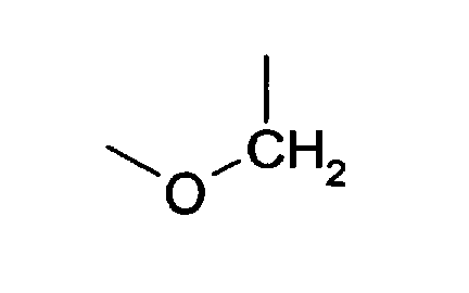
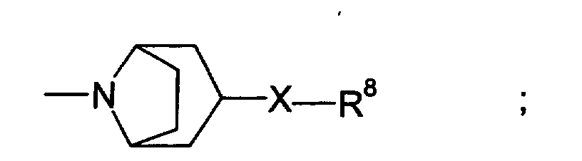
- An NR2B-selective NMDA antagonist that can be used in the methods of the present invention includes a compound of formula I or pharmaceutically acceptable acid addition salt or solvate thereof, wherein:
- the compounds of formula I can be prepared as follows.
- the compounds of formula I wherein R 2 and R 5 are taken together forming a chroman-4-ol ring, and R 1 , R 3 , and R 4 are hydrogen, can be prepared by one or more of the synthetic methods described and referred to in U.S. Patent No. 5,356,905 to Butler, issued October 18, 1994, which is incorporated herein by reference.
- the compounds of formula I wherein R 2 and R 5 are taken separately, and R 1 , R 2 , R 3 and R 4 are hydrogen, can be prepared by one or more of the synthetic methods described and referred to in U.S. Patent No. 5,185,343 to Chenard, issued February 9, 1993; U.S. Patent No.
- a preferred compound, (1S,2S)-1-(4-hydroxyphenyl)-2-(4-hydroxy-4-phenylpiperidin-1-yl)-1-propanol ((1S,2S) free base), which is designated as CP-101,606, and its tartrate salt, can be prepared as described in U.S. Patent No. 5,272,160, referred to above.
- the resolution of racemic 1-(4-hydroxyphenyl)-2-(4-hydroxy-4-phenylpiperidin-1-yl)-1-propanol to form the (1S,2S) free base and the corresponding (1R,2R) enantiomer can be carried out as described in U.S. Patent No. 6,008,233, referred to above.
- the anhydrous mesylate of the (1S,2S) free base can be prepared as described in U.S. Patent No. 5,272,160, referred to above.
- the anhydrous mesylate of the (1S,2S) free base when equilibrated in an 81% relative humidity environment, will convert to the mesylate salt trihydrate of the (1S,2S) enantiomer.
- the mesylate salt trihydrate of (1S,2S)-1-(4-hydroxyphenyl)-2-(4-hydroxy-4-phenylpiperidin-1-yl)-1-propanol can be prepared from the (1S,2S) free base as described in the U.S. Patent No. 6,008,233.
- (3R,4S)-3-(4-(4-fluorophenyl)-4-hydroxy-piperidin-1-yl]-chroman-4,7-diol ((3R,4S) chromanol)
- (3R,4S) chromanol) can be prepared as described in U.S. Patent No. 5,356,905, and U.S. Patent No. 5,744,483, which are referred to above.
- the starting materials and reagents required for the synthesis of the (3R,4S) chromanol are readily available, either commercially or according to synthetic methods disclosed in the literature.
- ifenprodil is shown below as Formula II, and may be prepared by methods analogous to those disclosed in U.S. Patent No. 3,509,164.
- eliprodil is shown below as Formula III, and may be prepared by methods analogous to those disclosed in U.S. Patent No. 4,690,931.
- NR2B subunit selective NMDA receptor antagonists useful in the practice of the invention can be used in the form of a pharmaceutically acceptable salt.
- pharmaceutically-acceptable salt is intended to include but not be limited to such salts as the hydrochloride, hydrobromide, sulfate, hydrogen sulfate, phosphate, hydrogen phosphate, dihydrogen phosphate, acetate, succinate, citrate, tartrate, lactate, mandelate, methanesulfonate (mesylate) and p-toluenesulfonate (tosylate) salts.
- the acid addition salts of the compounds used according to the methods of the present invention are readily prepared by reacting the base forms with the appropriate acid.
- the salt is of a monobasic acid (e.g., the hydrochloride, the hydrobromide, the p-toluenesulfonate, the acetate)
- the hydrogen form of a dibasic acid e.g., the hydrogen sulfate, the succinate
- the dihydrogen form of a tribasic acid e.g., the dihydrogen phosphate, the citrate
- at least one molar equivalent and usually a molar excess of the acid is employed.
- the free base and the acid are usually combined in a co-solvent from which the desired salt precipitates, or can be otherwise isolated by concentration and/or addition of a non-solvent.
- NMDA receptor antagonists having NR2B subunit selectivity that may be used according to the present invention include, e.g., those described in U.S. Patent No. 6,046,213; U.S. Patent No. 5,185,343; U.S. Patent No. 5,272,160; U.S. Patent No. 5,338,754; U.S. Patent No. 5,356,905; U.S. Patent No. 6,046,213; U.S. Patent No. 5,744,483; U.S. Patent No.
- NR2B subunit selective NMDA receptor antagonists that may be used according to the present invention are described in WO 97/32581; WO 98/18793; WO 97/23202; EP 0 824 098 A1; EP 0 846 683 A1; and DE 19739331, published November 26, 1998.
- NR2B NMDA receptor subunits that may be used according to the methods of the present invention are ifenprodil , supra, eliprodil (described in U.S. Patent No. 4,690,931), and compounds described in WO 97/23458; WO 97/23216; WO 97/23215; and WO 97/23214.
- NMDA receptors comprising an NR2B subunit by specifically binding to the NR2B subunit
- Screen compounds for inhibition of NMDA-induced current in recombinant Xenopus oocytes co-transfected with the NR1A subunit and the NR2B subunit (see, e.g., Monyer, et al., Science, 1992, 256:1217-1221).
- a compound's activity in inhibiting current in the recombinant cells comprising the NR2B subunit can be compared to its activity inhibiting NMDA-induced current in recombinant Xenopus oocytes expressing the NR1 subunit and NR2A, NR2C, and NR2D subunits. (See, Chenard and Menniti, supra ).
- One general method that can also generally predict whether or not a compound has NR2B subunit selectivity, for purposes of the present invention is a standard competitive binding assay using [ 3 H] radiolabeled racemic CP-101,606 (which contains [ 3 H] (+)-(1S, 2S)-1-(4-hydroxy-phenyl)-2-(4-hydroxy-4-phenylpiperidino)-1-propanol; see, for example, U.S. Patent 6,046,213). If a compound has an IC 50 of less than about 5 ⁇ M for inhibition of racemic [ 3 H] CP-101,606 binding to the NR2B subunit, than the compound has NR2B subunit selectivity for purposes of the present invention.
- An example of such an assay is as follows.
- NR2B subunit binding assay Example of NR2B subunit binding assay .
- Selectivity of compounds for the NR2B-subunit-containing NMDA receptor can be defined as an affinity for the racemic [ 3 H] CP-101,606 binding site in the forebrain of rats, as described in Chenard and Menniti, supra. This affinity is assessed in a radioligand binding assay as described below.
- Selective compounds are preferably those which displace specific binding of racemic [ 3 H]CP-101,606 from rat forebrain membranes with an IC 50 of about ⁇ 5 ⁇ M.
- racemic [ 3 H] (+)-(1S, 2S)-1-(4-hydroxy-phenyl)-2-(4-hydroxy-4-phenylpiperidino)-1-propanol is measured as described by Menniti et al. (CP-101,606, a potent neuroprotectant selective for forebrain neurons, European Journal of Pharmacology, 1997, 331:117-126).
- Forebrains of adult male CD rats are homogenized in 0.32M sucrose at 4°C. The crude nuclear pellet is removed by centrifugation at 1,000 x g for 10 min, and the supernatant centrifuged at 17,000 x g for 25 min.
- the resulting pellet is resuspended in 5mM Tris acetate pH 7.4 at 4°C for 10 min to lyse cellular particles and again centrifuged at 17,000 x g .
- the resulting pellet is washed twice in Tris acetate, resuspended at 10mg protein/ml and stored at -20°C until use.
- membranes are thawed, homogenized, and diluted to 0.5mg protein/ml with 50mM Tris HCI, pH 7.4. Compounds under study are added at various concentrations followed by racemic [ 3 H] CP-101,606 (specific activity 42.8 Ci/mmol, 5nM final concentration). Following incubation for 20 min at 30°C in a shaking water bath, samples are filtered onto Whatman GFB glass fiber filters using a MB-48R Cell Harvester (Brandel Research and Development Laboratories, Gaithersburg MD). Filters are washed for 10 s with ice cold Tris HCI buffer and the radioactivity trapped on the filter quantified by liquid scintillation spectroscopy. Nonspecific binding is determined in parallel incubations containing 100 ⁇ M racemic CP-101,606. Specific binding is defined as total binding minus nonspecific binding.
- That amount of a neuroprotective agent to be administered to a patient and constituting an "effective amount" will generally depend on the specific circumstances of the patient, including among other factors the patient's sex, weight, age, and general health, as well as the type and severity of the event or condition for which the patient is being treated.
- the effective amount of a neuroprotective agent will typically be determined by the attending physician using such information in combination with the results of clinical studies and published reports regarding the particular agent.
- an effective amount of an NR2B selective NMDA antagonist will range from about 0.02 mg/kg/day to about 10 mg/kg/day.
- dosages outside this range may be required, and these may be determined by the attending physician in view of the particular circumstances.
- the neuroprotective agent will generally be administered in the form of a pharmaceutical composition further comprising a pharmaceutically acceptable carrier or diluent as known in the art.
- a pharmaceutically acceptable carrier or diluent as known in the art.
- Such compositions are generally formulated in a conventional manner utilizing solid or liquid vehicles or diluents as appropriate to the mode of administration.
- excipients such as sodium citrate, calcium carbonate, and di-calcium phosphate may be employed, along with various disintegrants such as starch, preferably potato or tapioca starch, alginic acid and certain complex silicates, together with binding agents such as polyvinylpyrrolidone, sucrose, gelatin and acacia.
- lubricating agents such as, but not limited to, magnesium stearate, sodium lauryl sulfate and talc are often very useful for tableting purposes.
- Solid compositions of a similar type may also be employed as fillers in soft elastic and hard filled gelatin capsules. Preferred materials in this connection also include by way of example lactose or milk sugar as well as high molecular weight polyethylene glycols.
- the essential active ingredient may be combined with various sweetening or flavoring agents, coloring matter or dyes and, if so desired, emulsifying and/or suspending agents, together with diluents such as water, ethanol, propylene glycol, glycerin and various combinations thereof.
- solutions of a neuroprotective compound used in the methods of the present invention are formulated according to standard techniques.
- solutions of a neuroprotective compound in either sesame or peanut oil or in aqueous propylene glycol may be employed.
- the aqueous solutions should be suitably buffered if necessary and the liquid diluent first rendered isotonic.
- These aqueous solutions are suitable for intravenous injection purposes.
- the oily solutions are suitable for infra-articular, intramuscular and subcutaneous injection purposes. The preparation of all these solutions under sterile conditions is readily accomplished by standard pharmaceutical techniques well known to those skilled in the art.
- the present invention further provides a method for identifying whether a patient will benefit from treatment with a neuroprotective agent comprising determining whether the amount of at least one biomarker in a biological sample taken from the patient prior to an initial treatment with the neuroprotective agent is above a certain predetermined minimum threshold value, such that if the amount of the biomarker in the biological sample taken from the patient is above the minimum threshold value, then the patient is primarily identified as a patient who has suffered substantial neurological damage.
- a minimum threshold value can be about 0.2 ⁇ g/L in serum, particularly where the level of S-100b remains at or above that level for at least 24 hrs. As explained above, however, this value may vary depending on the circumstances of the individual patient.
- This method may also comprise determining whether the amount of the at least one biomarker in the biological sample taken from the patient prior to initial treatment with the neuroprotective agent is above a certain predetermined maximum threshold value, such that if the amount of the biomarker in the biological sample taken from the patient is above the maximum threshold value, then the patient suffering from neurological damage is secondarily identified as a patient suffering from such severe neurological damage that the patients expected outcome is considered poor.
- a certain predetermined maximum threshold value such that if the amount of the biomarker in the biological sample taken from the patient is above the maximum threshold value, then the patient suffering from neurological damage is secondarily identified as a patient suffering from such severe neurological damage that the patients expected outcome is considered poor.
- a level can be about 1.5 to 2.0 ⁇ g/L in serum, and preferably remains at or above that level for at least 24 hrs.
- This method will serve as a method to screen patients presenting with possible neurological damage to help distinguish those patients who can substantially benefit from treatment with a neuroprotective agent from those who may only benefit minimally if at all.
- S-100b levels were measured in the serum of patients suffering from severe contusive-type head trauma.
- a first group of 198 patients received treatment with CP-101,606 by constant infusion for 72 hours.
- a second group of 202 patients instead received a constant placebo infusion.
- Serum samples were collected from the patients at various time points out to 120 hours post-infusion ( Figure 1).
- S-100b levels were determined using a commercially available luminometric immunoassay kit from Byk-Sangtec Diagnostica GmbH & Co. (Dietzenbach, Germany).
- S-100b levels in the two groups of patients are presented in Figure 1. S-100b levels were significantly reduced in patients administered CP-101,606 compared to the placebo group.
- Plasma samples were collected from patients suffering from ischemic stroke at baseline, 1, 2, 3, 4, 4.5, 7, 30 and 90 days after injury.
- S100B was measured in all plasma samples and total concentration (in ⁇ g/L) was correlated to clinical outcome measures including 30 and 90 day NIHSS (National Institute of Health Stroke Scale) and infarct volume as measured by diffusion weighted MRI.
- NIHSS National Institute of Health Stroke Scale
- a logistic regression analysis was performed to examine the relationship between 72 hour S100B levels and NIHSS at 90 days. The model corrected for NIHSS baseline scores. Data showed that patients with 72 hour S100B levels were more likely to have poor NIHSS scores.
- a correlation analysis between actual infarct volume (as assessed by normalized diffusion weighted MRI) and 72 hour plasma S100B levels was also performed.
- Diffusion weighted MRI images were acquired at baseline and at 48 hours after stroke lesions.
- Logistically transformed normalized values (Baseline/48hrs) were compared to logistically transformed 72 hour S100B values.
- 72 hour plasma S100B values correlated with infarct volume as measured by DW-MRI.
Landscapes
- Health & Medical Sciences (AREA)
- Engineering & Computer Science (AREA)
- Life Sciences & Earth Sciences (AREA)
- Biomedical Technology (AREA)
- Chemical & Material Sciences (AREA)
- Bioinformatics & Cheminformatics (AREA)
- Medicinal Chemistry (AREA)
- General Health & Medical Sciences (AREA)
- Neurosurgery (AREA)
- Neurology (AREA)
- Nuclear Medicine, Radiotherapy & Molecular Imaging (AREA)
- Chemical Kinetics & Catalysis (AREA)
- Organic Chemistry (AREA)
- Pharmacology & Pharmacy (AREA)
- General Chemical & Material Sciences (AREA)
- Animal Behavior & Ethology (AREA)
- Public Health (AREA)
- Veterinary Medicine (AREA)
- Hematology (AREA)
- Urology & Nephrology (AREA)
- Immunology (AREA)
- Molecular Biology (AREA)
- Physics & Mathematics (AREA)
- General Physics & Mathematics (AREA)
- Pathology (AREA)
- Biochemistry (AREA)
- Diabetes (AREA)
- Psychology (AREA)
- Analytical Chemistry (AREA)
- Food Science & Technology (AREA)
- Microbiology (AREA)
- Proteomics, Peptides & Aminoacids (AREA)
- Biotechnology (AREA)
- Cell Biology (AREA)
- Emergency Medicine (AREA)
- Cardiology (AREA)
- Vascular Medicine (AREA)
- Psychiatry (AREA)
- Obesity (AREA)
- Hospice & Palliative Care (AREA)
Abstract
Description




Claims (10)
- A method to monitor the response of a patient being treated for neurological damage by administering a neuroprotective agent, comprising the steps of:such that a detectable reduction in the amount of the biomarker in the second biological sample compared to the amount of biomarker in the first biological sample indicates that the patient is responding positively to the treatment with the neuroprotective agent.(a) determining the amount of at least one biomarker in a first biological sample taken from the patient prior to an initial treatment with the neuroprotective agent;(b) determining the amount of the biomarker in at least a second biological sample taken from the patient subsequent to the initial treatment with the neuroprotective agent; and(c) comparing the amount of the biomarker in the second biological sample with the amount of the biomarker in the first biological sample;
- The method of claim 1, wherein the neurological damage is a condition or disease, or is caused by a condition, disease or event, selected from the group consisting of cerebral ischemia, cerebral infarction, head trauma, contusion, spinal cord injury, subarachnoid hemorrhage, cerebral hemorrhage, aneurysmal hemorrhage, cardiac infarction, hypoxia, anoxia, surgery, Alzheimer's disease, Parkinson's disease, multiple sclerosis, HIV-related neurodegeneration, cerebellar degeneration, seizure and ataxia.
- The method of claim 2, wherein the neurological damage is cerebral ischemia, cerebral infarction, head trauma, or spinal cord injury.
- The method of claim 1, wherein the biomarker is selected from the group consisting of S-100b, neuron-specific enolase (NSE), glial fibrillary acidic protein (GFAP), tau protein, haptoglobin, brain creatine kinase, isoprostane, myelin basic protein (MBP), or thrombomodulin.
- The method of claim 4, wherein the biomarker is S-100b or NSE.
- The method of claim 1, wherein the neuroprotective agent is selected from the group consisting of an excitatory amino acid receptor antagonist, a metabotropic glutamate receptor antagonists, GABA receptor antagonist; a NAALDase enzyme inhibitor, a calpain inhibitor, a p38 mitogen-activated protein kinase (MAPK) inhibitor, an estrogen enantiomer or derivative, a modulator of nitric oxide production, a calmodulin inhibitor, an adenosine receptor modulator, a purine receptor antagonist, a prosaposin receptor activity stimulator, and a synthetic oxygen carrier.
- The method of claim 6, wherein the neuroprotective agent is an NMDA receptor antagonist.
- The method of claim 1, wherein the amount of the biomarker in the biological sample is determined by ELISA, RIA, magnetic resonance spectroscopy, HPLC or mass spectrometry.
- An improvement to a method for treating a patient suffering from neurological damage by administration of a neuroprotective agent, wherein the improvement comprises monitoring the level of at least one biomarker in a biological sample taken from the patient at one or more time points during treatment with the neuroprotective agent so as to determine whether an effective amount of the neuroprotective agent is being administered to the patient.
- A method for identifying whether a patient will benefit from treatment with a neuroprotective agent, comprising determining whether the amount of at least one biomarker in a biological sample taken from the patient prior to an initial treatment with the neuroprotective agent is above a predetermined minimum threshold value, such that if the amount of the biomarker in the biological sample taken from the patient is above the minimum threshold value, then the patient is primarily identified as a patient who has suffered neurological damage.
Applications Claiming Priority (2)
| Application Number | Priority Date | Filing Date | Title |
|---|---|---|---|
| US32889001P | 2001-10-12 | 2001-10-12 | |
| US328890P | 2001-10-12 |
Publications (2)
| Publication Number | Publication Date |
|---|---|
| EP1310797A2 true EP1310797A2 (en) | 2003-05-14 |
| EP1310797A3 EP1310797A3 (en) | 2003-09-17 |
Family
ID=23282898
Family Applications (1)
| Application Number | Title | Priority Date | Filing Date |
|---|---|---|---|
| EP02256806A Withdrawn EP1310797A3 (en) | 2001-10-12 | 2002-09-30 | Method of monitoring neuroprotective treatment |
Country Status (6)
| Country | Link |
|---|---|
| US (1) | US20030129134A1 (en) |
| EP (1) | EP1310797A3 (en) |
| JP (1) | JP3666866B2 (en) |
| AU (1) | AU2002329570A1 (en) |
| CA (1) | CA2407214A1 (en) |
| WO (1) | WO2003032894A2 (en) |
Cited By (5)
| Publication number | Priority date | Publication date | Assignee | Title |
|---|---|---|---|---|
| EP1519194A1 (en) * | 2003-09-24 | 2005-03-30 | Roche Diagnostics GmbH | Use of gfap for identification of intracerebral hemorrhage |
| KR100644364B1 (en) | 2005-03-04 | 2006-11-10 | 한국생명공학연구원 | Parkinson's disease genetic markers and Parkinson's disease diagnostic kit using the same |
| US11994522B2 (en) | 2008-08-11 | 2024-05-28 | Banyan Biomarkers, Inc. | Biomarker detection process and assay of neurological condition |
| US12077601B2 (en) | 2016-10-28 | 2024-09-03 | Banyan Biomarkers, Inc. | Antibodies to ubiquitin C-terminal hydrolase L1 (UCH-L1) and glial fibrillary acidic protein (GFAP) and related methods |
| US12601749B2 (en) | 2008-08-11 | 2026-04-14 | Banyan Biomarkers, Inc. | Biomarker detection process and assay of neurological condition |
Families Citing this family (20)
| Publication number | Priority date | Publication date | Assignee | Title |
|---|---|---|---|---|
| WO2005041892A2 (en) * | 2003-11-03 | 2005-05-12 | Cornell Research Foundation, Inc | Purine receptor inhibition as a therapeutic strategy in spinal cord and brain |
| US7304171B2 (en) * | 2004-05-27 | 2007-12-04 | Migenix Corp. | Compounds and methods for cytoprotection |
| US20060024722A1 (en) * | 2004-07-30 | 2006-02-02 | Mark Fischer-Colbrie | Samples for detection of oncofetal fibronectin and uses thereof |
| JP5015779B2 (en) * | 2004-08-23 | 2012-08-29 | エモリー・ユニバーシテイ | Improved method for selecting pH-dependent compounds for in vivo therapy |
| US20060136806A1 (en) * | 2004-12-22 | 2006-06-22 | Pharmacyclics, Inc. | System and method for analysis of neurological condition |
| JP2009515542A (en) * | 2005-11-16 | 2009-04-16 | ノバルティス アクチエンゲゼルシャフト | Biomarker for anti-Nogo-A antibody treatment in spinal cord injury |
| US10632237B2 (en) | 2006-10-09 | 2020-04-28 | Minnetronix, Inc. | Tangential flow filter system for the filtration of materials from biologic fluids |
| WO2008105959A2 (en) | 2006-10-09 | 2008-09-04 | Neurofluidics, Inc. | Cerebrospinal fluid purification system |
| US10850235B2 (en) | 2006-10-09 | 2020-12-01 | Minnetronix, Inc. | Method for filtering cerebrospinal fluid (CSF) including monitoring CSF flow |
| US20090263507A1 (en) * | 2008-04-18 | 2009-10-22 | Warsaw Orthopedic, Inc. | Biological markers and response to treatment for pain, inflammation, neuronal or vascular injury and methods of use |
| JP5875514B2 (en) * | 2009-06-19 | 2016-03-02 | バンヤン・バイオマーカーズ・インコーポレーテッド | Neuromarker biomarker assay |
| AU2010291933B2 (en) | 2009-09-14 | 2016-09-08 | Banyan Biomarkers, Inc. | Micro-RNA, autoantibody and protein markers for diagnosis of neuronal injury |
| CN103299191B (en) * | 2010-08-13 | 2016-06-15 | 莫尔豪斯医学院 | Biomarkers of Stroke |
| US8648198B2 (en) | 2011-01-19 | 2014-02-11 | Cold Spring Harbor Laboratory | Phenylethanolamine-based NMDA receptor antagonists |
| EP2776587A4 (en) * | 2011-11-10 | 2015-07-15 | Exosome Diagnostics Inc | Cerebrospinal fluid assay |
| US11147540B2 (en) | 2015-07-01 | 2021-10-19 | Minnetronix, Inc. | Introducer sheath and puncture tool for the introduction and placement of a catheter in tissue |
| CA2994669C (en) | 2015-08-05 | 2020-11-17 | Minnetronix, Inc. | Tangential flow filter system for the filtration of materials from biologic fluids |
| EP3383448B1 (en) | 2015-12-04 | 2021-01-27 | Minnetronix Inc. | Systems for the conditioning of cerebrospinal fluid |
| RU2648515C1 (en) * | 2016-12-21 | 2018-03-26 | Общество с ограниченной ответственностью "ДРД" | Predicting the course and outcome of coma and post-coma unconscious states (including vegetative ones) with the help of blood tests |
| ES2927715T3 (en) | 2017-10-05 | 2022-11-10 | Fulcrum Therapeutics Inc | p38 kinase inhibitors reduce the expression of dux4 and the genes that follow it for the treatment of FSHD |
Family Cites Families (2)
| Publication number | Priority date | Publication date | Assignee | Title |
|---|---|---|---|---|
| IL124444A0 (en) * | 1998-05-12 | 1998-12-06 | Svetlana Dolina | Biochemical approach to diagnosis of predisposition to epilepsy and monitoring of antiepileptic treatment |
| US20020164668A1 (en) * | 2000-04-03 | 2002-11-07 | Durham L. Kathryn | Nucleic acid molecules, polypeptides and uses therefor, including diagnosis and treatment of alzheimer's disease |
-
2002
- 2002-09-13 AU AU2002329570A patent/AU2002329570A1/en not_active Abandoned
- 2002-09-13 WO PCT/IB2002/003775 patent/WO2003032894A2/en not_active Ceased
- 2002-09-30 EP EP02256806A patent/EP1310797A3/en not_active Withdrawn
- 2002-10-08 CA CA002407214A patent/CA2407214A1/en not_active Abandoned
- 2002-10-10 US US10/268,465 patent/US20030129134A1/en not_active Abandoned
- 2002-10-15 JP JP2002300861A patent/JP3666866B2/en not_active Expired - Fee Related
Cited By (7)
| Publication number | Priority date | Publication date | Assignee | Title |
|---|---|---|---|---|
| EP1519194A1 (en) * | 2003-09-24 | 2005-03-30 | Roche Diagnostics GmbH | Use of gfap for identification of intracerebral hemorrhage |
| WO2005029087A1 (en) * | 2003-09-24 | 2005-03-31 | Roche Diagnostics Gmbh | Use of gfap for identification of intracerebral hemorrhage |
| US10794918B2 (en) | 2003-09-24 | 2020-10-06 | Matthias Sitzer | Use of GFAP for identification of intracerebral hemorrhage |
| KR100644364B1 (en) | 2005-03-04 | 2006-11-10 | 한국생명공학연구원 | Parkinson's disease genetic markers and Parkinson's disease diagnostic kit using the same |
| US11994522B2 (en) | 2008-08-11 | 2024-05-28 | Banyan Biomarkers, Inc. | Biomarker detection process and assay of neurological condition |
| US12601749B2 (en) | 2008-08-11 | 2026-04-14 | Banyan Biomarkers, Inc. | Biomarker detection process and assay of neurological condition |
| US12077601B2 (en) | 2016-10-28 | 2024-09-03 | Banyan Biomarkers, Inc. | Antibodies to ubiquitin C-terminal hydrolase L1 (UCH-L1) and glial fibrillary acidic protein (GFAP) and related methods |
Also Published As
| Publication number | Publication date |
|---|---|
| EP1310797A3 (en) | 2003-09-17 |
| CA2407214A1 (en) | 2003-04-12 |
| AU2002329570A1 (en) | 2003-01-30 |
| JP3666866B2 (en) | 2005-06-29 |
| JP2003185657A (en) | 2003-07-03 |
| WO2003032894A3 (en) | 2012-03-08 |
| US20030129134A1 (en) | 2003-07-10 |
| WO2003032894A2 (en) | 2003-04-24 |
| AU2002329570A8 (en) | 2012-04-05 |
Similar Documents
| Publication | Publication Date | Title |
|---|---|---|
| US20030129134A1 (en) | Method of monitoring neuroprotective treatment | |
| US5498522A (en) | Methods for the use of spermidine/spermine N1 -acetyltransferase as a prognostic indicator and/or a tumor response marker | |
| US20130131111A1 (en) | Nmn modulators for the treatment of neurodegenerative disorders | |
| US20190298708A1 (en) | Methods of treating subjects with an elevated neurofilament light chain level | |
| US6884591B2 (en) | Peripheral marker of blood brain barrier permeability | |
| US20230273220A1 (en) | Methods for prediction, detection and monitoring of substanceuse disorders and/or an infection | |
| CA2477614A1 (en) | Biomarker for efficacy of appetite suppressant drugs | |
| Lindner et al. | Increased levels of truncated nerve growth factor receptor in urine of mildly demented patients with Alzheimer's disease | |
| US7754434B2 (en) | Diagnostic and examination method for eating disorder | |
| KR20230073574A (en) | Use of CASQ1-STIM2 binding for the prevention or treatment of skeletal muscle diseases caused by abnormal external calcium influx | |
| JP4707654B2 (en) | Ischemic heart disease risk group diagnostic | |
| US20030077678A1 (en) | Diagnostic kit for schizophrenia | |
| US7691578B2 (en) | Methods and compositions for treating obesity | |
| US20240255529A1 (en) | Novel assay and novel methods of treating aging-related conditions | |
| US20250060373A1 (en) | Novel assay and novel methods of treating hutchinson-gilford progeria syndrome | |
| US20060198788A1 (en) | Method of evaluating compound efficacious in treating obesity by using slc25a10 | |
| US20220357344A1 (en) | Prognosis prediction method of idiopathic pulmonary fibrosis | |
| CA2724454A1 (en) | Method of identifying compounds that modulate regulation of iron respon se elements | |
| HK40115326A (en) | Novel assay and novel methods of treating hutchinson-gilford progeria syndrome | |
| CA3106565A1 (en) | Method for diagnosing a liver disease |
Legal Events
| Date | Code | Title | Description |
|---|---|---|---|
| PUAI | Public reference made under article 153(3) epc to a published international application that has entered the european phase |
Free format text: ORIGINAL CODE: 0009012 |
|
| 17P | Request for examination filed |
Effective date: 20021012 |
|
| AK | Designated contracting states |
Designated state(s): AT BE BG CH CY CZ DE DK EE ES FI FR GB GR IE IT LI LU MC NL PT SE SK TR |
|
| AX | Request for extension of the european patent |
Extension state: AL LT LV MK RO SI |
|
| PUAL | Search report despatched |
Free format text: ORIGINAL CODE: 0009013 |
|
| AK | Designated contracting states |
Kind code of ref document: A3 Designated state(s): AT BE BG CH CY CZ DE DK EE ES FI FR GB GR IE IT LI LU MC NL PT SE SK TR |
|
| AX | Request for extension of the european patent |
Extension state: AL LT LV MK RO SI |
|
| AKX | Designation fees paid |
Designated state(s): AT BE BG CH CY CZ DE DK EE ES FI FR GB GR IE IT LI LU MC NL PT SE SK TR |
|
| STAA | Information on the status of an ep patent application or granted ep patent |
Free format text: STATUS: THE APPLICATION IS DEEMED TO BE WITHDRAWN |
|
| 18D | Application deemed to be withdrawn |
Effective date: 20080402 |