EP1309861B1 - Functional protein arrays - Google Patents
Functional protein arrays Download PDFInfo
- Publication number
- EP1309861B1 EP1309861B1 EP01956692A EP01956692A EP1309861B1 EP 1309861 B1 EP1309861 B1 EP 1309861B1 EP 01956692 A EP01956692 A EP 01956692A EP 01956692 A EP01956692 A EP 01956692A EP 1309861 B1 EP1309861 B1 EP 1309861B1
- Authority
- EP
- European Patent Office
- Prior art keywords
- dna
- protein
- proteins
- translation
- beads
- Prior art date
- Legal status (The legal status is an assumption and is not a legal conclusion. Google has not performed a legal analysis and makes no representation as to the accuracy of the status listed.)
- Expired - Lifetime
Links
- 238000003498 protein array Methods 0.000 title claims abstract description 40
- 108090000623 proteins and genes Proteins 0.000 claims abstract description 148
- 102000004169 proteins and genes Human genes 0.000 claims abstract description 145
- 238000013519 translation Methods 0.000 claims abstract description 36
- 238000013518 transcription Methods 0.000 claims abstract description 27
- 230000035897 transcription Effects 0.000 claims abstract description 27
- 108090000765 processed proteins & peptides Proteins 0.000 claims abstract description 18
- 108020004999 messenger RNA Proteins 0.000 claims abstract description 11
- 102000004196 processed proteins & peptides Human genes 0.000 claims abstract description 11
- 210000004671 cell-free system Anatomy 0.000 claims abstract description 10
- 108020004414 DNA Proteins 0.000 claims description 105
- 239000011324 bead Substances 0.000 claims description 93
- 238000000034 method Methods 0.000 claims description 42
- 230000003993 interaction Effects 0.000 claims description 27
- 108010093488 His-His-His-His-His-His Proteins 0.000 claims description 16
- 239000003446 ligand Substances 0.000 claims description 15
- 238000006243 chemical reaction Methods 0.000 claims description 11
- 239000012528 membrane Substances 0.000 claims description 11
- 102000008394 Immunoglobulin Fragments Human genes 0.000 claims description 10
- 108010021625 Immunoglobulin Fragments Proteins 0.000 claims description 10
- 239000003153 chemical reaction reagent Substances 0.000 claims description 10
- 239000011521 glass Substances 0.000 claims description 9
- 241000283973 Oryctolagus cuniculus Species 0.000 claims description 7
- 210000001995 reticulocyte Anatomy 0.000 claims description 7
- 240000004808 Saccharomyces cerevisiae Species 0.000 claims description 6
- 230000001580 bacterial effect Effects 0.000 claims description 6
- PXHVJJICTQNCMI-UHFFFAOYSA-N Nickel Chemical compound [Ni] PXHVJJICTQNCMI-UHFFFAOYSA-N 0.000 claims description 5
- 239000000020 Nitrocellulose Substances 0.000 claims description 5
- 229920001220 nitrocellulos Polymers 0.000 claims description 5
- 241000209140 Triticum Species 0.000 claims description 4
- 235000021307 Triticum Nutrition 0.000 claims description 4
- 150000001413 amino acids Chemical class 0.000 claims description 4
- 229910021645 metal ion Inorganic materials 0.000 claims description 2
- 229910052759 nickel Inorganic materials 0.000 claims description 2
- FWMNVWWHGCHHJJ-SKKKGAJSSA-N 4-amino-1-[(2r)-6-amino-2-[[(2r)-2-[[(2r)-2-[[(2r)-2-amino-3-phenylpropanoyl]amino]-3-phenylpropanoyl]amino]-4-methylpentanoyl]amino]hexanoyl]piperidine-4-carboxylic acid Chemical compound C([C@H](C(=O)N[C@H](CC(C)C)C(=O)N[C@H](CCCCN)C(=O)N1CCC(N)(CC1)C(O)=O)NC(=O)[C@H](N)CC=1C=CC=CC=1)C1=CC=CC=C1 FWMNVWWHGCHHJJ-SKKKGAJSSA-N 0.000 claims 1
- 125000003275 alpha amino acid group Chemical group 0.000 claims 1
- 210000003527 eukaryotic cell Anatomy 0.000 claims 1
- 210000001236 prokaryotic cell Anatomy 0.000 claims 1
- 238000000338 in vitro Methods 0.000 abstract description 26
- 230000015572 biosynthetic process Effects 0.000 abstract description 9
- 238000003786 synthesis reaction Methods 0.000 abstract description 9
- 238000004519 manufacturing process Methods 0.000 abstract description 8
- 238000002955 isolation Methods 0.000 abstract description 3
- 238000003752 polymerase chain reaction Methods 0.000 description 46
- 239000000203 mixture Substances 0.000 description 41
- 230000014616 translation Effects 0.000 description 38
- 108060001084 Luciferase Proteins 0.000 description 36
- 239000005089 Luciferase Substances 0.000 description 36
- 230000014509 gene expression Effects 0.000 description 36
- 239000012634 fragment Substances 0.000 description 31
- 101100015729 Drosophila melanogaster drk gene Proteins 0.000 description 27
- 101150098203 grb2 gene Proteins 0.000 description 27
- 238000011065 in-situ storage Methods 0.000 description 25
- 230000027455 binding Effects 0.000 description 19
- 238000009739 binding Methods 0.000 description 19
- 210000004027 cell Anatomy 0.000 description 16
- 108010001336 Horseradish Peroxidase Proteins 0.000 description 15
- 230000000694 effects Effects 0.000 description 15
- 238000002702 ribosome display Methods 0.000 description 15
- 238000003491 array Methods 0.000 description 13
- 229920000936 Agarose Polymers 0.000 description 11
- 108091034117 Oligonucleotide Proteins 0.000 description 11
- 238000001514 detection method Methods 0.000 description 11
- 239000006228 supernatant Substances 0.000 description 11
- LOKCTEFSRHRXRJ-UHFFFAOYSA-I dipotassium trisodium dihydrogen phosphate hydrogen phosphate dichloride Chemical compound P(=O)(O)(O)[O-].[K+].P(=O)(O)([O-])[O-].[Na+].[Na+].[Cl-].[K+].[Cl-].[Na+] LOKCTEFSRHRXRJ-UHFFFAOYSA-I 0.000 description 10
- 239000002953 phosphate buffered saline Substances 0.000 description 10
- 238000003757 reverse transcription PCR Methods 0.000 description 10
- 241000588724 Escherichia coli Species 0.000 description 9
- 239000000427 antigen Substances 0.000 description 9
- 108091007433 antigens Proteins 0.000 description 9
- 102000036639 antigens Human genes 0.000 description 9
- 239000003418 antiprogestin Substances 0.000 description 9
- 230000003623 progesteronic effect Effects 0.000 description 9
- 238000001243 protein synthesis Methods 0.000 description 9
- 238000004458 analytical method Methods 0.000 description 8
- 238000003556 assay Methods 0.000 description 7
- 239000002299 complementary DNA Substances 0.000 description 7
- 230000004850 protein–protein interaction Effects 0.000 description 7
- 238000010276 construction Methods 0.000 description 6
- 238000011534 incubation Methods 0.000 description 6
- 239000000047 product Substances 0.000 description 6
- 238000012216 screening Methods 0.000 description 6
- 238000001262 western blot Methods 0.000 description 6
- 108091028043 Nucleic acid sequence Proteins 0.000 description 5
- 239000002033 PVDF binder Substances 0.000 description 5
- 230000001413 cellular effect Effects 0.000 description 5
- 230000006870 function Effects 0.000 description 5
- 239000013612 plasmid Substances 0.000 description 5
- 229920002981 polyvinylidene fluoride Polymers 0.000 description 5
- 239000000243 solution Substances 0.000 description 5
- 238000011144 upstream manufacturing Methods 0.000 description 5
- 238000005406 washing Methods 0.000 description 5
- 108020004705 Codon Proteins 0.000 description 4
- 102000004190 Enzymes Human genes 0.000 description 4
- 108090000790 Enzymes Proteins 0.000 description 4
- 101150026303 HEX1 gene Proteins 0.000 description 4
- FFEARJCKVFRZRR-BYPYZUCNSA-N L-methionine Chemical compound CSCC[C@H](N)C(O)=O FFEARJCKVFRZRR-BYPYZUCNSA-N 0.000 description 4
- 108010038807 Oligopeptides Proteins 0.000 description 4
- 102000015636 Oligopeptides Human genes 0.000 description 4
- 108010008281 Recombinant Fusion Proteins Proteins 0.000 description 4
- 102000007056 Recombinant Fusion Proteins Human genes 0.000 description 4
- 235000014680 Saccharomyces cerevisiae Nutrition 0.000 description 4
- 108010090804 Streptavidin Proteins 0.000 description 4
- 230000000295 complement effect Effects 0.000 description 4
- 238000002474 experimental method Methods 0.000 description 4
- 238000000605 extraction Methods 0.000 description 4
- 239000000499 gel Substances 0.000 description 4
- RWSXRVCMGQZWBV-WDSKDSINSA-N glutathione Chemical compound OC(=O)[C@@H](N)CCC(=O)N[C@@H](CS)C(=O)NCC(O)=O RWSXRVCMGQZWBV-WDSKDSINSA-N 0.000 description 4
- 230000000977 initiatory effect Effects 0.000 description 4
- 229930182817 methionine Natural products 0.000 description 4
- 102000039446 nucleic acids Human genes 0.000 description 4
- 108020004707 nucleic acids Proteins 0.000 description 4
- 150000007523 nucleic acids Chemical class 0.000 description 4
- 238000012546 transfer Methods 0.000 description 4
- 241000894006 Bacteria Species 0.000 description 3
- 108091035707 Consensus sequence Proteins 0.000 description 3
- 102000005720 Glutathione transferase Human genes 0.000 description 3
- 108010070675 Glutathione transferase Proteins 0.000 description 3
- 108700026244 Open Reading Frames Proteins 0.000 description 3
- 239000004793 Polystyrene Substances 0.000 description 3
- HEMHJVSKTPXQMS-UHFFFAOYSA-M Sodium hydroxide Chemical compound [OH-].[Na+] HEMHJVSKTPXQMS-UHFFFAOYSA-M 0.000 description 3
- 108010006785 Taq Polymerase Proteins 0.000 description 3
- 238000010367 cloning Methods 0.000 description 3
- 238000005859 coupling reaction Methods 0.000 description 3
- 239000000284 extract Substances 0.000 description 3
- ZRALSGWEFCBTJO-UHFFFAOYSA-O guanidinium Chemical compound NC(N)=[NH2+] ZRALSGWEFCBTJO-UHFFFAOYSA-O 0.000 description 3
- RAXXELZNTBOGNW-UHFFFAOYSA-N imidazole Natural products C1=CNC=N1 RAXXELZNTBOGNW-UHFFFAOYSA-N 0.000 description 3
- 239000006166 lysate Substances 0.000 description 3
- UEGPKNKPLBYCNK-UHFFFAOYSA-L magnesium acetate Chemical compound [Mg+2].CC([O-])=O.CC([O-])=O UEGPKNKPLBYCNK-UHFFFAOYSA-L 0.000 description 3
- 238000005259 measurement Methods 0.000 description 3
- 239000002184 metal Substances 0.000 description 3
- 229910052751 metal Inorganic materials 0.000 description 3
- 229920001184 polypeptide Polymers 0.000 description 3
- 229920000136 polysorbate Polymers 0.000 description 3
- 229920002223 polystyrene Polymers 0.000 description 3
- 238000002360 preparation method Methods 0.000 description 3
- 238000011084 recovery Methods 0.000 description 3
- 239000000523 sample Substances 0.000 description 3
- 238000012360 testing method Methods 0.000 description 3
- 230000005030 transcription termination Effects 0.000 description 3
- 239000011534 wash buffer Substances 0.000 description 3
- 238000001086 yeast two-hybrid system Methods 0.000 description 3
- YBJHBAHKTGYVGT-ZKWXMUAHSA-N (+)-Biotin Chemical compound N1C(=O)N[C@@H]2[C@H](CCCCC(=O)O)SC[C@@H]21 YBJHBAHKTGYVGT-ZKWXMUAHSA-N 0.000 description 2
- 108091032973 (ribonucleotides)n+m Proteins 0.000 description 2
- 108010024636 Glutathione Proteins 0.000 description 2
- 238000012408 PCR amplification Methods 0.000 description 2
- 241001494479 Pecora Species 0.000 description 2
- 102000000395 SH3 domains Human genes 0.000 description 2
- 108050008861 SH3 domains Proteins 0.000 description 2
- FAPWRFPIFSIZLT-UHFFFAOYSA-M Sodium chloride Chemical compound [Na+].[Cl-] FAPWRFPIFSIZLT-UHFFFAOYSA-M 0.000 description 2
- 108091081024 Start codon Proteins 0.000 description 2
- 230000002776 aggregation Effects 0.000 description 2
- 238000004220 aggregation Methods 0.000 description 2
- 125000003277 amino group Chemical group 0.000 description 2
- 238000013459 approach Methods 0.000 description 2
- 238000000376 autoradiography Methods 0.000 description 2
- 210000004899 c-terminal region Anatomy 0.000 description 2
- 230000015556 catabolic process Effects 0.000 description 2
- 239000013592 cell lysate Substances 0.000 description 2
- 239000003795 chemical substances by application Substances 0.000 description 2
- 238000010168 coupling process Methods 0.000 description 2
- 238000006731 degradation reaction Methods 0.000 description 2
- 239000003398 denaturant Substances 0.000 description 2
- 238000000326 densiometry Methods 0.000 description 2
- 238000010586 diagram Methods 0.000 description 2
- 238000010790 dilution Methods 0.000 description 2
- 239000012895 dilution Substances 0.000 description 2
- 238000010195 expression analysis Methods 0.000 description 2
- 229960003180 glutathione Drugs 0.000 description 2
- 238000013537 high throughput screening Methods 0.000 description 2
- 238000009396 hybridization Methods 0.000 description 2
- 210000003000 inclusion body Anatomy 0.000 description 2
- BPHPUYQFMNQIOC-NXRLNHOXSA-N isopropyl beta-D-thiogalactopyranoside Chemical compound CC(C)S[C@@H]1O[C@H](CO)[C@H](O)[C@H](O)[C@H]1O BPHPUYQFMNQIOC-NXRLNHOXSA-N 0.000 description 2
- 238000004949 mass spectrometry Methods 0.000 description 2
- 239000002245 particle Substances 0.000 description 2
- 238000002823 phage display Methods 0.000 description 2
- 230000006916 protein interaction Effects 0.000 description 2
- 238000000746 purification Methods 0.000 description 2
- 108020003175 receptors Proteins 0.000 description 2
- 102000005962 receptors Human genes 0.000 description 2
- 230000000717 retained effect Effects 0.000 description 2
- 238000010839 reverse transcription Methods 0.000 description 2
- 238000013207 serial dilution Methods 0.000 description 2
- JQWHASGSAFIOCM-UHFFFAOYSA-M sodium periodate Chemical compound [Na+].[O-]I(=O)(=O)=O JQWHASGSAFIOCM-UHFFFAOYSA-M 0.000 description 2
- 239000007787 solid Substances 0.000 description 2
- 230000009870 specific binding Effects 0.000 description 2
- 239000007858 starting material Substances 0.000 description 2
- 238000003860 storage Methods 0.000 description 2
- 239000000758 substrate Substances 0.000 description 2
- 230000014621 translational initiation Effects 0.000 description 2
- 241000701447 unidentified baculovirus Species 0.000 description 2
- 108091023037 Aptamer Proteins 0.000 description 1
- 102000053602 DNA Human genes 0.000 description 1
- 238000000018 DNA microarray Methods 0.000 description 1
- 238000002965 ELISA Methods 0.000 description 1
- 108010058643 Fungal Proteins Proteins 0.000 description 1
- 230000010663 Gene Expression Interactions Effects 0.000 description 1
- 108091006054 His-tagged proteins Proteins 0.000 description 1
- 101100395023 Neurospora crassa (strain ATCC 24698 / 74-OR23-1A / CBS 708.71 / DSM 1257 / FGSC 987) his-7 gene Proteins 0.000 description 1
- VEQPNABPJHWNSG-UHFFFAOYSA-N Nickel(2+) Chemical compound [Ni+2] VEQPNABPJHWNSG-UHFFFAOYSA-N 0.000 description 1
- 239000004677 Nylon Substances 0.000 description 1
- 108010026552 Proteome Proteins 0.000 description 1
- 108091034057 RNA (poly(A)) Proteins 0.000 description 1
- 108020004511 Recombinant DNA Proteins 0.000 description 1
- 102000002278 Ribosomal Proteins Human genes 0.000 description 1
- 108010000605 Ribosomal Proteins Proteins 0.000 description 1
- 239000012722 SDS sample buffer Substances 0.000 description 1
- 102000014400 SH2 domains Human genes 0.000 description 1
- 108050003452 SH2 domains Proteins 0.000 description 1
- 101710137500 T7 RNA polymerase Proteins 0.000 description 1
- 241000700605 Viruses Species 0.000 description 1
- 230000001594 aberrant effect Effects 0.000 description 1
- 239000002253 acid Substances 0.000 description 1
- 150000007513 acids Chemical class 0.000 description 1
- 230000004913 activation Effects 0.000 description 1
- 239000012082 adaptor molecule Substances 0.000 description 1
- 238000001042 affinity chromatography Methods 0.000 description 1
- 238000001261 affinity purification Methods 0.000 description 1
- 239000011543 agarose gel Substances 0.000 description 1
- 125000003172 aldehyde group Chemical group 0.000 description 1
- 230000003321 amplification Effects 0.000 description 1
- PYKYMHQGRFAEBM-UHFFFAOYSA-N anthraquinone Natural products CCC(=O)c1c(O)c2C(=O)C3C(C=CC=C3O)C(=O)c2cc1CC(=O)OC PYKYMHQGRFAEBM-UHFFFAOYSA-N 0.000 description 1
- 150000004056 anthraquinones Chemical class 0.000 description 1
- 230000001363 autoimmune Effects 0.000 description 1
- 230000008901 benefit Effects 0.000 description 1
- 229960002685 biotin Drugs 0.000 description 1
- 235000020958 biotin Nutrition 0.000 description 1
- 239000011616 biotin Substances 0.000 description 1
- 229940098773 bovine serum albumin Drugs 0.000 description 1
- 210000004556 brain Anatomy 0.000 description 1
- 239000000872 buffer Substances 0.000 description 1
- 230000006037 cell lysis Effects 0.000 description 1
- 230000008859 change Effects 0.000 description 1
- 238000012412 chemical coupling Methods 0.000 description 1
- 125000003636 chemical group Chemical group 0.000 description 1
- 230000008878 coupling Effects 0.000 description 1
- 230000037029 cross reaction Effects 0.000 description 1
- 210000000805 cytoplasm Anatomy 0.000 description 1
- 238000004925 denaturation Methods 0.000 description 1
- 230000036425 denaturation Effects 0.000 description 1
- 238000013461 design Methods 0.000 description 1
- 229940000406 drug candidate Drugs 0.000 description 1
- 238000005516 engineering process Methods 0.000 description 1
- 230000002255 enzymatic effect Effects 0.000 description 1
- 238000001704 evaporation Methods 0.000 description 1
- 230000008020 evaporation Effects 0.000 description 1
- 230000001605 fetal effect Effects 0.000 description 1
- 230000005714 functional activity Effects 0.000 description 1
- 238000010230 functional analysis Methods 0.000 description 1
- 238000002825 functional assay Methods 0.000 description 1
- 230000002068 genetic effect Effects 0.000 description 1
- 210000004524 haematopoietic cell Anatomy 0.000 description 1
- 230000001900 immune effect Effects 0.000 description 1
- 238000001727 in vivo Methods 0.000 description 1
- 230000006698 induction Effects 0.000 description 1
- 230000001939 inductive effect Effects 0.000 description 1
- 208000015181 infectious disease Diseases 0.000 description 1
- 230000036512 infertility Effects 0.000 description 1
- 238000011835 investigation Methods 0.000 description 1
- 230000004807 localization Effects 0.000 description 1
- 239000000463 material Substances 0.000 description 1
- 238000002156 mixing Methods 0.000 description 1
- 238000012986 modification Methods 0.000 description 1
- 230000004048 modification Effects 0.000 description 1
- 230000004001 molecular interaction Effects 0.000 description 1
- 239000013642 negative control Substances 0.000 description 1
- 229910001453 nickel ion Inorganic materials 0.000 description 1
- 238000003199 nucleic acid amplification method Methods 0.000 description 1
- 229920001778 nylon Polymers 0.000 description 1
- 239000004033 plastic Substances 0.000 description 1
- 238000002264 polyacrylamide gel electrophoresis Methods 0.000 description 1
- 229920002704 polyhistidine Polymers 0.000 description 1
- 230000008569 process Effects 0.000 description 1
- 238000000159 protein binding assay Methods 0.000 description 1
- 230000026447 protein localization Effects 0.000 description 1
- 239000012460 protein solution Substances 0.000 description 1
- 238000003908 quality control method Methods 0.000 description 1
- 239000011541 reaction mixture Substances 0.000 description 1
- 238000011160 research Methods 0.000 description 1
- 230000019491 signal transduction Effects 0.000 description 1
- 230000011664 signaling Effects 0.000 description 1
- 239000010703 silicon Substances 0.000 description 1
- 229910052710 silicon Inorganic materials 0.000 description 1
- 150000003384 small molecules Chemical class 0.000 description 1
- 239000011780 sodium chloride Substances 0.000 description 1
- AJPJDKMHJJGVTQ-UHFFFAOYSA-M sodium dihydrogen phosphate Chemical compound [Na+].OP(O)([O-])=O AJPJDKMHJJGVTQ-UHFFFAOYSA-M 0.000 description 1
- 238000002415 sodium dodecyl sulfate polyacrylamide gel electrophoresis Methods 0.000 description 1
- 229910000162 sodium phosphate Inorganic materials 0.000 description 1
- 241000894007 species Species 0.000 description 1
- 239000000126 substance Substances 0.000 description 1
- 238000005382 thermal cycling Methods 0.000 description 1
- 230000002103 transcriptional effect Effects 0.000 description 1
- 230000009466 transformation Effects 0.000 description 1
- 230000009261 transgenic effect Effects 0.000 description 1
- 238000011830 transgenic mouse model Methods 0.000 description 1
- XLYOFNOQVPJJNP-UHFFFAOYSA-N water Substances O XLYOFNOQVPJJNP-UHFFFAOYSA-N 0.000 description 1
- 210000005253 yeast cell Anatomy 0.000 description 1
Images







Classifications
-
- C—CHEMISTRY; METALLURGY
- C40—COMBINATORIAL TECHNOLOGY
- C40B—COMBINATORIAL CHEMISTRY; LIBRARIES, e.g. CHEMICAL LIBRARIES
- C40B30/00—Methods of screening libraries
- C40B30/04—Methods of screening libraries by measuring the ability to specifically bind a target molecule, e.g. antibody-antigen binding, receptor-ligand binding
-
- C—CHEMISTRY; METALLURGY
- C12—BIOCHEMISTRY; BEER; SPIRITS; WINE; VINEGAR; MICROBIOLOGY; ENZYMOLOGY; MUTATION OR GENETIC ENGINEERING
- C12P—FERMENTATION OR ENZYME-USING PROCESSES TO SYNTHESISE A DESIRED CHEMICAL COMPOUND OR COMPOSITION OR TO SEPARATE OPTICAL ISOMERS FROM A RACEMIC MIXTURE
- C12P21/00—Preparation of peptides or proteins
- C12P21/02—Preparation of peptides or proteins having a known sequence of two or more amino acids, e.g. glutathione
-
- G—PHYSICS
- G01—MEASURING; TESTING
- G01N—INVESTIGATING OR ANALYSING MATERIALS BY DETERMINING THEIR CHEMICAL OR PHYSICAL PROPERTIES
- G01N33/00—Investigating or analysing materials by specific methods not covered by groups G01N1/00 - G01N31/00
- G01N33/48—Biological material, e.g. blood, urine; Haemocytometers
- G01N33/50—Chemical analysis of biological material, e.g. blood, urine; Testing involving biospecific ligand binding methods; Immunological testing
- G01N33/68—Chemical analysis of biological material, e.g. blood, urine; Testing involving biospecific ligand binding methods; Immunological testing involving proteins, peptides or amino acids
- G01N33/6803—General methods of protein analysis not limited to specific proteins or families of proteins
- G01N33/6842—Proteomic analysis of subsets of protein mixtures with reduced complexity, e.g. membrane proteins, phosphoproteins, organelle proteins
-
- G—PHYSICS
- G01—MEASURING; TESTING
- G01N—INVESTIGATING OR ANALYSING MATERIALS BY DETERMINING THEIR CHEMICAL OR PHYSICAL PROPERTIES
- G01N33/00—Investigating or analysing materials by specific methods not covered by groups G01N1/00 - G01N31/00
- G01N33/48—Biological material, e.g. blood, urine; Haemocytometers
- G01N33/50—Chemical analysis of biological material, e.g. blood, urine; Testing involving biospecific ligand binding methods; Immunological testing
- G01N33/68—Chemical analysis of biological material, e.g. blood, urine; Testing involving biospecific ligand binding methods; Immunological testing involving proteins, peptides or amino acids
- G01N33/6803—General methods of protein analysis not limited to specific proteins or families of proteins
- G01N33/6845—Methods of identifying protein-protein interactions in protein mixtures
Definitions
- An array is a precisely ordered arrangement of elements, allowing them to be displayed and examined in parallel (1). It usually comprises a set of individual species of molecules or particles arranged in a regular grid format; the array can be used to detect interactions, based on recognition or selection, with a second set of molecules or particles applied to it. Arrays possess advantages for the handling and investigation of multiple samples. Theyprovide a fixed location for each element such that those scoring positive in an assay are immediately identified; they have the capacity to be comprehensive and of high density; they can be made and screened by high throughput robotic procedures using small volumes of reagents; and they allow the comparison of each assay value with the results of many identical assays.
- the array format is well established for global analysis of nucleic acids, and oligonucleotide and cDNA arrays (DNA chips) are used for gene expression analysis.
- DNA chips oligonucleotide and cDNA arrays
- large numbers (e.g. thousands) of DNA hybridisation probes are attached in an ordered pattern to a surface such as nylon, glass or silicon and hybridised to fluorescently labelled whole cell mRNA or cDNA; the quantitative signals on each array element are measured in parallel by means of a reader device.
- the array approach may also be adapted for display of peptides and proteins; the elements displayed may be a set of related proteins or peptides, or the entire protein complement of an organism.
- Protein array technology allows high throughput screening for gene expression and molecular interactions. It is possible to use protein arrays to examine in parallel the functions of thousands of proteins previously known only by their DNA sequence. For functional information to be obtained, the arrayed proteins must be in native form. However, some preparative methods cause protein denaturation, as may occur during extraction or release of recombinant proteins from bacteria, and the use of arrays from such starting material is therefore limited to applications determined only by the primary sequence of the protein rather than tertiary structure. In order to develop high throughput approaches to global protein analysis which can yield functional information, methods for producing arrays in which proteins retain their functions are required.
- Arrays of immobilised proteins can be used to demonstrate a binding reaction, as where the array is exposed to an entity such as an antibody or ligand, which may be directly or indirectly labelled, and binding demonstrated by localisation of the label to a particular segment of the array. Mass spectrometry may also be used to identify binding interactions on the array. Alternatively, the arrayed proteins may be in solution and used to study biochemical function. Potential uses of protein arrays which have been discussed in the literature (1-12) include identification of antibodies and analysis of antibody specificity, measurement of global protein expression, identification of ligand-receptor interactions and protein-protein interactions, and screening and selecting proteins or ligands from libraries. (i) Expression profiling.
- an antibody array One type of protein array which has been proposed is based on immobilisation of antibodies at a surface (an antibody array).
- the reaction of an antibody array with cellular proteins can provide a global quantitative readout of all the proteins expressed at any particular time (proteome analysis).
- the array is probed with fluorescently labelled proteins from two different cell states; cell lysates are labelled by different fluorophores and mixed such that the colour acts as a readout for the change in abundance.
- Antibody detection A second application is the detection of antibodies against cellular proteins, where either or both partners are unknown.
- an array of cellular proteins can be used to select antibodies from libraries of soluble antibodies or from phage-display or ribosome-display libraries.
- the antigen array can also be used to analyse antibodies in small amounts of patient sera, as during infections or in autoimmune conditions.
- Ligand screening An array of potential target proteins, such as receptors, can be used as a screen for selection of ligands which may be possible drug candidates, including small molecules, peptides, aptamers, nucleic acids or synthetic scaffolds.
- Detection of protein-protein interactions A further use for protein arrays is in the detection of protein-protein interactions. Each protein in the genome may interact with a number of partners, so for the approximately 100,000 proteins encoded in the human genome there may exist millions of interactions.
- Such interactions are often measured by yeast two-hybrid (cell-based) methods but these may fail to measure interactions involving secreted proteins, proteins with disulphide bridges and membrane bound proteins such as receptors.
- An array method would be highly desirable in these cases and may reveal interactions which are not detected by the cellular methods.
- the arrays described to date are composed either of purified proteins or proteins expressed in living cells or viruses.
- Early examples were peptide arrays, in which peptides were chemically synthesised on a solid support and used to identify epitopes recognised by antibodies (2).
- Peptide arrays can be chemically synthesised up to a length of about 30 amino acids, but are unable to produce full length folded proteins.
- protein arrays can be made by chemical or noncovalent attachment of preformed proteins onto suitable surfaces, such as treated glass slides or absorptive membranes such as nitrocellulose or PVDF.
- suitable surfaces such as treated glass slides or absorptive membranes such as nitrocellulose or PVDF.
- affinity tag e.g. hexahistidine
- GST glutathione S-transferase
- Arrays can be prepared by inducing the simultaneous expression of large numbers of cDNA clones in an appropriate vector system and high speed arraying of protein products.
- Bussow et al. (6) arrayed proteins expressed from cDNA clones of a human fetal brain cDNA expression library hEx 1 cloned in Escherichia coli.
- the his6 (hexahistidine)-tagged proteins were induced from individual colonies grown in 384-well microtiter plates and gridded onto high density PVDF filter membranes prior to expression induction with IPTG. Release of the proteins from the bacterial cytoplasm created an array of proteins immobilised on the filters. Two example proteins were identified on the filters using antibodies.
- Lueking et al . 9 gridded purified protein solutions from the hEx 1 library onto PVDF filters, in an extension of classical dot-blotting methodology.
- small-scale protein expression clones of the hEx 1 library were grown in 96-well microtiter plates and induced with IPTG; cells were lysed with 6M guanidinium HCl and supernatants filtered through a 96 well filter plate onto a PVDF membrane.
- peptide- and his6-tagged proteins were expressed from E. coli and isolated with Ni-NTA agarose. Results of high throughput screening showed quite a number of false positives, i.e.
- Holt et al . (7) screened the hEx1 library to identify specific antibodies, reactive with denatured proteins, using 12 well-expressed antibody fragments of previously unknown specificity.
- Four specific interactions were identified
- 48 highly purified, native proteins were arrayed on a nitrocellulose membrane, by spotting using a 96-well dot blot apparatus. The proteins were overexpressed in bacteria or baculovirus and purified to homogeneity.
- the dot blot array was reacted with a number of different radiolabelled probes (protein. DNA, RNA, ligand), followed by autoradiography and densitometry, and showed to behave in a functional manner, i.e. probes interacted with partner molecules with the expected specificity.
- Afanassiev et al. (5) describe a method for making protein arrays using chemical coupling of proteins to an agarose film on microscope slides; the agarose is activated by sodium periodate to reveal aldehyde groups which bind amino groups on the protein. Varying amounts of an antigen (BAD) or an anti-BAD antibody (6A11) were immobilised and binding of the partner molecule detected by a fluorescent second reagent.
- BAD antigen
- 6A11 anti-BAD antibody
- WO 00/04382 A discloses an array of proteins, comprising proteins, e.g. sFc antibody fragments, immobilised to a substrate through an affinity tag introduced into the protein, wherein the affinity tag comprises polyhistidine.
- the proteins of the array are prepared by expression from recombinant DNA in vitro.
- the array is formed in situ in a single step by performing protein expression in wells, on surfaces, or in the presence of beads, any of which are coated with immobilising molecules (affinity ligands) such as metal ions or antibodies.
- immobilising molecules affinity ligands
- Starting material can include genomic DNA, mRNA, cloned DNA fragments, or cDNA libraries, etc.
- the input DNA constructs for in vitro transcription/translation may be obtained by PCR (polymerase chain reaction) or RT (reverse transcription)-PCR amplification, using primers designed on any known DNA sequences, such as those from databases and genome projects.
- cloned plasmid DNA may be used.
- the cell free systems for protein synthesis are those such as rabbit reticulocyte, wheat germ, yeast or bacterial extracts. Numbers of individual native proteins or domains may be produced in parallel directly from the PCR DNA constructs. In one embodiment the reaction is carried out in the wells of multiwell plates.
- proteins may be synthesised directly onto surfaces, such as glass, membranes or beads, for example by carrying out the reaction in microdroplets.
- the construct DNA may be added to the reaction volume or droplet or pre-immobilised to the surface.
- a DNA array is effectively created first and then used to create the protein array.
- the proteins can be adapted for rapid isolation, immobilisation or identification by inclusion of sequences such as hexahistidine or other peptide tags. If the wells or other surfaces in, or on, which the proteins are produced are precoated with an immobilising reagent such as nickel ions or anti-tag antibodies, the array will be formed as the proteins are produced in situ and reagent molecules can be removed by washing.
- the reaction may alternatively be performed in the presence of beads coated with an immobilising reagent, the beads being subsequently distributed in a gridded array format.
- the proteins can be transferred (e.g. by gridding robot) to secondary surfaces, such as plastic, glass, agarose, beads, nitrocellulose or other membranes.
- Target molecules such as labelled ligands, proteins or nucleic acids
- Target molecules may then be exposed to the array and binding to individual array locations detected by means of enzyme-coupled reaction, fluorescence, autoradiography or mass spectrometry.
- the arrays can thereby be used for screening of antibodies, ligands, protein interactions, etc.
- the arrayed proteins may be used in solution in order to perform biochemical studies. Retaining the gridded format, the solutions may also be transferred to filters or plates precoated with target molecules such as antigens, and binding detected by labelled secondary reagents.
- target molecules may be those of display libraries, such as phage or ribosome display libraries, in which the individual proteins are linked to encoding DNA or mRNA.
- interacting molecules are identified by amplification and identification of the linked DNA or mRNA, for example by cloning phage or by PCR, RT-PCR, hybridisation or other methods.
- the above methodology provides a rapid means of obtaining functional protein arrays directly from DNA, including proteins or domains known only from DNA sequence. It can replace cloning, in vivo expression systems and purification procedures. It avoids the problems of inclusion bodies, aggregation and degradation often encountered with bacterial expression. Since PCR and in vitro translation can be carried out using many samples simultaneously in parallel, such arrays will provide a high throughput capacity for analysis of protein expression, functional activity and interactions, making use in particular of the genetic information from genome projects.
- the constructs for in vitro transcription/translation include: upstream T7 promoter and protein expression signals designed to allow genes to be expressed in either eukaryotic systems (Figure 2a) or in both prokaryotic and eukaryotic systems ( Figure 2b); upstream or downstream sequences encoding a flexible linker (19 residues) and a tag sequence such as (his)6 [SEQ ID No. 1] or (his)6-SRAWRHPQFGG-(his)6 [SEQ ID No. 2] for protein immobilisation by metal affinity binding ( Figure 3) and/or a further peptide tag sequence for recognition by antibody or ligand; and a downstream translation stop codon.
- the gene of interest is amplified separately by PCR or RT-PCR using specific primers derived from known DNA sequences, and the upstream and downstream elements are incorporated into the construct by PCR assembly.
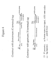
- a DNA fragment encoding a transcription termination region is also included in the construct ( Figure 4).
- a chemical group for covalent linkage such as a terminal amino group, can be introduced via modification of the 3' primer used in PCR.
- the surface or bead is prepared for attachment by appropriate chemistry, such as DNA ImmobiliserTM anthraquinone photo-coupling (Exiqon).
- Figure 10 outlines a general PCR strategy for construction of DNA suitable for in vitro protein synthesis for PISA, using single chain antibody V H /K fragments as example.
- the construct contains a T7 promoter for transcription by T7 RNA polymerase and Kozak sequence for translation initiation in cell free eukaryotic systems.
- a double (His) 6 -tag domain was designed.
- a flexible 19 residue linker Robotson and Sauer, 1998, Proc. Natl Acad. Sci.
- V H /K fragments were linked to the double (His) 6 -tag domain (V H /K-His) was produced by PCR with the following primers.
- the plasmid PTA-His contains a fragment encoding (in order) a flexible linker and a double (His) 6 -tag, followed by two stop codons, a polyA tail and a transcription termination region.
- the sequence of this fragment was: GC TCT AGA ggc ggt ggc tct ggt ggc ggt tct ggc ggt ggc ggt ggc ggc ggc ggc ggc ggc ggc ggc ggc ggc ggc AAA CGG GCT GAT GCT GCA CAT CAC CAT CAC TCT AGA GCT TGG CGT CAC CCG CAG TTC GGT GGT CAC CAC CAC CAC TAA TAA (A) 28 CCG CTG AGC AAT AAC TAG CAT AAC CCC TTG GGG CCT CTA AAC GGG TCT TGA GGG GTT TIT TG CTG AAA GGA GGA ACT
- Lower case sequence linker encoding a 19 amino acid sequence; underlined sequence: double (His) 6 tag; bold: stop codons; (A) 28 : poly A region which contains 28x A. Italics, underlined sequence: transcriptional terminator.
- V H /K-linker and double (His) 6 -tag domain were mixed and added to a PCR mixture containing 2.5 ⁇ l 10x PCR buffer (supplied with Taq DNA polymerase), 1 ⁇ l 2.5mM dNTPs, 1U Taq DNA polymerase and H 2 O to a final volume of 25 ⁇ l.
- a PCR mixture containing 2.5 ⁇ l 10x PCR buffer (supplied with Taq DNA polymerase), 1 ⁇ l 2.5mM dNTPs, 1U Taq DNA polymerase and H 2 O to a final volume of 25 ⁇ l.
- 2 ⁇ l of the mixture was subjected to a second PCR in 50 ⁇ l for 30 cycles using primers T7Ab/back and His-tag/for to generate V H /K-His (see Figure 10).
- the gridded format for transcription and translation comprises microwells of appropriate plates (e.g. 96, 384 or 1536 well polystyrene plates).
- Individual DNA constructs (1 ⁇ g) are dispensed into the wells, each containing a small volume (e.g. 1-50 ⁇ l) of cell free coupled transcription/translation systems such as the rabbit reticulocyte TNT T7 Quick for PCR DNA system (Promega), the TNT coupled wheat germ extract system (Promega) or E. coli S30 extract systems, together with methionine (0.02 mM).
- a small volume e.g. 1-50 ⁇ l
- cell free coupled transcription/translation systems such as the rabbit reticulocyte TNT T7 Quick for PCR DNA system (Promega), the TNT coupled wheat germ extract system (Promega) or E. coli S30 extract systems, together with methionine (0.02 mM).
- methionine 0.02 mM
- the proteins may be bound either by metal affinity on surfaces or beads coated with nickel or by antibody directed against the tag sequence. Therefore, if the wells in which the translation reaction occurs are precoated with Ni-NTA (nitrilotriacetic acid), such as HisSorb plates and strips (Qiagen), or anti-tag antibody, the proteins will be bound to the surface immediately after production to generate the array in situ. Similarly in the presence of beads coated with Ni-NTA the protein will become bound to the surface of the bead.
- Ni-NTA nitrilotriacetic acid
- HisSorb plates and strips Qiagen
- anti-tag antibody anti-tag antibody
- the TNT reaction may also be carried out in noncoated wells and the translation mixture transferred, by manual or automated procedure, to another immobilising surface, such as glass coated with Ni-NTA, beads coated with Ni-NTA, or nitrocellulose or PVDF filter membranes. The unbound material is washed away leaving bound protein.
- another immobilising surface such as glass coated with Ni-NTA, beads coated with Ni-NTA, or nitrocellulose or PVDF filter membranes.
- the unbound material is washed away leaving bound protein.
- the transcription/translation reactions are carried out on other surfaces, for example in droplets distributed on the surface of glass, membranes or agarose.
- the droplets may be oil-covered to prevent evaporation.
- the DNA constructs are first attached covalently or noncovalently to the surface, which may be a polystyrene microwell, modified glass, membrane, beads, agarose, or other surface, thus forming an immobilised DNA array on which protein synthesis can occur.
- the surface may also be coupled to immobilising reagents such as Ni-NTA or anti-tag antibody, in order to immobilise the protein as it is produced.
- the protein synthesis reaction is then performed in situ, for example in microwells or in microdroplets of the cell free system placed onto the DNA locations on glass, etc.
- an immobilised DNA array is converted into a protein array via in vitro transcription/translation.
- translated protein can be demonstrated using incorporated radiolabel or by antibody against a defined tag sequence shared by all the constructs. In this way the content of different wells or array locations can be normalised. Functionality may be demonstrated by specific ligand binding or enzyme activity, as appropriate to the protein.
- Example 1 Creation of a functional protein array element by simultaneous expression and immobilisation of an antibody fragment in situ .
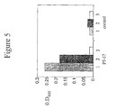
- PCR fragment (0.5 ⁇ gm) was mixed with 20 ⁇ l of coupled transcription/translation 'TNT for PCR DNA system' (Promega), total volume 25 ⁇ l, and the mixture was incubated at 30°C for 1 hour on Ni-NTA coated wells of an 8-well HisSorb strip (Qiagen).
- a non-antibody PCR fragment was used in the same transcription/translation and incubation conditions. After incubation, the strips were washed three times with PBS (phosphate buffered saline), 0.05% Tween.
- HRP horseradish peroxidase
- Example 2 Creation of a functional protein array element by expression in vitro followed by immobilisation of an antibody fragment to a separate surface.
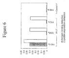
- the P5-17 construct was transcribed and translated in microwells which were not coated with immobilising ligand, using the above conditions. After 1 hour, the well contents (translation mixture) were diluted 4-fold with PBS and 50 ⁇ l was transferred to Ni-NTAwells of HisSorb strips. After 1 hour, the wells were washed and exposed to biotinylated progesterone-BSA, BSA or CEA, followed after 1 hour by HRP-streptavidin.
- Figure 6 shows that the P5-17 protein was successfully produced and immobilised in the separate well, demonstrated by positive anti- ⁇ binding, and that it was specific for progesterone-BSA.
- a protein array element can be produced by in vitro transcription and translation of PCR-produced DNA and transfer of the product to the surface of an immobilising well, with retained function of the protein.
- a mouse antibody V H /K fragment of the anti-progesterone DB3 was produced as above using two different constructs. In one, the single hexahistidine tag sequence was included whereas in the other the sequence was deleted. Protein synthesis was carried out in HisSorb wells. Only the (His)6-containing construct was detected after translation by HRP-linked anti-mouse ⁇ , implying that the immobilisation in situ requires the hexahistidine sequence.
- Example 4 Functional analysis of a human single chain anti-progesterone V H /K (P5-17) by in situ protein array.
- a DNA construct encoding fragment P5-17 was generated by PCR.
- the construct contains a T7 promoter and kozak sequence for in vitro protein synthesis, a double His-tag for protein immobilization and a polyA tail and transcription termination region for efficient protein production.
- Protein expression was carried out by adding the PCR construct to Promega 'TNT Quick for PCR' kit. The mixture was incubated on individual wells of a Ni + -NTA coated plate (Qiagen) so that protein generation and in situ immobilisation proceeded simultaneously. Each well contained 25 ⁇ l of the TNT translation mixture and the mixture was incubated at 30°C for 2-3hrs with shaking.
- Example 5 Quantitative estimation of the expression and in situ immobilisation of an antibody fragment by western blotting.
- V H /K fragment produced as above was analysed by SDS-PAGE either before or after immobilisation on the microwell plate. To estimate the amount of fragment made, standard amounts of VH/K purified from E. coli were run alongside on the gel. Figure 7 indicate that about 150 ng V H /K could be generated in 25 ⁇ l TNT mixture. After in situ immobilisation, about 50% of the total V H /K remained in the supernatant and 30% was eluted from the plate in this case, indicating a binding of about 50ng.
- Example 6 Screening of cloned antibody V H /K fragments from a library by a functional protein in situ array (PISA).
- the array was established by the protein in situ array procedure using PCR products from individual E. coli clones carrying DNA encoding human anti-progesterone V H /K fragments.
- the clones were obtained by E. coli transformation with a transgenic mouse V H /K library either before or after progesterone-BSA selection (He et al. 1999, J. Immunological Methods 231: 105).
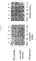
- the array elements were displayed in duplicate, with pre-selection clones numbered 1-5 (top) and post selection clones labelled P5-8, 10,16,17.
- the array was developed with either biotinylated progesterone-BSA followed by HRP-linked streptavidin (left) or HRP linked sheep anti-human ⁇ (right).
- the array shows that the 4 clones after antigen selection were positive for antigen binding whereas those before selection were negative, a result confirmed independently by E. coli expression.
- the anti- ⁇ detection showed that the V H /K fragments were expressed and immobilised for all clones.
- Example 7 Coupled expression and immobilisation of luciferase on magnetic beads.
- Luciferase was chosen for generation of a functional enzyme immobilised on the solid surface of magnetic beads using the PISA procedure.
- a construct encoding luciferase with the C-terminal double His-tag (Luci-His) was produced by PCR as described for V H /K; as a control, luciferase DNA lacking the His-tag domain (Luci) was also PCR generated.
- Primers for PCR generation of luciferase DNA were as follows:
- Example 8 Generation of protein in situ array (PISA) elements from immobilised DNA.
- PCR DNA was immobilised on magnetic beads and used as template to generate His-tagged proteins which became immobilised in situ on the surface ofNi-NTA coupled wells or magnetic agarose beads.
- PCR DNA fragments encoding the human anti-progesterone V H /K fragment P5-17 or luciferase was biotinylated using a 3' primer labelled with biotin. Coupling to beads was achieved by mixing the biotinylated PCR fragment with streptavidin-linked magnetic beads (Promega) and incubating at room temperature for 30 min with gentle rotation. The DNA-coupled beads were collected and washed three times with 0.5ml phosphate buffered saline (PBS).
- PBS phosphate buffered saline
- TNT mixture containing 0.02mM methionine and 0.5mM Mg acetate was added to a mixture of luciferase DNA-coupled beads and Ni-NTA coated beads (Qiagen). The mixture was incubated at 30°C for 2hr with gentle shaking. The beads were washed as above and luciferase activity measured by luminometry. A TNT mixture containing luciferase DNA-coated beads, but without Ni-NTA coated beads, was used as control.
- Figure 12(b) shows that both supernatant and beads from the TNT reaction mixture containing DNA-coupled and Ni-NTA coated beads generated luciferase activity, whereas with the control mixture lacking Ni-NTA beads, enzyme activity was only detected in the supernatant.
- Example 9 Time-course for creating a protein in situ array (PISA) on Ni-NTA wells and magnetic beads.
- PISA protein in situ array
- Example 10 Immobilisation of His-tagged P5-17 V H /K after cell free synthesis and transfer to Ni-NTA coated wells with serial dilution.
- the His-tagged P5-17 V H /K fragment was expressed in 50 ⁇ l rabbit reticulocyte lysate. After serial two-fold dilution, 25 ⁇ l was added in duplicate to individual Ni-NTA coated wells. After 2 h incubation at 30°C, wells were washed and P5-17 immobilisation was detected using HRP-linked anti-human ⁇ chain ( Figure 14). The result shows that V H /K was detectable on the well surface after up to 4 fold dilution.
- Example 11 Analysis of protein immobilisation onto Ni-NTA coated beads by Western blotting.
- Example 12 Re-use of luciferase-immobilised beads after storage.
- luciferase-immobilised beads were generated as in example 7 and luciferase activity from both free in the supernatant and immobilised on the beads was analysed in parallel. After the first measurement, the beads were washed three times with washing buffer and twice with PBS; they were resuspended in 50 ⁇ l PBS and stored at -20°C for one week. The supernatant was also stored at -20°C for the same period. The second and third measurements of luciferase activity including washing and storage of the samples were performed as for the first.
- Figure 16 shows that PISA-immobilised luciferase could be reassayed twice with positive results.
- Example 13 Efficiency of consensus sequence for both prokaryotic and eukaryctic expression.
- the designed PISA construct includes a novel consensus sequence for protein translation initiation in both prokaryotic and eukaryotic systems ( Figure 2).
- Figure 17 shows that the sequence allowed production of functional luciferase in both systems.
- a comparison also showed that it generated luciferase as efficiently as the regular sequence for eukaryotic systems ( Figure 17a).
- Example 14 Demonstration of protein-protein interactions by combination of ribosome display and PISA: Interaction of Grb2 with Vav/N-SH3.
- Vav and Grb2 Interaction of the signalling proteins Vav and Grb2 (Ye and Baltimore (1994) PNAS 91:12629-12633) was chosen as a model with which to demonstrate protein-protein interaction through a combination ofribosome display and PISA protein arrays.
- Vav binds to the adaptor molecule Grb2 to initiate signal transduction via Ras activation.
- Vav contains Src homology 2 and 3 domains (SH2, SH3), in the order (N-SH3)-SH2-(C-SH3).
- Grb2 Interaction with Grb2 was first identified by yeast two-hybrid screen and subsequently shown by filter binding assay to involve a highly specific binding between the N-SH3 domain of Vav and the C-SH3 domain of Grb2 (Ye and Baltimore, 1994).
- the Grb2 C-SH3 domain bound the N-SH3 domain of Vav, but not the Vav/SH2-(C-SH3) domains.
- Ye and Baltimore also prepared GST-Grb2 coupled to glutathione-conjugated agarose beads in order to detect interaction with Vav and showed that full-length Grb2 on beads precipitated full length Vav from cell lysates.
- Vav/N-SH3 comprising the N-terminal SH3 domain
- Vav/SH2-C-SH3 comprising the SH2 domain and linked C-terminal SH3 domain
- full length Grb2 by PCR, from plasmids provided by Dr Martin Turner (Babraham Institute), using primers based on their corresponding DNA sequence.
- a human C ⁇ domain was assembled at the C-terminus of the protein, while in the constructs for protein array (PISA) format the double (His) 6 domain was added to the C-terminus (no C ⁇ domain).
- Ribosome display for Vav/N-SH3 and Vav/SH2-C-SH3 proteins was by the eukaryotic method (He and Taussig (1997) Nucl. Acids Res. 25:5132). PISA-immobilised, full length Grb2 was generated on Ni-NTA coated beads by cell free synthesis as described herein. Grb2 beads were mixed with ribosome displayed Vav/N-SH3, Vav/SH2-C-SH3 or a 1:1 mixture of Vav/N-SH3 : Vav/SH2-C-SH3, respectively. The interactions were carried out on ice for 2 hr and the ribosome complexes interacting with Grb2 beads were isolated by a magnetic concentrator.
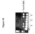
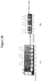
- Figure 20 shows while both Vav/N-SH3 and Vav/SH2-c-SH3 were amplified proportionally by RT-PCR from the original libraries, only Vav/N-SH3 was demonstrably recovered after interaction of library mixtures of different ratios with Grb2-linked beads, demonstrating selection of the interacting molecule Vav/N-SH3.
Landscapes
- Life Sciences & Earth Sciences (AREA)
- Health & Medical Sciences (AREA)
- Engineering & Computer Science (AREA)
- Molecular Biology (AREA)
- Chemical & Material Sciences (AREA)
- Physics & Mathematics (AREA)
- Immunology (AREA)
- Organic Chemistry (AREA)
- General Health & Medical Sciences (AREA)
- Biomedical Technology (AREA)
- Biochemistry (AREA)
- Urology & Nephrology (AREA)
- Hematology (AREA)
- Bioinformatics & Cheminformatics (AREA)
- Proteomics, Peptides & Aminoacids (AREA)
- Biotechnology (AREA)
- Bioinformatics & Computational Biology (AREA)
- Microbiology (AREA)
- Medicinal Chemistry (AREA)
- Biophysics (AREA)
- General Chemical & Material Sciences (AREA)
- Wood Science & Technology (AREA)
- General Physics & Mathematics (AREA)
- Pathology (AREA)
- Chemical Kinetics & Catalysis (AREA)
- Food Science & Technology (AREA)
- Cell Biology (AREA)
- Analytical Chemistry (AREA)
- Zoology (AREA)
- General Engineering & Computer Science (AREA)
- Genetics & Genomics (AREA)
- Peptides Or Proteins (AREA)
- Preparation Of Compounds By Using Micro-Organisms (AREA)
- Light Receiving Elements (AREA)
- Investigating Or Analysing Biological Materials (AREA)
Abstract
Description
- An array is a precisely ordered arrangement of elements, allowing them to be displayed and examined in parallel (1). It usually comprises a set of individual species of molecules or particles arranged in a regular grid format; the array can be used to detect interactions, based on recognition or selection, with a second set of molecules or particles applied to it. Arrays possess advantages for the handling and investigation of multiple samples. Theyprovide a fixed location for each element such that those scoring positive in an assay are immediately identified; they have the capacity to be comprehensive and of high density; they can be made and screened by high throughput robotic procedures using small volumes of reagents; and they allow the comparison of each assay value with the results of many identical assays. The array format is well established for global analysis of nucleic acids, and oligonucleotide and cDNA arrays (DNA chips) are used for gene expression analysis. In a familiar format, large numbers (e.g. thousands) of DNA hybridisation probes are attached in an ordered pattern to a surface such as nylon, glass or silicon and hybridised to fluorescently labelled whole cell mRNA or cDNA; the quantitative signals on each array element are measured in parallel by means of a reader device.
- The array approach may also be adapted for display of peptides and proteins; the elements displayed may be a set of related proteins or peptides, or the entire protein complement of an organism. Protein array technology allows high throughput screening for gene expression and molecular interactions. It is possible to use protein arrays to examine in parallel the functions of thousands of proteins previously known only by their DNA sequence. For functional information to be obtained, the arrayed proteins must be in native form. However, some preparative methods cause protein denaturation, as may occur during extraction or release of recombinant proteins from bacteria, and the use of arrays from such starting material is therefore limited to applications determined only by the primary sequence of the protein rather than tertiary structure. In order to develop high throughput approaches to global protein analysis which can yield functional information, methods for producing arrays in which proteins retain their functions are required.
- Arrays of immobilised proteins can be used to demonstrate a binding reaction, as where the array is exposed to an entity such as an antibody or ligand, which may be directly or indirectly labelled, and binding demonstrated by localisation of the label to a particular segment of the array. Mass spectrometry may also be used to identify binding interactions on the array. Alternatively, the arrayed proteins may be in solution and used to study biochemical function. Potential uses of protein arrays which have been discussed in the literature (1-12) include identification of antibodies and analysis of antibody specificity, measurement of global protein expression, identification of ligand-receptor interactions and protein-protein interactions, and screening and selecting proteins or ligands from libraries. (i) Expression profiling. One type of protein array which has been proposed is based on immobilisation of antibodies at a surface (an antibody array). In principle, the reaction of an antibody array with cellular proteins can provide a global quantitative readout of all the proteins expressed at any particular time (proteome analysis). In one version, for differential display, the array is probed with fluorescently labelled proteins from two different cell states; cell lysates are labelled by different fluorophores and mixed such that the colour acts as a readout for the change in abundance. (ii) Antibody detection. A second application is the detection of antibodies against cellular proteins, where either or both partners are unknown. Thus an array of cellular proteins can be used to select antibodies from libraries of soluble antibodies or from phage-display or ribosome-display libraries. The antigen array can also be used to analyse antibodies in small amounts of patient sera, as during infections or in autoimmune conditions. (iii) Ligand screening. An array of potential target proteins, such as receptors, can be used as a screen for selection of ligands which may be possible drug candidates, including small molecules, peptides, aptamers, nucleic acids or synthetic scaffolds. (iv) Detection of protein-protein interactions. A further use for protein arrays is in the detection of protein-protein interactions. Each protein in the genome may interact with a number of partners, so for the approximately 100,000 proteins encoded in the human genome there may exist millions of interactions. Such interactions are often measured by yeast two-hybrid (cell-based) methods but these may fail to measure interactions involving secreted proteins, proteins with disulphide bridges and membrane bound proteins such as receptors. An array method would be highly desirable in these cases and may reveal interactions which are not detected by the cellular methods.
- The arrays described to date are composed either of purified proteins or proteins expressed in living cells or viruses. Early examples were peptide arrays, in which peptides were chemically synthesised on a solid support and used to identify epitopes recognised by antibodies (2). Peptide arrays can be chemically synthesised up to a length of about 30 amino acids, but are unable to produce full length folded proteins.
- Clearly, protein arrays can be made by chemical or noncovalent attachment of preformed proteins onto suitable surfaces, such as treated glass slides or absorptive membranes such as nitrocellulose or PVDF. For high throughput studies, such as proteomics or library screening, this requires methods for the preparation, purification and immobilisation in parallel of large numbers of proteins. Methods for production of recombinant proteins from bacteria for assay in an arrayed format have been described (3,6-10). Proteins can be expressed as constructs fused to an affinity tag (e.g. hexahistidine) or glutathione S-transferase (GST), recovered by cell lysis and used either as crude lysates or after affinity purification (e.g. on Ni-NTA metal affinity columns). However, the production of recombinant proteins in bacterial systems can be problematic due to aggregation, insoluble inclusion bodies and/or degradation of the product, while eukaryotic systems suffer from lower yields and high demands on sterility or time consuming cloning procedures (e.g. Baculovirus). Where denaturants are used in the extraction the protein will often be rendered nonfunctional. Once the proteins have been isolated, various technical formats, substrates, production methods and detection systems are available (reviewed in 8).
- Martzen et al. (3) purified most of the soluble yeast proteins from Saccharomyces cerevisiae by glutathione agarose affinity chromatography from 6144 yeast strains each of which contained a plasmid with a different yeast ORF (open reading frame) fused to GST. Proteins were assayed in solution for a particular enzymatic activity. Since the proteins were purified in native form, this constituted a functional protein array, although the proteins were not immobilised on a surface. Yeast cells have been used to create a 'living' recombinant protein array, containing about 6000 colonies, each of which expresses a different ORF Gal4-fusion protein (4). This is basically a cellular, yeast two hybrid screen performed in a 96 well plate format.
- Arrays can be prepared by inducing the simultaneous expression of large numbers of cDNA clones in an appropriate vector system and high speed arraying of protein products. Bussow et al. (6) arrayed proteins expressed from cDNA clones of a human fetal brain cDNA
expression library hEx 1 cloned in Escherichia coli. The his6 (hexahistidine)-tagged proteins were induced from individual colonies grown in 384-well microtiter plates and gridded onto high density PVDF filter membranes prior to expression induction with IPTG. Release of the proteins from the bacterial cytoplasm created an array of proteins immobilised on the filters. Two example proteins were identified on the filters using antibodies. While this method allows the operator to screen expression libraries, the extraction procedure used 0.5M NaOH, during which process proteins were denatured and therefore rendered nonfunctional. In another report (below), proteins were extracted and solubilised using 6M guanidinium HCl, which is also a denaturant. Other drawbacks of this procedure as a means ofproducing an array are that clones must be extensively screened for in frame expression and that cDNA libraries contain many clones which lack the 5' end (N-terminal), may have multiple copies of some genes and poor representation of others. - Lueking et al. (9) gridded purified protein solutions from the hEx 1 library onto PVDF filters, in an extension of classical dot-blotting methodology. For high throughput, small-scale protein expression, clones of the
hEx 1 library were grown in 96-well microtiter plates and induced with IPTG; cells were lysed with 6M guanidinium HCl and supernatants filtered through a 96 well filter plate onto a PVDF membrane. For larger scale production ofpurified proteins, peptide- and his6-tagged proteins were expressed from E. coli and isolated with Ni-NTA agarose. Results of high throughput screening showed quite a number of false positives, i.e. proteins detected by anti-tag which were in fact out of frame with the tag, and antibody specificity screening often showed unexpected crossreactions, mostly with ribosomal proteins, for no apparent reason. The use of guanidinium HCl denatures the proteins and may cause aberrant results. - Holt et al. (7) screened the hEx1 library to identify specific antibodies, reactive with denatured proteins, using 12 well-expressed antibody fragments of previously unknown specificity. Four specific interactions were identified In another example of dot blotting, Ge described an array system for detection of protein interactions with other proteins, DNA, RNA and small ligands (12). In this case, 48 highly purified, native proteins were arrayed on a nitrocellulose membrane, by spotting using a 96-well dot blot apparatus. The proteins were overexpressed in bacteria or baculovirus and purified to homogeneity. The dot blot array was reacted with a number of different radiolabelled probes (protein. DNA, RNA, ligand), followed by autoradiography and densitometry, and showed to behave in a functional manner, i.e. probes interacted with partner molecules with the expected specificity.
- Afanassiev et al. (5) describe a method for making protein arrays using chemical coupling of proteins to an agarose film on microscope slides; the agarose is activated by sodium periodate to reveal aldehyde groups which bind amino groups on the protein. Varying amounts of an antigen (BAD) or an anti-BAD antibody (6A11) were immobilised and binding of the partner molecule detected by a fluorescent second reagent.
- WO 00/04382 A (Zyomyx Inc.) discloses an array of proteins, comprising proteins, e.g. sFc antibody fragments, immobilised to a substrate through an affinity tag introduced into the protein, wherein the affinity tag comprises polyhistidine. The proteins of the array are prepared by expression from recombinant DNA in vitro.
- Ohuchi Shoji et al. "In vitro method for the generation of protein libraries using PCR amplification of a single DNA molecule and coupled transcription/translation". Nucleic Acids Research, Vol. 26, no. 19, 1 October 1998, pages 4339-4346, discloses a method for creating a protein array comprising preparing DNA constructs in a gridded format (multiwell plate), transcribing and translating the DNA in a coupled transcription/translation system inside the gridded format. The translated proteins are not attached to the well surface as they are produced to create the protein array.
-
- 1. Emili A.Q. and Cagney G. (2000) Large-scale functional analysis using peptide or protein arrays. Nature Biotechnology 18:393-397.
- 2. Geysen H.M., Meloen, R.H. and Barteling S.J. (1984) Use of peptide synthesis to probe viral antigens for epitopes to a resolution of a single amino acid. Proc. Natl Acad. Sci. USA 81:3998-4002.
- 3. Martzen M.R., McCraith S.M., Spinelli S.L., Torres F.M., Fields S., Grayhack E.J. and Phizicky E.M. (1992) A biochemical genomics approach for identifying genes by the activity of their products.
Science Nov 5; 286(5442):1153-5. - 4. Uetz P. et al. (2000) A comprehensive analysis of protein-protein interactions in Saccharomyces cerevisiae. Nature 403, 623-627.
- 5. Afanassiev V., Hanemann V. and Wolfl, S. (2000) Preparation of DNA and protein microarrays on glass slides coated with an agarose film. Nucleic Acids Research 28:E66.
- 6. Bussow K., Cahill D., et al. (1998) A method for global protein expression and antibody screening on high-density filters of an arrayed cDNA library. Nucleic Acids Res. 26:5007-5008.
- 7. Holt L.J., Bussow K., Walter G and Tomlinson I.M. (2000) By-passing selection: direct screening for antibody-antigen interactions using protein arrays. Nucleic Acids Research 28:e72.
- 8. Walter G., Bussow K., Cahill D., Lueking A. and Lehrach H. (2000) Protein arrays for gene expression and molecular interaction screening. Current Opinion in Microbiology 3:298-302.
- 9. Lueking A., Horn, M., Eickhoff, H., Bussow, K., Lehrach H. And Walter G. (1999) Protein Microarrays for gene Expression and Antibody Screening. Anal. Biochem. 270:103-111.
- 10. Bussow K., Nordhoff E., Lubbert C., Lehrach H and Walter G. (2000) A human cDNA library for high-throughput protein expression screening. Genomics 65:1-8.
- 11. Pandey A. and Mann M. (2000) Proteomics to study genes and genomes. Nature 405:837-846.
- 12. Ge H. (2000) UPA, a universal protein array system for quantitative detection of protein-protein, protein-DNA, protein-RNA and protein-ligand interactions. Nucleic Acids Research 28, e3.
- We describe a method for producing a protein array starting from DNA in which a number of native, functional proteins or domains are produced in parallel by in vitro synthesis using a cell free system for transcription and translation, followed by immobilisation of the products in a gridded format on a surface, using an isolation sequence tag incorporated into the proteins. In one embodiment, the array is formed in situ in a single step by performing protein expression in wells, on surfaces, or in the presence of beads, any of which are coated with immobilising molecules (affinity ligands) such as metal ions or antibodies.
- Starting material can include genomic DNA, mRNA, cloned DNA fragments, or cDNA libraries, etc. The input DNA constructs for in vitro transcription/translation may be obtained by PCR (polymerase chain reaction) or RT (reverse transcription)-PCR amplification, using primers designed on any known DNA sequences, such as those from databases and genome projects. Alternatively, cloned plasmid DNA may be used. The cell free systems for protein synthesis are those such as rabbit reticulocyte, wheat germ, yeast or bacterial extracts. Numbers of individual native proteins or domains may be produced in parallel directly from the PCR DNA constructs. In one embodiment the reaction is carried out in the wells of multiwell plates. In another embodiment, proteins may be synthesised directly onto surfaces, such as glass, membranes or beads, for example by carrying out the reaction in microdroplets. The construct DNA may be added to the reaction volume or droplet or pre-immobilised to the surface. In the latter case, a DNA array is effectively created first and then used to create the protein array. The proteins can be adapted for rapid isolation, immobilisation or identification by inclusion of sequences such as hexahistidine or other peptide tags. If the wells or other surfaces in, or on, which the proteins are produced are precoated with an immobilising reagent such as nickel ions or anti-tag antibodies, the array will be formed as the proteins are produced in situ and reagent molecules can be removed by washing. The reaction may alternatively be performed in the presence of beads coated with an immobilising reagent, the beads being subsequently distributed in a gridded array format. Alternatively, the proteins can be transferred (e.g. by gridding robot) to secondary surfaces, such as plastic, glass, agarose, beads, nitrocellulose or other membranes.
- Target molecules, such as labelled ligands, proteins or nucleic acids, may then be exposed to the array and binding to individual array locations detected by means of enzyme-coupled reaction, fluorescence, autoradiography or mass spectrometry. The arrays can thereby be used for screening of antibodies, ligands, protein interactions, etc. In some cases the arrayed proteins may be used in solution in order to perform biochemical studies. Retaining the gridded format, the solutions may also be transferred to filters or plates precoated with target molecules such as antigens, and binding detected by labelled secondary reagents.
- The arrays can also be linked to library display systems. Thus, target molecules may be those of display libraries, such as phage or ribosome display libraries, in which the individual proteins are linked to encoding DNA or mRNA. After binding to the array, interacting molecules are identified by amplification and identification of the linked DNA or mRNA, for example by cloning phage or by PCR, RT-PCR, hybridisation or other methods.
- By utilising in vitro synthesis, the above methodology provides a rapid means of obtaining functional protein arrays directly from DNA, including proteins or domains known only from DNA sequence. It can replace cloning, in vivo expression systems and purification procedures. It avoids the problems of inclusion bodies, aggregation and degradation often encountered with bacterial expression. Since PCR and in vitro translation can be carried out using many samples simultaneously in parallel, such arrays will provide a high throughput capacity for analysis of protein expression, functional activity and interactions, making use in particular of the genetic information from genome projects.
- The constructs for in vitro transcription/translation (Figure 1) include: upstream T7 promoter and protein expression signals designed to allow genes to be expressed in either eukaryotic systems (Figure 2a) or in both prokaryotic and eukaryotic systems (Figure 2b); upstream or downstream sequences encoding a flexible linker (19 residues) and a tag sequence such as (his)6 [SEQ ID No. 1] or (his)6-SRAWRHPQFGG-(his)6 [SEQ ID No. 2] for protein immobilisation by metal affinity binding (Figure 3) and/or a further peptide tag sequence for recognition by antibody or ligand; and a downstream translation stop codon. To create such constructs, the gene of interest is amplified separately by PCR or RT-PCR using specific primers derived from known DNA sequences, and the upstream and downstream elements are incorporated into the construct by PCR assembly.
- For covalent immobilisation of constructs to a surface or bead, a DNA fragment encoding a transcription termination region is also included in the construct (Figure 4). In addition, a chemical group for covalent linkage, such as a terminal amino group, can be introduced via modification of the 3' primer used in PCR. The surface or bead is prepared for attachment by appropriate chemistry, such as DNA ImmobiliserTM anthraquinone photo-coupling (Exiqon).
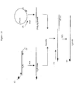
- Figure 10 outlines a general PCR strategy for construction of DNA suitable for in vitro protein synthesis for PISA, using single chain antibody VH/K fragments as example. The construct contains a T7 promoter for transcription by T7 RNA polymerase and Kozak sequence for translation initiation in cell free eukaryotic systems. To increase the efficiency of protein immobilisation on a Ni-NTA coated surface and allow the re-use of the protein arrays, a double (His)6-tag domain was designed. A flexible 19 residue linker (Robinson and Sauer, 1998, Proc. Natl Acad. Sci. USA 95: 5929-5934) is placed between the protein to be arrayed and the His-tag domain, in order to reduce any possible interference of the tag sequence on folding of the attached protein. A poly(A)28 tail and a transcription terminator are incorporated at the 3' end of the DNA to increase transcription efficiency. To ensure translation termination and release of the nascent polypeptide from the ribosome complex, two stop codons (TAATAA [SEQ ID No. 3]) are included following the double (His)6-tag sequence.
- A construct in which VH/K fragments were linked to the double (His)6-tag domain (VH/K-His) was produced by PCR with the following primers.
-
- T7 Ab/back: 5'-GCA GC T AAT ACG ACT CAC TAT AGG AAC AGA CCA CC ATG (C/G) AG GT(G/C) CA(G/C) CTC GAG (C/G)AG TCT GG-3' [SEQ ID No. 4]. This primer provides the T7 promoter and Kozak signal (underlined) and a degenerate sequence complementary to the 5' region of the antibody heavy chain (italics). The initiation codon is indicated in bold.
- Ab-linker/for: 5'-GCC ACC GCC TCT AGA GCG GCT CAG CGT CAG GGT GCT GCT-3' [SEQ ID No. 5]. This provides a sequence complementary to the 3' region of the human κ constant domain (Cκ) and a sequence (underlined) overlappping a linker-tag/back primer (see below) used for generation of the double (His)6-tag domain.
-
- Linker-tag/back: 5'-GC TCT AGA GGC GGT GGC TCT GGT GGC GGT TCT GGC GGT GGC ACC GGT GGC GGT TCT GGC GGT GGC AAA CGG GCT GAT GCT GCA [SEQ ID No. 6]. This provides a sequence (underlined) overlapping the Ab-linker/for primer used for VH/K-linker construction (above) and the linker sequence for PCR generation of the double (His)6-tag domain (below).
- His-tag/for : 5'-TCC GGA TAT AGT TCC TCC-3' [SEQ ID No. 7].
- The plasmid PTA-His contains a fragment encoding (in order) a flexible linker and a double (His)6-tag, followed by two stop codons, a polyA tail and a transcription termination region. The sequence of this fragment was: GC TCT AGA ggc ggt ggc tct ggt ggc ggt tct ggc ggt ggc acc ggt ggc ggt tct ggc ggt ggc AAA CGG GCT GAT GCT GCA CAT CAC CAT CAC CAT CAC TCT AGA GCT TGG CGT CAC CCG CAG TTC GGT GGT CAC CAC CAC CAC CAC CAC TAA TAA (A)28 CCG CTG AGC AAT AAC TAG CAT AAC CCC TTG GGG CCT CTA AAC GGG TCT TGA GGG GTT TIT TG CTG AAA GGA GGA ACT ATA TCC GGA [SEQ ID No. 8]. Lower case sequence: linker encoding a 19 amino acid sequence; underlined sequence: double (His)6 tag; bold: stop codons; (A)28 : poly A region which contains 28x A. Italics, underlined sequence: transcriptional terminator.
- In general, standard PCR was carried out for 30 cycles to obtain the VH/K-linker fragment and double (His)6-tag domain in separate reactions using Taq polymerase (Qiagen, UK) according to the manufacturer's instructions. The resulting fragments were analysed and eluted from a 1% agarose gel using a gel extraction kit (Qiagen, UK). For assembly, equal amounts (total 10-50ng) of VH/K-linker and double (His)6-tag domain were mixed and added to a PCR mixture containing 2.5µl 10x PCR buffer (supplied with Taq DNA polymerase), 1µl 2.5mM dNTPs, 1U Taq DNA polymerase and H2O to a final volume of 25µl. After thermal cycling for 8 cycles (94°C for 30 sec, 54°C for 1 min and 72°C for 1 min), 2µl of the mixture was subjected to a second PCR in 50µl for 30 cycles using primers T7Ab/back and His-tag/for to generate VH/K-His (see Figure 10).
- In one embodiment the gridded format for transcription and translation comprises microwells of appropriate plates (e.g. 96, 384 or 1536 well polystyrene plates). Individual DNA constructs (1µg) are dispensed into the wells, each containing a small volume (e.g. 1-50 µl) of cell free coupled transcription/translation systems such as the rabbit reticulocyte TNT T7 Quick for PCR DNA system (Promega), the TNT coupled wheat germ extract system (Promega) or E. coli S30 extract systems, together with methionine (0.02 mM). To label the protein, 35S methionine or other labelled amino acids may be included. Plates are incubated for 1 hour at 30 °C. Where the proteins contain immobilisation sequences, such as single or double hexahistidine tags or specific peptide tag sequence, they may be bound either by metal affinity on surfaces or beads coated with nickel or by antibody directed against the tag sequence. Therefore, if the wells in which the translation reaction occurs are precoated with Ni-NTA (nitrilotriacetic acid), such as HisSorb plates and strips (Qiagen), or anti-tag antibody, the proteins will be bound to the surface immediately after production to generate the array in situ. Similarly in the presence of beads coated with Ni-NTA the protein will become bound to the surface of the bead. The TNT reaction may also be carried out in noncoated wells and the translation mixture transferred, by manual or automated procedure, to another immobilising surface, such as glass coated with Ni-NTA, beads coated with Ni-NTA, or nitrocellulose or PVDF filter membranes. The unbound material is washed away leaving bound protein.
- In alternative embodiments, the transcription/translation reactions are carried out on other surfaces, for example in droplets distributed on the surface of glass, membranes or agarose. The droplets may be oil-covered to prevent evaporation.
- In embodiments where protein synthesis is carried out on immobilised DNA, the DNA constructs are first attached covalently or noncovalently to the surface, which may be a polystyrene microwell, modified glass, membrane, beads, agarose, or other surface, thus forming an immobilised DNA array on which protein synthesis can occur. The surface may also be coupled to immobilising reagents such as Ni-NTA or anti-tag antibody, in order to immobilise the protein as it is produced. The protein synthesis reaction is then performed in situ, for example in microwells or in microdroplets of the cell free system placed onto the DNA locations on glass, etc. Thus, in this embodiment, an immobilised DNA array is converted into a protein array via in vitro transcription/translation.
- The presence of translated protein can be demonstrated using incorporated radiolabel or by antibody against a defined tag sequence shared by all the constructs. In this way the content of different wells or array locations can be normalised. Functionality may be demonstrated by specific ligand binding or enzyme activity, as appropriate to the protein.
- To demonstrate the creation of a functional protein array element in situ, a construct encoding a single-chain human anti-progesterone antibody VH/K fragment (P5-17) was used (He et al., 1999, J. Immunol. Methods, 231:105). To prepare the construct for the protein array, the T7 promoter, expression signal and the double hexahistidine tag were incorporated into this fragment by PCR assembly. The PCR fragment (0.5 µgm) was mixed with 20µl of coupled transcription/translation 'TNT for PCR DNA system' (Promega), total volume 25µl, and the mixture was incubated at 30°C for 1 hour on Ni-NTA coated wells of an 8-well HisSorb strip (Qiagen). As control, a non-antibody PCR fragment was used in the same transcription/translation and incubation conditions. After incubation, the strips were washed three times with PBS (phosphate buffered saline), 0.05% Tween. To demonstrate that the antibody fragment was expressed and immobilised on the surface of the wells, HRP (horseradish peroxidase)-anti-human κ antibody was applied. To test the binding activity of the immobilised P5-17, the wells were incubated with biotinylated progesterone-BSA (P-BSA) for 1 hour followed by detection with HRP-streptavidin for another hour. The HRP activity was developed and the colour read by ELISA reader at 450nm.
- The results of this two component array experiment show that the antibody fragment was successfully expressed in vitro, became bound in situ to the surface of the well (positive anti-κ binding) and was functional by the criterion of specific antigen binding (Figure 5). This result demonstrates that an individual protein array location can be constructed in a microwell by in vitro transcription and translation of PCR DNA and simultaneous immobilisation of the product to the surface of the well, with retained function of the protein.
- The P5-17 construct was transcribed and translated in microwells which were not coated with immobilising ligand, using the above conditions. After 1 hour, the well contents (translation mixture) were diluted 4-fold with PBS and 50 µl was transferred to Ni-NTAwells of HisSorb strips. After 1 hour, the wells were washed and exposed to biotinylated progesterone-BSA, BSA or CEA, followed after 1 hour by HRP-streptavidin. Figure 6 shows that the P5-17 protein was successfully produced and immobilised in the separate well, demonstrated by positive anti-κ binding, and that it was specific for progesterone-BSA. Thus a protein array element can be produced by in vitro transcription and translation of PCR-produced DNA and transfer of the product to the surface of an immobilising well, with retained function of the protein.
- In order to rule out nonspecific protein localisation, a mouse antibody VH/K fragment of the anti-progesterone DB3 was produced as above using two different constructs. In one, the single hexahistidine tag sequence was included whereas in the other the sequence was deleted. Protein synthesis was carried out in HisSorb wells. Only the (His)6-containing construct was detected after translation by HRP-linked anti-mouse κ, implying that the immobilisation in situ requires the hexahistidine sequence.
- A DNA construct encoding fragment P5-17 was generated by PCR. The construct contains a T7 promoter and kozak sequence for in vitro protein synthesis, a double His-tag for protein immobilization and a polyA tail and transcription termination region for efficient protein production. Protein expression was carried out by adding the PCR construct to Promega 'TNT Quick for PCR' kit. The mixture was incubated on individual wells of a Ni+-NTA coated plate (Qiagen) so that protein generation and in situ immobilisation proceeded simultaneously. Each well contained 25µl of the TNT translation mixture and the mixture was incubated at 30°C for 2-3hrs with shaking. After washing with PBS-Tween (3 times), wells were treated with either biotinylated progesterone-BSA (P-BSA) or HRP-linked sheep anti-human-κ. For detection of biotinylated antigen, HRP-linked streptavidin was used. As controls, biotinylated BSA (BSA) and anti-mouse -κ (mouse) were used. Figure 6 shows while biotinylated BSA produced no binding, the biotinylated progesterone-BSA was strongly bound (yellow colour). Similarly, the presence of P5-17 was only detected by anti-human-k (blue) and not by anti-mouse-κ. This experiment thus demonstrates the functional expression and immobilisation of P5-17 fragment in the 'protein in situ array' format (PISA).
- The antibody VH/K fragment produced as above was analysed by SDS-PAGE either before or after immobilisation on the microwell plate. To estimate the amount of fragment made, standard amounts of VH/K purified from E. coli were run alongside on the gel. Figure 7 indicate that about 150 ng VH/K could be generated in 25µl TNT mixture. After in situ immobilisation, about 50% of the total VH/K remained in the supernatant and 30% was eluted from the plate in this case, indicating a binding of about 50ng.
- The array was established by the protein in situ array procedure using PCR products from individual E. coli clones carrying DNA encoding human anti-progesterone VH/K fragments. The clones were obtained by E. coli transformation with a transgenic mouse VH/K library either before or after progesterone-BSA selection (He et al. 1999, J. Immunological Methods 231: 105). In Figure 8, the array elements were displayed in duplicate, with pre-selection clones numbered 1-5 (top) and post selection clones labelled P5-8, 10,16,17. The array was developed with either biotinylated progesterone-BSA followed by HRP-linked streptavidin (left) or HRP linked sheep anti-human κ (right). The array shows that the 4 clones after antigen selection were positive for antigen binding whereas those before selection were negative, a result confirmed independently by E. coli expression. The anti-κ detection showed that the VH/K fragments were expressed and immobilised for all clones.
- Luciferase was chosen for generation of a functional enzyme immobilised on the solid surface of magnetic beads using the PISA procedure. A construct encoding luciferase with the C-terminal double His-tag (Luci-His) was produced by PCR as described for VH/K; as a control, luciferase DNA lacking the His-tag domain (Luci) was also PCR generated. Primers for PCR generation of luciferase DNA were as follows:
- T7 Luci/back: GCA GC TAA TAC GAC TCA CTA TAG GAA CAG ACC ACC ATG GAA GAC GCC AAA AAC [SEQ ID No. 9]. The T7 promoter and Kozak signal are underlined. Italics indicate the sequence complementary to the 5' region of luciferase. ATG is the initiation codon.
- Luci-linker/for: GCC ACC GCC TCT AGA GCG CAA TTT GGA CTT TCC GCC [SEQ ID No. 10]. The underlined sequence overlaps a linker-tag/back primer (see above) used for generation of the double (His)6-tag domain.
- After cell free expression in the presence of Ni-NTA coated magnetic beads, the latter were separated from the translation mixture and washed. Luciferase activity free in the translation mixture supernatant and immobilised on the beads was measured using a luminometer (Figure 11). While the luciferase construct lacking the His-tag domain only produced activity in the translation mixture, Luci-His generated activity both in the mixture and on the beads, demonstrating the successful immobilisation of functional luciferase through the double-His tag domain.
- In this example, PCR DNA was immobilised on magnetic beads and used as template to generate His-tagged proteins which became immobilised in situ on the surface ofNi-NTA coupled wells or magnetic agarose beads.
- PCR DNA fragments encoding the human anti-progesterone VH/K fragment P5-17 or luciferase was biotinylated using a 3' primer labelled with biotin. Coupling to beads was achieved by mixing the biotinylated PCR fragment with streptavidin-linked magnetic beads (Promega) and incubating at room temperature for 30 min with gentle rotation. The DNA-coupled beads were collected and washed three times with 0.5ml phosphate buffered saline (PBS).
- To create an array element of P5-17 VH/K by PISA on a Ni-NTA coated well surface, 25µl of TNT mixture containing 0.02mM methionine and 0.5mM Mg acetate was mixed with P5-17 DNA-coupled beads (above) and the mixture added into the well of a Ni-NTA coated plate (Ni-NTA HisSorb strips, Qiagen). After incubation for 2 hrs with shaking, the plate was washed 100µl wash buffer (50mM NaH2PO4, 300mM NaCl, 20mM imidazole, pH 8.0), followed by a final wash with 100µl phosphate-buffered saline (PBS). The wells were probed with either biotinylated progesterone-BSA or HRP-linked anti-human κ chain. TNT mixture without PCR-coated beads was used as control. Figure 12(a) shows while the negative control (TNT mixture without DNA-coupled beads) had no detectedbinding activity, wells incubated with P5-17 DNA-coupled beads generated positive signals using either the antigen, biotinylated progesterone-BSA, and HRP-linked streptavidin, or HRP-linked anti-human κ chain.
- To create an array element of luciferase by PISA on the surface of Ni-NTA-coated agarose beads, 25µl of TNT mixture containing 0.02mM methionine and 0.5mM Mg acetate was added to a mixture of luciferase DNA-coupled beads and Ni-NTA coated beads (Qiagen). The mixture was incubated at 30°C for 2hr with gentle shaking. The beads were washed as above and luciferase activity measured by luminometry. A TNT mixture containing luciferase DNA-coated beads, but without Ni-NTA coated beads, was used as control. Figure 12(b) shows that both supernatant and beads from the TNT reaction mixture containing DNA-coupled and Ni-NTA coated beads generated luciferase activity, whereas with the control mixture lacking Ni-NTA beads, enzyme activity was only detected in the supernatant.
- These experiments demonstrate the potential to generate immobilised protein from immobilised DNA by in vitro synthesis (PISA method). This could be used to convert a DNA array into a protein array.
- To measure the optimal time for immobilisation of in vitro synthesised protein onto Ni-NTA coated wells, 100µl of TNT mixture containing P5-17 VH/K DNA was subdivided into 25µl aliquots and added to four Ni-NTA coated wells. The mixtures were incubated at 30°C for 1, 2, 4 and 7 hours respectively. After washes, HRP-linked anti-human κ was used to detect the immobilised P5-17 antibody fragment (Figure 13 a). Two hours incubation produced the highest level of VH/K immobilisation.
- To measure the optimal time for immobilisation of in vitro synthesised protein onto Ni-NTA linked magnetic beads, 100µl of TNT mixture containing luciferase DNA was subdivided into 25µl aliquots and added to Ni-NTA coated beads. The mixtures were incubated at 30°C for 1, 2, 4 and 7 hours. After washes, luciferase activity was measured by luminometry (Figure 13b). This revealed that while both free and immobilised luciferase activity reached apeak in activity in 2hr, free luciferase declined significantly after 2 hours.
- The His-tagged P5-17 VH/K fragment was expressed in 50µl rabbit reticulocyte lysate. After serial two-fold dilution, 25µl was added in duplicate to individual Ni-NTA coated wells. After 2 h incubation at 30°C, wells were washed and P5-17 immobilisation was detected using HRP-linked anti-human κ chain (Figure 14). The result shows that VH/K was detectable on the well surface after up to 4 fold dilution.
- Western blotting was applied to estimate the efficiency of protein immobilisation onto Ni-NTA coated beads. Both P5-17 human anti-progesterone VH/K and luciferase were immobilised onto Ni-NTA coated beads using the PISA procedure. The beads were then boiled in SDS sample buffer before running on PAGE, in parallel with supernatant protein remaining in the TNT mixture after removal of beads. After Western blotting, transferred protein was detected with HRP-linked anti-(His)6 antibody and quantified by densitometry. The results show that 40-50% of the translated protein was eluted from Ni-NTA coated beads (Figure 15, arrowed) (cf. similar result using Ni-NTA coated wells, example 5).
- To test if arrayed protein immobilised by the PISA procedure can be re-assayed, luciferase-immobilised beads were generated as in example 7 and luciferase activity from both free in the supernatant and immobilised on the beads was analysed in parallel. After the first measurement, the beads were washed three times with washing buffer and twice with PBS; they were resuspended in 50µl PBS and stored at -20°C for one week. The supernatant was also stored at -20°C for the same period. The second and third measurements of luciferase activity including washing and storage of the samples were performed as for the first. Figure 16 shows that PISA-immobilised luciferase could be reassayed twice with positive results.
- The designed PISA construct includes a novel consensus sequence for protein translation initiation in both prokaryotic and eukaryotic systems (Figure 2). In order to validate its effectiveness, a luciferase construct containing the consensus initiation sequence was tested for protein expression in both coupled rabbit reticulocyte lysate and coupled E. coli S30 systems. Figure 17 shows that the sequence allowed production of functional luciferase in both systems. A comparison also showed that it generated luciferase as efficiently as the regular sequence for eukaryotic systems (Figure 17a).
- E: eukaryotic initiation sequence alone.
- PE: combined prokaryotic and eukaryotic initiation sequences (novel)
- Interaction of the signalling proteins Vav and Grb2 (Ye and Baltimore (1994) PNAS 91:12629-12633) was chosen as a model with which to demonstrate protein-protein interaction through a combination ofribosome display and PISA protein arrays. In intact mammalian haematopoietic cells, Vav binds to the adaptor molecule Grb2 to initiate signal transduction via Ras activation. Vav contains
Src homology - We generated DNA for the Vav/N-SH3 fragment (comprising the N-terminal SH3 domain), Vav/SH2-C-SH3 (comprising the SH2 domain and linked C-terminal SH3 domain), and full length Grb2 by PCR, from plasmids provided by Dr Martin Turner (Babraham Institute), using primers based on their corresponding DNA sequence. In the constructs for ribosome display, a human Cκ domain was assembled at the C-terminus of the protein, while in the constructs for protein array (PISA) format the double (His)6 domain was added to the C-terminus (no Cκ domain).
- Ribosome display for Vav/N-SH3 and Vav/SH2-C-SH3 proteins was by the eukaryotic method (He and Taussig (1997) Nucl. Acids Res. 25:5132). PISA-immobilised, full length Grb2 was generated on Ni-NTA coated beads by cell free synthesis as described herein. Grb2 beads were mixed with ribosome displayed Vav/N-SH3, Vav/SH2-C-SH3 or a 1:1 mixture of Vav/N-SH3 : Vav/SH2-C-SH3, respectively. The interactions were carried out on ice for 2 hr and the ribosome complexes interacting with Grb2 beads were isolated by a magnetic concentrator. After washing the beads three times with 50µl washing buffer (PBS, 0.1% BSA, 0.05% Tween, 5mM Mg acetate) followed by two washes with water, the beads were subjected to RT-PCR using universal primers for both Vav/N-SH3 and Vav/SH2-C-SH3 constructs (T7A1 and evol 4). Figure 18 shows that while DNA encoding Vav/SH2-C-SH3 was not detected, Vav/N-SH3 was efficiently recovered from both Vav/N-SH3 and the 1:1 mixture, a result consistent with the reported interaction between Grb2 and Vav/N-SH3.
- To confirm the interaction observed above, the displayed format of the proteins was reversed, i.e Vav/N-SH3 and Vav/SH2-C-SH3 were expressed as PISA-immobilised proteins on magnetic beads and Grb2 was in the form of a ribosome display complex. The conditions for interaction and washing were as above. Figure 19 shows that RT-PCR recovery of Grb2 DNA wasmuch stronger from Vav/N-SH3 beads than from Vav/SH2-C-SH3 beads. This again confirms a specific interaction between Grb2 and Vav/N-SH3.
- To test if interacting molecules could be selected from a ribosome display library, mixtures of ribosome complexes comprising Vav/N-SH3 and Vav/SH2-C-SH3 in 1:1,1:2 and 1:5 ratios were produced and reacted with PISA-generated Grb2 beads. Conditions for interaction followed by washes were as in (a) above. DNA encoding the selected interacting molecules was recovered by RT-PCR. As a comparison, the unselected ribosome display libraries were also subjected to RT-PCR using the same primers as in DNA recovery. Figure 20 shows while both Vav/N-SH3 and Vav/SH2-c-SH3 were amplified proportionally by RT-PCR from the original libraries, only Vav/N-SH3 was demonstrably recovered after interaction of library mixtures of different ratios with Grb2-linked beads, demonstrating selection of the interacting molecule Vav/N-SH3.
- These experiments demonstrate that the combined use of ribosome display and PISA can detect protein-protein interactions and allows recovery and identification of DNA for the interacting partners. It will be possible thereby to screen ribosome display libraries against PISA protein arrays for discovery of novel interactions.
-
- Figure 1: Diagram of DNA constructs for in vitro expression and protein immobilisation.
- Figure 2(a): Upstream sequence elements [SEQ ID No.11] for eukaryotic expression, showing T7 promoter (italics), and Kozak (italics) sequences.
- Figure 2(b): Upstream sequence elements [SEQ ID No. 12] forbothprokaryotic and eukaryotic expression, showing T7 promoter (italics), Shine Delgarno (S/D) sequence (underlined) and Kozak (italics) sequences.
- Figure 3: DNA and protein sequences of hexahistidine and flexible linker.
[SEQ ID Nos: Fig. 3a = SEQ ID Nos. 13 (DNA) and 14 (protein)
Fig. 3b = SEQ ID Nos. 15 (DNA) and 16 (protein)
Fig. 3c = SEQ ID Nos. 17 (DNA) and 18 (protein)
Fig. 3d = SEQ ID Nos. 19 (DNA) and 20 (protein).] - Figure 4: Diagram of DNA constructs for immobilisation prior to in vitro expression.
- Figure 5: Construction of a functional protein array element in situ after transcription and translation of DNA in vitro
The P5-17 protein or control protein were synthesised from PCR DNA in vitro in 3 duplicated wells each and immobilised in situ via a double hexahistidine tag to Ni-NTA surface of HisSorb wells.- 1: Binding of biotin-labelled progesterone-BSA, followed by HRP-streptavidin detection.
- 2. Binding of HRP-anti-human-κ antibodies.
- 3. Binding of HRP-streptavidin
- Figure 6: Construction of a functional protein array element by transfer of protein to a secondary surface after transcription and translation of DNA in vitro
The P5-17 protein or control proteins with double hexahistidine tags were synthesised from PCR DNA in vitro in noncoated polystyrene wells and transferred to HisSorb wells coated with Ni-NTA. Specific binding was detected with biotinylated progesterone-bovine serum albumin (*P-BSA), biotinylated BSA (*BSA), or biotinylated carcinembryonic antigen (*CEA) followed by HRP-streptavidin detection. Protein expression was detected with HRP-anti-human-κ antibodies. - Figure 7: Protein in situ array (PISA) of human single chain anti-progesterone VH/K (P5-17). (For explanation see Results, Example 4): y=yellow colour; b=blue colour.
- Figure 8: Protein in situ array (PISA): Western blot quantitation of expression and immobilization of human VH/K antibody fragment. Key: C = TNT Control, T = Total, U = Unbound fraction; P = PISA bound fraction (eluted).
- Figure 9: Protein in situ array (PISA) arrays of cloned human VH/K fragments from a transgenic library before and after selection in ARM display: y=yellow colour; b=blue colour.
- Figure 10: Illustration of construction of DNA suitable for in vitro protein synthesis for PISA, using single chain antibody VH/K fragments as example.
- Figure 11: Functional assay of free and PISA-immobilised luciferase by luminometry. Supernatant: TNT mixture after incubation with Ni-NTA magnetic agarose beads. NC: TNT mixture control lacking PCR DNA Luci: TNT mixture containing the PCR construct encoding luciferase without double-His tag domain. Luci-His: TNT mixture containing the PCR construct encoding luciferase fused with double-His tag domain.
- Figure 12: Generation of protein array elements using immobilised PCR DNA. P5-17 human anti-progesterone VH/K fragment was immobilised on a Ni-NTA coated well (imm P5-17) after TNT cell free synthesis from P5-17 DNA-coupled beads. Control: DNA-coupled beads omitted.
Luciferase was immobilised on Ni-NTA beads after TNT cell free synthesis from luciferase DNA-coupled beads (TNT + DNA-beads + Ni-beads). Controls: Ni-NTA beads alone; TNT + DNA-coupled beads only. - Figure 13: Time-course of cell free protein synthesis by TNT and immobilisation in situ on Ni-NTA coated wells and magnetic beads.
Immobilisation of P5-17 human anti-progesterone VH/K fragment onto Ni-NTA coated wells; assay by binding of biotinylated progesterone-BSA and streptavidin-HRP.
Immobilisation of luciferase on Ni-NTA coated beads; activity measured by luminometry. - Figure 14: P5-17 VH/K immobilisation after cell free synthesis, serial dilution and transfer to Ni-NTA coated wells. VH/K was detected by HRP linked anti-κ antibody.
- Figure 15: Western blotting analysis of protein immobilised in situ onto Ni-NTA coated beads after cell free transcription / translation (PISA method).
S: free protein in supernatant after removal of beads; B: protein eluted from beads.- (a) P5-17 VH/K
- (b) Luciferase
- Figure 16: Repeated assay of PISA-immobilised luciferase on beads.
PISA-immobilised luciferase on Ni-NTA coated magnetic beads was assayed by luminometry; the beads were washed after each use and stored at -20°C for one week between 1st and 2nd, and
2nd and 3rd assays. Supernatant was stored similarly. - Figure 17: Efficiency of consensus translation initiation sequence for both prokaryotic and eukaryotic expression.
- (a) Expression in Coupled rabbit reticulocyte system
- (b) Expression in Coupled E.coli S30 system
- NC: cell free TNT mixture without DNA
- E: DNA construct containing eukaryotic sequence alone
- PE: DNA construct containing consensus sequence
- Figure 18: Interaction of Vav/N-SH3 ribosome display complex with PISA-immobilised Grb2.
The marked tracks are
NC: Solution control for RT-PCR.- 1. Vav/N-SH3 ribosome complexes interacted with Grb2 beads
- 2. Vav/SH2-C-SH3 ribosome complexes interacted with Grb2 beads
- 3. Ribosome complexes, expressed from a 1:1 mixture of Vav/N-SH3 and Vav/SH2-C-SH3 DNA, interacted with Grb2 beads
- Figure 19: Interaction of ribosome display complex Grb2 with PISA-immobilised Vav/N-SH3.
The marked tracks are- 1. Grb2 ribosome complexes interacted with Vav/SH2-C-SH3 beads
- 2. Grb2 ribosome complexes interacted with Vav/N-SH3 beads
- Figure 20: Selection of protein-protein interaction from a library mixture.
The ribosome complex libraries were mixtures of Vav/N-SH3 and Vav/SH2-c-SH3 in the ratios 1:1, 1:2 and 1:5 as marked.- Gel (A): RT-PCR result after interaction of the mixtures with Grb2 beads
- Gel (B): RT-PCR of starting mixtures
-
- <110> Discerna Limited
- <120> Functional Protein Arrays
- <130> 42312pct/jmd/mar
- <140>
<141> - <160> 20
- <170> PatentIn Ver. 2.1
- <210> 1
<211> 6
<212> PRT
<213> Artificial Sequence - <220>
<223> Description of Artificial Sequence: Polypeptide - <400> 1

- <210> 2
<211> 23
<212> PRT
<213> Artificial Sequence - <220>
<223> Description of Artificial Sequence: Polypeptide - <400> 2

- <210> 3
<211> 6
<212> DNA
<213> Artificial Sequence - <220>
<223> Description of Artificial Sequence: Oligonucleotide - <400> 3
taataa 6 - <210> 4
<211> 61
<212> DNA
<213> Artificial Sequence - <220>
<223> Description of Artificial Sequence: PCR Primer - <400> 4

- <210> 5
<211> 39
<212> DNA
<213> Artificial Sequence - <220>
<223> Description of Artificial Sequence: PCR Primer - <400> 5
gccaccgcct ctagagcggc tcagcgtcag ggtgctgct 39 - <210> 6
<211> 83
<212> DNA
<213> Artificial Sequence - <220>
<223> Description of Artificial Sequence: PCR Primer - <400> 6

- <210> 7
<211> 18
<212> DNA
<213> Artificial Sequence - <220>
<223> Description of Artificial Sequence: PCR Primer - <400> 7
tccggatata gttcctcc 18 - <210> 8
<211> 272
<212> DNA
<213> Artificial Sequence - <220>
<223> Description of Artificial Sequence: Oligonucleotide - <400> 8

- <210> 9
<211> 53
<212> DNA
<213> Artificial Sequence - <220>
<223> Description of Artificial Sequence: Oligonucleotide - <400> 9
gcagctaata cgactcacta taggaacaga ccaccatgga agacgccaaa aac 53 - <210> 10
<211> 36
<212> DNA
<213> Artificial Sequence - <220>
<223> Description of Artificial Sequence: Oligonucleotide - <400> 10
gccaccgcct ctagagcgca atttggactt tccgcc 36 - <210> 11
<211> 38
<212> DNA
<213> Artificial Sequence - <220>
<223> Description of Artificial Sequence: Oligonucleotide - <400> 11
gcagctaata cgactcacta taggaacaga ccaccatg 38 - <210> 12
<211> 42
<212> DNA
<213> Artificial Sequence - <220>
<223> Description of Artificial Sequence: Oligonucleotide - <400> 12
gcagctaata cgactcacta tagggagaag gagaccacca tg 42 - <210> 13
<211> 75
<212> DNA
<213> Artificial Sequence - <220>
<223> Description of Artificial Sequence: Oligonucleotide - <400> 13

- <210> 14
<211> 25
<212> PRT
<213> Artificial Sequence - <220>
<223> Description of Artificial Sequence: Oligopeptide - <400> 14

- <210> 15
<211> 126
<212> DNA
<213> Artificial Sequence - <220>
<223> Description of Artificial Sequence: Oligonucleotide - <400> 15

- <210> 16
<211> 42
<212> PRT
<213> Artificial Sequence - <220>
<223> Description of Artificial Sequence: Oligopeptide - <400> 16

- <210> 17
<211> 75
<212> DNA
<213> Artificial Sequence - <220>
<223> Description of Artificial Sequence: Oligonucleotide - <400> 17

- <210> 18
<211> 25
<212> PRT
<213> Artificial Sequence - <220>
<223> Description of Artificial Sequence: Oligopeptide - <400> 18

- <210> 19
<211> 126
<212> DNA
<213> Artificial Sequence - <220>
<223> Description of Artificial Sequence: Oligonucleotide - <400> 19

- <210> 20
<211> 42
<212> PRT
<213> Artificial Sequence - <220>
<223> Description of Artificial Sequence: Oligopeptide - <400> 20

Claims (13)
- A method for creating a protein array, wherein:(a) DNA constructs are prepared containing sequences allowing transcription and translation by a cell free system and sequences enabling covalent or noncovalent attachment of the encoded protein, domain or peptide to a surface or bead;(b) the DNA constructs are distributed in a gridded format into wells, onto surfaces or in the presence of a bead, any of which carries a ligand or reagent for immobilisation of the proteins;(c) the DNA is transcribed and translated, in the gridded format, using a cell free system; and(d) the proteins, domains or peptides produced become attached to the well, surface or bead as they are produced to create the protein array.
- A method for creating a protein array, wherein:(a) DNA constructs are prepared containing sequences allowing transcription and translation by a cell free system and sequences enabling covalent or noncovalent attachment of the encoded protein, domain or peptide to a surface or bead;(b) the DNA is distributed in a gridded format and immobilised either covalently or noncovalently, in wells, on a surface or a bead, any of which carry a ligand or reagent for immobilisation of the proteins;(c) the DNA is transcribed and translated, in the gridded format, using a cell free system; and(d) the proteins, domains or peptides produced become attached to the well, the surface or bead as they are produced, to create the protein array.
- The method according to either of claim 1 or claim 2, wherein the DNA is first transcribed into mRNA and the mRNA then translated in a separate reaction.
- The method according to any preceding claim, wherein the cell free system enables coupled transcription and translation of DNA, or allows separate transcription of DNA and translation of mRNA, and is of rabbit reticulocyte, wheat germ, yeast, bacterial or other eukaryotic or prokaryotic origin.
- The method according to any proceeding claim, in which the proteins, domains or peptides include double hexahistidine sequences for immobilisation through interaction with metal ions such as nickel and in which the two hexahisitidine sequences of the double hexahistidine sequence are separated by an amino acid spacer sequence.
- The method according to claim 5, in which the amino acid spacer sequence is about 11 amino acids in length.
- The method according to either of claim 6 or claim 7, in which the double hexahistidine sequence separated by an amino acid spacer sequence has the amino acid sequence (his)6-SFiAWRHPQFGG-(his)6 [SEQ ID NO: 2].
- The method according to any of claims 5 to 7, in which the DNA or mRNA molecules are covalently or noncovalently bound to a surface or bead, and wherein the surface or bead also carries a ligand or reagent for immobilisation of the proteins, such that the individual proteins become attached to the surface or bead as they are produced.
- The method according to any of claims 5 to 8, in which a protein array is created from DNA constructs which can be expressed in either or both prokaryotic and eukaryotic cell free systems as a result of inclusion of appropriate primer sequences.
- The method according to any of claims 5 to 9, in which a protein array is created using cell free systems which enable coupled transcription and translation of DNA, or which allow separate transcription of DNA and translation of mRNA, and are of rabbit reticulocyte, wheat germ, yeast, bacterial or other eukaryotic or prokaryotic origin.
- The method according to any of claims 5 to 10, in which coupled transcription/translation of DNA, or separate translation of mRNA, is carried out in microwells or on the surface of glass, nitrocellulose or other membrane, or other supporting medium or in the presence of beads.
- The method according to any of claims 5 to 11, in which the proteins arrayed are single chain antibody fragments.
- The method according to any of claims 5 to 12, in which the proteins arrayed retain and display their native function.
Applications Claiming Priority (11)
| Application Number | Priority Date | Filing Date | Title |
|---|---|---|---|
| GB0020016A GB0020016D0 (en) | 2000-08-15 | 2000-08-15 | Functional protein arrays |
| GB0020016 | 2000-08-15 | ||
| GB0029309A GB0029309D0 (en) | 2000-12-01 | 2000-12-01 | Functional protein arrays |
| GB0029309 | 2000-12-01 | ||
| GB0106610A GB0106610D0 (en) | 2001-03-16 | 2001-03-16 | Functional protein arrays |
| GB0106610 | 2001-03-16 | ||
| GB0113883A GB0113883D0 (en) | 2001-06-07 | 2001-06-07 | Functional protein arrays |
| GB0113883 | 2001-06-07 | ||
| GB0117232 | 2001-07-14 | ||
| GB0117232A GB0117232D0 (en) | 2001-07-14 | 2001-07-14 | Functional protein arrays |
| PCT/GB2001/003657 WO2002014860A1 (en) | 2000-08-15 | 2001-08-15 | Functional protein arrays |
Publications (2)
| Publication Number | Publication Date |
|---|---|
| EP1309861A1 EP1309861A1 (en) | 2003-05-14 |
| EP1309861B1 true EP1309861B1 (en) | 2006-06-14 |
Family
ID=27515975
Family Applications (1)
| Application Number | Title | Priority Date | Filing Date |
|---|---|---|---|
| EP01956692A Expired - Lifetime EP1309861B1 (en) | 2000-08-15 | 2001-08-15 | Functional protein arrays |
Country Status (12)
| Country | Link |
|---|---|
| US (2) | US7674752B2 (en) |
| EP (1) | EP1309861B1 (en) |
| JP (1) | JP4719403B2 (en) |
| AT (1) | ATE330224T1 (en) |
| AU (2) | AU7861301A (en) |
| CA (1) | CA2419490C (en) |
| CY (1) | CY1105271T1 (en) |
| DE (1) | DE60120713T2 (en) |
| DK (1) | DK1309861T3 (en) |
| ES (1) | ES2266224T3 (en) |
| PT (1) | PT1309861E (en) |
| WO (1) | WO2002014860A1 (en) |
Cited By (1)
| Publication number | Priority date | Publication date | Assignee | Title |
|---|---|---|---|---|
| US8609344B2 (en) | 2001-01-23 | 2013-12-17 | President And Fellows Of Harvard College | Nucleic-acid programmable protein arrays |
Families Citing this family (108)
| Publication number | Priority date | Publication date | Assignee | Title |
|---|---|---|---|---|
| ATE408816T1 (en) * | 2001-05-02 | 2008-10-15 | Sigma Genosys Ltd | METHOD FOR DETERMINING SECONDARY MOLECULE MODIFICATIONS USING ARRAYS |
| US7297553B2 (en) | 2002-05-28 | 2007-11-20 | Nanosphere, Inc. | Method for attachment of silylated molecules to glass surfaces |
| WO2003006676A2 (en) | 2001-07-13 | 2003-01-23 | Nanosphere, Inc. | Method for immobilizing molecules onto surfaces |
| DE10145226A1 (en) * | 2001-09-13 | 2003-04-10 | Lifebits Ag | Manufacture of carrier-bound molecules |
| US7105347B2 (en) | 2002-07-30 | 2006-09-12 | Corning Incorporated | Method and device for protein delivery into cells |
| JP2004105070A (en) * | 2002-09-18 | 2004-04-08 | Inst Of Physical & Chemical Res | Method for producing ligand-binding protein using cell-free protein synthesis system and use thereof |
| US7691580B2 (en) | 2003-01-29 | 2010-04-06 | Corning Incorporated | Reverse protein delivery into cells on coded microparticles |
| JP2007528718A (en) * | 2003-08-07 | 2007-10-18 | 株式会社セルフリーサイエンス | Reagent for protein chip production |
| US20050260653A1 (en) * | 2004-04-14 | 2005-11-24 | Joshua Labaer | Nucleic-acid programmable protein arrays |
| WO2006002934A1 (en) * | 2004-06-30 | 2006-01-12 | Rina Netzwerk Rna Technologien Gmbh | Blueprint biochips |
| US20090054255A1 (en) * | 2004-07-01 | 2009-02-26 | The Regents Of The University Of California | Microfluidic devices and methods |
| JPWO2006052016A1 (en) * | 2004-11-12 | 2008-05-29 | 独立行政法人科学技術振興機構 | Protein synthesis method, solid phase immobilized mRNA and protein synthesizer |
| US20060234265A1 (en) * | 2005-03-21 | 2006-10-19 | Jim Richey | Microarrays having multi-functional, compartmentalized analysis areas and methods of use |
| US20070003954A1 (en) * | 2005-05-12 | 2007-01-04 | The Board Of Regents Of The University Of Texas System | Protein and antibody profiling using small molecule microarrays |
| GB0511717D0 (en) * | 2005-06-09 | 2005-07-13 | Babraham Inst | Repeatable protein arrays |
| EP1757942A1 (en) * | 2005-08-26 | 2007-02-28 | Max-Delbrück-Centrum | Array for identification of protein-protein interactions and method for manufacturing the same |
| US8148302B2 (en) | 2005-10-19 | 2012-04-03 | The United States Of America As Represented By The Department Of Health And Human Services | In situ assembling of protein microarrays |
| WO2007061981A2 (en) | 2005-11-21 | 2007-05-31 | Lumera Corporation | Surface plasmon resonance spectrometer with an actuator-driven angle scanning mechanism |
| US7463358B2 (en) * | 2005-12-06 | 2008-12-09 | Lumera Corporation | Highly stable surface plasmon resonance plates, microarrays, and methods |
| US8178316B2 (en) | 2006-06-29 | 2012-05-15 | President And Fellows Of Harvard College | Evaluating proteins |
| JP5187932B2 (en) * | 2006-11-01 | 2013-04-24 | 独立行政法人科学技術振興機構 | Biomolecule assay chip |
| WO2010127186A1 (en) | 2009-04-30 | 2010-11-04 | Prognosys Biosciences, Inc. | Nucleic acid constructs and methods of use |
| EP2435827A1 (en) * | 2009-05-29 | 2012-04-04 | The Board of Regents of The University of Texas System | Peptoid ligands for isolation and treatment of autoimmune t-cells |
| CA2764153C (en) * | 2009-06-02 | 2021-07-27 | The Board Of Regents Of The University Of Texas System | Identification of small molecules recognized by antibodies in subjects with neurodegenerative diseases |
| TW201124726A (en) * | 2009-10-16 | 2011-07-16 | Univ Texas | Compositions and methods for producing coded peptoid libraries |
| US20190300945A1 (en) | 2010-04-05 | 2019-10-03 | Prognosys Biosciences, Inc. | Spatially Encoded Biological Assays |
| AU2011237729B2 (en) | 2010-04-05 | 2014-04-03 | Prognosys Biosciences, Inc. | Spatially encoded biological assays |
| US10787701B2 (en) | 2010-04-05 | 2020-09-29 | Prognosys Biosciences, Inc. | Spatially encoded biological assays |
| JP5883386B2 (en) | 2010-08-27 | 2016-03-15 | 国立大学法人 東京大学 | Protein or peptide printing method, protein array or peptide array production method, and functional protein or functional peptide identification method |
| DE102011010307A1 (en) | 2011-02-03 | 2012-08-09 | Albert-Ludwigs-Universität Freiburg | Apparatus and method for producing protein microarrays |
| EP2694709B1 (en) | 2011-04-08 | 2016-09-14 | Prognosys Biosciences, Inc. | Peptide constructs and assay systems |
| GB201106254D0 (en) | 2011-04-13 | 2011-05-25 | Frisen Jonas | Method and product |
| CN103765212A (en) | 2011-06-27 | 2014-04-30 | 杰克逊实验室 | Methods and compositions for treating cancer and autoimmune diseases |
| JP6104913B2 (en) | 2011-08-26 | 2017-03-29 | クラインフェルド、 アラン、 マークKLEINFELD, Alan, Marc | Development and use of fluorescent probes for unbound bilirubin |
| US8987174B2 (en) | 2011-10-28 | 2015-03-24 | Prognosys Biosciences, Inc. | Methods for manufacturing molecular arrays |
| US9701959B2 (en) | 2012-02-02 | 2017-07-11 | Invenra Inc. | High throughput screen for biologically active polypeptides |
| JP6015894B2 (en) * | 2012-03-19 | 2016-10-26 | 国立大学法人埼玉大学 | Protein array production method and protein array |
| US9593365B2 (en) | 2012-10-17 | 2017-03-14 | Spatial Transcriptions Ab | Methods and product for optimising localised or spatial detection of gene expression in a tissue sample |
| EP3462174B1 (en) | 2013-03-15 | 2021-12-29 | Arizona Board of Regents on behalf of Arizona State University | Biosensor microarray compositions and methods |
| EP2972366B1 (en) | 2013-03-15 | 2020-06-17 | Prognosys Biosciences, Inc. | Methods for detecting peptide/mhc/tcr binding |
| DK3013983T3 (en) | 2013-06-25 | 2023-03-06 | Prognosys Biosciences Inc | SPATIALLY ENCODED BIOLOGICAL ASSAYS USING A MICROFLUIDIC DEVICE |
| EP3040418A4 (en) | 2013-08-30 | 2017-04-12 | The University of Tokyo | Production method for protein array or peptide array, identification method for functional protein or functional peptide, protein array or peptide array, and identification kit for functional protein or functional peptide |
| US10288608B2 (en) | 2013-11-08 | 2019-05-14 | Prognosys Biosciences, Inc. | Polynucleotide conjugates and methods for analyte detection |
| US10584390B2 (en) | 2014-08-06 | 2020-03-10 | The Texas A&M University System | Tritrichomonas foetus nucleic acid detection methods |
| CN120425026A (en) | 2015-04-10 | 2025-08-05 | 十程基因技术瑞典公司 | Spatial differentiation of biological samples and multiplex nucleic acid analysis |
| US11519033B2 (en) | 2018-08-28 | 2022-12-06 | 10X Genomics, Inc. | Method for transposase-mediated spatial tagging and analyzing genomic DNA in a biological sample |
| US20220290217A1 (en) | 2018-12-10 | 2022-09-15 | 10X Genomics, Inc. | Resolving spatial arrays using deconvolution |
| US11649485B2 (en) | 2019-01-06 | 2023-05-16 | 10X Genomics, Inc. | Generating capture probes for spatial analysis |
| US11926867B2 (en) | 2019-01-06 | 2024-03-12 | 10X Genomics, Inc. | Generating capture probes for spatial analysis |
| EP3976820A1 (en) | 2019-05-30 | 2022-04-06 | 10X Genomics, Inc. | Methods of detecting spatial heterogeneity of a biological sample |
| US11926817B2 (en) | 2019-08-09 | 2024-03-12 | Nutcracker Therapeutics, Inc. | Microfluidic apparatus and methods of use thereof |
| WO2021092433A2 (en) | 2019-11-08 | 2021-05-14 | 10X Genomics, Inc. | Enhancing specificity of analyte binding |
| EP4055185A1 (en) | 2019-11-08 | 2022-09-14 | 10X Genomics, Inc. | Spatially-tagged analyte capture agents for analyte multiplexing |
| CN121022991A (en) | 2019-12-23 | 2025-11-28 | 10X基因组学有限公司 | Spatial analysis using RNA templated ligation |
| EP4081777B1 (en) | 2019-12-23 | 2026-02-18 | 10X Genomics, Inc. | Reversible fixing reagents and methods of use thereof |
| EP4081656A1 (en) | 2019-12-23 | 2022-11-02 | 10X Genomics, Inc. | Compositions and methods for using fixed biological samples in partition-based assays |
| US12365942B2 (en) | 2020-01-13 | 2025-07-22 | 10X Genomics, Inc. | Methods of decreasing background on a spatial array |
| US12405264B2 (en) | 2020-01-17 | 2025-09-02 | 10X Genomics, Inc. | Electrophoretic system and method for analyte capture |
| US11732299B2 (en) | 2020-01-21 | 2023-08-22 | 10X Genomics, Inc. | Spatial assays with perturbed cells |
| US11702693B2 (en) | 2020-01-21 | 2023-07-18 | 10X Genomics, Inc. | Methods for printing cells and generating arrays of barcoded cells |
| US20210230681A1 (en) | 2020-01-24 | 2021-07-29 | 10X Genomics, Inc. | Methods for spatial analysis using proximity ligation |
| US11821035B1 (en) | 2020-01-29 | 2023-11-21 | 10X Genomics, Inc. | Compositions and methods of making gene expression libraries |
| US12076701B2 (en) | 2020-01-31 | 2024-09-03 | 10X Genomics, Inc. | Capturing oligonucleotides in spatial transcriptomics |
| US12110541B2 (en) | 2020-02-03 | 2024-10-08 | 10X Genomics, Inc. | Methods for preparing high-resolution spatial arrays |
| US11898205B2 (en) | 2020-02-03 | 2024-02-13 | 10X Genomics, Inc. | Increasing capture efficiency of spatial assays |
| US11732300B2 (en) | 2020-02-05 | 2023-08-22 | 10X Genomics, Inc. | Increasing efficiency of spatial analysis in a biological sample |
| WO2021158925A1 (en) | 2020-02-07 | 2021-08-12 | 10X Genomics, Inc. | Quantitative and automated permeabilization performance evaluation for spatial transcriptomics |
| US11835462B2 (en) | 2020-02-11 | 2023-12-05 | 10X Genomics, Inc. | Methods and compositions for partitioning a biological sample |
| US12281357B1 (en) | 2020-02-14 | 2025-04-22 | 10X Genomics, Inc. | In situ spatial barcoding |
| US12399123B1 (en) | 2020-02-14 | 2025-08-26 | 10X Genomics, Inc. | Spatial targeting of analytes |
| US11891654B2 (en) | 2020-02-24 | 2024-02-06 | 10X Genomics, Inc. | Methods of making gene expression libraries |
| US11926863B1 (en) | 2020-02-27 | 2024-03-12 | 10X Genomics, Inc. | Solid state single cell method for analyzing fixed biological cells |
| US11768175B1 (en) | 2020-03-04 | 2023-09-26 | 10X Genomics, Inc. | Electrophoretic methods for spatial analysis |
| WO2021216708A1 (en) | 2020-04-22 | 2021-10-28 | 10X Genomics, Inc. | Methods for spatial analysis using targeted rna depletion |
| CN116134308B (en) | 2020-05-19 | 2025-05-27 | 10X基因组学有限公司 | Electrophoresis cartridge and instrument |
| WO2021237087A1 (en) | 2020-05-22 | 2021-11-25 | 10X Genomics, Inc. | Spatial analysis to detect sequence variants |
| EP4414459B1 (en) | 2020-05-22 | 2025-09-03 | 10X Genomics, Inc. | Simultaneous spatio-temporal measurement of gene expression and cellular activity |
| WO2021242834A1 (en) | 2020-05-26 | 2021-12-02 | 10X Genomics, Inc. | Method for resetting an array |
| US12265079B1 (en) | 2020-06-02 | 2025-04-01 | 10X Genomics, Inc. | Systems and methods for detecting analytes from captured single biological particles |
| EP4158054B1 (en) | 2020-06-02 | 2025-04-16 | 10X Genomics, Inc. | Spatial transcriptomics for antigen-receptors |
| WO2021247543A2 (en) | 2020-06-02 | 2021-12-09 | 10X Genomics, Inc. | Nucleic acid library methods |
| US12031177B1 (en) | 2020-06-04 | 2024-07-09 | 10X Genomics, Inc. | Methods of enhancing spatial resolution of transcripts |
| WO2021252499A1 (en) | 2020-06-08 | 2021-12-16 | 10X Genomics, Inc. | Methods of determining a surgical margin and methods of use thereof |
| US12435363B1 (en) | 2020-06-10 | 2025-10-07 | 10X Genomics, Inc. | Materials and methods for spatial transcriptomics |
| WO2021252747A1 (en) | 2020-06-10 | 2021-12-16 | 1Ox Genomics, Inc. | Fluid delivery methods |
| EP4446430A3 (en) | 2020-06-10 | 2024-12-18 | 10X Genomics, Inc. | Methods for determining a location of an analyte in a biological sample |
| WO2021263111A1 (en) | 2020-06-25 | 2021-12-30 | 10X Genomics, Inc. | Spatial analysis of dna methylation |
| US12209280B1 (en) | 2020-07-06 | 2025-01-28 | 10X Genomics, Inc. | Methods of identifying abundance and location of an analyte in a biological sample using second strand synthesis |
| US11761038B1 (en) | 2020-07-06 | 2023-09-19 | 10X Genomics, Inc. | Methods for identifying a location of an RNA in a biological sample |
| US11981960B1 (en) | 2020-07-06 | 2024-05-14 | 10X Genomics, Inc. | Spatial analysis utilizing degradable hydrogels |
| US12553898B1 (en) | 2020-08-10 | 2026-02-17 | 10X Genomics, Inc. | Fluorescent hybridization of antibody-oligonucleotide for multiplexing and signal amplification |
| US11981958B1 (en) | 2020-08-20 | 2024-05-14 | 10X Genomics, Inc. | Methods for spatial analysis using DNA capture |
| WO2022061150A2 (en) | 2020-09-18 | 2022-03-24 | 10X Geonomics, Inc. | Sample handling apparatus and image registration methods |
| US11926822B1 (en) | 2020-09-23 | 2024-03-12 | 10X Genomics, Inc. | Three-dimensional spatial analysis |
| US11827935B1 (en) | 2020-11-19 | 2023-11-28 | 10X Genomics, Inc. | Methods for spatial analysis using rolling circle amplification and detection probes |
| EP4121555B1 (en) | 2020-12-21 | 2026-02-25 | 10X Genomics, Inc. | Methods for capturing probes and/or barcodes |
| EP4421491A3 (en) | 2021-02-19 | 2024-11-27 | 10X Genomics, Inc. | Method of using a modular assay support device |
| AU2022238446A1 (en) | 2021-03-18 | 2023-09-07 | 10X Genomics, Inc. | Multiplex capture of gene and protein expression from a biological sample |
| EP4428246B1 (en) | 2021-04-14 | 2025-12-24 | 10X Genomics, Inc. | Methods of measuring mislocalization of an analyte |
| EP4320271B1 (en) | 2021-05-06 | 2025-03-19 | 10X Genomics, Inc. | Methods for increasing resolution of spatial analysis |
| WO2022256503A1 (en) | 2021-06-03 | 2022-12-08 | 10X Genomics, Inc. | Methods, compositions, kits, and systems for enhancing analyte capture for spatial analysis |
| US12553805B2 (en) | 2021-08-02 | 2026-02-17 | 10X Genomics, Inc. | Methods of preserving a biological sample |
| EP4196605B1 (en) | 2021-09-01 | 2024-12-04 | 10X Genomics, Inc. | Methods for blocking a capture probe on a spatial array |
| WO2023086880A1 (en) | 2021-11-10 | 2023-05-19 | 10X Genomics, Inc. | Methods, compositions, and kits for determining the location of an analyte in a biological sample |
| WO2023102118A2 (en) | 2021-12-01 | 2023-06-08 | 10X Genomics, Inc. | Methods, compositions, and systems for improved in situ detection of analytes and spatial analysis |
| EP4441711A1 (en) | 2021-12-20 | 2024-10-09 | 10X Genomics, Inc. | Self-test for pathology/histology slide imaging device |
| WO2024102809A1 (en) | 2022-11-09 | 2024-05-16 | 10X Genomics, Inc. | Methods, compositions, and kits for determining the location of multiple analytes in a biological sample |
| WO2026019982A1 (en) * | 2024-07-17 | 2026-01-22 | The Board Of Trustees Of The Leland Stanford Junior University | Systems and methods for high-throughput protein characterization |
Family Cites Families (9)
| Publication number | Priority date | Publication date | Assignee | Title |
|---|---|---|---|---|
| US5843701A (en) * | 1990-08-02 | 1998-12-01 | Nexstar Pharmaceticals, Inc. | Systematic polypeptide evolution by reverse translation |
| JPH1025299A (en) | 1996-07-12 | 1998-01-27 | Nec Corp | Aligned peptide, and detection of bound/interaction site in protein using the same |
| DK0971946T3 (en) * | 1997-01-21 | 2006-10-30 | Gen Hospital Corp | Selection of proteins using RNA-protein fusions |
| US6207370B1 (en) * | 1997-09-02 | 2001-03-27 | Sequenom, Inc. | Diagnostics based on mass spectrometric detection of translated target polypeptides |
| US6087102A (en) | 1998-01-07 | 2000-07-11 | Clontech Laboratories, Inc. | Polymeric arrays and methods for their use in binding assays |
| US6406921B1 (en) | 1998-07-14 | 2002-06-18 | Zyomyx, Incorporated | Protein arrays for high-throughput screening |
| PT1137812E (en) | 1998-12-02 | 2007-05-31 | Adnexus Therapeutics Inc | Dna-protein fusions and uses thereof |
| US20010039014A1 (en) * | 2000-01-11 | 2001-11-08 | Maxygen, Inc. | Integrated systems and methods for diversity generation and screening |
| ATE538380T1 (en) | 2001-01-23 | 2012-01-15 | Harvard College | NUCLEIC ACID PROGRAMMABLE PROTEIN ARRAYS |
-
2001
- 2001-08-15 US US10/344,607 patent/US7674752B2/en not_active Expired - Fee Related
- 2001-08-15 AT AT01956692T patent/ATE330224T1/en not_active IP Right Cessation
- 2001-08-15 CA CA2419490A patent/CA2419490C/en not_active Expired - Fee Related
- 2001-08-15 PT PT01956692T patent/PT1309861E/en unknown
- 2001-08-15 WO PCT/GB2001/003657 patent/WO2002014860A1/en not_active Ceased
- 2001-08-15 ES ES01956692T patent/ES2266224T3/en not_active Expired - Lifetime
- 2001-08-15 AU AU7861301A patent/AU7861301A/en active Pending
- 2001-08-15 DE DE60120713T patent/DE60120713T2/en not_active Expired - Lifetime
- 2001-08-15 EP EP01956692A patent/EP1309861B1/en not_active Expired - Lifetime
- 2001-08-15 AU AU2001278613A patent/AU2001278613B2/en not_active Ceased
- 2001-08-15 JP JP2002519938A patent/JP4719403B2/en not_active Expired - Fee Related
- 2001-08-15 DK DK01956692T patent/DK1309861T3/en active
-
2006
- 2006-08-30 CY CY20061101233T patent/CY1105271T1/en unknown
-
2009
- 2009-01-09 US US12/351,465 patent/US20110245093A1/en not_active Abandoned
Cited By (1)
| Publication number | Priority date | Publication date | Assignee | Title |
|---|---|---|---|---|
| US8609344B2 (en) | 2001-01-23 | 2013-12-17 | President And Fellows Of Harvard College | Nucleic-acid programmable protein arrays |
Also Published As
| Publication number | Publication date |
|---|---|
| CY1105271T1 (en) | 2010-03-03 |
| US20110245093A1 (en) | 2011-10-06 |
| DK1309861T3 (en) | 2006-10-23 |
| ES2266224T3 (en) | 2007-03-01 |
| DE60120713D1 (en) | 2006-07-27 |
| WO2002014860A8 (en) | 2002-06-06 |
| AU7861301A (en) | 2002-02-25 |
| PT1309861E (en) | 2006-10-31 |
| JP4719403B2 (en) | 2011-07-06 |
| US7674752B2 (en) | 2010-03-09 |
| EP1309861A1 (en) | 2003-05-14 |
| AU2001278613B2 (en) | 2006-09-07 |
| US20040161748A1 (en) | 2004-08-19 |
| ATE330224T1 (en) | 2006-07-15 |
| WO2002014860A1 (en) | 2002-02-21 |
| CA2419490C (en) | 2010-01-26 |
| JP2004506898A (en) | 2004-03-04 |
| DE60120713T2 (en) | 2007-06-06 |
| CA2419490A1 (en) | 2002-02-21 |
Similar Documents
| Publication | Publication Date | Title |
|---|---|---|
| US7674752B2 (en) | Functional protein arrays | |
| AU2001278613A1 (en) | Functional protein arrays | |
| Hayhurst et al. | High-throughput antibody isolation | |
| JP6038759B2 (en) | Detectable nucleic acid tag | |
| EP1816192A1 (en) | LINKER FOR CONSTRUCTING mRNA-PUROMYCIN-PROTEIN CONJUGATE | |
| US20040048311A1 (en) | Use of collections of binding sites for sample profiling and other applications | |
| US20020137053A1 (en) | Collections of binding proteins and tags and uses thereof for nested sorting and high throughput screening | |
| CN1251593A (en) | Selection of proteins using RNA-protein fusions | |
| US20030143612A1 (en) | Collections of binding proteins and tags and uses thereof for nested sorting and high throughput screening | |
| US6686154B2 (en) | Screening of phage displayed peptides without clearing of the cell culture | |
| CN114127281A (en) | Proximity interaction analysis | |
| Konthur et al. | High-throughput applications of phage display in proteomic analyses | |
| EP4189085A1 (en) | Systems and methods for assaying a plurality of polypeptides | |
| Feilner et al. | Proteomic studies using microarrays | |
| He et al. | Detection of protein–protein interactions by ribosome display and protein in situ immobilisation | |
| GB2388371A (en) | Protein arrays | |
| Lueking et al. | Protein microarrays-a tool for the post-genomic era | |
| US7585815B2 (en) | High throughput protein production screening | |
| CA2393703A1 (en) | Assay system for detecting analytes and a method for the preparation thereof and use thereof | |
| US20230193245A1 (en) | Methods and compositions for making and using peptide arrays | |
| Coleman et al. | Cell-free protein expression screening and protein immobilization using protein microarrays | |
| Haab | Practical approaches to protein microarrays | |
| AU2003212842A1 (en) | Use of collections of binding sites for sample profiling and other applications | |
| AU2012203152A1 (en) | Nucleic-acid programmable protein arrays |
Legal Events
| Date | Code | Title | Description |
|---|---|---|---|
| PUAI | Public reference made under article 153(3) epc to a published international application that has entered the european phase |
Free format text: ORIGINAL CODE: 0009012 |
|
| 17P | Request for examination filed |
Effective date: 20030310 |
|
| AK | Designated contracting states |
Designated state(s): AT BE CH CY DE DK ES FI FR GB GR IE IT LI LU MC NL PT SE TR |
|
| AX | Request for extension of the european patent |
Extension state: AL LT LV MK RO SI |
|
| 17Q | First examination report despatched |
Effective date: 20050525 |
|
| GRAP | Despatch of communication of intention to grant a patent |
Free format text: ORIGINAL CODE: EPIDOSNIGR1 |
|
| GRAS | Grant fee paid |
Free format text: ORIGINAL CODE: EPIDOSNIGR3 |
|
| GRAA | (expected) grant |
Free format text: ORIGINAL CODE: 0009210 |
|
| AK | Designated contracting states |
Kind code of ref document: B1 Designated state(s): AT BE CH CY DE DK ES FI FR GB GR IE IT LI LU MC NL PT SE TR |
|
| REG | Reference to a national code |
Ref country code: GB Ref legal event code: FG4D |
|
| REG | Reference to a national code |
Ref country code: CH Ref legal event code: EP |
|
| REG | Reference to a national code |
Ref country code: IE Ref legal event code: FG4D |
|
| REF | Corresponds to: |
Ref document number: 60120713 Country of ref document: DE Date of ref document: 20060727 Kind code of ref document: P |
|
| REG | Reference to a national code |
Ref country code: SE Ref legal event code: TRGR |
|
| REG | Reference to a national code |
Ref country code: GR Ref legal event code: EP Ref document number: 20060402951 Country of ref document: GR |
|
| REG | Reference to a national code |
Ref country code: CH Ref legal event code: NV Representative=s name: PATENTANWAELTE SCHAAD, BALASS, MENZL & PARTNER AG |
|
| REG | Reference to a national code |
Ref country code: DK Ref legal event code: T3 |
|
| REG | Reference to a national code |
Ref country code: PT Ref legal event code: SC4A Effective date: 20060904 |
|
| ET | Fr: translation filed | ||
| REG | Reference to a national code |
Ref country code: ES Ref legal event code: FG2A Ref document number: 2266224 Country of ref document: ES Kind code of ref document: T3 |
|
| PLBE | No opposition filed within time limit |
Free format text: ORIGINAL CODE: 0009261 |
|
| STAA | Information on the status of an ep patent application or granted ep patent |
Free format text: STATUS: NO OPPOSITION FILED WITHIN TIME LIMIT |
|
| 26N | No opposition filed |
Effective date: 20070315 |
|
| PGFP | Annual fee paid to national office [announced via postgrant information from national office to epo] |
Ref country code: DK Payment date: 20090821 Year of fee payment: 9 Ref country code: ES Payment date: 20090821 Year of fee payment: 9 Ref country code: IE Payment date: 20090825 Year of fee payment: 9 Ref country code: MC Payment date: 20090820 Year of fee payment: 9 |
|
| PGFP | Annual fee paid to national office [announced via postgrant information from national office to epo] |
Ref country code: AT Payment date: 20090820 Year of fee payment: 9 Ref country code: FI Payment date: 20090824 Year of fee payment: 9 Ref country code: LU Payment date: 20090824 Year of fee payment: 9 Ref country code: NL Payment date: 20090820 Year of fee payment: 9 Ref country code: PT Payment date: 20090813 Year of fee payment: 9 Ref country code: TR Payment date: 20090811 Year of fee payment: 9 |
|
| PGFP | Annual fee paid to national office [announced via postgrant information from national office to epo] |
Ref country code: CY Payment date: 20090814 Year of fee payment: 9 |
|
| PGFP | Annual fee paid to national office [announced via postgrant information from national office to epo] |
Ref country code: BE Payment date: 20090821 Year of fee payment: 9 |
|
| PGFP | Annual fee paid to national office [announced via postgrant information from national office to epo] |
Ref country code: GR Payment date: 20090825 Year of fee payment: 9 |
|
| REG | Reference to a national code |
Ref country code: PT Ref legal event code: MM4A Free format text: LAPSE DUE TO NON-PAYMENT OF FEES Effective date: 20110215 |
|
| BERE | Be: lapsed |
Owner name: *DISCERNA LTD Effective date: 20100831 |
|
| REG | Reference to a national code |
Ref country code: NL Ref legal event code: V1 Effective date: 20110301 |
|
| REG | Reference to a national code |
Ref country code: DK Ref legal event code: EBP |
|
| PG25 | Lapsed in a contracting state [announced via postgrant information from national office to epo] |
Ref country code: MC Free format text: LAPSE BECAUSE OF NON-PAYMENT OF DUE FEES Effective date: 20100831 |
|
| PG25 | Lapsed in a contracting state [announced via postgrant information from national office to epo] |
Ref country code: CY Free format text: LAPSE BECAUSE OF NON-PAYMENT OF DUE FEES Effective date: 20100815 Ref country code: FI Free format text: LAPSE BECAUSE OF NON-PAYMENT OF DUE FEES Effective date: 20100815 Ref country code: NL Free format text: LAPSE BECAUSE OF NON-PAYMENT OF DUE FEES Effective date: 20110301 Ref country code: PT Free format text: LAPSE BECAUSE OF NON-PAYMENT OF DUE FEES Effective date: 20110215 Ref country code: AT Free format text: LAPSE BECAUSE OF NON-PAYMENT OF DUE FEES Effective date: 20100815 |
|
| PG25 | Lapsed in a contracting state [announced via postgrant information from national office to epo] |
Ref country code: GR Free format text: LAPSE BECAUSE OF NON-PAYMENT OF DUE FEES Effective date: 20110302 |
|
| PG25 | Lapsed in a contracting state [announced via postgrant information from national office to epo] |
Ref country code: BE Free format text: LAPSE BECAUSE OF NON-PAYMENT OF DUE FEES Effective date: 20100831 Ref country code: IE Free format text: LAPSE BECAUSE OF NON-PAYMENT OF DUE FEES Effective date: 20100816 |
|
| PG25 | Lapsed in a contracting state [announced via postgrant information from national office to epo] |
Ref country code: DK Free format text: LAPSE BECAUSE OF NON-PAYMENT OF DUE FEES Effective date: 20100831 |
|
| REG | Reference to a national code |
Ref country code: ES Ref legal event code: FD2A Effective date: 20111019 |
|
| PG25 | Lapsed in a contracting state [announced via postgrant information from national office to epo] |
Ref country code: ES Free format text: LAPSE BECAUSE OF NON-PAYMENT OF DUE FEES Effective date: 20100816 |
|
| PG25 | Lapsed in a contracting state [announced via postgrant information from national office to epo] |
Ref country code: LU Free format text: LAPSE BECAUSE OF NON-PAYMENT OF DUE FEES Effective date: 20100815 |
|
| PG25 | Lapsed in a contracting state [announced via postgrant information from national office to epo] |
Ref country code: TR Free format text: LAPSE BECAUSE OF NON-PAYMENT OF DUE FEES Effective date: 20100815 |
|
| PGFP | Annual fee paid to national office [announced via postgrant information from national office to epo] |
Ref country code: DE Payment date: 20140826 Year of fee payment: 14 Ref country code: CH Payment date: 20140822 Year of fee payment: 14 |
|
| PGFP | Annual fee paid to national office [announced via postgrant information from national office to epo] |
Ref country code: GB Payment date: 20140725 Year of fee payment: 14 Ref country code: SE Payment date: 20140814 Year of fee payment: 14 Ref country code: FR Payment date: 20140901 Year of fee payment: 14 |
|
| PGFP | Annual fee paid to national office [announced via postgrant information from national office to epo] |
Ref country code: IT Payment date: 20140829 Year of fee payment: 14 |
|
| REG | Reference to a national code |
Ref country code: DE Ref legal event code: R119 Ref document number: 60120713 Country of ref document: DE |
|
| REG | Reference to a national code |
Ref country code: SE Ref legal event code: EUG |
|
| REG | Reference to a national code |
Ref country code: CH Ref legal event code: PL |
|
| GBPC | Gb: european patent ceased through non-payment of renewal fee |
Effective date: 20150815 |
|
| PG25 | Lapsed in a contracting state [announced via postgrant information from national office to epo] |
Ref country code: LI Free format text: LAPSE BECAUSE OF NON-PAYMENT OF DUE FEES Effective date: 20150831 Ref country code: CH Free format text: LAPSE BECAUSE OF NON-PAYMENT OF DUE FEES Effective date: 20150831 Ref country code: IT Free format text: LAPSE BECAUSE OF NON-PAYMENT OF DUE FEES Effective date: 20150815 |
|
| PG25 | Lapsed in a contracting state [announced via postgrant information from national office to epo] |
Ref country code: SE Free format text: LAPSE BECAUSE OF NON-PAYMENT OF DUE FEES Effective date: 20150816 |
|
| REG | Reference to a national code |
Ref country code: FR Ref legal event code: ST Effective date: 20160429 |
|
| PG25 | Lapsed in a contracting state [announced via postgrant information from national office to epo] |
Ref country code: GB Free format text: LAPSE BECAUSE OF NON-PAYMENT OF DUE FEES Effective date: 20150815 Ref country code: DE Free format text: LAPSE BECAUSE OF NON-PAYMENT OF DUE FEES Effective date: 20160301 |
|
| PG25 | Lapsed in a contracting state [announced via postgrant information from national office to epo] |
Ref country code: FR Free format text: LAPSE BECAUSE OF NON-PAYMENT OF DUE FEES Effective date: 20150831 |