EP1305767B1 - Method for remote medical monitoring incorporating video processing - Google Patents
Method for remote medical monitoring incorporating video processing Download PDFInfo
- Publication number
- EP1305767B1 EP1305767B1 EP01937563.3A EP01937563A EP1305767B1 EP 1305767 B1 EP1305767 B1 EP 1305767B1 EP 01937563 A EP01937563 A EP 01937563A EP 1305767 B1 EP1305767 B1 EP 1305767B1
- Authority
- EP
- European Patent Office
- Prior art keywords
- patient
- camera
- value
- task
- options
- Prior art date
- Legal status (The legal status is an assumption and is not a legal conclusion. Google has not performed a legal analysis and makes no representation as to the accuracy of the status listed.)
- Expired - Lifetime
Links
Images







Classifications
-
- A—HUMAN NECESSITIES
- A61—MEDICAL OR VETERINARY SCIENCE; HYGIENE
- A61B—DIAGNOSIS; SURGERY; IDENTIFICATION
- A61B5/00—Measuring for diagnostic purposes; Identification of persons
- A61B5/103—Measuring devices for testing the shape, pattern, colour, size or movement of the body or parts thereof, for diagnostic purposes
- A61B5/1036—Measuring load distribution, e.g. podologic studies
- A61B5/1038—Measuring plantar pressure during gait
-
- A—HUMAN NECESSITIES
- A61—MEDICAL OR VETERINARY SCIENCE; HYGIENE
- A61B—DIAGNOSIS; SURGERY; IDENTIFICATION
- A61B5/00—Measuring for diagnostic purposes; Identification of persons
- A61B5/103—Measuring devices for testing the shape, pattern, colour, size or movement of the body or parts thereof, for diagnostic purposes
- A61B5/11—Measuring movement of the entire body or parts thereof, e.g. head or hand tremor or mobility of a limb
- A61B5/112—Gait analysis
-
- A—HUMAN NECESSITIES
- A61—MEDICAL OR VETERINARY SCIENCE; HYGIENE
- A61B—DIAGNOSIS; SURGERY; IDENTIFICATION
- A61B5/00—Measuring for diagnostic purposes; Identification of persons
- A61B5/103—Measuring devices for testing the shape, pattern, colour, size or movement of the body or parts thereof, for diagnostic purposes
- A61B5/11—Measuring movement of the entire body or parts thereof, e.g. head or hand tremor or mobility of a limb
- A61B5/1126—Measuring movement of the entire body or parts thereof, e.g. head or hand tremor or mobility of a limb using a particular sensing technique
- A61B5/1128—Measuring movement of the entire body or parts thereof, e.g. head or hand tremor or mobility of a limb using a particular sensing technique using image analysis
-
- A—HUMAN NECESSITIES
- A61—MEDICAL OR VETERINARY SCIENCE; HYGIENE
- A61B—DIAGNOSIS; SURGERY; IDENTIFICATION
- A61B5/00—Measuring for diagnostic purposes; Identification of persons
- A61B5/40—Detecting, measuring or recording for evaluating the nervous system
- A61B5/4005—Detecting, measuring or recording for evaluating the nervous system for evaluating the sensory system
- A61B5/4023—Evaluating sense of balance
-
- G—PHYSICS
- G06—COMPUTING OR CALCULATING; COUNTING
- G06T—IMAGE DATA PROCESSING OR GENERATION, IN GENERAL
- G06T7/00—Image analysis
- G06T7/20—Analysis of motion
-
- G—PHYSICS
- G06—COMPUTING OR CALCULATING; COUNTING
- G06V—IMAGE OR VIDEO RECOGNITION OR UNDERSTANDING
- G06V40/00—Recognition of biometric, human-related or animal-related patterns in image or video data
- G06V40/20—Movements or behaviour, e.g. gesture recognition
- G06V40/23—Recognition of whole body movements, e.g. for sport training
- G06V40/25—Recognition of walking or running movements, e.g. gait recognition
-
- G—PHYSICS
- G16—INFORMATION AND COMMUNICATION TECHNOLOGY [ICT] SPECIALLY ADAPTED FOR SPECIFIC APPLICATION FIELDS
- G16H—HEALTHCARE INFORMATICS, i.e. INFORMATION AND COMMUNICATION TECHNOLOGY [ICT] SPECIALLY ADAPTED FOR THE HANDLING OR PROCESSING OF MEDICAL OR HEALTHCARE DATA
- G16H10/00—ICT specially adapted for the handling or processing of patient-related medical or healthcare data
- G16H10/60—ICT specially adapted for the handling or processing of patient-related medical or healthcare data for patient-specific data, e.g. for electronic patient records
-
- G—PHYSICS
- G16—INFORMATION AND COMMUNICATION TECHNOLOGY [ICT] SPECIALLY ADAPTED FOR SPECIFIC APPLICATION FIELDS
- G16H—HEALTHCARE INFORMATICS, i.e. INFORMATION AND COMMUNICATION TECHNOLOGY [ICT] SPECIALLY ADAPTED FOR THE HANDLING OR PROCESSING OF MEDICAL OR HEALTHCARE DATA
- G16H30/00—ICT specially adapted for the handling or processing of medical images
- G16H30/40—ICT specially adapted for the handling or processing of medical images for processing medical images, e.g. editing
-
- G—PHYSICS
- G16—INFORMATION AND COMMUNICATION TECHNOLOGY [ICT] SPECIALLY ADAPTED FOR SPECIFIC APPLICATION FIELDS
- G16H—HEALTHCARE INFORMATICS, i.e. INFORMATION AND COMMUNICATION TECHNOLOGY [ICT] SPECIALLY ADAPTED FOR THE HANDLING OR PROCESSING OF MEDICAL OR HEALTHCARE DATA
- G16H40/00—ICT specially adapted for the management or administration of healthcare resources or facilities; ICT specially adapted for the management or operation of medical equipment or devices
- G16H40/60—ICT specially adapted for the management or administration of healthcare resources or facilities; ICT specially adapted for the management or operation of medical equipment or devices for the operation of medical equipment or devices
- G16H40/67—ICT specially adapted for the management or administration of healthcare resources or facilities; ICT specially adapted for the management or operation of medical equipment or devices for the operation of medical equipment or devices for remote operation
-
- A—HUMAN NECESSITIES
- A61—MEDICAL OR VETERINARY SCIENCE; HYGIENE
- A61B—DIAGNOSIS; SURGERY; IDENTIFICATION
- A61B5/00—Measuring for diagnostic purposes; Identification of persons
- A61B5/0002—Remote monitoring of patients using telemetry, e.g. transmission of vital signals via a communication network
- A61B5/0004—Remote monitoring of patients using telemetry, e.g. transmission of vital signals via a communication network characterised by the type of physiological signal transmitted
- A61B5/0013—Medical image data
-
- A—HUMAN NECESSITIES
- A61—MEDICAL OR VETERINARY SCIENCE; HYGIENE
- A61B—DIAGNOSIS; SURGERY; IDENTIFICATION
- A61B5/00—Measuring for diagnostic purposes; Identification of persons
- A61B5/0002—Remote monitoring of patients using telemetry, e.g. transmission of vital signals via a communication network
- A61B5/0015—Remote monitoring of patients using telemetry, e.g. transmission of vital signals via a communication network characterised by features of the telemetry system
- A61B5/002—Monitoring the patient using a local or closed circuit, e.g. in a room or building
-
- A—HUMAN NECESSITIES
- A61—MEDICAL OR VETERINARY SCIENCE; HYGIENE
- A61B—DIAGNOSIS; SURGERY; IDENTIFICATION
- A61B5/00—Measuring for diagnostic purposes; Identification of persons
- A61B5/02—Detecting, measuring or recording for evaluating the cardiovascular system, e.g. pulse, heart rate, blood pressure or blood flow
- A61B5/0205—Simultaneously evaluating both cardiovascular conditions and different types of body conditions, e.g. heart and respiratory condition
Definitions
- the present invention relates to techniques for monitoring the medical condition of a subject or subject/patient most especially neuromuscular motor activity, and, to a method and apparatus for monitoring a subject or subject/patient at a remote site from a central station by means of interactive visual, audio and data transmission communications. While the invention is also suitable for use in any situation where any subject or subject/patient is to be monitored at a site remote from a central station, an important application is the monitoring and caring for the elderly (both well and ill persons) in the home environment. Thus, the present invention can also be said to relate to the field of geriatric care. Further, the techniques disclosed may also be utilized in a hospital or clinic setting inasmuch as they facilitate diagnostic techniques useful for any person and at any local or remote location. Additionally the techniques and methods have application to other disciplines including, but not limited to, facilitating psychological diagnosis and monitoring, and interrogation monitoring and analysis. Finally, the invention enables conduct of physiological analysis utilizing a single video camera and without markers.
- the described invention is a neuromuscular and motor activity remote monitoring system for any person in need of remote assessment of physiological, psychological, and other parameters. From the subject/patient care aspect one of the important applications of this system is monitoring and care of the elderly geriatric population. This is due to the complexity of the holistic approach to geriatric care, which will be elaborated below.
- Subject/patient follow-up is presently done by means of home visits of nurses which are of sporadic nature, time consuming and generally very expensive.
- a visiting nurse can perform about 5-6 home visits per day. The visits have to be short and can usually not be carried out on a daily basis.
- a visiting nurse program provides no facilities for continuous monitoring of the subject/patient and thus no sophisticated care, except in fortuitous circumstances, in times of emergency. The remainder of day after the visiting nurse has left is often a period of isolation and loneliness for the subject/patient.
- the existing home care nursing facilities divert skilled nurses, a scarce commodity, from the hospital environment and uses them in a highly inefficient manner due to the wide dispersion of the subject/patients and the lack of sophisticated diagnostic facilities in the subject/patient's home. Clearly, the practice of visiting nurses leaves much to be desired.
- U.S. Patent Nos. 5,441,047 and 5,544,649 disclose inexpensive procedures for facilitating such diagnoses or investigating such predispositions in a large subject/patient population wherein the kinematic condition of the subject/patient can be investigated or where the appearance, and reflex activity of the subject/patient can be investigated with ease.
- the '047 and '649 patents describe an ambulatory (in the home) subject/patient health monitoring system wherein a health care worker at a central station monitors the subject/patient, while the subject/patient is at a remote location.
- the subject/patient may be a person having a specific medical condition being monitored or may be an elderly person desiring general medical surveillance in the home environment.
- Video transmission cameras are provided at the subject/patient's remote location and at the central station such that the subject/patient and the health care worker are in interactive visual and audio communication.
- a communications network such as an interactive cable television is used for this purpose.
- Various medical condition sensing and monitoring equipment are placed in the subject/patient's home, depending on the particular medical needs of the subject/patient.
- the subject/patient's medical condition is measured or sensed in the home and the resulting data is transmitted to the central station for analysis and display.
- the health care worker then is placed into interactive visual communication with the subject/patient concerning the subject/patient's general well being, as well as the subject/patient's medical condition.
- the health care worker can make "home visits" electronically, twenty-four hours a day, seven days a week in a non-intrusive, cost effective, privacy protecting manner.
- U. S. Patent 5,980,429 (“the ⁇ 429 patent”) describes a monitoring technique for training programs where effectiveness of training is assessed through a thorough comparison of quantitative and qualitative measured parameters with quantitative and qualitative benchmark data.
- Qualitative parameters relate to the accuracy of performing the tasks and quantitative parameters to a number of successive performances in a certain period of training.
- training tasks are arranged by level of difficulty of performance.
- the measured results of a given task are categorized according to the completeness and accuracy of its performance.
- Benchmark data are defined from previous subject's performances or from data derived from corresponding reference groups.
- the "New Balance Master" test for assessment of motor and balance ability of a subject/patient described in the '429 patent is a typical example of a well- designed clinical test.
- the training program is intended for persons with an intact central nervous system, i.e., can only be applied in specific injuries.
- the assessment always relates to a person that does his hardest exercises on his own highest level.
- the arrangement of the motor tasks by degree of difficulty may be adequate even if the accuracy of the assessment is restricted by dividing the results of measurements over 3 to 5 categories.
- an elderly subject performing tasks in accordance with such testing schemes will mostly carry out the tasks on a far lower level than his or her maximum abilities. So, the tasks should be designed with a degree of difficulty but free from essential muscle tension and unstable poses that could lead to a fall. Moreover, nervous and physiological disorders, often present in elderly persons, necessitate individual tailoring of the difficulty level of the tests. Taking into account the fact that in elderly persons many essential disorders, associated with motor difficulties, evolve very gradually, the monitoring system should be able to detect small trend changes in motor behavior of the subject/patient.
- the present invention is directed to improvements to the interactive video and audio subject/patient monitoring system disclosed in the '047 and ⁇ 649 patents by providing a video processing technique whereby motion characteristics are readily discerned from video image data of a subject/patient.
- subject/patient as used herein is to be interpreted broadly to include elderly persons, persons actively being treated or monitored for specific medical ailments, as well as persons who need to have their general medical condition and gait and balance status monitored by practitioners for any reason, for example: astronauts in space stations etc. Additionally, persons who are being monitored for psychological condition, mood, truth telling and general physical condition are considered subject/patients.
- interrogation techniques which rely upon physical response to evince truthfulness or falsehood may utilize variations of the techniques disclosed herein along and in combination with other physical response measurements such as respiration, heart rate, etc.
- a system of motor tasks and protocols for their execution that is particularly adapted for use in the interactive television and audio monitoring system as well as being useful at a hospital or clinic site where clinicians work directly with subject/patients or any other remote monitoring scenario.
- An example of a series of tests for facilitating diagnosis is disclosed wherein the tests are susceptible of qualitative, quantitative and/or image processing review and analysis.
- the term "medical" herein is broadly inclusive of physical, psychological and behavior conditions by way of example.
- the image processing techniques may enable diagnosis associated with skeletal and muscular movement in a highly quantitative and recordable manner.
- the video image of the patient performing each of the tasks in the protocol may be captured by a video acquisition system.
- the video image data is received in the form of a sequence of equally spaced (in the time domain) images.
- the acquired images may then be compressed using standard image compression techniques such as that defined by MJPEG compressed images in order to reduce the data content to allow storage on a local disk or to allow transmission to a remote location.
- standard image compression techniques such as that defined by MJPEG compressed images
- a pre-processing element follows the image acquisition.
- the purpose of the preprocessing element is to remove the background and produce a sequence of silhouette images or outline images of the patient on a frame-by-frame or field-by-field basis by applying a sequence of standard image processing operations on the acquired images.
- the image processing operations may include contrast control, brightness control, segmentation and edge detection.
- the pre-processing sequence of operations may be manually defined by an operator of the system or automatically discovered through the use of quality indicators and feedback to search for the best set of operations.
- the pre-processing may be performed at the central location or at the remote location in a distributed system.
- the result of the pre-processing may be a plurality of matrices (one per frame or field of the original video image sequence) containing the silhouette or outline image.
- the resultant pre-processed file may be transferred to the central facility for continued post processing.
- a post-processing element may follow the pre-processing.
- the purpose of the post-processing is to make measurements of various parameters of the silhouette or outline of the patient on a frame-by-frame or field-by-field basis.
- the aforementioned parameters or mathematical combinations of these parameters may represent physiological indices associated with gait and balance as with other neuromuscular motor activities.
- An example of a measured parameter in the space domain is the maximum height that a patient's foot is raised from the floor.
- An example of a measured parameter in the time domain is the time it takes a patient to complete a cycle while walking.
- the results of the measurements from the post-processing may be stored for long-term monitoring and trending.
- the system may provide for the definition of predefined normal value ranges for the results as well as adaptive individual normal value ranges based on the historical data from a particular patient An alarm notification may be provided when a value falls outside the expected normal range.
- a graphical "finger print" of the patients walking pattern may be obtained by summing the individual matrices provided from the output of the pre-processing and storing the result in a resultant matrix. This is similar to placing each outline from a single frame onto a transparency and placing the transparencies one on top of the other.
- the resulting picture provides a unique characteristic template of the patient that may be analyzed and compared to previously stored templates of the patient. Changes from the normal historical pattern represent changes in the neuromuscular motor activity of the patient that may be indicative of physiological problems.
- Each protocol that may be used to test the patient is an action or a plurality of actions that the patient must perform.
- Some of the protocols such as walking in a straight line or walking in place produce direct physiological protocols produce the indirect physiological information such as the ability to complete a sequence of balance tasks each of which are subsequently more difficult resulting in a numerical grade.
- The may be combined tests may be designed to elicit all necessary physical and neurological information relating to a patient's ability to perform neuromuscular motor tasks.
- the tasks may be particularly chosen to maximize safety and to minimize the probability of the patient falling down or otherwise losing balance when performing the tasks.
- tasks may be preferably ordered by difficulty within certain tests.
- the level of difficulty associated with the said task may thereby offer an inherent qualitative measure. This qualitative information quantized by the degree of difficulty may be stored together with other quantitative results.
- the use of degree of difficulty may have particular relevance in rehabilitation.
- the system of the present invention consists of two distinct entities Figure 1 , one is a subject/patient unit, located in the area where the subject/patient performs the measurement, this unit is also typically referred to as the home unit 11 since it is placed in the home of the subject/patient in one of the preferred embodiments and a second entity, a central unit 12 located in the area where the medical practitioner administrates the conduction of the measurements and analyzes the results.
- the two entities are connected via a wideband communication channel 13.
- This configuration allows for the central unit to be separated from the home unit. It also allows for a single central unit to communicate either serially or simultaneously with more than one home unit. In both of these instances a public carrier provides the communication channel where the Internet is one of the possible means of communications.
- the configuration also allows for local use such as in a clinic or hospital where the communication channel may be a local area network. It further allows for very remote use such as the possible to communicate between a ground control and an orbiting space shuttle.
- the system of the present invention uses or incorporates relatively inexpensive home medical monitoring equipment that includes one or more cameras.
- a home monitoring system it may include additional sensors and measuring devices for the particular physiological/medical parameters to be monitored.
- the subject/patient's remote location/home equipment is simple to use and modular to allow for the accommodation of the monitoring device to the specific needs of each subject/patient.
- the remote home unit includes the absolutely needed components of the measuring device while most of the sophisticated elements are located in the central unit.
- the raw data, including video image data is transmitted to the central station, which includes all of the needed sophistication to allow for the storage, transformation, display and interpretation of the data. The need for expensive equipment in the remote location/home is thus avoided.
- the central station includes a computer-based multi-channel data analysis and display unit that enables the interpretation, display, and storage of the transmitted data. This central station is preferably equipped with alarm mechanisms to alert the staff to any aberration from the expected.
- the central station further includes apparatus for the communication of data to all authorities involved in the wide spectrum of the subject/patient's needs, e.g., emergency care agencies, the subject/patient's physicians, nursing services, social workers, etc.
- the central station is preferably provided with the capability of automatically scanning predesignated subject/patient remote/home units at predetermined intervals to provide continuous supervision of specific parameters.
- the central station may monitor continuously one or more parameters, e.g., ECG, blood pressure, respiration, etc.
- ECG electrocardial pressure
- respiration etc.
- the embodiment disclosed enables one highly trained nurse or subject/patient-monitoring personnel located at the control center to supervise and monitor as many as 50 subject/patients either seriatim or substantially simultaneously. Whereas a visiting nurse may only be able to visit 5 or 6 homes per day in person, a nurse at the central station may be able to visit 5 or 6 subject/patients per hour by making electronic "home visits".
- Any broadband communication system is suitable for the bi-directional real time contact with the subject/patient in the remote location/home.
- One such medium is cable television that provides an already existing, widespread and highly suitable system via cable modems for interactive visual communication with most residential units in densely populated urban areas.
- the ambulatory subject/patient monitoring system integrating the latest advances in biomedical technology with cable television or any other available and suitable communication system provide safe and accurate general and medical supervision for the geriatric/homebound population in their own, natural environment.
- a typical embodiment of the home system is shown in figure 2 .
- a single camera 22 is used to take a motion picture video of the subject/patient 21.
- the camera has a wide angle and provides for remote control from the central station for pan, tilt, zoom as well as other controls such as brightness and contrast.
- the camera will provide 30 frames per second and transmit composites or supervideo signal in NTSC standard to the video acquisition 23.
- the video acquisition will acquire the analog video signal and convert it to a digital Y Cr and Cb values.
- the Y Cr and Cb values are then passed through a video compression component that compresses the video.
- the compressed video may be locally stored in a disk 24 for later transmission, this is necessary in the case that the desired communication bandwidth is not appropriate to transfer the compressed video in realtime or directly transferred as part of an integrated video-conferencing system 25.
- the integrated video-conferencing system 25 provides for interactive audio-visual interactions with the central station so that the subject/patient may see and speak with the medical practitioner on the home TV set 26 and the medical practitioner may see the subject/patient on the center monitor.
- the interactive communication is an important aspect of the system in that it allows for the medical practitioner to instruct the subject/patient to perform the testing and to monitor the subject/patient while the tests are being performed. It should be noted that the video quality required for monitoring the subject/patient may be less than the video quality required for the automated neuromuscular motor analysis of the patient.
- Video conferencing may be performed using either standard H.323 compression or using more proprietary compression techniques such as wavelet compression.
- the audio signal may be digitized and received and transmitted in either a compressed or non-compressed format. Should the video be stored locally as a file on disk 24, it would transmitted to the central station as a file using common communication transfer protocols such as TCP/IP to insure integrity.
- the acquisition, storage and video-conferencing may be implemented using a standard PC, in this case the various elements are implemented as PCBs which reside on the bus of the PC.
- the control program and the video conferencing application run under the operating system of the PC.
- An alternative implementation would be to use an embedded system with a microprocessor and DSP to perform the acquisition, compression and video-conferencing functions. Communications is provided by an interface to a standard communication modem. This may be a cable modem, xDSL, ISDN or other broadband communication modem.
- a typical embodiment of the nurse station is shown in figure 3 .
- Images arrive into the central station from the communication system 31 and are stored on a local disk 32. This element also provides for video-conferencing with the home system.
- the on-line images are seen in a window on the local display 33.
- Images stored on the local disk 32 are then preprocessed by a preprocessing application 34.
- the preprocessing application removes background information, obtains an outline or silhouette of the subject/patient on a frame by frame or field by field bases and performs x and y segmentation of the images.
- the results of the preprocessing are stored in an x and y segmentation file of the image 35.
- the x and y segmentation files may be used to obtain a fingerprint of the image figure xx or may be used for post processing 36.
- Post processing transforms the results of the x and y segmentation to meaningful quantitative and qualitative information as a function of the specific test which was performed.
- Post processing are the set of algorithms used to extract the desired data for the x and y segmentation and to perform the necessary manipulation of this data based on the features which are desired to extract from the data.
- the post processed data is stored in a data base 37 together with other patient information. This data becomes part of the historical record of the patient and may be used to compare present data with past data, trigger alarms as seen on a display window 38 when any parameter resides outside the expected range and be used by reporting applications 39.
- the data may be queried by other application programs which may run either on the local system or on a remote system which communicates with the data base.
- application programs which may run either on the local system or on a remote system which communicates with the data base.
- An example of the latter is a system running at a doctors office which may monitor the results of a particular patient.
- the protocols described hereinafter have significantly broader application and utility. That is, the described image monitoring may be conducted at any site including in hospital or at a clinic. In every circumstance, the images and other data are recorded, analyzed, stored or retained and compared with historic information or standard information. The practice of the described image processing and protocols at a hospital site will, for example, be a useful tool for facilitating diagnosis by the medical practitioner. Thus, the described invention, though detailed in the context of remote monitoring, is equally applicable for on-site monitoring of subject/patient condition, both ill and well, and is an important tool for facilitating diagnosis for on-site use.
- the prevent invention utilizes a multifaceted approach to facilitating medical diagnosis relying upon the interactive technology heretofore described and the technology described in various co-pending applications.
- An important feature of such multifaceted approach to facilitating diagnosis and a principal feature of the present invention is video image processing of a patient to facilitate analysis of gait, balance, skeletal condition, muscle condition and coordination and other physiological features and conditions. Briefly, video images of the outline of the body or a part of the body are recorded and analyzed either by a trained physician or nurse or by machine analysis in an effort to detect (a) a base line of performance or condition (2) variation from norms (3) changes over time (4) changes under stress, and (5) changes in emergency conditions.
- the imaging techniques may be utilized by themselves or in combination with other techniques that facilitate diagnosis, including traditional techniques, as well as test techniques described herein.
- the images are, in the system described, obtained by using video camera input signals obtained from the subject/patient location.
- the general techniques of image analysis are not restricted to such a monitoring system. Thus, they may be used, for example on site at a hospital.
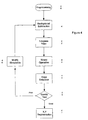
- Figure 4 there is illustrated, a system for performing video processing of images from a single video camera without markers on the subject in accordance with the present invention.
- Figure 4 illustrates a preferred grouping of algorithms or subsystems 41-47.
- the system illustrated in Figure 4 is implemented using PC or similar processing device executing software instructions stored in memory.
- PC or similar processing device executing software instructions stored in memory.
- implementations of the functionality described below, other than those illustrated in Figure 4 are possible as a matter of design choice.
- implementations need not be restricted to a software implementations; for example, dedicated hardware devices may be used to implement certain portions of the functionality described below.
- the processing illustrated in Figure 4 and described below is preferably carried out automatically upon reception of video image data.
- the video image data may be stored indefinitely, and the processing of Figure 4 carried out only upon command.
- Video image data from a single camera is received (via cable 31, for example) by a pre-processing subsystem 40.
- the video image data is color video data in a digitized and compressed format having a compression ratio of about 10.
- the camera 22 providing the video image data be positioned and fixed (so as to provide a static viewing area) at a location approximately 3 meters away from a gait path or other performance area to be used by the subject/patient.
- the pre-processing subsystem 40 performs conditioning steps necessary to place the video image data in a form suitable for continued processing.
- the pre-processing may include, but is not limited to, noise suppression (smoothing) filters, and color preprocessing (brightness/contrast adjustment, histogram stretch, decreasing the number of used colors, i.e., bit-per-pixel).
- noise suppression smoothing
- color preprocessing blueness/contrast adjustment, histogram stretch, decreasing the number of used colors, i.e., bit-per-pixel.
- interlaced fields may be used, rather than frames.
- the video image data output 47 by the pre-processing subsystem 40 is essentially a series of digitized images of the subject/patient, preferably performing one or more tasks such as walking, etc.
- Each frame output by the pre-processing subsystem 40 is provided to a quality subsystem 45 and a segmentation subsystem 47.
- the quality subsystem checks the quality of each image and modifies the various thresholds used in the processing if the quality is not acceptable.
- an image of just the background alone i.e., the area where the subject/patient is to be filmed, can be obtained by the system.
- This background-only image provides current background parameters.
- the calibration subsystem 46 then reads the environment parameters from a previous session for comparison with the current background parameters.
- Environment parameters include the color distribution model (illumination model) and camera parameters such as the lines or pixals, contrast sensitivity, etc. for a previous session.
- the enviroment parameters may also include a subject/patient's figure templates, i.e., data generally describing the subject/patient's dimensions and appearance.
- the successful video processing settings of the previous session are provided to the segmentation subsystem 47 for application to the image currently being processed.
- the processing parameters are chosen interactively applying varying combinations of processing tools to small portions of the images.
- segmentation refers to the process of creating binary (i.e., all pixels are either black or white) or gray-scale (i.e., all pixels assume a value somewhere between the extremes of black and white) images of the subject/patient.
- segmentation refers to the process of creating binary (i.e., all pixels are either black or white) or gray-scale (i.e., all pixels assume a value somewhere between the extremes of black and white) images of the subject/patient.
- edge detection processing can be performed to discern the edge's of the subject/patient's image. For example, edge detection filters combined with thresholding (in which all values above a certain threshold are deemed black or white, and all values below the threshold are deemed the opposite) may be used.
- edge detection filters in combination with edge tracing as taught, for example, by S.M. Smith in "Reviews of Optic Flow, Motion Segmentation, Edge finding and Corner Finding". Techn. Report TR97SMS1, Oxford Centre for Functional Magnetic Resonance Imaging of the Brain (FMRIB) Department of Clinical Neurology, Oxford University, Oxford, UK, 1997, the teaching of which are incorporated herein by this reference.
- background or environment parameters provided by the segmentation subsystem 47 may be used to subtract the background colors, leaving substantially little more than the subject/patient's image.
- the background parameters can be pixel color values, stored in a table, corresponding to various regions (preferably non-contiguous) within a given image. All pixels values in the image near or identical to the stored pixel values are then subtracted to remove the background content.
- the pixels of all images are first separated into a small number of groups according to features of images distinguishable by their color (using statistical criteria as taught, for example, by D. Comaniciu and P. Meer in "Robust Analysis of Feature Spaces: Color Image Segmentation", Techn.
- Figure 8 illustrates an example of a binary image resulting from a color image in which the background content has been removed in accordance with the teachings above, and which has been passed through thresholding.
- the binary or gray-scale images resulting from the binary processing 43 are provided to a boundary tracing subsystem 44 where the binary or gray-scale images are processed to provide only an outline or silhouette of the subject/patient in each image.
- a so-called x-silhouette is used for this purpose.
- An x-silhouette comprises all points of a binary or gray-scale image lying at the leftmost and rightmost points lying on a horizontal line through any section of the subject/patient's figure.
- boundary tracing is to take a given image, with or without background subtraction, and shrink the image by a known factor, i.e., by a factor of 4 to 5 thus producing an image which is 1/4 or 1/5 the size of the original image and rescaled back up to the original size using the same factor.
- the result is a reduce resolution image.
- this technique is used periodically, i.e., every 5 th , 10 th , etc. image.
- the boundary thus generated is then used as an approximation to the subject/patient's figure boundary. All image processing and boundary tracing procedures are then performed in a relatively small neighborhood of this approximate boundary. Additionally, the approximations may be used as a basis, along with a complete silhouette, for interpolating points otherwise missing from incomplete silhouettes.
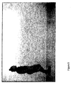
- a picture of the subject is shown in Figure 5 .
- the goal of the image processing is to remove the background and obtain a silhouette of the image.
- a second image exists in the system, taken without the subject in the picture. This image is referred to as the background image.
- the background is subtracted from the image of Figure 5 on a pixel by pixel basis. This means essentially that if each pixel in a given frame is designated as an element of a matrix Pixel[FRAME,I,J] where the pixel located in the I th row and the J th column of the frame FRAME then the subtraction output is obtained by performing the subtraction of Pixel[FRAME,I,J] - Pixel[REFERENCE,I,J].
- the result of the subtraction is then filtered using a low pass averaging 7x7 pixel filter. This operation produces the image seen in Figure 6 .
- the next step is to produce a binary image by setting a threshold such that all pixels values above the threshold are set to black and all values below the threshold are set to white, see Figure 7 .
- the next step is to perform edge detection on the binary image.
- the result of the edge detection is shown in Figure 8 .
- the output of edge detection is used to obtain couplets of data points in the x direction.
- the first member of the couplet is the start of the edge detected image in a given row and the second member of the couplet is the end of the edge detected image for the same row.
- X l [j][k] and X r [j][k] are the x-coordinates of the most left and right points respectively in the intersection of the subject/patient's figure on the i-th frame with the j-th row
- N rows is the number of rows in a fixed rectangle containing the union of the subject/patient's figures in all frames
- N frames is the number of frames.
- left and right x-velocities and x-acceleration matrices i.e. the matrices defined as the first and the second differences of the matrices X l [j][k] and X r [j][k] with respect to the k-th frame may also be defined.
- the silhouettes generated in this manner are combined as a pixel-wise conjunction or superposition of all silhouettes to provide a motion portrait.
- Img[1], Img[2], ... Img[N] be sequence of silhouettes generated as described above.
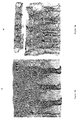
- An exemplary motion portrait, comprising a union of full silhouettes, is illustrated in FIG. 9 . As shown, each of the black pixels corresponds to an object in motion. It is understood that motion portraits may be constructed from x-silhouettes, y-silhouettes or full silhouettes and, in one embodiment of the invention, motion portraits from each type of silhouette are provided.
- the motion portraits thus provided may be used to detect anomalies or other artifacts in the subject/patient's performance.
- the motion portrait will typically exhibit a smooth appearance in that the motion-based pixels will be evenly distributed throughout the motion portrait.
- the motion portrait will often be characterized by clusters of motion-based pixels with noticeable frequencies.
- Figure 9a shows a motion "finger print" of a normal walk, which can be easily distinguished from the motion finger print of an abnormal walk Figure 9b .
- motion portraits can be processed by a motion characteristics calculation in the post processing stage.
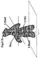
- motion characteristics include any qualitative measurements that may be calculated based on the motion portraits. For example, discrete motion characteristics such as step length, the duration of carrying out of stages of motion tests, characteristic frequencies of the motion (especially, of the gait tests) can be calculated. Head motion characteristics (i.e., the slope, the top-point trajectory) or, more generally, trajectories of characteristic points of the subject/patient's figure (i.e., the center of gravity, knees, heels etc.) may also be directly calculated from the motion portrait data.
- Typical data extracted from the x segmentation matrices, Figure 10 are the coordinate values of the subject/patient summit, the extreme left and right points of the outline at the hands level, and the spectrum of the subject/patient's instant velocity on a frame by frame or field by field bases.
- Typical values from the y segmentation include timing (initial contact, push off and middle swing instances for both sides, the step width, and the sagittal projection of sole-floor angle at some instances for both sides.
- Other information include the silhouette area, the coordinates of the arbitrary center of mass of the silhouette. Since all of the silhouette outlines are defined for each frame or field and because each frame or field represents an inherent clock timing a graph can be made showing the various parameters and the subsequent changes in these parameters over time.
- the oscillations of the parameters obtained from the x segmentation have extremes that in normal walking strictly correspond to temporal parameters revealed from the foot-floor contact events.
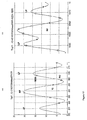
- the graphs in Figure 11 illustrate the above process.
- This graph shows the sagittal projection of the head summit motion vertical component.
- the X axis of the graph is the time reference in frames (ie 1/30 second per frame).
- the Y axis is a distance in pixels.
- Vertical motion is projected with a resolution of 1 pixel or 0.5 cm.
- the motion in the frontal plane is projected with a resolution of 1.5 cm.
- Markers in the graph show the various timing events on the curves.
- An asterisk marks the initial contact (IC), a square marks the initial push off (PO) and a circle marks the middle single support (MSS).
- RF is used to indicate the right step and LF is used to indicate the left step.
- the terminology of these events are common in the gait and balance laboratory.
- the left graph is a pattern of normal walking of a young man.
- the right graph in Figure 11 corresponds to walking of the same man but with his right knee mechanically restricted.
- Frontal motion of the summit causes asymmetry in the height of the peaks. Nearer to the camera side produces higher peaks. This difference expressed in centimeters is rather close to the width of the walking base. The difference between the minimums is much smaller because the lowest points of the summit is very close to the central (sagittal plane)
- the knee stiffness produces asymmetry in the minimum deepness.
- fourier transforms of all of the above motion characteristics, including discrete sine- and cosine- transforms, power spectrum, etc. transforms may be calculated, see Figure 12 .
- motion characteristics While not exhaustive of all motion characteristics that may be calculated based on motion portraits, may be used by a physician or other skilled practitioner to determine the existence of trends demonstrating improvement, deterioration or no change in a subject/patient's condition.
- previous data including, but not limited to, motion portraits and/or motion characteristics data from one or more previous sessions with a given subject/patient
- the previous data may also comprise normal data for the subject/patient, i.e., data corresponding to a time when the subject/patient was generally in good health.
- motion characteristics data may be compared against predetermined thresholds (for example, determined based on an averaging of values for a given characteristic in a large sample of similarly situated subject/patients) to assess the subject/patient's condition.
- predetermined thresholds for example, determined based on an averaging of values for a given characteristic in a large sample of similarly situated subject/patients
- video image processing may be combined with a task oriented examination and diagnostic protocol.
- An example of such a protocol is discussed hereinafter. Briefly, a series of patient tests or exercises are specified for performance by the subject. Physiological measurement, qualitative analysis of the responses, quantitative recording of vital signs, and video imaging are all recorded with a composite result being indicative of the health of the subject. The subject will be diagnosed with respect to illness or infirmity and with healthy subjects, a normal baseline will be established. Thus the following protocol is an example which may be varied or alternative protocols may be adopted.
- a system of motor tasks and a protocol for carrying them out should be defined such that the different environment in which they are performed, is taken into account.
- motion, gait and balance testing should take into account the following conditions: (a) as a rule, the nervous system of the subject/patient is disordered because of age and/or disease; (b) absolute safety, i.e.
- a zero probability of falling during testing is required because of the absence of assistance that could provide a guarantee against an accidental fall; (c) only remote oral and visual instructions, warnings and explanations are available for fall prevention; (d) no expensive gait analysis techniques can be effectively used; (e) testing procedures should performed quickly and easily; (f) only one Pan-Tilt-Zoom type video camera is used; and (g) the monitoring technique and comprehensive examination must reveal any kind of change in the medical condition of the subject/patient relating to his balancing ability.
- An audio-visual recording system and image processing is the preferred means for obtaining kinematic information resulting from performance of various tasks selected for a given subject/patient.
- only one video camera is required for this purpose; it's orientation, zoom, shutter, gain and white balance can be controlled remotely from the central station.
- other instruments may also be used, including an ECG instrument for indirect measurement of exertion or equipment to monitor quantitatively, e.g., blood pressure non-invasively before and after an exercise.
- additional video cameras may be used to monitor activities of the subject/patient or for comparative analyses and data acquisition from distinct orientations.
- test there are eleven (11) tests or groups of tasks.
- the tests are designed to enable checking all mechanical and nervous mechanisms of stability, and an exemplary set are listed in Appendix A with a more detailed explanation of each test set forth in Appendix B.
- the procedure of the examination is adapted to conditions of monitoring in the home without any physical assistance though it may be used in a hospital or personally monitored environment.
- Safety is ensured through the use of verbal instructions and warnings.
- many tasks have been modified or even discarded, e.g., stepping over an obstacle, transferring, etc.
- Other tasks standard tasks have been modified to provide acceptable conditions for audio-visual recording in restricted room sizes, e.g., free level walking, changing speed, abrupt stop etc. All tasks including dynamic perturbations of balancing in standing are self-initiated without any use of external forces.
- each task is grouped under a corresponding test according to the test identification number. Furthermore, within each group of related tasks, the tasks are generally ranked in order of increasing difficulty.
- the basic check is performed to provide an entire body of physical and neurological information relating to balancing ability of the given subject/patient. Given the relatively small number of tasks to be completed, it is anticipated that the basic check or protocol can be performed in 5 to 7 minutes.
- a definition may be arrived at according to either the predefined basic test scheme or a current decision of the practitioner.
- Quantitative data such as temporal and spatial parameters of a gait cycle (i.e., the motion characteristics described above), resulting from analysis of image data are instrumental, being obviously objective assessments of a subject/patient's state.
- Appendix C lists quantitative variables that may be measured, the types of units (if any) applicable to each variable, and a data type used to express the value of each variable.
- Appendix D first sets forth a list of Graded Results expressive of various qualitative variables.
- the Graded Scales list also set forth in Appendix D, shows the various ranges or scales of measurements relative to each qualitative variable. In this manner, observations by practitioner may be translated into relatively objective data that may be tracked in a manner similar to the quantitative data.
- the present invention proposes a technique, comprised of a specific approach to obtaining, analyzing and representing results; a set of tests mostly containing a subset of motor tasks; and a protocol of carrying out the examination.
- Results of the examination are divided into two parts: first, a level of difficulty of performance of a current motor task for a given subject/patient that immediately and objectively gives an initial approximation to a measurement of the motor ability of the subject/patient and, second, all other quantitative, graded qualitative results of measurements and image processing. Additionally, desired objective and fine monitoring are achieved by comparing session results with each other.
- the set of tests is complete enough to check all essential aspects of the ability to maintain balance in rest, through steady locomotion and maneuvers.
- the tests are groups of motor tasks arranged by their level of difficulty. This arrangement may be quite individual for a given subject/patient with a certain combination of nervous and physiological disorders.
- the tasks are safe and informative.
- the protocol of examination provides validity through the comparison of session results. This is attained through the standardization (for each subject/patient) of the procedures of implementation and conditions of audio-visual recording.
- the protocol is adapted to home conditions and the absence of an assistant: for safety, all maneuvers are preferably carried out near the wall, corner or other support where necessary. Because a health care practitioner at the central station deals only with objective facts concerning the performance of the tasks, they are not burdened with the interpretation of results. A more skilled practitioner can then perform off-line analysis of the data obtained during the examination.
- One example of a task and their assessment correlating qualitative, quantitative, and video image results follows:
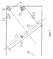
- One given test is produced by having the subject walk in a walk in a straight line perpendicular to the camera Figure 13 .
- the subject walks this path for a prescribed number of times. Each time the opposite side of the subject is close to the camera.
- the walk is filmed and transferred to the central station.
- the video images for each path are appended one to the other to obtain a longer time (i.e. more walking cycles) for statistical analysis.
- the image then goes through the preprocessing cycle to produce x and y segmentation matrices.
- Information pertaining to the gait and balance cycle for mid single support, push off, initial contact, see Figure 14 are extracted for each cycle.
- the information is used to produce a graph such as shown in Figure 11 .
- the graph is then analyzed to build a table of data as shown in Figure 15 .
- the data in figure 15 along with the segmentation matrices are stored in the database. Abnormal data causes an alarm message to appear on the screen.
- Camera settings front view, zoom in on the face and neck.
- Task 1002 O-ring fingers touching
- the patient sits in the armchair leaning to its back with hands on the arm-rests. He/ she rises one hand a little, approximately to a chest level and turn it so that all touches could be recognized by the camera.
- Task 1003 Pupils reaction to light
- the patient sits straight in the armchair with hands on the arm-rests looking directly at the camera.
- the patient sits straight in the armchair with hands on the arm-rests looking directly at the camera.
- Filming en face zoom in on the face so that stretched outside arms could be placed inside the frame.
- the patient sits in the armchair opposite to the camera.
- the arms are bent in elbows and lifted a little so that hands are on the chest level and forearms are horizontal.
- the index-fingers match one another.
- Filming en face zoom in on the chest so that stretched outside arms were inside the frame.
- Task 1006 Tremor while slitting
- Filming from the front zoom in on the lower part of body so that feet and hands were inside the frame.
- the patient sits at table or in the armchair with a clipboard on the knees and write or draw a certain text or a figure. Then he/she shows it to the camera.
- This set of motor tasks check a range of motion (ROM) in large joints and strength of main muscle groups associated with a gait and balance performance.
- the patient stands on the "walkway” with right side to the camera so that the sagittal plane is perpendicular to the camera axis. His/her left foot is lifted a little and left hand touches the wall or other support (a chair, quad-cane etc.).
- the patient stands on the "walkway" right side to the camera. He/she presses by long enough T-shape rod (quad-cane, swab, wiper etc.) on to right toe with all his/ her weight (or part of it) in order to load the forefoot.
- T-shape rod quad-cane, swab, wiper etc.
- Option 1 Bend your right knee as much as possible with slight bending of the hip. Keep it so for 1 - 2 sec. Lift the thigh as high as possible with knee bent. Keep it so for a second and try to straight the leg (extend the knee). Put the heel on the floor in front of you a little and try to extend it up to end, it is possible to press slightly the knee by the right hand. Stand straight.
- Options 3 and 4 Press the rod strongly with help of your trunk weight. Try to lift the toe. If that is impossible decrease the load by straightening out gradually. Fit the appropriate load and try to lift the toe. Repeat the exercise twice.
- Task 1102 checks a mobility in subtalar joints, 1006 checks rotation in hip joints and 1107 checks ability of gluteus maxims to lift the pelvic hemisphere with attached leg. The all three tasks need the same initial pose therefore it is reasonable to carry out them together.
- the patient stand with his back to wall 15 - 20 cm apart. He/she is supported by the elbows touching the wall and the forearms are near to be perpendicular to the wall.
- Tasks name options applied, options fulfilled and failure causes.
- Task 1102 Foot pronation (graded), Foot supination (graded).
- Task 1106 Hip rotation, external (graded), Hip rotation, internal (graded).
- Task 1107 Pelvic obliquity (one value).
- the patient stands back to the camera and face to a wall or any other support. He/she puts the both hand on the support on the chest or shoulders level.
- the camera axis is perpendicular to a coronal plane. Tee whole body is in the frame.
- Task 1108 Frontal bending of the trunk
- the patient stands straight on the "walkway" face to the camera with arms hanging free and feet with the shoulder size distance.
- Trunk bending (graded).
- the patient stands opposite to the camera on the "walkway" with his/her right side 'looking" at the camera.
- the arms are hanged free.
- the camera axis is perpendicular to the sagittal plane of the body.
- the whole body is in the frame.
- the task checks ROM of the neck movements at straight standing.
- the patient stands on the 'walkway" face to the camera with arms hanging free.
- the camera axis is perpendicular to the coronal plane. Zoom in on the face and neck.
- the chest, neck and head are in the frame.
- the patient sits in front of the camera in the armchair with a horizontal seat and relatively straight back.
- the feet are supported (reach the floor with whole area).
- the arms are on the knees or folded. The patient does not lean the chair-back or arm-rests.
- Task 1202 Sitting, the feet are unsupported
- the patient sits in front of the camera in the armchair with a horizontal seat and relatively straight back.
- the feet are unsupported (are in the air).
- the arms are on the knees or folded. The patient does not lean the chair-back or arm-rests.
- Task 1203 Standing with a cane in front
- This "tripod" is easier for balancing than bipedal standing and more difficult than sitting.
- the difficulty of the performance may be fitted by variation of a distance between the quad-cane and feet.
- the patient stands on the 'walkway" opposite to the camera slightly supporting on the cane located in front.
- Task 1204 Bipedal standing with a pelvic size base
- the patients stands on the "walkway", face to the camera with his/her legs parallel. Such distance between feet is the reference base. The arms are hanged free.
- Task 1205 Standing with two feet together
- Bipedal standing with a narrow base is more difficult than with a wide one.
- the patients stands on the "walkway", face to the camera with his/her legs close one to another. The arms are hanged free.
- Task 1206 Standing on the foot and tip-toe
- the foot _tip-toe base is an adjusting parameter changing the difficulty of the task performance.
- the patients stands on the "walkway", face to the camera, on the feet and opposite tip-toe.
- the arms are hanged free.
- Bipedal standing with a narrow base and decreased area is more difficult than previous task.
- the two tip-toes base is an adjusting parameter changing the difficulty of the task performance.
- the patients stands on the "walkway", face to the camera, on the two tip-toes.
- the arms are hanged free.
- Task 1208 Standing on the tip-toe and a cane alongside
- the cane_tip-toe base is an adjusting parameter changing the difficulty of the task performance.
- the patients stands on the "walkway", face to the camera, on the tip-toe and a cane alongside.
- the opposite foot is lifted a little and the arm is hanged free.
- the hand located on the tip-toe side slightly holds the cane.
- the height of the cane is fitted so that the patient stands straight.
- the patients stands on the "walkway", face to the camera, in the tandem pose.
- the arms are hanged free. If there is not confidence in patient's stepping strategy of balancing he/she must to stand in a corner or in the other place providing an emergent support from sides.
- Task 1210 Standing on the one foot
- feet strategy Standing on one foot is difficult because of asymmetry and decreased possibility to control the balance by means of a proprioceptive mechanism (so called, feet strategy).
- the patients stands on the "walkway", face to the camera, on the one foot.
- the opposite foot is lifted a little and the arm is hanged free.
- feet strategy Standing on one tip-toe is most difficult task because of asymmetry and absence of the possibility to control the balance by means of a proprioceptive mechanism (so called, feet strategy).
- the patients stands on the "walkway", face to the camera, on the one tip-toe.
- the opposite foot is lifted a little and the arm is hanged free.
- the so-called reach tests are good predictors of the occurrence of falls.
- the reach tests are rather dangerous without any assistance and must be carried out in a corner and/or near a wall to prevent fall during examination. Therefore, generally, the camera axis and reach direction are not perpendicular each to other and an error in definition of the reach distance is unpredictable. To avoid this inaccuracy the calibrated cylinder is put on the arm stretched out in reach direction during the examination.
- the ratio between reach distance and the cylinder is independent of angle of view and desired value can be obtained in absolute units (cm, inches etc.).
- the patient sits on a stool or in an armchair with a horizontal seat and low arm-rests with the feet lifted a little. He/she stretches out both arms forward and tries to reach the wall in front of him/her until stepping on the floor. The distance corresponding to a slow voluntary motion is measured.
- the patient sits on a stool or in armchair before the wall (or an other barrier) with the legs slightly lifted and arms stretched out forward and almost horizontally.
- the distance to the wall is 5 - 10 cm longer then maximal reach distance for the given patient.
- the patient is turned to the camera by side so that the mentioned above cylinder can be seen by the camera.
- An angle of view must be right as possible but deviation up to 30 degree are acceptable.
- the camera axis is perpendicular to the sagittal plane as possible. Whole body is in the frame.
- Task 1302 Sitting reach, lateral
- the patient sits on a stool or in an armchair with a horizontal seat and low arm-rests with the feet lifted a little. He/she stretches out both arms outside and tries to reach the wall in side of him/her until stepping on the floor. The distance corresponding to a slow voluntary motion is measured.
- the patient sits on a stool or in armchair in a corner or with a wall on the right side. His/her legs are slightly lifted and arms stretched outside almost horizontally. The distance to the walls is 5 - 10 cm longer then maximal reach distance for the given patient.
- the patient is turned to the camera by the face (or the back) that the mentioned above cylinders can be seen by the camera. An angle of view must be right as possible but deviation up to 30 degree are acceptable.
- the camera axis is perpendicular to the sagittal plane as possible. Whole body is in the frame.
- the patient stands before wall. He/she stretches out both arms forward and tries to reach the wall. The distance corresponding to a slow voluntary motion is measured.
- the patient stands before the wall (or an other barrier) with arms stretched out forward and almost horizontally.
- the distance to the wall is 5 - 10 cm longer then maximal reach distance for the given patient.
- the patient is turned to the camera by side so that the mentioned above cylinder may be seen by the camera.
- An angle of view must be right as possible but deviation up to 30 degree are acceptable.
- the camera axis is perpendicular to the sagittal plane as possible. Whole body is in the frame.
- Task 1304 Standing reach, lateral
- the patient stands in a corner, back to the camera, the arms stretched outside almost horizontally.
- the distance to the walls is 5 - 10 cm longer then maximal reach distance for the given patient.
- the patient is turned to the camera by the back, the mentioned above cylinders can be seen by the camera.
- An angle of view must be right as possible but deviation up to 30 degree are acceptable.
- the camera axis is perpendicular to the sagittal plane as possible. Whole body is in the frame.
- Resistance to mechanical perturbation may be assessed in home conditions only by self-perturbation methods when a fast shift of COG is created by means of relevant body movements. Although it is impossible to simulate the unexpected external disturbance such as a trip or collision in a crowded place but the self-induced disturbances occurring in everyday activity (bending, turning, transferring and so on) may be simulated. Moreover, only the self-induced shifts of COG reveal very important mechanism of anticipatory reactions to an arisen toppling force. Simulation of an external pushes requires a special equipment as a dynamic force plate and preventing falls assistance. Therefore it can not be realized under usual home conditions.
- the task with the difficulty level preceded to the difficulty level of the hardest for a given patient task is selected for an examination with a self-initiated mechanical perturbation.
- Task 1401 Self-initiated sway
- the patient swings himself with increasing amplitude until he/she will be forced to make a step.
- the eyes are closed.
- the patient stands with a distance from his back to a wall about 30 - 35 cm.
- the sagittal sway is measured relative to the foot length and the frontal sway is measured relative to a reference base (when the legs are parallel).
- Option 1 The patient stands in a corner, back to the wall and side to the camera, the arms are hanged free.
- the leg are parallel.
- the feet are parallel to the sagittal plane as possible. If only one foot can to be parallel to the sagittal plane then this side is turned to the camera.
- the distance to the wall is 35 - 40 cm.
- Option 2 The patient stands in the corner back to the camera and face to the corner.
- His leg are parallel. The distance to the corner is about 70 cm.
- the upper arms are close to sides of the trunk.
- the forearms are lifted with the palms "looking" forward. The pose is straight but without excessive tension.
- the palms provides support when the sway will exceed the limits of stability.
- Option 1 Filming from the side.
- the camera axis is perpendicular to the sagittal plane as possible.
- Whole body is in the frame.
- Option 2 Filming from the back.
- the camera axis is parallel to the sagittal plane as possible.
- Whole body is in the frame.
- Option 1 Close your eyes. Swing yourself around the ankle forward and backward without bending in the hip and knee (as a piece of log). Increase gradually the range of your oscillation until you will lean the wall or make a step forward.
- Option 2 Close your eyes. Swing yourself right and left around the feet without bending the trunk. Increase gradually the range of your oscillations until your palms will touch the walls.
- Tasks 1402 + 1403 COG shift caused by arm abduction and adduction
- the perturbation is initiated by abduction and adduction (lifting and lowering) of the arm.
- the patient stands in a corner, side to the wall and face or back to the camera, the arms are hanged free.
- the distance to the wall is 35 - 40 cm.
- Tasks name options applied, options fulfilled and failure causes.
- Task 1404 + 1405 Moving a gripped weight laterally and medially
- the possibility of stepping must be provided. If the patient stands on the one leg then a wall must be near to him on the supporting side and opposite arm is free for the movement. If the patient stands in bipedal pose then he/she must be in a corner back to the camera.
- the space between body and wall has to be about 40-50 cm. It is convenient to belt the forearm with soft gymnastic weight about 1 - 3 kg. This weight is a parameter of monitoring.
- the patient stands in a corner, side to the wall and face or back to the camera, the arm near the wall is hanged free.
- the other arm is bent in the elbow and hand is close to the side of chest
- the distance to the wall is 35 - 40 cm.
- Tasks name options applied, options fulfilled and failure causes.
- Tasks 1406 + 1407 COG shift caused by leg abduction and adduction
- the perturbation is initiated by abduction and adduction (frontal lifting and lowering) of the leg.
- the patient stands in a corner, side to the wall and face or back to the camera, the arms are hanged free.
- the distance to the wall is 35 - 40 cm.
- Tasks name options applied, options fulfilled and failure causes.
- a head motion with eyes closed very often causes a power disturbance in balancing despite of absent of any essential mechanical pushes. It is evident that this phenomenon is associated with a cerebellum functionality and therefore should be examined.
- the task with the difficulty level preceded to the difficulty level of the hardest for a given patient task is selected for an examination with a self-initiated non-mechanical perturbation.
- Task 1501 Balancing with head stretched up
- the perturbation is initiated stretching the head up with the eyes closed.
- the possibility of stepping must be provided. If the patient stands on the one leg then a wall must be near to him on the supporting side and opposite leg is free for stepping. If the patient stands in bipedal pose then he/she must be in a corner back to the camera. The space between body and wall has to be about 40 - 50 cm.
- the patient stands in a corner, side to the wall(-s) and face or back to the camera, the arms are hanged free.
- the distance to the wall(-s) is 35 - 40 cm.
- Tasks 1502 + 1503 Balancing with head thrown back and bowed
- the perturbation is initiated by throwing back and bowing of the head.
- the possibility of stepping must be provided. If the patient stands on the one leg then a wall must be near to him on the supporting side and opposite leg is free for stepping. If the patient stands in bipedal pose then he/she must be in a corner back to the camera. The space between body and wall has to be about 40 - 50 cm.
- the patient stands in a corner, side to the wall(-s) and face or back to the camera, the arms are hanged free.
- the distance to the wall(-s) is 35 - 40 cm.
- Tasks name options applied, options fulfilled and failure causes.
- Tasks 1504 + 1505 Balancing with head thrown back and bowed
- the perturbation is initiated by turning the head clockwise and counter-clockwise.
- the possibility of stepping must be provided. If the patient stands on the one leg then a wall must be near to him on the supporting side and opposite leg is free for stepping. If the patient stands in bipedal pose then he/she must be in a corner back to the camera.
- the space between body and wall has to be about 40 - 50 cm.
- the patient stands in a corner, side to the wall(-s) and face or back to the camera, the arms are hanged free.
- the distance to the wall(-s) is 35 - 40 cm.
- Tasks name options applied, options fulfilled and failure causes.
- Free level walking and walking in place are associated with balancing in motion when various dynamic perturbations as expected as well as random accompany the steady locomotion. Resistance to these perturbations is examined so.
- Such features as clearance, velocity, width of the base and especially its stability, characterize the capacity to adapt to an environment conditions. Limp and other distortions of normal gait pattern are associated with musculoskeletal and nervous disorders.
- the patient stands as straight as possible at a center of the "walkway" face to the camera for recording of his/her static pose. Then he/she turns side and back to the camera. So the three views of the posture are obtained and camera is tuned.
- the patient stands on a left edge of the "walkway" preparing to start with the certain leg.
- the leg is selected so that the patient will come in to the central zone with his/her right leg.
- the patient Before accomplishment of walking in place the patient stands as straight as possible at a center of the "walkway" face to the camera for recording of his/her static pose. Then he/she turns side and back to the camera. So the three views of the posture are obtained and camera is tuned.
- the patient stands as straight as possible at a center of the "walkway" face to the camera for recording of his/her static pose. Then he/she turns side and back to the camera. So the three views of the posture are obtained and camera is tuned. Then the patient stands at the same place preparing to start walking in place.
- Task 1701 Free walking in place with eyes closed
- the task checks the capacity to conserve a position while relatively simple and automatic movements are performing without visual support. Also balancing in frontal plane and synergy can be assessed.
- the patients stands on the "walkway” opposite to the camera, face or back to it. His/her coronal plane is directed along the "walkway”.
- Tasks name options applied, options fulfilled and failure causes.
- Task 1702 Free level walking with eyes closed
- the task checks the capacity to keep a direction while walking without visual support. Also a lack in automation and balancing problems can be revealed. The operator stops walking by command if an obstacle or a wall will be too near.
- the patient makes 1 - 2 trials with the eyes opened to set a reference points for the direction of walking and to estimate boundaries of the "walkway". Then he/she stands one end of the "walkway” in order to start with the closed eyes.
- Task 1703 Walking a line with normal step length
- the patients stands at the one end of the "walkway” with his/her sagittal plane along it.
- Tasks name options applied, options fulfilled and failure causes.
- Task 1704 Walking a line with arms folded and with normal stop length
- Tasks name options applied, options fulfilled and failure causes.
- Task 1705 Tandem waking
- Tandem walking is walking a line with a shortest steps length (a heel of the front leg is just after a forefoot of the rear leg). That is a complicated option of the task "Walking a line with normal step length” and checks the same features.
- the patients stands in tandem pose at the one end of the "walkway". His/her sagittal plane is directed along the "walkway”.
- Tasks name options applied, options fulfilled and failure causes.
- the test checks an ability of a voluntary changing of an initial steady locomotion.
- the time between an intention to go and the real implementation is defined as delay in starting of walking after the command "Go!.
- the number of steps made by the patient after the command "Stop! characterize his/her ability to interrupt a steady motion.
- the patients stands at the end of the "walkway” so to be visible to the camera. His/her sagittal plane is directed along the "walkway”.
- Task 1802 Changing speed
- the task checks an ability to decrease the speed voluntary.
- the patients stands at the end of the "walkway”. His/her sagittal plane is directed along the "walkway”.
- the task 1804 checks an ability to adapt the direction and speed to external conditions.
- the task 1906 relates to the test: "Single motor task”. It checks an ability to start and perform an isolated motor stereotype.
- Implementation of the both tasks is associated with a risk of "freezing" and/or dizziness.
- the rout of the patient is organized so that he/she is near a support while going around and turning, e.g. he/she goes between camera and the obstacle and turns to the wall after the obstacle, passes between the obstacle and the wall and stops after one - two steps. Then he/she do the task 1906 (turn 180E) beginning to revolve by his face to the wall and so having the possibility to support during all the time.
- the patients stands at the end of the "walkway”. His/her sagittal plane is directed along the "walkway”.
- An obstacle (a chair, walker or quad cane) stands at the center of the "walkway” a meter apart from a wall. If the "walkway" is a diagonal of the room it is required to reserve .2 m in the other place near a wall inside a field of view of the camera.
- Tasks name options applied, options fulfilled and failure causes.
- Task 1805 Stepping on drawn or appearing targets
- the task checks an ability to control the whole body in motion.
- the targets are rectangular drawn on a carpet (floor) or a light spot appearing in front of the patients. He/she tries to tread on the targets while walking. The number of the hits and quickness of performing are assessed. Also the secondary events of a balance loss may occur.
- the patients stands at the end of the "walkway”. His/her sagittal plane is directed along the "walkway”.
- a human nervous system and locomotor apparatus deal with unsteady transient processes while performing a single motor task. This condition helps to reveal additional disorders to those that manifested themselves in the examinations of a steady motion.
- the task checks presence of tremor without the visual support.
- the patients stands on the "walkway", the face to the camera and his/her coronal plane is perpendicular to the camera axis.
- the camera axis is perpendicular to the coronal plane of the patient. Zoom in on the patient's upper body but so that his/her stretched out arms and fingers were in the frame. The fingers must be in the focus.
Landscapes
- Health & Medical Sciences (AREA)
- Engineering & Computer Science (AREA)
- Life Sciences & Earth Sciences (AREA)
- General Health & Medical Sciences (AREA)
- Medical Informatics (AREA)
- Public Health (AREA)
- Biomedical Technology (AREA)
- Physics & Mathematics (AREA)
- Biophysics (AREA)
- Veterinary Medicine (AREA)
- Animal Behavior & Ethology (AREA)
- Surgery (AREA)
- Pathology (AREA)
- Heart & Thoracic Surgery (AREA)
- Molecular Biology (AREA)
- Dentistry (AREA)
- Oral & Maxillofacial Surgery (AREA)
- Physiology (AREA)
- Epidemiology (AREA)
- Primary Health Care (AREA)
- Computer Vision & Pattern Recognition (AREA)
- Multimedia (AREA)
- Radiology & Medical Imaging (AREA)
- Nuclear Medicine, Radiotherapy & Molecular Imaging (AREA)
- Theoretical Computer Science (AREA)
- General Physics & Mathematics (AREA)
- Human Computer Interaction (AREA)
- Neurology (AREA)
- Neurosurgery (AREA)
- Social Psychology (AREA)
- Psychiatry (AREA)
- Business, Economics & Management (AREA)
- General Business, Economics & Management (AREA)
- Measuring And Recording Apparatus For Diagnosis (AREA)
- Measurement Of The Respiration, Hearing Ability, Form, And Blood Characteristics Of Living Organisms (AREA)
- Medical Treatment And Welfare Office Work (AREA)
Description
- Related subject matter is found in
U.S. Patent No. 5,441,047 , andU.S. Patent No. 5,544,649 , the teachings of which patents are hereby incorporated by this reference. Additional related subject matter is found inU.S. Provisional Patent Application Serial No. 60/205,186 filed May 18, 2000 U.S. Provisional Patent Application Serial No. 60/205,279 - The present invention relates to techniques for monitoring the medical condition of a subject or subject/patient most especially neuromuscular motor activity, and, to a method and apparatus for monitoring a subject or subject/patient at a remote site from a central station by means of interactive visual, audio and data transmission communications. While the invention is also suitable for use in any situation where any subject or subject/patient is to be monitored at a site remote from a central station, an important application is the monitoring and caring for the elderly (both well and ill persons) in the home environment. Thus, the present invention can also be said to relate to the field of geriatric care. Further, the techniques disclosed may also be utilized in a hospital or clinic setting inasmuch as they facilitate diagnostic techniques useful for any person and at any local or remote location. Additionally the techniques and methods have application to other disciplines including, but not limited to, facilitating psychological diagnosis and monitoring, and interrogation monitoring and analysis. Finally, the invention enables conduct of physiological analysis utilizing a single video camera and without markers.
- Most of the resources for modem medicine have been invested in the development of highly sophisticated hospital facilities. Therefore, institutional patient care has become prohibitively expensive, in many cases overused and for a substantial number of patients potentially harmful. The tendency to substitute costly institutional patient care with effective and cost containing extra-institutional, ambulatory medical facilities is gaining rapid momentum. These attempts however are still confined to the delivery of care in outpatient clinics to which the patient has to come to obtain medical care, or home nurse visits which are short, scarce, and insufficient. The combination of growing sophistication in ambulatory monitoring technology together with the explosive development of telecommunication provides the ideal substrate to enable the development of highly sophisticated, reliable and affordable, remotely controlled, remote monitoring capabilities which can monitor, analyze and assess many physiological parameters in any potential subject and in any possible location. Such a system is specially suited to provide a sophisticated platform or home care facilities for a wide spectrum of subject/patient.
- The described invention is a neuromuscular and motor activity remote monitoring system for any person in need of remote assessment of physiological, psychological, and other parameters. From the subject/patient care aspect one of the important applications of this system is monitoring and care of the elderly geriatric population. This is due to the complexity of the holistic approach to geriatric care, which will be elaborated below.
- Modem society with its improvement in living conditions and advanced health care has brought about a marked prolongation of life expectancy. This change has resulted in a dramatic and progressive increase in the geriatric population. A large percentage of the geriatric population needs continuous general, as well as medical, supervision and care. For example, supervision of daily activities such as dressing, personal hygiene, eating and safety as well as supervision of their health status is necessary. Furthermore, the relief of loneliness and anxiety is a major, yet unsolved, problem to be addressed. These and other facets of the management of the ever increasing geriatric population have yet to be successfully addressed and solved.
- The creation of retirement facilities and old age homes, as well as other geriatric facilities, provide only a partial solution to the problems facing the geriatric population. The geriatric population, a constantly increasing fraction of society, has become increasingly dependent upon the delivery of home health and general care, which has its own set of challenges and drawbacks.
- The notion of ambulatory (home environment) subject/patient care is gaining increased popularity and importance. This shift in subject/patient care from the "sheltered" institutional milieu to the subject/patient's home, work place, or recreational environment is due primarily to a radical change in concepts. That is, specialists in geriatric care now tend to keep the aged in their own natural environment for as long as possible.
- Except for scarce model organizations, home care is still carried out either by the subject/patient's family or by nonprofessional help, or, in the usual circumstance, by professional, highly trained personnel at very significant expense. The monitoring equipment at home care facilities is usually minimal or nonexistent, and the subject/patient has to be transported to the doctor's office or other diagnostic facility to allow proper evaluation and treatment.
- Subject/patient follow-up is presently done by means of home visits of nurses which are of sporadic nature, time consuming and generally very expensive. A visiting nurse can perform about 5-6 home visits per day. The visits have to be short and can usually not be carried out on a daily basis. Moreover, a visiting nurse program provides no facilities for continuous monitoring of the subject/patient and thus no sophisticated care, except in fortuitous circumstances, in times of emergency. The remainder of day after the visiting nurse has left is often a period of isolation and loneliness for the subject/patient. The existing home care nursing facilities divert skilled nurses, a scarce commodity, from the hospital environment and uses them in a highly inefficient manner due to the wide dispersion of the subject/patients and the lack of sophisticated diagnostic facilities in the subject/patient's home. Clearly, the practice of visiting nurses leaves much to be desired.
- These considerations apply to the general population as well, as the spiraling cost of hospital care has lead to a dramatic increase in the use of outpatient care as a treatment modality.
- Additional facts support development of an improved home health care system especially for a geriatric population. In particular, falls are a major health problem among the elderly, causing injury, disability and death. One third (some studies suggest half) of those over the age of 65 suffer at least one fall each year. The rate of falling increases to 40% among those who exceed the age of 80. According to the National Safety Council, falls accounted for one-third of the death total for the elderly. Those who survive falls may have restricted activity, soft-tissue injuries, or fractures. It is estimated that up to 5% of falls by elderly persons result in fractures. A similar percent result in soft-tissue injury requiring hospitalization or immobilization for an extended period. It is estimated that hip fractures resulting from falls cost approximately $2 billion in the United States during 1980. Falls are mentioned as a contributing factor to admissions to nursing homes.
- The factors leading to falls can be divided into two main groups: environmental factors and medical factors. In spite of the difficulty in the surveillance of subject/patient condition before a fall, almost all researchers share the conclusion that environmental hazards are decreasingly important in causing falls as age increases. A clear correlation between clinical or medical problems and the incident of falls by the elderly has been established. Many of these medical problems of the elderly or infirm can be detected by simple clinical observation. For example gait and balance abnormality may indicate difficulty with neurologic and musculoskeletal functions that may contribute to physical instability. Changes in gait can be identified by the following: slow speed, short step length, narrow stride width, wide range of stepping frequency, a large variability of step length, and increasing variability with increasing frequency.
- Thus, there are relatively straightforward techniques that facilitate diagnosis of a predisposition or likelihood of falls among the elderly.
U.S. Patent Nos. 5,441,047 and5,544,649 ("the '047 and '649 patents") disclose inexpensive procedures for facilitating such diagnoses or investigating such predispositions in a large subject/patient population wherein the kinematic condition of the subject/patient can be investigated or where the appearance, and reflex activity of the subject/patient can be investigated with ease. In particular, the '047 and '649 patents describe an ambulatory (in the home) subject/patient health monitoring system wherein a health care worker at a central station monitors the subject/patient, while the subject/patient is at a remote location. The subject/patient may be a person having a specific medical condition being monitored or may be an elderly person desiring general medical surveillance in the home environment. Video transmission cameras are provided at the subject/patient's remote location and at the central station such that the subject/patient and the health care worker are in interactive visual and audio communication. A communications network such as an interactive cable television is used for this purpose. Various medical condition sensing and monitoring equipment are placed in the subject/patient's home, depending on the particular medical needs of the subject/patient. The subject/patient's medical condition is measured or sensed in the home and the resulting data is transmitted to the central station for analysis and display. The health care worker then is placed into interactive visual communication with the subject/patient concerning the subject/patient's general well being, as well as the subject/patient's medical condition. Thus, the health care worker can make "home visits" electronically, twenty-four hours a day, seven days a week in a non-intrusive, cost effective, privacy protecting manner. - While the '047 and '649 patents (incorporated herewith by reference) represent an improvement over prior art techniques, additional testing methods are needed to properly and fully exploit the opportunities provided by remote diagnosis facilitation systems as well as hospital or clinic site diagnosis. The common approach to functional testing of the motor ability of elderly persons is based on scoring the performance of complex functional tests such as "Get Up and Go", "Fregley Ataxia Test Battery" and others. The resulting score is used for estimating the risk of falling, mainly, for purposes of epidemiological studies. Each test consists of a set of simple motor tasks. For clinical decision-making, physicians generally take into account the results of the separate tasks, instead of the resulting score of the whole test. It is known that the ability to perform a simple motion task is directly connected with concrete medical conditions of a subject/patient whereas the resulting score only provides a general impression. These tests very often require special equipment (e.g., static and dynamic force plates with or without feedback, markers on specific body locations, and 6 to 10 meters of walkway etc.). Moreover these tests require the presence of an assistant to guide the test and insure the accuracy and safety of its performances. As a rule, such testing can only be realized in a clinic that has the required equipment and a sufficiently skilled staff. Likewise, such testing is too expensive for everyday practice, usually cannot be repeated as often as necessary and therefore cannot be used to perform thorough monitoring of elderly subject/patients with restricted capacities to visit clinics.
-
U. S. Patent 5,980,429 ("the `429 patent") describes a monitoring technique for training programs where effectiveness of training is assessed through a thorough comparison of quantitative and qualitative measured parameters with quantitative and qualitative benchmark data. Qualitative parameters relate to the accuracy of performing the tasks and quantitative parameters to a number of successive performances in a certain period of training. To obtain a sequence of qualitative benchmark data, training tasks are arranged by level of difficulty of performance. The measured results of a given task are categorized according to the completeness and accuracy of its performance. Thus a system for objective assessment is realized. Benchmark data are defined from previous subject's performances or from data derived from corresponding reference groups. The "New Balance Master" test for assessment of motor and balance ability of a subject/patient described in the '429 patent is a typical example of a well- designed clinical test. The training program is intended for persons with an intact central nervous system, i.e., can only be applied in specific injuries. Moreover, the assessment always relates to a person that does his hardest exercises on his own highest level. Hence the arrangement of the motor tasks by degree of difficulty may be adequate even if the accuracy of the assessment is restricted by dividing the results of measurements over 3 to 5 categories. - However, an elderly subject performing tasks in accordance with such testing schemes, without any assistant, will mostly carry out the tasks on a far lower level than his or her maximum abilities. So, the tasks should be designed with a degree of difficulty but free from essential muscle tension and unstable poses that could lead to a fall. Moreover, nervous and physiological disorders, often present in elderly persons, necessitate individual tailoring of the difficulty level of the tests. Taking into account the fact that in elderly persons many essential disorders, associated with motor difficulties, evolve very gradually, the monitoring system should be able to detect small trend changes in motor behavior of the subject/patient.
- Thus, it can be seen that techniques leveraging the availability of video image data, i.e., that build upon and improve the teachings of the '047 and `649 patents, would be an important advancement of the art. Furthermore, testing methodologies, including testing contents and protocols should take advantage of these advances, thereby maintaining and improving the diagnostic reliability of current medical techniques.
- The present invention is directed to improvements to the interactive video and audio subject/patient monitoring system disclosed in the '047 and `649 patents by providing a video processing technique whereby motion characteristics are readily discerned from video image data of a subject/patient. (The term "subject/patient" as used herein is to be interpreted broadly to include elderly persons, persons actively being treated or monitored for specific medical ailments, as well as persons who need to have their general medical condition and gait and balance status monitored by practitioners for any reason, for example: astronauts in space stations etc. Additionally, persons who are being monitored for psychological condition, mood, truth telling and general physical condition are considered subject/patients. By way of example, but not limitation, interrogation techniques which rely upon physical response to evince truthfulness or falsehood may utilize variations of the techniques disclosed herein along and in combination with other physical response measurements such as respiration, heart rate, etc.) Furthermore, there is provided a system of motor tasks and protocols for their execution that is particularly adapted for use in the interactive television and audio monitoring system as well as being useful at a hospital or clinic site where clinicians work directly with subject/patients or any other remote monitoring scenario. An example of a series of tests for facilitating diagnosis is disclosed wherein the tests are susceptible of qualitative, quantitative and/or image processing review and analysis. The term "medical" herein is broadly inclusive of physical, psychological and behavior conditions by way of example.
- The image processing techniques may enable diagnosis associated with skeletal and muscular movement in a highly quantitative and recordable manner. Thus, the video image of the patient performing each of the tasks in the protocol may be captured by a video acquisition system. The video image data is received in the form of a sequence of equally spaced (in the time domain) images. The acquired images may then be compressed using standard image compression techniques such as that defined by MJPEG compressed images in order to reduce the data content to allow storage on a local disk or to allow transmission to a remote location. Unlike neuromuscular motor activity measured in standard gait and balance laboratories which require a multitude of cameras and require that markers be placed on the patient, the present invention, without limiting it, may produce adequate results with the use of a single camera and without the use of markers.
- A pre-processing element follows the image acquisition. The purpose of the preprocessing element is to remove the background and produce a sequence of silhouette images or outline images of the patient on a frame-by-frame or field-by-field basis by applying a sequence of standard image processing operations on the acquired images. The image processing operations, well known in the art, may include contrast control, brightness control, segmentation and edge detection. The pre-processing sequence of operations may be manually defined by an operator of the system or automatically discovered through the use of quality indicators and feedback to search for the best set of operations.
- The pre-processing may be performed at the central location or at the remote location in a distributed system. The result of the pre-processing may be a plurality of matrices (one per frame or field of the original video image sequence) containing the silhouette or outline image. In the case where the pre-processing is performed at the patient location, the resultant pre-processed file may be transferred to the central facility for continued post processing.
- A post-processing element may follow the pre-processing. The purpose of the post-processing is to make measurements of various parameters of the silhouette or outline of the patient on a frame-by-frame or field-by-field basis. The aforementioned parameters or mathematical combinations of these parameters may represent physiological indices associated with gait and balance as with other neuromuscular motor activities. An example of a measured parameter in the space domain is the maximum height that a patient's foot is raised from the floor. An example of a measured parameter in the time domain is the time it takes a patient to complete a cycle while walking.
- The results of the measurements from the post-processing may be stored for long-term monitoring and trending. The system may provide for the definition of predefined normal value ranges for the results as well as adaptive individual normal value ranges based on the historical data from a particular patient An alarm notification may be provided when a value falls outside the expected normal range.
- A graphical "finger print" of the patients walking pattern may be obtained by summing the individual matrices provided from the output of the pre-processing and storing the result in a resultant matrix. This is similar to placing each outline from a single frame onto a transparency and placing the transparencies one on top of the other. The resulting picture provides a unique characteristic template of the patient that may be analyzed and compared to previously stored templates of the patient. Changes from the normal historical pattern represent changes in the neuromuscular motor activity of the patient that may be indicative of physiological problems.
- Each protocol that may be used to test the patient is an action or a plurality of actions that the patient must perform. Some of the protocols such as walking in a straight line or walking in place produce direct physiological protocols produce the indirect physiological information such as the ability to complete a sequence of balance tasks each of which are subsequently more difficult resulting in a numerical grade. The may be combined tests may be designed to elicit all necessary physical and neurological information relating to a patient's ability to perform neuromuscular motor tasks. The tasks may be particularly chosen to maximize safety and to minimize the probability of the patient falling down or otherwise losing balance when performing the tasks. To this end, tasks may be preferably ordered by difficulty within certain tests. The level of difficulty associated with the said task may thereby offer an inherent qualitative measure. This qualitative information quantized by the degree of difficulty may be stored together with other quantitative results. The use of degree of difficulty may have particular relevance in rehabilitation.
- The invention is defined in
claims - Any embodiment which is in contradiction to the subject-matter to one of these claims is not part of the invention.
- These and other advantages and features of the subject invention will become apparent from the detailed description of the invention that follows.
- In the detailed description of presently preferred embodiments of the present invention which follows, reference will be made to the drawings comprised of the following Figures, wherein like reference numerals refer to like elements in the various views and wherein:
-
Figure 1 is a simplified, overall functional block diagram of the system -
Figure 2 is a schematic diagram illustrating one possible configuration of the home system -
Figure 3 is a schematic diagram illustrating one possible configuration of the central station.. -
Figure 4 is a flowchart illustrating a method for video image processing in accordance with the present invention; -
Figure 5 is an exemplary raw video image prior to processing -
Figure 6 is an exemplary image after background subtraction and filtering -
Figure 7 is an exemplary binary image in accordance with the present invention; -
Figure 8 is an exemplary x-silhouette in accordance with the present invention; -
Figures 9A and 9B are an exemplary motion portraits in accordance with the present invention; and -
Figure 10 is a drawing showing the various parameter measured from the segmentation data. -
Figure 11 is an exemplary graphical representation of the outcome of walking in a straight line. -
Figure 12 is an exemplary graphical representation of the spectral density of walking in a straight line. -
Figure 13 is an exemplary home layout for walking in a straight line. -
Figure 14 is an exemplary drawing showing the sequence of events in a walking cycle. -
Figure 15 is an exemplary report showing the values obtained from one test based on walking in a straight line. - The system of the present invention consists of two distinct entities
Figure 1 , one is a subject/patient unit, located in the area where the subject/patient performs the measurement, this unit is also typically referred to as thehome unit 11 since it is placed in the home of the subject/patient in one of the preferred embodiments and a second entity, acentral unit 12 located in the area where the medical practitioner administrates the conduction of the measurements and analyzes the results. The two entities are connected via awideband communication channel 13. This configuration allows for the central unit to be separated from the home unit. It also allows for a single central unit to communicate either serially or simultaneously with more than one home unit. In both of these instances a public carrier provides the communication channel where the Internet is one of the possible means of communications. The configuration also allows for local use such as in a clinic or hospital where the communication channel may be a local area network. It further allows for very remote use such as the possible to communicate between a ground control and an orbiting space shuttle. - The system of the present invention uses or incorporates relatively inexpensive home medical monitoring equipment that includes one or more cameras. When used as part of a home monitoring system it may include additional sensors and measuring devices for the particular physiological/medical parameters to be monitored. The subject/patient's remote location/home equipment is simple to use and modular to allow for the accommodation of the monitoring device to the specific needs of each subject/patient. To reduce production costs and to avoid complex maintenance problems, the remote home unit includes the absolutely needed components of the measuring device while most of the sophisticated elements are located in the central unit. The raw data, including video image data, is transmitted to the central station, which includes all of the needed sophistication to allow for the storage, transformation, display and interpretation of the data. The need for expensive equipment in the remote location/home is thus avoided.
- The central station includes a computer-based multi-channel data analysis and display unit that enables the interpretation, display, and storage of the transmitted data. This central station is preferably equipped with alarm mechanisms to alert the staff to any aberration from the expected. The central station further includes apparatus for the communication of data to all authorities involved in the wide spectrum of the subject/patient's needs, e.g., emergency care agencies, the subject/patient's physicians, nursing services, social workers, etc.
- The central station is preferably provided with the capability of automatically scanning predesignated subject/patient remote/home units at predetermined intervals to provide continuous supervision of specific parameters. In some instances, the central station may monitor continuously one or more parameters, e.g., ECG, blood pressure, respiration, etc. The embodiment disclosed enables one highly trained nurse or subject/patient-monitoring personnel located at the control center to supervise and monitor as many as 50 subject/patients either seriatim or substantially simultaneously. Whereas a visiting nurse may only be able to visit 5 or 6 homes per day in person, a nurse at the central station may be able to visit 5 or 6 subject/patients per hour by making electronic "home visits".
- Any broadband communication system is suitable for the bi-directional real time contact with the subject/patient in the remote location/home. One such medium is cable television that provides an already existing, widespread and highly suitable system via cable modems for interactive visual communication with most residential units in densely populated urban areas. The ambulatory subject/patient monitoring system integrating the latest advances in biomedical technology with cable television or any other available and suitable communication system provide safe and accurate general and medical supervision for the geriatric/homebound population in their own, natural environment.
- It will be appreciated that as advances in telecommunications develop, other techniques for transmission of video signals between a central station and the home may be desirable and economically feasible. For example, satellite and radio transmission of the video signal and/or monitored medical data, or transmission via modem through the telephone lines, may also prove satisfactory. In due time the system will possible using the internet or similar computer networks as it's main communication system both for the data transmission to and from the remote location/home as well as the use of the database by other authorities such as physicians insurers, government agencies, etc.
- A typical embodiment of the home system is shown in
figure 2 . Asingle camera 22 is used to take a motion picture video of the subject/patient 21. The camera has a wide angle and provides for remote control from the central station for pan, tilt, zoom as well as other controls such as brightness and contrast. Typically the camera will provide 30 frames per second and transmit composites or supervideo signal in NTSC standard to the video acquisition 23. The video acquisition will acquire the analog video signal and convert it to a digital Y Cr and Cb values. The Y Cr and Cb values are then passed through a video compression component that compresses the video. The compressed video may be locally stored in adisk 24 for later transmission, this is necessary in the case that the desired communication bandwidth is not appropriate to transfer the compressed video in realtime or directly transferred as part of an integrated video-conferencing system 25. In addition the integrated video-conferencing system 25 provides for interactive audio-visual interactions with the central station so that the subject/patient may see and speak with the medical practitioner on thehome TV set 26 and the medical practitioner may see the subject/patient on the center monitor. The interactive communication is an important aspect of the system in that it allows for the medical practitioner to instruct the subject/patient to perform the testing and to monitor the subject/patient while the tests are being performed. It should be noted that the video quality required for monitoring the subject/patient may be less than the video quality required for the automated neuromuscular motor analysis of the patient. Video conferencing may be performed using either standard H.323 compression or using more proprietary compression techniques such as wavelet compression. The audio signal may be digitized and received and transmitted in either a compressed or non-compressed format. Should the video be stored locally as a file ondisk 24, it would transmitted to the central station as a file using common communication transfer protocols such as TCP/IP to insure integrity. The acquisition, storage and video-conferencing may be implemented using a standard PC, in this case the various elements are implemented as PCBs which reside on the bus of the PC. The control program and the video conferencing application run under the operating system of the PC. An alternative implementation would be to use an embedded system with a microprocessor and DSP to perform the acquisition, compression and video-conferencing functions. Communications is provided by an interface to a standard communication modem. This may be a cable modem, xDSL, ISDN or other broadband communication modem. - A typical embodiment of the nurse station is shown in
figure 3 . Images arrive into the central station from thecommunication system 31 and are stored on alocal disk 32. This element also provides for video-conferencing with the home system. The on-line images are seen in a window on thelocal display 33. Images stored on thelocal disk 32 are then preprocessed by apreprocessing application 34. The preprocessing application removes background information, obtains an outline or silhouette of the subject/patient on a frame by frame or field by field bases and performs x and y segmentation of the images. The results of the preprocessing are stored in an x and y segmentation file of the image 35. The x and y segmentation files may be used to obtain a fingerprint of the image figure xx or may be used forpost processing 36. Post processing transforms the results of the x and y segmentation to meaningful quantitative and qualitative information as a function of the specific test which was performed. Post processing are the set of algorithms used to extract the desired data for the x and y segmentation and to perform the necessary manipulation of this data based on the features which are desired to extract from the data. The post processed data is stored in adata base 37 together with other patient information. This data becomes part of the historical record of the patient and may be used to compare present data with past data, trigger alarms as seen on adisplay window 38 when any parameter resides outside the expected range and be used by reportingapplications 39. Owing to the fact that the data resides in a database the data may be queried by other application programs which may run either on the local system or on a remote system which communicates with the data base. An example of the latter is a system running at a doctors office which may monitor the results of a particular patient. - While an important aspect of the described techniques is remote site monitoring, the protocols described hereinafter have significantly broader application and utility. That is, the described image monitoring may be conducted at any site including in hospital or at a clinic. In every circumstance, the images and other data are recorded, analyzed, stored or retained and compared with historic information or standard information. The practice of the described image processing and protocols at a hospital site will, for example, be a useful tool for facilitating diagnosis by the medical practitioner. Thus, the described invention, though detailed in the context of remote monitoring, is equally applicable for on-site monitoring of subject/patient condition, both ill and well, and is an important tool for facilitating diagnosis for on-site use.
- The prevent invention utilizes a multifaceted approach to facilitating medical diagnosis relying upon the interactive technology heretofore described and the technology described in various co-pending applications. An important feature of such multifaceted approach to facilitating diagnosis and a principal feature of the present invention, is video image processing of a patient to facilitate analysis of gait, balance, skeletal condition, muscle condition and coordination and other physiological features and conditions. Briefly, video images of the outline of the body or a part of the body are recorded and analyzed either by a trained physician or nurse or by machine analysis in an effort to detect (a) a base line of performance or condition (2) variation from norms (3) changes over time (4) changes under stress, and (5) changes in emergency conditions. The imaging techniques may be utilized by themselves or in combination with other techniques that facilitate diagnosis, including traditional techniques, as well as test techniques described herein. The images are, in the system described, obtained by using video camera input signals obtained from the subject/patient location. However, the general techniques of image analysis are not restricted to such a monitoring system. Thus, they may be used, for example on site at a hospital.
- Referring now to
Figure 4 , there is illustrated, a system for performing video processing of images from a single video camera without markers on the subject in accordance with the present invention. In particular,Figure 4 illustrates a preferred grouping of algorithms or subsystems 41-47. In one embodiment of the present invention, the system illustrated inFigure 4 is implemented using PC or similar processing device executing software instructions stored in memory. Those having ordinary skill in the art will recognize that various implementations of the functionality described below, other than those illustrated inFigure 4 , are possible as a matter of design choice. Furthermore, implementations need not be restricted to a software implementations; for example, dedicated hardware devices may be used to implement certain portions of the functionality described below. Finally, the processing illustrated inFigure 4 and described below is preferably carried out automatically upon reception of video image data. Alternatively, the video image data may be stored indefinitely, and the processing ofFigure 4 carried out only upon command. - Video image data from a single camera is received (via
cable 31, for example) by apre-processing subsystem 40. Preferably, the video image data is color video data in a digitized and compressed format having a compression ratio of about 10. In practice, it is preferred that thecamera 22 providing the video image data be positioned and fixed (so as to provide a static viewing area) at a location approximately 3 meters away from a gait path or other performance area to be used by the subject/patient. Thepre-processing subsystem 40 performs conditioning steps necessary to place the video image data in a form suitable for continued processing. For example, the pre-processing may include, but is not limited to, noise suppression (smoothing) filters, and color preprocessing (brightness/contrast adjustment, histogram stretch, decreasing the number of used colors, i.e., bit-per-pixel). As one alternative, interlaced fields may be used, rather than frames. The videoimage data output 47 by thepre-processing subsystem 40 is essentially a series of digitized images of the subject/patient, preferably performing one or more tasks such as walking, etc. - Each frame output by the
pre-processing subsystem 40 is provided to aquality subsystem 45 and asegmentation subsystem 47. The quality subsystem checks the quality of each image and modifies the various thresholds used in the processing if the quality is not acceptable. - Thus, prior to filming the subject/patient, an image of just the background alone, i.e., the area where the subject/patient is to be filmed, can be obtained by the system. This background-only image provides current background parameters. The
calibration subsystem 46 then reads the environment parameters from a previous session for comparison with the current background parameters. Environment parameters include the color distribution model (illumination model) and camera parameters such as the lines or pixals, contrast sensitivity, etc. for a previous session. The enviroment parameters may also include a subject/patient's figure templates, i.e., data generally describing the subject/patient's dimensions and appearance. In the case where the previous and current environment parameters are very similar (as would be the case, for example, where the subject/patient always performs the testing at the same location), the successful video processing settings of the previous session are provided to thesegmentation subsystem 47 for application to the image currently being processed. In the case of a new subject/patient or of essentially different previous and current environment parameters or inconsistent results of the test processing with the setting of the previous session, the processing parameters are chosen interactively applying varying combinations of processing tools to small portions of the images. - The parameters selected by the
calibration subsystem 46 and the video image data from the pre-processing subsystem 41-45 are provided to thesegmentation subsystem 47. In the context of the present invention, segmentation refers to the process of creating binary (i.e., all pixels are either black or white) or gray-scale (i.e., all pixels assume a value somewhere between the extremes of black and white) images of the subject/patient. To this end, a variety of techniques may be employed. One technique, edge detection processing, can be performed to discern the edge's of the subject/patient's image. For example, edge detection filters combined with thresholding (in which all values above a certain threshold are deemed black or white, and all values below the threshold are deemed the opposite) may be used. Alternatively, edge detection filters in combination with edge tracing as taught, for example, by S.M. Smith in "Reviews of Optic Flow, Motion Segmentation, Edge finding and Corner Finding". Techn. Report TR97SMS1, Oxford Centre for Functional Magnetic Resonance Imaging of the Brain (FMRIB) Department of Clinical Neurology, Oxford University, Oxford, UK, 1997, the teaching of which are incorporated herein by this reference. - As noted above, background or environment parameters provided by the
segmentation subsystem 47 may be used to subtract the background colors, leaving substantially little more than the subject/patient's image. Thus, the background parameters can be pixel color values, stored in a table, corresponding to various regions (preferably non-contiguous) within a given image. All pixels values in the image near or identical to the stored pixel values are then subtracted to remove the background content. In yet another alternative, the pixels of all images are first separated into a small number of groups according to features of images distinguishable by their color (using statistical criteria as taught, for example, by D. Comaniciu and P. Meer in "Robust Analysis of Feature Spaces: Color Image Segmentation", Techn. Report, Rutgers University, Piscataway NJ, 1997, the teachings of which are incorporated herein by this reference). Subsequently, the images are binarized using one of the above mentioned methods, e.g., edge detection and thresholding. Finally, combinations of the above techniques may be used.Figure 8 illustrates an example of a binary image resulting from a color image in which the background content has been removed in accordance with the teachings above, and which has been passed through thresholding. - The binary or gray-scale images resulting from the binary processing 43 are provided to a boundary tracing subsystem 44 where the binary or gray-scale images are processed to provide only an outline or silhouette of the subject/patient in each image. In one embodiment, a so-called x-silhouette is used for this purpose. An x-silhouette comprises all points of a binary or gray-scale image lying at the leftmost and rightmost points lying on a horizontal line through any section of the subject/patient's figure.
- Another alternative method for boundary tracing is to take a given image, with or without background subtraction, and shrink the image by a known factor, i.e., by a factor of 4 to 5 thus producing an image which is 1/4 or 1/5 the size of the original image and rescaled back up to the original size using the same factor. The result is a reduce resolution image. By subtracting the reduced resolution image from the original image, a good approximation of the subject/patient's boundary is provided. In one embodiment of the present invention, this technique is used periodically, i.e., every 5th, 10th, etc. image. The boundary thus generated is then used as an approximation to the subject/patient's figure boundary. All image processing and boundary tracing procedures are then performed in a relatively small neighborhood of this approximate boundary. Additionally, the approximations may be used as a basis, along with a complete silhouette, for interpolating points otherwise missing from incomplete silhouettes.
- The following is an example of the use of the above methods. A picture of the subject is shown in
Figure 5 . The goal of the image processing is to remove the background and obtain a silhouette of the image. A second image exists in the system, taken without the subject in the picture. This image is referred to as the background image. The background is subtracted from the image ofFigure 5 on a pixel by pixel basis. This means essentially that if each pixel in a given frame is designated as an element of a matrix Pixel[FRAME,I,J] where the pixel located in the Ith row and the Jth column of the frame FRAME then the subtraction output is obtained by performing the subtraction of Pixel[FRAME,I,J] - Pixel[REFERENCE,I,J]. The result of the subtraction is then filtered using a low pass averaging 7x7 pixel filter. This operation produces the image seen inFigure 6 . - The next step is to produce a binary image by setting a threshold such that all pixels values above the threshold are set to black and all values below the threshold are set to white, see
Figure 7 . The next step is to perform edge detection on the binary image. The result of the edge detection is shown inFigure 8 . The output of edge detection is used to obtain couplets of data points in the x direction. The first member of the couplet is the start of the edge detected image in a given row and the second member of the couplet is the end of the edge detected image for the same row. - The matrix of couplets of the rows is referred to as the x segmentation matrix. A similar process is performed on each of the columns resulting in a y segmentation matrix. Further still, an x-segementation matrix, i.e. the matrix W indexed by a row index, j, and a frame index, k, is defined as:

where Xl[j][k] and Xr[j][k] are the x-coordinates of the most left and right points respectively in the intersection of the subject/patient's figure on the i-th frame with the j-th row, Nrows is the number of rows in a fixed rectangle containing the union of the subject/patient's figures in all frames, and Nframes is the number of frames. Based on the x-segmentation matrix, left and right x-velocities and x-acceleration matrices, i.e. the matrices defined as the first and the second differences of the matrices Xl[j][k] and Xr[j][k] with respect to the k-th frame may also be defined. - One example of the use of the silhouettes, regardless of how they are produced, the silhouettes generated in this manner are combined as a pixel-wise conjunction or superposition of all silhouettes to provide a motion portrait. Stated another way, let Img[1], Img[2], ... Img[N] be sequence of silhouettes generated as described above. The union of all Img[j] for j = 1 to N is a motion portrait. An exemplary motion portrait, comprising a union of full silhouettes, is illustrated in
FIG. 9 . As shown, each of the black pixels corresponds to an object in motion. It is understood that motion portraits may be constructed from x-silhouettes, y-silhouettes or full silhouettes and, in one embodiment of the invention, motion portraits from each type of silhouette are provided. - Depending on the condition of the subject/patient performing the tests, the motion portraits thus provided may be used to detect anomalies or other artifacts in the subject/patient's performance. For example, when a normal healthy subject/patient performs a gait test, the motion portrait will typically exhibit a smooth appearance in that the motion-based pixels will be evenly distributed throughout the motion portrait. On the other hand, for the same subject/patient walking with even a slight limp, the motion portrait will often be characterized by clusters of motion-based pixels with noticeable frequencies. Although such visual analysis of motion portraits can be used by skilled practitioners to detect various medical conditions, the present invention also provides for more analytical analysis of motion portraits.
Figure 9a shows a motion "finger print" of a normal walk, which can be easily distinguished from the motion finger print of an abnormal walkFigure 9b . - In particular, motion portraits can be processed by a motion characteristics calculation in the post processing stage. Generally, motion characteristics include any qualitative measurements that may be calculated based on the motion portraits. For example, discrete motion characteristics such as step length, the duration of carrying out of stages of motion tests, characteristic frequencies of the motion (especially, of the gait tests) can be calculated. Head motion characteristics (i.e., the slope, the top-point trajectory) or, more generally, trajectories of characteristic points of the subject/patient's figure (i.e., the center of gravity, knees, heels etc.) may also be directly calculated from the motion portrait data. Typical data extracted from the x segmentation matrices,
Figure 10 , are the coordinate values of the subject/patient summit, the extreme left and right points of the outline at the hands level, and the spectrum of the subject/patient's instant velocity on a frame by frame or field by field bases. Typical values from the y segmentation include timing (initial contact, push off and middle swing instances for both sides, the step width, and the sagittal projection of sole-floor angle at some instances for both sides. Other information include the silhouette area, the coordinates of the arbitrary center of mass of the silhouette. Since all of the silhouette outlines are defined for each frame or field and because each frame or field represents an inherent clock timing a graph can be made showing the various parameters and the subsequent changes in these parameters over time. The oscillations of the parameters obtained from the x segmentation have extremes that in normal walking strictly correspond to temporal parameters revealed from the foot-floor contact events. - The graphs in
Figure 11 illustrate the above process. This graph shows the sagittal projection of the head summit motion vertical component. The X axis of the graph is the time reference in frames (ie 1/30 second per frame). The Y axis is a distance in pixels. Vertical motion is projected with a resolution of 1 pixel or 0.5 cm. The motion in the frontal plane is projected with a resolution of 1.5 cm. Markers in the graph show the various timing events on the curves. An asterisk marks the initial contact (IC), a square marks the initial push off (PO) and a circle marks the middle single support (MSS). RF is used to indicate the right step and LF is used to indicate the left step. The terminology of these events are common in the gait and balance laboratory. The left graph is a pattern of normal walking of a young man. The right graph inFigure 11 corresponds to walking of the same man but with his right knee mechanically restricted. Frontal motion of the summit causes asymmetry in the height of the peaks. Nearer to the camera side produces higher peaks. This difference expressed in centimeters is rather close to the width of the walking base. The difference between the minimums is much smaller because the lowest points of the summit is very close to the central (sagittal plane) The knee stiffness produces asymmetry in the minimum deepness. Finally, fourier transforms of all of the above motion characteristics, including discrete sine- and cosine- transforms, power spectrum, etc. transforms may be calculated, seeFigure 12 . - These motion characteristics, while not exhaustive of all motion characteristics that may be calculated based on motion portraits, may be used by a physician or other skilled practitioner to determine the existence of trends demonstrating improvement, deterioration or no change in a subject/patient's condition.
- Alternatively, a comparison subsystem is provided. In particular, previous data including, but not limited to, motion portraits and/or motion characteristics data from one or more previous sessions with a given subject/patient, is stored. The previous data may also comprise normal data for the subject/patient, i.e., data corresponding to a time when the subject/patient was generally in good health. By comparing current motion characteristics and/or motion portraits with previous or normal motion characteristics/motion portraits, practitioners can detect difference between the two and, based on their experience and expertise, draw diagnostic conclusions.
- As another alternative, motion characteristics data may be compared against predetermined thresholds (for example, determined based on an averaging of values for a given characteristic in a large sample of similarly situated subject/patients) to assess the subject/patient's condition. Health care practitioners and those generally having skill in the art will recognize that a variety of comparison and/or other analysis methods may be used in assessing a subject/patient's condition.
- The use of video image processing, as described, may be combined with a task oriented examination and diagnostic protocol. An example of such a protocol is discussed hereinafter. Briefly, a series of patient tests or exercises are specified for performance by the subject. Physiological measurement, qualitative analysis of the responses, quantitative recording of vital signs, and video imaging are all recorded with a composite result being indicative of the health of the subject. The subject will be diagnosed with respect to illness or infirmity and with healthy subjects, a normal baseline will be established. Thus the following protocol is an example which may be varied or alternative protocols may be adopted.
- In order to properly exploit the power of such monitoring, a system of motor tasks and a protocol for carrying them out should be defined such that the different environment in which they are performed, is taken into account. In particular, motion, gait and balance testing should take into account the following conditions: (a) as a rule, the nervous system of the subject/patient is disordered because of age and/or disease; (b) absolute safety, i.e. a zero probability of falling during testing is required because of the absence of assistance that could provide a guarantee against an accidental fall; (c) only remote oral and visual instructions, warnings and explanations are available for fall prevention; (d) no expensive gait analysis techniques can be effectively used; (e) testing procedures should performed quickly and easily; (f) only one Pan-Tilt-Zoom type video camera is used; and (g) the monitoring technique and comprehensive examination must reveal any kind of change in the medical condition of the subject/patient relating to his balancing ability.
- Although the last requirement seems to be incompatible with the previous conditions, a compromise can be found: (a) for nervous disorders, motor tasks can be ranked within one test, checking a specific kind of activity and not trying to arrange the tests by difficulty of implementation; (b) a protocol of examination, including conditions of audio visual recording, can be strictly standardized for a given subject/patient; (c) monitoring can be organized to make time-comparisons of the measurements and observations; (d) safety can be provided by extracting from the complete functional tests (from "Get Up and Go", for example) only the necessary tasks, and combining them so that they may be done near a wall, a chair back, corner or other similar support.
- An audio-visual recording system and image processing, as described above relative to
Figures 1-4 , is the preferred means for obtaining kinematic information resulting from performance of various tasks selected for a given subject/patient. As noted above, only one video camera is required for this purpose; it's orientation, zoom, shutter, gain and white balance can be controlled remotely from the central station. Optionally, other instruments may also be used, including an ECG instrument for indirect measurement of exertion or equipment to monitor quantitatively, e.g., blood pressure non-invasively before and after an exercise. Further still, additional video cameras may be used to monitor activities of the subject/patient or for comparative analyses and data acquisition from distinct orientations. - In a preferred embodiment, there are eleven (11) tests or groups of tasks. The tests are designed to enable checking all mechanical and nervous mechanisms of stability, and an exemplary set are listed in Appendix A with a more detailed explanation of each test set forth in Appendix B.
- The procedure of the examination is adapted to conditions of monitoring in the home without any physical assistance though it may be used in a hospital or personally monitored environment. Safety is ensured through the use of verbal instructions and warnings. To further ensure safety, many tasks have been modified or even discarded, e.g., stepping over an obstacle, transferring, etc. Other tasks standard tasks have been modified to provide acceptable conditions for audio-visual recording in restricted room sizes, e.g., free level walking, changing speed, abrupt stop etc. All tasks including dynamic perturbations of balancing in standing are self-initiated without any use of external forces. As shown in Appendix A, each task is grouped under a corresponding test according to the test identification number. Furthermore, within each group of related tasks, the tasks are generally ranked in order of increasing difficulty.
- For any given subject/patient, about 8-10 tasks in total are selected from among the various tests as a basic check for regular monitoring. The basic check is performed to provide an entire body of physical and neurological information relating to balancing ability of the given subject/patient. Given the relatively small number of tasks to be completed, it is anticipated that the basic check or protocol can be performed in 5 to 7 minutes.
- Ordering the tasks by their level of difficulty saves on the amount of tasks that have to be carried out because, for each test, it is generally enough to carry out only one or two tasks given the historical data for the subject/patient. That is, once a subject/patient has demonstrated an ability or inability to a test at a given level of difficulty, later examinations can be tailored to include only those tasks at the next higher or lower difficulties levels. Also, such a procedure is the most informative in that the maximum amount of the features are revealed. The practitioner immediately obtains the key value: the difficulty level that, together with relevant information from the conversation with the subject/patient, serves as a basis for continuing, interrupting or changing the current examination program. Of course, the tasks may be flexibly altered during an examination as deemed necessary by the practitioner. For example, where undesired changes are detected or ambiguous results obtained, the practitioner can investigate more thoroughly by asking the subject/patient to perform other tasks within the same test. In this manner, a definition may be arrived at according to either the predefined basic test scheme or a current decision of the practitioner.
- As a result of task implementation, three kinds of data are provided: quantitative, qualitative and images. Quantitative data, such as temporal and spatial parameters of a gait cycle (i.e., the motion characteristics described above), resulting from analysis of image data are instrumental, being obviously objective assessments of a subject/patient's state. Appendix C lists quantitative variables that may be measured, the types of units (if any) applicable to each variable, and a data type used to express the value of each variable.
- Qualitative data comes from observation (evoked balance strategies, anticipation reactions etc.) and special efforts are required for making this criteria fully objective. Appendix D first sets forth a list of Graded Results expressive of various qualitative variables. The Graded Scales list, also set forth in Appendix D, shows the various ranges or scales of measurements relative to each qualitative variable. In this manner, observations by practitioner may be translated into relatively objective data that may be tracked in a manner similar to the quantitative data.
- Additionally, because all tasks within one test are arranged by their degree of difficulty, the order of the tasks becomes a benchmark value because of its objective nature. Stated another way, the difficulty level of each task inherently serves as a benchmark. In order to provide a fine scale for grading, a set of tasks, each with a minor increase in difficulty, is defined for benchmark testing. If the gap in such a "scale" becomes too large then a new task can be inserted. For example, a non-standard task "Standing on foot and opposite toe" covers the space between the "Two feet together" and the "One leg stance" tasks. Some of the tasks can be adjusted to a finer gradation with the help of certain mechanical parameters. For example, putting a weight on a subject/patient's wrist during the task "Dynamic Center of Gravity (COG) Shift" varies the difficulty level and could play the role of a fine calibration tool. Navigation of the subject/patient over such a set of benchmark data directly points to improvement or deterioration of his/her condition.
- In summary, standard functional tests of gait and balance abilities in elderly subject/patients inadequately meet the requirements of the health monitoring service of ambulatory (in home) subject/patients as well as hospital site subject/patients. Therefore, the present invention proposes a technique, comprised of a specific approach to obtaining, analyzing and representing results; a set of tests mostly containing a subset of motor tasks; and a protocol of carrying out the examination. Results of the examination are divided into two parts: first, a level of difficulty of performance of a current motor task for a given subject/patient that immediately and objectively gives an initial approximation to a measurement of the motor ability of the subject/patient and, second, all other quantitative, graded qualitative results of measurements and image processing. Additionally, desired objective and fine monitoring are achieved by comparing session results with each other.
- The set of tests is complete enough to check all essential aspects of the ability to maintain balance in rest, through steady locomotion and maneuvers. The tests are groups of motor tasks arranged by their level of difficulty. This arrangement may be quite individual for a given subject/patient with a certain combination of nervous and physiological disorders. The tasks are safe and informative. The protocol of examination provides validity through the comparison of session results. This is attained through the standardization (for each subject/patient) of the procedures of implementation and conditions of audio-visual recording. The protocol is adapted to home conditions and the absence of an assistant: for safety, all maneuvers are preferably carried out near the wall, corner or other support where necessary. Because a health care practitioner at the central station deals only with objective facts concerning the performance of the tasks, they are not burdened with the interpretation of results. A more skilled practitioner can then perform off-line analysis of the data obtained during the examination. One example of a task and their assessment correlating qualitative, quantitative, and video image results follows:
- One given test is produced by having the subject walk in a walk in a straight line perpendicular to the camera
Figure 13 . The subject walks this path for a prescribed number of times. Each time the opposite side of the subject is close to the camera. The walk is filmed and transferred to the central station. The video images for each path are appended one to the other to obtain a longer time (i.e. more walking cycles) for statistical analysis. The image then goes through the preprocessing cycle to produce x and y segmentation matrices. Information pertaining to the gait and balance cycle for mid single support, push off, initial contact, seeFigure 14 , are extracted for each cycle. The information is used to produce a graph such as shown inFigure 11 . The graph is then analyzed to build a table of data as shown inFigure 15 . The data infigure 15 along with the segmentation matrices are stored in the database. Abnormal data causes an alarm message to appear on the screen. - While the foregoing detailed description sets forth preferably preferred embodiments of the invention, it will be understood that many variations may be made to the embodiments disclosed herein without departing from the scope of the invention. This true spirit and scope of the present invention is defined by the appended claims, to be interpreted in light of the foregoing specifications.
-
ID Name Description 10 Conversation Face, eyes, nose, O-ring, tremor etc. 11 Range of Motion (ROM) and strength Musculoskeletal condition 12 Balancing in rest Balancing in sitting and standing 13 Reach Sitting and standing reach tests 14 Mechanical self-perturbation fast Shift of the Center of Gravity caused by motions 15 Non mechanical self perturbation Instability caused by slow head motion 16 Free walking Level walking and walking in place 17 Complicated walking Walking a line, tandem walking and etc. 18 Maneuvers Stepping around an obstacle and so on 19 Single motor task Sitting to standing and vise versa, etc. 20 Two motor tasks together Gazing while walking, etc. -
ID Name Test ID Difficulty level 1001 Gazing into the camera 10 Null 1002 O-ring fingers touching 10 Null 1003 Pupils light reaction 10 Null 1004 Nose test 10 Null 1005 Finger to finger test 10 Null 1006 Tremor while sitting 10 Null 1007 Writing 10 Null 1101 Dorsi-Plantar flexion in ankle 11 Null 1102 Pronation-supination in subtalar join 11 Null 1103 Knee flexion-Extension 11 Null 1104 Hip Flexion-Extension 11 Null 1105 Hip Adduction-Abduction 11 Null 1106 Hip Rotation 11 Null 1107 Gluteus medius strength 11 Null 1108 Frontal bending of the trunk 11 Null 1109 Twisting of the trunk 11 Null 1110 Neck mobility 11 Null 1201 Sitting, feet on the floor 12 0 1202 Sitting, feet unsupported 12 1 1203 Standing with a cane in front 12 2 1204 Bipedal standing with a pelvic size base 12 3 1205 Standing with both feet together 12 4 1206 Standing on the foot and tip-toe 12 5 1207 Standing on two tip-toes 12 6 1208 Standing on the tip-toe and a cane alongside 12 7 1209 Tandem standing 12 8 1210 Standing on one foot 12 9 1211 Standing on one tip-toe 12 10 1301 Sitting reach, anterior 13 0 1302 Sitting reach, lateral 13 1 1303 Standing reach, anterior 13 2 1304 Standing reach, lateral 13 3 1401 Self initiated sway 14 0 1402 COG shift caused by arm abduction 14 1 1403 COG shift caused by arm adduction 14 1 1404 Moving a gripped weight laterally 14 2 1405 Moving a gripped weight medially 14 2 1406 COG shift by leg abduction 14 3 1407 COG shift by leg adduction 14 3 1501 Balancing with head stretching up 15 0 1502 Balancing with head turning back & bowing 15 1 1503 Balancing with head turning 15 2 1601 Free level walking 16 0 1602 Free walking in place 16 1 1701 Free walking in place with eyes closed 17 0 1702 Free level walking with eyes closed 17 1 1703 Walking a line with normal step length 17 2 1704 1703 + arms folded 17 3 1705 Tandem walking 17 4 1801 Ignition of walking 18 0 1802 Changing speed 18 1 1803 Abrupt stop 18 2 1804 Stepping around an obstacle 18 3 1805 Stepping on drawn or appearing targets 18 4 1901 Romberg with arms stretched forward 19 0 1902 Looking behind with trunk twisting 19 1 1903 Lateral walking 19 2 1904 Lateral walking with "scissors" 19 3 1905 Sitting to standing and vice versa 19 4 1906 Turn 180 degrees 19 5 2001 Shaking an imaginary hand with standing up 20 0 2002 Gazing into the camera while walking 20 1 - Several motor tasks can be implemented during Conversation when patient's face, hands and feet are seen in details and patient is in a rest position in the armchair.
- An ability to gaze at a selected object despite of a relatively fast motion of the head hampering the gaze conditions.
- 1. Turning the head right and left; 2. Raising and bowing the head.
- Initial mise-en-scene: The patient sits in the armchair opposite to the camera leaning backward with hands on the arm-rests.
- Camera settings: front view, zoom in on the face and neck.
- Instruction: Sit quiet. Look at the camera for 3 sec. Try to gaze into the camera while turning your head right and left fast as possible but without any discomfort. Do 3 (three) cycles.
- [The same with raising and bowing the head]
-

- Task name, options applied, options fulfilled and failure causes.
- Gazing quality (graded),
- Gazing features (composite),
- Consequent touching of the thumb-tip by tip of all other fingers: index, middle, ring and little. The touching should be done as fast as possible and with eyes closed.
- 1. Right hand, 2. Left hand.
- The patient sits in the armchair leaning to its back with hands on the arm-rests. He/ she rises one hand a little, approximately to a chest level and turn it so that all touches could be recognized by the camera.
- Filming en face, zoom in on the working hand and the face.
- Put your right hand into camera field of view so that your fingers motion will be seen well. Ok! Make a cycle of touches to the thumb-tip by tips of all other fingers: the index, middle, ring, little, little, ring, middle and index fingers. Do the exercise 3 (three) times.
{The same for the left hand] -

- Task name, options applied, options fulfilled and failure causes.
-
- O-ring touches quickness (one value),
- O-ring touches hits (one value),
- Performance quality (graded)
- Face asymmetry (graded),
- Bags under the eyes (graded).
- Checks an acuteness of the pupils reaction to an abrupt change in illumination.
- None.
- The patient sits straight in the armchair with hands on the arm-rests looking directly at the camera.
- Filming en face, zoom in on the face.
- Sit straight looking at the camera lens. Attention! Now the light will be increased for 3 - 5 sec. Try do not wink!
[Repeat the task twice] - # 15 sec
- Task name, fulfillment and failure causes.
-
- Pupils reaction (graded),
- Face asymmetry (graded),
- Bags under the eyes (graded).
- Checks an ability to find with the eyes closed a tip of the nose with tip of the index-finger.
- 1. By the right hand, 2. By the left hand.
- The patient sits straight in the armchair with hands on the arm-rests looking directly at the camera.
- Filming en face, zoom in on the face so that stretched outside arms could be placed inside the frame.
- Close the eyes. Stretch your right arm outside with the index-finger extended. Touch your nose with tip of the index-finger. Put the hand in place. Do this exercise 3 (three) times. Open your eyes.
[Repeat the task for the left hand] -

- Task name, options applied, options fulfilled and failure causes.
-
- Successful attempts (one value),
- Performance quality (graded),
- Face asymmetry (graded),
- Bags under the eyes (graded).
- Checks an ability to match tips of the index-fingers while the eyes are closed.
- 1. Right to left, 2. Left to right.
- The patient sits in the armchair opposite to the camera. The arms are bent in elbows and lifted a little so that hands are on the chest level and forearms are horizontal. The index-fingers match one another.
- Filming en face, zoom in on the chest so that stretched outside arms were inside the frame.
- Do several circular motions by your right hand and stretch your right arm outside. Then find the tip of your left (immovable) index-finger by the tip of your right index-finger. Ok!
Close your eyes and do this exercise 3 (three) times.
[Repeat the exercises for the left to right option] -

- Task name, options applied, options fulfilled and failure causes.
-
- Successful attempts (one value),
- Performance quality (graded),
- Face asymmetry (graded),
- Bags under the eyes (graded).
- Checks an existence of a hands and/or feet tremor.
- None.
- The patient sits quietly in the armchair with the feet on the floor and hands on the knees. Whole sole's area contacts the floor.
- Filming from the front, zoom in on the lower part of body so that feet and hands were inside the frame.
- Sit quietly for 15 sec.
- 15 sec
- Task name.
-
- Tremor amplitude (one value),
- Tremor frequency (one value),
- Side and limbs involved (composite).
- Check changes in handwriting that could be associated with the nervous disorders.
- 1. Right hand, 2. Left hand.
- The patient sits at table or in the armchair with a clipboard on the knees and write or draw a certain text or a figure. Then he/she shows it to the camera.
- Zoom in on the clipboard which is perpendicular to camera axis as strictly as possible.
- Write (draw) your usual text (pattern) by right and left hands.
Please, show it to the camera. - # 30 sec
- Task name, options applied, options fulfilled and failure causes.
-
- Changes in handwriting,
- Tremor of line drawing
- This set of motor tasks check a range of motion (ROM) in large joints and strength of main muscle groups associated with a gait and balance performance.
- Slow lifting and descending on the tip-toes permits to estimate an ability of the ankle. Lifting the thigh and bending in knee check hip and knee flexion. Knee and hip extension also are checking in this position.
- Dorsiflexion under loading is tested in another position applying the weight of upper body to resist the dorsiflexion.
- 1. Right, 2. Left, 3. Dorsi with loading, Right, 4. Dorsi with loading, Left.
- Protocol.
- The patient stands on the "walkway" with right side to the camera so that the sagittal plane is perpendicular to the camera axis. His/her left foot is lifted a little and left hand touches the wall or other support (a chair, quad-cane etc.)..
- The patient stands on the "walkway" right side to the camera. He/she presses by long enough T-shape rod (quad-cane, swab, wiper etc.) on to right toe with all his/ her weight (or part of it) in order to load the forefoot.
- Filming from side.
-
Options 1 and 2: two feet and hand holding a support must be in the frame. Space between the floor and heel must be easy recognized. -
Options 3 and 4: Loaded foot and rod must be in the frame. Space between the toe and the floor must be easy recognized. - Tasks 1103 &1104. Option 1: Bend your right knee as much as possible with slight bending of the hip. Keep it so for 1 - 2 sec. Lift the thigh as high as possible with knee bent. Keep it so for a second and try to straight the leg (extend the knee). Put the heel on the floor in front of you a little and try to extend it up to end, it is possible to press slightly the knee by the right hand. Stand straight.
- Raise your right heel as it possible by flexing the knee with thigh vertical. Try to extend the hip (move it backward) without leaning the trunk forward. Try to extend the knee and keep so for a second.
- Turn 1800□ , stay straight and do the same with the left side.
-
Options 3 and 4: Press the rod strongly with help of your trunk weight. Try to lift the toe. If that is impossible decrease the load by straightening out gradually. Fit the appropriate load and try to lift the toe. Repeat the exercise twice. -
-
Options 1 & 2: 15 * 4 = 60 sec. -
Options 3 & 4: 5 * 4 = 20 sec. - Task name, options applied, options fulfilled and failure causes.
-
- Slow lifting time (one value),
- Slow descending time (one value),
- Foot to floor angle (one value),
- Range of motion grade (graded),
- Loading grade (graded),
- Knee flexion (one value),
- Knee extension (one value),
- Hip flexion (one value),
- Hip extension (one value).
- Task 1102 checks a mobility in subtalar joints, 1006 checks rotation in hip joints and 1107 checks ability of gluteus medius to lift the pelvic hemisphere with attached leg. The all three tasks need the same initial pose therefore it is reasonable to carry out them together.
- 1. Right, 2. Left.
- The patient stand with his back to wall 15 - 20 cm apart. He/she is supported by the elbows touching the wall and the forearms are near to be perpendicular to the wall.
- Filming from front. The camera axis is perpendicular to the frontal plane. The whole body is in the frame. When pelvic hemisphere is lifted the space under foot, supination and pronation of the foot must be seen evidently.
- Stand straight. You have to raise the pelvic hemispheres by turns without help of your trunk bending.
- Raise as high as it is possible your right pelvic hemisphere with the leg hung as a log (stretched down). Keep it so for three seconds. {Task 1107}. Rotate your right leg in the hip out and inside 3 (three) times {Task 1106} and then supinate and pronate the foot also 3 (three) times {Task 1102}.
[Repeat the same exercise for left side].
Notation: if the gluteus medius is too weak to lift the leg remarkably do that with help of trunk bending to check at least mobility in the hip and subtalar joints. -

- Tasks name, options applied, options fulfilled and failure causes.
-
Task 1102: Foot pronation (graded), Foot supination (graded). Task 1106: Hip rotation, external (graded), Hip rotation, internal (graded). Task 1107: Pelvic obliquity (one value). - Checks the lateral and medial movement of the legs.
- 1. Right, 2. Left.
- The patient stands back to the camera and face to a wall or any other support. He/she puts the both hand on the support on the chest or shoulders level.
- Filming from back. The camera axis is perpendicular to a coronal plane. Tee whole body is in the frame.
- Raise your right leg outside as high as possible. Keep it there for 1-2 seconds.
- Move it inside (behind the left leg) as far as possible. Keep it so for 1-2 seconds.
- Do this exercise twice.
[Repeat the task for the left leg]. -

- Task name, options applied, options fulfilled and failure causes.
-
- Hip abduction (one value),
- Hip adduction (one value).
- ROM of a frontal inclination of the trunk
- 1. Right side, 2. Left side.
- The patient stands straight on the "walkway" face to the camera with arms hanging free and feet with the shoulder size distance.
- Filming from the front, the camera axis is perpendicular to the coronal plane. Whole body is in the frame.
- Bend rather slowly your trunk to the right side as far as possible without twisting it.
Slowly return to the straight pose. Do this exercise to the left side.
Do this cycle twice. -

- Task name, options applied, options fulfilled and failure causes.
- Trunk bending (graded).
- Checks the ROM of trunk twisting, e.g. mutual rotation of the pelvis and shoulder girdle.
- 1. Clockwise, 2. Counter-clockwise.
- The patient stands opposite to the camera on the "walkway" with his/her right side 'looking" at the camera. The arms are hanged free.
- Filming from side. The camera axis is perpendicular to the sagittal plane of the body. The whole body is in the frame.
- Move your right shoulder forward and right pelvic hemisphere backward rather slowly as far as possible. Keep so for a second. Return to the initial posture. Now move the right shoulder backward and the pelvic hemisphere forward. Return.
[Do the exercise twice]. -

- Task name, options applied, options fulfilled and failure causes.
- Trunk twisting (graded)
- The task checks ROM of the neck movements at straight standing.
- 1.Clockwise, 2.Counter-clockwise, 3. Up, 4.Down, 5. Right, 6.Left
- The patient stands on the 'walkway" face to the camera with arms hanging free.
- Filming from the front. The camera axis is perpendicular to the coronal plane. Zoom in on the face and neck. The chest, neck and head are in the frame.
- Turn your head slowly clockwise and counter-clockwise. look at the camera for a second after returning to the initial pose.
Raise and bow your head slowly with maximal range. Look at the camera for a second when you are in the initial pose.
Bend your neck rightward and leftward rather gently. Look at the camera after the return.
Do this exercise twice. -

- Task name, options applied, options fulfilled and failure causes.
- Neck twisting (graded),
- Neck bending (graded).
- This test defines the ability to keep balance in the most complex poses for the given patient. Thus, most balancing mechanisms manifest themselves.
- Easiest test on balancing. Only rescue and preventing reactions could be evoked.
- 1.Eyes opened, 2. Eyes closed.
- The patient sits in front of the camera in the armchair with a horizontal seat and relatively straight back. The feet are supported (reach the floor with whole area). The arms are on the knees or folded. The patient does not lean the chair-back or arm-rests.
- If stool is used the patient sits in a corner or in the place that prevents from falls backward and to sides when the balance will be lost.
- Filming from the front. Whole body is in the frame.
- {Close your eyes} . Try to sit straight as possible for 1 min. {Open your eyes in the minute or if the balance will be lost}.
- 60 sec or less.
- Task name, options applied, options fulfilled and failure causes.
-
- Balancing time (one value),
- Evoked balancing strategies (composite).
- Check the balancing ability in less stable situation then in task 1201.
- 1. Eyes are opened, 2. Eyes are closed.
- The patient sits in front of the camera in the armchair with a horizontal seat and relatively straight back. The feet are unsupported (are in the air). The arms are on the knees or folded. The patient does not lean the chair-back or arm-rests.
- If stool is used the patient sits in a corner or in the place that prevents from falls backward and to sides when the balance will be lost.
- Filming from the front. The camera axis is perpendicular to the coronal plane. Whole body is in the frame.
- {Close your eyes}. Try to sit straight as possible for 30 sec. {Open your eyes in the half a minute or if the balance will be lost}.
- 30 sec or less.
- Task name, options applied, options fulfilled and failure causes.
-
- Balancing time (one value),
- Evoked balancing strategies (composite).
- This "tripod" is easier for balancing than bipedal standing and more difficult than sitting.
- The difficulty of the performance may be fitted by variation of a distance between the quad-cane and feet.
- 1. Eyes are opened, 2. Eyes are closed.
- The patient stands on the 'walkway" opposite to the camera slightly supporting on the cane located in front.
- Filming from the front. The camera axis is perpendicular to the coronal plane.
Whole body is in the frame. - {Close your eyes}. Try to stand straight as possible for 60 sec. {Open your eyes in the minute or if the balance will be lost}.
- 60 sec or less.
- Task name, options applied, options fulfilled and failure causes.
-
- Balancing time (one value),
- Cane to feet distance (one value),
- Evoked balancing strategies (composite).
- Easiest standing position for balancing.
- 1. Eyes are opened, 2. Eyes are closed.
- The patients stands on the "walkway", face to the camera with his/her legs parallel. Such distance between feet is the reference base. The arms are hanged free.
- Filming from the front. The camera axis is perpendicular to the coronal plane. Whole body is in the frame.
- {Close your eyes}. Try to stand straight as possible for one minute. {Open your eyes in the minute or when the balance will be lost}.
- 60 sec or less.
- Task name, options applied, options fulfilled and failure causes.
-
- Balancing time (one value),
- Evoked balancing strategies (composite).
- Bipedal standing with a narrow base is more difficult than with a wide one.
- 1. Eyes are opened, 2. Eyes are closed.
- The patients stands on the "walkway", face to the camera with his/her legs close one to another. The arms are hanged free.
- Filming from the front. The camera axis is perpendicular to the coronal plane. Whole body is in the frame.
- {Close your eyes}. Try to stand straight as possible for half a minute. {Open your eyes in the half of a minute or when the balance will be lost}.
- 30 sec or less.
- Task name, options applied, options fulfilled and failure causes.
-
- Balancing time (one value),
- Evoked balancing strategies (composite).
- Bipedal standing with a narrow base and decreased area is more difficult than previous task. The foot _tip-toe base is an adjusting parameter changing the difficulty of the task performance.
-
- 1. Eyes are opened and the right tip-toe, 2.Eyes are opened and the left tip-toe,
- 3. Eyes are closed and the right tip-toe, 4.Eyes are closed and the left tip-toe.
- The patients stands on the "walkway", face to the camera, on the feet and opposite tip-toe. The arms are hanged free.
- Filming from the front. The camera axis is perpendicular to the coronal plane. Whole body is in the frame.
- {Close your eyes}. Try to stand straight as possible for half a minute. {Open your eyes in the half of a minute or when the balance will be lost}.
- 30 sec or less.
- Task name, options applied, options fulfilled and failure causes.
-
- Balancing time (one value),
- Foot_tip-toe base (one value),
- Evoked balancing strategies (composite).
- Bipedal standing with a narrow base and decreased area is more difficult than previous task. The two tip-toes base is an adjusting parameter changing the difficulty of the task performance.
- 1. Eyes are opened, 2.Eyes are closed.
- The patients stands on the "walkway", face to the camera, on the two tip-toes. The arms are hanged free.
- Filming from the front. The camera axis is perpendicular to the coronal plane. Whole body is in the frame.
- {Close your eyes}. Try to stand straight as possible for 15 sec. {Open your eyes in 15 sec or when the balance will be lost}.
- 15 sec or less.
- Task name, options applied, options fulfilled and failure causes.
-
- Balancing time (one value),
- Two tip-toes base (one value),
- Evoked balancing strategies (composite).
- Bipedal standing with a less supporting area than in previous task. The cane_tip-toe base is an adjusting parameter changing the difficulty of the task performance.
-
- 1. Eyes are opened and the right tip-toe, 2.Eyes are opened and the left tip-toe,
- 3. Eyes are closed and the right tip-toe, 4.Eyes are closed and the left tip-toe.
- The patients stands on the "walkway", face to the camera, on the tip-toe and a cane alongside. The opposite foot is lifted a little and the arm is hanged free. The hand located on the tip-toe side slightly holds the cane. The height of the cane is fitted so that the patient stands straight.
- Filming from the front. The camera axis is perpendicular to the coronal plane. Whole body is in the frame.
- {Close your eyes}. Try to stand straight as possible for 15 sec. {Open your eyes in 15 sec or when the balance will be lost}.
- 15 sec or less.
- Task name, options applied, options fulfilled and failure causes.
-
- Balancing time (one value),
- Cane_tip-toe base (one value),
- Evoked balancing strategies (composite).
- Bipedal standing with a narrow base when one foot is located just behind of the second one often is very difficult for balancing because the role of the feet strategy is reduced. If the stepping strategy is inadequate or absent then the task should be carrying out in a corner or in the place providing support on both sides.
-
- 1. Eyes are opened and right leg ahead, 2. Eyes are opened and left leg is ahead,
- 3. Eyes are closed and right leg ahead , 4. Eyes are closed and left leg is ahead,
- The patients stands on the "walkway", face to the camera, in the tandem pose. The arms are hanged free. If there is not confidence in patient's stepping strategy of balancing he/she must to stand in a corner or in the other place providing an emergent support from sides.
- Filming from the front. The camera axis is perpendicular to the coronal plane. Whole body is in the frame.
- {Close your eyes}. Try to stand straight as possible for 15 sec. {Open your eyes in 15 sec or when the balance will be lost}.
- 15 sec or less.
- Task name, options applied, options fulfilled and failure causes.
-
- Balancing time (one value),
- Evoked balancing strategies (composite).
- Standing on one foot is difficult because of asymmetry and decreased possibility to control the balance by means of a proprioceptive mechanism (so called, feet strategy).
-
- 1. Eyes are opened and the right leg, 2.Eyes are opened and the left leg,
- 3. Eyes are closed and the right leg, 4.Eyes are closed and the left leg.
- The patients stands on the "walkway", face to the camera, on the one foot. The opposite foot is lifted a little and the arm is hanged free.
- Filming from the front. The camera axis is perpendicular to the coronal plane. Whole body is in the frame.
- {Close your eyes}. Try to stand straight as possible for 15 sec. {Open your eyes in 15 sec or when the balance will be lost}.
- 15 sec or less.
- Task name, options applied, options fulfilled and failure causes.
-
- Balancing time (one value),
- Evoked balancing strategies (composite).
- Standing on one tip-toe is most difficult task because of asymmetry and absence of the possibility to control the balance by means of a proprioceptive mechanism (so called, feet strategy).
-
- 1. Eyes are opened and the right tip-toe, 2.Eyes are opened and the left tip-toe,
- 3. Eyes are closed and the right tip-toe, 4.Eyes are closed and the left tip-toe.
- The patients stands on the "walkway", face to the camera, on the one tip-toe. The opposite foot is lifted a little and the arm is hanged free.
- Filming from the front. The camera axis is perpendicular to the coronal plane. Whole body is in the frame.
- {Close your eyes}. Try to stand straight as possible for 10 sec. {Open your eyes in 10 sec or when the balance will be lost}.
- 10 sec or less.
- Task name, options applied, options fulfilled and failure causes.
-
- Balancing time (one value),
- Evoked balancing strategies (composite).
- The so-called reach tests (i.e., while the patient inclines as a piece of a log trying to move his/her COG up to the physical or geometrical limits of stability) are good predictors of the occurrence of falls. The reach tests are rather dangerous without any assistance and must be carried out in a corner and/or near a wall to prevent fall during examination. Therefore, generally, the camera axis and reach direction are not perpendicular each to other and an error in definition of the reach distance is unpredictable. To avoid this inaccuracy the calibrated cylinder is put on the arm stretched out in reach direction during the examination. The ratio between reach distance and the cylinder is independent of angle of view and desired value can be obtained in absolute units (cm, inches etc.).
- The patient sits on a stool or in an armchair with a horizontal seat and low arm-rests with the feet lifted a little. He/she stretches out both arms forward and tries to reach the wall in front of him/her until stepping on the floor. The distance corresponding to a slow voluntary motion is measured.
- The patient sits on a stool or in armchair before the wall (or an other barrier) with the legs slightly lifted and arms stretched out forward and almost horizontally. The distance to the wall is 5 - 10 cm longer then maximal reach distance for the given patient. The patient is turned to the camera by side so that the mentioned above cylinder can be seen by the camera. An angle of view must be right as possible but deviation up to 30 degree are acceptable.
- Filming from the side. The camera axis is perpendicular to the sagittal plane as possible. Whole body is in the frame.
- Reach the wall {barrier} slowly, try to keep yourself from abrupt achievement of the wall {barrier}. Drop your feet on the floor and touch the wall when balancing will be too hard.
- 10 sec or less.
- Task name, fulfillment and failure causes.
- Reach sagittal (one value).
- The patient sits on a stool or in an armchair with a horizontal seat and low arm-rests with the feet lifted a little. He/she stretches out both arms outside and tries to reach the wall in side of him/her until stepping on the floor. The distance corresponding to a slow voluntary motion is measured.
- 1. Right, 2. Left.
- The patient sits on a stool or in armchair in a corner or with a wall on the right side. His/her legs are slightly lifted and arms stretched outside almost horizontally. The distance to the walls is 5 - 10 cm longer then maximal reach distance for the given patient. The patient is turned to the camera by the face (or the back) that the mentioned above cylinders can be seen by the camera. An angle of view must be right as possible but deviation up to 30 degree are acceptable.
- Filming from the front (or the back). The camera axis is perpendicular to the sagittal plane as possible. Whole body is in the frame.
- Reach the wall slowly, try to keep yourself from abrupt achievement of the wall Drop your feet on the floor and touch the wall when balancing will be too hard.
-

- Task name, options applied, options fulfilled and failure causes.
- Reach lateral (one value).
- The patient stands before wall. He/she stretches out both arms forward and tries to reach the wall. The distance corresponding to a slow voluntary motion is measured.
- The patient stands before the wall (or an other barrier) with arms stretched out forward and almost horizontally. The distance to the wall is 5 - 10 cm longer then maximal reach distance for the given patient. The patient is turned to the camera by side so that the mentioned above cylinder may be seen by the camera. An angle of view must be right as possible but deviation up to 30 degree are acceptable.
- Filming from the side. The camera axis is perpendicular to the sagittal plane as possible. Whole body is in the frame.
- Reach the wall {barrier} slowly, try to keep yourself from abrupt achievement of the wall {barrier}. Step forward and touch the wall when balancing will be too hard.
- 10 sec or less.
- Task name, fulfillment and failure causes.
- Reach sagittal (one value).
- The patient stands in a corner, face to the walls, stretches both arms outside and tries to reach the walls in side of him/her until leaning the wall. Such pose provides safety on the stage of the balance loss. In contrary, reach while standing face to the camera is very dangerous. The distance corresponding to a slow voluntary motion is measured.
- 1. Right, 2. Left.
- The patient stands in a corner, back to the camera, the arms stretched outside almost horizontally. The distance to the walls is 5 - 10 cm longer then maximal reach distance for the given patient. The patient is turned to the camera by the back, the mentioned above cylinders can be seen by the camera. An angle of view must be right as possible but deviation up to 30 degree are acceptable.
- Filming from the back. The camera axis is perpendicular to the sagittal plane as possible. Whole body is in the frame.
- Reach the wall slowly, try to keep yourself from abrupt achievement of the wall Step aside and touch the wall when balancing will be too hard.
- 10 * 2 = 20 sec or less.
- Task name, options applied, options fulfilled and failure causes.
- Reach lateral (one value).
- Resistance to mechanical perturbation may be assessed in home conditions only by self-perturbation methods when a fast shift of COG is created by means of relevant body movements. Although it is impossible to simulate the unexpected external disturbance such as a trip or collision in a crowded place but the self-induced disturbances occurring in everyday activity (bending, turning, transferring and so on) may be simulated. Moreover, only the self-induced shifts of COG reveal very important mechanism of anticipatory reactions to an arisen toppling force. Simulation of an external pushes requires a special equipment as a dynamic force plate and preventing falls assistance. Therefore it can not be realized under usual home conditions.
- But with help of the self-initiated disturbances the all balancing strategies can be revealed and estimated. The good common sense combined with an adequate stepping strategy provide the good resistance to the external perturbations.
- The task with the difficulty level preceded to the difficulty level of the hardest for a given patient task is selected for an examination with a self-initiated mechanical perturbation.
- The patient swings himself with increasing amplitude until he/she will be forced to make a step. The eyes are closed. For safety the patient stands with a distance from his back to a wall about 30 - 35 cm. The sagittal sway is measured relative to the foot length and the frontal sway is measured relative to a reference base (when the legs are parallel).
- 1. Sagittal, 2. Frontal
- Option 1: The patient stands in a corner, back to the wall and side to the camera, the arms are hanged free. The leg are parallel. The feet are parallel to the sagittal plane as possible. If only one foot can to be parallel to the sagittal plane then this side is turned to the camera. The distance to the wall is 35 - 40 cm.
- Option 2: The patient stands in the corner back to the camera and face to the corner.
- His leg are parallel. The distance to the corner is about 70 cm. The upper arms are close to sides of the trunk. The forearms are lifted with the palms "looking" forward. The pose is straight but without excessive tension. The palms provides support when the sway will exceed the limits of stability.
- Option 1: Filming from the side. The camera axis is perpendicular to the sagittal plane as possible. Whole body is in the frame.
- Option 2: Filming from the back. The camera axis is parallel to the sagittal plane as possible. Whole body is in the frame.
- Option 1: Close your eyes. Swing yourself around the ankle forward and backward without bending in the hip and knee (as a piece of log). Increase gradually the range of your oscillation until you will lean the wall or make a step forward.
- Option 2: Close your eyes. Swing yourself right and left around the feet without bending the trunk. Increase gradually the range of your oscillations until your palms will touch the walls.
-

- Task name, options applied, options fulfilled and failure causes.
-
- Sway anterior (one value),
- Sway posterior (one value),
- Sway frontal, right (one value),
- Sway frontal, left (one value),
- Balancing strategies (composite).
- The perturbation is initiated by abduction and adduction (lifting and lowering) of the arm.
- The possibility of stepping must be provided. If the patient stands on the one leg then a wall must be near to him on the supporting side and opposite arm is free for the movement. If the patient stands in bipedal pose then he/she must be in a corner back to the camera. The space between body and wall has to be about 40 - 50 cm. It is possible to belt the forearm with soft gymnastic weight in order to enhance the perturbation. This weight is a parameter of monitoring.
- 1. Right, 2. Left, 3. Reference test.
- The patient stands in a corner, side to the wall and face or back to the camera, the arms are hanged free. The distance to the wall is 35 - 40 cm.
- Filming from the front or back. The camera axis is parallel to the coronal plane as possible. Whole body is in the frame.
- Keep the balance for 3 sec and if you will feel yourself steady lift your right {left} arm outside as fast as possible and keep it horizontally. Try to keep the balance in the new position for 10 sec.
- Stand in the initial position but with your right {left} arm stretched outside horizontally. Keep the balance for 3 sec and if you feel yourself steady lower your arm as fast as possible. Try to keep the balance for 10 sec. Turn 180

Repeat the exercise for the second arm. -

- Tasks name, options applied, options fulfilled and failure causes.
-
- Perturbation time (one value),
- Total time (one value),
- Post disturbance stability time (one value)
- Second wave of balancing (one value),
- Delay in second wave (one value),
- Second balancing time (one value),
- Anticipation acuteness (graded),
- Balancing strategies (composite),
- Weight applied.
- In result of an extension and a flexion of the arm in the elbow a weight attached to it causes a horizontal shift of the COG.
- The possibility of stepping must be provided. If the patient stands on the one leg then a wall must be near to him on the supporting side and opposite arm is free for the movement. If the patient stands in bipedal pose then he/she must be in a corner back to the camera. The space between body and wall has to be about 40-50 cm. It is convenient to belt the forearm with soft gymnastic weight about 1 - 3 kg. This weight is a parameter of monitoring.
- 1. Right, 2. Left, 3. Reference test.
- The patient stands in a corner, side to the wall and face or back to the camera, the arm near the wall is hanged free. The other arm is bent in the elbow and hand is close to the side of chest The distance to the wall is 35 - 40 cm.
- Filming from the front or back. The camera axis is parallel to the coronal plane as possible. Whole body is in the frame.
- Keep the balance for 3 sec and if you will feel yourself steady stretch your right {left}
arm outside as fast as possible and keep it horizontally. Try to keep the balance in the new position for 10 sec. - Stand in the initial position but with your right {left} arm stretched
outside horizontally. Keep the balance for 3 sec and if you feel yourself steady pull your arm inside as fast as possible. Try to keep the balance for 10 sec. - Turn 180
- Repeat the exercise for the second arm.
-

- Tasks name, options applied, options fulfilled and failure causes.
-
- Perturbation time (one value),
- Total time (one value),
- Post disturbance stability time (one value)
- Second wave of balancing (one value),
- Delay in second wave (one value),
- Second balancing time (one value),
- Anticipation acuteness (graded),
- Balancing strategies (composite),
- Weight applied (one value)
- The perturbation is initiated by abduction and adduction (frontal lifting and lowering) of the leg.
- The possibility of stepping must be provided. If the patient stands on the one leg then a wall must be near to him on the supporting side and opposite leg is free for the movement. If the patient stands in bipedal pose then he/she must be in a corner back to the camera. The space between body and wall has to be about 40 - 50 cm.
- It is possible to belt the shank with soft gymnastic weight in order to enhance the perturbation. This weight is a parameter of monitoring.
- 1. Right, 2. Left, 3.Reference test.
- The patient stands in a corner, side to the wall and face or back to the camera, the arms are hanged free. The distance to the wall is 35 - 40 cm.
- Filming from the front or back. The camera axis is parallel to the coronal plane as possible. Whole body is in the frame.
- Keep the balance for 3 sec and if you will feel yourself steady lift your right {left} leg outside as fast as possible and keep it so. Try to keep the balance in the new position for 10 sec.
- Stand in the initial position but with your right {left} leg lifted outside horizontally. Keep the balance for 3 sec and if you feel yourself steady lower your leg as fast as possible. Try to keep the balance for 10 sec.
- Turn 180E.
- Repeat the exercise for the second leg.
-

- Tasks name, options applied, options fulfilled and failure causes.
-
- Perturbation time (one value),
- Total time (one value),
- Post disturbance stability time (one value)
- Second wave of balancing (one value),
- Delay in second wave (one value),
- Second balancing time (one value),
- Anticipation acuteness (graded),
- Balancing strategies (composite),
- Weight applied (one value).
- A head motion with eyes closed very often causes a power disturbance in balancing despite of absent of any essential mechanical pushes. It is evident that this phenomenon is associated with a cerebellum functionality and therefore should be examined.
- The task with the difficulty level preceded to the difficulty level of the hardest for a given patient task is selected for an examination with a self-initiated non-mechanical perturbation.
- The perturbation is initiated stretching the head up with the eyes closed.
- The possibility of stepping must be provided. If the patient stands on the one leg then a wall must be near to him on the supporting side and opposite leg is free for stepping. If the patient stands in bipedal pose then he/she must be in a corner back to the camera. The space between body and wall has to be about 40 - 50 cm.
- Reference test.
- The patient stands in a corner, side to the wall(-s) and face or back to the camera, the arms are hanged free. The distance to the wall(-s) is 35 - 40 cm.
- Filming from the front or back. The camera axis is parallel to the coronal plane as possible. Whole body is in the frame.
- Close your eyes.
- Keep the balance for 3 sec and if you will feel yourself steady stretch your head up keep it so. Try to keep the balance in the new position for 10 sec.
- 15 sec or less.
- Task name, fulfillment and failure causes.
-
- Perturbation time (one value),
- Total time (one value),
- Post disturbance stability time (one value)
- Second wave of balancing (one value),
- Delay in second wave (one value),
- Second balancing time (one value),
- Anticipation acuteness (graded),
- Balancing strategies (composite).
- The perturbation is initiated by throwing back and bowing of the head.
- The possibility of stepping must be provided. If the patient stands on the one leg then a wall must be near to him on the supporting side and opposite leg is free for stepping. If the patient stands in bipedal pose then he/she must be in a corner back to the camera. The space between body and wall has to be about 40 - 50 cm.
- 1. Up, 2. Down, 3. Reference test
- The patient stands in a corner, side to the wall(-s) and face or back to the camera, the arms are hanged free. The distance to the wall(-s) is 35 - 40 cm.
- Filming from the front or back. The camera axis is parallel to the coronal plane as possible. Whole body is in the frame.
- Close your eyes.
- Keep the balance for 3 sec and if you will feel yourself steady throw back your head so. Try to keep the balance in the new position for 10 sec. Stand in the initial position. Close your eyes.
- Keep the balance for 3 sec and if you feel yourself steady bow your head as low as possible. Try to keep the balance for 10 sec.
-

- Tasks name, options applied, options fulfilled and failure causes.
-
- Perturbation time (one value),
- Total time (one value),
- Post disturbance stability time (one value)
- Second wave of balancing (one value),
- Delay in second wave (one value),
- Second balancing time (one value),
- Anticipation acuteness (graded),
- Balancing strategies (composite),
- The perturbation is initiated by turning the head clockwise and counter-clockwise. The possibility of stepping must be provided. If the patient stands on the one leg then a wall must be near to him on the supporting side and opposite leg is free for stepping. If the patient stands in bipedal pose then he/she must be in a corner back to the camera. The space between body and wall has to be about 40 - 50 cm.
- 1. Clockwise, 2. Counter-clockwise, 3. Reference test.
- The patient stands in a corner, side to the wall(-s) and face or back to the camera, the arms are hanged free. The distance to the wall(-s) is 35 - 40 cm.
- Filming from the front or back. The camera axis is parallel to the coronal plane as possible. Whole body is in the frame.
- Close your eyes.
- Keep the balance for 3 sec and if you will feel yourself steady turn your head clockwise up to end and keep it so. Try to keep the balance in the new position for 10 sec.
- Stand in the initial position. Close your eyes.
- Keep the balance for 3 sec and if you feel yourself steady turn your head counter-clockwise up to end and keep it so. Try to keep the balance for 10 sec.
-

- Tasks name, options applied, options fulfilled and failure causes.
-
- Perturbation time (one value),
- Total time (one value),
- Post disturbance stability time (one value)
- Second wave of balancing (one value),
- Delay in second wave (one value),
- Second balancing time (one value),
- Anticipation acuteness (graded),
- Balancing strategies (composite),
- Free level walking and walking in place are associated with balancing in motion when various dynamic perturbations as expected as well as random accompany the steady locomotion. Resistance to these perturbations is examined so. Such features as clearance, velocity, width of the base and especially its stability, characterize the capacity to adapt to an environment conditions. Limp and other distortions of normal gait pattern are associated with musculoskeletal and nervous disorders.
- Usually in home only short "walkway" (about 3 - 4 m long) is available. But due to a fact that in elderly the stride is rather shortened (100 cm or less) the central part of the "walkway" corresponds to steady walking and includes a full stride. To obtain minimal statistics 10 - 15 trials have to be carried out.
- On the other hand, only central part about 1.5 m is suitable enough for accurate enough image processing of records obtained by a single camera. It is very desirable to mark the center of the "walkway".
- None.
- The patient stands as straight as possible at a center of the "walkway" face to the camera for recording of his/her static pose. Then he/she turns side and back to the camera. So the three views of the posture are obtained and camera is tuned.
- Then the patient stands on a left edge of the "walkway" preparing to start with the certain leg. The leg is selected so that the patient will come in to the central zone with his/her right leg.
- When the patient is on the opposite edge he/she starts so that to come in to the central zone with the left leg.
- Filming from the side. The camera axis is perpendicular to the "walkway". Whole body is in the frame. The zoom is widest. Shutter is 100 or more. Iris is full and gain is tuned accordingly to the illumination.
- {After recording of standing at the center of "walkway"}.
- Go with your right {left} leg. At the opposite end turn back and start with your right {left} leg.
- Make 10 trials.
- 6 * 10 = 60 sec or more.
- Task name, fulfillment and failure causes.
-
- Posture: Lumbar lordosis (graded),
- Scoliosis (graded),
- Stoop (graded),
- Thoracic kyphosis (graded),
- Posture distortion grade (graded).
- Gate: Orthopaedic aid (composite),
- Instability signs (composite),
- Loss of balance events (one value),
- Failures (one value),
- Sudden stop (one value),
- Gait patterns (composite),
- Gait disorders (composite),
- Side involved (composite),
- Velocity (one value),
- Clearance, right (graded),
- Clearance, left (graded),
- Arm-leg asynergia, right (one value),
- Arm-leg asynergia, left (one value),
- Cycle duration (one value),
- Cycle variance (one value),
- Single support, right (one value),
- Single support, left (one value),
- Single support variance, right (one value),
- Single support variance, left (one value),
- Double support, right ahead (one value),
- Double support, left ahead (one value),
- Step length, right (one value),
- Step length, left (one value),
- Step length variance, right (one value),
- Step length variance, left (one value),
- Base width (one value),
- Base variance, left to right (one value),
- Base variance, right to left (one value),
- Foot to floor angle at middle swing, right (one value),
- Foot to floor angle at middle swing, left (one value),
- Foot to floor angle at terminal swing, right (one value),
- Foot to floor angle at terminal swing, left (one value),
- Foot to floor angle at initial contact, right (one value),
- Foot to floor angle at initial contact, right (one value).
- For estimation of stability in walking it is very important the front and rear views of a walking subject. But usually in home only short "walkway" (about 3 - 4 m long) in one direction is available. Hence with help of a single camera one can examine only one of two acceptable views (sagittal and frontal). For stability assessment is preferable to use a sagittal view for examination of a locomotion (progression) and a frontal view for walking in place. This exercise gives almost all features of balancing in motion and a powerful statistics, about 100 steps or more.
- Before accomplishment of walking in place the patient stands as straight as possible at a center of the "walkway" face to the camera for recording of his/her static pose. Then he/she turns side and back to the camera. So the three views of the posture are obtained and camera is tuned.
- It is very desirable to mark the center of the "walkway" where the posture recording and walking in place should be performed.
- 1. Front, 2. Back
- The patient stands as straight as possible at a center of the "walkway" face to the camera for recording of his/her static pose. Then he/she turns side and back to the camera. So the three views of the posture are obtained and camera is tuned. Then the patient stands at the same place preparing to start walking in place.
- Filming from the face {back}. The camera axis is perpendicular to the "walkway". Whole body is in the frame. Shutter is 100 or more. Iris is full and gain is tuned accordingly to the illumination.
- {After recording of standing at the center of "walkway"}.
Walk in place with your own pace for one minute.
Turn 180E. Repeat the exercise. -

- Task name, options applied, options fulfilled and failure causes.
-
- Posture: Lumbar lordosis (graded),
- Scoliosis (graded),
- Stoop (graded),
- Thoracic kyphosis (graded),
- Posture distortion grade (graded).
- Gait: Orthopaedic aid (composite),
- Side involved (composite),
- Instability signs (composite),
- Loss of balance events (one value),
- Failures (one value),
- Sudden stop (one value),
- Gait patterns (composite),
- Gait disorders (composite),
- Side involved (composite),
- Clearance, right (graded),
- Clearance, left (graded),
- Arm-leg asynergia, right (one value),
- Arm-leg asynergia, left (one value),
- Cycle duration (one value),
- Cycle variance (one value),
- Single support, right (one value),
- Single support, left (one value),
- Single support variance, right (one value),
- Single support variance, left (one value),
- Double support, right ahead (one value),
- Double support, left ahead (one value),
- Base width (one value),
- Base variance, left to right (one value),
- Base variance, right to left (one value),
- Foot progression, right (graded),
- Foot progression, left (graded).
- Steady walking with irregular options reveals an ability of a patient to adapt to new circumstances and/or learn a new stereotype. The conditions for balancing are more complicated and so hidden disorders in motor control of the whole body could be detected.
- The task checks the capacity to conserve a position while relatively simple and automatic movements are performing without visual support. Also balancing in frontal plane and synergy can be assessed.
- The operator stops the patient by command if he/she has wandered too near to an obstacle.
- A large statistics may be obtained in this task.
- 1. Front, 2. Back.
- The patients stands on the "walkway" opposite to the camera, face or back to it. His/her coronal plane is directed along the "walkway".
- Filming from the front or back. The camera axis is perpendicular to the "walkway". Whole body is in the frame. The zoom is widest. Shutter is 100 or more. Iris is full and gain is tuned accordingly to the illumination.
- Close your eyes. Walk in place for 30 sec. Stop immediately when I command you " to Stop!"
- Turn 180E and do the same exercise.
- 30 * 2 = 60 sec.
- Tasks name, options applied, options fulfilled and failure causes.
-
- Orthopedic aid (composite),
- Side involved (composite),
- Performance quality (graded),
- Instability signs (composite),
- Loss of balance events (one value),
- Failures (one value),
- Sudden stop (one value),
- Balancing strategies (composite),
- Cycle duration (one value),
- Cycle variance (one value),
- Clearance, right (graded),
- Clearance, left (graded),
- Deviation to Right (one value),
- Deviation to Left (one value).
- The task checks the capacity to keep a direction while walking without visual support.
Also a lack in automation and balancing problems can be revealed.
The operator stops walking by command if an obstacle or a wall will be too near. - None.
- The patient makes 1 - 2 trials with the eyes opened to set a reference points for the direction of walking and to estimate boundaries of the "walkway". Then he/she stands one end of the "walkway" in order to start with the closed eyes.
- Filming from the side. The camera axis is perpendicular to the "walkway". Whole body is in the frame. The zoom is widest. Shutter is 100 or more. Iris is full and gain is tuned accordingly to the illumination.
- Close your eyes. Go to next end stop there and open your eyes. Stop immediately if I shell command you "Stop!"
Do the same in opposite direction. -

- Task name, fulfillment and failure causes.
-
- Orthopedic aid (composite),
- Side involved (composite),
- Performance quality (graded),
- Instability signs (composite),
- Loss of balance events (one value),
- Failures (one value),
- Sudden stop (one value),
- Balancing strategies (composite),
- Velocity (one value),
- Clearance, right (graded),
- Clearance, left (graded),
- Deviation to Right (one value),
- Deviation to Left (one value).
- Walking with extremely narrow base checks an ability to keep the direction and balance, especially with the eyes closed. Also, high level disorders such as apraxia, fear, abrupt stop and freezing may appear.
- 1. Eyes opened, 2.Eyes closed.
- The patients stands at the one end of the "walkway" with his/her sagittal plane along it.
- Filming from the side. The camera axis is perpendicular to the "walkway". Whole body is in the frame. The zoom is widest. Shutter is 100 or more. Iris is full and gain is tuned accordingly to the illumination.
- {Close your eyes}. Walk a line with arbitrary velocity. Keep the direction. {Stop immediately when I command you "to Stop!"}
Turn 180E and do the same exercise. -

- Tasks name, options applied, options fulfilled and failure causes.
-
- Orthopedic aid (composite),
- Side involved (composite),
- Performance quality (graded),
- Instability signs (composite),
- Loss of balance events (one value),
- Failures (one value),
- Sudden stop (one value),
- Balancing strategies (composite),
- Quickness (one value),
- Deviation to Right (one value),
- Deviation to Left (one value).
- Walking with extremely narrow base checks an ability to keep the direction and balance, especially with the eyes closed. Folding the arms makes balancing more difficult.
- Also, high level disorders such as apraxia, fear, abrupt stop and freezing may appear.
- 1. Eyes opened, 2.Eyes closed.
- The patients stands at the one end of the "walkway" with arms folded on the chest. His/her sagittal plane is directed along the "walkway":
- Filming from the side. The camera axis is perpendicular to the "walkway". Whole body is in the frame. The zoom is widest. Shutter is 100 or more. Iris is full and gain is tuned accordingly to the illumination.
- {Close your eyes}. Walk a line with arbitrary velocity. Keep the direction. {Stop immediately when I command you "to Stop!"}
Turn 180E and do the same exercise. -

- Tasks name, options applied, options fulfilled and failure causes.
-
- Orthopedic aid (composite),
- Side involved (composite),
- Performance quality (graded),
- Instability signs (composite),
- Loss of balance events (one value),
- Failures (one value),
- Sudden stop (one value),
- Balancing strategies (composite),
- Quickness (one value),
- Deviation to Right (one value),
- Deviation to Left (one value).
- Tandem walking is walking a line with a shortest steps length (a heel of the front leg is just after a forefoot of the rear leg). That is a complicated option of the task "Walking a line with normal step length" and checks the same features.
- 1. Eyes opened, 2. Eyes closed.
- The patients stands in tandem pose at the one end of the "walkway". His/her sagittal plane is directed along the "walkway".
- Filming from the side. The camera axis is perpendicular to the "walkway". Whole body is in the frame. The zoom is widest. Shutter is 100 or more. Iris is full and gain is tuned accordingly to the illumination.
- {Close your eyes}. Walk a line with arbitrary velocity. Keep the direction. {Stop immediately when I command you "to Stop!"}
Turn 180E and do the same exercise. -

- Tasks name, options applied, options fulfilled and failure causes.
-
- Orthopedic aid (composite),
- Side involved (composite),
- Performance quality (graded),
- Instability signs (composite),
- Loss of balance events (one value),
- Failures (one value),
- Sudden stop (one value),
- Balancing strategies (composite),
- Quickness (one value),
- Deviation to Right (one value),
- Deviation to Left (one value).
- The test checks an ability of a voluntary changing of an initial steady locomotion.
- The time between an intention to go and the real implementation is defined as delay in starting of walking after the command "Go!". The number of steps made by the patient after the command "Stop!" characterize his/her ability to interrupt a steady motion.
- None.
- The patients stands at the end of the "walkway" so to be visible to the camera. His/her sagittal plane is directed along the "walkway".
- Filming from the side. The camera axis is perpendicular to the "walkway".
Whole body is in the frame. The zoom is widest. Shutter is 100 or more. Iris is full and gain is tuned accordingly to the illumination. - Go! [When the patient achieves a center of the "walkway" to give the next command] Stop!
Go to the end. Turn 180E. Move a little to be visible. [Repeat the exercise]. -

- Tasks name, fulfillment and failure causes.
-
- Orthopedic aid (composite),
- Side involved (composite),
- Task 1801:
- Ignition delay (one value).
- Task 1803:
- Performance quality (graded),
- Instability signs (composite),
- Loss of balance events (one value),
- Balancing strategies (composite),
- Number of steps to stop (one value).
- The task checks an ability to decrease the speed voluntary.
- None.
- The patients stands at the end of the "walkway". His/her sagittal plane is directed along the "walkway".
- Filming from the side. The camera axis is perpendicular to the "walkway". Whole body is in the frame. The zoom is widest. Shutter is 100 or more. Iris is full and gain is tuned accordingly to the illumination.
- Go! [After two-three steps to give the next command]. Go slowly!
[At the end of the "walkway"] Turn 180E! [Repeat the exercise in opposite direction]. Go to the end. Turn 180E. Move a little to be visible. [Repeat the exercise]. -

- Task name, fulfillment and failure causes.
-
- Orthopedic aid (composite),
- Side involved (composite),
- Performance quality (graded),
- Instability signs (composite),
- Loss of balance events (one value),
- Sudden stop (one value),
- Balancing strategies (composite),
- Number of steps to decelerate (one value).
- The task 1804 checks an ability to adapt the direction and speed to external conditions.
- The task 1906 relates to the test: "Single motor task". It checks an ability to start and perform an isolated motor stereotype.
- Implementation of the both tasks is associated with a risk of "freezing" and/or dizziness. The rout of the patient is organized so that he/she is near a support while going around and turning, e.g. he/she goes between camera and the obstacle and turns to the wall after the obstacle, passes between the obstacle and the wall and stops after one - two steps. Then he/she do the task 1906 (turn 180E) beginning to revolve by his face to the wall and so having the possibility to support during all the time.
- 1. Clockwise, 2. Counter-clockwise.
- The patients stands at the end of the "walkway". His/her sagittal plane is directed along the "walkway". An obstacle (a chair, walker or quad cane) stands at the center of the "walkway" a meter apart from a wall. If the "walkway" is a diagonal of the room it is required to reserve .2 m in the other place near a wall inside a field of view of the camera.
- It is preferable but not necessary to film from the side.
Whole body is in the frame. The zoom is widest. Shutter is 100 or more. Iris is full and gain is tuned accordingly to the illumination. - Go around the obstacle by the following way: from the camera to the wall, between the obstacle and the wall and turn a little from the wall to a central line of the "walkway", make 1 - 2 steps and stop.
Turn 180E revolving to the wall.
[Repeat the exercise in opposite direction]. -

- Tasks name, options applied, options fulfilled and failure causes.
-
- Tasks 1804 and 1906:
- Orthopedic aid (composite),
- Side involved (composite),
- Performance quality (graded),
- Instability signs (composite),
- Loss of balance events (one value),
- Balancing strategies (composite),
- Task 1804:
- Sudden stop (one value),
- Manner of going around (composite),
- Number of steps to go around (one value),
- Duration (one value).
- Task 1906:
- Number of steps to turn (one value),
- Duration (one value).
- The task checks an ability to control the whole body in motion.
The targets are rectangular drawn on a carpet (floor) or a light spot appearing in front of the patients. He/she tries to tread on the targets while walking. The number of the hits and quickness of performing are assessed. Also the secondary events of a balance loss may occur. - None.
- The patients stands at the end of the "walkway". His/her sagittal plane is directed along the "walkway".
- Filming from the side. The camera axis is perpendicular to the "walkway".
Whole body is in the frame. The zoom is widest. Shutter is 100 or more. Iris is full and gain is tuned accordingly to the illumination. - Go, try to step on targets.
[At the end]. Turn 180E. [Repeat the exercise in opposite direction]. -

- Task name, fulfillment and failure causes.
-
- Orthopedic aid (composite),
- Side involved (composite),
- Performance quality (graded),
- Instability signs (composite),
- Loss of balance events (one value),
- Sudden stop (one value),
- Balancing strategies (composite),
- Quickness (one value),
- Hitting quality, Right (one value),
- Hitting quality, Left (one value).
- A human nervous system and locomotor apparatus deal with unsteady transient processes while performing a single motor task. This condition helps to reveal additional disorders to those that manifested themselves in the examinations of a steady motion.
- The task checks presence of tremor without the visual support.
- None.
- The patients stands on the "walkway", the face to the camera and his/her coronal plane is perpendicular to the camera axis.
- Filming from the front. The camera axis is perpendicular to the coronal plane of the patient. Zoom in on the patient's upper body but so that his/her stretched out arms and fingers were in the frame. The fingers must be in the focus.
- Close your eyes. Stretch your arms and palms forward and aside a little. Try to stand so for 30 sec.
Do this exercise twice. -

- Task name, fulfillment and failure causes.
-
- Balancing strategies (composite),
- Tremor amplitude, hand, Right (one value),
- Tremor amplitude, hand, Left (one value),
- Tremor frequency, hand, Right (one value),
- Tremor frequency, hand, Left (one value),
- The task checks an ability to make voluntary a relatively seldom set of motions: looking over the shoulder as behind as possible with maximal twisting of the trunk. This task is a combination of a simple aim and some muscle efforts to reach it. Also the secondary events of a balance loss, such as dizziness, may occur.
- 1. Clockwise, 2. Counter-clockwise.
- The patients stands in the corner, back to the camera.
- Filming from the back. The camera axis is perpendicular to the coronal plane as possible. Whole body is in the frame.
- Try to look in to the camera over your Right shoulder. Keep so for a second. Back to initial pose. Do the same over your Left shoulder.
-

- Task name, options applied, options fulfilled and failure causes.
-
- Performance quality (graded),
- Instability signs (composite),
- Balancing strategies (composite),
- Duration (one value),
- Ignition delay (one value).
- The task checks an ability to start and to walk by a rather unusual way.
Also the secondary events of a balance loss may occur. - 1. Right, 2. Left.
- The patients stands at the end of the "walkway". His/her coronal plane is directed along the "walkway".
- Filming from the back. The camera axis is perpendicular to the "walkway".
Whole body is in the frame. The zoom is widest. Shutter is 100 or more. Iris is full and gain is tuned accordingly to the illumination. - Walk sidewise without crossing your legs.
[At the end]. Walk in the opposite direction.
[Repeat the exercise twice]. -

- Task name, options applied, options fulfilled and failure causes.
-
- Orthopedic aid (composite),
- Side involved (composite),
- Performance quality (graded),
- Instability signs (composite),
- Loss of balance events (one value),
- Sudden stop (one value),
- Balancing strategies (composite),
- Ignition delay (one value),
- Quickness (one value),
- Abduction (one-value).
- The task checks an ability to start and to walk by an unusual way with a rather fine coordination of the legs. The task is associated with some risk to fall and has to be carried out near a wall.
The secondary events of a balance loss may occur. - 1. Right, 2. Left.
- The patients stands at the end of the "walkway". His/her coronal plane is directed along the "walkway". One leg must be in front of the other in order to prevent catching one foot with other leg.
- Filming from the back. The camera axis is perpendicular to the "walkway".
- Whole body is in the frame. The zoom is widest. Shutter is 100 or more. Iris is full and gain is tuned accordingly to the illumination.
- Walk sidewise crossing your legs.
[At the end]. Walk in the opposite direction. Try to change the leading leg. -

- Task name, options applied, options fulfilled and failure causes.
-
- Orthopedic aid (composite),
- Side involved (composite),
- Performance quality (graded),
- Instability signs (composite),
- Loss of balance events (one value),
- Sudden stop (one value),
- Balancing strategies (composite),
- Ignition delay (one value),
- Quickness (one value),
- Abduction (one-value),
- Adduction (one value).
- The task checks the ability to stand up and sit down without help of hands. Also lowering of the blood pressure at standing and balancing just after standing may be evaluated.
- 1. Up, 2. Down.
- The patients sits in the armchair with his/her feet tightly supported by the floor. He/ she can to bend so that the COG comes into the supporting area in order to stand up without help of the arms. Simultaneously, patient's weight and blood pressure may be measured.
- Filming from the front. The camera axis is perpendicular to a coronal plane of the patient. Whole body is in the frame when he/she is standing.
- Try stand up without help of your arms.
Stand for a minute in order to measure your blood pressure.
Try to sit down without help of your arms. -

- Task name, options applied, options fulfilled and failure causes.
-
- Orthopedic aid (composite),
- Side involved (composite),
- Performance quality (graded),
- Instability signs (composite),
- Balancing strategies (composite),
- Ignition delay (one value),
- Sitting down quickness (one value),
- Standing up quickness (one value),
- Preparation time (one value),
- Support grade (graded),
- Right/Left division of the support (graded)
- The test checks an ability to coordinate implementation of two different motor tasks simultaneously. That relates to more specific properties of the central nervous system than in the case of implementation of a single task.
- The task checks the ability to stand up and do something different in the same time. The patient rises from an armchair and tries to shank an imaginary hand of a friend.
- None.
- Protocol: .
- The patients sits in the armchair with his/her feet tightly supported by the floor. He/ she can to bend so that the COG comes into the supporting area in order to stand up without help of the arms. Simultaneously, patient's weight may be measured.
- Filming from the front. The camera axis is perpendicular to a coronal plane of the patient. Whole body is in the frame when he/she is standing.
- Try stand up without help of your arms and to shake a hand of yours imaginary friend.
[If OK]. Sit down please.
[If not OK] Try to do this with support. [If OK] Sit down please.
[Repeat thetask 3 times]. -

- Task name, fulfillment and failure causes.
-
- Orthopedic aid (composite),
- Side involved (composite),
- Performance quality (graded),
- Instability signs (composite),
- Balancing strategies (composite),
- Ignition delay (one value),
- Preparation time (one value),
- Support grade (graded),
- Fall into armchair (one value)
- The task checks the ability to walk and do something different in the same time. The patient walks by "walkway" and gaze into the camera that stands aside.
- 1. Right, 2. Left.
- The patients stands at the end of the "walkway". His/her sagittal plane is directed along the "walkway".
- Filming from the side. The camera axis is perpendicular to the "walkway".
Whole body is in the frame. The zoom is widest. Shutter is 100 or more. Iris is full and gain is tuned accordingly to the illumination. - Try to gaze into the camera while walking forward and backward.
-

- Task name, options applied, options fulfilled and failure causes.
-
- Performance quality (graded),
- Loss of balance events (one value),
- Instability signs (composite),
- Balancing strategies (composite),
- Sudden stop (one value),
- Gazing features (composite),
- Gazing quality (graded),
- Duration (one value)
-
Name Units Data type 1 None null 2 Abduction degrees integer 3 Active pelvic obliquity degrees integer 4 Adduction degrees integer 5 Applied weight kg float 6 Arm-Leg asynergia, Left % of cycle integer 7 Arm-Leg asynergia, Right % of cycle integer 8 Balancing time sec integer 9 Base variance, Left % integer 10 Base variance, Right % integer 11 Base width % of ref. base integer 12 Cane_ tip-toe base % of ref. base integer 13 Cane to feet distance % of foot length integer 14 Cycle duration sec float 15 Cycle variance % integer 16 Delay in second wave sec integer 17 Deviation to Left % of ref. base integer 18 Deviation to Right % of ref. base integer 19 Diastolic Blood Pressure mm Hg integer 20 Dorsiflexion degrees integer 21 Duration sec integer 22 Double support, Left ahead % of cycle integer 23 Double support, Right ahead % of cycle integer 24 Exposure time sec integer 25 Failures % of steps integer 26 Fall into armchair boolean 27 Foot _Tip-toe base % of ref. base integer 28 Foot to floor angle at Init. Cont. degrees integer 29 Foot to floor angle at Mid. Sw. degrees integer 30 Foot to fl. ang. before Term. Sw. degrees integer 31 Further normalization boolean 32 Heart Rate gain % integer 33 Heel off, Left % of cycle integer 34 Heel off, Right % of cycle integer 35 Hip abduction degrees integer 36 Hip adduction degrees integer 37 Hip extension degrees integer 38 Hip flexion degrees integer 39 Hitting quality, Left hits/steps float 40 Hitting quality, Right hits/steps float 41 Ignition delay sec integer 42 Knee flexion degrees integer 43 Knee extension degrees integer 44 Loss of balance events % of steps integer 45 Number of steps to go around integer 46 Number of steps to stop integer 47 Number of steps to turn integer 48 Number of step to decelerate 49 Nystagmus boolean 50 O-ring touching quickness touches/sec float 51 Pelvic obliquity degrees integer 52 Perturbation time sec float 53 Post disturbance stability time sec integer 54 Preparation time sec integer 55 Quickness step/sec float 56 Reach lateral % of ref. base float 57 Reach sagittal % of support area length float 58 Second balancing time sec integer 59 Second wave of balancing boolean 60 Single support, Left % of cycle integer 61 Single support, Right % of cycle integer 62 Single support variance, Left % integer 63 Single support variance, Right % integer 64 Sitting down quickness sec float 65 Slow descending time sec integer 66 Slow lifting time sec integer 67 Standing up quickness sec float 68 Step length, Left % of foot length integer 69 Step length, Left Variance % integer 70 Step length, Right % of foot length integer 71 Step length, Right Variance % integer 72 Step width, Left % of ref. base integer 73 Step width, Left Variance % integer 74 Step width, Right % of ref. base integer 75 Step width, Right Variance % integer 76 Stride length m float 77 Successful attempts hits/attempts float 78 Sudden stop % of trials integer 79 Sway frontal, Left % of ref. base integer 80 Sway frontal, Right % of ref. base integer 81 Sway sagittal, Left % of foot length integer 82 Sway sagittal, Right % of foot length integer 83 Systolic Blood Pressure mm Hg integer 84 Total time of balancing sec integer 85 Tremor amplitude,foot,Left cm float 86 Tremor amplitude,foot,Right cm float 87 Tremor amplitude,hand,Left cm float 88 Tremor amplitude,hand,Right cm float 89 Tremor frequency,foot,Left Hz integer 90 Tremor frequency,foot, Right Hz integer 91 Tremor frequency,hand, Left Hz integer 92 Tremor frequency,hand,Right Hz integer 93 Two tip-toes base % of ref./ base integer 94 Velocity km/h float -
ID Name 1 Null 2 Anticipation reaction acuteness 3 Bag under eye 4 Clearance, Left side 5 Clearance, Right side 6 Dorsiflexion loading grade 7 Face asymmetry 8 Foot progression, Left 9 Foot progression, Right 10 Foot pronation 11 Foot supination 12 Gazing quality 13 Gluteus medius strength 15 Hip rotation, external 16 Hip rotation, internal 17 Lumbar lordosis 18 Neck bending 19 Neck twisting 20 Performance quality 21 Posture distortion grade 22 Posture evolution 23 Pupil reactions 24 Range of Motion grade 25 Restrictions in looking behind 26 Right/Left ratio of the support 27 Scoliosis 28 Stoop 29 Support grade 30 Thoracic kyphosis 31 Trunk bending 32 Trunk twisting -
Name Scale Data type 1 Anticipation reaction acuteness adequate string 2 Anticipation reaction acuteness moderate string 3 Anticipation reaction acuteness mild string 4 Anticipation reaction acuteness none string 5 Bag under eye none string 6 Bag under eye small string 7 Bag under eye moderate string 8 Bag under eye large string 9 Clearance, Left side excessive string 10 Clearance, Left side normal string 11 Clearance, Left side reduced string 12 Clearance, Left side mild string 13 Clearance, Left side absent string 14 Clearance, Right side excessive string 15 Clearance, Right side normal string 16 Clearance, Right side reduced string 17 Clearance, Right side mild string 18 Clearance, Right side absent string 19 Dorsiflexion loading grade full string 20 Dorsiflexion loading grade moderate string 21 Dorsiflexion loading grade mild string 22 Dorsiflexion loading grade none string 23 Face asymmetry none string 24 Face asymmetry slight string 25 Face asymmetry evident string 26 Face asymmetry severe string 27 Foot progression, Left normal string 28 Foot progression, Left too external string 29 Foot progression, Left too internal string 30 Foot progression, Right normal string 31 Foot progression, Right too external string 32 Foot progression, Right too internal string 33 Foot pronation full string 34 Foot pronation reduced string 35 Foot pronation mild string 36 Foot pronation absent string 37 Foot supination full string 38 Foot supination reduced string 39 Foot supination mild string 40 Foot supination absent string 41 Gazing quality good string 42 Gazing quality moderate string 43 Gazing quality low string 44 Gazing quality bad string 45 Gluteus medius strength adequate string 46 Gluteus medius strength moderate string 47 Gluteus medius strength reduced string 48 Gluteus medius strength none string 49 Hip rotation, external excessive string 50 Hip rotation, external adequate string 51 Hip rotation, external reduced string 53 Hip rotation, external mild string 54 Hip rotation, external none string 55 Hip rotation, internal excessive string 56 Hip rotation, internal adequate string 57 Hip rotation, internal reduced string 58 Hip rotation, internal mild string 59 Hip rotation, internal none string 60 Lumbar lordosis absent string 61 Lumbar lordosis mild string 62 Lumbar lordosis moderate string 63 Lumbar lordosis severe string 64 Neck bending excessive string 65 Neck bending adequate string 66 Neck bending reduced string 67 Neck bending mild string 68 Neck bending none string 69 Neck twisting excessive string 70 Neck twisting adequate string 71 Neck twisting reduced string 72 Neck twisting mild string 73 Neck twisting none string 74 Performance quality confident string 75 Performance quality unsure string 76 Performance quality erroneous string 77 Performance quality disable string 78 Posture distortion absent string 79 Posture distortion mild string 80 Posture distortion moderate string 81 Posture distortion severe string 82 Posture evolution much better string 83 Posture evolution better string 84 Posture evolution the same string 85 Posture evolution worse string 86 Posture evolution much worse string 87 Posture evolution very bad string 88 Pupil reactions normal string 89 Pupil reactions moderate string 90 Pupil reactions inadequate string 91 Pupil reactions absent string 92 Range of Motion grade excessive string 93 Range of Motion grade adequate string 94 Range of Motion grade reduced string 95 Range of Motion grade mild string 96 Range of Motion grade none string 97 Restrictions in looking behind free string 98 Restrictions in looking behind trunk stiffness string 99 Restrictions in looking behind neck stiffness string 100 Restrictions in looking behind trunk & neck stiffness string 101 Right/Left ratio of the support Right only string 102 Right/Left ratio of the support much more string 103 Right/Left ratio of the support moderate more string 104 Right/Left ratio of the support more a little string 105 Right/Left ratio of the support near equal string 106 Right/Left ratio of the support less a little string 107 Right/Left ratio of the support moderate less string 108 Right/Left ratio of the support essentially less string 109 Right/Left ratio of the support Left only string 110 Scoliosis absent string 111 Scoliosis mild string 112 Scoliosis moderate string 113 Scoliosis severe string 114 Stoop absent string 115 Stoop mild string 116 Stoop moderate string 117 Stoop severe string 118 Support grade none string 119 Support grade touch string 120 Support grade slight string 121 Support grade moderate string 122 Support grade full string 123 Trunk bending excessive string 124 Trunk bending adequate string 125 Trunk bending reduced string 126 Trunk bending mild string 127 Trunk bending none string 128 Trunk twisting excessive string 129 Trunk twisting adequate string 130 Trunk twisting reduced string 131 Trunk twisting mild string 132 Trunk twisting none string -
ID Name 1 Null 2 Balancing strategies 3 Causes of a task failure 4 Gait patterns 5 Gazing features 6 Instability signs 7 Manner of Going around 8 Manner of a Turn 180□ 9 Orthopaedic aid 10 Side involved 11 Walking disorders -
ID Name Composite Results 1 feet strategy Balancing strategies 2 hip strategy Balancing strategies 3 rescue reactions Balancing strategies 4 stepping strategy Balancing strategies 5 prevention reactions Balancing strategies 6 none Balancing strategies 10 musculoskeletal restriction Causes of a task failure 11 instability Causes of a task failure 12 fear Causes of a task failure 13 refusal Causes of a task failure 14 contraindicated Causes of a task failure 15 others Causes of a task failure 20 healthy elderly Gait patterns 21 cautious gait Gait patterns 22 senile gait Gait patterns 23 limp Gait patterns 24 festinating gait Gait patterns 25 hysterical gait Gait patterns 26 helicopod Gait patterns 27 dysrhythmia Gait patterns 28 parkinsonian Gait patterns 29 paraplegic gait Gait patterns 30 hemiplegic gait Gait patterns 31 scissors Gait patterns 32 freezing Gait patterns 33 waddling Gait patterns 34 veering Gait patterns 35 antalgic Gait patterns 36 ataxic/staggering/topple Gait patterns 37 isolated gait ignition failure Gait patterns 38 abasia Gait patterns 39 apraxia Gait patterns 50 stable gazing Gazing features 51 wandering glance Gazing features 52 bizarre looking at Gazing features 53 glance head asynergia Gazing features 54 stopping in order to gaze Gazing features 60 none Instability signs 61 rescue reactions Instability signs 62 stepping strategy Instability signs 63 prevention reactions Instability signs 64 dizziness Instability signs 65 drop attack Instability signs 70 sagittal stepping Manner of Going around 71 lateral stepping Manner of Going around 72 with support Manner of Going around 73 sagittal stepping Manner of Turn 180□ 74 lateral stepping Manner of Turn 180□ 75 with support Manner of Turn 180□ 80 none Orthopaedic aid 81 AFO Orthopaedic aid 82 shoes Orthopaedic aid 83 orthosis Orthopaedic aid 84 below knee prosthesis Orthopaedic aid 85 above knee prosthesis Orthopaedic aid 86 cane Orthopaedic aid 87 crutches Orthopaedic aid 88 walker Orthopaedic aid 100 none Side involved 101 right Side involved 102 left Side involved 103 both Side involved 110 none Walking disorders 111 peroneal gait Walking disorders 112 stamping Walking disorders 113 foot slap Walking disorders 114 dragging toe Walking disorders 115 crouch gait Walking disorders 116 calcaneal gait Walking disorders 117 talipes equinus gait Walking disorders 118 talipes equino-varus gait Walking disorders 119 pigeon-toed gait Walking disorders 120 inadequate shock absorbtion Walking disorders 121 gluteus maximus weakness Walking disorders 122 gluteus medius weakness Walking disorders 123 pain at Init. Contact and in Stance Walking disorders
Claims (24)
- A method of processing video image data, the method comprising the steps of:generating, based on the video image data, a plurality of silhouettes of a figure (21) represented in the video image data;calculating motion characteristics based on the plurality of silhouettes;comparing said calculated motion characteristics of the figure with previously established motion characteristics for the figure; andidentifying the differences between the calculated characteristics and the previously established characteristics to evaluate the calculated motion characteristics derived from the video image data.
- The method of claim 1 wherein the previously established motion characteristics comprise a predefined motion sequence.
- The method of claim 1 including the step of establishing limits for differences between the calculated and previously established characteristics and
providing a sensor to indicate circumstances exceeding the limits. - The method of any of claims 1 to 3 including the further step of recording at least one of the calculated and previously established characteristics.
- The method of any of claims 1 to 3 including the further step of recording the differences in the calculated and previously established characteristics.
- The method of any of claims 1 to 3 including the further step of establishing a quantitative measure of the differences.
- The method of any preceding claim comprising the steps of:forming a plurality of matrices, each matrix representing one of said silhouettes of the figure (21) in a frame of the video image data;establishing a motion portrait by summing the plurality of matrices and based on the plurality of silhouettes; andcalculating said motion characteristics from the plurality of matrices and based on the plurality of silhouettes wherein the motion characteristics are used to characterize the figure.
- A method of measuring characteristics of a figure (21) comprising in combination the steps of:video recording the figure (21) performing a predefined routine of movement;processing, by a processor, the recorded video image, said processing comprises generating a plurality of silhouettes of the figure and defining a silhouette pattern of the movement;comparing the silhouette pattern with a standard;determining the deviation of the silhouette pattern characteristic from the standard.
- The method of claim 8 wherein the standard is a normalized representative sample for the predefined routine.
- The method of claim 8 wherein the standard is a historic recording of the figure in motion.
- The method of claims 9 or 10 further including a sensor to detect a deviation from the standard.
- The method of claim 11 wherein the deviation exceeds a limit and a sensor is provided to detect the exceeded limit.
- The method of claim 12 wherein the deviation is announced.
- The method of claims 8 to 13 wherein the video image is recorded and stored.
- The method of claims 8 to 14 including the step of performing a plurality of recordings of the figure (21), each recording constituting a distinct pattern or routine.
- The method of claim 15 wherein the patterns or routines are of varied difficulty.
- The method of claim 15 wherein the patterns or routines are of increasing difficulty.
- The method of claim 15 wherein the routine or pattern is varied quantitatively.
- The method of claim 15 wherein the routine or pattern is varied qualitatively.
- The method of claims 8 to 19 wherein the routine comprises a collection of at least two sets of tasks and wherein at least one task is conducted during a single recording session.
- The method of claims 8 to 20 wherein at least one qualitative measurement is conducted during a single video recording session.
- The method of claim 20 including the step of recording at least one qualitative measurement for each task.
- The method of claim 8 including the step of recording at least one qualitative measurement.
- The method of claim 7 or 8 using a single video image source.
Applications Claiming Priority (5)
| Application Number | Priority Date | Filing Date | Title |
|---|---|---|---|
| US20527900P | 2000-05-18 | 2000-05-18 | |
| US20518600P | 2000-05-18 | 2000-05-18 | |
| US205186P | 2000-05-18 | ||
| US205279P | 2000-05-18 | ||
| PCT/US2001/016180 WO2001088836A1 (en) | 2000-05-18 | 2001-05-18 | Method and apparatus for remote medical monitoring incorporating video processing and system of motor tasks |
Publications (3)
| Publication Number | Publication Date |
|---|---|
| EP1305767A1 EP1305767A1 (en) | 2003-05-02 |
| EP1305767A4 EP1305767A4 (en) | 2004-05-12 |
| EP1305767B1 true EP1305767B1 (en) | 2014-03-19 |
Family
ID=26900194
Family Applications (1)
| Application Number | Title | Priority Date | Filing Date |
|---|---|---|---|
| EP01937563.3A Expired - Lifetime EP1305767B1 (en) | 2000-05-18 | 2001-05-18 | Method for remote medical monitoring incorporating video processing |
Country Status (5)
| Country | Link |
|---|---|
| US (2) | US6816603B2 (en) |
| EP (1) | EP1305767B1 (en) |
| AU (1) | AU2001263282A1 (en) |
| ES (1) | ES2467154T3 (en) |
| WO (1) | WO2001088836A1 (en) |
Cited By (1)
| Publication number | Priority date | Publication date | Assignee | Title |
|---|---|---|---|---|
| WO2014205420A3 (en) * | 2013-06-21 | 2015-10-29 | Arizona Board Of Regents For The University Of Arizona | System and method for detecting neuromotor disorder |
Families Citing this family (101)
| Publication number | Priority date | Publication date | Assignee | Title |
|---|---|---|---|---|
| US5935060A (en) * | 1996-07-12 | 1999-08-10 | First Opinion Corporation | Computerized medical diagnostic and treatment advice system including list based processing |
| US6206829B1 (en) | 1996-07-12 | 2001-03-27 | First Opinion Corporation | Computerized medical diagnostic and treatment advice system including network access |
| US5660176A (en) | 1993-12-29 | 1997-08-26 | First Opinion Corporation | Computerized medical diagnostic and treatment advice system |
| USRE43433E1 (en) | 1993-12-29 | 2012-05-29 | Clinical Decision Support, Llc | Computerized medical diagnostic and treatment advice system |
| US6234964B1 (en) | 1997-03-13 | 2001-05-22 | First Opinion Corporation | Disease management system and method |
| US7156809B2 (en) | 1999-12-17 | 2007-01-02 | Q-Tec Systems Llc | Method and apparatus for health and disease management combining patient data monitoring with wireless internet connectivity |
| CA2398823A1 (en) | 2000-02-14 | 2001-08-23 | First Opinion Corporation | Automated diagnostic system and method |
| US20070118389A1 (en) | 2001-03-09 | 2007-05-24 | Shipon Jacob A | Integrated teleconferencing system |
| IL160472A0 (en) * | 2001-08-20 | 2004-07-25 | Method and apparatus for treatment of patients | |
| DE10146680A1 (en) * | 2001-09-21 | 2003-04-10 | Christian Reuter | Monitoring system for monitoring the course of neurological diseases |
| US20030058111A1 (en) * | 2001-09-27 | 2003-03-27 | Koninklijke Philips Electronics N.V. | Computer vision based elderly care monitoring system |
| FR2832916B1 (en) * | 2001-11-30 | 2004-11-26 | Marie Madeleine Renee Bernard | AUTOMATED VISION CONFERENCE GONIOMETER |
| US20030120527A1 (en) * | 2001-12-26 | 2003-06-26 | Andres Palomo | Computer-implemented system and method for productivity enhancement |
| US7835926B1 (en) * | 2002-08-29 | 2010-11-16 | Telehealth Broadband Llc | Method for conducting a home health session using an integrated television-based broadband home health system |
| US10009577B2 (en) | 2002-08-29 | 2018-06-26 | Comcast Cable Communications, Llc | Communication systems |
| JP2004227638A (en) * | 2003-01-21 | 2004-08-12 | Sony Corp | Data recording medium, data recording method and apparatus, data reproducing method and apparatus, and data transmission method and apparatus |
| US7949544B2 (en) * | 2003-04-02 | 2011-05-24 | Starwriter, Llc | Integrated system for generation and retention of medical records |
| US7330566B2 (en) * | 2003-05-15 | 2008-02-12 | Microsoft Corporation | Video-based gait recognition |
| US7780595B2 (en) | 2003-05-15 | 2010-08-24 | Clinical Decision Support, Llc | Panel diagnostic method and system |
| US8998919B2 (en) * | 2003-06-25 | 2015-04-07 | DePuy Synthes Products, LLC | Assembly tool for modular implants, kit and associated method |
| US7582092B2 (en) | 2003-06-25 | 2009-09-01 | Depuy Products, Inc. | Assembly tool for modular implants and associated method |
| US7297166B2 (en) | 2003-06-25 | 2007-11-20 | Depuy Products, Inc. | Assembly tool for modular implants and associated method |
| US8712510B2 (en) * | 2004-02-06 | 2014-04-29 | Q-Tec Systems Llc | Method and apparatus for exercise monitoring combining exercise monitoring and visual data with wireless internet connectivity |
| JP2005244502A (en) * | 2004-02-25 | 2005-09-08 | Pioneer Electronic Corp | Optical disk recording apparatus, optical disk reproducing apparatus, and multilayer type optical disk |
| US20130304453A9 (en) * | 2004-08-20 | 2013-11-14 | Juergen Fritsch | Automated Extraction of Semantic Content and Generation of a Structured Document from Speech |
| US7584103B2 (en) * | 2004-08-20 | 2009-09-01 | Multimodal Technologies, Inc. | Automated extraction of semantic content and generation of a structured document from speech |
| US7535368B2 (en) * | 2004-09-10 | 2009-05-19 | General Electric Company | System and method for measuring and reporting changes in walking speed |
| GB0420682D0 (en) * | 2004-09-17 | 2004-10-20 | Elliot David W | Method for marking goods and packaging |
| US9081879B2 (en) | 2004-10-22 | 2015-07-14 | Clinical Decision Support, Llc | Matrix interface for medical diagnostic and treatment advice system and method |
| WO2007020568A2 (en) | 2005-08-19 | 2007-02-22 | Philips Intellectual Property & Standards Gmbh | System and method of analyzing the movement of a user |
| US7639782B2 (en) * | 2005-08-23 | 2009-12-29 | Ge Medical Systems Israel, Ltd. | Methods and systems for automatic patient table positioning |
| US8024204B1 (en) | 2005-12-28 | 2011-09-20 | United Services Automobile Association | Systems and methods of automating determination of low body mass risk |
| US8019628B1 (en) | 2005-12-28 | 2011-09-13 | United Services Automobile Association | Systems and methods of automating determination of hepatitis risk |
| US7945462B1 (en) | 2005-12-28 | 2011-05-17 | United Services Automobile Association (Usaa) | Systems and methods of automating reconsideration of cardiac risk |
| US10468139B1 (en) | 2005-12-28 | 2019-11-05 | United Services Automobile Association | Systems and methods of automating consideration of low body mass risk |
| US8005694B1 (en) | 2005-12-28 | 2011-08-23 | United Services Automobile Association | Systems and methods of automating consideration of low cholesterol risk |
| US20070287894A1 (en) * | 2006-05-01 | 2007-12-13 | Lee Keat J | Diagnostic method including uniform encoding of information |
| US7555437B2 (en) * | 2006-06-14 | 2009-06-30 | Care Cam Innovations, Llc | Medical documentation system |
| US20070299665A1 (en) * | 2006-06-22 | 2007-12-27 | Detlef Koll | Automatic Decision Support |
| CN101489481A (en) * | 2006-07-12 | 2009-07-22 | 皇家飞利浦电子股份有限公司 | Health management device |
| DE102006043231A1 (en) * | 2006-09-14 | 2008-03-27 | Siemens Ag Österreich | Information system based on digital television |
| US7532126B2 (en) * | 2006-12-19 | 2009-05-12 | National Yang-Ming University | Remote homecare monitoring system and method thereof |
| US20080262512A1 (en) * | 2007-04-02 | 2008-10-23 | Doheny Eye Institute | Thrombolysis In Retinal Vessels With Ultrasound |
| US8117047B1 (en) * | 2007-04-16 | 2012-02-14 | Insight Diagnostics Inc. | Healthcare provider organization |
| US8303309B2 (en) | 2007-07-13 | 2012-11-06 | Measured Progress, Inc. | Integrated interoperable tools system and method for test delivery |
| EP2203896B1 (en) * | 2007-10-26 | 2019-04-24 | Koninklijke Philips N.V. | Method and system for selecting the viewing configuration of a rendered figure |
| US8556912B2 (en) | 2007-10-30 | 2013-10-15 | DePuy Synthes Products, LLC | Taper disengagement tool |
| US8518050B2 (en) | 2007-10-31 | 2013-08-27 | DePuy Synthes Products, LLC | Modular taper assembly device |
| US20090292552A1 (en) * | 2008-05-20 | 2009-11-26 | Chen Jiunn-Rong | Integrated and interactive health-management system |
| US20090327890A1 (en) * | 2008-06-26 | 2009-12-31 | Raytheon Company | Graphical user interface (gui), display module and methods for displaying and comparing skin features |
| US20090326334A1 (en) * | 2008-06-26 | 2009-12-31 | Anthony Vallone | Health condition detection, storage, and presentation |
| DE102008036759A1 (en) * | 2008-08-07 | 2010-02-11 | Esg Elektroniksystem- Und Logistik-Gmbh | Apparatus and method for testing systems with visual output |
| JP5157753B2 (en) * | 2008-08-27 | 2013-03-06 | カシオ計算機株式会社 | Image processing apparatus, image processing method, and image processing program |
| US20100125175A1 (en) * | 2008-11-20 | 2010-05-20 | Anthony Vallone | Icon-based healthcare interfaces based on health condition events |
| US8344526B2 (en) * | 2009-03-25 | 2013-01-01 | Bhat Nikhil | Energy generating supports |
| US20100293132A1 (en) * | 2009-05-15 | 2010-11-18 | Tischer Steven N | Methods, Systems, and Products for Detecting Maladies |
| US20120095722A1 (en) * | 2009-07-10 | 2012-04-19 | Koninklijke Philips Electronics N.V. | Fall prevention |
| US8891851B2 (en) * | 2009-07-15 | 2014-11-18 | Glenn F. Spaulding | Home healthcare management system and hardware |
| US20110032103A1 (en) * | 2009-08-07 | 2011-02-10 | Bhat Nikhil | Motion detection system |
| US8521490B2 (en) * | 2009-10-02 | 2013-08-27 | Nova Southeastern University | Statistical model for predicting falling in humans |
| US20110106557A1 (en) * | 2009-10-30 | 2011-05-05 | iHAS INC | Novel one integrated system for real-time virtual face-to-face encounters |
| US8698888B2 (en) * | 2009-10-30 | 2014-04-15 | Medical Motion, Llc | Systems and methods for comprehensive human movement analysis |
| DK2504435T3 (en) | 2009-11-26 | 2019-12-09 | Quark Pharmaceuticals Inc | SIRNA CONNECTIONS INCLUDING TERMINAL SUBSTITUTIONS |
| NL2004660C2 (en) * | 2010-05-04 | 2011-11-07 | Technologies88 B V | Device and method for motion capture and analysis. |
| US8533921B2 (en) | 2010-06-15 | 2013-09-17 | DePuy Synthes Products, LLC | Spiral assembly tool |
| US9095452B2 (en) | 2010-09-01 | 2015-08-04 | DePuy Synthes Products, Inc. | Disassembly tool |
| WO2012040554A2 (en) | 2010-09-23 | 2012-03-29 | Stryker Corporation | Video monitoring system |
| US8959102B2 (en) | 2010-10-08 | 2015-02-17 | Mmodal Ip Llc | Structured searching of dynamic structured document corpuses |
| CA2816589A1 (en) | 2010-11-05 | 2012-05-10 | Nike International Ltd. | Method and system for automated personal training |
| US9977874B2 (en) | 2011-11-07 | 2018-05-22 | Nike, Inc. | User interface for remote joint workout session |
| US9283429B2 (en) | 2010-11-05 | 2016-03-15 | Nike, Inc. | Method and system for automated personal training |
| US9457256B2 (en) | 2010-11-05 | 2016-10-04 | Nike, Inc. | Method and system for automated personal training that includes training programs |
| US12327624B2 (en) | 2010-11-05 | 2025-06-10 | Nike, Inc. | User interface for remote joint workout session |
| EP2646939A2 (en) * | 2010-11-30 | 2013-10-09 | Orange | Phr/emr retrieval system based on body part recognition and method of operation thereof |
| US10420982B2 (en) | 2010-12-13 | 2019-09-24 | Nike, Inc. | Fitness training system with energy expenditure calculation that uses a form factor |
| RU2603047C2 (en) * | 2011-01-25 | 2016-11-20 | Новартис Аг | System and methods for medical use of motion imaging and capture |
| CN103813764B (en) | 2011-04-06 | 2017-04-19 | 德普伊新特斯产品有限责任公司 | Instrument components for implantation of revision hip prosthesis and orthopedic procedures using it |
| US9202012B2 (en) | 2011-06-17 | 2015-12-01 | Covidien Lp | Vascular assessment system |
| US8771206B2 (en) * | 2011-08-19 | 2014-07-08 | Accenture Global Services Limited | Interactive virtual care |
| US9811639B2 (en) | 2011-11-07 | 2017-11-07 | Nike, Inc. | User interface and fitness meters for remote joint workout session |
| TWI474173B (en) * | 2012-02-21 | 2015-02-21 | Hon Hai Prec Ind Co Ltd | Assistance system and assistance method |
| WO2013156633A1 (en) * | 2012-04-16 | 2013-10-24 | Bicave Consulting S.L. | Method for monitoring patients and providing remote assistance |
| CN110559618B (en) | 2012-06-04 | 2021-08-03 | 耐克创新有限合伙公司 | System and method for integrating fitness and athletic scores |
| US9875450B1 (en) | 2012-08-29 | 2018-01-23 | James Robert Hendrick, III | System and method of automated healthcare assessments and event inferences |
| TWI545964B (en) * | 2012-09-14 | 2016-08-11 | 鴻海精密工業股份有限公司 | Image white balancing method and image capturing device using same |
| US9092697B2 (en) | 2013-02-07 | 2015-07-28 | Raytheon Company | Image recognition system and method for identifying similarities in different images |
| US20140372443A1 (en) * | 2013-06-12 | 2014-12-18 | John Delaney | Tracking System and Method |
| US20180184964A1 (en) * | 2014-06-30 | 2018-07-05 | Cerora, Inc. | System and signatures for a multi-modal physiological periodic biomarker assessment |
| US10083233B2 (en) * | 2014-09-09 | 2018-09-25 | Microsoft Technology Licensing, Llc | Video processing for motor task analysis |
| US20160110593A1 (en) * | 2014-10-17 | 2016-04-21 | Microsoft Corporation | Image based ground weight distribution determination |
| WO2016081994A1 (en) * | 2014-11-24 | 2016-06-02 | Quanticare Technologies Pty Ltd | Gait monitoring system, method and device |
| DE102016101546B8 (en) | 2015-02-10 | 2024-05-16 | Siemens Healthineers Ag | Procedure and system for processing electronic documents |
| CN105147297A (en) * | 2015-07-29 | 2015-12-16 | 昌晓军 | Novel child's excessive action correction system |
| JP6519429B2 (en) * | 2015-09-28 | 2019-05-29 | ブラザー工業株式会社 | Information output device |
| US10779733B2 (en) | 2015-10-16 | 2020-09-22 | At&T Intellectual Property I, L.P. | Telemedicine application of video analysis and motion augmentation |
| US10765402B2 (en) * | 2015-11-23 | 2020-09-08 | QT Ultrasound LLC | Automatic laterality identification for ultrasound tomography systems |
| US10150034B2 (en) | 2016-04-11 | 2018-12-11 | Charles Chungyohl Lee | Methods and systems for merging real world media within a virtual world |
| US10485454B2 (en) | 2017-05-24 | 2019-11-26 | Neuropath Sprl | Systems and methods for markerless tracking of subjects |
| EP3714466A4 (en) | 2017-11-22 | 2021-08-18 | 3M Innovative Properties Company | Automated code feedback system |
| US12531163B2 (en) | 2021-02-11 | 2026-01-20 | Microsoft Technology Licensing, Llc. | Medical intelligence system and method |
| US20230385021A1 (en) * | 2022-05-24 | 2023-11-30 | Augmedix Operating Corporation | Processing audio conversation data for medical data generation |
Family Cites Families (20)
| Publication number | Priority date | Publication date | Assignee | Title |
|---|---|---|---|---|
| US4457599A (en) * | 1982-08-11 | 1984-07-03 | Sawicki Walter M | Composite photography apparatus and method |
| US4779095A (en) * | 1986-10-28 | 1988-10-18 | H & G Systems, Inc. | Image change detection system |
| US5335288A (en) * | 1992-02-10 | 1994-08-02 | Faulkner Keith W | Apparatus and method for biometric identification |
| US5469536A (en) * | 1992-02-25 | 1995-11-21 | Imageware Software, Inc. | Image editing system including masking capability |
| DE69330813T2 (en) * | 1992-03-06 | 2002-06-13 | Omron Corp., Kyoto | IMAGE PROCESSOR AND METHOD THEREFOR |
| US5441047A (en) * | 1992-03-25 | 1995-08-15 | David; Daniel | Ambulatory patient health monitoring techniques utilizing interactive visual communication |
| US5961332A (en) * | 1992-09-08 | 1999-10-05 | Joao; Raymond Anthony | Apparatus for processing psychological data and method of use thereof |
| KR100276681B1 (en) * | 1992-11-07 | 2001-01-15 | 이데이 노부유끼 | Video camera system |
| US5625410A (en) * | 1993-04-21 | 1997-04-29 | Kinywa Washino | Video monitoring and conferencing system |
| US5579026A (en) * | 1993-05-14 | 1996-11-26 | Olympus Optical Co., Ltd. | Image display apparatus of head mounted type |
| US6206829B1 (en) * | 1996-07-12 | 2001-03-27 | First Opinion Corporation | Computerized medical diagnostic and treatment advice system including network access |
| US5471382A (en) * | 1994-01-10 | 1995-11-28 | Informed Access Systems, Inc. | Medical network management system and process |
| CA2125300C (en) * | 1994-05-11 | 1999-10-12 | Douglas J. Ballantyne | Method and apparatus for the electronic distribution of medical information and patient services |
| US5854856A (en) * | 1995-07-19 | 1998-12-29 | Carnegie Mellon University | Content based video compression system |
| US6177940B1 (en) * | 1995-09-20 | 2001-01-23 | Cedaron Medical, Inc. | Outcomes profile management system for evaluating treatment effectiveness |
| EP0969901A1 (en) * | 1997-03-12 | 2000-01-12 | Neurocom International, Inc | System and method for monitoring training programs |
| US6352517B1 (en) * | 1998-06-02 | 2002-03-05 | Stephen Thomas Flock | Optical monitor of anatomical movement and uses thereof |
| US6645147B1 (en) * | 1998-11-25 | 2003-11-11 | Acuson Corporation | Diagnostic medical ultrasound image and system for contrast agent imaging |
| US6288753B1 (en) * | 1999-07-07 | 2001-09-11 | Corrugated Services Corp. | System and method for live interactive distance learning |
| US6323776B1 (en) * | 1999-12-21 | 2001-11-27 | Snap-On Technologies, Inc. | Method and apparatus of automatically identifying faults in a machine vision measuring system |
-
2001
- 2001-05-18 EP EP01937563.3A patent/EP1305767B1/en not_active Expired - Lifetime
- 2001-05-18 WO PCT/US2001/016180 patent/WO2001088836A1/en not_active Ceased
- 2001-05-18 US US10/276,465 patent/US6816603B2/en not_active Expired - Lifetime
- 2001-05-18 ES ES01937563.3T patent/ES2467154T3/en not_active Expired - Lifetime
- 2001-05-18 AU AU2001263282A patent/AU2001263282A1/en not_active Abandoned
- 2001-05-18 US US10/276,624 patent/US20030181790A1/en not_active Abandoned
Cited By (1)
| Publication number | Priority date | Publication date | Assignee | Title |
|---|---|---|---|---|
| WO2014205420A3 (en) * | 2013-06-21 | 2015-10-29 | Arizona Board Of Regents For The University Of Arizona | System and method for detecting neuromotor disorder |
Also Published As
| Publication number | Publication date |
|---|---|
| EP1305767A1 (en) | 2003-05-02 |
| WO2001088836A1 (en) | 2001-11-22 |
| US6816603B2 (en) | 2004-11-09 |
| AU2001263282A1 (en) | 2001-11-26 |
| US20030228033A1 (en) | 2003-12-11 |
| US20030181790A1 (en) | 2003-09-25 |
| ES2467154T3 (en) | 2014-06-12 |
| EP1305767A4 (en) | 2004-05-12 |
Similar Documents
| Publication | Publication Date | Title |
|---|---|---|
| WO2001088836A1 (en) | Method and apparatus for remote medical monitoring incorporating video processing and system of motor tasks | |
| Michelini et al. | Two-dimensional video gait analysis: A systematic review of reliability, validity, and best practice considerations | |
| US12357244B2 (en) | System and method for diagnosing and notification regarding the onset of a stroke | |
| Giggins et al. | Rehabilitation exercise assessment using inertial sensors: a cross-sectional analytical study | |
| Hussaini et al. | Categorization of compensatory motions in transradial myoelectric prosthesis users | |
| KR101576526B1 (en) | Apparatus and Method for Supporting Rehabilitation Exercise | |
| JP7057589B2 (en) | Medical information processing system, gait state quantification method and program | |
| US20060056655A1 (en) | Patient monitoring apparatus | |
| Jung et al. | Multiple classification of gait using time-frequency representations and deep convolutional neural networks | |
| Boston et al. | A measure of body movement coordination during repetitive dynamic lifting | |
| US20240266053A1 (en) | Computer system, method, and program for estimating condition of subject | |
| TWI774500B (en) | Intelligent orthotic and its surveillance system | |
| KR20100013821A (en) | Remote-diagnosis system and method using network | |
| CN117015339A (en) | Systems and methods for remote motion assessment | |
| Vieira et al. | Changes on walking during street crossing situations and on dorsiflexion strength of older Caribbean Americans after an exercise program: a pilot study | |
| Yasser et al. | Computer-based machine learning model for supporting and healing acl injury for athletes | |
| US20250322964A1 (en) | Ai-assisted, data driven, real time posture detection for physiotherapy, fall prevention, and frailty assessments | |
| TR2022012723A1 (en) | Smart elderly care system at home. | |
| Alhammad et al. | Review of Physical Activities Recognition for Patients with Spinal Cord Injuries | |
| Xu et al. | Ankle proprioception in sitting and standing: Association with static and dynamic balance in subacute stroke patients | |
| Patil et al. | Wearables, Internet of Things on Evaluation of Movement in Neck Pain Patients | |
| Rehman et al. | Innovative hip-knee health evaluation: Leveraging pose estimation for single-leg stance postural stability assessment | |
| Wells et al. | Movement fluency metrics during multi-phase sit-to-walk and reach-to-grasp: Test-retest reliability and agreement between laboratory-based and portable 3D motion analysis systems | |
| Chawan | Person Identification and Tinetti Score Assessment Using Balance Parameters to Determine Fall Risk | |
| Bagalà | From fall-risk assessment to fall detection: inertial sensors in the clinical routine and daily life |
Legal Events
| Date | Code | Title | Description |
|---|---|---|---|
| PUAI | Public reference made under article 153(3) epc to a published international application that has entered the european phase |
Free format text: ORIGINAL CODE: 0009012 |
|
| 17P | Request for examination filed |
Effective date: 20021217 |
|
| AK | Designated contracting states |
Designated state(s): AT BE CH CY DE DK ES FI FR GB GR IE IT LI LU MC NL PT SE TR |
|
| AX | Request for extension of the european patent |
Extension state: AL LT LV MK RO SI |
|
| A4 | Supplementary search report drawn up and despatched |
Effective date: 20040331 |
|
| RIC1 | Information provided on ipc code assigned before grant |
Ipc: 7A 61B 5/00 B Ipc: 7A 61B 5/103 A Ipc: 7A 61B 5/11 B |
|
| 17Q | First examination report despatched |
Effective date: 20040723 |
|
| 17Q | First examination report despatched |
Effective date: 20040723 |
|
| GRAP | Despatch of communication of intention to grant a patent |
Free format text: ORIGINAL CODE: EPIDOSNIGR1 |
|
| INTG | Intention to grant announced |
Effective date: 20130925 |
|
| GRAS | Grant fee paid |
Free format text: ORIGINAL CODE: EPIDOSNIGR3 |
|
| GRAA | (expected) grant |
Free format text: ORIGINAL CODE: 0009210 |
|
| AK | Designated contracting states |
Kind code of ref document: B1 Designated state(s): AT BE CH CY DE DK ES FI FR GB GR IE IT LI LU MC NL PT SE TR |
|
| REG | Reference to a national code |
Ref country code: GB Ref legal event code: FG4D |
|
| REG | Reference to a national code |
Ref country code: CH Ref legal event code: EP |
|
| REG | Reference to a national code |
Ref country code: IE Ref legal event code: FG4D |
|
| RIN2 | Information on inventor provided after grant (corrected) |
Inventor name: MICHLIN, MARK Inventor name: DAVID, ZIPORA Inventor name: GARBER, SEMION Inventor name: DAVID, DANIEL Inventor name: MARGOLIS, AVMIR |
|
| REG | Reference to a national code |
Ref country code: AT Ref legal event code: REF Ref document number: 657116 Country of ref document: AT Kind code of ref document: T Effective date: 20140415 |
|
| REG | Reference to a national code |
Ref country code: DE Ref legal event code: R083 Ref document number: 60148662 Country of ref document: DE |
|
| REG | Reference to a national code |
Ref country code: DE Ref legal event code: R096 Ref document number: 60148662 Country of ref document: DE Effective date: 20140430 |
|
| REG | Reference to a national code |
Ref country code: ES Ref legal event code: FG2A Ref document number: 2467154 Country of ref document: ES Kind code of ref document: T3 Effective date: 20140612 |
|
| REG | Reference to a national code |
Ref country code: NL Ref legal event code: VDEP Effective date: 20140319 |
|
| REG | Reference to a national code |
Ref country code: AT Ref legal event code: MK05 Ref document number: 657116 Country of ref document: AT Kind code of ref document: T Effective date: 20140319 |
|
| PG25 | Lapsed in a contracting state [announced via postgrant information from national office to epo] |
Ref country code: SE Free format text: LAPSE BECAUSE OF FAILURE TO SUBMIT A TRANSLATION OF THE DESCRIPTION OR TO PAY THE FEE WITHIN THE PRESCRIBED TIME-LIMIT Effective date: 20140319 Ref country code: CY Free format text: LAPSE BECAUSE OF FAILURE TO SUBMIT A TRANSLATION OF THE DESCRIPTION OR TO PAY THE FEE WITHIN THE PRESCRIBED TIME-LIMIT Effective date: 20140319 Ref country code: FI Free format text: LAPSE BECAUSE OF FAILURE TO SUBMIT A TRANSLATION OF THE DESCRIPTION OR TO PAY THE FEE WITHIN THE PRESCRIBED TIME-LIMIT Effective date: 20140319 |
|
| PG25 | Lapsed in a contracting state [announced via postgrant information from national office to epo] |
Ref country code: BE Free format text: LAPSE BECAUSE OF FAILURE TO SUBMIT A TRANSLATION OF THE DESCRIPTION OR TO PAY THE FEE WITHIN THE PRESCRIBED TIME-LIMIT Effective date: 20140319 Ref country code: NL Free format text: LAPSE BECAUSE OF FAILURE TO SUBMIT A TRANSLATION OF THE DESCRIPTION OR TO PAY THE FEE WITHIN THE PRESCRIBED TIME-LIMIT Effective date: 20140319 |
|
| PG25 | Lapsed in a contracting state [announced via postgrant information from national office to epo] |
Ref country code: AT Free format text: LAPSE BECAUSE OF FAILURE TO SUBMIT A TRANSLATION OF THE DESCRIPTION OR TO PAY THE FEE WITHIN THE PRESCRIBED TIME-LIMIT Effective date: 20140319 |
|
| REG | Reference to a national code |
Ref country code: DE Ref legal event code: R097 Ref document number: 60148662 Country of ref document: DE |
|
| PG25 | Lapsed in a contracting state [announced via postgrant information from national office to epo] |
Ref country code: PT Free format text: LAPSE BECAUSE OF FAILURE TO SUBMIT A TRANSLATION OF THE DESCRIPTION OR TO PAY THE FEE WITHIN THE PRESCRIBED TIME-LIMIT Effective date: 20140721 Ref country code: LU Free format text: LAPSE BECAUSE OF FAILURE TO SUBMIT A TRANSLATION OF THE DESCRIPTION OR TO PAY THE FEE WITHIN THE PRESCRIBED TIME-LIMIT Effective date: 20140518 |
|
| REG | Reference to a national code |
Ref country code: CH Ref legal event code: PL |
|
| PLBE | No opposition filed within time limit |
Free format text: ORIGINAL CODE: 0009261 |
|
| STAA | Information on the status of an ep patent application or granted ep patent |
Free format text: STATUS: NO OPPOSITION FILED WITHIN TIME LIMIT |
|
| PG25 | Lapsed in a contracting state [announced via postgrant information from national office to epo] |
Ref country code: DK Free format text: LAPSE BECAUSE OF FAILURE TO SUBMIT A TRANSLATION OF THE DESCRIPTION OR TO PAY THE FEE WITHIN THE PRESCRIBED TIME-LIMIT Effective date: 20140319 Ref country code: MC Free format text: LAPSE BECAUSE OF FAILURE TO SUBMIT A TRANSLATION OF THE DESCRIPTION OR TO PAY THE FEE WITHIN THE PRESCRIBED TIME-LIMIT Effective date: 20140319 Ref country code: LI Free format text: LAPSE BECAUSE OF NON-PAYMENT OF DUE FEES Effective date: 20140531 Ref country code: CH Free format text: LAPSE BECAUSE OF NON-PAYMENT OF DUE FEES Effective date: 20140531 |
|
| 26N | No opposition filed |
Effective date: 20141222 |
|
| REG | Reference to a national code |
Ref country code: IE Ref legal event code: MM4A |
|
| REG | Reference to a national code |
Ref country code: DE Ref legal event code: R097 Ref document number: 60148662 Country of ref document: DE Effective date: 20141222 |
|
| PG25 | Lapsed in a contracting state [announced via postgrant information from national office to epo] |
Ref country code: IE Free format text: LAPSE BECAUSE OF NON-PAYMENT OF DUE FEES Effective date: 20140518 |
|
| REG | Reference to a national code |
Ref country code: FR Ref legal event code: PLFP Year of fee payment: 16 |
|
| PG25 | Lapsed in a contracting state [announced via postgrant information from national office to epo] |
Ref country code: GR Free format text: LAPSE BECAUSE OF FAILURE TO SUBMIT A TRANSLATION OF THE DESCRIPTION OR TO PAY THE FEE WITHIN THE PRESCRIBED TIME-LIMIT Effective date: 20140620 |
|
| PG25 | Lapsed in a contracting state [announced via postgrant information from national office to epo] |
Ref country code: TR Free format text: LAPSE BECAUSE OF FAILURE TO SUBMIT A TRANSLATION OF THE DESCRIPTION OR TO PAY THE FEE WITHIN THE PRESCRIBED TIME-LIMIT Effective date: 20140319 |
|
| REG | Reference to a national code |
Ref country code: FR Ref legal event code: PLFP Year of fee payment: 17 |
|
| REG | Reference to a national code |
Ref country code: FR Ref legal event code: PLFP Year of fee payment: 18 |
|
| PGFP | Annual fee paid to national office [announced via postgrant information from national office to epo] |
Ref country code: DE Payment date: 20200528 Year of fee payment: 20 Ref country code: ES Payment date: 20200601 Year of fee payment: 20 Ref country code: FR Payment date: 20200525 Year of fee payment: 20 |
|
| PGFP | Annual fee paid to national office [announced via postgrant information from national office to epo] |
Ref country code: IT Payment date: 20200522 Year of fee payment: 20 Ref country code: GB Payment date: 20200527 Year of fee payment: 20 |
|
| REG | Reference to a national code |
Ref country code: DE Ref legal event code: R082 Ref document number: 60148662 Country of ref document: DE |
|
| REG | Reference to a national code |
Ref country code: DE Ref legal event code: R071 Ref document number: 60148662 Country of ref document: DE |
|
| REG | Reference to a national code |
Ref country code: GB Ref legal event code: PE20 Expiry date: 20210517 |
|
| REG | Reference to a national code |
Ref country code: ES Ref legal event code: FD2A Effective date: 20210826 |
|
| PG25 | Lapsed in a contracting state [announced via postgrant information from national office to epo] |
Ref country code: GB Free format text: LAPSE BECAUSE OF EXPIRATION OF PROTECTION Effective date: 20210517 |
|
| PG25 | Lapsed in a contracting state [announced via postgrant information from national office to epo] |
Ref country code: ES Free format text: LAPSE BECAUSE OF EXPIRATION OF PROTECTION Effective date: 20210519 |