EP1304375A1 - Method of preparing antibody by gene immunization - Google Patents
Method of preparing antibody by gene immunization Download PDFInfo
- Publication number
- EP1304375A1 EP1304375A1 EP01951992A EP01951992A EP1304375A1 EP 1304375 A1 EP1304375 A1 EP 1304375A1 EP 01951992 A EP01951992 A EP 01951992A EP 01951992 A EP01951992 A EP 01951992A EP 1304375 A1 EP1304375 A1 EP 1304375A1
- Authority
- EP
- European Patent Office
- Prior art keywords
- antibody
- protein
- expression vector
- antigenic protein
- terminal side
- Prior art date
- Legal status (The legal status is an assumption and is not a legal conclusion. Google has not performed a legal analysis and makes no representation as to the accuracy of the status listed.)
- Granted
Links
Images

Classifications
-
- C—CHEMISTRY; METALLURGY
- C07—ORGANIC CHEMISTRY
- C07K—PEPTIDES
- C07K16/00—Immunoglobulins [IG], e.g. monoclonal or polyclonal antibodies
-
- C—CHEMISTRY; METALLURGY
- C07—ORGANIC CHEMISTRY
- C07K—PEPTIDES
- C07K16/00—Immunoglobulins [IG], e.g. monoclonal or polyclonal antibodies
- C07K16/06—Immunoglobulins [IG], e.g. monoclonal or polyclonal antibodies from serum
-
- C—CHEMISTRY; METALLURGY
- C07—ORGANIC CHEMISTRY
- C07K—PEPTIDES
- C07K2319/00—Fusion polypeptide
Definitions
- the invention of the present application relates to a method for producing an antibody by gene immunization. More specifically, the invention relates to a method of enabling easy production of an antibody useful as drugs, diagnostic agents, reagents for the research, and etc., and to an expression vector used in this method.
- An antibody has widely been utilized as reagents for the research for the purpose of detection, purification, elimination, inhibition of a protein or the like, because it has property of recognizing specific protein and binding thereto. Recently, it has widely been used not only as reagents for the research but also as drugs or diagnostic agents.
- ovalbumin was fused in the downstream of transmembrane domain of transferrin receptor to form a membrane type and it was injected intramuscularly or subcutaneously to mice in order to investigate an effect of the expression site of antigenic protein on the efficacy of gene immunization.
- the titer of the antibodies generated rather decreased since the protein was converted into a membrane type.
- the purpose of the invention of present application is to provide a method for producing antibodies to proteins, which it was difficult to produce in so far known gene immunization methods.
- the purpose of the invention is to provide an expression vector used in the above-mentioned method for producing an antibody.
- the present application provides a method for producing an antibody which comprises inoculating an expression vector expressing a fusion protein to an animal, isolating an antibody against an antigenic protein from the animal and purifying the antibody, wherein the fusion protein is an antigenic protein fused with the C-terminal of a transmembrane domain of which the N-terminal side is located in the cell and the C-terminal side is out of the cell.
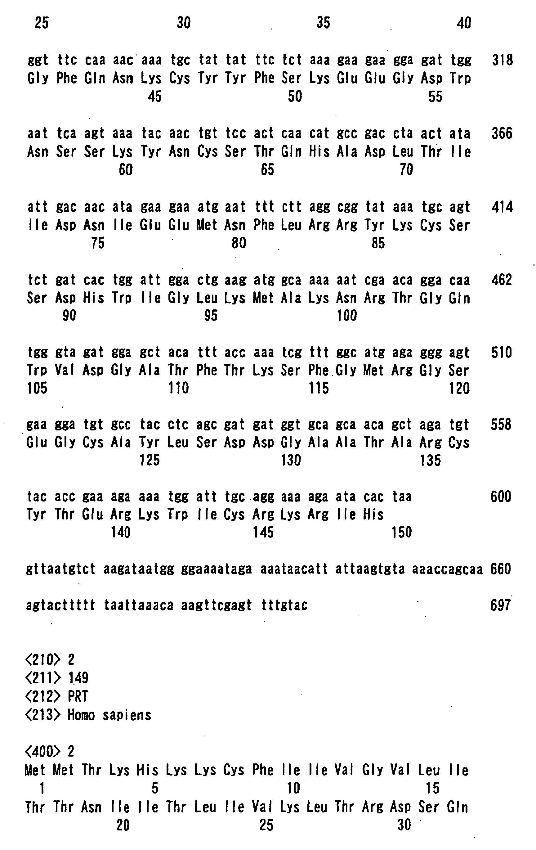
- the transmembrane domain is a polypeptide having at least the amino acid sequence from 1st to 26th of SEQ ID NO. 2.
- the application also provides an expression vector expressing a fusion protein in which an antigenic protein is fused with the C-terminal side of transmembrane domain of which the N-terminal side is located in the cell and the C-terminal side is out of the cell.
- the transmembrane domain is a polypeptide having at least the amino acid sequence from 1st to 26th of SEQ ID NO. 2.
- the expression vector to be inoculated to animals may be constructed as an expression vector having a fusion polynucleotide that consists of a polynucleotide encoding an antigenic protein and a polynucleotide encoding a transmembrane domain.
- an antigenic protein any one that can generate an antigen-antibody reaction in vivo may be used.
- the polynucleotide encoding an antigenic protein may be any one of genomic DNA, cDNA, synthetic DNA, etc., as far as it has an open reading frame (ORF).
- ORF open reading frame
- any domain may be used as far as its N-terminal side is in the cell and the C-terminal side is out of the cell.
- transmembrane domains of type II-membrane proteins or those of multispan-type membrane proteins may be used.
- the proteins that an antigenic protein is fused to the C-terminal side of these transmembrane domains take forms that the antigenic protein portion exists on the surface of the cell membrane.
- transmembrane domain for example, that of human type-II membrane protein HP10085 (SEQ ID NO: 2) may be used.
- the transmembrane domain to be fused with an antigenic protein is a polypeptide containing at least 1st methionine (Met) to 26th lysine (Lys) of SEQ ID NO: 2.
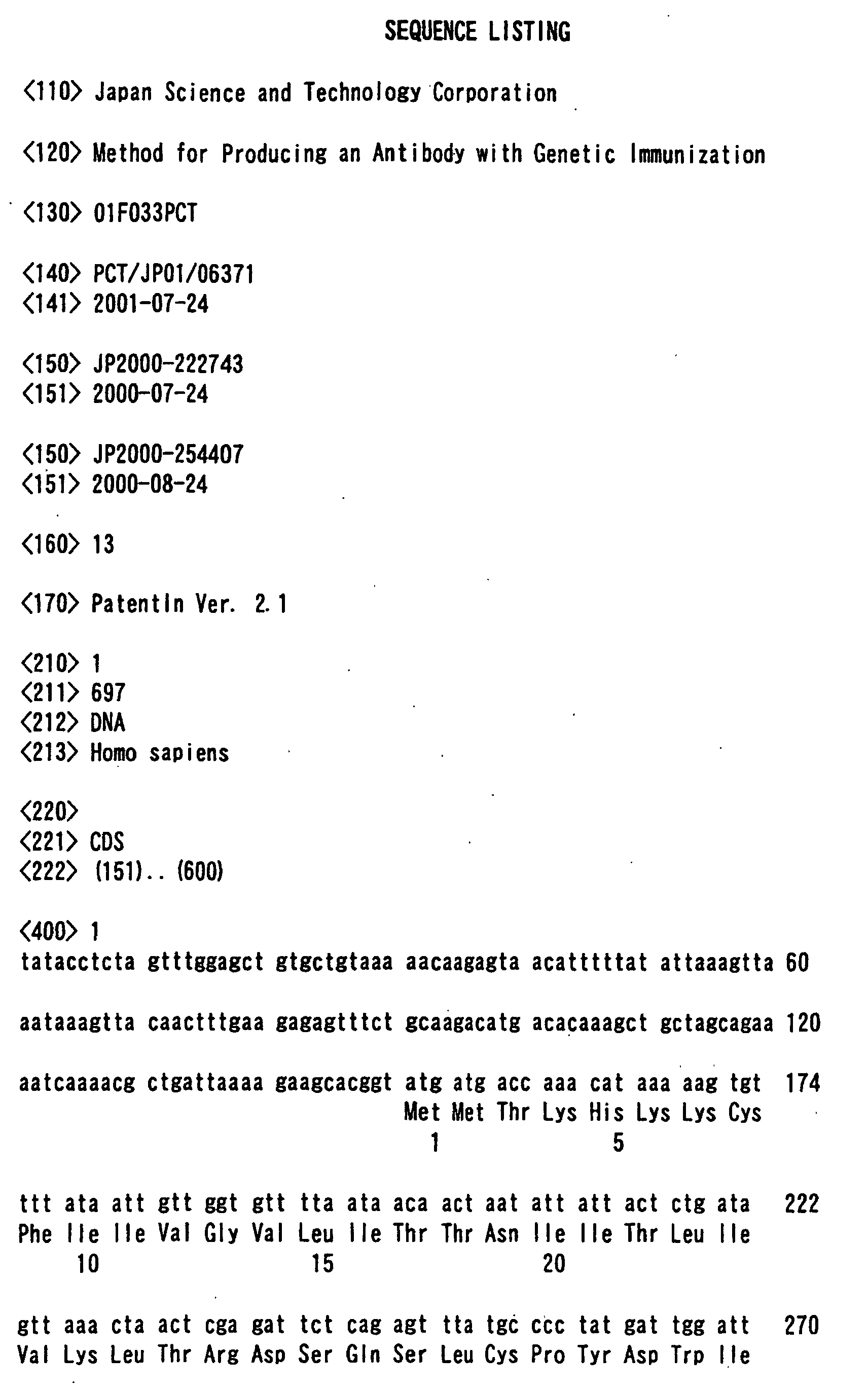
- the polynucleotide encoding the polypeptide contains at least the base sequence from 151st - 228th of SEQ ID NO: 1 (cDNA of human type-II membrane protein HP10085).
- This polynucleotide is linked to a polynucleotide encoding the above-mentioned antigenic protein, and the expression vector can be constructed using the fusion polynucleotide which expresses a fusion protein that an antigenic protein is fused to the C-terminal side of the transmembrane domain (polypeptide).
- any ones for eucaryotic cells may be used as far as they contain a promoter, a splicing region, a poly (A) addition site, etc., such as pKA1, pCDM8, pSVK3, pMSG, pSVL, pBK-CMV, pBK-RSV, EBV vectors, and the like.
- the two above-described polynucleotides are cloned to these vectors to make expression vectors that express respectively coded proteins as fusion proteins.
- Animals to which an expression vector is inoculated include mammals such as mouse, rat, rabbit, etc., and birds such as chicken, etc., which are generally used in producing antibodies. Inoculation of the expression vectors to animals may preferably be achieved using a gene gun technology and the like. When a gene gun technology is used, the expression vector is adsorbed on gold particles and emitted as a jet to the skin with gas pressure and the like for inoculation.
- the amount of the expression vector to be inoculated is variable depending on the species of animal and may preferably be in a range of 0.1 ⁇ g- 1 mg/animal. One-shot inoculation is acceptable, but it is desirable to carry out the inoculation twice or more at regular intervals in order to assure generation of the antibodies.
- the generation of the antibody may be confirmed by collecting blood, separating serum, and examining the binding reaction with the antigenic protein. For example, a known method such as enzyme immunoassay (ELISA), Western blotting, immuno-precipitation, antibody staining, and the like may be used. After confirmation of the presence of the antibody in the serum by these methods, the serum may be used as a polyclonal antibody specimen as it is or may be purified by affinity column chromatography to yield IgG. Alternatively, the spleen may be taken out from the animal acquiring immunity and the monoclonal antibody can be produced in a conventional manner.
- ELISA enzyme immunoassay
- Western blotting Western blotting
- immuno-precipitation immuno-precipitation
- antibody staining and the like
- the serum may be used as a polyclonal antibody specimen as it is or may be purified by affinity column chromatography to yield IgG.
- the spleen may be taken out from the animal acquiring immunity and the monoclon
- urokinase When urokinase is used as an antigenic protein, 3 kinds of expression vectors were used, that is, for secretion expression, for membrane form expression, and for intracellular expression. That is, the following vectors were respectively used: for secretion expression, pSSD1-UPA22 which expresses the signal sequence and protease domain of urokinase (Yokoyama-Kobayashi et al., Gene 163: 193-196, 1995); for membrane form expression, pSSD3-10085H which expresses a protein prepared by fusing a sequence from the N-terminal side to the 35th proline (Pro) of type II-membrane protein HP10085 (SEQ ID NO: 2) with the protease domain of urokinase (Yokoyama-Kobayashi et al., Gene 228: 161-167, 1999); for intracellular expression, pSSD1-UPA2 which expresses only the protease
- Fig. 1 shows the structure of respective fusion gene portions. In every case, the structure is the same except the portion encoding fusion protein. That is, they have the early promoter of SV40, 16S mRNA splicing region of SV40 and poly (A) addition site of SV40.
- protein HP10496 localized in the nuclear spliceosome was used.
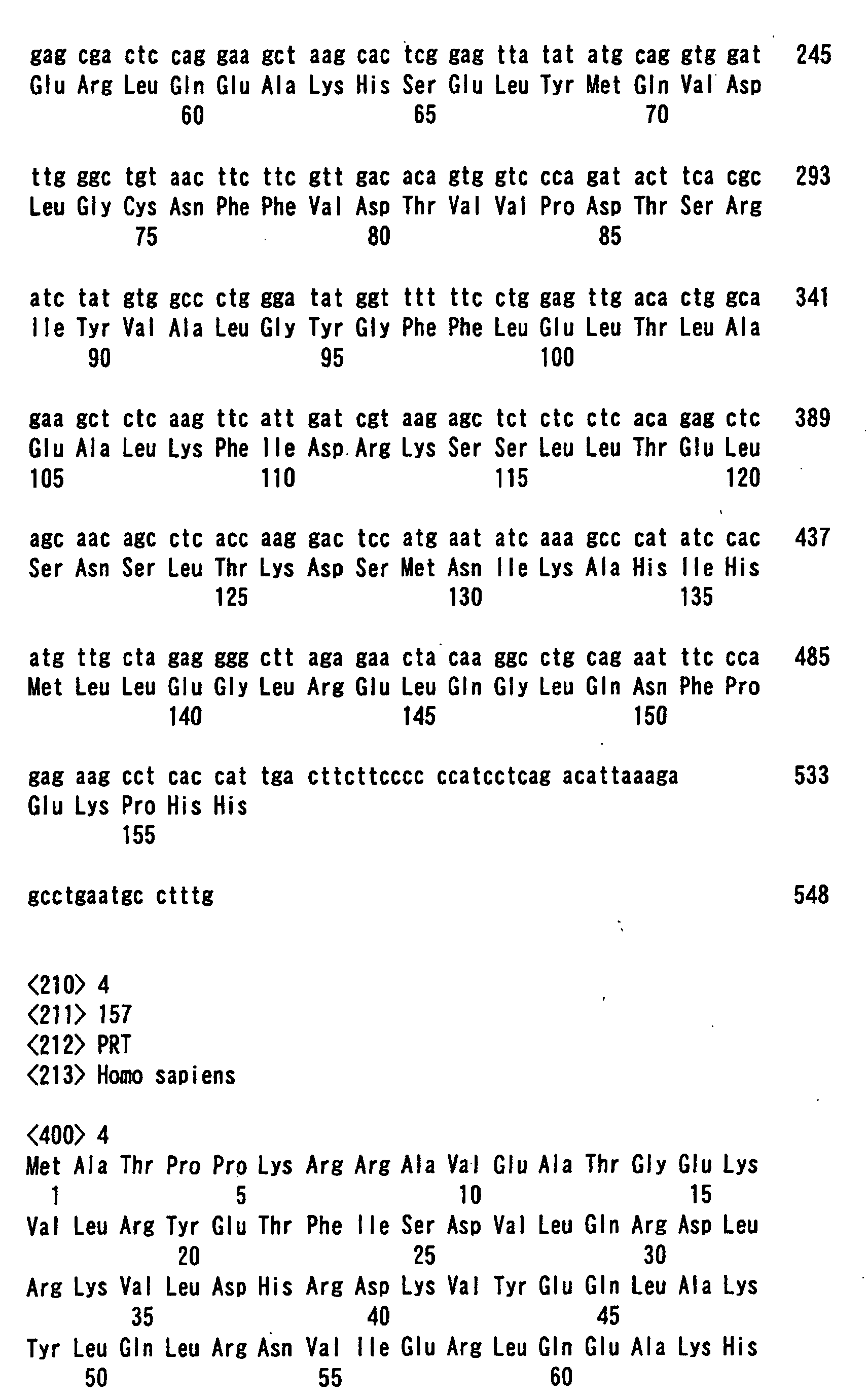
- cDNA clone pHP10496 encoding this protein was cloned from a human gastric cancer cDNA library (WO98/21328). This has the base sequence of SEQ ID NO: 3 as well as ORF which encodes protein HP10496 comprising the amino acid sequence of SEQ ID NO: 4.
- a 30mer sense primer (oligonucleotide of SEQ ID NO: 5) starting from a translation initiation codon to which a EcoRV recognition site was added and a 30 mer antisense primer (oligonucleotide of SEQ ID NO: 6) containing a stop codon to which an EcoRV recognition site was added
- the translation region was amplified by PCR using pHP10496 as template.
- the PCR product was digested with EcoRV, and inserted into an EcoRV-NotI (blunt ends) site of a membrane-type of urokinase expression vector pSSD3-10085N to construct a fusion gene expression vector pHP10085N-10496.
- an expression vector for expressing a protein fused with glutathione-S-transferase (GST) in Escherichia coli was constructed.
- GST glutathione-S-transferase
- the PCR product was digested with EcoRI and Sail and inserted into the EcoRI- Sail site of pGEX-5X-1 (Pharmacia Corp.). After confirmation of the sequence, a host Escherichia coli BL21 was transformed and incubated on an LB medium at 37°C for 5 hours. Then, IPTG was added at a final concentration of 0.4 mM, and the mixture was further incubated at 37°C for 2.5 hours.
- the cells were centrifuged, dissolved in a lysis buffer solution (50mM Tris-HCl (pH7.5), 1 mM EDTA, 1% Triton X-100, 0.2% SDS, 0.2 mM PMSF), frozen once at -80°C and thawed, and destroyed by sonication. After centrifugation at 1000 ⁇ g for 30 minutes, Glutathione Sepharose 4B was added to the supernatant, and the mixture was incubated at 4°C for 1 hour. The beads were washed well, and the fusion protein was eluted with an elution buffer solution (10 mM Tris-HCl, 50 mM glutathione). As a result, fusion protein GST-HP10496 having molecular weight of approximately 47 kDa was obtained.
- a lysis buffer solution 50mM Tris-HCl (pH7.5), 1 mM EDTA, 1% Triton X-100, 0.2% SDS, 0.2 m
- Gene immunization was achieved with a gene gun (Helios Gene Gun System; Japan Bio-Rad Laboratories).
- Expression vector plasmid DNA was attached onto gold particles according to the protocol. DNA-coated gold particles corresponding to 2 ⁇ g of plasmid DNA were shot into the skin of inguinal region of 3 mice (BALB/c) per sample. The immunization was carried out twice a week for 4 weeks, and the blood was then collected.
- urokinase As an antigenic protein, commercially available urokinase (Wakamori Jun-yaku) or GST-HP10496 expressed in Escherichia coli was coated on a plate and it was used in ELISA. The serum obtained by gene immunization with urokinase or nuclear protein HP10496 as antigen was used to measure the titer of the antibodies. The results are shown in Figs. 2 and 3. In any cases, production of antibodies was recognized only in immunization with the vector expressing the proteins to which the N-terminal of HP10085 was fused.
- a human full-length cDNA data bank From a human full-length cDNA data bank, the following 5 species of type II-membrane protein cDNAs, HP01347 (SEQ ID NO: 9), HP10328 (SEQ ID NO: 10), HP10390 (SEQ ID NO: 11), HP10433 (SEQ ID NO: 12), and HP10481 (SEQ ID NO: 13) were identified, and the vectors expressing fusion proteins of urokinase with the respective transmembrane domains were constructed (Yokoyama-Kobayashi et al., Gene 228: 161-167, 1999).
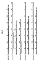
- Fig. 4 shows the respective N-terminal amino acid sequences.
- an antibody against an antigenic protein which it was difficult to produce in the so far known gene immunization, can be produced.
- the resulting an antibody is useful as drugs, diagnostic agents, and reagents for the research.
Landscapes
- Chemical & Material Sciences (AREA)
- Organic Chemistry (AREA)
- Health & Medical Sciences (AREA)
- General Health & Medical Sciences (AREA)
- Biochemistry (AREA)
- Biophysics (AREA)
- Life Sciences & Earth Sciences (AREA)
- Genetics & Genomics (AREA)
- Medicinal Chemistry (AREA)
- Molecular Biology (AREA)
- Proteomics, Peptides & Aminoacids (AREA)
- Immunology (AREA)
- Peptides Or Proteins (AREA)
- Preparation Of Compounds By Using Micro-Organisms (AREA)
Abstract
Description
- The invention of the present application relates to a method for producing an antibody by gene immunization. More specifically, the invention relates to a method of enabling easy production of an antibody useful as drugs, diagnostic agents, reagents for the research, and etc., and to an expression vector used in this method.
- An antibody has widely been utilized as reagents for the research for the purpose of detection, purification, elimination, inhibition of a protein or the like, because it has property of recognizing specific protein and binding thereto. Recently, it has widely been used not only as reagents for the research but also as drugs or diagnostic agents.
- In producing antibodies, it has so far been general to use a method that a large amount of protein as an antigen is purified and injected to an animal or animals such as rabbits or mice to collect antibodies generated in sera. It required, however, much time and a great deal of labor to obtain a large amount of a purified antigenic protein. It is desired to provide a more convenient method for producing antibodies, accordingly.
- Recently, it was reported that when a gene coding for an influenza virus nucleoprotein is integrated into an expression vector and intramuscularly injected directly as DNA to mice, then virus proteins are produced in the murine bodies and additionally the antibody against these proteins are generated in the sera. (Ulmer et al., Science 259: 1745-1749, 1993; Ginsbert et al., "Vaccines 93"). As a result, this expression vector received much attention as a new type of vaccine, that is, DNA vaccine, since mice have acquired immunity to virus. Thus, it has been designated as gene immunization that an expression vector for an antigenic protein is inoculated directly to an animal to generate immunity. In using gene immunization, however, in some cases, the titer of the generated antibody is very low or no antibody is generated depending on the kind of the antigen used.
- It was reported as an example of gene immunization that ovalbumin was fused in the downstream of transmembrane domain of transferrin receptor to form a membrane type and it was injected intramuscularly or subcutaneously to mice in order to investigate an effect of the expression site of antigenic protein on the efficacy of gene immunization. The titer of the antibodies generated, however, rather decreased since the protein was converted into a membrane type. (Boyle et al., Int. Immunol. 9: 1897-1906, 1997).
- The purpose of the invention of present application is to provide a method for producing antibodies to proteins, which it was difficult to produce in so far known gene immunization methods.
- Additionally, the purpose of the invention is to provide an expression vector used in the above-mentioned method for producing an antibody.
- The present application, as the invention for solving the above-described problems, provides a method for producing an antibody which comprises inoculating an expression vector expressing a fusion protein to an animal, isolating an antibody against an antigenic protein from the animal and purifying the antibody, wherein the fusion protein is an antigenic protein fused with the C-terminal of a transmembrane domain of which the N-terminal side is located in the cell and the C-terminal side is out of the cell.
- In a preferred embodiment in this method for producing an antibody, the transmembrane domain is a polypeptide having at least the amino acid sequence from 1st to 26th of SEQ ID NO. 2.
- The application also provides an expression vector expressing a fusion protein in which an antigenic protein is fused with the C-terminal side of transmembrane domain of which the N-terminal side is located in the cell and the C-terminal side is out of the cell.
- In a preferred embodiment of this expression vector, the transmembrane domain is a polypeptide having at least the amino acid sequence from 1st to 26th of SEQ ID NO. 2.
-
- Fig. 1 shows the structure of urokinase fusion gene.
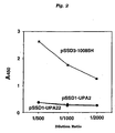
- Fig. 2 is an example illustrative of determination of antibody titer by ELISA when gene immunization has been carried out with a urokinase expression vector.
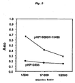
- Fig. 3 is an example illustrative of determination of antibody titer by ELISA when gene immunization has been carried out with a nuclear protein HP10496 expression vector.
- Fig. 4 shows the respective N-terminal amino acid sequences of fusion proteins comprising urokinase and transmembrane domains in a variety of membrane proteins.
-
- In a method for producing antibodies according to the invention, the expression vector to be inoculated to animals may be constructed as an expression vector having a fusion polynucleotide that consists of a polynucleotide encoding an antigenic protein and a polynucleotide encoding a transmembrane domain.
- As for an antigenic protein, any one that can generate an antigen-antibody reaction in vivo may be used. The polynucleotide encoding an antigenic protein may be any one of genomic DNA, cDNA, synthetic DNA, etc., as far as it has an open reading frame (ORF). When the antigenic protein is an inherent secretory protein, it is used after removal of the signal sequence peptide originally possessed by the protein.
- As for the transmembrane domain, any domain may be used as far as its N-terminal side is in the cell and the C-terminal side is out of the cell. For example, transmembrane domains of type II-membrane proteins or those of multispan-type membrane proteins may be used. The proteins that an antigenic protein is fused to the C-terminal side of these transmembrane domains take forms that the antigenic protein portion exists on the surface of the cell membrane. As for the transmembrane domain, for example, that of human type-II membrane protein HP10085 (SEQ ID NO: 2) may be used. In this case, the transmembrane domain to be fused with an antigenic protein is a polypeptide containing at least 1st methionine (Met) to 26th lysine (Lys) of SEQ ID NO: 2. The polynucleotide encoding the polypeptide contains at least the base sequence from 151st - 228th of SEQ ID NO: 1 (cDNA of human type-II membrane protein HP10085). This polynucleotide is linked to a polynucleotide encoding the above-mentioned antigenic protein, and the expression vector can be constructed using the fusion polynucleotide which expresses a fusion protein that an antigenic protein is fused to the C-terminal side of the transmembrane domain (polypeptide).
- As for vectors expressing fusion proteins, any ones for eucaryotic cells may be used as far as they contain a promoter, a splicing region, a poly (A) addition site, etc., such as pKA1, pCDM8, pSVK3, pMSG, pSVL, pBK-CMV, pBK-RSV, EBV vectors, and the like. The two above-described polynucleotides are cloned to these vectors to make expression vectors that express respectively coded proteins as fusion proteins.
- Animals to which an expression vector is inoculated include mammals such as mouse, rat, rabbit, etc., and birds such as chicken, etc., which are generally used in producing antibodies. Inoculation of the expression vectors to animals may preferably be achieved using a gene gun technology and the like. When a gene gun technology is used, the expression vector is adsorbed on gold particles and emitted as a jet to the skin with gas pressure and the like for inoculation. The amount of the expression vector to be inoculated is variable depending on the species of animal and may preferably be in a range of 0.1 µg- 1 mg/animal. One-shot inoculation is acceptable, but it is desirable to carry out the inoculation twice or more at regular intervals in order to assure generation of the antibodies.
- The generation of the antibody may be confirmed by collecting blood, separating serum, and examining the binding reaction with the antigenic protein. For example, a known method such as enzyme immunoassay (ELISA), Western blotting, immuno-precipitation, antibody staining, and the like may be used. After confirmation of the presence of the antibody in the serum by these methods, the serum may be used as a polyclonal antibody specimen as it is or may be purified by affinity column chromatography to yield IgG. Alternatively, the spleen may be taken out from the animal acquiring immunity and the monoclonal antibody can be produced in a conventional manner.
- The following examples serve to illustrate the invention in more detail and specifically but are not intended as a limitation thereof. In these examples, basic procedures for recombination of DNA and enzyme reactions are carried out according to the articles, "Molecular Cloning; A laboratory manual", Cold Spring Harbor Laboratory, 1989. Restriction enzymes and a variety of modified enzymes were obtained from Takara Shuzo Co., Ltd., unless otherwise stated. The compositions of buffer solutions in respective enzyme reactions and the reaction conditions were set according to the specification attached.
- When urokinase is used as an antigenic protein, 3 kinds of expression vectors were used, that is, for secretion expression, for membrane form expression, and for intracellular expression. That is, the following vectors were respectively used: for secretion expression, pSSD1-UPA22 which expresses the signal sequence and protease domain of urokinase (Yokoyama-Kobayashi et al., Gene 163: 193-196, 1995); for membrane form expression, pSSD3-10085H which expresses a protein prepared by fusing a sequence from the N-terminal side to the 35th proline (Pro) of type II-membrane protein HP10085 (SEQ ID NO: 2) with the protease domain of urokinase (Yokoyama-Kobayashi et al., Gene 228: 161-167, 1999); for intracellular expression, pSSD1-UPA2 which expresses only the protease domain of urokinase by eliminating the signal sequence of urokinase (Yokoyama-Kobayashi et al., Gene 163: 193-196, 1995). Fig. 1 shows the structure of respective fusion gene portions. In every case, the structure is the same except the portion encoding fusion protein. That is, they have the early promoter of SV40, 16S mRNA splicing region of SV40 and poly (A) addition site of SV40.
- As an antigenic protein, protein HP10496 localized in the nuclear spliceosome was used. cDNA clone pHP10496 encoding this protein was cloned from a human gastric cancer cDNA library (WO98/21328). This has the base sequence of SEQ ID NO: 3 as well as ORF which encodes protein HP10496 comprising the amino acid sequence of SEQ ID NO: 4. Using a 30mer sense primer (oligonucleotide of SEQ ID NO: 5) starting from a translation initiation codon to which a EcoRV recognition site was added and a 30 mer antisense primer (oligonucleotide of SEQ ID NO: 6) containing a stop codon to which an EcoRV recognition site was added, the translation region was amplified by PCR using pHP10496 as template. The PCR product was digested with EcoRV, and inserted into an EcoRV-NotI (blunt ends) site of a membrane-type of urokinase expression vector pSSD3-10085N to construct a fusion gene expression vector pHP10085N-10496.
- In order to obtain an antigenic protein for antibody detection, an expression vector for expressing a protein fused with glutathione-S-transferase (GST) in Escherichia coli was constructed. Using a 28mer sense primer (oligonucleotide of SEQ ID NO: 7) starting from a translation initiation codon to which a EcoRI recognition site was added and a 32 mer antisense primer (oligonucleotide of SEQ ID NO: 8) containing a stop codon to which a SalI recognition site was added, the translation region was amplified by PCR using pHP10496 as template. The PCR product was digested with EcoRI and Sail and inserted into the EcoRI- Sail site of pGEX-5X-1 (Pharmacia Corp.). After confirmation of the sequence, a host Escherichia coli BL21 was transformed and incubated on an LB medium at 37°C for 5 hours. Then, IPTG was added at a final concentration of 0.4 mM, and the mixture was further incubated at 37°C for 2.5 hours. The cells were centrifuged, dissolved in a lysis buffer solution (50mM Tris-HCl (pH7.5), 1 mM EDTA, 1% Triton X-100, 0.2% SDS, 0.2 mM PMSF), frozen once at -80°C and thawed, and destroyed by sonication. After centrifugation at 1000 × g for 30 minutes, Glutathione Sepharose 4B was added to the supernatant, and the mixture was incubated at 4°C for 1 hour. The beads were washed well, and the fusion protein was eluted with an elution buffer solution (10 mM Tris-HCl, 50 mM glutathione). As a result, fusion protein GST-HP10496 having molecular weight of approximately 47 kDa was obtained.
- Gene immunization was achieved with a gene gun (Helios Gene Gun System; Japan Bio-Rad Laboratories). Expression vector plasmid DNA was attached onto gold particles according to the protocol. DNA-coated gold particles corresponding to 2 µg of plasmid DNA were shot into the skin of inguinal region of 3 mice (BALB/c) per sample. The immunization was carried out twice a week for 4 weeks, and the blood was then collected.
- As an antigenic protein, commercially available urokinase (Wakamori Jun-yaku) or GST-HP10496 expressed in Escherichia coli was coated on a plate and it was used in ELISA. The serum obtained by gene immunization with urokinase or nuclear protein HP10496 as antigen was used to measure the titer of the antibodies. The results are shown in Figs. 2 and 3. In any cases, production of antibodies was recognized only in immunization with the vector expressing the proteins to which the N-terminal of HP10085 was fused.
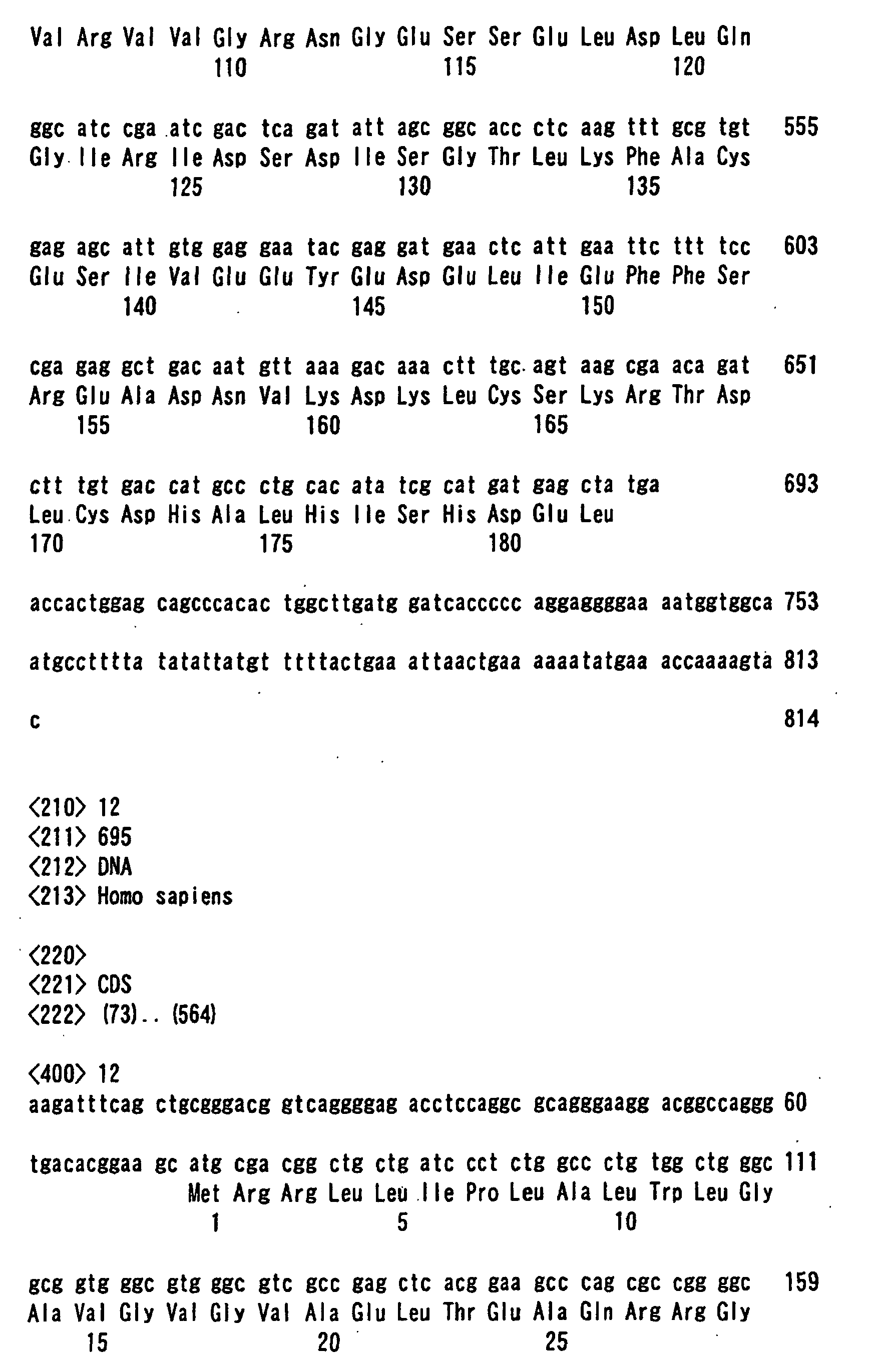
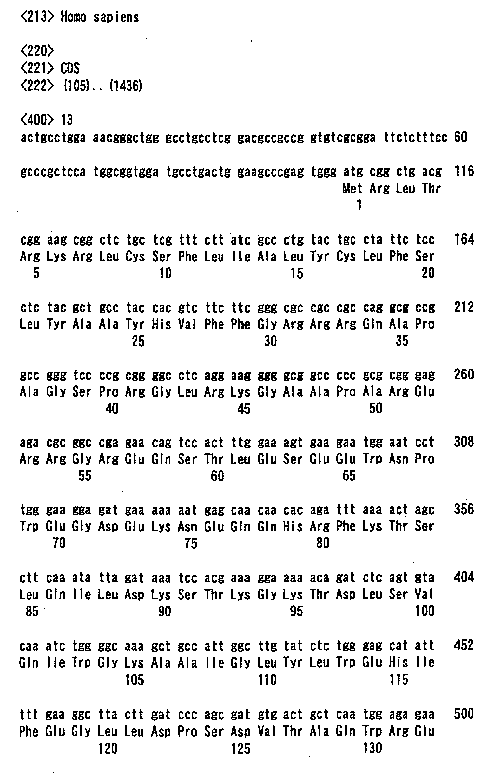
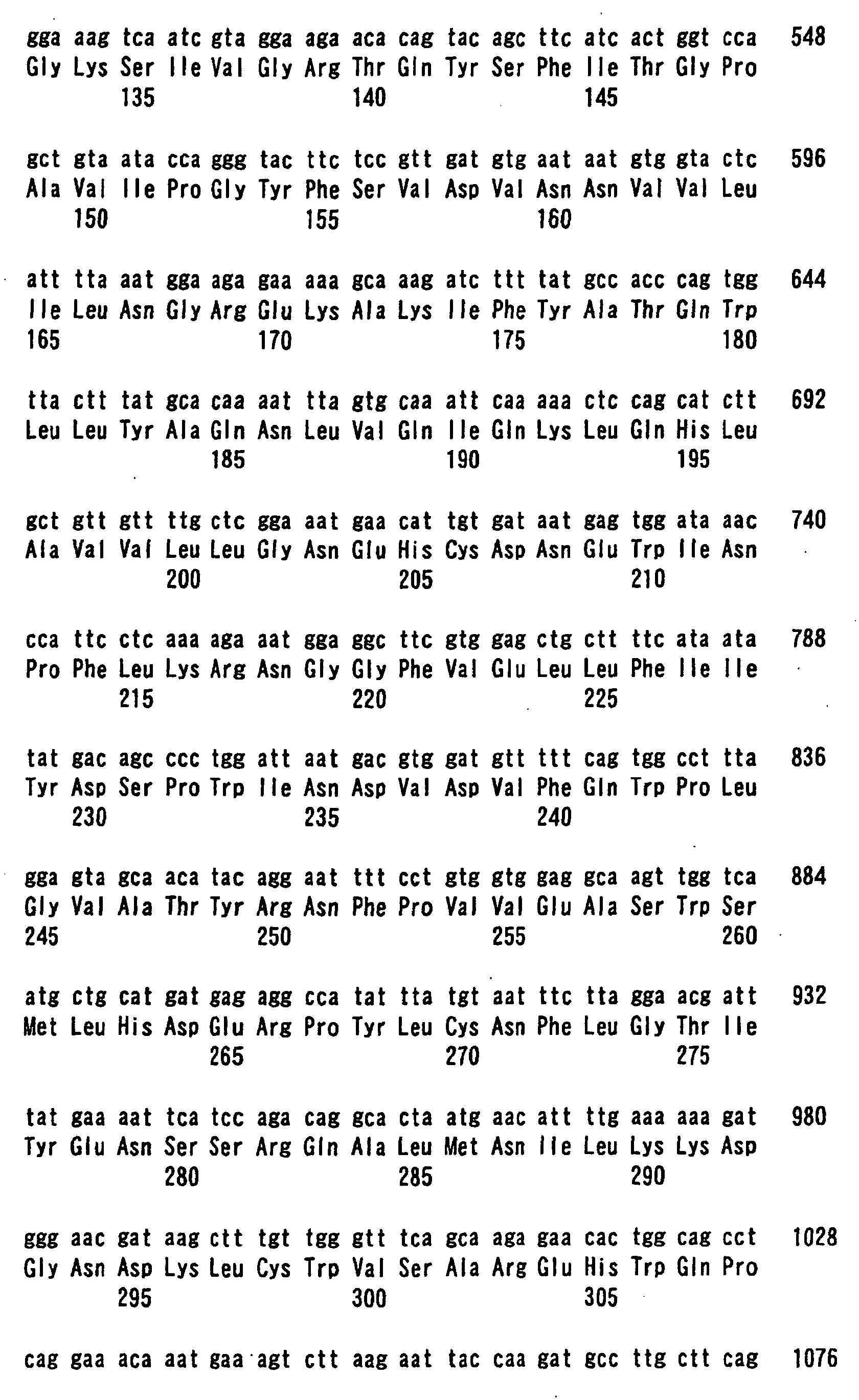
- From a human full-length cDNA data bank, the following 5 species of type II-membrane protein cDNAs, HP01347 (SEQ ID NO: 9), HP10328 (SEQ ID NO: 10), HP10390 (SEQ ID NO: 11), HP10433 (SEQ ID NO: 12), and HP10481 (SEQ ID NO: 13) were identified, and the vectors expressing fusion proteins of urokinase with the respective transmembrane domains were constructed (Yokoyama-Kobayashi et al., Gene 228: 161-167, 1999). Fig. 4 shows the respective N-terminal amino acid sequences.
- The gene immunization was carried out with these expression vectors according to the method as described in the above item (3), and the antibodies to urokinase were detected according to the method as described in the above item (4). In respective cases, production of antibodies was recognized in the same as in HP10085.
- According to the present invention, an antibody against an antigenic protein, which it was difficult to produce in the so far known gene immunization, can be produced. The resulting an antibody is useful as drugs, diagnostic agents, and reagents for the research.







Claims (4)
- A method for producing an antibody which comprises inoculating an expression vector expressing a fusion protein to an animal, isolating an antibody against an antigenic protein from the animal and purifying the antibody, wherein the fusion protein is an antigenic protein fused with the C-terminal of a transmembrane domain of which the N-terminal side is located in the cell and the C-terminal side is out of the cell.
- The method for producing an antibody of claim 1, wherein the transmembrane domain is a polypeptide having at least the amino acid sequence from 1st to 26th of SEQ ID NO. 2.
- An expression vector expressing a fusion protein in which an antigenic protein is fused with the C-terminal of transmembrane domain of which the N-terminal side is located in the cell and the C-terminal side is out of the cell.
- The expression vector of claim 3, wherein the transmembrane domain is a polypeptide having at least the amino acid sequence from 1st to 26th of SEQ ID NO. 2.
Applications Claiming Priority (5)
| Application Number | Priority Date | Filing Date | Title |
|---|---|---|---|
| JP2000222743 | 2000-07-24 | ||
| JP2000222743 | 2000-07-24 | ||
| JP2000254407 | 2000-08-24 | ||
| JP2000254407 | 2000-08-24 | ||
| PCT/JP2001/006371 WO2002008416A1 (en) | 2000-07-24 | 2001-07-24 | Method of preparing antibody by gene immunization |
Publications (3)
| Publication Number | Publication Date |
|---|---|
| EP1304375A1 true EP1304375A1 (en) | 2003-04-23 |
| EP1304375A4 EP1304375A4 (en) | 2004-05-19 |
| EP1304375B1 EP1304375B1 (en) | 2009-10-14 |
Family
ID=26596574
Family Applications (1)
| Application Number | Title | Priority Date | Filing Date |
|---|---|---|---|
| EP01951992A Expired - Lifetime EP1304375B1 (en) | 2000-07-24 | 2001-07-24 | Method of preparing antibody by gene immunization |
Country Status (6)
| Country | Link |
|---|---|
| US (1) | US20030148468A1 (en) |
| EP (1) | EP1304375B1 (en) |
| JP (1) | JP4368578B2 (en) |
| AU (1) | AU2001272791A1 (en) |
| DE (1) | DE60140192D1 (en) |
| WO (1) | WO2002008416A1 (en) |
Cited By (2)
| Publication number | Priority date | Publication date | Assignee | Title |
|---|---|---|---|---|
| EP1687330A4 (en) * | 2003-10-28 | 2007-03-14 | Protemix Discovery Ltd | Peptides with anti-obesity activity and other related uses |
| US20120093825A1 (en) * | 2010-10-18 | 2012-04-19 | Delphi Genetics Sa | Method for producing antibody using "naked" expression vector expressing type ii transmembrane fusion protein |
Families Citing this family (2)
| Publication number | Priority date | Publication date | Assignee | Title |
|---|---|---|---|---|
| EP2280030A3 (en) | 2001-04-10 | 2011-06-15 | Agensys, Inc. | Nucleic acids and corresponding proteins useful in the detection and treatment of various cancers |
| GB2399899B (en) * | 2003-03-27 | 2005-06-22 | Micron Technology Inc | Active memory command engine and method |
Family Cites Families (1)
| Publication number | Priority date | Publication date | Assignee | Title |
|---|---|---|---|---|
| US6734172B2 (en) * | 1998-11-18 | 2004-05-11 | Pacific Northwest Research Institute | Surface receptor antigen vaccines |
-
2001
- 2001-07-24 JP JP2002513899A patent/JP4368578B2/en not_active Expired - Fee Related
- 2001-07-24 WO PCT/JP2001/006371 patent/WO2002008416A1/en not_active Ceased
- 2001-07-24 EP EP01951992A patent/EP1304375B1/en not_active Expired - Lifetime
- 2001-07-24 AU AU2001272791A patent/AU2001272791A1/en not_active Abandoned
- 2001-07-24 DE DE60140192T patent/DE60140192D1/en not_active Expired - Lifetime
- 2001-07-24 US US10/088,859 patent/US20030148468A1/en not_active Abandoned
Non-Patent Citations (3)
| Title |
|---|
| GUPTA ANUSHREE ET AL: "Identification of novel transmembrane gene sequence and its use for cell-surface targeting of beta subunit of human chorionic gonadotropin" DNA AND CELL BIOLOGY, vol. 17, no. 7, July 1998 (1998-07), pages 573-581, XP001179793 ISSN: 1044-5498 * |
| See also references of WO0208416A1 * |
| WAINE G J ET AL: "Genetic immunization of mice with DNA encoding the 23 kDa transmembrane surface protein of Schistosoma japonicum (Sj23) induces antigen-specific immunoglobulin G antibodies" PARASITE IMMUNOLOGY (OXFORD), vol. 21, no. 7, July 1999 (1999-07), pages 377-381, XP002273350 ISSN: 0141-9838 * |
Cited By (3)
| Publication number | Priority date | Publication date | Assignee | Title |
|---|---|---|---|---|
| EP1687330A4 (en) * | 2003-10-28 | 2007-03-14 | Protemix Discovery Ltd | Peptides with anti-obesity activity and other related uses |
| US20120093825A1 (en) * | 2010-10-18 | 2012-04-19 | Delphi Genetics Sa | Method for producing antibody using "naked" expression vector expressing type ii transmembrane fusion protein |
| US9234042B2 (en) * | 2010-10-18 | 2016-01-12 | Delphi Genetics Sa | Method for producing antibody using “naked” expression vector expressing type II transmembrane fusion protein |
Also Published As
| Publication number | Publication date |
|---|---|
| DE60140192D1 (en) | 2009-11-26 |
| WO2002008416A1 (en) | 2002-01-31 |
| AU2001272791A1 (en) | 2002-02-05 |
| US20030148468A1 (en) | 2003-08-07 |
| EP1304375A4 (en) | 2004-05-19 |
| EP1304375B1 (en) | 2009-10-14 |
| JP4368578B2 (en) | 2009-11-18 |
Similar Documents
| Publication | Publication Date | Title |
|---|---|---|
| AU2002254683B2 (en) | Binding molecules for Fc-region polypeptides | |
| Ståhl et al. | A dual expression system for the generation, analysis and purification of antibodies to a repeated sequence of the Plasmodium falciparum antigen Pf155/RESA | |
| AU768631B2 (en) | Method for producing antibodies acting against a polypeptide that only recognises the coding nucleic acid | |
| CZ291097A3 (en) | Expression system of heterologous proteins as fused proteins | |
| CN117304280B (en) | Recombinant RSV F protein and application thereof | |
| WO2001021804B1 (en) | Chlamydia antigens and corresponding dna fragments and uses thereof | |
| EP1304375B1 (en) | Method of preparing antibody by gene immunization | |
| GB2226563A (en) | Streptolysin o fragments | |
| JP2013527752A (en) | HPV antibody screening method using fusion polypeptide HPV antigen | |
| US6482607B1 (en) | Expression vector for use in a one-step purification protocol | |
| EP0706574B1 (en) | Methods for peptide synthesis and purification | |
| WO1997007132A1 (en) | Epitope tagging system | |
| JPH05503849A (en) | Human receptor for TSH and sequences encoding this receptor | |
| EP0879244A1 (en) | Mycoplasma recombinant polypeptide vaccines | |
| WO1998014463B1 (en) | MAGE-10 ENCODING cDNA, THE TUMOR REJECTION ANTIGEN PRECURSOR MAGE-10, ANTIBODIES SPECIFIC TO THE MOLECULE, AND USES THEREOF | |
| Gurunath et al. | Enhancing peptide antigenicity by helix stabilization | |
| JPWO2002008416A1 (en) | Genetic immunization to generate antibodies | |
| Schwarz et al. | Non-specific cross-reacting antigen: characterization of specific and cross-reacting epitopes | |
| KR100528721B1 (en) | A mouse monoclonal antibody specific for the human asialoglycoprotein receptor, the hybridoma cell line secreting this antibody and the generation and verification method thereof | |
| EP0655502A1 (en) | Cho cells transformed with a recombinant plasmid expressing human secreting type thyroid peroxidase and preparation thereof | |
| CN108017694B (en) | PORF65 recombinant protein and its preparation method and application | |
| CN102659925A (en) | Human egg zona pellucida protein-4 epitope minimal motif peptide identified by anti-recombinant human ZP4N and ZP4C polyantibodies | |
| JP2004518410A5 (en) | ||
| Na et al. | Generation of self-antigen reactive, anti-urocortin specific antibodies by immunization of recombinantly expressed urocortin fusion proteins | |
| CN103290047A (en) | A method for quickly preparing polyclonal antibodies of plant leucine-rich single transmembrane receptor protein kinase and applications of the prepared polyclonal antibodies |
Legal Events
| Date | Code | Title | Description |
|---|---|---|---|
| PUAI | Public reference made under article 153(3) epc to a published international application that has entered the european phase |
Free format text: ORIGINAL CODE: 0009012 |
|
| 17P | Request for examination filed |
Effective date: 20020425 |
|
| AK | Designated contracting states |
Designated state(s): AT BE CH CY DE DK ES FI FR GB GR IE IT LI LU MC NL PT SE TR |
|
| RIC1 | Information provided on ipc code assigned before grant |
Ipc: 7C 07K 16/06 B Ipc: 7C 12N 15/09 A |
|
| A4 | Supplementary search report drawn up and despatched |
Effective date: 20040406 |
|
| RBV | Designated contracting states (corrected) |
Designated state(s): DE FR GB |
|
| RAP1 | Party data changed (applicant data changed or rights of an application transferred) |
Owner name: JAPAN SCIENCE AND TECHNOLOGY AGENCY |
|
| 17Q | First examination report despatched |
Effective date: 20041005 |
|
| 17Q | First examination report despatched |
Effective date: 20041005 |
|
| GRAP | Despatch of communication of intention to grant a patent |
Free format text: ORIGINAL CODE: EPIDOSNIGR1 |
|
| GRAS | Grant fee paid |
Free format text: ORIGINAL CODE: EPIDOSNIGR3 |
|
| GRAA | (expected) grant |
Free format text: ORIGINAL CODE: 0009210 |
|
| AK | Designated contracting states |
Kind code of ref document: B1 Designated state(s): DE FR GB |
|
| REG | Reference to a national code |
Ref country code: GB Ref legal event code: FG4D |
|
| REF | Corresponds to: |
Ref document number: 60140192 Country of ref document: DE Date of ref document: 20091126 Kind code of ref document: P |
|
| PLBE | No opposition filed within time limit |
Free format text: ORIGINAL CODE: 0009261 |
|
| STAA | Information on the status of an ep patent application or granted ep patent |
Free format text: STATUS: NO OPPOSITION FILED WITHIN TIME LIMIT |
|
| 26N | No opposition filed |
Effective date: 20100715 |
|
| PGFP | Annual fee paid to national office [announced via postgrant information from national office to epo] |
Ref country code: FR Payment date: 20100805 Year of fee payment: 10 Ref country code: DE Payment date: 20100723 Year of fee payment: 10 |
|
| PGFP | Annual fee paid to national office [announced via postgrant information from national office to epo] |
Ref country code: GB Payment date: 20100722 Year of fee payment: 10 |
|
| GBPC | Gb: european patent ceased through non-payment of renewal fee |
Effective date: 20110724 |
|
| REG | Reference to a national code |
Ref country code: FR Ref legal event code: ST Effective date: 20120330 |
|
| PG25 | Lapsed in a contracting state [announced via postgrant information from national office to epo] |
Ref country code: FR Free format text: LAPSE BECAUSE OF NON-PAYMENT OF DUE FEES Effective date: 20110801 Ref country code: DE Free format text: LAPSE BECAUSE OF NON-PAYMENT OF DUE FEES Effective date: 20120201 |
|
| REG | Reference to a national code |
Ref country code: DE Ref legal event code: R119 Ref document number: 60140192 Country of ref document: DE Effective date: 20120201 |
|
| PG25 | Lapsed in a contracting state [announced via postgrant information from national office to epo] |
Ref country code: GB Free format text: LAPSE BECAUSE OF NON-PAYMENT OF DUE FEES Effective date: 20110724 |