EP1303751B1 - Dyes and methods of detection of nucleic acid in immature red blood cells - Google Patents
Dyes and methods of detection of nucleic acid in immature red blood cells Download PDFInfo
- Publication number
- EP1303751B1 EP1303751B1 EP01933047A EP01933047A EP1303751B1 EP 1303751 B1 EP1303751 B1 EP 1303751B1 EP 01933047 A EP01933047 A EP 01933047A EP 01933047 A EP01933047 A EP 01933047A EP 1303751 B1 EP1303751 B1 EP 1303751B1
- Authority
- EP
- European Patent Office
- Prior art keywords
- dye
- reticulocytes
- solution
- fluorescence
- dyes
- Prior art date
- Legal status (The legal status is an assumption and is not a legal conclusion. Google has not performed a legal analysis and makes no representation as to the accuracy of the status listed.)
- Expired - Lifetime
Links
- ZQVKTHRQIXSMGY-UHFFFAOYSA-N CCc(cc1)ccc1C(O)=O Chemical compound CCc(cc1)ccc1C(O)=O ZQVKTHRQIXSMGY-UHFFFAOYSA-N 0.000 description 1
Images










Classifications
-
- C—CHEMISTRY; METALLURGY
- C09—DYES; PAINTS; POLISHES; NATURAL RESINS; ADHESIVES; COMPOSITIONS NOT OTHERWISE PROVIDED FOR; APPLICATIONS OF MATERIALS NOT OTHERWISE PROVIDED FOR
- C09B—ORGANIC DYES OR CLOSELY-RELATED COMPOUNDS FOR PRODUCING DYES, e.g. PIGMENTS; MORDANTS; LAKES
- C09B23/00—Methine or polymethine dyes, e.g. cyanine dyes
- C09B23/02—Methine or polymethine dyes, e.g. cyanine dyes the polymethine chain containing an odd number of >CH- or >C[alkyl]- groups
-
- G—PHYSICS
- G01—MEASURING; TESTING
- G01N—INVESTIGATING OR ANALYSING MATERIALS BY DETERMINING THEIR CHEMICAL OR PHYSICAL PROPERTIES
- G01N1/00—Sampling; Preparing specimens for investigation
- G01N1/28—Preparing specimens for investigation including physical details of (bio-)chemical methods covered elsewhere, e.g. G01N33/50, C12Q
- G01N1/30—Staining; Impregnating ; Fixation; Dehydration; Multistep processes for preparing samples of tissue, cell or nucleic acid material and the like for analysis
-
- G—PHYSICS
- G01—MEASURING; TESTING
- G01N—INVESTIGATING OR ANALYSING MATERIALS BY DETERMINING THEIR CHEMICAL OR PHYSICAL PROPERTIES
- G01N21/00—Investigating or analysing materials by the use of optical means, i.e. using sub-millimetre waves, infrared, visible or ultraviolet light
- G01N21/17—Systems in which incident light is modified in accordance with the properties of the material investigated
- G01N21/47—Scattering, i.e. diffuse reflection
-
- G—PHYSICS
- G01—MEASURING; TESTING
- G01N—INVESTIGATING OR ANALYSING MATERIALS BY DETERMINING THEIR CHEMICAL OR PHYSICAL PROPERTIES
- G01N21/00—Investigating or analysing materials by the use of optical means, i.e. using sub-millimetre waves, infrared, visible or ultraviolet light
- G01N21/62—Systems in which the material investigated is excited whereby it emits light or causes a change in wavelength of the incident light
- G01N21/63—Systems in which the material investigated is excited whereby it emits light or causes a change in wavelength of the incident light optically excited
- G01N21/64—Fluorescence; Phosphorescence
- G01N21/6428—Measuring fluorescence of fluorescent products of reactions or of fluorochrome labelled reactive substances, e.g. measuring quenching effects, using measuring "optrodes"
-
- G—PHYSICS
- G01—MEASURING; TESTING
- G01N—INVESTIGATING OR ANALYSING MATERIALS BY DETERMINING THEIR CHEMICAL OR PHYSICAL PROPERTIES
- G01N1/00—Sampling; Preparing specimens for investigation
- G01N1/28—Preparing specimens for investigation including physical details of (bio-)chemical methods covered elsewhere, e.g. G01N33/50, C12Q
- G01N1/30—Staining; Impregnating ; Fixation; Dehydration; Multistep processes for preparing samples of tissue, cell or nucleic acid material and the like for analysis
- G01N2001/302—Stain compositions
-
- Y—GENERAL TAGGING OF NEW TECHNOLOGICAL DEVELOPMENTS; GENERAL TAGGING OF CROSS-SECTIONAL TECHNOLOGIES SPANNING OVER SEVERAL SECTIONS OF THE IPC; TECHNICAL SUBJECTS COVERED BY FORMER USPC CROSS-REFERENCE ART COLLECTIONS [XRACs] AND DIGESTS
- Y10—TECHNICAL SUBJECTS COVERED BY FORMER USPC
- Y10S—TECHNICAL SUBJECTS COVERED BY FORMER USPC CROSS-REFERENCE ART COLLECTIONS [XRACs] AND DIGESTS
- Y10S436/00—Chemistry: analytical and immunological testing
- Y10S436/80—Fluorescent dyes, e.g. rhodamine
-
- Y—GENERAL TAGGING OF NEW TECHNOLOGICAL DEVELOPMENTS; GENERAL TAGGING OF CROSS-SECTIONAL TECHNOLOGIES SPANNING OVER SEVERAL SECTIONS OF THE IPC; TECHNICAL SUBJECTS COVERED BY FORMER USPC CROSS-REFERENCE ART COLLECTIONS [XRACs] AND DIGESTS
- Y10—TECHNICAL SUBJECTS COVERED BY FORMER USPC
- Y10T—TECHNICAL SUBJECTS COVERED BY FORMER US CLASSIFICATION
- Y10T436/00—Chemistry: analytical and immunological testing
- Y10T436/10—Composition for standardization, calibration, simulation, stabilization, preparation or preservation; processes of use in preparation for chemical testing
-
- Y—GENERAL TAGGING OF NEW TECHNOLOGICAL DEVELOPMENTS; GENERAL TAGGING OF CROSS-SECTIONAL TECHNOLOGIES SPANNING OVER SEVERAL SECTIONS OF THE IPC; TECHNICAL SUBJECTS COVERED BY FORMER USPC CROSS-REFERENCE ART COLLECTIONS [XRACs] AND DIGESTS
- Y10—TECHNICAL SUBJECTS COVERED BY FORMER USPC
- Y10T—TECHNICAL SUBJECTS COVERED BY FORMER US CLASSIFICATION
- Y10T436/00—Chemistry: analytical and immunological testing
- Y10T436/10—Composition for standardization, calibration, simulation, stabilization, preparation or preservation; processes of use in preparation for chemical testing
- Y10T436/101666—Particle count or volume standard or control [e.g., platelet count standards, etc.]
Definitions
- Distribution of fluorescence intensities of a relatively large reticulocyte population can be determined by flow cytometry in a fast and reliable manner, and different maturation stages of reticulocytes, as reflected by differences in RNA content, can be distinguished.
- NRBCs nucleated red blood cells
- NRBCs nucleated red blood cells
- NRBCs nucleated red blood cells
- NRBCs contain DNA.
- NRBCs normally occur in the bone marrow but not in peripheral blood.
- certain diseases such as anemia and leukemia, NRBCs also occur in peripheral blood. Therefore, it is of clinical importance to measure NRBC.
- NRBCs contain DNA
- fluorescence detection and enumeration of NRBCs can be possible by staining these cells with dyes that recognize DNA.
- dyes, and compositions which enable staining of both intracellular DNA and RNA, so that both NRBCs and reticulocytes can be enumerated via the use of the same dye employing different methods.
- the present invention provides novel dyes, both red and blue-excitable, which bind nucleic acids and permit detection of reticulocytes via fluorescence at ambient temperatures.
- the dyes of the invention are adapted to methods which preferentially permit rapid detection of reticulocytes in the absence of substantial background fluorescence.
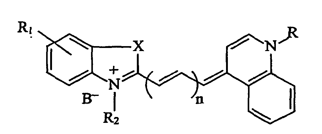
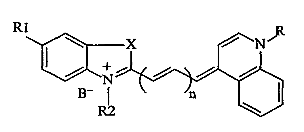
- the present invention provides three groups of novel dyes that are set forth as:
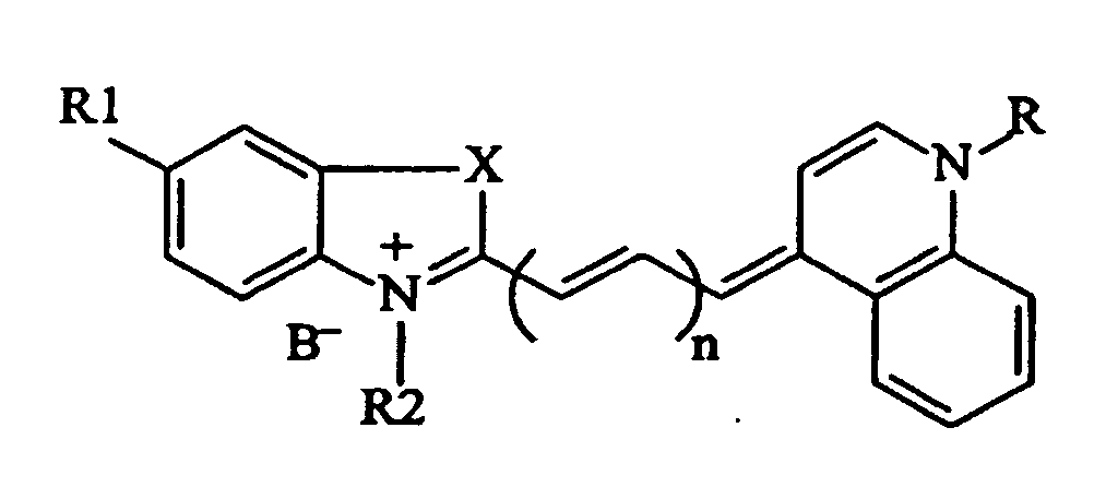
- the invention provides a dye, termed herein, ReticRed1 having the following formula.
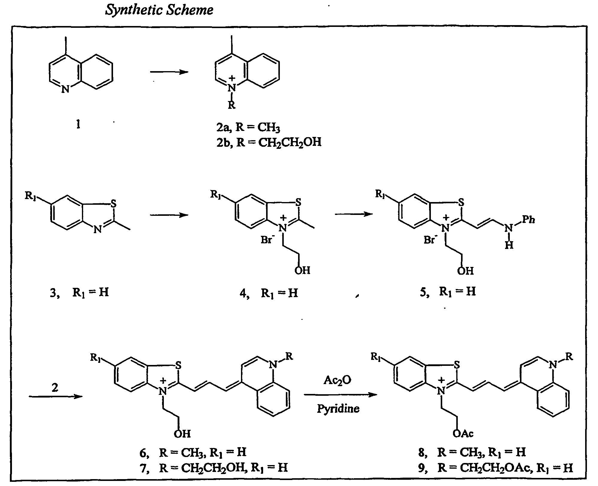
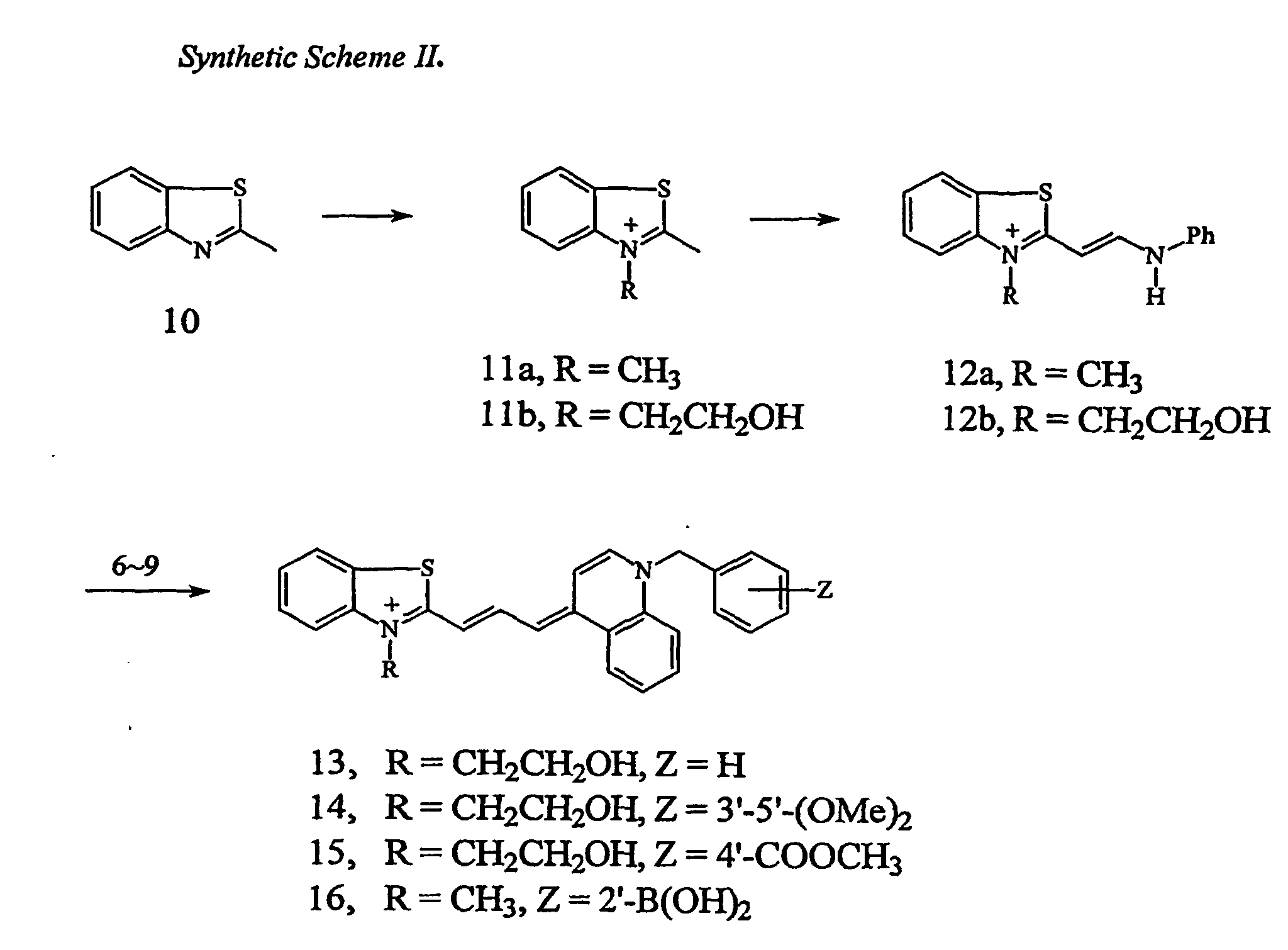
- ReticRed1 is further described in Example 1A, compound (6).
- n is 1
- R1 is H
- R2 CH 2 CH 2 OH
- R CH 2 CH 2 OH
- X is S
- B - is Br-.
- the invention provides a dye, termed ReticRed2, having the formula: With reference to the general formula, in ReticRed2 n is 1, R 1 is H, R is CH(CH 3 ) 2 , X is S, R 2 is CH 2 CH 2 OH, and B - is Br - .
- the invention provides a dye, termed ReticRed3, having the formula: With reference to the general formula, in ReticRed3 n is 1, R 1 is H, R is CH 3 , R 2 is CH 2 CH 2 OH, X is S, and B - is Br - .
- the invention provides a dye, termed ReticRed4, having the formula: With reference to the general formula, in ReticRed4, n is 1, R 1 is CH 3 , R is CH 3 , R 2 is CH 2 CH 2 OH, X is S, and B - is Br - .
- the invention provides a dye, termed ReticRed5, having the formula: With reference to the general formula, in ReticRed5, n is 1, R 1 is CH 3 , R is CH(CH 3 ) 2 , R 2 is CH 2 CH 2 OH, X is S, and B - is Br - .
- the invention provides a blue-excitable dye, termed ReticBlue1, where with reference to the general formula, n is 0.
- ReticBlue1 a blue-excitable dye
- n is 0.
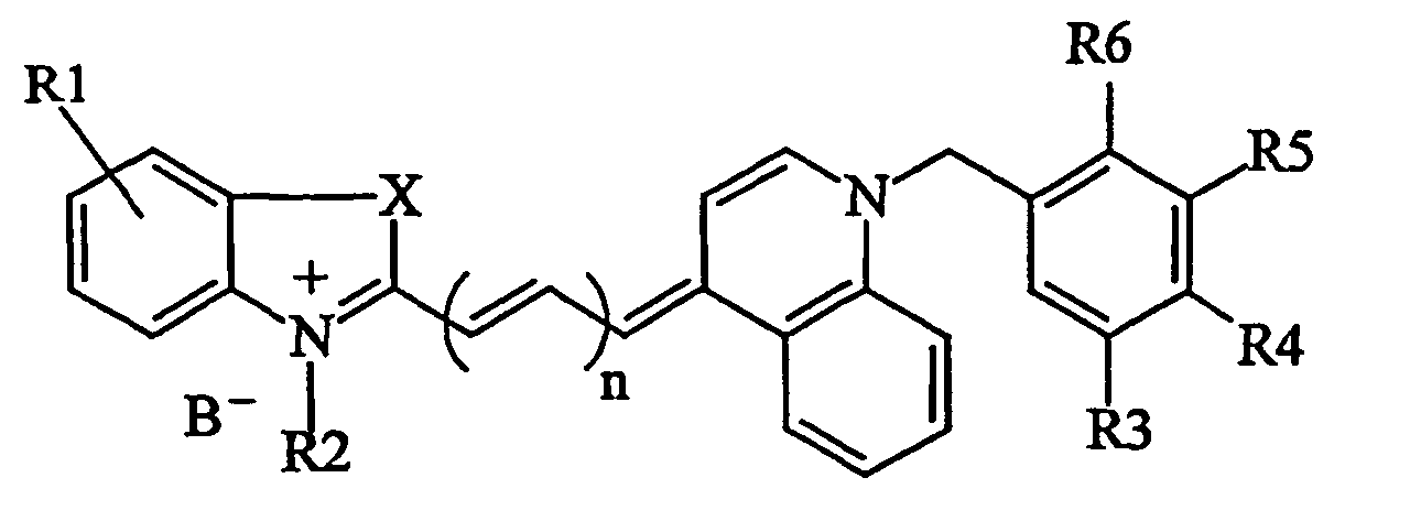
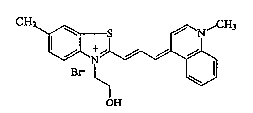
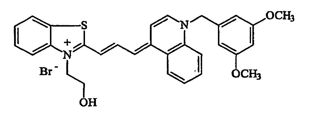
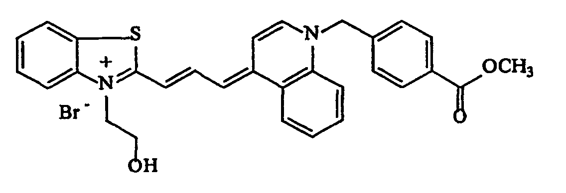
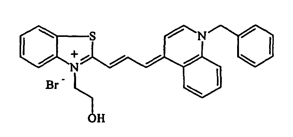
- the invention provides a dye, termed herein, Dye-3 having the following formula: Dye-3 is further described in Example I C.
- the invention provides a dye, termed Dye-4, having the formula: Dye-4 is further described in Example 1C.
- the invention provides a dye, termed Dye-5, having the formula: Dye-5 is further described in Example 1C.
- the dyes are dissolved in an appropriate solvent to form solutions.
- a solvent is meant to describe any liquid that can dissolve a solid, liquid, or gas.
- a preferred embodiment of the invention utilizes water-based or miscible liquids, such as dimethylsulfoxide (DMSO), methanol, ethanol, and mixtures thereof. Selection of a suitable solvent is however not a limitation on the present invention.
- DMSO dimethylsulfoxide
- methanol methanol
- ethanol ethanol
- Selection of a suitable solvent is however not a limitation on the present invention.
- DMSO dimethylsulfoxide
- a suitable solvent typically a relatively high concentration of the dye is dissolved in a suitable solvent to obtain a stock solution.
- the dye is dissolved in DMSO at a dye concentration ranging from 0. 5 mM to 10 mM, and most preferably, 1 to 5 mM.
- the dye composition of the invention can be utilized for a variety of purposes.
- a preferred embodiment of this invention is the use of the dye composition for staining, detection, and analysis of nucleic acids.
- Another preferred embodiment of this invention is the use of the dye composition for staining, detection, and analysis of reticulocytes in whole blood.
- Yet another preferred embodiment of this invention is the use of the dye for staining, detection and analysis of nucleated red blood cells in whole blood.
- use of the dyes and reagents of the present invention is not so limited. In one particularly desirable embodiment, for example, where the dye contacts intact cells, it can be desirable to formulate one or more of the dyes into a mixture.
- a sample be contacted with a dye composition of the invention in the presence of at least one surfactant and optionally, a sulfonic acid or a salt thereof.
- buffers may be employed in situations where maintaining the pH of the dye composition is necessary.
- the pH of the dye reagent is maintained in the range of about 6 to about 9. More preferably, a pH in the range of about 7 to about 7.5 is obtained.
- the buffer may be selected from a variety of buffers known to those of skill in the art to be used in the compositions of the invention and include, without limitation, phosphate buffered saline (PBS) or isotonic saline, such as ISOTON®II diluent, U.S. Patent 3,962,125, [Beckman Coulter, Inc., Miami, Florida], or the like.
- buffers may also be used to adjust the concentration of one or more of the components of the composition of this invention.
- Preservatives may also be added to the compositions of the invention, and may be selected from, but is not limited to, 5-Chloro-2-methyl-4-isothiazolin-3-one, and 2-methyl-4-isothiazolin-3-one [such preservatives may be purchased commercially, e.g., as ProClin 300 or ProClin 150].
- At least one surfactant is used in the rapid staining composition.
- the surfactants include a detergent and a second surfactant which functions as a sphering agent for a cell.
- the surfactants may be designed such that a single surfactant functions as both a detergent and a sphering agent.
- the surfactants may be designed such that a single surfactant provides the necessary surfactant function, but no sphering reagent function is required.
- Surfactants to be used in the composition of the invention may be selected from among the anionic surfactant ammonium perfluoralkyl carboxylate [commercially available as Fluorad® FC-143 (3M Company, Minneapolis, Minnesota)], sodium lauroyl myristoyl lactylate [commercially available as Pationic® 138C (R.I.T.A.
- the surfactant selected may function as a sphering agent for red cells when the concentration and osmolarity is appropriately adjusted.
- a sphering agent may be readily selected by one of skill in the art.
- a preferred sphering reagent is based on the non-ionic surfactant Dodecyl- ⁇ -D-maltoside, which suitably is in solution with a buffer such as phosphate buffered saline.
- the concentration of the sphering reagent in the composition is most preferably from about 3 ⁇ g/ml to about 50 ⁇ g/ml with a mOsm in the range of about 200 to about 400 mOSm, and preferably from about 250 mOsm to about 350 mOsm.
- concentration and osmolarity as needed or desired to isovolumetrically sphere the cells, taking into consideration the surfactant selected.
- the selection of the sphering reagent may eliminate the need for the detergent
- a detergent is included in the dye composition of the invention.
- Detergents to be used are selected from among non-ionic detergents. Desirably, these detergents are used at a concentration between about 0 to about 1%. In one preferred embodiment of the invention, the detergents are used at concentrations of between about 0.001% to about 0.5%, and in a more preferred embodiment are in the range from about 0.005% to 0.1%.
- One currently preferred detergent is octylphenoxypoly(ethyleneoxy)ethanol [commercially available as Igepal® CA-630 (Sigma N-6507) or Nonidet P-40 (Sigma)].
- detergents examples include, ethyoxylated octylphenol [commercially available as Triton X-100 (Sigma T9284)], and linear alcohol alkoxylates [commercially available as Plurafac® A-38 (BASF Corp) or Plurafac® A-39 (BASF Corp)].
- these detergents are mixed with a sample (or vice versa), e.g., 1-2 ⁇ l of whole blood or the like, or separately formulated into a composition of the invention.
- a sulfonic acid, or a salt thereof is included in the composition of the invention.
- the sulfonic acid or salt thereof acts as a further facilitator of transporting the dye composition through the cell membrane and is used at a concentration ranging from about 0.01 to about 250 ⁇ M, most preferably from 0.01 to about 50 ⁇ M.
- One embodiment of this invention utilizes p-toluenesulfonic acid.
- a p-toluenesulfonate salt can be substituted for p-toluenesulfonic acid.
- salts include sodium, potassium, silver, zinc, and barium p-toluenesulfonate salts.
- Some examples of such salts are commercially available, e.g., from Sigma-Aldrich. Other suitable sulfonates may be readily selected for use in the invention.
- compositions of the invention can contain a single dye, optionally in the presence of other dye.
- a composition of the invention can contain one or more of the dye compounds of this invention, or any combination thereof.
- the dye composition of the invention is useful in staining a variety of nucleic acids, including DNA and RNA.
- the dye composition of the invention can stain the nucleic acids.
- a method is provided for contacting a sample containing nucleic acids with the dye compositions of the invention.
- the method involves contacting a sample containing nucleic acids with a dye composition of the invention, optionally in the presence of other dyes and reagents known to those of skill in the art.
- the dye composition of the invention are particularly well suited for use in the detection and enumeration of reticulocytes, which contain RNA.
- the method of the invention therefore involves contacting a reticulocyte, containing nucleic acid, with one or more dye compositions of the invention.
- the method involves contacting the blood cells with a dye composition of the invention in the presence of at least one surfactant, optionally together with a preservative and sulfonic reagent.
- the surfactant(s) can include a detergent and/or a sphering agent.
- the cells can be contacted with a composition containing these components.
- one or more of these components can be delivered separately, for example by adding the component(s) directly to the sample.
- the mixture is then incubated for a suitable period of time. As previously discussed, the incubation time will be less than 1 minute. However, if desired for purposes of convenience, the incubation period may be either extended or shortened.
- the incubation time can be increased to greater than 1 minute.
- this mixing and incubation may be performed at temperatures between approximately 20°C to 40°C. However, in a preferred embodiment of this invention, temperatures ranging from 22°C to 28°C, may be utilized.
- the method of the invention requires incubation of the dye for a period ranging from above 0 seconds to about 1 minute to stain reticulocytes, and the staining can be accomplished at room temperatures.
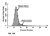
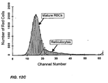
- incubation time is eliminated because the blood sample is mixed with the dye composition and thereafter immediately analyzed by the instrument. See Example 5, Fig. 7 and Fig. 8 and Example 7, Figs. 10A-2B, 11A-11C, 12A-12C.
- Prior art staining techniques using red-excitable dyes for reticulocytes in this time frame require incubation at elevated temperatures and/or additional use of toxic ionophoric compounds.
- Reticulocytes stained with the dye compositions, according to the method of the invention are preferably enumerated in an automatic flow cytometer. However, these cells may also be counted by a manual procedure or automated microscopy.
- the method of the invention facilitates transport of the nucleic acid-specific dyes of this invention across the cell membrane in the time frame desirable for automated flow cytometry.
- Automatic flow cytometers are well known in the art, and the present invention is not limited to the use of any particular flow cytometer.
- a preferred embodiment of the invention entails the detection and enumeration of reticulocytes using the XLTM flow cytometer [Beckman Coulter Inc., Miami, Florida]. Different analytical techniques can be employed in the enumeration of reticulocytes by flow cytometric measurements including, but not limited to light scatter, fluorescence, optical absorption, axial light loss, DC electrical impedance, and radio frequency (RF) conductivity.
- RF radio frequency
- light scatter gates are used to isolate red cells, and fluorescent gates are then used to delineate reticulocytes from mature red cells and enumerate the reticulocytes.
- DC and light scatter gates are used to isolate red cells, and fluorescent gates are then used to delineate reticulocytes from mature red cells and enumerate the reticulocytes.
- using flow cytometers DC and fluorescent measurement alone are used to delineate reticulocytes from mature red cells and enumerate the reticulocytes.
- the composition includes a red-excitable dye of the invention, such as ReticRed1, ReticRed3, Dye-1, Dye-2 or Dye-3
- a red-excitable dye of the invention such as ReticRed1, ReticRed3, Dye-1, Dye-2 or Dye-3
- it permits the use of a less expensive excitation source than argon lasers used for other presently available reticulocyte dyes, such as CPO, AO [Seligman, Am. J. Hematology , 14 :57 91983)], TO [Lee et. al ., Cytometry, 7 :508 (1986)], Auramine-O and Pyronin-Y.
- the red-excitable dye composition of the invention advantageously permits the use of a red laser as the excitation source.
- a red diode laser, a high power light emitting diode (LED), or a red helium-neon laser can be used to conduct reticulocyte counting using a
- the lasers may be selected from, but are not limited to, argon, helium-neon, diode, and diode pumped solid-state lasers, depending upon the excitation wavelength of the dye of the invention selected for detection of reticulocytes. Selection of suitable light sources, and appropriate excitation wavelengths, are not a limitation on the present invention.
- Suitable excitation wavelengths may be readily determined by one of skill in the art. Examples of suitable excitation wavelengths in the red spectral range include those in the range of 600 nm to 725 nm, and preferably 630 nm to 670 nm. Other suitable excitation sources and wavelengths may be readily selected by one of skill in the art, taking into consideration the dye selected for use in the method and compositions of the invention.
- red-excitable dye compounds of this invention such as ReticRed1, ReticRed2, ReticRed3, ReticRed4, ReticRed5, ReticRed6, Dye-1, Dye-2 or Dye-3 etc.
- ReticRed1, ReticRed2, ReticRed3, ReticRed4, ReticRed5, ReticRed6, Dye-1, Dye-2 or Dye-3 etc. are essentially non-fluorescent or very weakly fluorescent in aqueous solutions in the absence of nucleic acids (unbound state).
- problems associated with background fluorescence are minimal, and reticulocytes can be detected with high sensitivity using this dye.
- the present invention permits reticulocytes to be rapidly stained with the dyes of the invention for subsequent flow analysis.
- the method of the invention differs from both the prior art Pyronin-Y [Tanke et al, cited above] staining procedure requiring fixation of the cells and the prior art fluorescence staining procedures involving membrane permeable dyes such as CPO (reticONETM) or Thiazole Orange (Retic-CountTM) that does not require cell-fixation, but requires long incubation with the blood to accomplish staining.
- the solid was then triturated with acetone (100 mL). The resultant solid was collected, washed with ethyl acetate (2 X 50 mL) and dried in the air. The filtrate was concentrated to a small volume and a second crop of solid was also collected. The combined solid was dried in an oven under high vacuum at 50°C overnight. There was obtained 7.26 g (84%) of product.
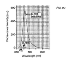
- Solutions of the dyes of this invention bound to RNA were prepared by combining 2 ⁇ l of the 5mM stock solution of the dye and a 5 ml solution of RNA dissolved in PBS at a RNA concentration of 1 mg/ml. Fluorescence spectrum of each Dye/RNA solution was measured with a spectrofluorometer using an excitation wavelength of 633 nm.
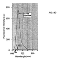
- Solutions of the dyes of this invention bound to DNA were prepared by combining 2 ⁇ l of the 5mM stock solution of the dye and a 5 ml solution of DNA dissolved in PBS at a DNA concentration of 0.5 mg/ml Fluorescence spectrum of each Dye/DNA solution was measured with a spectrofluorometer using an excitation wavelength of 633 nm.
- a fluorescent dye As a suitable candidate for reticulocyte enumeration, the fluorescence properties of several different compounds were determined. For reticulocyte enumeration, it is most desirable to have dyes that are weakly fluorescent when they are not bound to RNA, but show significant enhancement in fluorescence intensity when they are bound to RNA. However, it is not possible to predict which dye compound will have this ideal combination effects on its fluorescence activity both in absence and in presence of RNA. Significantly, the inventors found that the dyes of the present invention had favorable fluorescence properties required for sensitive detection of RNA.
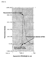
- Fig. 1B comparison of fluorescence intensity of several compounds in presence of RNA in solution are presented.
- the fluorescence intensities of the dyes are shown relative to the fluorescence intensity of a commercial dye Syto 62 (Molecular Probes Inc.) used herein as a reference standard.
- Syto 62 Molecular Probes Inc.
- ReticRed1 For measurements using ReticRed1, 1 ⁇ l of whole blood was added to 1 ml of an isotonic saline solution containing a 5 ⁇ M ReticRed1 (diluted from the 5 mM stock solution described in Example 1 C) solution and incubated for 10 and 40 minutes. The sample was then analyzed in a XLTM flow cytometer modified to incorporate a red HeNe laser (632.8 nm) as described in Example 1E. When whole blood was treated with ReticRed1 and analyzed in flow, red fluorescence in the 660 nm band resulted from a population of red cells, and indicated the presence of nucleic acid bound to ReticRed1 in such cells.
- 1 ⁇ l of whole blood (from the same donor whose blood was used to conduct the analyses described in example 3 above) was added to 1ml of a solution comprising a sphering reagent that spheres the red cells, the detergent Igepal (Sigma CA-630) at a concentration of 0.01%, the dye ReticRed1 at a concentration of 5 ⁇ M, and a solution of p-toluenesulfonic acid monohydrate at a final concentration of 5 ⁇ M, and incubated for about 1 minute.
- the sample was then analyzed in a XLTM flow cytometer modified to incorporate a red HeNe laser (632.8 nm).
- the sphering reagent is a solution comprising about 20 ⁇ g/ml Dodecyl- ⁇ -D-maltoside and 0.05% Proclin 300 in PBS at about pH 7.4 and about 290 mOsm.
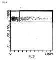
- the sample so stained with the dye ReticRed1 was then analyzed in a flow cytometer using 632.8 nm excitation from a HeNe laser. The result of this experiment is shown in Fig. 4.
- the distribution of the cells in this forward scatter versus fluorescence dotplot clearly shows the reticulocytes with high fluorescence intensity.
- the percentage of reticulocytes enumerated by this experiment was 7.5 %.
- An independent reference value for reticulocyte percentage in this sample was 8%.
- reticulocytes are distinguished from the mature red blood cells by the higher fluorescence intensities associated with them.
- the percentage of reticulocytes enumerated by this experiment was 13.8 %.
- An independent reference value for reticulocyte percentage measured for this sample using the conventional Retic-CountTM reticulocyte enumeration method (using Thiazole orange excited at 488 nm) in a FACSCAN flow cytometer was 13.3%.
- Fig. 8 The result of this experiment is shown in Fig. 8.
- fluorescence measurements were conducted in a spectrofluorometer by exciting each dye solution at 633 nm under appropriate conditions, recording the fluorescence spectrum of each sample, and measuring the intensities at the peak of these fluorescence spectra.
- a stock solution of each compound in DMSO was prepared (as described in Example 1D).
- a portion of the stock solution for the Dye-1 was diluted in PBS to obtain a dye concentration of 2 ⁇ l.
- 1 ml of this dilute solution was placed in a glass cuvette and fluorescence spectrum of this solution was measured by a spectrofluorometer using an excitation wavelength of 633 nm.
- RNA solution 1ml of a solution of free Torula yeast RNA (Sigma) dissolved in PBS at a concentration of 1 mg/ml, and a measured volume of the dye stock solution of the Dye-1 was mixed to obtain a dye concentration of 2 ⁇ M in the RNA solution.
- the fluorescence spectrum of this dye solution in presence of RNA was measured by illuminating the sample at 633 nm.
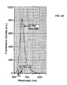
- Fig. 9A shows the comparison of the fluorescence spectra of the dye Dye-1 in PBS solution with (see the upper curve) and without RNA (see the lower curve) present in the solution.
- RNA solution For fluorescence measurements in DNA, 1ml of a solution of free Calf thymus DNA (Sigma) dissolved in PBS at a concentration of 0.5 mg/ml, and a measured volume of the dye stock solution was mixed to obtain a dye concentration of 2 ⁇ M in the RNA solution. Next, the fluorescence spectrum of the dye solution was measured by illuminating the sample at 633 nm.
- Fig. 9B shows the comparison of the fluorescence spectra of the dye Dye-1 in PBS solution with (see the upper curve) and without DNA (see the lower curve) present in the solution.
- Table 3 compares the relative fluorescence intensities of the dyes of the present invention, Dye-1 and Dye-2 (concentration 2 ⁇ M), when they are not bound to RNA (unbound to RNA), when they are in presence of RNA (bound to RNA), and when they are in presence of DNA (bound to DNA). These intensities were obtained at the peak of the respective fluorescence spectra as described above. DYE (conc. 2 ⁇ m) Fluorescence Intensity Unbound to RNA Bound to RNA 1mg/ml Bound to DNA 0.5mg/ml Dye-1 10 825 578 Dye-2 18 560 760
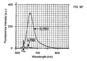
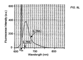
- Table 4 compares the fluorescence intensities of the Group-III dyes (Dye-3 through Dye-6) of the present invention, when they are not bound to RNA (unbound to RNA) and when they are in presence of RNA (bound to RNA). The intensities were measured at the peak of the emission spectra.
- Table-5 compares the fluorescence intensities of the Group-III dyes (Dye-3 through Dye-6) of the present invention, when they are not bound to DNA (unbound to DNA) and when they are in presence of DNA (bound to DNA). The intensities were measured at the peak of the emission spectra.
- DYE (conc. 2 ⁇ m) Fluorescence Intensity Unbound to RNA Bound to RNA 1mg/ml Dye-3 10 328 Dye-4 12 370 Dye-5 10 420 Dye-6 6 410 DYE Fluorescence Intensity Unbound to DNA Bound to DNA 0.5 mg/ml Dye-3 15 400
- Dye-4 12 440 Dye-5 10 660 Dye-6 4 380
- Example 7 Rapid staining of reticulocytes with Dye-1 using whole blood samples
- a solution comprising a sphering reagent that spheres the red cells, the detergent Igepal (Sigma CA-630) at a concentration of 0.01%, the dye Dye-1 at a concentration of 5 ⁇ M, and aspirated, immediately thereafter, into an XLTM flow cytometer, modified to incorporate a red HeNe laser (632.8 nm), for analysis.
- the sphering reagent is a solution comprising about 20 ⁇ g/ml Dodecyl- ⁇ -D-maltoside and 0.05% Proclin 300 in PBS at about pH 7.4 and about 290 mOsm.
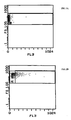
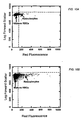
- Fig. 10A shows the fluorescence from reticulocytes relative to the mature red blood cells for this sample.
- the percentage of reticulocytes enumerated by the present method was 1.9%, which was within the expected normal range.
- Fig. 10B shows the fluorescence from reticulocytes relative to the mature red blood cells for this sample.
- the distribution of the cells in this forward scatter versus fluorescence dotplot clearly shows the reticulocytes with relatively high fluorescence intensity.
- the percentage of reticulocytes enumerated by this experiment was 10.6%
- An independent reference value for reticulocyte percentage in this sample was 12%.
- Fig. 10C shows the fluorescence from reticulocytes relative to the mature red blood cells for this sample.
- the distribution of the cells in this forward scatter versus fluorescence dotplot clearly shows the reticulocytes with high fluorescence intensity.
- the percentage of reticulocytes enumerated by this experiment was 26.3%.
- An independent reference value for reticulocyte percentage in this sample was 27%.
- Example 8 Rapid staining of reticulocytes with Dye-2 using whole blood
- Dye-2 can also rapidly pass through the cell membrane of reticulocytes in presence of the reagent compositions of this invention, thus reducing the incubation time for the staining significantly.
- Experiments using three different blood samples are presented to demonstrate rapid staining of reticulocytes by the dye Dye-2. We refer to these three experiments as Run 4, Run 5 and Run 6 respectively.
- a solution comprising a sphering reagent that spheres the red cells, the detergent Igepal (Sigma CA-630) at a concentration of 0.01%, the dye Dye-2 at a concentration of 5 ⁇ M, and aspirated, immediately thereafter, into an XLTM flow cytometer, modified to incorporate a red HeNe laser (632.8 nm), for analysis.
- the sphering reagent is a solution comprising about 20 ⁇ g/ml Dodecyl- ⁇ -D-maltoside and 0.05% Proclin 300 in PBS at about pH 7.4 and about 290 mOsm.
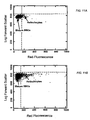
- Fig. 11A shows the fluorescence from reticulocytes relative to the mature red blood cells for this sample.
- the percentage of reticulocytes enumerated by the present method was 1.8%.
- Fig. 11B shows the fluorescence from reticulocytes relative to the mature red blood cells for this sample.
- the distribution of the cells in this forward scatter versus fluorescence dotplot clearly shows the reticulocytes with high fluorescence intensity.
- the percentage of reticulocytes enumerated by this experiment was 12.5%.
- An independent reference value for reticulocyte percentage in this sample was 12%.
- Fig. 11C shows the fluorescence from reticulocytes relative to the mature red blood cells for this sample.
- the distribution of the cells in this forward scatter versus fluorescence dotplot clearly shows the reticulocytes with high fluorescence intensity.
- the percentage of reticulocytes enumerated by this experiment was 24 %.
- An independent reference value for reticulocyte percentage in this sample was 27%.
- Example 9 Rapid staining of reticulocytes with Dye-3 using whole blood samples
- a solution comprising a sphering reagent that spheres the red cells, the detergent Igepal (Sigma CA-630) at a concentration of 0.01%, the dye Dye-3 at a concentration of 5 ⁇ M, and aspirated, immediately thereafter, into an XLTM flow cytometer, modified to incorporate a red HeNe laser (632.8 nm), for analysis.
- the sphering reagent is a solution comprising about 20 ⁇ g/ml Dodecyl- ⁇ -D-maltoside and 0.05% Proclin® 300 in PBS at about pH 7.4 and about 290 mOsm. The percentage of reticulocytes enumerated by the present method was 2.3% (see Fig. 4A).
Landscapes
- Health & Medical Sciences (AREA)
- Chemical & Material Sciences (AREA)
- Physics & Mathematics (AREA)
- Life Sciences & Earth Sciences (AREA)
- Immunology (AREA)
- Pathology (AREA)
- General Physics & Mathematics (AREA)
- Analytical Chemistry (AREA)
- Biochemistry (AREA)
- General Health & Medical Sciences (AREA)
- Engineering & Computer Science (AREA)
- Biomedical Technology (AREA)
- Molecular Biology (AREA)
- Chemical Kinetics & Catalysis (AREA)
- Optics & Photonics (AREA)
- Nuclear Medicine, Radiotherapy & Molecular Imaging (AREA)
- Organic Chemistry (AREA)
- Investigating Or Analysing Biological Materials (AREA)
- Measuring Or Testing Involving Enzymes Or Micro-Organisms (AREA)
Description

wherein m is 0, 1, 2, or 3; X is O, S, or C(CH3)2; R is CH3, CH(CH3)2, CH2CH2OH, alkyl, alkylsulfonate, or hydroxyalkyl; and B- is a counteranion.
and,

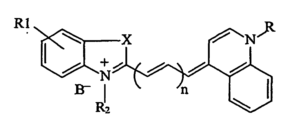
wherein m is 0, 1, 2, or 3; X is O, S, or C(CH3)2; R is CH3, CH(CH3)2, CH2CH2OH, alkyl, alkylsulfonate, or hydroxyalkyl; and B- is a counteranion.







| DYE | Fluorescence Intensity | Fluorescence enhancement ratio (Bound/Unbound) | |
| Unbound to RNA | Bound to RNA | ||
| ReticRed1 | 2.49 | 791.23 | 317.763 |
| ReticRed2 | 1.72 | 643.77 | 374.285 |
| ReticRed3 | 1.28 | 639.68 | 499.75 |
| ReticRed4 | 1.72 | 713.4 | 414.767 |
| ReticRed5 | 2.24 | 656.06 | 292.884 |
| ReticRed6 | 2.75 | 619.2 | 225.164 |

| DYE (conc. 2 µm) | Fluorescence Intensity | ||
| Unbound to RNA | Bound to RNA 1mg/ml | Bound to DNA 0.5mg/ml | |
| Dye-1 | 10 | 825 | 578 |
| Dye-2 | 18 | 560 | 760 |
| DYE (conc. 2 µm) | Fluorescence Intensity | |
| Unbound to RNA | Bound to RNA 1mg/ml | |
| Dye-3 | 10 | 328 |
| Dye-4 | 12 | 370 |
| Dye-5 | 10 | 420 |
| Dye-6 | 6 | 410 |
| DYE | Fluorescence Intensity | |
| Unbound to DNA | Bound to DNA 0.5 mg/ml | |
| Dye-3 | 15 | 400 |
| Dye-4 | 12 | 440 |
| Dye-5 | 10 | 660 |
| Dye-6 | 4 | 380 |
Claims (24)
- A dye composition comprising(a) a dye having the formula:wherein,

n is 0, 1, 2, or 3; R1 is H, alkyl, or an alkoxy group; R2 is CH2(CH2)mOH, wherein m is 0, 1, 2, or 3; X is O, S, or C(CH3)2; R is CH3, CH(CH3)2, CH2CH2OH, alkyl, alkylsulfonate, or hydroxyalkyl and B- is a counteranion; and(b) one or more components selected from the group consisting of a surfactant, a preservative and a sulfonic acid or a salt thereof. - The dye composition according to claim 1, wherein n is 1, R1 is H, R is CH2CH2OH, R2 is CH2CH2OH, and X is S.
- The dye composition according to claim 1, wherein n is 1, R1 is H, R is CH(CH3)2, R2 is CH2CH2OH and X is S.
- The dye composition according to claim 1, wherein n is 1, R1 is H, R is CH3, R2 is CH2CH2OH, and X is S.
- The dye composition according to claim 1, wherein n is 1, R1 is CH3, R is CH3, R2 is CH2CH2OH, and X is S.
- The dye composition according to claim 1, wherein n is 1, R1 is CH3, R is CH(CH3)2, R2 is CH2CH2OH, and X is S.
- The dye composition according to claim 1, wherein n is 1, R1 is CH3, R is CH2CH2OH, R2 is CH2CH2OH, and X is S.
- The dye composition according to claim 1, wherein said component is a surfactant.
- The dye composition according to claim 1, wherein said component is a sulfonic acid or a salt thereof.
- A method for staining nucleic acid comprising the step of contacting a sample of blood cells containing nucleic acid with a dye having the formula:wherein,
n is 0, 1, 2, or 3; R1 is H, alkyl, or an alkoxy group; R2 is CH2(CH2)mOH, wherein m is 0, 1, 2, or 3; X is O, S, or C(CH3)2; R is CH3, CH(CH3)2, CH2CH2OH, alkyl, alkylsulfonate, or hydroxyalkyl, and B- is a counteranion. - The method according to claim 10, wherein said blood cells comprise nucleated red blood cells or reticulocytes.
- A dye having the formula:wherein, n is 0, 1, 2, or 3; R1 is H, alkyl, or an alkoxy group; R2 is CH2(CH2)mOAc,

wherein m is 0, 1, 2, or 3; X is O, S, or C(CH3)2; R is CH3, CH(CH3)2, CH2CH2OAc, alkyl, alkylsulfonate, or hydroxyalkyl and B- is a counteranion. - A dye according to the formula:wherein n is 0, 1, or 2; R1 is H, alkyl, or an alkoxy group; R2 is CH2(CH2)mOH, or CH3 ; X is O, S, or C(CH3)2; B- is a counter anion, and R3, R4, R5, R6 are various substituents as represented by the formulae for the specific compounds Dye-3 through Dye-6.

- The dye according to claim 12, wherein n = 1; R1 = H; R = CH3, R2 = CH2CH2OAc; X = S, and B- is Br-.
- The dye according to claim 12, wherein n = 1, R1 = H, R2 = R = CH2CH2OAc, X = S, and B- is Br-.
- The dye according to claim 13, wherein n= 1; X = S; R1= CH2CH2OH; R2 = R4 = R5=H; R3=CO.CH3; and B- is Br-.
- The dye according to claim 13, wherein n= 1; X = S; R1= CH2CH2OH; R2 = R4 = R5=H; R3=CO.CH3; and B- is Br-.
- The dye according to claim 13, wherein n= 1; X = S; R1= CH2CH2OH; R2= R3= R4= R5= H; and B- is Br-.
- The dye according to claim 13, wherein n=1; X = S; R1= CH2CH2OH; R2= R3= R4= H; R5= B(OH)2; and B- is I-.
- The dye composition for staining reticulocytes according to claims 12 or 13, further comprising a surfactant.
- The dye composition according to claims 12 or 13, further comprising a sulfonic acid or a salt thereof.
- The dye composition according to claims 12 or 13, further comprising a preservative.
- A method for analysis of a blood cell sample comprising the steps of:(a) contacting a blood cell sample containing nucleated red blood cells or reticulocytes with a compound according to claim 12 or claim 13 such that the nucleated red blood cells or reticulocytes are stained by the said compound; and (b) analyzing the stained blood cell sample by flow cytometry to detect the presence of reticulocytes.
- The method according to claim 23, wherein said analysis by flow cytometry includes measurement of one fluorescence parameter and at least one parameter selected from the group consisting of light scatter, axial light loss, DC electrical impedance, and radio frequency (RF) conductivity and combinations thereof.
Applications Claiming Priority (3)
| Application Number | Priority Date | Filing Date | Title |
|---|---|---|---|
| US09/566,298 US6368864B1 (en) | 2000-05-05 | 2000-05-05 | Dyes and methods of reticulocyte enumeration |
| US566298 | 2000-05-05 | ||
| PCT/US2001/014522 WO2001086264A1 (en) | 2000-05-05 | 2001-05-04 | Dyes and methods of detection of nucleic acid in immature red blood cells |
Publications (3)
| Publication Number | Publication Date |
|---|---|
| EP1303751A1 EP1303751A1 (en) | 2003-04-23 |
| EP1303751A4 EP1303751A4 (en) | 2003-05-28 |
| EP1303751B1 true EP1303751B1 (en) | 2005-08-10 |
Family
ID=24262311
Family Applications (1)
| Application Number | Title | Priority Date | Filing Date |
|---|---|---|---|
| EP01933047A Expired - Lifetime EP1303751B1 (en) | 2000-05-05 | 2001-05-04 | Dyes and methods of detection of nucleic acid in immature red blood cells |
Country Status (5)
| Country | Link |
|---|---|
| US (2) | US6368864B1 (en) |
| EP (1) | EP1303751B1 (en) |
| JP (1) | JP4831914B2 (en) |
| DE (1) | DE60112585T2 (en) |
| WO (1) | WO2001086264A1 (en) |
Families Citing this family (35)
| Publication number | Priority date | Publication date | Assignee | Title |
|---|---|---|---|---|
| US6495692B1 (en) * | 1996-12-10 | 2002-12-17 | Abbott Laboratories | Helium-neon excitable reticulocyte dyes derivable from halolepidines |
| US6368864B1 (en) * | 2000-05-05 | 2002-04-09 | Coulter International Corp. | Dyes and methods of reticulocyte enumeration |
| EP1625385A1 (en) * | 2003-05-07 | 2006-02-15 | Novasite Pharmaceuticals, Inc. | Multiplexed multitarget screening method |
| ATE520661T1 (en) * | 2003-06-27 | 2011-09-15 | Univ Maryland | HETEROCYCLIC COMPOUNDS WITH QUATERNARY NITROGEN FOR THE DETECTION OF AQUEOUS MONOSACHARIDES IN PHYSIOLOGICAL LIQUIDS |
| US7446202B2 (en) | 2003-12-05 | 2008-11-04 | Molecular Probes, Inc. | Cyanine dye compounds |
| US7776529B2 (en) | 2003-12-05 | 2010-08-17 | Life Technologies Corporation | Methine-substituted cyanine dye compounds |
| JP3881667B2 (en) | 2003-12-24 | 2007-02-14 | 信一郎 礒部 | Biomolecule detection method and labeling dye and labeling kit used therefor |
| EP1712911B1 (en) | 2003-12-24 | 2011-08-17 | Shinichiro Isobe | Method for detecting biomolecule, labeling dye used therefor, and labeling kit |
| EP1885718B1 (en) | 2005-05-11 | 2017-03-15 | Life Technologies Corporation | Fluorescent chemical compounds having high selectivity for double stranded dna, and methods for their use |
| JP5214603B2 (en) | 2007-06-25 | 2013-06-19 | シスメックス株式会社 | Reagent for analysis of immature leukocytes and reagent kit for analysis |
| MX2009012997A (en) | 2007-06-25 | 2010-03-30 | Sysmex Corp | Reagent and reagent kit for analysis of primitive leukocyte. |
| CN101349644B (en) * | 2007-07-20 | 2012-06-27 | 深圳迈瑞生物医疗电子股份有限公司 | Leukocytes classification agent and use method thereof |
| US8102161B2 (en) * | 2007-09-25 | 2012-01-24 | Tdk Corporation | Stable output in a switching power supply by smoothing the output of the secondary coil |
| US20100041045A1 (en) * | 2007-11-15 | 2010-02-18 | Sigma-Aldrich Co. | Nucleic acid fluorescent stains |
| US20090131279A1 (en) * | 2007-11-15 | 2009-05-21 | Sigma Aldrich Company | Nucleic acid fluorescent stains |
| CN101475754A (en) * | 2008-01-04 | 2009-07-08 | 深圳迈瑞生物医疗电子股份有限公司 | Asymmetric cyanine fluorescent dye, composition and application in biological sample dyeing |
| CN101602762B (en) * | 2008-06-10 | 2013-10-16 | 深圳迈瑞生物医疗电子股份有限公司 | Asymmetric cyanine compound, preparation method and application thereof |
| CN101726579B (en) * | 2008-10-17 | 2014-06-18 | 深圳迈瑞生物医疗电子股份有限公司 | Blood test reagent and method |
| CN101723874B (en) * | 2008-10-31 | 2013-09-11 | 深圳迈瑞生物医疗电子股份有限公司 | Cyanine compound and application thereof in dyeing biological samples |
| CN101750476B (en) * | 2008-12-08 | 2015-06-03 | 深圳迈瑞生物医疗电子股份有限公司 | Blood analysis reagent and use method thereof |
| CN102308214B (en) | 2008-12-12 | 2014-06-25 | 贝克曼考尔特公司 | Multicolor flow cytometry compositions comprising unconjugated phycobiliproteins |
| US8367358B2 (en) * | 2008-12-17 | 2013-02-05 | Shenzhen Mindray Bio-Medical Electronics Co., Ltd. | Reagent, kit and method for differentiating and counting leukocytes |
| US7943320B2 (en) * | 2008-12-30 | 2011-05-17 | Canon U.S. Life Sciences, Inc. | Unsymmetrical cyanine dyes for high resolution nucleic acid melting analysis |
| CN101988082B (en) * | 2009-07-31 | 2015-04-08 | 深圳迈瑞生物医疗电子股份有限公司 | Leukocyte classified counting reagent, kit and preparation method thereof and method of leukocyte classified counting |
| US9097704B2 (en) | 2010-05-05 | 2015-08-04 | Abbott Laboratories | Method for hematology analysis |
| CN103917868B (en) | 2011-05-04 | 2016-08-24 | 雅培制药有限公司 | Basophilic granulocyte analyzes system and method |
| US9091624B2 (en) | 2011-05-04 | 2015-07-28 | Abbott Laboratories | White blood cell analysis system and method |
| ES2902648T3 (en) | 2011-05-04 | 2022-03-29 | Abbott Lab | Nucleated Red Blood Cell Analysis Method and Automated Hematology Analyzer |
| CN103172590A (en) * | 2013-03-21 | 2013-06-26 | 常州大学 | Squaraines chemical sensor for colorimetrically identifying Fe and Cu ion and preparation method thereof |
| CN104749144A (en) * | 2013-12-31 | 2015-07-01 | 深圳迈瑞生物医疗电子股份有限公司 | Blood cell detection reagent, blood cell processing method and blood cell identification method |
| CN106687810B (en) * | 2014-12-31 | 2019-10-22 | 深圳迈瑞生物医疗电子股份有限公司 | A non-diagnostic nucleated red blood cell alarm method, device and flow cytometer |
| EP3371591A4 (en) * | 2015-11-02 | 2018-11-14 | Chiranjit Deka | Light scatter based apparatus and methods for hematology analysis using only three detectors |
| CN111602046B (en) * | 2018-04-28 | 2024-01-09 | 深圳迈瑞生物医疗电子股份有限公司 | A blood analyzer and analysis method |
| DE102020000677B4 (en) | 2020-02-01 | 2023-01-12 | Hochschule Anhalt | Universal flow meter and method of controlling a universal flow meter |
| CN117529646A (en) * | 2021-12-31 | 2024-02-06 | 深圳迈瑞动物医疗科技股份有限公司 | Animal reticulocyte detection method and sample analyzer |
Family Cites Families (26)
| Publication number | Priority date | Publication date | Assignee | Title |
|---|---|---|---|---|
| US3962125A (en) | 1975-01-13 | 1976-06-08 | Coulter Diagnostics, Inc. | Multi-purpose diluent for use in blood analysis by electronic instrumentation of the coulter type |
| JPS5772150A (en) * | 1980-10-23 | 1982-05-06 | Ishihara Sangyo Kaisha Ltd | Electrophotographic sensitive material |
| US4957870A (en) | 1985-11-01 | 1990-09-18 | Becton, Dickinson And Company | Detection of Reticulocytes, RNA and DNA |
| US4883867A (en) | 1985-11-01 | 1989-11-28 | Becton, Dickinson And Company | Detection of reticulocytes, RNA or DNA |
| DE3677916D1 (en) * | 1985-11-01 | 1991-04-11 | Becton Dickinson Co | DETECTION OF RETICULOYCYTES. |
| US5312921A (en) * | 1990-03-14 | 1994-05-17 | Regents Of The University Of California | Dyes designed for high sensitivity detection of double-stranded DNA |
| US5360739A (en) | 1991-12-05 | 1994-11-01 | Miles Inc. | Methods for the identification and characterization of reticulocytes in whole blood |
| US5284771A (en) | 1991-12-05 | 1994-02-08 | Miles Inc. | Reagent compositions and their use in sphering cells |
| US5321130A (en) * | 1992-02-10 | 1994-06-14 | Molecular Probes, Inc. | Unsymmetrical cyanine dyes with a cationic side chain |
| JPH06242533A (en) * | 1993-02-15 | 1994-09-02 | Konica Corp | Silver halide photographic sensitive material |
| US5436134A (en) * | 1993-04-13 | 1995-07-25 | Molecular Probes, Inc. | Cyclic-substituted unsymmetrical cyanine dyes |
| US5658751A (en) * | 1993-04-13 | 1997-08-19 | Molecular Probes, Inc. | Substituted unsymmetrical cyanine dyes with selected permeability |
| US5563070A (en) * | 1993-05-28 | 1996-10-08 | Omron Corporation | Method of counting reticulocytes |
| US5808077A (en) * | 1993-06-30 | 1998-09-15 | Abbott Laboratories | Intercalators having affinity for DNA and methods of use |
| IL113805A0 (en) | 1994-05-23 | 1995-08-31 | Coulter Corp | Detection of reticulocytes |
| US5691204A (en) | 1995-04-21 | 1997-11-25 | Abbott Laboratories | Compositions and methods for the rapid analysis of reticulocytes |
| JP3425830B2 (en) | 1995-10-06 | 2003-07-14 | シスメックス株式会社 | New compounds and their uses |
| JP3485436B2 (en) * | 1996-04-12 | 2004-01-13 | シスメックス株式会社 | Reticulocyte measuring reagent and measuring method |
| TW379284B (en) * | 1996-04-12 | 2000-01-11 | Toa Medical Electronics | Agent for detecting reticulocyte |
| US6495692B1 (en) * | 1996-12-10 | 2002-12-17 | Abbott Laboratories | Helium-neon excitable reticulocyte dyes derivable from halolepidines |
| FR2759166B1 (en) | 1997-01-31 | 1999-04-23 | Abx Sa | COLOR REAGENT FOR THE DETERMINATION OF BLOOD CELLS |
| US6002003A (en) * | 1998-04-14 | 1999-12-14 | Beckman Instruments, Inc. | Cyanine dye activating group with improved coupling selectivity |
| US6271035B1 (en) * | 1998-10-20 | 2001-08-07 | Coulter International Corp. | Methods and compositions for rapid staining of nucleic acids in whole cells |
| US6197593B1 (en) * | 1998-10-20 | 2001-03-06 | Coulter International Corp. | Method for enumerating blood cells |
| JP3886271B2 (en) | 1998-11-27 | 2007-02-28 | シスメックス株式会社 | Reagent for classification counting of erythroblast and classification counting method |
| US6368864B1 (en) * | 2000-05-05 | 2002-04-09 | Coulter International Corp. | Dyes and methods of reticulocyte enumeration |
-
2000
- 2000-05-05 US US09/566,298 patent/US6368864B1/en not_active Expired - Lifetime
-
2001
- 2001-05-04 US US09/849,155 patent/US6551831B2/en not_active Expired - Lifetime
- 2001-05-04 WO PCT/US2001/014522 patent/WO2001086264A1/en not_active Ceased
- 2001-05-04 EP EP01933047A patent/EP1303751B1/en not_active Expired - Lifetime
- 2001-05-04 JP JP2001583158A patent/JP4831914B2/en not_active Expired - Fee Related
- 2001-05-04 DE DE60112585T patent/DE60112585T2/en not_active Expired - Lifetime
Also Published As
| Publication number | Publication date |
|---|---|
| US6551831B2 (en) | 2003-04-22 |
| EP1303751A4 (en) | 2003-05-28 |
| DE60112585D1 (en) | 2005-09-15 |
| US20020037589A1 (en) | 2002-03-28 |
| JP2003532790A (en) | 2003-11-05 |
| WO2001086264A1 (en) | 2001-11-15 |
| EP1303751A1 (en) | 2003-04-23 |
| DE60112585T2 (en) | 2006-05-24 |
| JP4831914B2 (en) | 2011-12-07 |
| US6368864B1 (en) | 2002-04-09 |
Similar Documents
| Publication | Publication Date | Title |
|---|---|---|
| US6551831B2 (en) | Dyes and methods of detection of nucleic acid in immature red blood cells | |
| US8367358B2 (en) | Reagent, kit and method for differentiating and counting leukocytes | |
| CN101349644B (en) | Leukocytes classification agent and use method thereof | |
| US8940499B2 (en) | Reagent for blood analysis and method of use thereof | |
| US6004816A (en) | Reagent and method for classification and counting of leukocytes | |
| US4883867A (en) | Detection of reticulocytes, RNA or DNA | |
| EP1542008B1 (en) | Method of classifying and counting leucocytes | |
| EP0767382B1 (en) | Fluorescent compounds and their use for measuring reticulocytes | |
| US5563070A (en) | Method of counting reticulocytes | |
| US4957870A (en) | Detection of Reticulocytes, RNA and DNA | |
| US6955872B2 (en) | Dye compositions which provide enhanced differential fluorescence and light scatter characteristics | |
| EP0513762B1 (en) | Reagent and method for analyzing cells in urine | |
| US5691204A (en) | Compositions and methods for the rapid analysis of reticulocytes | |
| EP1890143B1 (en) | Reagent for sample analysis, reagent kit for sample analysis and method for sample analysis | |
| US8273329B2 (en) | Cyanine compounds, compositions including these compounds and their use in cell analysis | |
| US6271035B1 (en) | Methods and compositions for rapid staining of nucleic acids in whole cells | |
| CN101750476A (en) | Blood analysis reagent and use method thereof | |
| US5804448A (en) | Method of staining cellular material and analyzing the same | |
| CA1155041A (en) | Fluorescent nucleic acid stains | |
| EP4245812A1 (en) | Leukocyte classification reagent, erythrocyte analysis reagent, kits, and analysis method | |
| CN120721608A (en) | Urine sample analysis method and urine sample analysis reagent |
Legal Events
| Date | Code | Title | Description |
|---|---|---|---|
| PUAI | Public reference made under article 153(3) epc to a published international application that has entered the european phase |
Free format text: ORIGINAL CODE: 0009012 |
|
| 17P | Request for examination filed |
Effective date: 20021128 |
|
| AK | Designated contracting states |
Designated state(s): AT BE CH CY DE DK ES FI FR GB GR IE IT LI LU MC NL PT SE TR |
|
| RIN1 | Information on inventor provided before grant (corrected) |
Inventor name: GUPTA, RAVINDER Inventor name: TSAY, TSONG-TSEH Inventor name: SZYDLO, STEPHEN Inventor name: SHEN, GENE, G.-Y. Inventor name: DEKA, CHIRANJIT Inventor name: LEE, SONG, Y. |
|
| A4 | Supplementary search report drawn up and despatched |
Effective date: 20030415 |
|
| RIC1 | Information provided on ipc code assigned before grant |
Ipc: 7G 01N 33/48 B Ipc: 7G 01N 21/76 A Ipc: 7C 09B 23/00 B |
|
| RBV | Designated contracting states (corrected) |
Designated state(s): DE FR GB |
|
| 17Q | First examination report despatched |
Effective date: 20040810 |
|
| GRAP | Despatch of communication of intention to grant a patent |
Free format text: ORIGINAL CODE: EPIDOSNIGR1 |
|
| GRAS | Grant fee paid |
Free format text: ORIGINAL CODE: EPIDOSNIGR3 |
|
| GRAA | (expected) grant |
Free format text: ORIGINAL CODE: 0009210 |
|
| AK | Designated contracting states |
Kind code of ref document: B1 Designated state(s): DE FR GB |
|
| REG | Reference to a national code |
Ref country code: GB Ref legal event code: FG4D |
|
| REF | Corresponds to: |
Ref document number: 60112585 Country of ref document: DE Date of ref document: 20050915 Kind code of ref document: P |
|
| ET | Fr: translation filed | ||
| PLBE | No opposition filed within time limit |
Free format text: ORIGINAL CODE: 0009261 |
|
| STAA | Information on the status of an ep patent application or granted ep patent |
Free format text: STATUS: NO OPPOSITION FILED WITHIN TIME LIMIT |
|
| 26N | No opposition filed |
Effective date: 20060511 |
|
| REG | Reference to a national code |
Ref country code: FR Ref legal event code: PLFP Year of fee payment: 16 |
|
| REG | Reference to a national code |
Ref country code: FR Ref legal event code: PLFP Year of fee payment: 17 |
|
| REG | Reference to a national code |
Ref country code: FR Ref legal event code: PLFP Year of fee payment: 18 |
|
| PGFP | Annual fee paid to national office [announced via postgrant information from national office to epo] |
Ref country code: FR Payment date: 20200312 Year of fee payment: 20 |
|
| PGFP | Annual fee paid to national office [announced via postgrant information from national office to epo] |
Ref country code: DE Payment date: 20200422 Year of fee payment: 20 |
|
| PGFP | Annual fee paid to national office [announced via postgrant information from national office to epo] |
Ref country code: GB Payment date: 20200422 Year of fee payment: 20 |
|
| REG | Reference to a national code |
Ref country code: DE Ref legal event code: R071 Ref document number: 60112585 Country of ref document: DE |
|
| REG | Reference to a national code |
Ref country code: GB Ref legal event code: PE20 Expiry date: 20210503 |
|
| PG25 | Lapsed in a contracting state [announced via postgrant information from national office to epo] |
Ref country code: GB Free format text: LAPSE BECAUSE OF EXPIRATION OF PROTECTION Effective date: 20210503 |