EP1302538A2 - Three dimensional crystal structure of human sorbitol dehydrogenase and uses thereof - Google Patents
Three dimensional crystal structure of human sorbitol dehydrogenase and uses thereof Download PDFInfo
- Publication number
- EP1302538A2 EP1302538A2 EP02022322A EP02022322A EP1302538A2 EP 1302538 A2 EP1302538 A2 EP 1302538A2 EP 02022322 A EP02022322 A EP 02022322A EP 02022322 A EP02022322 A EP 02022322A EP 1302538 A2 EP1302538 A2 EP 1302538A2
- Authority
- EP
- European Patent Office
- Prior art keywords
- hsdh
- ligand
- computer
- nadh
- subunit
- Prior art date
- Legal status (The legal status is an assumption and is not a legal conclusion. Google has not performed a legal analysis and makes no representation as to the accuracy of the status listed.)
- Withdrawn
Links
- 101001054614 Homo sapiens Sorbitol dehydrogenase Proteins 0.000 title claims abstract description 84
- 239000013078 crystal Substances 0.000 title claims abstract description 34
- 239000003446 ligand Substances 0.000 claims abstract description 44
- BOPGDPNILDQYTO-NNYOXOHSSA-N nicotinamide-adenine dinucleotide Chemical compound C1=CCC(C(=O)N)=CN1[C@H]1[C@H](O)[C@H](O)[C@@H](COP(O)(=O)OP(O)(=O)OC[C@@H]2[C@H]([C@@H](O)[C@@H](O2)N2C3=NC=NC(N)=C3N=C2)O)O1 BOPGDPNILDQYTO-NNYOXOHSSA-N 0.000 claims abstract description 40
- 229930027945 nicotinamide-adenine dinucleotide Natural products 0.000 claims abstract description 40
- 238000000034 method Methods 0.000 claims abstract description 33
- 238000004422 calculation algorithm Methods 0.000 claims abstract description 4
- 150000001875 compounds Chemical class 0.000 claims description 59
- 108090000623 proteins and genes Proteins 0.000 claims description 30
- 102000004169 proteins and genes Human genes 0.000 claims description 28
- HCHKCACWOHOZIP-UHFFFAOYSA-N Zinc Chemical group [Zn] HCHKCACWOHOZIP-UHFFFAOYSA-N 0.000 claims description 25
- 229910052725 zinc Inorganic materials 0.000 claims description 21
- 230000003197 catalytic effect Effects 0.000 claims description 16
- XLYOFNOQVPJJNP-UHFFFAOYSA-N water Substances O XLYOFNOQVPJJNP-UHFFFAOYSA-N 0.000 claims description 16
- 230000000694 effects Effects 0.000 claims description 15
- 238000005094 computer simulation Methods 0.000 claims description 13
- 238000003556 assay Methods 0.000 claims description 12
- 229910052739 hydrogen Inorganic materials 0.000 claims description 9
- 239000001257 hydrogen Substances 0.000 claims description 9
- 238000012545 processing Methods 0.000 claims description 8
- 238000012360 testing method Methods 0.000 claims description 7
- PTFCDOFLOPIGGS-UHFFFAOYSA-N Zinc dication Chemical compound [Zn+2] PTFCDOFLOPIGGS-UHFFFAOYSA-N 0.000 claims description 6
- 150000001413 amino acids Chemical class 0.000 claims description 6
- 230000002255 enzymatic effect Effects 0.000 claims description 5
- 125000000539 amino acid group Chemical group 0.000 claims description 4
- 238000004458 analytical method Methods 0.000 abstract description 10
- 230000003993 interaction Effects 0.000 abstract description 9
- 108090000790 Enzymes Proteins 0.000 abstract description 6
- 102000004190 Enzymes Human genes 0.000 abstract description 6
- 238000009510 drug design Methods 0.000 abstract description 3
- 238000002424 x-ray crystallography Methods 0.000 abstract description 2
- 238000002017 high-resolution X-ray diffraction Methods 0.000 abstract 1
- 102100026974 Sorbitol dehydrogenase Human genes 0.000 description 35
- 108010009384 L-Iditol 2-Dehydrogenase Proteins 0.000 description 34
- 235000018102 proteins Nutrition 0.000 description 28
- 210000004027 cell Anatomy 0.000 description 21
- RJFAYQIBOAGBLC-BYPYZUCNSA-N Selenium-L-methionine Chemical group C[Se]CC[C@H](N)C(O)=O RJFAYQIBOAGBLC-BYPYZUCNSA-N 0.000 description 18
- RJFAYQIBOAGBLC-UHFFFAOYSA-N Selenomethionine Natural products C[Se]CCC(N)C(O)=O RJFAYQIBOAGBLC-UHFFFAOYSA-N 0.000 description 18
- HEMHJVSKTPXQMS-UHFFFAOYSA-M Sodium hydroxide Chemical compound [OH-].[Na+] HEMHJVSKTPXQMS-UHFFFAOYSA-M 0.000 description 18
- 229960002718 selenomethionine Drugs 0.000 description 17
- 239000003112 inhibitor Substances 0.000 description 14
- 238000002441 X-ray diffraction Methods 0.000 description 13
- 125000004429 atom Chemical group 0.000 description 13
- PEDCQBHIVMGVHV-UHFFFAOYSA-N Glycerine Chemical compound OCC(O)CO PEDCQBHIVMGVHV-UHFFFAOYSA-N 0.000 description 11
- 239000000243 solution Substances 0.000 description 11
- FBPFZTCFMRRESA-FSIIMWSLSA-N D-Glucitol Natural products OC[C@H](O)[C@H](O)[C@@H](O)[C@H](O)CO FBPFZTCFMRRESA-FSIIMWSLSA-N 0.000 description 10
- 239000000600 sorbitol Substances 0.000 description 10
- 101000685304 Homo sapiens L-serine dehydratase/L-threonine deaminase Proteins 0.000 description 8
- 101000724404 Homo sapiens Saccharopine dehydrogenase Proteins 0.000 description 8
- 239000002609 medium Substances 0.000 description 8
- 239000000203 mixture Substances 0.000 description 8
- 239000011701 zinc Substances 0.000 description 8
- 108010021809 Alcohol dehydrogenase Proteins 0.000 description 7
- 102000007698 Alcohol dehydrogenase Human genes 0.000 description 7
- 229930091371 Fructose Natural products 0.000 description 7
- RFSUNEUAIZKAJO-ARQDHWQXSA-N Fructose Chemical compound OC[C@H]1O[C@](O)(CO)[C@@H](O)[C@@H]1O RFSUNEUAIZKAJO-ARQDHWQXSA-N 0.000 description 7
- 239000005715 Fructose Substances 0.000 description 7
- 230000002547 anomalous effect Effects 0.000 description 7
- 210000004185 liver Anatomy 0.000 description 7
- 239000000523 sample Substances 0.000 description 7
- JKMHFZQWWAIEOD-UHFFFAOYSA-N 2-[4-(2-hydroxyethyl)piperazin-1-yl]ethanesulfonic acid Chemical compound OCC[NH+]1CCN(CCS([O-])(=O)=O)CC1 JKMHFZQWWAIEOD-UHFFFAOYSA-N 0.000 description 6
- WHUUTDBJXJRKMK-UHFFFAOYSA-N Glutamic acid Natural products OC(=O)C(N)CCC(O)=O WHUUTDBJXJRKMK-UHFFFAOYSA-N 0.000 description 6
- DNIAPMSPPWPWGF-UHFFFAOYSA-N Propylene glycol Chemical compound CC(O)CO DNIAPMSPPWPWGF-UHFFFAOYSA-N 0.000 description 6
- VHJLVAABSRFDPM-QWWZWVQMSA-N dithiothreitol Chemical compound SC[C@@H](O)[C@H](O)CS VHJLVAABSRFDPM-QWWZWVQMSA-N 0.000 description 6
- YBYRMVIVWMBXKQ-UHFFFAOYSA-N phenylmethanesulfonyl fluoride Chemical compound FS(=O)(=O)CC1=CC=CC=C1 YBYRMVIVWMBXKQ-UHFFFAOYSA-N 0.000 description 6
- 208000002249 Diabetes Complications Diseases 0.000 description 5
- 206010012655 Diabetic complications Diseases 0.000 description 5
- 241000124008 Mammalia Species 0.000 description 5
- BFNBIHQBYMNNAN-UHFFFAOYSA-N ammonium sulfate Chemical compound N.N.OS(O)(=O)=O BFNBIHQBYMNNAN-UHFFFAOYSA-N 0.000 description 5
- 229910052921 ammonium sulfate Inorganic materials 0.000 description 5
- 239000003153 chemical reaction reagent Substances 0.000 description 5
- 206010012601 diabetes mellitus Diseases 0.000 description 5
- 229940088598 enzyme Drugs 0.000 description 5
- 238000011534 incubation Methods 0.000 description 5
- 235000006109 methionine Nutrition 0.000 description 5
- 238000012986 modification Methods 0.000 description 5
- 230000004048 modification Effects 0.000 description 5
- 125000004433 nitrogen atom Chemical group N* 0.000 description 5
- 239000006228 supernatant Substances 0.000 description 5
- LFQSCWFLJHTTHZ-UHFFFAOYSA-N Ethanol Chemical compound CCO LFQSCWFLJHTTHZ-UHFFFAOYSA-N 0.000 description 4
- DHMQDGOQFOQNFH-UHFFFAOYSA-N Glycine Chemical compound NCC(O)=O DHMQDGOQFOQNFH-UHFFFAOYSA-N 0.000 description 4
- 235000011130 ammonium sulphate Nutrition 0.000 description 4
- 229960000723 ampicillin Drugs 0.000 description 4
- AVKUERGKIZMTKX-NJBDSQKTSA-N ampicillin Chemical compound C1([C@@H](N)C(=O)N[C@H]2[C@H]3SC([C@@H](N3C2=O)C(O)=O)(C)C)=CC=CC=C1 AVKUERGKIZMTKX-NJBDSQKTSA-N 0.000 description 4
- 239000007864 aqueous solution Substances 0.000 description 4
- 239000000872 buffer Substances 0.000 description 4
- 125000004432 carbon atom Chemical group C* 0.000 description 4
- 239000002299 complementary DNA Substances 0.000 description 4
- 238000013500 data storage Methods 0.000 description 4
- 238000002050 diffraction method Methods 0.000 description 4
- 229940079593 drug Drugs 0.000 description 4
- 239000003814 drug Substances 0.000 description 4
- 239000000284 extract Substances 0.000 description 4
- 235000011187 glycerol Nutrition 0.000 description 4
- 125000004430 oxygen atom Chemical group O* 0.000 description 4
- 239000001509 sodium citrate Substances 0.000 description 4
- 239000000758 substrate Substances 0.000 description 4
- 125000004434 sulfur atom Chemical group 0.000 description 4
- IJGRMHOSHXDMSA-UHFFFAOYSA-N Atomic nitrogen Chemical compound N#N IJGRMHOSHXDMSA-UHFFFAOYSA-N 0.000 description 3
- 101100177114 Caenorhabditis elegans his-69 gene Proteins 0.000 description 3
- 108020004414 DNA Proteins 0.000 description 3
- 208000007342 Diabetic Nephropathies Diseases 0.000 description 3
- 208000032131 Diabetic Neuropathies Diseases 0.000 description 3
- 206010063547 Diabetic macroangiopathy Diseases 0.000 description 3
- 206010054044 Diabetic microangiopathy Diseases 0.000 description 3
- 206010012689 Diabetic retinopathy Diseases 0.000 description 3
- 241001198387 Escherichia coli BL21(DE3) Species 0.000 description 3
- WQZGKKKJIJFFOK-GASJEMHNSA-N Glucose Natural products OC[C@H]1OC(O)[C@H](O)[C@@H](O)[C@@H]1O WQZGKKKJIJFFOK-GASJEMHNSA-N 0.000 description 3
- GUBGYTABKSRVRQ-QKKXKWKRSA-N Lactose Natural products OC[C@H]1O[C@@H](O[C@H]2[C@H](O)[C@@H](O)C(O)O[C@@H]2CO)[C@H](O)[C@@H](O)[C@H]1O GUBGYTABKSRVRQ-QKKXKWKRSA-N 0.000 description 3
- BAWFJGJZGIEFAR-NNYOXOHSSA-O NAD(+) Chemical compound NC(=O)C1=CC=C[N+]([C@H]2[C@@H]([C@H](O)[C@@H](COP(O)(=O)OP(O)(=O)OC[C@@H]3[C@H]([C@@H](O)[C@@H](O3)N3C4=NC=NC(N)=C4N=C3)O)O2)O)=C1 BAWFJGJZGIEFAR-NNYOXOHSSA-O 0.000 description 3
- 241000700159 Rattus Species 0.000 description 3
- 238000002835 absorbance Methods 0.000 description 3
- 235000001014 amino acid Nutrition 0.000 description 3
- KRKNYBCHXYNGOX-UHFFFAOYSA-N citric acid Chemical compound OC(=O)CC(O)(C(O)=O)CC(O)=O KRKNYBCHXYNGOX-UHFFFAOYSA-N 0.000 description 3
- 238000013461 design Methods 0.000 description 3
- 201000009101 diabetic angiopathy Diseases 0.000 description 3
- 208000033679 diabetic kidney disease Diseases 0.000 description 3
- 239000003085 diluting agent Substances 0.000 description 3
- 239000003937 drug carrier Substances 0.000 description 3
- 238000011156 evaluation Methods 0.000 description 3
- 239000012634 fragment Substances 0.000 description 3
- 235000013922 glutamic acid Nutrition 0.000 description 3
- 239000004220 glutamic acid Substances 0.000 description 3
- 230000012010 growth Effects 0.000 description 3
- 239000007788 liquid Substances 0.000 description 3
- 239000000463 material Substances 0.000 description 3
- 229930182817 methionine Natural products 0.000 description 3
- 125000001360 methionine group Chemical group N[C@@H](CCSC)C(=O)* 0.000 description 3
- 239000000178 monomer Substances 0.000 description 3
- 210000005036 nerve Anatomy 0.000 description 3
- 239000008194 pharmaceutical composition Substances 0.000 description 3
- 239000013612 plasmid Substances 0.000 description 3
- 229920005862 polyol Polymers 0.000 description 3
- 150000003077 polyols Chemical class 0.000 description 3
- 239000000047 product Substances 0.000 description 3
- NLJMYIDDQXHKNR-UHFFFAOYSA-K sodium citrate Chemical compound O.O.[Na+].[Na+].[Na+].[O-]C(=O)CC(O)(CC([O-])=O)C([O-])=O NLJMYIDDQXHKNR-UHFFFAOYSA-K 0.000 description 3
- 238000003860 storage Methods 0.000 description 3
- 239000011592 zinc chloride Substances 0.000 description 3
- JIAARYAFYJHUJI-UHFFFAOYSA-L zinc dichloride Chemical compound [Cl-].[Cl-].[Zn+2] JIAARYAFYJHUJI-UHFFFAOYSA-L 0.000 description 3
- KZMAWJRXKGLWGS-UHFFFAOYSA-N 2-chloro-n-[4-(4-methoxyphenyl)-1,3-thiazol-2-yl]-n-(3-methoxypropyl)acetamide Chemical compound S1C(N(C(=O)CCl)CCCOC)=NC(C=2C=CC(OC)=CC=2)=C1 KZMAWJRXKGLWGS-UHFFFAOYSA-N 0.000 description 2
- USFZMSVCRYTOJT-UHFFFAOYSA-N Ammonium acetate Chemical compound N.CC(O)=O USFZMSVCRYTOJT-UHFFFAOYSA-N 0.000 description 2
- 108010039627 Aprotinin Proteins 0.000 description 2
- VTYYLEPIZMXCLO-UHFFFAOYSA-L Calcium carbonate Chemical compound [Ca+2].[O-]C([O-])=O VTYYLEPIZMXCLO-UHFFFAOYSA-L 0.000 description 2
- 206010007749 Cataract diabetic Diseases 0.000 description 2
- 241000588724 Escherichia coli Species 0.000 description 2
- 239000004471 Glycine Substances 0.000 description 2
- FFEARJCKVFRZRR-BYPYZUCNSA-N L-methionine Chemical compound CSCC[C@H](N)C(O)=O FFEARJCKVFRZRR-BYPYZUCNSA-N 0.000 description 2
- 229920001030 Polyethylene Glycol 4000 Polymers 0.000 description 2
- ATUOYWHBWRKTHZ-UHFFFAOYSA-N Propane Chemical compound CCC ATUOYWHBWRKTHZ-UHFFFAOYSA-N 0.000 description 2
- 101001054606 Rattus norvegicus Sorbitol dehydrogenase Proteins 0.000 description 2
- PLXBWHJQWKZRKG-UHFFFAOYSA-N Resazurin Chemical compound C1=CC(=O)C=C2OC3=CC(O)=CC=C3[N+]([O-])=C21 PLXBWHJQWKZRKG-UHFFFAOYSA-N 0.000 description 2
- 238000009825 accumulation Methods 0.000 description 2
- 229960004405 aprotinin Drugs 0.000 description 2
- 239000011230 binding agent Substances 0.000 description 2
- 230000004071 biological effect Effects 0.000 description 2
- 210000004369 blood Anatomy 0.000 description 2
- 239000008280 blood Substances 0.000 description 2
- 229960003669 carbenicillin Drugs 0.000 description 2
- FPPNZSSZRUTDAP-UWFZAAFLSA-N carbenicillin Chemical compound N([C@H]1[C@H]2SC([C@@H](N2C1=O)C(O)=O)(C)C)C(=O)C(C(O)=O)C1=CC=CC=C1 FPPNZSSZRUTDAP-UWFZAAFLSA-N 0.000 description 2
- 238000005119 centrifugation Methods 0.000 description 2
- 238000004587 chromatography analysis Methods 0.000 description 2
- 238000010367 cloning Methods 0.000 description 2
- 229910000960 colored gold Inorganic materials 0.000 description 2
- 238000002425 crystallisation Methods 0.000 description 2
- 230000008025 crystallization Effects 0.000 description 2
- 235000018417 cysteine Nutrition 0.000 description 2
- XUJNEKJLAYXESH-UHFFFAOYSA-N cysteine Natural products SCC(N)C(O)=O XUJNEKJLAYXESH-UHFFFAOYSA-N 0.000 description 2
- 238000013480 data collection Methods 0.000 description 2
- 238000011161 development Methods 0.000 description 2
- 201000007025 diabetic cataract Diseases 0.000 description 2
- 239000000539 dimer Substances 0.000 description 2
- 239000000975 dye Substances 0.000 description 2
- 238000002330 electrospray ionisation mass spectrometry Methods 0.000 description 2
- 230000005284 excitation Effects 0.000 description 2
- 238000002474 experimental method Methods 0.000 description 2
- 239000013604 expression vector Substances 0.000 description 2
- 239000000945 filler Substances 0.000 description 2
- 239000008103 glucose Substances 0.000 description 2
- HNDVDQJCIGZPNO-UHFFFAOYSA-N histidine Natural products OC(=O)C(N)CC1=CN=CN1 HNDVDQJCIGZPNO-UHFFFAOYSA-N 0.000 description 2
- 125000004435 hydrogen atom Chemical group [H]* 0.000 description 2
- ZPNFWUPYTFPOJU-LPYSRVMUSA-N iniprol Chemical compound C([C@H]1C(=O)NCC(=O)NCC(=O)N[C@H]2CSSC[C@H]3C(=O)N[C@@H](CCCCN)C(=O)N[C@@H](C)C(=O)N[C@@H](CCCNC(N)=N)C(=O)N[C@H](C(N[C@H](C(=O)N[C@@H](CCCNC(N)=N)C(=O)N[C@@H](CC=4C=CC(O)=CC=4)C(=O)N[C@@H](CC=4C=CC=CC=4)C(=O)N[C@@H](CC=4C=CC(O)=CC=4)C(=O)N[C@@H](CC(N)=O)C(=O)N[C@@H](C)C(=O)N[C@@H](CCCCN)C(=O)N[C@@H](C)C(=O)NCC(=O)N[C@@H](CC(C)C)C(=O)N[C@@H](CSSC[C@H](NC(=O)[C@H](CC(O)=O)NC(=O)[C@H](CCC(O)=O)NC(=O)[C@H](C)NC(=O)[C@H](CO)NC(=O)[C@H](CCCCN)NC(=O)[C@H](CC=4C=CC=CC=4)NC(=O)[C@H](CC(N)=O)NC(=O)[C@H](CC(N)=O)NC(=O)[C@H](CCCNC(N)=N)NC(=O)[C@H](CCCCN)NC(=O)[C@H](C)NC(=O)[C@H](CCCNC(N)=N)NC2=O)C(=O)N[C@@H](CCSC)C(=O)N[C@@H](CCCNC(N)=N)C(=O)N[C@@H]([C@@H](C)O)C(=O)N[C@@H](CSSC[C@H](NC(=O)[C@H](CC=2C=CC=CC=2)NC(=O)[C@H](CC(O)=O)NC(=O)[C@H]2N(CCC2)C(=O)[C@@H](N)CCCNC(N)=N)C(=O)N[C@@H](CC(C)C)C(=O)N[C@@H](CCC(O)=O)C(=O)N2[C@@H](CCC2)C(=O)N2[C@@H](CCC2)C(=O)N[C@@H](CC=2C=CC(O)=CC=2)C(=O)N[C@@H]([C@@H](C)O)C(=O)NCC(=O)N2[C@@H](CCC2)C(=O)N3)C(=O)NCC(=O)NCC(=O)N[C@@H](C)C(O)=O)C(=O)N[C@@H](CCC(N)=O)C(=O)N[C@H](C(=O)N[C@@H](CC=2C=CC=CC=2)C(=O)N[C@H](C(=O)N1)C(C)C)[C@@H](C)O)[C@@H](C)CC)=O)[C@@H](C)CC)C1=CC=C(O)C=C1 ZPNFWUPYTFPOJU-LPYSRVMUSA-N 0.000 description 2
- BPHPUYQFMNQIOC-NXRLNHOXSA-N isopropyl beta-D-thiogalactopyranoside Chemical compound CC(C)S[C@@H]1O[C@H](CO)[C@H](O)[C@H](O)[C@H]1O BPHPUYQFMNQIOC-NXRLNHOXSA-N 0.000 description 2
- 210000003734 kidney Anatomy 0.000 description 2
- 238000012417 linear regression Methods 0.000 description 2
- 239000012139 lysis buffer Substances 0.000 description 2
- HQKMJHAJHXVSDF-UHFFFAOYSA-L magnesium stearate Chemical compound [Mg+2].CCCCCCCCCCCCCCCCCC([O-])=O.CCCCCCCCCCCCCCCCCC([O-])=O HQKMJHAJHXVSDF-UHFFFAOYSA-L 0.000 description 2
- 238000005259 measurement Methods 0.000 description 2
- 229910052757 nitrogen Inorganic materials 0.000 description 2
- 239000002997 ophthalmic solution Substances 0.000 description 2
- 230000003287 optical effect Effects 0.000 description 2
- 230000037361 pathway Effects 0.000 description 2
- 239000008188 pellet Substances 0.000 description 2
- VLTRZXGMWDSKGL-UHFFFAOYSA-N perchloric acid Chemical compound OCl(=O)(=O)=O VLTRZXGMWDSKGL-UHFFFAOYSA-N 0.000 description 2
- 230000010412 perfusion Effects 0.000 description 2
- 239000000546 pharmaceutical excipient Substances 0.000 description 2
- 229920001223 polyethylene glycol Polymers 0.000 description 2
- 238000003752 polymerase chain reaction Methods 0.000 description 2
- 238000002360 preparation method Methods 0.000 description 2
- 230000002265 prevention Effects 0.000 description 2
- 238000000746 purification Methods 0.000 description 2
- 125000000714 pyrimidinyl group Chemical group 0.000 description 2
- 238000011160 research Methods 0.000 description 2
- 210000001525 retina Anatomy 0.000 description 2
- 239000002904 solvent Substances 0.000 description 2
- 238000007619 statistical method Methods 0.000 description 2
- 239000003826 tablet Substances 0.000 description 2
- 210000002700 urine Anatomy 0.000 description 2
- 239000013598 vector Substances 0.000 description 2
- 125000000143 2-carboxyethyl group Chemical group [H]OC(=O)C([H])([H])C([H])([H])* 0.000 description 1
- 229930024421 Adenine Natural products 0.000 description 1
- GFFGJBXGBJISGV-UHFFFAOYSA-N Adenine Chemical compound NC1=NC=NC2=C1N=CN2 GFFGJBXGBJISGV-UHFFFAOYSA-N 0.000 description 1
- 229920001817 Agar Polymers 0.000 description 1
- GUBGYTABKSRVRQ-XLOQQCSPSA-N Alpha-Lactose Chemical compound O[C@@H]1[C@@H](O)[C@@H](O)[C@@H](CO)O[C@H]1O[C@@H]1[C@@H](CO)O[C@H](O)[C@H](O)[C@H]1O GUBGYTABKSRVRQ-XLOQQCSPSA-N 0.000 description 1
- UXVMQQNJUSDDNG-UHFFFAOYSA-L Calcium chloride Chemical compound [Cl-].[Cl-].[Ca+2] UXVMQQNJUSDDNG-UHFFFAOYSA-L 0.000 description 1
- 208000002177 Cataract Diseases 0.000 description 1
- 108020004705 Codon Proteins 0.000 description 1
- 101710088194 Dehydrogenase Proteins 0.000 description 1
- 241000620209 Escherichia coli DH5[alpha] Species 0.000 description 1
- 108010010803 Gelatin Proteins 0.000 description 1
- 239000007995 HEPES buffer Substances 0.000 description 1
- 238000010268 HPLC based assay Methods 0.000 description 1
- 239000004705 High-molecular-weight polyethylene Substances 0.000 description 1
- 206010022998 Irritability Diseases 0.000 description 1
- 239000007836 KH2PO4 Substances 0.000 description 1
- QNAYBMKLOCPYGJ-REOHCLBHSA-N L-alanine Chemical compound C[C@H](N)C(O)=O QNAYBMKLOCPYGJ-REOHCLBHSA-N 0.000 description 1
- 150000008575 L-amino acids Chemical class 0.000 description 1
- 240000007472 Leucaena leucocephala Species 0.000 description 1
- 235000010643 Leucaena leucocephala Nutrition 0.000 description 1
- GDBQQVLCIARPGH-UHFFFAOYSA-N Leupeptin Natural products CC(C)CC(NC(C)=O)C(=O)NC(CC(C)C)C(=O)NC(C=O)CCCN=C(N)N GDBQQVLCIARPGH-UHFFFAOYSA-N 0.000 description 1
- 238000007476 Maximum Likelihood Methods 0.000 description 1
- 241001465754 Metazoa Species 0.000 description 1
- 229910017621 MgSO4-7H2O Inorganic materials 0.000 description 1
- 241001139947 Mida Species 0.000 description 1
- 229910017234 MnSO4 H2O Inorganic materials 0.000 description 1
- 229910017237 MnSO4-H2O Inorganic materials 0.000 description 1
- 229910017228 MnSO4—H2O Inorganic materials 0.000 description 1
- 102000016943 Muramidase Human genes 0.000 description 1
- 108010014251 Muramidase Proteins 0.000 description 1
- 241000699670 Mus sp. Species 0.000 description 1
- 108010062010 N-Acetylmuramoyl-L-alanine Amidase Proteins 0.000 description 1
- 108091028043 Nucleic acid sequence Proteins 0.000 description 1
- 238000012408 PCR amplification Methods 0.000 description 1
- 238000010222 PCR analysis Methods 0.000 description 1
- 235000019483 Peanut oil Nutrition 0.000 description 1
- 241001494479 Pecora Species 0.000 description 1
- 239000002202 Polyethylene glycol Substances 0.000 description 1
- 108010076504 Protein Sorting Signals Proteins 0.000 description 1
- 101000685313 Rattus norvegicus L-serine dehydratase/L-threonine deaminase Proteins 0.000 description 1
- 101000724395 Rattus norvegicus Saccharopine dehydrogenase Proteins 0.000 description 1
- BUGBHKTXTAQXES-UHFFFAOYSA-N Selenium Chemical compound [Se] BUGBHKTXTAQXES-UHFFFAOYSA-N 0.000 description 1
- 238000012300 Sequence Analysis Methods 0.000 description 1
- 244000000231 Sesamum indicum Species 0.000 description 1
- XUIMIQQOPSSXEZ-UHFFFAOYSA-N Silicon Chemical compound [Si] XUIMIQQOPSSXEZ-UHFFFAOYSA-N 0.000 description 1
- BQCADISMDOOEFD-UHFFFAOYSA-N Silver Chemical compound [Ag] BQCADISMDOOEFD-UHFFFAOYSA-N 0.000 description 1
- FAPWRFPIFSIZLT-UHFFFAOYSA-M Sodium chloride Chemical compound [Na+].[Cl-] FAPWRFPIFSIZLT-UHFFFAOYSA-M 0.000 description 1
- DBMJMQXJHONAFJ-UHFFFAOYSA-M Sodium laurylsulphate Chemical compound [Na+].CCCCCCCCCCCCOS([O-])(=O)=O DBMJMQXJHONAFJ-UHFFFAOYSA-M 0.000 description 1
- 229920002472 Starch Polymers 0.000 description 1
- 229930006000 Sucrose Natural products 0.000 description 1
- CZMRCDWAGMRECN-UGDNZRGBSA-N Sucrose Chemical compound O[C@H]1[C@H](O)[C@@H](CO)O[C@@]1(CO)O[C@@H]1[C@H](O)[C@@H](O)[C@H](O)[C@@H](CO)O1 CZMRCDWAGMRECN-UGDNZRGBSA-N 0.000 description 1
- JZRWCGZRTZMZEH-UHFFFAOYSA-N Thiamine Natural products CC1=C(CCO)SC=[N+]1CC1=CN=C(C)N=C1N JZRWCGZRTZMZEH-UHFFFAOYSA-N 0.000 description 1
- 108010001244 Tli polymerase Proteins 0.000 description 1
- 229920004890 Triton X-100 Polymers 0.000 description 1
- 239000013504 Triton X-100 Substances 0.000 description 1
- 238000010521 absorption reaction Methods 0.000 description 1
- 238000000862 absorption spectrum Methods 0.000 description 1
- 230000004913 activation Effects 0.000 description 1
- 239000004480 active ingredient Substances 0.000 description 1
- 229960000643 adenine Drugs 0.000 description 1
- 239000008272 agar Substances 0.000 description 1
- 235000004279 alanine Nutrition 0.000 description 1
- 239000000783 alginic acid Substances 0.000 description 1
- 235000010443 alginic acid Nutrition 0.000 description 1
- 229920000615 alginic acid Polymers 0.000 description 1
- 229960001126 alginic acid Drugs 0.000 description 1
- 150000004781 alginic acids Chemical class 0.000 description 1
- 230000003321 amplification Effects 0.000 description 1
- 238000012801 analytical assay Methods 0.000 description 1
- 239000012736 aqueous medium Substances 0.000 description 1
- 239000007900 aqueous suspension Substances 0.000 description 1
- QVGXLLKOCUKJST-UHFFFAOYSA-N atomic oxygen Chemical compound [O] QVGXLLKOCUKJST-UHFFFAOYSA-N 0.000 description 1
- WQZGKKKJIJFFOK-VFUOTHLCSA-N beta-D-glucose Chemical compound OC[C@H]1O[C@@H](O)[C@H](O)[C@@H](O)[C@@H]1O WQZGKKKJIJFFOK-VFUOTHLCSA-N 0.000 description 1
- 238000010256 biochemical assay Methods 0.000 description 1
- 230000037396 body weight Effects 0.000 description 1
- KGBXLFKZBHKPEV-UHFFFAOYSA-N boric acid Chemical compound OB(O)O KGBXLFKZBHKPEV-UHFFFAOYSA-N 0.000 description 1
- 229910000019 calcium carbonate Inorganic materials 0.000 description 1
- 239000001110 calcium chloride Substances 0.000 description 1
- 229910001628 calcium chloride Inorganic materials 0.000 description 1
- 239000001506 calcium phosphate Substances 0.000 description 1
- 229910000389 calcium phosphate Inorganic materials 0.000 description 1
- 235000011010 calcium phosphates Nutrition 0.000 description 1
- 229940041514 candida albicans extract Drugs 0.000 description 1
- 230000010261 cell growth Effects 0.000 description 1
- 230000008859 change Effects 0.000 description 1
- 238000006243 chemical reaction Methods 0.000 description 1
- 230000001684 chronic effect Effects 0.000 description 1
- 238000004040 coloring Methods 0.000 description 1
- 238000010276 construction Methods 0.000 description 1
- 229910000366 copper(II) sulfate Inorganic materials 0.000 description 1
- XDTHNROWHAAVPJ-UHFFFAOYSA-N cp-166572, 2-hydroxymethyl-4-(4-n,n-dimethylaminosulfonyl-1-piperazino)-pyrimidine Chemical compound C1CN(S(=O)(=O)N(C)C)CCN1C1=CC=NC(CO)=N1 XDTHNROWHAAVPJ-UHFFFAOYSA-N 0.000 description 1
- 238000002447 crystallographic data Methods 0.000 description 1
- 125000000151 cysteine group Chemical group N[C@@H](CS)C(=O)* 0.000 description 1
- 230000001086 cytosolic effect Effects 0.000 description 1
- 238000010586 diagram Methods 0.000 description 1
- 238000009792 diffusion process Methods 0.000 description 1
- 230000029087 digestion Effects 0.000 description 1
- 239000012470 diluted sample Substances 0.000 description 1
- 229910001873 dinitrogen Inorganic materials 0.000 description 1
- 239000007884 disintegrant Substances 0.000 description 1
- BNIILDVGGAEEIG-UHFFFAOYSA-L disodium hydrogen phosphate Chemical compound [Na+].[Na+].OP([O-])([O-])=O BNIILDVGGAEEIG-UHFFFAOYSA-L 0.000 description 1
- 229910000397 disodium phosphate Inorganic materials 0.000 description 1
- 239000012153 distilled water Substances 0.000 description 1
- 238000009826 distribution Methods 0.000 description 1
- 239000002552 dosage form Substances 0.000 description 1
- 239000003995 emulsifying agent Substances 0.000 description 1
- 230000001804 emulsifying effect Effects 0.000 description 1
- 210000003743 erythrocyte Anatomy 0.000 description 1
- 230000029142 excretion Effects 0.000 description 1
- YAGKRVSRTSUGEY-UHFFFAOYSA-N ferricyanide Chemical compound [Fe+3].N#[C-].N#[C-].N#[C-].N#[C-].N#[C-].N#[C-] YAGKRVSRTSUGEY-UHFFFAOYSA-N 0.000 description 1
- 239000000796 flavoring agent Substances 0.000 description 1
- -1 flavorings Substances 0.000 description 1
- 230000004907 flux Effects 0.000 description 1
- 235000013355 food flavoring agent Nutrition 0.000 description 1
- 235000003599 food sweetener Nutrition 0.000 description 1
- 230000008014 freezing Effects 0.000 description 1
- 238000007710 freezing Methods 0.000 description 1
- 239000000499 gel Substances 0.000 description 1
- 239000008273 gelatin Substances 0.000 description 1
- 229920000159 gelatin Polymers 0.000 description 1
- 239000007903 gelatin capsule Substances 0.000 description 1
- 235000019322 gelatine Nutrition 0.000 description 1
- 235000011852 gelatine desserts Nutrition 0.000 description 1
- 230000024924 glomerular filtration Effects 0.000 description 1
- 230000004153 glucose metabolism Effects 0.000 description 1
- XLYOFNOQVPJJNP-ZSJDYOACSA-N heavy water Substances [2H]O[2H] XLYOFNOQVPJJNP-ZSJDYOACSA-N 0.000 description 1
- 230000002209 hydrophobic effect Effects 0.000 description 1
- 125000002887 hydroxy group Chemical group [H]O* 0.000 description 1
- 125000004029 hydroxymethyl group Chemical group [H]OC([H])([H])* 0.000 description 1
- 201000001421 hyperglycemia Diseases 0.000 description 1
- 238000007901 in situ hybridization Methods 0.000 description 1
- 238000000338 in vitro Methods 0.000 description 1
- 238000001727 in vivo Methods 0.000 description 1
- 230000001939 inductive effect Effects 0.000 description 1
- 239000004615 ingredient Substances 0.000 description 1
- 238000002347 injection Methods 0.000 description 1
- 239000007924 injection Substances 0.000 description 1
- 230000002452 interceptive effect Effects 0.000 description 1
- 238000007918 intramuscular administration Methods 0.000 description 1
- 238000007912 intraperitoneal administration Methods 0.000 description 1
- 238000001990 intravenous administration Methods 0.000 description 1
- 238000011835 investigation Methods 0.000 description 1
- 239000008101 lactose Substances 0.000 description 1
- GDBQQVLCIARPGH-ULQDDVLXSA-N leupeptin Chemical compound CC(C)C[C@H](NC(C)=O)C(=O)N[C@@H](CC(C)C)C(=O)N[C@H](C=O)CCCN=C(N)N GDBQQVLCIARPGH-ULQDDVLXSA-N 0.000 description 1
- 108010052968 leupeptin Proteins 0.000 description 1
- 239000007937 lozenge Substances 0.000 description 1
- 239000000314 lubricant Substances 0.000 description 1
- 239000004325 lysozyme Substances 0.000 description 1
- 229960000274 lysozyme Drugs 0.000 description 1
- 235000010335 lysozyme Nutrition 0.000 description 1
- 235000019359 magnesium stearate Nutrition 0.000 description 1
- ISPYRSDWRDQNSW-UHFFFAOYSA-L manganese(II) sulfate monohydrate Chemical compound O.[Mn+2].[O-]S([O-])(=O)=O ISPYRSDWRDQNSW-UHFFFAOYSA-L 0.000 description 1
- 238000013507 mapping Methods 0.000 description 1
- 239000012528 membrane Substances 0.000 description 1
- 238000000302 molecular modelling Methods 0.000 description 1
- 229910000402 monopotassium phosphate Inorganic materials 0.000 description 1
- 230000007830 nerve conduction Effects 0.000 description 1
- 229960002715 nicotine Drugs 0.000 description 1
- 238000003199 nucleic acid amplification method Methods 0.000 description 1
- 238000003305 oral gavage Methods 0.000 description 1
- 210000000056 organ Anatomy 0.000 description 1
- 239000003960 organic solvent Substances 0.000 description 1
- 230000008520 organization Effects 0.000 description 1
- 230000010355 oscillation Effects 0.000 description 1
- 229910052760 oxygen Inorganic materials 0.000 description 1
- 239000001301 oxygen Substances 0.000 description 1
- 238000007911 parenteral administration Methods 0.000 description 1
- 230000007170 pathology Effects 0.000 description 1
- 239000000312 peanut oil Substances 0.000 description 1
- 229950000964 pepstatin Drugs 0.000 description 1
- 108010091212 pepstatin Proteins 0.000 description 1
- FAXGPCHRFPCXOO-LXTPJMTPSA-N pepstatin A Chemical compound OC(=O)C[C@H](O)[C@H](CC(C)C)NC(=O)[C@H](C)NC(=O)C[C@H](O)[C@H](CC(C)C)NC(=O)[C@H](C(C)C)NC(=O)[C@H](C(C)C)NC(=O)CC(C)C FAXGPCHRFPCXOO-LXTPJMTPSA-N 0.000 description 1
- 125000004437 phosphorous atom Chemical group 0.000 description 1
- 229910052698 phosphorus Inorganic materials 0.000 description 1
- 238000013492 plasmid preparation Methods 0.000 description 1
- 239000001267 polyvinylpyrrolidone Substances 0.000 description 1
- 229920000036 polyvinylpyrrolidone Polymers 0.000 description 1
- 235000013855 polyvinylpyrrolidone Nutrition 0.000 description 1
- GNSKLFRGEWLPPA-UHFFFAOYSA-M potassium dihydrogen phosphate Chemical compound [K+].OP(O)([O-])=O GNSKLFRGEWLPPA-UHFFFAOYSA-M 0.000 description 1
- 239000008057 potassium phosphate buffer Substances 0.000 description 1
- 239000000843 powder Substances 0.000 description 1
- 239000001294 propane Substances 0.000 description 1
- 239000012460 protein solution Substances 0.000 description 1
- 150000003230 pyrimidines Chemical class 0.000 description 1
- 239000011541 reaction mixture Substances 0.000 description 1
- 230000009467 reduction Effects 0.000 description 1
- 238000012827 research and development Methods 0.000 description 1
- 230000002207 retinal effect Effects 0.000 description 1
- 150000003839 salts Chemical class 0.000 description 1
- 238000005070 sampling Methods 0.000 description 1
- 210000003497 sciatic nerve Anatomy 0.000 description 1
- 238000012216 screening Methods 0.000 description 1
- 229910052711 selenium Inorganic materials 0.000 description 1
- 239000011669 selenium Substances 0.000 description 1
- 239000008159 sesame oil Substances 0.000 description 1
- 235000011803 sesame oil Nutrition 0.000 description 1
- 150000004760 silicates Chemical class 0.000 description 1
- 229910052710 silicon Inorganic materials 0.000 description 1
- 239000010703 silicon Substances 0.000 description 1
- 229910052709 silver Inorganic materials 0.000 description 1
- 239000004332 silver Substances 0.000 description 1
- 238000004088 simulation Methods 0.000 description 1
- 238000002791 soaking Methods 0.000 description 1
- 239000011734 sodium Substances 0.000 description 1
- 239000011780 sodium chloride Substances 0.000 description 1
- 238000002415 sodium dodecyl sulfate polyacrylamide gel electrophoresis Methods 0.000 description 1
- 235000019333 sodium laurylsulphate Nutrition 0.000 description 1
- 239000007787 solid Substances 0.000 description 1
- 239000008247 solid mixture Substances 0.000 description 1
- 238000013222 sprague-dawley male rat Methods 0.000 description 1
- 230000006641 stabilisation Effects 0.000 description 1
- 238000011105 stabilization Methods 0.000 description 1
- 238000010561 standard procedure Methods 0.000 description 1
- 239000008107 starch Substances 0.000 description 1
- 235000019698 starch Nutrition 0.000 description 1
- ZSJLQEPLLKMAKR-GKHCUFPYSA-N streptozocin Chemical compound O=NN(C)C(=O)N[C@H]1[C@@H](O)O[C@H](CO)[C@@H](O)[C@@H]1O ZSJLQEPLLKMAKR-GKHCUFPYSA-N 0.000 description 1
- 229960001052 streptozocin Drugs 0.000 description 1
- 238000007920 subcutaneous administration Methods 0.000 description 1
- 239000000126 substance Substances 0.000 description 1
- 239000005720 sucrose Substances 0.000 description 1
- 229910052717 sulfur Inorganic materials 0.000 description 1
- 239000000375 suspending agent Substances 0.000 description 1
- 239000003765 sweetening agent Substances 0.000 description 1
- 239000006188 syrup Substances 0.000 description 1
- 235000020357 syrup Nutrition 0.000 description 1
- 230000009897 systematic effect Effects 0.000 description 1
- 239000000454 talc Substances 0.000 description 1
- 229910052623 talc Inorganic materials 0.000 description 1
- 235000012222 talc Nutrition 0.000 description 1
- 230000001225 therapeutic effect Effects 0.000 description 1
- 235000019157 thiamine Nutrition 0.000 description 1
- KYMBYSLLVAOCFI-UHFFFAOYSA-N thiamine Chemical compound CC1=C(CCO)SCN1CC1=CN=C(C)N=C1N KYMBYSLLVAOCFI-UHFFFAOYSA-N 0.000 description 1
- 229960003495 thiamine Drugs 0.000 description 1
- 239000011721 thiamine Substances 0.000 description 1
- 238000011200 topical administration Methods 0.000 description 1
- 238000012546 transfer Methods 0.000 description 1
- QORWJWZARLRLPR-UHFFFAOYSA-H tricalcium bis(phosphate) Chemical compound [Ca+2].[Ca+2].[Ca+2].[O-]P([O-])([O-])=O.[O-]P([O-])([O-])=O QORWJWZARLRLPR-UHFFFAOYSA-H 0.000 description 1
- HRXKRNGNAMMEHJ-UHFFFAOYSA-K trisodium citrate Chemical compound [Na+].[Na+].[Na+].[O-]C(=O)CC(O)(CC([O-])=O)C([O-])=O HRXKRNGNAMMEHJ-UHFFFAOYSA-K 0.000 description 1
- 229940038773 trisodium citrate Drugs 0.000 description 1
- 239000012137 tryptone Substances 0.000 description 1
- 210000003462 vein Anatomy 0.000 description 1
- 230000005428 wave function Effects 0.000 description 1
- 239000012138 yeast extract Substances 0.000 description 1
- 229940110280 zinc methionine Drugs 0.000 description 1
Images

















































































Classifications
-
- C—CHEMISTRY; METALLURGY
- C12—BIOCHEMISTRY; BEER; SPIRITS; WINE; VINEGAR; MICROBIOLOGY; ENZYMOLOGY; MUTATION OR GENETIC ENGINEERING
- C12N—MICROORGANISMS OR ENZYMES; COMPOSITIONS THEREOF; PROPAGATING, PRESERVING, OR MAINTAINING MICROORGANISMS; MUTATION OR GENETIC ENGINEERING; CULTURE MEDIA
- C12N9/00—Enzymes; Proenzymes; Compositions thereof; Processes for preparing, activating, inhibiting, separating or purifying enzymes
- C12N9/0004—Oxidoreductases (1.)
- C12N9/0006—Oxidoreductases (1.) acting on CH-OH groups as donors (1.1)
-
- C—CHEMISTRY; METALLURGY
- C07—ORGANIC CHEMISTRY
- C07K—PEPTIDES
- C07K2299/00—Coordinates from 3D structures of peptides, e.g. proteins or enzymes
Definitions
- the present invention relates to the three-dimensional X-ray crystal structure of human sorbitol dehydrogenase (hSDH), the modeling of new structures, the binding mode of a SDH ligand, and the binding mode of the cofactor NADH, as well as methods for rational drug design based on the use of such data.
- hSDH human sorbitol dehydrogenase
- SDH Sorbitol dehydrogenase
- Excess flux through SDH creates an imbalance in the cytoplasmic NAD + /NADH ratio and leads to a "pseudohypoxic" state that may be the determinant of pathology in diabetic tissues, thus making SDH a therapeutic target for the treatment of diabetic complications.
- hSDH human sorbitol dehydrogenase
- U.S. Patent No. 5,728,704 discloses certain pyrimidine compounds that act as SDH ligands resulting in lowering fructose levels in the tissues of mammals affected by diabetes (e.g., nerve, kidney and retina tissue) and are therefore useful in the treatment and/or prevention of diabetic complications such as diabetic neuropathy, diabetic retinopathy, diabetic nephropathy, diabetic microangiopathy and diabetic macroangiopathy in mammals.
- diabetes e.g., nerve, kidney and retina tissue
- the model was based on the structures of human ⁇ 3 alcohol dehydrogenase, human ⁇ alcohol dehydrogenase and horse liver alcohol dehydrogenase.
- the tertiary structure of human ⁇ 3 alcohol dehydrogenase was used as the template for the construction of the model.
- rat sorbitol dehydrogenase complexed with NAD + has been determined to 3.1 angstroms resolution (Johansson, K. et al., (2001) Chemico-Biological Interactions 130-132 (1-2): 351-358).
- the coordinates are not publicly available and therefore no direct comparison between the rat and human enzyme is possible.
- rat SDH appears to have the same overall quaternary and tertiary structure as human SDH, although, without the three dimensional coordinates, a detailed analysis is not possible.
- the crystal form may optionally contain NADH, a zinc ion, or a ligand that binds to the hSDH or a subunit thereof.
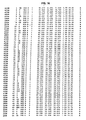
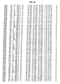
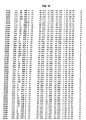
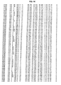
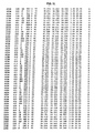
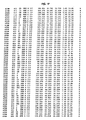
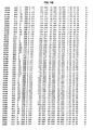
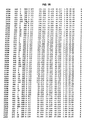
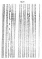
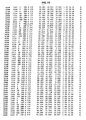
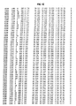
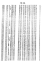
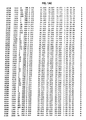
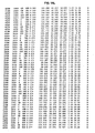
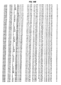
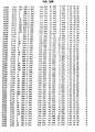
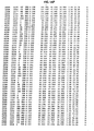
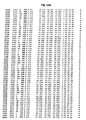
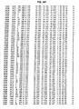
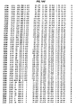
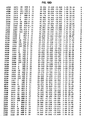
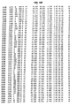
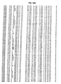
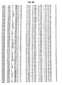
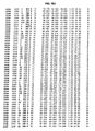
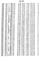
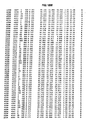
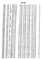
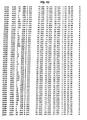
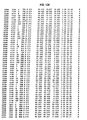
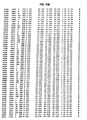
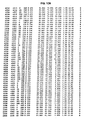
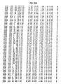
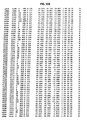
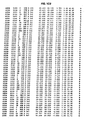
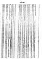
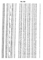
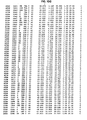
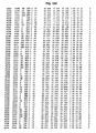
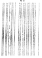
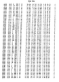
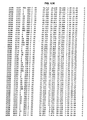
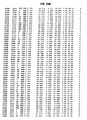
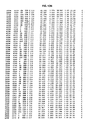
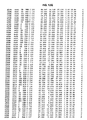
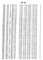
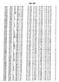
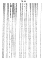
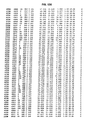
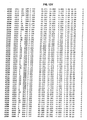
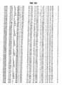
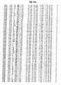
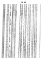
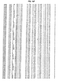
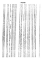
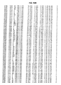
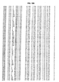
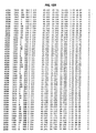
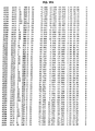
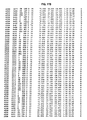
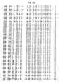
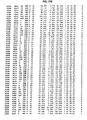
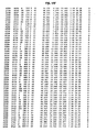
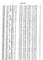
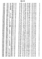
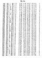
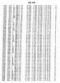
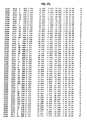
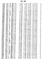
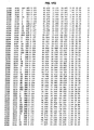
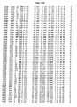
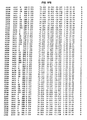
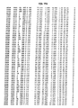
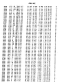
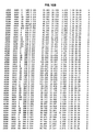
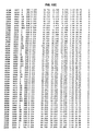
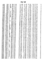
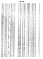
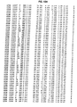
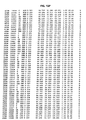
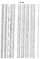
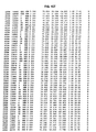
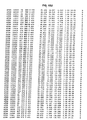
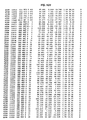
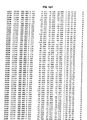
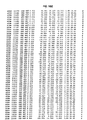
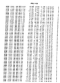
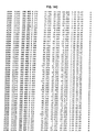
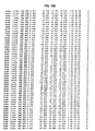
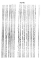
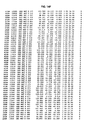
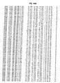
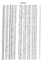
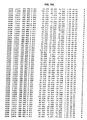
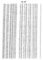
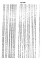
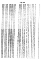
- a three dimensional structure of hSDH comprises atomic coordinates as given in Figure 1 and any three-dimensional structure thereof having an overall fold as illustrated in Figure 2 and is of a protein containing at least about 80% of the amino acids in SEQ. ID. NO. 1.
- the atomic coordinates of hSDH, or a fragment, analog, or variant thereof may be used to model a hSDH protein, or a hSDH-related protein.
- a model may also be developed based on a binding site identified herein.
- a catalytic site of hSDH comprising the cofactor NADH, a catalytic zinc atom, and hSDH amino acid residues or water molecules located within 10 angstroms of a hSDH ligand as given in Table 4.
- a binding site for a hSDH ligand comprising hSDH amino acid residues or water molecules which form either hydrogen bonds or van der Waals contacts to said ligand as defined in Table 2 or Table 3.
- the binding site may further comprise the cofactor NADH and a catalytic zinc atom.
- Another aspect of the present invention is a computer readable medium having stored thereon atomic coordinate/X-ray diffraction data defining the three dimensional structure of a temary complex of hSDH, NADH, and a ligand that binds to hSDH, or a fragment, analog, or variant thereof.
- a computer medium having stored thereon the computer model output data defining the three dimensional structure of a ternary complex of hSDH, NADH, and a ligand that binds to hSDH or a subunit thereof.
- the computer model is derived from analysis of atomic coordinate/X-ray diffraction data defining the three dimensional structure of a ternary complex of hSDH, NADH, and a ligand that binds to hSDH or a subunit thereof.
- a method for identifying a ligand of hSDH or a subunit thereof comprising the steps of: (I) providing a computer readable medium having stored thereon computer model output data defining the three dimensional structure of a ternary complex of hSDH, NADH, and a ligand that binds to hSDH or a subunit thereof; (2) providing a computer readable medium having stored thereon computer model output data defining the three dimensional structure of a potential ligand that binds to hSDH or a subunit thereof; (3) providing a computer system comprising a computer and a computer algorithm where the computer system is capable of processing the computer model and the output data of steps (1) and (2); and (4) processing the computer model output data of steps (1) and (2) using the computer system of step (3) wherein the processing calculates the ability of the potential ligand to bind to hSDH or a subunit thereof.
- the method may further comprise the steps of (5) incorporating a test compound of the potential
- Compound (I) refers to N,N -dimethyl-4-(2-hydroxymethyl-pyrimidin-4-yl)-piperazine-1-sulfonamide having the following structural formula:
- computer-based system or “computer system” refers to the hardware means, software means, and data storage means used to analyze the amino acid sequence and/or atomic coordinate/x-ray diffraction data of the present invention.
- computer readable medium refers to any available media which can be accessed by a computer.
- computer readable media can comprise RAM, ROM, EEPROM, CD-ROM or other optical disk storage (read only or rewritable), magnetic disk storage or other magnetic storage devices, or any other medium which can be used to store the amino acid sequence and/or atomic coordinate/x-ray diffraction data of the present invention and which can be accessed by a computer.
- the present invention provides methods for expressing, purifying and crystallizing hSDH to form a crystal which diffracts x-rays with sufficiently high resolution to allow determination of the three-dimensional structure of the hSDH, or a portion or subdomain thereof.
- the three-dimensional structure is useful for rational design of ligands of hSDH.
- the crystals may be used for determining the binding mode of potential ligands (e.g., inhibitors) by soaking the compounds into the crystals and then determining the structure of the complex.
- a novel crystalline ternary complex of hSDH complexed with NADH and a SDH ligand is provided as well as methods for using the crystalline form and active site to identify potential hSDH ligands.
- Atomic coordinates of a novel crystalline ternary complex of SDH complexed with NADH and Compound (I) generated from x-ray diffraction are provided in Figure 1.
- Human SDH functions biologically as a tetramer with 357 residues (38 kDa) per subunit.
- Crystals of hSDH were prepared by forming a complex of the protein with NADH and Compound (I) prior to setting up the crystallization trials. In the crystal structure, each subunit contains one catalytic zinc atom, one molecule of the required cofactor, NADH, and one molecule of Compound (I).
- the structure reveals the binding mode of NADH and of the inhibitor (Compound (I)).
- Compound (I) is bound directly to the catalytic zinc ion, through a hydroxyl group and the nitrogen atom N3.
- the position of Compound (I) is also stabilized by a stacking interaction with the nicotinimide ring of NADH, bound to hSDH.
- the atomic coordinate/x-ray diffraction data may be used to create a physical model which can then be used to design molecular models of compounds that may interact with the determined active sites, binding sites or other structural or functional domains or subdomains of the hSDH.
- the atomic coordinate/x-ray diffraction data of the ternary complex may be represented as atomic model output data on computer readable media which can be used in a computer modeling system to calculate different molecules expected to interact with the determined active sites, binding sites, or other structural or functional domains or subdomains of the hSDH.
- computer analysis of the data allows one to calculate the three-dimensional interaction of the hSDH and the ligand to confirm that the ligand binds to, or changes the conformation of, particular domain(s) or subdomain(s) of the hSDH.
- the binding mode of the required cofactor NADH and the nature of its interactions with the ligand can also be evaluated.
- Ligands identified from the analysis of the physical or computer model can then be synthesized and tested for biological activity;with an appropriate screen.
- the atomic coordinate/x-ray diffraction data of the present invention are generally provided on computer readable media.
- a skilled artisan can then access the data and analyze it for structure determination and/or rational ligand (e.g., inhibitor) design using a computer-based system.
- a typical computer system includes hardware means, software means, and data storage means.
- the hardware means typically includes a central processing unit (CPU), input means, output means and data storage means.
- CPU central processing unit
- a target structural motif' or target motif' refers to any rationally selected sequence or combination of sequences in which the sequence(s) are chosen based on a three-dimensional configuration or electron density map which is formed upon the folding of the target motif.
- target motifs include, but are not limited to, enzymatic active sites, structural subdomains, epitopes, functional domains and signal sequences.
- a variety of structural formats for the input and output means can be used to input and output the information in the computer-based systems of the present invention.
- comparing means can be used to compare a target sequence or target motif with the data storage means to identify structural motifs or interpret electron density maps derived in part from the atomic coordinate/x-ray diffraction data.
- One skilled in the art can readily recognize any one of the publicly available computer modeling programs that can be used.
- Suitable software that can be used to view, analyze, design, and/or model a protein include AlchemyTM, LabVisionTM, SybylTM, MolcaddTM, LeapfrogTM, MatchmakerTM, GenefoldTM and SitelTM (available from Tripos Inc., St.
- the present invention also provides inhibitors of SDH activity identified or designed by the methods of the present invention. Once a potential ligand is identified from the computer analysis described above, then the ligand can be synthesized and tested for biological activity using an appropriate screening method.
- SDH inhibitors lower fructose levels in the tissues of mammals affected by diabetes (e.g., nerve, kidney and retina tissue) and are therefore useful in the treatment and/or prevention of diabetic complications such as diabetic neuropathy, diabetic retinopathy, diabetic nephropathy, diabetic microangiopathy and diabetic macroangiopathy in mammals.
- One method for testing SDH inhibitor activity involves the use of male Sprague-Dawley rats (200-400 g). Diabetes is induced in some of the rats by a tail vein injection of streptozocin, 85 mg/kg. Twenty-four hours later, groups of diabetic rats are given a single dose of the test compound (0.01 - 300 mg/kg) by oral gavage. Animals are sacrificed 4-6 hours after dosing and blood and sciatic nerves are harvested. Tissues and cells are extracted with 6% perchloric acid.
- Sorbitol in erythrocytes and nerves is measured by a modification of the method developed by R. S. Clements, et al. ( Science , 166 , 1007-8, (1969)).
- Aliquots of tissue extracts are added to an assay system which has final concentrations of reagents of 0.033M glycine, pH 9.4, 800 ⁇ M ⁇ -nicotine adenine dinucleotide, and 4 units/mL of sorbitol dehydrogenase. After incubation for 30 minutes at room temperature, sample fluorescence is determined on a fluorescence spectrophotometer with excitation at 366 nm and emission at 452 nm. After subtracting appropriate blanks, the amount of sorbitol in each sample is determined from a linear regression of sorbitol standards processed in the same manner as the tissue extracts.
- Fructose is determined by a modification of the method described by M. Ameyama, Methods in Enzymology , 89 , 20-25 (1982). Resazurin is substituted for ferricyanide. Aliquots of tissue extracts are added to the assay system, which has final concentrations of reagents of 1.2M citric acid, pH 4.5, 13 ⁇ M resazurin, 3.3 units/mL of fructose dehydrogenase and 0.068% Triton X-100. After incubation for 60 minutes at room temperature, sample fluorescence is determined on a fluorescence spectrophotometer with excitation at 560 nm and emission at 580 nm. After subtracting appropriate blanks, the amount of fructose in each sample is determined from a linear regression of fructose standards processed in the same manner as the tissue extracts.
- Compounds identified by the methods of the present invention may be used to treat and/or prevent diabetic complications such as diabetic neuropathy, diabetic retinopathy, diabetic nephropathy, diabetic microangiopathy and diabetic macroangiopathy in mammals.
- the SDH inhibitor is preferably administered as a pharmaceutical composition.
- the active compounds and compositions may be administered to a subject in need of treatment by a variety of conventional routes of administration, including orally, parenterally and topically.
- SDH inhibitors and their pharmaceutically acceptable salts will be administered orally or parenterally at dosages between about 0.01 and about 50 mg/kg body weight of the subject to be treated per day, preferably from about 0.1 to 15 mg/kg, in single or divided doses.
- some variation in dosage will necessarily occur depending on the condition of the subject being treated.
- the person responsible for administration will, in any event, determine the appropriate dose for the individual subject.
- the active compounds and compositions may be administered alone or in combination with pharmaceutically acceptable carriers, in either single or multiple doses.
- suitable pharmaceutical carriers include inert solid diluents or fillers, sterile aqueous solutions and various organic solvents.
- the pharmaceutical compositions formed by combining the active compounds and the pharmaceutically acceptable carriers are then readily administered in a variety of dosage forms such as tablets, powders, lozenges, syrups, injectable solutions and the like.
- These pharmaceutical compositions can, if desired, contain additional ingredients such as flavorings, binders, excipients and the like.
- tablets containing various excipients such as sodium citrate, calcium carbonate and calcium phosphate may be employed along with various disintegrants such as starch, alginic acid and certain complex silicates, together with binding agents such as polyvinylpyrrolidone, sucrose, gelatin and acacia.
- binding agents such as polyvinylpyrrolidone, sucrose, gelatin and acacia.
- lubricating agents such as magnesium stearate, sodium lauryl sulfate and talc are often useful for tabletting purposes.
- Solid compositions of a similar type may also be employed as fillers in soft and hard filled gelatin capsules. Preferred materials for this include lactose or milk sugar and high molecular weight polyethylene glycols.
- the essential active ingredient therein may be combined with various sweetening or flavoring agents, coloring matter or dyes and, if desired, emulsifying or suspending agents, together with diluents such as water, ethanol, propylene glycol, glycerin and combinations thereof.
- solutions of the active compounds and compositions in sesame or peanut oil, aqueous propylene glycol, or in sterile aqueous solutions may be employed.
- aqueous solutions should be suitably buffered if necessary and the liquid diluent first rendered isotonic with sufficient saline or glucose.
- aqueous solutions are especially suitable for intravenous, intramuscular, subcutaneous and intraperitoneal administration.
- the sterile aqueous media employed are all readily available by standard techniques known to those skilled in the art.
- the active compounds and compositions may be employed in the preparation of ophthalmic solutions.
- ophthalmic solutions are of principal interest for the treatment of diabetic cataracts by topical administration.
- the active compounds and compositions are administered to the eye in the form of an ophthalmic preparation prepared in accordance with conventional pharmaceutical practice.
- hSDH gene (SEQ. ID. NO. 1: Accession No. U07361) is available from Entrez Protein, National Center for Biotechnology Information, National Library of Medicine, Bethesda, MD.
- the gene was recloned from a human liver cDNA library and inserted into an expression vector.
- the expression vector was re-sequenced to confirm the correctness of the DNA insert.
- Some natural variation in the sequence of human SDH (on the order of 10-20% at the amino acid level) is expected. Such variants may or may not have functional significance and are expected to have the same or similar three-dimensional structures to the crystal structure described here.
- hSDH human SDH
- PCR polymerase chain reaction
- PCR amplification was carried out under standard conditions using Vent polymerase (available from New England Biolabs Inc., Beverly, MA) and the human liver cDNA library as a template.
- the PCR-amplified product was digested with Ndel and Xho l, gel purified and ligated into pET23a (available from Novagen, Madison, WI). The ligation mixture was transformed into E.
- coli DH5 ⁇ cells and plated on LB+agar plates containing ampicillin (100 ⁇ g/mL). A few colonies were picked, grown in LB+ ampicillin and used for plasmid preparation. These recombinant plasmids were subjected to restriction digestion and PCR analysis to confirm the presence of the insert.
- the DNA sequence of the 1071 base pair insert in pET23a was confirmed (identical to the human SDH sequence GenBank Accession No. U07361, SEQ. ID. NO. 1)) on both strands using a 373A Automated fluorescent sequencer (available from Applied Biosystems, Foster City, CA). The gene coded for a 357-amino acid protein.
- One of the positive clones, hSDH/pET23a 1/3 was transformed into E.coli BL21 (DE3) cells and used for expression experiments.
- the cells were induced with 0.4 mM IPTG for 4 hours at 26°C and harvested by centrifugation.
- the cell paste was either used for experiments or stored frozen at -20°C until needed.
- the protein was expressed minimally even under non-inducing conditions. Although the protein was expressed in higher quantities when induced at 37°C, incubation at 26°C gave higher soluble expression and overall yield.
- a glycerol stock of E. coli BL21 (DE3) cells harboring hSDH/pET23a 1/3 were cultured overnight at 37°C with shaking at 300 rpm in 2x YT media (16 g bacto-tryptone, 10 g bacto-yeast extract, 5 g NaCI and 900 mL distilled water) containing 100 ⁇ g/mL carbenicillin. Plasmid DNA was prepared from these cells using the Qiagen Prep Spin procedure (see, Qiagen product literature). The purified plasmid was transformed into B834(DE3) cells from Novagen Inc. (Madison, WI) and used for expression in selenomethionine media.
- the selenomethionine (SeMet) media contained 10 g/L (NH 4 ) 2 SO 4 , 2.16 g/L Na 2 HPO 4 , 1.28 g/L KH 2 PO 4 , 5 g/L NaCI, 0.4 g/L trisodium citrate (Sigma), 0.2 g/L MgSO 4 -7H 2 O, 2 mg/L thiamine, 10 g/L glycerol, 0.1 g/L carbenicillin, 40 mg/L of all L-amino acids except methionine, 100 mg/L D, L-selenomethionine, 10 mg/L CaCl 2 , 4 mg/L H 3 BO 3 , 1.71 mg/L MnSO 4 -H 2 O, 2 mg/L ZnSO 4 -7H 2 O, 0.373 mg/L CuSO 4 -5H 2 O, 0.4 mg/L CoCl 2 -6H 2 O, 0.2 mg/L Na 2 MoO 4 -2H 2 O
- the pH of the media was adjusted to 7.0. Fernbach tribaffled flasks (2.8 liter) were used for growth of a 500 mL culture.
- a 500 mL culture of hSDH clone in B834(DE3) in SeMet Media was inoculated from an overnight culture grown at 30°C in the same media. The culture was grown at 30°C and 300 rpm until the O.D. 600 reached 0.5 to 1.0. At this point, the cells were induced with IPTG to a final concentration of 1.0 mM and growth was continued for another 24 hours at room temperature. The cells were harvested by centrifugation and stored at -80°C until use.
- E. coli BL21(DE3) pET 23a/hSDH cells (40-50 grams) were resuspended with two volumes of lysis buffer (20 mM 4-(2-hydroxyethyl)-1-piperazineethanesulfonic acid (HEPES) /NaOH pH 8, 5 ⁇ M ZnCl 2 , 2 mM dithiothreitol (DTT), 1 mM phenylmethyl sulfonyl fluoride (PMSF), and 20 ⁇ g/mL aprotinin).
- HEPES 4-(2-hydroxyethyl)-1-piperazineethanesulfonic acid
- DTT dithiothreitol
- PMSF phenylmethyl sulfonyl fluoride
- lysis buffer containing 2 mg/mL lysozyme were added and the mixture stirred on ice for 60 minutes.
- the cells were sonicated for two periods of 3 minutes in 30 mL aliquots, while in an ice bath, using a BransonTM 450 sonifier (available from Branson Ultrasonics Corp., Danbury, CT) set to a 50% duty cycle, power setting #6.
- the sonicated cells were centrifuged at 12,000 rpm for 10 minutes at 4°C in a Sorvall SS-34 rotor. The supernatant was decanted and the pellet discarded.
- ammonium sulfate was added to a final concentration of 40% (equivalent to 226 mg NH 4 + SO 4 - /mL supernatant). The solution was incubated on ice for 30 minutes, and then centrifuged at 12,000 rpm for 10 minutes in GSA rotor at 4°C. The supernatant was removed and ammonium sulfate was added to a final saturation of 60% (equal to 135 mg/mL of the original supernatant). The mixture was incubated on ice for 30 minutes, and then centrifuged as before.
- the 40-60% ammonium sulfate pellet was dissolved with 100 mL 20 mM Hepes/NaOH, pH 8.0, 5 ⁇ M ZnCl 2 , 2 mM DTT, 1 mM PMSF and 1 ⁇ g/mL each of aprotinin, pepstatin and leupeptin. The sample was dialyzed overnight at 4°C against four liters of the same buffer.
- a Green A dye column (AmiconTM type XK-50, 100 ml bed volume; available from Millipore Corporation, Bedford, MA) was equilibrated with 800 ml Buffer A (20 mM Hepes/NaOH pH 8, 5 ⁇ M ZnCl 2 , and 2 mM DTT). The 40-60% ammonium sulfate cut was loaded onto the column at a rate of 5 mL/minute. This load contained about 2000 mg protein, of which approximately 50 - 200 mg was hSDH protein.
- the column was eluted with Buffer B (Buffer A plus 1.0 M NaCI) at 5 mL/minute as follows: 0% B for 100 min (5 column volumes), 0-100% B for 200 min (10 column volumes), 100% B for 40 min (2 column volumes).
- Buffer B Buffer A plus 1.0 M NaCI
- Four minute fractions were collected and assayed for activity. Two peaks of activity were identified; a major peak which eluted between 15-30% B and a minor peak which eluted between 30-40% B. Each peak was pooled separately.
- the major peak was concentrated to about 50 mL in an AmiconTM cell using a YM-30 membrane (available from Millipore Corporation, Bedford, MA) and dialyzed overnight at 4°C against 2 liters of Buffer A.
- Electrospray mass spectrometry indicated a major peak centered at 38178 and a lesser peak centered at 38112 mass units (expected mass 38202).
- N-terminal sequence analysis indicated AAAAKPNNLSLV with approximately 29% of the protein missing the first alanine.
- Selenomethionine substituted hSDH was purified in the same way as the wild type protein. Electrospray mass spectrometry indicated a major peak centered at 38565 and a lesser peak centered at 38492 mass units (expected mass for 7 selenomethionines and 1 Zn 2+ is 38534). Two purified lots of selenomethionine substituted SDH had two masses; 38565 and 38492. These are +11 and +9 of the predicted masses of 38554 (des Met) and 38483 (des Met Ala) forms with all 8 methionines substituted with selenomethionine. The selenomethionine substituted protein was assayed using the same method as wild type protein and the activity was determined to be 11.6 +/-0.9 Units/mg.
- CS cryostabilization solution
- PEG 4000 polyethylene glycol
- 37.5 mM NH 4 OAc polyethylene glycol
- 50 mM sodium citrate pH 6.15, 8.75% glycerol
- 0.07 mM NADH 0.14 mM Compound (I)
- the crystals were first transferred to 10 microliters of well solution (described above) and 5 microliters of CS were added. Additional volumes of CS were added at 10 minute intervals as follows; 3 steps of 5 microliters, followed by 2 steps of 10 microliters. Finally, the crystal was transferred to 10 microliters of undiluted CS for 10 minutes, after which it was frozen either directly in a liquid nitrogen gas stream or in liquid propane.
- the data were measured from a single crystal at 100 °K at beamline X12C at the National Synchrotron Light Source at the Brookhaven National Laboratory in Upton, New York.
- the X-ray absorption spectrum around the K edge of the anomalously scattering selenomethionine atoms was measured using a simple X-ray detector mounted at right angles to the X-ray beam and in the horizontal plane to capture fluorescence as an indirect measure of absorption.
- the inflection point (L1), peak (L2) and remote wavelength (L3, expected to be the Zn peak) were identified as 0.979148, 0.978712, and 1.249998 from the fluorescence scan.
- the structure was solved using the multiple anomalous diffraction method (MAD) as implemented in Crystallography and NMR System (CNS) beta release version.
- MAD multiple anomalous diffraction method
- CNS NMR System
- methionines There are 8 methionines in hSDH. Assuming that the N-terminal methionine is disordered, this leaves 7 selenomethionine sites per monomer. Assuming space group P6 2 , four molecules of hSDH (the biologically relevant tetramer) in the asymmetric unit and a high Matthews coefficient of 3.8, we expected to find at least 28 selenomethionine sites.
- the native Patterson map showed a large peak at 0, 0, 0.4 indicating that the asymmetric unit exhibited translational symmetry.
- a real space transform to translate molecule 1 by 0, 0, 93 angstroms, creating molecule 2.
- Molecules 1 and 2 were combined and examination of the unit cell in the program O in both space groups P6 2 22 and P6 2 suggested that the correct space group was P6 2 and that the asymmetric unit contained two dimers belonging to two different tetramers.
- Coordinate files corresponding to the two dimers were made and used to create a new file of the 28 selenomethionine and 4 zinc positions.
- a heavy atom search was recalculated in space group P6 2 using L2 (peak) anomalous differences and confirmed a subset of these solutions. Single anomalous difference phases to 3 angstroms using L2 and the 32 sites derived from the coordinates were calculated.
- a log-likelihood gradient map revealed 3 other peaks greater than 5 sigma which corresponded to partially occupied alternate conformations of selenomethionines 107 and 309 and revealed that residue 185 was a selenomethionine, rather than glutamic acid.
- the structure was refined using 5% of the reflections as a test set.
- One molecule was manually refit in O and the other 3 generated by application of non-crystallographic symmetry.
- Prior to torsional dynamics, f and f" were refined for selenium (“SE”) and zinc ion (“ZN+2”) and included in an anomalous library for all subsequent steps.
- SE selenium
- ZN+2 zinc ion
- the structure was refined using all data between 99.0 and 1.9 angstroms.
- R free 23.0%
- R factor 21.5%
- root mean square deviations in bond lengths of 0.009 and bond angles of 1.51 degrees for four molecules of human SDH consisting of residues 1-356, 4 molecules of Compound (I), 4 molecules of NADH and 1299 water molecules. All residues lie within the allowed regions of the Ramachandran plot.
- Table 1, Part c shows the statistics of the refined structure of the SDH/NADH/Compound (I) complex. These statistics give the contents of the asymmetric unit of the crystal and provide the statistics required to evaluate the quality of the refined structure.
- Table 1 Part (a) below provides the parameters of the crystals that were used to solve the structure.
- Table 1 Part (b) below provides the details and statistical analysis of the X-ray diffraction data measured.
- Table 1 Part (c) below provides the details of the final, refined structure and a statistical analysis of the correctness of the refined structure.
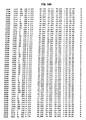
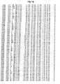
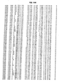
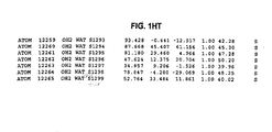
- Figure 1 The X-ray coordinates of the refined structure are listed in Figure 1, along with the B factors for each atom. The occupancy of each atom is fixed at 1:0. This coordinate set is generated by the last refinement cycle of the structure using the program CNS (Brunger, A. T. et al., (1998) Acta Crystallographica D54, 905-921).
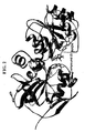
- Figure 2 is a ribbon diagram showing the overall fold of one subunit of the biological tetramer of the enzyme human sorbitol dehydrogenase (SEQ. ID. NO.1) (made using Molscript; P. Kraulis (1991) J. Appl. Cryst ., 24, 946-950) and the location of Compound (I), NADH, and the catalytic zinc atom.
- SEQ. ID. NO.1 human sorbitol dehydrogenase
- the protein is colored gray
- the zinc atom is colored gray
- Compound (I) and NADH are shown as ball and stick representations with carbon atoms in green, oxygen atoms in red, nitrogen atoms in blue, sulfur atom in yellow and phosphorus atom in purple.
- the overall fold of one subunit of hSDH is similar to that of the alcohol dehydrogenases, consisting of two beta barrel domains with the active site located in between the two.
- the catalytic site is marked by the presence of the zinc atom.
- the zinc atom is chelated by interactions with cysteine 44, histidine 69, a water molecule (Wat 64) and by two interactions with Compound (I).
- the specific contacts and the distances between the zinc atom and each chelating atom are given in Table 5 below.
- Figure 5 (made using Molscript; P. Kraulis (1991) J. Appl. Cryst ., 24, 946-950; carbon atoms in green, nitrogen atoms in blue, oxygen atoms in red, sulfur atoms in yellow) shows the vicinity of the zinc atom and the relative positions of cysteine 44, histidine 69 and Wat 64.
- Water molecule 64 also forms hydrogen bonds to two other side chains of hSDH, glutamic acid 155 and glutamic acid 70.
- the hydrogen bonds formed between Wat 64, the zinc atom, and the two glutamic acid side chains serve to stabilize the position of this water molecule.
- the structure also shows how Compound (I) chelates the catalytic zinc atom through interactions between nitrogen 3 of the pyrimidine ring and oxygen 30 of the hydroxymethyl group.
- Compound (I) sterically inhibits substrate binding and blocks access of the substrate to the catalytic zinc atom.
- Compound (I) also interacts with the required cofactor, NADH as shown in Figure 4 (made using Molscript; P. Kraulis (1991) J. Appl. Cryst ., 24, 946-950; carbon atoms in green, nitrogen atoms in blue, oxygen atoms in red, sulfur atoms in yellow, except for Compound (I) where the carbons are colored gold).
- the pyrimidine ring of Compound (I) stacks over the nicotinimide ring of NADH forming a tight hydrophobic contact.
- FIG. 3 An overall view of the binding site of Compound (I) is shown in Figure 3. All residues of hSDH which form either hydrogen bonds or van der Waals contacts to Compound (I) are shown, in addition to NADH, Wat 64 and the catalytic zinc atom (made using Molscript; P. Kraulis (1991) J. Appl. Cryst ., 24, 946-950; carbon atoms in green, nitrogen atoms in blue, oxygen atoms in red, sulfur atoms in yellow, except for Compound (I) where the carbons are colored gold).
- a list of the residues shown in Figure 3 is given in Table 3 below.
- the binding site of the inhibitor may also be described as those residues of the protein within 10 angstroms of any atom of the compound.
- Table 4 lists all residues of SDH which are within 10 angstroms of any atom of Compound (I).
- Asymmetric unit biological tetramer
- b Data Collection Statistics Wavelength ( ⁇ ) 0.97949 0.97977 0.96859 No.
- Table 2 lists the hydrogen bonds formed between Compound (I) and the hSDH protein and the distances between the non-hydrogen atoms of the hydrogen bonds in the crystal structure. Hydrogen Bonds between SDH and Compound (I).
- Compound (I) Residue Atom Distance ( ⁇ ) O30 Wat 64 O 2.98 Glu 155 OE1 3.37 Glu 155 OE2 2.54 His 69 NE2 3.05 N3 Ser 46 OG 3.25
- Table 3 lists those residues of hSDH forming van der Waals contacts to Compound (I) in the crystal structure. 'Van der Waals contacts" is defined herein as distances between non-hydrogen atoms from about 3.4 to about 4.5 angstroms. Van der Waals contacts between Compound (I) and SDH. Cys 44 His 46 Tyr 50 lie 56 Phe 59 His 69 Phe 118 Thr 121 Glu 155 Leu 274 Phe 297 Arg 298 Tyr 299
- Table 4 lists those residues of hSDH within 10 angstroms of any atom of Compound (I) in the crystal structure.
- the binding site of Compound (I) may also be described as those residues of the protein within 10 angstroms of any atom of the inhibitor.
- Table 5 lists the ligands which coordinate the catalytic zinc atom of hSDH and the distances between such atoms and the zinc.
- Zn ligands Residue Atom Distance ( ⁇ ) to Zn Cys 44 SG 2.56 His 69 NE2 2.36
- Compound (I) N3 2.15
- Compound (I) 030 2.35 Wat 64 O 2.0
Landscapes
- Life Sciences & Earth Sciences (AREA)
- Chemical & Material Sciences (AREA)
- Health & Medical Sciences (AREA)
- Genetics & Genomics (AREA)
- Organic Chemistry (AREA)
- Engineering & Computer Science (AREA)
- Bioinformatics & Cheminformatics (AREA)
- Zoology (AREA)
- Wood Science & Technology (AREA)
- Molecular Biology (AREA)
- Microbiology (AREA)
- Biotechnology (AREA)
- Biomedical Technology (AREA)
- Biochemistry (AREA)
- General Engineering & Computer Science (AREA)
- General Health & Medical Sciences (AREA)
- Medicinal Chemistry (AREA)
- Enzymes And Modification Thereof (AREA)
- Measuring Or Testing Involving Enzymes Or Micro-Organisms (AREA)
Abstract
Description
- The present invention relates to the three-dimensional X-ray crystal structure of human sorbitol dehydrogenase (hSDH), the modeling of new structures, the binding mode of a SDH ligand, and the binding mode of the cofactor NADH, as well as methods for rational drug design based on the use of such data.
- The development of various diabetic complications is believed to be the result of increases of glucose entry into cells due to chronic hyperglycemia, activation of glucose metabolism through the polyol pathway, and accumulation of polyols (such as sorbitol). Sorbitol dehydrogenase (SDH), the second enzyme in the polyol pathway, oxidizes sorbitol to fructose and reduces NAD+ to NADH. Excess flux through SDH creates an imbalance in the cytoplasmic NAD+/NADH ratio and leads to a "pseudohypoxic" state that may be the determinant of pathology in diabetic tissues, thus making SDH a therapeutic target for the treatment of diabetic complications.
- The amino acid and protein sequence for the human sorbitol dehydrogenase (hSDH) enzyme is available from Entrez Protein or GenBank, National Center for Biotechnology Information, National Library of Medicine, Bethesda, MD (Accession No. Q00796 or Accession No. U07361). The sequence and the procedures used to determine the sequence for SDH are described in Lee, F.K., et al., "The Human Sorbitol Dehydrogenase Gene: cDNA Cloning, Sequence Determination, and Mapping by Fluorescence in situ Hybridization," Genomics, 21(2), 354-358 (1994).
- U.S. Patent No. 5,728,704 discloses certain pyrimidine compounds that act as SDH ligands resulting in lowering fructose levels in the tissues of mammals affected by diabetes (e.g., nerve, kidney and retina tissue) and are therefore useful in the treatment and/or prevention of diabetic complications such as diabetic neuropathy, diabetic retinopathy, diabetic nephropathy, diabetic microangiopathy and diabetic macroangiopathy in mammals. A molecular modeling investigation of human sorbitol dehydrogenase complexed with the substrate sorbitol and the SDH inhibitor, 4-[4-(N,N-dimethylsulfamoyl)-piperazino]-2-hydroxymethylprimidine, was reported by D. Darmanin and O. EI-Kabbani in Bioorg. Med. Chem. Lett., 10, 1101-1104 (2000). The model was based on the structures of human β3 alcohol dehydrogenase, human α alcohol dehydrogenase and horse liver alcohol dehydrogenase. The tertiary structure of human β3 alcohol dehydrogenase was used as the template for the construction of the model.
- The crystal structure of rat sorbitol dehydrogenase complexed with NAD+ has been determined to 3.1 angstroms resolution (Johansson, K. et al., (2001) Chemico-Biological Interactions 130-132 (1-2): 351-358). The coordinates are not publicly available and therefore no direct comparison between the rat and human enzyme is possible. Based on the images in the publication, rat SDH appears to have the same overall quaternary and tertiary structure as human SDH, although, without the three dimensional coordinates, a detailed analysis is not possible.
- The present invention provides a crystal form of human sorbitol dehydrogenase, or a subunit thereof, wherein the crystal form is defined as a crystal of space group P62 and cell constants having the values a =134.814 Å ± 15%, b =134.814 Å ± 15%, c = 225.184 Å ± 15%, α = 90.0°, β = 90.0°, and γ =120.0°. The crystal form may optionally contain NADH, a zinc ion, or a ligand that binds to the hSDH or a subunit thereof.
- In another embodiment of the present invention, a three dimensional structure of hSDH is provided that comprises atomic coordinates as given in Figure 1 and any three-dimensional structure thereof having an overall fold as illustrated in Figure 2 and is of a protein containing at least about 80% of the amino acids in SEQ. ID. NO. 1. The atomic coordinates of hSDH, or a fragment, analog, or variant thereof may be used to model a hSDH protein, or a hSDH-related protein. A model may also be developed based on a binding site identified herein.
- In yet another embodiment of the present invention, a catalytic site of hSDH is provided comprising the cofactor NADH, a catalytic zinc atom, and hSDH amino acid residues or water molecules located within 10 angstroms of a hSDH ligand as given in Table 4.
- In still another embodiment of the present invention, a binding site for a hSDH ligand is provided comprising hSDH amino acid residues or water molecules which form either hydrogen bonds or van der Waals contacts to said ligand as defined in Table 2 or Table 3. The binding site may further comprise the cofactor NADH and a catalytic zinc atom.
- Another aspect of the present invention is a computer readable medium having stored thereon atomic coordinate/X-ray diffraction data defining the three dimensional structure of a temary complex of hSDH, NADH, and a ligand that binds to hSDH, or a fragment, analog, or variant thereof.
- In another aspect of the present invention, a computer medium is provided having stored thereon the computer model output data defining the three dimensional structure of a ternary complex of hSDH, NADH, and a ligand that binds to hSDH or a subunit thereof. The computer model is derived from analysis of atomic coordinate/X-ray diffraction data defining the three dimensional structure of a ternary complex of hSDH, NADH, and a ligand that binds to hSDH or a subunit thereof.
- In yet another embodiment of the present invention, a method is provided for identifying a ligand of hSDH or a subunit thereof comprising the steps of: (I) providing a computer readable medium having stored thereon computer model output data defining the three dimensional structure of a ternary complex of hSDH, NADH, and a ligand that binds to hSDH or a subunit thereof; (2) providing a computer readable medium having stored thereon computer model output data defining the three dimensional structure of a potential ligand that binds to hSDH or a subunit thereof; (3) providing a computer system comprising a computer and a computer algorithm where the computer system is capable of processing the computer model and the output data of steps (1) and (2); and (4) processing the computer model output data of steps (1) and (2) using the computer system of step (3) wherein the processing calculates the ability of the potential ligand to bind to hSDH or a subunit thereof. The method may further comprise the steps of (5) incorporating a test compound of the potential ligand in a biological hSDH activity assay; and (6) determining whether the test compound inhibits enzymatic activity in the assay.
- As used herein, the term "Compound (I)" refers to N,N-dimethyl-4-(2-hydroxymethyl-pyrimidin-4-yl)-piperazine-1-sulfonamide having the following structural formula:
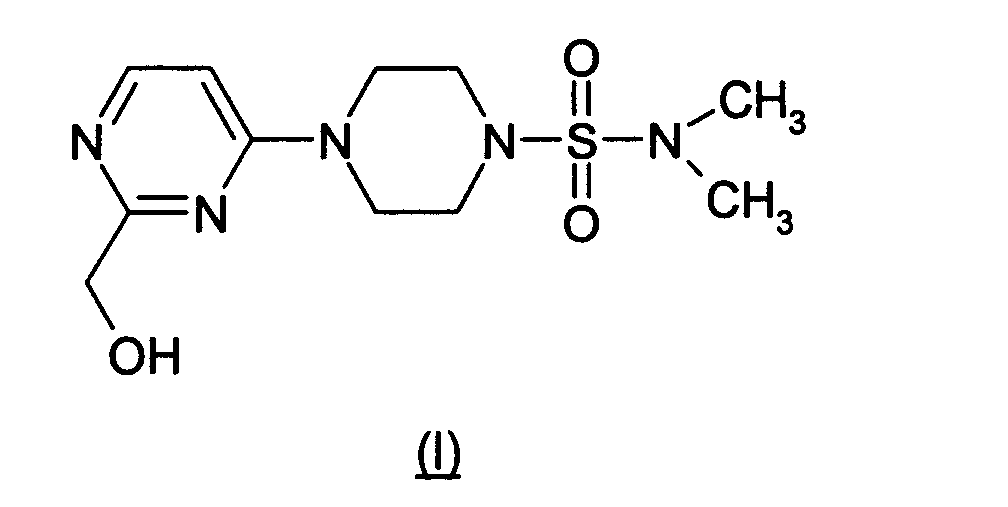
- The term "computer-based system" or "computer system" refers to the hardware means, software means, and data storage means used to analyze the amino acid sequence and/or atomic coordinate/x-ray diffraction data of the present invention.
- The term "computer readable medium" or "computer readable media" refers to any available media which can be accessed by a computer. For example, computer readable media can comprise RAM, ROM, EEPROM, CD-ROM or other optical disk storage (read only or rewritable), magnetic disk storage or other magnetic storage devices, or any other medium which can be used to store the amino acid sequence and/or atomic coordinate/x-ray diffraction data of the present invention and which can be accessed by a computer.
-
- Figure 1 presents the atomic coordinates derived from X-ray diffraction data (atomic coordinate/X-ray diffraction data) defining the three dimensional structure of a ternary complex of hSDH complexed with NADH and a SDH ligand (i.e., Compound (I) as defined above).
- Figure 2 presents the overall fold of one subunit with Compound (I) and NADH bound.
- Figure 3 shows the residues forming either hydrogen bonds or van der Waals contacts to Compound (I).
- Figure 4 shows the stacking interaction between Compound (I) and NADH.
- Figure 5 shows the ligands of the protein and Compound (I) which coordinate the catalytic zinc ion.
-
- The present invention provides methods for expressing, purifying and crystallizing hSDH to form a crystal which diffracts x-rays with sufficiently high resolution to allow determination of the three-dimensional structure of the hSDH, or a portion or subdomain thereof. The three-dimensional structure is useful for rational design of ligands of hSDH. The crystals may be used for determining the binding mode of potential ligands (e.g., inhibitors) by soaking the compounds into the crystals and then determining the structure of the complex.
- In one aspect of the present invention, a novel crystalline ternary complex of hSDH complexed with NADH and a SDH ligand (including a novel SDH active site) is provided as well as methods for using the crystalline form and active site to identify potential hSDH ligands.
- Atomic coordinates of a novel crystalline ternary complex of SDH complexed with NADH and Compound (I) generated from x-ray diffraction are provided in Figure 1. Human SDH functions biologically as a tetramer with 357 residues (38 kDa) per subunit. Crystals of hSDH were prepared by forming a complex of the protein with NADH and Compound (I) prior to setting up the crystallization trials. In the crystal structure, each subunit contains one catalytic zinc atom, one molecule of the required cofactor, NADH, and one molecule of Compound (I).
- The structure reveals the binding mode of NADH and of the inhibitor (Compound (I)). Compound (I) is bound directly to the catalytic zinc ion, through a hydroxyl group and the nitrogen atom N3. The position of Compound (I) is also stabilized by a stacking interaction with the nicotinimide ring of NADH, bound to hSDH.
- Another aspect of the present invention is the use of the crystalline form and active site to identify potential SDH ligands. The atomic coordinate/x-ray diffraction data may be used to create a physical model which can then be used to design molecular models of compounds that may interact with the determined active sites, binding sites or other structural or functional domains or subdomains of the hSDH. Alternatively, the atomic coordinate/x-ray diffraction data of the ternary complex may be represented as atomic model output data on computer readable media which can be used in a computer modeling system to calculate different molecules expected to interact with the determined active sites, binding sites, or other structural or functional domains or subdomains of the hSDH. For example, computer analysis of the data allows one to calculate the three-dimensional interaction of the hSDH and the ligand to confirm that the ligand binds to, or changes the conformation of, particular domain(s) or subdomain(s) of the hSDH. The binding mode of the required cofactor NADH and the nature of its interactions with the ligand can also be evaluated. Ligands identified from the analysis of the physical or computer model can then be synthesized and tested for biological activity;with an appropriate screen.
- The atomic coordinate/x-ray diffraction data of the present invention are generally provided on computer readable media. A skilled artisan can then access the data and analyze it for structure determination and/or rational ligand (e.g., inhibitor) design using a computer-based system. A typical computer system includes hardware means, software means, and data storage means. The hardware means typically includes a central processing unit (CPU), input means, output means and data storage means. One skilled in the art will readily appreciate which of the currently available computer-based systems are suitable for use in the practice of the present invention.
- A variety of commercially available software programs are available for conducting the analysis and comparison of data in the computer-based system. One skilled in the art will readily recognize which of the available algorithms or implementing software packages for conducting computer analyses can be utilized or adapted for use in the computer-based system. As used herein, "a target structural motif' or "target motif' refers to any rationally selected sequence or combination of sequences in which the sequence(s) are chosen based on a three-dimensional configuration or electron density map which is formed upon the folding of the target motif. There are a variety of target motifs known in the art. Protein target motifs include, but are not limited to, enzymatic active sites, structural subdomains, epitopes, functional domains and signal sequences. A variety of structural formats for the input and output means can be used to input and output the information in the computer-based systems of the present invention.
- A variety of comparing means can be used to compare a target sequence or target motif with the data storage means to identify structural motifs or interpret electron density maps derived in part from the atomic coordinate/x-ray diffraction data. One skilled in the art can readily recognize any one of the publicly available computer modeling programs that can be used.
- Suitable software that can be used to view, analyze, design, and/or model a protein include Alchemy™, LabVision™, Sybyl™, Molcadd™, Leapfrog™, Matchmaker™, Genefold™ and Sitel™ (available from Tripos Inc., St. Louis, MO.); Quanta™, Cerius2™, X-Plor™, CNS™, Catalyst™, Modeller™, ChemX™, Ludi™, Insight™, Discover™, Cameleon™ and Iditis™ (available from Accelrys Inc., Princeton NJ); Rasmol™ (available from Glaxo Research and Development, Greenford, Middlesex, U.K.); MOE™ (available from Chemical Computing Group, Montreal, Quebec, Canada); Maestro™ (available from Shrödinger Inc.,); Midas/MidasPlus™ (available from UCSF, San Francisco, CA); VRML (webviewer- freeware on the internet); Chime (MDL - freeware on the intemet); MOIL (available from University of Illinois, Urbana-Champaign, IL); MacroModel™ and GRASP™ (available from Columbia University, New York, NY); Ribbon™ (available from University of Alabama, Tuscaloosa, Alabama); NAOMI™ (available from Oxford University, Oxford, UK); Explorer Eyechem™ (available from Silicon Graphics Inc., Mountain View, CA.); Univision™ (available from Cray Research Inc., Seattle WA); Molscript™ and O (available from Uppsala University, Uppsala, Sweden); Chem 3D™ and Protein Expert™ (available from Cambridge Scientific); Chain™ (available from Baylor College of Medicine, Houston, TX); Spartan™, MacSpartan™ and Titan™ (available from Wavefunction Inc., Irvine, CA); VMD™ (available from U. Illinois/Beckman Institute); Sculpt™ (available from Interactive Simulations, Inc., Portland, OR.); Procheck™ (available from Brookhaven National Laboratory, Upton, New York); DGEOM (available from QCPE - Quantum Chemistry Program Exchange, Indiana University Bloomington, IN); RE_VIEW (available from Brunel University, London, UK); Xmol (available from Minnesota Supercomputing Center, University of Minnesota, Minneapolis, MN); Hyperchem™ (available from Hypercube, Inc., Gainesville, FL); MD Display (available from University of Washington, Seattle, WA); PKB (available from National Center for Biotechnology Information, NIH, Bethesda, MD); Molecular Discovery Programmes (available from Molecular Discovery Limited, Mayfair, London); Growmol™ (available from Thistlesoft, Morris Township, N.J); MICE (available from the San Diego Supercomputer Center. La Jolla, CA); Yummie and MCPro (available from Yale University, New Haven CT); and upgraded versions thereof.
- The present invention also provides inhibitors of SDH activity identified or designed by the methods of the present invention. Once a potential ligand is identified from the computer analysis described above, then the ligand can be synthesized and tested for biological activity using an appropriate screening method.
- Compounds that act as SDH inhibitors lower fructose levels in the tissues of mammals affected by diabetes (e.g., nerve, kidney and retina tissue) and are therefore useful in the treatment and/or prevention of diabetic complications such as diabetic neuropathy, diabetic retinopathy, diabetic nephropathy, diabetic microangiopathy and diabetic macroangiopathy in mammals. One method for testing SDH inhibitor activity involves the use of male Sprague-Dawley rats (200-400 g). Diabetes is induced in some of the rats by a tail vein injection of streptozocin, 85 mg/kg. Twenty-four hours later, groups of diabetic rats are given a single dose of the test compound (0.01 - 300 mg/kg) by oral gavage. Animals are sacrificed 4-6 hours after dosing and blood and sciatic nerves are harvested. Tissues and cells are extracted with 6% perchloric acid.
- Sorbitol in erythrocytes and nerves is measured by a modification of the method developed by R. S. Clements, et al. (Science, 166, 1007-8, (1969)). Aliquots of tissue extracts are added to an assay system which has final concentrations of reagents of 0.033M glycine, pH 9.4, 800 µM β-nicotine adenine dinucleotide, and 4 units/mL of sorbitol dehydrogenase. After incubation for 30 minutes at room temperature, sample fluorescence is determined on a fluorescence spectrophotometer with excitation at 366 nm and emission at 452 nm. After subtracting appropriate blanks, the amount of sorbitol in each sample is determined from a linear regression of sorbitol standards processed in the same manner as the tissue extracts.
- Fructose is determined by a modification of the method described by M. Ameyama, Methods in Enzymology, 89, 20-25 (1982). Resazurin is substituted for ferricyanide. Aliquots of tissue extracts are added to the assay system, which has final concentrations of reagents of 1.2M citric acid, pH 4.5, 13 µM resazurin, 3.3 units/mL of fructose dehydrogenase and 0.068% Triton X-100. After incubation for 60 minutes at room temperature, sample fluorescence is determined on a fluorescence spectrophotometer with excitation at 560 nm and emission at 580 nm. After subtracting appropriate blanks, the amount of fructose in each sample is determined from a linear regression of fructose standards processed in the same manner as the tissue extracts.
- SDH activity is measured by a modification of the method described by U. Gerlach, Methodology of Enzymatic Analyses , edited by H. U. Bergmeyer, 3, 112-117 (1983). Aliquots of sera or urine are added to the assay system, which has final concentrations of reagents of 0.1M potassium phosphate buffer, pH 7.4, 5 mM NADH, 20 mM sorbitol, and 0.7 units/mL of sorbitol dehydrogenase. After incubation for 10 minutes at room temperature, the average change in sample absorbance is determined at 340 nm. SDH activity is generally presented as milliOD340 units/minute (OD340 = optical density at 340 nm).
- Other useful methods for evaluating SDH inhibitor activity are described in Geisen, K., et al., Arzneim.-Forsch./Drug Res., 44(11), 1032-1043 (1994). The methods described therein include in vivo studies (e.g., evaluations of blood and organ sampling, sorbitol excretion in the urine, motor nerve conduction velocity, glomerular filtration rate and cataract development); in vitro incubation and perfusion studies (e.g., evaluations of sorbitol accumulation in erythocytes and perfusion of the liver); histological techniques (e.g., evaluation of retinal capillaries); biochemical and analytical assays (e.g., enzymatic substrate assays, GLC assays, and HPLC assays). Those skilled in the art will know how to evaluate inhibitor activity using one or more of the several methods known in the art or a modification thereof.
- Compounds identified by the methods of the present invention may be used to treat and/or prevent diabetic complications such as diabetic neuropathy, diabetic retinopathy, diabetic nephropathy, diabetic microangiopathy and diabetic macroangiopathy in mammals. The SDH inhibitor is preferably administered as a pharmaceutical composition.
- The active compounds and compositions may be administered to a subject in need of treatment by a variety of conventional routes of administration, including orally, parenterally and topically. In general, SDH inhibitors and their pharmaceutically acceptable salts will be administered orally or parenterally at dosages between about 0.01 and about 50 mg/kg body weight of the subject to be treated per day, preferably from about 0.1 to 15 mg/kg, in single or divided doses. However, some variation in dosage will necessarily occur depending on the condition of the subject being treated. The person responsible for administration will, in any event, determine the appropriate dose for the individual subject.
- The active compounds and compositions may be administered alone or in combination with pharmaceutically acceptable carriers, in either single or multiple doses. Suitable pharmaceutical carriers include inert solid diluents or fillers, sterile aqueous solutions and various organic solvents. The pharmaceutical compositions formed by combining the active compounds and the pharmaceutically acceptable carriers are then readily administered in a variety of dosage forms such as tablets, powders, lozenges, syrups, injectable solutions and the like. These pharmaceutical compositions can, if desired, contain additional ingredients such as flavorings, binders, excipients and the like. Thus, for purposes of oral administration, tablets containing various excipients such as sodium citrate, calcium carbonate and calcium phosphate may be employed along with various disintegrants such as starch, alginic acid and certain complex silicates, together with binding agents such as polyvinylpyrrolidone, sucrose, gelatin and acacia. Additionally, lubricating agents such as magnesium stearate, sodium lauryl sulfate and talc are often useful for tabletting purposes. Solid compositions of a similar type may also be employed as fillers in soft and hard filled gelatin capsules. Preferred materials for this include lactose or milk sugar and high molecular weight polyethylene glycols. When aqueous suspensions or elixirs are desired for oral administration, the essential active ingredient therein may be combined with various sweetening or flavoring agents, coloring matter or dyes and, if desired, emulsifying or suspending agents, together with diluents such as water, ethanol, propylene glycol, glycerin and combinations thereof.
- For parenteral administration, solutions of the active compounds and compositions in sesame or peanut oil, aqueous propylene glycol, or in sterile aqueous solutions may be employed. Such aqueous solutions should be suitably buffered if necessary and the liquid diluent first rendered isotonic with sufficient saline or glucose. These particular aqueous solutions are especially suitable for intravenous, intramuscular, subcutaneous and intraperitoneal administration. In this connection, the sterile aqueous media employed are all readily available by standard techniques known to those skilled in the art.
- The active compounds and compositions may be employed in the preparation of ophthalmic solutions. Such ophthalmic solutions are of principal interest for the treatment of diabetic cataracts by topical administration. For the treatment of diabetic cataracts, the active compounds and compositions are administered to the eye in the form of an ophthalmic preparation prepared in accordance with conventional pharmaceutical practice.
- The following examples illustrate various aspects of the present invention and are not intended to limit the scope of the invention as defined by the claims.
- Compound I was prepared using the general procedures disclosed in U.S. Patent No. 5,215,990. Unless designated otherwise, reagents used in the following examples are generally available from a variety of vendors who supply chemical reagents such as Fisher Scientific Products (Hanover Park, IL).
- Sequence data for the hSDH gene (SEQ. ID. NO. 1: Accession No. U07361) is available from Entrez Protein, National Center for Biotechnology Information, National Library of Medicine, Bethesda, MD. To generate the protein for hSDH, the gene was recloned from a human liver cDNA library and inserted into an expression vector. The expression vector was re-sequenced to confirm the correctness of the DNA insert. Some natural variation in the sequence of human SDH (on the order of 10-20% at the amino acid level) is expected. Such variants may or may not have functional significance and are expected to have the same or similar three-dimensional structures to the crystal structure described here.
- A pET vector system was used for expression of human SDH (hSDH). The hSDH gene previously identified in a human liver cDNA library was used as a template for polymerase chain reaction (PCR) amplification. (See, Iwata, T., et al., "Structural Organization of the Human Sorbitol Dehydrogenase Gene (SORD)", Genomics, 26, 55-62 (1995).) Two primers were designed to amplify the DNA fragment coding for hSDH. The 5' primer (5'-GGAATTCCATATGGCGGCGGCGGCCAAGCCC-3') (SEQ. ID.NO.2) introduced a unique NdeI site while the 3' primer (5'-GGCCGCTCGAGCTATTAGGGATTCTGGTCACTGGG-3') (SEQ.ID.NO.3) introduced a unique Xhol site preceded by TAA and TAG stop codons. PCR amplification was carried out under standard conditions using Vent polymerase (available from New England Biolabs Inc., Beverly, MA) and the human liver cDNA library as a template. The PCR-amplified product was digested with Ndel and Xhol, gel purified and ligated into pET23a (available from Novagen, Madison, WI). The ligation mixture was transformed into E. coli DH5α cells and plated on LB+agar plates containing ampicillin (100 µg/mL). A few colonies were picked, grown in LB+ ampicillin and used for plasmid preparation. These recombinant plasmids were subjected to restriction digestion and PCR analysis to confirm the presence of the insert. The DNA sequence of the 1071 base pair insert in pET23a was confirmed (identical to the human SDH sequence GenBank Accession No. U07361, SEQ. ID. NO. 1)) on both strands using a 373A Automated fluorescent sequencer (available from Applied Biosystems, Foster City, CA). The gene coded for a 357-amino acid protein. One of the positive clones, hSDH/
pET23a 1/3, was transformed into E.coli BL21 (DE3) cells and used for expression experiments. - E. coli BL21 (DE3) cells harboring hSDH/
pET23a 1/3 were grown at 37°C in LB medium supplemented with ampicillin (100 µg/mL) to an A600 = 0.7. The cells were induced with 0.4 mM IPTG for 4 hours at 26°C and harvested by centrifugation. The cell paste was either used for experiments or stored frozen at -20°C until needed. When expressed in LB+ ampicillin medium, the protein was expressed minimally even under non-inducing conditions. Although the protein was expressed in higher quantities when induced at 37°C, incubation at 26°C gave higher soluble expression and overall yield. - A glycerol stock of E. coli BL21 (DE3) cells harboring hSDH/
pET23a 1/3 were cultured overnight at 37°C with shaking at 300 rpm in 2x YT media (16 g bacto-tryptone, 10 g bacto-yeast extract, 5 g NaCI and 900 mL distilled water) containing 100 µg/mL carbenicillin. Plasmid DNA was prepared from these cells using the Qiagen Prep Spin procedure (see, Qiagen product literature). The purified plasmid was transformed into B834(DE3) cells from Novagen Inc. (Madison, WI) and used for expression in selenomethionine media. - The selenomethionine (SeMet) media contained 10 g/L (NH4)2SO4, 2.16 g/L Na2HPO4, 1.28 g/L KH2PO4, 5 g/L NaCI, 0.4 g/L trisodium citrate (Sigma), 0.2 g/L MgSO4-7H2O, 2 mg/L thiamine, 10 g/L glycerol, 0.1 g/L carbenicillin, 40 mg/L of all L-amino acids except methionine, 100 mg/L D, L-selenomethionine, 10 mg/L CaCl2, 4 mg/L H3BO3, 1.71 mg/L MnSO4-H2O, 2 mg/L ZnSO4-7H2O, 0.373 mg/L CuSO4-5H2O, 0.4 mg/L CoCl2-6H2O, 0.2 mg/L Na2MoO4-2H2O. The pH of the media was adjusted to 7.0. Fernbach tribaffled flasks (2.8 liter) were used for growth of a 500 mL culture. A 500 mL culture of hSDH clone in B834(DE3) in SeMet Media was inoculated from an overnight culture grown at 30°C in the same media. The culture was grown at 30°C and 300 rpm until the O.D.600 reached 0.5 to 1.0. At this point, the cells were induced with IPTG to a final concentration of 1.0 mM and growth was continued for another 24 hours at room temperature. The cells were harvested by centrifugation and stored at -80°C until use.
- The method was modified from a published procedure (Maret and Auld, Biochemistry, 27, 1622-1628 (1988)). E. coli BL21(DE3) pET 23a/hSDH cells (40-50 grams) were resuspended with two volumes of lysis buffer (20 mM 4-(2-hydroxyethyl)-1-piperazineethanesulfonic acid (HEPES) /
NaOH pH Branson™ 450 sonifier (available from Branson Ultrasonics Corp., Danbury, CT) set to a 50% duty cycle,power setting # 6. The sonicated cells were centrifuged at 12,000 rpm for 10 minutes at 4°C in a Sorvall SS-34 rotor. The supernatant was decanted and the pellet discarded. - To fractionate the supernatant, ammonium sulfate was added to a final concentration of 40% (equivalent to 226 mg NH4 +SO4 -/mL supernatant). The solution was incubated on ice for 30 minutes, and then centrifuged at 12,000 rpm for 10 minutes in GSA rotor at 4°C. The supernatant was removed and ammonium sulfate was added to a final saturation of 60% (equal to 135 mg/mL of the original supernatant). The mixture was incubated on ice for 30 minutes, and then centrifuged as before.
- The 40-60% ammonium sulfate pellet was dissolved with 100
mL 20 mM Hepes/NaOH, pH 8.0, 5 µM ZnCl2, 2 mM DTT, 1 mM PMSF and 1 µg/mL each of aprotinin, pepstatin and leupeptin. The sample was dialyzed overnight at 4°C against four liters of the same buffer. - A Green A dye column (Amicon™ type XK-50, 100 ml bed volume; available from Millipore Corporation, Bedford, MA) was equilibrated with 800 ml Buffer A (20 mM Hepes/
NaOH pH - Half of the dialyzed material was loaded at a rate of 2 mL/minute onto a Mono S column (10/10; available from Pharmacia) equilibrated in Buffer A at 4°C. The column was eluted as follows: 0% B for 20 minutes, a gradient of 0-15% B for 120 minutes, a gradient of 15-100% B for 20 minutes followed by 100% B for 10 minutes. Two minute fractions were collected. The second half of the dialyzed green A column peak was then chromatographed in the same way. Material prepared in this way was at least 95% pure by silver-stained SDS-PAGE and had a specific activity between 8-10 U in the assay described below. Electrospray mass spectrometry indicated a major peak centered at 38178 and a lesser peak centered at 38112 mass units (expected mass 38202). N-terminal sequence analysis indicated AAAAKPNNLSLV with approximately 29% of the protein missing the first alanine.
- An aliquot of each fraction (with an average concentration of 0.1-0.2 mg/mL) was diluted 20-fold in a solution containing 20 mM Hepes/NaOH,
pH pH 10, at 25°C. The absorbance of the sample was read at 340 nm. The expected absorbance of hSDH at 280 nM (OD280 0.1%) is 0.600. The specific activity of the enzyme is defined as micromoles of NADH produced per minute per milligram at 25°C and were calculated as follows: µM/min/mg = [(ΔOD340)(1000 µM/mM)(0.001 L)]/[(6.22 )(mM/L)-1(min)(mg SDH in assay)]. - Selenomethionine substituted hSDH was purified in the same way as the wild type protein. Electrospray mass spectrometry indicated a major peak centered at 38565 and a lesser peak centered at 38492 mass units (expected mass for 7 selenomethionines and 1 Zn2+ is 38534). Two purified lots of selenomethionine substituted SDH had two masses; 38565 and 38492. These are +11 and +9 of the predicted masses of 38554 (des Met) and 38483 (des Met Ala) forms with all 8 methionines substituted with selenomethionine. The selenomethionine substituted protein was assayed using the same method as wild type protein and the activity was determined to be 11.6 +/-0.9 Units/mg.
- Both the wild type and selenomethionine substituted proteins were concentrated to 2.0 mg/mL in a 20 mM Hepes/NaOH, pH 7.8 containing 100 mM NaCI, 2 mM DTT, 0.1 mM NADH and 0.2 mM of Compound (I). Crystals were grown in hanging drops by vapor diffusion at 22 °C. The well solution consisted of 150 mM NH4OAc, 100 mM sodium citrate, pH 6.15, 30
% PEG 4000 and 2.5 mM DTT. Six microliters of protein solution were mixed with 3 microliters of well solution. Under these conditions, large (0.3 x 1.0 mm) crystals appeared in 4 to 8 days. The crystals belong to space group P62 with unit cell constants a = b = 134.42 angstroms and c = 224.52 angstroms. The crystal can be described by the parameters given in Table 1, Part (a) below. - Prior to data measurement, the crystals were stabilized by stepwise transfer into a cryostabilization solution (CS) consisting of 30% PEG 4000 (polyethylene glycol), 37.5 mM NH4OAc, 50 mM sodium citrate, pH 6.15, 8.75% glycerol, 0.07 mM NADH and 0.14 mM Compound (I). The crystals were first transferred to 10 microliters of well solution (described above) and 5 microliters of CS were added. Additional volumes of CS were added at 10 minute intervals as follows; 3 steps of 5 microliters, followed by 2 steps of 10 microliters. Finally, the crystal was transferred to 10 microliters of undiluted CS for 10 minutes, after which it was frozen either directly in a liquid nitrogen gas stream or in liquid propane.
- The data were measured from a single crystal at 100 °K at beamline X12C at the National Synchrotron Light Source at the Brookhaven National Laboratory in Upton, New York. The X-ray absorption spectrum around the K edge of the anomalously scattering selenomethionine atoms was measured using a simple X-ray detector mounted at right angles to the X-ray beam and in the horizontal plane to capture fluorescence as an indirect measure of absorption. The inflection point (L1), peak (L2) and remote wavelength (L3, expected to be the Zn peak) were identified as 0.979148, 0.978712, and 1.249998 from the fluorescence scan. A complete three wavelength MAD dataset to a maximum resolution of 1.9 Å using inverse beam geometry was measured using the Brandeis™ B4 detector. See, Phillips, W.C., et al., "Multiple CCD detector for macromolecular X-ray crystallography", Journal of Applied Crystallography, 33, 243-251 (2000). For each wavelength, a ten degree wedge of data was measured, after which phi was rotated by 180 degrees and the same wedge of data recollected to measure the Bijvoet pairs. Keeping the Bijvoet pairs and each wavelength separate, the data were reduced and scaled using Denzo and Scalepack. See, e.g., Otwinowski, Z. & Minor, W., "Processing of X-ray diffraction data collected in oscillation mode," Methods in Enzymology, 276, 307-326 (1997). The data collection statistics for each wavelength are given in Table 1, Part b.
- The structure was solved using the multiple anomalous diffraction method (MAD) as implemented in Crystallography and NMR System (CNS) beta release version. See, Brunger, A.T., et al., "Crystallography and NMR System; A New Software Suite for Macromolecular Structure Determination," Acta Crystallographica, D54, 905-921 (1998). There are 8 methionines in hSDH. Assuming that the N-terminal methionine is disordered, this leaves 7 selenomethionine sites per monomer. Assuming space group P62, four molecules of hSDH (the biologically relevant tetramer) in the asymmetric unit and a high Matthews coefficient of 3.8, we expected to find at least 28 selenomethionine sites.
- The intensities derived from Scalepack© (available from HKL Research, Inc., Charlottesville, VA) for each wavelength were converted to separate CNS reflection files which were then merged together, and scaled using L1 (to which Wilson scaling had been applied) as the reference dataset.
- Initial phasing was done in a higher symmetry
space group P6 2 or 422, which was chosen based on scaling statistics in Scalepack© and a systematic search for the 0,0,l reflections in the native dataset. Based on SAD phasing statistics and map quality,space group P6 222 was subsequently chosen. The structure was re-solved in the correct space group P62 as a result of the analysis described below. Because the initial coordinates were built into electron density maps phased inspace group P6 222, those phases were calculated as follows. - In
space group P6 - The map revealed electron density of sufficient size to accommodate one additional molecule. However, this quality of the density was too poor to identify side chains unambiguously and efforts to place a second molecule in the asymmetric unit using molecular replacement methods did not yield a solution which packed properly. We therefore reconsidered whether the space group was correct.
- The native Patterson map showed a large peak at 0, 0, 0.4 indicating that the asymmetric unit exhibited translational symmetry. Using the vector from this peak, we used a real space transform to translate
molecule 1 by 0, 0, 93 angstroms, creatingmolecule 2.Molecules space groups P6 222 and P62 suggested that the correct space group was P62 and that the asymmetric unit contained two dimers belonging to two different tetramers. - Coordinate files corresponding to the two dimers were made and used to create a new file of the 28 selenomethionine and 4 zinc positions. A heavy atom search was recalculated in space group P62 using L2 (peak) anomalous differences and confirmed a subset of these solutions. Single anomalous difference phases to 3 angstroms using L2 and the 32 sites derived from the coordinates were calculated. A log-likelihood gradient map revealed 3 other peaks greater than 5 sigma which corresponded to partially occupied alternate conformations of
selenomethionines residue 185 was a selenomethionine, rather than glutamic acid. Four sites corresponding tomethionine 185 for each monomer were added to the site database for a total of 36 selenomethionine sites and 4 zinc sites. The phases were recalculated to 1.9 angstroms and subjected to density modification using a solvent content of 35%. - The structure was refined using 5% of the reflections as a test set. One molecule was manually refit in O and the other 3 generated by application of non-crystallographic symmetry. Standard protocols of rigid body refinement, overall B factor refinement of each monomer, grouped B factor refinement of main chain and side chain atoms, minimization, torsional dynamics, individual B factor refinement and automatic water picking, were used. Prior to torsional dynamics, f and f" were refined for selenium ("SE") and zinc ion ("ZN+2") and included in an anomalous library for all subsequent steps. The structure was refined using all data between 99.0 and 1.9 angstroms. The final refinement statistics are good: Rfree = 23.0%, R factor = 21.5%, with root mean square deviations in bond lengths of 0.009 and bond angles of 1.51 degrees for four molecules of human SDH consisting of residues 1-356, 4 molecules of Compound (I), 4 molecules of NADH and 1299 water molecules. All residues lie within the allowed regions of the Ramachandran plot. Table 1, Part c shows the statistics of the refined structure of the SDH/NADH/Compound (I) complex. These statistics give the contents of the asymmetric unit of the crystal and provide the statistics required to evaluate the quality of the refined structure.
- Table 1, Part (a) below provides the parameters of the crystals that were used to solve the structure. Table 1, Part (b) below provides the details and statistical analysis of the X-ray diffraction data measured. Table 1, Part (c) below provides the details of the final, refined structure and a statistical analysis of the correctness of the refined structure.
- The X-ray coordinates of the refined structure are listed in Figure 1, along with the B factors for each atom. The occupancy of each atom is fixed at 1:0. This coordinate set is generated by the last refinement cycle of the structure using the program CNS (Brunger, A. T. et al., (1998) Acta Crystallographica D54, 905-921). Figure 2 is a ribbon diagram showing the overall fold of one subunit of the biological tetramer of the enzyme human sorbitol dehydrogenase (SEQ. ID. NO.1) (made using Molscript; P. Kraulis (1991) J. Appl. Cryst., 24, 946-950) and the location of Compound (I), NADH, and the catalytic zinc atom. The protein is colored gray, the zinc atom is colored gray, Compound (I) and NADH are shown as ball and stick representations with carbon atoms in green, oxygen atoms in red, nitrogen atoms in blue, sulfur atom in yellow and phosphorus atom in purple.
- The overall fold of one subunit of hSDH is similar to that of the alcohol dehydrogenases, consisting of two beta barrel domains with the active site located in between the two.
- The catalytic site is marked by the presence of the zinc atom. The zinc atom is chelated by interactions with
cysteine 44,histidine 69, a water molecule (Wat 64) and by two interactions with Compound (I). The specific contacts and the distances between the zinc atom and each chelating atom are given in Table 5 below. Figure 5 (made using Molscript; P. Kraulis (1991) J. Appl. Cryst., 24, 946-950; carbon atoms in green, nitrogen atoms in blue, oxygen atoms in red, sulfur atoms in yellow) shows the vicinity of the zinc atom and the relative positions ofcysteine 44,histidine 69 andWat 64.Water molecule 64 also forms hydrogen bonds to two other side chains of hSDH,glutamic acid 155 andglutamic acid 70. The hydrogen bonds formed betweenWat 64, the zinc atom, and the two glutamic acid side chains serve to stabilize the position of this water molecule. - The structure also shows how Compound (I) chelates the catalytic zinc atom through interactions between
nitrogen 3 of the pyrimidine ring andoxygen 30 of the hydroxymethyl group. Compound (I) sterically inhibits substrate binding and blocks access of the substrate to the catalytic zinc atom. - Compound (I) also interacts with the required cofactor, NADH as shown in Figure 4 (made using Molscript; P. Kraulis (1991) J. Appl. Cryst., 24, 946-950; carbon atoms in green, nitrogen atoms in blue, oxygen atoms in red, sulfur atoms in yellow, except for Compound (I) where the carbons are colored gold). The pyrimidine ring of Compound (I) stacks over the nicotinimide ring of NADH forming a tight hydrophobic contact.
- An overall view of the binding site of Compound (I) is shown in Figure 3. All residues of hSDH which form either hydrogen bonds or van der Waals contacts to Compound (I) are shown, in addition to NADH,
Wat 64 and the catalytic zinc atom (made using Molscript; P. Kraulis (1991) J. Appl. Cryst., 24, 946-950; carbon atoms in green, nitrogen atoms in blue, oxygen atoms in red, sulfur atoms in yellow, except for Compound (I) where the carbons are colored gold). A list of the residues shown in Figure 3 is given in Table 3 below. - The binding site of the inhibitor (e.g., Compound (I)) may also be described as those residues of the protein within 10 angstroms of any atom of the compound. Table 4 below lists all residues of SDH which are within 10 angstroms of any atom of Compound (I).
Crystallographic data and Refinement Statistics for SDH/NADH/Compound (I) Complex (a) Crystal Parameters Space group P62 Cell constants: a = b = 134.814 Å, c = 225.184 Å α=β=90.0°,γ= 120.0° Asymmetric unit: biological tetramer (b) Data Collection Statistics Wavelength (Å) 0.97949 0.97977 0.96859 No. of Unique Reflections 84,713 85,068 86,799 Fold Redundancy 13 13 13 Resolution (Å) 30.0 - 1.9 30.0 - 1.9 30.0 - 1.9 Last Shell (Å) 1.93 - 1.9 1.93 - 1.9 1.93 - 1.9 Chi2 1.55 (1.35) 1.14 (1.01) 1.30 (0.107) Rmerge 0.123 (0.691) 0.101 (0.821) 1.07 (0.0) I/error 25.9 (2.3) 24.6 (1.5) 25.9 (1.4) %Completeness 88.6 (46.0) 88.6 (46.2) 90.2 (52.6) (c) Refinement Statistics Protein 1 - 356 (4 molecules) ligands Compound (I) (4 molecules) NADH (4 molecules) water molecules 1299 Resolution 99.0 - 1.9 Å Last Shell (Å) 1.93 - 1.9 R 0.216 (0.300) Rfree 0.228 (0.306) rms bond 0.013 Å rms angles 2.17° - Table 2 lists the hydrogen bonds formed between Compound (I) and the hSDH protein and the distances between the non-hydrogen atoms of the hydrogen bonds in the crystal structure.
Hydrogen Bonds between SDH and Compound (I). Compound (I) Residue Atom Distance (Å) O30 Wat 64 O 2.98 Glu 155OE1 3.37 Glu 155OE2 2.54 His 69 NE2 3.05 N3 Ser 46 OG 3.25 - Table 3 lists those residues of hSDH forming van der Waals contacts to Compound (I) in the crystal structure. 'Van der Waals contacts" is defined herein as distances between non-hydrogen atoms from about 3.4 to about 4.5 angstroms.
Van der Waals contacts between Compound (I) and SDH. Cys 44 His 46 Tyr 50 lie 56 Phe 59 His 69 Phe 118 Thr 121 Glu 155 Leu 274 Phe 297 Arg 298 Tyr 299 - Table 4 lists those residues of hSDH within 10 angstroms of any atom of Compound (I) in the crystal structure. The binding site of Compound (I) may also be described as those residues of the protein within 10 angstroms of any atom of the inhibitor.
Residues of hSDH within 10 Å of Compound (I) 42-50 55-60 64 68-70 93 95 111 114 118-122 154-160 183 272 274-275 296-299 - Table 5 lists the ligands which coordinate the catalytic zinc atom of hSDH and the distances between such atoms and the zinc.
Zn ligands Residue Atom Distance (Å) to Zn Cys 44 SG 2.56 His 69 NE2 2.36 Compound (I) N3 2.15 Compound (I) 030 2.35 Wat 64 O 2.0 
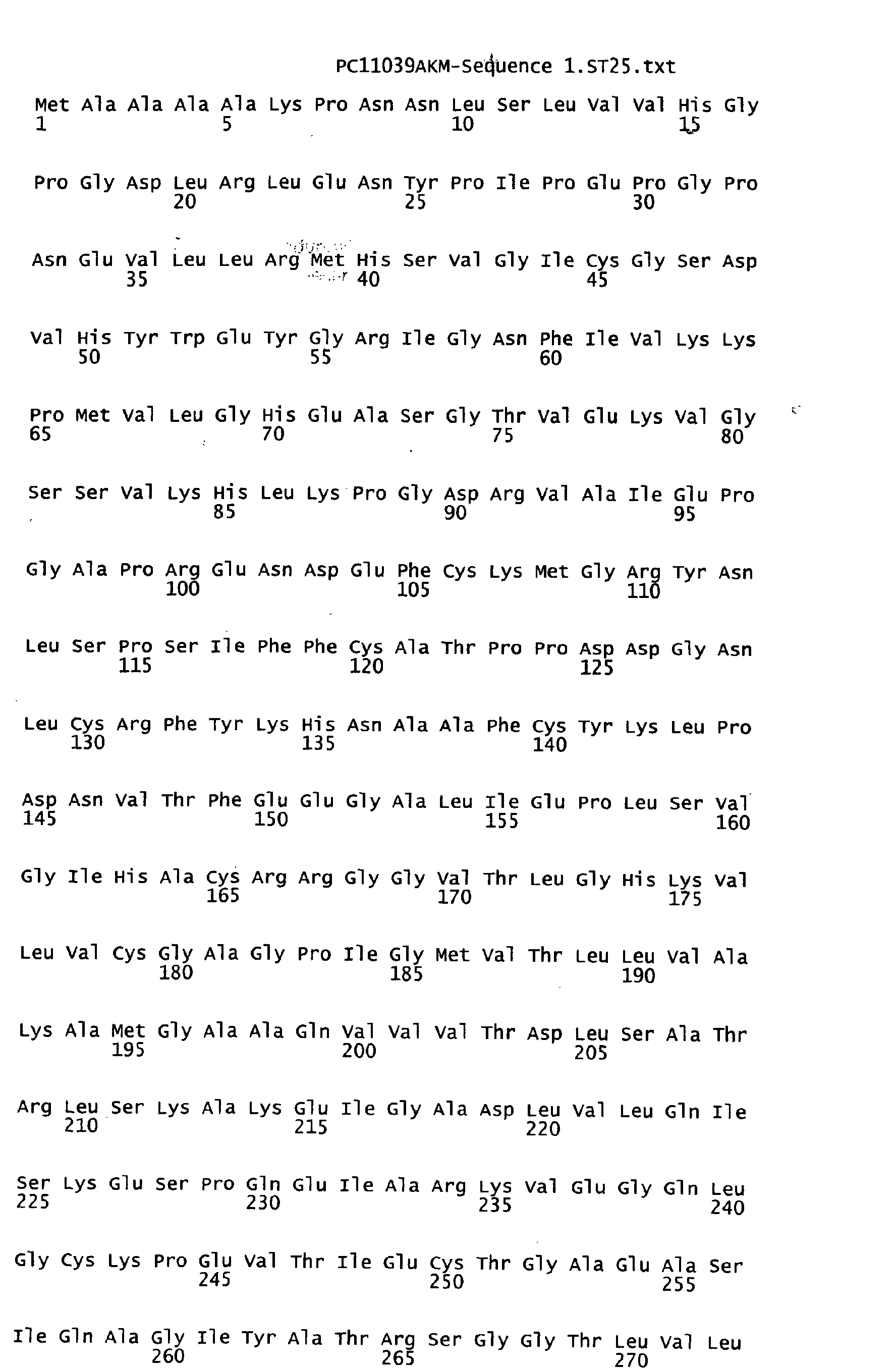

Claims (8)
Applications Claiming Priority (2)
| Application Number | Priority Date | Filing Date | Title |
|---|---|---|---|
| US32939601P | 2001-10-15 | 2001-10-15 | |
| US329396P | 2001-10-15 |
Publications (2)
| Publication Number | Publication Date |
|---|---|
| EP1302538A2 true EP1302538A2 (en) | 2003-04-16 |
| EP1302538A3 EP1302538A3 (en) | 2004-01-14 |
Family
ID=23285195
Family Applications (1)
| Application Number | Title | Priority Date | Filing Date |
|---|---|---|---|
| EP02022322A Withdrawn EP1302538A3 (en) | 2001-10-15 | 2002-10-08 | Three dimensional crystal structure of human sorbitol dehydrogenase and uses thereof |
Country Status (6)
| Country | Link |
|---|---|
| US (1) | US20030119160A1 (en) |
| EP (1) | EP1302538A3 (en) |
| JP (1) | JP2003135057A (en) |
| AU (1) | AU2002341257A1 (en) |
| CA (1) | CA2403258A1 (en) |
| WO (1) | WO2003033643A2 (en) |
Families Citing this family (1)
| Publication number | Priority date | Publication date | Assignee | Title |
|---|---|---|---|---|
| ES2324128A1 (en) * | 2005-09-29 | 2009-07-30 | Proyecto De Biomedicina Cima, S.L. | METHOD FOR THE DIAGNOSIS OF HEPATOCELLULAR CARCINOMA THROUGH THE USE OF MOLECULAR MARKERS. |
Family Cites Families (2)
| Publication number | Priority date | Publication date | Assignee | Title |
|---|---|---|---|---|
| WO1994007867A1 (en) * | 1992-09-28 | 1994-04-14 | Pfizer Inc. | Substituted pyrimidines for control of diabetic complications |
| US6074822A (en) * | 1995-11-03 | 2000-06-13 | Board Of Trustees Operating Michigan State University | Method for testing for risk of diabetes |
-
2002
- 2002-09-30 WO PCT/IB2002/004034 patent/WO2003033643A2/en not_active Ceased
- 2002-09-30 AU AU2002341257A patent/AU2002341257A1/en not_active Abandoned
- 2002-10-08 EP EP02022322A patent/EP1302538A3/en not_active Withdrawn
- 2002-10-10 JP JP2002297160A patent/JP2003135057A/en active Pending
- 2002-10-11 CA CA002403258A patent/CA2403258A1/en not_active Abandoned
- 2002-10-11 US US10/269,875 patent/US20030119160A1/en not_active Abandoned
Non-Patent Citations (4)
| Title |
|---|
| IWATA TAKESHI ET AL: "Structural organization of the human sorbitol dehydrogenase gene (SORD)" GENOMICS, vol. 26, no. 1, 1995, pages 55-62, XP001154796 ISSN: 0888-7543 * |
| JOHANSSON K ET AL: "Crystal structure of sorbitol dehydrogenase" CHEMICO-BIOLOGICAL INTERACTIONS, ELSEVIER SCIENCE IRLAND, IR, vol. 130-132, 30 January 2001 (2001-01-30), pages 351-358, XP002963302 ISSN: 0009-2797 * |
| LEE F K ET AL: "THE HUMAN SORBITOL DEHYDROGENASE GENE: CDNA CLONING, SEQUENCE DETERMINATION, AND MAPPING BY FLORESCENCE IN SITU HYBRIDIZATION" GENOMICS, ACADEMIC PRESS, SAN DIEGO, US, vol. 21, no. 2, 15 May 1994 (1994-05-15), pages 354-358, XP001078865 ISSN: 0888-7543 * |
| PAULY THOMAS A ET AL: "X-ray crystallographic and kinetic studies of human sorbitol dehydrogenase." STRUCTURE (CAMBRIDGE, MASS.: 2001) UNITED STATES SEP 2003, vol. 11, no. 9, September 2003 (2003-09), pages 1071-1085, XP001153279 ISSN: 0969-2126 * |
Also Published As
| Publication number | Publication date |
|---|---|
| WO2003033643A3 (en) | 2004-05-27 |
| WO2003033643A2 (en) | 2003-04-24 |
| CA2403258A1 (en) | 2003-04-15 |
| EP1302538A3 (en) | 2004-01-14 |
| AU2002341257A1 (en) | 2003-04-28 |
| US20030119160A1 (en) | 2003-06-26 |
| JP2003135057A (en) | 2003-05-13 |
Similar Documents
| Publication | Publication Date | Title |
|---|---|---|
| Pauly et al. | X-ray crystallographic and kinetic studies of human sorbitol dehydrogenase | |
| Gerhardt et al. | The structural basis of riboflavin binding to Schizosaccharomyces pombe 6, 7-dimethyl-8-ribityllumazine synthase | |
| Scapin et al. | Crystal structure of orotate phosphoribosyltransferase | |
| Nguyen et al. | Two endoplasmic reticulum PDI peroxidases increase the efficiency of the use of peroxide during disulfide bond formation | |
| Blaszczyk et al. | Crystal structure of unligated guanylate kinase from yeast reveals GMP-induced conformational changes | |
| Rebelo et al. | Gene sequence and crystal structure of the aldehyde oxidoreductase from Desulfovibrio desulfuricans ATCC 27774 | |
| Oikonomakos et al. | Allosteric inhibition of glycogen phosphorylase a by the potential antidiabetic drug 3-isopropyl 4-(2-chlorophenyl)-1, 4-dihydro-1-ethyl-2-methyl-pyridine-3, 5, 6-tricarboxylate | |
| Oikonomakos et al. | Kinetic and crystallographic studies of glucopyranosylidene spirothiohydantoin binding to glycogen phosphorylase b | |
| Phillips et al. | A 2.8 Å resolution structure of 6-phosphogluconate dehydrogenase from the protozoan parasiteTrypanosoma brucei: comparison with the sheep enzyme accounts for differences in activity with coenzyme and substrate analogues | |
| Huang et al. | Crystal structures of Fms1 and its complex with spermine reveal substrate specificity | |
| Pereira et al. | Structure of shikimate kinase from Mycobacterium tuberculosis reveals the binding of shikimic acid | |
| Lee et al. | Crystal structure of the rat liver fructose-2, 6-bisphosphatase based on selenomethionine multiwavelength anomalous dispersion phases | |
| Ladame et al. | Crystal structure of Trypanosoma cruzi glyceraldehyde‐3‐phosphate dehydrogenase complexed with an analogue of 1, 3‐bisphospho‐d‐glyceric acid: Selective inhibition by structure‐based design | |
| Rigden et al. | The 2.3 Å X-ray crystal structure of S. cerevisiae phosphoglycerate mutase | |
| Kedzierski et al. | Structure of Leishmania mexicana phosphomannomutase highlights similarities with human isoforms | |
| US6356845B1 (en) | Crystallization and structure determination of Staphylococcus aureus UDP-N-acetylenolpyruvylglucosamine reductase (S. aureus MurB) | |
| US20030119160A1 (en) | Three dimensional crystal structure of human sorbitol dehydrogenase and uses thereof | |
| Blaise et al. | Crystal structure of glutamyl-queuosine tRNAAsp synthetase complexed with L-glutamate: structural elements mediating tRNA-independent activation of glutamate and glutamylation of tRNAAsp anticodon | |
| EP1629092B1 (en) | CRYSTALLOGRAPHIC STRUCTURE OF TRYPANOSOMA CRUZI PROLINE RACEMASE (TcPRACA) AND USES THEREOF | |
| US20030073134A1 (en) | Crystals and structures of 2C-methyl-D-erythritol 2,4-cyclodiphosphate synthase MECPS | |
| Carey et al. | At the outer part of the active site in Trypanosoma cruzi glucokinase: The role of phenylalanine 337 | |
| US20030187220A1 (en) | Crystals and structures of a flavin mononucleotide binding protein (FMNBP) | |
| CN101496014A (en) | Influenza virus neuraminidase crystal structure and their use thereof | |
| US20040253178A1 (en) | Crystals and structures of spleen tyrosine kinase SYKKD | |
| JP2004180565A (en) | Crystallization and structure of prostaglandin F synthase and use thereof |
Legal Events
| Date | Code | Title | Description |
|---|---|---|---|
| PUAI | Public reference made under article 153(3) epc to a published international application that has entered the european phase |
Free format text: ORIGINAL CODE: 0009012 |
|
| AK | Designated contracting states |
Designated state(s): AT BE BG CH CY CZ DE DK EE ES FI FR GB GR IE IT LI LU MC NL PT SE SK TR |
|
| AX | Request for extension of the european patent |
Extension state: AL LT LV MK RO SI |
|
| PUAL | Search report despatched |
Free format text: ORIGINAL CODE: 0009013 |
|
| AK | Designated contracting states |
Kind code of ref document: A3 Designated state(s): AT BE BG CH CY CZ DE DK EE ES FI FR GB GR IE IT LI LU MC NL PT SE SK TR |
|
| AX | Request for extension of the european patent |
Extension state: AL LT LV MK RO SI |
|
| RIC1 | Information provided on ipc code assigned before grant |
Ipc: 7G 01N 33/574 B Ipc: 7C 12N 9/04 A |
|
| 17P | Request for examination filed |
Effective date: 20040714 |
|
| AKX | Designation fees paid |
Designated state(s): AT BE BG CH CY CZ DE DK EE ES FI FR GB GR IE IT LI LU MC NL PT SE SK TR |
|
| STAA | Information on the status of an ep patent application or granted ep patent |
Free format text: STATUS: THE APPLICATION IS DEEMED TO BE WITHDRAWN |
|
| 18D | Application deemed to be withdrawn |
Effective date: 20060503 |