TECHNICAL FIELD
The present invention relates to a novel neuropeptide and, more
particularly, to a novel peptide that is obtained from brains of
Octopus vulgaris (including optic glands, optic tracts, and optic
lobes) and has the gonadotropin-releasing hormone (GnRH) activity.
The present invention also relates to a precursor polypeptide of such
a peptide and a gene encoding the same.
BACKGROUND ART
Invertebrates have a complex endocrine system that has developed
along with their nervous system. In invertebrates, various
endogenous peptides are known that serve as neurotransmitters,
neuromodulators or neurohormones.
Mollusks generally have a simpler nervous system as compared to
higher animals. Their nervous systems therefore serve as a useful
tool in studying the mechanisms of information processing. Any
knowledge obtained at cellular level about the information processing
mechanisms of mollusks is considered generally applicable to the
information processing mechanisms of nervous systems of higher
animals. For this reason, a significant amount of effort has been
made to find neurotransmitters of mollusks.
For example, an endogenous neuropeptide obtained from mussel is
disclosed in Japanese Patent Laid-Open Publication No. Hei 1-221392.
A novel peptide isolated from ganglia of Achatina fulica is disclosed
in Japanese Patent Laid-Open Publication No. Hei 2-286696. This
peptide includes a D-amino acid and is found to function as an
endogenous neurotransmitter. Japanese Patent Laid-Open Publication
No. 6-56890 discloses a neuropeptide isolated from ganglia of a
Hungarian snail.
Aside from those described above, various other neuropeptides
have been isolated from mollusks and have been identified. Examples
include Ala-Pro-Gly-Trp-NH2, myomodulin-CARP, small cardioactive
peptide (SCP), buccalin, and Phe-Met-Arg-Phe-NH2 (FMRF-amide) (M.
Kobayashi and Y. Muneoka, Zool. Sci., 7, 801 (1990); Y. Muneoka and
M. Kobayashi, Experimentia 48, 448 (1992); Youjiro. Muneoka, Journal
of Pesticide Science, 18, S191 (1993); Y. Muneoka, T. Takahashi, M.
Kobayashi, "Perspective in Comparative Endocrinology", National
Research Council of Canada, 1994, p109; A. Di Cosmo and C. Di Cristo,
J. Comp. Neurol., 398, 1-12 (1998)).
Mollusks form one of the largest families of invertebrates. Of
different species of mollusks, octopuses belong to a group called
Cephalopoda. The animals have highly developed brains and far more
advanced motor and sensory functions than other members of mollusks.
The neurosecretory system of octopuses has been extensively studied.
In 1956, Boycott and Young discovered that cutting the optic tract of
the animal results in enlarged optic glands and development of
gonads. Later, the Wells showed that the gland secretes a
gonadotropic hormone (M. J. Wells and J. Wells, J. Exp. Biol., 36, 1-33
(1959)). It turned out that the hormone is a protein hormone and
affects the growth of germ cells, the maturation of gonad-related
organs, and the synthesis of yolk protein. The hormone is also
involved in parenting behaviors of octopuses after their laying eggs
(Atlas of endocrine organs, Japan Society for Comparative
Endocrinology. ed., Kodansha).
Gonadotropin-releasing hormone (GnRH) was first isolated in 1971
by Schally et al., from the porcine hypothalamus and was then
discovered as luteinizing hormone-releasing hormone (LHRH). Its
structure was also determined. Since then GnRHs have been studied by
many researchers. To date, many GnRHs have been found in different
vertebrates such as mammals, birds, and salmon. Also, some suggest
that many invertebrates include tissues and substances that are
immunoreactive to GnRH (A. Di Cosmo and C. Di Cristo, J. Comp.
Neurol., 398, 1-12 (1998)).
It has been shown that, in mammals, GnRHs are secreted from
hypothalamus and act on the pituitary gland to cause the release of
gonadotropic hormones. These GnRHs are each a decapeptide.
In octopuses, however, the structure of the gonadotropic hormone
has yet to be determined although it is known, as described above,
that the gonadotropins are secreted from their optic glands.
Moreover, no GnRH has yet been isolated from octopuses, nor has its
structure been determined.
Accordingly, it is an objective of the present invention to
identify, and determine the structure of, a novel GnRH of octopuses.
It is another objective of the present invention to understand the
activities of the GnRH, the structure of which is determined
according to the present invention, both in octopuses and in other
invertebrates and vertebrates.
A further objective of the present invention is to provide a
novel peptide that can serve as a useful reagent in studying
correlations between the structure and the activities of GnRH and in
studying the information processing mechanisms of nervous systems in
higher animals. A still further objective of the present invention
is to provide a novel peptide that can be used as a base compound in
the development of pesticides and drugs.
It is yet another objective of the present invention to identify
a precursor polypeptide of the GnRH and a gene encoding the precursor
for the purpose of expanding our knowledge about evolution of
animals. It is yet another objective of the present invention to
provide a method for producing the GnRH.
DISCLOSURE OF THE INVENTION
In an effort to isolate GnRH from octopuses, the present
inventors conducted extensive studies on various physiologically
active peptides obtained from the brains of about 100 specimens of
O.
vulgaris by investigating the activities of the peptides to enhance
the heart beat of the animals. As a result, the present inventors
isolated and purified a peptide having the following amino acid
sequence (1), determined its structure, and verified the structure by
total synthesis:
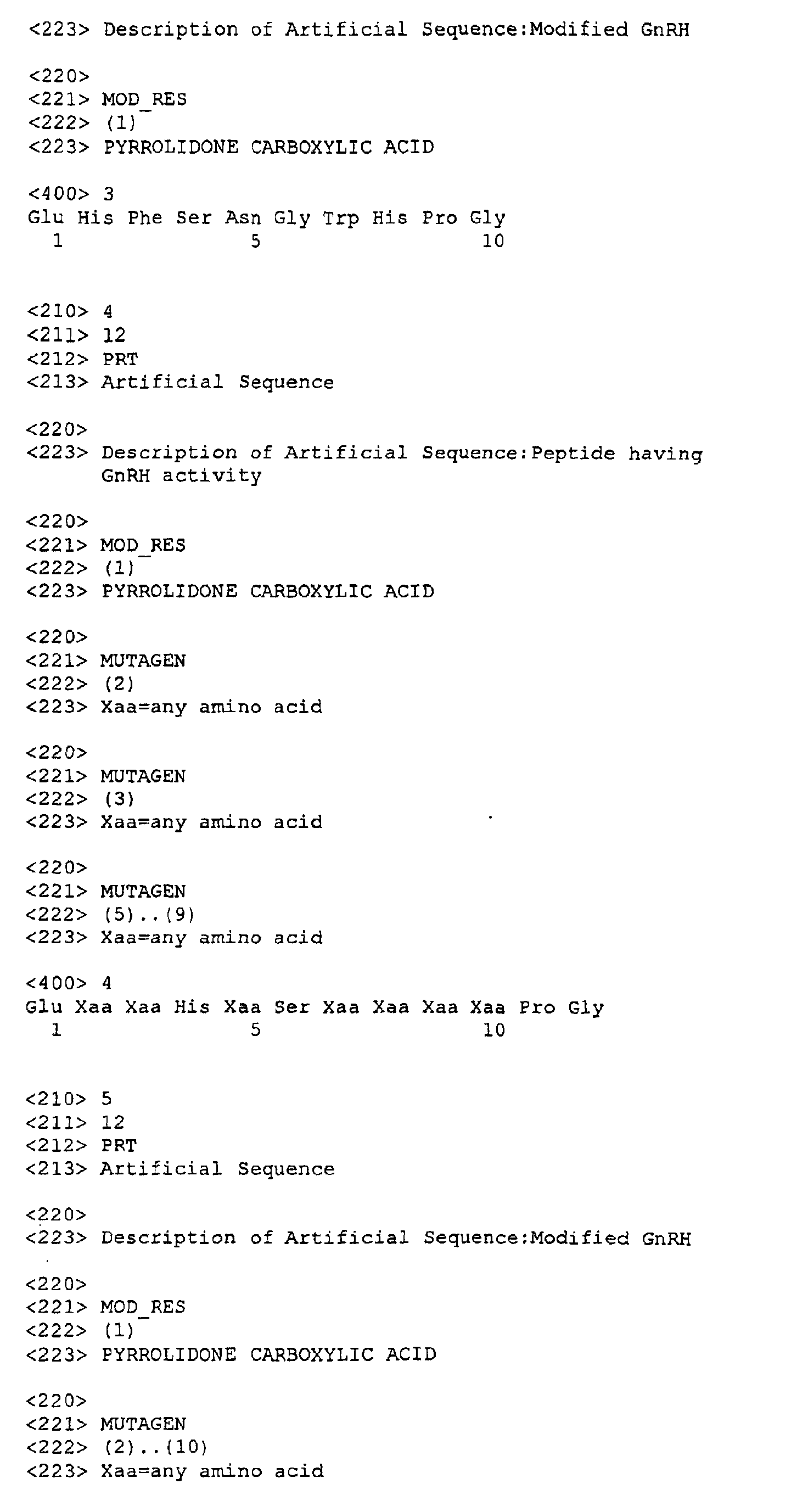
wherein <Glu represents pyroglutamic acid. This peptide may be
referred to as Compound (1), hereinafter. Unless specified, all
amino acids are represented by the three-letter codes defined by the
IUPAC nomenclature system.
As shown in Fig. 5, Compound (1) has a very high homology to the
GnRHs identified in vertebrates. Unlike known decapeptide GnRHs,
Compound (1) turned out to be a dodecapeptide.
The present inventors further conducted tests using quails and
proved that Compound (1) has the activity of GnRH, namely, the
activity to cause the release of luteinizing hormone (LH) from the
pituitary gland of quails.
The present inventors also prepared a primer from the above
amino acid sequence and performed a gene sequence analysis using the
reverse transcriptase polymerase chain reaction (RT-PCR) on the total
RNA obtained from the brains of O. vulgaris. In this manner, the
present inventors determined that the precursor polypeptide of the
peptide of the present invention has a total primary amino acid
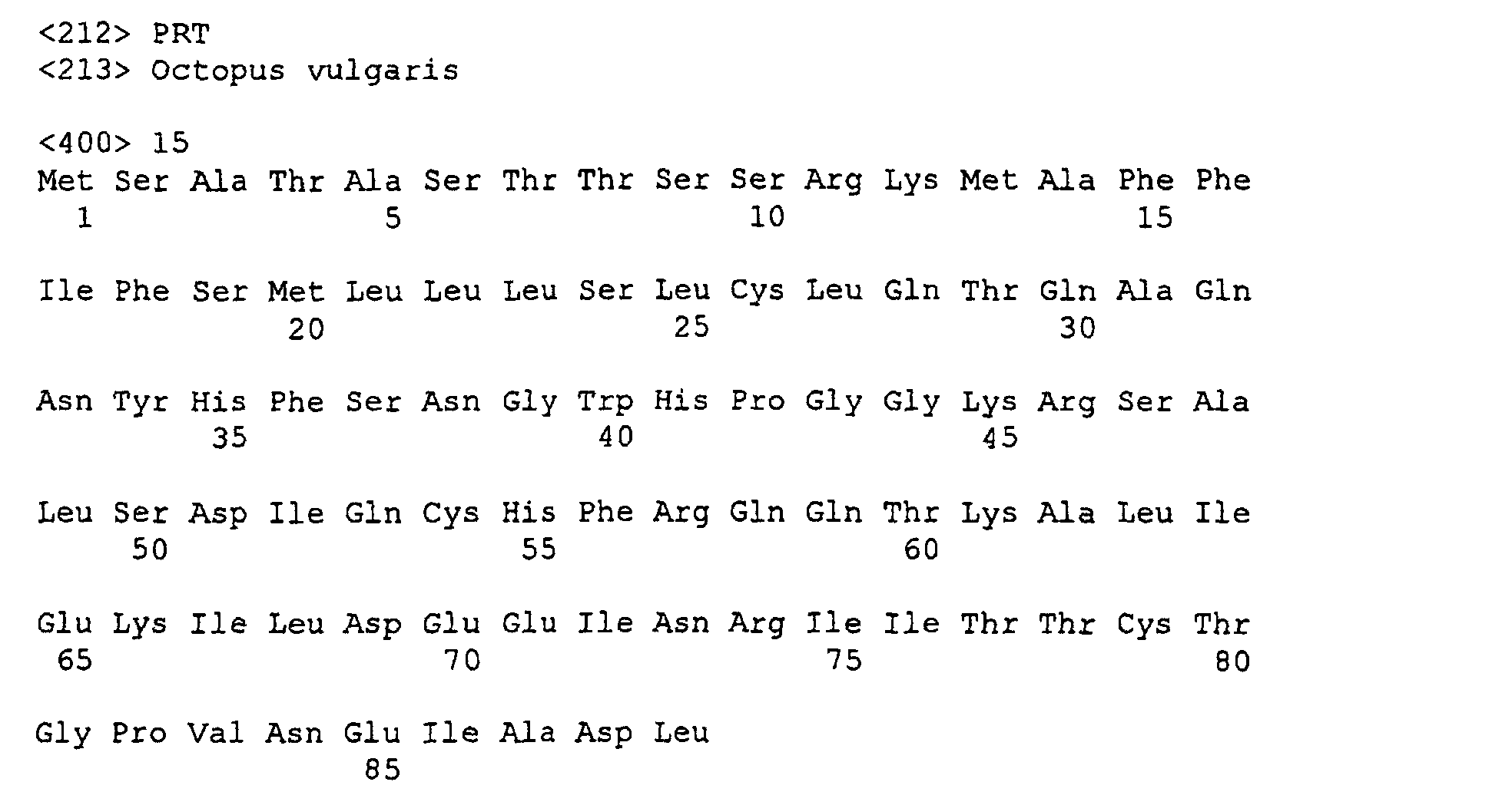
sequence shown as SEQ ID NO: 15.
The present inventors further determined that the gene encoding
the precursor polypeptide has a base sequence shown as SEQ ID NO: 14,
thereby completing the present invention.
Moreover, taking advantage of the decapeptide GnRH identified in
vertebrates, the present inventors synthesized a new peptide similar
to the peptide of the amino acid sequence (1) isolated from the
brains of O. vulgaris by inserting two amino acid residues, Asn-Tyr,
into the N-terminal region of the decapeptide GnRH between the first
<Glu and the second His residues and examined its physiological
activities.
Among other GnRHs, chicken-II-type GnRH (cGnRH-II) shown in Fig.
5 is commonly found in various classes of animals as well as in
chickens. For this reason, the two amino acid residues, Asn-Tyr,
were inserted into the N-terminal region of cGnRH-II between the
first <Glu and the second His residues of the peptide to give a new
peptide represented by the following amino acid sequence (2):
wherein <Glu represents pyroglutamic acid. The peptide may be
referred to as Compound (2), hereinafter. Compound (2) was examined
for the activity to enhance the heart beat of
O. vulgaris and it was
observed that Compound (2) showed an activity similar to Compound (1)
isolated from the brains of
O. vulgaris whereas cGnRH-II showed no
such activity.
Furthermore, the present inventors deleted amino acids 2 and 3
from the N-terminal region of the dodecapeptide, which was isolated
from invertebrates and had the GnRH activity, to convert it into a
decapeptide similar to the GnRH isolated from vertebrates. As a
result, it was observed that the decapeptide retained the initial
GnRH activity.
To rephrase, the present inventors confirmed that the peptide
represented by the following amino acid sequence (3), which is
obtained by deletion of
amino acids 2 and 3 from the N-terminal
region of Compound (1) isolated from the brains of
O. vulgaris,
exhibits the GnRH activity:
wherein <Glu represents pyroglutamic acid. The peptide may be
referred to as Compound (3), hereinafter.
Accordingly, the present invention provides a dodecapeptide
having the activity of gonadotropin-releasing hormone (GnRH) and
having the following amino acid sequence (I):
wherein <Glu represents pyroglutamic acid and Zaa represents any
amino acid.
The amino acid sequence (I) of the above-described peptide
having the GnRH activity may be more specifically defined by the
following amino acid sequence (II):
wherein <Glu represents pyroglutamic acid and Zaa represents any
amino acid.
The amino acid sequence (I) of the above-described peptide
having the GnRH activity may be even more specifically defined by the
following amino acid sequence (III):
wherein <Glu represents pyroglutamic acid, Zaa represents any amino
acid, and Yaa represents aromatic amino acid.
Of the peptides having the GnRH activity provided by the present
invention, particularly preferred are those represented by the
following amino acid sequences (1), (2) and (3) :
wherein <Glu represents pyroglutamic acid.
The present invention further provides a method for producing a
pharmaceutical agent or a pesticide containing as an active
ingredient the peptide of the amino acid sequence (I) having the GnRH
activity, as well as a method for producing juvenile octopuses.
According to one aspect of the present invention, there is
provided a precursor polypeptide having either an amino acid sequence
of SEQ ID NO: 2, an amino acid sequence obtained by deletion or
substitution of part of the amino acid of SEQ ID NO: 15, or an amino
acid sequence obtained by addition of one or more amino acids to the
amino acid sequence obtained by deletion or substitution of part of
the amino acid of SEQ ID NO: 15.
According to another aspect of the present invention, there are
provided a gene encoding the peptide and the precursor polypeptide of
the present invention, in particular a gene having a base sequence of
SEQ ID NO: 14, a vector incorporating these genes, and a host
transformed by using the vector, and a method for producing, by means
of genetic engineering, the peptide or the precursor polypeptide of
the present invention by taking advantage of these genes.
BRIEF DESCRIPTION OF THE DRAWINGS
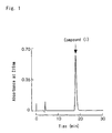
Fig. 1 is a diagram depicting the results of reversed-phase HPLC
showing the final elution pattern of Compound (1), a peptide
according to the present invention in Example 1.
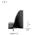
Fig. 2 is a diagram showing the heart beat of O. vulgaris when
an amount of Compound (1), the peptide of the present invention,
corresponding to one specimen of O. vulgaris was added to a chamber
in Example 5.
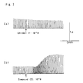
Fig. 3 includes diagrams showing the results of Example 5,
wherein the diagrams show variation in the heart beat of O. vulgaris
(a) when 10-5 M cGnRH-II was added to the chamber and (b) when 10-5 M
Compound (2), the peptide of the present invention, was added to the
chamber, respectively.
Fig. 4 is a graph showing the amounts of released luteinizing
hormone (LH) when Compound (1), the peptide of the present invention,
was applied to the pituitary gland of a quail in Example 6.
Fig. 5 is a comparative diagram showing structures of Compound
(1), the peptide of the present invention, and of gonadotropin-releasing
hormones identified in different vertebrates.
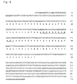
Fig. 6 shows the amino acid sequence of the precursor
polypeptide of the present invention, in which the part corresponding
to the peptide of the present invention is underlined.
BEST MODE FOR CARRYING OUT THE INVENTION
Novel peptides provided by the present invention act to cause
the release of luteinizing hormone (LH) from the pituitary gland in
quails and to enhance the heart beat in Octopus vulgaris. For this
reason, the peptides can be isolated and purified from the brains of
O. vulgaris on the basis of this activity toward the hearts of O.
vulgaris as follows:
In one example, hot water extracts of brains of O. vulgaris are
prepared. To the extract solution, acetic acid is added to a
concentration of 3%. The solution is allowed to cool and is then
centrifuged to obtain crude extracts. The extracts are adsorbed onto
a C18 cartridge (e.g., Sep-Pak® Vac C18 cartridge manufactured by
WATERS Co., Ltd.) and are then eluted with a 60% methanol containing
0.1% trifluoroacetic acid to collect a peptide fraction. The peptide
fraction can be subjected to ion-exchange chromatography, reversed-phase
chromatography, or other chromatography techniques to separate
and purify peptides of interest.
Each being a dodecapeptide, the peptides of the present
invention can be readily synthesized by solid-phase synthesis using a
common peptide synthesizer (e.g., Peptide synthesizer 433A,
manufactured by PE biosystems Japan Co., Ltd.) or any common
techniques in synthetic organic chemistry. When necessary, the crude
peptide products obtained by these techniques may be purified by
common purification techniques such as reversed-phase high-performance
liquid chromatography and crystallization.
The amino acid sequence of the precursor polypeptide of the
peptide, as well as the base sequence encoding the precursor
polypeptide in accordance with the present invention, may be
determined in the following manner:
Using a primer synthesized based on the amino acid sequence of
the present peptide obtained in the above-described manner, total RNA
prepared from the brains of O. vulgaris is subjected to the reverse
transcriptase polymerase chain reaction (RT-PCR) to determine the
gene sequence of about 750 bp. Subsequently, the 5'-RACE and the 3'-RACE
techniques are used to determine the gene sequence on the 5'-end
and the 3'-end, respectively. According to the present invention, it
was determined that the precursor polypeptide of the present peptide
has an amino acid sequence shown as SEQ ID NO: 15 and the gene
encoding the precursor polypeptide has a base sequence shown as SEQ
ID NO: 14.
Accordingly, the precursor polypeptide of the peptide of the
present invention, as well as the peptide of the present invention,
can be produced using recombinant DNA techniques. When it is desired
to produce the precursor polypeptide by using a recombinant DNA
technique, a vector incorporating for example the gene of SEQ ID NO:
3 may be introduced into hosts for transformation. The hosts are
subsequently cultured or grown, and the precursor polypeptide of
interest is isolated and purified from the hosts or the culture
solution of the hosts.
To obtain the peptide of interest from the precursor
polypeptide, the precursor polypeptide may be processed and modified
using an enzyme. If necessary, the peptide product may be subjected
to an isolation or a purification process.
The peptides of the present invention each have an activity to
enhance the heart beat of O. vulgaris and have a structure quite
analogous to GnRH (peptide) of vertebrates. The peptides also act on
the pituitary gland of quails to cause the release of luteinizing
hormone. Furthermore, studies of the peptides of the present
invention suggested that bird GnRH is related to the activity to
enhance the heart beat in octopuses. These observations collectively
indicate that the peptide in accordance with the present invention is
in fact a hormone (peptide) involved in the release of gonadotropins
in octopuses and other invertebrates, as well as in vertebrates.
Accordingly, the peptide of the present invention can serve as a
useful material both for studying biology of octopuses including
mechanisms of their reproduction and for studying structural
correlations between GnRHs of invertebrates and those of vertebrates.
Thus, the peptide will be of significant usefulness in the study of
animal evolution. Also, the peptide of the present invention is
expected to find use not only in the development of new substances
with the activity of gonadotropin-releasing hormone, but also in the
development of new medicines and pesticides using the peptide itself
or peptide analogues thereof, as well as of new techniques for the
production of juvenile octopuses.
For example, the peptide of the present invention may be used as
an active ingredient in a medicine and may be formulated along with a
pharmacologically acceptable excipient into proper oral or parenteral
preparations, such as capsules, tablets, and injections.
Specifically, the peptide of the present invention may be admixed
with an excipient, such as lactose, starch or a derivative thereof,
or a derivative of cellulose, and is encapsulated in gelatin capsules
to form a capsule preparation.
When it is desired to form the peptide of the present invention
into the form of tablets, a binder, such as carboxymethylcellulose
sodium, alginic acid, and gum arabic, and water are added to the
peptide in addition to the aforementioned excipient. The mixture is
then kneaded and, if necessary, is formed into the form of granules.
Subsequently, a lubricant, such as talc and magnesium stearate, is
added to the mixture and the mixture is formed into tablets by a
common compression tablet-making machine.
When it is desired to form the peptide of the present invention
into preparations for parenteral administration, the peptide may be
dissolved, along with a solubilizer, into sterilized distilled water
or sterilized saline and is placed in sealed ampoules to provide a
preparation for injection. When necessary, a stabilizer, a buffering
agent, or other additives may be added to the preparation.
Alternatively, the peptide of the present invention may be placed in
vials in the form of powder so that, upon use, the powder can be
dissolved in sterilized distilled water to form a solution
preparation. These parenteral preparations may be delivered by
intravenous administration or intravenous drip infusion.
Preferably, the dosage of the preparation containing the peptide
of the present invention as an active ingredient is adjusted in
consideration of various factors, including the disease to be
treated, symptoms of the patient, severity of the symptom, age of the
patient, and route of administration. In case of oral
administration, the preparation is typically administered at a dose
of 0.1 to 1,000 mg/day/human, preferably at a dose of 1 to 500
mg/day/human, whereas in case of parenteral administration, the
preparation may be administered at a dose that is about 1/100 to
about 1/2 the dose for oral administration.
Because of its nature as GnRH, the peptide of the present
invention can be administered to cephalopods such as octopuses to
cause the animals to reach sexual maturation at an earlier stage, so
that the animals can be induced to start laying eggs at an earlier
stage. Breeding and culturing these animals can facilitate
production of juvenile cephalopods.
EXAMPLES
The present invention will now be described in detail in the
following with reference to Examples, which are not intended to limit
the scope of the invention in any way.
Example 1: Isolation of cardioactive peptides from O. vulgaris
(a) Crude extraction
Brain tissue (including optic glands, optic tracts, and optic
lobes) was taken from 100 specimens of Octopus vulgaris and was
frozen in liquid nitrogen. The frozen tissue (180 g) was boiled in
900 ml distilled water for 10 min. After cooling, acetic acid was
added to a concentration of 3% and the tissue was homogenized and
then centrifuged at 4°C at 12,000 x g for 30 min to obtain a
supernatant. To the resulting pellet, 200 ml of 3% acetic acid was
added and the mixture was homogenized again for extraction.
Subsequently, the mixture was centrifuged under the same conditions
to obtain a supernatant. The supernatant was collected and
concentrated in vacuo to a volume of about 200 ml to give crude
extracts.
(b) Adsorption to C18 cartridge
To the crude extracts obtained in the step (a) above, 5 ml of 6
M HCl was added and the mixture was centrifuged at 4°C at 12,000 x g
for 30 min. The resulting supernatant was passed through Sep-Pak® Vac
C18 cartridge (10 g, 35 cc, manufactured by WATERS Co., Ltd.) to
allow the solutes to be adsorbed onto the cartridge. The cartridge
was then washed with 200 ml of 0.1% trifluoroacetic acid (TFA,
hereinafter), and the retained materials were then eluted with 100 ml
of 60% methanol/0.1% TFA. The eluate was concentrated in vacuo and
was freeze-dried to obtain 150 mg dried products.
(c) Reversed-phase high-performance liquid column chromatography (1)
Using Capcell pak C18 UG80 (5 µm, 10 mm x 250 mm, manufactured
by SHISEIDO Co., Ltd.), the dried products obtained in the step (b)
above were subjected to the reversed-phase high-performance liquid
column chromatography (reversed-phase HPLC). Specifically, the dried
products in 0.1% TFA (pH 2.2) were passed through the column at a
flow rate of 1.5 ml/min and the retained materials were eluted with a
linear gradient of acetonitrile in which the concentration of
acetonitrile in TEA was linearly varied from 0% to 60% over 60 min.
The eluate was collected in 3 ml fractions based on the UV-absorbance
at 215 nm.
Each fraction was bioassayed according to the scheme described
in Example 5 below and, as a result, it was found that a fraction
eluted while the concentration of acetonitrile was varied from 26% to
30% had an activity to enhance the heart beat of O. vulgaris.
(d) Cation-exchange column chromatography
Using TSK gel SP-5PW (10 µm, 7.5 mm x 75 mm, manufactured by
TOSOH Co., Ltd.), the active fraction obtained in the step (c) above
was subjected to the cation-exchange column chromatography (cation-exchange
HPLC). Specifically, the fraction was loaded onto the
column and was eluted with a 10 mM phosphate buffer (pH 7.0) at a
flow rate of 1.0 ml/min while the concentration of NaCl in the
phosphate buffer was linearly varied from 0 M to 0.6 M over 60 min
(linear gradient). The eluate was collected in 2 ml fractions and
each fraction was bioassayed. The activity was found in the fraction
eluted when the concentration of NaCl was 0 M.
(e) Reversed-phase HPLC (2)
Using Capcell pak C18 UG80 (5 µm, 4.6 mm x 150 mm, manufactured
by SHISEIDO Co., Ltd.), the active fraction obtained in the step (d)
above was subjected to the reversed-phase HPLC. Specifically, the
fraction was loaded onto the column and was eluted with 0.1% TFA (pH
2.2) at a flow rate of 1.0 ml/min while the concentration of
acetonitrile in TFA was linearly varied from 10 to 30% (linear
gradient). The eluate was collected in 1 ml fractions. The activity
was found in the fraction eluted when the concentration of
acetonitrile was about 19.5%.
(f) Reversed-phase HPLC (3)
Using Capcell pak C18 UG80 (5 µm, 4.6 mm x 150 mm, manufactured
by SHISEIDO Co., Ltd.), the active fraction obtained in the step (e)
above was subjected to the reversed-phase HPLC. Specifically, the
fraction was loaded onto the column and was eluted with 0.1% TFA (pH
2.2) having an acetonitrile concentration of 18% at a flow rate of
0.5 ml/min. A compound that exhibited a single peak was obtained at
a retention time of 17.5 min. The compound was designated as
Compound (1). A diagram showing the results of the reversed-phase
HPLC was shown in Fig. 1.
Example 2: Structure determination of Compound (1)
The structure analysis of Compound (1), purified in Example 1,
by Shimadzu PSQ-1 protein sequencer (manufactured by SHIMADZU Co.,
Ltd.) could not determine the amino acid sequence of the peptide due
to the modified N-terminal. Thus, the amino acid composition of the
peptide was analyzed instead. The results are shown in Table 1
below.
| Amino acid composition of the peptide |
| Amino acid | pmol | Molar ratio |
| Asx | 51.1 | 1.5 |
| Glx | 42.6 | 1.3 |
| Ser | 32.7 | 1.0 |
| Gly | 70.0 | 2.1 |
| His | 57.9 | 1.7 |
| Pro | 37.1 | 1.1 |
| Tyr | 24.2 | 0.7 |
| Phe | 33.8 | 1.0 |
The molecular weight of Compound (1) was determined by Q-TOF
(Micromass, manufactured by UK Co., Ltd.). The measurements are
shown in Table 2 below.
| Mass spectrum data of the peptide |
| Compound | Molecular formula | Calculated value
(M+2H)2+ | Calculated value
(M+2H)2+ |
| Before treatment with enzyme | C66H80N20O17 | 713.30 | 713.30 |
| After treatment with enzyme | C61H75N19O15 | 657.79 | 657.85 |
It was determined from the analysis of the amino acid
composition and the measurement of the molecular weight that the N-terminal
of Compound (1) includes a pyroglutamic acid residue.
Accordingly, Compound (1) was treated with an enzyme capable of
cleaving off the first pyroglutamic acid residue (pyroglutamate
aminopeptidase, manufactured by TAKARA SHUZO Co., Ltd.) and was again
analyzed by the protein sequencer.
Consequently, the amino acid sequence of Compound (1) was
determined, starting from the second amino acid residue, and the
presence of Trp, which cannot be detected by the composition
analysis, was verified.
The amino acid sequence determined is shown in Table 3 below.
| Amino acid sequence of the peptide after treatment with enzyme (pmol) |
| Cycle | Amino acid | |
| 1 | Asn | 27 |
| 2 | Tyr | 25 |
| 3 | His | 7 |
| 4 | Phe | 23 |
| 5 | Ser | 3 |
| 6 | Asn | 16 |
| 7 | Gly | 12 |
| 8 | Trp | 3 |
| 9 | His | 2 |
| 10 | Pro | 5 |
| 11 | Gly | 4 |
To make sure that the enzyme treatment had worked as intended,
the molecular weight of Compound (1) after the treatment was again
determined by Q-TOF (Micromass, manufactured by UK Co., Ltd.) in the
same manner and, as a result, it was verified that the first
pyroglutamic acid residue had been cleaved off. The results are also
shown in Table 2 above.
Through the foregoing instrumental analyses, it was determined
that Compound (1), a neuropeptide isolated and purified from
O.
vulgaris, has the following amino acid sequence (1):
wherein <Glu represents pyroglutamic acid.
Using a fully automated peptide synthesizer 433A (PE Biosystems
Japan), Compound (1) was synthesized according to FastMoc® chemistry.
In the synthesis, an Fmoc-NH-SAL-A resin (manufactured by
WATANABE CHEMICAL INDUSTRIES, Ltd.) to serve as a solid support was
used, along with pyroglutamic acid, Fmoc-Asn-(Trt), Fmoc-Tyr-(tBu),
Fmoc-His-(Trt), Fmoc-Phe, Fmoc-Ser-(tBu), Fmoc-Gly, Fmoc-Trp-(tBoc),
and Fmoc-Pro.
As used herein, Fmoc represents 9-fluorenylmethoxycarbonyl, tBu
represents t-butyl, Trt represents trityl, and tBoc represents t-butyloxycarbonyl.
A 10 ml mixture of 2.5% 1,2-ethanedithiol/2.5% water/95% TEA
containing 100 mg 2-methylindole was used for the purposes of
cleavage of the crude peptide from the peptide resin upon completion
of the reaction and deprotection of the crude peptide. After the
reaction, the reaction mixture was filtered. Ether was then added to
the filtrates to cause the peptide to precipitate. The precipitates
were washed three times with ether to obtain about 100 mg of crude
peptide, about 10 mg of which was then purified by the reversed-phased
HPLC to obtain about 6 mg of purified peptide.
When subjected to the reversed-phase HPLC using Capcell pak C18,
the purified peptide eluted exactly at the same retention time as
Compound (1). Also, the synthetic peptide exhibited the same
cardioactivity toward the heart beat of O. vulgaris as did the
naturally-occurring peptide.
Example 4: Synthesis of Compounds (2) and (3) by the solid phase
method
Peptides represented by the following amino acid sequences (2)
and (3) were synthesized in the same manner as in Example 3 above:
wherein <Glu represents pyroglutamic acid.
The heart beat activity of O. vulgaris was measured according to
the method proposed by Morishita et al (Fumihiro Morishita, Biochem.
Biophys. Res. Commun., 240, 354-358 (1997)). Specifically, the heart
was taken from O. vulgaris and the atria were removed from the heart.
A cannula was inserted from each atrium into the ventricle and was at
the same time connected to a chamber (80 ml in volume) by tying with
a cotton thread. The aorta was pinched to prevent leakage of liquid
from the ventricle and was connected to a tension transducer. This
completed preparation of the heart to serve as a specimen for the
assay. Each cannula was adjusted to permit a 1 to 2 ml/min flow of
artificial sea water (containing 0.1% glucose). Each of the analytes
to be assayed was dissolved in 1 ml of the same artificial sea-water,
and the resulting solution was injected into the heart through the
cannulae. The variation of the heart beat was recorded.
As test compounds, Compound (1) isolated from the brains of O.
vulgaris, Compound (2), and cGnRH-II, a vertebrate GnRH isolated from
chickens and used to derive Compound (2) therefrom, were used.
As shown in Fig. 2, the results indicate that Compound (1)
enhanced the heart beat of O. vulgaris.
As shown in Fig. 3(b), Compound (2) also enhanced the heart
beat of O. vulgaris by increasing both the heart rate and the
amplitude of the heart beat. In contrast, cGnRH-II, as can be seen
from Fig. 3(a), did not have any effect on the heart rate or the
amplitude of the heart beat of O. vulgaris.
Example 6: Effects on the pituitary glands of quails
Cells taken from the pituitary glands of quails were inoculated
onto a 24-well plate at 7.5 x 10⁵ cells/well (substantially equivalent
to two animals). The cells were then incubated for 3 days in a
culture medium containing fetal calf serum. The cells were incubated
for another 24 hrs in 1 ml serum-free medium. After the incubation
period, the peptide solution was added to the culture medium to final
concentrations of 0, 10-9, 10-8, 10-7, 10-6, and 10-5 M, respectively,
and the cultures were further incubated for 4 hrs. The supernatant
was then collected from each culture. For each concentration, 5
samples were subjected to radioimmunoassay and measurements were
taken. The data were statistically analyzed by one-way analysis of
variance (ANOVA).
The results are shown in Fig. 4. As shown, the statistical
analysis revealed that Compound (1), the peptide of the present
invention, significantly increased the release of luteinizing hormone
(LH).
When tested in the same manner, Compound (3) also proved to
increase the release of luteinizing hormone (LH).
Example 7: Determination of the entire amino acid structure of the
precursor polypeptide and determination of the base sequence of the
gene encoding the polypeptide
(1) Preparation of total RNA of the brain of O. vulgaris
About 1 g of the brain of O. vulgaris was crushed in liquid
nitrogen, dissolved in 10 ml TRIzol® reagent (GIBCO BRL Co., Ltd.),
and homogenized. At room temperature, the homogenates were allowed
to stand for 5 min and were divided into 1 ml aliquots. To each
aliquot, 200 µl chloroform was added and the mixture was stirred.
The mixture was then centrifuged (15,000 rpm, 15 min, 4°C) in a cold
centrifuge (SAKUMA SEISAKUSHO Co., Ltd.). Following the
centrifugation, the top aqueous layer was removed and 0.5 ml
isopropanol was added to the aqueous phase. The mixture was then
allowed to stand for 10 min at room temperature and was further
centrifuged (15,000 rpm, 10 min, 4°C) in the cold centrifuge. The
supernatant was removed and the pellet was resuspended in 1 ml 75%
ethanol. The suspension was centrifuged again (10,000 rpm, 5 min,
4°C). Subsequently, the supernatant was removed and the pellet was
air-dried for about 10 min. The RNA pellet was then dissolved in 10
µl DPEC-treated water by incubating at 60°C for 10 min. In this
manner, about 3 mg of total RNA was obtained.
(2) degenerate 3'-RACE
The following degenerate primers were designed based on the
amino acid sequence of the peptide isolated from the brain of
O.
vulgaris and synthesized by an ordinary method:
wherein each letter is based on the definition in "Table of
Nucleotide Codes" (
Biology experiment illustrated, supplement to
Cell
Engineering, SHUJUNSHA Co., Ltd., The same codes are used,
hereinafter).
Using a 5'/3'-RACE Kit (Boehringer Mannheim Co., Ltd.),
degenerate 3'-RACE was performed according to the following protocol:
2 µg total RNA, 4 µl cDNA synthesis buffer, 2 µl dNTP mix, 1 µl oligo
dT-anchor primer (12.5 pmol/µl), 1 µl AMV reverse transcriptase (20
units/µl), and DEPC-treated water were added to one another to give a
total volume of 20 µl. The mixture was incubated at 55°C for 60 min
and then at 65°C for 10 min to synthesize 1st-strand cDNA.
Next, 1st-3'-RACE was performed according to the following
procedure: 3 µl 1st-strand cDNA, 5 µl 10 x PCR buffer, 8 µl dNTP mix,
5 µl Prtmer-1 (100 pmol/µl), 1 µl PCR anchor primer (12.5 pmol/µl),
0.5 µl TaKaRa Ex Taq® (TAKARA SHUZO Co., Ltd), and water were mixed
with each other to give a total volume of 50 µl. The mixture was
incubated at 94°C for 5 min, followed by 30 cycles of 94°C for 30
sec, 49°C for 30 sec, and 72°C for 2 min, which was followed by
incubation at 72°C for 5 min. GeneAmp PCR System 2400 thermal cycler
(Perkin Elmer Co., Ltd.) was used to carry out the PCR.
Subsequently, the 1st-PCR products were purified through a spin
column (MicroSpin® S-400, manufactured by Amersham Pharmacia Co.,
Ltd.) and were subjected to 2nd-3'-RACE according to the following
procedure: 5 µl 1st-PCR products, 5 µl 10 x PCR buffer, 8 µl dNTP
mix, 5 µl Primer-2 (100 pmol/µl), 1 µl PCR anchor primer (12.5
pmol/µl), 0.5 µl TaKaRa Ex Taq® (TAKARA SHUZO Co., Ltd), and water
were mixed with each other to give a total volume of 50 µl. The
mixture was incubated at 94°C for 5 min, followed by 30 cycles of
94°C for 30 sec, 51°C for 30 sec, and 72°C for 1 min, which was
followed by incubation at 72°C for 7 min.
Five microliter of the reaction mixture was then electrophoresed
on a 1.5% agarose gel. Bands were observed at about 400 bp and about
600 bp, indicating amplification of two PCR products.
(3) Collection of PCR products
Five microliter of the PCR products was electrophoresed on a
1.5% agarose gel and the two PCR product bands formed at about 400 bp
and about 600 bp were cut out. Using Gel Extraction Kit (QIAGEN Co.,
Ltd.), DNA was collected from the gel. To confirm the results, 3 µl
of each of the collected DNA fragments was electrophoresed on a 1.5%
agarose gel. As a result, the two PCR products were observed at
about 400 bp and about 600 bp, respectively.
(4) Ligation of the PCR products
The two PCR products, sized about 400 bp and about 600 bp,
respectively, were individually purified through a spin column. For
each of the PCR products, 3 µl of PCR product, 2 µl TA cloning vector
pCR2.1 (Invitrogene Co., Ltd.), and 5 µl Ligation High (TOYOBO Co.,
Ltd.) were mixed with each other, and the mixture was allowed to
react for 1 hr at 16°C (ligation).
(5) Transformation of E. coli
Each of the two 10 µl ligation mixtures obtained in the step (4)
above was mixed with highly competent E. coli DH5α (TOYOBO Co.,
Ltd.). The mixture was placed in an ice bath for 30 min and was then
subjected to heat-shock at 42°C for 50 sec, followed by chilling on
ice for 2 min. After chilling, 1 ml SOC medium was added and the
mixture was incubated at 37°C for 10 min. Subsequently, 35 µl X-gal
was evenly spread onto the surface of LB/Amp agar medium (LB
containing 50 µg/ml ampicilin), and 50 µl transformant was inoculated
onto the medium. The remaining transformant was centrifuged at
10,000 rpm for 1 min to a volume of about 100 µl and the whole
transformant was also inoculated onto the LB/Amp agar medium. The
culture medium was incubated overnight at 37°C.
(6) Colony PCR
Using the colonies obtained above as a template, the colony PCR
was performed. Specifically, the E. coli cells, 5 µl 10x reaction
buffer, 5 µl 2 mM dNTPs, 3 µl 25 mM MgCl2, 0.5 µl M13FW primer (100
pmol/µl), 0.5 µl M13RV primer (100 pmol/µl), 0.5 µl rTaq DNA
polymerase (TOYOBO Co., Ltd.), and water were mixed with each other
to give a total volume of 50 µl. The mixture was incubated at 90°C
for 10 min, followed by 30 cycles of 94°C for 30 sec, 55°C for 30
sec, and 72°C for 1 min. Three microliter of the reaction mixture
was electrophoresed on a 1.5% agarose gel.
The M13FW and M13RV primers, the base sequences of which are
shown below, were synthesized by an ordinary method:
The colony PCR products of the size of interest (about 600 bp
and about 800 bp) were purified through a spin column and were
sequenced by DNA Sequencing Kit (PE Biosystems, Co., Ltd.). ABI
PRISM 310 Genetic Analyzer (PE Biosystems, Co., Ltd.) was used for
sequencing.
Through the sequence analysis by a gene-analyzing software
GENETYX-MAC (SOFTWARE KAIHATSU Co., Ltd.), the cDNA sequence of the
about 800 bp fragment was determined. A further DNA sequencing
revealed that the cDNA sequence of the fragment of about 600 bp was
in fact part of the cDNA sequence of the fragment of about 800 bp.
(8) 5'-RACE
The following primers were synthesized according to the base
sequences of the cDNA fragments.
Using 5'/3'-RACE Kit (Boehringer Mannheim Co., Ltd.), 5'-RACE
was performed according to the following protocol: 2 µg total RNA, 1
µl 5'-RACE-1 (12.5 pmol/µl), 4 µl cDNA synthesis buffer, 2 µl dNTP
mix, 1 µl AMV reverse transcriptase (20 units/µl), and DEPC-treated
water were mixed with each other to give a total volume of 20 µl.
The mixture was incubated at 55°C for 60 min and then at 65°C for
another 10 min to synthesize 1st-strand cDNA.
After purification through a spin column, the 1st-strand cDNA.
was mixed with 2.5 µl reaction buffer and 2.5 µl 2 mM dNTP, and the
mixture was incubated at 94°C for 3 min, followed by chilling on ice.
Subsequently, 1 µl terminal transferase (10 units/µl) was added to
the mixture, and the mixture was incubated at 37°C for 20 min and
then at 70°C for another 10 min to synthesize dA-tailed cDNA.
Next, 1st-PCR and 2nd-PCR were conducted under the following
conditions:
(a) 1st-PCR
Three microliter dA-tailed cDNA, 5 µl 10 x PCR buffer, 8 µl dNTP
mix, 1 µl 5'-RACE-2 (12.5 pmol/µl), 1 µl oligo dT-anchor primer (12.5
pmol/µl), 0.5 µl TaKaRa Ex Taq® (TAKARA SHUZO Co., Ltd) and water were
mixed with each other to give a total volume of 50 µl. The mixture
was incubated at 94°C for 5 min, followed by 30 cycles of 94°C for 30
sec, 55°C for 30 sec, and 72°C for 3 min, which was followed by
incubation at 72°C for 5 min.
(b) 2nd-PCR
Five microliter of the 1st-PCR product purified through a spin
column, 5 µl 10 x PCR buffer, 8 µl dNTP mix, 1 µl 5'-RACE-3 (12.5
pmol/µl), 1 µl PCR-anchor primer (12.5 pmol/µl), 0.5 µl TaKaRa Ex Taq®
(TAKARA SHUZO Co., Ltd) and water were mixed with each other to give
a total volume of 50 µl. PCR was then carried out by using the same
cycle conditions as in the 1st-PCR.
The resulting 2nd-PCR product was electrophoresed on a 1.5%
agarose gel. As a result, a band was observed at about 600 bp.
The base sequence of the 2nd-PCR product was determined
according to the protocol described in (4), (5), (6), and (7) above.
Through the foregoing analyses, the size of cDNA (about 700 bp)
and the base sequence were determined for the gene encoding the
precursor polypeptide of the peptide of the present invention.
Also, the estimated number of amino acids (89 amino acid residues),
as well as the amino acid sequence, was determined for the precursor
polypeptide of the peptide of the present invention.
The amino acid sequence of the precursor polypeptide and the
base sequence of the gene (cDNA) encoding the precursor polypeptide
are shown as SEQ ID NO: 15 and SEQ ID NO: 14, respectively. Also,
the part of the amino acid sequence of the precursor polypeptide that
corresponds to the peptide of the present invention is shown in Fig.
6.
In the figure, the part corresponding to the amino acid sequence
of the peptide of the present invention is underlined.
Example 8: Exemplary preparation
| Composition: Compound (1) |
10 g |
| lactose |
125 g |
| microcrystalline cellulose |
25 g |
| corn starch |
25 g |
| 5% hydroxypropyl methylcellulose |
100 ml |
| magnesium stearate |
5 g |
According to a known method, the above components were kneaded,
granulated, dried and shaped into tablets to obtain tablets, each
weighing 190 mg and containing 10 mg of Compound (1) as an active
ingredient.
INDUSTRIAL APPLICABILITY
As shown in Fig. 5, the peptide of the present invention, which
acts to enhance the heart beat of O. vulgaris, has a structure highly
analogous to gonadotropin-releasing hormone (peptide) of vertebrates.
Together with this finding, the fact that the peptide acts on the
pituitary gland of quails to cause the release of luteinizing hormone
implies that the peptide is in fact a hormone (peptide) involved in
the release of gonadotropin in octopuses.
Thus, studying the activities of the present peptide will help
understand biology of octopuses including their breeding mechanisms
as well as processes of animal evolution.
Not only does the peptide of the present invention serve as a
useful tool in developing new substances with the activity of
gonadotropin-releasing hormone, but it also may lead to development
of new medicines and pesticides that make use of the peptide itself
or peptide analogues, as well as of new techniques for the production
of juvenile octopuses.