EP1295936A1 - Method of preparing heart muscle cells and method of searching for remedy for heart diseases - Google Patents
Method of preparing heart muscle cells and method of searching for remedy for heart diseases Download PDFInfo
- Publication number
- EP1295936A1 EP1295936A1 EP01945643A EP01945643A EP1295936A1 EP 1295936 A1 EP1295936 A1 EP 1295936A1 EP 01945643 A EP01945643 A EP 01945643A EP 01945643 A EP01945643 A EP 01945643A EP 1295936 A1 EP1295936 A1 EP 1295936A1
- Authority
- EP
- European Patent Office
- Prior art keywords
- heart muscle
- muscle cells
- heart
- cells
- apoptosis
- Prior art date
- Legal status (The legal status is an assumption and is not a legal conclusion. Google has not performed a legal analysis and makes no representation as to the accuracy of the status listed.)
- Granted
Links
Images


Classifications
-
- G—PHYSICS
- G01—MEASURING; TESTING
- G01N—INVESTIGATING OR ANALYSING MATERIALS BY DETERMINING THEIR CHEMICAL OR PHYSICAL PROPERTIES
- G01N33/00—Investigating or analysing materials by specific methods not covered by groups G01N1/00 - G01N31/00
- G01N33/48—Biological material, e.g. blood, urine; Haemocytometers
- G01N33/50—Chemical analysis of biological material, e.g. blood, urine; Testing involving biospecific ligand binding methods; Immunological testing
- G01N33/5005—Chemical analysis of biological material, e.g. blood, urine; Testing involving biospecific ligand binding methods; Immunological testing involving human or animal cells
- G01N33/5008—Chemical analysis of biological material, e.g. blood, urine; Testing involving biospecific ligand binding methods; Immunological testing involving human or animal cells for testing or evaluating the effect of chemical or biological compounds, e.g. drugs, cosmetics
- G01N33/5044—Chemical analysis of biological material, e.g. blood, urine; Testing involving biospecific ligand binding methods; Immunological testing involving human or animal cells for testing or evaluating the effect of chemical or biological compounds, e.g. drugs, cosmetics involving specific cell types
- G01N33/5061—Muscle cells
-
- A—HUMAN NECESSITIES
- A61—MEDICAL OR VETERINARY SCIENCE; HYGIENE
- A61P—SPECIFIC THERAPEUTIC ACTIVITY OF CHEMICAL COMPOUNDS OR MEDICINAL PREPARATIONS
- A61P9/00—Drugs for disorders of the cardiovascular system
-
- C—CHEMISTRY; METALLURGY
- C12—BIOCHEMISTRY; BEER; SPIRITS; WINE; VINEGAR; MICROBIOLOGY; ENZYMOLOGY; MUTATION OR GENETIC ENGINEERING
- C12N—MICROORGANISMS OR ENZYMES; COMPOSITIONS THEREOF; PROPAGATING, PRESERVING, OR MAINTAINING MICROORGANISMS; MUTATION OR GENETIC ENGINEERING; CULTURE MEDIA
- C12N5/00—Undifferentiated human, animal or plant cells, e.g. cell lines; Tissues; Cultivation or maintenance thereof; Culture media therefor
- C12N5/06—Animal cells or tissues; Human cells or tissues
- C12N5/0602—Vertebrate cells
- C12N5/0652—Cells of skeletal and connective tissues; Mesenchyme
- C12N5/0657—Cardiomyocytes; Heart cells
-
- C—CHEMISTRY; METALLURGY
- C12—BIOCHEMISTRY; BEER; SPIRITS; WINE; VINEGAR; MICROBIOLOGY; ENZYMOLOGY; MUTATION OR GENETIC ENGINEERING
- C12N—MICROORGANISMS OR ENZYMES; COMPOSITIONS THEREOF; PROPAGATING, PRESERVING, OR MAINTAINING MICROORGANISMS; MUTATION OR GENETIC ENGINEERING; CULTURE MEDIA
- C12N2503/00—Use of cells in diagnostics
- C12N2503/02—Drug screening
-
- C—CHEMISTRY; METALLURGY
- C12—BIOCHEMISTRY; BEER; SPIRITS; WINE; VINEGAR; MICROBIOLOGY; ENZYMOLOGY; MUTATION OR GENETIC ENGINEERING
- C12N—MICROORGANISMS OR ENZYMES; COMPOSITIONS THEREOF; PROPAGATING, PRESERVING, OR MAINTAINING MICROORGANISMS; MUTATION OR GENETIC ENGINEERING; CULTURE MEDIA
- C12N2509/00—Methods for the dissociation of cells, e.g. specific use of enzymes
-
- G—PHYSICS
- G01—MEASURING; TESTING
- G01N—INVESTIGATING OR ANALYSING MATERIALS BY DETERMINING THEIR CHEMICAL OR PHYSICAL PROPERTIES
- G01N2510/00—Detection of programmed cell death, i.e. apoptosis
-
- G—PHYSICS
- G01—MEASURING; TESTING
- G01N—INVESTIGATING OR ANALYSING MATERIALS BY DETERMINING THEIR CHEMICAL OR PHYSICAL PROPERTIES
- G01N2800/00—Detection or diagnosis of diseases
- G01N2800/32—Cardiovascular disorders
- G01N2800/324—Coronary artery diseases, e.g. angina pectoris, myocardial infarction
Definitions
- This invention relates to a method of preparing highly pure primary heart muscle cells easily in a large amount, which is useful for searching for remedies for heart diseases having an inhibitory action on apoptosis of heart muscle cells or an enhancing action on heart muscle cell-protective signals, as well as a method of searching for such remedies efficiently and highly sensitively by dealing with a large number of samples.
- This invention relates in particular to a method of searching for remedies for heart diseases, characterized by preparing highly pure primary-culture of heart muscle cells easily in a large amount from the heart of an animal embryo or a newborn and detecting heart muscle cell apoptosis efficiently and highly sensitively by using the prepared heart muscle cells, in order to search for prophylactic and therapeutic agents for heart diseases having an inhibitory action on apoptosis of heart muscle cells or an enhancing action on heart muscle cell-protective signals by using the highly pure primary heart muscle cells.
- Apoptosis is concerned closely with morphology and histogenesis in the development process, maintenance of homeostasis, and protection of the living body, and refers to cell death having an important role in maintaining the life of individuals.
- apoptosis is excessively induced or inhibited to cause functional disorders in various organs, leading to diseases (Shin Yonehara, Saishin Igaku (Latest Medicine), vol. 54, p. 825, 1999). Accordingly, there is demand for an understanding of diseases from the viewpoint of apoptosis and for development of diagnostic methods or remedies based on the molecular mechanism.
- Apoptosis is opposite to cell proliferation. Accordingly, heart muscle cells, that is, finally differentiated cells not growing, are considered less related to apoptosis. However, it was revealed in recent years that apoptosis is concerned closely with onset or progress of various heart diseases (R. Sanders Williams, The New England Journal of Medicine, vol. 341, p. 759, 1999). Heart muscle cells are not proliferated so that when heart muscle cells disappear by apoptosis, the contracting function should be maintained by surviving cells only. Accordingly, disappearance of heart muscle cells beyond the necessary threshold for maintaining the contracting function of the heart would result in abnormal heart functions, leading to diseases.
- apoptosis of heart muscle cells is observed in various animal models with cardiac insufficiency or in patients with cardiac insufficiency, and it is noted that disappearance and dropout of heart muscle cells by apoptosis may be concerned with onset and progress of cardiac insufficiency (Narula, J. et al., The New England Journal of Medicine, vol. 335, p. 1182, 1996). It is further recognized that in heart muscle cells in patients with cardiac insufficiency, an apoptosis-inhibitory factor Bcl-2 is expressed in excess, which is shown to be a possible mechanism for compensating for cardiac insufficiency (Olivetti, G. et al., The New England Journal of Medicine, vol. 336, p.
- sFas has an inhibitory activity on apoptosis serum levels of soluble Fas (sFas has an inhibitory activity on apoptosis) deficient in a membrane penetration domain, secreted due to splicing of a Fas receptor known to induce apoptosis, are increased significantly in proportion to severeness in NYHA class (New York Heart Association Functional Class) but independently of fundamental diseases, and thus an increase in serum levels of sFas is considered to be a compensatory mechanism for inhibiting promotion of apoptosis at the time of cardiac insufficiency (Nishigaki, K. et al., Journal of the American College of Cardiology, vol. 29, p.
- DNase I deoxyribonuclease I
- heart muscle cells are prepared by a method of isolating the cells by digesting mainly animal hearts with a protease such as trypsin or collagenase.
- digestion with a protease such as trypsin or collagenase is carried out generally after the heart of an animal embryo or a newborn is fragmented to facilitate digestion with the protease (Kiyota Goto & Hiroyuki Kaneko, Shinzo Kekkan Kenkyu Hoho No Kaihatsu (Development in Method of Studying Heart and Blood Vessels) edited by Setsuro Ebashi, page 3, Japan Scientific Societies Press (JSSP) (1983)).
- Another known method of preparing heart muscle cells involves excising heart muscle cells from a matured animal, setting the cells in a Langendolff type perfusion device, washing blood away and perfusing a solution of a protease such as trypsin or collagenase (Haruo Sugi & Yukio Hiramoto: Jikken Seibutsugaku Koza (Lecture on Experimental Biology), vol. 10, p. 27 (1984)).
- a protease such as trypsin or collagenase
- the density of the cells should be reduced to a certain degree because it is considered that at higher density, cells are contacted with one another to induce protective signals and to produce protection factors, and further the activity of intracellular dehydrogenase activity in viable heart muscle cells in a serum-free state is low, and therefore it is difficult to detect apoptosis of heart muscle cells quantitatively by e.g. a method of reduction of MTT (3-(4,5-dimethylthiazol-2-yl)-2,5-diphenyltetrazolium bromide) (Tada et al., Journal of Immunological Methods), vol. 93, p. 157 (1986)).
- MTT 3-(4,5-dimethylthiazol-2-yl)-2,5-diphenyltetrazolium bromide
- the present inventors found that fragmented heart tissue of an animal embryo or a newborn is washed with a phosphate-buffered physiological saline before treatment with a protease such as trypsin or collagenase, and residual erythrocytes are removed by a low-isotonic hemolysis procedure, whereby highly pure primary heart muscle cells can be prepared easily in a large amount.
- this preparative method is also referred to as the method of preparing heart muscle cells according to this invention.
- the present inventors also found a method of efficiently and sensitively detecting apoptosis of heart muscle cells induced by removing serum from a culture medium, which comprises adding serum to the culture, culturing the cells for about 17 hours to revitalize the cells, and then determining the number of viable cells by using a cell counting kit-8 (a product of Dojin Kagaku Kenkyusho) with WST-8 (2-(2-methoxy-4-nitrophenyl)-3-(4-nitrophenyl)-5-(2,4-disulfophenyl)-2H-tetrazolium, monosodium salt) as a coloring substrate .
- this method is also referred to as the method of detecting apoptosis according to this invention.
- the present invention relates to [1] a method of easily preparing a large amount of highly pure primary-culture heart muscle cells useful for searching for compounds having an inhibitory action on apoptosis of heart muscle cells, compounds having an agonistic action on gp130-mediated receptors or compounds having an enhancing action on heart muscle cell-protective signals, [2] a method of detecting these compounds efficiently and highly sensitively, [3] a searching method (screening method) for dealing with a large number of samples, which comprises the above preparative method in combination with the above detection method, and [4] heart muscle cell apoptosis inhibitors, gp130-mediated receptor agonists, heart muscle cell-protective signal enhancers, or prophylactic and/or therapeutic agents for heart diseases, comprising compounds obtained by the searching method or salts thereof.
- This invention relates in particular to:
- This method of the present invention is suited for, but is not limited to, preparation of a large amount of heart muscle cells from the heart of an animal, e.g. rat or mouse, embryo or a newborn, from which blood is hardly washed away with a Langendolff type perfusion device or the like.
- the heart is excised, and the excised heart is washed with a phosphate-buffered physiological saline and then fragmented.
- the fragmented heart tissue is washed with 4 to 5 times with a phosphate-buffered physiological saline or the like, whereby non-heart muscle cells derived from blood can be efficiently removed.
- this heart tissue fragments are digested with a protease such as trypsin or collagenase to separate the cells.
- a protease such as trypsin or collagenase
- the conditions such as the type of enzyme, the reaction temperature and the reaction time can be suitably selected depending on the type of animal.
- the enzyme reaction can be terminated by adding a serum-containing medium. After the reaction is terminated, the cells are collected by centrifuging the cell-containing suspension, and then suspended in a suitable medium. After the cell mass is removed, the cell-containing medium is placed in a Petri dish and cultured for 1 hour.
- the floating cells are recovered from the Petri dish and subjected to hemolysis with a low-isotonic solution or the like to remove residual erythrocytes.
- the cells after hemolysis with a low-isotonic solution or the like are suspended in a suitable medium, then placed in a Petri dish and cultured for about 24 hours, whereby the heart muscle cells adhere to the Petri dish.
- a majority of cells not adhering at this stage to the Petri dish are damaged heart muscle cells, and these cells can be easily removed by mildly shaking the Petri dish to stir the culture in the Petri dish and exchanging the medium with a fresh one.
- heart muscle cells of low purity obtained by the conventional method contaminating blood-derived non-heart muscle cells and damaged heart muscle cells are aggregated to adhere to a Petri dish, and thus these cells cannot be sufficiently removed by stirring the Petri dish and exchanging the medium. Accordingly, the plate to which the heart muscle cells adhere should be further washed in order to obtain highly pure heart muscle cells.
- heart muscle cells of e.g. a rat newborn are prepared, the rat newborn is anesthetized under ether, the abdomen is sterilized and the heart is excised.
- the excised heart is washed with e.g. a phosphate-buffered physiological saline (pH 7.4) and fragmented with scissors etc.
- the size of the fragment is preferably 1 mm 3 or less.
- the fragmented heart tissue is washed 4 to 5 times with e.g. a phosphate-buffered physiological saline (pH 7.4) to sufficiently remove non-heart muscle cells derived from blood.
- the heart tissue fragments are digested with a protease such as trypsin or collagenase to separate the cells.
- a protease such as trypsin or collagenase
- the conditions such as the type of optimum enzyme, the reaction temperature and the reaction time are varied depending on the type of animal used; for example, when heart muscle cells from rat newborns are used, the cells from 10 newborns are treated for 15 minutes with 5 ml enzyme solution (prepared by dissolving 1.25 mg trypsin (Difco) and 0.25 mg collagenase (Sigma) per ml of PBS), and 2.5 ml enzyme solution is added additionally twice at a 15-minutes interval, and the cells are kept at 37°C under stirring with a stirrer.
- enzyme solution prepared by dissolving 1.25 mg trypsin (Difco) and 0.25 mg collagenase (Sigma) per ml of PBS
- the enzyme reaction is terminated by adding a medium such as Medium 199 containing serum such as FCS or the like at a final concentration of 10 %.
- a medium such as Medium 199 containing serum such as FCS or the like at a final concentration of 10 %.
- the cells are collected by centrifuging the cell-containing suspension, and then suspended in a suitable medium such as Medium 199.
- a filter such as a cell strainer (Falcon)
- the cell-containing medium is placed in a Petri dish and cultured for 1 hour in a CO 2 incubator set at 5 % CO 2 , 37°C.
- a filter such as a cell strainer (Falcon)
- the cell-containing medium is placed in a Petri dish and cultured for 1 hour in a CO 2 incubator set at 5 % CO 2 , 37°C.
- cells from 10 newborns are suspended in about 50 ml medium.
- the floating cells are recovered from the Petri dish and subjected to hemolysis with a low-isotonic solution to remove residual erythrocytes.
- a low-isotonic solution prepared by dissolving 8.29 g NH 4 Cl, 1.0 g KHCO 3 and 37 mg EDTA/2Na in 1 L of water) and left for 3 minutes, whereby erythrocytes can be disrupted.
- the cells after hemolysis with the low-isotonic solution are suspended at a density of about 3 ⁇ 10 4 cells/well (96-wells plate) in a suitable medium such as Medium 199, then placed in a Petri dish and cultured for about 24 hours in a CO 2 incubator set at 5 % CO 2 , 37°C. After culture, the Petri dish is mildly shaken thus stirring the culture in the Petri dish, and then the medium is exchanged with a fresh one, whereby highly pure heart muscle cells are obtained.
- a suitable medium such as Medium 199
- the heart muscle cells obtained by the method of the invention are adjusted to a suitable cell density and cultured in a serum-free medium, whereby apoptosis can be induced, and the survival rate of the heart muscle cells after culture for 4 days can be reduced to 50 % or less.
- a culture of heart muscle cells having apoptosis induced by removing serum is supplemented with serum and further cultured for about 6 to 30 hours (preferably about 17 hours) to increase the dehydrogenase activity of the viable cells, whereby apoptosis of the heart muscle cells can be detected easily and highly sensitively by e.g. a commercial cell counting kit with WST-8 used as the coloration substrate.
- the heart muscle cells obtained by the method of the invention are adjusted to a cell density of 3 ⁇ 10 4 cells/well (96-wells plate) and cultured in serum-free medium such as Medium 199 in a CO 2 incubator set at 5 % CO 2 , 37°C for 4 days, thus inducing apoptosis to reduce the survival rate of the heart muscle cells to 50 % or less.
- Serum such as FCS is added at a final concentration of 10 % to the culture, which is then cultured for about 17 hours and measured for its dehydrogenase activity (reduction activity) with MTT or WST-8 as the coloration substrate, to determine the number of viable cells.
- a large amount of highly pure primary heart muscle cells can be obtained more easily than by the conventional method. Further, apoptosis of heart muscle cells induced by removing serum from the culture can be detected efficiently and highly sensitively.
- the method of the invention can be applied not only to heart muscle cells derived from rats but also to those derived from mammals such as mice, guinea pigs, rabbits, dogs etc.
- the method of detecting apoptosis according to this invention is performed by using the heart muscle cells obtained by the method of preparing heart muscle cells according to this invention, whereby compounds inhibiting apoptosis or salts thereof can be screened.
- compounds inhibiting apoptosis or salts thereof can be screened by comparing the number of viable cells in the case (i) where apoptosis of the heart muscle cells obtained by the present method of preparing heart muscle cells is induced in the presence of a test compound, with that in the case (ii) where apoptosis of the heart muscle cells is induced in the absence of a test compound.
- this screening method is also referred to as the screening method of the invention.
- screening can be carried out by the method described in the Examples described later or by its analogous method.
- the compounds obtained by the screening method or salts thereof have an inhibitory action on apoptosis of heart muscle cells, an agonistic action on gp130-mediated receptors and/or an enhancing action on heart muscle cell-protective signals, and can thus be used as prophylactic and/or therapeutic agents for heart diseases (e.g., congestive myocardiopathy, hypertrophic myocardiopathy with obstruction, hypertrophic myocardiopathy without obstruction, idiopathic myocardiopathy, myocardial infarction, chronic cardiac insufficiency etc.).
- heart diseases e.g., congestive myocardiopathy, hypertrophic myocardiopathy with obstruction, hypertrophic myocardiopathy without obstruction, idiopathic myocardiopathy, myocardial infarction, chronic cardiac insufficiency etc.
- Test compounds include, for example, peptides, proteins, non-peptide compounds, synthetic compounds, fermentation products, cell extracts, plant extracts and animal tissue extracts, which may be novel or known compounds.
- a test compound showing inhibition by about 20 % or more, preferably 30 % or more, more preferably about 50 % or more in terms of the number of viable cells in the case (i), as compared to that in the case (ii), can be selected as a compound inhibiting apoptosis of heart muscle cells.
- the compounds obtained by the screening method of the invention include 2-(2-pyridyl)-4H-1,3-benzothiazin-4-one (Compound 1) etc.
- the compounds obtained by the screening method of the invention or salts thereof are selected from the above-mentioned test compounds, e.g. peptides, proteins, non-peptide compounds, synthetic compounds, fermentation products, cell extracts, plant extracts, animal tissue extracts and plasma, and have an inhibitory activity on apoptosis of heart muscle cells.
- test compounds e.g. peptides, proteins, non-peptide compounds, synthetic compounds, fermentation products, cell extracts, plant extracts, animal tissue extracts and plasma, and have an inhibitory activity on apoptosis of heart muscle cells.
- the salts of the compounds include pharmaceutically acceptable salts with acids (inorganic acids, organic acids) or bases (inorganic bases, organic bases).
- the compounds obtained by the screening method of the invention or salts thereof can be used orally for example in the form of tablets which may be coated with sugar, capsules, elixirs, microcapsules etc., or parenterally in the form of injections such as a sterile solution and a suspension in water or with other pharmaceutically acceptable liquid.
- these preparations can be manufactured by mixing the compound or a salt thereof with a physiologically acceptable carrier, a flavoring agent, an excipient, a vehicle, an antiseptic agent, a stabilizer, a binder, etc. in a unit dosage form required in a generally accepted manner that is applied to making pharmaceutical preparations.
- the effective component in the preparation is controlled in such a dose that an appropriate dose is obtained within the specified range given.
- Additives miscible with tablets, capsules, etc. include a binder such as gelatin, corn starch, tragacanth gum, and gum arabic, an excipient such as crystalline cellulose, a swelling agent such as corn starch, gelatin and alginic acid, a lubricant such as magnesium stearate, a sweetening agent such as sucrose, lactose and saccharin, and a flavoring agent such as peppermint, Gaultheria oil and cherry.
- a binder such as gelatin, corn starch, tragacanth gum, and gum arabic
- an excipient such as crystalline cellulose
- a swelling agent such as corn starch, gelatin and alginic acid
- a lubricant such as magnesium stearate
- a sweetening agent such as sucrose, lactose and saccharin
- a flavoring agent such as peppermint, Gaultheria oil and cherry.
- liquid carriers such as oils and fats may further be used together
- a sterile composition for injection may be formulated by conventional procedures used to make pharmaceutical compositions, e.g., by dissolving or suspending the active ingredients in a vehicle such as water for injection with a naturally occurring vegetable oil such as sesame oil and coconut oil, etc. to prepare the pharmaceutical composition.
- an aqueous medium for injection examples include physiological saline and an isotonic solution containing glucose and other auxiliary agents (e.g., D-sorbitol, D-mannitol, sodium chloride, etc.) and may be used in combination with an appropriate dissolution aid such as an alcohol (e.g., ethanol or the like), a polyalcohol (e.g., propylene glycol and polyethylene glycol), a nonionic surfactant (e.g., polysorbate 80TM and HCO-50), etc.
- an alcohol e.g., ethanol or the like
- a polyalcohol e.g., propylene glycol and polyethylene glycol
- a nonionic surfactant e.g., polysorbate 80TM and HCO-50
- the oily medium examples include sesame oil and soybean oil, which may also be used in combination with a dissolution aid such as benzyl benzoate and benzyl alcohol.
- the injection may further be formulated with a buffer (e.g., phosphate buffer, sodium acetate buffer, etc.), a soothing agent (e.g., benzalkonium chloride, procaine hydrochloride, etc.), a stabilizer (e.g., human serum albumin, polyethylene glycol, etc.), a preservative (e.g., benzyl alcohol, phenol, etc.), an antioxidant, etc.
- a buffer e.g., phosphate buffer, sodium acetate buffer, etc.
- a soothing agent e.g., benzalkonium chloride, procaine hydrochloride, etc.
- a stabilizer e.g., human serum albumin, polyethylene glycol, etc.
- a preservative e.g., benzyl alcohol, phenol, etc.
- an antioxidant e.g., benzyl alcohol, phenol, etc.
- the preparation can be administered into mammals (e.g., humans, rats, mice, guinea pigs, rabbits, birds, sheep, swine, bovine, horses, cats, dogs, monkeys, etc.).
- mammals e.g., humans, rats, mice, guinea pigs, rabbits, birds, sheep, swine, bovine, horses, cats, dogs, monkeys, etc.
- the dose of the obtained compound or a salt thereof varies depending on the intended disease, subject for administration, etc.
- the compound or a salt thereof is administered generally into an adult (weighing 60 kg) in a daily dose of about 0.1 to 100 mg, preferably about 1.0 to 50 mg, more preferably about 1.0 to 20 mg.
- Example 1 Comparison in Number of Cells and Purity between Heart Muscle Cells Obtained by the Method of the Invention and Heart Muscle Cells Obtained by Conventional Method
- Wister rat newborns (within 1 day after birth) available from Charles River were used.
- the rat newborns were anesthetized under ether, their abdomens were sterilized with 70 % ethanol and their hearts were excised with tweezers.
- the excised heart was washed with a phosphate-buffered physiological saline (T900 produced by Takara) and fragmented with surgical scissors.
- T900 phosphate-buffered physiological saline
- Medium 199 (Gibco) containing 10 % fetal calf serum (Biowiker) was added in a half volume relative to the enzyme solution, to terminate the enzyme reaction, and the cells were filtered with a cell strainer and then centrifuged at 400 ⁇ g for 5 minutes, whereby the cells were collected. Then, the cells from the 10 newborns were suspended in 50 ml Medium 199 containing 10 % fetal calf serum, plated in 100-ml Petri dishes (Iwaki) in a volume of 10 ml/disk and cultured for 1 hour in a CO 2 incubator set at 5 % CO 2 , 37°C. The floating cells were recovered, filtered with a cell strainer and centrifuged at 400 ⁇ g for 5 minutes to collect the heart muscle cells.
- Medium 199 Gibco
- 10 % fetal calf serum Biowiker
- the heart muscle cells from the 10 newborns were suspended in 2 ml low-isotonic solution (solution prepared by dissolving 8.29 g NH 4 Cl, 1.0 g KHCO 3 , 37 mg EDTA/2Na in 1 L of water) and left for 3 minutes, whereby erythrocytes were disrupted.
- 10 ml Medium 199 containing 10 % fetal calf serum was added thereto, and the heart muscle cells were collected by centrifugation at 400 ⁇ g for 5 minutes.
- the cell pellet was suspended in Medium 199 containing 10 % fetal calf serum and then filtered with a cell strainer. In the conventional method, this low-isotonic hemolysis procedure was not conducted.
- the number of heart muscle cells was counted with an erythrocyte counting plate.
- 7.8 ⁇ 10 6 heart muscle cells were obtained from the hearts of 10 newborns, but contaminated with erythrocytes about 7 times as high as the heart muscle cells.
- 1.4 ⁇ 10 7 heart muscle cells almost free of erythrocytes were obtained from the hearts of 10 rat newborns.
- the cells prepared in each of the conventional method and the method of the invention were suspended at a density of 4 ⁇ 10 5 cells/ml in Medium 199 containing 10 % fetal calf serum and then cultured in a CO 2 incubator set at 5 % CO 2 , 37°C, and the adherent cells were recovered, and the purity of the heart muscle cells were confirmed by flow cytometry (FACSan, produced by Becton Dickinson).
- the 2 ⁇ 10 6 cells were washed twice with a phosphate-buffered physiological saline and fixed with 70 % ethanol.
- the fixed cells were washed twice with a wash buffer (phosphate-buffered physiological saline containing 0.2 % fetal calf serum and 0.1 % NaN 3 ) and then suspended in 200 ⁇ l wash buffer, and after a mouse anti ⁇ -actinin antibody (Sigma) was added thereto, the cells were kept at 4°C for 30 minutes in the dark.
- the cells were washed twice with the wash buffer and then suspended in 200 ⁇ l wash buffer, and after a FITC-labeled anti-mouse antibody (Jackson Immuno Research Laboratory) was added thereto, the cells were kept at 4°C for 30 minutes in the dark.
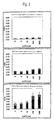
- the cells were washed twice with the wash buffer, suspended in 500 ⁇ l wash buffer and analyzed by flow cytometry. The results are shown in Fig. 1.
- the purity of the heart muscle cells was about 70 % in the conventional method and about 90 % in the method of the invention.
- Example 2 Comparison of Sensitivity in Detection of Inhibitory Action of Leukemia Inhibitory Factor on Apoptosis
- heart muscle cells of rat newborns obtained by the present method of preparing heart muscle cells as described in Example 1, their apoptosis was induced by removing serum from the culture, and the inhibitory action of recombinant human leukemia inhibitory factor (hLIF, produced by Peprotech) on the apoptosis was detected by the conventional method with MTT as the coloration substrate and by the present method of detecting apoptosis with WST-8 or MTT as the coloration substrate, and the sensitivity of the two methods was compared.
- hLIF human leukemia inhibitory factor
- the muscle cells obtained in the method of preparing heart muscle cells according to the invention were suspended at a density of 3 ⁇ 10 5 cells/ml in Medium 199 containing 10 % fetal calf serum, put onto a 96-wells plate in a volume of 0.1 ml/well, and then cultured for 1 day in a CO 2 incubator set at 5 % CO 2 , 37°C.
- the plate was stirred with a micromixer (Taiyo Kagaku Kogyo Co., Ltd.) and the medium was exchanged 3 times with serum-free Medium 199, followed by adding 0 to 1000 U/ml hLIF.
- the culture was incubated for additional 4 days to induce apoptosis.
- the number of viable cells in the culture was measured directly with MTT as the coloration substrate.
- fetal calf serum was added thereto at a concentration of 10 %, and the cells were further cultured for about 17 hours in a CO 2 incubator set at 5 % CO 2 , 37°C, and the number of viable cells was determined by using WST-8 or MTT as the coloration substrate.
- Fig. 2 shows the results of measurement of the inhibitory activity of hLIF at various concentrations on apoptosis of heart muscle cells by the conventional method with MTT as the coloration substrate and by the present method of detecting apoptosis with WST-8 or MTT as the coloration substrate.
- the conventional method the tendency of inhibition of apoptosis of heart muscle cells by hLIF can be seen, but the increase in the average absorbance by adding 1000 U/ml hLIF, as compared with the control group to which hLIF had not been added, was as very low as 0.03 or less. This is below the limit of detection by the measuring device, and the conventional method cannot be applied to screening with a large number of samples.
- the average absorbance of the control group to which hLIF had not been added was as high as 0.1 or more, and by adding 1000 U/ml hLIF, the absorbance was further increased by nearly 0.1, and thus the method of the invention is by far more accurate than the conventional method.
- This method would be applicable to screening where the absorbance or turbidity of a sample itself is not troublesome or for the purpose of searching for a sample indicating a very strong apoptosis-inhibiting activity equal to or higher than that of 1000 U/ml hLIF.
- the detection sensitivity of a measuring device and noises attributable to the absorbance or turbidity of a sample itself can be often troublesome, and it is thus desirable that an increase of 0.1 or more in absorbance is used as selection reference for correctly selecting an active sample. It was found that in the method of detecting apoptosis according to this invention in the case where WST-8 was used as the coloration substrate, the sensitivity was further improved, the average absorbance of the control group to which hLIF had not been added was 0.2 or more, and by adding 1000 U/ml hLIF, the average absorbance was increased by 0.3 or more, while noises were tolerable.
- the method of detecting apoptosis according to this invention can be used to detect not only the activity of a sample exhibiting, at an equal level to 1000 U/ml hLIF, an inhibitory action on apoptosis of heart muscle cells, an agonistic action on gp130-mediated receptors or an enhancing action on heart muscle cell-protective signals, but also the activity of a mixture of many samples by establishing suitable selection reference.
- apoptosis which comprises culturing heart muscle cells on a 96-well plate obtained by the present method of preparing heart muscle cells, then removing serum from the culture to induce apoptosis, also adding 1000 U/ml hLIF to inhibit the apoptosis, and detecting the apoptosis with WST-8 as the coloration substrate.
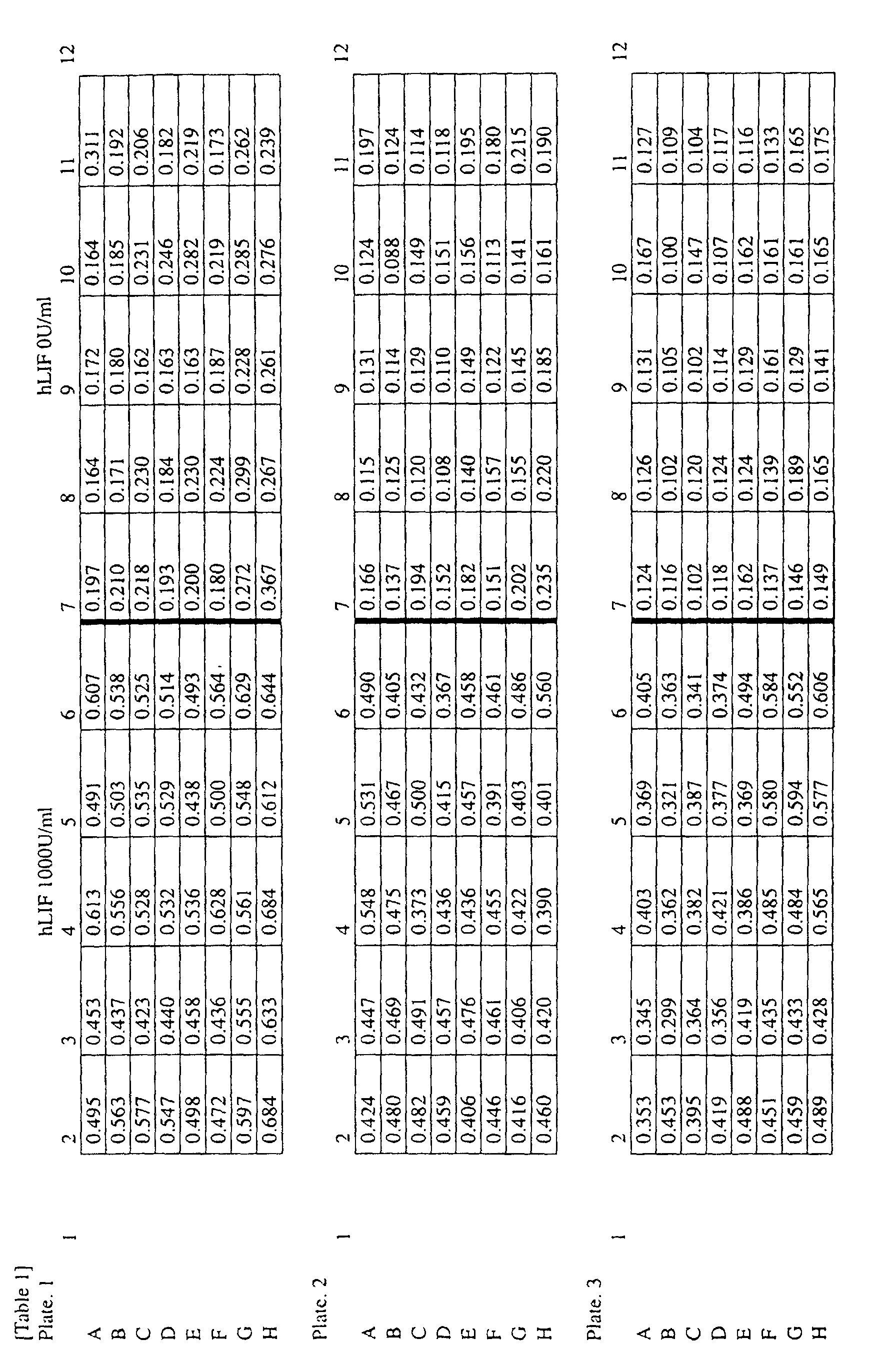
- the heart muscle cells obtained in the method described in Example 1 were suspended at a density of 3 ⁇ 10 5 cells/ml in Medium 199 containing 10 % fetal bovine serum, and put in a volume of 0.1 ml/well onto each well of 2nd to 11th rows on each of three 96-wells plates. The 1st and 12th rows were used as the blank free of the cells.
- the cells were cultured for 1 day in a CO 2 incubator set at 5 % CO 2 , 37°C, and then stirred with a micromixer (Taiyo Kagaku Kogyo Co., Ltd.) and then the medium was exchanged 3 times with serum-free Medium 199 to remove serum.
- the average absorbance of the control group not containing hLIF was 0.22 ⁇ 0.039 on plate 1,0.15 ⁇ 0.028 on plate 2 and 0.13 ⁇ 0.020 on plate 3, and by adding 1000 U/ml hLIF, the absorbance was increased to 0.54 ⁇ 0.055, 0.45 ⁇ 0.035 and 0.43 ⁇ 0.059, respectively.
- noises attributable to the absorbance or turbidity of a sample itself can be troublesome, and it is thus desired that an increase of 0.1 or more in absorbance be used as selection reference for correctly selecting an active sample.
- 120 samples exhibiting, at a level equal to 1000 U/ml hLIF, an inhibitory action on apoptosis of heart muscle cells, an agonistic action on gp130-mediated receptors and an enhancing action on heart muscle cell-protective signals can be selected from 240 samples.
- Insulin like growth factor-1 is known to inhibit apoptosis of heart muscle cells by activating Akt (Fujio Y. et al., Circulation, vol. 101, p. 660, 2000). Accordingly, the inhibitory action of recombinant human IGF-1 (hIGF-1, produced by Peprotech) on heart muscle cell apoptosis induced by removing serum from a culture was measured by the present method of detecting apoptosis with WST-8 as a coloration substrate.
- heart muscle cells obtained by the method of preparing heart muscle cells according to the invention were suspended at a density of 3 ⁇ 10 5 cells/ml in Medium 199 containing 10 % fetal calf serum and cultured for 1 day in a CO 2 incubator set at 5 % CO 2 , 37°C.
- the cells were stirred with a micromixer (Taiyo Kagaku Kogyo Co., Ltd.) and the medium was exchanged 3 times with serum-free Medium 199, and hIGF-1 was added thereto at a final concentration of 0 to 100 nM.
- the culture was cultured for additional 4 days to induce apoptosis.
- Fetal calf serum was added thereto at a concentration of 10 %, and the cells were further cultured for about 17 hours in a CO 2 incubator set at 5 % CO 2 , 37°C, and the number of viable cells was determined with WST-8 as the coloration substrate.
- Fig. 3 shows the results of measurement of the inhibitory activity of hIGF-1 at various concentrations on apoptosis of heart muscle cells. As shown in the result, this method can also be used to search for the compound showing a protective action on heart muscles in a mechanism different from that of gp130 signal.
- Tablets were produced in a usual manner from Compound 1 (100 mg), lactose (165 mg), corn starch (25 mg), polyvinyl alcohol (4 mg) and magnesium stearate (1 mg).
- Highly pure heart muscle cells can be produced easily in a large amount by the method of preparing heart muscle cells according to this invention. Further, apoptosis of the heart muscle cells can be detected efficiently and highly sensitively by the method of detecting apoptosis according to this invention.
- the screening method of the invention can be used for the efficient and highly sensitive screening of compounds inhibiting apoptosis of heart muscle cells or salts thereof, and can deal with a large number of samples for screening rapidly and efficiently, whereby compounds having an inhibitory action on apoptosis of heart muscle cells, compounds having an agonistic action on gp130-mediated receptors or compounds having an enhancing action on heart muscle cell-protective signals can be obtained.
- Such compounds are useful as prophylactic and/or therapeutic agents for heart diseases.
Landscapes
- Health & Medical Sciences (AREA)
- Life Sciences & Earth Sciences (AREA)
- Engineering & Computer Science (AREA)
- Biomedical Technology (AREA)
- Chemical & Material Sciences (AREA)
- Cell Biology (AREA)
- Bioinformatics & Cheminformatics (AREA)
- Immunology (AREA)
- Biotechnology (AREA)
- Organic Chemistry (AREA)
- General Health & Medical Sciences (AREA)
- Hematology (AREA)
- Wood Science & Technology (AREA)
- Biochemistry (AREA)
- Molecular Biology (AREA)
- Microbiology (AREA)
- Genetics & Genomics (AREA)
- Urology & Nephrology (AREA)
- Zoology (AREA)
- Medicinal Chemistry (AREA)
- Cardiology (AREA)
- General Physics & Mathematics (AREA)
- Analytical Chemistry (AREA)
- Physics & Mathematics (AREA)
- Food Science & Technology (AREA)
- Pathology (AREA)
- General Engineering & Computer Science (AREA)
- Toxicology (AREA)
- Rheumatology (AREA)
- Tropical Medicine & Parasitology (AREA)
- Chemical Kinetics & Catalysis (AREA)
- Veterinary Medicine (AREA)
- Public Health (AREA)
- Animal Behavior & Ethology (AREA)
- Pharmacology & Pharmacy (AREA)
- Nuclear Medicine, Radiotherapy & Molecular Imaging (AREA)
- General Chemical & Material Sciences (AREA)
- Heart & Thoracic Surgery (AREA)
- Measuring Or Testing Involving Enzymes Or Micro-Organisms (AREA)
- Investigating Or Analysing Biological Materials (AREA)
Abstract
Description

Elemental analysis of C13H8N2OS
Calculated (%): C: 64.98, H: 3.36, N: 11.66
Found (%): C: 64.93, H: 3.31, N: 11.59
1H-NMR (CDCl3)δ: 7.50-7.75(m, 4H), 7.85-8.00(m, 1H), 8.50-8.60(m,2H), 8.70-8.80(m, 1H)
IR (KBr): 1660cm-1
Melting point: 173.5-174.3°C
Claims (19)
- A method of preparing primary heart muscle cells, characterized in that after non-heart muscle cells are removed from fragmented heart tissue, the heart tissue is digested with a protease.
- The preparative method according to claim 1, characterized in that the non-heart muscle cells are removed by washing the fragmented heart tissue with a phosphate-buffered physiological saline.
- A method of preparing primary heart muscle cells, characterized in that after fragmented heart tissue is digested with a protease, erythrocytes are removed.
- The preparative method according to claim 3, characterized in that erythrocytes are removed by hemolysis treatment.
- A method of preparing primary heart muscle cells, characterized by hemolysis treatment of heart muscle cells isolated from heart tissue.
- A method of preparing primary heart muscle cells, characterized in that after non-heart muscle cells are removed from fragmented heart tissue, the heart tissue is digested with a protease, and then erythrocytes are removed.
- The preparative method according to claim 6, characterized in that the fragmented heart tissue is washed with a phosphate-buffered physiological saline to remove non-heart muscle cells, and the heart tissue digested with a protease is subjected to hemolysis treatment to remove erythrocytes.
- A method of detecting apoptosis of heart muscle cells, characterized in that after serum is added to a culture of heart muscle cells having apoptosis induced by culturing the cells in a serum-free medium, the cells are cultured, and the number of viable heart muscle cells is determined.
- The detection method according to claim 8, wherein the number of viable heart muscle cells is determined by measuring an intracellular dehydrogenase activity.
- The detection method according to claim 8, wherein the culture time after addition of serum is about 6 to 30 hours.
- The detection method according to claim 9, wherein the intracellular dehydrogenase activity is measured by using MTT or WST-8.
- The detection method according to claim 8, wherein the primary heart muscle cells obtained by the preparative method described in any one of claims 1 to 7 are cultured in a serum-free medium.
- A method of screening a compound inhibiting apoptosis or a salt thereof, characterized in that by the detection method described in claim 12, the number of viable cells in a case (i) where apoptosis is induced in the presence of a test compound is compared with that in a case (ii) where apoptosis is induced in the absence of a test compound.
- An inhibitor of apoptosis of heart muscle cells, comprising a compound obtained by the screening method described in claim 13 or a salt thereof.
- A gp 130-mediated receptor agonist, comprising a compound obtained by the screening method described in claim 13 or a salt thereof.
- An enhancer of heart muscle cell-protective signal, comprising a compound obtained by the screening method described in claim 13 or a salt thereof.
- A prophylactic and/or therapeutic agent for heart diseases, comprising the heart muscle cell apoptosis inhibitor described in claim 14, the gp130-mediated receptor agonist described in claim 15 or the heart muscle cell-protective signal enhancer described in claim 16.
- A method of preventing and treating heart diseases, characterized in that an effective dose of the heart muscle cell apoptosis inhibitor described in claim 14, the gp130-mediated receptor agonist described in claim 15 or the heart muscle cell-protective signal enhancer described in claim 16 is administered to mammals.
- Use of the heart muscle cell apoptosis inhibitor described in claim 14, the gp130-mediated receptor agonist described in claim 15 or the heart muscle cell-protective signal enhancer described in claim 16 for producing a prophylactic and/or therapeutic agent for heart diseases.
Priority Applications (1)
| Application Number | Priority Date | Filing Date | Title |
|---|---|---|---|
| EP08075845A EP2090663A3 (en) | 2000-06-28 | 2001-06-27 | Method of preparing heart muscle cells and method of searching for remedy for heart diseases |
Applications Claiming Priority (3)
| Application Number | Priority Date | Filing Date | Title |
|---|---|---|---|
| JP2000194805 | 2000-06-28 | ||
| JP2000194805 | 2000-06-28 | ||
| PCT/JP2001/005520 WO2002000850A1 (en) | 2000-06-28 | 2001-06-27 | Method of preparing heart muscle cells and method of searching for remedy for heart diseases |
Related Child Applications (1)
| Application Number | Title | Priority Date | Filing Date |
|---|---|---|---|
| EP08075845A Division EP2090663A3 (en) | 2000-06-28 | 2001-06-27 | Method of preparing heart muscle cells and method of searching for remedy for heart diseases |
Publications (3)
| Publication Number | Publication Date |
|---|---|
| EP1295936A1 true EP1295936A1 (en) | 2003-03-26 |
| EP1295936A4 EP1295936A4 (en) | 2004-11-10 |
| EP1295936B1 EP1295936B1 (en) | 2008-10-29 |
Family
ID=18693571
Family Applications (2)
| Application Number | Title | Priority Date | Filing Date |
|---|---|---|---|
| EP08075845A Withdrawn EP2090663A3 (en) | 2000-06-28 | 2001-06-27 | Method of preparing heart muscle cells and method of searching for remedy for heart diseases |
| EP01945643A Expired - Lifetime EP1295936B1 (en) | 2000-06-28 | 2001-06-27 | Method of preparing heart muscle cells and method of searching for remedy for heart diseases |
Family Applications Before (1)
| Application Number | Title | Priority Date | Filing Date |
|---|---|---|---|
| EP08075845A Withdrawn EP2090663A3 (en) | 2000-06-28 | 2001-06-27 | Method of preparing heart muscle cells and method of searching for remedy for heart diseases |
Country Status (6)
| Country | Link |
|---|---|
| US (2) | US20030175847A1 (en) |
| EP (2) | EP2090663A3 (en) |
| AT (1) | ATE412766T1 (en) |
| AU (1) | AU2001267844A1 (en) |
| DE (1) | DE60136358D1 (en) |
| WO (1) | WO2002000850A1 (en) |
Cited By (2)
| Publication number | Priority date | Publication date | Assignee | Title |
|---|---|---|---|---|
| EP2226633A1 (en) * | 2009-03-06 | 2010-09-08 | Charité - Universitätsmedizin Berlin | Uses and methods for the identification of a cytoprotective compound involving gp130 or LIFRalpha |
| US8168718B2 (en) | 2005-12-09 | 2012-05-01 | Exxonmobil Chemical Patents Inc. | Thermoplastic vulcanizate adhesive compositions |
Families Citing this family (2)
| Publication number | Priority date | Publication date | Assignee | Title |
|---|---|---|---|---|
| JP4520855B2 (en) * | 2002-09-17 | 2010-08-11 | 株式会社 ジャパン・ティッシュ・エンジニアリング | Cell disperser, cell dispersal cassette and tissue holding member |
| CN108949673A (en) * | 2018-08-13 | 2018-12-07 | 武汉华联科生物技术有限公司 | A kind of primary separation method of Fetal Rat rat cardiomyocyte |
Family Cites Families (8)
| Publication number | Priority date | Publication date | Assignee | Title |
|---|---|---|---|---|
| US3948725A (en) * | 1974-05-30 | 1976-04-06 | Godo Shusei Kabushiki Kaisha | Neutral protease useful for animal tissue and cell culture |
| JPH0372872A (en) * | 1988-07-12 | 1991-03-28 | Bio Kagaku Kenkyusho:Kk | Serum-free culture medium and method for culturing cell of mammals using the same |
| US5543318A (en) * | 1991-06-12 | 1996-08-06 | Smith; David A. | Method of isolation, culture and proliferation of human atrial myocytes |
| US5378698A (en) * | 1991-10-21 | 1995-01-03 | Shionogi & Co., Ltd. | Benzothiazepine derivatives |
| US5919449A (en) * | 1995-05-30 | 1999-07-06 | Diacrin, Inc. | Porcine cardiomyocytes and their use in treatment of insufficient cardiac function |
| US6133233A (en) * | 1997-02-18 | 2000-10-17 | Kansas State University Research Foundation | Peptide modulation of reperfusion injury |
| WO1999011771A1 (en) * | 1997-09-04 | 1999-03-11 | Science Research Laboratory, Inc. | Cell separation using electric fields |
| AU748334B2 (en) * | 1997-10-16 | 2002-05-30 | Board Of Regents, The University Of Texas System | Models and treatments for cardiac hypertrophy in relation with NF-AT3 function |
-
2001
- 2001-06-27 AU AU2001267844A patent/AU2001267844A1/en not_active Abandoned
- 2001-06-27 DE DE60136358T patent/DE60136358D1/en not_active Expired - Lifetime
- 2001-06-27 WO PCT/JP2001/005520 patent/WO2002000850A1/en not_active Ceased
- 2001-06-27 AT AT01945643T patent/ATE412766T1/en not_active IP Right Cessation
- 2001-06-27 EP EP08075845A patent/EP2090663A3/en not_active Withdrawn
- 2001-06-27 US US10/311,119 patent/US20030175847A1/en not_active Abandoned
- 2001-06-27 EP EP01945643A patent/EP1295936B1/en not_active Expired - Lifetime
-
2008
- 2008-02-07 US US12/069,074 patent/US20080160553A1/en not_active Abandoned
Cited By (3)
| Publication number | Priority date | Publication date | Assignee | Title |
|---|---|---|---|---|
| US8168718B2 (en) | 2005-12-09 | 2012-05-01 | Exxonmobil Chemical Patents Inc. | Thermoplastic vulcanizate adhesive compositions |
| EP2226633A1 (en) * | 2009-03-06 | 2010-09-08 | Charité - Universitätsmedizin Berlin | Uses and methods for the identification of a cytoprotective compound involving gp130 or LIFRalpha |
| WO2010099979A3 (en) * | 2009-03-06 | 2010-11-18 | Charité - Universitätsmedizin Berlin Gliedkörperschaft Der Freien Universität Berlin Und Der Humboldt-Universität Berlin | Uses and methods for the identification of a cytoprotective compound involving gp130 or lifralpha |
Also Published As
| Publication number | Publication date |
|---|---|
| AU2001267844A1 (en) | 2002-01-08 |
| DE60136358D1 (en) | 2008-12-11 |
| ATE412766T1 (en) | 2008-11-15 |
| WO2002000850A1 (en) | 2002-01-03 |
| EP1295936A4 (en) | 2004-11-10 |
| EP2090663A3 (en) | 2009-11-11 |
| US20080160553A1 (en) | 2008-07-03 |
| EP1295936B1 (en) | 2008-10-29 |
| US20030175847A1 (en) | 2003-09-18 |
| EP2090663A2 (en) | 2009-08-19 |
Similar Documents
| Publication | Publication Date | Title |
|---|---|---|
| CN110577931B (en) | Intermittent hypoxia treatment stem cell source exosome and application thereof in myocardial tissues | |
| JP6648259B2 (en) | Improved umbilical cord-derived adherent stem cell, method for producing the same and use thereof | |
| US20100183568A1 (en) | Method for production of hepatic-lobule-like cell cluster from adipose-tissue-derived cell | |
| KR102034496B1 (en) | Bioartificial proximal tubule systems and methods of use | |
| CN103930542A (en) | Brown adipocyte compositions and methods | |
| US20080090765A1 (en) | Compositions for modulating growth of embryonic and adult kidney tissue and uses for treating kidney damage | |
| JP5388297B2 (en) | Adipo cluster | |
| US20080160553A1 (en) | Method of preparing heart muscle cells and method of searching for remedy for heart diseases | |
| Mecham | [12] Modulation of elastin synthesis: In vitro models | |
| CN111575227B (en) | Method for establishing human-derived diabetic cardiomyopathy model | |
| TWI732298B (en) | Mammalian cell preservation solution containing acarbose or stachyose | |
| CN106190959A (en) | A kind of CBH culture medium inducing chicken PECTORAL LIMB SKELETON to break up and differentiation method thereof | |
| CN114246883A (en) | A kind of pharmaceutical composition and its therapeutic agent and application | |
| Li et al. | An improved two-step method for extraction and purification of primary cardiomyocytes from neonatal mice | |
| CN107446883B (en) | Mutual proliferation promoting effect of Treg cells and myocardial fibroblasts during co-culture | |
| TW202319536A (en) | Mesenchymal stem cells for use in the treatment of chronic kidney disease | |
| JP2002078483A (en) | Method for preparing myocardial cell and method for searching cardiopathy therapeutic agent | |
| JP7190567B2 (en) | Biomarkers for proliferating and migrating ability prediction of mesenchymal stem cells and their uses | |
| CN117778319A (en) | A method for regulating macrophage polarization | |
| CN113925972B (en) | Application of OTUB1 protein in treating osteoporosis | |
| CN117430714A (en) | Methods for constructing chimeric antigen receptors, modified immune cells, and animal models of immune hepatitis and liver fibrosis | |
| EP3795677B1 (en) | Composition for promoting stem cell differentiation, comprising progenitor cell culture solution and multilayer graphene film, and use thereof | |
| CA3260880A1 (en) | Cryopreserved intermediate and potency assay for same | |
| Poret et al. | Isolation, proliferation and differentiation of rhesus macaque adipose-derived stem cells | |
| JP4012399B2 (en) | Simple screening of drugs for regenerative medicine |
Legal Events
| Date | Code | Title | Description |
|---|---|---|---|
| PUAI | Public reference made under article 153(3) epc to a published international application that has entered the european phase |
Free format text: ORIGINAL CODE: 0009012 |
|
| 17P | Request for examination filed |
Effective date: 20021203 |
|
| AK | Designated contracting states |
Designated state(s): AT BE CH CY DE DK ES FI FR GB GR IE IT LI LU MC NL PT SE TR Kind code of ref document: A1 Designated state(s): AT BE CH CY DE DK ES FI FR GB GR IE IT LI LU MC NL PT SE TR |
|
| AX | Request for extension of the european patent |
Extension state: AL LT LV MK RO SI |
|
| RIC1 | Information provided on ipc code assigned before grant |
Ipc: 7C 12N 5/06 B Ipc: 7C 12Q 1/02 A |
|
| A4 | Supplementary search report drawn up and despatched |
Effective date: 20040921 |
|
| 17Q | First examination report despatched |
Effective date: 20041118 |
|
| RAP1 | Party data changed (applicant data changed or rights of an application transferred) |
Owner name: TAKEDA PHARMACEUTICAL COMPANY LIMITED |
|
| 17Q | First examination report despatched |
Effective date: 20041118 |
|
| 17Q | First examination report despatched |
Effective date: 20041118 |
|
| 17Q | First examination report despatched |
Effective date: 20041118 |
|
| GRAP | Despatch of communication of intention to grant a patent |
Free format text: ORIGINAL CODE: EPIDOSNIGR1 |
|
| GRAS | Grant fee paid |
Free format text: ORIGINAL CODE: EPIDOSNIGR3 |
|
| GRAA | (expected) grant |
Free format text: ORIGINAL CODE: 0009210 |
|
| RIN1 | Information on inventor provided before grant (corrected) |
Inventor name: KOYAMA, NOBUYUKI Inventor name: HIROTA, HISAO Inventor name: KIMURA, HARUHIDE Inventor name: KISHIMOTO, TADAMITSU Inventor name: TANIDA, SEIICHI Inventor name: TAKIHARA, KEIKO |
|
| AK | Designated contracting states |
Kind code of ref document: B1 Designated state(s): AT BE CH CY DE DK ES FI FR GB GR IE IT LI LU MC NL PT SE TR |
|
| REG | Reference to a national code |
Ref country code: GB Ref legal event code: FG4D |
|
| REG | Reference to a national code |
Ref country code: CH Ref legal event code: NV Representative=s name: BOHEST AG Ref country code: CH Ref legal event code: EP |
|
| REG | Reference to a national code |
Ref country code: IE Ref legal event code: FG4D |
|
| REF | Corresponds to: |
Ref document number: 60136358 Country of ref document: DE Date of ref document: 20081211 Kind code of ref document: P |
|
| NLV1 | Nl: lapsed or annulled due to failure to fulfill the requirements of art. 29p and 29m of the patents act | ||
| PG25 | Lapsed in a contracting state [announced via postgrant information from national office to epo] |
Ref country code: AT Free format text: LAPSE BECAUSE OF FAILURE TO SUBMIT A TRANSLATION OF THE DESCRIPTION OR TO PAY THE FEE WITHIN THE PRESCRIBED TIME-LIMIT Effective date: 20081029 Ref country code: ES Free format text: LAPSE BECAUSE OF FAILURE TO SUBMIT A TRANSLATION OF THE DESCRIPTION OR TO PAY THE FEE WITHIN THE PRESCRIBED TIME-LIMIT Effective date: 20090209 |
|
| PG25 | Lapsed in a contracting state [announced via postgrant information from national office to epo] |
Ref country code: FI Free format text: LAPSE BECAUSE OF FAILURE TO SUBMIT A TRANSLATION OF THE DESCRIPTION OR TO PAY THE FEE WITHIN THE PRESCRIBED TIME-LIMIT Effective date: 20081029 Ref country code: PT Free format text: LAPSE BECAUSE OF FAILURE TO SUBMIT A TRANSLATION OF THE DESCRIPTION OR TO PAY THE FEE WITHIN THE PRESCRIBED TIME-LIMIT Effective date: 20090330 Ref country code: NL Free format text: LAPSE BECAUSE OF FAILURE TO SUBMIT A TRANSLATION OF THE DESCRIPTION OR TO PAY THE FEE WITHIN THE PRESCRIBED TIME-LIMIT Effective date: 20081029 |
|
| PG25 | Lapsed in a contracting state [announced via postgrant information from national office to epo] |
Ref country code: BE Free format text: LAPSE BECAUSE OF FAILURE TO SUBMIT A TRANSLATION OF THE DESCRIPTION OR TO PAY THE FEE WITHIN THE PRESCRIBED TIME-LIMIT Effective date: 20081029 Ref country code: DK Free format text: LAPSE BECAUSE OF FAILURE TO SUBMIT A TRANSLATION OF THE DESCRIPTION OR TO PAY THE FEE WITHIN THE PRESCRIBED TIME-LIMIT Effective date: 20081029 |
|
| PG25 | Lapsed in a contracting state [announced via postgrant information from national office to epo] |
Ref country code: IT Free format text: LAPSE BECAUSE OF FAILURE TO SUBMIT A TRANSLATION OF THE DESCRIPTION OR TO PAY THE FEE WITHIN THE PRESCRIBED TIME-LIMIT Effective date: 20081029 Ref country code: SE Free format text: LAPSE BECAUSE OF FAILURE TO SUBMIT A TRANSLATION OF THE DESCRIPTION OR TO PAY THE FEE WITHIN THE PRESCRIBED TIME-LIMIT Effective date: 20090129 |
|
| PLBE | No opposition filed within time limit |
Free format text: ORIGINAL CODE: 0009261 |
|
| STAA | Information on the status of an ep patent application or granted ep patent |
Free format text: STATUS: NO OPPOSITION FILED WITHIN TIME LIMIT |
|
| 26N | No opposition filed |
Effective date: 20090730 |
|
| PG25 | Lapsed in a contracting state [announced via postgrant information from national office to epo] |
Ref country code: MC Free format text: LAPSE BECAUSE OF NON-PAYMENT OF DUE FEES Effective date: 20090630 |
|
| PG25 | Lapsed in a contracting state [announced via postgrant information from national office to epo] |
Ref country code: IE Free format text: LAPSE BECAUSE OF NON-PAYMENT OF DUE FEES Effective date: 20090627 |
|
| PG25 | Lapsed in a contracting state [announced via postgrant information from national office to epo] |
Ref country code: GR Free format text: LAPSE BECAUSE OF FAILURE TO SUBMIT A TRANSLATION OF THE DESCRIPTION OR TO PAY THE FEE WITHIN THE PRESCRIBED TIME-LIMIT Effective date: 20090130 |
|
| PG25 | Lapsed in a contracting state [announced via postgrant information from national office to epo] |
Ref country code: LU Free format text: LAPSE BECAUSE OF NON-PAYMENT OF DUE FEES Effective date: 20090627 |
|
| PGFP | Annual fee paid to national office [announced via postgrant information from national office to epo] |
Ref country code: FR Payment date: 20110621 Year of fee payment: 11 Ref country code: CH Payment date: 20110614 Year of fee payment: 11 |
|
| PG25 | Lapsed in a contracting state [announced via postgrant information from national office to epo] |
Ref country code: TR Free format text: LAPSE BECAUSE OF FAILURE TO SUBMIT A TRANSLATION OF THE DESCRIPTION OR TO PAY THE FEE WITHIN THE PRESCRIBED TIME-LIMIT Effective date: 20081029 |
|
| PGFP | Annual fee paid to national office [announced via postgrant information from national office to epo] |
Ref country code: GB Payment date: 20110622 Year of fee payment: 11 |
|
| PG25 | Lapsed in a contracting state [announced via postgrant information from national office to epo] |
Ref country code: CY Free format text: LAPSE BECAUSE OF FAILURE TO SUBMIT A TRANSLATION OF THE DESCRIPTION OR TO PAY THE FEE WITHIN THE PRESCRIBED TIME-LIMIT Effective date: 20081029 |
|
| PGFP | Annual fee paid to national office [announced via postgrant information from national office to epo] |
Ref country code: DE Payment date: 20110622 Year of fee payment: 11 |
|
| REG | Reference to a national code |
Ref country code: CH Ref legal event code: PL |
|
| REG | Reference to a national code |
Ref country code: CH Ref legal event code: PL |
|
| GBPC | Gb: european patent ceased through non-payment of renewal fee |
Effective date: 20120627 |
|
| REG | Reference to a national code |
Ref country code: FR Ref legal event code: ST Effective date: 20130228 |
|
| REG | Reference to a national code |
Ref country code: DE Ref legal event code: R119 Ref document number: 60136358 Country of ref document: DE Effective date: 20130101 |
|
| PG25 | Lapsed in a contracting state [announced via postgrant information from national office to epo] |
Ref country code: DE Free format text: LAPSE BECAUSE OF NON-PAYMENT OF DUE FEES Effective date: 20130101 Ref country code: LI Free format text: LAPSE BECAUSE OF NON-PAYMENT OF DUE FEES Effective date: 20120630 Ref country code: FR Free format text: LAPSE BECAUSE OF NON-PAYMENT OF DUE FEES Effective date: 20120702 Ref country code: GB Free format text: LAPSE BECAUSE OF NON-PAYMENT OF DUE FEES Effective date: 20120627 Ref country code: CH Free format text: LAPSE BECAUSE OF NON-PAYMENT OF DUE FEES Effective date: 20120630 |