EP1288312A2 - Method for determining quinolone resistance of tubercle bacilli - Google Patents
Method for determining quinolone resistance of tubercle bacilli Download PDFInfo
- Publication number
- EP1288312A2 EP1288312A2 EP02255875A EP02255875A EP1288312A2 EP 1288312 A2 EP1288312 A2 EP 1288312A2 EP 02255875 A EP02255875 A EP 02255875A EP 02255875 A EP02255875 A EP 02255875A EP 1288312 A2 EP1288312 A2 EP 1288312A2
- Authority
- EP
- European Patent Office
- Prior art keywords
- amino acid
- mutation
- gene
- acid residue
- gyrase
- Prior art date
- Legal status (The legal status is an assumption and is not a legal conclusion. Google has not performed a legal analysis and makes no representation as to the accuracy of the status listed.)
- Withdrawn
Links
Images

Classifications
-
- C—CHEMISTRY; METALLURGY
- C12—BIOCHEMISTRY; BEER; SPIRITS; WINE; VINEGAR; MICROBIOLOGY; ENZYMOLOGY; MUTATION OR GENETIC ENGINEERING
- C12Q—MEASURING OR TESTING PROCESSES INVOLVING ENZYMES, NUCLEIC ACIDS OR MICROORGANISMS; COMPOSITIONS OR TEST PAPERS THEREFOR; PROCESSES OF PREPARING SUCH COMPOSITIONS; CONDITION-RESPONSIVE CONTROL IN MICROBIOLOGICAL OR ENZYMOLOGICAL PROCESSES
- C12Q1/00—Measuring or testing processes involving enzymes, nucleic acids or microorganisms; Compositions therefor; Processes of preparing such compositions
- C12Q1/68—Measuring or testing processes involving enzymes, nucleic acids or microorganisms; Compositions therefor; Processes of preparing such compositions involving nucleic acids
- C12Q1/6876—Nucleic acid products used in the analysis of nucleic acids, e.g. primers or probes
- C12Q1/6888—Nucleic acid products used in the analysis of nucleic acids, e.g. primers or probes for detection or identification of organisms
- C12Q1/689—Nucleic acid products used in the analysis of nucleic acids, e.g. primers or probes for detection or identification of organisms for bacteria
-
- C—CHEMISTRY; METALLURGY
- C12—BIOCHEMISTRY; BEER; SPIRITS; WINE; VINEGAR; MICROBIOLOGY; ENZYMOLOGY; MUTATION OR GENETIC ENGINEERING
- C12Q—MEASURING OR TESTING PROCESSES INVOLVING ENZYMES, NUCLEIC ACIDS OR MICROORGANISMS; COMPOSITIONS OR TEST PAPERS THEREFOR; PROCESSES OF PREPARING SUCH COMPOSITIONS; CONDITION-RESPONSIVE CONTROL IN MICROBIOLOGICAL OR ENZYMOLOGICAL PROCESSES
- C12Q2600/00—Oligonucleotides characterized by their use
- C12Q2600/156—Polymorphic or mutational markers
Definitions
- the present invention relates to a method and kit for determining quinolon resistance of srtains of tubercle bacilli ( Mycobacterium tuberculosis ), more specifically, a method and kit for determining quinolon resistance by detecting a novel mutation in the gyrase A gene, which imparts the quinolon resistance phenotype to tubercle bacilli.
- Drug resistance phenotype acquisition mechanisms of bacteria include (1) such change of the cell wall structure that permeation of a drug into a cell should be blocked, (2) acquisition of a gene coding for an enzyme that decomposes or inactivates a drug, (3) change of a compound in a cell as a target of drug and so forth. Acquisition of these phenotypes are acheived by transmission among bacteria via transposable genetic elements such as plasmid and transposon, change of chromosomes or the like.
- the mechanism of resistance of tubercle bacilli to rifampicin is resulted from nucleotide substitution occurring in the gene encoding the RNA polymerase ⁇ subunit protein, which is a target substance of rifampicin (A Telenti et al., Lancet, 1993, 341: 647-650). Furthermore, nucleotide substitution in the rpsL and rrs genes or the like induces streptomycin resistance (Y. Suzuki, et al., J. Applied. Microbiol., 1997, 83: 634-640). Thus, mechanisms for acquisition of resistance phenotype by a mutation in a gene involved in each target molecule exist for some antituberculous drugs.
- quinolon resistance is acquired by introduction of a nucleotide mutation into a gene encoding the DNA gyrase activity, topoisomerase IV activity or a membrane integral protein involved in uptake and excretion of a substance into and from a cell (Cambau E., et al., Res. Microbiol., 1996, 147: 52-59; Ferrero L., et al., Antimicrob. Agents Chemother., 1995, 39: 1554-1558) or the like.
- gyrase A One of the target compounds of quinolon is gyrase A, and a mutation in the gyrase A gene greatly contributes to resistance to clinically important quinolon molecular species (ciprofloxacin, ofloxacin etc., Cambau E., et al., J. Infect. Dis., 1994, 170: 479-483).
- An object of the present invention is to provide a method and kit for determining quinolon resistance of tubercle bacilli by identifying a novel mutation in the gyrase A gene involved in acquisition of quinolon resistance phenotype in tubercle bacilli and utilizing the mutation.
- the inventors of the present invention assiduously studied in order to achieve the aforementioned object. As a result, they found a novel mutation imparting a quinolon resistance phenotype in the gyrase A gene sequence of tubercle bacilli and accomplished the present invention.
- the present invention provides the followings.
- a novel mutation in the gyrase A gene involved in quinolon resistance phenotype acquisition by tubercle bacilli was identified by the present invention. By utilizing this mutation, a novel method and kit for determining quinolon resistance of tubercle bacilli are provided.
- Fig. 1 shows alignment of nucleotide sequences of gyrA genes from strains of tubercle bacilli resistant to quinolon.
- the present invention provides a method for determining quinolon resistance of a tubercle bacillus strain by detecting a mutation in the gyrase A gene (hereinafter, referred to as " gyrA ”) of the bacterial strain.
- gyrA gyrase A gene
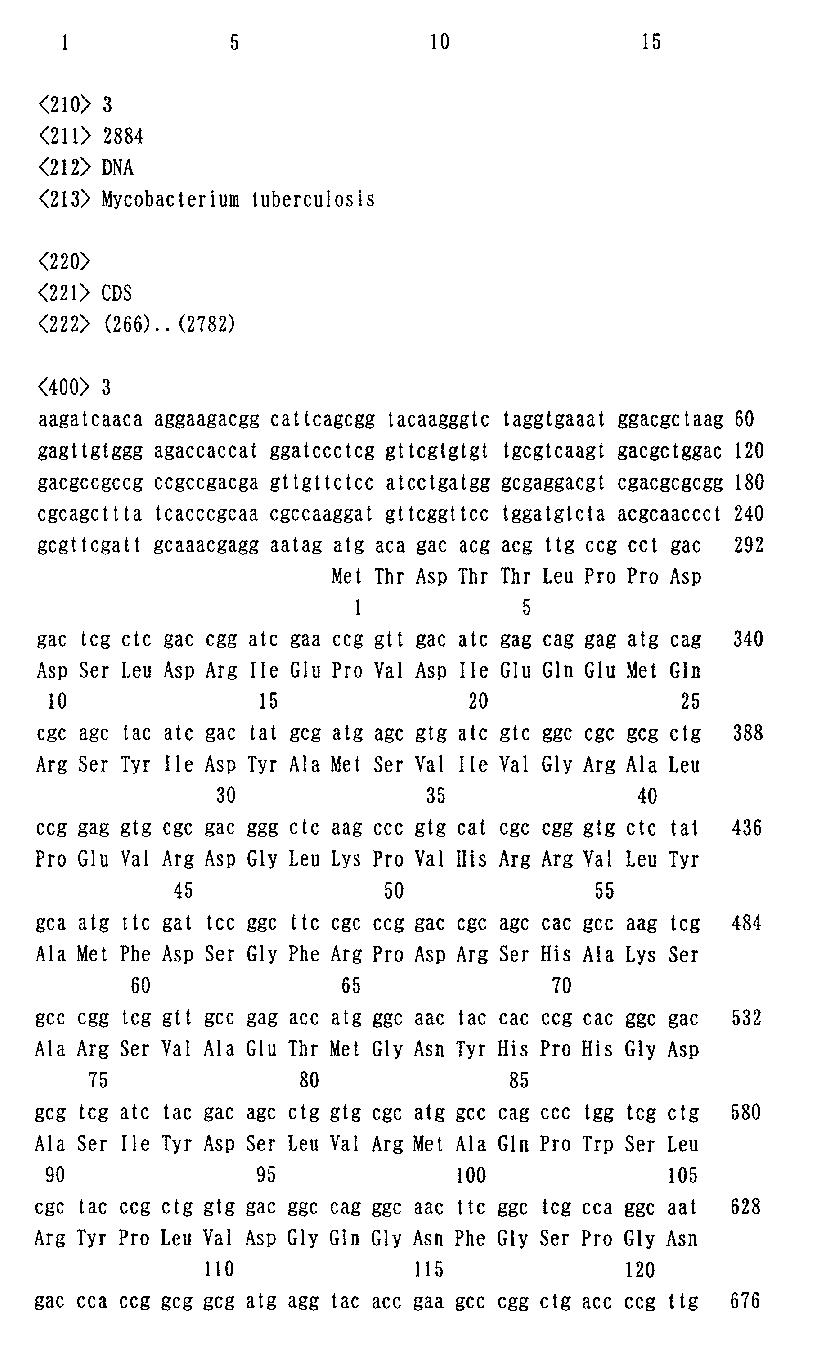
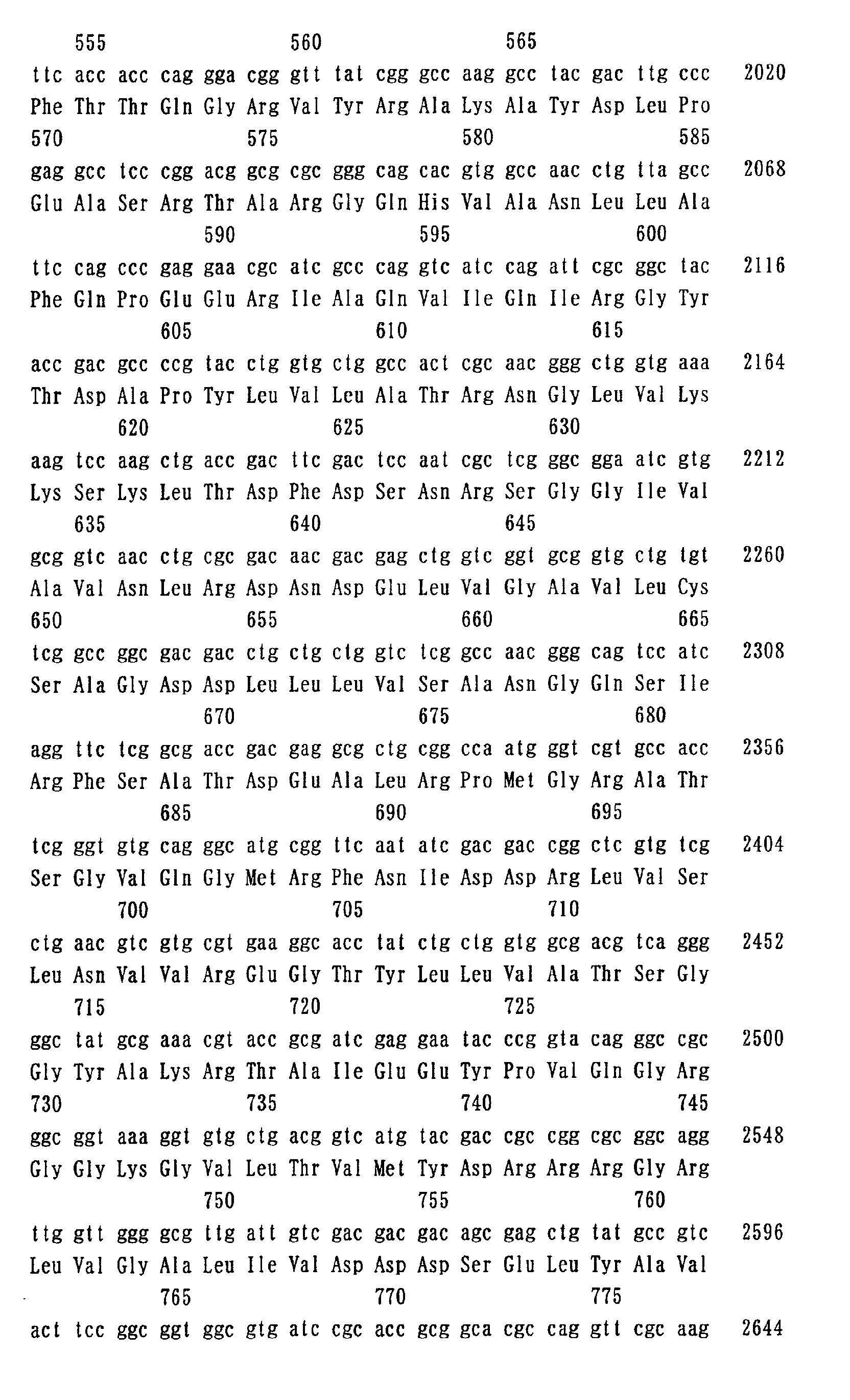
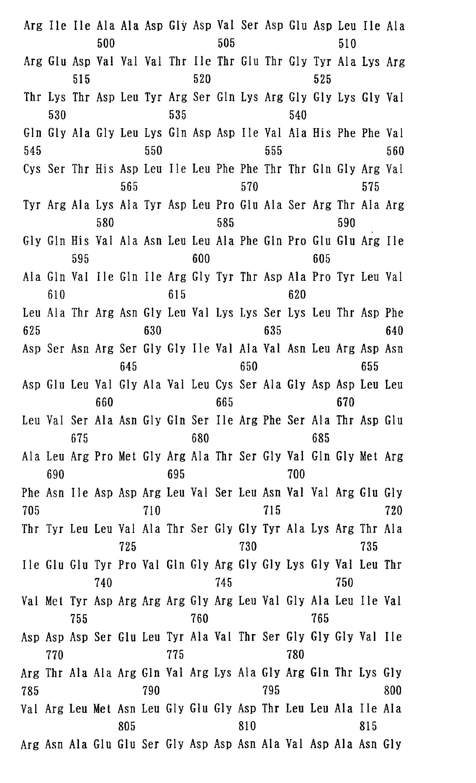
- SEQ ID NO: 3 the sequence of nucleotide numbers 2041-4923 is shown as SEQ ID NO: 3.
- amino acid sequence encoded by the nucleotide sequence is shown as SEQ ID NO: 4.
- the amino acid sequence is shown in both of SEQ ID NOS: 3 and 4.
- the method of the present invention is characterized in that the mutation in the aforementioned gyrA gene is a substitution of another amino acid residue for an amino acid residue corresponding to the 89th aspartic acid residue in the amino acid sequence encoded by the gyrA gene.
- Fig. 1 shows comparison of nucleotide sequences of a region around the mutation point. Further, a nucleotide sequence of the aforementioned region in the gyrA gene of a wild strain is shown as SEQ ID NO: 1. The amino acid sequence encoded by this sequence is shown as SEQ ID NO: 2.
- SEQ ID NO: 1 corresponds to the nucleotide numbers 511-560 in the nucleotide sequence including the gyrA gene shown as SEQ ID NO: 3.
- the amino acid sequence of SEQ ID NO: 2 corresponds to the amino acid numbers 83-98 in the amino acid sequence encoded by the gyrA gene and shown as SEQ ID NO: 4.
- an asparagine residue can be mentioned.
- substitution of T for G that is the first nucleotide of a codon coding for an aspartic acid residue can be mentioned.
- a specific example of this mutation is a mutation occurring at a position corresponding to the 530th nucleotide in the nucleotide sequence of the gyrA gene shown as SEQ ID NO: 3.
- the nucleotide or amino acid numbers in the nucleotide sequence or the amino acid sequence shown as SEQ ID NO: 3 or 4 may be changed due to a mutation in the gyrA gene occurring at a position that is not within a codon corresponding to the 89th aspartic acid residue.
- a gyrase A having such a mutation has an amino acid sequence including substitution, deletion or insertion of one or several amino acids in the amino acid sequence shown as SEQ ID NO: 4.
- the present invention can also be applied to a gene coding for the gyrase A including such a mutation so long as the mutation is involved in quinolon resistance.
- amino acid residue corresponding to the 89th aspartic acid residue and "position corresponding to the 530th nucleotide” refer to an amino acid residue or a nucleotide residue corresponding to such a position in the sequence shown as SEQ ID NO: 3 or 4 when the nucleotide numbers or amino acid numbers are changed as described above.
- an amino acid residue corresponding to the 89th aspartic acid residue means an aspartic acid residue that is the 88th amino acid residue from the N-terminus.
- known mutations in the gyrA gene involved in quinolon resistance and/or mutations yet to be found in future may be detected in addition to the substitution of an amino acid residue corresponding to the 89th aspartic acid residue.
- Mutations in the gyrA gene of tubercle bacillus can be detected in the same manner as in usual mutation detecting methods so long as a single nucleotide polymorphism can be detected.
- a method for detecting a mutation by directly determining the nucleotide sequence of the gyrA gene there can be mentioned a method for detecting a mutation by directly determining the nucleotide sequence of the gyrA gene.
- examples of a method for detecting a mutation utilizing PCR include methods such as the template-directed dye-terminator incorporation method (Chen, X. et al., Proc. Natl. Acad. Sci. USA, 20, 10756-10761 (1997)), the dye-labeled oligonucleotide ligation method (Chen, X.
- Luminex method utilizing flow cytometry and methods utilizing matrix-assisted laser desorption ionization mass spectrometry (MALDI-TOFMS) are also known and can be used for the present invention.
- MALDI-TOFMS matrix-assisted laser desorption ionization mass spectrometry
- microarray methods (WO95/11995, USP 5,837,832), which are widely used in recent years, such as the single base sequencing on oligomicroarray method (Drmanac, R. et al., Genomics, 2, 114-128 (1989)) can also be used.
- the microarray methods are techniques in which a large number of capture nucleic acids are immobilized on a solid phase, a labeled target nucleic acid is hybridized with the capture nucleic acids to be captured, and the presence or absence or existence ratio of the target nucleic acid is analyzed exhaustively.
- a method of detecting a target mutation in the gyrA gene by using an oligonucleotide immobilized on a substrate will be shown below.
- oligonucleotide immobilized on the substrate used is an oligonucleotide having a nucleotide sequence of a region including the position of the aforementioned mutation in the gyrA gene (hereinafter, referred to as "capture oligo").
- the capture oligo When the capture oligo is designed, it is usually preferred that a position corresponding to the aforementioned mutation included in the capture oligo should exist in the center portion of the capture oligo. When the capture oligo is too short, detection of the hybridization becomes difficult. When it is too long, inhibition of the hybridization due to a sequence unique to that type does not occur. Therefore, a length in the range of 10 to 24 nucleotides is preferred.
- a spacer or a nucleotide that does not form a hydrogen bond with any nucleotide can be introduced into the oligonucleotide sequence to avoid the aforementioned failure.
- a peptide nucleic acid may also be used. Since a hybrid formed by a peptide nucleic acid with a nucleic acid derived from a test human genome has a higher Tm (melting temperature) in comparison with that obtained by using an oligonucleotide, it is expected that a stable hybridization signal can be obtained.
- the peptide nucleic acid can be readily synthesized by using a usual peptide synthesizer.
- oligonucleotide preparation of chromosomal DNA, hybridization and PCR can be performed according to usual methods well known to those skilled in the art (refer to Maniatis, T. et al., "Molecular Cloning A Laboratory Manual, Second Edition", Cold Spring Harbor Laboratory Press (1989)). Further, the oligonucleotide can be synthesized by using a commercially available DNA synthesizer.
- Material of the substrate on which the oligonucleotide is immobilized is not particularly limited so long as the oligonucleotide can be stably immobilized thereon.
- glass, synthetic resins such as polycarbonate and plastic can be mentioned.
- shape of the substrate is not also particularly limited, a plate-like or film-like substrate can be mentioned.
- the substrate preferably has a uniform and planar surface.
- the oligonucleotide can be immobilized on the substrate by using a method used in a usual hybridization method such as physical adsorption, electrical coupling and formation of a molecular covalent bond.
- a method used in a usual hybridization method such as physical adsorption, electrical coupling and formation of a molecular covalent bond.
- a method using a substrate of which surface is coated with a compound having a carbodiimide group or an isocyanate group Japanese Patent Laid-open Publication (Kokai) No. 08-023975.
- the substrate coated with a compound having a carbodiimide group or an isocyanate group on the surface thereof can be prepared by coating the substrate surface with a macromolecular compound having a carbodiimide group or an isocyanate group.
- the oligonucleotides can be immobilized with a covalent bond on the substrate by irradiating with a ultraviolet ray.
- a linker for bonding the carbodiimide group or isocyanate group and the oligonucleotide a compound having an amino group or an imino group having high reactivity with the carbodiimide group or isocyanate group is used.
- imino group the compound can be bonded with a carbodiimide group or an isocyanate group by polymerizing thymine to either one of the ends of capture oligo.
- the oligonucleotide can be immobilized on the substrate by, for example, spotting an oligonucleotide solution on the substrate by using a spotting machine.
- the oligonucleotide solution is preferably spotted in a substantially circular shape.
- multiple kinds of oligonucleotide are each usually spotted at multiple positions on a single substrate, and these spots are preferably arranged in a grid pattern.
- the spot size is 1000 ⁇ m in diameter
- the total number of spots is preferably 1600 spots/cm 2 or less and 40 x 40 or less of spots are preferably provided when they are spotted in a square pattern.
- the spot size is 10 ⁇ m in diameter
- the total number is preferably 400 spots/cm 2 or less
- 20 x 20 or less of spots are preferably provided when they are spotted in a square pattern.
- the vertical and horizontal sizes of the pattern are different, the numbers of the spots along the vertical and horizontal directions may be adjusted depending on shape of the pattern.
- the presence or absence of a specific mutation in the gyrA gene can be determined by hybridizing a nucleic acid fragment probe including a region corresponding to the position of the aforementioned mutation in the gyrA gene of chromosomal DNA of tubercle bacillus (referred to as "nucleic acid probe") with a wild type capture oligo and a mutant capture oligo and examining with which type of capture oligo the probe hybridized. That is, to which drug the strain has resistance can be detected depending on the hybridization position on the substrate.
- the nucleic acid can be prepared from tubercle bacilli in the same manner as in a usual method for preparing nucleic acids from bacteria.
- DNA can be prepared according to the method described in Maniatis, T. et al., "Molecular Cloning A Laboratory Manual, Second Edition", Cold Spring Harbor Laboratory Press (1989).
- the nucleic acid probe can be prepared by amplifying a nucleic acid by using primers designed based on the nucleotide sequence of the capture oligo.
- DNA is usually used as the nucleic acid probe, RNA may also be used.
- a method of amplifying a nucleic acid there can be mentioned, for example, a method of amplifying a nucleic acid as DNA by polymerase chain reaction (PCR) and a method of amplifying a nucleic acid as RNA by the in vitro transcription method.
- PCR polymerase chain reaction
- the primers used in PCR are designed so that the nucleic acid probe should include a specific mutation site in the gyrA gene and sequences on both sides thereof.
- primers there can be mentioned oligonucleotides having the nucleotide sequences shown as SEQ ID NOS: 5 and 6.
- a labeled nucleic acid probe can be obtained.
- the nucleic acid probe may also be labeled during or after the nucleic acid amplification reaction.
- labeling substances similar to those used for a probe employed in usual hybridization such as a fluorescence substance or hapten can be used.
- the fluorescence substance include fluorescein (FITC), Rhodamine, phycoerythrin (PE), Texas Red, cyanine fluorescent dyes and so forth.
- FITC fluorescein
- PE phycoerythrin
- cyanine fluorescent dyes and so forth.
- Specific examples of the hapten include biotin, digoxigenin (Dig), dinitrophenyl (DNP) and so forth.
- the primers for preparing a nucleic acid probe can be included in a kit for determining quinolon resistance of tubercle bacilli together with a substrate on which the oligonucleotide is immobilized.
- the hybridization can be performed in the same manner as in usual nucleic acid hybridization. A specific method will be exemplified below.
- a nucleic acid probe is added to a mixed solution containing a salt solution such as SSC (standard saline citrate), a blocking solution containing sodium dodecylsulfate (SDS), bovine serum albumin (BSA) or the like and additives for promoting a hybridization reaction.
- a salt solution such as SSC (standard saline citrate), a blocking solution containing sodium dodecylsulfate (SDS), bovine serum albumin (BSA) or the like and additives for promoting a hybridization reaction.
- SSC standard saline citrate
- SDS sodium dodecylsulfate
- BSA bovine serum albumin
- a solution of 5 x SSC or 3 M tetramethylammonium chloride is added onto the substrate and heated (usually 37-50°C) to remove oligonucleotides that form a nonspecific hybrid or do not form specific hybrid from the substrate so that only specific hybrids should be selectively left on the substrate.
- the hybrids are detected by using the fluorescence substance or hapten introduced into the nucleic acid probe.
- a hapten When a hapten is used, a solution containing a conjugate of a protein that recognizes the hapten or protein that bonds to the hapten and alkaline phosphatase, horseradish peroxidase or the like (enzyme conjugate) is added onto the substrate and allowed to react at room temperature for several dozens of minutes.
- a nonspecific adsorption reaction of the enzyme conjugate and the substrate can be prevented by completely coating regions on the substrate with a protein such as BSA except for the regions in which the oligonucleotides are immobilized before a bonding reaction of the hapten and the enzyme conjugate is performed.
- This treatment can be performed by, after the oligonucleotides are immobilized, adding a solution of a protein such as BSA onto the substrate and leaving it at room temperature for several dozens of minutes. After the bonding reaction of the enzyme conjugate and the hapten of the nucleic acid probe is completed, only enzyme conjugates that bond to the hapten in the nucleic acid probe are left on the substrate by washing the substrate with an appropriate buffer containing a surfactant to remove enzyme conjugates that did not bond to the hapten.
- a protein such as BSA
- a compound that becomes insoluble only when only a bonding product of the hapten and the enzyme conjugate exists is added.
- the production of the insoluble compound is amplified by the enzymatic reaction, and thus the hybrids are visualized.
- nitroblue tetrazolium chloride (NBT) and 5-bromo-4-chloro-3-indolyl phosphate, p-toluidine salt (BCIP) are used when the enzyme in the enzyme conjugate is alkaline phosphatase.
- NBT nitroblue tetrazolium chloride
- BCIP p-toluidine salt
- the enzyme in the enzyme conjugate is alkaline phosphatase.
- TMB 3,3',5,5'-tetramethylbenzidine
- Quinolon resistance of tubercle bacillius is determined based on the obtained results of the hybridization by examining pigmentation or fluorescent color development at positions at which the capture oligos are immobilized.
- the kit of the present invention is a kit for determining quinolon resistance of a tubercle bacillus strain by detecting a mutation in the gyrA gene of the bacterial strain, which includes an oligonucleotide for detecting a substitution of another amino acid residue for an amino acid residue corresponding to the 89th aspartic acid residue in the amino acid sequence encoded by the gyrase A gene.
- an oligonucleotide a capture oligo or a substrate on which the capture oligo is immobilized can be mentioned.
- the kit of the present invention may include primers for preparing a nucleic acid probe, labeled nucleic acid probe, buffer, reagents for hybridization such as enzyme conjugate that recognizes hapten and so forth.
- levofloxacin As quinolon to which the present invention can be applied, there can be mentioned levofloxacin, ciprofloxacin, ofloxacin, gatifloxacin and so forth.
- Genomic DNA was prepared from each of the quinolon-resistant strains isolated as described above by the method of Kent et al. (Kent, P.T., et al., Mycobacteriology, A Guide for the Level III Laboratory, p.31, U.S. Department of Health and Human Service, Public Health Service, Center for Disease Control, 1985). PCR was performed for the obtained genome by using primers having the nucleotide sequences shown as SEQ ID NOS: 5 and 6.
- rTaq (Takara Shuzo) was used for PCR.
- PCR was performed by a reaction at 98°C for 1 minute, 35 cycles of reactions at 98°C for 5 seconds, at 55°C for 10 seconds and at 72°C for 20 seconds, and a reaction at 72°C for 1 minute.
- the amplification product was separated by 1% agarose gel electrophoresis, and a gel containing a 0.4 kbp band was excised. This gel piece was placed into a 1.5-mL micro tube and frozen at -135°C for 15 minutes. Then, the gel was centrifuged at 15000 rpm for 10 minutes to obtain a supernatant.
- Resulting DNA solution was used as a template together with a primer shown as SEQ ID NO: 5 and Big Dye Terminator kit manufactured by ABI to perform a sequencing reaction. After completion of the reaction, the sequence was analyzed by using a genetic analyzer manufactured by ABI.
- Fig. 1 shows alignment of the obtained sequences.
- the number shown at the right end of each sequence represents the number of sequences that had the same mutation among the determined sequences.
- substitution of A for G at the 530th position in the nucleotide sequence shown as SEQ ID NO: 3 is a novel mutation.
- substitution of A for G at the 530th position and substitution of A for G at the 545th position constitute a novel combination of mutations.
- Other mutations were the same as the mutations already reported (Antimicrob. Agents Chemother., 1996, 40 (8), 1768-1774; J. Infect. Dis., 1996, 174, 1127-1130; Eur. J. Clin. Microbiol. Infect. Dis., 1997, 16, 395-398; J. Infect. Dis., 2000, 182, 517-525).
Landscapes
- Chemical & Material Sciences (AREA)
- Life Sciences & Earth Sciences (AREA)
- Analytical Chemistry (AREA)
- Proteomics, Peptides & Aminoacids (AREA)
- Organic Chemistry (AREA)
- Zoology (AREA)
- Wood Science & Technology (AREA)
- Health & Medical Sciences (AREA)
- Engineering & Computer Science (AREA)
- Microbiology (AREA)
- Immunology (AREA)
- Molecular Biology (AREA)
- Biotechnology (AREA)
- Biophysics (AREA)
- Physics & Mathematics (AREA)
- Biochemistry (AREA)
- Bioinformatics & Cheminformatics (AREA)
- General Engineering & Computer Science (AREA)
- General Health & Medical Sciences (AREA)
- Genetics & Genomics (AREA)
- Measuring Or Testing Involving Enzymes Or Micro-Organisms (AREA)
Abstract
Description






Claims (7)
- A method for determining quinolon resistance of a Mycobacterium tuberculosis strain comprising detecting a mutation in a gyrase A gene of the bacterial strain, wherein the mutation is substitution of another amino acid residue for an amino acid residue corresponding to the 89th aspartic acid residue in the amino acid sequence encoded by the gyrase A gene.
- The method according to claim 1, wherein the other amino acid residue is an asparagine residue.
- The method according to claim 1 or 2, wherein the amino acid sequence encoded by the gyrase A gene is the amino acid sequence of SEQ ID NO: 4 or an amino acid sequence of SEQ ID NO: 4 including substitution, deletion or insertion of one or several amino acid residues.
- The method according to any one of claims 1 to 3, wherein the mutation is substitution of T for G that is the first nucleotide of a codon coding for an aspartic acid residue.
- The method according to claim 4, wherein the mutation occurs at a position corresponding to the 530th nucleotide in the nucleotide sequence of SEQ ID NO: 3.
- The method according to any one of claims 1 to 5, further comprising detecting a substitution of A for a base corresponding to G at the 545th position in the nucleotide sequence of SEQ ID NO: 3.
- A kit for determining quinolon resistance of a Mycobacterium tuberculosis strain by detecting a mutation in a gyrase A gene of the bacterial strain, comprising an oligonucleotide for detecting a substitution of another amino acid residue for an amino acid residue corresponding to the 89th aspartic acid residue in an amino acid sequence encoded by the gyrase A gene.
Applications Claiming Priority (2)
| Application Number | Priority Date | Filing Date | Title |
|---|---|---|---|
| JP2001267769 | 2001-09-04 | ||
| JP2001267769A JP2003070478A (en) | 2001-09-04 | 2001-09-04 | Method for determining quinolone resistance in Mycobacterium tuberculosis |
Publications (2)
| Publication Number | Publication Date |
|---|---|
| EP1288312A2 true EP1288312A2 (en) | 2003-03-05 |
| EP1288312A3 EP1288312A3 (en) | 2003-04-23 |
Family
ID=19093857
Family Applications (1)
| Application Number | Title | Priority Date | Filing Date |
|---|---|---|---|
| EP02255875A Withdrawn EP1288312A3 (en) | 2001-09-04 | 2002-08-22 | Method for determining quinolone resistance of tubercle bacilli |
Country Status (4)
| Country | Link |
|---|---|
| US (1) | US20030049668A1 (en) |
| EP (1) | EP1288312A3 (en) |
| JP (1) | JP2003070478A (en) |
| CA (1) | CA2396887A1 (en) |
Families Citing this family (2)
| Publication number | Priority date | Publication date | Assignee | Title |
|---|---|---|---|---|
| CN110468186A (en) * | 2019-09-11 | 2019-11-19 | 福建省农业科学院畜牧兽医研究所 | Whether a kind of 11 type Riemerellosis Anatipestifers of identification are to the drug resistant method of fluoroquinolones |
| CN117230222A (en) * | 2023-10-18 | 2023-12-15 | 鲲鹏基因(北京)科技有限责任公司 | Composition, kit and method for detecting quinolone resistance of mycobacterium tuberculosis |
Family Cites Families (4)
| Publication number | Priority date | Publication date | Assignee | Title |
|---|---|---|---|---|
| DE4421901A1 (en) * | 1994-06-23 | 1996-01-04 | Bayer Ag | A rapid DNA test for the detection of quinolone-resistant Staphylococcus aureus pathogens in clinical specimens |
| AU762314B2 (en) * | 1998-04-01 | 2003-06-19 | Government Of The United States Of America, As Represented By The Secretary Of The Department Of Health And Human Services, The | Oligonucleotide probes for detecting enterobacteriaceae and quinolone-resistant enterobacteriaceae |
| EP1077265A1 (en) * | 1999-08-19 | 2001-02-21 | Merlin Gesellschaft Für Mikrobiologische Diagnostika Mbh | Method for the classification of bacteria by genotyping |
| EP1169482A1 (en) * | 1999-04-10 | 2002-01-09 | Merlin Gesellschaft für mikrobiologische Diagnostika mbH | Genotypic classification method |
-
2001
- 2001-09-04 JP JP2001267769A patent/JP2003070478A/en active Pending
-
2002
- 2002-08-15 US US10/219,219 patent/US20030049668A1/en not_active Abandoned
- 2002-08-22 EP EP02255875A patent/EP1288312A3/en not_active Withdrawn
- 2002-09-04 CA CA002396887A patent/CA2396887A1/en not_active Abandoned
Also Published As
| Publication number | Publication date |
|---|---|
| CA2396887A1 (en) | 2003-03-04 |
| US20030049668A1 (en) | 2003-03-13 |
| EP1288312A3 (en) | 2003-04-23 |
| JP2003070478A (en) | 2003-03-11 |
Similar Documents
| Publication | Publication Date | Title |
|---|---|---|
| EP0528882B1 (en) | A thermostable ligase mediated dna amplification system for the detection of genetic diseases | |
| US5830711A (en) | Thermostable ligase mediated DNA amplification system for the detection of genetic diseases | |
| CN112301016B (en) | Application of novel mlCas12a protein in nucleic acid detection | |
| JPH11504517A (en) | Nucleic acid detection and amplification by chemical bonding of oligonucleotides | |
| CN105705515A (en) | Plurality of transposase adapters for DNA manipulations | |
| EP0935675B1 (en) | Nucleic acid primers and probes for detecting oncogenic human papillomaviruses | |
| CN102186991A (en) | DNA Quantification or Detection Methods | |
| US20050147973A1 (en) | Immobilized probes | |
| JP2001103981A (en) | Mycobacterium tuberculosis diagnostic kit | |
| JPH06209797A (en) | Specific gene probe for diagnostic studies of Candida albicans | |
| AU1791200A (en) | Process for labeling a ribonucleic acid, and labeled rna fragments which are obtained thereby | |
| EP1580281A2 (en) | Method for microbial antibiotic resistance detection | |
| EP1288312A2 (en) | Method for determining quinolone resistance of tubercle bacilli | |
| US20080096203A1 (en) | Method and device for detecting quinolone-resistant escherichia coli | |
| KR19990022595A (en) | Nucleic Acid Probes and Amplified Oligonucleotides for Neisseria Species | |
| JP2012161319A (en) | β2 ADRENERGIC POLYMORPHISM DETECTION | |
| EP3198027B1 (en) | Fast and highly specific dna-based multiplex detection | |
| US20060223084A1 (en) | Methods and compositions for detecting Bacillus anthracis | |
| JPH11127876A (en) | Small triple-stranded PNA oligo | |
| KR20230098013A (en) | An improved method for simultaneously detecting and identifying Helicobacter pylori, and an antibiotics-resistance gene thereof, and a use therefor based PCR-Reverse blot hybridization assay | |
| JP3178014B2 (en) | Method for measuring RNA | |
| US20030219755A1 (en) | Compositions and methods for performing hybridization assays using target enhanced signal amplification (TESA) | |
| WO2025049432A1 (en) | Methods for editing hla-a in cells pre-screened for the absence of one or both alleles of hla-h*01 | |
| CN115427567A (en) | Method for determining single-base mutation of erm (41) gene of acid-fast bacterium belonging to Mycobacterium abscessus complex, and primer set and probe used in the method | |
| JP2010515452A (en) | DNA chip for detection of Staphylococcus aureus |
Legal Events
| Date | Code | Title | Description |
|---|---|---|---|
| PUAI | Public reference made under article 153(3) epc to a published international application that has entered the european phase |
Free format text: ORIGINAL CODE: 0009012 |
|
| AK | Designated contracting states |
Kind code of ref document: A2 Designated state(s): AT BE BG CH CY CZ DE DK EE ES FI FR GB GR IE IT LI LU MC NL PT SE SK TR Designated state(s): AT BE BG CH CY CZ DE DK EE ES FI FR GB GR IE IT LI LU MC NL PT SE SK TR |
|
| AX | Request for extension of the european patent |
Extension state: AL LT LV MK RO SI |
|
| PUAL | Search report despatched |
Free format text: ORIGINAL CODE: 0009013 |
|
| AK | Designated contracting states |
Designated state(s): AT BE BG CH CY CZ DE DK EE ES FI FR GB GR IE IT LI LU MC NL PT SE SK TR |
|
| AX | Request for extension of the european patent |
Extension state: AL LT LV MK RO SI |
|
| 17P | Request for examination filed |
Effective date: 20031010 |
|
| AKX | Designation fees paid |
Designated state(s): DE FR GB |
|
| STAA | Information on the status of an ep patent application or granted ep patent |
Free format text: STATUS: THE APPLICATION HAS BEEN WITHDRAWN |
|
| 18W | Application withdrawn |
Effective date: 20050704 |