EP1284298A2 - Identification and use of molecules implicated in pain - Google Patents
Identification and use of molecules implicated in pain Download PDFInfo
- Publication number
- EP1284298A2 EP1284298A2 EP02255229A EP02255229A EP1284298A2 EP 1284298 A2 EP1284298 A2 EP 1284298A2 EP 02255229 A EP02255229 A EP 02255229A EP 02255229 A EP02255229 A EP 02255229A EP 1284298 A2 EP1284298 A2 EP 1284298A2
- Authority
- EP
- European Patent Office
- Prior art keywords
- pain
- protein
- gene sequence
- isolated
- sequence
- Prior art date
- Legal status (The legal status is an assumption and is not a legal conclusion. Google has not performed a legal analysis and makes no representation as to the accuracy of the status listed.)
- Withdrawn
Links
- 208000002193 Pain Diseases 0.000 title claims abstract description 145
- 230000036407 pain Effects 0.000 title claims abstract description 131
- 108090000623 proteins and genes Proteins 0.000 claims abstract description 129
- 230000014509 gene expression Effects 0.000 claims abstract description 108
- 150000007523 nucleic acids Chemical group 0.000 claims abstract description 103
- 150000001875 compounds Chemical class 0.000 claims abstract description 102
- 239000003446 ligand Substances 0.000 claims abstract description 89
- 108090000765 processed proteins & peptides Proteins 0.000 claims abstract description 72
- 102000004196 processed proteins & peptides Human genes 0.000 claims abstract description 58
- 229920001184 polypeptide Polymers 0.000 claims abstract description 41
- 230000001105 regulatory effect Effects 0.000 claims abstract description 39
- 238000012216 screening Methods 0.000 claims abstract description 39
- 108091028043 Nucleic acid sequence Proteins 0.000 claims abstract description 38
- 230000004044 response Effects 0.000 claims abstract description 35
- 239000013598 vector Substances 0.000 claims abstract description 33
- 238000009396 hybridization Methods 0.000 claims abstract description 30
- 210000000278 spinal cord Anatomy 0.000 claims abstract description 23
- 206010064012 Central pain syndrome Diseases 0.000 claims abstract description 22
- 241000282414 Homo sapiens Species 0.000 claims abstract description 22
- 241000124008 Mammalia Species 0.000 claims abstract description 21
- 230000002981 neuropathic effect Effects 0.000 claims abstract description 21
- 239000002773 nucleotide Substances 0.000 claims abstract description 17
- 125000003729 nucleotide group Chemical group 0.000 claims abstract description 17
- 238000003745 diagnosis Methods 0.000 claims abstract description 11
- 150000001413 amino acids Chemical class 0.000 claims abstract description 8
- 238000012217 deletion Methods 0.000 claims abstract description 4
- 230000037430 deletion Effects 0.000 claims abstract description 4
- 210000004027 cell Anatomy 0.000 claims description 66
- 238000000034 method Methods 0.000 claims description 62
- 102000004169 proteins and genes Human genes 0.000 claims description 33
- 102000004316 Oxidoreductases Human genes 0.000 claims description 31
- 108090000854 Oxidoreductases Proteins 0.000 claims description 31
- 206010012601 diabetes mellitus Diseases 0.000 claims description 23
- 239000012634 fragment Substances 0.000 claims description 23
- 239000000758 substrate Substances 0.000 claims description 22
- 102000004157 Hydrolases Human genes 0.000 claims description 21
- 108090000604 Hydrolases Proteins 0.000 claims description 21
- ZSJLQEPLLKMAKR-GKHCUFPYSA-N streptozocin Chemical compound O=NN(C)C(=O)N[C@H]1[C@@H](O)O[C@H](CO)[C@@H](O)[C@@H]1O ZSJLQEPLLKMAKR-GKHCUFPYSA-N 0.000 claims description 20
- 102000005962 receptors Human genes 0.000 claims description 19
- 108020003175 receptors Proteins 0.000 claims description 19
- 108010078791 Carrier Proteins Proteins 0.000 claims description 18
- 229960001052 streptozocin Drugs 0.000 claims description 17
- 102000003688 G-Protein-Coupled Receptors Human genes 0.000 claims description 15
- 108090000045 G-Protein-Coupled Receptors Proteins 0.000 claims description 15
- 102000003960 Ligases Human genes 0.000 claims description 12
- 108090000364 Ligases Proteins 0.000 claims description 12
- 108091000080 Phosphotransferase Proteins 0.000 claims description 12
- 102000020233 phosphotransferase Human genes 0.000 claims description 12
- 108010083674 Myelin Proteins Proteins 0.000 claims description 10
- 102000006386 Myelin Proteins Human genes 0.000 claims description 10
- 208000027418 Wounds and injury Diseases 0.000 claims description 10
- 230000006378 damage Effects 0.000 claims description 10
- 102000052510 DNA-Binding Proteins Human genes 0.000 claims description 9
- 230000001537 neural effect Effects 0.000 claims description 9
- 208000000094 Chronic Pain Diseases 0.000 claims description 8
- 102000004317 Lyases Human genes 0.000 claims description 8
- 108090000856 Lyases Proteins 0.000 claims description 8
- 230000001684 chronic effect Effects 0.000 claims description 8
- 239000003814 drug Substances 0.000 claims description 8
- 208000014674 injury Diseases 0.000 claims description 8
- 210000005012 myelin Anatomy 0.000 claims description 8
- 101710096438 DNA-binding protein Proteins 0.000 claims description 7
- 102000004357 Transferases Human genes 0.000 claims description 6
- 108090000992 Transferases Proteins 0.000 claims description 6
- 210000005036 nerve Anatomy 0.000 claims description 6
- 230000001086 cytosolic effect Effects 0.000 claims description 5
- 238000004519 manufacturing process Methods 0.000 claims description 5
- 108090000365 Cytochrome-c oxidases Proteins 0.000 claims description 4
- 102100024580 L-lactate dehydrogenase B chain Human genes 0.000 claims description 4
- 108010088373 Neurofilament Proteins Proteins 0.000 claims description 4
- 102000008763 Neurofilament Proteins Human genes 0.000 claims description 4
- 230000009471 action Effects 0.000 claims description 4
- 108010087599 lactate dehydrogenase 1 Proteins 0.000 claims description 4
- 230000002438 mitochondrial effect Effects 0.000 claims description 4
- 102100030686 Beta-sarcoglycan Human genes 0.000 claims description 3
- 102000015554 Dopamine receptor Human genes 0.000 claims description 3
- 108050004812 Dopamine receptor Proteins 0.000 claims description 3
- 108010000775 Hydroxymethylglutaryl-CoA synthase Proteins 0.000 claims description 3
- 102100028888 Hydroxymethylglutaryl-CoA synthase, cytoplasmic Human genes 0.000 claims description 3
- 102000005396 glutamine synthetase Human genes 0.000 claims description 3
- 108020002326 glutamine synthetase Proteins 0.000 claims description 3
- 101150029062 15 gene Proteins 0.000 claims description 2
- KISWVXRQTGLFGD-UHFFFAOYSA-N 2-[[2-[[6-amino-2-[[2-[[2-[[5-amino-2-[[2-[[1-[2-[[6-amino-2-[(2,5-diamino-5-oxopentanoyl)amino]hexanoyl]amino]-5-(diaminomethylideneamino)pentanoyl]pyrrolidine-2-carbonyl]amino]-3-hydroxypropanoyl]amino]-5-oxopentanoyl]amino]-5-(diaminomethylideneamino)p Chemical compound C1CCN(C(=O)C(CCCN=C(N)N)NC(=O)C(CCCCN)NC(=O)C(N)CCC(N)=O)C1C(=O)NC(CO)C(=O)NC(CCC(N)=O)C(=O)NC(CCCN=C(N)N)C(=O)NC(CO)C(=O)NC(CCCCN)C(=O)NC(C(=O)NC(CC(C)C)C(O)=O)CC1=CC=C(O)C=C1 KISWVXRQTGLFGD-UHFFFAOYSA-N 0.000 claims description 2
- 108010053971 ADP-Ribosylation Factors Proteins 0.000 claims description 2
- 102000016954 ADP-Ribosylation Factors Human genes 0.000 claims description 2
- 102100039601 ARF GTPase-activating protein GIT1 Human genes 0.000 claims description 2
- 101710194905 ARF GTPase-activating protein GIT1 Proteins 0.000 claims description 2
- 108091006112 ATPases Proteins 0.000 claims description 2
- 102100022089 Acyl-[acyl-carrier-protein] hydrolase Human genes 0.000 claims description 2
- 102000057290 Adenosine Triphosphatases Human genes 0.000 claims description 2
- 108010053754 Aldehyde reductase Proteins 0.000 claims description 2
- 102100027265 Aldo-keto reductase family 1 member B1 Human genes 0.000 claims description 2
- 101001130274 Arabidopsis thaliana Ras-related protein RABF1 Proteins 0.000 claims description 2
- 102100027221 CD81 antigen Human genes 0.000 claims description 2
- 101100220087 Caenorhabditis elegans cdc-37 gene Proteins 0.000 claims description 2
- 101000871494 Caenorhabditis elegans m7GpppX diphosphatase Proteins 0.000 claims description 2
- 101100079899 Caenorhabditis elegans nfx-1 gene Proteins 0.000 claims description 2
- 102000005870 Coenzyme A Ligases Human genes 0.000 claims description 2
- 102100039441 Cytochrome b-c1 complex subunit 2, mitochondrial Human genes 0.000 claims description 2
- 101710195468 Cytochrome b-c1 complex subunit 2, mitochondrial Proteins 0.000 claims description 2
- 102100030878 Cytochrome c oxidase subunit 1 Human genes 0.000 claims description 2
- 102000000634 Cytochrome c oxidase subunit IV Human genes 0.000 claims description 2
- 102100036238 Dihydropyrimidinase Human genes 0.000 claims description 2
- 102100034583 Dolichyl-diphosphooligosaccharide-protein glycosyltransferase subunit 1 Human genes 0.000 claims description 2
- 108010024882 Electron Transport Complex III Proteins 0.000 claims description 2
- 102000015782 Electron Transport Complex III Human genes 0.000 claims description 2
- 108010039731 Fatty Acid Synthases Proteins 0.000 claims description 2
- 102000008924 GPCR kinases Human genes 0.000 claims description 2
- 108050000878 GPCR kinases Proteins 0.000 claims description 2
- 102100022951 Gamma-secretase subunit APH-1A Human genes 0.000 claims description 2
- 102000004878 Gelsolin Human genes 0.000 claims description 2
- 108090001064 Gelsolin Proteins 0.000 claims description 2
- 101710113864 Heat shock protein 90 Proteins 0.000 claims description 2
- 101710159099 Histone H3.3a Proteins 0.000 claims description 2
- 101000914479 Homo sapiens CD81 antigen Proteins 0.000 claims description 2
- 101000919849 Homo sapiens Cytochrome c oxidase subunit 1 Proteins 0.000 claims description 2
- 101000757496 Homo sapiens Gamma-secretase subunit APH-1A Proteins 0.000 claims description 2
- 101000925095 Homo sapiens Protein EFR3 homolog A Proteins 0.000 claims description 2
- 108010011449 Long-chain-fatty-acid-CoA ligase Proteins 0.000 claims description 2
- 101710159527 Maturation protein A Proteins 0.000 claims description 2
- 101710091157 Maturation protein A2 Proteins 0.000 claims description 2
- 102000004866 Microtubule-associated protein 1B Human genes 0.000 claims description 2
- 108090001040 Microtubule-associated protein 1B Proteins 0.000 claims description 2
- 102000047918 Myelin Basic Human genes 0.000 claims description 2
- 101710107068 Myelin basic protein Proteins 0.000 claims description 2
- 101710138657 Neurotoxin Proteins 0.000 claims description 2
- 102100035402 Nuclear RNA export factor 1 Human genes 0.000 claims description 2
- 101710139195 Nuclear RNA export factor 1 Proteins 0.000 claims description 2
- 102000004884 Nucleobindin Human genes 0.000 claims description 2
- 108090001016 Nucleobindin Proteins 0.000 claims description 2
- 108010089430 Phosphoproteins Proteins 0.000 claims description 2
- 102000007982 Phosphoproteins Human genes 0.000 claims description 2
- 108010022181 Phosphopyruvate Hydratase Proteins 0.000 claims description 2
- 102000012288 Phosphopyruvate Hydratase Human genes 0.000 claims description 2
- 101710093543 Probable non-specific lipid-transfer protein Proteins 0.000 claims description 2
- 102100033963 Protein EFR3 homolog A Human genes 0.000 claims description 2
- 229940096437 Protein S Drugs 0.000 claims description 2
- 102000013009 Pyruvate Kinase Human genes 0.000 claims description 2
- 108020005115 Pyruvate Kinase Proteins 0.000 claims description 2
- 102100025138 Ras-related protein Rab-5C Human genes 0.000 claims description 2
- 102000003661 Ribonuclease III Human genes 0.000 claims description 2
- 108010057163 Ribonuclease III Proteins 0.000 claims description 2
- 102000013674 S-100 Human genes 0.000 claims description 2
- 108700021018 S100 Proteins 0.000 claims description 2
- 101150092843 SEC1 gene Proteins 0.000 claims description 2
- 102000017500 Septin 2 Human genes 0.000 claims description 2
- 108050005721 Septin 2 Proteins 0.000 claims description 2
- 108010030161 Serine-tRNA ligase Proteins 0.000 claims description 2
- 102100040597 Serine-tRNA ligase, mitochondrial Human genes 0.000 claims description 2
- 101710119798 Stearoyl-CoA desaturase 2 Proteins 0.000 claims description 2
- 102100033930 Stearoyl-CoA desaturase 5 Human genes 0.000 claims description 2
- 102000019259 Succinate Dehydrogenase Human genes 0.000 claims description 2
- 108010012901 Succinate Dehydrogenase Proteins 0.000 claims description 2
- 102100039934 Ubiquilin-1 Human genes 0.000 claims description 2
- 101710173441 Ubiquilin-1 Proteins 0.000 claims description 2
- 230000003140 astrocytic effect Effects 0.000 claims description 2
- 210000004556 brain Anatomy 0.000 claims description 2
- 230000001419 dependent effect Effects 0.000 claims description 2
- 230000004069 differentiation Effects 0.000 claims description 2
- 108091022884 dihydropyrimidinase Proteins 0.000 claims description 2
- 101150002882 git1 gene Proteins 0.000 claims description 2
- 230000004770 neurodegeneration Effects 0.000 claims description 2
- 210000005044 neurofilament Anatomy 0.000 claims description 2
- 239000002581 neurotoxin Substances 0.000 claims description 2
- 231100000618 neurotoxin Toxicity 0.000 claims description 2
- 108010074916 ribophorin Proteins 0.000 claims description 2
- 210000004116 schwann cell Anatomy 0.000 claims description 2
- 102000027008 syntaxin binding proteins Human genes 0.000 claims description 2
- 108091008500 syntaxin binding proteins Proteins 0.000 claims description 2
- 102000017099 Myelin-Associated Glycoprotein Human genes 0.000 claims 2
- 108010013731 Myelin-Associated Glycoprotein Proteins 0.000 claims 2
- 230000003828 downregulation Effects 0.000 claims 2
- 102000034534 Cotransporters Human genes 0.000 claims 1
- 108020003264 Cotransporters Proteins 0.000 claims 1
- 102000036181 Fatty Acid Elongases Human genes 0.000 claims 1
- 108010058732 Fatty Acid Elongases Proteins 0.000 claims 1
- 101000573234 Homo sapiens NADH dehydrogenase [ubiquinone] 1 alpha subcomplex subunit 8 Proteins 0.000 claims 1
- 208000032984 Intraoperative Complications Diseases 0.000 claims 1
- 102100026377 NADH dehydrogenase [ubiquinone] 1 alpha subcomplex subunit 8 Human genes 0.000 claims 1
- 102000002023 NADH:ubiquinone oxidoreductases Human genes 0.000 claims 1
- 108050009313 NADH:ubiquinone oxidoreductases Proteins 0.000 claims 1
- 102100039087 Peptidyl-alpha-hydroxyglycine alpha-amidating lyase Human genes 0.000 claims 1
- -1 cytosolic (X52625) Proteins 0.000 claims 1
- 239000003085 diluting agent Substances 0.000 claims 1
- 239000001177 diphosphate Substances 0.000 claims 1
- XPPKVPWEQAFLFU-UHFFFAOYSA-J diphosphate(4-) Chemical compound [O-]P([O-])(=O)OP([O-])([O-])=O XPPKVPWEQAFLFU-UHFFFAOYSA-J 0.000 claims 1
- 235000011180 diphosphates Nutrition 0.000 claims 1
- 239000003937 drug carrier Substances 0.000 claims 1
- 229910052816 inorganic phosphate Inorganic materials 0.000 claims 1
- 235000020978 long-chain polyunsaturated fatty acids Nutrition 0.000 claims 1
- 108010007262 peptidylglycine monooxygenase Proteins 0.000 claims 1
- 239000008194 pharmaceutical composition Substances 0.000 claims 1
- 125000003275 alpha amino acid group Chemical group 0.000 abstract description 11
- 230000019491 signal transduction Effects 0.000 abstract description 11
- 239000000047 product Substances 0.000 description 107
- 238000012360 testing method Methods 0.000 description 75
- 230000027455 binding Effects 0.000 description 66
- 239000002299 complementary DNA Substances 0.000 description 45
- 238000006243 chemical reaction Methods 0.000 description 33
- 241001465754 Metazoa Species 0.000 description 31
- 239000000203 mixture Substances 0.000 description 31
- 238000005516 engineering process Methods 0.000 description 29
- 235000018102 proteins Nutrition 0.000 description 27
- XLYOFNOQVPJJNP-UHFFFAOYSA-N water Chemical compound O XLYOFNOQVPJJNP-UHFFFAOYSA-N 0.000 description 26
- 241000700159 Rattus Species 0.000 description 20
- 102000039446 nucleic acids Human genes 0.000 description 20
- 108020004707 nucleic acids Proteins 0.000 description 20
- 102000004190 Enzymes Human genes 0.000 description 19
- 108090000790 Enzymes Proteins 0.000 description 19
- 238000003556 assay Methods 0.000 description 19
- 210000001519 tissue Anatomy 0.000 description 16
- 108020004999 messenger RNA Proteins 0.000 description 15
- 108091033319 polynucleotide Proteins 0.000 description 14
- 102000040430 polynucleotide Human genes 0.000 description 14
- 239000002157 polynucleotide Substances 0.000 description 14
- 230000000694 effects Effects 0.000 description 13
- 239000011159 matrix material Substances 0.000 description 13
- 238000002360 preparation method Methods 0.000 description 13
- 238000012546 transfer Methods 0.000 description 13
- 108091032973 (ribonucleotides)n+m Proteins 0.000 description 12
- 239000003068 molecular probe Substances 0.000 description 12
- LFQSCWFLJHTTHZ-UHFFFAOYSA-N Ethanol Chemical compound CCO LFQSCWFLJHTTHZ-UHFFFAOYSA-N 0.000 description 11
- 238000001514 detection method Methods 0.000 description 11
- 230000006870 function Effects 0.000 description 11
- HEDRZPFGACZZDS-UHFFFAOYSA-N Chloroform Chemical compound ClC(Cl)Cl HEDRZPFGACZZDS-UHFFFAOYSA-N 0.000 description 10
- 108020004414 DNA Proteins 0.000 description 10
- 230000015572 biosynthetic process Effects 0.000 description 10
- 239000013604 expression vector Substances 0.000 description 9
- 230000003040 nociceptive effect Effects 0.000 description 9
- 229910019142 PO4 Inorganic materials 0.000 description 8
- 239000000872 buffer Substances 0.000 description 8
- 208000004296 neuralgia Diseases 0.000 description 8
- NBIIXXVUZAFLBC-UHFFFAOYSA-K phosphate Chemical compound [O-]P([O-])([O-])=O NBIIXXVUZAFLBC-UHFFFAOYSA-K 0.000 description 8
- 239000010452 phosphate Substances 0.000 description 8
- 239000013612 plasmid Substances 0.000 description 8
- 210000002027 skeletal muscle Anatomy 0.000 description 8
- ZKHQWZAMYRWXGA-KQYNXXCUSA-J ATP(4-) Chemical compound C1=NC=2C(N)=NC=NC=2N1[C@@H]1O[C@H](COP([O-])(=O)OP([O-])(=O)OP([O-])([O-])=O)[C@@H](O)[C@H]1O ZKHQWZAMYRWXGA-KQYNXXCUSA-J 0.000 description 7
- ZKHQWZAMYRWXGA-UHFFFAOYSA-N Adenosine triphosphate Natural products C1=NC=2C(N)=NC=NC=2N1C1OC(COP(O)(=O)OP(O)(=O)OP(O)(O)=O)C(O)C1O ZKHQWZAMYRWXGA-UHFFFAOYSA-N 0.000 description 7
- 108091006027 G proteins Proteins 0.000 description 7
- 102000030782 GTP binding Human genes 0.000 description 7
- 108091000058 GTP-Binding Proteins 0.000 description 7
- 241000699666 Mus <mouse, genus> Species 0.000 description 7
- ZSJLQEPLLKMAKR-UHFFFAOYSA-N Streptozotocin Natural products O=NN(C)C(=O)NC1C(O)OC(CO)C(O)C1O ZSJLQEPLLKMAKR-UHFFFAOYSA-N 0.000 description 7
- 239000003153 chemical reaction reagent Substances 0.000 description 7
- 210000004292 cytoskeleton Anatomy 0.000 description 7
- 238000002474 experimental method Methods 0.000 description 7
- 230000000670 limiting effect Effects 0.000 description 7
- 208000021722 neuropathic pain Diseases 0.000 description 7
- 239000008188 pellet Substances 0.000 description 7
- 238000002731 protein assay Methods 0.000 description 7
- 239000000523 sample Substances 0.000 description 7
- 238000012163 sequencing technique Methods 0.000 description 7
- 108091026890 Coding region Proteins 0.000 description 6
- ISWSIDIOOBJBQZ-UHFFFAOYSA-N Phenol Chemical compound OC1=CC=CC=C1 ISWSIDIOOBJBQZ-UHFFFAOYSA-N 0.000 description 6
- 238000002372 labelling Methods 0.000 description 6
- 210000002569 neuron Anatomy 0.000 description 6
- 230000002265 prevention Effects 0.000 description 6
- 238000000746 purification Methods 0.000 description 6
- 238000003259 recombinant expression Methods 0.000 description 6
- 230000002441 reversible effect Effects 0.000 description 6
- 239000008223 sterile water Substances 0.000 description 6
- 239000000126 substance Substances 0.000 description 6
- 238000003786 synthesis reaction Methods 0.000 description 6
- 238000010361 transduction Methods 0.000 description 6
- 230000026683 transduction Effects 0.000 description 6
- FAPWRFPIFSIZLT-UHFFFAOYSA-M Sodium chloride Chemical compound [Na+].[Cl-] FAPWRFPIFSIZLT-UHFFFAOYSA-M 0.000 description 5
- 101710172711 Structural protein Proteins 0.000 description 5
- 230000033228 biological regulation Effects 0.000 description 5
- 238000010804 cDNA synthesis Methods 0.000 description 5
- 238000012512 characterization method Methods 0.000 description 5
- 238000011161 development Methods 0.000 description 5
- 230000018109 developmental process Effects 0.000 description 5
- 230000037149 energy metabolism Effects 0.000 description 5
- 230000002255 enzymatic effect Effects 0.000 description 5
- 238000000605 extraction Methods 0.000 description 5
- 210000005230 lumbar spinal cord Anatomy 0.000 description 5
- 230000008569 process Effects 0.000 description 5
- 238000011160 research Methods 0.000 description 5
- 210000003497 sciatic nerve Anatomy 0.000 description 5
- KCXVZYZYPLLWCC-UHFFFAOYSA-N EDTA Chemical compound OC(=O)CN(CC(O)=O)CCN(CC(O)=O)CC(O)=O KCXVZYZYPLLWCC-UHFFFAOYSA-N 0.000 description 4
- 241000588724 Escherichia coli Species 0.000 description 4
- 108091060211 Expressed sequence tag Proteins 0.000 description 4
- 229920002527 Glycogen Polymers 0.000 description 4
- 239000000370 acceptor Substances 0.000 description 4
- 239000011324 bead Substances 0.000 description 4
- 230000008901 benefit Effects 0.000 description 4
- 230000001413 cellular effect Effects 0.000 description 4
- 230000008859 change Effects 0.000 description 4
- 238000003776 cleavage reaction Methods 0.000 description 4
- 238000010367 cloning Methods 0.000 description 4
- 239000000975 dye Substances 0.000 description 4
- 239000003623 enhancer Substances 0.000 description 4
- 229940096919 glycogen Drugs 0.000 description 4
- NOESYZHRGYRDHS-UHFFFAOYSA-N insulin Chemical compound N1C(=O)C(NC(=O)C(CCC(N)=O)NC(=O)C(CCC(O)=O)NC(=O)C(C(C)C)NC(=O)C(NC(=O)CN)C(C)CC)CSSCC(C(NC(CO)C(=O)NC(CC(C)C)C(=O)NC(CC=2C=CC(O)=CC=2)C(=O)NC(CCC(N)=O)C(=O)NC(CC(C)C)C(=O)NC(CCC(O)=O)C(=O)NC(CC(N)=O)C(=O)NC(CC=2C=CC(O)=CC=2)C(=O)NC(CSSCC(NC(=O)C(C(C)C)NC(=O)C(CC(C)C)NC(=O)C(CC=2C=CC(O)=CC=2)NC(=O)C(CC(C)C)NC(=O)C(C)NC(=O)C(CCC(O)=O)NC(=O)C(C(C)C)NC(=O)C(CC(C)C)NC(=O)C(CC=2NC=NC=2)NC(=O)C(CO)NC(=O)CNC2=O)C(=O)NCC(=O)NC(CCC(O)=O)C(=O)NC(CCCNC(N)=N)C(=O)NCC(=O)NC(CC=3C=CC=CC=3)C(=O)NC(CC=3C=CC=CC=3)C(=O)NC(CC=3C=CC(O)=CC=3)C(=O)NC(C(C)O)C(=O)N3C(CCC3)C(=O)NC(CCCCN)C(=O)NC(C)C(O)=O)C(=O)NC(CC(N)=O)C(O)=O)=O)NC(=O)C(C(C)CC)NC(=O)C(CO)NC(=O)C(C(C)O)NC(=O)C1CSSCC2NC(=O)C(CC(C)C)NC(=O)C(NC(=O)C(CCC(N)=O)NC(=O)C(CC(N)=O)NC(=O)C(NC(=O)C(N)CC=1C=CC=CC=1)C(C)C)CC1=CN=CN1 NOESYZHRGYRDHS-UHFFFAOYSA-N 0.000 description 4
- PHTQWCKDNZKARW-UHFFFAOYSA-N isoamylol Chemical compound CC(C)CCO PHTQWCKDNZKARW-UHFFFAOYSA-N 0.000 description 4
- 239000002480 mineral oil Substances 0.000 description 4
- 235000010446 mineral oil Nutrition 0.000 description 4
- 230000000897 modulatory effect Effects 0.000 description 4
- 239000000041 non-steroidal anti-inflammatory agent Substances 0.000 description 4
- 229940021182 non-steroidal anti-inflammatory drug Drugs 0.000 description 4
- 230000037361 pathway Effects 0.000 description 4
- 230000007017 scission Effects 0.000 description 4
- 210000001032 spinal nerve Anatomy 0.000 description 4
- 238000013518 transcription Methods 0.000 description 4
- 230000035897 transcription Effects 0.000 description 4
- 241000701161 unidentified adenovirus Species 0.000 description 4
- 238000010200 validation analysis Methods 0.000 description 4
- 108020004635 Complementary DNA Proteins 0.000 description 3
- 241000238631 Hexapoda Species 0.000 description 3
- 208000004454 Hyperalgesia Diseases 0.000 description 3
- KFZMGEQAYNKOFK-UHFFFAOYSA-N Isopropanol Chemical compound CC(C)O KFZMGEQAYNKOFK-UHFFFAOYSA-N 0.000 description 3
- 102000043136 MAP kinase family Human genes 0.000 description 3
- 108091054455 MAP kinase family Proteins 0.000 description 3
- 102000001253 Protein Kinase Human genes 0.000 description 3
- 102000003923 Protein Kinase C Human genes 0.000 description 3
- 108090000315 Protein Kinase C Proteins 0.000 description 3
- HEMHJVSKTPXQMS-UHFFFAOYSA-M Sodium hydroxide Chemical compound [OH-].[Na+] HEMHJVSKTPXQMS-UHFFFAOYSA-M 0.000 description 3
- DBMJMQXJHONAFJ-UHFFFAOYSA-M Sodium laurylsulphate Chemical compound [Na+].CCCCCCCCCCCCOS([O-])(=O)=O DBMJMQXJHONAFJ-UHFFFAOYSA-M 0.000 description 3
- 230000003321 amplification Effects 0.000 description 3
- 238000004458 analytical method Methods 0.000 description 3
- 210000000170 cell membrane Anatomy 0.000 description 3
- 239000001913 cellulose Substances 0.000 description 3
- 229920002678 cellulose Polymers 0.000 description 3
- 210000004978 chinese hamster ovary cell Anatomy 0.000 description 3
- KRKNYBCHXYNGOX-UHFFFAOYSA-N citric acid Chemical compound OC(=O)CC(O)(C(O)=O)CC(O)=O KRKNYBCHXYNGOX-UHFFFAOYSA-N 0.000 description 3
- 238000010276 construction Methods 0.000 description 3
- 230000029087 digestion Effects 0.000 description 3
- 239000012153 distilled water Substances 0.000 description 3
- 229940079593 drug Drugs 0.000 description 3
- 108020001507 fusion proteins Proteins 0.000 description 3
- 102000037865 fusion proteins Human genes 0.000 description 3
- 230000006698 induction Effects 0.000 description 3
- 238000000670 ligand binding assay Methods 0.000 description 3
- 230000033001 locomotion Effects 0.000 description 3
- 239000003550 marker Substances 0.000 description 3
- 239000000463 material Substances 0.000 description 3
- 238000005259 measurement Methods 0.000 description 3
- 230000007246 mechanism Effects 0.000 description 3
- 239000012528 membrane Substances 0.000 description 3
- 230000037353 metabolic pathway Effects 0.000 description 3
- 210000000653 nervous system Anatomy 0.000 description 3
- 229930027945 nicotinamide-adenine dinucleotide Natural products 0.000 description 3
- 238000003199 nucleic acid amplification method Methods 0.000 description 3
- 230000002093 peripheral effect Effects 0.000 description 3
- 125000002467 phosphate group Chemical group [H]OP(=O)(O[H])O[*] 0.000 description 3
- 230000026731 phosphorylation Effects 0.000 description 3
- 238000006366 phosphorylation reaction Methods 0.000 description 3
- 108060006633 protein kinase Proteins 0.000 description 3
- 238000011002 quantification Methods 0.000 description 3
- 230000002285 radioactive effect Effects 0.000 description 3
- 239000011535 reaction buffer Substances 0.000 description 3
- 239000011541 reaction mixture Substances 0.000 description 3
- 241000894007 species Species 0.000 description 3
- 238000006467 substitution reaction Methods 0.000 description 3
- 230000001225 therapeutic effect Effects 0.000 description 3
- 230000014616 translation Effects 0.000 description 3
- 241000701447 unidentified baculovirus Species 0.000 description 3
- 108020005345 3' Untranslated Regions Proteins 0.000 description 2
- USFZMSVCRYTOJT-UHFFFAOYSA-N Ammonium acetate Chemical compound N.CC(O)=O USFZMSVCRYTOJT-UHFFFAOYSA-N 0.000 description 2
- 210000002237 B-cell of pancreatic islet Anatomy 0.000 description 2
- 108700020911 DNA-Binding Proteins Proteins 0.000 description 2
- 230000004568 DNA-binding Effects 0.000 description 2
- 108010014303 DNA-directed DNA polymerase Proteins 0.000 description 2
- 102000016928 DNA-directed DNA polymerase Human genes 0.000 description 2
- MYMOFIZGZYHOMD-UHFFFAOYSA-N Dioxygen Chemical compound O=O MYMOFIZGZYHOMD-UHFFFAOYSA-N 0.000 description 2
- ZHNUHDYFZUAESO-UHFFFAOYSA-N Formamide Chemical compound NC=O ZHNUHDYFZUAESO-UHFFFAOYSA-N 0.000 description 2
- 101150112014 Gapdh gene Proteins 0.000 description 2
- 108700039691 Genetic Promoter Regions Proteins 0.000 description 2
- 108010033040 Histones Proteins 0.000 description 2
- 241000282412 Homo Species 0.000 description 2
- UFHFLCQGNIYNRP-UHFFFAOYSA-N Hydrogen Chemical compound [H][H] UFHFLCQGNIYNRP-UHFFFAOYSA-N 0.000 description 2
- MHAJPDPJQMAIIY-UHFFFAOYSA-N Hydrogen peroxide Chemical group OO MHAJPDPJQMAIIY-UHFFFAOYSA-N 0.000 description 2
- 102000008394 Immunoglobulin Fragments Human genes 0.000 description 2
- 108010021625 Immunoglobulin Fragments Proteins 0.000 description 2
- 102000004877 Insulin Human genes 0.000 description 2
- 108090001061 Insulin Proteins 0.000 description 2
- XEEYBQQBJWHFJM-UHFFFAOYSA-N Iron Chemical compound [Fe] XEEYBQQBJWHFJM-UHFFFAOYSA-N 0.000 description 2
- RRHGJUQNOFWUDK-UHFFFAOYSA-N Isoprene Chemical compound CC(=C)C=C RRHGJUQNOFWUDK-UHFFFAOYSA-N 0.000 description 2
- TWRXJAOTZQYOKJ-UHFFFAOYSA-L Magnesium chloride Chemical compound [Mg+2].[Cl-].[Cl-] TWRXJAOTZQYOKJ-UHFFFAOYSA-L 0.000 description 2
- 102000018697 Membrane Proteins Human genes 0.000 description 2
- 108010052285 Membrane Proteins Proteins 0.000 description 2
- BAWFJGJZGIEFAR-NNYOXOHSSA-O NAD(+) Chemical compound NC(=O)C1=CC=C[N+]([C@H]2[C@@H]([C@H](O)[C@@H](COP(O)(=O)OP(O)(=O)OC[C@@H]3[C@H]([C@@H](O)[C@@H](O3)N3C4=NC=NC(N)=C4N=C3)O)O2)O)=C1 BAWFJGJZGIEFAR-NNYOXOHSSA-O 0.000 description 2
- 206010028980 Neoplasm Diseases 0.000 description 2
- 208000028389 Nerve injury Diseases 0.000 description 2
- 102000003840 Opioid Receptors Human genes 0.000 description 2
- 108090000137 Opioid Receptors Proteins 0.000 description 2
- 238000012408 PCR amplification Methods 0.000 description 2
- 102000007079 Peptide Fragments Human genes 0.000 description 2
- 108010033276 Peptide Fragments Proteins 0.000 description 2
- 238000002123 RNA extraction Methods 0.000 description 2
- 102000007056 Recombinant Fusion Proteins Human genes 0.000 description 2
- 108010008281 Recombinant Fusion Proteins Proteins 0.000 description 2
- 206010070834 Sensitisation Diseases 0.000 description 2
- 108010052164 Sodium Channels Proteins 0.000 description 2
- 102000018674 Sodium Channels Human genes 0.000 description 2
- 239000004098 Tetracycline Substances 0.000 description 2
- 241000700605 Viruses Species 0.000 description 2
- 150000008065 acid anhydrides Chemical class 0.000 description 2
- 230000001154 acute effect Effects 0.000 description 2
- 208000005298 acute pain Diseases 0.000 description 2
- 238000001042 affinity chromatography Methods 0.000 description 2
- 150000001299 aldehydes Chemical class 0.000 description 2
- 238000002869 basic local alignment search tool Methods 0.000 description 2
- 201000011510 cancer Diseases 0.000 description 2
- YKPUWZUDDOIDPM-SOFGYWHQSA-N capsaicin Chemical compound COC1=CC(CNC(=O)CCCC\C=C\C(C)C)=CC=C1O YKPUWZUDDOIDPM-SOFGYWHQSA-N 0.000 description 2
- 230000015556 catabolic process Effects 0.000 description 2
- 210000003169 central nervous system Anatomy 0.000 description 2
- 238000005119 centrifugation Methods 0.000 description 2
- 239000007795 chemical reaction product Substances 0.000 description 2
- HVYWMOMLDIMFJA-DPAQBDIFSA-N cholesterol Chemical compound C1C=C2C[C@@H](O)CC[C@]2(C)[C@@H]2[C@@H]1[C@@H]1CC[C@H]([C@H](C)CCCC(C)C)[C@@]1(C)CC2 HVYWMOMLDIMFJA-DPAQBDIFSA-N 0.000 description 2
- 238000004587 chromatography analysis Methods 0.000 description 2
- 230000000295 complement effect Effects 0.000 description 2
- 230000000536 complexating effect Effects 0.000 description 2
- 238000006731 degradation reaction Methods 0.000 description 2
- 229910001882 dioxygen Inorganic materials 0.000 description 2
- 208000037265 diseases, disorders, signs and symptoms Diseases 0.000 description 2
- 208000035475 disorder Diseases 0.000 description 2
- 210000003527 eukaryotic cell Anatomy 0.000 description 2
- 239000007850 fluorescent dye Substances 0.000 description 2
- 238000001215 fluorescent labelling Methods 0.000 description 2
- 210000001035 gastrointestinal tract Anatomy 0.000 description 2
- 230000002068 genetic effect Effects 0.000 description 2
- 230000034659 glycolysis Effects 0.000 description 2
- 210000004408 hybridoma Anatomy 0.000 description 2
- 239000001257 hydrogen Substances 0.000 description 2
- 229910052739 hydrogen Inorganic materials 0.000 description 2
- 230000007062 hydrolysis Effects 0.000 description 2
- 238000006460 hydrolysis reaction Methods 0.000 description 2
- 238000000338 in vitro Methods 0.000 description 2
- 238000001727 in vivo Methods 0.000 description 2
- 238000010348 incorporation Methods 0.000 description 2
- 238000002347 injection Methods 0.000 description 2
- 239000007924 injection Substances 0.000 description 2
- 229940125396 insulin Drugs 0.000 description 2
- 230000003993 interaction Effects 0.000 description 2
- QWTDNUCVQCZILF-UHFFFAOYSA-N isopentane Chemical compound CCC(C)C QWTDNUCVQCZILF-UHFFFAOYSA-N 0.000 description 2
- 238000000021 kinase assay Methods 0.000 description 2
- 210000004962 mammalian cell Anatomy 0.000 description 2
- 229910052751 metal Inorganic materials 0.000 description 2
- 239000002184 metal Substances 0.000 description 2
- 244000005700 microbiome Species 0.000 description 2
- 230000004048 modification Effects 0.000 description 2
- 238000012986 modification Methods 0.000 description 2
- 238000012544 monitoring process Methods 0.000 description 2
- BQJCRHHNABKAKU-KBQPJGBKSA-N morphine Chemical compound O([C@H]1[C@H](C=C[C@H]23)O)C4=C5[C@@]12CCN(C)[C@@H]3CC5=CC=C4O BQJCRHHNABKAKU-KBQPJGBKSA-N 0.000 description 2
- 230000008764 nerve damage Effects 0.000 description 2
- 238000007857 nested PCR Methods 0.000 description 2
- BOPGDPNILDQYTO-NNYOXOHSSA-N nicotinamide-adenine dinucleotide Chemical compound C1=CCC(C(=O)N)=CN1[C@H]1[C@H](O)[C@H](O)[C@@H](COP(O)(=O)OP(O)(=O)OC[C@@H]2[C@H]([C@@H](O)[C@@H](O2)N2C3=NC=NC(N)=C3N=C2)O)O1 BOPGDPNILDQYTO-NNYOXOHSSA-N 0.000 description 2
- 210000000287 oocyte Anatomy 0.000 description 2
- 230000008058 pain sensation Effects 0.000 description 2
- 208000033808 peripheral neuropathy Diseases 0.000 description 2
- 230000008488 polyadenylation Effects 0.000 description 2
- 238000003752 polymerase chain reaction Methods 0.000 description 2
- 210000001236 prokaryotic cell Anatomy 0.000 description 2
- 238000000163 radioactive labelling Methods 0.000 description 2
- 238000011552 rat model Methods 0.000 description 2
- 230000009467 reduction Effects 0.000 description 2
- 230000002829 reductive effect Effects 0.000 description 2
- 230000010076 replication Effects 0.000 description 2
- 230000003938 response to stress Effects 0.000 description 2
- 230000001177 retroviral effect Effects 0.000 description 2
- 239000011780 sodium chloride Substances 0.000 description 2
- 239000007790 solid phase Substances 0.000 description 2
- 238000004611 spectroscopical analysis Methods 0.000 description 2
- 239000006228 supernatant Substances 0.000 description 2
- 229960002180 tetracycline Drugs 0.000 description 2
- 229930101283 tetracycline Natural products 0.000 description 2
- 235000019364 tetracycline Nutrition 0.000 description 2
- 150000003522 tetracyclines Chemical class 0.000 description 2
- CFMYXEVWODSLAX-QOZOJKKESA-N tetrodotoxin Chemical compound O([C@@]([C@H]1O)(O)O[C@H]2[C@@]3(O)CO)[C@H]3[C@@H](O)[C@]11[C@H]2[C@@H](O)N=C(N)N1 CFMYXEVWODSLAX-QOZOJKKESA-N 0.000 description 2
- 229950010357 tetrodotoxin Drugs 0.000 description 2
- CFMYXEVWODSLAX-UHFFFAOYSA-N tetrodotoxin Natural products C12C(O)NC(=N)NC2(C2O)C(O)C3C(CO)(O)C1OC2(O)O3 CFMYXEVWODSLAX-UHFFFAOYSA-N 0.000 description 2
- 230000002103 transcriptional effect Effects 0.000 description 2
- 238000013519 translation Methods 0.000 description 2
- 241001430294 unidentified retrovirus Species 0.000 description 2
- 230000003612 virological effect Effects 0.000 description 2
- YFGBQHOOROIVKG-BHDDXSALSA-N (2R)-2-[[(2R)-2-[[2-[[2-[[(2S)-2-amino-3-(4-hydroxyphenyl)propanoyl]amino]acetyl]amino]acetyl]amino]-3-phenylpropanoyl]amino]-4-methylsulfanylbutanoic acid Chemical compound C([C@H](C(=O)N[C@H](CCSC)C(O)=O)NC(=O)CNC(=O)CNC(=O)[C@@H](N)CC=1C=CC(O)=CC=1)C1=CC=CC=C1 YFGBQHOOROIVKG-BHDDXSALSA-N 0.000 description 1
- 108090000067 ADP-Ribosylation Factor 6 Proteins 0.000 description 1
- 102000034257 ADP-Ribosylation Factor 6 Human genes 0.000 description 1
- RZVAJINKPMORJF-UHFFFAOYSA-N Acetaminophen Chemical compound CC(=O)NC1=CC=C(O)C=C1 RZVAJINKPMORJF-UHFFFAOYSA-N 0.000 description 1
- 229920001817 Agar Polymers 0.000 description 1
- 102100024075 Alpha-internexin Human genes 0.000 description 1
- 239000005695 Ammonium acetate Substances 0.000 description 1
- 206010002091 Anaesthesia Diseases 0.000 description 1
- BSYNRYMUTXBXSQ-UHFFFAOYSA-N Aspirin Chemical compound CC(=O)OC1=CC=CC=C1C(O)=O BSYNRYMUTXBXSQ-UHFFFAOYSA-N 0.000 description 1
- 244000063299 Bacillus subtilis Species 0.000 description 1
- 235000014469 Bacillus subtilis Nutrition 0.000 description 1
- 102100039705 Beta-2 adrenergic receptor Human genes 0.000 description 1
- 102400000748 Beta-endorphin Human genes 0.000 description 1
- 101800005049 Beta-endorphin Proteins 0.000 description 1
- 241000244203 Caenorhabditis elegans Species 0.000 description 1
- 102000005701 Calcium-Binding Proteins Human genes 0.000 description 1
- 108010045403 Calcium-Binding Proteins Proteins 0.000 description 1
- 102000000584 Calmodulin Human genes 0.000 description 1
- 108010041952 Calmodulin Proteins 0.000 description 1
- CURLTUGMZLYLDI-UHFFFAOYSA-N Carbon dioxide Chemical compound O=C=O CURLTUGMZLYLDI-UHFFFAOYSA-N 0.000 description 1
- 108090000606 Carbon-Carbon Lyases Proteins 0.000 description 1
- 102000004158 Carbon-Carbon Lyases Human genes 0.000 description 1
- 108090000355 Carbon-Nitrogen Lyases Proteins 0.000 description 1
- 102000003961 Carbon-Nitrogen Lyases Human genes 0.000 description 1
- 108090000508 Carbon-halide lyases Proteins 0.000 description 1
- 102000016289 Cell Adhesion Molecules Human genes 0.000 description 1
- 108010067225 Cell Adhesion Molecules Proteins 0.000 description 1
- 108010031896 Cell Cycle Proteins Proteins 0.000 description 1
- 102000005483 Cell Cycle Proteins Human genes 0.000 description 1
- ACTIUHUUMQJHFO-UHFFFAOYSA-N Coenzym Q10 Natural products COC1=C(OC)C(=O)C(CC=C(C)CCC=C(C)CCC=C(C)CCC=C(C)CCC=C(C)CCC=C(C)CCC=C(C)CCC=C(C)CCC=C(C)CCC=C(C)C)=C(C)C1=O ACTIUHUUMQJHFO-UHFFFAOYSA-N 0.000 description 1
- 241000557626 Corvus corax Species 0.000 description 1
- 102000012410 DNA Ligases Human genes 0.000 description 1
- 108010061982 DNA Ligases Proteins 0.000 description 1
- 102000004163 DNA-directed RNA polymerases Human genes 0.000 description 1
- 108090000626 DNA-directed RNA polymerases Proteins 0.000 description 1
- 241000702421 Dependoparvovirus Species 0.000 description 1
- 108700033069 EC 1.97.-.- Proteins 0.000 description 1
- 108700034774 EC 4.99.-.- Proteins 0.000 description 1
- 102100031780 Endonuclease Human genes 0.000 description 1
- 108010092674 Enkephalins Proteins 0.000 description 1
- 241000701867 Enterobacteria phage T7 Species 0.000 description 1
- 108010013369 Enteropeptidase Proteins 0.000 description 1
- 102100029727 Enteropeptidase Human genes 0.000 description 1
- 241000206602 Eukaryota Species 0.000 description 1
- 108010074860 Factor Xa Proteins 0.000 description 1
- 102100035111 Farnesyl pyrophosphate synthase Human genes 0.000 description 1
- 108010074122 Ferredoxins Proteins 0.000 description 1
- 108010057366 Flavodoxin Proteins 0.000 description 1
- 102000017934 GABA-B receptor Human genes 0.000 description 1
- 108060003377 GABA-B receptor Proteins 0.000 description 1
- 102000013446 GTP Phosphohydrolases Human genes 0.000 description 1
- 108091006109 GTPases Proteins 0.000 description 1
- 208000012671 Gastrointestinal haemorrhages Diseases 0.000 description 1
- 108700028146 Genetic Enhancer Elements Proteins 0.000 description 1
- 108010026318 Geranyltranstransferase Proteins 0.000 description 1
- 102000003886 Glycoproteins Human genes 0.000 description 1
- 108090000288 Glycoproteins Proteins 0.000 description 1
- 102000051366 Glycosyltransferases Human genes 0.000 description 1
- 108700023372 Glycosyltransferases Proteins 0.000 description 1
- 101000684275 Homo sapiens ADP-ribosylation factor 3 Proteins 0.000 description 1
- 101001130437 Homo sapiens Ras-related protein Rap-2b Proteins 0.000 description 1
- 101000632314 Homo sapiens Septin-6 Proteins 0.000 description 1
- 241000598171 Human adenovirus sp. Species 0.000 description 1
- 208000035154 Hyperesthesia Diseases 0.000 description 1
- HEFNNWSXXWATRW-UHFFFAOYSA-N Ibuprofen Chemical compound CC(C)CC1=CC=C(C(C)C(O)=O)C=C1 HEFNNWSXXWATRW-UHFFFAOYSA-N 0.000 description 1
- 108060003951 Immunoglobulin Proteins 0.000 description 1
- 206010065390 Inflammatory pain Diseases 0.000 description 1
- 108091092195 Intron Proteins 0.000 description 1
- 102400000243 Leu-enkephalin Human genes 0.000 description 1
- URLZCHNOLZSCCA-VABKMULXSA-N Leu-enkephalin Chemical compound C([C@@H](C(=O)N[C@@H](CC(C)C)C(O)=O)NC(=O)CNC(=O)CNC(=O)[C@@H](N)CC=1C=CC(O)=CC=1)C1=CC=CC=C1 URLZCHNOLZSCCA-VABKMULXSA-N 0.000 description 1
- 108010022337 Leucine Enkephalin Proteins 0.000 description 1
- 229940124647 MEK inhibitor Drugs 0.000 description 1
- 102400000988 Met-enkephalin Human genes 0.000 description 1
- 108010042237 Methionine Enkephalin Proteins 0.000 description 1
- 102000009664 Microtubule-Associated Proteins Human genes 0.000 description 1
- 108010020004 Microtubule-Associated Proteins Proteins 0.000 description 1
- 102000004232 Mitogen-Activated Protein Kinase Kinases Human genes 0.000 description 1
- 108090000744 Mitogen-Activated Protein Kinase Kinases Proteins 0.000 description 1
- 241000713862 Moloney murine sarcoma virus Species 0.000 description 1
- 241000699670 Mus sp. Species 0.000 description 1
- CMWTZPSULFXXJA-UHFFFAOYSA-N Naproxen Natural products C1=C(C(C)C(O)=O)C=CC2=CC(OC)=CC=C21 CMWTZPSULFXXJA-UHFFFAOYSA-N 0.000 description 1
- 241000244206 Nematoda Species 0.000 description 1
- 229930193140 Neomycin Natural products 0.000 description 1
- 101710109612 Neurofilament medium polypeptide Proteins 0.000 description 1
- 102100034002 Neurotensin receptor type 2 Human genes 0.000 description 1
- 101710098141 Neurotensin receptor type 2 Proteins 0.000 description 1
- 102000004108 Neurotransmitter Receptors Human genes 0.000 description 1
- 108090000590 Neurotransmitter Receptors Proteins 0.000 description 1
- 108700020796 Oncogene Proteins 0.000 description 1
- 102100026466 POU domain, class 2, transcription factor 3 Human genes 0.000 description 1
- 101710084413 POU domain, class 2, transcription factor 3 Proteins 0.000 description 1
- 108091005804 Peptidases Proteins 0.000 description 1
- 206010035226 Plasma cell myeloma Diseases 0.000 description 1
- 101710182846 Polyhedrin Proteins 0.000 description 1
- 239000004365 Protease Substances 0.000 description 1
- 102100029796 Protein S100-A10 Human genes 0.000 description 1
- 101710110950 Protein S100-A10 Proteins 0.000 description 1
- 241000589516 Pseudomonas Species 0.000 description 1
- CZPWVGJYEJSRLH-UHFFFAOYSA-N Pyrimidine Chemical compound C1=CN=CN=C1 CZPWVGJYEJSRLH-UHFFFAOYSA-N 0.000 description 1
- 108010092799 RNA-directed DNA polymerase Proteins 0.000 description 1
- 102100031421 Ras-related protein Rap-2b Human genes 0.000 description 1
- 108020004511 Recombinant DNA Proteins 0.000 description 1
- 102100037486 Reverse transcriptase/ribonuclease H Human genes 0.000 description 1
- 241000283984 Rodentia Species 0.000 description 1
- 101150025571 SELENOF gene Proteins 0.000 description 1
- 101150093282 SG12 gene Proteins 0.000 description 1
- 108091006162 SLC17A6 Proteins 0.000 description 1
- 108091006299 SLC2A2 Proteins 0.000 description 1
- 240000004808 Saccharomyces cerevisiae Species 0.000 description 1
- NGFMICBWJRZIBI-JZRPKSSGSA-N Salicin Natural products O([C@H]1[C@H](O)[C@@H](O)[C@@H](O)[C@H](CO)O1)c1c(CO)cccc1 NGFMICBWJRZIBI-JZRPKSSGSA-N 0.000 description 1
- 241000293869 Salmonella enterica subsp. enterica serovar Typhimurium Species 0.000 description 1
- 206010039491 Sarcoma Diseases 0.000 description 1
- 206010039670 Sciatic nerve injury Diseases 0.000 description 1
- 102100027982 Septin-6 Human genes 0.000 description 1
- 241000256251 Spodoptera frugiperda Species 0.000 description 1
- 241000191940 Staphylococcus Species 0.000 description 1
- 241000187747 Streptomyces Species 0.000 description 1
- NINIDFKCEFEMDL-UHFFFAOYSA-N Sulfur Chemical compound [S] NINIDFKCEFEMDL-UHFFFAOYSA-N 0.000 description 1
- 239000005864 Sulphur Substances 0.000 description 1
- 108700026226 TATA Box Proteins 0.000 description 1
- 102000011040 TRPV Cation Channels Human genes 0.000 description 1
- 108010062740 TRPV Cation Channels Proteins 0.000 description 1
- 108020004566 Transfer RNA Proteins 0.000 description 1
- 102000004338 Transferrin Human genes 0.000 description 1
- 108090000901 Transferrin Proteins 0.000 description 1
- 206010067584 Type 1 diabetes mellitus Diseases 0.000 description 1
- 208000025865 Ulcer Diseases 0.000 description 1
- 108091023045 Untranslated Region Proteins 0.000 description 1
- 241000251539 Vertebrata <Metazoa> Species 0.000 description 1
- 102100038036 Vesicular glutamate transporter 2 Human genes 0.000 description 1
- 206010048010 Withdrawal syndrome Diseases 0.000 description 1
- HCHKCACWOHOZIP-UHFFFAOYSA-N Zinc Chemical compound [Zn] HCHKCACWOHOZIP-UHFFFAOYSA-N 0.000 description 1
- XJLXINKUBYWONI-DQQFMEOOSA-N [[(2r,3r,4r,5r)-5-(6-aminopurin-9-yl)-3-hydroxy-4-phosphonooxyoxolan-2-yl]methoxy-hydroxyphosphoryl] [(2s,3r,4s,5s)-5-(3-carbamoylpyridin-1-ium-1-yl)-3,4-dihydroxyoxolan-2-yl]methyl phosphate Chemical compound NC(=O)C1=CC=C[N+]([C@@H]2[C@H]([C@@H](O)[C@H](COP([O-])(=O)OP(O)(=O)OC[C@@H]3[C@H]([C@@H](OP(O)(O)=O)[C@@H](O3)N3C4=NC=NC(N)=C4N=C3)O)O2)O)=C1 XJLXINKUBYWONI-DQQFMEOOSA-N 0.000 description 1
- 229960001138 acetylsalicylic acid Drugs 0.000 description 1
- 102000005421 acetyltransferase Human genes 0.000 description 1
- 108020002494 acetyltransferase Proteins 0.000 description 1
- 238000001261 affinity purification Methods 0.000 description 1
- 239000008272 agar Substances 0.000 description 1
- 239000011543 agarose gel Substances 0.000 description 1
- 239000000556 agonist Substances 0.000 description 1
- 125000000217 alkyl group Chemical group 0.000 description 1
- 206010053552 allodynia Diseases 0.000 description 1
- 102000007362 alpha-Crystallins Human genes 0.000 description 1
- 108010007908 alpha-Crystallins Proteins 0.000 description 1
- 108010011385 alpha-internexin Proteins 0.000 description 1
- NGFMICBWJRZIBI-UHFFFAOYSA-N alpha-salicin Natural products OC1C(O)C(O)C(CO)OC1OC1=CC=CC=C1CO NGFMICBWJRZIBI-UHFFFAOYSA-N 0.000 description 1
- 230000004075 alteration Effects 0.000 description 1
- 230000037354 amino acid metabolism Effects 0.000 description 1
- 229940043376 ammonium acetate Drugs 0.000 description 1
- 235000019257 ammonium acetate Nutrition 0.000 description 1
- AVKUERGKIZMTKX-NJBDSQKTSA-N ampicillin Chemical compound C1([C@@H](N)C(=O)N[C@H]2[C@H]3SC([C@@H](N3C2=O)C(O)=O)(C)C)=CC=CC=C1 AVKUERGKIZMTKX-NJBDSQKTSA-N 0.000 description 1
- 229960000723 ampicillin Drugs 0.000 description 1
- 230000037005 anaesthesia Effects 0.000 description 1
- 230000036592 analgesia Effects 0.000 description 1
- 208000007502 anemia Diseases 0.000 description 1
- 238000010171 animal model Methods 0.000 description 1
- 239000005557 antagonist Substances 0.000 description 1
- 239000003242 anti bacterial agent Substances 0.000 description 1
- 230000003110 anti-inflammatory effect Effects 0.000 description 1
- 229940088710 antibiotic agent Drugs 0.000 description 1
- 229940125681 anticonvulsant agent Drugs 0.000 description 1
- 239000001961 anticonvulsive agent Substances 0.000 description 1
- 239000000935 antidepressant agent Substances 0.000 description 1
- 229940005513 antidepressants Drugs 0.000 description 1
- 239000000427 antigen Substances 0.000 description 1
- 108091007433 antigens Proteins 0.000 description 1
- 102000036639 antigens Human genes 0.000 description 1
- 239000008346 aqueous phase Substances 0.000 description 1
- 206010003246 arthritis Diseases 0.000 description 1
- 125000003118 aryl group Chemical group 0.000 description 1
- 210000001130 astrocyte Anatomy 0.000 description 1
- 230000001580 bacterial effect Effects 0.000 description 1
- 239000013602 bacteriophage vector Substances 0.000 description 1
- 230000003542 behavioural effect Effects 0.000 description 1
- 108010014499 beta-2 Adrenergic Receptors Proteins 0.000 description 1
- WOPZMFQRCBYPJU-NTXHZHDSSA-N beta-endorphin Chemical compound C([C@@H](C(=O)N[C@@H](CCCCN)C(=O)N[C@@H](CC(N)=O)C(=O)N[C@@H](C)C(=O)N[C@@H]([C@@H](C)CC)C(=O)N[C@@H]([C@@H](C)CC)C(=O)N[C@@H](CCCCN)C(=O)N[C@@H](CC(N)=O)C(=O)N[C@@H](C)C(=O)N[C@@H](CC=1C=CC(O)=CC=1)C(=O)N[C@@H](CCCCN)C(=O)N[C@@H](CCCCN)C(=O)NCC(=O)N[C@@H](CCC(N)=O)C(O)=O)NC(=O)[C@H](CC(C)C)NC(=O)[C@@H](NC(=O)[C@@H](NC(=O)[C@H](CC(C)C)NC(=O)[C@H]1N(CCC1)C(=O)[C@@H](NC(=O)[C@H](CCC(N)=O)NC(=O)[C@H](CO)NC(=O)[C@H](CCCCN)NC(=O)[C@H](CCC(N)=O)NC(=O)[C@H](CO)NC(=O)[C@@H](NC(=O)[C@H](CCSC)NC(=O)[C@H](CC=1C=CC=CC=1)NC(=O)CNC(=O)CNC(=O)[C@@H](N)CC=1C=CC(O)=CC=1)[C@@H](C)O)[C@@H](C)O)C(C)C)[C@@H](C)O)C1=CC=CC=C1 WOPZMFQRCBYPJU-NTXHZHDSSA-N 0.000 description 1
- 230000008236 biological pathway Effects 0.000 description 1
- 230000005540 biological transmission Effects 0.000 description 1
- 229960002504 capsaicin Drugs 0.000 description 1
- 235000017663 capsaicin Nutrition 0.000 description 1
- 150000001720 carbohydrates Chemical class 0.000 description 1
- 235000011089 carbon dioxide Nutrition 0.000 description 1
- 239000011203 carbon fibre reinforced carbon Substances 0.000 description 1
- 108010076637 carbon-sulfur lyase Proteins 0.000 description 1
- 102000028406 carbon-sulfur lyase Human genes 0.000 description 1
- 125000002915 carbonyl group Chemical group [*:2]C([*:1])=O 0.000 description 1
- 238000004113 cell culture Methods 0.000 description 1
- 108091092328 cellular RNA Proteins 0.000 description 1
- 230000005754 cellular signaling Effects 0.000 description 1
- 150000005829 chemical entities Chemical class 0.000 description 1
- 235000012000 cholesterol Nutrition 0.000 description 1
- 239000013599 cloning vector Substances 0.000 description 1
- 235000017471 coenzyme Q10 Nutrition 0.000 description 1
- ACTIUHUUMQJHFO-UPTCCGCDSA-N coenzyme Q10 Chemical compound COC1=C(OC)C(=O)C(C\C=C(/C)CC\C=C(/C)CC\C=C(/C)CC\C=C(/C)CC\C=C(/C)CC\C=C(/C)CC\C=C(/C)CC\C=C(/C)CC\C=C(/C)CCC=C(C)C)=C(C)C1=O ACTIUHUUMQJHFO-UPTCCGCDSA-N 0.000 description 1
- 238000004891 communication Methods 0.000 description 1
- 238000005056 compaction Methods 0.000 description 1
- 238000004590 computer program Methods 0.000 description 1
- 210000002808 connective tissue Anatomy 0.000 description 1
- 239000000470 constituent Substances 0.000 description 1
- 238000007796 conventional method Methods 0.000 description 1
- 230000021953 cytokinesis Effects 0.000 description 1
- 230000007423 decrease Effects 0.000 description 1
- 230000007547 defect Effects 0.000 description 1
- 230000030609 dephosphorylation Effects 0.000 description 1
- 238000006209 dephosphorylation reaction Methods 0.000 description 1
- 239000003599 detergent Substances 0.000 description 1
- 235000013367 dietary fats Nutrition 0.000 description 1
- 235000014113 dietary fatty acids Nutrition 0.000 description 1
- 238000007865 diluting Methods 0.000 description 1
- 238000010790 dilution Methods 0.000 description 1
- 239000012895 dilution Substances 0.000 description 1
- 239000013024 dilution buffer Substances 0.000 description 1
- AFABGHUZZDYHJO-UHFFFAOYSA-N dimethyl butane Natural products CCCC(C)C AFABGHUZZDYHJO-UHFFFAOYSA-N 0.000 description 1
- 238000002224 dissection Methods 0.000 description 1
- 239000012636 effector Substances 0.000 description 1
- 239000006274 endogenous ligand Substances 0.000 description 1
- RTZKZFJDLAIYFH-UHFFFAOYSA-N ether Substances CCOCC RTZKZFJDLAIYFH-UHFFFAOYSA-N 0.000 description 1
- ZMMJGEGLRURXTF-UHFFFAOYSA-N ethidium bromide Chemical compound [Br-].C12=CC(N)=CC=C2C2=CC=C(N)C=C2[N+](CC)=C1C1=CC=CC=C1 ZMMJGEGLRURXTF-UHFFFAOYSA-N 0.000 description 1
- 229960005542 ethidium bromide Drugs 0.000 description 1
- 239000000194 fatty acid Substances 0.000 description 1
- 229930195729 fatty acid Natural products 0.000 description 1
- 230000004129 fatty acid metabolism Effects 0.000 description 1
- 230000004136 fatty acid synthesis Effects 0.000 description 1
- 150000004665 fatty acids Chemical class 0.000 description 1
- 230000002349 favourable effect Effects 0.000 description 1
- 239000000835 fiber Substances 0.000 description 1
- 229960002390 flurbiprofen Drugs 0.000 description 1
- SYTBZMRGLBWNTM-UHFFFAOYSA-N flurbiprofen Chemical compound FC1=CC(C(C(O)=O)C)=CC=C1C1=CC=CC=C1 SYTBZMRGLBWNTM-UHFFFAOYSA-N 0.000 description 1
- 230000037433 frameshift Effects 0.000 description 1
- 230000004927 fusion Effects 0.000 description 1
- 208000030304 gastrointestinal bleeding Diseases 0.000 description 1
- 239000000499 gel Substances 0.000 description 1
- 238000001476 gene delivery Methods 0.000 description 1
- 210000004602 germ cell Anatomy 0.000 description 1
- 239000010520 ghee Substances 0.000 description 1
- 150000002303 glucose derivatives Chemical class 0.000 description 1
- 150000002340 glycosyl compounds Chemical class 0.000 description 1
- 108700014210 glycosyltransferase activity proteins Proteins 0.000 description 1
- 244000144993 groups of animals Species 0.000 description 1
- 150000003278 haem Chemical group 0.000 description 1
- BCQZXOMGPXTTIC-UHFFFAOYSA-N halothane Chemical compound FC(F)(F)C(Cl)Br BCQZXOMGPXTTIC-UHFFFAOYSA-N 0.000 description 1
- 229960003132 halothane Drugs 0.000 description 1
- 230000036541 health Effects 0.000 description 1
- 230000010196 hermaphroditism Effects 0.000 description 1
- 238000013537 high throughput screening Methods 0.000 description 1
- 230000006801 homologous recombination Effects 0.000 description 1
- 238000002744 homologous recombination Methods 0.000 description 1
- BHEPBYXIRTUNPN-UHFFFAOYSA-N hydridophosphorus(.) (triplet) Chemical compound [PH] BHEPBYXIRTUNPN-UHFFFAOYSA-N 0.000 description 1
- 230000003301 hydrolyzing effect Effects 0.000 description 1
- 230000000917 hyperalgesic effect Effects 0.000 description 1
- 201000001421 hyperglycemia Diseases 0.000 description 1
- 229960001680 ibuprofen Drugs 0.000 description 1
- 230000003053 immunization Effects 0.000 description 1
- 238000002649 immunization Methods 0.000 description 1
- 102000018358 immunoglobulin Human genes 0.000 description 1
- 230000001976 improved effect Effects 0.000 description 1
- 238000011534 incubation Methods 0.000 description 1
- 230000001939 inductive effect Effects 0.000 description 1
- 230000005764 inhibitory process Effects 0.000 description 1
- 230000000977 initiatory effect Effects 0.000 description 1
- 210000003963 intermediate filament Anatomy 0.000 description 1
- 210000005049 internexin Anatomy 0.000 description 1
- 230000004068 intracellular signaling Effects 0.000 description 1
- 238000011835 investigation Methods 0.000 description 1
- 150000002500 ions Chemical class 0.000 description 1
- 229910052742 iron Inorganic materials 0.000 description 1
- 230000002427 irreversible effect Effects 0.000 description 1
- 230000007794 irritation Effects 0.000 description 1
- 210000003734 kidney Anatomy 0.000 description 1
- 238000011813 knockout mouse model Methods 0.000 description 1
- URLZCHNOLZSCCA-UHFFFAOYSA-N leu-enkephalin Chemical compound C=1C=C(O)C=CC=1CC(N)C(=O)NCC(=O)NCC(=O)NC(C(=O)NC(CC(C)C)C(O)=O)CC1=CC=CC=C1 URLZCHNOLZSCCA-UHFFFAOYSA-N 0.000 description 1
- 150000002632 lipids Chemical class 0.000 description 1
- 210000004185 liver Anatomy 0.000 description 1
- 238000010841 mRNA extraction Methods 0.000 description 1
- 229910001629 magnesium chloride Inorganic materials 0.000 description 1
- 229910021645 metal ion Inorganic materials 0.000 description 1
- 150000002739 metals Chemical class 0.000 description 1
- 125000002496 methyl group Chemical group [H]C([H])([H])* 0.000 description 1
- 230000002025 microglial effect Effects 0.000 description 1
- 238000000520 microinjection Methods 0.000 description 1
- 102000021160 microtubule binding proteins Human genes 0.000 description 1
- 108091011150 microtubule binding proteins Proteins 0.000 description 1
- 239000002829 mitogen activated protein kinase inhibitor Substances 0.000 description 1
- ZAHQPTJLOCWVPG-UHFFFAOYSA-N mitoxantrone dihydrochloride Chemical compound Cl.Cl.O=C1C2=C(O)C=CC(O)=C2C(=O)C2=C1C(NCCNCCO)=CC=C2NCCNCCO ZAHQPTJLOCWVPG-UHFFFAOYSA-N 0.000 description 1
- 201000005518 mononeuropathy Diseases 0.000 description 1
- 229960005181 morphine Drugs 0.000 description 1
- 210000003205 muscle Anatomy 0.000 description 1
- 230000035772 mutation Effects 0.000 description 1
- 201000000050 myeloid neoplasm Diseases 0.000 description 1
- 229960002009 naproxen Drugs 0.000 description 1
- CMWTZPSULFXXJA-VIFPVBQESA-N naproxen Chemical compound C1=C([C@H](C)C(O)=O)C=CC2=CC(OC)=CC=C21 CMWTZPSULFXXJA-VIFPVBQESA-N 0.000 description 1
- 230000017074 necrotic cell death Effects 0.000 description 1
- 229960004927 neomycin Drugs 0.000 description 1
- 230000003988 neural development Effects 0.000 description 1
- 230000007514 neuronal growth Effects 0.000 description 1
- 230000009689 neuronal regeneration Effects 0.000 description 1
- 201000001119 neuropathy Diseases 0.000 description 1
- 230000007823 neuropathy Effects 0.000 description 1
- 239000002858 neurotransmitter agent Substances 0.000 description 1
- IJGRMHOSHXDMSA-UHFFFAOYSA-N nitrogen Substances N#N IJGRMHOSHXDMSA-UHFFFAOYSA-N 0.000 description 1
- 229910052757 nitrogen Inorganic materials 0.000 description 1
- 229910017464 nitrogen compound Inorganic materials 0.000 description 1
- 150000002830 nitrogen compounds Chemical class 0.000 description 1
- 210000000929 nociceptor Anatomy 0.000 description 1
- 230000001473 noxious effect Effects 0.000 description 1
- 229940127240 opiate Drugs 0.000 description 1
- 230000000945 opiatelike Effects 0.000 description 1
- 229940005483 opioid analgesics Drugs 0.000 description 1
- 210000004789 organ system Anatomy 0.000 description 1
- 230000001590 oxidative effect Effects 0.000 description 1
- 125000004043 oxo group Chemical group O=* 0.000 description 1
- 125000004430 oxygen atom Chemical group O* 0.000 description 1
- 229940124583 pain medication Drugs 0.000 description 1
- 229960005489 paracetamol Drugs 0.000 description 1
- 230000036961 partial effect Effects 0.000 description 1
- 238000010647 peptide synthesis reaction Methods 0.000 description 1
- 210000000578 peripheral nerve Anatomy 0.000 description 1
- 210000001322 periplasm Anatomy 0.000 description 1
- 238000002823 phage display Methods 0.000 description 1
- 230000000144 pharmacologic effect Effects 0.000 description 1
- 239000012071 phase Substances 0.000 description 1
- WRLGYAWRGXKSKG-UHFFFAOYSA-M phenobarbital sodium Chemical compound [Na+].C=1C=CC=CC=1C1(CC)C(=O)NC([O-])=NC1=O WRLGYAWRGXKSKG-UHFFFAOYSA-M 0.000 description 1
- 230000002980 postoperative effect Effects 0.000 description 1
- 239000002244 precipitate Substances 0.000 description 1
- 238000001556 precipitation Methods 0.000 description 1
- 238000002953 preparative HPLC Methods 0.000 description 1
- 238000004393 prognosis Methods 0.000 description 1
- 230000000644 propagated effect Effects 0.000 description 1
- 238000001742 protein purification Methods 0.000 description 1
- 230000017854 proteolysis Effects 0.000 description 1
- 238000013102 re-test Methods 0.000 description 1
- 238000010188 recombinant method Methods 0.000 description 1
- NPCOQXAVBJJZBQ-UHFFFAOYSA-N reduced coenzyme Q9 Natural products COC1=C(O)C(C)=C(CC=C(C)CCC=C(C)CCC=C(C)CCC=C(C)CCC=C(C)CCC=C(C)CCC=C(C)CCC=C(C)CCC=C(C)C)C(O)=C1OC NPCOQXAVBJJZBQ-UHFFFAOYSA-N 0.000 description 1
- 238000011069 regeneration method Methods 0.000 description 1
- 239000011347 resin Substances 0.000 description 1
- 229920005989 resin Polymers 0.000 description 1
- 230000026416 response to pain Effects 0.000 description 1
- 108091008146 restriction endonucleases Proteins 0.000 description 1
- 238000010839 reverse transcription Methods 0.000 description 1
- 238000012552 review Methods 0.000 description 1
- 108020004418 ribosomal RNA Proteins 0.000 description 1
- JQXXHWHPUNPDRT-WLSIYKJHSA-N rifampicin Chemical compound O([C@](C1=O)(C)O/C=C/[C@@H]([C@H]([C@@H](OC(C)=O)[C@H](C)[C@H](O)[C@H](C)[C@@H](O)[C@@H](C)\C=C\C=C(C)/C(=O)NC=2C(O)=C3C([O-])=C4C)C)OC)C4=C1C3=C(O)C=2\C=N\N1CC[NH+](C)CC1 JQXXHWHPUNPDRT-WLSIYKJHSA-N 0.000 description 1
- 229960001225 rifampicin Drugs 0.000 description 1
- NGFMICBWJRZIBI-UJPOAAIJSA-N salicin Chemical compound O[C@@H]1[C@@H](O)[C@H](O)[C@@H](CO)O[C@H]1OC1=CC=CC=C1CO NGFMICBWJRZIBI-UJPOAAIJSA-N 0.000 description 1
- 229940120668 salicin Drugs 0.000 description 1
- 150000003839 salts Chemical class 0.000 description 1
- 230000008313 sensitization Effects 0.000 description 1
- 210000002966 serum Anatomy 0.000 description 1
- 230000035939 shock Effects 0.000 description 1
- 239000001509 sodium citrate Substances 0.000 description 1
- NLJMYIDDQXHKNR-UHFFFAOYSA-K sodium citrate Chemical compound O.O.[Na+].[Na+].[Na+].[O-]C(=O)CC(O)(CC([O-])=O)C([O-])=O NLJMYIDDQXHKNR-UHFFFAOYSA-K 0.000 description 1
- 238000001228 spectrum Methods 0.000 description 1
- 210000003594 spinal ganglia Anatomy 0.000 description 1
- 210000004988 splenocyte Anatomy 0.000 description 1
- 238000013222 sprague-dawley male rat Methods 0.000 description 1
- 230000000087 stabilizing effect Effects 0.000 description 1
- 239000007858 starting material Substances 0.000 description 1
- 230000003068 static effect Effects 0.000 description 1
- 230000003637 steroidlike Effects 0.000 description 1
- 230000000638 stimulation Effects 0.000 description 1
- 125000004354 sulfur functional group Chemical group 0.000 description 1
- 230000001629 suppression Effects 0.000 description 1
- 208000024891 symptom Diseases 0.000 description 1
- 230000000946 synaptic effect Effects 0.000 description 1
- 230000005062 synaptic transmission Effects 0.000 description 1
- 238000010189 synthetic method Methods 0.000 description 1
- 231100001274 therapeutic index Toxicity 0.000 description 1
- 238000005382 thermal cycling Methods 0.000 description 1
- 230000000699 topical effect Effects 0.000 description 1
- 239000012581 transferrin Substances 0.000 description 1
- 230000009466 transformation Effects 0.000 description 1
- 230000009261 transgenic effect Effects 0.000 description 1
- 102000035160 transmembrane proteins Human genes 0.000 description 1
- 108091005703 transmembrane proteins Proteins 0.000 description 1
- 230000004102 tricarboxylic acid cycle Effects 0.000 description 1
- 239000001226 triphosphate Substances 0.000 description 1
- 235000011178 triphosphate Nutrition 0.000 description 1
- 125000002264 triphosphate group Chemical class [H]OP(=O)(O[H])OP(=O)(O[H])OP(=O)(O[H])O* 0.000 description 1
- 229940035936 ubiquinone Drugs 0.000 description 1
- 231100000397 ulcer Toxicity 0.000 description 1
- 241001515965 unidentified phage Species 0.000 description 1
- 210000000689 upper leg Anatomy 0.000 description 1
- 239000013603 viral vector Substances 0.000 description 1
- 238000005406 washing Methods 0.000 description 1
- 210000005253 yeast cell Anatomy 0.000 description 1
- 229910052725 zinc Inorganic materials 0.000 description 1
- 239000011701 zinc Substances 0.000 description 1
Classifications
-
- C—CHEMISTRY; METALLURGY
- C07—ORGANIC CHEMISTRY
- C07K—PEPTIDES
- C07K14/00—Peptides having more than 20 amino acids; Gastrins; Somatostatins; Melanotropins; Derivatives thereof
- C07K14/435—Peptides having more than 20 amino acids; Gastrins; Somatostatins; Melanotropins; Derivatives thereof from animals; from humans
- C07K14/705—Receptors; Cell surface antigens; Cell surface determinants
-
- A—HUMAN NECESSITIES
- A61—MEDICAL OR VETERINARY SCIENCE; HYGIENE
- A61P—SPECIFIC THERAPEUTIC ACTIVITY OF CHEMICAL COMPOUNDS OR MEDICINAL PREPARATIONS
- A61P29/00—Non-central analgesic, antipyretic or antiinflammatory agents, e.g. antirheumatic agents; Non-steroidal antiinflammatory drugs [NSAID]
-
- C—CHEMISTRY; METALLURGY
- C07—ORGANIC CHEMISTRY
- C07K—PEPTIDES
- C07K14/00—Peptides having more than 20 amino acids; Gastrins; Somatostatins; Melanotropins; Derivatives thereof
- C07K14/435—Peptides having more than 20 amino acids; Gastrins; Somatostatins; Melanotropins; Derivatives thereof from animals; from humans
- C07K14/46—Peptides having more than 20 amino acids; Gastrins; Somatostatins; Melanotropins; Derivatives thereof from animals; from humans from vertebrates
- C07K14/47—Peptides having more than 20 amino acids; Gastrins; Somatostatins; Melanotropins; Derivatives thereof from animals; from humans from vertebrates from mammals
-
- C—CHEMISTRY; METALLURGY
- C07—ORGANIC CHEMISTRY
- C07K—PEPTIDES
- C07K14/00—Peptides having more than 20 amino acids; Gastrins; Somatostatins; Melanotropins; Derivatives thereof
- C07K14/435—Peptides having more than 20 amino acids; Gastrins; Somatostatins; Melanotropins; Derivatives thereof from animals; from humans
- C07K14/46—Peptides having more than 20 amino acids; Gastrins; Somatostatins; Melanotropins; Derivatives thereof from animals; from humans from vertebrates
- C07K14/47—Peptides having more than 20 amino acids; Gastrins; Somatostatins; Melanotropins; Derivatives thereof from animals; from humans from vertebrates from mammals
- C07K14/4701—Peptides having more than 20 amino acids; Gastrins; Somatostatins; Melanotropins; Derivatives thereof from animals; from humans from vertebrates from mammals not used
- C07K14/4702—Regulators; Modulating activity
-
- C—CHEMISTRY; METALLURGY
- C12—BIOCHEMISTRY; BEER; SPIRITS; WINE; VINEGAR; MICROBIOLOGY; ENZYMOLOGY; MUTATION OR GENETIC ENGINEERING
- C12N—MICROORGANISMS OR ENZYMES; COMPOSITIONS THEREOF; PROPAGATING, PRESERVING, OR MAINTAINING MICROORGANISMS; MUTATION OR GENETIC ENGINEERING; CULTURE MEDIA
- C12N9/00—Enzymes; Proenzymes; Compositions thereof; Processes for preparing, activating, inhibiting, separating or purifying enzymes
-
- C—CHEMISTRY; METALLURGY
- C12—BIOCHEMISTRY; BEER; SPIRITS; WINE; VINEGAR; MICROBIOLOGY; ENZYMOLOGY; MUTATION OR GENETIC ENGINEERING
- C12N—MICROORGANISMS OR ENZYMES; COMPOSITIONS THEREOF; PROPAGATING, PRESERVING, OR MAINTAINING MICROORGANISMS; MUTATION OR GENETIC ENGINEERING; CULTURE MEDIA
- C12N9/00—Enzymes; Proenzymes; Compositions thereof; Processes for preparing, activating, inhibiting, separating or purifying enzymes
- C12N9/10—Transferases (2.)
- C12N9/12—Transferases (2.) transferring phosphorus containing groups, e.g. kinases (2.7)
-
- C—CHEMISTRY; METALLURGY
- C12—BIOCHEMISTRY; BEER; SPIRITS; WINE; VINEGAR; MICROBIOLOGY; ENZYMOLOGY; MUTATION OR GENETIC ENGINEERING
- C12N—MICROORGANISMS OR ENZYMES; COMPOSITIONS THEREOF; PROPAGATING, PRESERVING, OR MAINTAINING MICROORGANISMS; MUTATION OR GENETIC ENGINEERING; CULTURE MEDIA
- C12N9/00—Enzymes; Proenzymes; Compositions thereof; Processes for preparing, activating, inhibiting, separating or purifying enzymes
- C12N9/14—Hydrolases (3)
- C12N9/16—Hydrolases (3) acting on ester bonds (3.1)
-
- C—CHEMISTRY; METALLURGY
- C12—BIOCHEMISTRY; BEER; SPIRITS; WINE; VINEGAR; MICROBIOLOGY; ENZYMOLOGY; MUTATION OR GENETIC ENGINEERING
- C12N—MICROORGANISMS OR ENZYMES; COMPOSITIONS THEREOF; PROPAGATING, PRESERVING, OR MAINTAINING MICROORGANISMS; MUTATION OR GENETIC ENGINEERING; CULTURE MEDIA
- C12N9/00—Enzymes; Proenzymes; Compositions thereof; Processes for preparing, activating, inhibiting, separating or purifying enzymes
- C12N9/14—Hydrolases (3)
- C12N9/48—Hydrolases (3) acting on peptide bonds (3.4)
- C12N9/50—Proteinases, e.g. Endopeptidases (3.4.21-3.4.25)
- C12N9/64—Proteinases, e.g. Endopeptidases (3.4.21-3.4.25) derived from animal tissue
- C12N9/6421—Proteinases, e.g. Endopeptidases (3.4.21-3.4.25) derived from animal tissue from mammals
-
- C—CHEMISTRY; METALLURGY
- C12—BIOCHEMISTRY; BEER; SPIRITS; WINE; VINEGAR; MICROBIOLOGY; ENZYMOLOGY; MUTATION OR GENETIC ENGINEERING
- C12Q—MEASURING OR TESTING PROCESSES INVOLVING ENZYMES, NUCLEIC ACIDS OR MICROORGANISMS; COMPOSITIONS OR TEST PAPERS THEREFOR; PROCESSES OF PREPARING SUCH COMPOSITIONS; CONDITION-RESPONSIVE CONTROL IN MICROBIOLOGICAL OR ENZYMOLOGICAL PROCESSES
- C12Q1/00—Measuring or testing processes involving enzymes, nucleic acids or microorganisms; Compositions therefor; Processes of preparing such compositions
- C12Q1/68—Measuring or testing processes involving enzymes, nucleic acids or microorganisms; Compositions therefor; Processes of preparing such compositions involving nucleic acids
- C12Q1/6876—Nucleic acid products used in the analysis of nucleic acids, e.g. primers or probes
- C12Q1/6883—Nucleic acid products used in the analysis of nucleic acids, e.g. primers or probes for diseases caused by alterations of genetic material
-
- A—HUMAN NECESSITIES
- A01—AGRICULTURE; FORESTRY; ANIMAL HUSBANDRY; HUNTING; TRAPPING; FISHING
- A01K—ANIMAL HUSBANDRY; AVICULTURE; APICULTURE; PISCICULTURE; FISHING; REARING OR BREEDING ANIMALS, NOT OTHERWISE PROVIDED FOR; NEW BREEDS OF ANIMALS
- A01K2217/00—Genetically modified animals
- A01K2217/05—Animals comprising random inserted nucleic acids (transgenic)
-
- A—HUMAN NECESSITIES
- A01—AGRICULTURE; FORESTRY; ANIMAL HUSBANDRY; HUNTING; TRAPPING; FISHING
- A01K—ANIMAL HUSBANDRY; AVICULTURE; APICULTURE; PISCICULTURE; FISHING; REARING OR BREEDING ANIMALS, NOT OTHERWISE PROVIDED FOR; NEW BREEDS OF ANIMALS
- A01K2217/00—Genetically modified animals
- A01K2217/07—Animals genetically altered by homologous recombination
- A01K2217/075—Animals genetically altered by homologous recombination inducing loss of function, i.e. knock out
-
- A—HUMAN NECESSITIES
- A61—MEDICAL OR VETERINARY SCIENCE; HYGIENE
- A61K—PREPARATIONS FOR MEDICAL, DENTAL OR TOILETRY PURPOSES
- A61K38/00—Medicinal preparations containing peptides
-
- C—CHEMISTRY; METALLURGY
- C12—BIOCHEMISTRY; BEER; SPIRITS; WINE; VINEGAR; MICROBIOLOGY; ENZYMOLOGY; MUTATION OR GENETIC ENGINEERING
- C12Q—MEASURING OR TESTING PROCESSES INVOLVING ENZYMES, NUCLEIC ACIDS OR MICROORGANISMS; COMPOSITIONS OR TEST PAPERS THEREFOR; PROCESSES OF PREPARING SUCH COMPOSITIONS; CONDITION-RESPONSIVE CONTROL IN MICROBIOLOGICAL OR ENZYMOLOGICAL PROCESSES
- C12Q2600/00—Oligonucleotides characterized by their use
- C12Q2600/158—Expression markers
Definitions
- the present invention relates to nucleic acids, their expression products and pathways involved in pain, and their use in screening for molecules that can alleviate pain.
- the invention further relates to methods for the assay and diagnosis of pain in patients.
- Pain is currently classified into four general types. Post-operative acute pain can be successfully treated with existing pain medications of e.g. the opioid and non-steroidal anti-inflammatory (NSAID) types, and is usually short-term and self-limiting.
- NSAID non-steroidal anti-inflammatory
- a second type of pain present e.g. in cancer and arthritis, is also responsive to medication initially with a NSAID and in its later stages with opioids.
- Neuropathic pain arises from damage to the central or peripheral nerve systems, and is more effectively treated with antidepressants or anticonvulsants.
- a fourth type of pain called central sensitization results from changes in the central nervous system as a result of chronic pain, these changes often being irreversible and difficult to treat. Nerve pain from shingles or diabetes falls into this and the neuropathic category.
- Opiates such as morphine belong to a traditional class of pain-relieving compounds that are now recognized as binding to opiate receptors.
- Naturally occurring polypeptides have also been found to have opiate-like effects on the central nervous system, and these include ⁇ -endorphin, met-enkephalin and leu-enkephalin.
- Salicin was isolated at the beginning of the 19th century, and from that discovery a number of NSAIDs such as aspirin, paracetamol, ibuprofen, flurbiprofen and naproxen were developed. NSAIDs are by far the most widely used pain-relieving compounds, but can exhibit side effects, in particular irritation of the GI tract that can lead to the formation of ulcers, gastrointestinal bleeding and anemia.
- Second messenger systems have also been shown to be important since knockout-mice lacking protein kinase C (PKC) ⁇ were reported to respond to acute pain e.g. from a hot surface, but not to respond to neuropathic pain when their spinal nerves are injured (Malmberg et al ., Science, 278, 279-283 (1997).
- PKC protein kinase C
- the present invention is based on sequences that are down-regulated in two models of chronic pain, namely streptozocin-induced diabetes and chronic constrictive injury (CCI) to a nerve leading to the spine, for example the sciatic nerve.
- CCI chronic constrictive injury
- the invention relates to the use in the screening of compounds that are effective in the treatment of pain, or in the diagnosis of pain, of:
- the invention further provides a compound that is useful in the treatment or diagnosis of pain and that modulates the action of an expression product of a gene sequence that becomes down-regulated in the spinal cord of a mammal in response to first and second models of pain, for example being down-regulated both in response to streptozocin induced diabetes and in response to chronic constrictive injury to a nerve leading into the spine.
- the invention also relates to the use of naturally occurring compounds such as peptide ligands of the expression products of the above gene sequences and their associated signal transduction pathways for the treatment of pain.
- the inventors have identified nucleotide sequences that give rise to expression products listed in Tables I - VI, that become differentially expressed in the spinal cord in response to two distinct chronic pain stimuli, for example neuropathic and/or central sensitization pain stimuli, and that are believed to be involved in the transduction of pain.
- Tables I - VI * denotes more preferred nucleic acid sequences and ** denotes most preferred nucleic acid sequences. These nucleic acid sequences have not previously been implicated in the transduction of pain.
- nucleic acid sequences were obtained as a result of the investigation whose function in the transduction of pain has been previously confirmed and established. These nucleic acid sequences are not part of this invention. Any of the nucleic acid sequences and expression products can be used to develop screening technologies for the identification of novel molecules for the prevention or treatment of pain. These screening technologies could also be used to ascribe new pain therapeutic indications to molecules that have not previously been identified as being useful for the prevention or treatment of pain. Furthermore, the said nucleic acid sequences can be used as diagnostic tools and for the development of diagnostic tools.
- Recombinant expression vectors comprising a nucleic acid can be employed to express any of the nucleic acid sequences of the invention.
- the expression products derived from such vector constructs can be used to develop screening technologies for the identification of molecules that can be used to prevent or treat pain, and in the development of diagnostic tools for the identification and characterization of pain.
- the expression vectors may also be used for constructing transgenic non-human animals.
- Gene expression requires that appropriate signals be provided in the vectors, said signals including various regulatory elements such as enhancers/promoters from viral and/or mammalian sources that drive expression of the genes or nucleic acid sequences of interest in host cells.
- the regulatory sequences of the expression vectors used in the invention are operably linked to the nucleic acid sequence encoding the pain-associated protein of interest or a peptide fragment thereof.
- recombinant expression vectors include origins of replication, selectable markers, and a promoter derived from a highly expressed gene to direct transcription of a downstream nucleoide sequence.
- the heterologous nucleotide sequence is assembled in an appropriate frame with the translation, initiation and termination sequences, and if applicable a leader sequence to direct the expression product into the periplasmic space, the extra-cellular medium or cell membrane.
- preferred vectors will comprise an origin of replication from the desired host, a suitable promoter and an enhancer, and also any necessary ribosome binding sites, polyadenylation site, transcriptional termination sequences, and optionally 5'-flanking non-transcribed sequences.
- DNA sequences derived from the SV40 or CMV viral genomes, for example SV40 or CMV origin, early promoters, enhancers, and polyadenylation sites may be used to provide the required non-transcribed genetic elements.
- a recombinant expression vector used in the invention advantageously also comprises an untranscribed polynucleotide region located at the 3'end of the coding sequence (ORF), this 3'-UTR (untranslated region) polynucleotide being useful for stabilizing the corresponding mRNA or for increasing the expression rate of the vector insert if this 3'-UTR harbours regulation signal elements such as enhancer sequences.
- Suitable promoter regions used in the expression vectors are chosen taking into account the host cell in which the nucleic acid sequence is to be expressed.
- a suitable promoter may be heterologous with respect to the nucleic acid sequence for which it controls the expression, or alternatively can be endogenous to the native polynucleotide containing the coding sequence to be expressed. Additionally, the promoter is generally heterologous with respect to the recombinant vector sequences within which the construct promoter/coding sequence has been inserted.
- Preferred promoters are the LacI, LacZ, T3 or T7 bacteriophage RNA polymerase promoters, the lambda PR, PL and Trp promoters (see EP-0 036 776), the polyhedrin promoter, or the p10 protein promoter from baculovirus (kit Novagen; Smith et al ., (1983); O'Reilly et al . (1992)).
- Preferred selectable marker genes contained in the expression recombinant vectors used in the invention for selection of transformed host cells are preferably dehydrofolate reductase or neomycin resistance for eukaryotic cell culture, TRP1 for S. cerevisiae or tetracycline, rifampicin or ampicillin resistance in E. coli, or Levamsaccharase for Mycobacteria, this latter marker being a negative selection marker.
- Preferred bacterial vectors are listed hereafter as illustrative but not limitative examples: pQE70, pQE60, pQE-9 (Quiagen), pD10, phagescript, psiX174, p.Bluescript SK, pNH8A, pNH16A, pNH18A, pNH46A (Stratagene); pKK223-3, pKK233-3, pDR540, pRIT5 (Pharmacia); pWLNEO, pSV2CAT, pOG44, pXT1, pSG (Stratagene); pSVK3, pBPV, pMSG, pSVL (Pharmacia); pQE-30 (QIA express).
- Preferred bacteriophage recombinant vectors of the invention are P1 bacteriophage vectors such as described by Sternberg N.L. (1992;1994).
- a suitable vector for the expression of any of the pain associated polypeptides used in the invention or fragments thereof is a baculovirus vector that can be propagated in insect cells and in insect cell-lines.
- a specific suitable host vector system is the pVL 1392/1393 baculovirus transfer vector (Pharmingen) that is used to transfect the SF9 cell line (ATCC N°CRL 1711) that is derived from Spodoptera frugiperda.
- the recombinant expression vectors of in the invention may also be derived from an adenovirus. Suitable adenoviruses are described by Feldman and Steig (1996) or Ohno et al. (1994). Another preferred recombinant adenovirus is the human adenovirus type two or five (Ad 2 or Ad 5) or an adenovirus of animal origin (Patent Application WO 94/26914)
- retroviruses selected from the group consisting of Mink-Cell Focus Inducing Virus, Murine Sarcoma Virus, and Ross Sarcoma Virus.
- retroviral vectors are those described in Roth et al . (1996), in PCT Application WO 93/25234, in PCT Application WO 94/06920, and also in Roux et al . (1989), Julan et al .(1992) and Nada et al. (1991).
- AAV Adeno Associated Viruses
- Host cells that endogenously express pain associated polypeptides or have been transformed or transfected with one of the nucleic acid ssequences described herein, or with one of the recombinant vector described above, particularly a recombinant expression vector, can be used in the present invention. Also included are host cells that are transformed (prokaryotic cells) or are transfected (eukaryotic cells) with a recombinant vector such as one of those described above.
- Preferred host cells used as recipients for the expression vectors used in the invention are the following:
- nucleic acid sequence of any of Tables I - X When a nucleic acid sequence of any of Tables I - X is expressed using a neuronal cell line, the sequence can be expressed through an endogenous promoter or native neuronal promoter, or an exogenous promoter.
- exogenous promoters include SV40 and CMV and eukaryotic promoters such as the tetracycline promoter.
- the preferred promoter when pain associated molecules are endogenously expressed is an endogenous promoter.
- a preferred promoter in a recombinant cell line is the CMV promoter.
- these host cells have also been transfected or transformed with a polynucleotide or a recombinant vector for the expression of a natural ligand of any of the nucleic acid sequences of any of Tables I - VI or a modulator of these expression products.
- PROTEINS POLYPEPTIDES AND FRAGMENTS
- the expression products of the nucleic acid sequences of Tables I - VI or fragment(s) thereof can be prepared using recombinant technology, from cell lines or by chemical synthesis.
- Recombinant methods, chemical methods or chemical synthetic methods can be used to modify a gene in order to introduce into the gene product, or a fragment of the gene product, features such as recognition tags, cleavage sites or other modifications.
- the endogenous expression system or recombinant expression system should allow the expression products to be expressed in a manner that will allow the production of a functional protein or fragment thereof which can be purified.
- Preferred cell lines are those that allow high levels of expression of polypeptide or fragments thereof.
- Such cell lines include cell lines which naturally express the nucleic acid sequence of Tables I - VI or common mammalian cell lines such as CHO cells or COS cells, etc, or more specific neuronal cell lines such as PC12.
- common mammalian cell lines such as CHO cells or COS cells, etc
- neuronal cell lines such as PC12.
- other cell types that are commonly used for recombinant protein production such as insect cells, amphibian cells such as oocytes, yeast and prokaryotic cell lines such as E. coli can also be used.
- Tables I - VI or fragments thereof can be utilized in screens to identify potential therapeutic ligands, either as a purified protein, as a protein chimera such as those produced in phage display, as a cell membrane (lipid or detergent) preparation, or in intact cells.
- the invention also relates generally to the use of proteins, peptides and peptide fragments for the development of screening technologies for the identification of molecules for the prevention or treatment of pain, and the development of diagnostic tools for the identification and characterization of pain.
- These peptides include expression products of the nucleic acid sequences of Tables I - VI and purified or isolated polypeptides or fragments thereof having at least 90%, preferably 95%, more preferably 98% and most preferably 99% sequence identity with the any of the expression products of nucleic acid sequences of Tables I - VI. Expressed peptides and fragments of any of these nucleic acid sequences can be used to develop screening technologies for the identification of novel molecules for the prevention or treatment of pain. These screening technologies could also be used to ascribe new pain therapeutic indications to molecules, which have not previously been ascribed for the prevention or treatment of pain. Furthermore the said expressed peptides and fragments can be used as diagnostic tools and for the development of diagnostic tools.
- the test compound can be a peptide, protein or chemical entity, either alone or in combination(s), or in a mixture with any substance.
- the test compound may even represent a library of compounds.
- a test compound with is contacted with an expression product of one of the sequences of Tables I - VI, and the ability of said test compound to interact with said expression product is determined, e.g. the ability of the test compound(s) to bind to the expression product is determined.
- the expression product can be a part of an intact cell, lipidic preparation or a purified polypeptide(s), optionally attached to a support, such as beads, a column or a plate etc.
- Binding of the test compound is preferably performed in the presence of a ligand to allow an assessment of the binding activity of each test compound.
- the ligand may be contacted with the expression product either before, simultaneously or after the test compound.
- the ligand should be detectable and/or quantifiable.
- the ligand can be labelled in a number of ways, for example with a chromophore, radioactive, fluorescent, phosphorescent, enzymatic or antibody label. Methods of labelling are known to those in the art. If the ligand is not directly detectable it should be amenable to detection and quantification by secondary detection, which may employ the above technologies. Alternatively the expression product or fragment thereof can be detectable or quantifiable. This can be achieved in a similar manner to that described above.
- Binding of the test compound modifies the interaction of the ligand with its binding site and changes the affinity or binding of the ligand for/to its binding site.
- the difference between the observed amount of ligand bound relative to the theoretical maximum amount of ligand bound (or to the ligand bound in the absence of a test compound under the same conditions) is a reflection of the binding ability (and optionally the amount and/or affinity) of a test compound to bind the expression product.
- the amount of test compound bound to the expression product can be determined by a combination of chromatography and spectroscopy. This can be achieved with technologies such as Biacore (Amersham Pharmacia).
- the amount of test compound bound to the expression product can also be determined by direct measurement of the change in mass upon compound or ligand binding to the expression product.
- the expression product, compound or ligand can be fluorescently labelled and the association of expression product with the test compound can be followed by changes in Fluorescence Energy Transfer (FRET).
- FRET Fluorescence Energy Transfer
- the invention therefore includes a method of screening for pain alleviating compounds, comprising:
- the ligand may be added prior to, simultaneously with or after contact of the test compound with the expression product.
- Non limiting examples and methodology can be gained from the teachings of the Molecular Probes handbook and references therein (Molecular Probes, Inc., 4849 Pitchford Ave, Eugene, USA), Methods in neurotransmitter receptor analysis (Yamamura HI., Enna, SJ., and Kuhar, MJ., Raven Press New York, the Glaxo Pocket Guide to Pharmacology , Dr. Michael Sheehan, Glaxo Group Research Ltd, Ware, Herts SG12 0DP), Bylund DB and Murrin LC (2000, Life Sciences, 67 (24) 2897-911), Owicki JC ( 2000, J.
- kinases have the ability to add phosphate molecules to specific residues in ligands such as binding peptides in the presence of a substrate such as adenosine triphosphate (ATP). Formation of a complex between the kinase, the ligand and substrate results in the transfer of a phosphate group from the substrate to the ligand. Compounds that modulate the activity of the kinase can be determined with a kinase functional screen.
- ATP adenosine triphosphate
- Functional screening for modulators of kinase activity therefore involves contacting one or more a test compounds with an expression product of one of the nucleic acid sequences of Tables I - VI which encodes a kinase, and determining the ability of said test compound to modulate the transfer of a phosphate group from the substrate to the ligand.
- the expression product can be part of an intact cell or of a lipidic preparation or it can be a purified polypeptide(s), optionally attached to a support, for example beads, a column, or a plate. Binding is preferably performed in the presence of ligand and substrate to allow an assessment of the binding activity of each test compound.
- the ligand should contain a specific kinase recognition sequence and it should not be phosphorylated at its phosphoryation site.
- the ligand and/or substrate may be contacted with the kinase either before, simultaneously or after the test compound.
- the substrate may be labelled with a kinase transferable labelled phosphate.
- the assay being monitored by the phosphorylation state of the substrate and/or the ligand.
- the ligand should be such that its phosphorylation state can be determined.
- An alternative method to do this is to label the ligand with a phosphorylation-state-sensitive molecule.
- the ligand can be labelled in a number of ways, for example with a chromophore, radioactive, fluorescent, phosphorescent, enzymatic or antibody label. If the ligand is not directly detectable it should be amenable to detection and quantification by secondary detection, which may employ the above technologies. Such technologies are known to those in the art.
- Binding of the test compound to the kinase modifies its ability to transfer a phosphate group from the substrate to the ligand.
- the difference between the observed amount of phosphate transfer relative to the theoretical maximum amount of phosphate transfer is a reflection of the modulatory effect of the test compound.
- the degree of phosphate transfer can be determined by a combination of chromatography and spectroscopy.
- the extent of phosphorylation of the ligand peptide or dephosphorylation of the substrate can also be determined by direct measurement. This can be achieved with technologies such as Biacore (Amersham Pharmacia).
- the invention also provides a method for screening compounds for the ability to relieve pain, which comprises:
- ligand, substrate and/or other essential molecules may be added prior to contacting the test compound with expression product of step (a) or after step (a).
- Non limiting examples and methodology can be gained from the teachings of the Molecular Probes handbook and references therein (Molecular Probes, Inc., 4849 Pitchford Ave, Eugene, USA), Methods in Molecular Biology 2000 ; 99: 191-201, Oncogene 2000 20; 19(49): 5690-701, and FASAB Journal, (10, 6, P55, P1458, 1996, Pocius D Amrein K et al ).
- An expression product of any of the nucleic acid sequences of Tables I - VI which encodes a receptor, and in particular any of the nucleic acid sequences listed in Table II, is amenable to screening using receptor assay technology.
- Receptors are membrane associated proteins that initiate intracellular signalling upon ligand binding. Therefore, the identification of molecules for the prevention and treatment of pain can be achieved with the use of a ligand binding assay, as outlined above.
- a ligand binding assay would utilize an endogenous or non-endogenous ligand as a component of the ligand binding assay. The binding of this ligand to the receptor in the presence of one or more test compounds would be measured as described above.
- the invention therefore includes a method of screening for pain alleviating compounds, comprising:
- An expression product of any of the nucleic acid sequences of Tables I - VI which encodes a transporter protein, and in particular any of the nucleic acid sequences listed in Table III, is amenable to screening using transporter protein assay technology.
- Non limiting examples of technologies and methodologies are given by Carroll FI, et al (1995, Medical Research Review, Sep15 (5) p419-444), Veldhuis JD and Johnson Ml (1994, Neurosci. Biobehav Rep., winter 18(4) 605-12), Hediger MA and Nussberger S (1995, Expt Nephrol, July-Aug 3(4) p211-218, Endou H and Kanai Y, (1999, Nippon Yakurigaku Zasshi, Oct.
- transporter proteins The main function of transporter proteins is to facilitate the movement of molecules across a cellular membrane.
- Compounds that modulate the activity of transporter proteins can be determined with a transporter protein functional screen.
- Functional screening for modulators of transporter proteins comprises contacting at least one test compound with an expression product as aforesaid which is a transporter protein and determining the ability of said test compound to modulate the activity of said transporter protein.
- the expression product can be part of an intact cell, or lipidic preparation, optionally attached to a support, for example beads, a column or a plate. Binding is preferably performed in the presence of the molecule to be transported, which should only able to pass through a cell membrane or lipidic matrix with the aid of the transporter protein.
- the molecule to be transported should be able to be followed when it moves into a cell or through a lipidic matrix.
- the molecule to be transported is labelled to aid in characterization, e.g. with a chromophore, radioactive, fluorescent, phosphorescent, enzymatic or antibody label. If the molecule to be transported is not directly detectable it should be amenable to detection and quantification by secondary detection, which may employ the above technologies.
- the molecule to be transported may be contacted with the transporter protein before, simultaneously with or after the test compound.
- the difference between the observed amount of transported molecule in a cell/or through a lipidic matrix relative to the theoretical maximum amount is a reflection of the modulatory effect of the test compound.
- the invention further provides a method for screening compounds for their ability to relieve pain, comprising
- An expression product of any of the nucleic acid sequences of Tables I - VI that encodes a G-protein coupled receptor protein, and in particular any of the nucleic acid sequences listed in Table IV, is amenable to screening using G-protein coupled receptor protein assay technology.
- GPCRs G-protein coupled receptor proteins
- GPCRs are membrane associated proteins whose main function is to transduce a signal through a cellular membrane.
- GPCRs undergo a conformational change that allows complexing of the GPCRs with a G-protein.
- G-proteins possess a GTP/GDP binding site.
- the formation of the G-protein/ligand complex allows exchange of GTP for GDP, resulting in a conformational change of the G-protein. This conformational change initiates signal transduction.
- Functional screening for modulators of GPCRs comprises contacting at least one test compound with an expression product as aforesaid which is a G-protein coupled receptor protein, and assessing the ability of the test compound(s) to modulate the exchange of GTP for GDP or the modulation of the GPCR signal transduction pathway.
- the expression product can be part of an intact cell or lipidic preparation, optionally attached to a support, for example beads, a column or a plate. Binding is preferably performed in the presence of a ligand, G-protein and GTP/GDP to allow an assessment of the binding activity of each test compound. Alternatively components of the signaling pathway are also included.
- a labeled GTP is used and the ability of the test compound(s) to modulate the exchange of GTP to GDP is determined.
- a further optional characteristic of the assay can be the inclusion of a reporter molecule that enables monitoring the regulation of the signaling pathway.
- the ligand may be contacted with the GPCR before, simultaneously with, or after the test compound. Binding of the test compound to the GPCR modifies its ability to modulate the exchange of GTP for GDP and hence the modulation of signal transduction.
- the difference between the observed amount of GTP exchanged for GDP relative to the theoretical maximum amount of GTP is a reflection of the modulatory effect of the test compound.
- the relative activities of signal transduction reporter molecules are also a reflection of the modulatory effect of the test compound.
- Non limiting examples of technologies and methodologies can be found in Molecular Probes handbook and references therein (Molecular Probes, Inc., 4849 Pitchford Ave, Eugene, USA), Glaxo Pocket Guide to Pharmacology, (Michael Sheehan, Pharmacology Division staff, Glaxo Group Research Ltd., Ware, Herts SC12 0DP) and Xing et al (2000, J. Recept. Signal. Transduct. Res. 20(40 189-210).
- the invention provides a method of screening compounds for their ability to relieve pain, comprising:
- An expression product of any of the nucleic acid sequences of Tables I - VI that encodes a DNA-binding protein, and in particular any of the nucleic acid sequences listed in Table V, is amenable to screening using DNA-binding protein assay technology.
- DNA binding proteins are proteins that are able to complex with DNA.
- the complexing of the DNA binding protein with the DNA in some instances requires a specific nucleic acid sequence.
- Screens can be developed in a similar manner to ligand binding screens as previously indicated and will utilise DNA as the ligand.
- DNA-binding protein assays can be carried using similar principles described in ligand binding assays as described above.
- Non limiting examples of methodology and technology can be found in the teachings of Haukanes BI and Kvam C (Biotechnology, 1993 Jan 11 60-63), Alberts B et al (Molecular Biology of the Cell, 1994, 3 rd Edn., Garland Publications Inc, Kirigiti P and Machida CA (2000 Methods Mol Biol, 126, 431-51) and Molecular Probes handbook and references therein (Molecular Probes, Inc., 4849 Pitchford Ave., Eugene, USA).
- the invention therefore includes a method of screening for pain alleviating compounds, comprising:
- Each class of enzyme has a defined function.
- Ligases have the property of being able to splice molecules together. This is achieved with the conversion of ATP substrate to AMP. Therefore, the activity of a ligase can be followed by monitoring the conversion of ATP to AMP.
- Such technologies are known to those in the art. Non limiting examples and methodologies are illustrated by Ghee. T. Tan et al (1996,Biochem J. 314, 993-1000, Yang SW et al (1992, 15: 89(6) 2227-31 and refrences therein, and in Molecular Probes handbook and references therein (Molecular Probes, Inc., 4849 Pitchford Ave, Eugene, USA).
- the invention also provides a methods of screening for pain alleviating compounds, comprising;
- Lyases are enzymes which catalyse the cleavage of by reactions other than hydrolysis. These enzymes can be grouped into seven groups according to type of bond cleaved. These groups are carbon-carbon lyases (E.C. No 4.1), carbon-oxygene lyases (E.C. No 4.2), carbon-nitrogen lyases (E.C. No 4.3), carbon-sulphur lyase (E.C. No 4.4) carbon-halide lyases (E.C. No 4.5), phosphorous-oxygene lyases (E.C. No 4.6) and other lyases (E.C. No 4.99) (Analytical Biochemistry 3 rd Edn, David J.
- Oxidoreductases are enzymes that catalyse the transfer of hydrogen or oxygen atoms or electrons. These enzymes can by sub-grouped into twenty categories according to their specific mode of action. These groups are oxidoreductases acting on the CH-OH group of donors (E.C. Nol.1 oxidoreductases acting on the aldehyde or oxo group of donors (E.C. No 1.2), oxidoreductases acting on the CH-CH group of the donor (E.C. No 1.3), oxidoreductases acting on the CH-NH2 group of donors (E.C. 1.4), oxidoreductases acting on the CH-NH group of donor (E.C.
- Transferases are enzymes that catalyse the transfer of specific groups. They are classified into eight sub groups according to function, transferring one-carbon group (E.C. No 2.1), Transfering aldehyde or ketonic residues (E.C. No 2.2), Acetyltransferases (E.C. 2.3), glycosyltransferases (E.C. No 2.4), transferring alkyl or aryl groups other than methyl groups (E.C. No 2.5), transferring nitrogeneous groups (E.C. No 2.6), transferring phosphorous-containing groups (E.C. No 2.7) and transferring sulphur-containing groups (E.C. No 2.8) (Analytical Biochemistry 3 rd Edn, David J.
- Hydrolases are enzymes that catalyse hydrolytic reactions and are sub-grouped into eleven classes according to the type of reaction they carry out. Hydrolases acting on ester bonds (E.C. No 3.1), hydrolases acting on glycosyl compounds (E.C. No 3.2), hydrolases acting on ether bonds (E.C. No 3.3), hydrolases acting on peptide bonds (E.C. No 3.4), hydrolases acting on carbon-nitrogen bonds, other than peptide bonds (E.C. No 3.5), hydrolases acting on acid anhydrides (E.C. No 3.6), hydrolases acting on acid anhydrides (E.C. No 3.6), hydrolases acting on carbon-carbon bonds (E.C.
- any of the nucleotide sequences described in Tables I - VI or homologues thereof may be inserted by means of an appropriate vector into the genome of a lower vertebrate or of an invertebrate animal or may be inactivated or down regulated in the genome of said animal.
- the resulting genetically modified animal may be used for screening compounds for effectiveness in the regulation of pain.
- the invertebrate may, for example, be a nematode e.g. Caenorhabditis elegans, which is a favourable organism for the study of response to noxious stimuli.
- C. elegans can be manipulated using homologous recombination technology which allows direct replacement of nucleic acids encoding C. elegans with their identified mammalian counterpart. Replacement of these nucleic acids with those nucleic acids outlined above would allow for the direct screening of test compound(s) with their expression products.
- Any of the pain-related genes described above may be ligated into a plasmid and introduced into oocytes of the worm by microinjection to produce germline transformants. Successful plasmid injection into C. elegans and expression of inserted sequences has been reported by Devgen B.V., Ghent, Belgium.
- a further method for screening compounds for ability to modify response to pain comprises:
- Pain associated polypeptides and fragments thereof can be detected at the tissue and cellular levels with the use of affinity peptides, ligands and substrates, which will enable a skilled person to define more precisely a patient's ailment and help in the prescription of a medicament.
- affinity peptides are characterized in that firstly they are able to bind specifically to a pain associated polypeptide, and secondly that they are capable of being detected.
- Such peptides can take the form of a peptide or polypeptide for example an antibody domain or fragment, or a peptide/polypeptide ligand or substrate, or a polypeptide complex such as an antibody.
- Antibodies these may be polyclonal or monoclonal, and include antibodies derived from immunized animals or from hybridomas, or derivatives thereof such as humanized antibodies, Fab or F(ab')2 antibody fragments or any other antibody fragment retaining the antigen binding specificity.
- Antibodies directed against pain-associated gene product molecules may be produced according to conventional techniques, including the immunization of a suitable mammal with the peptides or polypeptides or fragment thereof.
- Polyclonal antibodies can be obtained directly from the serum of immunized animals.
- Monoclonal antibodies are usually produced from hybridomas, resulting from a fusion between splenocytes of immunized animals and an immortalized cell line (such as a myeloma). Fragments of said antibodies can be produced by protease cleavage, according to known techniques.
- Single chain antibodies can be prepared according to the techniques described in US 4,946,778. Detection of these affinity peptides could be achieved by labeling.
- affinity peptides, ligands and substrates could themselves be detected with the use of a molecule that has specific affinity to the peptides, ligands and substrates and is itself labeled.
- the invention further provides a kit comprising;
- Pain associated nucleic acid sequences can be characterized at the tissue and cellular levels with the use of complimentary nucleic acid sequences. Detection of the level of expression of pain associated nucleic acid sequences can help in the prognosis of a pain condition and the prescription of a medicament.
- These complimentary nucleic acids are characterized in that they can hybridize to a pain associated nucleic acid sequence and their presence can be detected through various techniques. Such techniques are known to those in the art and may include detection by polymerase chain reaction or detection by labeling of complimentary nucleic acid sequences by enzymatic labeling, affinity labeling fluorescent labeling or radio labeling.
- Complimentary strand nucleic acid sequences of this invention are 10 to 50 bases long, more preferably 15 to 50 bases long and most preferably 15 to 30 bases long, and hybridize to the coding sequence of the nucleic acid sequence.
- a further aspect of this invention is a kit that comprises:
- Subtractive hybridization enables the identification of nucleic acid sequences whose expression profiles are modified by a stimulus.
- a system in the case of this invention a nociceptive stimulus on an animal model
- all observed changes in the level of nucleic acid sequence expression are due to the reaction of the system to the stimulus. Characterization of these changes in expression by way of identification of nucleic acid sequence and level of expression is both identification and validation.
- the inventors have developed a four step process which allows for the simultaneous identification and validation of nucleic acid sequences whose expression are regulated by a pain stimulus, preferably a chronic pain stimulus, and more preferably a diabetic pain stimulus.
- This process may comprise the following steps:
- the effect of the selected nociceptive stimulus on the test animal needs to be confirmable.
- the test subjects are therefore a species that has a "developed" nervous system, preferably similar to that of humans, most preferably rats or mice.
- the nociceptive stimulus is analogous to known pain paradigms in humans.
- One such paradigm of pain is the pain associated with diabetes, which can be induced in rodents with the use of streptozotocin (STZ).
- STZ streptozotocin
- the present application requires the sequences to be down-regulated in two pain models which may be, but are not limited to models of neuropathic pain and/or central sensitization, and in which diabetic pain provides the first model and mechanical damage e.g. to a nerve leading into the spine can provide an appropriate second model.
- STZ Streptozotocin induces hyperglycemia and Type 1 diabetes mellitus in rats.
- STZ contains a glucose analogue that allows it to be taken up by the glucose transporter 2 present on the surface of pancreatic ⁇ cells, the site of insulin synthesis.
- STZ causes a reduction in the level of nicotinamide adenine dinucleotide (NAD + ).
- NAD + nicotinamide adenine dinucleotide
- the decrease in NAD + levels eventually leads to necrosis of the pancreatic ⁇ cell, causing a reduction in insulin levels and then diabetes, leading to neuropathy (diabetic) and neuropathic pain (R. B. Weiss, Cancer Treat.
- the diabetic rat model has been shown to be a reliable model of hyperalgesia.
- RNA extraction of whole spinal cord nervous tissue would provide a way of identifying nucleic acid sequences whose expression in spinal tissue is modulated by streptozotocin induced diabetes or by a mechanical nerve damage model for neuropathic and/or central sensitization pain e.g. CCI.
- Test subjected to the nociceptive stimulus
- control animals were sacrificed, and the tissue to be studied e.g. neural tissue separated. Techniques for so doing vary widely from animal to animal and will be familiar to skilled persons.
- a cDNA library can be prepared from total RNA extracted from neural tissue of the test and control animals. Where possible, however, it is preferred to isolate the mRNA from the total RNA of the test and control animals, by affinity chromatography on oligo (dT)-cellulose, and then reverse transcribe the mRNA from the test and control animals to give test and control cDNA. Converting mRNA from the test and control animals to corresponding cDNA may be carried out by any suitable reverse transcription method, e.g. a method as described by Gubler & Hoffman, Gene, 25, 263-269 (1983). If desired a proprietary kit may be used e.g. the CapFinder PCR cDNA Library Construction Kit (Life Technologies) which is based on long-distance PCR and permits the construction of cDNA libraries from nanograms of total RNA.
- the reverse transcribed cDNA of the test and control animals is subjected to subtractive hybridisation and amplification so that differentially expressed sequences become selectively amplified and commonly expressed sequences become suppressed, so as to over-produce DNA associated with said nociceptic stimulus.
- subtractive hybridisation methods can be used, but the preferred method is so-called suppression subtractive hybridisation, see US-A-5565340 and Diatchenko et al, Proc. Nat. Acad. Sci. USA, 93, 6025-6030 (1996), the disclosures of which are herein incorporated by reference. Kits for carrying out this method are available from CLONTECH Laboratories, Inc.
- the differentially expressed cDNA is ligated into a cloning vector, after which cells of E. coli are transformed with the vector and cultured. Positive clones are selected and lysed to release plasmids containing the cDNA insert. The plasmids are primed using forward and reverse primers to either side of the cloning site and the cDNA insert is sequenced. Vector and adaptor sequences are then removed from the output data from the sequencer, leaving only the nucleotide sequence of the differentially expressed gene. The sequence is then checked against data held in a database for homology to known nucleotide sequences and genes, including expressed sequence tags (ESTs) and coding sequences for proteins.
- ESTs expressed sequence tags
- the inventors have identified nucleic acid sequences of the MAP kinase pathway, a previously non pain-associated biological pathway.
- the inventors have subsequently shown that intra-spinal injection of a MEK inhibitor (MEK is part of the MAP kinase pathway) produces a powerful inhibition of pain (Patent application No US 60/144292).
- MEK is part of the MAP kinase pathway
- the MAP kinase is also implicated in acute inflammatory pain (Woolf et al , Nature Neuroscience 1999).
- STZ streptozotocin
- CCI Chronic Constrictive Injury
- Rats were anaesthetized with i.p. sodium phenobarbital, after which the common left sciatic nerve was exposed at the level of the middle of the thigh by blunt dissection through the biceps femoris and proximal to the sciatic trifurcation.
- Four ligatures (4.0 braided silk) were tied loosely around it with about 1mm spacing. The muscle was closed in layers and two wound clips were applied to close the skin incision. The wound was then covered with topical antibiotics.
- Static allodynia (a form of hyperlagesia) was measured using a method described by Chaplan let al, "Quantitative assessment of tactile allodinya in the rat paw", J. Neurosci. Methods, 53, 55-63 (1994).
- a series of von Frey filaments of different buckling weight i.e. the load required for the filament to bend
- the starting filament had a buckling weight of 20g. Lifting of the paw was taken to be a positive result, in which case a filament with the next lowest buckling weight was used for the next measurement.
- mRNAs In contrast to ribosomal RNA and transfer RNA, the vast majority of mRNAs of mammalian cells carry tracts of poly(A + ) at their 3' termini. mRNAs can therefore be separated from the bulk of cellular RNA by affinity chromatography on oligo (dT)-cellulose. mRNA was extracted from Total RNA using the MESSAGEMAKER Kit (Life Technologies) in which mRNA (previously heated to 65°C in order to disrupt secondary structures and so expose the poly (A + ) tails) was bound to oligo(dT) cellulose under high salt concentrations (0.5M NaCl) in a filter syringe.
- the technique used was based on that of the CLONTECH PCR Select Subtraction Kit.
- the following protocol was performed using STZ-treated lumbar spinal cord Poly A + RNA as the 'Tester' and Sham lumbar spinal cord poly A + RNA as the 'Driver' (Forward Subtraction).
- a second subtraction experiment using the Sham lumbar spinal cord mRNA as the 'Tester' and STZ treated lumbar spinal cord mRNA as the 'Driver' (Reverse Subtraction) was performed in parallel using the same reagents and protocol. As a control for both experiments, the subtraction was also carried out using human skeletal muscle mRNA that had been provided by the manufacturer.
- the supernatant was removed carefully, then 500 ⁇ l of 80% ethanol was added to the pellet.
- the tubes were centrifuged at 14,000 rpm for 10 min and the supernatant was removed so that the pellet could be air-dried.
- the precipitate was then dissolved in 50 ⁇ l of H 2 O. 6 ⁇ l was transferred to a fresh microcentrifuge tube. The remainder of the sample was stored at -20°C until needed.
- the adaptors were not ligated to the driver cDNA.
- a ligation master mix was prepared by combining 3 ⁇ l of sterile water, 2 ⁇ l of 5X ligation buffer and 1 ⁇ l T4 DNA ligase (400 units/ ⁇ l) per reaction. 2 ⁇ l of adaptor 1 (10 ⁇ M) was then added to 2 ⁇ l of the diluted tester cDNA. To this, 6 ⁇ l of the ligation master mix was also added. The tube was therefore labeled Tester 1-1. In a separate tube, 2 ⁇ l of the adaptor 2R (10 ⁇ M) was mixed with 2 ⁇ l of the diluted tester cDNA and 6 ⁇ l of the master mix. This tube was named Tester 1-2.
- Tester 1-1 and 2 ⁇ l of Tester 1-2 were then placed into fresh tubes. These would later be used as the unsubtracted tester control. The remainder of the contents of Tester 1-1 and Tester 1-2 tubes were then centrifuged briefly and incubated at 16°C overnight. The ligation reaction was stopped by adding 1 ⁇ l of EDTA/glycogen mix and the samples were heated at 72°C for 5 min in order to inactivate the ligase. In doing so, preparation of the experimental and control skeletal muscle adaptor-ligated tester cDNAs was complete.
- a master mix for all of the reaction tubes plus one additional tube was made up by adding 18.5 ⁇ l of sterile H 2 O, 2.5 ⁇ l of 10X PCR reaction buffer, 0.5 ⁇ l of dNTP mix (10 mM), and 0.5 ⁇ l of 50X Advantage cDNA Polymerase Mix, per reaction, into a fresh tube. 22 ⁇ l of this master mix was then aliquotted into each of the 4 reaction tubes prepared above. The contents of the tubes were overlaid with 50 ⁇ l of mineral oil. The reaction mix was incubated in a thermal cycler at 75°C for 5 min in order to extend the adaptors.
- 1.5 ⁇ l of the Adaptor 1-ligated Tester 1-1 was combined with 1.5 ⁇ l of the Rsa I-digested driver cDNA, prepared earlier and 1 ⁇ l of 4X Hybridisation buffer. This process was then repeated combining the Adaptor 2R-ligated Tester 1-2 with the Rsa I-digested driver cDNA and 4X hybridisation buffer. The samples were incubated in a thermal cycler at 98°C for 1.5 min followed by incubation at 68°C for 8 hr.
- the pipette tip was then touched onto the mineral oil/sample interface of the tube containing hybridisation sample 2. The entire sample was drawn partway into the tip before it was removed from the tube in order to draw a small amount of air into the tip. The pipette tip was then touched onto the interface of the tube containing the freshly denatured driver (i.e. the tip contained both samples separated by a small pocket of air) before the entire mixture was transferred to the tube containing hybridisation sample 1. The reaction was then incubated at 68°C overnight. 200 ⁇ l of dilution buffer was added to the tube, which was then heated in a thermal cycler at 68°C for 7 min. The product of this second hybridisation was stored at -20°C.
- PCR reactions were set up: (1) The forward-subtracted experimental cDNA, (2) the unsubtracted tester control (see preparation of the adaptor ligated tester cDNA), (3) the reverse-subtracted experimental cDNA, (4) the unsubtracted tester control for the reverse subtraction, (5) the subtracted control skeletal muscle cDNA, (6) the unsubtracted tester control for the control subtraction, and (7) the PCR control subtracted cDNA (provided in the kit).
- the PCR control subtracted cDNA was required to provide a positive PCR control as it contained a successfully subtracted mixture of Hae III-digested ⁇ X174 DNA.
- the PCR templates were prepared by aliquotting 1 ⁇ l of each diluted cDNA (i.e., each subtracted sample from the second hybridisation and the corresponding diluted unsubtracted tester control produced by the adaptor ligation see above) into an appropriately labeled tube. 1 ⁇ l of the PCR control subtracted cDNA was placed into a fresh tube. A master mix for all of the primary PCR tubes, plus one additional reaction, was then prepared by combining 19.5 ⁇ l of sterile water, 2.5 ⁇ l of 10X PCR reaction buffer, 0.5 ⁇ l of dNTP Mix (10 mM), 1.0 ⁇ l of PCR primer 1 (10 ⁇ M) and 0.5 ⁇ l of 50X Advantage cDNA Polymerase Mix.
- a master mix for the secondary PCRs (plus an additional reaction) was set up by combining 18.5 ⁇ l of sterile water, 2.5 ⁇ l of 10X PCR reaction buffer, 1.0 ⁇ l of Nested PCR primer 1 (10 ⁇ M), 1.0 ⁇ l of Nested PCR primer 2R (10 ⁇ M), 0.5 ⁇ l of dNTP Mix (10 mM) and 0.5 ⁇ l of 50X Advantage cDNA Polymerase Mix per reaction. 24 ⁇ l of this Master Mix was then added into each reaction tube containing the 1 ⁇ l diluted primary PCR mixture. The following PCR protocol was then carried out: 94°C 10 sec, 68°C 30 sec and 72°C 1.5 min (12 cycles). The reaction products were then stored at -20°C.
- the products of the PCR amplification were ligated into the pCR2.1-TOPO vector using a T/A cloning kit (Invitrogen), transformed into TOPO One Shot competent cells according to the manufacturers protocol and grown up on LB (Luria-Bertani) Agar plates overnight at 37°C. 1,000 colonies were then individually picked (using fresh sterile tips) and dipped into 5 ⁇ l of sterile water which had been aliquotted previously into 96 well PCR plates. The water/colonies were heated in a thermal cycler at 100°C for 10 minutes in order to burst the cells, thus releasing the plasmids containing a differentially expressed cDNA insert.
- the 5 ⁇ l of water/plasmid was then used as a template in a PCR reaction (see below) using M13 Forward and Reverse primers (10 ng/ ⁇ l), complementary to the M13 site present on either side of the cloning site on the vector.
- 5 ⁇ l of the PCR product was then run on a 2% agarose gel and stained by ethidium bromide. PCR products of an amplified insert were identified and 5 ⁇ l of the remainder of the PCR product (i.e. from the 15 ⁇ l that had not been run on the gel) was diluted 1/10 with water. 5 ⁇ l of the diluted PCR product was then used as a template in a sequencing reaction.
- the reaction was carried out on a thermal cycler for 25 cycles of 10 seconds at 96°C, 20 seconds at 50°C and 4 minutes at 60°C. Each reaction product was then purified through a hydrated Centri-Sep column, and lyophilised.
- the pellets were resuspended in Template Supression Reagent and sequenced on an ABI Prism 310 Genetic Analyser.
- the analyser uses an ion laser to excite the specific donor dye that transfers its energy to the acceptor dye, which emits a specific energy spectrum that can be read by the sequencer.
- NCBI databaase National Center for Biotechnology Information, National Library of Medicine, National Institutes of Health, Bethesda, Maryland, MD 20894, USA, www.ncbi.nlm.nih.gov). These sequence listings are given for the purposes of identification only.
- the invention includes the use of subsequently revised versions of the above sequences (which may incorporate small differences to the version set out herein) and homologous sequences or similar proteins in other species as determined by a high percentage identity (e.g. above 50%, preferably above 90%), length of alignment and functional equivalence.
Landscapes
- Health & Medical Sciences (AREA)
- Chemical & Material Sciences (AREA)
- Life Sciences & Earth Sciences (AREA)
- Organic Chemistry (AREA)
- Zoology (AREA)
- Genetics & Genomics (AREA)
- Engineering & Computer Science (AREA)
- Wood Science & Technology (AREA)
- Bioinformatics & Cheminformatics (AREA)
- General Health & Medical Sciences (AREA)
- Molecular Biology (AREA)
- Biochemistry (AREA)
- Medicinal Chemistry (AREA)
- Proteomics, Peptides & Aminoacids (AREA)
- Biotechnology (AREA)
- General Engineering & Computer Science (AREA)
- Biomedical Technology (AREA)
- Microbiology (AREA)
- Biophysics (AREA)
- Gastroenterology & Hepatology (AREA)
- Toxicology (AREA)
- Immunology (AREA)
- Analytical Chemistry (AREA)
- Cell Biology (AREA)
- Physics & Mathematics (AREA)
- Pathology (AREA)
- Public Health (AREA)
- Rheumatology (AREA)
- Pharmacology & Pharmacy (AREA)
- Animal Behavior & Ethology (AREA)
- General Chemical & Material Sciences (AREA)
- Veterinary Medicine (AREA)
- Chemical Kinetics & Catalysis (AREA)
- Nuclear Medicine, Radiotherapy & Molecular Imaging (AREA)
- Pain & Pain Management (AREA)
- Medicines That Contain Protein Lipid Enzymes And Other Medicines (AREA)
- Pharmaceuticals Containing Other Organic And Inorganic Compounds (AREA)
- Measuring Or Testing Involving Enzymes Or Micro-Organisms (AREA)
- Peptides Or Proteins (AREA)
- Investigating Or Analysing Biological Materials (AREA)
Abstract
Description
- The present invention relates to nucleic acids, their expression products and pathways involved in pain, and their use in screening for molecules that can alleviate pain. The invention further relates to methods for the assay and diagnosis of pain in patients.
- Pain is currently classified into four general types. Post-operative acute pain can be successfully treated with existing pain medications of e.g. the opioid and non-steroidal anti-inflammatory (NSAID) types, and is usually short-term and self-limiting. A second type of pain, present e.g. in cancer and arthritis, is also responsive to medication initially with a NSAID and in its later stages with opioids. Neuropathic pain arises from damage to the central or peripheral nerve systems, and is more effectively treated with antidepressants or anticonvulsants. A fourth type of pain called central sensitization results from changes in the central nervous system as a result of chronic pain, these changes often being irreversible and difficult to treat. Nerve pain from shingles or diabetes falls into this and the neuropathic category. Changes occur where pain is at first poorly controlled and gradually progress to the point where a person is sensitive to stimuli which would not normally cause pain, for example a light touch. People with pain of this kind often describe a widening of the pain area to include areas which had originally not been injured or which were thought not to be involved in pain. This classification is, however based on clinical symptoms rather than on the underlying pain mechanisms.
- Opiates such as morphine belong to a traditional class of pain-relieving compounds that are now recognized as binding to opiate receptors. Naturally occurring polypeptides have also been found to have opiate-like effects on the central nervous system, and these include β-endorphin, met-enkephalin and leu-enkephalin.
- Salicin was isolated at the beginning of the 19th century, and from that discovery a number of NSAIDs such as aspirin, paracetamol, ibuprofen, flurbiprofen and naproxen were developed. NSAIDs are by far the most widely used pain-relieving compounds, but can exhibit side effects, in particular irritation of the GI tract that can lead to the formation of ulcers, gastrointestinal bleeding and anemia.
- Interest in the neurobiology of pain is developing: see a colloquium sponsored by the US National Academy of Sciences in December 1998 concerning the neurobiology of pain and reviewed in The Scientist 13[1], 12, 1999. Many pain mechanisms were discussed including the role of the capsaicin receptors in pain, (M.J. Caterina et al., Nature, 389, 816-824, 1997). Large dosages of capsaicin were reported to disable that receptor, (W.R. Robbins et al., Anesthesia and Analgesia, 86, 579-583, 1998). Additionally, a tetrodotoxin-resistant sodium channel found in small diameter pain-sensing neurons (PN3) was discussed (A.N. Akopian et al., Nature, 379, 257-262, 1986) and L. Sangameswaran et al., Journal of Biological Chemistry, 271, 953-956, 1996). Its involvement in transmission and sensitization to pain signals has been reported, (S.D. Novakovic et al., Journal of Neuroscience, 18, 2174-2187, 1998). A further tetrodotoxin-resistant sodium channel has been reported (S. Tate et al., Nature Neuroscience, 1, 653-655 1998).
- Second messenger systems have also been shown to be important since knockout-mice lacking protein kinase C (PKC) γ were reported to respond to acute pain e.g. from a hot surface, but not to respond to neuropathic pain when their spinal nerves are injured (Malmberg et al., Science, 278, 279-283 (1997).
- Present methods for identifying novel compounds that relieve pain of one or more of the types indicated above suffer from the defect that they are dependent either on the relatively limited number of receptors known to be involved in pain or on the empirical identification of new receptors which is an uncertain process. In relation to known receptors, for example the opioid receptor, research directed to improved compounds offers the possibility of screenining compounds that have a better therapeutic ratio and fewer side effects. This does not lead naturally to compounds for different pain receptors that have new modes of action and new and qualitatively different benefits. Even when newly identified additional receptors are taken into account, known receptors revolve around tens of gene products. However, there are between 30,000 and 40,000 genes in the genome of an animal and more of them are concerned with nervous system function than with peripheral function. We therefore concluded that a large number of receptors and pathways are important to the transduction of pain, but up to now have remained unknown.
- It is an object of the invention to provide sequences of genetic material for which no role in pain has previously been disclosed, and which are useful, for example, in:
- identifying metabolic pathways for the transduction of pain
- identifying from said metabolic pathways compounds having utility in the diagnosis or treatment of pain
- producing proteins and polypeptides with a role in the transduction of pain;
- producing genetically modified non-human animals that are useful in the screening of compounds having utility in the treatment or diagnosis of pain.
- Identifying ligand molecules for receptors involved in said metabolic pathways and having utility in the treatment of pain.
- It is yet a further object of the invention to provide research tools, for example non-human animals and microorganisms, that can be used in screening compounds for pharmacological activity, especially pain-reducing activity.
- The present invention is based on sequences that are down-regulated in two models of chronic pain, namely streptozocin-induced diabetes and chronic constrictive injury (CCI) to a nerve leading to the spine, for example the sciatic nerve.
- In one aspect, the invention relates to the use in the screening of compounds that are effective in the treatment of pain, or in the diagnosis of pain, of:
- (a) an isolated gene sequence that is down regulated in the spinal cord of a mammal in response to first and second models of pain, for example in response to streptozocin-induced diabetes and in response to a chronic constrictive injury to a nerve leading into the spine;
- (b) an isolated gene sequence having at least 80% sequence identity with any of the nucleic acid sequences of Tables I - VI, preferably 85% sequence identity, more preferably 90%, increasingly preferably 95%, most preferably 99%.
- (c) an isolated nucleic acid sequence comprising a sequence that is hybridizable to any of the gene sequences according to (a) or (b) under stringent hybridisation conditions;
- (d) a recombinant vector comprising any of the gene sequences according to (a) to (c);
- (e) a host cell containing the vector according to (d);
- (f) a non-human animal, for use in the screening of compounds that are effective in the treatment of pain, or in the diagnosis of pain, having in its genome an introduced gene sequence or a removed or down-regulated nucleotide sequence, said sequence becoming down-regulated in the spinal cord of a mammal in response to first and second models of pain, particularly neuropathic or sensitisation pain, for example in response to streptozocin-induced diabetes and in response to a chronic constrictive injury to a nerve leading into the spine;
- (g) an isolated polypeptide containing an amino acid sequence at least 90% identical to an amino acid sequence encoded by a nucleotide sequence according to any one of (a) to (d), or a variant thereof with sequential amino acid deletions from the C terminus and/or the N-terminus; or
- (h) an isolated antibody that binds specifically to the isolated polypeptide according to (g).
-
- The invention further provides a compound that is useful in the treatment or diagnosis of pain and that modulates the action of an expression product of a gene sequence that becomes down-regulated in the spinal cord of a mammal in response to first and second models of pain, for example being down-regulated both in response to streptozocin induced diabetes and in response to chronic constrictive injury to a nerve leading into the spine.
- The invention also relates to the use of naturally occurring compounds such as peptide ligands of the expression products of the above gene sequences and their associated signal transduction pathways for the treatment of pain.
- Within the context of the present invention:
- "Comprising" means consisting of or including. Thus nucleic acid comprising a defined sequence includes nucleic acid that may contain a full-length gene or full-length cDNA. The gene may include any of the naturally occurring regulatory sequence(s), such as a transcription and translation start site, a promoter, a TATA box in the case of eukaryotes, and transcriptional and translational stop sites. Further, nucleic acid comprising a cDNA or gene may include any appropriate regulatory sequences for the efficient expression thereof in vitro.
- "Isolated" requires that the material be removed from its original environment (e.g. the natural environment if it is naturally occurring). For example, a naturally occurring polynucleotide or a peptide present in a living animal is not isolated, but the same polynucleotide or peptide, separated from some or all of the coexisting materials in the natural system, is isolated. Such polynucleotide can be part of a vector and/or such polynucleotide or peptide can be part of a composition, and still be isolated in that the vector or composition is not a part of its natural environment.
- "Mechanistically distinct" in relation to pain models implies that the pain is induced by mechanisms that differ in kind rather than being variants of a similar pain model. Thus diabetic pain and chronic constrictive pain models are mechanistically distinct, whereas spinal nerve ligation models and sciatic nerve ligation models which both work by ligation are not.
- "Purified" does not require absolute purity; instead it is intended as a relative definition. Purification of starting materials or natural materials from their native environment to at least one order of magnitude, preferably two or three orders, and more preferably four or five orders of magnitude is expressly contemplated.
- "Nucleic acid sequence" or "gene sequence" means a sequence of nucleotides or any variant or homologue thereof, or truncated or extended sequence thereof, and is preferably indicated by a Genebank accession number. Also within the scope of the present invention are down-regulated nucleic acid sequences which encode expression products which are components of signaling pathways. This invention also includes any variant or homologue or truncated or extended sequence of the down-regulated nucleotide sequence. Also within the scope of the present invention, the term "nucleic acid(s) product", or "expression product" or "gene product" or a combination of these terms refers without being biased, to any, protein(s), polypeptide(s), peptide(s) or fragment(s) encoded by the down-regulated nucleotide sequence.
- "Operably linked" refers to a linkage of polynucleotide elements in a functional relationship. For instance, a promoter or an enhancer is operably linked to a coding sequence if it regulates the transcription of the coding sequence. In particular, two DNA molecules (such as a polynucleotide containing a promoter region, and a polynucleotide encoding a desired polypeptide) are said to be "operably linked" if the nature of the linkage between the two polynucleotides does not (1) result in the introduction of a frame-shift mutation and (2) interfere with the ability of the polynucleotide containing the promoter to direct the transcription of the coding polynucleotide. "Gene product" refers to polypeptide - which is interchangeable with the term protein - which is encoded by a nucleotide sequence and includes single-chain polypeptide molecules as well as multiple-polypeptide complexes where individual constituent polypeptides are linked by covalent or non-covalent means. Polypeptides of the present invention may be produced by synthetic means (e.g. as described by Geysen et al., 1996) or by recombinant means.The terms "variant", "homologue", "fragment", "analogue" or "derivative" in relation to the amino acid sequence for the polypeptide of the present invention includes any substitution of, variation of, modification of, replacement of, deletion of or addition of one (or more) amino acid from or to the sequence providing the resultant polypeptide has the native gene product activity. In particular, the term "homologue" covers homology with respect to structure and/or function. With respect to sequence homology, there is at least 90%, more preferably at least 95% homology to an amino acid sequence encoded by the relevant nucleotide sequence shown in Tables I - VI, preferably there is at least 98% homology. Typically, for the variant, homologue or fragment of the present invention, the types of amino acid substitutions that could be made should maintain the hydrophobicity/hydrophilicity of the amino acid sequence. Amino acid substitutions may include the use of non-naturally occurring amino acid analogues.In addition, or in the alternative, the protein itself could be produced using chemical methods to synthesize a polypeptide, in whole or in part. For example, peptides can be synthesized by solid phase techniques, cleaved from the resin, and purified by preparative high performance liquid chromatography (e.g. Creighton (1983) Proteins Structures and Molecular Principles, WH Freeman and Co., New York, NY, USA). The composition of the synthetic peptides may be confirmed by amino acid analysis or sequencing (e.g. the Edman degradation procedure).Direct peptide synthesis can be performed using various solid-phase techniques (Roberge JY et al Science Vol 269 1995 202-204) and automated synthesis may be achieved, for example, using the ABI 431 A Peptide Synthesizer (Perkin Elmer) in accordance with the instructions provided by the manufacturer. Additionally, the amino acid sequence of a gene product, or any part thereof, may be altered during direct synthesis and/or combined using chemical methods with a sequence from other subunits, or any part thereof, to produce a variant polypeptide.In another embodiment of the invention, a gene product natural, modified or recombinant amino acid sequence may be ligated to a heterologous sequence to encode a fusion protein. For example, for screening of libraries for compounds and peptide agonists and antagonists of gene product activity, it may be useful to encode a chimeric gene product expressing a heterologous epitope that is recognised by a commercially available antibody. A fusion protein may also be engineered to contain a cleavage site located between a gene product sequence and the heterologous protein sequence, so that the gene product may be cleaved and purified away from the heterologous moiety.The gene product may also be expressed as a recombinant protein with one or more additional polypeptide domains added to facilitate protein purification. Such purification facilitating domains include, but are not limited to, metal chelating peptides such as histidine-tryptophan modules that allow purification on immobilised metals (Porath J, Protein Expr Purif Vol 3 1992 p263-281), protein A domains that allow purification on immobilised immunoglobulin, and the domain utilised in the FLAGS extension/affinity purification system (Immunex Corp, Seattle, WA, USA). The inclusion of a cleavable linker sequence such as Factor XA or enterokinase (Invitrogen, San Diego, CA, USA) between the purification domain and the gene product is useful to facilitate purification.
- "Pain" includes chronic pain, neuropathic pain, pain arising from central sensitisation, and in particular diabetic pain.
- "Stringent hybridization conditions" is a recognized term in the art and for a given nucleic acid sequence refers to those conditions which permit hybridization of that sequence to its complementary sequence and closely homologous sequences. Conditions of high stringency may be illustrated in relation to filter-bound DNA as for example 2X SSC, 65°C (where SSC = 0.15M sodium chloride, 0.015M sodium citrate, pH 7.2), or as 0.5M NaHPO4, 7% sodium dodecyl sulfate (SDS), 1mM EDTA, at 65°C, and washing in 0.1xSCC/0.1% SDS at 68°C (Ausubel F.M. et al., eds, 1989, Current Protocols in Molecular Biology, Vol. I, Green Publishing Associates, Inc., and John Wiley & Sons Inc., New York, at p. 2. 10.3). Hybridization conditions can be rendered highly stringent by raising the temperature and/or by the addition of increasing amounts of formamide, to destabilize the hybrid duplex of non-homologous nucleic acid sequence relative to homologous and closely homologous nucleic acid sequences. Thus, particular hybridisation conditions can be readily manipulated, and will generally be chosen depending on the desired results.
- "Variants or homologues" include (a) sequence variations of naturally existing gene(s) resulting from polymorphism(s), mutation(s), or other alteration(s) as compared to the above identified sequences, and which do not deprive the encoded protein of function (b) recombinant DNA molecules, such as cDNA molecules encoding genes indicated by the relevant Genebank accession numbers and (c) any sequence that hybridizes with the above nucleic acids under stringent conditions and encodes a functional protein or fragment thereof.
- The inventors have identified nucleotide sequences that give rise to expression products listed in Tables I - VI, that become differentially expressed in the spinal cord in response to two distinct chronic pain stimuli, for example neuropathic and/or central sensitization pain stimuli, and that are believed to be involved in the transduction of pain. In Tables I - VI, * denotes more preferred nucleic acid sequences and ** denotes most preferred nucleic acid sequences. These nucleic acid sequences have not previously been implicated in the transduction of pain.
- The validity of the present experimental procedure was confirmed by the fact that nucleotide sequences were obtained as a result of the investigation whose function in the transduction of pain has been previously confirmed and established. These nucleic acid sequences are not part of this invention. Any of the nucleic acid sequences and expression products can be used to develop screening technologies for the identification of novel molecules for the prevention or treatment of pain. These screening technologies could also be used to ascribe new pain therapeutic indications to molecules that have not previously been identified as being useful for the prevention or treatment of pain. Furthermore, the said nucleic acid sequences can be used as diagnostic tools and for the development of diagnostic tools.
Sequences whose expression products are kinases Expression product or name (Reference) Rat Accession Number Mouse accession number Human Accession Number Reaction Assay Pyruvate kinase, M1 and M2 subunit s (Refs :1 - 2) M24359 (SEQ ID No's 1-3) X97047 X56494 Tissue-specific promoter; Carbohydrate kinase Kinase Sequences whose expression products are receptors Expression Product or Name (Reference) Rat Accession Number Mouse accession Number Human Accession Number Reaction Assay Dopamine receptor D.sub.1** (Ref : 3) I58000 (SEQ ID NO 4) Dopamine receptor Receptor Putative GABA-B1a receptor** (Ref : 4) AF114168 (SEQ ID No's 5-6) GABA-B Receptor Receptor Sequences whose expression products are transporters Expression Product or Name Rat Accession Number Mouse Accession Number Human Accession Number Reaction Assay Differentiation-associated Na-dependent inorganic phosphate cotransporter (Ref : N/A) AF271235 (SEQ ID NO's 7-8) Trans-membrane phosphate transport Transporter Putative vacuolar assembly protein VSP41 gene (Ref : N/A) U87309 AAM47563. 1 (SEQ ID No 9) Vacuolar assembly and traffic Transporter Sequences whose expression products are G-protein coupled receptor proteins Expression product or Name Rat Accession Number Mouse Accession Number Human Accession Number Function Assay Git1 (G-protein-coupled receptor kinase-interactor 1 ; GPCR kinase-associated ADP-ribosylation factor) (Ref : 5) AF085693 (SEQ ID NO 10-11) Regulation of activity of ARF6 in phosphatidyl-inisitol 3-kinase signalling pathways ; β2 adrenergic receptor regulation Ligand binding Sequences whose expression products are DNA-binding proteins Expression Product or Name Rat Accession Number Mouse Accession Number Human Accession Number Reaction Assay Putative histone H3.3A (Ref: 6) X91866 M11354(SEQ ID NO 12-13) DNA binding DNA binding Sequences whose expression products are other enzymes Expression product or Name Rat Accession Number Mouse Accession Number Human Accession Number Reaction Assay 3-Hydroxy 3-methylglutaryl coenzyme A synthase, cytosolic * (Ref: 7) X52625 (SEQ ID No's 14-15) Cholesterol biosynthesis Ligase Acyl-CoA synthetase II, brain (Ref : N/A) D360666 (N/A) Fatty acid metabolism Ligase Farnesyl diphosphate synthase ** (Ref : 8) M34477 (Seq ID No's 16-17) Isoprene biosynthesis Ligase Bendless protein (Ref : N /A) AB032739 (Seq ID No's 18-19) E12457 Protein degradation Ligase fatty acid synthase (Ref: 9) X62888 (SEQ ID No's 20-21) Fatty acid synthesis Ligase Glutamine synthetase (EC M91652 (SEQ ID Amino acid metabolism Ligase 6.3.1.2) ** (Ref : 10) NO's 22-23) Putative seryl-tRNA synthetase (Ref No : 11) X91257 (SEQ ID NO's 24-25) Ligase Ligase Enolase, alpha alpha, non-neuronal (NNE) (REF NO 12) X02610 X52379 M14328 (SEQ ID NO's 26-27) Glycolysis Lyase Aldose reductase, lens (AREC 11.1.21) (REF : 13) X05884 (SEQ ID NO's 28-29) Reduces carbonyl Oxidoreductase Cytochrome-c oxidase I, mitochondrial (Ref : 14) S79304 (SEQ ID NO's 30-31) Mitochrondrial energy metabloism Oxidoreductase Lactate dehydrogenase-B (LDH-B) (Ref : 15) U07181 (SEQ ID NO's 32-33) X51905 Y00711 Glycolysis Oxidoreductase Putative cytochrome c oxidase VIB (EC 1.9.3.1) (Ref : 16) X13923 (SEQ ID NO's 34-35) Mitochrondrial energy metabolism Oxidoreductase Putative NADH: ubiquinone oxidoreductasePG IV subunit (Ref : 17) AF044953 (SEQ ID No's 36-37) Mitochrondrial energy metabolism Oxidoreductase Putative succinate dehydrogenase flavoprotein (Ref: N/A) AF095938 (SEQ ID No's 38-39) AF171022 TCA cycle Oxidoreductase Putative ubiquinol-cytochrome-c reductase (EC 1.10.2.2) core protein II (REF: 18) J04973 (SEQ ID No's 40-41) Mitochrondrial energy metabolism Oxidoreductase Stearoyl-coA desaturase 2 * (Ref: N/A) AB032243 (SEQ ID N0's 42-43) M26270 Fatty acid biosynthesis Oxidoreductase Ribophorin I * (Ref: 19) X05300 (SEQ ID No's 44-45) Glycosolation Transferase Sulfotransferase-like protein (REF : 20) AF188699 (SEQ ID No's 46-47) Transferase Transferase ATP synthase, H+, alpha subunit, mitochondrial (EC 3.6.1.34) (Ref: N/A) X56133 (SEQ ID No's 48-49) Mitochrondrial energy metabolism Hydrolase F1F0 ATPase U00926 Mitochrondrial Hydrolase delta subunit (REF : 21) (SEQ ID No's 50-51) energy metabolism Putative dihydropyrimidin ase related protein * (REF: 22) D78013 (SEQ ID NO's 52-53) Pyrimidine degradation Hydrolase Heat shock protein 90 (REF : 23) S45392 (SEQ ID NO's 114-115) M18186 M16660 Cell protection Hydrolase Myelin basic protein S (MBP S) (REF :24) K00512 SEQ ID No 54) Myelin structural protein Ligand binding Transferrin (Ref 25) D38380 (SEQ ID No's 55-56) Iron transport Ligand binding Neurofilament, light molecular weight (NF-L) (Ref : 26) AF031880 (SEQ ID No's 57-58) Cytoskeleton Ligand binding Myelin-associayted glycoprotein (MAG) (Ref : 27) M1680 (SEQ ID NO's 59-60) M31811 Cell adhesion molecule for postnatal neural development Ligand binding NF-M middle molecular weight neurofilament protein (Ref : 28) M18628 (SEQ ID No's 61-62) Cytoskeleton Ligand binding Neuro-degeneration associated-protein 1 (Ref: 29) D32249 (SEQ ID No's 63-64) Protein sorting; synaptic communication and plasticity Ligand binding S-100 protein β-subunit (REF: 30) X01090 (SEQ ID No 65) Zinc and calcium binding Ligand binding Microtubule-associated protein 1b (Map 1b) (Ref: 31) X60370 (SEQ ID No's 66-67) L06237 Cytoskeleton protein, neuronal growth/ regeneration; microtubule binding protein Ligand binding Putative cdc 37 homolog (Ref: 32) D26564 (Seq ID No's 68-69) Cell signalling ; cell cycle protein Ligand binding Putative ras-related protein Rab-5c (Ref : 33) U11293 (SEQ ID No's 70-71) Small GTP binding protein Ligand binding Putative gelsolin (Ref: 34) J04953 (SEQ ID No's 72-73) Cytoskeleton protein Ligand binding Cd81 antigen (target of antiproliferative antibody 1) (Ref : 35) U19894 (SEQ ID No's 74-75) X59047 M33680 Regulator for neuron-induced astrocyte differentiation; microglial Ligand binding effector functions Mobp81 (Myelin-associated/Oligo-dendrocytic basic protein 81) (Ref : 36) X87900 (SEQ ID No's 76-77) Myelin compaction Ligand binding Syntaxin binding protein n-sec1, sec1 homolog (Ref : N/A) BC002869 (Seq ID No's 78-79) Neuro-transmission Ligand binding Alpha-internexin (Ref: 37) M73049 (SEQ ID No's 80-81) Cytoskeleton protein; neuronal intermediate filament that can self-assemble Ligand binding Putative β-sarcoglycan A3b (Ref: N/a) AB024921 (SEQ ID No's 82-83) Cytoskeleton protein Ligand binding CGI-78 protein Ref: 38) AF151835 AF151835 (SEQ ID No's 84-85) Unknown Ligand binding KIAA0143 (Ref: 39) D63477 (SEQ ID NO's 86-87) Transmembrane protein Ligand binding Septin 2 (KIAA0128 ; Ref 39) D50918 (SEQ ID No's 88-89) GTPase; cytokinesis Ligand binding Nucleobindin (Ref: 40) Z36277 (SEQ ID No's 90-91) Unknown Ligand binding Myelin protein SR13 (Ref: 41) M69139 (SEQ ID NO's 92-93) S55427 Myelin structural protein Ligand binding B-Actin, cytoplasmic (Ref: 42) V01217 (SEQ ID NO's 94-95) X03672 Structural protein Ligand binding Ly6/neurotoxin (Lynx1) homolog (Ref: 43) AF141377 (SEQ ID No's 96-97) Neuro-transmitter modulator Ligand binding Astrocytic phosphoprotein; PFA 15 gene (Ref: N/A) AJ243949 (SEQ ID NO's 98-99) X86694 PKC substrate Ligand binding PLIC-1 (Ref: N/A) AF177345 (SEQ ID NO 100-101) Cytoskeleton interaction Ligand binding Nfx1 (tip associating protein (TAP) gene) (Ref: N/A) AF093139 (SEQ ID NO's 102-103) AF093140 Unknown Logand binding Alpha-Crystallin B (Ref N/A) U04320 SEQ ID NO's 104- M73741 M28638 Stress response ; heat shock element Ligand binding 105) Heat shock-like protein 70 kD (Ref : 44) X70065 (SEQ ID No's 106-107) U73744 Y00371 Stress response Ligand binding Tau microtubule-associated protein (Ref : 45) X79321 (SEQ ID NO's 108-109) Microtubule associated protein expressed in neurons Ligand binding Myelin, Schwann cell, Peripheral (P-0) (Ref : 46) K03242 (SEQ ID No's 110-111) Myelin structural protein Ligand binding B-Tubulin class 1 (Ref: 47) AB011679 (SEQ ID NO's 112-113) X04663 AF14139 Structural protein Ligand binding Putative ribonuclease III (Ref : N/A) AF116910 (SEQ ID NO's 116-117) RNA hydrolysis Hydrolase - Recombinant expression vectors comprising a nucleic acid can be employed to express any of the nucleic acid sequences of the invention. The expression products derived from such vector constructs can be used to develop screening technologies for the identification of molecules that can be used to prevent or treat pain, and in the development of diagnostic tools for the identification and characterization of pain. The expression vectors may also be used for constructing transgenic non-human animals.
- Gene expression requires that appropriate signals be provided in the vectors, said signals including various regulatory elements such as enhancers/promoters from viral and/or mammalian sources that drive expression of the genes or nucleic acid sequences of interest in host cells. The regulatory sequences of the expression vectors used in the invention are operably linked to the nucleic acid sequence encoding the pain-associated protein of interest or a peptide fragment thereof.
- Generally, recombinant expression vectors include origins of replication, selectable markers, and a promoter derived from a highly expressed gene to direct transcription of a downstream nucleoide sequence. The heterologous nucleotide sequence is assembled in an appropriate frame with the translation, initiation and termination sequences, and if applicable a leader sequence to direct the expression product into the periplasmic space, the extra-cellular medium or cell membrane.
- In a specific embodiment wherein the vector is adapted for expressing desired sequences in mammalian host cells, preferred vectors will comprise an origin of replication from the desired host, a suitable promoter and an enhancer, and also any necessary ribosome binding sites, polyadenylation site, transcriptional termination sequences, and optionally 5'-flanking non-transcribed sequences. DNA sequences derived from the SV40 or CMV viral genomes, for example SV40 or CMV origin, early promoters, enhancers, and polyadenylation sites may be used to provide the required non-transcribed genetic elements.
- A recombinant expression vector used in the invention advantageously also comprises an untranscribed polynucleotide region located at the 3'end of the coding sequence (ORF), this 3'-UTR (untranslated region) polynucleotide being useful for stabilizing the corresponding mRNA or for increasing the expression rate of the vector insert if this 3'-UTR harbours regulation signal elements such as enhancer sequences.
- Suitable promoter regions used in the expression vectors are chosen taking into account the host cell in which the nucleic acid sequence is to be expressed. A suitable promoter may be heterologous with respect to the nucleic acid sequence for which it controls the expression, or alternatively can be endogenous to the native polynucleotide containing the coding sequence to be expressed. Additionally, the promoter is generally heterologous with respect to the recombinant vector sequences within which the construct promoter/coding sequence has been inserted. Preferred promoters are the LacI, LacZ, T3 or T7 bacteriophage RNA polymerase promoters, the lambda PR, PL and Trp promoters (see EP-0 036 776), the polyhedrin promoter, or the p10 protein promoter from baculovirus (kit Novagen; Smith et al., (1983); O'Reilly et al. (1992)).
- Preferred selectable marker genes contained in the expression recombinant vectors used in the invention for selection of transformed host cells are preferably dehydrofolate reductase or neomycin resistance for eukaryotic cell culture, TRP1 for S. cerevisiae or tetracycline, rifampicin or ampicillin resistance in E. coli, or Levamsaccharase for Mycobacteria, this latter marker being a negative selection marker.
- Preferred bacterial vectors are listed hereafter as illustrative but not limitative examples: pQE70, pQE60, pQE-9 (Quiagen), pD10, phagescript, psiX174, p.Bluescript SK, pNH8A, pNH16A, pNH18A, pNH46A (Stratagene); pKK223-3, pKK233-3, pDR540, pRIT5 (Pharmacia); pWLNEO, pSV2CAT, pOG44, pXT1, pSG (Stratagene); pSVK3, pBPV, pMSG, pSVL (Pharmacia); pQE-30 (QIA express).
- Preferred bacteriophage recombinant vectors of the invention are P1 bacteriophage vectors such as described by Sternberg N.L. (1992;1994).
- A suitable vector for the expression of any of the pain associated polypeptides used in the invention or fragments thereof, is a baculovirus vector that can be propagated in insect cells and in insect cell-lines. A specific suitable host vector system is the pVL 1392/1393 baculovirus transfer vector (Pharmingen) that is used to transfect the SF9 cell line (ATCC N°CRL 1711) that is derived from Spodoptera frugiperda.
- The recombinant expression vectors of in the invention may also be derived from an adenovirus. Suitable adenoviruses are described by Feldman and Steig (1996) or Ohno et al. (1994). Another preferred recombinant adenovirus is the human adenovirus type two or five (Ad 2 or Ad 5) or an adenovirus of animal origin (Patent Application WO 94/26914)
- Particularly preferred retrovirus for the preparation or construction of retroviral in vitro or in vivo gene delivery vehicles include retroviruses selected from the group consisting of Mink-Cell Focus Inducing Virus, Murine Sarcoma Virus, and Ross Sarcoma Virus. Other preferred retroviral vectors are those described in Roth et al. (1996), in PCT Application WO 93/25234, in PCT Application WO 94/06920, and also in Roux et al. (1989), Julan et al.(1992) and Nada et al. (1991).
- Yet, another viral vector system that is contemplated is the Adeno Associated Viruses (AAV) such as those described by Flotte et al. (1992), Samulski et al. (1989) and McLaughlin et al. (1996).
- Host cells that endogenously express pain associated polypeptides or have been transformed or transfected with one of the nucleic acid ssequences described herein, or with one of the recombinant vector described above, particularly a recombinant expression vector, can be used in the present invention. Also included are host cells that are transformed (prokaryotic cells) or are transfected (eukaryotic cells) with a recombinant vector such as one of those described above.
- Preferred host cells used as recipients for the expression vectors used in the invention are the following:
- (a) prokaryotic host cells: Escherichia coli, strains. (i.e. DH5-α, strain) Bacillus subtilis, Salmonella typhimurium and strains from species like Pseudomonas, Streptomyces and Staphylococcus for the expression of up and down-regulated nucleic acid sequence modulated by pain, characterized by having at least 80% sequence identity with any of the nucleic acid sequences of Tables I - VI. Plasmid propagation in these host cells can provide plasmids for transfecting other cells.
- (b) eukaryotic host cells: HeLa cells (ATCC N°CCL2; N°CCL2.1; N°CCL2.2), Cv 1 cells (ATCC N°CCL70), COS cells (ATCC N°CRL 1650; N°CRL 1651), Sf-9 cells (ATCC N°CRL 1711), C127 cells (ATCC N°CRL-1804), 3T3 cells (ATCC N°CRL-6361), CHO cells (ATCC N°CCL-61), human kidney 293 cells (ATCC N° 45504; N°CRL-1573), BHK (ECACC N°84100 501; N°84111301), PC12 (ATCC N° CRL-1721), NT2, SHSYSY (ATCC N° CRL-2266), NG108 (ECACC N°88112302) and F11, SK-N-SH (ATCC N° CRL-HTB-11), SK-N-BE(2) (ATCC N° CRL-2271), IMR-32 (ATCC N° CCL-127). A preferred system to which the nucleic acids of the invention can be expressed are neuronal cell lines such as PC12, NT2, SHSY5Y, NG108 and F11, SK-N-SH, SK-N-BE(2) , IMR-32 cell lines, COS cells, 3T3 cells, HeLa cells, 292 cells and CHO cells. The above cell lines could be used for the expression of any of the nucleic acid sequences of Tables I - VI.
-
- When a nucleic acid sequence of any of Tables I - X is expressed using a neuronal cell line, the sequence can be expressed through an endogenous promoter or native neuronal promoter, or an exogenous promoter. Suitable exogenous promoters include SV40 and CMV and eukaryotic promoters such as the tetracycline promoter. The preferred promoter when pain associated molecules are endogenously expressed is an endogenous promoter. A preferred promoter in a recombinant cell line is the CMV promoter.
- In a specific embodiment of the host cells described above, these host cells have also been transfected or transformed with a polynucleotide or a recombinant vector for the expression of a natural ligand of any of the nucleic acid sequences of any of Tables I - VI or a modulator of these expression products.
- The expression products of the nucleic acid sequences of Tables I - VI or fragment(s) thereof can be prepared using recombinant technology, from cell lines or by chemical synthesis. Recombinant methods, chemical methods or chemical synthetic methods can be used to modify a gene in order to introduce into the gene product, or a fragment of the gene product, features such as recognition tags, cleavage sites or other modifications. For efficient polypeptide production, the endogenous expression system or recombinant expression system should allow the expression products to be expressed in a manner that will allow the production of a functional protein or fragment thereof which can be purified. Preferred cell lines are those that allow high levels of expression of polypeptide or fragments thereof. Such cell lines include cell lines which naturally express the nucleic acid sequence of Tables I - VI or common mammalian cell lines such as CHO cells or COS cells, etc, or more specific neuronal cell lines such as PC12. However, other cell types that are commonly used for recombinant protein production such as insect cells, amphibian cells such as oocytes, yeast and prokaryotic cell lines such as E. coli can also be used.
- The expression products of Tables I - VI or fragments thereof can be utilized in screens to identify potential therapeutic ligands, either as a purified protein, as a protein chimera such as those produced in phage display, as a cell membrane (lipid or detergent) preparation, or in intact cells.
- The invention also relates generally to the use of proteins, peptides and peptide fragments for the development of screening technologies for the identification of molecules for the prevention or treatment of pain, and the development of diagnostic tools for the identification and characterization of pain. These peptides include expression products of the nucleic acid sequences of Tables I - VI and purified or isolated polypeptides or fragments thereof having at least 90%, preferably 95%, more preferably 98% and most preferably 99% sequence identity with the any of the expression products of nucleic acid sequences of Tables I - VI. Expressed peptides and fragments of any of these nucleic acid sequences can be used to develop screening technologies for the identification of novel molecules for the prevention or treatment of pain. These screening technologies could also be used to ascribe new pain therapeutic indications to molecules, which have not previously been ascribed for the prevention or treatment of pain. Furthermore the said expressed peptides and fragments can be used as diagnostic tools and for the development of diagnostic tools.
- As discussed above, we have identified nucleic acid sequences whose expression is regulated by pain, particularly chronic pain and more particularly diabetic pain. The expression products of these nucleic acids can be used for screening ligand molecules for their ability to prevent or treat pain, and particularly, but not exclusively, chronic pain. The main types of screens that can be used are described below. The test compound can be a peptide, protein or chemical entity, either alone or in combination(s), or in a mixture with any substance. The test compound may even represent a library of compounds.
- The expression products of any of the nucleic acid sequences of Tables I - VI or fragments thereof can be utilized in a ligand binding screen format, a functional screen format or invivo format. Examples of screening formats are provided.
- In ligand binding screening a test compound with is contacted with an expression product of one of the sequences of Tables I - VI, and the ability of said test compound to interact with said expression product is determined, e.g. the ability of the test compound(s) to bind to the expression product is determined. The expression product can be a part of an intact cell, lipidic preparation or a purified polypeptide(s), optionally attached to a support, such as beads, a column or a plate etc.
- Binding of the test compound is preferably performed in the presence of a ligand to allow an assessment of the binding activity of each test compound. The ligand may be contacted with the expression product either before, simultaneously or after the test compound. The ligand should be detectable and/or quantifiable. To achieve this, the ligand can be labelled in a number of ways, for example with a chromophore, radioactive, fluorescent, phosphorescent, enzymatic or antibody label. Methods of labelling are known to those in the art. If the ligand is not directly detectable it should be amenable to detection and quantification by secondary detection, which may employ the above technologies. Alternatively the expression product or fragment thereof can be detectable or quantifiable. This can be achieved in a similar manner to that described above.
- Binding of the test compound modifies the interaction of the ligand with its binding site and changes the affinity or binding of the ligand for/to its binding site. The difference between the observed amount of ligand bound relative to the theoretical maximum amount of ligand bound (or to the ligand bound in the absence of a test compound under the same conditions) is a reflection of the binding ability (and optionally the amount and/or affinity) of a test compound to bind the expression product.
- Alternatively, the amount of test compound bound to the expression product can be determined by a combination of chromatography and spectroscopy. This can be achieved with technologies such as Biacore (Amersham Pharmacia). The amount of test compound bound to the expression product can also be determined by direct measurement of the change in mass upon compound or ligand binding to the expression product. Alternatively, the expression product, compound or ligand can be fluorescently labelled and the association of expression product with the test compound can be followed by changes in Fluorescence Energy Transfer (FRET).
- The invention therefore includes a method of screening for pain alleviating compounds, comprising:
- a) contacting a test compound or test compounds in the presence of a ligand with an expression product of any of the nucleic acid sequences of Tables I - VI or with a cell expressing at least one copy of the expression product or with a lipidic matrix containing at least one copy of the expression product;
- b) determining the binding of the test compound to the expression product, and
- c) selecting test compounds on the basis of their binding abilities.
-
- In the above method, the ligand may be added prior to, simultaneously with or after contact of the test compound with the expression product. Non limiting examples and methodology can be gained from the teachings of the Molecular Probes handbook and references therein (Molecular Probes, Inc., 4849 Pitchford Ave, Eugene, USA), Methods in neurotransmitter receptor analysis (Yamamura HI., Enna, SJ., and Kuhar, MJ., Raven Press New York, the Glaxo Pocket Guide to Pharmacology, Dr. Michael Sheehan, Glaxo Group Research Ltd, Ware, Herts SG12 0DP), Bylund DB and Murrin LC (2000, Life Sciences, 67 (24) 2897-911), Owicki JC (2000, J. biomol Screen (5) 297-306), Alberts et al (1994, Molecular Biology of the Cell, 3rd Edn, Garland Publication Inc), Butler JE, (2000 Methods 22(1):4-23, Sanberg SA (2000, Curr Opin Biotechnol 11(1) 47-53), and Christopoulos A (1999, biochem Pharmacol 58(5) 735-48).
- The expression product of any of the nucleic acid sequences of Tables I - VI which encode a kinase, and in particular the nucleic acid sequence listed in Table I, is amenable to screening using kinase assay technology.
- Kinases have the ability to add phosphate molecules to specific residues in ligands such as binding peptides in the presence of a substrate such as adenosine triphosphate (ATP). Formation of a complex between the kinase, the ligand and substrate results in the transfer of a phosphate group from the substrate to the ligand. Compounds that modulate the activity of the kinase can be determined with a kinase functional screen. Functional screening for modulators of kinase activity therefore involves contacting one or more a test compounds with an expression product of one of the nucleic acid sequences of Tables I - VI which encodes a kinase, and determining the ability of said test compound to modulate the transfer of a phosphate group from the substrate to the ligand.
- The expression product can be part of an intact cell or of a lipidic preparation or it can be a purified polypeptide(s), optionally attached to a support, for example beads, a column, or a plate. Binding is preferably performed in the presence of ligand and substrate to allow an assessment of the binding activity of each test compound.
- The ligand should contain a specific kinase recognition sequence and it should not be phosphorylated at its phosphoryation site. The ligand and/or substrate may be contacted with the kinase either before, simultaneously or after the test compound. Optionally the substrate may be labelled with a kinase transferable labelled phosphate. The assay being monitored by the phosphorylation state of the substrate and/or the ligand. The ligand should be such that its phosphorylation state can be determined. An alternative method to do this is to label the ligand with a phosphorylation-state-sensitive molecule. To achieve this, the ligand can be labelled in a number of ways, for example with a chromophore, radioactive, fluorescent, phosphorescent, enzymatic or antibody label. If the ligand is not directly detectable it should be amenable to detection and quantification by secondary detection, which may employ the above technologies. Such technologies are known to those in the art.
- Binding of the test compound to the kinase modifies its ability to transfer a phosphate group from the substrate to the ligand. The difference between the observed amount of phosphate transfer relative to the theoretical maximum amount of phosphate transfer is a reflection of the modulatory effect of the test compound. Alternatively, the degree of phosphate transfer can be determined by a combination of chromatography and spectroscopy. The extent of phosphorylation of the ligand peptide or dephosphorylation of the substrate can also be determined by direct measurement. This can be achieved with technologies such as Biacore (Amersham Pharmacia).
- The invention also provides a method for screening compounds for the ability to relieve pain, which comprises:
- (a) contacting one or more test compounds in the presence of ligand and substrate with an expression product of any of the sequences of Tables I - VI which is a kinase or with a cell containing at least 1 copy of the expression product or with a lipidic matrix containing at least 1 copy of an expression product;
- (b) determining the amount of phosphate transfer from the substrate to the ligand; and
- (c) selecting test compounds on the basis of their capacity to modulate phosphate transfer.
-
- Optionally ligand, substrate and/or other essential molecules may be added prior to contacting the test compound with expression product of step (a) or after step (a). Non limiting examples and methodology can be gained from the teachings of the Molecular Probes handbook and references therein (Molecular Probes, Inc., 4849 Pitchford Ave, Eugene, USA), Methods in Molecular Biology 2000; 99: 191-201, Oncogene 2000 20; 19(49): 5690-701, and FASAB Journal, (10, 6, P55, P1458, 1996, Pocius D Amrein K et al).
- An expression product of any of the nucleic acid sequences of Tables I - VI which encodes a receptor, and in particular any of the nucleic acid sequences listed in Table II, is amenable to screening using receptor assay technology.
- Receptors are membrane associated proteins that initiate intracellular signalling upon ligand binding. Therefore, the identification of molecules for the prevention and treatment of pain can be achieved with the use of a ligand binding assay, as outlined above. Such an assay would utilize an endogenous or non-endogenous ligand as a component of the ligand binding assay. The binding of this ligand to the receptor in the presence of one or more test compounds would be measured as described above. Such is the nature of receptors that the assay is usually, but not exclusively performed with a receptor as an intact cell or membraneous preparation.
- The invention therefore includes a method of screening for pain alleviating compounds, comprising:
- a) contacting a test compound or test compounds in the presence of a ligand with a cell expressing at least one copy of the expression product of any of the sequences of Tables I - VI which is a receptor or with a lipidic matrix containing at least one copy of the expression product;
- b) determining the binding of the test compound to the expression product, and
- c) selecting test compounds on the basis of their binding abilities.
-
- An expression product of any of the nucleic acid sequences of Tables I - VI which encodes a transporter protein, and in particular any of the nucleic acid sequences listed in Table III, is amenable to screening using transporter protein assay technology. Non limiting examples of technologies and methodologies are given by Carroll FI, et al (1995, Medical Research Review, Sep15 (5) p419-444), Veldhuis JD and Johnson Ml (1994, Neurosci. Biobehav Rep., winter 18(4) 605-12), Hediger MA and Nussberger S (1995, Expt Nephrol, July-Aug 3(4) p211-218, Endou H and Kanai Y, (1999, Nippon Yakurigaku Zasshi, Oct. 114 Suppl 1:1p-16p), Olivier B et al (2000, Prog. Drug Res., 54, 59-119), Braun A et al (2000, Eur J Pharm Sci, oct 11, Suppl 2 S51-60) and Molecular Probes handbook and references therein (Molecular Probes, Inc., 4849 Pitchford Ave, Eugene, USA).
- The main function of transporter proteins is to facilitate the movement of molecules across a cellular membrane. Compounds that modulate the activity of transporter proteins can be determined with a transporter protein functional screen. Functional screening for modulators of transporter proteins comprises contacting at least one test compound with an expression product as aforesaid which is a transporter protein and determining the ability of said test compound to modulate the activity of said transporter protein. The expression product can be part of an intact cell, or lipidic preparation, optionally attached to a support, for example beads, a column or a plate. Binding is preferably performed in the presence of the molecule to be transported, which should only able to pass through a cell membrane or lipidic matrix with the aid of the transporter protein. The molecule to be transported should be able to be followed when it moves into a cell or through a lipidic matrix. Preferably the molecule to be transported is labelled to aid in characterization, e.g. with a chromophore, radioactive, fluorescent, phosphorescent, enzymatic or antibody label. If the molecule to be transported is not directly detectable it should be amenable to detection and quantification by secondary detection, which may employ the above technologies. The molecule to be transported may be contacted with the transporter protein before, simultaneously with or after the test compound. If binding of the test compound to the transporter protein modifies its ability to transport molecules through a membraneous or lipidic matrix, then the difference between the observed amount of transported molecule in a cell/or through a lipidic matrix relative to the theoretical maximum amount is a reflection of the modulatory effect of the test compound.
- The invention further provides a method for screening compounds for their ability to relieve pain, comprising
- a) contacting at least one test compound in the presence of transporter molecules with a cell containing at least one copy of an expression product of any of the sequences of Tables I - VI which is a transporter protein or with a lipidic matrix containing at least one copy of the expression product;
- c) measuring the movement of transported molecules into or from the cell, or across the lipidic matrix; and
- d) selecting test compounds on the basis of their ability to modulate the movement of transported molecules.
-
- An expression product of any of the nucleic acid sequences of Tables I - VI that encodes a G-protein coupled receptor protein, and in particular any of the nucleic acid sequences listed in Table IV, is amenable to screening using G-protein coupled receptor protein assay technology.
- G-protein coupled receptor proteins (GPCRs) are membrane associated proteins whose main function is to transduce a signal through a cellular membrane. Upon ligand binding, GPCRs undergo a conformational change that allows complexing of the GPCRs with a G-protein. G-proteins possess a GTP/GDP binding site. The formation of the G-protein/ligand complex allows exchange of GTP for GDP, resulting in a conformational change of the G-protein. This conformational change initiates signal transduction.
- Functional screening for modulators of GPCRs comprises contacting at least one test compound with an expression product as aforesaid which is a G-protein coupled receptor protein, and assessing the ability of the test compound(s) to modulate the exchange of GTP for GDP or the modulation of the GPCR signal transduction pathway. The expression product can be part of an intact cell or lipidic preparation, optionally attached to a support, for example beads, a column or a plate. Binding is preferably performed in the presence of a ligand, G-protein and GTP/GDP to allow an assessment of the binding activity of each test compound. Alternatively components of the signaling pathway are also included. In particular, in a preferred embodiment, a labeled GTP is used and the ability of the test compound(s) to modulate the exchange of GTP to GDP is determined. A further optional characteristic of the assay can be the inclusion of a reporter molecule that enables monitoring the regulation of the signaling pathway. The ligand may be contacted with the GPCR before, simultaneously with, or after the test compound. Binding of the test compound to the GPCR modifies its ability to modulate the exchange of GTP for GDP and hence the modulation of signal transduction. The difference between the observed amount of GTP exchanged for GDP relative to the theoretical maximum amount of GTP is a reflection of the modulatory effect of the test compound. Likewise the relative activities of signal transduction reporter molecules are also a reflection of the modulatory effect of the test compound. Non limiting examples of technologies and methodologies can be found in Molecular Probes handbook and references therein (Molecular Probes, Inc., 4849 Pitchford Ave, Eugene, USA), Glaxo Pocket Guide to Pharmacology, (Michael Sheehan, Pharmacology Division staff, Glaxo Group Research Ltd., Ware, Herts SC12 0DP) and Xing et al (2000, J. Recept. Signal. Transduct. Res. 20(40 189-210).
- The invention provides a method of screening compounds for their ability to relieve pain, comprising:
- a) contacting at least one test compound in the presence of a ligand, GTP/GDP, G-protein with a cell containing at least one copy of an expression product of a sequence of Tables I - VI as aforesaid which is a G-protein coupled receptor protein or with a lipidic matrix containing at least one copy of the expression product;
- b) measuring the exchange of GTP for GDP, and
- c) selecting test compounds on the basis of their ability to modulate said exchange. In the above method, the ligand, GTP/GDP, G-protein and other essential molecules can be added before, simultaneously with or after the contacting of the test compound(s) with the cell line or lipidic matrix in step (a).
-
- An expression product of any of the nucleic acid sequences of Tables I - VI that encodes a DNA-binding protein, and in particular any of the nucleic acid sequences listed in Table V, is amenable to screening using DNA-binding protein assay technology.
- DNA binding proteins are proteins that are able to complex with DNA. The complexing of the DNA binding protein with the DNA in some instances requires a specific nucleic acid sequence. Screens can be developed in a similar manner to ligand binding screens as previously indicated and will utilise DNA as the ligand. DNA-binding protein assays can be carried using similar principles described in ligand binding assays as described above. Non limiting examples of methodology and technology can be found in the teachings of Haukanes BI and Kvam C (Biotechnology, 1993 Jan 11 60-63), Alberts B et al (Molecular Biology of the Cell, 1994, 3rd Edn., Garland Publications Inc, Kirigiti P and Machida CA (2000 Methods Mol Biol, 126, 431-51) and Molecular Probes handbook and references therein (Molecular Probes, Inc., 4849 Pitchford Ave., Eugene, USA).
- The invention therefore includes a method of screening for pain alleviating compounds, comprising:
- a) contacting a test compound or test compounds in the presence of a plurality of nucleic acid sequences with an expression product of any of the nucleic acid sequences of Tables I - VI which is a DNA binding protein or with cells expressing at least one copy of the expression product or with a lipidic matrix containing at least one copy of the expression product;
- b) determining the binding of the test compound to the expression product, and
- c) selecting test compounds on the basis of their binding abilities.
In the above method, the plurality of nucleic acid sequence may be added prior to, simultaneously with or after contact of the test compound with the expression product. -
- Expression products of any of the nucleic acid sequences of Tables I - VI that encode other enzymes, e.g. ligases, lyases, oxidoreductases, transferases and hydrolases, and in particular any of the nucleic acid sequences listed in Table VI, is amenable to screening using appropriate assay technology.
- Each class of enzyme has a defined function. Ligases have the property of being able to splice molecules together. This is achieved with the conversion of ATP substrate to AMP. Therefore, the activity of a ligase can be followed by monitoring the conversion of ATP to AMP. Such technologies are known to those in the art. Non limiting examples and methodologies are illustrated by Ghee. T. Tan et al (1996,Biochem J. 314, 993-1000, Yang SW et al (1992, 15: 89(6) 2227-31 and refrences therein, and in Molecular Probes handbook and references therein (Molecular Probes, Inc., 4849 Pitchford Ave, Eugene, USA).
- The invention also provides a methods of screening for pain alleviating compounds, comprising;
- a) contacting one or more test compounds in the presence of ATP with an expression product of any of the sequences of Tables I - VI which is a ligase or with a cell expressing at least 1 copy of a expression product or with a lipidic matrix containing at least 1 copy of an expression product which is a ligase;
- b) determining the amount of ATP converted to AMP, and
- c) selecting test compounds on the basis of their ability to modulate said conversion.
-
- Lyases are enzymes which catalyse the cleavage of by reactions other than hydrolysis. These enzymes can be grouped into seven groups according to type of bond cleaved. These groups are carbon-carbon lyases (E.C. No 4.1), carbon-oxygene lyases (E.C. No 4.2), carbon-nitrogen lyases (E.C. No 4.3), carbon-sulphur lyase (E.C. No 4.4) carbon-halide lyases (E.C. No 4.5), phosphorous-oxygene lyases (E.C. No 4.6) and other lyases (E.C. No 4.99) (Analytical Biochemistry 3rd Edn, David J. Holme and Hazel Peck, Longman press). The enzyme commission number (E.C.) of the International Union of Biochemistry relates to the type of reaction catalysed by the enzyme. Further teachings on how to develop assays and screens for lyases can be obtained from Methods in Enzymology (Academic Press).
- Oxidoreductases are enzymes that catalyse the transfer of hydrogen or oxygen atoms or electrons. These enzymes can by sub-grouped into twenty categories according to their specific mode of action. These groups are oxidoreductases acting on the CH-OH group of donors (E.C. Nol.1 oxidoreductases acting on the aldehyde or oxo group of donors (E.C. No 1.2), oxidoreductases acting on the CH-CH group of the donor (E.C. No 1.3), oxidoreductases acting on the CH-NH2 group of donors (E.C. 1.4), oxidoreductases acting on the CH-NH group of donor (E.C. 1.5), oxidoreductases acting on the NADH or NADPH (E.C. No 1.6), oxidoreductases acting on other nitrogen compounds as donors (E.C. No 1.7), oxidoreductases acting on a sulphur group of donors (E.C. No 1.8), oxidoreductases acting on a haem group of donors (E.C. No1.9), oxidoreductases acting on diphenols and related substances as donors (E.C. 1.10), oxidoreductases acting on hydrogen peroxide as acceptor (E.C. No 1.11), oxidoreductases acting on hydrogen as donor (E.C. No 1.12), oxidoreductases acting on single doners with incorporation of molecular oxygen (E.C. No 1.13), oxidoreductases acting on paired donors with incorporation of molecular oxygen (E.C. No 1.14), oxidoreductases acting on superoxide radicals as acceptors (E.C. 1.15), oxidoreductases oxidizing metal ions (E.C. No 1.16), oxidoreductases acting on -CH2 groups (E.C. No 1.17), oxidoreductases acting on reduced ferredoxin as donor (E.C. No 1.18), oxidoreductases acting on reduced flavodoxin as donor (E.C. No 1.19) and other oxidoreductases (E.C. No 1.97) (Analytical Biochemistry 3rd Edn, David J. Holme and Hazel Peck, Longman Press). The enzyme commission number (E.C.) of the International Union of Biochemistry relates to the type of reaction catalysed by the enzyme. Further teachings on how to develop assays and screens for oxidoreductases can be obtained from Methods in Enzymology (Academic Press) with special reference to volume 249.
- Transferases are enzymes that catalyse the transfer of specific groups. They are classified into eight sub groups according to function, transferring one-carbon group (E.C. No 2.1), Transfering aldehyde or ketonic residues (E.C. No 2.2), Acetyltransferases (E.C. 2.3), glycosyltransferases (E.C. No 2.4), transferring alkyl or aryl groups other than methyl groups (E.C. No 2.5), transferring nitrogeneous groups (E.C. No 2.6), transferring phosphorous-containing groups (E.C. No 2.7) and transferring sulphur-containing groups (E.C. No 2.8) (Analytical Biochemistry 3rd Edn, David J. Holme and Hazel Peck, Longman Press). The enzyme commission number (E.C.) of the International Union of Biochemistry relates to the type of reaction catalysed by the enzyme. Further teachings on how to develop assays and screens for transferases can be obtained from Methods in Enzymology (Academic Press).
- Hydrolases are enzymes that catalyse hydrolytic reactions and are sub-grouped into eleven classes according to the type of reaction they carry out. Hydrolases acting on ester bonds (E.C. No 3.1), hydrolases acting on glycosyl compounds (E.C. No 3.2), hydrolases acting on ether bonds (E.C. No 3.3), hydrolases acting on peptide bonds (E.C. No 3.4), hydrolases acting on carbon-nitrogen bonds, other than peptide bonds (E.C. No 3.5), hydrolases acting on acid anhydrides (E.C. No 3.6), hydrolases acting on acid anhydrides (E.C. No 3.6), hydrolases acting on carbon-carbon bonds (E.C. No 3.7), hydrolases acting on halide bonds (E.C. No 3.8), hydrolases acting on phosphorous-nitrogen bonds (E.C. No 3.9), hydrolases acting on sulphur-nitrogen bonds (E.C. No 3.10) and hydrolases acting on carbon-phosphorous bonds (Analytical Biochemistry 3rd Edn, David J. Holme and Hazel Peck, Longman Press). The enzyme commission number (E.C.) of the International Union of Biochemistry relates to the type of reaction catalysed by the enzyme. Further teachings on how to develop assays and screens for hydrolases can be obtained from Methods in Enzymology (Academic Press) with special reference to volume 249.
- Any of the nucleotide sequences described in Tables I - VI or homologues thereof may be inserted by means of an appropriate vector into the genome of a lower vertebrate or of an invertebrate animal or may be inactivated or down regulated in the genome of said animal. The resulting genetically modified animal may be used for screening compounds for effectiveness in the regulation of pain. The invertebrate may, for example, be a nematode e.g. Caenorhabditis elegans, which is a favourable organism for the study of response to noxious stimuli. Its genome sequence has been determined, see Science, 282, 2012 (1998), it can be bred and handled with the speed of a micro-organism (it is a self-fertilizing hermaphrodite) and can therefore be used in a high throughput screening format (WO 00/63424, WO 00/63425, WO 00/63426 and WO 00/63427), and it offers a full set of organ systems, including a simple nervous system and contains many similarly functioning genes and signaling pathways to mammals. A thermal avoidance model based on a reflexive withdrawal reaction to an acute heat stimulus has been described by Wittenburg et al, Proc. Natl. Acad. Sci.USA, 96, 10477-10482 (1999), and allows the screening of compounds for the treatment of pain with the modulation of pain sensation as an endpoint.
- The genome of C. elegans can be manipulated using homologous recombination technology which allows direct replacement of nucleic acids encoding C. elegans with their identified mammalian counterpart. Replacement of these nucleic acids with those nucleic acids outlined above would allow for the direct screening of test compound(s) with their expression products. Any of the pain-related genes described above may be ligated into a plasmid and introduced into oocytes of the worm by microinjection to produce germline transformants. Successful plasmid injection into C. elegans and expression of inserted sequences has been reported by Devgen B.V., Ghent, Belgium. It is also possible to produce by routine methods worms in which the target sequences are down-regulated or not expressed (knock-out worms). Further non limiting examples of methodology and technology can be found in the teachings of Hazendonk et al (1997, Nat genet. 17(1) 119-21), Alberts et al, (1994, Molecular Biology of the Cell 3rd Ed. Garland Publishing Inc, Caenorhabditis elegans is anatomically and genetically simple), Broverman S et al, (1993, PNAS USA 15;90(10) 4359-63) and Mello et al (1991, 10(12)3959-70).
- A further method for screening compounds for ability to modify response to pain, e.g. relieve pain, comprises:
- (a) contacting one or more test compounds with at least one C elegans containing at least one copy of a sequence as set out above;
- (b) subjecting the C. elegans to a nociceptive stimulus;
- (c) observing the response of the C. elegans to said stimulus; and
- d) selecting test compounds on the basis of their ability to modify the response of C. elegans to said stimulus.
-
- Pain associated polypeptides and fragments thereof can be detected at the tissue and cellular levels with the use of affinity peptides, ligands and substrates, which will enable a skilled person to define more precisely a patient's ailment and help in the prescription of a medicament. Such affinity peptides are characterized in that firstly they are able to bind specifically to a pain associated polypeptide, and secondly that they are capable of being detected. Such peptides can take the form of a peptide or polypeptide for example an antibody domain or fragment, or a peptide/polypeptide ligand or substrate, or a polypeptide complex such as an antibody.
- The preparation of such peptides and polypeptides are known to those in the art. Antibodies, these may be polyclonal or monoclonal, and include antibodies derived from immunized animals or from hybridomas, or derivatives thereof such as humanized antibodies, Fab or F(ab')2 antibody fragments or any other antibody fragment retaining the antigen binding specificity.
- Antibodies directed against pain-associated gene product molecules may be produced according to conventional techniques, including the immunization of a suitable mammal with the peptides or polypeptides or fragment thereof. Polyclonal antibodies can be obtained directly from the serum of immunized animals. Monoclonal antibodies are usually produced from hybridomas, resulting from a fusion between splenocytes of immunized animals and an immortalized cell line (such as a myeloma). Fragments of said antibodies can be produced by protease cleavage, according to known techniques. Single chain antibodies can be prepared according to the techniques described in US 4,946,778. Detection of these affinity peptides could be achieved by labeling. technologies which allow detection of peptides, such as enzymatic labeling, fluorescence labeling or radio-labeling are well known to those in the art. Optionally these affinity peptides, ligands and substrates could themselves be detected with the use of a molecule that has specific affinity to the peptides, ligands and substrates and is itself labeled.
- The invention further provides a kit comprising;
- (a) affinity peptide and/or ligand and/or substrate for an expression product of a gene sequence that is down-regulated in the spinal cord of a mammal in response to first and second models of neuropathic or central sensitization pain ; and
- (b) a defined quantity of an expression product of a gene sequence that is down-regulated
in the spinal cord of a mammal both in response to first and second models of
neuropathic or central sensitization pain,
for simultaneous, separate or sequential use in detecting and/or quantifying an expression product of a gene sequence that is down-regulated in the spinal cord of a mammal in response to first and second models of neuropathic or central sensitization pain. -
- Pain associated nucleic acid sequences can be characterized at the tissue and cellular levels with the use of complimentary nucleic acid sequences. Detection of the level of expression of pain associated nucleic acid sequences can help in the prognosis of a pain condition and the prescription of a medicament. These complimentary nucleic acids are characterized in that they can hybridize to a pain associated nucleic acid sequence and their presence can be detected through various techniques. Such techniques are known to those in the art and may include detection by polymerase chain reaction or detection by labeling of complimentary nucleic acid sequences by enzymatic labeling, affinity labeling fluorescent labeling or radio labeling. Complimentary strand nucleic acid sequences of this invention are 10 to 50 bases long, more preferably 15 to 50 bases long and most preferably 15 to 30 bases long, and hybridize to the coding sequence of the nucleic acid sequence.
- A further aspect of this invention is a kit that comprises:
- (a) nucleic acid sequences capable of hybridization to a nucleic acid sequence that is down-regulated in the spinal cord of a mammal in response to first and second models of neuropathic or central sensitization pain; and
- (b) a defined quantity of one or more nucleic acid sequences capable of hybridization to a nucleic acid sequence that is down-regulated in the spinal cord of a mammal in response to first and second models of neuropathic or central sensitization pain, for simultaneous, separate or sequential use in detecting and/or quantifying a gene sequence that is down-regulated in the spinal cord of a mammal in response to first and second models of neuropathic or central sensitization pain.
-
- Subtractive hybridization enables the identification of nucleic acid sequences whose expression profiles are modified by a stimulus. Upon stimulation of a system (in the case of this invention a nociceptive stimulus on an animal model) all observed changes in the level of nucleic acid sequence expression are due to the reaction of the system to the stimulus. Characterization of these changes in expression by way of identification of nucleic acid sequence and level of expression is both identification and validation.
- The inventors have developed a four step process which allows for the simultaneous identification and validation of nucleic acid sequences whose expression are regulated by a pain stimulus, preferably a chronic pain stimulus, and more preferably a diabetic pain stimulus. This process may comprise the following steps:
- (a) induction of a nociceptive stimulus in test animals;
- (b) extraction of nucleic acids from specific neuronal tissue of test and control animals;
- (c) selective amplification of differentially expressed nucleic acid sequences; and
- (d) identification and characterization of differentially expressed gene products that are modulated by a nociceptive stimulus.
-
- The above process is described in more detail below.
- The effect of the selected nociceptive stimulus on the test animal needs to be confirmable. The test subjects are therefore a species that has a "developed" nervous system, preferably similar to that of humans, most preferably rats or mice. Advantageously, the nociceptive stimulus is analogous to known pain paradigms in humans. One such paradigm of pain is the pain associated with diabetes, which can be induced in rodents with the use of streptozotocin (STZ). The present application requires the sequences to be down-regulated in two pain models which may be, but are not limited to models of neuropathic pain and/or central sensitization, and in which diabetic pain provides the first model and mechanical damage e.g. to a nerve leading into the spine can provide an appropriate second model.
- Streptozotocin (STZ) induces hyperglycemia and Type 1 diabetes mellitus in rats. In particular, STZ contains a glucose analogue that allows it to be taken up by the glucose transporter 2 present on the surface of pancreatic β cells, the site of insulin synthesis. Once inside the cell, STZ causes a reduction in the level of nicotinamide adenine dinucleotide (NAD+). The decrease in NAD+ levels eventually leads to necrosis of the pancreatic β cell, causing a reduction in insulin levels and then diabetes, leading to neuropathy (diabetic) and neuropathic pain (R. B. Weiss, Cancer Treat. Rep., 66, 427-438 1982, Guy et al, Diabetologica, 28, 131-137 1985; Ziegler et al, Pain, 34, 1-10 1988; Archer et al, J. Neural. Neurosurgeon. Psychiatry, 46, 491-499 1983). The diabetic rat model has been shown to be a reliable model of hyperalgesia. We have used an STZ-induced diabetic rat model to create a state of hyperlagesia that can be compared with control animals (Courteix et al, Pain, 53, 81-88 1993).
- Three models of neuropathic and/or central sensitization pain in rats, which involve nerve injury, may be used, see Ralston, DD (1998) Present models of neuropathic pain. Pain Rev. 5: 83-100. The injuries are caused by (1) loosely tying four chromic gut sutures around the sciatic nerve (CCI model developed by Bennett,GJ and Y-K Xie, A peripheral mononeuropathy in rat that produces disorders of pain sensation like those seen in man, Pain 33: 87-107 (1988), (2) tightly ligating one third to one half of the fibers in the sciatic nerve (model developed by Seltzer, Z, R Dubner, Y Shire, A novel behavioral model of neuropathic pain disorders produced in rats by partial sciatic nerve injury, Pain 43: 205-218, 1990), and (3) tightly ligating the dorsal spinal nerve of a rat at the L5 or L5 and L6 levels (L5 model developed by Kim, SH and JM Chung, An experimental model for peripheral neuropathy produced by segmental spinal nerve ligation in the rat, Pain 50:355-363, 1992).
- The inventors have determined that RNA extraction of whole spinal cord nervous tissue would provide a way of identifying nucleic acid sequences whose expression in spinal tissue is modulated by streptozotocin induced diabetes or by a mechanical nerve damage model for neuropathic and/or central sensitization pain e.g. CCI. Test (subjected to the nociceptive stimulus) and control animals were sacrificed, and the tissue to be studied e.g. neural tissue separated. Techniques for so doing vary widely from animal to animal and will be familiar to skilled persons.
- A cDNA library can be prepared from total RNA extracted from neural tissue of the test and control animals. Where possible, however, it is preferred to isolate the mRNA from the total RNA of the test and control animals, by affinity chromatography on oligo (dT)-cellulose, and then reverse transcribe the mRNA from the test and control animals to give test and control cDNA. Converting mRNA from the test and control animals to corresponding cDNA may be carried out by any suitable reverse transcription method, e.g. a method as described by Gubler & Hoffman, Gene, 25, 263-269 (1983). If desired a proprietary kit may be used e.g. the CapFinder PCR cDNA Library Construction Kit (Life Technologies) which is based on long-distance PCR and permits the construction of cDNA libraries from nanograms of total RNA.
- The reverse transcribed cDNA of the test and control animals is subjected to subtractive hybridisation and amplification so that differentially expressed sequences become selectively amplified and commonly expressed sequences become suppressed, so as to over-produce DNA associated with said nociceptic stimulus. A wide range of subtractive hybridisation methods can be used, but the preferred method is so-called suppression subtractive hybridisation, see US-A-5565340 and Diatchenko et al, Proc. Nat. Acad. Sci. USA, 93, 6025-6030 (1996), the disclosures of which are herein incorporated by reference. Kits for carrying out this method are available from CLONTECH Laboratories, Inc.
- The differentially expressed cDNA is ligated into a cloning vector, after which cells of E. coli are transformed with the vector and cultured. Positive clones are selected and lysed to release plasmids containing the cDNA insert. The plasmids are primed using forward and reverse primers to either side of the cloning site and the cDNA insert is sequenced. Vector and adaptor sequences are then removed from the output data from the sequencer, leaving only the nucleotide sequence of the differentially expressed gene. The sequence is then checked against data held in a database for homology to known nucleotide sequences and genes, including expressed sequence tags (ESTs) and coding sequences for proteins.
- The importance of the sequences that we have identified in pain is confirmed by the fact that genes have been identified using this method that represent nucleic acid sequences which have previously been implicated in pain, including Calmodulin (pRCM1, Genebank X13933), Enkephalin (Genebank Y07503) and Neurotensin receptor type 2 (Genebank X97121).
- The inventors have identified nucleic acid sequences of the MAP kinase pathway, a previously non pain-associated biological pathway. The inventors have subsequently shown that intra-spinal injection of a MEK inhibitor (MEK is part of the MAP kinase pathway) produces a powerful inhibition of pain (Patent application No US 60/144292). Subsequently, it has been shown that the MAP kinase is also implicated in acute inflammatory pain (Woolf et al, Nature Neuroscience 1999).
- The invention will now be further described in the following Example.
- Diabetes was induced in adult (150-200g) male Sprague-Dawley rats (n=6) as described by Courteix et al (supra). Animals were injected intraperitoneally with streptozotocin (STZ)(50 mg/kg) dissolved in distilled water. Control or sham animals (age-matched animals, n=6) were injected with distilled water only.
- Rats were anaesthetized with i.p. sodium phenobarbital, after which the common left sciatic nerve was exposed at the level of the middle of the thigh by blunt dissection through the biceps femoris and proximal to the sciatic trifurcation. Four ligatures (4.0 braided silk) were tied loosely around it with about 1mm spacing. The muscle was closed in layers and two wound clips were applied to close the skin incision. The wound was then covered with topical antibiotics.
- Static allodynia (a form of hyperlagesia) was measured using a method described by Chaplan let al, "Quantitative assessment of tactile allodinya in the rat paw", J. Neurosci. Methods, 53, 55-63 (1994). A series of von Frey filaments of different buckling weight (i.e. the load required for the filament to bend) were applied to the plantar surface of the right hand paw. The starting filament had a buckling weight of 20g. Lifting of the paw was taken to be a positive result, in which case a filament with the next lowest buckling weight was used for the next measurement. The test was continued until a filament was found for which there was an absence of response for longer than 5 seconds whereas a re-test with the next heaviest filament gave a positive response. Animals were considered hyperalgesic if their thresholds were found to be <4g of those of comparable untreated rats, see Calcutt & Chaplan, "Spinal pharmacology of tactile allodynia in diabetic rats", British J. Pharmacol, 122, 1478-1482 (1997).
- STZ-treated, CCI-treated and control animals were anaesthetized with 4% halothane and perfused with ice-cold 0.9% saline containing 1% citric acid (pH adjusted to 7.4 with NaOH). The animals were decapitated and the lumbar spinal cord exposed. A 2-centimetre length of spinal cord ending at L6 (lumbar-6 forward) was removed from the spinal column. Attached dorsal root ganglia and contaminating spinal connective tissues were removed. The spinal cord tissue was snap frozen in dry ice and isopentane. In the experiments that follow, procedures on the streptozocin-treated and control groups of animals are disclosed. It will be understood that for identification of CCI-treated animals the same experiments are performed, but using tissues from the CCI-treated animals and from control animals.
- Total RNA was extracted from the pooled male rat tissues of the streptozocin-treated and control groups using the TRIZOL Reagent Kit (Life Technologies). Briefly, tissue samples were homogenised fully using a Polytron homogenizer in 1ml of TRIZOL reagent per 50-100mg of tissue. Homogenized samples were then incubated at room temperature for 5 minutes and phase separated using 0.2ml chloroform per 1ml TRIZOL reagent followed by centrifugation at 3,000g. The aqueous phase was transferred to a fresh tube and the RNA precipitated with an equal volume of isopropyl alcohol and followed by centrifugation at 10,000g. The RNA pellet was washed in 75% ethanol and re-centrifuged. The pellet was then air dried and re-suspended in water.
- In contrast to ribosomal RNA and transfer RNA, the vast majority of mRNAs of mammalian cells carry tracts of poly(A+) at their 3' termini. mRNAs can therefore be separated from the bulk of cellular RNA by affinity chromatography on oligo (dT)-cellulose. mRNA was extracted from Total RNA using the MESSAGEMAKER Kit (Life Technologies) in which mRNA (previously heated to 65°C in order to disrupt secondary structures and so expose the poly (A+) tails) was bound to oligo(dT) cellulose under high salt concentrations (0.5M NaCl) in a filter syringe. Unbound RNA was then washed away and the poly(A+) mRNA eluted in distilled water. A tenth of the volume of 7.5 M Ammonium Acetate, 50µg of glycogen/ml mRNA sample and 2 volumes of absolute alcohol were then added to the samples which were placed at -20°C overnight. Following precipitation, the mRNA was spun down at 12,000g for 30 minutes at 4°C. RNAase free water was used to re-suspend the pellets, which were then and stored at -80°C.
- The technique used was based on that of the CLONTECH PCR Select Subtraction Kit. The following protocol was performed using STZ-treated lumbar spinal cord Poly A + RNA as the 'Tester' and Sham lumbar spinal cord poly A+ RNA as the 'Driver' (Forward Subtraction). A second subtraction experiment using the Sham lumbar spinal cord mRNA as the 'Tester' and STZ treated lumbar spinal cord mRNA as the 'Driver' (Reverse Subtraction) was performed in parallel using the same reagents and protocol. As a control for both experiments, the subtraction was also carried out using human skeletal muscle mRNA that had been provided by the manufacturer.
- 2 µg of PolyA+ RNA and 1 µl of cDNA synthesis primer (10 µM) were combined in a 0.5ml Eppendorf tube and sterile H2O was added where necessary to achieve a final volume of 5 µl. The contents were mixed gently and incubated in a thermal cycler at 70°C for 2 min. The tubes were then cooled on ice for two minutes, after which 2 µl of 5X First-strand buffer, 1 µl of dNTP mix (10 mM each), sterile H2O and 1 µl of AMV reverse transcriptase (20 units/µl) was also added. The tubes were then placed at 42°C for 1.5 hr in an air incubator. First-strand cDNA synthesis was terminated by placing the tubes on ice. (the human skeletal muscle cDNA produced by this step was used as the 'control driver' in later steps).
- 48.4 µl of Sterile H2O, 16.0 µl of 5X Second-strand buffer, 1.6 µl of dNTP mix (10 mM) and 4.0 µl of 20X Second-strand enzyme cocktail were added to each of the first-strand synthesis reaction tubes. The contents were then mixed and incubated at 16°C in a thermal cycler for 2 hr. 6 units (2 µl) of T4 DNA polymerase was then introduced and the tubes were incubated for a further 30 min at 16°C. In order to terminate second-strand synthesis, 4 µl of 20X EDTA/glycogen mix was added. A phenol:chloroform extraction was then carried out using the following protocol:-
- 100 µl of phenol:chloroform:isoamyl alcohol (25:24:1) was added to the tubes which were then vortexed thoroughly and centrifuged at 14,000 rpm for 10 min at room temperature. The top aqueous layer was removed and placed in a fresh tube. 100 µl of chloroform:isoamyl alcohol (24:1) was then added to the aqueous layer and the tubes were again vortexed and centrifuged at 14,000 rpm for 10 min. 40 µl of 4 M NH4OAc and 300 µl of 95% ethanol were then added and the tubes centrifuged at 14,000 rpm for 20 min. The supernatant was removed carefully, then 500 µl of 80% ethanol was added to the pellet. The tubes were centrifuged at 14,000 rpm for 10 min and the supernatant was removed so that the pellet could be air-dried. The precipitate was then dissolved in 50 µl of H2O. 6 µl was transferred to a fresh microcentrifuge tube. The remainder of the sample was stored at -20°C until needed.
- All products of the above procedures were subjected to a restriction digest, using the restriction endonuclease Rsa I, in order to generate ds cDNA fragments that are short and thus are optimal for subtraction hybridisation due to the standard kinetics of the hybridisation. Also, as Rsa I makes a double stranded cut in the middle of a recognition sequence, 'blunt ends' of a known nucleotide sequence are produced allowing ligation of adaptors onto these ends in a later step. The following reagents were added to the 6 µl product of the second hybridisation (see above): 43.5 µl of ds cDNA, 5.0 µl 10X Rsa I restriction buffer and 1.5 µl of Rsa I (10 units/ µl). The reaction mixture was incubated at 37°C for 1.5 hr. 2.5 µl of 20X EDTA/glycogen mix was used to terminate the reaction. A phenol:chloroform extraction was then performed as above (second-strand cDNA synthesis section). The pellet produced was then dissolved in 5.5 µl of H2O and stored at -20°C until needed. The preparation of the experimental 'Driver' cDNAs and the control skeletal muscle cDNA was thus completed.
- The adaptors were not ligated to the driver cDNA.
- 1 µl of each Rsa I-digested experimental cDNA (from the Rsa I Digestion above) was diluted with 5 µl of sterile water. Preparation of the control skeletal muscle tester cDNA was then undertaken by briefly centrifuging the tube containing control DNA (Hae III-digest of X174 DNA [3 ng/ µl]) and diluting 2 µl of the DNA with 38 µl of sterile water (to 150 ng/ml). 1 µl of control skeletal muscle cDNA (from the Rsa I Digestion) was then mixed with 5 µl of the diluted X174/ Hae III DNA (150 ng/ml) in order to produce the control skeletal muscle tester cDNA.
- A ligation master mix was prepared by combining 3 µl of sterile water, 2 µl of 5X ligation buffer and 1 µl T4 DNA ligase (400 units/µl) per reaction. 2 µl of adaptor 1 (10 µM) was then added to 2 µl of the diluted tester cDNA. To this, 6 µl of the ligation master mix was also added. The tube was therefore labeled Tester 1-1. In a separate tube, 2 µl of the adaptor 2R (10 µM) was mixed with 2 µl of the diluted tester cDNA and 6 µl of the master mix. This tube was named Tester 1-2.
- 2 µl of Tester 1-1 and 2 µl of Tester 1-2 were then placed into fresh tubes. These would later be used as the unsubtracted tester control. The remainder of the contents of Tester 1-1 and Tester 1-2 tubes were then centrifuged briefly and incubated at 16°C overnight. The ligation reaction was stopped by adding 1 µl of EDTA/glycogen mix and the samples were heated at 72°C for 5 min in order to inactivate the ligase. In doing so, preparation of the experimental and control skeletal muscle adaptor-ligated tester cDNAs was complete.
- 1 µl from each unsubtracted tester control was then removed and diluted into 1 ml of water. These samples were set aside as they were to be used later for PCR (see below). All of the samples were stored at -20°C
- 1 µl of each ligated cDNA was diluted into 200 µl of water and the following reagents were then combined in four separate tubes:
Component Tube: 1 2 3 4 Tester 1-1 (ligated to Adaptor 1) 1 1 - - Tester 1-2 (ligated to Adaptor 2R) - - 1 1 G3PDH 3' primer (10 µM) 1 1 1 1 G3PDH 5' primer (10 µM) - 1 - 1 PCR primer 1 (10 µM) 1 - 1 - Total volume µl 3 3 3 3 - A master mix for all of the reaction tubes plus one additional tube was made up by adding 18.5 µl of sterile H2O, 2.5 µl of 10X PCR reaction buffer, 0.5 µl of dNTP mix (10 mM), and 0.5 µl of 50X Advantage cDNA Polymerase Mix, per reaction, into a fresh tube. 22 µl of this master mix was then aliquotted into each of the 4 reaction tubes prepared above. The contents of the tubes were overlaid with 50 µl of mineral oil. The reaction mix was incubated in a thermal cycler at 75°C for 5 min in order to extend the adaptors. The following protocol was then carried out immediately in a thermal cycler (Perkin-Elmer GeneAmp PCR Systems 2400): 94°C for 30 sec (1 cycle), 94°C 10 sec, 65°C 30 sec and then 68°C 2.5 min (25 cycles)
- 1.5 µl of the Adaptor 1-ligated Tester 1-1 was combined with 1.5 µl of the Rsa I-digested driver cDNA, prepared earlier and 1 µl of 4X Hybridisation buffer. This process was then repeated combining the Adaptor 2R-ligated Tester 1-2 with the Rsa I-digested driver cDNA and 4X hybridisation buffer. The samples were incubated in a thermal cycler at 98°C for 1.5 min followed by incubation at 68°C for 8 hr.
- 1 µl of Driver cDNA (i.e. the Rsa I-digested cDNA (see above)), 1 µl 4X Hybridisation buffer and 2 µl Sterile H2O were all combined in a fresh tube. 1 µl of this mix was then removed and placed in a new tube, overlaid with 1 drop of mineral oil and incubated at 98°C for 1.5 min in order to denature the driver. The following procedure was used to simultaneously mix the driver with hybridisation samples 1 and 2 (prepared in the first hybridisation), thus ensuring that the two hybridisation samples were mixed together only in the presence of freshly denatured driver: A micropipettor was set at 15 µl. The pipette tip was then touched onto the mineral oil/sample interface of the tube containing hybridisation sample 2. The entire sample was drawn partway into the tip before it was removed from the tube in order to draw a small amount of air into the tip. The pipette tip was then touched onto the interface of the tube containing the freshly denatured driver (i.e. the tip contained both samples separated by a small pocket of air) before the entire mixture was transferred to the tube containing hybridisation sample 1. The reaction was then incubated at 68°C overnight. 200 µl of dilution buffer was added to the tube, which was then heated in a thermal cycler at 68°C for 7 min. The product of this second hybridisation was stored at -20°C.
- Seven PCR reactions were set up: (1) The forward-subtracted experimental cDNA, (2) the unsubtracted tester control (see preparation of the adaptor ligated tester cDNA), (3) the reverse-subtracted experimental cDNA, (4) the unsubtracted tester control for the reverse subtraction, (5) the subtracted control skeletal muscle cDNA, (6) the unsubtracted tester control for the control subtraction, and (7) the PCR control subtracted cDNA (provided in the kit). The PCR control subtracted cDNA was required to provide a positive PCR control as it contained a successfully subtracted mixture of Hae III-digested X174 DNA.
- The PCR templates were prepared by aliquotting 1 µl of each diluted cDNA (i.e., each subtracted sample from the second hybridisation and the corresponding diluted unsubtracted tester control produced by the adaptor ligation see above) into an appropriately labeled tube. 1 µl of the PCR control subtracted cDNA was placed into a fresh tube. A master mix for all of the primary PCR tubes, plus one additional reaction, was then prepared by combining 19.5 µl of sterile water, 2.5 µl of 10X PCR reaction buffer, 0.5 µl of dNTP Mix (10 mM), 1.0 µl of PCR primer 1 (10 µM) and 0.5 µl of 50X Advantage cDNA Polymerase Mix. 24 µl of Master Mix was then aliquotted into each of the 7 reaction tubes prepared above and the mixture was overlaid with 50 µl of mineral oil, before being incubated in a thermal cycler at 75°C for 5 min in order to extend the adaptors. Thermal cycling was then immediately started using the following protocol: 94°C 25 sec (1 cycle), 94°C 10 sec, 66°C 30 sec and 72°C 1.5 min (32 cycles).
- 3 µl of each primary PCR mixture was then diluted in 27 µl of H2O, 1 µl of each of these dilutions was then placed into a fresh tube.
- A master mix for the secondary PCRs, (plus an additional reaction) was set up by combining 18.5 µl of sterile water, 2.5 µl of 10X PCR reaction buffer, 1.0 µl of Nested PCR primer 1 (10 µM), 1.0 µl of Nested PCR primer 2R (10 µM), 0.5 µl of dNTP Mix (10 mM) and 0.5 µl of 50X Advantage cDNA Polymerase Mix per reaction. 24 µl of this Master Mix was then added into each reaction tube containing the 1 µl diluted primary PCR mixture. The following PCR protocol was then carried out: 94°C 10 sec, 68°C 30 sec and 72°C 1.5 min (12 cycles). The reaction products were then stored at -20°C.
- The products of the PCR amplification (enriched for differentially expressed cDNAs) were ligated into the pCR2.1-TOPO vector using a T/A cloning kit (Invitrogen), transformed into TOPO One Shot competent cells according to the manufacturers protocol and grown up on LB (Luria-Bertani) Agar plates overnight at 37°C. 1,000 colonies were then individually picked (using fresh sterile tips) and dipped into 5 µl of sterile water which had been aliquotted previously into 96 well PCR plates. The water/colonies were heated in a thermal cycler at 100°C for 10 minutes in order to burst the cells, thus releasing the plasmids containing a differentially expressed cDNA insert. The 5 µl of water/plasmid was then used as a template in a PCR reaction (see below) using M13 Forward and Reverse primers (10 ng/µl), complementary to the M13 site present on either side of the cloning site on the vector. 5 µl of the PCR product was then run on a 2% agarose gel and stained by ethidium bromide. PCR products of an amplified insert were identified and 5 µl of the remainder of the PCR product (i.e. from the 15 µl that had not been run on the gel) was diluted 1/10 with water. 5 µl of the diluted PCR product was then used as a template in a sequencing reaction.
- A sequencing reaction containing M13 primer (3.2 pmol/µl), 'BigDye' reaction mix (i.e. AmpliTaq® DNA polymerase, MgCl2, buffer and fluorescent dNTPs [each of the four deoxynucleoside triphosphates is linked to a specific fluorescent donor dye which in turn is attached to a specific acceptor dye]) and cDNA template (diluted PCR product) was set up. The reaction was carried out on a thermal cycler for 25 cycles of 10 seconds at 96°C, 20 seconds at 50°C and 4 minutes at 60°C. Each reaction product was then purified through a hydrated Centri-Sep column, and lyophilised. The pellets were resuspended in Template Supression Reagent and sequenced on an ABI Prism 310 Genetic Analyser. The analyser uses an ion laser to excite the specific donor dye that transfers its energy to the acceptor dye, which emits a specific energy spectrum that can be read by the sequencer.
- The differentially expressed genes of the streptozocin-induced diabetes experiment and of the CCI experiment were sequenced at Parke-Davis, Cambridge and at the applicant's core sequencing facility in Ann Arbor, MI, USA.
- The sequencing results were analysed using the computer program CHROMAS in which the vector and adaptor sequences were clipped off, leaving only the nucleotide sequence of the differentially expressed gene. Each sequence was then checked for homology to known genes, Expressed Sequence Tags (ESTs) and Proteins using various Basic Local Alignment Search Tool (BLAST) searches against the Genbank sequence database at the National Centre for Biotechnology Information, Bethesda, Maryland, USA (NCBI).
- Lists were derived called STZup and STZdown and CCIup and CCIdown that contain the nucleic acid sequences from the forward and back subtracted libraries respectively. In each list there are given accession numbers and descriptions for the known rat genes identified, and where available corresponding mouse or human genes. Sequences that are considered to be of interest and that are down-regulated both in a streptozocin-induced diabetes model and in a chronic constrictive injury pain model are identified in Tables I - VI above and are listed below.
- References have been given where available for the sequences that have been found, and sequence listings have been given in the form in which they currently appear in publicly searchable databases e.g. the NCBI databaase (National Center for Biotechnology Information, National Library of Medicine, National Institutes of Health, Bethesda, Maryland, MD 20894, USA, www.ncbi.nlm.nih.gov). These sequence listings are given for the purposes of identification only. The invention includes the use of subsequently revised versions of the above sequences (which may incorporate small differences to the version set out herein) and homologous sequences or similar proteins in other species as determined by a high percentage identity (e.g. above 50%, preferably above 90%), length of alignment and functional equivalence.
- 1. Noguchi, T., Inoue, H. and Tanaka, T., The M-1- and M-2-type isozymes of rat pyruvate kinase are produced from the same gene by alternative RNA splicing, J. Biol. Chem. 261, 13807-13812 (1986).
- 2. Takenaka, M., Noguchi, T., Inoue, H., Yamada, K., Matsuda ,T. and Tanaka, T., Rat pyruvate kinase M gene: Its complete structure and characterization of the 5'-flanking region, J. Biol. Chem. 264, 2363-2367 (1989).
- 3. Zhou, Q.Y., Grandy, D.K., Thambi, L., Kushner, J.A., Van Tol, H.H., Cone R., Pribnow, D., Salon, J., Bunzow, J.R. and Civelli, O., Cloning and expression of human and rat D1 dopamine receptors, Nature, 347 (6288), 76-80 (1990)
- 4. Sullivan, R., Chateauneuf, A., Coulombe, N., Kolakowski, L.F. Jr., Johnson, M.P., Hebert, T.E., Ethier, N., Belley, M., Metters, K., Abramovitz, M., O'Neill, G.P. and Ng, G.Y.K., Coexpression of full-length gamma-aminobutyric acid(B) (GABA(B)) receptors with truncated receptors and metabotropic glutamate receptor 4 supports the GABA(B) heterodimer as the functional receptor, J. Pharmacol. Exp. Ther. 293 (2), 460-467 (2000)
- 5. Premont, R.T., Claing, A., Vitale, N., Freeman, J.L., Pitcher, J.A., Patton, W.A., Moss, J., Vaughan, M. and Lefkowitz, R.J., beta2-Adrenergic receptor regulation by GIT1, a G protein-coupled receptor kinase-associated ADP ribosylation factor GTPase-activating protein, Proc. Natl. Acad. Sci. U.S.A., 95 (24), 14082-14087 (1998).
- 6. Wells, D. and Kedes, L., Structure of a human histone cDNA: evidence that basally expressed histone genes have intervening sequences and encode polyadenylylated mRNAs, Proc. Natl. Acad. Sci. U.S.A., 82 (9), 2834-2838 (1985).
- 7. Ayte, J., Gil-Gomez, G. and Hegardt, F.G., Nucleotide sequence of a rat liver cDNA encoding the cytosolic 3-hydroxy-3-methylglutaryl coenzyme A synthase, Nucleic Acids Res., 18 (12), 3642 (1990).
- 8. Teruya, J.H., Kutsunai, S.Y., Spear, D.H., Edwards, P.A. and Clarke, C.F., Testis-specific transcriptional initiation sites of rat farnesyl pyrophosphate synthetase mRNA, Mol. Cell. Biol., 10, 2315-2326 (1990).
- 9. Beck, K.F., Schreglmann, R., Stathopulos, I., Klein, H., Hoch, J. and Schweizer, M., The fatty acid synthase (FAS) gene and its promoter in Rattus norvegicus, DNA Seq., 2 (6), 359-386 (1992).
- 10. Mill, J.F., Mearow, K.M., Purohit, H.J., Haleem-Smith, H., King, R. and Freese, E., Cloning and functional characterization of the rat glutamine synthetase gene, Brain Res. Mol. Brain Res., 9 (3), 197-207 (1991).
- 11. Vincent, C., Tarbouriech, N. and Hartlein, M., Genomic organization, cDNA sequence, bacterial expression, and purification of human seryl-tRNA synthase, Eur. J. Biochem., 250 (1), 77-84 (1997).
- 12. Giallongo, A., Feo, S., Moore, R., Croce, C.M. and Showe, L.C., Molecular cloning and nucleotide sequence of a full-length cDNA for human alpha enolase, Proc. Natl. Acad. Sci. U.S.A., 83 (18), 6741-6745 (1986).
- 13. Carper, D., Nishimura, C., Shinohara, T., Dietzchold, B., Wistow, G., Craft, C., Kador, P. and Kinoshita, J.H., Aldose reductase and p-crystallin belong to the same protein superfamily as aldehyde reductase, FEBS Lett. 220 (1), 209-213 (1987).
- 14. Ku, C.Y., Lu, Q., Ussuf, K.K., Weinstock, G.M. and Sanborn, B.M., Hormonal regulation of cytochrome oxidase subunit messenger RNAs in rat Sertoli cells, Mol. Endocrinol. 5 (11), 1669-1676 (1991).
- 15. Tsuji, S., Qureshi, M.A., Hou, E.W., Fitch, W.M. and Li, S.S.-L., Evolutionary relationships of lactate dehydrogenases from mammals, birds, an amphibian, fish, barley, and bacteria: LDH cDNA sequences from Xenopus, pig and rat, Proc. Natl. Acad. Sci. U.S.A. 91, 9392-9396 (1994).
- 16. Taanman, J.W., Schrage, C., Ponne, N., Bolhuis, P., de Vries, H. and Agsteribbe,E., Nucleotide sequence of cDNA encoding subunit VIb of human cytochrome c oxidase, Nucleic acids research. 17 (4), 1766 (1989).
- 17. Triepels, R., van den Heuvel, L., Loeffen, J., Smeets, R., Trijbels, F. and Smeitink, J., The nuclear-encoded human NADH: ubiquinone oxidoreductase NDUFA8 subunit: cDNA cloning, chromosomal localization, tissue distribution, and mutation detection in complex-I-deficient patients, Hum. Genet. 103 (5), 557-563 (1998).
- 18. Hosokawa, Y., Suzuki, H., Toda, H., Nishikimi, M. and Ozawa, T. Complementary DNA encoding core protein II of human mitochondrial cytochrome bc-1 complex: Substantial diversity in deduced primary structure from its yeast counterpart, J. Biol. Chem. 264, 13483-13488 (1989).
- 19. Harnik-Ort, V., Prakash, K., Marcantonio, E., Colman, D.R., Rosenfeld, M.G., Adesnik, M., Sabatini, D.D. and Kreibich, G. Isolation and characterization of cDNA clones for rat ribophorin I: complete coding sequence and in vitro synthesis and insertion of the encoded product into endoplasmic reticulum membranes, J. Cell Biol. 104(4), 855-863 (1987).
- 20. Falany, C.N., Xie, X., Wang, J., Ferrer, J. and Falany, J.L., Molecular cloning and expression of novel sulphotransferase-like cDNAs from human and rat brain, Biochem. J. 346 Pt 3, 857-864 (2000).
- 21. Pan, W., Ko, Y.H. and Pedersen, P.L., Delta subunit of rat liver mitochondrial ATP synthase: molecular description and novel insights into the nature of its association with the F1-moiety, Biochemistry 37 (19), 6911-6923 (1998).
- 22. Hamajima, N., Matsuda, K., Sakata, S., Tamaki, N., Sasaki, M. and Nonaka, M., A novel gene family defined by human dihydropyrimidinase and three related proteins with differential tissue distribution, Gene 180 (1-2), 157-163 (1996).
- 23. McGuire, J.A., Poellinger, L., Wikstrom, A.C. and Gustafsson, J.A., Cloning and regulation by glucocorticoid receptor ligands of a rat hsp90 J. Steroid Biochem. Mol. Biol. 42 (8), 813-822 (1992).
- 24. Roach, A., Boylan, K.B., Horvath, S., Prusiner, S.B. and Hood, L.E., Characterization of cloned cdna representing rat myelin basic protein: absence of expression in brain of shiverer mutant mice, Cell 34, 799-806 (1983)
- 25. Hoshino, A., Hisayasu, S. and Shimada, T., Complete sequence analysis of rat transferrin and expression of transferrin but not lactoferrin in the digestive glands, Comp. Biochem. Physiol. B 113, 491-497 (1996).
- 26. Chin, S.S. and Liem, R.K., Expression of rat neurofilament proteins NF-L and NF-M in transfected non-neuronal cells, Eur. J. Cell Biol. 50 (2), 475-490 (1989).
- 27. Lai, C., Brow, M.A.D., Nave, K.-A., Noronha, A.B., Quarles, R.H., Bloom, F.E., Milner, R.J. and Sutcliffe, J.G., Two forms of lB236/myelin-associated glycoprotein, a cell adhesion molecule for postnatal neural development, are produced by alternative splicing, Proc. Natl. Acad. Sci. U.S.A. 84, 4337-4341 (1987).
- 28. Napolitano, E.W., Chin, S.S.M., Colman, D.R. and Liem, R.K.H., Complete amino acid sequence and in vitro expression of rat NF-M, the middle molecular weight neurofilament protein, J. Neurosci. 7, 2590-2599 (1987).
- 29. Nakayama, M., Miyake, T., Gahara, Y., Ohara, O. and Kitamura, T., A novel RING-H2 motif protein downregulated by axotomy: its characteristic localization at the postsynaptic density of axosomatic synapse, J. Neurosci. 15 (7 Pt 2), 5238-5248 (1995).
- 30. Kuwano, R., Usui, H., Maeda, T., Fukui, T., Yamanari, N., Ohtsuka, E., Ikehara, M. and Takahashi, Y., Molecular cloning and the complete nucleotide sequence of cDNA to mRNA for S-100 protein of rat brain, Nucleic Acids Res. 12 (19), 7455-7465 (1984).
- 31. Zauner, W., Kratz, J., Staunton, J., Feick, P. and Wiche, G., Identification of two distinct microtubule binding domains on recombinant rat MAP 1B, Eur. J. Cell Biol. 57 (1), 66-74 (1992).
- 32. Ozaki, T., Irie, K. and Sakiyama, S., Molecular cloning and cell cycle-dependent expression of a novel gene that is homologous to cdc37, DNA Cell Biol. 14 (12), 1017-1023 (1995).
- 33. Albertsen, H.M., Smith, S.A., Mazoyer, S.S., Fujimoto, E., Stevens, J., Williams, B., Rodriguez, P., Cropp, C.S., Slijepcevic, P. and Carlson,M., A physical map and candidate genes in the BRCA1 region on chromosome 17q12-21, Nature Genet. 7 (4), 472-479 (1994).
- 34. Dieffenbach, C.W., SenGupta, D.N., Krause, D., Sawzak, D. and Silverman,R.H., Cloning of murine gelsolin and its regulation during differentiation of embryonal carcinoma cells, J. Biol. Chem. 264, 13281-13288 (1989).
- 35. Geisert, E.E. Jr., Murphy, T.P., Irwin, M.H. and Larjava, H., A novel cell adhesion molecule, G-CAM, found on cultured rat glia , Neurosci. Lett. 133 (2), 262-266 (1991); Irwin, .H. and Geisert, .E. Jr., The upregulation of a glial cell surface antigen at the astrocytic scar in the rat, Neurosci. Lett. 154 (1-2), 57-60 (1993); Geisert, E.E. Jr., Yang, L. and Irwin,M.H., Astrocyte growth, reactivity, and the target of the antiproliferative antibody, TAPA, J. Neurosci. 16 (17), 5478-5487 (1996).
- 36. Holz, A., Schaeren-Wiemers, N., Schaefer, C., Pott, U., Colello, R.J. and Schwab,M.E., Molecular and developmental characterization of novel cDNAs of the myelin-associated/oligodendrocytic basic protein, J. Neurosci. 16 (2), 467-477 (1996).
- 37. Ching, G.Y. and Liem, R.K.H., Structure of the gene for the neuronal intermediate filament protein alpha-internexin and functional analysis of its promoter, J. Biol. Chem. 266, 19459-19468 (1991).
- 38. Lai, C.H., Chou, C.Y., Ch'ang, L.Y., Liu, C.S. and Lin, Wc., Identification of novel human genes evolutionarily conserved in caenorhabditis elegans by comparative proteomics, Genome Res. 10 (5), 703-713 (2000).
- 39. Nagase, T., Seki, N., Tanaka, A., Ishikawa, K. and Nomura, N., Prediction of the coding sequences of unidentified human genes. IV. The coding sequences of 40 new genes (KIAA0121-KIAA0160) deduced by analysis of cDNA clones from human cell line KG-1, DNA Res. 2 (4), 167-174 (1995).
- 40. Wendel, M., Sommarin, Y., Bergman, T. and Heinegard, D., Isolation, characterization, and primary structure of a calcium-binding 63-kDa bone protein, J. Biol. Chem. 270 (11), 6125-6133 (1995).
- 41. Welcher, A.A., Suter, U., De Leon, M., Snipes, G.J. and Shooter, E.M., A myelin protein is encoded by the homologue of a growth arrest-specific gene, Proc. Natl. Acad. Sci. U.S.A. 88, 7195-7199 (1991).
- 42. Nudel, U., Zakut, R., Shani, M., Neuman, S., Levy, Z. and Yaffe, D., The nucleotide sequence of the rat cytoplasmic beta-actin gene, Nucleic Acids Res. 11 (6), 1759-1771 (1983).
- 43. Miwa, J.M., Ibanez-Tallon, I., Crabtree, G.W., Sanchez, R., Sali, A., Role, L.W. and Heintz,N., lynx1, an endogenous toxin-like modulator of nicotinic acetylcholine receptors in the mammalian CNS, Neuron 23 (1), 105-114 (1999).
- 44. Rothermel, E., Detzler, E., Walter, L., Levan, G. and Gunther, E., Characterization and mapping of a highly conserved processed pseudogene and an intron-carrying gene of the heat shock cognate protein 70 (Hsc70) gene family in the rat, Mamm. Genome 6 (9), 602-606 (1995).
- 45. Sadot, E., Marx, R., Barg, J., Behar, L. and Ginzburg, I., Complete sequence of 3'-untranslated region of Tau from rat central nervous system. Implications for mRNA heterogeneity, J. Mol. Biol. 241 (2), 325-331 (1994).
- 46. Lemke, G. and Axel, R., Isolation and sequence of a cDNA encoding the major
structural protein of peripheral myelin, Cell 40, 501-508 (1985).






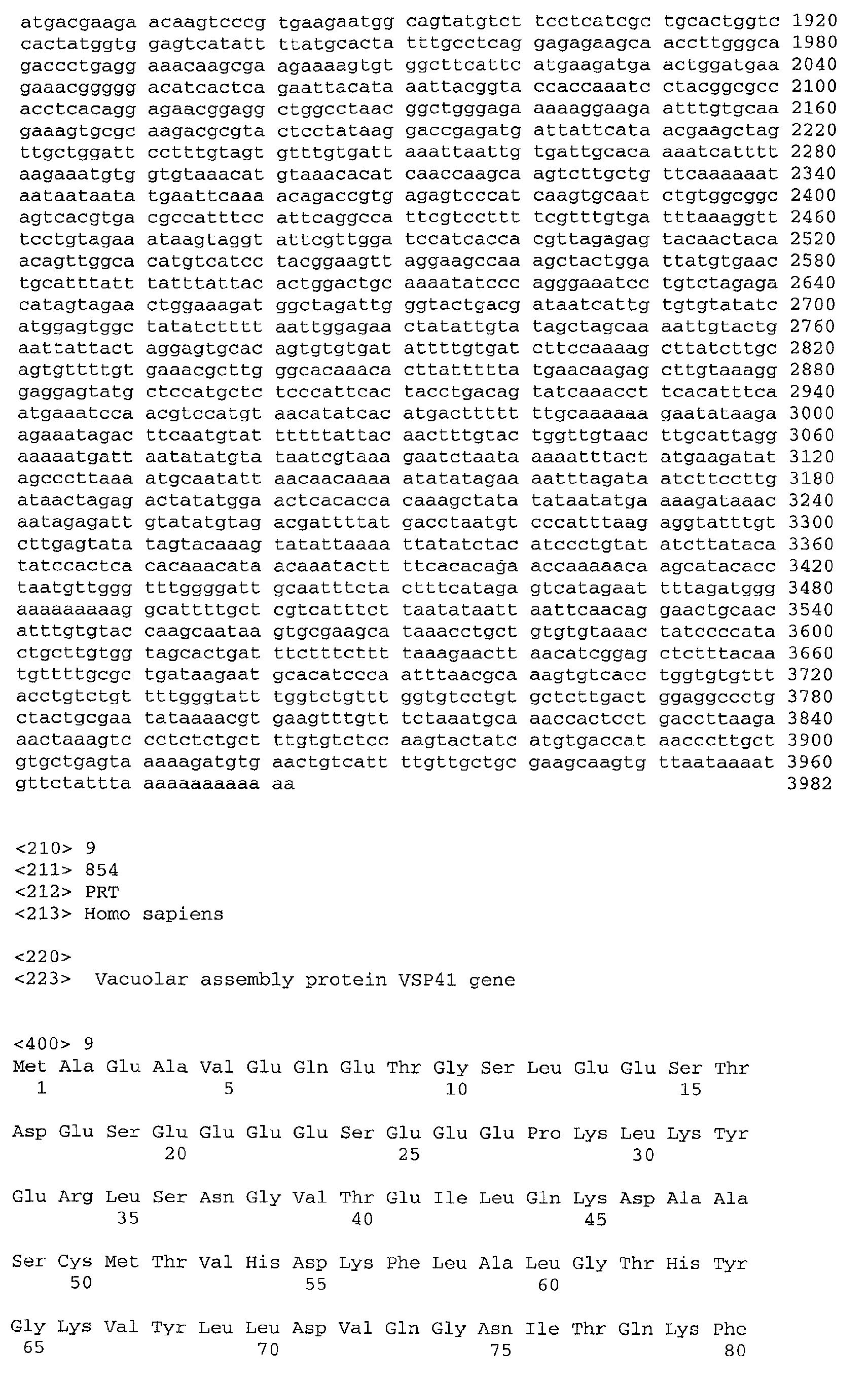
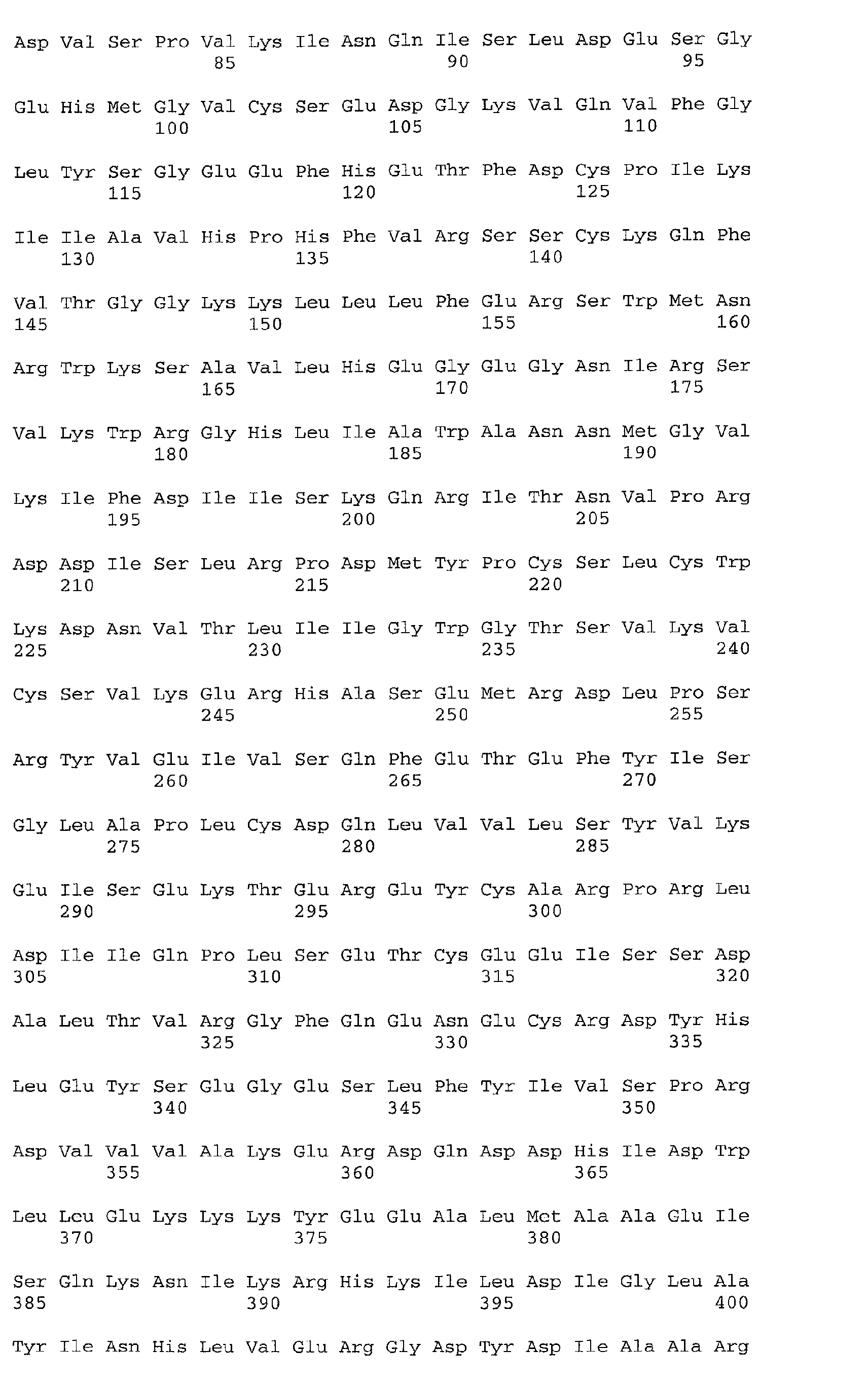

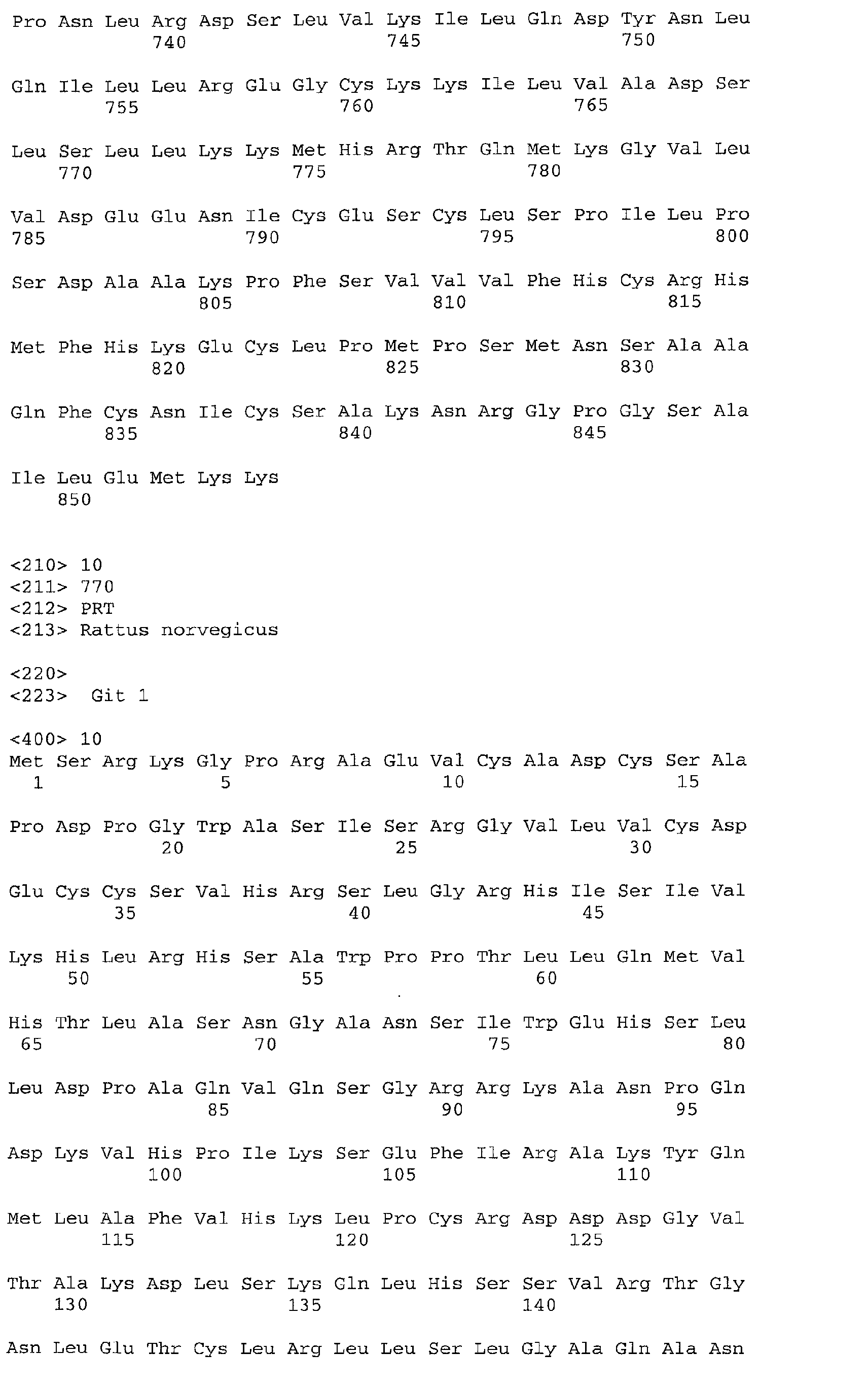
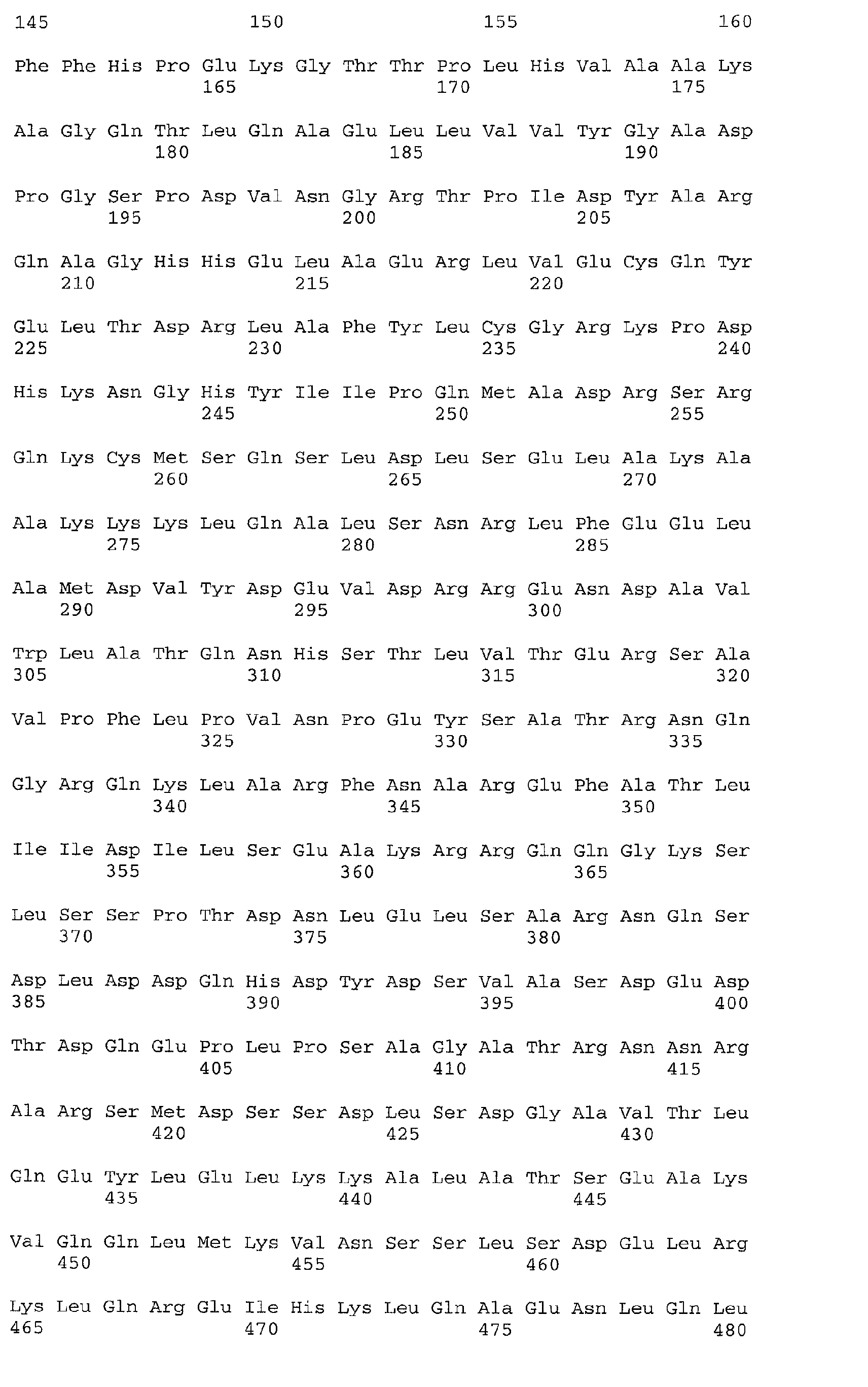
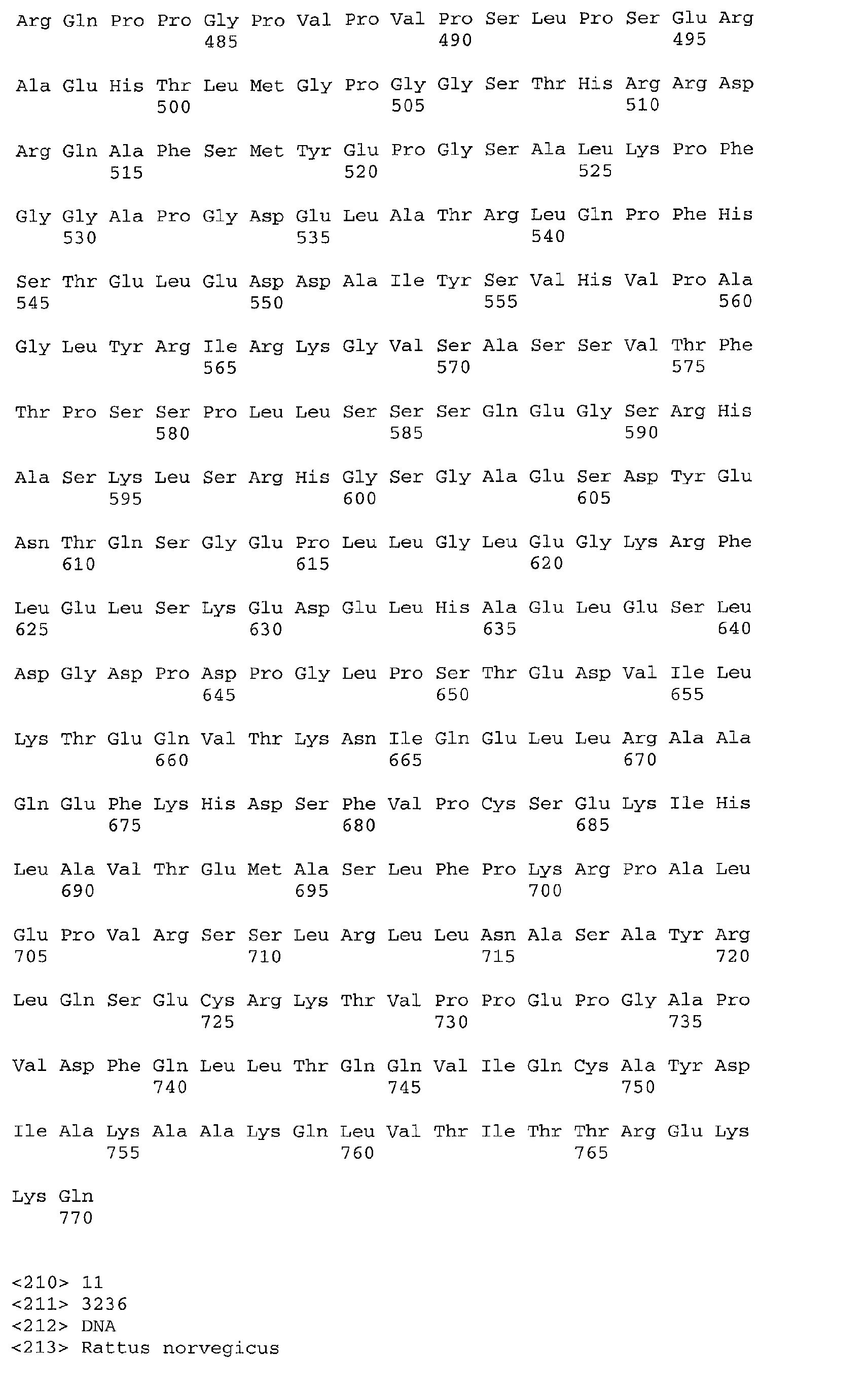


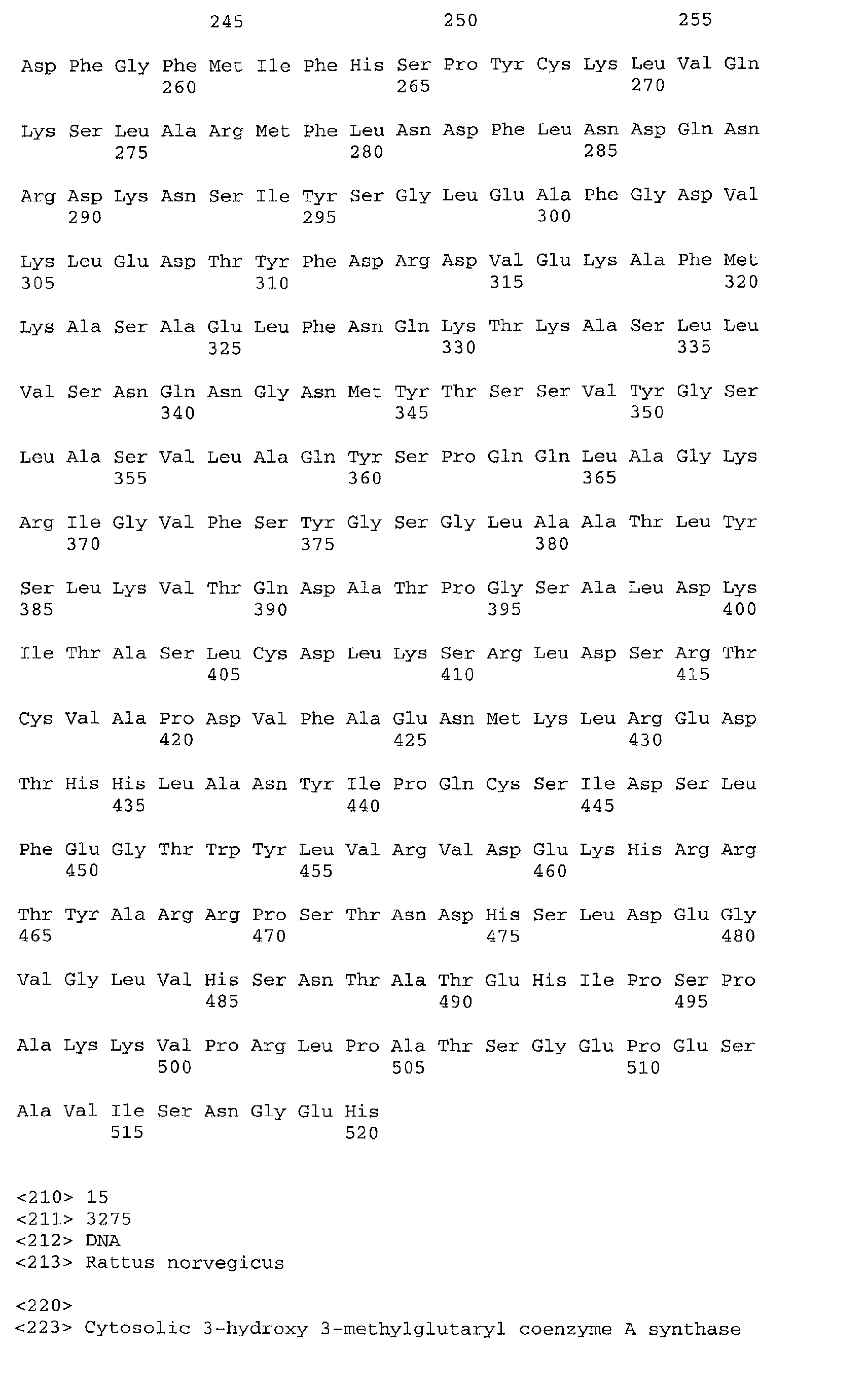


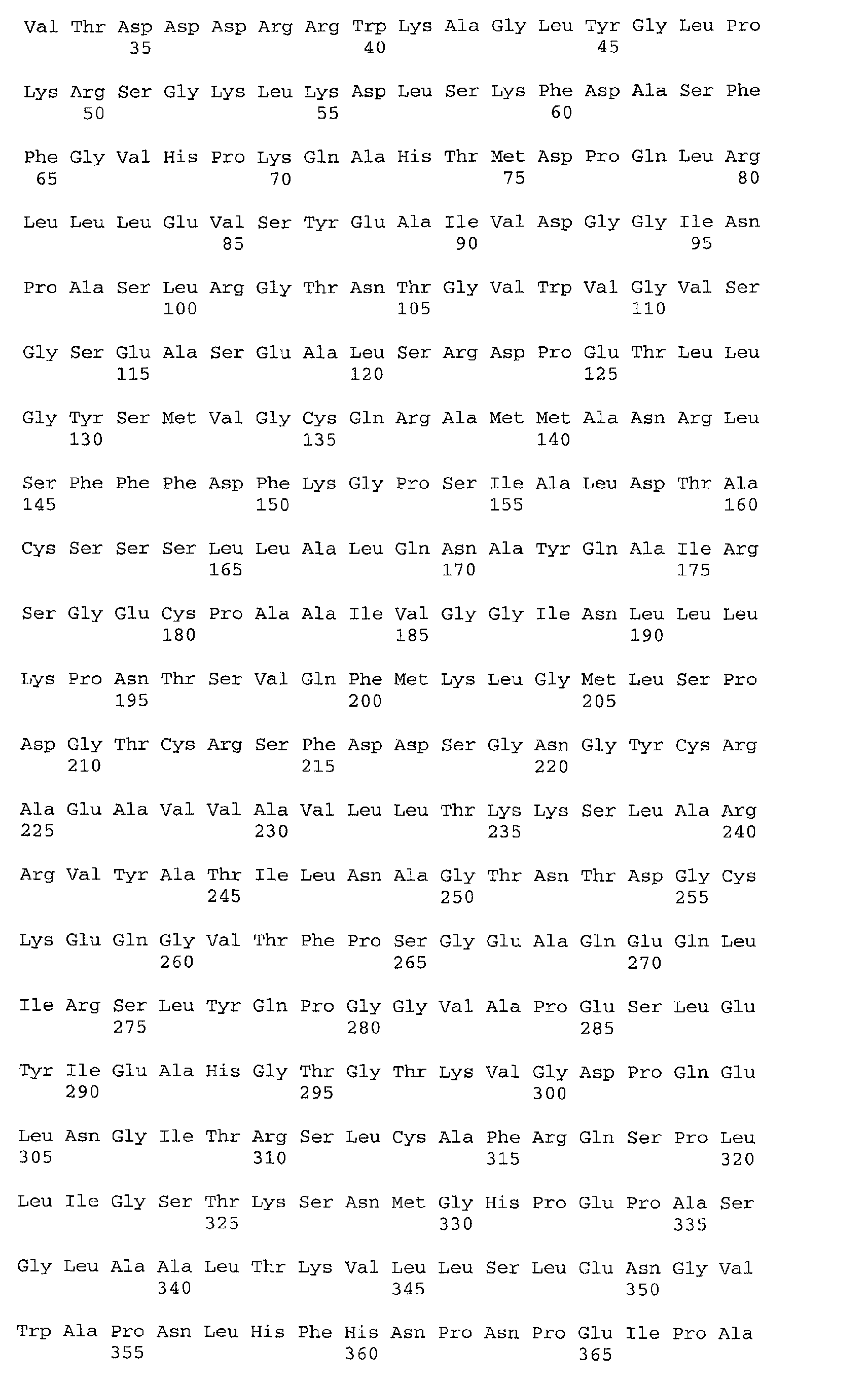
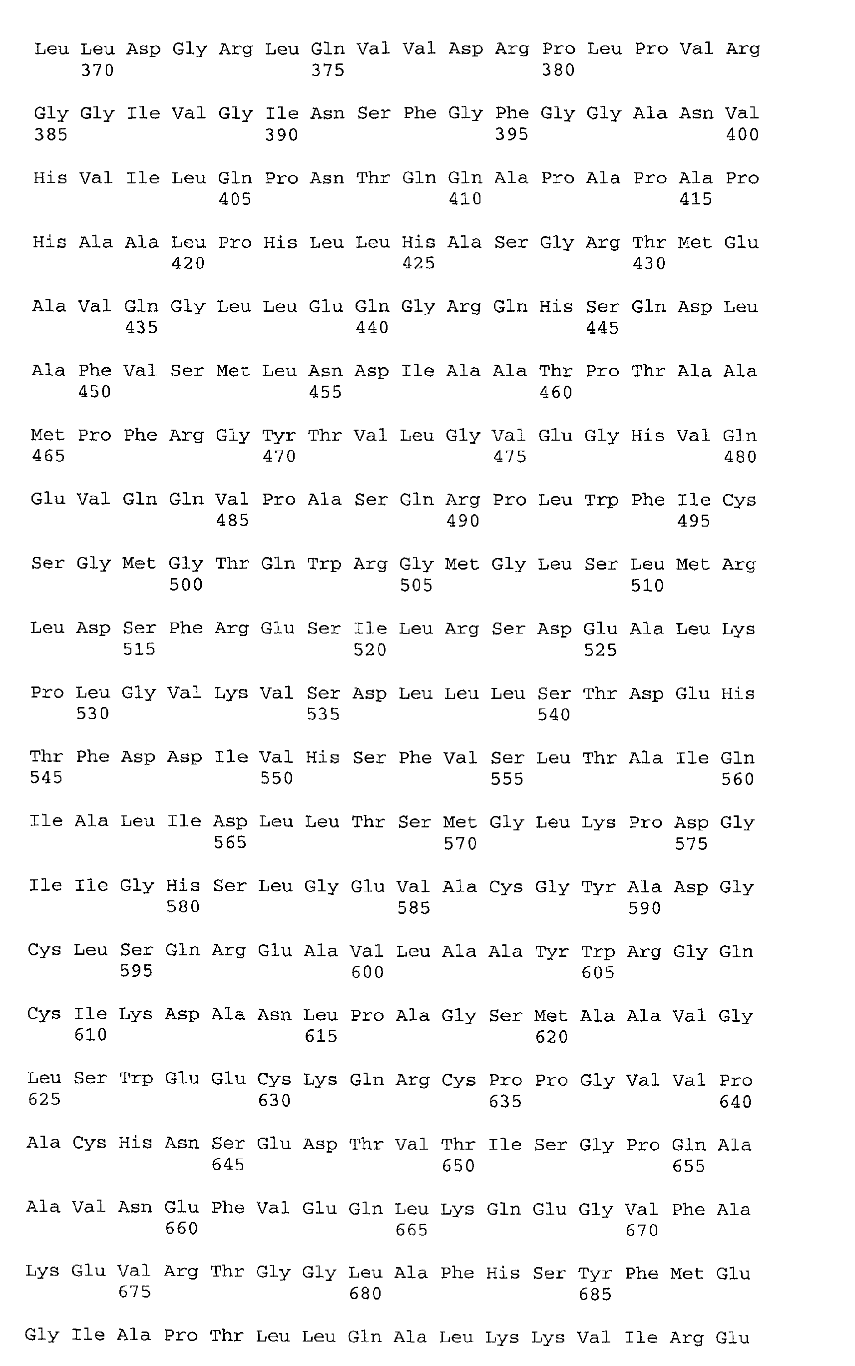


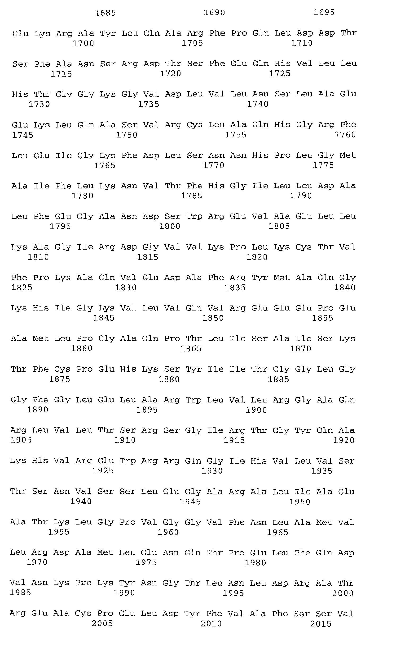
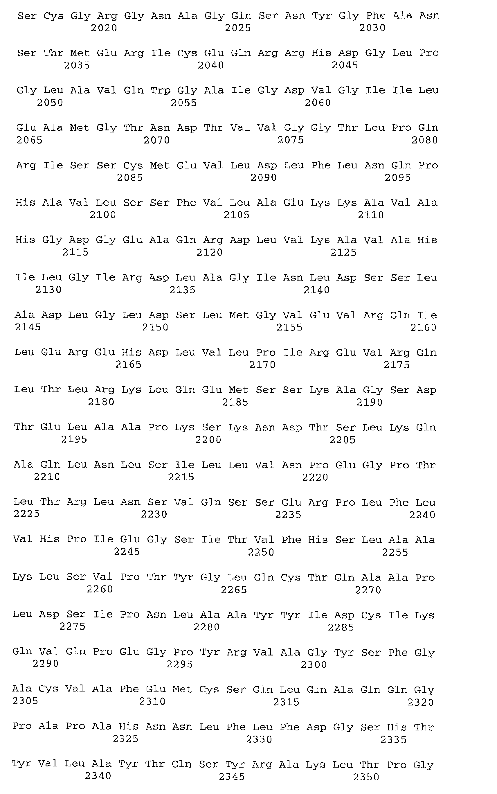
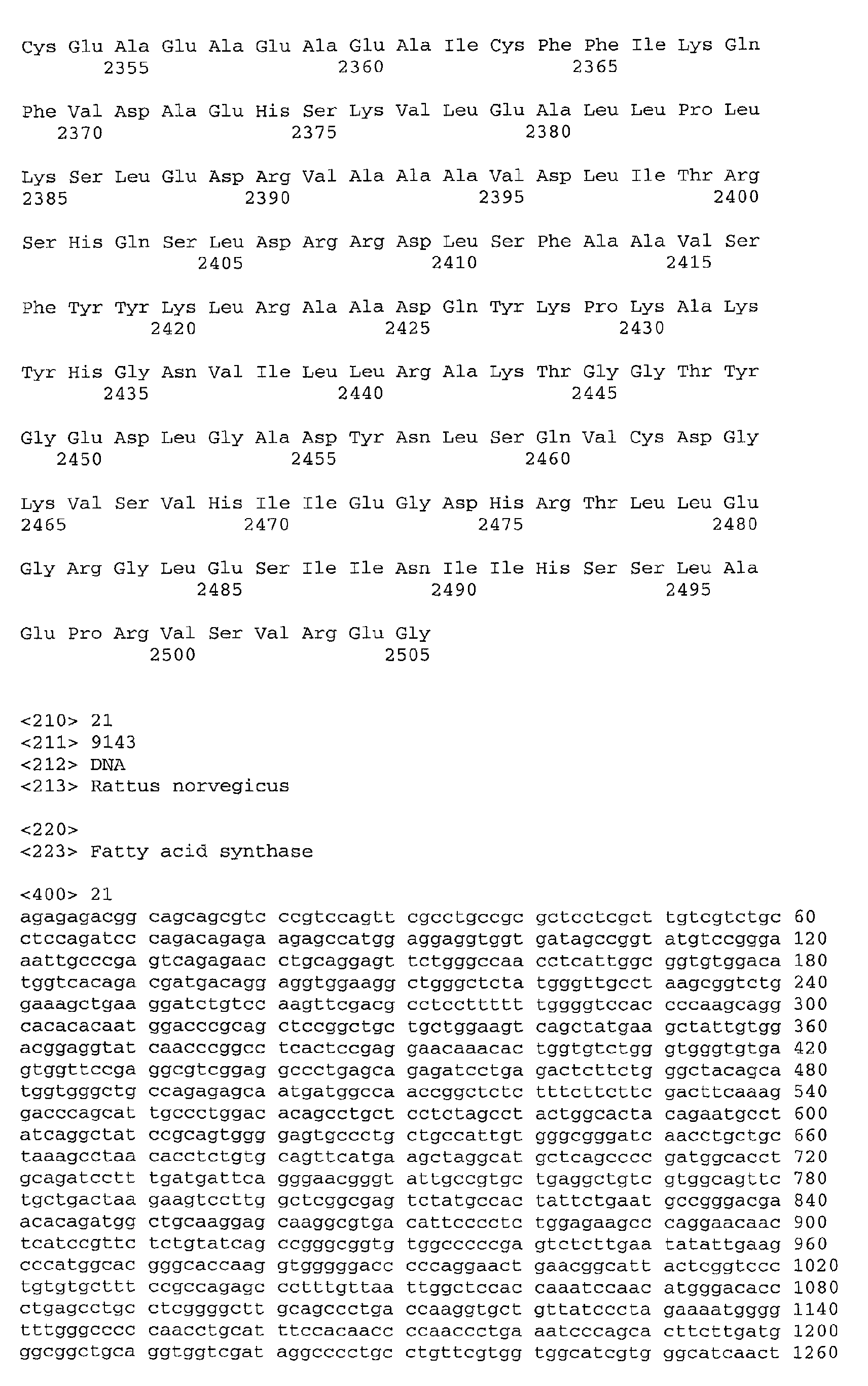

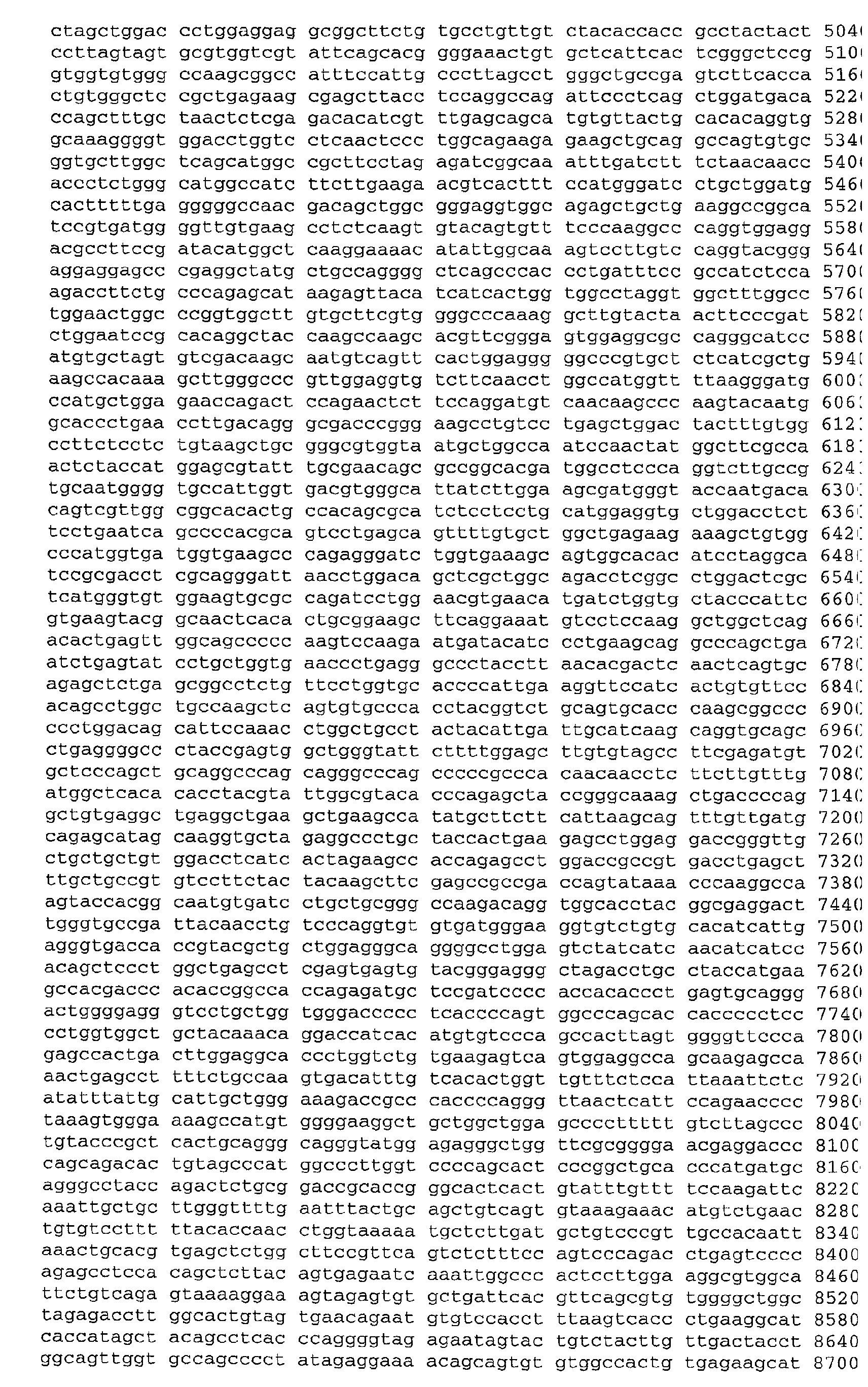


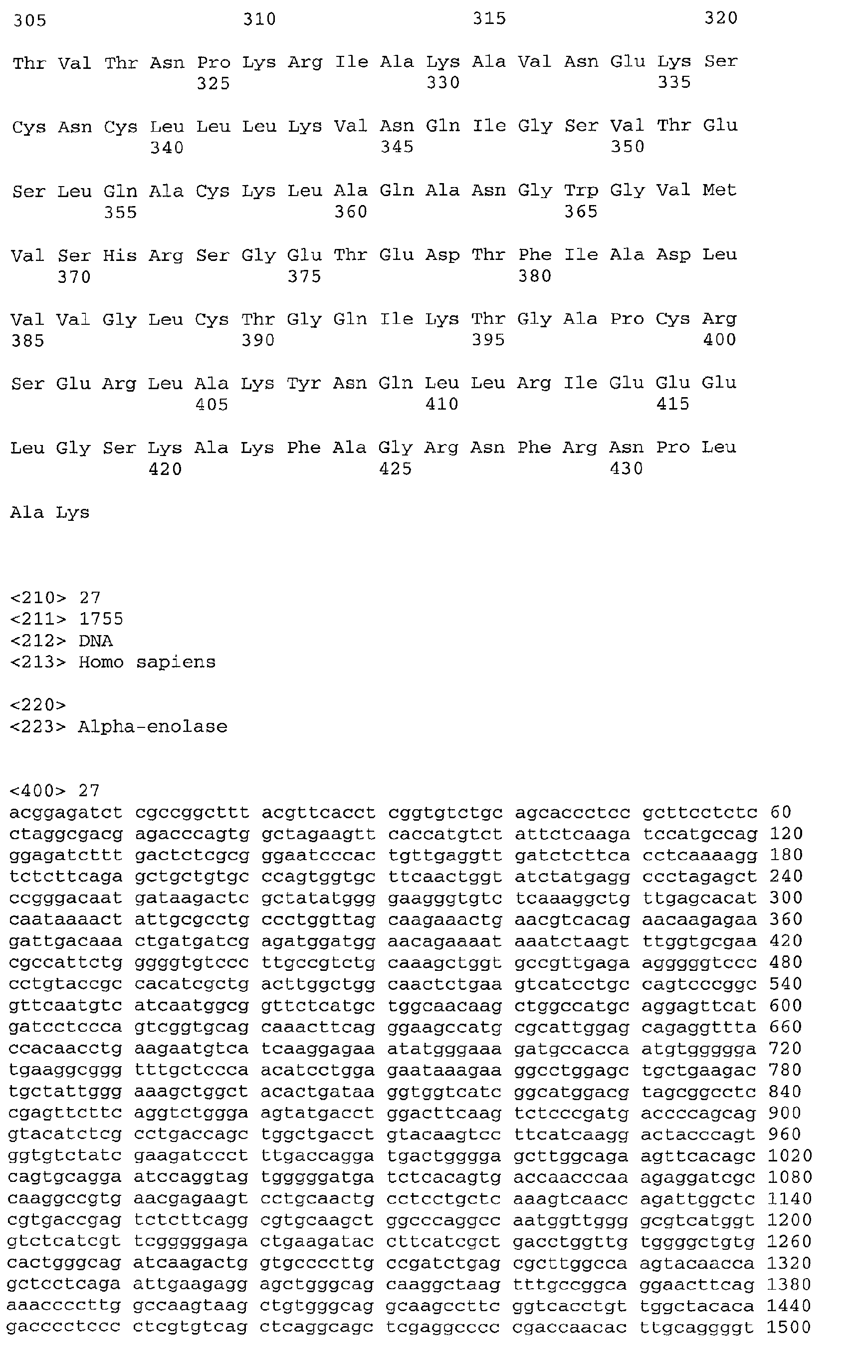

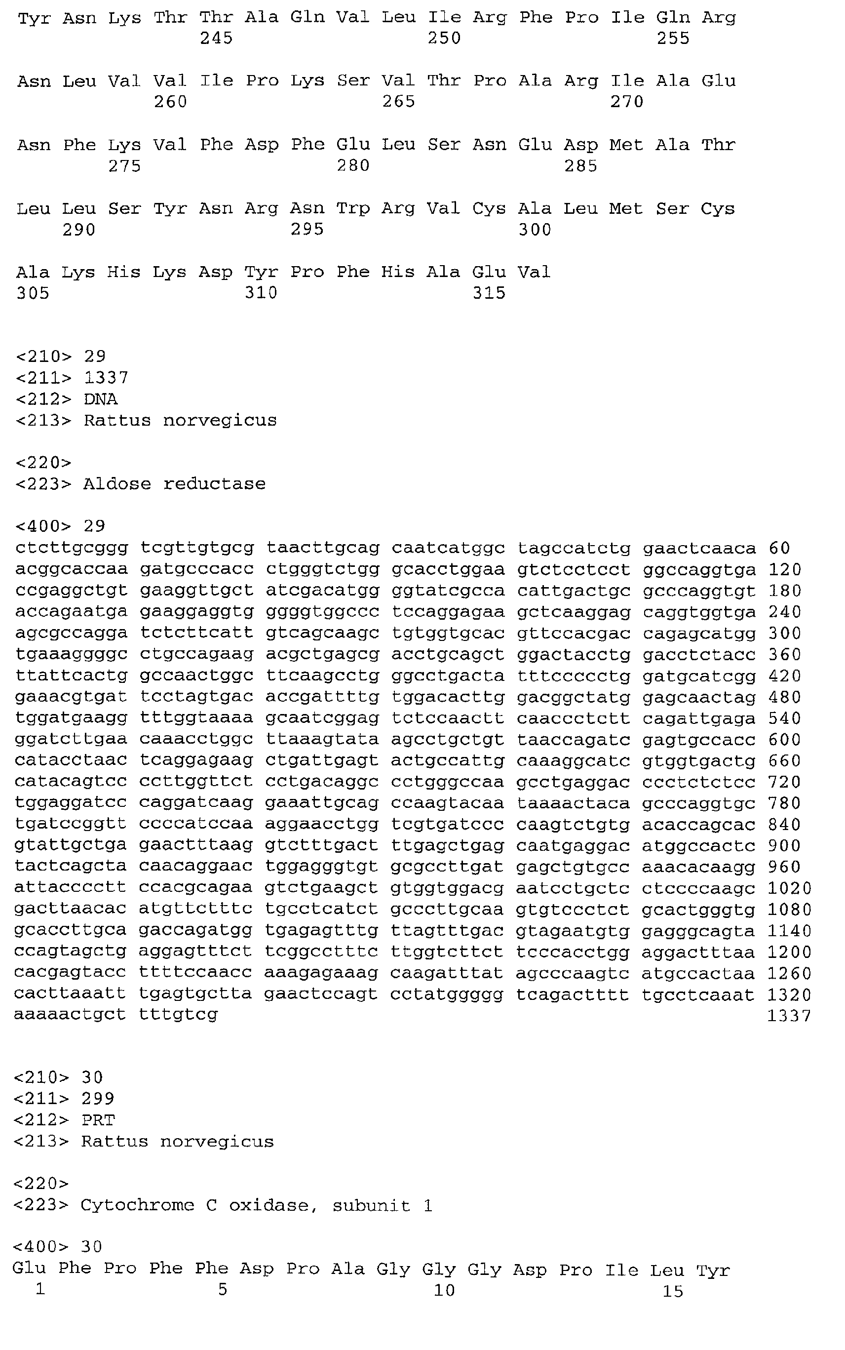
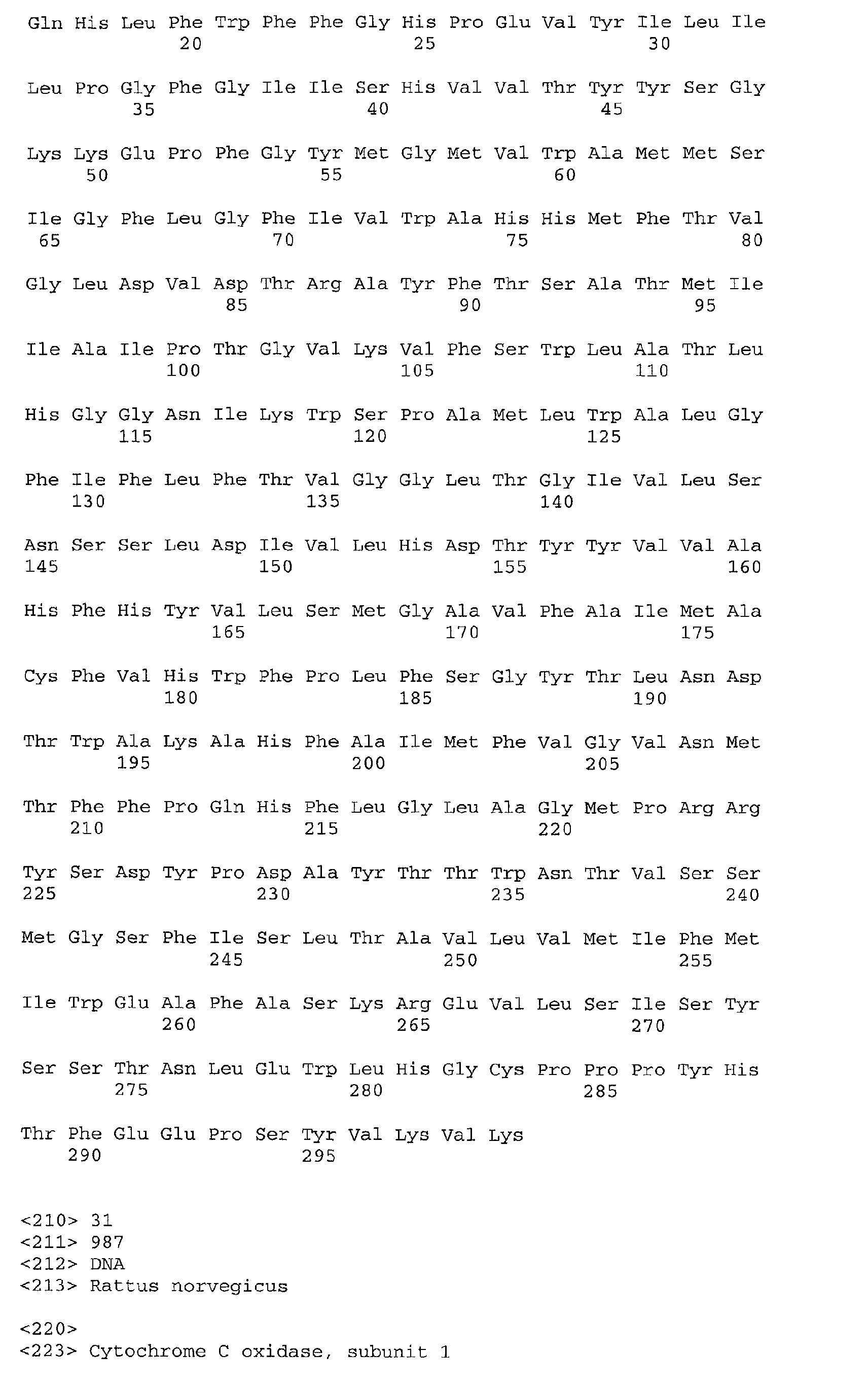

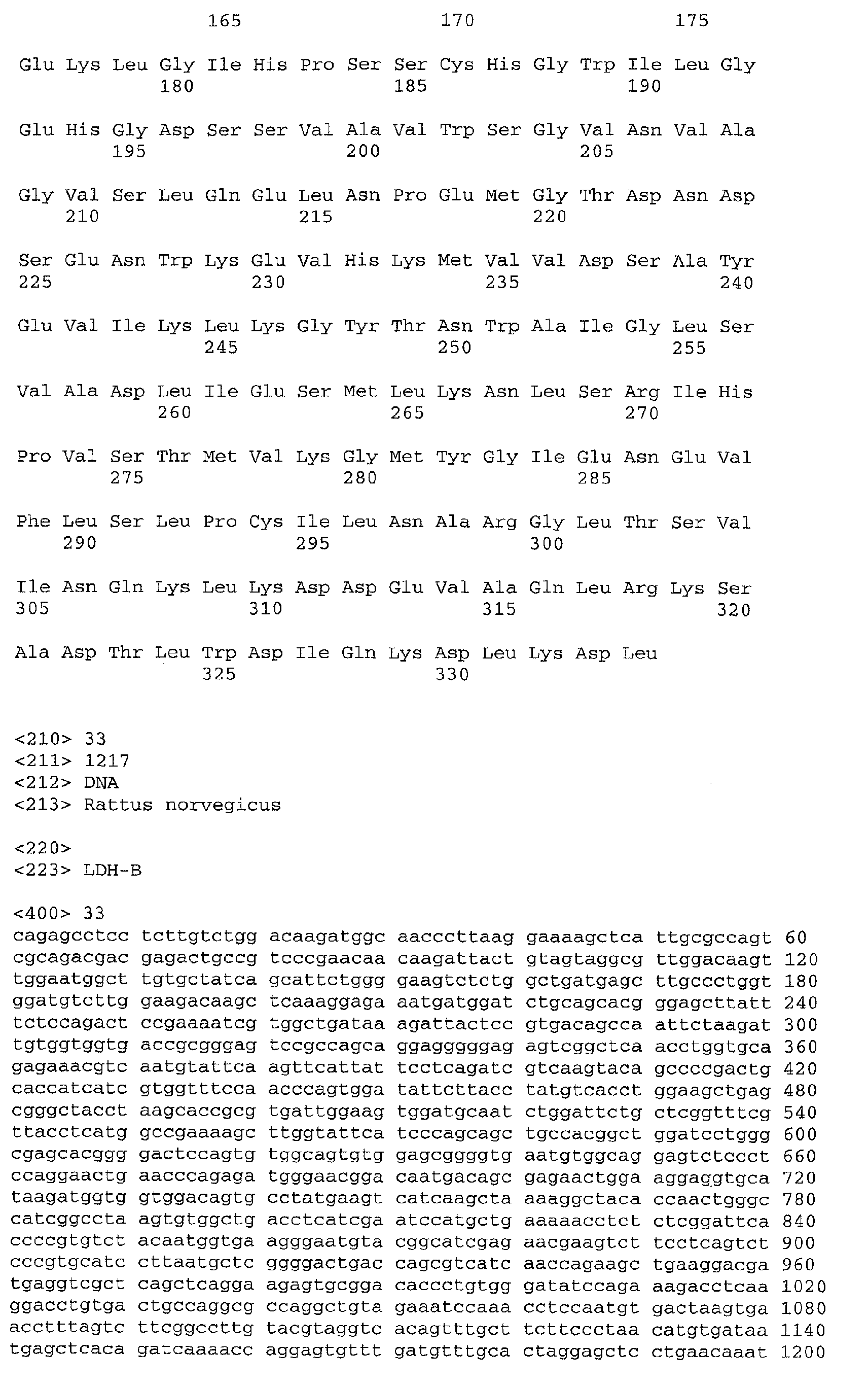

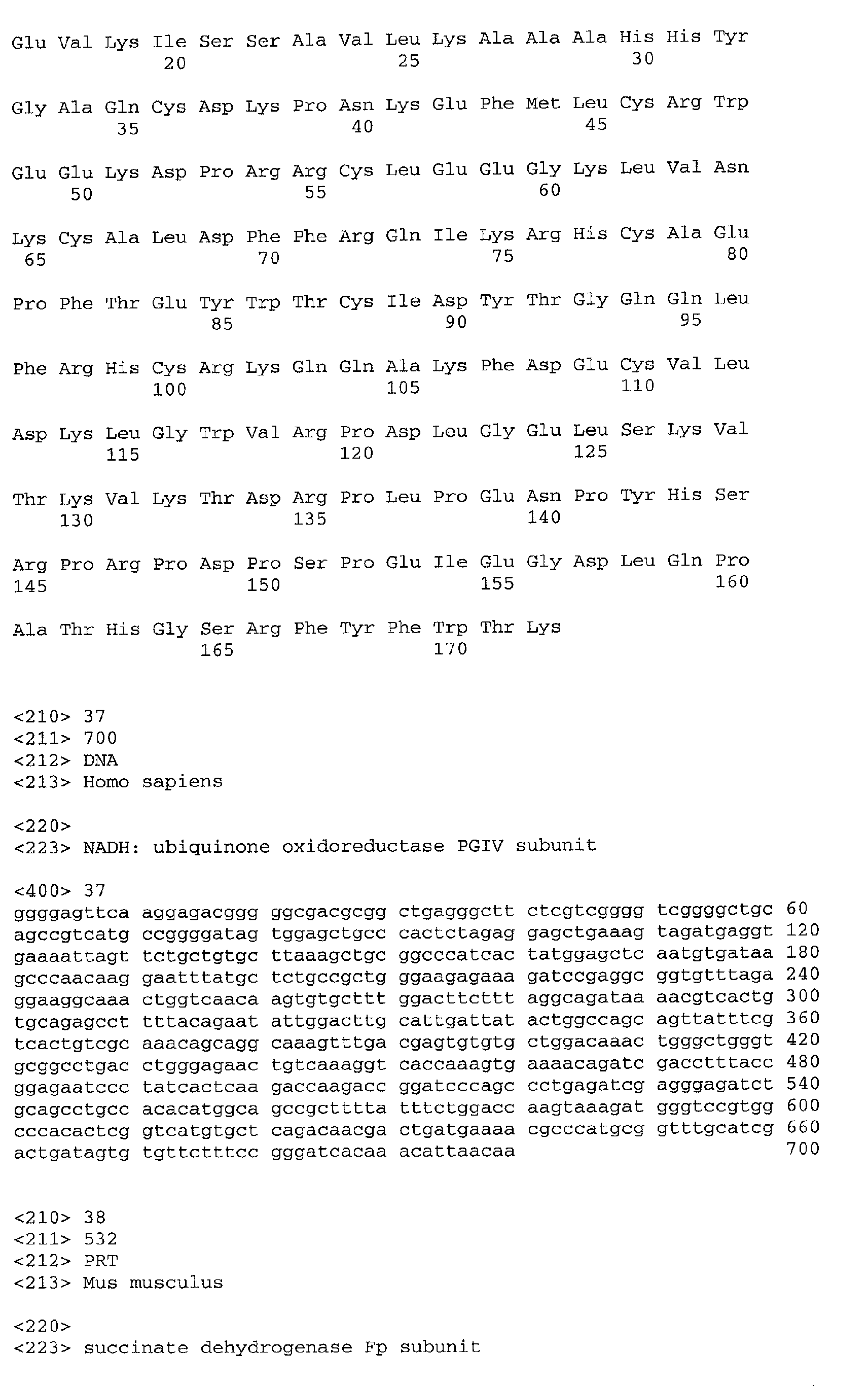


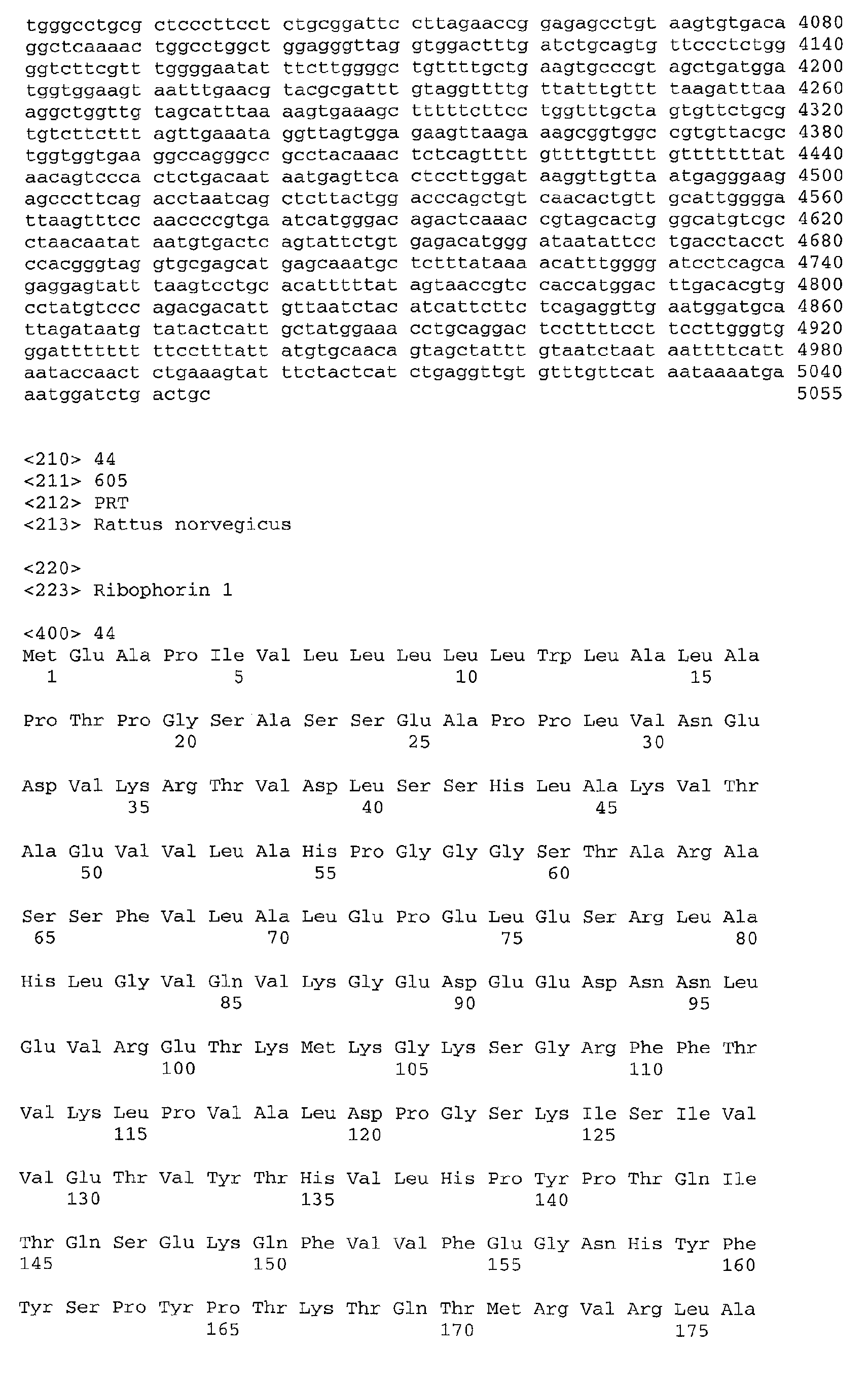


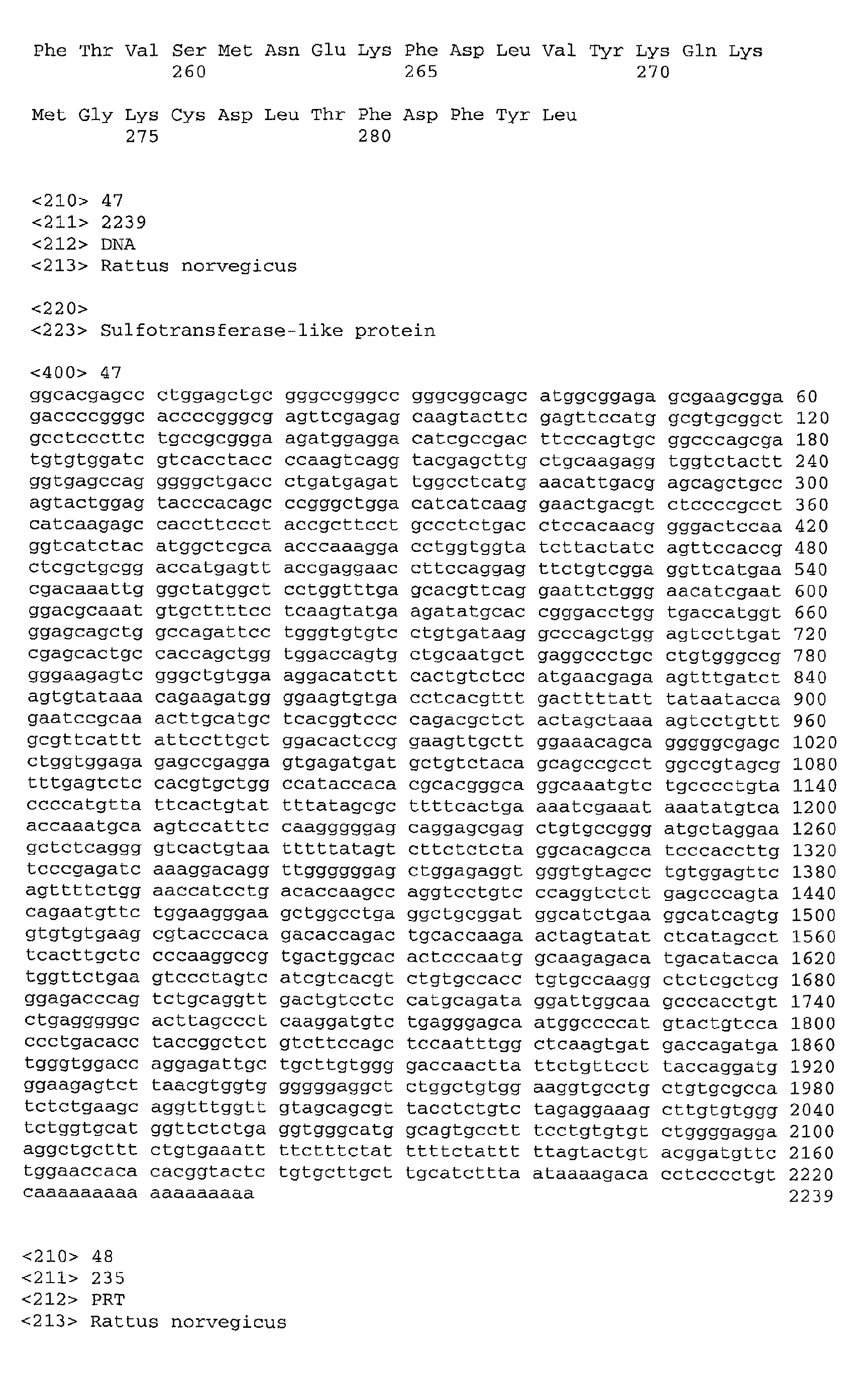


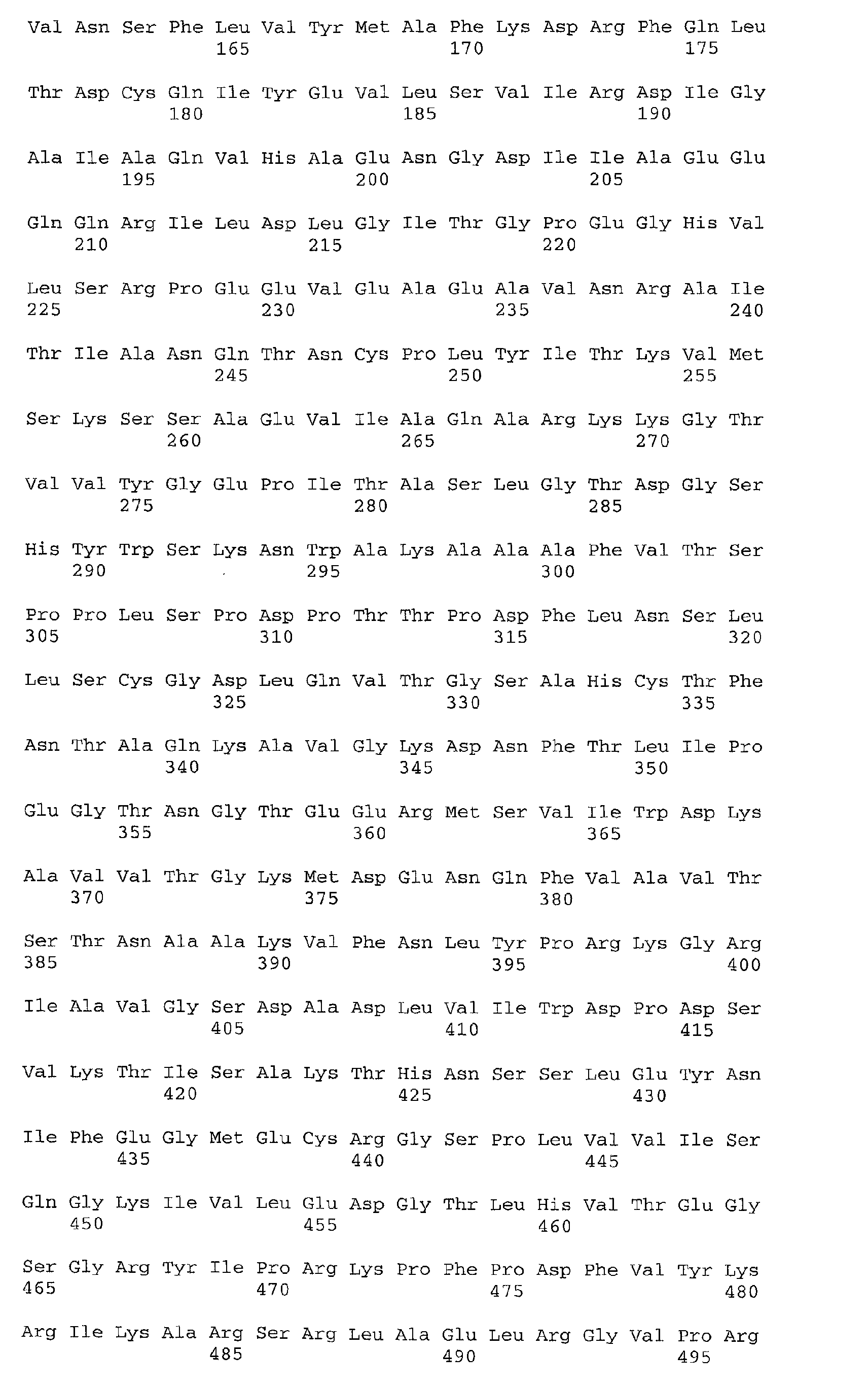






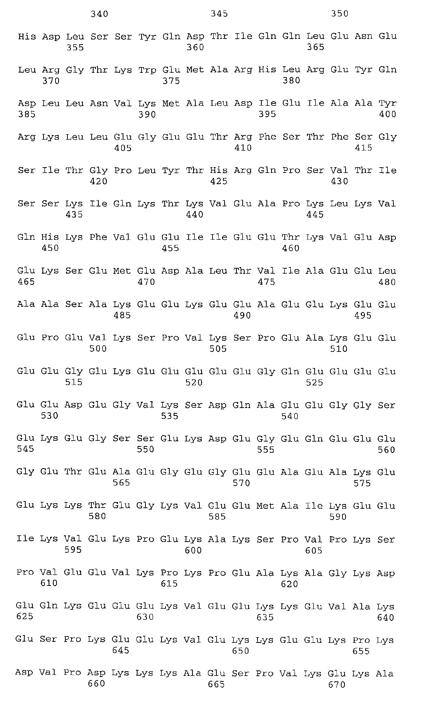

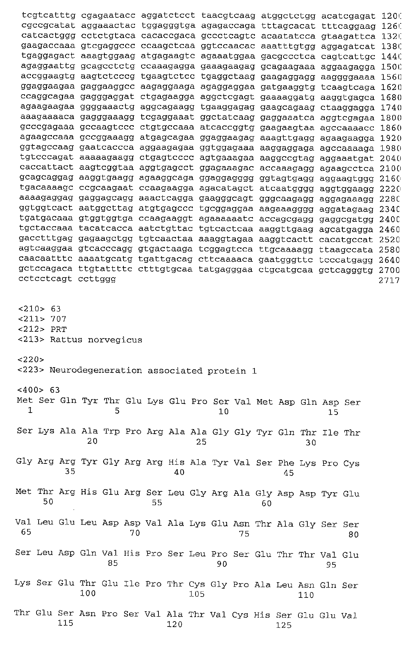
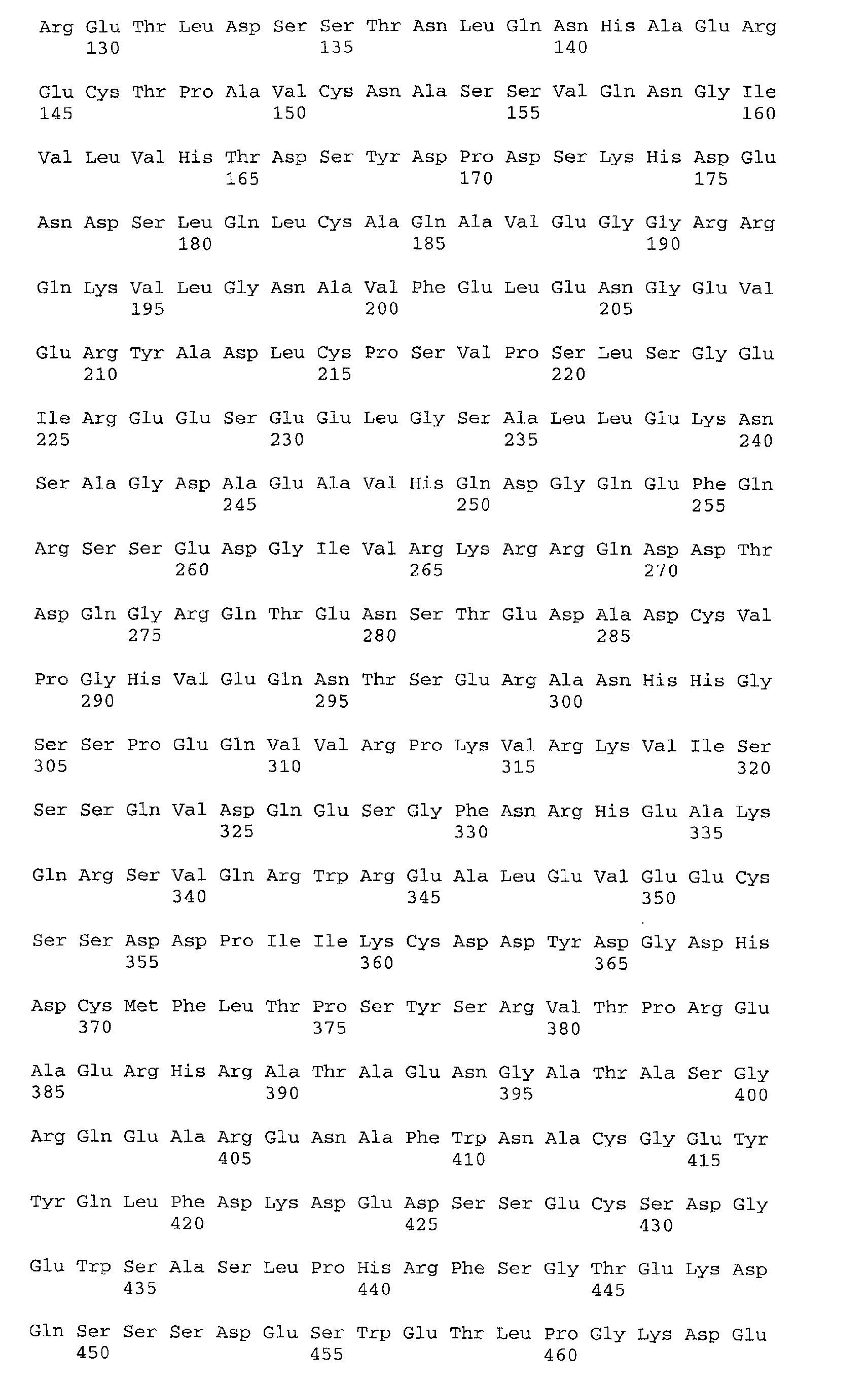
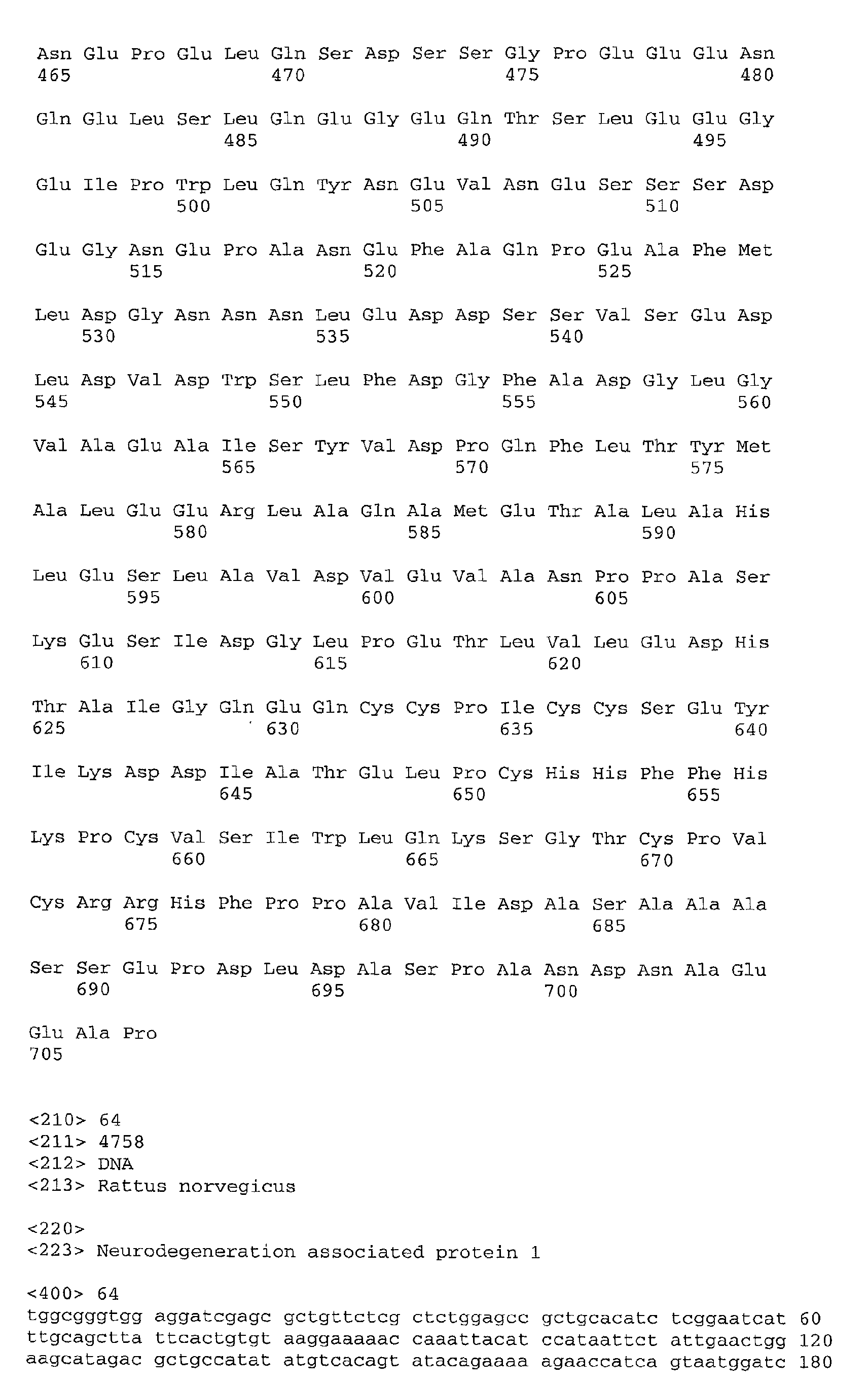






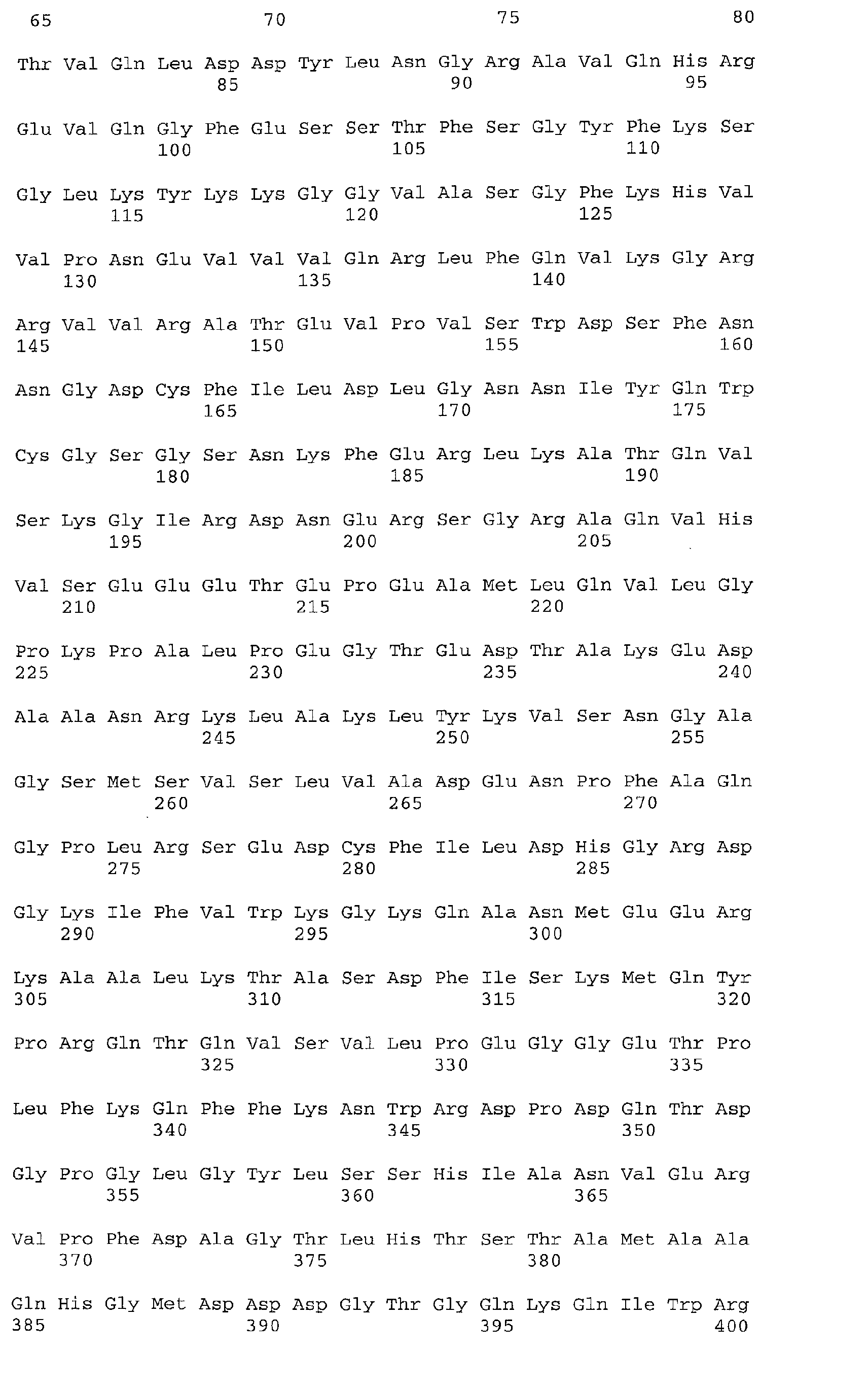


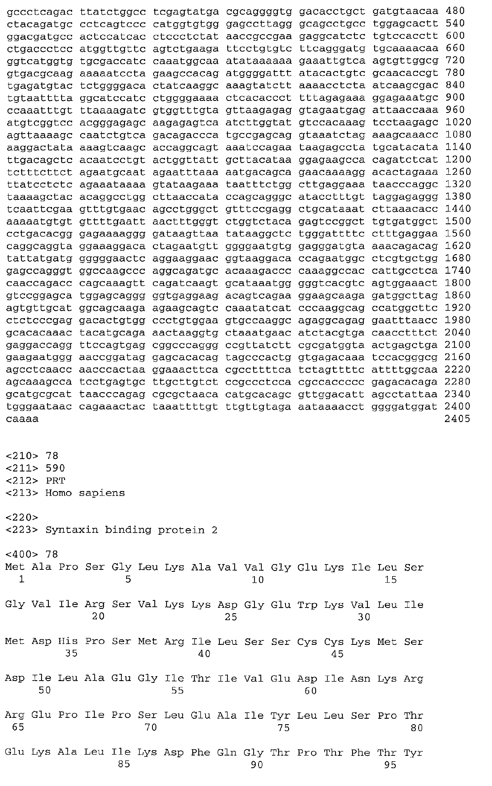

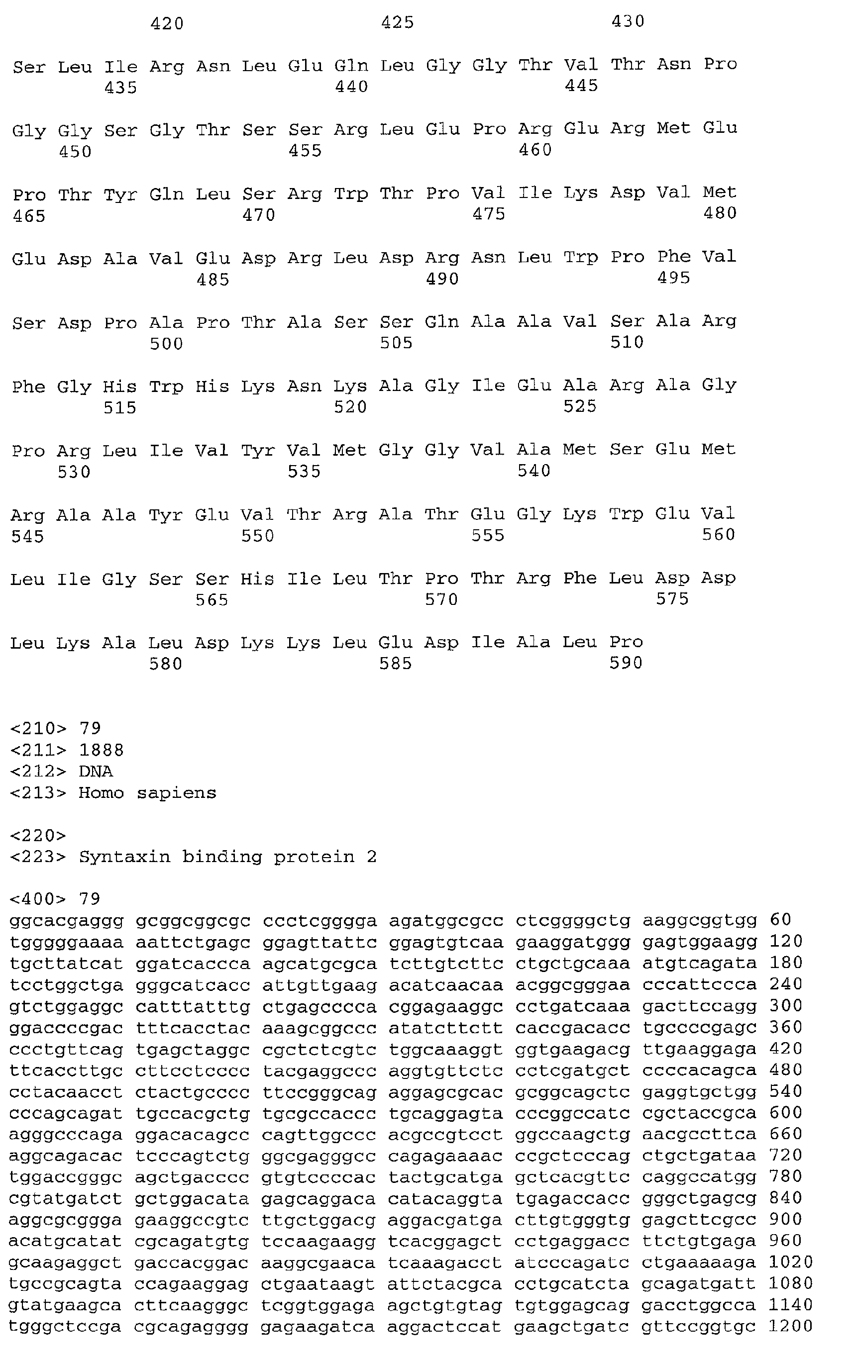


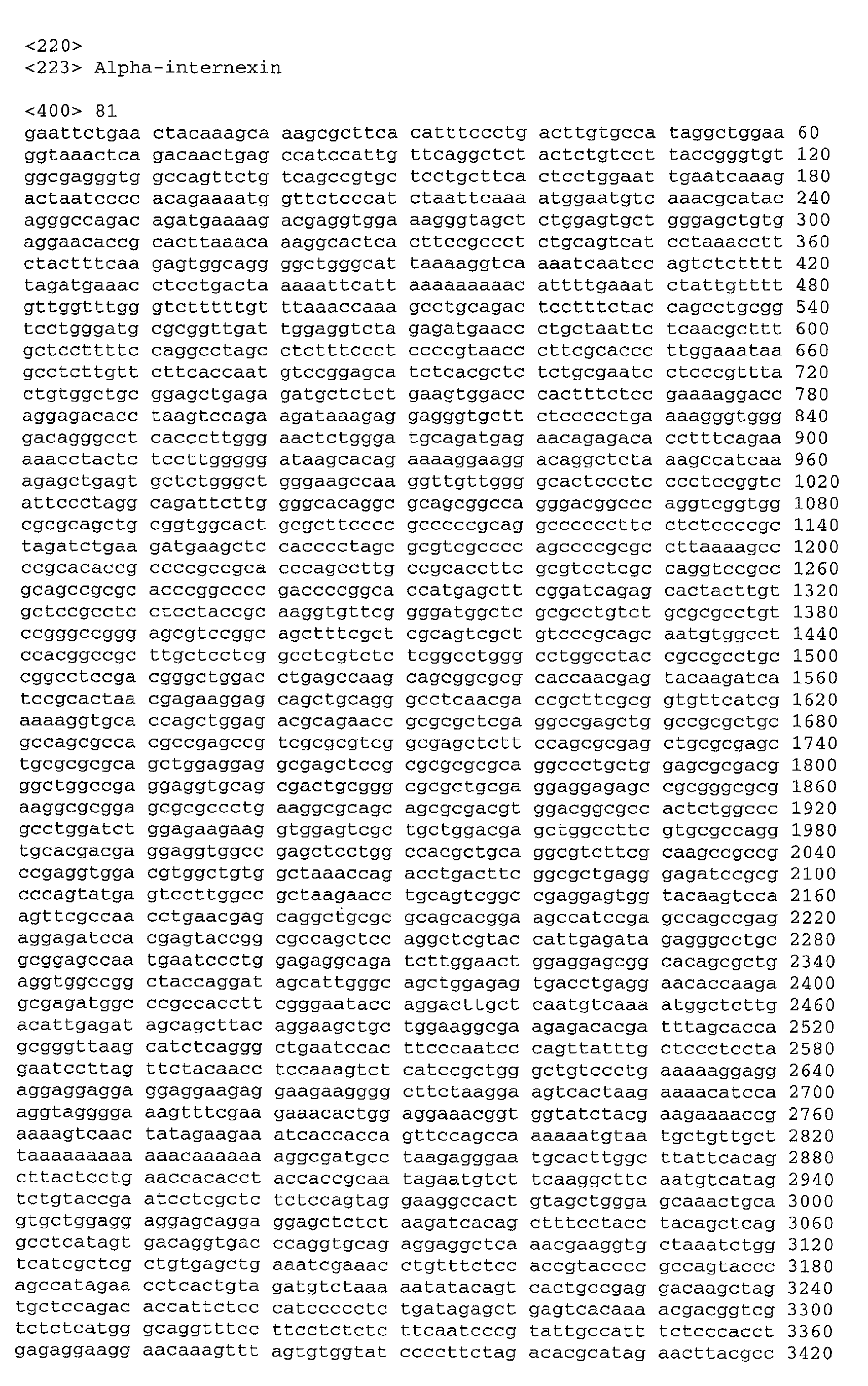

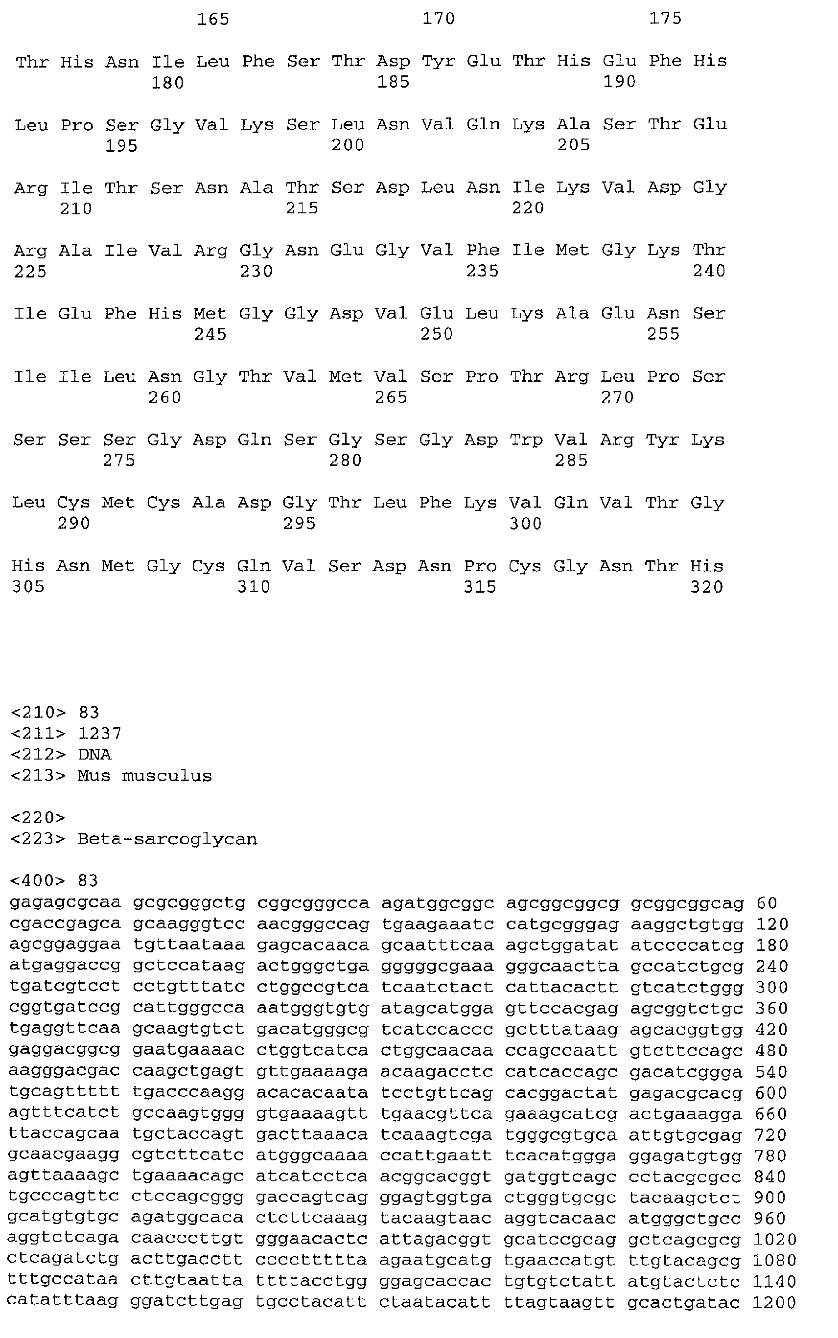
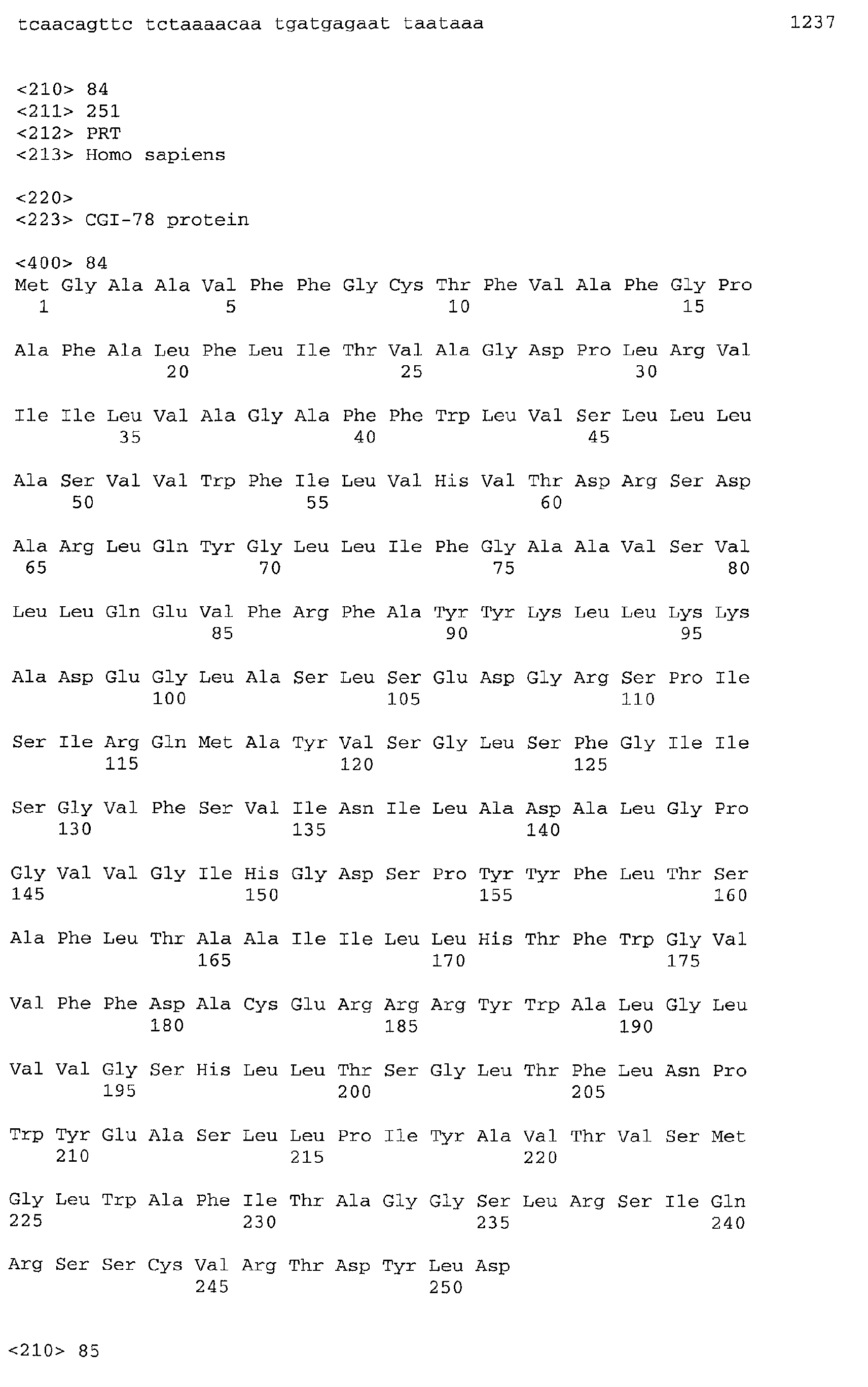

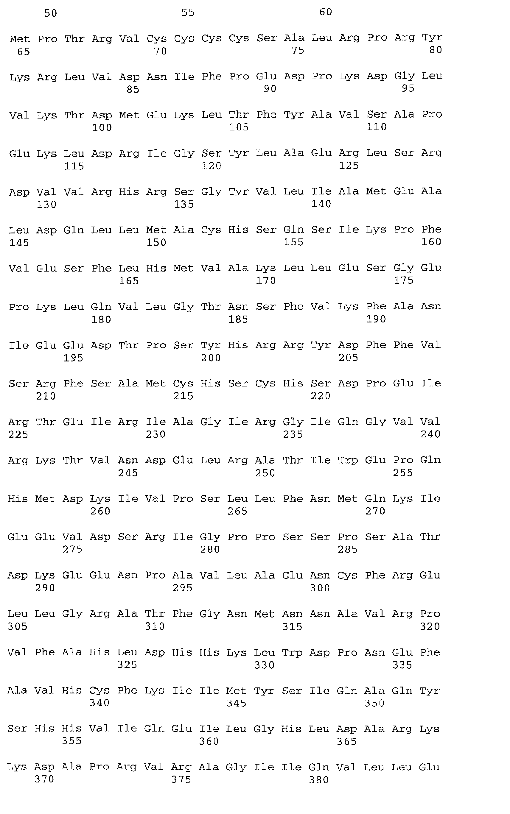
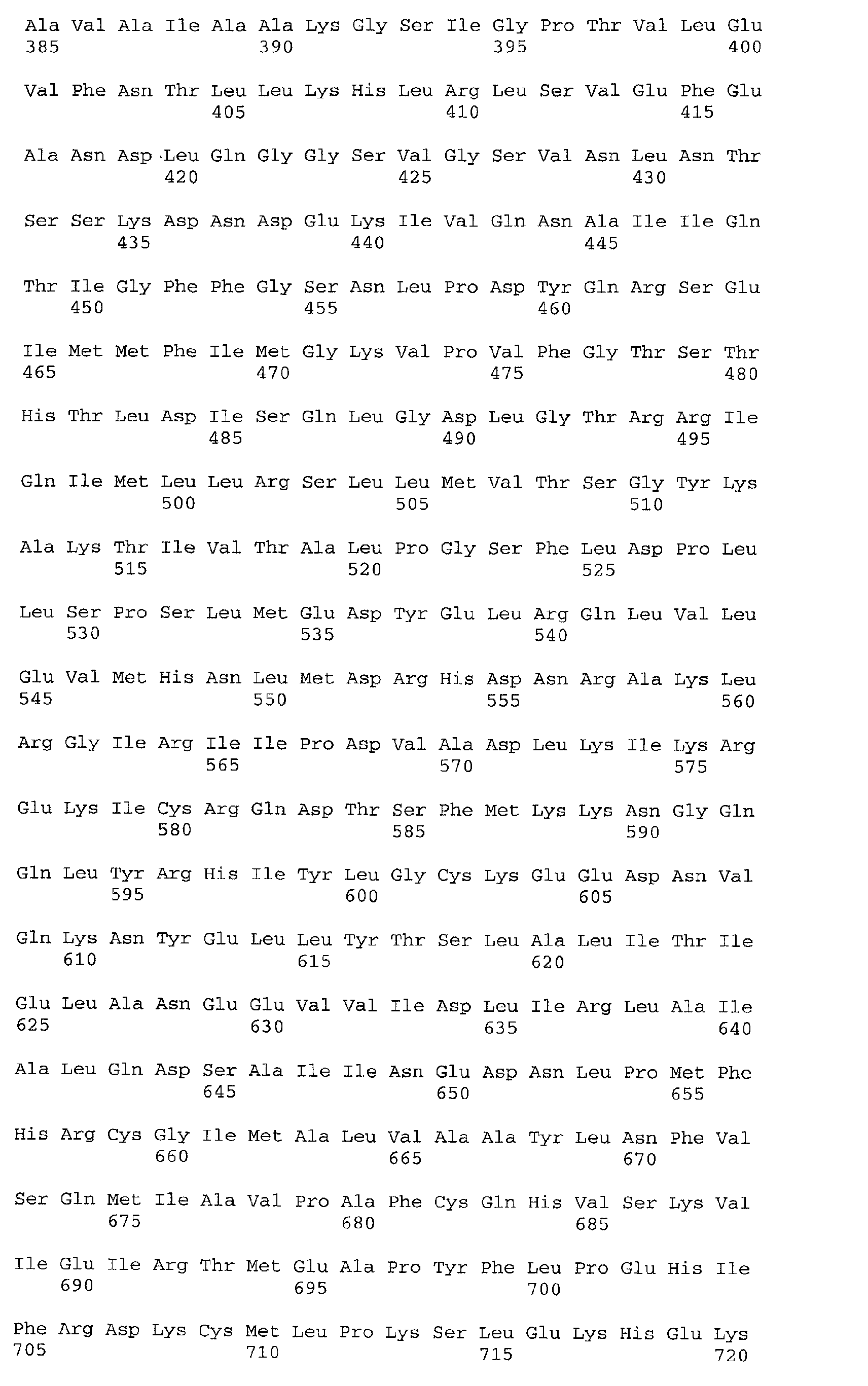



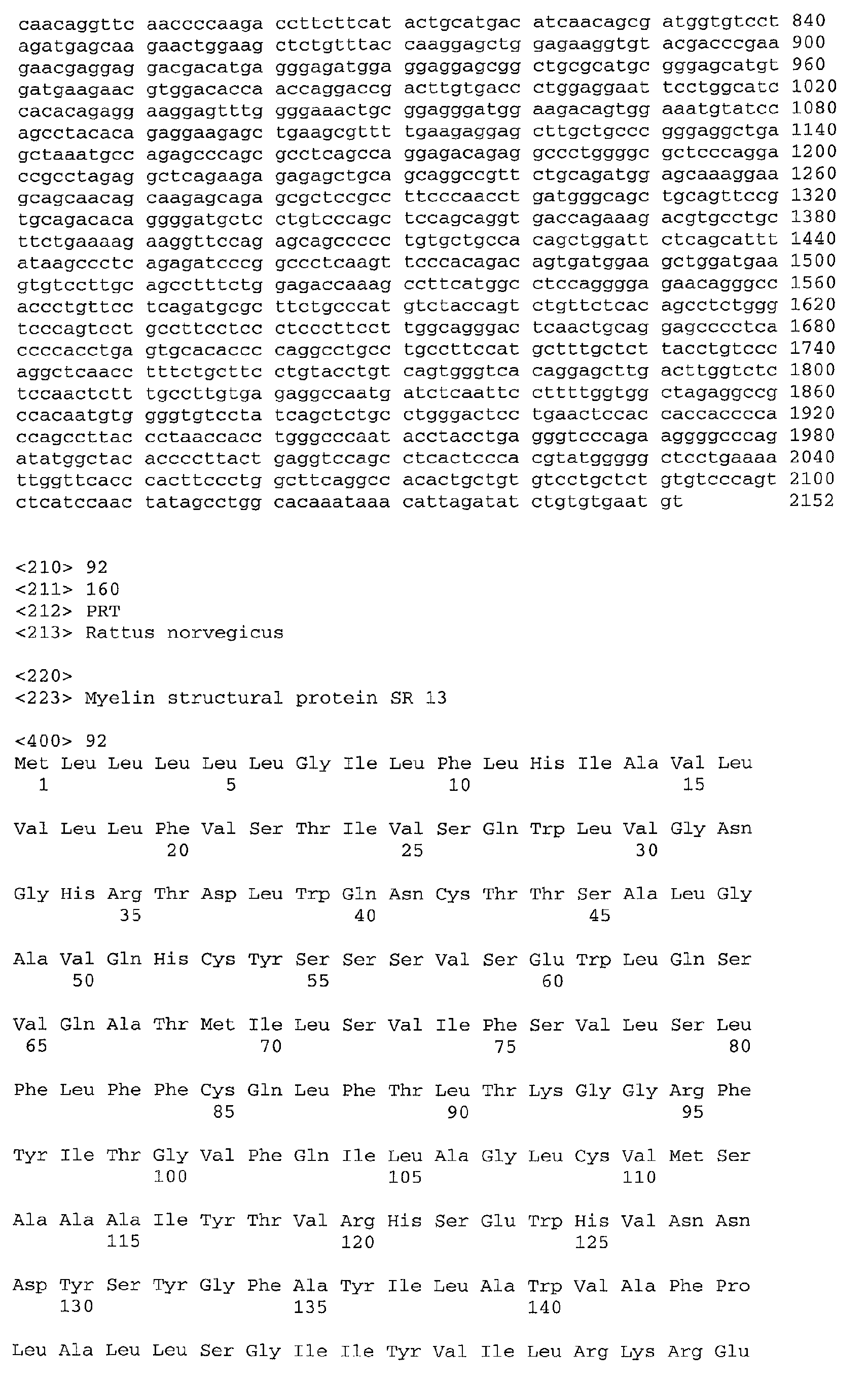


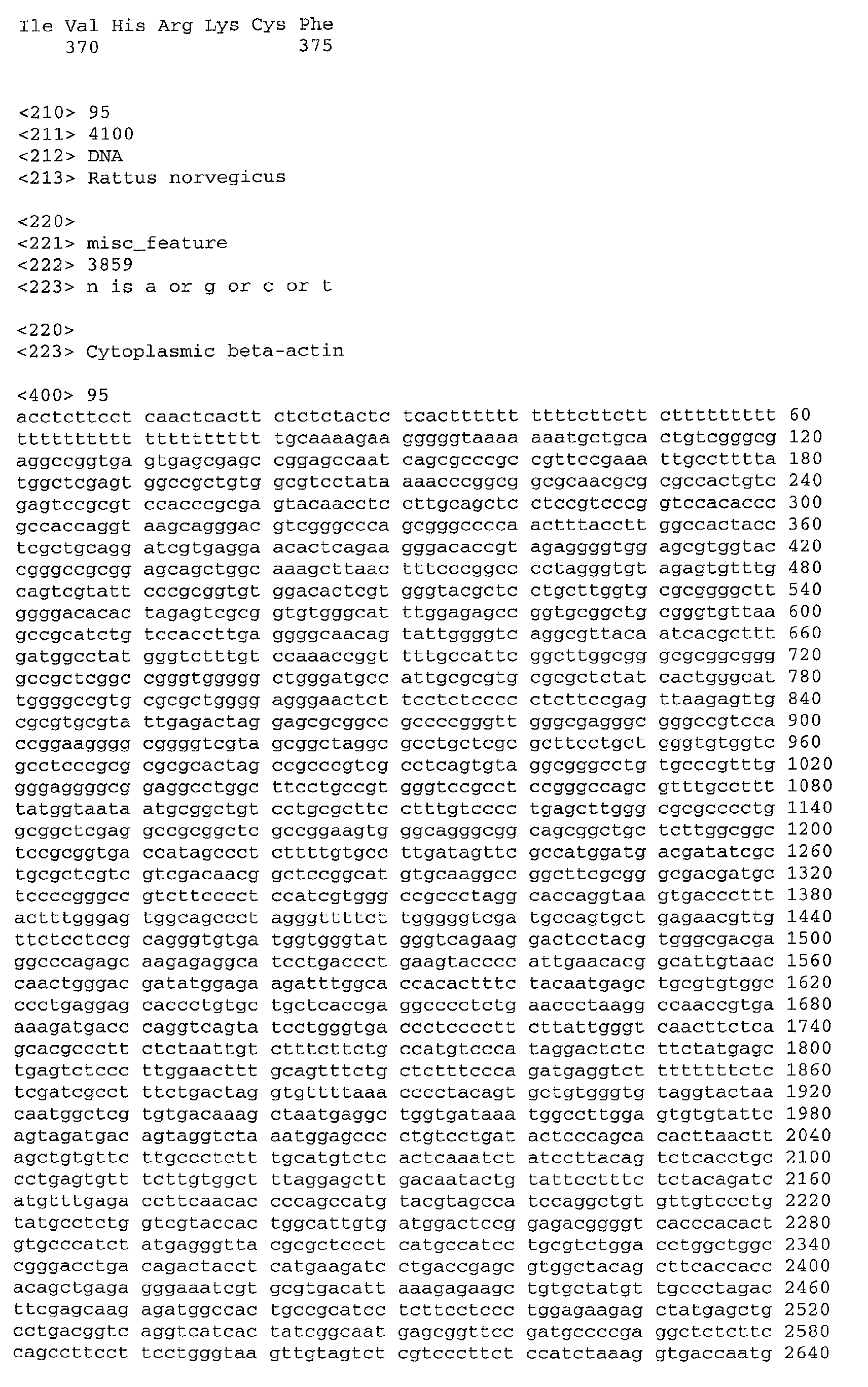
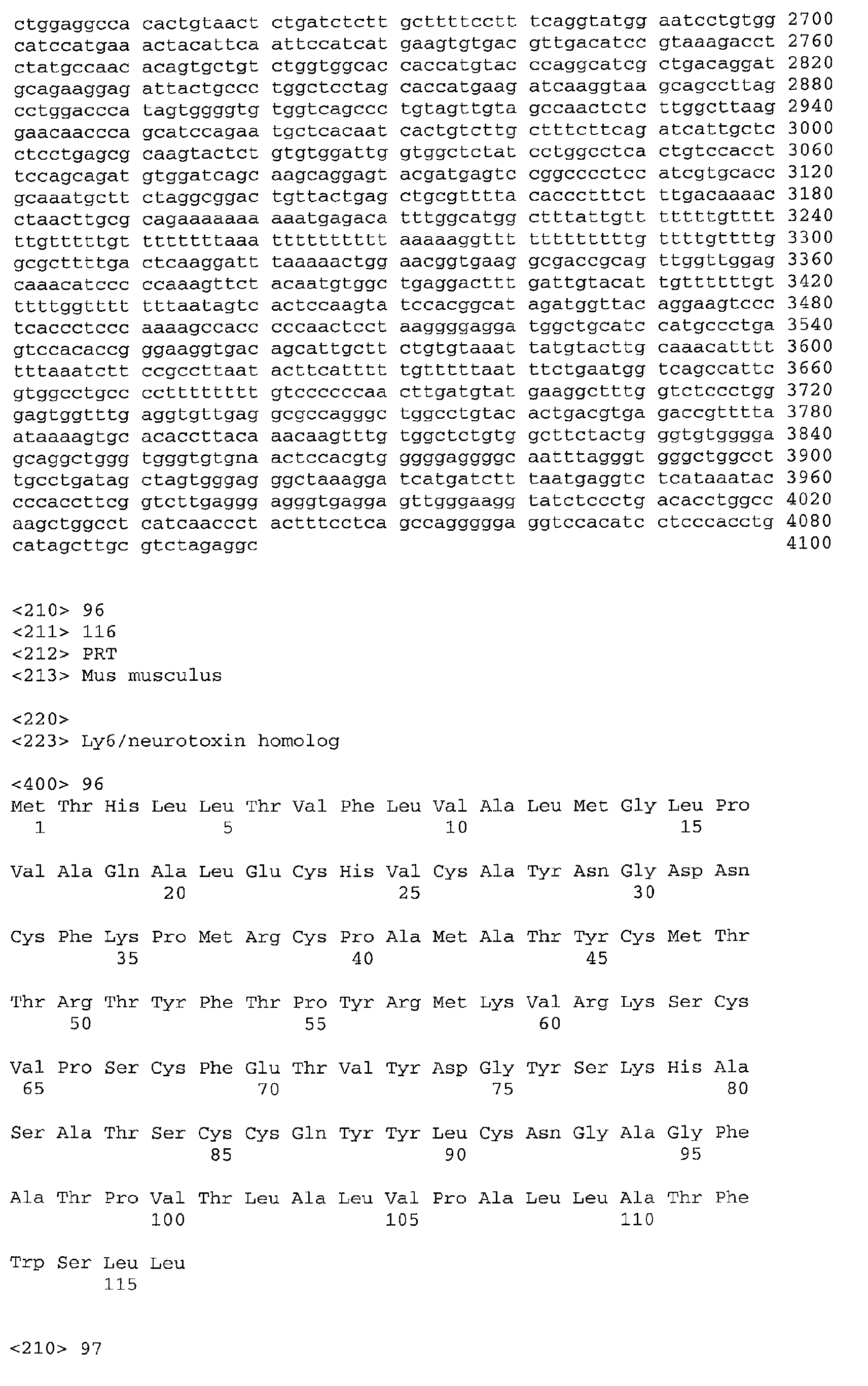


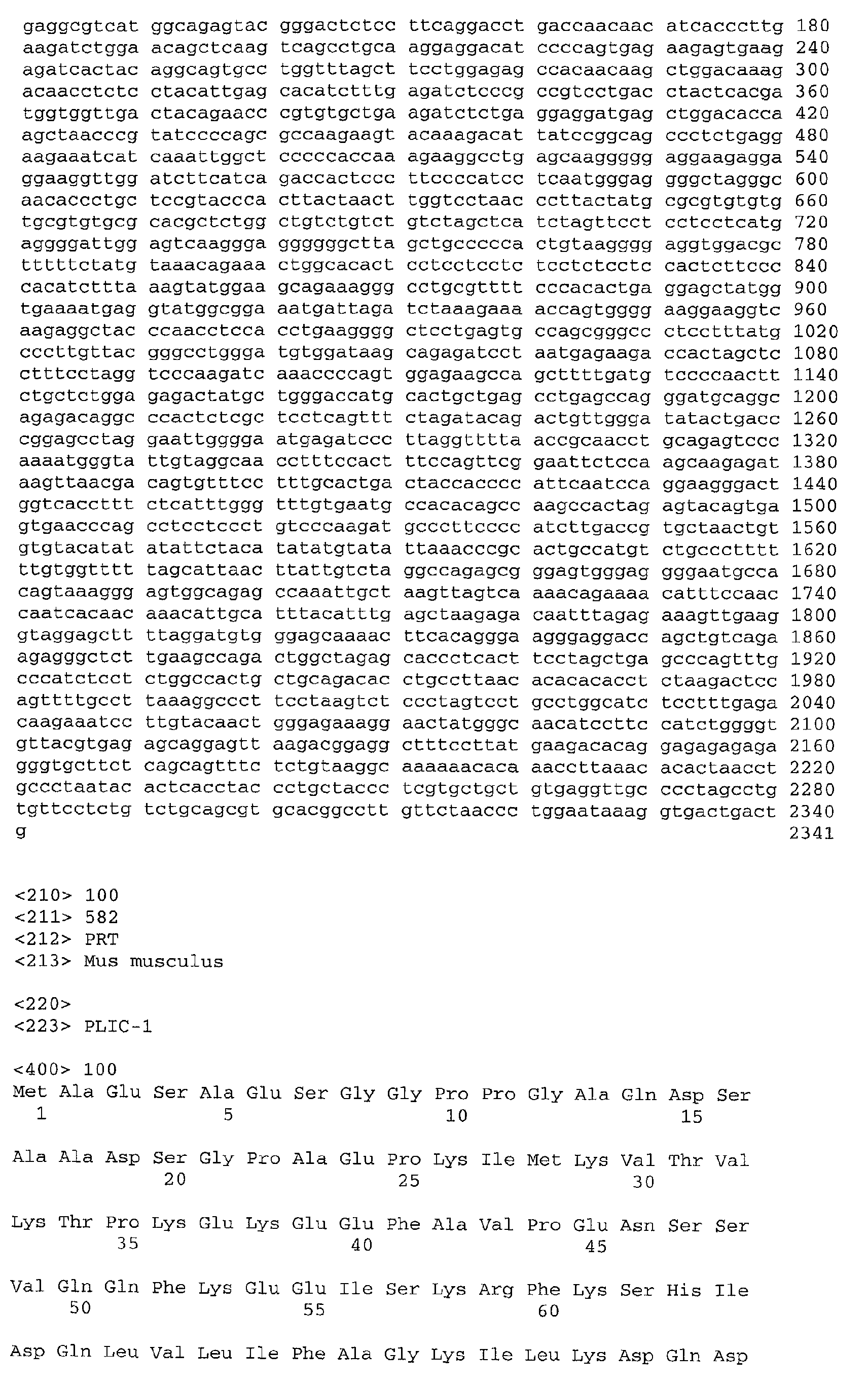


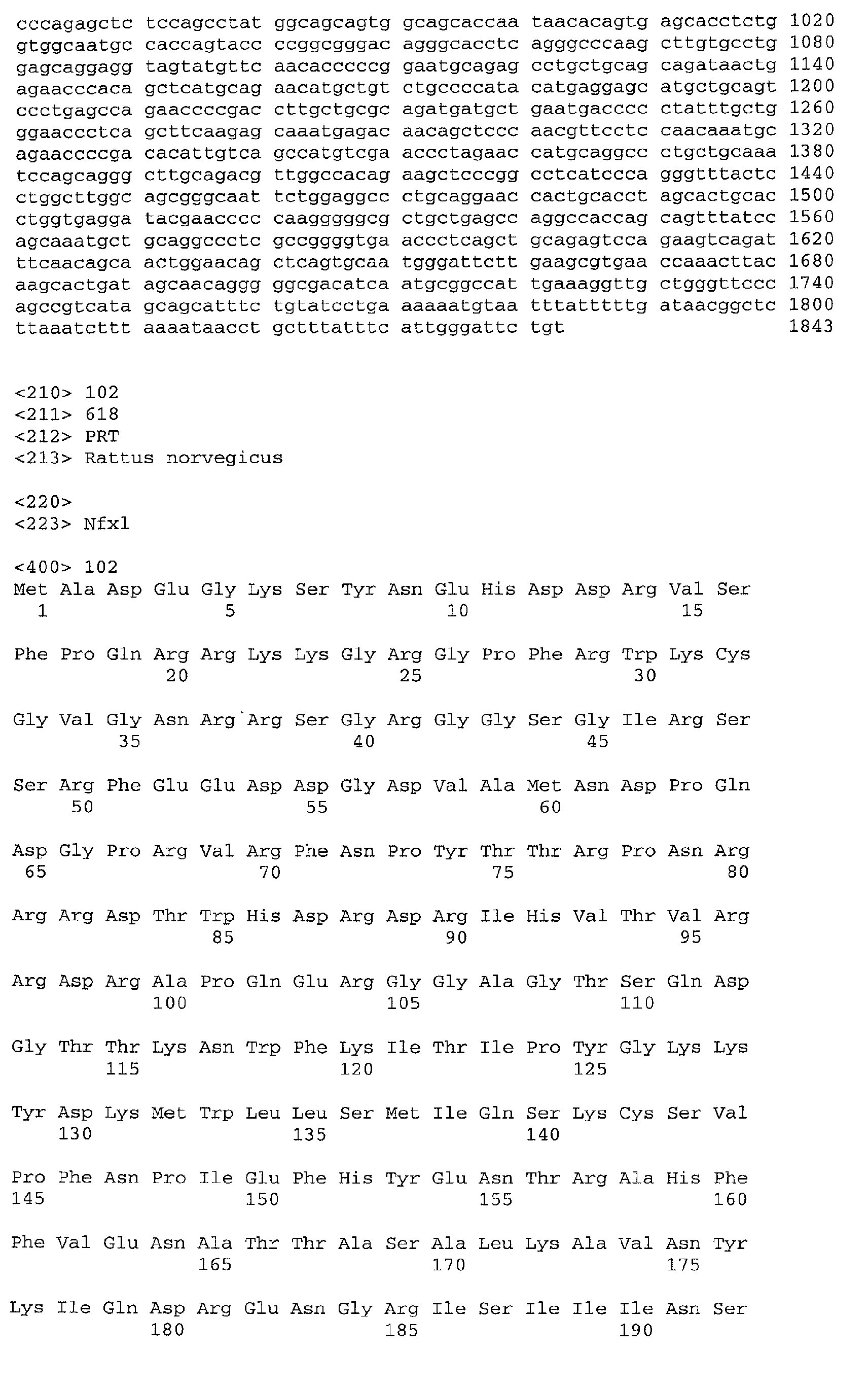
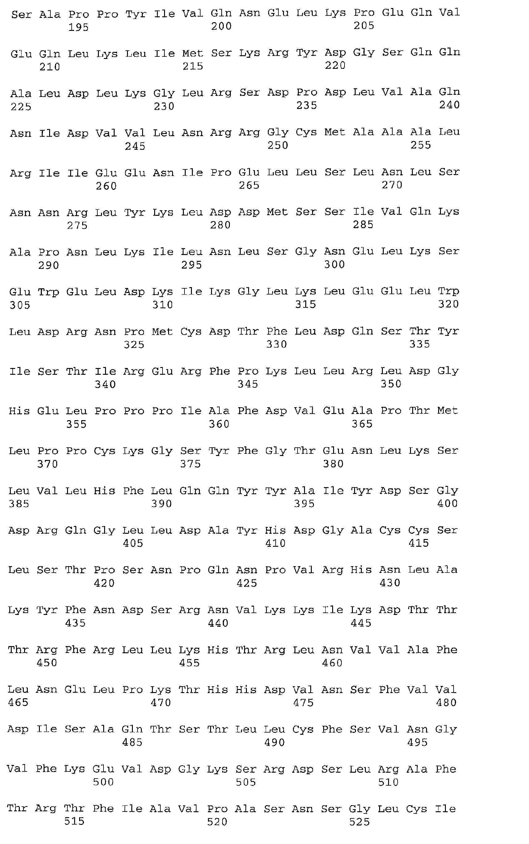
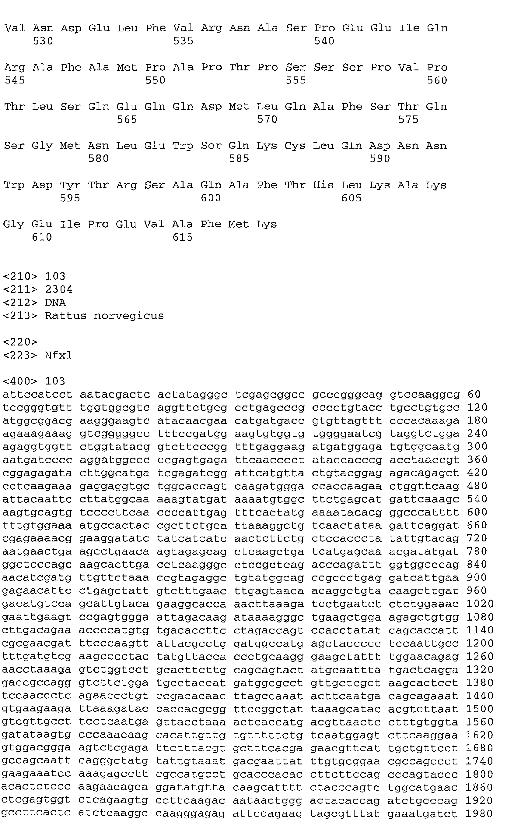

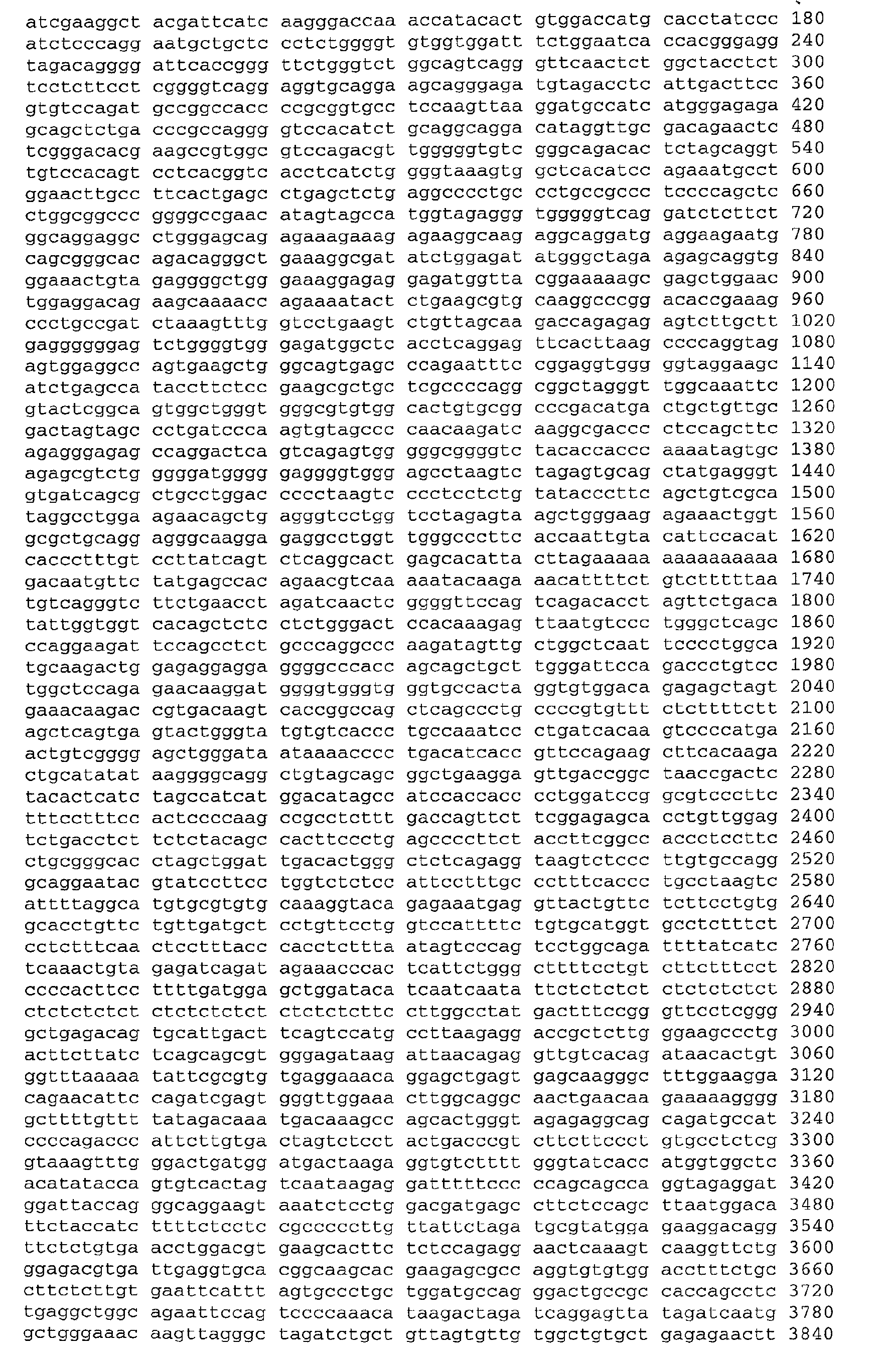


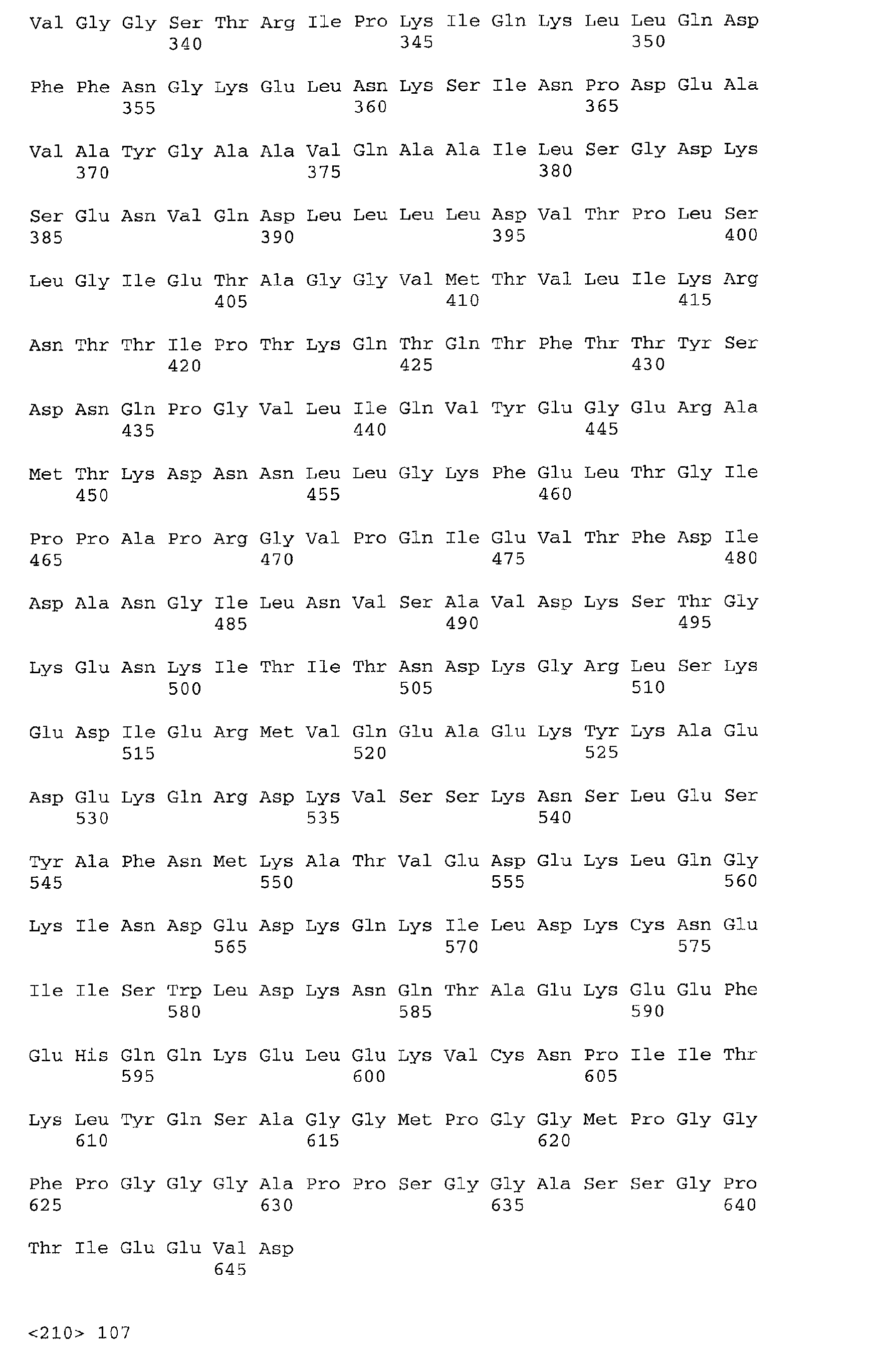
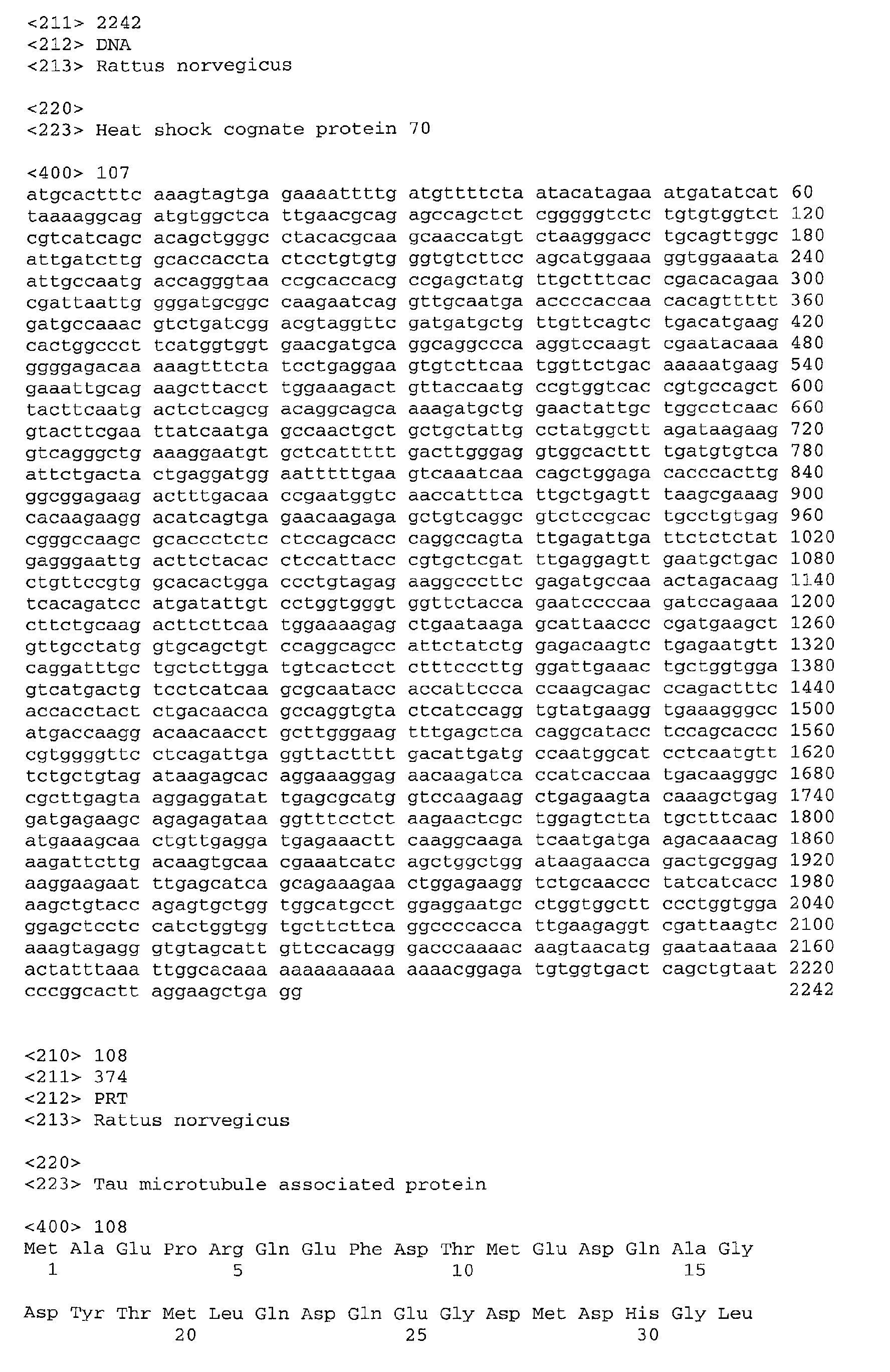
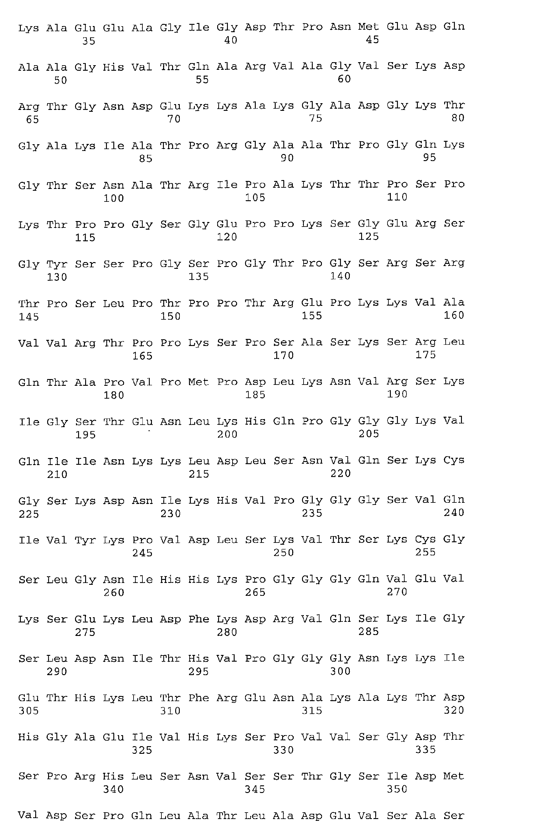
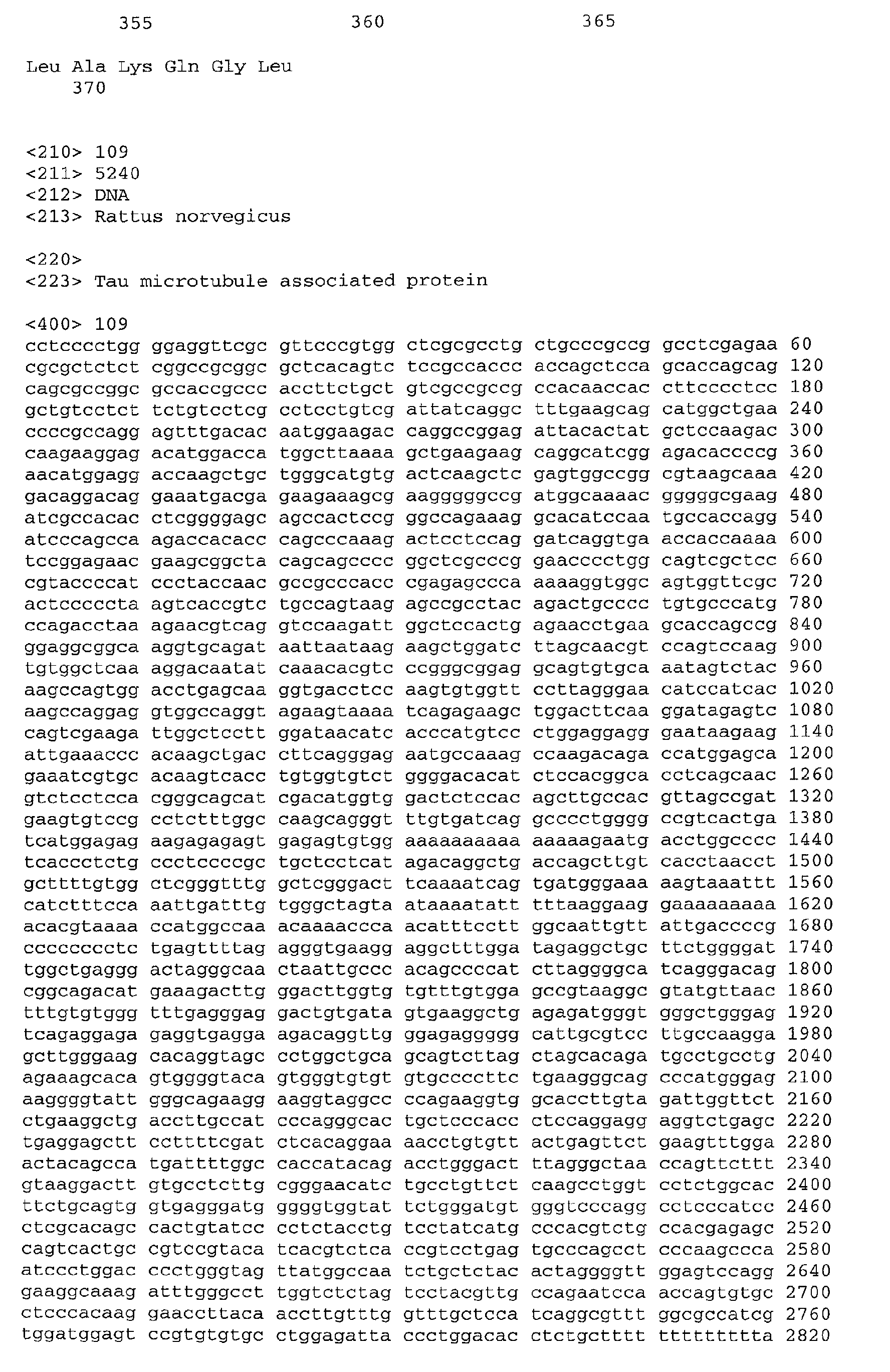





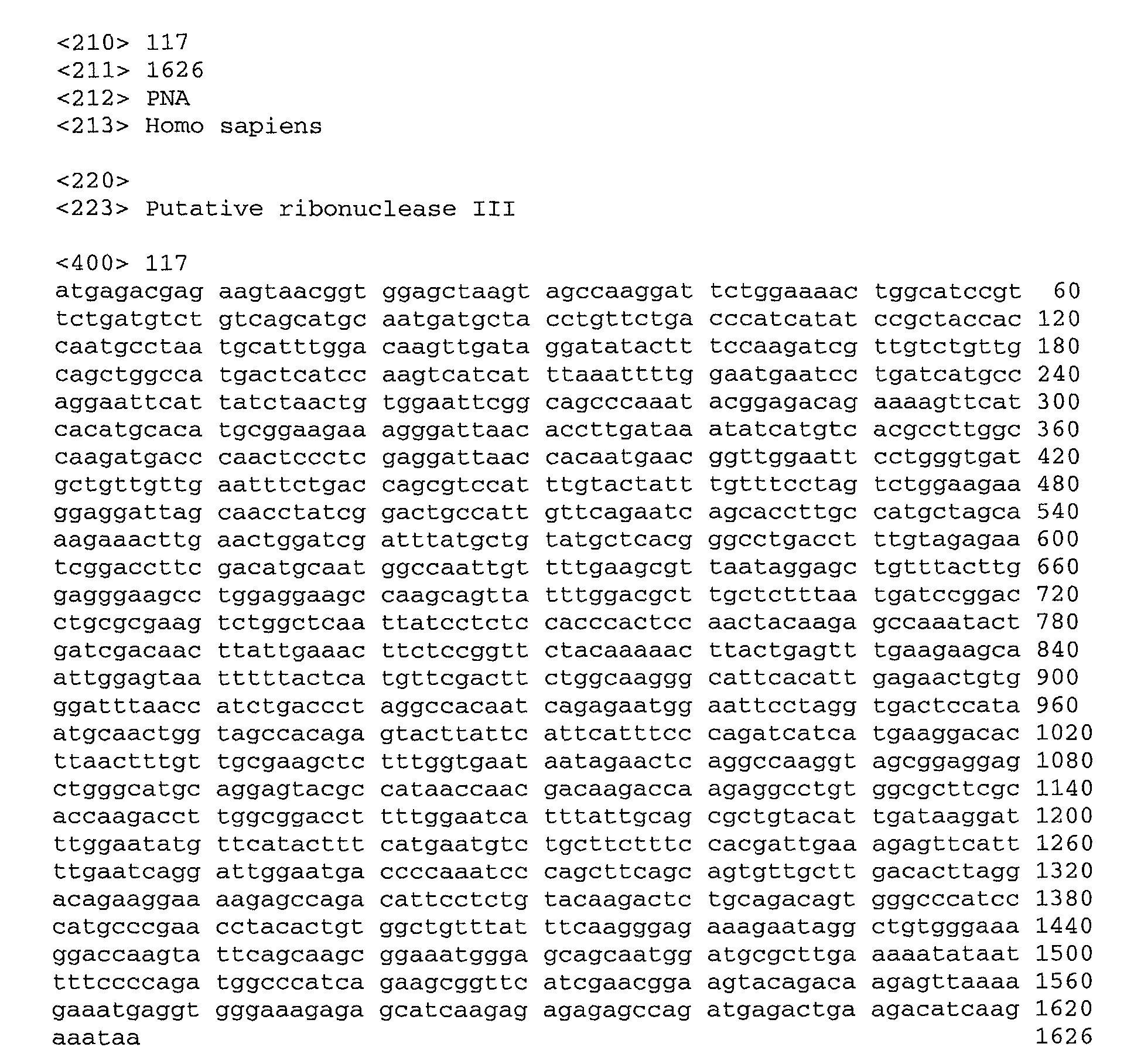
-
Claims (40)
- Use of:in the screening of compounds for the treatment of pain, or for the diagnosis of pain.(a) an isolated gene sequence that is down-regulated in the spinal cord of a mammal in response to mechanistically distinct first and second models of neuropathic or central sensitization pain;(b) an isolated gene sequence comprising a nucleic acid sequence of any of Tables I to VI;(c) an isolated gene sequence having at least 80% sequence identity with a nucleic acid sequence of any of Tables I to VI;(d) an isolated nucleic acid sequence that is hybridizable to any of the gene sequences according to (a), (b) or (c) under stringent hybridisation conditions;(e) a recombinant vector comprising a gene sequence or nucleic acid sequence according to any one of (a) to (d);(f) a host cell containing the vector according to (e);(g) a non-human animal having in its genome an introduced gene sequence or nucleic acid sequence or a removed or down-regulated gene sequence or nucleic acid sequence according to any one of (a) to (d);(h) an isolated polypeptide comprising an amino acid sequence at least 90% identical to an amino acid sequence encoded by a nucleotide sequence according to any one of (a) to (d), or a polypeptide variant thereof with sequential amino acid deletions from the C terminus and/or the N-terminus;(i) an isolated polypeptide encoded by a nucleotide sequence according to any one of (a) to (d); or(j) an isolated antibody that binds specifically to a polypeptide according to (h) or (i);
- Use according to claim 1, wherein the isolated gene sequence is down-regulated both in response to streptozocin-induced diabetes and in response to surgical injury of a nerve leading to the spine.
- Use according to claim 1 or 2 wherein the isolated gene sequence encodes a kinase.
- Use according to claim 1, 2 or 3, wherein the isolated gene sequence encodes an expression product or fragment thereof of pyruvate kinase, M1 and M2 subunits (M24359 ; X97047; X56494).
- Use according to claim 1 or 2, wherein the isolated gene sequence encodes an expression product or fragment thereof of a receptor.
- Use according to claim 1, 2 or 5, wherein the isolated gene sequence encodes dopamine receptor D.sub.1 (I58000).
- Use according to claim 1 or 2, wherein the isolated gene sequence encodes a transporter.
- Use according to claim 1, 2 or 7, wherein the isolated gene sequence encodes differentiation-associated Na+-dependent inorganic phosphate cotransporter (AF271235) or putative vacuolar assembly protein VSP41 gene (U87309).
- Use according to claim 1 or 2, wherein the isolated gene sequence encodes a G-protein coupled receptor protein.
- Use according to claim 1, 2 or 9, wherein the isolated gene sequence encodes Git1 (G-protein-coupled receptor kinase-interactor 1 ; GPCR kinase-associated ADP-ribosylation factor) (AF085693).
- Use according to claim 1 or 2, wherein the isolated gene sequence encodes a DNA-binding protein.
- Use according to claim 1, 2 or 11, wherein the isolated gene sequence encodes putative histone H3.3A (X91866 ; M11354).
- Use according to claim 1 or 2, wherein the isolated gene sequence encodes a ligase.
- Use according to claim 1, 2 or 13, wherein the isolated gene sequence encodes 3-Hydroxy 3-methylglutaryl coenzyme A synthase, cytosolic (X52625), acyl-CoA synthetase II, brain (D360666) famesyl diphosphate synthase (M34477), bendless protein (AB032739; E12457), fatty acid synthase (X62888), glutamine synthetase (EC 6.3.1.2) (M91652), or putative seryl-tRNA synthetase (X91257).
- Use according to claim 1 or 2, wherein the isolated gene sequence encodes a lyase.
- Use according to claim 1, 2 or 15, wherein the lyase is enolase, alpha alpha, non-neuronal (NNE) (X02610; X52379; M14328).
- Use according to claim 1 or 2, wherein the isolated gene sequence encodes an oxidoreductase.
- Use according to claim 1, 2 or 17, wherein the isolated gene sequence encodes aldose reductase, lens (AREC 11.1.21) (X05884) cytochrome-c oxidase I, mitochondrial (S79304), lactate dehydrogenase-B (LDH-B) (U07181; X51905; Y00711), putative cytochrome c oxidase VIB (EC 1.9.3.1) (X13923), putative NADH: ubiquinone oxidoreductase PGIV subunit (AF044953), putative succinate dehydrogenase flavoprotein (AF095938; AF171022), putative ubiquinol―cytochrome-c reductase (EC 1.10.2.2) core protein II (J04973) or stearoyl-coA desaturase 2 (AB032243; M26270).
- Use according to claim 1 or 2, wherein the isolated sequence encodes a transferase.
- Use according to claim 1, 2 or 19, wherein the isolated sequence encodes ribophorin I (X05300) or sulfotransferase-like protein (AF188699).
- Use according to claim 1 or 2, wherein the isolated sequence encodes a hydrolase.
- Use according to claim 1, 2 or 21, wherein the isolated sequence encodes ATP synthase, H+, alpha subunit, mitochondrial (EC 3.6.1.34) (X56133), F1F0 ATPase delta subunit (U00926), putative dihydropyrimidinase related protein (D78013), heat shock protein 90 (S45392; M18186; M16660), or putative ribonuclease III (AF116910).
- Use according to claim 1 or 2, wherein the isolated sequence encodes myelin basic protein S (MBP S) (K00512), transferring (D38380), neurofilament, light molecular weight (NF-L) (AF031880), myelin-associated glycoprotein (MAG) (M16800; M31811), NF-M middle molecular weight neurofilament protein (M18628) neuro-degeneration associated- protein 1 (D32249), S-100 protein β-subunit (X01090), microtubule-associated protein 1b (Map 1b) (X60370; L06237), putative cdc 37 homolog (D26564), putative ras-related protein Rab-5c (U11293), putative gelsolin (J04953), Cd81 antigen (target of antiproliferative antibody 1) (U19894; X59047; M33680), Mobp81 (Myelin-associated/Oligo-dendrocytic basic protein 81) (X87900), syntaxin binding protein n-secl, sec1 homolog, A-internexin (M73049), putative β-sarcoglycan A3b (AB024921), CGI-78 protein (AF151835), KIAA0143 (D63477), septin 2 (D50918), Nucleobindin (Z36277), myelin protein SR13 (M69139; S55427), B-Actin, cytoplasmic (V01217; X03672), ly6/neurotoxin (Lynx1) homolog (AD141377), astrocytic phosphoprotein ; PFA 15 gene (AJ243949; X86694), PLIC-1 (AF177345), Nfx1 (tip associating protein (TAP) gene) (AF093139; AF093140), A-Crystallin B (U04320; M73741 ; M28638), heat shock-like protein 70 kD (X70065; U73744; Y00371), tau microtubule-associated protein (X79321), myelin, Schwann cell, Perioheral (P-0) (K03242 ), B-Tubulin class 1 (AB011679; X04663; AF14139), putative long-chain polyunsaturated fatty acid elongation enzyme (Helo1) or peptidylglycine alpha-amidating monooxygenase 1.
- A non-human animal having in its genome an introduced gene sequence or a removed or down-regulated gene sequence, said sequence being down-regulated in the spinal cord of a mammal in response to first and second models of neuropathic or central sensitisation pain.
- A non-human animal according to claim 24, wherein said gene sequence becomes down regulated both in response to streptozocin induced diabetes and in response to chronic constriction injury.
- A non-human animal according to claim 24 or 25, wherein the introduced gene sequence is according to any of claims 1 to 23.
- A non-human animal according to any one of claims 24 to 26 which is C. elegans.
- A kit comprising;(a) affinity peptide and/or ligand and/or substrate for an expression product of a gene sequence that is down-regulated in the spinal cord of a mammal in response to a mechanistically distinct first and second models of neuropathic or central sensitization pain ; and(b) a defined quantity of an expression product of a gene sequence that is down-regulated in the spinal cord of a mammal both in response to first and second models of neuropathic or central sensitization pain,
for simultaneous, separate or sequential use in detecting and/or quantifying down-regulation of a gene sequence in the spinal cord of a mammal in response to first and second models of neuropathic or central sensitization pain. - A kit according to claim 28, wherein the gene sequence is defined in any one of claims 1 to 23.
- A kit comprising:(a) nucleic acid sequences capable of hybridization to a nucleic acid sequence that is down-regulated in the spinal cord of a mammal in response to first and second models of neuropathic or central sensitization pain; and(b) a defined quantity of one or more nucleic acid sequences capable of hybridization to a nucleic acid sequence that is down-regulated in the spinal cord of a mammal in response to first and second models of neuropathic or central sensitization pain,
for simultaneous, separate or sequential use in detecting and/or quantifying down-regulation of a gene sequence in the spinal cord of a mammal in response to first and second models of neuropathic or central sensitization pain. - The kit of claim 30, wherein the gene sequence is according to any of claims 1 to 23.
- A compound that modulates the action of an expression product of a gene sequence that is down-regulated in the spinal cord of a mammal in response to first and second models of neuropathic or central sensitization pain.
- A compound according to claim 32 wherein the gene sequence is listed in Tables I to VI.
- A compound according to claim 32 or 33 wherein the nucleotide sequence is according to any one of claims 1 to 23.
- A compound according to any one of claims 32 to 34 for use as a medicament.
- A compound according to any one of claims 32 to 35 for the treatment or diagnosis of pain.
- A pharmaceutical composition comprising a compound according to any one of claims 32 to 36 and a pharmaceutically acceptable carrier or diluent.
- Use of a compound according to any one of claims 32 to 36 in the manufacture of a medicament for the treatment or diagnosis of pain.
- Use of a compound according to any one of claims 32 to 36 in the manufacture of a medicament for the treatment or diagnosis of chronic pain.
- A method of treatment of pain, which comprises administering to a patient an effective amount of a compound according to any one of claims 32 to 36.
Applications Claiming Priority (4)
| Application Number | Priority Date | Filing Date | Title |
|---|---|---|---|
| GB0118354 | 2001-07-27 | ||
| GB0118354A GB2377940A (en) | 2001-07-27 | 2001-07-27 | Identification and use of molecules implicated in pain |
| GB0202883 | 2002-02-07 | ||
| GB0202883A GB0202883D0 (en) | 2002-02-07 | 2002-02-07 | Identification and use of molecules implicated in pain |
Publications (2)
| Publication Number | Publication Date |
|---|---|
| EP1284298A2 true EP1284298A2 (en) | 2003-02-19 |
| EP1284298A3 EP1284298A3 (en) | 2004-05-26 |
Family
ID=26246366
Family Applications (1)
| Application Number | Title | Priority Date | Filing Date |
|---|---|---|---|
| EP02255229A Withdrawn EP1284298A3 (en) | 2001-07-27 | 2002-07-26 | Identification and use of molecules implicated in pain |
Country Status (4)
| Country | Link |
|---|---|
| US (1) | US20040058326A1 (en) |
| EP (1) | EP1284298A3 (en) |
| JP (1) | JP2003156488A (en) |
| CA (1) | CA2391112A1 (en) |
Cited By (11)
| Publication number | Priority date | Publication date | Assignee | Title |
|---|---|---|---|---|
| WO2003052070A3 (en) * | 2001-12-13 | 2003-11-20 | Exelixis Inc | Vps41s as modifiers of cell death and methods of use |
| WO2004000882A3 (en) * | 2002-06-24 | 2004-03-11 | Ares Trading Sa | Proteins |
| WO2005089791A1 (en) * | 2004-03-09 | 2005-09-29 | Board Of Regents, The University Of Texas System | Pea15 as a tumor suppressor gene |
| EP1485400A4 (en) * | 2002-02-20 | 2005-11-30 | Isis Pharmaceuticals Inc | HUMANE RNASE III AND COMPOSITIONS AND ITS APPLICATIONS |
| WO2004087875A3 (en) * | 2003-03-25 | 2005-12-01 | Incyte Corp | Nucleic acid-associated proteins |
| RU2404246C1 (en) * | 2009-06-18 | 2010-11-20 | Учреждение Российской академии наук Институт биоорганической химии им. академиков М.М. Шемякина и Ю.А. Овчинникова РАН | RECOMBINANT PLASMID DNA pET22b(+)/"вд"-LINKS 1, CODING PROTEIN WITH LINKS 1 PROPERTIES, AND STRAIN OF Escherichia coli BL21 (DE3)/pET22b(+)/"вд"-LINKS 1 - PRODUCENT OF PROTEIN WITH PROPERTIES OF HUMAM LINKS 1 |
| CN1952662B (en) * | 2005-08-26 | 2011-05-18 | 中国科学院上海生命科学研究院 | Application of star colloid cell phosphorylation protein PEA-15 |
| US20120124684A1 (en) * | 2008-12-01 | 2012-05-17 | San Diego State University (Sdsu) Foundation | Compositions and methods using alpha beta-crystallin in protecting the myocardium from ischemia/reperfusion injury |
| US9023806B2 (en) * | 2004-05-04 | 2015-05-05 | Universite D'angers | Peptide capable of altering tubulin polymerization and use thereof for inhibiting cell proliferation |
| WO2015101493A1 (en) * | 2013-12-31 | 2015-07-09 | Global Bioenergies | 3-hydroxyisovalerate (hiv) synthase variants |
| US10188707B2 (en) | 2014-01-13 | 2019-01-29 | Berg, LLC | Enolase 1 (Eno1) compositions and uses thereof |
Families Citing this family (7)
| Publication number | Priority date | Publication date | Assignee | Title |
|---|---|---|---|---|
| US7169751B2 (en) * | 2003-02-04 | 2007-01-30 | Bayer Healthcare Ag | Method of identifying a compound capable of treating a pain or a painful disorder |
| EP2195032A4 (en) * | 2007-09-11 | 2012-08-01 | Joslin Diabetes Center Inc | Phase ii detoxification and antioxidant activity |
| AU2011248357B2 (en) * | 2010-05-03 | 2017-02-23 | Pangu Biopharma Limited | Innovative discovery of therapeutic, diagnostic, and antibody compositions related to protein fragments of seryl-tRNA synthetases |
| JP6144673B2 (en) | 2011-06-03 | 2017-06-07 | オフィディオン インコーポレイティド | Compositions and methods for transport across the blood brain barrier |
| KR101127756B1 (en) | 2011-09-02 | 2012-03-23 | 한국과학기술원 | Screening of drug for attention deficit hyperactive disorder by using git1 knock-out mice as a novel adhd mouse model |
| CN110845576A (en) * | 2019-09-30 | 2020-02-28 | 浙江树人学院(浙江树人大学) | Turtle protein source has ACE inhibitory active peptide HDTYYVVAH and its use |
| TW202546000A (en) * | 2024-02-09 | 2025-12-01 | 賓夕法尼亞大學董事會 | Augmenting car t cell activity |
Family Cites Families (1)
| Publication number | Priority date | Publication date | Assignee | Title |
|---|---|---|---|---|
| PL351432A1 (en) * | 1999-04-15 | 2003-04-22 | Devgen Nv | Compound screening method |
-
2002
- 2002-07-24 US US10/205,331 patent/US20040058326A1/en not_active Abandoned
- 2002-07-25 CA CA002391112A patent/CA2391112A1/en not_active Abandoned
- 2002-07-26 EP EP02255229A patent/EP1284298A3/en not_active Withdrawn
- 2002-07-29 JP JP2002219631A patent/JP2003156488A/en active Pending
Non-Patent Citations (1)
| Title |
|---|
| None * |
Cited By (15)
| Publication number | Priority date | Publication date | Assignee | Title |
|---|---|---|---|---|
| US7491524B2 (en) | 2001-07-06 | 2009-02-17 | Isis Pharmaceuticals, Inc. | Human RNase III and compositions and uses thereof |
| WO2003052070A3 (en) * | 2001-12-13 | 2003-11-20 | Exelixis Inc | Vps41s as modifiers of cell death and methods of use |
| EP1485400A4 (en) * | 2002-02-20 | 2005-11-30 | Isis Pharmaceuticals Inc | HUMANE RNASE III AND COMPOSITIONS AND ITS APPLICATIONS |
| WO2004000882A3 (en) * | 2002-06-24 | 2004-03-11 | Ares Trading Sa | Proteins |
| WO2004087875A3 (en) * | 2003-03-25 | 2005-12-01 | Incyte Corp | Nucleic acid-associated proteins |
| WO2005089791A1 (en) * | 2004-03-09 | 2005-09-29 | Board Of Regents, The University Of Texas System | Pea15 as a tumor suppressor gene |
| US9023806B2 (en) * | 2004-05-04 | 2015-05-05 | Universite D'angers | Peptide capable of altering tubulin polymerization and use thereof for inhibiting cell proliferation |
| CN1952662B (en) * | 2005-08-26 | 2011-05-18 | 中国科学院上海生命科学研究院 | Application of star colloid cell phosphorylation protein PEA-15 |
| US20120124684A1 (en) * | 2008-12-01 | 2012-05-17 | San Diego State University (Sdsu) Foundation | Compositions and methods using alpha beta-crystallin in protecting the myocardium from ischemia/reperfusion injury |
| RU2404246C1 (en) * | 2009-06-18 | 2010-11-20 | Учреждение Российской академии наук Институт биоорганической химии им. академиков М.М. Шемякина и Ю.А. Овчинникова РАН | RECOMBINANT PLASMID DNA pET22b(+)/"вд"-LINKS 1, CODING PROTEIN WITH LINKS 1 PROPERTIES, AND STRAIN OF Escherichia coli BL21 (DE3)/pET22b(+)/"вд"-LINKS 1 - PRODUCENT OF PROTEIN WITH PROPERTIES OF HUMAM LINKS 1 |
| WO2015101493A1 (en) * | 2013-12-31 | 2015-07-09 | Global Bioenergies | 3-hydroxyisovalerate (hiv) synthase variants |
| US10364418B2 (en) | 2013-12-31 | 2019-07-30 | Global Bioenergies | 3-hydroxyisovalerate (HIV) synthase variants |
| US10188707B2 (en) | 2014-01-13 | 2019-01-29 | Berg, LLC | Enolase 1 (Eno1) compositions and uses thereof |
| US10188708B2 (en) | 2014-01-13 | 2019-01-29 | Berg Llc | Enolase 1 (Eno1) compositions and uses thereof |
| US11224641B2 (en) | 2014-01-13 | 2022-01-18 | Berg Llc | Enolase 1 (ENO1) compositions and uses thereof |
Also Published As
| Publication number | Publication date |
|---|---|
| CA2391112A1 (en) | 2003-01-27 |
| EP1284298A3 (en) | 2004-05-26 |
| JP2003156488A (en) | 2003-05-30 |
| US20040058326A1 (en) | 2004-03-25 |
Similar Documents
| Publication | Publication Date | Title |
|---|---|---|
| Ma et al. | A novel cytoplasmic protein that interacts with the Ah receptor, contains tetratricopeptide repeat motifs, and augments the transcriptional response to 2, 3, 7, 8-tetrachlorodibenzo-p-dioxin | |
| Yang et al. | Identification of a novel family of cell-surface proteins expressed in human vascular endothelium | |
| Muir et al. | AXOR12, a novel human G protein-coupled receptor, activated by the peptide KiSS-1 | |
| CA2391112A1 (en) | Identification and use of molecules implicated in pain | |
| Keller et al. | The γ2 subunit of GABAA receptors is a substrate for palmitoylation by GODZ | |
| EP1270724A2 (en) | Guanosine triphosphate-binding protein coupled receptors | |
| Uji et al. | Molecules interacting with PRIP-2, a novel Ins (1, 4, 5) P3 binding protein type 2: comparison with PRIP-1 | |
| US20030134301A1 (en) | Identification and use of molecules implicated in pain | |
| US20030138803A1 (en) | Identification and use of molecules implicated in pain | |
| US5710018A (en) | Mammalian influx peptide transporter | |
| US6075123A (en) | Cyclin-C variants, and diagnostic and therapeutic uses thereof | |
| US20070009916A1 (en) | Guanosine triphosphate-binding protein coupled receptors | |
| He et al. | Molecular cloning and characterization of CT120, a novel membrane-associated gene involved in amino acid transport and glutathione metabolism | |
| EP1281775A2 (en) | Identification and use of molecules implicated in pain | |
| US7078188B2 (en) | MUC17 encoding nucleic acid sequences, polypeptides, antibodies and methods of use thereof | |
| AU781670B2 (en) | Rheumatoid arthritis gene and method for diagnosing rheumatoid arthritis | |
| GB2377940A (en) | Identification and use of molecules implicated in pain | |
| EP1854881B1 (en) | METHODS FOR IDENTIFYING PURKINJE CELLS USING THE Corl2 GENE AS A TARGET | |
| US6639064B1 (en) | NRIF3, novel co-activator for nuclear hormone receptors | |
| WO2001049714A2 (en) | Nope polypeptides, encoding nucleic acids and methods of use | |
| US6673898B1 (en) | Placental organic anion transporter | |
| JP2005525079A (en) | ALS2 gene and amyotrophic lateral sclerosis type 2 | |
| JP2004242644A (en) | Guanosine triphosphate binding protein coupled receptor | |
| GB2383044A (en) | ALMS1 gene | |
| WO2005100566A1 (en) | Novel monkey gpr103 and monkey qrfp and method of evaluating compound by using gpr103 |
Legal Events
| Date | Code | Title | Description |
|---|---|---|---|
| PUAI | Public reference made under article 153(3) epc to a published international application that has entered the european phase |
Free format text: ORIGINAL CODE: 0009012 |
|
| 17P | Request for examination filed |
Effective date: 20020808 |
|
| AK | Designated contracting states |
Designated state(s): AT BE BG CH CY CZ DE DK EE ES FI FR GB GR IE IT LI LU MC NL PT SE SK TR |
|
| AX | Request for extension of the european patent |
Extension state: AL LT LV MK RO SI |
|
| RIC1 | Information provided on ipc code assigned before grant |
Ipc: 7C 12N 15/85 B Ipc: 7C 12N 5/10 B Ipc: 7C 07K 14/47 B Ipc: 7C 12Q 1/68 A |
|
| RAP1 | Party data changed (applicant data changed or rights of an application transferred) |
Owner name: WARNER-LAMBERT COMPANY LLC |
|
| PUAL | Search report despatched |
Free format text: ORIGINAL CODE: 0009013 |
|
| AK | Designated contracting states |
Kind code of ref document: A3 Designated state(s): AT BE BG CH CY CZ DE DK EE ES FI FR GB GR IE IT LI LU MC NL PT SE SK TR |
|
| AX | Request for extension of the european patent |
Extension state: AL LT LV MK RO SI |
|
| 17Q | First examination report despatched |
Effective date: 20041209 |
|
| AKX | Designation fees paid |
Designated state(s): AT BE BG CH CY CZ DE DK EE ES FI FR GB GR IE IT LI LU MC NL PT SE SK TR |
|
| 17Q | First examination report despatched |
Effective date: 20041209 |
|
| RAP1 | Party data changed (applicant data changed or rights of an application transferred) |
Owner name: WARNER-LAMBERT COMPANY LLC |
|
| STAA | Information on the status of an ep patent application or granted ep patent |
Free format text: STATUS: THE APPLICATION IS DEEMED TO BE WITHDRAWN |
|
| 18D | Application deemed to be withdrawn |
Effective date: 20070201 |